Crimean-Congo hemorrhagic fever: experience at a tertiary care hospital in Karachi, Pakistan
Upload
independentCategory
view
0download
0
Alam et al. BMC Infectious Diseases 2013, 13:201http://www.biomedcentral.com/1471-2334/13/201
RESEARCH ARTICLE Open Access
Genetic analysis and epidemiology of CrimeanCongo hemorrhagic fever viruses in Baluchistanprovince of PakistanMuhammad Masroor Alam, Adnan Khurshid, Salmaan Sharif, Shahzad Shaukat, Muhammad Suleman Rana,Mehar Angez and Syed Sohail Zahoor Zaidi*
Abstract
Background: Pakistan is considered as an endemic country for Crimean-Congo Hemorrhagic fever with numerousoutbreaks and sporadic cases reported during the past two decades. Majority of cases are reported from Baluchistanprovince with subsequent transmissions to non-endemic regions mainly through infected animals directly or viainfested ticks. We hereby describe the molecular investigations of CCHF cases reported during 2008 in Quetta city ofBaluchistan province.
Methods: Serum Samples from 44 patients, with clinical signs of hemorrhagic fever attending a tertiary care hospital inQuetta city, were collected and tested for CCHF virus antigen and genomic RNA, using capture IgM EIA kit andstandard RT-PCR assay, respectively. The partial S-gene fragments were directly sequenced to get information related tothe prevailing CCHFV genotypes and their molecular epidemiology in Pakistan.
Results: Out of the total forty four, sixteen (36%) samples were found positive for CCHF IgM. Similarly, viral RNA wasdetected in six (16%) samples. Phylogenetic analysis revealed that all study viruses belong to genotype Asia-1 withclosest similarity (99-100%) to the previously reported strains from Pakistan, Afghanistan and Iran.
Conclusion: We conclude that CCHF virus remains endemic within Baluchistan and its neighboring regions ofAfghanistan warranting a need of incessant surveillance activities.
Keywords: Crimean Congo hemorrhagic fever, Pakistan, CCHFV Asia type-1 genotype, Baluchistan,Molecular epidemiology
BackgroundCrimean Congo Haemorrhagic Fever (CCHF) virus be-longs to genus Nairovirus within the family Bunyaviridaewith a triple-segmented RNA genome. The Large or Lsegment encodes non-structural proteins which comprisethe RNA dependent RNA polymerase, and the medium(M) and small (S) segments encode structural proteins forthe surface glycoproteins and nucleocapsid respectively[1]. Although the disease was originally notified as febrileillness in the Western Crimea during 1940s, the etiologicalviral agent was not identified and reported until muchlater when a similar disease was reported from Congo in1956, leading to its name as Crimean-Congo haemorrhagic
* Correspondence: [email protected] of Virology, National Institute of Health, Chak Shahzad, ParkRoad, Islamabad 44000, Pakistan
© 2013 Alam et al.; licensee BioMed Central LCommons Attribution License (http://creativecreproduction in any medium, provided the or
fever in 1970s [2]. The family Bunyaviridae contains 5 gen-era including Orthobunyavirus, Hantavirus, Phlebovirus,Nairovirus and Tospovirus with more than 300 species [3].The genus Nairovirus currently contains 35 distinct vi-ruses of which only three are known to cause disease;CCHF, Dugbe and Nairobi sheep disease virus [4]. Allmembers of the genus Nairovirus are believed to be trans-mitted mainly through hard ticks especially species of theHyalomma, Rhipicephalus and Dermacenter [4]. CCHFvirus (CCHFV) only causes disease in humans and the in-cubation period ranges from 2–9 days depending uponthe route of exposure and viral inoculums [5]; 3.2 daysafter tick bite, 5 days after contact with infected livestockblood or tissue and 5.6 days after contact with infected hu-man blood [6]. Several important factors are thought tocontribute towards CCHF disease spread such as trade
td. This is an Open Access article distributed under the terms of the Creativeommons.org/licenses/by/2.0), which permits unrestricted use, distribution, andiginal work is properly cited.

Alam et al. BMC Infectious Diseases 2013, 13:201 Page 2 of 8http://www.biomedcentral.com/1471-2334/13/201
and exchange in livestock [4,7], transmission via migra-tory birds infested with infected ticks [8] and subsequentinfection of new hosts [4,9]. When infected ticks are in-troduced to new ecological zones, differences in tickfeeding preferences and vertebrate host availability alsocontribute to new transmission cycles [10]. Transmissionin nosocomial settings also contributes to the spread ofCCHFV [11].Previous reports have documented a wide geographical
distribution of CCHF virus ranging from Eastern Asia toAfrica and Europe with 15-60% mortality rates [3]. Thefirst CCHF case in Pakistan was reported in 1976 from a
Figure 1 Geographical map of Pakistan showing districts with IgM coreported to National Institute of Health, Islamabad, Pakistan. Districts(high disease burden districts) and grey color (low disease burden non-end(n = 57) found during 6 years (2003–2008) belong to Baluchistan province, folThe least number of cases from Sindh province indicates low prevalence of Cof Sindh province, also contains the diagnostic facilities so the suspected case
patient who underwent laparotomy at Rawalpindi GeneralHospital (13). Since 1976 to 2003, 14 epidemics, includ-ing 8 with nosocomial origin, have been reported in thecountry, mainly in the Western and North Westernareas [11] (Figure 1). In Baluchistan province, the firstsporadic case was documented in 1978, whereas re-corded outbreaks have occurred yearly since 1987 andthen in 1994, 1995, 1998, 2000 and 2001 [12,13]. Thus ithas been now known that CCHF virus circulates locallyin Baluchistan [14] that has been declared as the diseaseendemic area in Pakistan [12]. Confirmed CCHF caseshave also been reported from patients residing in
nfirmed CCHF cases found during the years 2003 to 2008with proportion of case burden have been indicated through blackemic districts with sporadic cases). Majority of IgM confirmed caseslowed by Khyber Pakhtunkhwa (n = 20); Punjab (n = 6); Sindh (n = 02).CHF infection in this province as well as the fact that Karachi, the capitals are not referred to National Institute of Health, Islamabad.

Alam et al. BMC Infectious Diseases 2013, 13:201 Page 3 of 8http://www.biomedcentral.com/1471-2334/13/201
Afghanistan as they usually seek treatments in hospitalsof Quetta city, the capital of Baluchistan which bordersAfghanistan.The National Institute of Health, Islamabad serves as
the country’s focal laboratory center receiving thousandsof different diagnostic samples including those fromCCHF suspected cases across the country. Until recently,the diagnosis of CCHF had been conducted throughserological testing using the CCHF Capture IgM ELISA;however, very recently, the molecular diagnostic ser-vices have been initiated at the institute’s Departmentof Virology. We hereby describe the application of suchimproved diagnostic facilities for the molecular detectionand genetic diversity of CCHF viruses detected in six pa-tients admitted to Fatima Jinnah General and Chest(FJG&C) Hospital, Quetta.
MethodsThe concept and design of the study was approvedthrough the Pakistan’s National Institute of Health In-ternal Review Board. This work was conducted on ar-chived samples received during 2008 that were stored at−80°C. Previously, the results of these samples werereported on the basis of IgM detection and only positivesamples were kept stored. We processed all such samplesfor the molecular detection of viral RNA and furthercharacterization. A total of forty four serum samples fromsuspected patients were received from FJG&C hospital,Quetta during September to October 2008 and were avail-able for this study with the demographic details of patients
Table 1 Demographic details of CCHF confirmed patients admi
S. No. Country of origin Signs and symptoms
1 Pakistan Fever, Headache and Nose bleeding
2 Pakistan Fever and Nose bleeding
3 Afghanistan Fever and Nose bleeding
4 Pakistan Fever and Nose bleeding
5 Afghanistan Fever and Nose bleeding
6 Pakistan Fever, Headache and Nose bleeding
7 Pakistan Fever and Nose bleeding
8 Pakistan Fever and Nose bleeding
9 Afghanistan Fever and Nose bleeding
10 Pakistan Fever, Headache and Nose bleeding
11 Afghanistan Fever, Headache and Nose bleeding
12 Pakistan Fever, Headache and Nose bleeding
13 Pakistan Fever and Nose bleeding
14 Pakistan Fever and Nose bleeding
15 Pakistan Fever and Nose bleeding
16 Pakistan Fever and Nose bleeding
Area of origin of patients where he/she encountered the disease has been given.Unfortunately, the information of disease outcome was not recorded. RT-PCR = Revvirus RNA.
as given in Table 1. The patients were admitted to the hos-pital as they fulfilled the case definition criteria for CCHF.Standard case definitions were practiced as laid by theWorld Health Organization. A “suspected” case is definedas any patient with sudden onset of illness with high-gradefever over 38.5°C for more than 72 hours and less than 10days, especially in CCHF endemic areas and among thosein contact with sheep or other livestock. A “probable” caseis defined when any patient with acute history of febrile ill-ness for 10 days or less accompanied with thrombo-cytopenia less than 50,000/mm3 and presence of any twoof the signs like petechial or purpuric rash, epistaxis,haematemesis, haemoptysis, blood in stools, ecchymosis,gum bleeding, or other hemorrhagic symptoms, however,diagnosis of a “confirmed” case is made if any probablecase with positive diagnosis of CCHF markers in bloodsample such as presence of IgG or IgM antibodies inserum and/or detection of viral nucleic acid in specimen.The formal consent was obtained from the subjects on astandard patient clinical records chart, however, the indi-vidual identity was kept anonymous while dispatched forthe laboratory procedures.All of the sixteen IgM positive samples were processed
for detection of viral RNA through RT-PCR. RNA wasextracted using QIAamp viral RNA minikit (QiagenGmbH, Germany) as per standard instructions. CCHFVRNA was detected by the specific amplification of a frag-ment of the viral S-segment using the protocol of Tinoet al., 1996 [15]. Briefly, One step RT-PCR reaction wasperformed with 1X PCR buffer, 0.4 pmol/ul of each
tted at Fatima Jinnah General and Chest Hospital, Quetta
Platelet count/ul of blood IgM ELISA RT-PCR
30000 Positive Negative
27000 Positive Negative
36000 Positive Negative
70000 Positive Positive
23000 Positive Negative
28000 Positive Positive
31000 Positive Negative
35000 Positive Negative
29000 Positive Negative
60000 Positive Positive
73000 Positive Negative
34000 Positive Negative
16000 Positive Positive
74000 Positive Negative
63000 Positive Positive
47000 Positive Positive
erse Transcriptase-PCR results based on S-gene primers used to detect CCHF

Alam et al. BMC Infectious Diseases 2013, 13:201 Page 4 of 8http://www.biomedcentral.com/1471-2334/13/201
primer (F2 and R3), 0.2 mM of each dNTPs, 21 units ofAMV-reverse transcriptase enzyme, 20 units of RNaseinhibitor with 5 units of Taq DNA polymerase. The re-action tubes containing 100ul reaction mix weresubjected to 42°C for 45 minutes followed by 40 cyclesof 94°C for 40 seconds, 38°C for 40 seconds and 72°Cfor 90 minutes. The second round was performed withthe same reagent concentrations using the primers F3and R2 but without reverse transcriptase and RNAaseinhibitor enzymes and the annealing temperature waschanged to 41°C. Amplified PCR products were then dir-ectly sequenced on ABI automated Genetic analyzer usingBig dye terminator cycle sequencing reaction kit v3.1.CCHFV sequences obtained in this fashion were analyzedwith Sequencher software (version 4.9; GeneCodes Cor-poration, USA) and contigs were constructed from con-sensus sequences. Multiple sequence alignments wereperformed with the ClustalW in MEGA 4.0 software.Alignments were generated with default parameters andadjusted manually. Distance matrices and phylogeneticanalyses were conducted using MEGA 4.0. The dendo-grams were drawn by Neighbor Joining method usingKimura 2-parameter model for estimating nucleotide se-quence distances. The partial S-gene sequences obtainedin this study have been submitted to the GenBank underaccession numbers KC869988 to KC869993.
ResultsThe forty four serum samples received from FJG&C hos-pital Quetta were all tested for CCHF IgM. Sixteen sam-ples (36%) were found positive for IgM (specific toCCHFV antigen) whereas viral RNA was detected in six(16%) samples. The mean age of IgM positive patientswas 30 years (range; 12–65) including 12 males and 4 fe-males. There was no significant difference found be-tween age and gender in relation to CCHF IgM positiveresults (P > 0.005).The partial S-gene segments amplified by RT-PCR
were sequenced to determine the genetic diversity ofCCHFV strains and their relationship with the alreadyreported viruses from Pakistan and other regions. 220bp obtained from the six sequences were found to be-long to Asia-1 genotype with closest match (99-100%) toCCHF viruses reported previously from Pakistan,Afghanistan and Iran in contrast to Iraq and MadagascarAsia-type 1 viruses (Figure 2).The phylogenetic reconstructions distinguished the
Middle East Asian viruses into two distinct sub-cladeswithin Asia-1 genotype. Subclade “A” represents virusesfrom Pakistan, Iran, Afghanistan and China whilesubclade “B” comprises viruses from Iraq, Madagascarand United Arab Emirates (UAE). All of the CCHFVstrains isolated from Pakistan fall under Clade-A having
99-100% similarity with viruses from Afghanistan, Iranand China.The pairwise nucleotide distances based on the num-
ber of substitutions per site were calculated usingMEGA 4.0. The CCHF virus strains collected in Pakistanduring 2008 had 98-100% similarity among each otherand to strains reported from Afghanistan (HM452305),China (U88414) and Iran (AY366373-79). These virusesshowed a higher level of divergence (5-16%) to the Asia-1Clade-B viruses.
DiscussionThis study describes the epidemiological and genetic in-vestigation of CCHF viruses detected in serum samplesof clinically suspected and subsequently laboratory con-firmed patients, received during September to October,2008. The study subjects include patients from Pakistanand Afghanistan that were admitted to Fatima JinnahGeneral and Chest Hospital, Quetta.In Pakistan, CCHFV is endemic to many areas of
Baluchistan that harbor ticks and appropriate vertebratehosts which remain in close contact with humans. Geo-graphically, Baluchistan is the largest province of Pakistanand occupies area of 348,189 km2 with hyper-arid climate.Livestock farming is the main work practice and source ofincome for majority of the population. 93% of the provin-cial land area comprises rangelands that cannot sustainvegetation throughout the year, leading to seasonal migra-tions of nomadic populations to areas within the Baluchi-stan province and bordering areas of Afghanistan alongwith their livestock in search of food and water for them-selves as well as their cattle and sheep flocks. These ani-mals may serve as disease carrier or carry CCHFVinfected ticks and become a major source of transmissionacross their migratory places.Although the CCHF infection has been reported in
Pakistan since 1976, the available data on CCHF casesduring the past few years is very limited. It is most likelythat many of cases have not been reported due to lack ofproper surveillance system in Pakistan. This concern hasbeen raised by previous studies [12,16] highlighting anunremitting need of surveillance, laboratory and medicalfacilities, especially in the CCHF endemic areas. This in-sufficiency in health services has led to alternative diag-nostic measures based on clinical presentations anddisease manifestations in patients. Although clinical diag-nosis is useful, the high mortality of CCHF and propensityfor nosocomial transmission warrant rapid laboratory con-firmation to control and minimize epidemics.Hyalomma tick species are considered as the main
transmission vector in European as well as Asian andAfrican countries especially H. marginatum marginatumand H. analiticum analiticum [8] however, there havebeen reports of other tick species found positive for

Figure 2 Phylogenetic analysis and reconstruction of genetic tree based on partial S-gene segment generated through Kimura-2parameter using the Neighbor Joining model. Horizontal branch lengths represent number of nucleotide differences between taxa (individualvirus sequence). Number at the nodes indicates bootstrap values shown above 50 to demonstrate the robustness of grouping using 1000 datasetsreplicas. Viruses from 7 genogroups (Asia-1, Asia-2, Euro-1, Euro-2, Africa-1, Africa-2 and Africa-3) are represented. Asia 1 genogroup constitutes virusespreviously reported from Pakistan, Afghanistan, Iran, UAE, Madagascar and Iraq. The dark squares represent viruses from this study while open circlesindicate viruses detected from Pakistan in previous years.
Alam et al. BMC Infectious Diseases 2013, 13:201 Page 5 of 8http://www.biomedcentral.com/1471-2334/13/201

Alam et al. BMC Infectious Diseases 2013, 13:201 Page 6 of 8http://www.biomedcentral.com/1471-2334/13/201
CCHF viruses like Ornithodoros lahorensis, Rhipicephalussanguineus, Rhipicephalus bursa, and dermacenter species[17]. For instance, CCHF virus was isolated fromBoophilus ticks in Madagascar, while a Greek isolate of thevirus was recovered from Rhipicephalus [18]. In admon-ition to the role of ticks, there have been findings thatCCHF infections usually occur in geographies with hot cli-mate, where land farming is common. Many cases arereported from areas with increased animal farming whereagricultural workers regularly come into contact with live-stock [4]. It is likely that in endemic areas, infected live-stock that notably remain asymptomatic may serve asvirus reservoirs. In Kosovo, 32.6% while in AzerbaijanShargi province of Iran, 25-80% of the sheep populationwas found seropositive during 1975–1999 survey forCCHF respectively [19]. This necessitates the importanceof generating information on the prevailing tick vectors inour country and their disease association to CCHF infec-tions. Similarly, most of the cases in Turkey during 2005were either the result of tick-bite or patients who had viralexposure through livestock handling [20]. To date sero-logical surveys of this nature have not been possible inPakistan, although CCHF cases reported in Karachi citywere linked to farming practices with sheep and goatsbrought from Baluchistan to Sindh province [21]. Simi-larly, large scale epidemiological surveys are required tostudy tick fauna in the context of CCHF infection. Thiswill be important in defining hotspots of disease andconstructing risk factors associated with particular regions.Such information will also help to define the range of tickspecies capable of hosting and transmitting CCHFV tohumans, ultimately building evidence for vector and dis-ease control programs that can directly support publichealth campaigns for populations who live in endemicareas.Genetic sequencing of CCHFV genome provides knowl-
edge of virus lineages which is a useful molecular epi-demiological information and helps to understand riskareas and the transmission patterns of viruses [22], inaddition to the long range relationships between outbreaksof disease. The genetic information of CCHF virusesreported from Pakistan is very limited due to lack of la-boratory facilitates, however, the information available todate represents data of samples collected intermittentlyduring 1976, 2000, 2001 and 2002 which were sent tointernational laboratories i.e. South Africa (National Insti-tute for Communicable Diseases, Sandringham) and US-Centers for Disease Control and Prevention. Geneticcharacterization of all such viruses revealed that they arephylogeneticaly clustered with the viruses from neighbor-ing Middle East Asian countries such as Iran, Afghanistanand UAE when their partial S-gene segments are com-pared [23]. Beyond the year 2002, no genetic informationof CCHF viruses have been available despite the increased
number of cases every year [16]. Our findings presentedhere further substantiate the existence of Asia-1 genotypein Pakistan explaining its vast endemicity in this region.The recent report of a CCHF virus (strain; AFG09-
2990) from Afghanistan showing 100% S-segment iden-tity with the Pakistani strains [24] provides support forthe close transmission links between these neighboringcountries. It has been reported that Iranian strains alsohold close similarity to Pak-Matin strain (Accession No.U75678) in Pakistan, indicating shared transmissionsources between the two countries [17]. Likewise, an-other study analyzed the random interchange of CCHFVsequences between countries and found that Asia-1genotype moves freely between Pakistan and Iran [14].Tahmasebi et al. support such assumptions by finding thatall CCHF viruses from Hamadan province of Iran clus-tered together with the CCHFV strains of Pakistan [17,19]proving evidence that the unchecked and uncontrolledanimal movements between these countries remain asource of sustained and constant introductions of CCHFVto naive populations [25]. In the same way, animal tradebetween Pakistan and Middle Eastern countries also pro-vide sources for the importation of CCHF virus fromPakistan to other countries, including UAE as evidencedby an outbreak in Dubai and was found linked to thetransportation of cattle from Pakistan [26]. TheMadagascar CCHFV strain isolated from Boophilusmicroplus species is believed to be introduced through cat-tle imported from Asia as these tick species occur primar-ily in Pakistan and India [23]. Two Chinese isolates (strainFub900009 and C-68031) of CCHF virus have shown closehomology with the viruses from Pakistan indicating fre-quent livestock trading or cultural and economic ex-changes [27,28]. The presence of viruses with similargenetic make-up but in different geographical areas indi-cates the movement of CCHF virus-infected livestock oruninfected livestock carrying infected ticks due to consid-erable movement of sheep and goats from Pakistan intothe Arabian Peninsula particularly at the time of religiousfestivals [7]. All such reports highlight that the CCHFV isnot only contained within specific areas of Pakistan buthas contributed to the transmission of virus across bordersespecially to the geographically neighboring countries [1].Likewise, unlawful animal trade and uncontrolled popula-tion movements occur between Pakistan and Afghanistanthrough Quetta city due to the very similar cultural andtribal civilizations inherent across the bordering areas ofboth countries. Baluchistan province is a major source ofanimal’s skin and hides production, and also serves as acorridor for receiving skins and hides from SouthernAfghanistan and Southeastern parts of Iranian Baluchistanvia the Taftan gateway [29].These findings provide significant evidence of frequent
animal trade and movements and call for a dire need of
Alam et al. BMC Infectious Diseases 2013, 13:201 Page 7 of 8http://www.biomedcentral.com/1471-2334/13/201
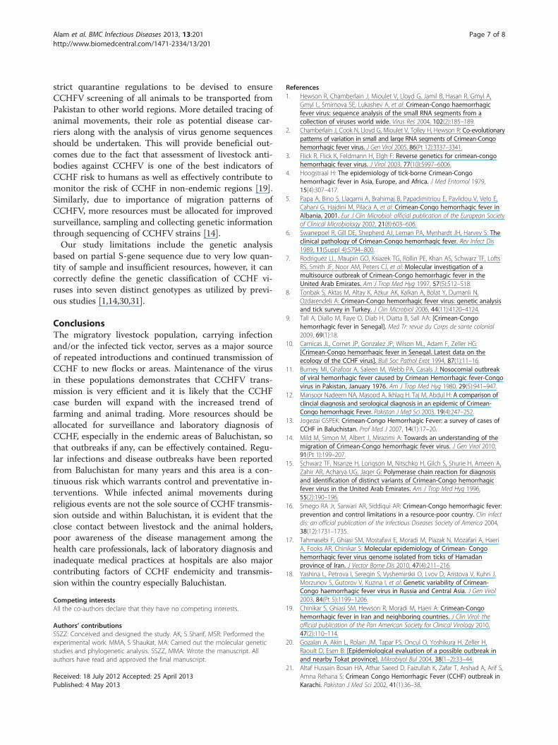
strict quarantine regulations to be devised to ensureCCHFV screening of all animals to be transported fromPakistan to other world regions. More detailed tracing ofanimal movements, their role as potential disease car-riers along with the analysis of virus genome sequencesshould be undertaken. This will provide beneficial out-comes due to the fact that assessment of livestock anti-bodies against CCHFV is one of the best indicators ofCCHF risk to humans as well as effectively contribute tomonitor the risk of CCHF in non-endemic regions [19].Similarly, due to importance of migration patterns ofCCHFV, more resources must be allocated for improvedsurveillance, sampling and collecting genetic informationthrough sequencing of CCHFV strains [14].Our study limitations include the genetic analysis
based on partial S-gene sequence due to very low quan-tity of sample and insufficient resources, however, it cancorrectly define the genetic classification of CCHF vi-ruses into seven distinct genotypes as utilized by previ-ous studies [1,14,30,31].
ConclusionsThe migratory livestock population, carrying infectionand/or the infected tick vector, serves as a major sourceof repeated introductions and continued transmission ofCCHF to new flocks or areas. Maintenance of the virusin these populations demonstrates that CCHFV trans-mission is very efficient and it is likely that the CCHFcase burden will expand with the increased trend offarming and animal trading. More resources should beallocated for surveillance and laboratory diagnosis ofCCHF, especially in the endemic areas of Baluchistan, sothat outbreaks if any, can be effectively contained. Regu-lar infections and disease outbreaks have been reportedfrom Baluchistan for many years and this area is a con-tinuous risk which warrants control and preventative in-terventions. While infected animal movements duringreligious events are not the sole source of CCHF transmis-sion outside and within Baluchistan, it is evident that theclose contact between livestock and the animal holders,poor awareness of the disease management among thehealth care professionals, lack of laboratory diagnosis andinadequate medical practices at hospitals are also majorcontributing factors of CCHF endemicity and transmis-sion within the country especially Baluchistan.
Competing interestsAll the co-authors declare that they have no competing interests.
Authors’ contributionsSSZZ: Conceived and designed the study. AK, S Sharif, MSR: Performed theexperimental work. MMA, S Shaukat, MA: Carried out the molecular geneticstudies and phylogenetic analysis. SSZZ, MMA: Wrote the manuscript. Allauthors have read and approved the final manuscript.
Received: 18 July 2012 Accepted: 25 April 2013Published: 4 May 2013
References1. Hewson R, Chamberlain J, Mioulet V, Lloyd G, Jamil B, Hasan R, Gmyl A,
Gmyl L, Smirnova SE, Lukashev A, et al: Crimean-Congo haemorrhagicfever virus: sequence analysis of the small RNA segments from acollection of viruses world wide. Virus Res 2004, 102(2):185–189.
2. Chamberlain J, Cook N, Lloyd G, Mioulet V, Tolley H, Hewson R: Co-evolutionarypatterns of variation in small and large RNA segments of Crimean-Congohemorrhagic fever virus. J Gen Virol 2005, 86(Pt 12):3337–3341.
3. Flick R, Flick K, Feldmann H, Elgh F: Reverse genetics for crimean-congohemorrhagic fever virus. J Virol 2003, 77(10):5997–6006.
4. Hoogstraal H: The epidemiology of tick-borne Crimean-Congohemorrhagic fever in Asia, Europe, and Africa. J Med Entomol 1979,15(4):307–417.
5. Papa A, Bino S, Llagami A, Brahimaj B, Papadimitriou E, Pavlidou V, Velo E,Cahani G, Hajdini M, Pilaca A, et al: Crimean-Congo hemorrhagic fever inAlbania, 2001. Eur J Clin Microbiol: official publication of the European Societyof Clinical Microbiology 2002, 21(8):603–606.
6. Swanepoel R, Gill DE, Shepherd AJ, Leman PA, Mynhardt JH, Harvey S: Theclinical pathology of Crimean-Congo hemorrhagic fever. Rev Infect Dis1989, 11(Suppl 4):S794–800.
7. Rodriguez LL, Maupin GO, Ksiazek TG, Rollin PE, Khan AS, Schwarz TF, LoftsRS, Smith JF, Noor AM, Peters CJ, et al: Molecular investigation of amultisource outbreak of Crimean-Congo hemorrhagic fever in theUnited Arab Emirates. Am J Trop Med Hyg 1997, 57(5):512–518.
8. Tonbak S, Aktas M, Altay K, Azkur AK, Kalkan A, Bolat Y, Dumanli N,Ozdarendeli A: Crimean-Congo hemorrhagic fever virus: genetic analysisand tick survey in Turkey. J Clin Microbiol 2006, 44(11):4120–4124.
9. Tall A, Diallo M, Faye O, Diab H, Diatta B, Sall AA: [Crimean-Congohemorrhagic fever in Senegal]. Med Tr: revue du Corps de sante colonial2009, 69(1):18.
10. Camicas JL, Cornet JP, Gonzalez JP, Wilson ML, Adam F, Zeller HG:[Crimean-Congo hemorrhagic fever in Senegal. Latest data on theecology of the CCHF virus]. Bull Soc Pathol Exot 1994, 87(1):11–16.
11. Burney MI, Ghafoor A, Saleen M, Webb PA, Casals J: Nosocomial outbreakof viral hemorrhagic fever caused by Crimean Hemorrhagic fever-Congovirus in Pakistan, January 1976. Am J Trop Med Hyg 1980, 29(5):941–947.
12. Mansoor Nadeem NA, Masood A, Ikhlaq H, Taj M, Abdul H: A comparison ofclincial diagnosis and serological diagnosis in an epidemic of Crimean-Congo hemorrhagic Fever. Pakistan J Med Sci 2003, 19(4):247–252.
13. Jogezai GSPEK: Crimean-Congo Hemorrhagic Fever: a survey of cases ofCCHF in Baluchistan. Prof Med J 2007, 14(1):17–20.
14. Mild M, Simon M, Albert J, Mirazimi A: Towards an understanding of themigration of Crimean-Congo hemorrhagic fever virus. J Gen Virol 2010,91(Pt 1):199–207.
15. Schwarz TF, Nsanze H, Longson M, Nitschko H, Gilch S, Shurie H, Ameen A,Zahir AR, Acharya UG, Jager G: Polymerase chain reaction for diagnosisand identification of distinct variants of Crimean-Congo hemorrhagicfever virus in the United Arab Emirates. Am J Trop Med Hyg 1996,55(2):190–196.
16. Smego RA Jr, Sarwari AR, Siddiqui AR: Crimean-Congo hemorrhagic fever:prevention and control limitations in a resource-poor country. Clin infectdis: an official publication of the Infectious Diseases Society of America 2004,38(12):1731–1735.
17. Tahmasebi F, Ghiasi SM, Mostafavi E, Moradi M, Piazak N, Mozafari A, HaeriA, Fooks AR, Chinikar S: Molecular epidemiology of Crimean- Congohemorrhagic fever virus genome isolated from ticks of Hamadanprovince of Iran. J Vector Borne Dis 2010, 47(4):211–216.
18. Yashina L, Petrova I, Seregin S, Vyshemirskii O, Lvov D, Aristova V, Kuhn J,Morzunov S, Gutorov V, Kuzina I, et al: Genetic variability of Crimean-Congo haemorrhagic fever virus in Russia and Central Asia. J Gen Virol2003, 84(Pt 5):1199–1206.
19. Chinikar S, Ghiasi SM, Hewson R, Moradi M, Haeri A: Crimean-Congohemorrhagic fever in Iran and neighboring countries. J Clin Virol: theofficial publication of the Pan American Society for Clinical Virology 2010,47(2):110–114.
20. Gozalan A, Akin L, Rolain JM, Tapar FS, Oncul O, Yoshikura H, Zeller H,Raoult D, Esen B: [Epidemiological evaluation of a possible outbreak inand nearby Tokat province]. Mikrobiyol Bul 2004, 38(1–2):33–44.
21. Altaf Hussain Bosan HA, Athar Saeed D, Faizullah K, Zafar T, Arshad A, Arif S,Amna Rehana S: Crimean Congo Hemorrhagic Fever (CCHF) outbreak inKarachi. Pakistan J Med Sci 2002, 41(1):36–38.

Alam et al. BMC Infectious Diseases 2013, 13:201 Page 8 of 8http://www.biomedcentral.com/1471-2334/13/201
22. Aradaib IE, Erickson BR, Mustafa ME, Khristova ML, Saeed NS, Elageb RM,Nichol ST: Nosocomial outbreak of Crimean-Congo hemorrhagic fever,Sudan. Emerg Infect Dis 2010, 16(5):837–839.
23. Burt FJ, Swanepoel R: Molecular epidemiology of African and AsianCrimean-Congo haemorrhagic fever isolates. Epidemiol Infect 2005,133(4):659–666.
24. Olschlager S, Gabriel M, Schmidt-Chanasit J, Meyer M, Osborn E, Conger NG,Allan PF, Gunther S: Complete sequence and phylogeneticcharacterisation of Crimean-Congo hemorrhagic fever virus fromAfghanistan. J Clin Virol: the official publication of the Pan American Societyfor Clinical Virology 2011, 50(1):90–92.
25. Izadi S, Holakouie-Naieni K, Majdzadeh SR, Chinikar S, Nadim A, Rakhshani F,Hooshmand B: Seroprevalence of Crimean-Congo hemorrhagic fever inSistan-va-Baluchestan province of Iran. Jpn J Infect Dis 2006, 59(5):326–328.
26. Suleiman MN, Muscat-Baron JM, Harries JR, Satti AG, Platt GS, Bowen ET,Simpson DI: Congo/Crimean haemorrhagic fever in Dubai. An outbreakat the Rashid Hospital. Lancet 1980, 2(8201):939–941.
27. Sun S, Dai X, Aishan M, Wang X, Meng W, Feng C, Zhang F, Hang C, Hu Z,Zhang Y: Epidemiology and phylogenetic analysis of crimean-congohemorrhagic fever viruses in xinjiang, china. J Clin Microbiol 2009,47(8):2536–2543.
28. Deyde VM, Khristova ML, Rollin PE, Ksiazek TG, Nichol ST: Crimean-Congohemorrhagic fever virus genomics and global diversity. J Virol 2006,80(17):8834–8842.
29. Raziq MYZR: Prospects of Livestock Production in Baluchistan. Pakistan VetJ 2010, 30(3):181–186.
30. Drosten C, Minnak D, Emmerich P, Schmitz H, Reinicke T: Crimean-Congohemorrhagic fever in Kosovo. J Clin Microbiol 2002, 40(3):1122–1123.
31. Papa A, Bozovi B, Pavlidou V, Papadimitriou E, Pelemis M, Antoniadis A:Genetic detection and isolation of crimean-congo hemorrhagic fevervirus, Kosovo, Yugoslavia. Emerg Infect Dis 2002, 8(8):852–854.
doi:10.1186/1471-2334-13-201Cite this article as: Alam et al.: Genetic analysis and epidemiology ofCrimean Congo hemorrhagic fever viruses in Baluchistan province ofPakistan. BMC Infectious Diseases 2013 13:201.
Submit your next manuscript to BioMed Centraland take full advantage of:
• Convenient online submission
• Thorough peer review
• No space constraints or color figure charges
• Immediate publication on acceptance
• Inclusion in PubMed, CAS, Scopus and Google Scholar
• Research which is freely available for redistribution
Submit your manuscript at www.biomedcentral.com/submit
Copyright © 2022 FDOKUMEN