Protein Arcs May Form Stable Pores in Lipid Membranes
20
Protein Arcs May Form Stable Pores in Lipid Membranes Lidia Prieto, Yi He, and Themis Lazaridis* Department of Chemistry, City College of New York, New York, New York ABSTRACT Electron microscopy and atomic force microscopy images of cholesterol-dependent cytolysins and related pro- teins that form large pores in lipid membranes have revealed the presence of incomplete rings, or arcs. Some evidence indicates that these arcs are inserted into the membrane and induce membrane leakage, but other experiments seem to refute that. Could such pores, only partially lined by protein, be kinetically and thermodynamically stable? How would the lipids be structured in such a pore? Using the antimicrobial peptide protegrin-1 as a model, we test the stability of pores only partially lined by peptide using all-atom molecular dynamics simulations in POPC and POPE/POPG membranes. The data show that, whereas pure lipid pores close rapidly, pores partially lined by protegrin arcs are stable for at least 300 ns. Estimates of the thermodynamic stability of these arcs using line tension data and implicit solvent calculations show that these arcs can be marginally stable in both zwit- terionic and anionic membranes. Arcs provide an explanation for the observed ion selectivity in protegrin electrophysiology ex- periments and could possibly be involved in other membrane permeabilization processes where lipids are thought to participate, such as those induced by antimicrobial peptides and colicins, as well as the Bax apoptotic pore. INTRODUCTION Toxin pores in lipid membranes are traditionally envisioned as oligomeric barrels in which certain segments from each monomer insert into the membrane and associate with each other to completely line the pore wall. This view has been corroborated by crystal structures of several b-toxins (1,2). However, electron microscopy (3–9) and atomic force microscopy (AFM) (10) images of cholesterol-dependent cytolysins (CDCs) show a large number of incomplete rings, or arcs, sometimes as frequently as complete rings. Similar structures have also been observed for perforin (11,12) and the membrane attack complex of the complement (13,14). The functional significance of these arcs is a matter of controversy (15). On the one hand, these arcs have been shown to cause ion leakage in membranes (16,17). Based on this, some authors have suggested that pore formation and oligomerization are simultaneous events (16). Other studies, however, support the prepore model, whereby monomers first oligomerize on the membrane surface form- ing a complete ring before inserting into the membrane (18,19). A conciliating model has been proposed where oligomerization can only take place on the membrane sur- face before insertion but incomplete rings can also insert into the membrane and form functional pores (20). If arcs do insert and form pores, this would mean that the part of the pore that is not lined by protein should be a free bilayer edge, most likely bent in a toroidal form (16,21). A free bilayer edge certainly costs free energy, but this cost could in principle be offset by the favorable insertion and oligomerization free energy of the protein arc. Complete rings should have lower free energy, but at low protein con- centration formation of a complete ring may not be possible. The question is whether incomplete rings have sufficient thermodynamic and/or kinetic stability inserted into the membrane. The fact that arcs can be observed by electron microscopy and AFM suggests that they are quite stable (22). Alternatively, arcs may only be metastable enough to cause leakage (15,20). This issue could have implications far beyond the field of CDCs. Lipid participation in pores has been invoked in several other cases, such as antimicro- bial peptides (23–25), colicins (26), diphtheria toxin (27), and the apoptotic pore of Bax (28) but the precise nature of the pore in these systems is unknown. Phosphatidylserine flip-flop caused by perforin has been suggested as an indica- tion of a toroidal pore (12). The toxins for which arcs have been identified are large proteins that assemble into pores containing 30–50 subunits (22). Computational studies of arcs made of these toxins would be exceedingly expensive. In addition, their oligo- meric structure is unknown, although the pore-forming structure is thought to consist of b-sheets (19). In the course of an implicit-solvent study (29) of b-barrels formed by protegrin-1, an 18-residue b-hairpin antimicrobial peptide stabilized by two disulfide bonds (30,31), we observed self-assembly of the peptides into arcs. We wondered then whether these arcs could be viable and proceeded to test this idea using explicit simulations. Protegrin has been shown to form ion channels in membranes (32,33) and has been the subject of extensive solid-state NMR (34,35) and computational (36) studies. Dozens of analogs have been synthesized and tested for activity (37,38). There is no evi- dence that protegrin itself forms arcs, but the system is small and simple enough to allow study by molecular dynamics simulations. The simulations show that peptide-arc struc- tures are kinetically stable on a timescale of 300 ns. Further arguments using macroscopic data and implicit solvent cal- culations suggest marginal thermodynamic stability as well. Submitted May 19, 2013, and accepted for publication November 22, 2013. *Correspondence: [email protected] Editor: Scott Feller. Ó 2014 by the Biophysical Society 0006-3495/14/01/0154/8 $2.00 http://dx.doi.org/10.1016/j.bpj.2013.11.4490 154 Biophysical Journal Volume 106 January 2014 154–161
-
Upload
independent -
Category
Documents
-
view
1 -
download
0
Transcript of Protein Arcs May Form Stable Pores in Lipid Membranes

154 Biophysical Journal Volume 106 January 2014 154–161
Protein Arcs May Form Stable Pores in Lipid Membranes
Lidia Prieto, Yi He, and Themis Lazaridis*Department of Chemistry, City College of New York, New York, New York
ABSTRACT Electron microscopy and atomic force microscopy images of cholesterol-dependent cytolysins and related pro-teins that form large pores in lipid membranes have revealed the presence of incomplete rings, or arcs. Some evidence indicatesthat these arcs are inserted into the membrane and induce membrane leakage, but other experiments seem to refute that. Couldsuch pores, only partially lined by protein, be kinetically and thermodynamically stable? How would the lipids be structured insuch a pore? Using the antimicrobial peptide protegrin-1 as a model, we test the stability of pores only partially lined by peptideusing all-atom molecular dynamics simulations in POPC and POPE/POPG membranes. The data show that, whereas pure lipidpores close rapidly, pores partially lined by protegrin arcs are stable for at least 300 ns. Estimates of the thermodynamic stabilityof these arcs using line tension data and implicit solvent calculations show that these arcs can be marginally stable in both zwit-terionic and anionic membranes. Arcs provide an explanation for the observed ion selectivity in protegrin electrophysiology ex-periments and could possibly be involved in other membrane permeabilization processes where lipids are thought to participate,such as those induced by antimicrobial peptides and colicins, as well as the Bax apoptotic pore.
INTRODUCTION
Toxin pores in lipid membranes are traditionally envisionedas oligomeric barrels in which certain segments from eachmonomer insert into the membrane and associate witheach other to completely line the pore wall. This view hasbeen corroborated by crystal structures of several b-toxins(1,2). However, electron microscopy (3–9) and atomic forcemicroscopy (AFM) (10) images of cholesterol-dependentcytolysins (CDCs) show a large number of incomplete rings,or arcs, sometimes as frequently as complete rings. Similarstructures have also been observed for perforin (11,12) andthe membrane attack complex of the complement (13,14).
The functional significance of these arcs is a matter ofcontroversy (15). On the one hand, these arcs have beenshown to cause ion leakage in membranes (16,17). Basedon this, some authors have suggested that pore formationand oligomerization are simultaneous events (16). Otherstudies, however, support the prepore model, wherebymonomers first oligomerize on the membrane surface form-ing a complete ring before inserting into the membrane(18,19). A conciliating model has been proposed whereoligomerization can only take place on the membrane sur-face before insertion but incomplete rings can also insertinto the membrane and form functional pores (20).
If arcs do insert and form pores, this would mean that thepart of the pore that is not lined by protein should be a freebilayer edge, most likely bent in a toroidal form (16,21). Afree bilayer edge certainly costs free energy, but this costcould in principle be offset by the favorable insertion andoligomerization free energy of the protein arc. Completerings should have lower free energy, but at low protein con-centration formation of a complete ring may not be possible.
Submitted May 19, 2013, and accepted for publication November 22, 2013.
*Correspondence: [email protected]
Editor: Scott Feller.
� 2014 by the Biophysical Society
0006-3495/14/01/0154/8 $2.00
The question is whether incomplete rings have sufficientthermodynamic and/or kinetic stability inserted into themembrane. The fact that arcs can be observed by electronmicroscopy and AFM suggests that they are quite stable(22). Alternatively, arcs may only be metastable enough tocause leakage (15,20). This issue could have implicationsfar beyond the field of CDCs. Lipid participation in poreshas been invoked in several other cases, such as antimicro-bial peptides (23–25), colicins (26), diphtheria toxin (27),and the apoptotic pore of Bax (28) but the precise natureof the pore in these systems is unknown. Phosphatidylserineflip-flop caused by perforin has been suggested as an indica-tion of a toroidal pore (12).
The toxins for which arcs have been identified are largeproteins that assemble into pores containing 30–50 subunits(22). Computational studies of arcs made of these toxinswould be exceedingly expensive. In addition, their oligo-meric structure is unknown, although the pore-formingstructure is thought to consist of b-sheets (19). In the courseof an implicit-solvent study (29) of b-barrels formed byprotegrin-1, an 18-residue b-hairpin antimicrobial peptidestabilized by two disulfide bonds (30,31), we observedself-assembly of the peptides into arcs. We wondered thenwhether these arcs could be viable and proceeded to testthis idea using explicit simulations. Protegrin has beenshown to form ion channels in membranes (32,33) and hasbeen the subject of extensive solid-state NMR (34,35) andcomputational (36) studies. Dozens of analogs have beensynthesized and tested for activity (37,38). There is no evi-dence that protegrin itself forms arcs, but the system is smalland simple enough to allow study by molecular dynamicssimulations. The simulations show that peptide-arc struc-tures are kinetically stable on a timescale of 300 ns. Furtherarguments using macroscopic data and implicit solvent cal-culations suggest marginal thermodynamic stability as well.
http://dx.doi.org/10.1016/j.bpj.2013.11.4490

FIGURE 1 (A) Cartoon representation of the protegrin arc structure
obtained in implicit simulations and used as the initial structure in the
explicit simulations run in this work. (B) Upper view of the same structure
in an implicit pore of R¼ 13 A and curvature K¼ 15 A. To see this figure in
color, go online.
Protein Arcs May Form Stable Pores in Lipid Membranes 155
METHODS
We have run 300-ns explicit simulations of a tetrameric protegrin arc struc-
ture obtained from implicit simulations (see the Supporting Material) in a
100% POPC membrane and a 70% POPE: 30% POPG membrane using
the program NAMD (39) with the CHARMM36 force field for the peptide
(40) and the lipids (41). The equilibrated initial membranes and the mem-
branes with a cylindrical 13 A pore are the ones used in previous work (29).
The 100A� 100A� 70A membrane systems were created and equilibrated
using the steps suggested by CHARMM-GUI (42,43). An additional 0.9-ns
run was carried out to ensure that equilibration has been achieved. The pore
was generated by eliminating the lipids in this region and filling the space
with TIP3P water. It was then equilibrated for 1.5 ns in the NPT ensemble
while constraining the positions of the lipid headgroups. The temperature of
all simulations was 303.15 K, consistent with experimental studies (44–46).
We ran three different simulations for each membrane: a), the pore sys-
tem in pure membranes with no constraints (the length of these NPT sim-
ulations is as long as it takes for the pore to close). These systems consist
of 257 lipids, 11,056 water molecules, 28 Cl� ions, and 28 Kþ ions in
the 100% POPC membrane and 191 POPE and 89 POPG lipids, 11,462
water molecules, 24 Cl� ions, and 112 Kþ ions in the anionic membrane.
Although the original membranes were symmetric, the process of creating
a pore made the membrane slightly asymmetric. b), 300-ns NPT simula-
tions of the equilibrated cylindrical pores with the protegrin arc inserted
into them lining the pore as it does in the implicit simulations. The new
systems, with the peptide constrained to its position, are equilibrated for
another 2 ns to let the membrane and water molecules adapt to it. The
size of these systems is 72 peptide residues, 252 (POPC) or 264 (180
POPE and 84 POPG) lipids, 10,468 or 10,970 water molecules, 52 or 50
Cl� ions, and 28 or 110 Kþ ions, for the zwitterionic and anionic systems,
respectively. c), 300-ns NPT simulations starting from the equilibrated
membranes without pores and the protegrin arc structure embedded in
them in a transmembrane orientation. The systems are first relaxed using
a conjugate gradient minimization and then equilibrated for 4 ns in the
NPT ensemble using constraints to fix the position of the peptides so that
the membrane can adjust around them. The simulations then continue for
300 ns without any constraints. These systems consist of the peptide arcs
(72 residues), 254 (POPC), or 279 (190 POPE and 89 POPG) lipid mole-
cules, 10,372 or 10,698 water molecules, 60 or 47 Cl� ions, and 27 or
112 Kþ ions, in the zwitterionic and anionic membranes, respectively.
To estimate the size of the pores we calculated the number of water
molecules that are close to the membrane center. More precisely, we
counted the number of water molecules with a z coordinate 58.5 A from
the membrane center (the membrane surface being parallel to the xy plane).
Because water enters this region only if it is in the pore (the hydrophobic
thickness of these membranes is ~27 A), this number corresponds to the
number of water molecules in the pore. The results obtained for the peptide
arc in this work are compared to the 100-ns NCNC parallel b-barrel simu-
lations run in previous work on protegrin (29).
FIGURE 2 Number of water molecules inside the pore (jzj % 8.5 A)
along the simulation. Green line: octameric protegrin rings inserted into
preformed cylindrical pores (29). Red line: tetrameric arc inserted into pre-
formed cylindrical pores. Black line: the same arc inserted into the mem-
brane without a pore. Gray line: preformed pores with no peptide. To see
this figure in color, go online.
RESULTS
The initial structure of the protegrin arc used in theseexplicit simulations was obtained by implicit membranestudies of the self-assembly of protegrin monomers in apreformed pore (see the Supporting Material). Most favor-able energetically is the tetrameric structure shown inFig. 1. Unlike the complete b-barrels (29), the peptidesexhibit a twist relative to each other. The orientation ofeach peptide gradually changes from tilting toward the up-per leaflet in the leftmost peptide (red in Fig. 1) to tilting to-ward the lower leaflet in the rightmost peptide (blue). Thisnatural twist cannot be accommodated in a larger arc.
Indeed, when six peptides were placed in the same configu-ration, the final structure was not a continuous arc but abroken one (Fig. S1 B). The structure of Fig. 1 led us to thinkthat closing of the barrel might incur some strain and thatprotegrin could be an appropriate system to investigatewhether pores lined by arcs rather than barrels or unassoci-ated peptides can exist in biological membranes.
We ran three types of simulations: preformed cylindricalpores in membranes without peptide, the arc structure ofFig. 1 in preformed cylindrical pores, and the same arcstructure inserted into membranes without a water pore.Two membrane systems were used, one zwitterionic(100% POPC) and one partially anionic (70% POPE: 30%POPG). To evaluate the size and stability of the pores inthese systems, we computed the number of water moleculesin the pore as a function of time (Fig. 2). In the absence of
Biophysical Journal 106(1) 154–161

156 Prieto et al.
peptides the pore closes rapidly: the number of water mole-cules decreases to zero in ~30 ns. Very early in the simula-tions (see 6 ns or 8 ns in Fig. 3 A), the membrane bends andlipid headgroups start to line the pore, which acquires a
FIGURE 3 (A) Snapshots along the simulations starting from preformed
pores in a zwitterionic and an anionic membrane with no peptide. The
INITIAL structures correspond to right before releasing all constraints. Pur-
ple balls-and-sticks: water molecules. Silver lines: lipid side chains. Blue and
tan spheres: lipid headgroup nitrogen and phosphorus atoms, respectively.
(B) Arc pore structures in the membranes starting from a preformed pore.
The INITIAL structures correspond to the ones right before releasing all con-
straints. Purple balls-and-sticks: water molecules. Silver lines: lipid side
chains. Blue and tan spheres: lipid headgroup nitrogen and phosphorus atoms,
respectively. Cyan licorice: disulphide bridges. Cartoon representations: pep-
tide backbone. (C) Final structure of the peptide in the zwitterionic (left) and
anionic (right) membranes (same as in B at 300 ns, eliminating membrane,
water, and residue side chains). To see this figure in color, go online.
Biophysical Journal 106(1) 154–161
toroidal shape, as observed previously (47). The headgroupdensity is lower in the pore than on the membrane surface(48). This pore is smaller than the original cylindrical oneand keeps getting narrower with the lipid headgroupsgradually returning to the membrane surface (28 or 20 nssnapshots in Fig. 3 A) until the pore closes (30 or 26 ns).In contrast, in the presence of the arcs, regardless of startingconditions, the pores remain open until the end of thesimulation.
The complete octameric barrels form very stable pores(29), enclosing 113 5 8 (zwitterionic membrane) or119 5 12 (anionic membrane) water molecules (averagesover the entire simulation). The arcs stabilize somewhatsmaller pores: 93 5 15 in the zwitterionic membrane and58 5 8 in the anionic membrane (averages over the last200 ns). This is not surprising: osmotic protection (16)and electrical conductance (22) experiments of CDCsshow that the arcs create pores of significantly smallersize than the complete rings. When the arcs are placed inthe membranes without preformed pores, we observe rapidinsertion of water in the membrane area in the equilibrationstage. The resulting pores are smaller than those in the pre-vious simulations, 44 5 7 water molecules in the zwitter-ionic membrane and 42 5 6 in the anionic membrane.The distribution of the number of water molecules insidethe pores is shown in Fig. S3. There are also differencesin the distribution of water along the pore axis betweenthe two types of simulations (Fig. 3 and Fig. S4 A): it isless evenly distributed in the simulations that start withoutpreformed pores (Fig. S5), presumably due to the limitedsimulation time. In either case, there is enough water inthe pore region to assert that a pore partially lined by a pro-tegrin arc is stable for at least 300 ns.
Fig. 3 B shows snapshots of the simulations of protegrinarcs in preformed cylindrical pores. On the protein side ofthe pore, the membrane retains a structure similar to thatof the complete rings in the same systems (29): the mem-brane curves slightly to adapt to the size of the arc, butthe pore remains essentially cylindrical (or semitoroidal)during the entire simulation. Where there is no peptide themembrane bends and the lipid headgroups line the pore.This is more visible in the POPC membrane. After 10 ns,one or both leaflets of the membranes start to curve. By75 ns, the lipidic region of the pore has a toroidal structure:the aqueous lumen is lined by lipid headgroups. We observea similar process in the simulations of the arcs in the mem-branes with no preformed pores (Fig. S4 A). However, inthis case there are fewer headgroups lining the water-lipidinterface of the pore and the membrane is less curved.Again, these differences probably reflect a lack of full equil-ibration. The structure of the pores created by arcs in thesesimulations (Fig. 3 B) is similar to what has been envisionedby several authors (15,22). The polar surface of the arc isprobably what drives water into it, in accord with observa-tions of water defects when arginines are placed in the
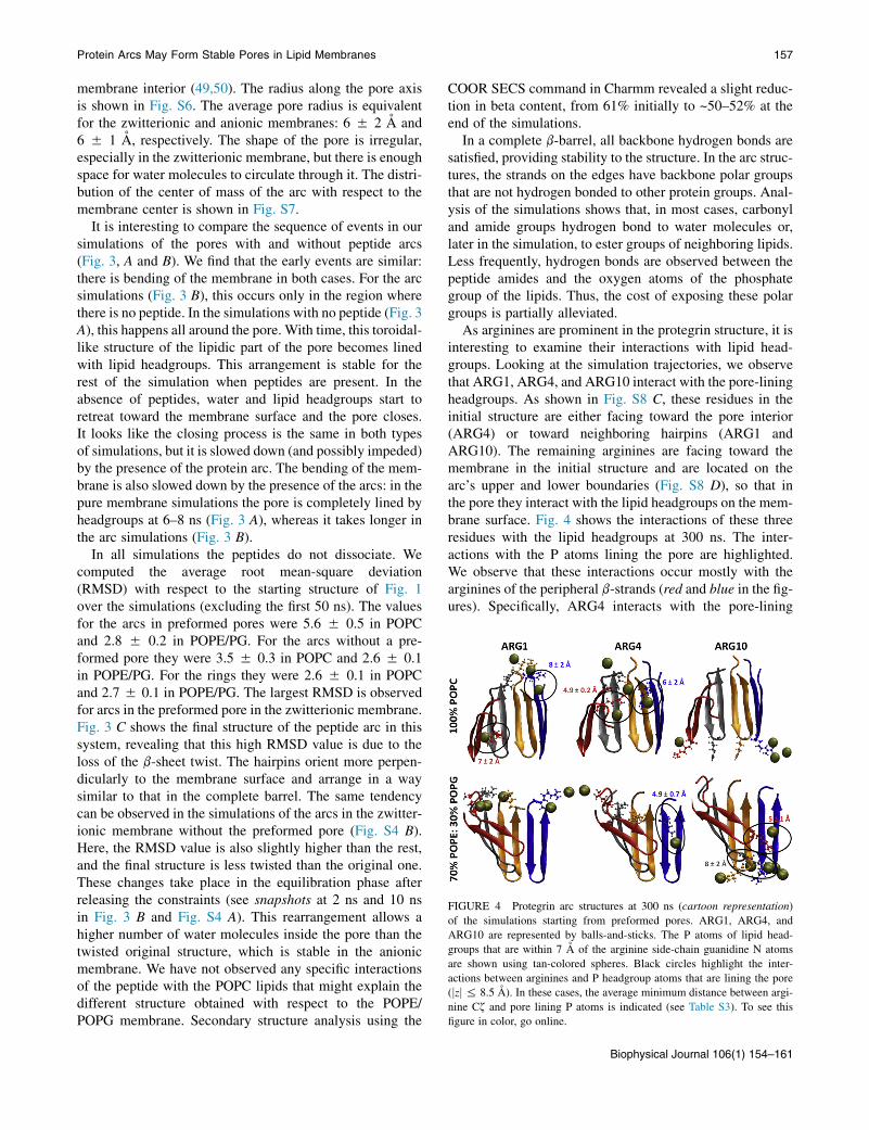
FIGURE 4 Protegrin arc structures at 300 ns (cartoon representation)
of the simulations starting from preformed pores. ARG1, ARG4, and
ARG10 are represented by balls-and-sticks. The P atoms of lipid head-
groups that are within 7 A of the arginine side-chain guanidine N atoms
are shown using tan-colored spheres. Black circles highlight the inter-
actions between arginines and P headgroup atoms that are lining the pore
(jzj % 8.5 A). In these cases, the average minimum distance between argi-
nine Cz and pore lining P atoms is indicated (see Table S3). To see this
figure in color, go online.
Protein Arcs May Form Stable Pores in Lipid Membranes 157
membrane interior (49,50). The radius along the pore axisis shown in Fig. S6. The average pore radius is equivalentfor the zwitterionic and anionic membranes: 6 5 2 A and6 5 1 A, respectively. The shape of the pore is irregular,especially in the zwitterionic membrane, but there is enoughspace for water molecules to circulate through it. The distri-bution of the center of mass of the arc with respect to themembrane center is shown in Fig. S7.
It is interesting to compare the sequence of events in oursimulations of the pores with and without peptide arcs(Fig. 3, A and B). We find that the early events are similar:there is bending of the membrane in both cases. For the arcsimulations (Fig. 3 B), this occurs only in the region wherethere is no peptide. In the simulations with no peptide (Fig. 3A), this happens all around the pore. With time, this toroidal-like structure of the lipidic part of the pore becomes linedwith lipid headgroups. This arrangement is stable for therest of the simulation when peptides are present. In theabsence of peptides, water and lipid headgroups start toretreat toward the membrane surface and the pore closes.It looks like the closing process is the same in both typesof simulations, but it is slowed down (and possibly impeded)by the presence of the protein arc. The bending of the mem-brane is also slowed down by the presence of the arcs: in thepure membrane simulations the pore is completely lined byheadgroups at 6–8 ns (Fig. 3 A), whereas it takes longer inthe arc simulations (Fig. 3 B).
In all simulations the peptides do not dissociate. Wecomputed the average root mean-square deviation(RMSD) with respect to the starting structure of Fig. 1over the simulations (excluding the first 50 ns). The valuesfor the arcs in preformed pores were 5.6 5 0.5 in POPCand 2.8 5 0.2 in POPE/PG. For the arcs without a pre-formed pore they were 3.5 5 0.3 in POPC and 2.6 5 0.1in POPE/PG. For the rings they were 2.6 5 0.1 in POPCand 2.7 5 0.1 in POPE/PG. The largest RMSD is observedfor arcs in the preformed pore in the zwitterionic membrane.Fig. 3 C shows the final structure of the peptide arc in thissystem, revealing that this high RMSD value is due to theloss of the b-sheet twist. The hairpins orient more perpen-dicularly to the membrane surface and arrange in a waysimilar to that in the complete barrel. The same tendencycan be observed in the simulations of the arcs in the zwitter-ionic membrane without the preformed pore (Fig. S4 B).Here, the RMSD value is also slightly higher than the rest,and the final structure is less twisted than the original one.These changes take place in the equilibration phase afterreleasing the constraints (see snapshots at 2 ns and 10 nsin Fig. 3 B and Fig. S4 A). This rearrangement allows ahigher number of water molecules inside the pore than thetwisted original structure, which is stable in the anionicmembrane. We have not observed any specific interactionsof the peptide with the POPC lipids that might explain thedifferent structure obtained with respect to the POPE/POPG membrane. Secondary structure analysis using the
COOR SECS command in Charmm revealed a slight reduc-tion in beta content, from 61% initially to ~50–52% at theend of the simulations.
In a complete b-barrel, all backbone hydrogen bonds aresatisfied, providing stability to the structure. In the arc struc-tures, the strands on the edges have backbone polar groupsthat are not hydrogen bonded to other protein groups. Anal-ysis of the simulations shows that, in most cases, carbonyland amide groups hydrogen bond to water molecules or,later in the simulation, to ester groups of neighboring lipids.Less frequently, hydrogen bonds are observed between thepeptide amides and the oxygen atoms of the phosphategroup of the lipids. Thus, the cost of exposing these polargroups is partially alleviated.
As arginines are prominent in the protegrin structure, it isinteresting to examine their interactions with lipid head-groups. Looking at the simulation trajectories, we observethat ARG1, ARG4, and ARG10 interact with the pore-liningheadgroups. As shown in Fig. S8 C, these residues in theinitial structure are either facing toward the pore interior(ARG4) or toward neighboring hairpins (ARG1 andARG10). The remaining arginines are facing toward themembrane in the initial structure and are located on thearc’s upper and lower boundaries (Fig. S8 D), so that inthe pore they interact with the lipid headgroups on the mem-brane surface. Fig. 4 shows the interactions of these threeresidues with the lipid headgroups at 300 ns. The inter-actions with the P atoms lining the pore are highlighted.We observe that these interactions occur mostly with thearginines of the peripheral b-strands (red and blue in the fig-ures). Specifically, ARG4 interacts with the pore-lining
Biophysical Journal 106(1) 154–161

158 Prieto et al.
P atoms in both the zwitterionic and anionic membrane.Table S3 shows that the Cz-P distance to the lipid head-groups lining the pore is smaller for the peripheral strandsthan for the central ones, where ARG4 points toward thepore center (see Fig. 4). Due to the different arc structuresin the zwitterionic and anionic membranes, the argininesthat interact with the lipid headgroups lining the pore differ.In the POPC membrane, ARG1 of the peripheral hairpinsshows a shorter P-Cz distance with the headgroups in thepore region than the other two monomers (see Table S3).In the POPE/POPG membrane, it is ARG10 that interactswith the lipids lining the pore. Due to the twist of the mono-mers, in this case it is the leftmost and its neighbor hairpins(red and gray) that produce this interaction. ARG1 andARG10 in the remaining two monomers mostly interactwith the lipid headgroups on the membrane surface (seeFig. 4 and Table S3). It appears that these arginines arecontributing to the stabilization of the pore. In addition tothe phosphates, arginines were observed to interact stronglywith glycerol groups, mostly from the same lipids as thephosphates, but also others.
To examine the ability of ions to cross the peptide-stabi-lized pores, we analyzed the movement of ions across thepore. We first identified the ions that enter the pore region(jzj % 8.5 A). In Fig. S9 we show several trajectories ofCl� ions in the simulations of the arcs in preformed pores.In the absence of voltage, we do not expect significant ionflow. Indeed, in most cases, they enter the pore region butdo not crossover to the other side of the membrane, occa-sionally spending quite some time in the pore region. Thenumber of ions entering the pore region is higher in thecase of POPC membranes, which may be due to the largerpore formed in these membranes. Despite the absence ofvoltage in our simulations, we observe two events of Cl�
ions crossing the zwitterionic membrane (highlighted witha circle in Fig. S9). This further supports the notion thatarcs may, indeed, be able to stabilize functional pores.Only one cation was close to the pore center in one simula-tion and it did not cross to the other side. Detailed analysisof ion conductance will be the subject of future work.
DISCUSSION
The explicit simulation results show that protegrin arcs arekinetically stable. That is, once they are placed in an in-serted configuration, they remain there for at least 300 ns.However, starting from free monomers in solution, willthey insert? In other words, are the arcs thermodynamicallystable? This question cannot be answered with explicit sim-ulations, at least not with our available resources. However,an approximate estimate can be made using macroscopicline tension data and implicit solvent simulations. The freeenergy of the inserted arc is equal to the free energy of form-ing the pore in the pure membrane plus the free energy oftransfer of the peptides from the flat membrane to the pre-
Biophysical Journal 106(1) 154–161
formed pore. The former is estimated as 2pRg, where g isthe line tension, and the latter from implicit solvent simula-tions. The line tension has been measured as 7 pN for PC(51) and 6 pN for PS membranes (52). This gives a freeenergy of forming an R ¼ 13 A pore equal to ~7 to8 kcal/mol for both membranes. Other measurements ofthe line tension give higher estimates (53). The effectiveenergy change upon transfer of four associated protegrinmonomers from water to the pore estimated using theIMM1 model (see the Supporting Material) is about�13 kcal/mol. For 30% anionic membranes, the effectiveenergy difference for four associated protegrin moleculesbetween the flat membrane and the pore is also about�13 kcal/mol (see the Supporting Material). The entropycost for localizing the tetramer in the pore should be smallin the latter case. In the former case it is more substantial,similar to the entropy of membrane binding and dependenton peptide concentration. At standard state, this entropy costhas been estimated as 1.3 kcal/mol (54). Thus, the favorabletransfer energy may be sufficient to overcome the unfavor-able free energy of pore formation, especially in anionicmembranes.
As mentioned in the Introduction, there is no experi-mental evidence that protegrin forms arcs. However, to thebest of our knowledge, there is no evidence that precludesthem either. The oriented circular dichroism data thatdetected a change in orientation of protegrin as a functionof concentration and hydration level (55) and the x-raydiffraction data that detected protegrin-induced membranethinning (56) and crystallized pores (57), cannot differen-tiate between complete and incomplete barrels. Further-more, the reported AFM work (46,58) did not havesufficient resolution to detect barrels or arcs. We hope thatin the near future, specifically designed experiments willbe able to tell us which of these two possibilities, if any, cor-responds to reality.
In this work, we simulated only arcs of NCNC paralleltopology based on our previous work that found thistopology to be most stable (29), as opposed to the NCCNparallel topology suggested by solid-state NMR experi-ments (59,60). The data that suggested oligomerization ofprotegrin into closed b-barrels in anionic lipids wereCODEX solid-state NMR experiments with 19F labels atpositions 7 and 12 (60). This experiment determines thenumber of such labels within 15 A of each other and allowsan estimate of the actual distances from the speed of decayof the signal. The 7-7 and 12-12 distances in the NCNCparallel barrel are ~11 A and averages of these distancescalculated over our arc simulations ranged from 8.6 to11.6 A, within the range of detection. In a nitroacetanilidecrystal with a 11.5 A distance between labels, Luo andHong measured a clear CODEX signal with 52 ms decayconstant (61), quite close to the 60 ms decay time measuredfor Phg7 in protegrin (60). In the barrels each label wouldhave two other labels at those distances, thus, (S/So)eq

Protein Arcs May Form Stable Pores in Lipid Membranes 159
~0.33. In the arcs half the labels would have two and halfwould have one, thus (S/So)eq should be between 0.33and 0.5, as was observed for Phg12. The 7-7 and 12-12distances in the NCCN parallel arrangement are actuallylonger than 11 A, because in that arrangement neighboringside chains point to different faces of the b-sheet. In theNCCN antiparallel model the 7-7 distance is 5.3 A but the12-12 distance is 14.2 A. The short distances of 6.5 and9 A inferred by Mani et al. for 7-7 and 12-12, respectively,do not seem compatible with any single closed barrel struc-ture. Further discussion of other solid-state NMR results canbe found in our previous publication (29).
The ion conductance properties of protegrin channelshave been studied by both experimental and theoreticalmethods. Electrophysiology studies found a weak anionselectivity in azolectin membranes and cation selectivityin lipopolysaccharide membranes (33). A computationalstudy of ion conductance based on the NCCN parallel barrelstructure found an anion selectivity that is much strongerthan in the experimental studies (62). An arc pore structureprovides a possible explanation of this discrepancy. If theconducting ions are exposed to lipid on one side as theycross the channel, they will be quite sensitive to the natureof the lipid and less sensitive to the positive charge of thepeptide. Calculations of conductance based on the arc struc-tures would provide a quantitative assessment of this idea.
The standard picture of a peptide-stabilized lipidic pore isa toroidal pore with several peptides dispersed in it withorientation perpendicular to the membrane (23). A differentpicture came from explicit simulation data, with a pore thatis much more disordered and peptides residing mostly onthe periphery of the pore (63). Other simulation studies ofpores stabilized by antimicrobial peptides produced resultsintermediate between the above extremes (47,64–67). Thiswork presents yet another paradigm of pore structure: linedby associated protein on one side, and by lipid headgroupsin toroidal form on the other. This idea seems to bemore in line with findings that aggregation is an essentialstep in peptide-induced pore formation (68). There arealso smaller toxins, such as the colicins (26) and diphtheriatoxin (27), which appear to permeabilize membranes asmonomers, inserting three or four helices into the mem-brane. For many of these systems there is not enough proteinto completely line a pore (69), which led to the idea that partof the pore is lipidic. Protein arcs offer a new, to our know-ledge, intriguing possibility as a solution to this conundrum.
SUPPORTING MATERIAL
Nine figures, three tables, references (70–78), and supporting data are avail-
able at http://www.biophysj.org/biophysj/supplemental/S0006-3495(13)
05750-0.
We thank Dr. C. Froelich for directing our attention to arcs and for many
fruitful discussions. We also thank Dr. R. Stark for informative discussions
on solid-state NMR.
This work was supported by the National Institutes of Health (NIH) (SC1-
AI084899). Infrastructure support was provided in part by RCMI grant
2G12RR03060-26A1/8G12MD007603-27 from NIH. L.P. acknowledges
a fellowship from the Spanish Ramon Areces Foundation and a subsequent
fellowship from the Spanish Foundation Caja Madrid. Computational re-
sources were provided by the CUNY High Performance Computing Center
and XSEDE, which is supported by the National Science Foundation (NSF)
(grant OCI-1053575).
REFERENCES
1. Parker, M. W., and S. C. Feil. 2005. Pore-forming protein toxins: fromstructure to function. Prog. Biophys. Mol. Biol. 88:91–142.
2. Iacovache, I., F. G. van der Goot, and L. Pernot. 2008. Pore formation:an ancient yet complex form of attack. Biochim. Biophys. Acta.1778:1611–1623.
3. Duncan, J. L., and R. Schlegel. 1975. Effect of streptolysin O on eryth-rocyte membranes, liposomes, and lipid dispersions. A protein-choles-terol interaction. J. Cell Biol. 67:160–174.
4. Rottem, S., R. M. Cole, ., M. C. Hardegree. 1982. Structural charac-teristics of tetanolysin and its binding to lipid vesicles. J. Bacteriol.152:888–892.
5. Olofsson, A., H. Hebert, and M. Thelestam. 1993. The projection struc-ture of perfringolysin O (Clostridium perfringens theta-toxin). FEBSLett. 319:125–127.
6. Morgan, P. J., S. C. Hyman, ., A. J. Rowe. 1994. Modeling thebacterial protein toxin, pneumolysin, in its monomeric and oligomericform. J. Biol. Chem. 269:25315–25320.
7. Sekino-Suzuki, N., M. Nakamura,., Y. Ohno-Iwashita. 1996. Contri-bution of individual tryptophan residues to the structure and activity oftheta-toxin (perfringolysin O), a cholesterol-binding cytolysin. Eur. J.Biochem. 241:941–947.
8. Morgan, P. J., S. C. Hyman, ., H. R. Saibil. 1995. Subunit organisa-tion and symmetry of pore-forming, oligomeric pneumolysin. FEBSLett. 371:77–80.
9. Korchev, Y. E., C. L. Bashford, ., T. J. Mitchell. 1998. A conservedtryptophan in pneumolysin is a determinant of the characteristics ofchannels formed by pneumolysin in cells and planar lipid bilayers.Biochem. J. 329:571–577.
10. Czajkowsky, D. M., E. M. Hotze, ., R. K. Tweten. 2004. Verticalcollapse of a cytolysin prepore moves its transmembrane beta-hairpinsto the membrane. EMBO J. 23:3206–3215.
11. Young, J. D. E., H. Hengartner, ., Z. A. Cohn. 1986. Purification andcharacterization of a cytolytic pore-forming protein from granules ofcloned lymphocytes with natural killer activity. Cell. 44:849–859.
12. Metkar, S. S., B. K. Wang, ., C. J. Froelich. 2011. Perforin rapidlyinduces plasma membrane phospholipid flip-flop. PLoS ONE.6:e24286.
13. Tschopp, J. 1984. Ultrastructure of the membrane attack complex ofcomplement. Heterogeneity of the complex caused by different degreeof C9 polymerization. J. Biol. Chem. 259:7857–7863.
14. Malinski, J. A., and G. L. Nelsestuen. 1989. Membrane permeability tomacromolecules mediated by the membrane attack complex.Biochemistry. 28:61–70.
15. Gilbert, R. J. C. 2005. Inactivation and activity of cholesterol-depen-dent cytolysins: what structural studies tell us. Structure. 13:1097–1106.
16. Palmer, M., R. Harris, ., S. Bhakdi. 1998. Assembly mechanism ofthe oligomeric streptolysin O pore: the early membrane lesion is linedby a free edge of the lipid membrane and is extended gradually duringoligomerization. EMBO J. 17:1598–1605.
17. Bayley, H. 1997. Toxin structure: part of a hole? Curr. Biol. 7:R763–R767.
Biophysical Journal 106(1) 154–161

160 Prieto et al.
18. Heuck, A. P., R. K. Tweten, and A. E. Johnson. 2003. Assembly andtopography of the prepore complex in cholesterol-dependent cytoly-sins. J. Biol. Chem. 278:31218–31225.
19. Tweten, R. K. 2005. Cholesterol-dependent cytolysins, a family ofversatile pore-forming toxins. Infect. Immun. 73:6199–6209.
20. Gilbert, R. J. C. 2002. Pore-forming toxins. Cell. Mol. Life Sci.59:832–844.
21. Bhakdi, S., J. Tranum-Jensen, and A. Sziegoleit. 1985. Mechanism ofmembrane damage by streptolysin-O. Infect. Immun. 47:52–60.
22. Gilbert, R. J. C. 2010. Cholesterol-dependent cytolysins. InProteins. Membrane Binding and Pore Formation. G. Anderluh andJ. Lakey, editors. Landes Bioscience and Busines Media, pp. 56–66.
23. Ludtke, S. J., K. He,., H. W. Huang. 1996. Membrane pores inducedby magainin. Biochemistry. 35:13723–13728.
24. Matsuzaki, K. 1999. Why and how are peptide-lipid interactionsutilized for self-defense? Magainins and tachyplesins as archetypes.Biochim. Biophys. Acta. 1462:1–10.
25. Yang, L., T. A. Harroun,., H. W. Huang. 2001. Barrel-stave model ortoroidal model? A case study on melittin pores. Biophys. J. 81:1475–1485.
26. Zakharov, S. D., E. A. Kotova, ., W. A. Cramer. 2004. On the role oflipid in colicin pore formation. Biochim. Biophys. Acta. 1666:239–249.
27. Gordon, M., and A. Finkelstein. 2001. The number of subunitscomprising the channel formed by the T domain of diphtheria toxin.J. Gen. Physiol. 118:471–480.
28. Qian, S., W. C. Wang, ., H. W. Huang. 2008. Structure of transmem-brane pore induced by Bax-derived peptide: evidence for lipidic pores.Proc. Natl. Acad. Sci. USA. 105:17379–17383.
29. Lazaridis, T., Y. He, and L. Prieto. 2013. Membrane interactions andpore formation by the antimicrobial peptide protegrin. Biophys. J.104:633–642.
30. Kokryakov, V. N., S. S. L. Harwig, ., R. I. Lehrer. 1993. Protegrins:leukocyte antimicrobial peptides that combine features of corticostaticdefensins and tachyplesins. FEBS Lett. 327:231–236.
31. Fahrner, R. L., T. Dieckmann,., J. Feigon. 1996. Solution structure ofprotegrin-1, a broad-spectrum antimicrobial peptide from porcineleukocytes. Chem. Biol. 3:543–550.
32. Mangoni, M. E., A. Aumelas, ., A. Chavanieu. 1996. Change inmembrane permeability induced by protegrin 1: implication of disul-phide bridges for pore formation. FEBS Lett. 383:93–98.
33. Sokolov, Y., T. Mirzabekov,., B. L. Kagan. 1999. Membrane channelformation by antimicrobial protegrins. Biochim. Biophys. Acta.1420:23–29.
34. Hong, M. 2007. Structure, topology, and dynamics of membranepeptides and proteins from solid-state NMR spectroscopy. J. Phys.Chem. B. 111:10340–10351.
35. Tang, M., andM. Hong. 2009. Structure and mechanism of beta-hairpinantimicrobial peptides in lipid bilayers from solid-state NMR spectros-copy. Mol. Biosyst. 5:317–322.
36. Bolintineanu, D. S., and Y. N. Kaznessis. 2011. Computational studiesof protegrin antimicrobial peptides: a review. Peptides. 32:188–201.
37. Chen, J., T. J. Falla, ., J. C. Fiddes. 2000. Development of protegrinsfor the treatment and prevention of oral mucositis: structure-activityrelationships of synthetic protegrin analogues. Biopolymers. 55:88–98.
38. Ostberg, N., and Y. Kaznessis. 2005. Protegrin structure-activity rela-tionships: using homology models of synthetic sequences to determinestructural characteristics important for activity. Peptides. 26:197–206.
39. Phillips, J. C., R. Braun, ., K. Schulten. 2005. Scalable moleculardynamics with NAMD. J. Comput. Chem. 26:1781–1802.
40. Mackerell, Jr., A. D., M. Feig, and C. L. Brooks, 3rd. 2004. Extendingthe treatment of backbone energetics in protein force fields: limitationsof gas-phase quantum mechanics in reproducing protein conforma-tional distributions in molecular dynamics simulations. J. Comput.Chem. 25:1400–1415.
Biophysical Journal 106(1) 154–161
41. Klauda, J. B., R. M. Venable, ., R. W. Pastor. 2010. Update of theCHARMM all-atom additive force field for lipids: validation on sixlipid types. J. Phys. Chem. B. 114:7830–7843.
42. Jo, S., T. Kim, and W. Im. 2007. Automated builder and database ofprotein/membrane complexes for molecular dynamics simulations.PLoS ONE. 2:e880.
43. Jo, S., J. B. Lim,., W. Im. 2009. CHARMM-GUI Membrane Builderfor mixed bilayers and its application to yeast membranes. Biophys. J.97:50–58.
44. Tang, M., A. J. Waring, and M. Hong. 2007. Phosphate-mediated argi-nine insertion into lipid membranes and pore formation by a cationicmembrane peptide from solid-state NMR. J. Am. Chem. Soc.129:11438–11446.
45. Tang, M., A. J. Waring, and M. Hong. 2008. Arginine dynamics in amembrane-bound cationic beta-hairpin peptide from solid-stateNMR. ChemBioChem. 9:1487–1492.
46. Lam, K. L. H., H. Wang, ., K. Y. C. Lee. 2012. Mechanism of struc-tural transformations induced by antimicrobial peptides in lipidmembranes. Biochim. Biophys. Acta. 1818:194–204.
47. Mihajlovic, M., and T. Lazaridis. 2010. Antimicrobial peptides intoroidal and cylindrical pores. Biochim. Biophys. Acta. 1798:1485–1493.
48. He, Y., L. Prieto, and T. Lazaridis. 2013. Modeling peptide binding toanionic membrane pores. J. Comput. Chem. 34:1463–1475.
49. Dorairaj, S., and T. W. Allen. 2007. On the thermodynamic stability ofa charged arginine side chain in a transmembrane helix. Proc. Natl.Acad. Sci. USA. 104:4943–4948.
50. MacCallum, J. L., W. F. D. Bennett, and D. P. Tieleman. 2011. Transferof arginine into lipid bilayers is nonadditive. Biophys. J. 101:110–117.
51. Srividya, N., and S. Muralidharan. 2008. Determination of the linetension of giant vesicles from pore-closing dynamics. J. Phys. Chem.B. 112:7147–7152.
52. Loi, S., G. Sun, ., H. J. Butt. 2002. Rupture of molecular thin filmsobserved in atomic force microscopy. II. Experiment. Phys. Rev. EStat. Nonlin. Soft Matter Phys. 66:031602.
53. May, S. 2000. A molecular model for the line tension of lipidmembranes. Eur. Phys. J. E. 3:37–44.
54. Ben-Tal, N., B. Honig,., A. Ben-Shaul. 2000. Association entropy inadsorption processes. Biophys. J. 79:1180–1187.
55. Heller, W. T., A. J. Waring, ., H. W. Huang. 1998. Multiple states ofbeta-sheet peptide protegrin in lipid bilayers. Biochemistry. 37:17331–17338.
56. Heller, W. T., A. J. Waring,., H. W. Huang. 2000. Membrane thinningeffect of the beta-sheet antimicrobial protegrin. Biochemistry.39:139–145.
57. Yang, L., T. M. Weiss, ., H. W. Huang. 2000. Crystallization of anti-microbial pores in membranes: magainin and protegrin. Biophys. J.79:2002–2009.
58. Lam, K. L. H., Y. Ishitsuka, ., K. Y. C. Lee. 2006. Mechanism ofsupported membrane disruption by antimicrobial peptide protegrin-1.J. Phys. Chem. B. 110:21282–21286.
59. Mani, R., M. Tang,., M. Hong. 2006. Membrane-bound dimer struc-ture of a beta-hairpin antimicrobial peptide from rotational-echodouble-resonance solid-state NMR. Biochemistry. 45:8341–8349.
60. Mani, R., S. D. Cady, ., M. Hong. 2006. Membrane-dependent olig-omeric structure and pore formation of a beta-hairpin antimicrobialpeptide in lipid bilayers from solid-state NMR. Proc. Natl. Acad. Sci.USA. 103:16242–16247.
61. Luo, W., and M. Hong. 2006. Determination of the oligomeric numberand intermolecular distances of membrane protein assemblies by aniso-tropic 1H-driven spin diffusion NMR spectroscopy. J. Am. Chem. Soc.128:7242–7251.
62. Bolintineanu, D. S., A. Sayyed-Ahmad, ., Y. N. Kaznessis. 2009.Poisson-Nernst-Planck models of nonequilibrium ion electrodiffusionthrough a protegrin transmembrane pore. PLOS Comput. Biol.5:e1000277.

Protein Arcs May Form Stable Pores in Lipid Membranes 161
63. Leontiadou, H., A. E. Mark, and S. J. Marrink. 2006. Antimicrobialpeptides in action. J. Am. Chem. Soc. 128:12156–12161.
64. Lin, J.-H., and A. Baumgaertner. 2000. Stability of a melittin pore in alipid bilayer: a molecular dynamics study. Biophys. J. 78:1714–1724.
65. Tieleman, D. P., B. Hess, and M. S. Sansom. 2002. Analysis and eval-uation of channel models: simulations of alamethicin. Biophys. J.83:2393–2407.
66. Irudayam, S. J., and M. L. Berkowitz. 2011. Influence of the arrange-ment and secondary structure of melittin peptides on the formationand stability of toroidal pores. Biochim. Biophys. Acta. 1808:2258–2266.
67. Mihajlovic, M., and T. Lazaridis. 2012. Charge distribution and imper-fect amphipathicity affect pore formation by antimicrobial peptides.Biochim. Biophys. Acta. 1818:1274–1283.
68. Matsuzaki, K., O. Murase, ., K. Miyajima. 1994. Orientational andaggregational states of magainin 2 in phospholipid bilayers. Biochem-istry. 33:3342–3349.
69. Kienker, P. K., K. S. Jakes, and A. Finkelstein. 2000. Protein transloca-tion across planar bilayers by the colicin Ia channel-forming domain:where will it end? J. Gen. Physiol. 116:587–598.
70. Lazaridis, T. 2003. Effective energy function for proteins in lipidmembranes. Proteins. 52:176–192.
71. Lazaridis, T., and M. Karplus. 1999. Effective energy function forproteins in solution. Proteins. 35:133–152.
72. Lazaridis, T. 2005. Implicit solvent simulations of peptide interactionswith anionic lipid membranes. Proteins. 58:518–527.
73. Mottamal, M., and T. Lazaridis. 2006. Voltage-dependent energetics ofalamethicin monomers in the membrane. Biophys. Chem. 122:50–57.
74. Zhan, H., and T. Lazaridis. 2012. Influence of the membrane dipolepotential on peptide binding to lipid bilayers. Biophys. Chem. 161:1–7.
75. Zhan, H., and T. Lazaridis. 2013. Inclusion of lateral pressure/curvaturestress effects in implicit membrane models. Biophys. J. 104:643–654.
76. Lazaridis, T. 2005. Structural determinants of transmembrane beta-barrels. J. Chem. Theory Comput. 1:716–722.
77. Mihajlovic, M., and T. Lazaridis. 2010. Antimicrobial peptides bindmore strongly to membrane pores. Biochim. Biophys. Acta.1798:1494–1502.
78. Humphrey, W., A. Dalke, and K. Schulten. 1996. VMD: visual molec-ular dynamics. J. Mol. Graph. 14:33–38, 27–28.
Biophysical Journal 106(1) 154–161

Supplementary material for
Protein Arcs May Form Stable Pores in Lipid Membranes
Lidia Prieto, Yi He, Themis Lazaridis*
Department of Chemistry, City College of New York,
160 Convent Ave, New York, NY 10031
* Corresponding author. Tel. (212) 650-8364 fax (212) 650-6107
Email: [email protected] Implicit simulations of protegrin self-assembly We have run simulations of protegrin self-assembly using the implicit membrane model IMM1 (1), an extension of the EEF1 implicit solvation model of water-soluble proteins (2). In IMM1 the solvation parameters are expressed as a linear combination of those in water and in the membrane (represented by cyclohexane), weighed according to the position of the atoms along the z-axis (the membrane normal). Dielectric screening also changes depending on the z coordinate to account for strengthening of electrostatic interactions in the membrane. IMM1 has been extended to include anionic surface charge (3), transmembrane voltage (4), dipole potential (5), and lateral pressure (6). In this work we apply a modification that allows for membrane pores (7, 8) in which the solvation free energy becomes a function not only of the z coordinate of the atoms, but also of their distance r from the z axis. Both cylindrical and toroidal pores can be studied by making the pore radius a function of z: R=Ro+kz’2, where z’=z/(T/2), Ro is the radius at z=0 and k is the curvature of the pore (0 for cylindrical pores). The sequence of protegrin is RGGRLCYCRRRFCVCVGR-NH2, with at +7 net charge. In previous work (9) we showed that, both in implicit and explicit simulations of protegrin beta barrels, the most stable arrangement for the protegrin barrel is an NCNC parallel (NCNCpar) arrangement of the protegrin hairpins. This structure was shown to be more stable than the NCCN parallel (NCCNpar) and NCCN antiparallel (NCCNanti) arrangements. Here we test the ability of the peptides to self-assemble starting from non-interacting positions within the pore. Four peptides at the NMR structure (10) (PDB code 1PG1) were placed parallel to the membrane normal at the interface of a 13 Å, k=15 Å toroidal pore at 90° intervals and a 1 ns simulation was run. Different starting orientations were tried, so that lateral association of the peptides would produce NCCNpar, NCCNanti, and NCNCpar topologies. When placed in an NCCNpar orientation the peptides moved out of the pore. For the remaining two topologies the peptides remained within the pore and associated.

For the NCNCpar arrangement the peptides assembled into an arc on one side of the pore (Fig. 1 in main text). In the NCCNanti topology the peptides also associated but did not line the pore and did not produce a continuous beta sheet (Fig. S1A). In addition, this oligomer has higher energy and less favorable transfer energy to the pore than the NCNCpar tetramer (Table S1). In fact, upon continuation of the simulation for a 2nd ns it starts to move out of the pore, whereas the NCNCpar tetramer remains in the pore. When 6 peptides were placed in an NCNCpar arrangement in the same pore, a broken beta sheet was observed with one of the hairpins near the center of the pore (Fig. S1B).
Table S1. Energies (kcal/mol) of self-assembled tetramers in a toroidal pore (R0=13 Å, k=15 Å). <W> is the average effective energy and <ΔWtr> is the average transfer energy, calculated as <ΔWtr>=<Wmem-Wwater>. Averages are calculated over the last 0.6 ns of each simulation.
<W> <ΔWtr>
NCNCpar -1671 ± 5 -13 ± 1
NCCNanti -1641 ± 8 -6 ± 3
The NCNCpar tetramer arc was also simulated in an anionic membrane pore using an extension of IMM1 that employs a numerical solution of the Poisson-Boltzmann equation (11). The membrane had a thickness of 26 Å and anionic lipid fraction of 0.3. The pore was toroidal with radius R=13 Å, curvature K=15 Å, and relative headgroup density at the center h=0.6. The peptide tetramer was aligned with the pore so that its axis coincided with the pore axis. Four 3-ns simulations were carried out with different initial random velocities. The peptide center of mass was initially constrained to be between +/- 6.5 Å. Then the constraint was removed and the simulations were continued for another 3 ns. The transfer energy was obtained from the average of the four simulations. The final conformations are shown in figure S2. The same tetramer was also simulated on the planar membrane. The
energies are given in Table S2.Table S2. Energy (kcal/mol) of a tetramer in a 30% anionic toroidal pore (R0=13 Å, k=15 Å, h=0.6) and on the planar membrane. <W> is the average effective energy and <ΔWtr> is the average transfer energy, calculated as <ΔWtr>=<Wmem-Wwater>.
Planar mem Pore Diff
<W> -1774 ± 11 -1786 ± 7 -12 ± 13
<ΔWtr> -28.7 ± 0.3 -42.6 ± 0.8 -13.9 ± 0.9

A
B
Figure S1. Upper views of the resulting structures from implicit simulations of the self-assembly of non-interacting protegrin monomers in a toroidal pore (R0=13 Å and k=15 Å). A. 4 protegrin monomers arranged in an NCCNanti topology. B. 6 protegrin monomers in NCNCpar topology.
Figure S2. Upper views of the resulting structures from four independent simulations of the protegrin tetramer arc in a 30% anionic toroidal pore (R0=13 Å, k=15 Å, h=0.6).

Table S3. Minimum distance between the Cζ atoms in the Arginine residues indicated (see Figure 4 in the main text) and the lipid head-group P atoms lining the non-protein part of the pore (|z| < 8.5Å) or the ones on the membrane surface (|z| > 8.5 Å). These values are averages over the last 100 ns. Red, grey, orange and blue are the colors of the monomers in main text Fig. 1. In bold are the distances highlighted in Fig. 4. A. 100% POPC membrane
RED GREY ORANGE BLUE
P-Cζ1 Pore 7 ± 2 13 ± 3 20 ± 6 8 ± 2
Surface 6 ± 2 6 ± 1 4.4 ± 0.3 8 ± 2
P-Cζ4 Pore 4.9 ± 0.2 10.3 ± 0.8 9.3 ± 0.7 6 ± 2
Surface 11 ± 1 9 ± 1 12 ± 1 12 ± 2
P-Cζ10 Pore 20 ± 3 19 ± 2 19 ± 2 19 ± 3
Surface 8 ± 2 12 ± 2 11 ± 1 4.4 ± 0.5
B. 70% POPE: 30% POPG membrane. RED GREY ORANGE BLUE
P-Cζ1 Pore 26 ± 4 22 ± 3 10 ± 3 12 ± 4
Surface 4.4 ± 0.3 6 ± 1 8 ± 2 5 ± 2
P-Cζ4 Pore 22 ± 3 18 ± 2 12 ± 2 4.9 ± 0.7
Surface 8 ± 1 8.4 ± 0.6 11 ± 1 10 ± 1
P-Cζ10 Pore 5 ± 1 8 ± 2 17 ± 3 20 ± 2
Surface 9 ± 2 5 ± 1 4.5 ± 0.4 4.6 ± 0.2

Figure S3. Distribution of the number of water molecules inside the pore (|z|< 8.5 Å). Green line: octameric protegrin rings inserted in preformed cylindrical pores (29). Red line: tetrameric arc inserted in preformed cylindrical pores. Black line: the same arc inserted in the membrane without a pore.

A
B
Figure S4. Arc pore structures in the membranes starting without a preformed pore. A. Snapshots along the simulations. The INITIAL structures correspond to the ones right before releasing all constraints. Purple balls-and-sticks: water molecules. Silver lines: lipid side-chains. Blue and tan spheres: lipid head group nitrogen and phosphorus atoms, respectively. Cyan licorice: disulphide bridges. Cartoon representations: peptide backbone. B. Structure at 300 ns of the arc peptide in the membrane (same as in A at 300 ns, eliminating membrane, water, and residue side chains). Figure made using VMD (12).

Figure S5. Distribution of water inside the pore (|z| ≤ 8.5 Å).

Figure S6. Radius of the pore along the membrane normal (z axis) for the arcs in the membranes with preformed pores after 300-ns simulations. The water inside the pores (|z|< 8.5 Å) is also shown with VMD-surface representation mode (the upper part of the graph shows the pore in the POPC membrane, lower part of the graph shows the pore in the POPE:POPG membrane). The radius was estimated as follows: the pore region (|z|<8.5) was divided in 0.5 Å slices and the number of water molecules in them was calculated, which allows calculation of the volume. Modeling these volumes as small cylinders, we can calculate the radius of the pore at that specific point.

Figure S7. Distribution of the position along the z axis with respect to the membrane center of the center of mass of the arcs in the simulations. The average positions are -5 ± 1 Å (initially -2.8) for POPC, -3 ± 2 Å (initially -2.7) for POPC no pore, 1 ± 1 Å (initially -0.23) for PE/PG, and -2 ± 1 Å (initially -0.04) for PE/PG no pore.

A
B
C
D
Figure S8. Residue distribution in the arc structure used as initial structure in our simulations (Figure 1 of the main text). Upper panel: Distribution of charged (A) and neutral (B) residues in the initial arc structure (Blue - positively charged, green - polar neutral, purple – hydrophobic, cyan – Cysteine disulfide bonds). Lower panel: Position in the initial arc structure of ARG residues pointing C) towards the pore (Black – ARG1, purple – ARG4, and cyan - ARG10, D) towards the membrane (Black – ARG9, purple – ARG11, and cyan – ARG18) in the initial arc structure.

Figure S9. z coordinate of some of the Cl- ions entering the pore region (|z| ≤ 8.5 Å). The circles highlight membrane-crossing events. In some cases the ions appear on one side and the other of the membrane without crossing the membrane center, due to the periodic boundary conditions.

Supporting References 1. Lazaridis T (2003) Effective energy function for proteins in lipid membranes.
Proteins-Structure Function and Genetics 52(2):176-192. 2. Lazaridis T & Karplus M (1999) Effective energy function for proteins in
solution. Proteins 35(2):133-152. 3. Lazaridis T (2005) Implicit solvent simulations of peptide interactions with
anionic lipid membranes. Proteins 58(3):518-527. 4. Mottamal M & Lazaridis T (2006) Voltage-dependent energetics of
alamethicin monomers in the membrane. Biophysical Chemistry 122(1):50-57.
5. Zhan H & Lazaridis T (2012) Influence of membrane dipole potential on peptide binding to lipid bilayers. Biophysical Chemistry 161:1-7.
6. Zhan H & Lazaridis T (2013) Inclusion of Lateral Pressure/Curvature Stress Effects in Implicit Membrane Models. Biophysical Journal 104(3):643-654.
7. Lazaridis T (2005) Structural determinants of transmembrane beta-barrels. Journal of Chemical Theory and Computation 1(4):716-722.
8. Mihajlovic M & Lazaridis T (2010) Antimicrobial peptides bind more strongly to membrane pores. Biochimica Et Biophysica Acta-Biomembranes 1798(8):1494-1502.
9. Lazaridis T, He Y, & Prieto L (2013) Membrane Interactions and Pore Formation by the Antimicrobial Peptide Protegrin. Biophysical Journal 104(3):633-642.
10. Fahrner RL, et al. (1996) Solution structure of protegrin-1, a broad-spectrum antimicrobial peptide from porcine leukocytes. Chemistry & Biology 3(7):543-550.
11. He Y, Prieto L, & Lazaridis T (2013) Modeling peptide binding to anionic membrane pores. J Comput Chem 34:1463-1475.
12. Humphrey W, Dalke A, & Schulten K (1996) VMD: Visual molecular dynamics. Journal of Molecular Graphics & Modelling 14(1):33-38.