Activity and architecture of pyroglutamate-modified amyloid-β (AβpE3-42) pores
10
Activity and Architecture of Pyroglutamate-Modified Amyloid‑β (Aβ pE3‑42 ) Pores Alan L. Gillman, †,# Hyunbum Jang, §,# Joon Lee, ‡ Srinivasan Ramachandran, †,‡ Bruce L. Kagan, ∥ Ruth Nussinov, §,⊥ and Fernando Teran Arce* ,†,‡ † Department of Bioengineering, University of California, San Diego, 9500 Gilman Drive, La Jolla, California 92093, United States ‡ Department of Mechanical and Aerospace Engineering and Material Science Program, University of California, San Diego, 9500 Gilman Drive, La Jolla, California 92093, United States § Cancer and Inflammation Program, National Cancer Institute at Frederick, Leidos Biomedical Research, Inc., Frederick National Laboratory for Cancer Research, Frederick, Maryland 21702, United States ∥ Department of Psychiatry, David Geffen School of Medicine, Semel Institute for Neuroscience and Human Behavior, University of California, 760 Westwood Plaza, Los Angeles, California 90024, United States ⊥ Department of Human Molecular Genetics and Biochemistry, Sackler School of Medicine, Tel Aviv University, Tel Aviv 69978, Israel * S Supporting Information ABSTRACT: Among the family of Aβ peptides, pyroglutamate-modified Aβ (Aβ pE ) peptides are particularly associated with cytotoxicity in Alzheimer’s disease (AD). They represent the dominant fraction of Aβ oligomers in the brains of AD patients, but their accumulation in the brains of elderly individuals with normal cognition is significantly lower. Accumulation of Aβ pE plaques precedes the formation of plaques of full-length Aβ (Aβ 1‑40/42 ). Most of these properties appear to be associated with the higher hydrophobicity of Aβ pE as well as an increased resistance to enzymatic degradation. However, the important question of whether Aβ pE peptides induce pore activity in lipid membranes and their potential toxicity compared with other Aβ pores is still open. Here we examine the activity of Aβ pE pores in anionic membranes using planar bilayer electrical recording and provide their structures using molecular dynamics simulations. We find that Aβ pE pores spontaneously induce ionic current across the membrane and have some similar properties to the other previously studied pores of the Aβ family. However, there are also some significant differences. The onset of Aβ pE3‑42 pore activity is generally delayed compared with Aβ 1‑42 pores. However, once formed, Aβ pE3‑42 pores produce increased ion permeability of the membrane, as indicated by a greater occurrence of higher conductance electrical events. Structurally, the lactam ring of Aβ pE peptides induces a change in the conformation of the N-terminal strands of the Aβ pE3‑42 pores. While the N-termini of wild-type Aβ 1−42 peptides normally reside in the bulk water region, the N-termini of Aβ pE3‑42 peptides tend to reside in the hydrophobic lipid core. These studies provide a first step to an understanding of the enhanced toxicity attributed to Aβ pE peptides. ■ INTRODUCTION The amyloid hypothesis states that accumulation of amyloid-β (Aβ) peptides in the brain is the primary driver of pathogenesis in Alzheimer’s disease (AD), including synapse loss and neuronal cell death. 1 The full-length Aβ 1‑42 peptide and its Aβ 17−42 fragment (p3) are formed via cleavage of the amyloid precursor protein (APP) by the action of three secretase enzymes. 1b,2 The Aβ pE3‑42 fragment is post-translationally generated by cleavage of the first two N-terminal amino acids of Aβ 1‑42 , leaving an exposed glutamate (E) residue in position 3. The lactam ring in the pyroglutamate (pE) residue is subsequently generated by intramolecular dehydration cata- lyzed by the glutaminyl cyclase (QC) enzyme. 3 pE-modified Aβs represent the dominant fraction of Aβ oligomers in brains of AD patients. 3 Autopsied brains of elderly patients with normal cognition also show accumulation of Aβ 1‑ 40/42 , but the amount of accumulated Aβ pE3‑ 42 is significantly lower. 4 Consequently, the ratio of Aβ pE3‑42 /Aβ 1‑42 oligomers is higher in AD brains than in brains of normal elderly individuals. 3,4 The larger accumulation of Aβ pE3‑42 in AD brains has been attributed to its increased stability and higher aggregation propensity. 3 These properties are attributed to the lactam ring in the pE-modified third residue as well as the loss of electrical charge in three residues during the conversion of Aβ 1‑42 to Aβ pE3‑42 , thus resulting in higher Aβ pE3‑42 hydro- phobicity and increased resistance to degradation by peptidases. 3 Significantly, it is also believed that the formation of Aβ pE3‑42 plaques precedes Aβ 1‑40/42 plaque formation. 3b This is supported by the observation that Aβ pE3‑42 plaques appear earlier than Aβ 1‑40/42 plaques in Down syndrome (DS) brains. 3 The additional copy of chromosome 21, characteristic of DS, is Received: April 27, 2014 Revised: June 10, 2014 Published: June 12, 2014 Article pubs.acs.org/JPCB © 2014 American Chemical Society 7335 dx.doi.org/10.1021/jp5040954 | J. Phys. Chem. B 2014, 118, 7335−7344 Open Access on 06/12/2015
Transcript of Activity and architecture of pyroglutamate-modified amyloid-β (AβpE3-42) pores
Activity and Architecture of Pyroglutamate-Modified Amyloid‑β(AβpE3‑42) PoresAlan L. Gillman,†,# Hyunbum Jang,§,# Joon Lee,‡ Srinivasan Ramachandran,†,‡ Bruce L. Kagan,∥
Ruth Nussinov,§,⊥ and Fernando Teran Arce*,†,‡
†Department of Bioengineering, University of California, San Diego, 9500 Gilman Drive, La Jolla, California 92093, United States‡Department of Mechanical and Aerospace Engineering and Material Science Program, University of California, San Diego, 9500Gilman Drive, La Jolla, California 92093, United States§Cancer and Inflammation Program, National Cancer Institute at Frederick, Leidos Biomedical Research, Inc., Frederick NationalLaboratory for Cancer Research, Frederick, Maryland 21702, United States∥Department of Psychiatry, David Geffen School of Medicine, Semel Institute for Neuroscience and Human Behavior, University ofCalifornia, 760 Westwood Plaza, Los Angeles, California 90024, United States⊥Department of Human Molecular Genetics and Biochemistry, Sackler School of Medicine, Tel Aviv University, Tel Aviv 69978,Israel
*S Supporting Information
ABSTRACT: Among the family of Aβ peptides, pyroglutamate-modified Aβ (AβpE)peptides are particularly associated with cytotoxicity in Alzheimer’s disease (AD). Theyrepresent the dominant fraction of Aβ oligomers in the brains of AD patients, but theiraccumulation in the brains of elderly individuals with normal cognition is significantlylower. Accumulation of AβpE plaques precedes the formation of plaques of full-length Aβ(Aβ1‑40/42). Most of these properties appear to be associated with the higherhydrophobicity of AβpE as well as an increased resistance to enzymatic degradation.However, the important question of whether AβpE peptides induce pore activity in lipidmembranes and their potential toxicity compared with other Aβ pores is still open. Here we examine the activity of AβpE pores inanionic membranes using planar bilayer electrical recording and provide their structures using molecular dynamics simulations.We find that AβpE pores spontaneously induce ionic current across the membrane and have some similar properties to the otherpreviously studied pores of the Aβ family. However, there are also some significant differences. The onset of AβpE3‑42 pore activityis generally delayed compared with Aβ1‑42 pores. However, once formed, AβpE3‑42 pores produce increased ion permeability of themembrane, as indicated by a greater occurrence of higher conductance electrical events. Structurally, the lactam ring of AβpEpeptides induces a change in the conformation of the N-terminal strands of the AβpE3‑42 pores. While the N-termini of wild-typeAβ1−42 peptides normally reside in the bulk water region, the N-termini of AβpE3‑42 peptides tend to reside in the hydrophobiclipid core. These studies provide a first step to an understanding of the enhanced toxicity attributed to AβpE peptides.
■ INTRODUCTION
The amyloid hypothesis states that accumulation of amyloid-β(Aβ) peptides in the brain is the primary driver of pathogenesisin Alzheimer’s disease (AD), including synapse loss andneuronal cell death.1 The full-length Aβ1‑42 peptide and itsAβ17−42 fragment (p3) are formed via cleavage of the amyloidprecursor protein (APP) by the action of three secretaseenzymes.1b,2 The AβpE3‑42 fragment is post-translationallygenerated by cleavage of the first two N-terminal amino acidsof Aβ1‑42, leaving an exposed glutamate (E) residue in position3. The lactam ring in the pyroglutamate (pE) residue issubsequently generated by intramolecular dehydration cata-lyzed by the glutaminyl cyclase (QC) enzyme.3
pE-modified Aβs represent the dominant fraction of Aβoligomers in brains of AD patients.3 Autopsied brains of elderlypatients with normal cognition also show accumulation ofAβ1‑40/42, but the amount of accumulated AβpE3‑42 issignificantly lower.4 Consequently, the ratio of AβpE3‑42/Aβ1‑42
oligomers is higher in AD brains than in brains of normalelderly individuals.3,4 The larger accumulation of AβpE3‑42 in ADbrains has been attributed to its increased stability and higheraggregation propensity.3 These properties are attributed to thelactam ring in the pE-modified third residue as well as the lossof electrical charge in three residues during the conversion ofAβ1‑42 to AβpE3‑42, thus resulting in higher AβpE3‑42 hydro-phobicity and increased resistance to degradation bypeptidases.3 Significantly, it is also believed that the formationof AβpE3‑42 plaques precedes Aβ1‑40/42 plaque formation.
3b Thisis supported by the observation that AβpE3‑42 plaques appearearlier than Aβ1‑40/42 plaques in Down syndrome (DS) brains.3
The additional copy of chromosome 21, characteristic of DS, is
Received: April 27, 2014Revised: June 10, 2014Published: June 12, 2014
Article
pubs.acs.org/JPCB
© 2014 American Chemical Society 7335 dx.doi.org/10.1021/jp5040954 | J. Phys. Chem. B 2014, 118, 7335−7344
Open Access on 06/12/2015
responsible for generating more APP, and thus individuals withDS are more likely to develop AD earlier in life.1a
Increasing evidence suggests that following initial inter-actions on the cell membrane, Aβ oligomers insert into themembrane and form pore structures.5 Cell toxicity results froman abrupt change in cell ionic concentration, producing loss ofcell homeostasis. Pore activity has been observed for full-lengthAβs,5a,b,6 Aβ fragments,2b,7 and point substitutions.2b,8 Inaddition to pore formation, Aβ-induced toxicity mechanismsinclude lipid extraction by peptides on the membrane surface.9
These mechanisms are not mutually exclusive, as a recent studysuggested that pore formation precedes nonspecific fragmenta-tion of the lipid membrane during amyloid fiber formation.10
To the best of our knowledge, there is currently noexperimental data for amyloid pore formation in vivo.AβpE3‑42 was observed to induce neurodegeneration and lethal
neurological deficits in transgenic mice.11 These AβpE3‑42 micedisplayed a significantly reduced survival rate compared withAβ1‑42 transgenic mice.11 In vitro, optical patch-clamping wasused to characterize the single-channel Ca2+ fluorescencetransients induced by Aβ1‑42 pores in membranes of Xenopuslaevis oocytes.5e However, most experimental evidence ofamyloid pore formation stems from model membrane studies.Here we characterize the electrical properties of pE-modified
Aβ in lipid membranes. We discuss the activity and structure ofAβpE3‑42 pores in anionic model cell membranes. We usedphosphoethanolamine (PE) and phosphoserine (PS) lipidheadgroups to mimic the brains of elderly patients.Phospholipids with ethanolamine (PE) head groups are oneof the dominant components in the brains of the elderly,12 andthese levels, as well as those of PS,13 have been found to changein AD brains.14 Using planar lipid bilayer (PLB) electrical
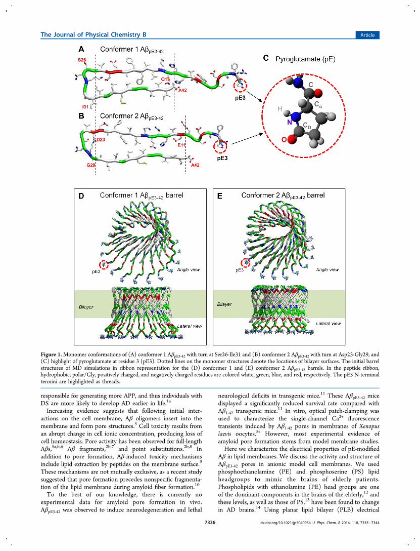
Figure 1.Monomer conformations of (A) conformer 1 AβpE3‑42 with turn at Ser26-Ile31 and (B) conformer 2 AβpE3‑42 with turn at Asp23-Gly29, and(C) highlight of pyroglutamate at residue 3 (pE3). Dotted lines on the monomer structures denote the locations of bilayer surfaces. The initial barrelstructures of MD simulations in ribbon representation for the (D) conformer 1 and (E) conformer 2 AβpE3‑42 barrels. In the peptide ribbon,hydrophobic, polar/Gly, positively charged, and negatively charged residues are colored white, green, blue, and red, respectively. The pE3 N-terminaltermini are highlighted as threads.
The Journal of Physical Chemistry B Article
dx.doi.org/10.1021/jp5040954 | J. Phys. Chem. B 2014, 118, 7335−73447336

recording, we show that AβpE3‑42 induces pore activity inanionic membranes, producing increased membrane perme-ability with respect to Aβ1‑42 pores. Using atomistic moleculardynamics (MD) simulations, we model the architecture ofAβpE3‑42 pores and correlate them with the activity observedexperimentally.
■ MATERIALS AND METHODSMaterials. AβpE3‑42 was purchased from Bachem (Torrance,
CA), and Aβ1‑42 was purchased from Bachem and Anaspec(Fremont, CA). The phospholipids 1,2-dioleoyl-sn-glycero-3-phosphoserine (DOPS) and 1-palmitoyl-2-oleoyl-sn-glycero-3-phosphoethanolamine (POPE) were purchased from AvantiPolar Lipids (Alabaster, AL). All other chemicals werepurchased from Sigma-Aldrich (St. Louis, MO).Peptide Handling. AβpE3‑42 and Aβ1‑42 peptides were
dissolved in Milli-Q water to a concentration of 1 mg/mL priorto being aliquoted for storage. These 50 μL aliquots werestored at −80 °C for a maximum of 60 days before use.Samples were thawed only once.Planar Lipid Bilayer Electrical Recording. We prepared
PLBs using the so-called “painted” technique.15 Bilayers wereformed from a 1:1 (w/w) mixture of DOPS and POPE inheptane at a total lipid concentration of 20 mg/mL.Spontaneous membrane formation occurs following theaddition of lipid directly over a pore with a diameter of ∼250μm in a Delrin septum (Warner Instruments, Delrin perfusioncup, volume 1 mL). In previous studies, this membranecomposition was shown to be stable for long recording times(∼4 h).16 As the electrolyte, we use 150 mM KCl, 10 mMHEPES (pH 7.4), and 1 mM MgCl2.Before performing electrical recordings, we verified that the
bilayer was stable for several minutes with low conductance(<10 pS) across ±100 mV applied voltage and that the systemcapacitance was >110 pF. When both criteria were met, peptidewas added directly to the cis (hot wire) side and stirred for 5min. Peptide concentration in the bilayer chamber was ∼10μM. Bilayer stability was monitored by periodic capacitancemeasurements throughout the course of the experiment.All traces were recorded in voltage clamp mode using the 2
kHz built-in filter cutoff of our BC-535 amplifier (WarnerInstruments, Hamden, CT). A sampling frequency of 15 kHzwas used for all data acquisition. We used a custom-madeLabVIEW program to record the current and Clampfit 10.2(Molecular Devices, Sunnyvale, CA) to analyze traces. Forrepresentation in Figures, we have filtered the recorded currentversus time traces with a digital Gaussian low-pass filter with acutoff frequency of 50 Hz.Atomistic Molecular Dynamics Simulations. Two U-
shaped Aβ monomer conformations with the β-strand-turn-β-strand motif were extracted from Aβ1‑42 fibrils, where thestructure was defined by hydrogen/deuterium-exchange NMRdata, side-chain packing constraints from pairwise mutagenesis,ssNMR and EM (PDB code: 2BEG),17 and small Aβ1‑40protofibrils (PDB codes: 2LMN and 2LMO),18 where thestructure was based on a ssNMR model. In both structures, theN-terminal coordinates, residues 1−16 for the former and 1−8for the latter structure, are missing due to disorder. We used theAβ1−16 coordinates, in the absence of Zn2+ (PDB code:1ZE7),19 for the missing portions of the peptides. For eachcombination of the N-terminal structure with the U-shapedmotifs, two Aβ1‑42 conformers were generated.6c,d,8 Conformer1 has a turn at Ser26-Ile31, and conformer 2 at Asp23-Gly29. In
the latter conformer, two C-terminal residues, Ile41 and Ala42,were added to create Aβ1‑42. To simulate pE, we removed thefirst two residues, Asp1 and Ala2, from each conformer andconverted Glu3 into pE3, generating AβpE3‑42. Because thestandard CHARMM20 force field does not provide a force fieldfor pE, we first created the pE molecular topology using theAvogadro software.21 Then, we calculated partial charges, bondlengths, angles, and torsional angles for the atoms in the pEresidue using the Gaussian09 program22 on a Biowulf cluster atthe NIH. The calculated parameters can be directly adopted inthe CHARMM20 program.Two AβpE3‑42 conformers, each derived from the wild-type
(WT) Aβ1‑42 conformers with different turns, still retain the U-shaped structure with the β-strand-turn-β-strand motif. Toconstruct AβpE3‑42 barrel structures, we inclined AβpE3‑42monomers ∼37° relative to the pore axis;7e then, an 18-foldrotational symmetry operation was performed with respect tothe pore axis creating an 18-mer AβpE3‑42 barrel (Figure 1). Wemodeled Aβ barrels with the β-sheet structure by mimickingnaturally occurring β-barrels observed in transmembraneproteins that are found frequently in the outer membranes ofbacteria, mitochondria, and chloroplasts. The β-barrel motif is alarge β-sheet composed of an even number of β-strands. Someknown structures of β-barrel membrane proteins have β-strandsranging in size from 8 to 22.23 We modeled 18-mer Aβ barrels,with 18 β-strands enclosing the solvated pore. This number isalso in the range of the number of β-strands for natural β-barrels ranging from 8 to 22, which can form a β-barrel motif.Our previous simulations for Aβ channels indicate that differentnumbers of Aβ monomers could produce channels withdifferent outer and pore dimensions.2b,6c,d,7c−e,8a,24 We foundthat Aβ channels obtained a preferred size range of 16−24 β-strands lining the pores.7c,d This range was also found to holdfor other toxic β-sheet channels: K3 channels with 24 β-strands,25 18- and 24-mer hIAPP channels,26 PG-1 channelswith 16−20 β-strands,27 and MAX channels with 20 β-strands.28 In agreement with AFM data, these channels haveouter and pore dimensions within the range found with AFM.In this work, the outer/pore diameters of the 18-mer AβpE3‑42barrels are in good agreement with the experimental AFMranges6b and the computational 18-mer wild-type Aβbarrels.6c,8a The AFM experiments provide images of channelswith a wide variety of sizes and shapes, but simulated Aβ barrelsare limited to cover all ranges of channel sizes that are imagedby AFM.To obtain a lipid bilayer, we constructed a unit cell
containing two layers of lipids. In the middle of the unit cell,lipid molecules were randomly selected from the library of thepreequilibrated state and replaced by pseudo vdW spheres atthe positions of the lipid headgroups, constituting the lipidbilayer topology.29 For a given number of lipid molecules, theoptimal value of lateral cell dimensions can be determined. Ananionic lipid bilayer composed of DOPS and POPE with amole ratio 1:2, containing a total of 420 lipids, constitutes theunit cell with TIP3P waters added at both sides. UpdatedCHARMM20 all-atom additive force field for lipids (C36)30 andthe modified TIP3P water model31 were used to construct theset of starting points and to relax the systems to a production-ready stage. The system contains Mg2+, K+, Ca2+, and Zn2+ atthe same concentration of 25 mM to satisfy a total cationconcentration near 100 mM. The bilayer system containing anAβpE barrel, lipids, salts, and water has almost 200 000 atoms.We generated at least 10 different initial configurations for each
The Journal of Physical Chemistry B Article
dx.doi.org/10.1021/jp5040954 | J. Phys. Chem. B 2014, 118, 7335−73447337
conformer AβpE3‑42 barrel for the relaxation process to obtainthe best initial configuration for a starting point. In the pre-equilibrium stages, a series of minimizations was performed forthe initial configurations to remove overlaps of the alkanechains in the lipids and to gradually relax the solvents aroundthe harmonically restrained peptides. The initial configurationswere gradually relaxed through dynamic cycles with electro-static cutoffs (12 Å). The harmonic restraints were graduallydiminished with the full Ewald electrostatics calculation andconstant temperature (Nose−Hoover) thermostat/barostat at303 K. For t < 30 ns, our simulation employed the NPAT(constant number of atoms, pressure, surface area, andtemperature) ensemble with a constant normal pressure appliedin the direction perpendicular to the membrane. After t = 30 ns,the simulations employed the NPT ensemble. Production runsof 100 ns for the starting points with the NAMD code32 wereperformed on a Biowulf cluster at the NIH. Averages weretaken after 30 ns, discarding initial transients. Analysis wasperformed with the CHARMM programming package.20
■ RESULTSPore Activity. PLB electrical recording data demonstrate
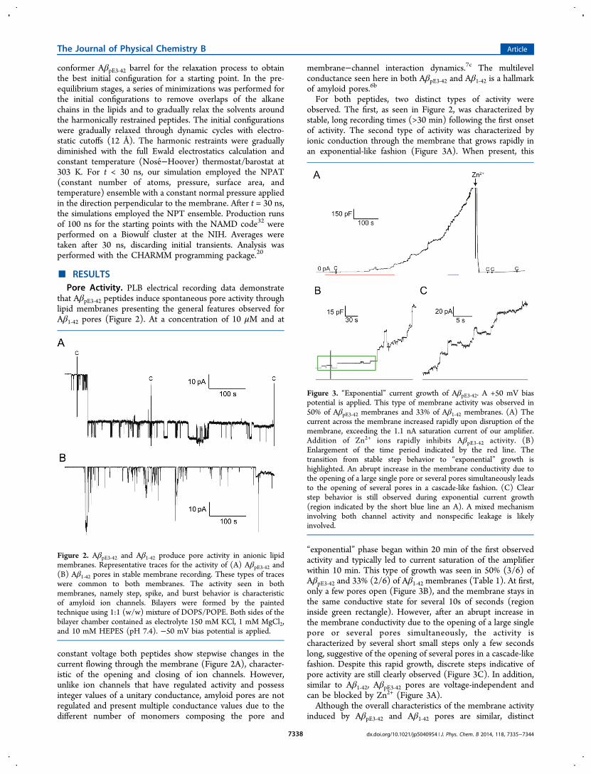
that AβpE3‑42 peptides induce spontaneous pore activity throughlipid membranes presenting the general features observed forAβ1‑42 pores (Figure 2). At a concentration of 10 μM and at
constant voltage both peptides show stepwise changes in thecurrent flowing through the membrane (Figure 2A), character-istic of the opening and closing of ion channels. However,unlike ion channels that have regulated activity and possessinteger values of a unitary conductance, amyloid pores are notregulated and present multiple conductance values due to thedifferent number of monomers composing the pore and
membrane−channel interaction dynamics.7c The multilevelconductance seen here in both AβpE3‑42 and Aβ1‑42 is a hallmarkof amyloid pores.6b
For both peptides, two distinct types of activity wereobserved. The first, as seen in Figure 2, was characterized bystable, long recording times (>30 min) following the first onsetof activity. The second type of activity was characterized byionic conduction through the membrane that grows rapidly inan exponential-like fashion (Figure 3A). When present, this
“exponential” phase began within 20 min of the first observedactivity and typically led to current saturation of the amplifierwithin 10 min. This type of growth was seen in 50% (3/6) ofAβpE3‑42 and 33% (2/6) of Aβ1‑42 membranes (Table 1). At first,only a few pores open (Figure 3B), and the membrane stays inthe same conductive state for several 10s of seconds (regioninside green rectangle). However, after an abrupt increase inthe membrane conductivity due to the opening of a large singlepore or several pores simultaneously, the activity ischaracterized by several short small steps only a few secondslong, suggestive of the opening of several pores in a cascade-likefashion. Despite this rapid growth, discrete steps indicative ofpore activity are still clearly observed (Figure 3C). In addition,similar to Aβ1‑42, AβpE3‑42 pores are voltage-independent andcan be blocked by Zn2+ (Figure 3A).Although the overall characteristics of the membrane activity
induced by AβpE3‑42 and Aβ1‑42 pores are similar, distinct
Figure 2. AβpE3‑42 and Aβ1‑42 produce pore activity in anionic lipidmembranes. Representative traces for the activity of (A) AβpE3‑42 and(B) Aβ1‑42 pores in stable membrane recording. These types of traceswere common to both membranes. The activity seen in bothmembranes, namely step, spike, and burst behavior is characteristicof amyloid ion channels. Bilayers were formed by the paintedtechnique using 1:1 (w/w) mixture of DOPS/POPE. Both sides of thebilayer chamber contained as electrolyte 150 mM KCl, 1 mM MgCl2,and 10 mM HEPES (pH 7.4). −50 mV bias potential is applied.
Figure 3. “Exponential” current growth of AβpE3‑42. A +50 mV biaspotential is applied. This type of membrane activity was observed in50% of AβpE3‑42 membranes and 33% of Aβ1‑42 membranes. (A) Thecurrent across the membrane increased rapidly upon disruption of themembrane, exceeding the 1.1 nA saturation current of our amplifier.Addition of Zn2+ ions rapidly inhibits AβpE3‑42 activity. (B)Enlargement of the time period indicated by the red line. Thetransition from stable step behavior to “exponential” growth ishighlighted. An abrupt increase in the membrane conductivity due tothe opening of a large single pore or several pores simultaneously leadsto the opening of several pores in a cascade-like fashion. (C) Clearstep behavior is still observed during exponential current growth(region indicated by the short blue line an A). A mixed mechanisminvolving both channel activity and nonspecific leakage is likelyinvolved.
The Journal of Physical Chemistry B Article
dx.doi.org/10.1021/jp5040954 | J. Phys. Chem. B 2014, 118, 7335−73447338
differences are present. Table 1 displays the characteristicparameters observed for the electrical activity of both AβpE3‑42and Aβ1‑42. The onset of Aβ1‑42 activity in PLB can be observedas soon as several minutes following peptide addition into thechamber and was typically seen within ∼60 min post-additionwith an average of 31.6 ± 25.7 min. AβpE3‑42 demonstrated asignificantly longer (p < 0.05 for a one-tail t test) and morevariable lag time, with an average of 98.2 ± 68.5 min.We next examined whether there was a difference between
AβpE3‑42 and Aβ1‑42 channel conductances. Figure 4 shows the
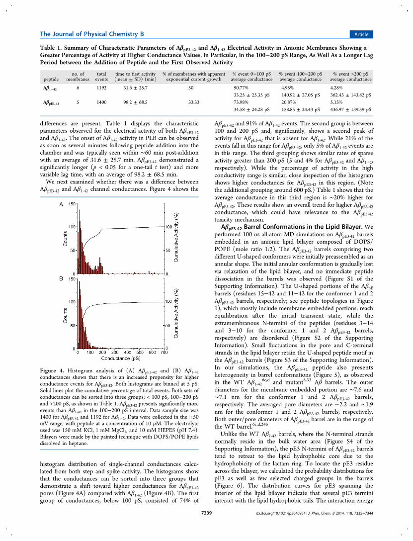
histogram distribution of single-channel conductances calcu-lated from both step and spike activity. The histograms showthat the conductances can be sorted into three groups thatdemonstrate a shift toward higher conductances for AβpE3‑42pores (Figure 4A) compared with Aβ1‑42 (Figure 4B). The firstgroup of conductances, below 100 pS, consisted of 74% of
AβpE3‑42 and 91% of Aβ1‑42 events. The second group is between100 and 200 pS and, significantly, shows a second peak ofactivity for AβpE3‑42 that is absent for Aβ1‑42. While 21% of theevents fall in this range for AβpE3‑42, only 5% of Aβ1‑42 events arein this range. The third grouping shows similar rates of sparseactivity greater than 200 pS (5 and 4% for AβpE3‑42 and Aβ1‑42,respectively). While the percentage of activity in the highconductivity range is similar, close inspection of the histogramshows higher conductances for AβpE3‑42 in this region. (Notethe additional grouping around 600 pS.) Table 1 shows that theaverage conductance in this third region is ∼20% higher forAβpE3‑42. These results show an overall trend for higher AβpE3‑42conductance, which could have relevance to the AβpE3‑42toxicity mechanism.
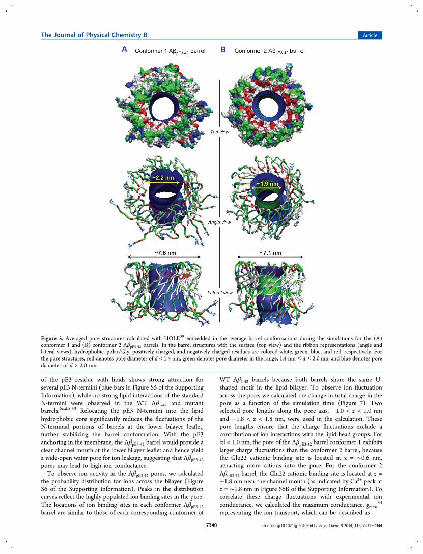
AβpE3‑42 Barrel Conformations in the Lipid Bilayer. Weperformed 100 ns all-atom MD simulations on AβpE3‑42 barrelsembedded in an anionic lipid bilayer composed of DOPS/POPE (mole ratio 1:2). The AβpE3‑42 barrels comprising twodifferent U-shaped conformers were initially preassembled as anannular shape. The initial annular conformation is gradually lostvia relaxation of the lipid bilayer, and no immediate peptidedissociation in the barrels was observed (Figure S1 of theSupporting Information). The U-shaped portions of the AβpEbarrels (residues 15−42 and 11−42 for the conformer 1 and 2AβpE3‑42 barrels, respectively; see peptide topologies in Figure1), which mostly include membrane embedded portions, reachequilibration after the initial transient state, while theextramembranous N-termini of the peptides (residues 3−14and 3−10 for the conformer 1 and 2 AβpE3‑42 barrels,respectively) are disordered (Figure S2 of the SupportingInformation). Small fluctuations in the pore and C-terminalstrands in the lipid bilayer retain the U-shaped peptide motif inthe AβpE3‑42 barrels (Figure S3 of the Supporting Information).In our simulations, the AβpE3‑42 peptide also presentsheterogeneity in barrel conformations (Figure 5), as observedin the WT Aβ1‑42
6c,d and mutant8,33 Aβ barrels. The outerdiameters for the membrane embedded portion are ∼7.6 and∼7.1 nm for the conformer 1 and 2 AβpE3‑42 barrels,respectively. The averaged pore diameters are ∼2.2 and ∼1.9nm for the conformer 1 and 2 AβpE3‑42 barrels, respectively.Both outer/pore diameters of AβpE3‑42 barrel are in the range ofthe WT barrel.6c,d,24b
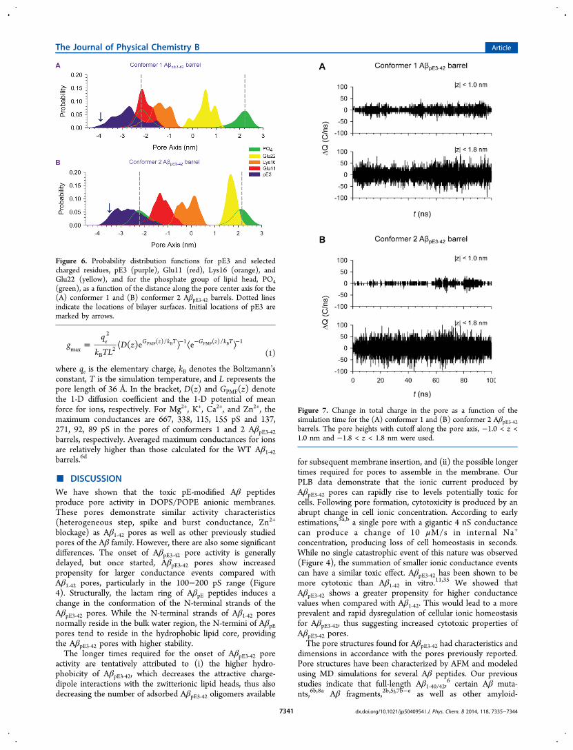
Unlike the WT Aβ1‑42 barrels, where the N-terminal strandsnormally reside in the bulk water area (Figure S4 of theSupporting Information), the pE3 N-termini of AβpE3‑42 barrelstend to retreat to the lipid hydrophobic core due to thehydrophobicity of the lactam ring. To locate the pE3 residueacross the bilayer, we calculated the probability distributions forpE3 as well as few selected charged groups in the barrels(Figure 6). The distribution curves for pE3 spanning theinterior of the lipid bilayer indicate that several pE3 terminiinteract with the lipid hydrophobic tails. The interaction energy
Table 1. Summary of Characteristic Parameters of AβpE3‑42 and Aβ1‑42 Electrical Activity in Anionic Membranes Showing aGreater Percentage of Activity at Higher Conductance Values, in Particular, in the 100−200 pS Range, As Well As a Longer LagPeriod between the Addition of Peptide and the First Observed Activity
peptideno. of
membranestotalevents
time to first activity(mean ± SD) (min)
% of membranes with apparentexponential current growth
% event 0−100 pSaverage conductance
% event 100−200 pSaverage conductance
% event >200 pSaverage conductance
Aβ1−42 6 1192 31.6 ± 25.7 50 90.77% 4.95% 4.28%33.25 ± 25.35 pS 140.92 ± 27.05 pS 362.43 ± 143.82 pS
AβpE3‑42 5 1400 98.2 ± 68.5 33.33 73.98% 20.87% 5.15%34.58 ± 24.28 pS 158.85 ± 24.43 pS 436.97 ± 139.59 pS
Figure 4. Histogram analysis of (A) AβpE3‑42 and (B) Aβ1‑42conductances shows that there is an increased propensity for higherconductance events for AβpE3‑42. Both histograms are binned at 5 pS.Solid lines plot the cumulative percentage of total events. Both sets ofconductances can be sorted into three groups; < 100 pS, 100−200 pSand >200 pS, as shown in Table 1. AβpE3‑42 presents significantly moreevents than Aβ1‑42 in the 100−200 pS interval. Data sample size was1400 for AβpE3‑42 and 1192 for Aβ1‑42. Data were collected in the ±50mV range, with peptide at a concentration of 10 μM. The electrolyteused was 150 mM KCl, 1 mM MgCl2, and 10 mM HEPES (pH 7.4).Bilayers were made by the painted technique with DOPS/POPE lipidsdissolved in heptane.
The Journal of Physical Chemistry B Article
dx.doi.org/10.1021/jp5040954 | J. Phys. Chem. B 2014, 118, 7335−73447339
of the pE3 residue with lipids shows strong attraction forseveral pE3 N-termini (blue bars in Figure S5 of the SupportingInformation), while no strong lipid interactions of the standardN-termini were observed in the WT Aβ1‑42 and mutantbarrels.6c,d,8,33 Relocating the pE3 N-termini into the lipidhydrophobic core significantly reduces the fluctuations of theN-terminal portions of barrels at the lower bilayer leaflet,further stabilizing the barrel conformation. With the pE3anchoring in the membrane, the AβpE3‑42 barrel would provide aclear channel mouth at the lower bilayer leaflet and hence yielda wide-open water pore for ion leakage, suggesting that AβpE3‑42pores may lead to high ion conductance.To observe ion activity in the AβpE3‑42 pores, we calculated
the probability distribution for ions across the bilayer (FigureS6 of the Supporting Information). Peaks in the distributioncurves reflect the highly populated ion binding sites in the pore.The locations of ion binding sites in each conformer AβpE3‑42barrel are similar to those of each corresponding conformer of
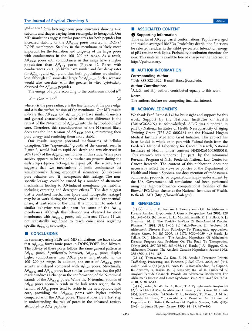
WT Aβ1‑42 barrels because both barrels share the same U-shaped motif in the lipid bilayer. To observe ion fluctuationacross the pore, we calculated the change in total charge in thepore as a function of the simulation time (Figure 7). Twoselected pore lengths along the pore axis, −1.0 < z < 1.0 nmand −1.8 < z < 1.8 nm, were used in the calculation. Thesepore lengths ensure that the charge fluctuations exclude acontribution of ion interactions with the lipid head groups. For|z| < 1.0 nm, the pore of the AβpE3‑42 barrel conformer 1 exhibitslarger charge fluctuations than the conformer 2 barrel, becausethe Glu22 cationic binding site is located at z = ∼0.6 nm,attracting more cations into the pore. For the conformer 2AβpE3‑42 barrel, the Glu22 cationic binding site is located at z =∼1.8 nm near the channel mouth (as indicated by Ca2+ peak atz = ∼1.8 nm in Figure S6B of the Supporting Information). Tocorrelate these charge fluctuations with experimental ionconductance, we calculated the maximum conductance, gmax,
34
representing the ion transport, which can be described as
Figure 5. Averaged pore structures calculated with HOLE38 embedded in the average barrel conformations during the simulations for the (A)conformer 1 and (B) conformer 2 AβpE3‑42 barrels. In the barrel structures with the surface (top view) and the ribbon representations (angle andlateral views), hydrophobic, polar/Gly, positively charged, and negatively charged residues are colored white, green, blue, and red, respectively. Forthe pore structures, red denotes pore diameter of d < 1.4 nm, green denotes pore diameter in the range, 1.4 nm ≤ d ≤ 2.0 nm, and blue denotes porediameter of d > 2.0 nm.
The Journal of Physical Chemistry B Article
dx.doi.org/10.1021/jp5040954 | J. Phys. Chem. B 2014, 118, 7335−73447340
= ⟨ ⟩ ⟨ ⟩− − −gq
k TLD z( )e ee G z k T G z k T
max
2
B2
( )/ 1 ( )/ 1PMF B PMF B
(1)
where qe is the elementary charge, kB denotes the Boltzmann’sconstant, T is the simulation temperature, and L represents thepore length of 36 Å. In the bracket, D(z) and GPMF(z) denotethe 1-D diffusion coefficient and the 1-D potential of meanforce for ions, respectively. For Mg2+, K+, Ca2+, and Zn2+, themaximum conductances are 667, 338, 115, 155 pS and 137,271, 92, 89 pS in the pores of conformers 1 and 2 AβpE3‑42barrels, respectively. Averaged maximum conductances for ionsare relatively higher than those calculated for the WT Aβ1‑42barrels.6d
■ DISCUSSIONWe have shown that the toxic pE-modified Aβ peptidesproduce pore activity in DOPS/POPE anionic membranes.These pores demonstrate similar activity characteristics(heterogeneous step, spike and burst conductance, Zn2+
blockage) as Aβ1‑42 pores as well as other previously studiedpores of the Aβ family. However, there are also some significantdifferences. The onset of AβpE3‑42 pore activity is generallydelayed, but once started, AβpE3‑42 pores show increasedpropensity for larger conductance events compared withAβ1‑42 pores, particularly in the 100−200 pS range (Figure4). Structurally, the lactam ring of AβpE peptides induces achange in the conformation of the N-terminal strands of theAβpE3‑42 pores. While the N-terminal strands of Aβ1‑42 poresnormally reside in the bulk water region, the N-termini of AβpEpores tend to reside in the hydrophobic lipid core, providingthe AβpE3‑42 pores with higher stability.The longer times required for the onset of AβpE3‑42 pore
activity are tentatively attributed to (i) the higher hydro-phobicity of AβpE3‑42, which decreases the attractive charge-dipole interactions with the zwitterionic lipid heads, thus alsodecreasing the number of adsorbed AβpE3‑42 oligomers available
for subsequent membrane insertion, and (ii) the possible longertimes required for pores to assemble in the membrane. OurPLB data demonstrate that the ionic current produced byAβpE3‑42 pores can rapidly rise to levels potentially toxic forcells. Following pore formation, cytotoxicity is produced by anabrupt change in cell ionic concentration. According to earlyestimations,5a,b a single pore with a gigantic 4 nS conductancecan produce a change of 10 μM/s in internal Na+
concentration, producing loss of cell homeostasis in seconds.While no single catastrophic event of this nature was observed(Figure 4), the summation of smaller ionic conductance eventscan have a similar toxic effect. AβpE3‑42 has been shown to bemore cytotoxic than Aβ1‑42 in vitro.11,35 We showed thatAβpE3‑42 shows a greater propensity for higher conductancevalues when compared with Aβ1‑42. This would lead to a moreprevalent and rapid dysregulation of cellular ionic homeostasisfor AβpE3‑42, thus suggesting increased cytotoxic properties ofAβpE3‑42 pores.The pore structures found for AβpE3‑42 had characteristics and
dimensions in accordance with the pores previously reported.Pore structures have been characterized by AFM and modeledusing MD simulations for several Aβ peptides. Our previousstudies indicate that full-length Aβ1‑40/42,
6 certain Aβ muta-nts,6b,8a Aβ fragments,2b,5j,7b−e as well as other amyloid-
Figure 6. Probability distribution functions for pE3 and selectedcharged residues, pE3 (purple), Glu11 (red), Lys16 (orange), andGlu22 (yellow), and for the phosphate group of lipid head, PO4(green), as a function of the distance along the pore center axis for the(A) conformer 1 and (B) conformer 2 AβpE3‑42 barrels. Dotted linesindicate the locations of bilayer surfaces. Initial locations of pE3 aremarked by arrows.
Figure 7. Change in total charge in the pore as a function of thesimulation time for the (A) conformer 1 and (B) conformer 2 AβpE3‑42barrels. The pore heights with cutoff along the pore axis, −1.0 < z <1.0 nm and −1.8 < z < 1.8 nm were used.
The Journal of Physical Chemistry B Article
dx.doi.org/10.1021/jp5040954 | J. Phys. Chem. B 2014, 118, 7335−73447341
s6a,b,25,27a,36 form heterogeneous pore structures showing 4−6subunits and shapes varying from rectangular to hexagonal. OurMD simulations suggest similar pore sizes for both peptides butincreased stability of the AβpE3‑42 pores inserted in DOPS/POPE membranes. Stability in the membrane is likely moreimportant for the formation and longevity of the larger poreswith conductances in the 100−200 pS range. As a result,AβpE3‑42 pores with conductances in this range have a higherpopulation than Aβ1‑42 pores (Figure 4). Pores withconductances >200 pS likely have similar and fast decay ratesfor AβpE3‑42 and Aβ1‑42, and thus both populations are similarlylow, although still somewhat larger for AβpE3‑42. Such a scenariowould also correlate with the greater in vitro cytotoxicityobserved for AβpE3‑42 peptides.The energy of a pore according to the continuum model is37
γ π σπ= −E r r2 2 (2)
where r is the pore radius, γ is the line tension at the pore edge,and σ is the surface tension of the membrane. Our MD resultsindicate that AβpE3‑42 and Aβ1‑42 pores have similar diametersand general characteristics, while the main difference is theretreat of the N-termini of AβpE3‑42 into the hydrophobic lipidcore. Therefore, this reconfiguration of the N-termini likelydecreases the line tension of AβpE3‑42 pores, minimizing theirpore energy and rendering them more stable.Our results suggest potential mechanisms of cellular
disruption. The “exponential” growth of the current, seen inFigure 3, would lead to rapid cell death and was observed in50% (3/6) of the AβpE3‑42 membranes in this study. While poreactivity appears to be the only mechanism present during theearly stages (green rectangle in Figure 3B), the activity tracesuggests that two mechanisms of ionic conductance actsimultaneously during exponential saturation: (i) stepwisepore behavior and (ii) nonspecific drift leakage. The non-specific leakage could be caused by a number of alternativemechanisms leading to Aβ-induced membrane permeability,including carpeting and detergent effects.9b The data suggestthat a combined mechanism (pore and nonspecific leakage)may be at work during the rapid growth of the “exponential”phase, at least some of the time. It is important to note thatsimilar behavior was also seen for some of the Aβ1‑42membranes. Although this behavior was observed for moremembranes with AβpE3‑42 pores, this difference (Table 1) wasnot statistically significant to reach conclusions regardingspecific AβpE3‑42 cytotoxity.
■ CONCLUSIONSIn summary, using PLBs and MD simulations, we have shownthat AβpE3‑42 forms ionic pores in DOPS/POPE lipid bilayers.The activity of these pores follows the same general pattern asAβ1‑42 pores. Significantly, however, AβpE3‑42 pores displayhigher conductances than Aβ1‑42 pores, in particular, in the100−200 pS range. In addition, the onset of AβpE3‑42 poreactivity is delayed compared with Aβ1‑42 pores. Structurally,AβpE3‑42 and Aβ1‑42 pores have similar dimensions, but the pE3residue induces a change in the conformation of the N-terminalstrands of the AβpE3‑42 pores. While the N-terminal strands ofAβ1‑42 pores normally reside in the bulk water region, the N-termini of AβpE pores tend to reside in the hydrophobic lipidcore, providing the AβpE3‑42 pores with higher stabilitycompared with the Aβ1‑42 pores. These studies are a first stepin understanding the role of pores in the enhanced toxicityattributed to AβpE peptides.
■ ASSOCIATED CONTENT*S Supporting InformationTime series of AβpE3‑42 barrel conformations. Peptide-averagedand residue-averaged RMSDs. Probability distribution functionsfor selected residues in the wild-type barrels. Interaction energyof pE3 residue with lipids. Probability distribution functions forions. This material is available free of charge via the Internet athttp://pubs.acs.org.
■ AUTHOR INFORMATIONCorresponding Author*Tel: 858-822-1322. E-mail: [email protected] Contributions#A.L.G. and H.J. authors contributed equally to this workNotesThe authors declare no competing financial interest.
■ ACKNOWLEDGMENTSWe thank Prof. Ratnesh Lal for his insight and support for thiswork. Support by the National Institutes of Health(R01AG028709) is acknowledged. A.L.G was supported inpart by National Institutes of Health Neuroplasticity of AgingTraining Grant (T32 AG 000216) and the Howard HughesMedical Institute Med Into Grad Initiative. This project hasbeen funded in whole or in part with Federal funds from theFrederick National Laboratory for Cancer Research, NationalInstitutes of Health, under contract HHSN261200800001E.This research was supported [in part] by the IntramuralResearch Program of NIH, Frederick National Lab, Center forCancer Research. The content of this publication does notnecessarily reflect the views or policies of the Department ofHealth and Human Services, nor does mention of trade names,commercial products, or organizations imply endorsement bythe U.S. Government. All simulations had been performedusing the high-performance computational facilities of theBiowulf PC/Linux cluster at the National Institutes of Health,Bethesda, MD (http://biowulf.nih.gov).
■ REFERENCES(1) (a) Tanzi, R. E.; Bertram, L. Twenty Years Of The Alzheimer’sDisease Amyloid Hypothesis: A Genetic Perspective. Cell 2005, 120(4), 545−555. (b) Iversen, L. L.; Mortishiresmith, R. J.; Pollack, S. J.;Shearman, M. S. The Toxicity In-Vitro Of Beta-Amyloid Protein.Biochem. J. 1995, 311, 1−16. (c) Jakob-Roetne, R.; Jacobsen, H.Alzheimer’s Disease: From Pathology To Therapeutic Approaches.Angew. Chem., Int. Ed. 2009, 48 (17), 3030−3059. (d) Hardy, J.;Selkoe, D. J. Medicine - The Amyloid Hypothesis Of Alzheimer’sDisease: Progress And Problems On The Road To Therapeutics.Science 2002, 297 (5580), 353−356. (e) Hardy, J. A.; Higgins, G. A.Alzheimers Disease: The Amyloid Cascade Hypothesis. Science 1992,256 (5054), 184−185.(2) (a) Thinakaran, G.; Koo, E. H. Amyloid Precursor ProteinTrafficking, Processing, and Function. J. Biol. Chem. 2008, 283 (44),29615−29619. (b) Jang, H.; Arce, F. T.; Ramachandran, S.; Capone,R.; Azimova, R.; Kagan, B. L.; Nussinov, R.; Lal, R. Truncated B-Amyloid Peptide Channels Provide An Alternative Mechanism ForAlzheimer’s Disease And Down Syndrome. Proc. Natl. Acad. Sci. U.S.A.2010, 6538−6543.(3) (a) Jawhar, S.; Wirths, O.; Bayer, T. A. Pyroglutamate Amyloid-B(Aβ): A Hatchet Man In Alzheimer Disease. J. Biol. Chem. 2011, 286(45), 38825−38832. (b) Saido, T. C.; Iwatsubo, T.; Mann, D. M. A.;Shimada, H.; Ihara, Y.; Kawashima, S. Dominant And DifferentialDeposition Of Distinct Beta-Amyloid Peptide Species, A-Beta(N3-(Pe)), In Senile Plaques. Neuron 1995, 14 (2), 457−466.
The Journal of Physical Chemistry B Article
dx.doi.org/10.1021/jp5040954 | J. Phys. Chem. B 2014, 118, 7335−73447342
(4) (a) Saido, T. C.; Yamaoharigaya, W.; Iwatsubo, T.; Kawashima, S.Amino- And Carboxyl-Terminal Heterogeneity Of Beta-AmyloidPeptides Deposited In Human Brain. Neurosci. Lett. 1996, 215 (3),173−176. (b) Piccini, A.; Russo, C.; Gliozzi, A.; Relini, A.; Vitali, A.;Borghi, R.; Giliberto, L.; Armirotti, A.; D’Arrigo, C.; Bachi, A.; et al.Beta-Amyloid Is Different In Normal Aging And In Alzheimer Disease.J. Biol. Chem. 2005, 280 (40), 34186−34192. (c) Tabaton, M.; Piccini,A. Role Of Water-Soluble Amyloid-Beta In The Pathogenesis OfAlzheimer’s Disease. Int. J. Exp. Pathol. 2005, 86 (3), 139−145.(5) (a) Arispe, N.; Pollard, H. B.; Rojas, E. Giant Multilevel CationChannels Formed By Alzheimer Disease Amyloid Beta-Protein [ABeta P-(1−40)] In Bilayer Membranes. Proc. Natl. Acad. Sci. U.S.A.1993, 90 (22), 10573−10577. (b) Arispe, N.; Rojas, E.; Pollard, H. B.Alzheimer Disease Amyloid B Protein Forms Calcium Channels InBilayer Membranes: Blockade By Tromethamine And Aluminum. Proc.Natl. Acad. Sci. U.S.A. 1993, 90 (2), 567−571. (c) Capone, R.; Quiroz,F. G.; Prangkio, P.; Saluja, I.; Sauer, A. M.; Bautista, M. R.; Turner, R.S.; Yang, J.; Mayer, M. Amyloid-Beta-Induced Ion Flux In ArtificialLipid Bilayers And Neuronal Cells: Resolving A Controversy.Neurotoxic. Res. 2009, 16 (1), 1−13. (d) Prangkio, P.; Yusko, E. C.;Sept, D.; Yang, J.; Mayer, M. Multivariate Analyses Of Amyloid-BetaOligomer Populations Indicate A Connection Between PoreFormation And Cytotoxicity. PLoS One 2012, 7, 10. (e) Demuro,A.; Smith, M.; Parker, I. Single-Channel Ca2+ Imaging ImplicatesAβ1−42 Amyloid Pores In Alzheimer’s Disease Pathology. J. Cell Biol.2011, 195 (3), 515−524. (f) Kagan, B. L.; Azimov, R.; Azimova, R.Amyloid Peptide Channels. J. Membr. Biol. 2004, 202 (1), 1−10.(g) Kagan, B. L.; Hirakura, Y.; Azimov, R.; Azimova, R.; Lin, M. C.The Channel Hypothesis Of Alzheimer’s Disease: Current Status.Peptides 2002, 23 (7), 1311−1315. (h) Kim, H. Y.; Cho, M. K.;Kumar, A.; Maier, E.; Siebenhaar, C.; Becker, S.; Fernandez, C. O.;Lashuel, H. A.; Benz, R.; Lange, A.; et al. Structural Properties OfPore-Forming Oligomers Of Alpha-Synuclein. J. Am. Chem. Soc. 2009,131 (47), 17482−17489. (i) Jang, H.; Connelly, L.; Arce, F. T.;Ramachandran, S.; Lal, R.; Kagan, B.; Nussinov, R. Alzheimer’sDisease: Which Type Of Amyloid-Preventing Drug Agents ToEmploy? Phys. Chem. Chem. Phys. 2013, 15, 8868−8877. (j) Jang,H.; Connelly, L.; Arce, F. T.; Ramachandran, S.; Kagan, B. L.; Lal, R.;Nussinov, R. Mechanisms For The Insertion Of Toxic, Fibril-LikeBeta-Amyloid Oligomers Into The Membrane. J. Chem. TheoryComput. 2013, 9 (1), 822−833.(6) (a) Lin, H. A. I.; Bhatia, R.; Lal, R. Amyloid B Protein Forms IonChannels: Implications For Alzheimer’s Disease Pathophysiology.FASEB J. 2001, 15 (13), 2433−2444. (b) Quist, A.; Doudevski, I.; Lin,H.; Azimova, R.; Ng, D.; Frangione, B.; Kagan, B.; Ghiso, J.; Lal, R.Amyloid Ion Channels: A Common Structural Link For Protein-Misfolding Disease. Proc. Natl. Acad. Sci. U.S.A. 2005, 102 (30),10427−10432. (c) Connelly, L.; Arce, F. T.; Jang, H.; Capone, R.;Kotler, S. A.; Ramachandran, S.; Kagan, B. L.; Nussinov, R.; Lal, R.Atomic Force Microscopy And MD Simulations Reveal Pore-LikeStructures Of All-D-Enantiomer Of Alzheimer’s B-Amyloid Peptide:Relevance To The Ion Channel Mechanism Of AD Pathology. J. Phys.Chem. B 2012, 116 (5), 1728−1735. (d) Capone, R.; Jang, H.; Kotler,S. A.; Connelly, L.; Arce, F. T.; Ramachandran, S.; Kagan, B. L.;Nussinov, R.; Lal, R. All-D-Enantiomer Of Beta-Amyloid PeptideForms Ion Channels In Lipid Bilayers. J. Chem. Theory Comput. 2012,8 (3), 1143−1152.(7) (a) Lin, M. C. A.; Kagan, B. L. Electrophysiologic Properties OfChannels Induced By A Beta 25−35 In Planar Lipid Bilayers. Peptides2002, 23 (7), 1215−1228. (b) Arce, F. T.; Jang, H.; Ramachandran, S.;Landon, P. B.; Nussinov, R.; Lal, R. Polymorphism Of Amyloid BPeptide In Different Environments: Implications For MembraneInsertion And Pore Formation. Soft Matter 2011, 7 (11), 5267−5273.(c) Jang, H.; Arce, F. T.; Capone, R.; Ramachandran, S.; Lal, R.;Nussinov, R. Misfolded Amyloid Ion Channels Present Mobile B-Sheet Subunits In Contrast To Conventional Ion Channels. Biophys. J.2009, 97 (11), 3029−3037. (d) Jang, H.; Arce, F. T.; Ramachandran,S.; Capone, R.; Lal, R.; Nussinov, R. Structural Convergence AmongDiverse, Toxic B-Sheet Ion Channels. J. Phys. Chem. B 2010, 114 (29),
9445−9451. (e) Jang, H.; Arce, F. T.; Ramachandran, S.; Capone, R.;Lal, R.; Nussinov, R. B-Barrel Topology Of Alzheimer’s B-Amyloid IonChannels. J. Mol. Biol. 2010, 404 (5), 917−934.(8) (a) Connelly, L.; Jang, H.; Arce, F. T.; Ramachandran, S.; Kagan,B. L.; Nussinov, R.; Lal, R. Effects Of Point Substitutions On TheStructure Of Toxic Alzheimer’s B-Amyloid Channels: Atomic ForceMicroscopy And Molecular Dynamics Simulations. Biochemistry 2012,51 (14), 3031−3038. (b) Connelly, L.; Jang, H.; Teran Arce, F.;Capone, R.; Kotler, S. A.; Ramachandran, S.; Kagan, B. L.; Nussinov,R.; Lal, R. Atomic Force Microscopy And MD Simulations RevealPore-Like Structures Of All-D-Enantiomer Of Alzheimer’s B-AmyloidPeptide: Relevance To The Ion Channel Mechanism Of ADPathology. J. Phys. Chem. B 2012, 116 (5), 1728−1735.(9) (a) Butterfield, S. M.; Lashuel, H. A. Amyloidogenic Protein−Membrane Interactions: Mechanistic Insight From Model Systems.Angew. Chem., Int. Ed. 2010, 49, 5628−5654. (b) Williams, T. L.;Serpell, L. C. Membrane And Surface Interactions Of Alzheimer’s ABeta Peptide - Insights Into The Mechanism Of Cytotoxicity. FEBS J.2011, 278 (20), 3905−3917.(10) Sciacca, M. F. M.; Kotler, S. A.; Brender, J. R.; Chen, J.; Lee, D.K.; Ramamoorthy, A. Two-Step Mechanism Of Membrane DisruptionBy A Beta Through Membrane Fragmentation And Pore Formation.Biophys. J. 2012, 103 (4), 702−710.(11) Wirths, O.; Breyhan, H.; Cynis, H.; Schilling, S.; Demuth, H. U.;Bayer, T. A. Intraneuronal Pyroglutamate-Abeta 3−42 TriggersNeurodegeneration And Lethal Neurological Deficits In A TransgenicMouse Model. Acta Neuropathol. 2009, 118 (4), 487−496.(12) Soderberg, M.; Edlund, C.; Alafuzoff, I.; Kristensson, K.;Dallner, G. Lipid-Composition In Different Regions Of The Brain InAlzheimers-Disease Senile Dementia Of Alzheimers Type. J. Neuro-chem. 1992, 59 (5), 1646−1653.(13) Wells, K.; Farooqui, A. A.; Liss, L.; Horrocks, L. A. NeuralMembrane Phospholipids In Alzheimer Disease. Neurochem. Res. 1995,20 (11), 1329−1333.(14) (a) Nitsch, R. M.; Blusztajn, J. K.; Pittas, A. G.; Slack, B. E.;Growdon, J. H.; Wurtman, R. J. Evidence For A Membrane Defect InAlzheimer-Disease Brain. Proc. Natl. Acad. Sci. U.S.A. 1992, 89 (5),1671−1675. (b) Svennerholm, L.; Gottfries, C. G. Membrane-Lipids,Selectively Diminished In Alzheimer Brains, Suggest Synapse Loss AsA Primary Event In Early-Onset Form (Type-I) And Demyelination InLate-Onset Form (Type-II). J. Neurochem. 1994, 62 (3), 1039−1047.(15) Mueller, P.; Rudin, D. O.; Tien, H. T.; Wescott, W. C.Reconstitution Of Cell Membrane Structure In Vitro And ItsTransformation Into An Excitable System. Nature 1962, 194 (4832),979−980.(16) Capone, R.; Jang, H.; Kotler, S. A.; Kagan, B. L.; Nussinov, R.;Lal, R. Probing Structural Features Of Alzheimer’s Amyloid-Beta PoresIn Bilayers Using Site-Specific Amino Acid Substitutions. Biochemistry2012, 51 (3), 776−785.(17) Luhrs, T.; Ritter, C.; Adrian, M.; Riek-Loher, D.; Bohrmann, B.;Doeli, H.; Schubert, D.; Riek, R. 3D Structure Of Alzheimer’sAmyloid-B(1−42) Fibrils. Proc. Natl. Acad. Sci. U. S. A. 2005, 102 (48),17342−17347.(18) Petkova, A. T.; Yau, W. M.; Tycko, R. Experimental ConstraintsOn Quaternary Structure In Alzheimer’s B-Amyloid Fibrils. Bio-chemistry 2006, 45 (2), 498−512.(19) Zirah, S.; Kozin, S. A.; Mazur, A. K.; Blond, A.; Cheminant, M.;Segalas-Milazzo, I.; Debey, P.; Rebuffat, S. Structural Changes OfRegion 1−16 Of The Alzheimer Disease Amyloid B-Peptide UponZinc Binding And In Vitro Aging. J. Biol. Chem. 2006, 281 (4), 2151−2161.(20) Brooks, B. R.; Bruccoleri, R. E.; Olafson, B. D.; States, D. J.;Swaminathan, S.; Karplus, M. Charmm - A Program For Macro-molecular Energy, Minimization, And Dynamics Calculations. J.Comput. Chem. 1983, 4 (2), 187−217.(21) Hanwell, M. D.; Curtis, D. E.; Lonie, D. C.; Vandermeersch, T.;Zurek, E.; Hutchison, G. R. Avogadro: An Advanced SemanticChemical Editor, Visualization, And Analysis Platform. J. Cheminform2012, 4 (1), 17.
The Journal of Physical Chemistry B Article
dx.doi.org/10.1021/jp5040954 | J. Phys. Chem. B 2014, 118, 7335−73447343

(22) Frisch, M. J.; Trucks, G. W.; Schlegel, H. B.; Scuseria, G. E.;Robb, M. A.; Cheeseman, J. R.; Scalmani, G.; Barone, V.; Mennucci,B.; Petersson, G. A.; et al. Gaussian 09, revision B.01; Gaussian, Inc.:Wallingford CT, 2009.(23) Schulz, G. E. The Structure Of Bacterial Outer MembraneProteins. Biochim. Biophys. Acta 2002, 1565 (2), 308−317.(24) (a) Capone, R.; Jang, H.; Kotler, S. A.; Kagan, B. L.; Nussinov,R.; Lal, R. Probing Structural Features Of Alzheimer’s Amyloid-BPores In Bilayers Using Site-Specific Amino Acid Substitutions.Biochemistry 2012, 51 (3), 776−785. (b) Jang, H.; Teran Arce, F.;Ramachandran, S.; Kagan, B. L.; Lal, R.; Nussinov, R. DisorderedAmyloidogenic Peptides May Insert Into The Membrane AndAssemble Into Common Cyclic Structural Motifs. Chem. Soc. Rev.2014, DOI: 10.1039/C3CS60459D. (c) Jang, H.; Zheng, J.; Lal, R.;Nussinov, R. New Structures Help The Modeling Of Toxic AmyloidβIon Channels. Trends Biochem. Sci. 2008, 33 (2), 91−100. (d) Jang, H.;Zheng, J.; Nussinov, R. Models Of B-Amyloid Ion Channels In TheMembrane Suggest That Channel Formation In The Bilayer Is ADynamic Process. Biophys. J. 2007, 93 (6), 1938−1949. (e) Jang, H.;Arce, F. T.; Ramachandran, S.; Kagan, B. L.; Lal, R.; Nussinov, R.Familial Alzheimer’s Disease Osaka Mutant (ΔE22) B-Barrels SuggestAn Explanation For The Different Aβ(1−40/42) Preferred Conforma-tional States Observed By Experiment. J. Phys. Chem. B 2013, 117(39), 11518−11529.(25) Mustata, M.; Capone, R.; Jang, H.; Arce, F. T.; Ramachandran,S.; Lal, R.; Nussinov, R. K3 Fragment Of Amyloidogenic Beta(2)-Microglobulin Forms Ion Channels: Implication For Dialysis RelatedAmyloidosis. J. Am. Chem. Soc. 2009, 131 (41), 14938−14945.(26) Zhao, J.; Luo, Y.; Jang, H. B.; Yu, X.; Wei, G. H.; Nussinov, R.;Zheng, J. Probing Ion Channel Activity Of Human Islet AmyloidPolypeptide (Amylin). Biochim. Biophys. Acta, Biomembr. 2012, 1818(12), 3121−3130.(27) (a) Capone, R.; Mustata, M.; Jang, H.; Arce, F. T.; Nussinov, R.;Lal, R. Antimicrobial Protegrin-1 Forms Ion Channels: MolecularDynamic Simulation, Atomic Force Microscopy, And ElectricalConductance Studies. Biophys. J. 2010, 98 (11), 2644−2652.(b) Jang, H.; Ma, B.; Lal, R.; Nussinov, R. Models Of Toxic Beta-Sheet Channels Of Protegrin-1 Suggest A Common SubunitOrganization Motif Shared With Toxic Alzheimer Beta-Amyloid IonChannels. Biophys. J. 2008, 95 (10), 4631−4642.(28) Gupta, K.; Jang, H.; Harlen, K.; Puri, A.; Nussinov, R.;Schneider, J. P.; Blumenthal, R. Mechanism Of Membrane PermeationInduced By Synthetic Beta-Hairpin Peptides. Biophys. J. 2013, 105 (9),2093−2103.(29) Woolf, T. B.; Roux, B. Molecular Dynamics Simulation Of TheGramicidin Channel In A Phospholipid Bilayer. Proc. Natl. Acad. Sci.U.S.A. 1994, 91 (24), 11631−11635.(30) Klauda, J. B.; Venable, R. M.; Freites, J. A.; O’Connor, J. W.;Tobias, D. J.; Mondragon-Ramirez, C.; Vorobyov, I.; Mackerell, A. D.;Pastor, R. W. Update Of The CHARMM All-Atom Additive ForceField For Lipids: Validation On Six Lipid Types. J. Phys. Chem. B 2010,114 (23), 7830−7843.(31) Durell, S. R.; Brooks, B. R.; Bennaim, A. Solvent-Induced ForcesBetween Two Hydrophilic Groups. J. Phys. Chem. 1994, 98, 2198−2202.(32) Phillips, J. C.; Braun, R.; Wang, W.; Gumbart, J.; Tajkhorshid,E.; Villa, E.; Chipot, C.; Skeel, R. D.; Kale, L.; Schulten, K. ScalableMolecular Dynamics With NAMD. J. Comput. Chem. 2005, 26, 1781−1802.(33) Jang, H.; Teran Arce, F.; Ramachandran, S.; Kagan, B. L.; Lal,R.; Nussinov, R. Familial Alzheimer’s Disease Osaka Mutant(Deltae22) Beta-Barrels Suggest An Explanation For The DifferentAbeta1−40/42 Preferred Conformational States Observed By Experi-ment. J. Phys. Chem. B 2013, 117 (39), 11518−11529.(34) Allen, T. W.; Andersen, O. S.; Roux, B. Energetics Of IonConduction Through The Gramicidin Channel. Proc. Natl. Acad. Sci.U.S.A. 2004, 101 (1), 117−122.(35) Nussbaum, J. M.; Schilling, S.; Cynis, H.; Silva, A.; Swanson, E.;Wangsanut, T.; Tayler, K.; Wiltgen, B.; Hatami, A.; Ronicke, R.;
Reymann, K.; Hutter-Paier, B.; Alexandru, A.; Jagla, W.; Graubner, S.;Glabe, C. G.; Demuth, H. U.; Bloom, G. S. Prion-Like Behaviour AndTau-Dependent Cytotoxicity Of Pyroglutamylated Amyloid-Beta.Nature 2012, 485 (7400), 651−655.(36) Jang, H.; Arce, F. T.; Mustata, M.; Ramachandran, S.; Capone,R.; Nussinov, R.; Lal, R. Antimicrobial Protegrin-1 Forms Amyloid-Like Fibrils With Rapid Kinetics Suggesting A Functional Link.Biophys. J. 2011, 100 (7), 1775−1783.(37) Bleicken, S.; Wagner, C.; Garcia-Saez, A. J. MechanisticDifferences In The Membrane Activity Of Bax And Bcl-Xl CorrelateWith Their Opposing Roles In Apoptosis. Biophys. J. 2013, 104 (2),421−431.(38) Smart, O. S.; Goodfellow, J. M.; Wallace, B. A. The PoreDimensions Of Gramicidin A. Biophys. J. 1993, 65 (6), 2455−2460.
The Journal of Physical Chemistry B Article
dx.doi.org/10.1021/jp5040954 | J. Phys. Chem. B 2014, 118, 7335−73447344