Iron Distribution in Geopolymer with Ferromagnetic Rich Precursor
Upload
chu-montpellierCategory
view
0download
0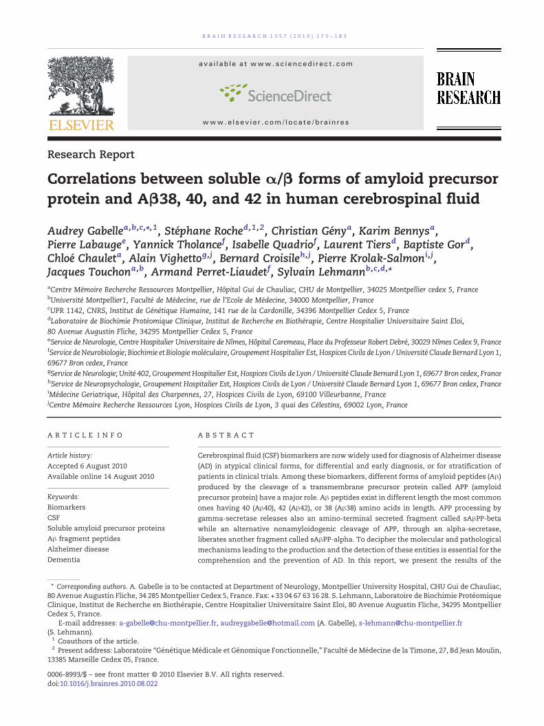
B R A I N R E S E A R C H 1 3 5 7 ( 2 0 1 0 ) 1 7 5 – 1 8 3
ava i l ab l e a t www.sc i enced i r ec t . com
www.e l sev i e r . com/ loca te /b ra i n res
Research Report
Correlations between soluble α/β forms of amyloid precursorprotein and Aβ38, 40, and 42 in human cerebrospinal fluid
Audrey Gabellea,b,c,⁎,1, Stéphane Roched,1,2, Christian Génya, Karim Bennysa,Pierre Labaugee, Yannick Tholancef, Isabelle Quadriof, Laurent Tiersd, Baptiste Gord,Chloé Chauleta, Alain Vighettog,j, Bernard Croisileh,j, Pierre Krolak-Salmoni,j,Jacques Touchona,b, Armand Perret-Liaudetf, Sylvain Lehmannb,c,d,⁎aCentre Mémoire Recherche Ressources Montpellier, Hôpital Gui de Chauliac, CHU de Montpellier, 34025 Montpellier cedex 5, FrancebUniversité Montpellier1, Faculté de Médecine, rue de l'Ecole de Médecine, 34000 Montpellier, FrancecUPR 1142, CNRS, Institut de Génétique Humaine, 141 rue de la Cardonille, 34396 Montpellier Cedex 5, FrancedLaboratoire de Biochimie Protéomique Clinique, Institut de Recherche en Biothérapie, Centre Hospitalier Universitaire Saint Eloi,80 Avenue Augustin Fliche, 34295 Montpellier Cedex 5, FranceeService de Neurologie, Centre Hospitalier Universitaire de Nîmes, Hôpital Caremeau, Place du Professeur Robert Debré, 30029 Nîmes Cedex 9, FrancefService deNeurobiologie; Biochimie et Biologiemoléculaire, GroupementHospitalier Est, Hospices Civils de Lyon /Université Claude Bernard Lyon 1,69677 Bron cedex, FrancegService de Neurologie; Unité 402, Groupement Hospitalier Est, Hospices Civils de Lyon / Université Claude Bernard Lyon 1, 69677 Bron cedex, FrancehService de Neuropsychologie, Groupement Hospitalier Est, Hospices Civils de Lyon / Université Claude Bernard Lyon 1, 69677 Bron cedex, FranceiMédecine Geriatrique, Hôpital des Charpennes, 27, Hospices Civils de Lyon, 69100 Villeurbanne, FrancejCentre Mémoire Recherche Ressources Lyon, Hospices Civils de Lyon, 3 quai des Célestins, 69002 Lyon, France
A R T I C L E I N F O
⁎ Corresponding authors. A. Gabelle is to be co80 Avenue Augustin Fliche, 34 285MontpellieClinique, Institut de Recherche en BiothérapCedex 5, France.
E-mail addresses: a-gabelle@chu-montpe(S. Lehmann).1 Coauthors of the article.2 Present address: Laboratoire “Génétique M
13385 Marseille Cedex 05, France.
0006-8993/$ – see front matter © 2010 Elsevidoi:10.1016/j.brainres.2010.08.022
A B S T R A C T
Article history:Accepted 6 August 2010Available online 14 August 2010
Cerebrospinal fluid (CSF) biomarkers are nowwidely used for diagnosis of Alzheimer disease(AD) in atypical clinical forms, for differential and early diagnosis, or for stratification ofpatients in clinical trials. Among these biomarkers, different forms of amyloid peptides (Aβ)produced by the cleavage of a transmembrane precursor protein called APP (amyloidprecursor protein) have amajor role. Aβ peptides exist in different length themost commonones having 40 (Aβ40), 42 (Aβ42), or 38 (Aβ38) amino acids in length. APP processing bygamma-secretase releases also an amino-terminal secreted fragment called sAβPP-betawhile an alternative nonamyloidogenic cleavage of APP, through an alpha-secretase,liberates another fragment called sAβPP-alpha. To decipher the molecular and pathologicalmechanisms leading to the production and the detection of these entities is essential for thecomprehension and the prevention of AD. In this report, we present the results of the
Keywords:BiomarkersCSFSoluble amyloid precursor proteinsAβ fragment peptidesAlzheimer diseaseDementia
ntacted at Department of Neurology, Montpellier University Hospital, CHU Gui de Chauliac,r Cedex 5, France. Fax: +33 04 67 63 16 28. S. Lehmann, Laboratoire de Biochimie Protéomiqueie, Centre Hospitalier Universitaire Saint Eloi, 80 Avenue Augustin Fliche, 34295 Montpellier
llier.fr, [email protected] (A. Gabelle), [email protected]
édicale et Génomique Fonctionnelle,” Faculté de Médecine de la Timone, 27, Bd JeanMoulin,
er B.V. All rights reserved.
176 B R A I N R E S E A R C H 1 3 5 7 ( 2 0 1 0 ) 1 7 5 – 1 8 3
multiplex measurement of CSF Aβ38, Aβ40, Aβ42, sAβPP-alpha, and sAβPP-beta in 60patients mostly with dementia eventually segregated between neurochemical dementiadiagnostic (NDD) positive and negative groups. The NDD classification was based on ourroutine Tau, P-tau181, and Aβ42 cutoff values. We confirmed previous findings regarding thecorrelation between sAβPP-alpha and sAβPP-beta, as well as the potential interest of thesenew biomarkers. We also studied the correlation between sAβPPs and Aβ peptides, as wellas between Aβ peptides themselves. We observed a strong correlation between Aβ38 andsAβPP-beta which suggested that the production of this peptide was in direct relation with βsecretase activities. We also reported a strong correlation between Aβ38 and Aβ40, whileAβ42 was correlated to these fragments only in nonpathological situations. These resultsenlighten the complex relationships between thesemolecularmarkers in both physiologicaland pathological situations. Our results are important for the further use of these analytesfor AD diagnosis as well as for validating the cell biological hypotheses of APP processingand Aβ fragment production.
© 2010 Elsevier B.V. All rights reserved.
1. Introduction
Alzheimer disease (AD) is a neurodegenerative disordercharacterized by significant cognitive deficits, language trou-bles, behavior changes and loss of functional autonomy. Asthe major cause of dementia in the elderly population, AD isapproaching epidemic proportions in the Western industrial-ized countries due to aging of the population. AD is becoming amajor public health issue with the increasing burden oncaregivers and on the health care system. AD characteristicneuropathological lesions are represented by amyloid plaquesand neurofibrillary tangles which coexist in the brain. Thediagnosis of AD based on clinical, neuropsychological, mor-phological, as well as functional neuroimaging can remaindubious in a considerable number of cases delaying thediagnosis and the medical care. These data are alreadypowerful but lead only to probability diagnosis. Recent newsets of diagnosis criteria including specific morphological(volumetric MRI), functional imagery (PET scan), and CSFbiomarkers (total and phosphorylated Tau (P-tau) proteins andAβ peptides) have been proposed to optimize the diagnosis(Dubois et al., 2007). When comparing AD patients to healthycontrols, CSF concentrations of Tau and P-tauwere reported tobe increased, while Aβ1–42 levels were decreased (Blennowand Hampel, 2003). Numerous studies have analysed thepotential of CSF biomarkers in terms of positive diagnosis,differential diagnosis, and prognosis as predictive marker ofthe conversion (mild cognitive impairment (MCI) to AD)(Gabelle et al., 2009; Herukka et al., 2007; Mattsson et al.,2009). In combination and using several published ratios,these biomarkers reached a sensitivity and specificity forpositive AD diagnosis beyond 80% (Ibach et al., 2006). Weobtained similar results in our routine practice on more than350 patients (unpublished results) using as cutoff values forP-tau and IATI (Innogenetics Aβ/Tau index), 60 and 0.8,respectively. The future development of therapeutic strategiesaiming at slowing down or even blocking the neurodegener-ative process could however benefit from earlier and moresensitive diagnosis AD biomarkers.
Recently, Lewczuk et al. (2010) brought to light the interestof CSF soluble amyloid precursor proteins (sAPP) as novelpotential biomarkers in the positive diagnosis of AD. sAPPs are
generated from APP following α and β cleavages (Kang et al.,1987). The nonamyloidogenic pathway that cleaves off APP inposition α (within the sequence corresponding to Aβ) leads tothe α-secretase-cleaved soluble APP (sAPPα) fragment. The β-secretase action on APP leads to the β-secretase-cleavedsoluble APP (sAPPβ). This β-secretase activity (BACE), com-bined with γ-secretase cleavages releases the three majorpeptide fragments Aβ40, Aβ42, and Aβ38 (Wiltfang et al., 2002)in a kinetics that is not yet fully elucidated. It is well knownthat most of the secreted Aβ fragments correspond to Aβ40,however, in AD, a prominent pathogenic role is given to Aβ42that aggregates muchmore rapidly due to the presence of twoadditional hydrophobic amino acids, and represents the mainconstituent of senile plaques (Iwatsubo et al., 1994). Whileinvestigating presenilin mutations and γ-secretase inhibitors,Page et al. (2008) observed that while Aβ42 and Aβ40productions were strongly related, Aβ38 generation by γ-secretase was not dependent on the previous entities.
The objective of the current study was to investigate therelationship between sAPPs and Aβ fragments in differentpathophysiological contexts. Our results confirmed the po-tential interest of sAPPs as AD biomarkers and provideimportant information on the complex correlation betweenthe production of Aβ fragments and sAPPs.
2. Results
2.1. sAPPα/β correlation and values in NDD groups
A significant correlation (Fig. 1A; r=0.87, P<0.0001) wasobserved between the values of sAPPα and sAPPβ in ourpopulation, independently of the NDD biological status. Whenconcentration of the biomarkers were compared betweenNDDgroups, higher mean values were obtained in NDD+(Figs. 1Band C) with a difference with NDD− significant for sAPPβ.These results were reminding of those of Lewczuk et al. (2010)who analyzed a larger population and therefore obtained dif-ferences with lower P values.
The potential interest of sAPPs for dementia diagnosiscould not be fully investigated in the present study that
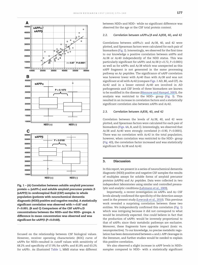
Fig. 1 – (A) Correlation between soluble amyloid precursorprotein α (sAPPα) and soluble amyloid precursor protein β(sAPPβ) in cerebrospinal fluid (CSF) samples in wholepopulation (patients with neurochemical dementiadiagnostic (NDD) positive and negative results). A statisticallysignificant correlation was observed with r=0.87 andP<0.001. (B and C) Comparison of the CSF sAPPα/βconcentrations between the NDD+and the NDD− groups. Adifference in mean concentration was observed and wassignificant for sAPPβ (P=0.0160).
177B R A I N R E S E A R C H 1 3 5 7 ( 2 0 1 0 ) 1 7 5 – 1 8 3
focused on the relationship between CSF biological values.However, receiver operating characteristic (ROC) curve ofsAPPs for NDD+resulted in cutoff values with sensitivity of68.2% and specificity of 57.9% for sAPPα and 81.8% and 63.2%for sAPPβ. As illustrated Table 1, MMS status was different
between NDD+and NDD− while no significant difference wasobserved for the age or the CSF total protein content.
2.2. Correlation between sAPPα/β and Aβ38, 40, and 42
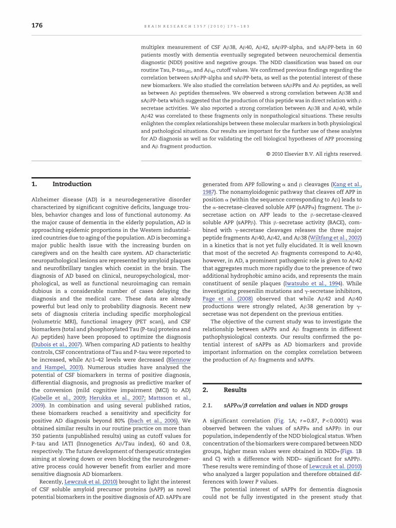
Correlations between sAPPα/β and Aβ38, 40, and 42 wereplotted, and Spearman factors were calculated for each pair ofbiomarkers (Fig. 2). Interestingly, we observed for the first timeto our knowledge a positive correlation between sAPPs andAβ38 or Aβ40 independently of the NDD status. This wasparticularly significant for sAPPβ and Aβ38 (r=0.72, P<0.0001)as well as for sAPPα and Aβ38 which was unexpected as thissAPP fragment is not generated in the same processingpathway as Aβ peptides. The significance of sAPP correlationwas however lower with Aβ40 than with Aβ38 and was notsignificant at all with Aβ42 (compare Figs. 2 AD, BE, and CF). AsAβ42 and in a lesser extend Aβ40 are involved in ADpathogenesis and CSF levels of these biomarkers are knownto be modified in the disease (Blennow and Hampel, 2003), theanalysis was restricted to the NDD− group (Fig. 3). Thisresulted in an increase in correlation factors and a statisticallysignificant correlation also between sAPPs and Aβ42.
2.3. Correlation between Aβ38, 40, and 42
Correlation between the levels of Aβ38, 40, and 42 wereplotted, and Spearman factors were calculated for each pair ofbiomarkers (Figs. 4A, B, and C). Interestingly, we observed thatAβ38 and Aβ40 were strongly correlated (r=0.90, P<0.0001).There was no correlation with Aβ42 in the total population;however, when correlation was restricted to the NDD− group(Fig. 4D), the correlation factor increased and was statisticallysignificant for Aβ38 and Aβ42.
3. Discussion
In this report, we present in a series of neurochemical dementiadiagnostic (NDD) positive and negative CSF samples the resultsof multiplex assays for soluble forms of amyloid precursorproteins (sAPPs) and Aβ peptides. Data were collected in twoindependent laboratories using similar and controlled preana-lytic and analytic conditions (Lehmann et al., 2009).
Importantly, a recent investigation on sAPPα and Aβ CSFlevels already confirmed the specificity of the detection assaysused in the present study (Lewczuk et al., 2010). This previouswork revealed a surprising correlation between these twoentities. We independently confirmed this correlation (Fig. 1)which was intriguing because it did not correspond to whatwould be intuitively expected. One could believe in fact thatthe production of sAPPβ would be inversely proportional tothat of sAPPα since their metabolic pathways are exclusive.Moreover, these fragments have opposite impact (toxic vs.neuroprotective). To our knowledge, no precise metabolic regu-lation has beendemonstrated betweenα andβAPP cleavages inthe literature, and further studies would be needed to explainthis positive correlation.
We also observed a slight increase in sAPP levels in NDD+samples compared to NDD− with a statistically significant
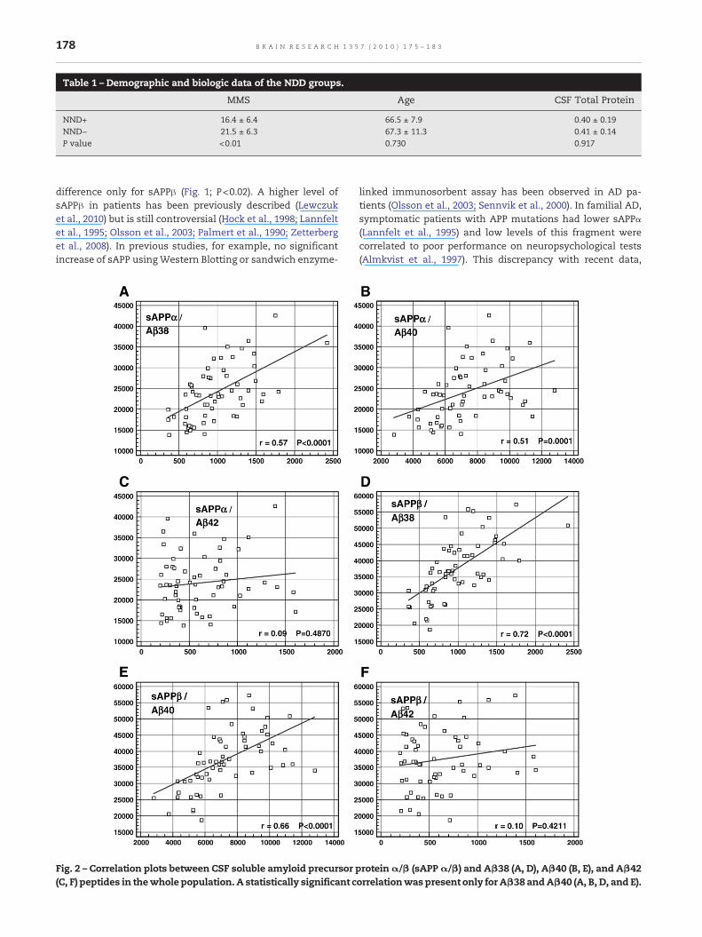
Table 1 – Demographic and biologic data of the NDD groups.
MMS Age CSF Total Protein
NND+ 16.4 ± 6.4 66.5 ± 7.9 0.40 ± 0.19NND− 21.5 ± 6.3 67.3 ± 11.3 0.41 ± 0.14P value <0.01 0.730 0.917
178 B R A I N R E S E A R C H 1 3 5 7 ( 2 0 1 0 ) 1 7 5 – 1 8 3
difference only for sAPPβ (Fig. 1; P<0.02). A higher level ofsAPPβ in patients has been previously described (Lewczuket al., 2010) but is still controversial (Hock et al., 1998; Lannfeltet al., 1995; Olsson et al., 2003; Palmert et al., 1990; Zetterberget al., 2008). In previous studies, for example, no significantincrease of sAPP usingWestern Blotting or sandwich enzyme-
Fig. 2 – Correlation plots between CSF soluble amyloid precursor(C, F) peptides in thewhole population. A statistically significant c
linked immunosorbent assay has been observed in AD pa-tients (Olsson et al., 2003; Sennvik et al., 2000). In familial AD,symptomatic patients with APP mutations had lower sAPPα(Lannfelt et al., 1995) and low levels of this fragment werecorrelated to poor performance on neuropsychological tests(Almkvist et al., 1997). This discrepancy with recent data,
proteinα/β (sAPPα/β) and Aβ38 (A, D), Aβ40 (B, E), and Aβ42orrelationwas present only for Aβ38 andAβ40 (A, B, D, and E).
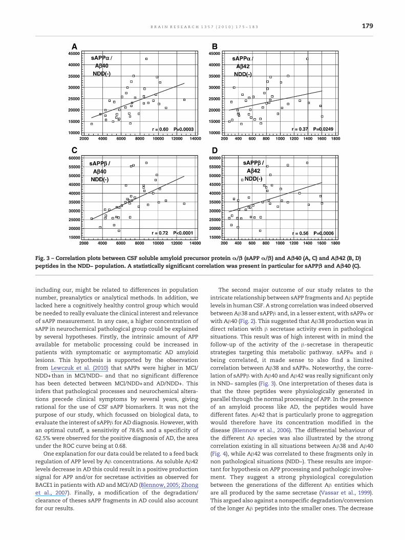
Fig. 3 – Correlation plots between CSF soluble amyloid precursor protein α/β (sAPP α/β) and Aβ40 (A, C) and Aβ42 (B, D)peptides in the NDD− population. A statistically significant correlation was present in particular for sAPPβ and Aβ40 (C).
179B R A I N R E S E A R C H 1 3 5 7 ( 2 0 1 0 ) 1 7 5 – 1 8 3
including our, might be related to differences in populationnumber, preanalytics or analytical methods. In addition, welacked here a cognitively healthy control group which wouldbe needed to really evaluate the clinical interest and relevanceof sAPP measurement. In any case, a higher concentration ofsAPP in neurochemical pathological group could be explainedby several hypotheses. Firstly, the intrinsic amount of APPavailable for metabolic processing could be increased inpatients with symptomatic or asymptomatic AD amyloidlesions. This hypothesis is supported by the observationfrom Lewczuk et al. (2010) that sAPPs were higher in MCI/NDD+than in MCI/NDD− and that no significant differencehas been detected between MCI/NDD+and AD/NDD+. Thisinfers that pathological processes and neurochemical altera-tions precede clinical symptoms by several years, givingrational for the use of CSF sAPP biomarkers. It was not thepurpose of our study, which focussed on biological data, toevaluate the interest of sAPPβ for AD diagnosis. However, withan optimal cutoff, a sensitivity of 78.6% and a specificity of62.5% were observed for the positive diagnosis of AD, the areaunder the ROC curve being at 0.68.
One explanation for our data could be related to a feed backregulation of APP level by Aβ concentrations. As soluble Aβ42levels decrease in AD this could result in a positive productionsignal for APP and/or for secretase activities as observed forBACE1 in patients with AD andMCI/AD (Blennow, 2005; Zhonget al., 2007). Finally, a modification of the degradation/clearance of theses sAPP fragments in AD could also accountfor our results.
The second major outcome of our study relates to theintricate relationship between sAPP fragments and Aβ peptidelevels in humanCSF. A strong correlationwas indeed observedbetween Aβ38 and sAPPβ and, in a lesser extent, with sAPPα orwith Aβ40 (Fig. 2). This suggested that Aβ38 production was indirect relation with β secretase activity even in pathologicalsituations. This result was of high interest with in mind thefollow-up of the activity of the β-secretase in therapeuticstrategies targeting this metabolic pathway. sAPPα and βbeing correlated, it made sense to also find a limitedcorrelation between Aβ38 and sAPPα. Noteworthy, the corre-lation of sAPPβwith Aβ40 and Aβ42 was really significant onlyin NND− samples (Fig. 3). One interpretation of theses data isthat the three peptides were physiologically generated inparallel through the normal processing of APP. In the presenceof an amyloid process like AD, the peptides would havedifferent fates. Aβ42 that is particularly prone to aggregationwould therefore have its concentration modified in thedisease (Blennow et al., 2006). The differential behaviour ofthe different Aβ species was also illustrated by the strongcorrelation existing in all situations between Aβ38 and Aβ40(Fig. 4), while Aβ42 was correlated to these fragments only innon pathological situations (NDD−). These results are impor-tant for hypothesis on APP processing and pathologic involve-ment. They suggest a strong physiological coregulationbetween the generations of the different Aβ entities whichare all produced by the same secretase (Vassar et al., 1999).This argued also against a nonspecific degradation/conversionof the longer Aβ peptides into the smaller ones. The decrease
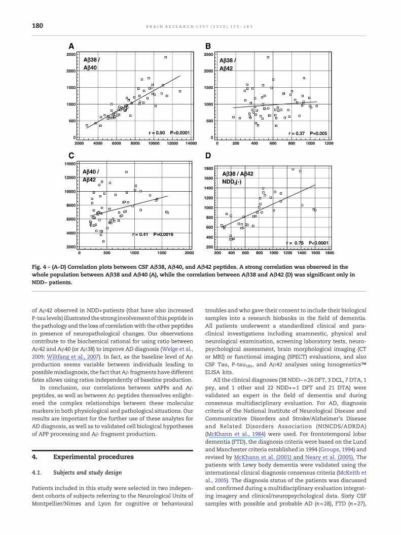
Fig. 4 – (A–D) Correlation plots between CSF Aβ38, Aβ40, and Aβ42 peptides. A strong correlation was observed in thewhole population between Aβ38 and Aβ40 (A), while the correlation between Aβ38 and Aβ42 (D) was significant only inNDD− patients.
180 B R A I N R E S E A R C H 1 3 5 7 ( 2 0 1 0 ) 1 7 5 – 1 8 3
of Aβ42 observed in NDD+patients (that have also increasedP-tau levels) illustrated the strong involvementof thispeptide inthepathology and the loss of correlationwith the other peptidesin presence of neuropathological changes. Our observationscontribute to the biochemical rational for using ratio betweenAβ42 and Aβ40 (or Aβ38) to improve AD diagnosis (Welge et al.,2009; Wiltfang et al., 2007). In fact, as the baseline level of Aβproduction seems variable between individuals leading topossiblemisdiagnosis, the fact that Aβ fragments have differentfates allows using ratios independently of baseline production.
In conclusion, our correlations between sAPPs and Aβpeptides, as well as between Aβ peptides themselves enlight-ened the complex relationships between these molecularmarkers in both physiological and pathological situations. Ourresults are important for the further use of these analytes forAD diagnosis, as well as to validated cell biological hypothesesof APP processing and Aβ fragment production.
4. Experimental procedures
4.1. Subjects and study design
Patients included in this study were selected in two indepen-dent cohorts of subjects referring to the Neurological Units ofMontpellier/Nimes and Lyon for cognitive or behavioural
troubles andwho gave their consent to include their biologicalsamples into a research biobanks in the field of dementia.All patients underwent a standardized clinical and para-clinical investigations including anamnestic, physical andneurological examination, screening laboratory tests, neuro-psychological assessment, brain morphological imaging (CTor MRI) or functional imaging (SPECT) evaluations, and alsoCSF Tau, P-tau181, and Aβ42 analyses using Innogenetics™ELISA kits.
All the clinical diagnoses (38 NDD−=26 DFT, 3 DCL, 7 DTA, 1psy, and 1 other and 22 NDD+=1 DFT and 21 DTA) werevalidated an expert in the field of dementia and duringconsensus multidisciplinary evaluation. For AD, diagnosiscriteria of the National Institute of Neurological Disease andCommunicative Disorders and Stroke/Alzheimer's Diseaseand Related Disorders Association (NINCDS/ADRDA)(McKhann et al., 1984) were used. For frontotemporal lobardementia (FTD), the diagnosis criteria were based on the LundandManchester criteria established in 1994 (Groups, 1994) andrevised by McKhann et al. (2001) and Neary et al. (2005). Thepatients with Lewy body dementia were validated using theinternational clinical diagnosis consensus criteria (McKeith etal., 2005). The diagnosis status of the patients was discussedand confirmed during a multidisciplinary evaluation integrat-ing imagery and clinical/neuropsychological data. Sixty CSFsamples with possible and probable AD (n=28), FTD (n=27),
181B R A I N R E S E A R C H 1 3 5 7 ( 2 0 1 0 ) 1 7 5 – 1 8 3
LBD (n=3), and other neurological disease (n=2) were ana-lyzed. As the purpose of the investigation was primarily thebiological relationships between the studied analytes, ratherthan their diagnosis interest, the samples were divided as inLewczuk et al. (2010) between neurochemical dementiadiagnosis-positive (NDD+) and negative (NDD−) using ourroutine P-tau and IATI (Innogenetics Aβ/Tau index) cutoffvalues of 60 and 0.8, respectively. A total of 22 samplescomplied with NDD+conditions, the 38 remaining being in theNDD− group.
4.2. CSF samples and routine assays
Lumbar puncture (LP) was performed in standardized condi-tions. LP was carried out at the L3/L4 or L4/L5 interspace afterexclusion of the potential contraindications. CSF sampleswere taken directly in similar polypropylene tubes to avoidvariation in adsorption of biomarkers to the container surface.All samples were transferred to the biochemical laboratories(CHU Montpellier/Nimes, Hospices Civils de Lyon) in less than4 hours. The CSF was centrifuged (1000×g, 10 min, at 4–8 °C,without break), and the supernatant was aliquoted by 0.5 mLin polypropylene tubes before storage at −80 °C. The threeroutine CSF biomarkers Tau, P-tau181, and Aβ42 were deter-mined with a standardized commercially available ELISA Kit(Innotest beta-amyloid 1–42 (Vanderstichele et al., 2000),Innotest hTau-Ag (Vandermeeren et al., 1993), and InnotestPhosphotau(181P) (Vanmechelen et al., 2000), Innogenetics,Ghent, Belgium) in both laboratories. Quality control programorganised by Hospices Civils de Lyon was used to study interlaboratories variability, using two CSF samples. The IATI scorewas calculated using the formula IATI=Aβ42/ (240+1.18×tau)as described (Hulstaert et al., 1999). Routine CSF analysesincluded glucose, total protein and cytology. Only samplesthat were assessed as red cells count <2000/mm3 were usedfor analyses.
4.3. Multiplex assays of CSF sAPPα/β and Aβ38, Aβ40,Aβ42
Detection of these analytes was performed independently inthe two laboratories using multiplex kits purchased fromMeso Scale Discovery (ref: K11120E, K11148E). All reagentswere provided with the kits. They contained a 96-well platewith two carbon electrodes precoated with either anti APPantibody 22C11 (sAPPα/β) or antibodies specific for Aβ38,Aβ40, Aβ42. The wells were incubated for 1 hour with theblock solution (bovine serum albumin) and washed four timeswith the TrisWash Buffer (TWB). For sAPPs duplex assay, 25 μlof standards (dilution of recombinant sAPPs) and sampleswere then added to all wells, and the plate was sealed andincubated for 1 hour at room temperature on an orbital shaker(300 rpm). At the end of the incubation, the wells were washedfour times using the TWB. Detection antibody (sAPPα/β) wasadded at 25 μl per well, and the plate was sealed and incubatedfor 1 hour at room temperature on an orbital shaker (300 rpm).For Aβ triplex assay, 25 μl of standards (dilution of recombi-nant Aβ) and sampleswere added at the same time of additionof 25 μl of detection antibody (6E10) to all wells, and the plateswere incubated during 2 hours. At the end of the incubation,
the plate was washed four times as before. About 150 μl of theMSD Read Buffer T was added to eachwell, and theMSD plateswere measured on the MSD Sector Imager 6000 plate reader.The raw data were measured as electrochemiluminescencesignal (light) detected by photodetectors and analysed usingthe Discovery Workbench 3.0 software (MSD). A 4-parameterlogistic fit curve was generated for each analyte using thestandards and the concentration of each sample calculated.The intra-assay coefficient of variation ranged from 0.2% to15.9%. The interlaboratory coefficient of variation ranged from0.3% to 17%.
4.4. Statistical analysis
If not stated otherwise, the results were presented asmediansand interquartile ranges. The Mann–Whitney–Wilcoxon testwas used to test the significance of the difference between twosample groups. Correlations between measured values wereanalyzedwith Spearman's correlation factor. Differences wereconsidered significant if P<0.05. Analyses were performedwith MedCalc (3.0).
Acknowledgments
This study was supported by the following grants by the EUgrants cNEUPRO (contract no. LSHM-CT-2007-037950) and bythe French Health Ministry (contract no. Rhônes Alpes PHRC-2004- 27/15). We thank Pr. JP Cristol (Service of Biochemistry)and Pr. B Klein (Director of IRB) for their support.
R E F E R E N C E S
Almkvist, O., Basun, H., Wagner, S.L., Rowe, B.A.,Wahlund, L.O., Lannfelt, L., 1997. Cerebrospinal fluid levelsof alpha-secretase-cleaved soluble amyloid precursor proteinmirror cognition in a Swedish family with Alzheimer diseaseand a gene mutation. Arch. Neurol. 54, 641–644.
Blennow, K., Hampel, H., 2003. CSF markers for incipientAlzheimer's disease. Lancet Neurol. 2, 605–613.
Blennow, K., 2005. CSF biomarkers for Alzheimer's disease: usein early diagnosis and evaluation of drug treatment. ExpertRev. Mol. Diagn. 5, 661–672.
Blennow, K., de Leon, M.J., Zetterberg, H., 2006. Alzheimer'sdisease. Lancet 368, 387–403.
Dubois, B., Feldman, H.H., Jacova, C., Dekosky, S.T.,Barberger-Gateau, P., Cummings, J., Delacourte, A., Galasko, D.,Gauthier, S., Jicha, G., Meguro, K., O'Brien, J., Pasquier, F.,Robert, P., Rossor, M., Salloway, S., Stern, Y., Visser, P.J.,Scheltens, P., 2007. Research criteria for the diagnosis ofAlzheimer's disease: revising the NINCDS–ADRDA criteria.Lancet Neurol. 6, 734–746.
Gabelle, A., Roche, S., Lehmann, S., 2009. CSF biomarkers:proteomics investigations and clinical applications inneurodegenerative disorders. Rev. Neurol. (Paris) 165, 213–222.
Groups, T.L.a.M., 1994. Clinical and neuropathological criteriafor frontotemporal dementia. The Lund and ManchesterGroups. J. Neurol. Neurosurg. Psychiatry 57, 416–418.
Herukka, S.K., Helisalmi, S., Hallikainen, M., Tervo, S., Soininen, H.,Pirttila, T., 2007. CSF Abeta42, Tau and phosphorylated Tau,APOE epsilon4 allele and MCI type in progressive MCI.Neurobiol. Aging 28, 507–514.
182 B R A I N R E S E A R C H 1 3 5 7 ( 2 0 1 0 ) 1 7 5 – 1 8 3
Hock, C., Golombowski, S., Muller-Spahn, F., Naser, W.,Beyreuther, K., Monning, U., Schenk, D., Vigo-Pelfrey, C.,Bush, A.M., Moir, R., Tanzi, R.E., Growdon, J.H., Nitsch, R.M.,1998. Cerebrospinal fluid levels of amyloid precursor proteinand amyloid beta-peptide in Alzheimer's disease and majordepression—inverse correlation with dementia severity. Eur.Neurol. 39, 111–118.
Hulstaert, F., Blennow, K., Ivanoiu, A., Schoonderwaldt, H.C.,Riemenschneider, M., De Deyn, P.P., Bancher, C., Cras, P.,Wiltfang, J., Mehta, P.D., Iqbal, K., Pottel, H., Vanmechelen, E.,Vanderstichele, H., 1999. Improved discrimination of ADpatients using beta-amyloid(1–42) and tau levels in CSF.Neurology 52, 1555–1562.
Ibach, B., Binder, H., Dragon, M., Poljansky, S., Haen, E., Schmitz, E.,Koch, H., Putzhammer, A., Kluenemann, H., Wieland, W.,Hajak, G., 2006. Cerebrospinal fluid tau and beta-amyloid inAlzheimer patients, disease controls and an age-matchedrandom sample. Neurobiol. Aging 27, 1202–1211.
Iwatsubo, T., Hasegawa, M., Ihara, Y., 1994. Neuronal and glialtau-positive inclusions in diverse neurologic diseases sharecommon phosphorylation characteristics. Acta Neuropathol.88, 129–136.
Kang, J., Lemaire, H.G., Unterbeck, A., Salbaum, J.M., Masters, C.L.,Grzeschik, K.H., Multhaup, G., Beyreuther, K., Muller-Hill, B.,1987. The precursor of Alzheimer's disease amyloid A4 proteinresembles a cell-surface receptor. Nature 325, 733–736.
Lannfelt, L., Basun, H., Wahlund, L.O., Rowe, B.A., Wagner, S.L.,1995. Decreased alpha-secretase-cleaved amyloid precursorprotein as a diagnostic marker for Alzheimer's disease. Nat.Med. 1, 829–832.
Lehmann, S., Roche, S., Allory, Y., Barthelaix, A., Beaudeux, J.L.,Berger, F., Betsou, F., Borg, J., Dupuy, A., Garin, J., Quillard, M.,Lizard, G., Peoc'h, K., Riviere, M., Ducoroy, P., 2009.Preanalytical guidelines for clinical proteomics investigationof biological fluids. Ann. Biol. Clin. (Paris) 67, 629–639.
Lewczuk, P., Kamrowski-Kruck, H., Peters, O., Heuser, I., Jessen, F.,Popp, J., Burger, K., Hampel, H., Frolich, L., Wolf, S., Prinz, B.,Jahn, H., Luckhaus, C., Perneczky, R., Hull, M., Schroder, J.,Kessler, H., Pantel, J., Gertz, H.J., Klafki, H.W., Kolsch, H.,Reulbach, U., Esselmann, H., Maler, J.M., Bibl, M., Kornhuber, J.,Wiltfang, J., 2010. Soluble amyloid precursor proteins inthe cerebrospinal fluid as novel potential biomarkers ofAlzheimer's disease: a multicenter study. Mol. Psychiatry 15,138–145.
Mattsson, N., Zetterberg, H., Hansson, O., Andreasen, N.,Parnetti, L., Jonsson, M., Herukka, S.K., van der Flier, W.M.,Blankenstein, M.A., Ewers, M., Rich, K., Kaiser, E., Verbeek, M.,Tsolaki, M., Mulugeta, E., Rosen, E., Aarsland, D., Visser, P.J.,Schroder, J., Marcusson, J., de Leon, M., Hampel, H., Scheltens,P., Pirttila, T., Wallin, A., Jonhagen, M.E., Minthon, L.,Winblad, B., Blennow, K., 2009. CSF biomarkers andincipient Alzheimer disease in patients with mild cognitiveimpairment. JAMA 302, 385–393.
McKeith, I.G., Dickson, D.W., Lowe, J., Emre, M., O'Brien, J.T.,Feldman, H., Cummings, J., Duda, J.E., Lippa, C., Perry, E.K.,Aarsland, D., Arai, H., Ballard, C.G., Boeve, B., Burn, D.J., Costa,D., Del Ser, T., Dubois, B., Galasko, D., Gauthier, S., Goetz, C.G.,Gomez-Tortosa, E., Halliday, G., Hansen, L.A., Hardy, J.,Iwatsubo, T., Kalaria, R.N., Kaufer, D., Kenny, R.A.,Korczyn, A., Kosaka, K., Lee, V.M., Lees, A., Litvan, I.,Londos, E., Lopez, O.L., Minoshima, S., Mizuno, Y.,Molina, J.A., Mukaetova-Ladinska, E.B., Pasquier, F.,Perry, R.H., Schulz, J.B., Trojanowski, J.Q., Yamada, M., 2005.Diagnosis and management of dementia with Lewy bodies:third report of the DLB Consortium. Neurology 65,1863–1872.
McKhann, G., Drachman, D., Folstein, M., Katzman, R., Price, D.,Stadlan, E.M., 1984. Clinical diagnosis of Alzheimer's disease:report of the NINCDS–ADRDA Work Group under the auspices
of Department of Health and Human Services Task Force onAlzheimer's Disease. Neurology 34, 939–944.
McKhann, G.M., Albert, M.S., Grossman, M., Miller, B., Dickson, D.,Trojanowski, J.Q., 2001. Clinical and pathological diagnosis offrontotemporal dementia: report of the Work Group onFrontotemporal Dementia and Pick's Disease. Arch. Neurol. 58,1803–1809.
Neary, D., Snowden, J., Mann, D., 2005. Frontotemporaldementia. Lancet Neurol. 4, 771–780.
Olsson, A., Hoglund, K., Sjogren, M., Andreasen, N., Minthon, L.,Lannfelt, L., Buerger, K., Moller, H.J., Hampel, H., Davidsson, P.,Blennow, K., 2003. Measurement of alpha- and beta-secretasecleaved amyloid precursor protein in cerebrospinal fluidfrom Alzheimer patients. Exp. Neurol. 183, 74–80.
Page, R.M., Baumann, K., Tomioka, M., Perez-Revuelta, B.I.,Fukumori, A., Jacobsen, H., Flohr, A., Luebbers, T., Ozmen, L.,Steiner, H., Haass, C., 2008. Generation of Abeta38 andAbeta42 is independently and differentially affected byfamilial Alzheimer disease-associated presenilin mutationsand gamma-secretase modulation. J. Biol. Chem. 283, 677–683.
Palmert, M.R., Usiak, M., Mayeux, R., Raskind, M.,Tourtellotte, W.W., Younkin, S.G., 1990. Soluble derivativesof the beta amyloid protein precursor in cerebrospinal fluid:alterations in normal aging and in Alzheimer's disease.Neurology 40, 1028–1034.
Sennvik, K., Fastbom, J., Blomberg, M., Wahlund, L.O.,Winblad, B., Benedikz, E., 2000. Levels of alpha- andbeta-secretase cleaved amyloid precursor protein in thecerebrospinal fluid of Alzheimer's disease patients. Neurosci.Lett. 278, 169–172.
Vandermeeren, M., Mercken, M., Vanmechelen, E., Six, J., van deVoorde, A., Martin, J.J., Cras, P., 1993. Detection of tau proteinsin normal and Alzheimer's disease cerebrospinal fluid with asensitive sandwich enzyme-linked immunosorbent assay. J.Neurochem. 61, 1828–1834.
Vanderstichele, H., Van Kerschaver, E., Hesse, C., Davidsson, P.,Buyse, M.A., Andreasen, N., Minthon, L., Wallin, A., Blennow,K., Vanmechelen, E., 2000. Standardization of measurementof beta-amyloid(1–42) in cerebrospinal fluid and plasma.Amyloid. 7, 245–258.
Vanmechelen, E., Vanderstichele, H., Davidsson, P.,Van Kerschaver, E., Van Der Perre, B., Sjogren, M., Andreasen,N., Blennow, K., 2000. Quantification of tau phosphorylatedat threonine 181 in human cerebrospinal fluid: a sandwichELISA with a synthetic phosphopeptide for standardization.Neurosci. Lett. 285, 49–52.
Vassar, R., Bennett, B.D., Babu-Khan, S., Kahn, S., Mendiaz, E.A.,Denis, P., Teplow, D.B., Ross, S., Amarante, P., Loeloff, R., Luo,Y., Fisher, S., Fuller, J., Edenson, S., Lile, J., Jarosinski, M.A.,Biere, A.L., Curran, E., Burgess, T., Louis, J.C., Collins, F.,Treanor, J., Rogers, G., Citron, M., 1999. beta-Secretasecleavage of Alzheimer's amyloid precursor protein by thetransmembrane aspartic protease BACE. Science 286,735–741.
Welge, V., Fiege, O., Lewczuk, P., Mollenhauer, B., Esselmann, H.,Klafki, H.W., Wolf, S., Trenkwalder, C., Otto, M., Kornhuber, J.,Wiltfang, J., Bibl, M., 2009. Combined CSF tau, p-tau181 andamyloid-beta 38/40/42 for diagnosing Alzheimer's disease. J.Neural Transm. 116, 203–212.
Wiltfang, J., Esselmann, H., Bibl, M., Smirnov, A., Otto, M.,Paul, S., Schmidt, B., Klafki, H.W., Maler, M., Dyrks, T., Bienert,M., Beyermann, M., Ruther, E., Kornhuber, J., 2002. Highlyconserved and disease-specific patterns of carboxyterminallytruncated Abeta peptides 1–37/38/39 in addition to 1–40/42in Alzheimer's disease and in patients with chronicneuroinflammation. J. Neurochem. 81, 481–496.
Wiltfang, J., Esselmann, H., Bibl, M., Hull, M., Hampel, H., Kessler,H., Frolich, L., Schroder, J., Peters, O., Jessen, F., Luckhaus, C.,Perneczky, R., Jahn, H., Fiszer, M., Maler, J.M., Zimmermann, R.,

183B R A I N R E S E A R C H 1 3 5 7 ( 2 0 1 0 ) 1 7 5 – 1 8 3
Bruckmoser, R., Kornhuber, J., Lewczuk, P., 2007. Amyloidbeta peptide ratio 42/40 but not A beta 42 correlates withphospho-Tau in patients with low- and high-CSF A beta 40load. J. Neurochem. 101, 1053–1059.
Zetterberg, H., Andreasson, U., Hansson, O., Wu, G.,Sankaranarayanan, S., Andersson, M.E., Buchhave, P., Londos,E., Umek, R.M., Minthon, L., Simon, A.J., Blennow, K., 2008.
Elevated cerebrospinal fluid BACE1 activity in incipientAlzheimer disease. Arch. Neurol. 65, 1102–1107.
Zhong, Z., Ewers, M., Teipel, S., Burger, K., Wallin, A., Blennow, K.,He, P., McAllister, C., Hampel, H., Shen, Y., 2007. Levels ofbeta-secretase (BACE1) in cerebrospinal fluid as a predictor ofrisk in mild cognitive impairment. Arch. Gen. Psychiatry 64,718–726.
Copyright © 2022 FDOKUMEN