Collagen extract obtained from Nile tilapia (Oreochromis ...

Cell Biology International 30 (2006) 66e73www.elsevier.com/locate/cellbi
An NMR method to characterize multiple water compartmentson mammalian collagen
Gary D. Fullerton a,*, Elena Nes a, Maxwell Amurao a, Andres Rahal a,Lada Krasnosselskaia a, Ivan Cameron b
a Radiology Department, University of Texas HSCSA, Floyd Curl Drive, San Antonio, TX 78229-3900, United Statesb Cellular and Structural Biology Department, University of Texas HSCSA, Floyd Curl Drive, San Antonio, TX 78229-3900, United States
Received 30 May 2005; accepted 30 September 2005
Abstract
A molecular model is proposed to explain water 1H NMR spin-lattice relaxation at different levels of hydration (NMR titration method ) oncollagen. A fast proton exchange model is used to identify and characterize protein hydration compartments at three distinct Gibbs free energylevels. The NMR titration method reveals a spectrum of water motions with three well-separated peaks in addition to bulk water that can beuniquely characterized by sequential dehydration. Categorical changes in water motion occur at critical hydration levels h (g water/g collagen)defined by integral multiples N ¼ 1, 4 and 24 times the fundamental hydration value of one water bridge per every three amino acid residues asoriginally proposed by Ramachandran in 1968. Changes occur at (1) the Ramachandran single water bridge between a positive amide and neg-ative carbonyl group at h1¼ 0.0658 g/g, (2) the Berendsen single water chain per cleft at h2¼ 0.264 g/g, and (3) full monolayer coverage withsix water chains per cleft level at h3¼ 1.584 g/g. The NMR titration method is verified by comparison of measured NMR relaxation compart-ments with molecular hydration compartments predicted from models of collagen structure. NMR titration studies of globular proteins using thehydration model may provide unique insight into the critical contributions of hydration to protein folding.� 2005 International Federation for Cell Biology. Published by Elsevier Ltd. All rights reserved.
Keywords: Collagen; Protein hydration; NMR relaxometry; MRI; Proton relaxation; Tendon; Hydration compartments
1. Introduction
The scientific community generally accepts that protein hy-dration and protein induced water structuring are fundamentalsources of protein structural behaviors that support life func-tions. Collagen is frequently selected as the model protein toelucidate structural and hydration relationships (Leikin et al.,1994, 1995, 1997, 2002; Privalov, 1982). Tendon has high con-centration of Type I collagen approaching 100% of the drybiomass in some instances. Molecular tropocollagen crystal-lizes spontaneously and as a result significant new structuralinformation is available with ever improving accuracy fromX-ray diffraction studies of both native and molecular ana-logues of collagen (Bella and Berman, 1996; Bella et al.,
* Corresponding author.
E-mail address: [email protected] (G.D. Fullerton).
1065-6995/$ - see front matter � 2005 International Federation for Cell Biolog
doi:10.1016/j.cellbi.2005.09.009
1994, 1995, 1996; Berisio et al., 2002; Fraser et al., 1979;Kramer et al., 1998, 1999, 2000, 2001; Miller and Scheraga,1976; Okuyama et al., 1977; Ramachandran, 1967; Rich andCrick, 1961; Yonath and Traub, 1969). The more recent,high-resolution studies consistently show a water bridge net-work surrounding the collagen molecule.
Structural studies provide a molecular model of collagen sup-porting the existence of three distinct categories of water bridges.The most tightly bound consists of one highly immobilized ‘‘wa-ter bridge’’ per every three protein residues hb¼ 18 Da/(3� 91.2 Da)¼ 0.0658 g water/g protein as originally proposedby Ramachandran (1967), Fullerton and Amurao (2005), andRamachandran and Ramakrishnan (1968). A second less immo-bilized water fraction consists of three additional cleft watersper tripeptide unit residing in the three groove-like depressionsbetween the peptide chains of the triple helix hc¼ 3�hb¼ 0.197 g water/g. The water bridge and three cleft waters
y. Published by Elsevier Ltd. All rights reserved.

67G.D. Fullerton et al. / Cell Biology International 30 (2006) 66e73
complete a chain of four water molecules per tripeptide. Thesechains form a triple helix of water in the three clefts of the a-protein chains of the collagen triple helix. Cleft water was pro-posed by Berendsen (1962) and discussed by others (Lim,1981; Privalov, 1982).Recently our laboratory showed that the re-maining water on native collagen (w1.62 g water/g protein) is inthe first interfacial monolayer him¼ 5� 0.263¼ 1.315 g water/g protein at higher energy relative to the first two compartmentsbut still lower than the free energy of bulk water (Fullerton andAmurao, 2005). As a result of these three water compartments wepredict NMR detectable changes in water motion at h1¼hb¼ 0.0658, h2¼ hbþ hc¼ 0.264 and h3¼ hbþ hcþ him¼1.58 g water/g protein or athi ¼ Ni � 0.0658 forNi ¼ 1, 4 and24.
This study tests the hypothesis that that the NMR spin-lattice relaxation time of water on native collagen is not char-acteristic of any of the three molecular compartments buta weighted average due to fast exchange between three hydra-tion fractions or water phases on the protein surface(Zimmerman and Brittin, 1957). The relaxation characteristicsof all three water phases can, however, be extracted and char-acterized by sequential NMR measurements using a titrationof water content (Fullerton et al., 1982, 1986). Tendon isuniquely suited to identifying the molecular origin of the threehydration fractions that may be typical of proteins in general.The collagen molecule is highly immobilized and aligned inprotein fiber structure that suppresses proton NMR signalfrom the protein due to static dipole coupling. The study con-firms that proton NMR signal emanates only from the liquidwater that shows the presence of three distinct hydration frac-tions in fast exchange equilibrium. The compartments ex-tracted using the NMR titration method quantitativelyconfirm the three molecular water bridge environments identi-fied in the preceding paragraph. The correlation of proteinNMR titration measurements with a molecular model of waterbridges holds promise as a general model to evaluate hydrationof both fibrillar and globular proteins.
2. Materials and methods
2.1. NMR titration measurements
Samples of bovine flexor tendon from three animals (age and sex un-
known) were obtained from a local slaughter house, dissected on site, diced
to 3 mm3 cubes and placed in separate pre-weighed 1$ diameter NMR sample
tubes for each animal. Tubes were immediately sealed to prevent dehydration.
The initial sample volume was approximately 6 cc when wet and approximately
3 cc when dry. Preliminary measurements of T1 relaxation reported here
were made at 10.7 MHz on a Praxis II pulsed NMR device with a primary
magnetic field strength of 0.25 T. The initial research plan was to use these
less accurate but rapid measurements at 10.7 MHz to provide pilot data for de-
signing of more accurate and complete experiments as a function of tempera-
ture at 500, 700 and 900 MHz. Results at 0.25 T are, however, sufficient to
substantiate the hypothesis and are therefore presented as stand alone experi-
ments. Praxis measurements of T1 use the saturation recovery (90-t-90) pulse
sequence with a repetition time TR exceeding five times the longest T1 relax-
ation time for the sample. The 90 � pulse is approximately 13 ms (bandwidth in
excess of 100 kHz). The sample chamber was equilibrated in the instrument at
22� 1 �C for at least 1 h prior to each measurement. Samples were out of the
vacuum for approximately 1e4 h. Typical measurement times in the NMR unit
were approximately 5e10 min. Random orientation of the tendon with respect
to the magnetic field was used to eliminate possibility of residual orientation
dependence. The signal after a recovery delay t was measured as an amplitude
of the free induction decay, A(t), with the first time point at 26 ms. Each data
point samples signal for 1 ms and 10 points following the peak signal were av-
eraged to reduce the measurement noise.
Each relaxation measurement consisted of 31 amplitude measurements with
a total of 30 sequential delay times with even increments of w20 ms each for
a maximum delay time of 30�Dt as shown in Fig. 1. Although delay times
were adjusted to evaluate signal amplitude variation over as much as two orders
ofmagnitude only single exponent T1 decaywas observed in all instances.Meas-
urements were repeated three times to improve the signal-to-noise ratio to 300:1
(0.3% noise) at the initial hydration level. A non-linear regression fit to the data
was calculated as shownwith a single exponent decay using twofitting constants,
A(N) and T1, using the equation A(N)� A(t)¼ A(N)(e�t/T1) and non-linear
regression from the Graph Pad program (Motulsky, 2003). The T1 was measured
for the tendon samples over the entire range of hydration fromnative hydration to
completely dry protein using the procedure described below. The tissue mass Mt
was measured at each hydration level to allow calculation of h ¼ Mw/Mp fol-
lowing measurement of the dry protein mass Mp (see next paragraph). The
mass of salt and other cosolutes was neglected for these calculations.
The original sample weights were approximately 5.4 g of tendon which
when fully dehydrated decreased to approximately 2.3 g of dry mass. Initial
dehydrations were achieved by placing the open NMR tubes in a vacuum
chamber at approximately 0.25 atmospheric pressure. Samples were removed
at intervals ranging initially from approximately 1 h to several days to achieve
significant reduction in the sample water content. After achieving equilibrium
mass at room temperature the temperature of the vacuum oven was increased
to 90 �C and the dehydration extended for another 30 days with intermittent
measurements of T1 taken until a new equilibrium and final dry mass for
the protein were achieved. The dehydration and T1 measurement protocol
for the tendon extended over a total period of approximately 3 months. An ex-
tended drying period was undertaken to assure hydration equilibrium over the
entire volume of the tendon. Due to the slow dehydration rate no measurable
dependence on the time interval between removal from the vacuum oven and
NMR measurement was detectable between 1 and 24 h (data not shown). The
measurement process was repeated for all three samples. Results for all three
samples were similar but only two are reported as one sample tube broke dur-
ing the final heating phase of the experiment.
2.2. NMR titration analysis of spin-lattice relaxation data
The water bridge hydration hypothesis identifies three hydration water
fractions in addition to a bulk water phase as shown in Fig. 2. For the protein
Fig. 1. T1 analysis of a saturation recovery curve for bovine tendon at the na-
tive hydration level is shown here. Solid circles stand for data points. Solid line
is a fit to the function lnðAðNÞ � AðtÞÞ ¼ at, where t is a delay time, AðtÞi is
an FID amplitude after the delay t and AðNÞ ¼ Aðt[T1Þ and a is the regres-
sion fit constant a ¼ 6:7 s�1, T1 ¼ 1=a ¼ 149� 1 ms, R2¼ 0.9992, signal-
to-noise ratio was never lower than 20:1.

68 G.D. Fullerton et al. / Cell Biology International 30 (2006) 66e73
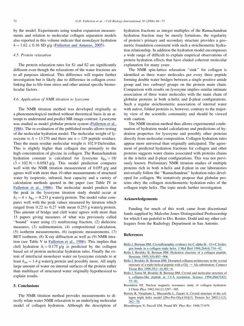
Fig. 2. (a) Conceptual cartoon for Zone 1 includes bulk water and shows the Gibbs free energy relationships of water molecules in different water bridge bonding
arrangements on the collagen molecule. At low hydration levels water molecules accumulate in the ‘‘Water Bridges’’ at the lowest free energy level. Higher energy
compartments are filled at higher hydrations by sequential filling of the ‘‘Cleft Water’’ and ‘‘Interfacial Monolayer Water until water spills over into the ‘‘Bulk’’
water compartment. (b) There is fast exchange of protons between these compartments when the tissue contains sufficient water to fill all the hydration compart-
ments and maintains a bulk water fraction as well. As protons sample all compartments during decay the NMR spin-lattice decay is monoexponential.
in dilute solution the hydration fractions are assumed to be in fast exchange
equilibrium on T1 relaxation scale such that the spin-lattice relaxation rate is
1=T1 ¼X
fi1=T1i
where fi is the fraction of water and 1/T1i is the spin-lattice relaxation rate of
the ith water fraction. There are three water fast exchange zones (Zones 1, 2and 3) and one protein fast exchange zone (Zone 4) to express relaxation
rate as a function of inverse hydration 1/h over the entire range h ¼ 0 (dry pro-
tein) to infinity (infinitely dilute protein solution). The definition of the hydra-
tion zones is summarized as follows:
Zone 1 (linear): all hydration compartments full and in fast exchange with
a variable bulk water compartment;
Zone 2 (linear): bulk water depleted leaving cleft water and water bridge
compartments full and in fast exchange with a variable interface monolayer
compartment;
Zone 3 (linear): bulk water and interface monolayer compartments depleted,
water bridge compartment full and in fast exchange with a variable cleft
water compartment; and
Zone 4 (non-linear): bulk, interface monolayer and cleft water compart-
ments depleted leaving tripeptide segments on the protein either occupied
or not occupied (with or without water bridges). Each tropocollagen has
many water bridge sites (w1000) to yield mean relaxation rate by spin-
diffusion during transition from full bridges to completely dry protein
relaxation rates.
The dilute protein case is shown in Figs. 2 and 3 as Zone 1 where 1/h �1/h3. The water consists of two fractions, hydration water
h3¼ hbþ hcþ him¼ 1.58 g water/g protein and bulk water of mass Mo. The
total hydration water mass M3¼ h3� Mp allows calculation of the fraction
of water f3¼ h3� Mp/Mw that interacts directly with the protein surface while
the bulk water fraction fo¼ Mo/Mw has the motional properties of pure bulk
water. The sum of all fractions f3þ fo¼ 1. The relaxation rate (R ¼ 1/T1) of
water is the weighted sum of the relaxation rates for bulk water, Ro, and the
mean relaxation rate for hydration water R3
fo þ f3 ¼ 1f3 ¼ h3 �Mp=Mw ¼ h3=hR1 ¼ foRo þ f3R3 ¼ Ro þ f3ðR3 �RoÞ ¼ Ro þ h3ðR3 �RoÞ1=h
where h ¼ Mw/Mp¼ the measured hydration of the sample.
The monolayer hydration h3¼ 1.58 g water/g dry protein is known from
the molecular model while mass of the tissue Mt, dry mass of the protein
Mp and relaxation rate of the tissue R1(h) are measured directly. The mass
of water Mw¼ Mt� Mp is calculated. Linear regression fits allow extraction
of the fitting constants Ro, the relaxation rate of the outer water fraction
(bulk water for Zone 1) and the mean relaxation rate R3 of the hydrated pro-
tein. The same method applied to Zone 2 gives the relaxation rate of the inter-
facial monolayer water Rim and Zone 3 analysis gives the relaxation rate for
cleft water Rc.
Zone 4 differs from the other zones because it describes the transition from
collagen with water bridges to collagen without bridges. There is a broad spec-
trum of NMR signal from protons (hydrogen nuclei) covalently bound in the
protein. A fraction of this protein signal appears in the water resonance range.
In Zone 4 the proton signal comes from solid-like protein in two possible
states either with or without water bridges. The observed change in relaxation
rate indicates that protein with water relaxes slightly more rapidly than colla-
gen without water bridges. The water bridges accelerate relaxation for protein
without structural water. This is likely through the well known mechanism of
spin-diffusion in solids (Edzes and Samulski, 1977). Thus there are two pro-
tein categories: category A with water bridges and category B without water.
As the signal from ‘‘solid like’’ water is much smaller than from protein we
can neglect the signal amplitude due to the water and calculate the relaxation
rate as follows:
RA¼ the relaxation rate of protein hydrogen nuclei with water bridges
RB¼ the relaxation rate of the protein hydrogen nuclei without water
bridges
fA¼ the fraction of the protein having one bridge per three residues
fB¼ the fraction of protein without a water bridge per three residues
h ¼ the hydration¼ Mw/Mp
hb¼ the single bridge hydration fraction
fA¼ (hMp)/(hbMp)¼ h/hb
fB ¼ 1� h=hbR1 ¼ fARA þ fBRB
R1 ¼ RB þ ½ðRA � RBÞ=hb�h
This last equation R1 is a linear function of h while the other three
equations are linear functions of 1/h. We plot all zones as a function of
1/h as this gives three linear sections and one curved line for Zone 4.
The four equations must agree at the intersections of adjacent sections.
This allows simplification to reduce the number of fitting constants to
five to yield the relationships summarized in Table 1.
2.3. Statistical analysis
The 1/T1 relaxation rate as a function of 1/h was evaluated using non-linear
least squares regression fit to the four zone multi-segment equations defined in

69G.D. Fullerton et al. / Cell Biology International 30 (2006) 66e73
Fig. 3. ‘‘Zonal Analysis’’ of relaxation rate versus the reciprocal of hydration
1/h. A multi-segment non-linear least squares regression fit (R2¼ 0.9916) for
n ¼ 74 measurements over the entire range of hydration from native to com-
pletely dry protein residue demonstrates the ability of the Molecular Model
to explain relaxation behavior. (A) Shows entire range of experiments. (B)
Focuses on data from Zone 3. (C) Focuses on data from Zone 2. Each zone is
identified as a range of hydrations by the molecular model as follows: Zone
1: h > 1.584 g water/h dry mass, Zone 2: 1.584 g/g> h > 0.264 g/g, Zone 3:
0.264> h > 0.0658 g/g and Zone 4: h < 0.0658 g/g. Extrapolation of Zone 2
data to Mp/Mw¼ 0 shows that the relaxation rate for bulk water Ro¼ 0.37 s�1
is significantly different p < 0.05 than Rim¼ 1.354 for surface water (see Table
2). We note that although Zone 2 is well described by the linear relationship
used in the present analysis. There are possible 2nd order regularities near
Mp/Mw¼ 1.3 and 3.0 g solids/g water. This could reflect small changes in wa-
ter organization in the interfacial monolayer fraction as neighboring water
molecules are removed. Note the lack of data in Zone 1 for native tendon
which indicates absence of bulk-like water on this tissue (see text for details).
Table 3 to evaluate the ability of the proposed molecular model to describe re-
laxation results over the entire hydration range (Motulsky, 2003).
3. Results
The results from samples S1 are shown in Fig. 3. Results onS2 were similar but not shown for brevity.
The details of the least squares fits are summarized inTable 2 for both the tendon S1 and S2 measurement series.The goodness of fit for tendon S1 was R2¼ 0.9916 andR2¼ 0.9910 for tendon S2. The best fit value, standard errorand 95% confidence interval for Rim, Rc, Rbp and Rpr are pre-sented as well as the direct measure of the spin-lattice relaxa-tion rate for water which was measured independently. Thecorrelation times calculated from the mean relaxation ratesare summarized in Table 3.
4. Discussion
4.1. NMR titration method
Comparison of the NMR titration measurements shown inFig. 3 (data points) with predictions of the four zone multi-component molecular model of collagen hydration (solidline) calculated with parameters from Table 1 demonstratesthe capacity of fast exchange relationships to accurately de-scribe the relaxation rate of water on collagen. The model pre-dictions of sharp demarcations at h1¼ hb¼ 0.0658,
Table 1
Multi-segment or zonal fast exchange equations for spin-lattice relaxation for
water on collagen
Zone Hydration
constraint
Relaxation rate (s�1)
1 h > 24hb 1/T1¼ Roþ (24hb/h)(5/6Rimþ 1/8Rcþ 1/24Rbp� Ro)
2 24hb� h � 4hb 1/T1¼ Rimþ (4hb/h)
(3/4Rcþ 1/4Rbp� Rim)
3 4hb� h � hb 1/T1¼ Rcþ hb/h (Rbp� Rc)
4 hb� h 1/T1¼ Rprþ h/hb (Rbp� Rpr)
Ro¼ relaxation rate of bulk water; Rim¼ relaxation rate of interfacial mono-
layer water; Rc¼ relaxation rate of embedded cleft water; Rbp¼ relaxation
rate of protein with water bridges filled; and Rpr¼ relaxation rate of protein
without water bridges.
Table 2
Fitting constants for statistical fits to NMR titration studies
Fitting constant Best fit Standard
error
95% Confidence
interval
Rb (s�1) 0.347 Measured
separately
Not visible
on collagen
Rim (s�1) e summer 1 1.354 0.1979 0.9588e1.749
Rim (s�1) e summer 2 1.349 0.2102 0.9292e1.770Rc (s
�1) e summer 1 32.84 0.3142 32.21e33.47
Rc (s�1) e summer 2 33.35 0.3869 32.58e34.12
Rbp (s�1) e summer 1 16.06 0.3099 15.44e16.67
Rbp (s�1) e summer 2 14.01 0.4119 13.18e14.83
Rpr (s�1) e summer 1 4.842 0.4094 4.025e5.660
Rpr (s�1) e summer 2 7.052 0.4127 6.227e7.877

70 G.D. Fullerton et al. / Cell Biology International 30 (2006) 66e73
h2¼ hbþ hc¼ 0.264 and h3¼ hbþ hcþ him¼ 1.58 g water/g protein or at hi ¼ Ni � 0.0658 for Ni ¼ 1, 4 and 24 is con-firmed. Thus all of the hydration fractions for collagen canbe described as integer multiples of h1¼ 0.0658 as proposedby Ramachandran and shown in Fig. 4. We refer to this funda-mental quantity as the Ramachandran hydration value. It canbe calculated for any protein for which molecular weight Aand number of residues n are known using the equation
hRa ¼ 18 Da=ð3�A=nÞ
as demonstrated for lysozyme below.
4.2. Water bridges
As shown in Fig. 5 the function of the water bridge and di-rect hydrogen bond is to serve as mechanisms to reduce the
Table 3
Correlation times for water calculated from data in Table 2 using BPP theory
(Bloembergen et al., 1948)
Bulk water 1/T1¼ 0.347 s�1 t¼ 6.7� 10�12 (Reference)
Orientational water 1/T1¼ 1.352 s�1 t¼ 2.6� 10�11 (Bulk� 4)
Cleft water 1/T1¼ 33.1 s�1 t¼ 6.55� 10�10 (Bulk� 100)
electrostatic energy of the protein by transferring positivecharge from amide sites to compensate negative charge fromcarbonyl groups. Direct hydrogen bonds occur when the sepa-ration is less than 3 A while water bridges are necessary whenseparations approach 5 A sufficient to allow insertion of awater molecule. Separations are controlled by stereotacticrestriction when the protein is folded. Hydrogen exchange ex-periments (Yee et al., 1974) showed extremely long exchangetimes (hours to days) for amide hydrogen atoms participatingin either direct or water bridge bonds to confirm the two-bondmodel proposed by Ramachandran. The NMR titration meas-urements extend this conclusion by confirming as proposedthat a water bridge provides the second bond per tripeptide.
4.3. Cleft water
We define cleft water as the additional three water mole-cules per tripeptide that bind in a hydrogen bonded networkto the protein as well as to the structural water bridge (also lo-cated in the cleft) to complete the four-water molecule chainper every three peptide residues in the molecular grooves ofthe collagen triple helix proposed as by Berendsen (1962)and Fullerton and Amurao (2005). We reason on the basis ofgeometry that cleft waters participate in double water bridges
Fig. 4. The Ramachandran proposal of one direct hydrogen bond per tripeptide, one water bridge and a second non-charge bridge water molecule was necessary to
complete the two-bond model proposed by his group. (a) Shows configuration when peptide three is other than hydroxyproline. (b) Shows the alternative tripeptide
hydrogen bonding configuration when hydroxyproline occupies position three in Chain A. In both instances hRa¼ 18 Da/(3�mean peptide molecular
weight)¼ 0.0658 g/g for collagen. Reproduced with permission (Ramachandran and Chandrasekharan, 1968).

71G.D. Fullerton et al. / Cell Biology International 30 (2006) 66e73
Fig. 5. Molecular water serves as a dielectric and thereby reduces the in vacuo electrostatic energy of the collagen molecule by aligning with the electric field
generated between positive amide and negative carbonyl charge sites on the neighboring a-protein chains. Water bridges are highly immobilized such that the
relaxation rates of protein with water bridge Rbp approach that of the solid protein Rpr without water as shown in Table 2.
between the remaining main chain amide group and neighbor-ing carbonyl groups; recall one of the three amides participatesin a direct bond (Rich and Crick, 1955), a second in a singlewater bridge (Ramachandran and Chandrasekharan, 1968)and only one remains. As discussed by Westerhof (1993) a sin-gle water binds to the positive amide but branches into doublewater bridges to bind with three water molecules to two neg-ative carbonyl groups when separations are too great for singlewater bridges. Thus direct bonds (shortest separation), struc-tural waters (intermediate separation) and cleft water (greatestseparation) use four waters to fill all the available hydrogenbonding sites on the collagen protein main chain. All remain-ing water bridges originate from less confined side chains suchas the positive hydroxyl group on hydroxyproline.
As shown in Table 2 the Rc relaxation rates for cleft wateron samples 1 and 2 are not significantly different. The corre-lation time calculated for the mean relaxation rate given inTable 3 shows that cleft water tumbles at a rate 100 times slowerthan bulk water. Slowed tumbling is attributed to two factors:(1) the water molecule are participating in double waterbridges between sites separated by distances w6.5 A and (2)the waters are mechanically compressed between proteinsurfaces (Jeffrey and Saenger, 1991; Westerhof, 1993). The in-tramolecular double water bridges form cleft water. Cleft wa-ter is the most rapidly relaxing water fraction and therebyserves as the relaxation sink for the entire water/protein systemthrough the mechanism of fast proton exchange. The motion ofwater in cleft water is sufficiently slowed that water tumblingresonates with the proton resonant frequency. As protons ex-change positions rapidly and relaxation in the cleft fractionis so rapid, most protons undergo spin-lattice relaxation whilein the cleft compartment. Thus the double water bridge cleftwater is the relaxation sink for the entire water/protein protonensemble. Cleft water hydration hc is three times the Rama-chandran hydration but the peak in the observed spin-latticerelaxation occurs at h2¼ hbþ hc¼ 0.264 g water/g protein.This for the first time identifies the source of short T1 relaxa-tion in tissue.
4.4. Interfacial monolayer water
As seen in Fig. 3 and Tables 2 and 3 the relaxation times ofthe outer most water compartment on the two native tendonsamples measured in Zone 2 do not differ significantly. How-ever, the mean Rim¼ 1.352 s�1 differs significantly fromRo¼ 0.347 s�1 measured separately for bulk water at thesame temperature. Hydration force measurements (Leikinet al., 1994) show that large mechanical or osmotic pressuresare necessary to provide the work to remove interfacial waterin the hydration range h ¼ 0.26e1.1 g water/g collagen. In a re-lated paper in this issue we show that water in this hydrationrange is contained in the first water monolayer on collagen(Fullerton and Amurao, 2005). Monolayer coverage on neigh-boring molecule provides double layer separation of approxi-mately 6.3 A between neighboring collagen surfaces. Theseparation accommodates a rapidly shifting hydrogen bondnetwork (Bella et al., 1994, 1995) consisting of intermoleculardouble and triple water bridges from positive charge sites onthe peptide side chains to nearby negative charge sites. We re-fer this water as interfacial monolayer water and exclude fromit the more confined cleft and structural water fractions that arestrictly intramolecular and associated with the protein mainchain. Thus hydration force, NMR titration and molecularstructure calculations all agree with the assertion that interfa-cial monolayer water is in a lower free energy state than bulkwater and there is little if any bulk water present on nativemammalian tendon.
The maximum extent of interfacial monolayer water is dif-ficult to determine accurately from the measurements in Fig. 3.Prior experiments on other protein solutions (Hallenga andKoenig, 1976) show that under dilute conditions of Zone 1the straight line extrapolates to the relaxation rate measuredfor bulk water or Ro¼ 0.347 s�1 on our instrument. The factthat the data in Zone 2 extrapolate to a number significantlydifferent therefore implies that there is a break in the linesomewhere between the lowest value of Mp/Mw and zerowhich is consistent with the dotted line at h ¼ 1.58 predicted
72 G.D. Fullerton et al. / Cell Biology International 30 (2006) 66e73
by the model. Experiments using tendon expansion measure-ments and relation to molecular collagen separation modelsalso reported in this volume indicate that monolayer hydrationh ¼ 1.62� 0.16 SD g/g (Fullerton and Amurao, 2005).
4.5. Protein relaxation
The protein relaxation rates for S1 and S2 are significantlydifferent even though the relaxations of the water fractions areto all purposes identical. This difference will require furtherinvestigation but is likely due to difference in collagen cross-linking due to life-time stress and other animal specific biomo-lecular factors.
4.6. Application of NMR titration to lysozyme
The NMR titration method was developed originally asa phenomenological method without theoretical basis in an at-tempt to understand and predict MR image contrast. Lysozymewas studied as model globular protein system (Fullerton et al.,1986). The re-evaluation of the published results allows testingof the molecular hydration model. The molecular weight of ly-sozyme is A ¼ 13 270 and there are n ¼ 129 peptide residues.Thus the mean residue molecular weight is 102.9 Da/residue.This is slightly higher than collagen due primarily to thehigh concentration of glycine in collagen. The Ramachandranhydration constant is calculated for lysozyme hRa¼ 18/(3� 102.9)¼ 0.0583 g/g. This model prediction compareswell with the NMR titration measurement of 0.055 g/g andagrees well with more than 10 other measurements of structuralwater by isopiestic, infrared, heat capacity and a variety ofcalculation methods quoted in the paper (see Table IV inFullerton et al., 1986). The molecular model predicts thatthe peak in the lysozyme titration study should occur ath2¼ 4� hRa¼ 0.233 g water/g protein. The model value com-pares well with the peak values measured by titration whichranged from 0.22 to 0.27 with mean 0.253 g water/g protein.This amount of bridge and cleft water agrees with more than15 papers giving measures of what was previously called‘‘bound’’ water using (1) nonfreezing fraction, (2) dielectricmeasures, (3) sedimentation, (4) compositional calculation,(5) isotherm measurements, (6) isopiestic measurements, (7)BET isotherm, (8) X-ray diffraction as well as (9) NMR titra-tion (see Table V in Fullerton et al., 1986). This implies thatcleft hydration hi ¼ 0.175 g/g is predicted by the collagenbased set of protein molecular hydration rules. Finally the ex-tent of interfacial monolayer water on lysozyme extends to atleast him¼ 1.4 g water/g protein and possibly more. All implylarge amount of water on internal surfaces of the protein ratherthan multilayer of structured water originally hypothesized toexplain results.
5. Conclusions
The NMR titration method provides measurements to di-rectly relate water NMR relaxation to an underlying molecularmodel of collagen hydration. Although the description of
hydration fractions as integer multiples of the Ramachandranhydration fraction may be merely fortuitous, the regularityof protein’s primary and secondary structure provides a geo-metric foundation consistent with such a stoichiometric hydra-tion relationship. In addition the hydration model encompassesa wide range of difficult to explain empirical observations ofprotein hydration effects that have eluded coherent molecularexplanation for many years.
The NMR spin-lattice relaxation ‘‘sink’’ for collagen isidentified as three water molecules per every three peptideforming double water bridges between a single positive amidegroup and two carbonyl groups on the protein main chain.Comparison with results on lysozyme implies similar intimateassociation of three water molecules with the main chain ofglobular proteins in both a-helix and b-pleat configurations.Such a regular stoichiometric association of internal waterwith native, folded proteins is, however, contrary to the major-ity view of the scientific community and should be viewedwith caution.
The NMR titration method thus allows experimental confir-mation of hydration model calculations and predictions of hy-dration properties for lysozyme and possibly other proteinsdirectly from molecular composition. Collagen hydration rulesappear more universal than originally anticipated. The agree-ment of predicted hydration fractions for collagen and otherproteins suggests water chains associated with proteins chainsin the a-helix and b-pleat configurations. This was not previ-ously known. Preliminary NMR titration studies of multipleproteins rich in both a-helix and b-pleat in our laboratoriesuniversally follow the ‘‘Ramachandran’’ hydration rules devel-oped for collagen. We tentatively propose that globular pro-teins obey the collagen stoichiometric hydration rules of thecollagen triple helix. The topic needs further investigation.
Acknowledgements
Funding for much of this work came from discretionalfunds supplied by Malcolm Jones Distinguished Professorshipfor which I am grateful to Drs. Reuter, Dodd and my other col-leagues from the Radiology Department in San Antonio.
References
Bella J, Berman HM. Crystallographic evidence for C alpha-H/O]C hydro-
gen bonds in a collagen triple helix. J Mol Biol 1996;264(4):734e42.
Bella J, Brodsky B, Berman HM. Hydration structure of a collagen peptide.
Structure 1995;3(9):893e906.Bella J, Brodsky B, Berman HM. Disrupted collagen architecture in the crystal
structure of a triple-helical peptide with a Gly/Ala substitution. Connect
Tissue Res 1996;35(1e4):401e6.
Bella J, Eaton M, Brodsky B, Berman HM. Crystal and molecular structure of
a collagen-like peptide at 1.9 A resolution. Science 1994;266(5182):
75e81.
Berendsen HJ. Nuclear magnetic resonance study of collagen hydration.
J Chem Phys 1962;16(12):3297e305.
Berisio R, Vitagliano L, Mazzarella L, Zagari A. Crystal structure of the col-
lagen triple helix model [(Pro-Pro-Gly)(10)](3). Protein Sci 2002;11(2):
262e70.Bloembergen N, Purcell EM, Pound RV. Phys Rev 1948;73:679.

73G.D. Fullerton et al. / Cell Biology International 30 (2006) 66e73
Edzes HT, Samulski ET. Cross relaxation and spin diffusion in proton NMR of
hydrated collagen. Nature 1977;265:521e3.
Fraser RD, MacRae TP, Suzuki E. Chain conformation in the collagen mole-
cule. J Mol Biol 1979;129(3):463e81.
Fullerton GD, Amurao MR. Evidence that collagen and tendon have monolayer
water coverage in the native state. Int J Cell Biol 2005.
Fullerton GD, Ord VA, Cameron IL. An evaluation of the hydration of lysozyme
by an NMR titration method. Biochim Biophys Acta 1986;869:230e46.Fullerton GD, Potter JL, Dornbluth NC. NMR relaxation of protons in tissues
and other macromolecular water solutions. Magn Reson Imaging 1982;
1(4):209e26.
Hallenga K, Koenig SH. Protein rotational relaxation as studied by solvent 1H
and 2H magnetic relaxation. Biochemistry 1976;15(19):4255e64.
Jeffrey GA, Saenger W. Hydrogen bonding in biological structures. Berlin:
Springer Verlag; 1991. p. 1e569.
Kramer RZ, Bella J, Brodsky B, Berman HM. The crystal and molecular struc-
ture of a collagen-like peptide with a biologically relevant sequence. J Mol
Biol 2001;311(1):131e47.
Kramer RZ, Bella J, Mayville P, Brodsky B, Berman HM. Sequence dependent
conformational variations of collagen triple-helical structure. Nat Struct
Biol 1999;6(5):454e7.
Kramer RZ, Venugopal MG, Bella J, Mayville P, Brodsky B, Berman HM.
Staggered molecular packing in crystals of a collagen-like peptide with
a single charged pair. J Mol Biol 2000;301(5):1191e205.
Kramer RZ, Vitagliano L, Bella J, Berisio R, Mazzarella L, Brodsky B, et al.
X-ray crystallographic determination of a collagen-like peptide with the
repeating sequence (Pro-Pro-Gly). J Mol Biol 1998;280(4):623e38.Leikin S, Parsegian VA, Yang W, Walrafen GE. Raman spectral evidence for
hydration forces between collagen triple helices. Proc Natl Acad Sci U S A
1997;94(21):11312e7.Leikin S, Rau DC, Parsegian VA. Direct measurement of forces between self-
assembled proteins: temperature-dependent exponential forces between
collagen triple helices. Proc Natl Acad Sci U S A 1994;91(1):276e80.
Leikin S, Rau DC, Parsegian VA. Temperature-favoured assembly of collagen
is driven by hydrophilic not hydrophobic interactions. Nat Struct Biol
1995;2(3):205e10.
Leikina E, Mertts MV, Kuznetsova N, Leikin S. Type I collagen is thermally
unstable at body temperature. Proc Natl Acad Sci U S A 2002;99(3):
1314e8.
Lim VI. A novel structural model for collagen: Waterecarbonyl helix. FEBS
Lett 1981;132:1e5.Miller MH, Scheraga HA. J Polymer Sci Symp 1976;54:171e200.
Motulsky H. Prism GraphPad. version 4. San Diego: GraphPad Software;
2003.
Okuyama K, Takayanagi M, Ashida T, Kakudo M. Polym J 1977;9:341e3.Privalov PL. Stability of proteins. Proteins which do not present a single co-
operative system. Adv Protein Chem 1982;35:1e104.
Ramachandran GN. In: Ramachandran GN, editor. Treatise on collagen. New
York: Academic Press; 1967. p. 103e83.
Ramachandran GN, Chandrasekharan R. Interchain hydrogen bonds via bound
water molecules in the collagen triple helix. Biopolymers 1968;6(11):
1649e58.Ramachandran GN, Ramakrishnan C. Molecular structure. In:
Ramachandran GN, Reddi AH, editors. Biochemistry of collagen. New
York: Plenum; 1968. p. 45e84.
Rich A, Crick F. The structure of collagen. Nature (London) 1955;176:176.
Rich A, Crick F. The molecular structure of collagen. J Mol Biol 1961;3:483.
Westerhof E. In: Westerhof E, editor. Water and biological macromolecules.
Boca Raton: CRC Press; 1993. p. 1e466.
Yee RY, Englander SW, Von Hippel PH. Native collagen has a two-bonded
structure. J Mol Biol 1974;83(1):1e16.
Yonath A, Traub W. Polymers of tripeptides as collagen models. IV. Structure
analysis of poly(L-proly-glycyl-L-proline). J Mol Biol 1969;43(3):461e77.Zimmerman JR, Brittin WE. Nuclear magnetic resonance in multiple phase
systems: life time of a water molecule in an absorbing phase on silica
gel. J Phys Chem 1957;6:1328e33.
Copyright © 2022 FDOKUMEN