Architectures and biogenesis of non-flagellar protein appendages in Gram-negative bacteria
10
EMBO open New EMBO Member’s Review Architectures and biogenesis of non-flagellar protein appendages in Gram-negative bacteria This is an open-access article distributed under the terms of the Creative Commons Attribution License, which permits distribution, and reproduction in any medium, provided the original author and source are credited. This license does not permit commercial exploitation or the creation of derivative works without specific permission. Remi Fronzes, Han Remaut and Gabriel Waksman* School of Crystallography, Institute of Structural and Molecular Biology, Birkbeck College/University College London, London, UK Bacteria commonly expose non-flagellar proteinaceous appendages on their outer surfaces. These extracellular structures, called pili or fimbriae, are employed in attach- ment and invasion, biofilm formation, cell motility or protein and DNA transport across membranes. Over the past 15 years, the power of molecular and structural techniques has revolutionalized our understanding of the biogenesis, structure, function and mode of action of these bacterial organelles. Here, we review the five known classes of Gram-negative non-flagellar appendages from a biosynthetic and structural point of view. The EMBO Journal (2008) 27, 2271–2280. doi:10.1038/ emboj.2008.155; Published online 31 July 2008 Subject Categories: microbiology & pathogens Keywords: bacterial pathogenesis; fimbriae; pilus; secretion system Introduction Anderson (1949) and Houwink (1949) were the first to notice the presence of non-flagellar appendages on the outer surface of bacteria. Early reports on bacterial appendages produced a confusing and varied nomenclature including ‘bristles’,‘cilia’, ‘filaments’, ‘fimbriae’, ‘fibrillae’, ‘pili’ or ‘needles’ (Duguid and Anderson, 1967). Over the years, however, the terms ‘pili’ (Latin, hairs—hair-like structures) and ‘fimbriae’ (Latin, threads) have most frequently been used, whereas the terms ‘filaments’ and ‘needles’ now designate appendages asso- ciated with two subclasses of type III secretion apparatus (Cornelis, 2006). In the post-genomic era, it has now become clear that known Gram-negative non-flagellar appendages are part of five major classes based on their biosynthetic path- way: chaperone–usher (CU) pili, curli, type IV pili, type III secretion needle and type IV secretion pili (Figure 1). CU pili/fimbriae The CU pathway is responsible for the synthesis of linear multisubunit pili/fimbriae mainly found in Enterobacteriaceae and in a number of Pseudomonas, Haemophilus, Bordetella, Xylella, Burkholderia, Acinetobacter and Ralstonia species (Sauer et al, 2004). As more bacterial genomes are sequenced, it has become clear that CU pili form the most abundant group of bacterial cell surface appendages. CU pili often constitute important virulence factors, responsible for specific host at- tachment and/or the evasion of host responses (Wright et al, 2007; Zavialov et al, 2007). Classification and nomenclature of CU fimbriae An early hierarchical classification of CU systems based on conserved structural elements in the chaperones identified two distinct subgroups, FGL- and FGS-chaperone assembled pili (FGL and FGS standing for ‘ F 1 G 1 - long’ and ‘ F 1 G 1 - short’, respectively (see details below)), which correspond with the assembly of thin fibrillar and rod-like pili, respectively (Hung et al, 1996). However, recent phylogenetic analysis of usher sequences in all 189 known CU systems revealed that only the FGL-assembled organelles form a monophyletic group, with FGS-assembled organelles composed of several diverse phylogenetic clades (Nuccio and Baumler, 2007). CU systems are now divided into 6 major clades: a-, b-, g- (subdivided into g 1 , g 2 , g 3 and g 4 ), k-, p- and s-fimbriae, based on common usher ancestry, and supported by similarities in operon structure and morphology of organelles within the separate clades (Nuccio and Baumler, 2007). CU pilus morphology and structure CU-assembled organelles are formed by linear, unbranched polymers of several hundreds to thousands of pilus subunits, also called pilins, that range in size from B12 to B20 kDa. CU organelles differ widely in complexity and morphology, ranging from thin fibrillar material to thick fimbriae com- posed of a helically wound rod and a distinct fibrillar tip structure. Fibrillar organelles are formed by CU systems belonging to the k (Escherichia coli K88- and K99-related fimbriae) and g 3 (FGL-chaperone-assembled organelles) fimbrial clades (Nuccio and Baumler, 2007). g 3 -Fimbriae are simple in archi- tecture, containing just a single polymerizing subunit, possibly Received: 22 May 2008; accepted: 7 July 2008; published online: 31 July 2008 *Corresponding author. Institute of Structural and Molecular Biology, Birkbeck College, University College London, Malet Street, London WC1E 7HX, UK. Tel.: þ 44 0207 631 6833; Fax: þ 44 0207 631 6833; E-mail: [email protected] The EMBO Journal (2008) 27, 2271–2280 | & 2008 European Molecular Biology Organization | Some Rights Reserved 0261-4189/08 www.embojournal.org & 2008 European Molecular Biology Organization The EMBO Journal VOL 27 | NO 17 | 2008 EMBO THE EMBO JOURNAL THE EMBO JOURNAL 2271
Transcript of Architectures and biogenesis of non-flagellar protein appendages in Gram-negative bacteria

EMBOopenNew EMBO Member’s Review
Architectures and biogenesis of non-flagellarprotein appendages in Gram-negative bacteria
This is an open-access article distributed under the terms of the Creative Commons Attribution License, which permitsdistribution,andreproduction inanymedium,provided theoriginalauthorandsourceare credited.This licensedoesnotpermit commercial exploitation or the creation of derivative works without specific permission.
Remi Fronzes, Han Remaut and GabrielWaksman*
School of Crystallography, Institute of Structural and Molecular Biology,Birkbeck College/University College London, London, UK
Bacteria commonly expose non-flagellar proteinaceous
appendages on their outer surfaces. These extracellular
structures, called pili or fimbriae, are employed in attach-
ment and invasion, biofilm formation, cell motility or
protein and DNA transport across membranes. Over the
past 15 years, the power of molecular and structural
techniques has revolutionalized our understanding of the
biogenesis, structure, function and mode of action of these
bacterial organelles. Here, we review the five known
classes of Gram-negative non-flagellar appendages from a
biosynthetic and structural point of view.
The EMBO Journal (2008) 27, 2271–2280. doi:10.1038/
emboj.2008.155; Published online 31 July 2008
Subject Categories: microbiology & pathogens
Keywords: bacterial pathogenesis; fimbriae; pilus; secretion
system
Introduction
Anderson (1949) and Houwink (1949) were the first to notice
the presence of non-flagellar appendages on the outer surface
of bacteria. Early reports on bacterial appendages produced a
confusing and varied nomenclature including ‘bristles’,‘cilia’,
‘filaments’, ‘fimbriae’, ‘fibrillae’, ‘pili’ or ‘needles’ (Duguid
and Anderson, 1967). Over the years, however, the terms
‘pili’ (Latin, hairs—hair-like structures) and ‘fimbriae’ (Latin,
threads) have most frequently been used, whereas the terms
‘filaments’ and ‘needles’ now designate appendages asso-
ciated with two subclasses of type III secretion apparatus
(Cornelis, 2006). In the post-genomic era, it has now become
clear that known Gram-negative non-flagellar appendages are
part of five major classes based on their biosynthetic path-
way: chaperone–usher (CU) pili, curli, type IV pili, type III
secretion needle and type IV secretion pili (Figure 1).
CU pili/fimbriae
The CU pathway is responsible for the synthesis of linear
multisubunit pili/fimbriae mainly found in Enterobacteriaceae
and in a number of Pseudomonas, Haemophilus, Bordetella,
Xylella, Burkholderia, Acinetobacter and Ralstonia species
(Sauer et al, 2004). As more bacterial genomes are sequenced,
it has become clear that CU pili form the most abundant group
of bacterial cell surface appendages. CU pili often constitute
important virulence factors, responsible for specific host at-
tachment and/or the evasion of host responses (Wright et al,
2007; Zavialov et al, 2007).
Classification and nomenclature of CU fimbriae
An early hierarchical classification of CU systems based on
conserved structural elements in the chaperones identified
two distinct subgroups, FGL- and FGS-chaperone assembled
pili (FGL and FGS standing for ‘F1G1-long’ and ‘F1G1-short’,
respectively (see details below)), which correspond with the
assembly of thin fibrillar and rod-like pili, respectively (Hung
et al, 1996). However, recent phylogenetic analysis of usher
sequences in all 189 known CU systems revealed that only
the FGL-assembled organelles form a monophyletic group,
with FGS-assembled organelles composed of several diverse
phylogenetic clades (Nuccio and Baumler, 2007). CU systems
are now divided into 6 major clades: a-, b-, g- (subdivided
into g1, g2, g3 and g4), k-, p- and s-fimbriae, based on
common usher ancestry, and supported by similarities in
operon structure and morphology of organelles within the
separate clades (Nuccio and Baumler, 2007).
CU pilus morphology and structure
CU-assembled organelles are formed by linear, unbranched
polymers of several hundreds to thousands of pilus subunits,
also called pilins, that range in size from B12 to B20 kDa.
CU organelles differ widely in complexity and morphology,
ranging from thin fibrillar material to thick fimbriae com-
posed of a helically wound rod and a distinct fibrillar tip
structure.
Fibrillar organelles are formed by CU systems belonging
to the k (Escherichia coli K88- and K99-related fimbriae) and
g3 (FGL-chaperone-assembled organelles) fimbrial clades
(Nuccio and Baumler, 2007). g3-Fimbriae are simple in archi-
tecture, containing just a single polymerizing subunit, possiblyReceived: 22 May 2008; accepted: 7 July 2008; published online:31 July 2008
*Corresponding author. Institute of Structural and Molecular Biology,Birkbeck College, University College London, Malet Street, LondonWC1E 7HX, UK. Tel.: þ 44 0207 631 6833; Fax: þ 44 0207 631 6833;E-mail: [email protected]
The EMBO Journal (2008) 27, 2271–2280 | & 2008 European Molecular Biology Organization | Some Rights Reserved 0261-4189/08
www.embojournal.org
&2008 European Molecular Biology Organization The EMBO Journal VOL 27 | NO 17 | 2008
EMBO
THE
EMBOJOURNAL
THE
EMBOJOURNAL
2271

accompanied by a non-polymerizing subunit that caps the fibres
at their distal end (Zavialov et al, 2007). At least in the case of E.
coli Afa/Dr adhesins, this additional, non-polymerizing subunit
serves as an invasin, inducing uptake of the bacteria by the
host cell (Garcia et al, 2000). FGL or g3 systems assemble into
thin flexible fibres of B2–3 nm thickness (often referred to as
fibrillae) that frequently curl up into a capsule-like amor-
phous mass on the bacterial surface (Keller et al, 2002).
k-Fimbrial systems contain 3–5 pilus subunit types, which
assemble into thin flexible fibrillae of 2–5 nm thickness that
extend individually from the bacterial surface (Hahn et al,
2000; Nuccio and Baumler, 2007).
Rod-like, or ‘typical’, fimbrial organelles are found in the
a-, g- and p-fimbrial clades, with UPEC P and type 1 pili
(p- and g1-fimbrial clades, respectively) and CS1 fimbriae in
enterotoxigenic E. coli (ETEC; a-fimbrial clade) as prototypi-
cal examples. These CU systems typically assemble 7- to
10-nm-thick rigid pili with an often remarkably complex
quaternary organization made up of multiple subunit types.
P pili, for example, comprise no less than six different subunit
types, arranged into two distinct subassemblies, a flexible
distal tip fibrillum of B2 nm diameter, displayed onto a long,
rigid 6.8-nm-wide pilus rod (Figure 1) (Kuehn et al, 1992).
The P pilus rod is formed by a homopolymer of 41000 copies
of the PapA subunit. The PapA homopolymer organizes into a
right-handed, one-start helical structure with a 2.5 nm pitch
and 3.3 subunits per turn. The P pilus tip fibrillum contains a
specialized adhesive subunit, PapG, present in a single copy
located at the distal end of the pilus and displayed onto a
short extended and flexible polymer of 5–10 PapE subunits.
Two additional subunit types, PapF and PapK, serve as single-
copy linker subunits between the tip adhesin and the PapE
polymer and the PapE polymer and the PapA rod, respec-
tively. Finally, the sixth pilus subunit, PapH, serves as
terminator subunit, halting pilus growth at the basal end by
arresting PapA polymerization (Verger et al, 2006). Type 1 pili
(g1-fimbrial clade) display a very similar architecture,
but form shorter tip fibrillae (Figure 1) (Hahn et al, 2002).
a-Fimbrial organelles (alternate CU pathway) such as ETEC
CS1 and CFA/I fimbriae lack a visible tip fibrillum all together
and here the adhesive subunit is connected directly to the
polymerizing major pilus subunit (Evans et al, 1979; Levine
et al, 1984). No functional or structural information is avail-
able for s- and b-fimbriae: these families were defined
entirely based on usher sequences and the fimbriae asso-
ciated with these systems have not yet been observed.
CU pilus assembly
The CU biosynthetic pathway forms a terminal branch of the
general secretory pathway and involves just two proprietary
Chaperone–usher pili Curli Type IV pili Type III secretion pili Type IV secretion pili
FimA/PapA
FimH/PapG
FimC:A/PapD:A
VirB9 /VirB7
VirB6 /VirB8
VirB11VirB4
VirB10
VirB5
VirB5
PilQ/PilP
PilE
PilC
PilG
PilF PilT
ATP ATP
ADP+Pi
ATPADP+Pi
ATPADP+Pi
Agrobacterium tumefaciensNeisseria gonorrhoeaeEscherichia coli Salmonella typhimuriumEscherichia coli
PrgH/PrgK
InvC
PrgI
CsgA
CsgA
6–12 nm 8–12 nm5–8 nm4–7 nm7–8 nm
1–4
μm
Up
to 2
0 μm
50 n
m o
r se
vera
l mic
rom
etre
s
InvG/InvH
VirB2
CsgG
CsgE CsgF
CsgA
PrgJ
CsgB
?
?
PrgI
PilE
SecYEGIM
OM
C
P
E
PilE
CsgAPapA
VirB2?
PrgI
FimF/PapE*
FimG/PapF
FimD/PapC
Up
to 2
μm
Up
to s
ever
al m
icro
met
res
Figure 1 Pili and their assembly machineries in Gram-negative bacteria. Schematic view of the different pili found at the surface of Gram-negative bacteria. Chaperone–usher (CU) pili and curli are fibres attached on the cell surface. For the CU pili, both the P and type 1 pilussubunits and assembly proteins are shown. * indicates (1) that there are 5–10 PapE subunits in the tip fibrillum of the P pilus, whereas theequivalent subunit in the type 1 pilus system, FimF, is present in only one copy; (2) that the PapK adaptor between the PapE and PapA polymersdoes not have an equivalent in type 1 pili. CU pili and curli are assembled by simple systems located at the outer membrane. Type IV pili, typeIII secretion and type IV secretion pili are assembled by large multisubunit complexes crossing the whole bacterial cell envelope.
Non-flagellar protein appendages in Gram-negative bacteriaR Fronzes et al
The EMBO Journal VOL 27 | NO 17 | 2008 &2008 European Molecular Biology Organization2272

assembly proteins: a specialized periplasmic chaperone and
outer membrane assembly platform, called the usher (Sauer
et al, 2004). Nascent pilus subunits cross the inner membrane
via the Sec translocase from where they travel to the outer
membrane usher as binary chaperone–subunit complexes.
The chaperone aids subunit folding in the periplasm and
maintains subunits in a polymerization-prone folding state
(Barnhart et al, 2000; Sauer et al, 2002; Zavialov et al, 2003;
Vetsch et al, 2004). The periplasmic chaperones are formed of
two Ig-like domains that meet at a right angle (Holmgren and
Branden, 1989). CU pilus subunits are characterized by a
non-canonical immunoglobulin fold, lacking the C-terminal
b-strand (Figure 2) (Choudhury et al, 1999; Sauer et al, 1999)
and by an unstructured N-terminal sequence termed
‘N-terminal extension (Nte)’. The missing strand leaves a
deep groove that runs along the entire subunit structure. In
the chaperone–subunit complexes, the chaperone inserts part
of a b-strand (strand G1) in the subunit’s groove and
thus complements in trans the incomplete fold of the
pilus subunit, a process called donor-strand complementa-
tion (Figure 3A, top panel) (Choudhury et al, 1999;
Sauer et al, 1999). During subunit polymerization, the com-
plementing b-strand donated by the chaperone is replaced
by the Nte peptide of a newly incoming subunit, a process
called donor-strand exchange (DSE) (Figure 3A, top
panel) (Sauer et al, 2002; Zavialov et al, 2003; Remaut
et al, 2006).
The DSE reaction proceeds through a zip-in-zip-out me-
chanism, whereby the process of DSE is initiated by the
insertion of a residue (the so-called ‘P5 residue’) in the
incoming subunit’s Nte into a binding pocket (the so-called
‘P5 pocket’) located within the receiving subunit’s groove.
This initial binding event is followed by the zippering-in of
the incoming Nte within the receiving groove, whereas the G1
strand of the chaperone zips out (Remaut et al, 2006)
(Figure 3A, bottom panel). As DSE progresses, the receiving
subunit structure transitioned from a chaperone-stabilized
high-energy semi-unfolded state to a lower energy folded
state (Sauer et al, 2002; Zavialov et al, 2003). Remarkably,
pilus biogenesis is an ATP-independent process. It is believed
that it is the folding energy released upon DSE that
drives subunit polymerization. Incorporation of the subunit
PapH in the growing pilus terminates pilus biogenesis. This is
because PapH is the only subunit deprived of a P5 pocket
and, consequently, is unable to undergo DSE (Verger et al,
2006).
Subunit polymerization occurs at the OM usher. The usher
recruits chaperone–subunit complexes to the outer mem-
brane, catalyses their ordered polymerization and is respon-
sible for pilus translocation to the outer surface (Nishiyama
et al, 2008; Remaut et al, 2008). OM ushers are B800 residue
proteins comprised of four functional domains. The N-term-
inal B125 residues form a distinct soluble periplasmic
domain that binds chaperone–subunit complexes (Ng et al,
2004; Nishiyama et al, 2005). A second periplasmic domain is
formed by the C-terminal B150 residues. Though essential,
the exact role of this domain is unclear. The central part of the
protein gives rise to the usher translocation channel. This
consists of a 24-stranded b-pore with an approximate inner
diameter of 45 A� 25 A, compatible with passage of folded
pilus subunits (Remaut et al, 2008) (Figure 3B). This b-barrel
domain is interrupted at strands 6 and 7 by a soluble
Chaperone–usher Curli T3SSType IV pili T4SS
Type IVa pilus
N
C
N
C
Type IVa pilin
Type IVb pilin
VirB5 homologueNeedle subunit
Chaperone–pilincomplex
Subunit–subunit complex
Pilus rod FibrillaeNeedle
Curlin
Figure 2 Pilins and pili structures. Chaperone–usher pathway: (top) crystal structure of PapD–PapA chaperone–subunit and PapA–PapAsubunit–subunit complexes (Verger et al, 2007). (Bottom) Rod: cryo-EM density map (Mu and Bullitt, 2006) and model of the PapA rod (Saueret al, 1999). (Bottom) Fibrillae: negative stain EM map (left) and model (right) of the SafA fibre (Salih et al, 2008). Curli: hypothetical CsgA andCsgB structure model (Barnhart and Chapman, 2006). Type IV pili: (top) structure of the N. gonorrhoeae GC pilin (type IVa pilin) (Craig et al,2006) and V. cholerae TcpA pilin (type IVb pilin) (Craig et al, 2003). (Bottom) Cryo-EM density map of the GC pilus at 12.5 A (left) andstructural model of the pilin assembly within the pilus (right) (Craig et al, 2006). Type III secretion pili: (top) Shigella flexneri needle subunitMxiH crystal structure (Deane et al, 2006). (Bottom) cryo-EM density map of the S. flexneri needle and structural model of the pilin within thepilus (Deane et al, 2006). Type IV secretion pili: crystal structure of pKM101 Trac (VirB5 homologue) (Yeo et al, 2003).
Non-flagellar protein appendages in Gram-negative bacteriaR Fronzes et al
&2008 European Molecular Biology Organization The EMBO Journal VOL 27 | NO 17 | 2008 2273
b-sandwich ‘plug’ domain (B110 residues) that blocks the
channel in its non-activated form. OM ushers function as
homodimeric twinned-pore complexes (Li et al, 2004; So
and Thanassi, 2006; Remaut et al, 2008). The cryo-EM
reconstruction of a three-subunit assembly intermediate of
the UPEC type 1 pilus shows that, remarkably, just a single
usher pore is used for pilus translocation (Figure 3B) (Remaut
et al, 2008). The requirement for the second usher copy to
make a functional assembly complex is believed to stem from
the need for two chaperone–subunit recruitment sites to
allow polymerization at the periplasmic side of the mem-
brane. In this model, both usher protomers alternatingly
recruit the incoming chaperone–subunit complexes to the
assembly platform through their N-terminal domain (see
details in Figure 3C) (Remaut et al, 2008).
Curli
Curli are a fairly recently identified class of bacterial fila-
ments expressed on the outer surfaces of Enterobacteriaceae
(Olsen et al, 1989; Barnhart and Chapman, 2006). In
Escherichia and Salmonella species, they form abundant,
highly aggregative flexible fibres of B4–7 nm diameter called
‘thin aggregative fimbriae’ that form the major proteinaceous
component of the extracellular matrix and together with
cellulose, are associated with a distinct multicellular mor-
photype on Congo red plates known as rdar (red, dry and
rough) (Zogaj et al, 2001; Chapman et al, 2002).
Curli has a role in bacterial pathogenesis by promoting cell
adhesion and invasion, and studies in mice reveal curli fibres
can enhance amyloidosis in the host by acting as a cross-
A
F1
AF
Nte
G1 F1
A
AF
DSE
F
G1FimH
Usher 1Usher 2
OM
E
PFimG
FimC
FimCFimA
Nte
FimF
P5 pocket
N1N2
1: chaperone–subunitrecruitment at N2 3: Dissociation C1 and N1
4: Chaperone–subunit
6: Dissociation C2 and N2
recruitment at N15: Donor-strand exchange A′ A
2: Donor-strand exchange A F
E
P
OMUsher2 Usher1
P
N2
C2A
1
N1F
G
C1
N2C2
AN1C1F
2 3
P′
P′′F
C2N2
6
A
5
4 C
C1
N1
A′
Nte
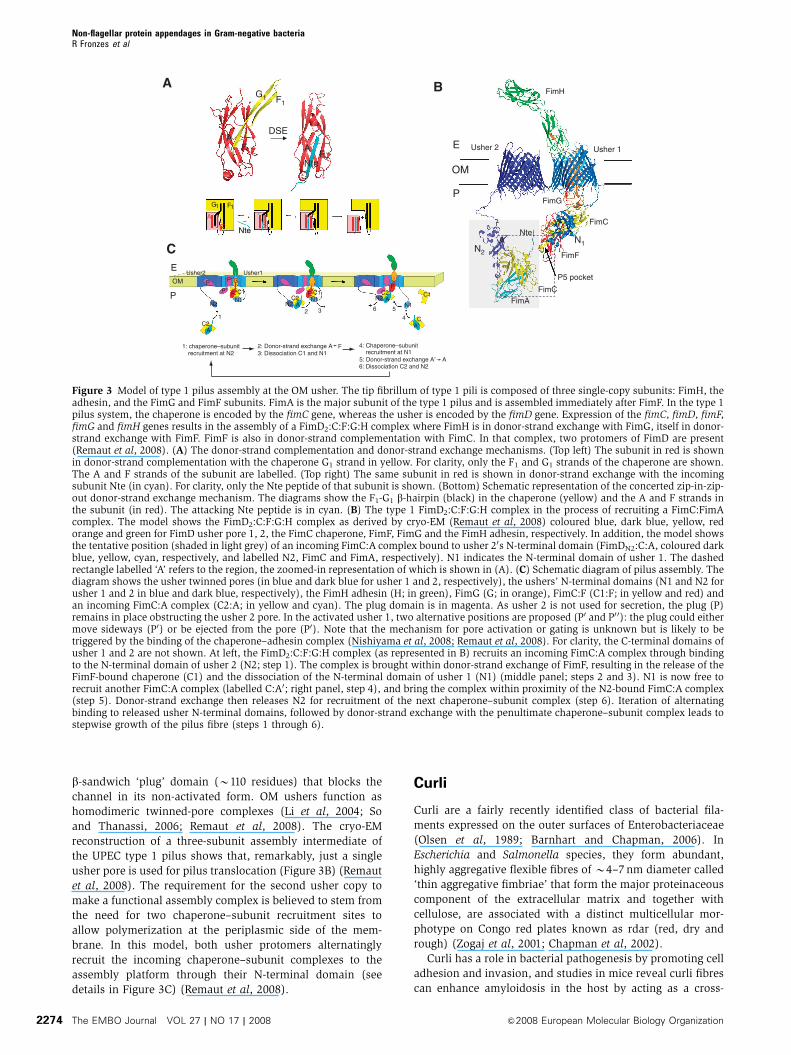
Figure 3 Model of type 1 pilus assembly at the OM usher. The tip fibrillum of type 1 pili is composed of three single-copy subunits: FimH, theadhesin, and the FimG and FimF subunits. FimA is the major subunit of the type 1 pilus and is assembled immediately after FimF. In the type 1pilus system, the chaperone is encoded by the fimC gene, whereas the usher is encoded by the fimD gene. Expression of the fimC, fimD, fimF,fimG and fimH genes results in the assembly of a FimD2:C:F:G:H complex where FimH is in donor-strand exchange with FimG, itself in donor-strand exchange with FimF. FimF is also in donor-strand complementation with FimC. In that complex, two protomers of FimD are present(Remaut et al, 2008). (A) The donor-strand complementation and donor-strand exchange mechanisms. (Top left) The subunit in red is shownin donor-strand complementation with the chaperone G1 strand in yellow. For clarity, only the F1 and G1 strands of the chaperone are shown.The A and F strands of the subunit are labelled. (Top right) The same subunit in red is shown in donor-strand exchange with the incomingsubunit Nte (in cyan). For clarity, only the Nte peptide of that subunit is shown. (Bottom) Schematic representation of the concerted zip-in-zip-out donor-strand exchange mechanism. The diagrams show the F1-G1 b-hairpin (black) in the chaperone (yellow) and the A and F strands inthe subunit (in red). The attacking Nte peptide is in cyan. (B) The type 1 FimD2:C:F:G:H complex in the process of recruiting a FimC:FimAcomplex. The model shows the FimD2:C:F:G:H complex as derived by cryo-EM (Remaut et al, 2008) coloured blue, dark blue, yellow, redorange and green for FimD usher pore 1, 2, the FimC chaperone, FimF, FimG and the FimH adhesin, respectively. In addition, the model showsthe tentative position (shaded in light grey) of an incoming FimC:A complex bound to usher 20s N-terminal domain (FimDN2:C:A, coloured darkblue, yellow, cyan, respectively, and labelled N2, FimC and FimA, respectively). N1 indicates the N-terminal domain of usher 1. The dashedrectangle labelled ‘A’ refers to the region, the zoomed-in representation of which is shown in (A). (C) Schematic diagram of pilus assembly. Thediagram shows the usher twinned pores (in blue and dark blue for usher 1 and 2, respectively), the ushers’ N-terminal domains (N1 and N2 forusher 1 and 2 in blue and dark blue, respectively), the FimH adhesin (H; in green), FimG (G; in orange), FimC:F (C1:F; in yellow and red) andan incoming FimC:A complex (C2:A; in yellow and cyan). The plug domain is in magenta. As usher 2 is not used for secretion, the plug (P)remains in place obstructing the usher 2 pore. In the activated usher 1, two alternative positions are proposed (P0 and P0 0): the plug could eithermove sideways (P0) or be ejected from the pore (P0). Note that the mechanism for pore activation or gating is unknown but is likely to betriggered by the binding of the chaperone–adhesin complex (Nishiyama et al, 2008; Remaut et al, 2008). For clarity, the C-terminal domains ofusher 1 and 2 are not shown. At left, the FimD2:C:F:G:H complex (as represented in B) recruits an incoming FimC:A complex through bindingto the N-terminal domain of usher 2 (N2; step 1). The complex is brought within donor-strand exchange of FimF, resulting in the release of theFimF-bound chaperone (C1) and the dissociation of the N-terminal domain of usher 1 (N1) (middle panel; steps 2 and 3). N1 is now free torecruit another FimC:A complex (labelled C:A0; right panel, step 4), and bring the complex within proximity of the N2-bound FimC:A complex(step 5). Donor-strand exchange then releases N2 for recruitment of the next chaperone–subunit complex (step 6). Iteration of alternatingbinding to released usher N-terminal domains, followed by donor-strand exchange with the penultimate chaperone–subunit complex leads tostepwise growth of the pilus fibre (steps 1 through 6).
Non-flagellar protein appendages in Gram-negative bacteriaR Fronzes et al
The EMBO Journal VOL 27 | NO 17 | 2008 &2008 European Molecular Biology Organization2274

seeding nuclei (Gophna et al, 2001; Lundmark, 2005). Curli
are also potent promoters of a proinflammatory response and
can enhance hypotension and bleeding disorders during
sepsis and septic chock by their absorption of contact-phase
proteins and fibrinogen (Herwald et al, 1998).
Curli structure and assembly
The genes involved in curli production are organized into two
adjacent divergently transcribed operons, csgBAC/agfBAC
and csgDEFG/agfDEFG in E. coli/Salmonella enterica, respec-
tively (Hammar et al, 1995; Collinson et al, 1996). Curli
biogenesis is proposed to follow an extracellular nucleation-
precipitation pathway that involves aggregation of the se-
creted major subunit (CsgA) onto a membrane-associated
minor subunit (CsgB) that exerts an effect as a nucleation
factor (Figure 1) (Hammar et al, 1996; Chapman et al, 2002).
Subunit secretion across the outer membrane is dependent on
a pore-forming outer membrane lipoprotein (CsgG)
(Robinson et al, 2006). Fibre assembly further requires two
periplasmic accessory proteins (CsgE and CsgF), both of
which can physically interact with the outer membrane
partner CsgG (Robinson et al, 2006). The exact role of these
periplasmic components is ill understood. CsgE appears to be
required for the secretion of the major curli subunit CsgA,
whereas CsgF influences the ability of secreted CsgA to
aggregate onto the bacterial surface, suggesting a role in
maturation or localization of the nucleator subunit, CsgB
(Chapman et al, 2002; Gibson et al, 2007). Recent data in
Salmonella suggest CsgC (AgfC) is also involved in the
assembly process as a periplasmic factor, with CsgC knockout
mutants influencing the structural features of aggregated
CsgA (Gibson et al, 2007). CsgD does not form part of the
assembly machinery but functions as a transcriptional reg-
ulator (Hammar et al, 1995).
Both curli subunits are B100 residues in size and each
contains five repeating sequence motifs, including conserved
glycines, glutamines and asparagines, commonly observed in
amyloidogenic peptides. Computer modelling of CsgA and
CsgB predicts a compact cross-b domain composed of five
stacking b-hairpins (Figure 2) (White et al, 2001; Barnhart
and Chapman, 2006). Curli are unbranched filaments likely
formed by the edge-to-edge aggregation of these cross-bdomains along the length of the fibre.
Type IV pili
Type IV pili are pilin polymers produced by many Gram-
negative bacteria, including major human pathogens such as
Neisseria gonorrhoeae, N. meningitidis, Pseudomonas aerugi-
nosa, Vibrio cholerae, S. enterica, Legionella pneumophila
and enteropathogenic E. coli (EPEC). Type IV pili are long
(1–4 mm) and flexible filaments of 5–8 nm in diameter that
can resist stress forces greater than 100 pN (Merz et al, 2000;
Maier et al, 2002). Some bacteria such as V. cholerae and
EPEC express bundled type IV pili (Craig et al, 2003;
Ramboarina et al, 2005). These pili are essential for a large
number of processes determining bacterial virulence, includ-
ing auto-aggregation, adhesion, twitching motility, biofilm
formation or cellular invasion (Craig et al, 2004; Burrows,
2005).
Classification of the pilins and maturation: type IVa and
IVb pilins
Type IV pilins are produced by rather evolutionarily divergent
Gram-negative bacteria. However, these proteins have been
grouped in the same class because they share common
sequence features and are post-translationally modified in
the same manner. Indeed, they are all produced as precursors
that possess a basic N-terminal leader sequence. Then signal
peptide removal and N-methylation are performed by a single
bi-functional endopeptidase first identified in P. aeruginosa
(PilD), which cleaves between an invariant glycine residue
and a phenylalanine (or rarely methionine) (Nunn and
Lory, 1991). All the type IV pilins contain a 30 amino-acid
hydrophobic N terminus with an invariant glutamic
acid residue at the fifth position of the mature polypeptide.
In addition, the C-terminal part of the pilin contains two
conserved cystein residues. Type IVa and IVb pilins differ
by the length of the signal peptide: type IVa pilin has a short
5–6 leader peptide, whereas type IVb pilins contain a
longer one (15–30 amino acids).
Pilus structure
For clarity, we chose to only present in this review the latest
full-length type IVa N. gonorrhoeae GC and N-terminally
truncated type IVb V. cholerae TcpA structures as representa-
tive examples of their respective families (Craig et al, 2003,
2006).
Type IVa and IVb pilins share conserved structural fea-
tures: a conserved structural core (Figure 2 in grey) flanked
by a conserved extended N-terminal a1 helix (Figure 2 in
blue), which is itself flanked by two variable regions
commonly called ab-loop and D-region (in orange and
green in Figure 2, respectively). The fold of the core domain
is different in type IVa and IVb pilins: whereas the GC pilin
displays four b-strands that are directly inter-connected, the
TcpA pilin shows a more complex connectivity and contains a
fifth b-strand. The two variable regions, a/b and D, are
exposed on both sides of the molecule. The a/b region
shows great structural variations among the type IV pilins.
In addition, O-glycosylation and phosphorylation occurs in
the GC pilin.
The structural model of N. gonorrhoeae GC pilus recently
proposed by Craig et al was deduced from a 12.5-A resolution
cryo-EM density map of the native pilus (Figure 2). In this
model, the pilus has an outer diameter of 60 A and a narrow
central channel between 6 and 11 A. The crystal structure of a
single native GC pilin was docked into the EM map. The
a1-helices of the pilin interact tightly with each other in a
three helices bundle to form the hydrophobic core of the pilus
within the filament. The globular heads are pointing out-
wards exposing the variable a/b and D-regions to the surface
of the pilus. In addition, the a/b and D-regions are also
involved in pilin–pilin interactions within the filament. The
overall type IVb pili architecture is very similar to that of type
IVa pili (Craig et al, 2003).
Assembly–disassembly
Unlike curli and CU pili, type IV pilus assembly requires a
large assembly machinery that is ATP driven and spans both
inner and outer membranes. Quite remarkably, and unique
among the different bacterial cell surface filaments, type IV
pili assembly can be reversed and the formed pili retracted.
Non-flagellar protein appendages in Gram-negative bacteriaR Fronzes et al
&2008 European Molecular Biology Organization The EMBO Journal VOL 27 | NO 17 | 2008 2275

Several bacterial processes such as twitching motility (Merz
et al, 2000) or DNA uptake (Aas et al, 2002) rely specifically
on this pilus disassembly mechanism. The assembly–disas-
sembly mechanism requires the presence of conserved
ATPases called PilB/F (P. aeruginosa/N. gonorrhoeae nomen-
clature) and PilT (Figure 1) (Turner et al, 1993). The PilB/F
ATPase would be required for pilus growth, whereas PilT
would catalyse a rapid depolymerization mechanism (1500
subunits/s) (Burrows, 2005). These two ATPases are homo-
logous to the ‘traffic ATPases’ found in type II and IV
secretion systems. In the Aquiflex aeolicus PilT structure
(Satyshur et al, 2007), it was proposed that a 15 A movement
of the C-terminal domain observed upon nucleotide hydro-
lysis and g-phosphate release would be sufficient to provide
the mechanical force necessary to extract pilin subunits from
the pilus during disassembly.
The pilus is likely to be assembled at the inner membrane
from a mature pilin ‘pool’ that is inserted into the inner
membrane through the N-terminal hydrophobic part of the
a1-helix (Strom and Lory, 1987). The cytosolic ATPase PilB is
thought to be recruited to the pilus base through an inner
membrane-embedded protein (possibly PilC/G; Figure 1;
Crowther et al, 2004). At the molecular level, the assem-
bly–disassembly mechanism is far from elucidated, and pre-
sent assembly–disassembly molecular models are highly
speculative (see details in Craig et al, 2006).
PilQ forms a B1 MDa dodecameric structure that forms the
outer membrane pore that assists the pilus across the outer
membrane (Figure 1) (Wolfgang et al, 2000; Collins et al,
2005). This protein is part of the outer membrane secretins
family, which comprises secretins used in type II and III
secretion or filamentous phage release mechanisms
(Marlovits et al, 2004; Chami et al, 2005). PilQ interacts
with the pilotin lipoprotein PilP (Hardie et al, 1996; Drake
et al, 1997; Balasingham et al, 2007). The pilotin proteins
have been shown to be important in the stabilization, the
oligomerization and/or the outer membrane localization of
many secretins.
Type III secretion-related appendage
Type III secretion-related appendages were first observed in
S. typhimurium as needle-like surface structures responsible
for bacterial entry into cultured epithelial cells and as a pilus-
like structure in P. syringae and other plant pathogens (called
the Hrp pilus) (Roine et al, 1997; Kubori et al, 1998). The
assembly of these structures was dependent on systems now
well established to form a flagellum-like secretion nanoma-
chine known as the type III secretion system (T3SS). T3SS
assembles a complex injectisome designed to secrete effector
proteins across the bacterial and host cell envelopes
(Cornelis, 2006). These secretory devices are found in a
range of animal and plant pathogens including
Enterobacteriaceae as well as Aeromonas, Burkholderia,
Chlamydia, Chromobacterium, Pseudomonas, Ralstonia,
Vibrio and Xanthomonas species. So far, no adhesive role
has been observed for T3SS appendages.
Needle structure and assembly
Injectisome assembly involves over 20 proteins (Cornelis,
2006). The injectisome basal structure forms a large cylind-
rical heterocomplex with two double rings that span the inner
and outer membranes and that are linked by a hollow
structure that crosses the intermediate periplasmic space
(Figure 1) (Kubori et al, 1998). At its cytoplasmic side, the
basal body is contacted by ATPases and accessory proteins
responsible for driving and ordering protein secretion and
filament assembly (Woestyn et al, 1994; Akeda and Galan,
2005; Muller et al, 2006). On the outer surface, the basal body
assembles a hollow filamentous structure.
The basic extracellular structure, as first identified in the
Salmonella SPI-1 T3SS (Kubori et al, 1998) and later observed
in detail from purified Shigella injectisomes (Tamano et al,
2000; Blocker et al, 2001), is a short rigid hollow ‘needle’ of
about 60 nm in length and inner diameter of 2–3 nm. The
basic needle is composed of a homopolymer of 100–150
B9 kDa subunits (PrgJ, MxiH, YscF and Escf in Salmonella,
Shigella, Yersinia and EPEC, respectively). These subunits
reveal a helix-loop-helix structure similar to the D0 portion of
flagellin (Figure 2) (Deane et al, 2006). Similar to flagellin,
needle subunits polymerize into a hollow superhelical struc-
ture. The distal end of the needle carries a distinct tip
complex composed of the protein LcrV (Yersinia), one of
the three translocators involved in pore formation in the
target cytoplasmic membrane (Mueller et al, 2005). LcrV
forms a dumbbell-shaped structure that has been modelled
as a pentameric complex at the tip of the needle and thought
of as providing the scaffolding base for the additional two
translocator proteins, assembled upon contact with the target
membrane (YopB and YopD in Yersinia) (Derewenda et al,
2004; Deane et al, 2006). In EPEC, this needle base is
extended by a longer flexible ‘filament’ up to 600 nm in
length (Knutton et al, 1998; Sekiya et al, 2001) and composed
of an EspA homopolymer. In plant pathogens, the needles are
replaced altogether by a long flexible ‘pilus’, the Hrp pilus
(Roine et al, 1997; Li et al, 2002).
Assembly of T3SS appendages is thought to follow a
mechanism similar to that seen in flagella (He et al, 2004;
Macnab, 2004). The components that form the base complex
are assembled mostly in a Sec-dependent manner. Filament
subunits and secretion substrates, however, lack classical
N-terminal leader sequences and are secreted through the
nascent secretion apparatus itself. In flagella, the extracellu-
lar hook and flagellar filament are formed by the secretion
of subunits directly from the cytoplasm through the base
complex and through the growing hollow filament (Macnab,
2004). In T3SS filaments, growth by subunit secretion at the
distal end has been directly demonstrated in the case of the
Hrp pilus and the EspA filament in P. syringae and EPEC
injectisomes, respectively (Li et al, 2002; Crepin et al, 2005).
The inner dimensions of the flagella and T3SS filaments
argue that filament subunits and T3SS effectors traverse the
structure in a largely extended, unfolded state.
In Yersinia, the length of the needle complex is linearly
correlated with the primary structure of the YscP protein,
therefore also called a ‘molecular ruler’ (Journet et al, 2003).
A hypothetical mechanism has been proposed in which the
molecular ruler resides inside the needle and allows secretion
of needle subunits until the needle reaches the length of the
unfolded, extended ruler protein. In the Salmonella SPI-1
T3SS, a different mechanism for needle length control has
been suggested (Marlovits et al, 2006). Purified Salmonella
injectisomes indicate the presence of an inner rod structure
(possibly composed of a specialized subunit, PrgJ) inside the
Non-flagellar protein appendages in Gram-negative bacteriaR Fronzes et al
The EMBO Journal VOL 27 | NO 17 | 2008 &2008 European Molecular Biology Organization2276

basal body of the secretion system. In the proposed model,
secretion of rod and needle subunits (PrgJ and PrgI, respec-
tively; Figure 1) occurs simultaneously. Completion of
the inner rod assembly signals a halt in secretion of rod
and needle subunits, thereby controlling needle length
(Marlovits et al, 2006).
Type IV secretion pili
Type IV secretion systems (T4SSs) are ancestrally derived
from the mating pair formation cluster proteins involved in
bacterial conjugation. T4SSs are also used by plant and
human pathogens such as Agrobacterium tumefaciens,
Bordetella pertussis, Legionella pneumonia and Helicobacter
pylori to secrete virulence factors (DNA and/or proteins) into
host cells. T4SS pili are thought to establish a stable and
specific contact between cells before substrate transfer
(Schroder and Lanka, 2005).
Morphology
T4SS pili are classified into two major groups: IncF-like pili
(conjugative pili produced by Inc-F, -H, -Tand -J systems) and
IncP-like pili (conjugative pili produced by Inc-P, -N, -W
systems) (Lawley et al, 2003) The IncF-like pili are long
(2–20 mm) and flexible appendages with 8–9 nm in diameter;
IncP-like pili are short (o1mm) and rigid rods with 8–12 nm
in diameter (Bradley, 1980). These different pili types are
assembled by two different classes of conjugative T4SSs:
IncP-type and IncF-type T4SSs. IncP-type T4SSs are type
IVa secretion systems. They are composed of VirB-like com-
ponents (in reference to the A. tumefaciens VirB/D4 T4SS
where 11 VirB proteins (VirB1-11) and 1 VirD proteins
(VirD4) are essential for T4SS structure, assembly and func-
tion (Figure 1)). IncF-type T4SSs are type IVb secretion
systems: they not only contain VirB-like components but
also additional proteins that do not have any counterpart in
IncP-type T4SSs.
In the case of T4SSs that are not involved in bacterial
conjugation, the morphology of the pilus is rarely known or
they cannot be classified into the subgroups defined above.
For example, the A. tumefaciens T-pilus has a different morphol-
ogy (T-pili are variable in length and flexible with 10nm in
diameter) than IncP-like and IncF-like pili even if the VirB/D4
T4SS organization is very similar to IncP conjugative T4SSs
(Eisenbrandt et al, 1999). H. pylori produces 100–200nm
needle-like type IV secretion-related appendages at the tip
of which CagA (a H. pylori T4SS substrate) can be found
(Kwok et al, 2007).
Pilus structure
Fibre X-ray diffraction and electron microscopy studies
of the F-pilus showed that these pili are cylindrical
filaments with an external diameter of 8 nm and an internal
lumen of 2 nm in diameter. These filaments exhibit a 5-fold
helical symmetry (Marvin and Folkhard, 1986).
In A. tumefaciens, it has been shown that VirB5 and
VirB7 are minor T-pilus components in addition to the major
structural pilin VirB2 (Schmidt-Eisenlohr et al, 1999; Lai
and Kado, 2000; Sagulenko et al, 2001b). These proteins have
homologues in other pathogenic bacteria such as Brucella suis
or Bartonella henselae (called VirB5/VirB7 in both species)
and also in conjugative plasmids such as pKM101 (TraC/TraN),
RP4 (TrbF/TrbH), R388 (TrwJ/TrwH) or F factor (TraE/TraV).
The atomic structure of VirB5 homologue of pKM101 conjugative
plasmid (called TraC) is composed of a three long helices bundle
flanked by a smaller globular part (Figure 2) (Yeo et al, 2003). It
has been suggested that VirB5 homologues may have adhesive
functions (Schmidt-Eisenlohr et al, 1999; Yeo et al, 2003).
Pilus assembly
Little is known concerning the assembly of the type IV
secretion pili. Before being assembled in the pilus, the pre-
pilin undergoes several conserved steps of maturation that
can be categorized into two classes.
After signal peptide removal, most of the IncF-like pilins are
inserted into the inner membrane and N-acetylated. In the case
of the F-pilus, the pilin TraA is N-acetylated by TraX (Moore
et al, 1993). TraQ is an inner membrane chaperone-like protein,
which is essential for the correct insertion and accumulation of
the pilin in the inner membrane (Lu et al, 2002).
The IncP-like pilins such as conjugative RP4 TrbC and
A. tumefaciens VirB2 are cyclic peptides (Eisenbrandt et al,
1999). In TrbC, the pre-pilin is 145 residues long and under-
goes a first maturation event where its last 27 amino acids are
cleaved by an unidentified endopeptidase. Then, the 36
amino-acid N-terminal signal peptide is removed by LepB.
Finally, the plasmid-encoded TraF is responsible for TrbC
cyclization (Kalkum et al, 2002).
Pilus assembly is clearly dependent on the integrity
of the T4SS. T4SSs, where each component of the
T4SS has been mutated, fail to assemble a pilus (Fullner
et al, 1996; Haase et al, 1995). Pilus production and substrate
transfer functions in T4SSs are clearly distinct: mutations
in some T4SS components or pilins have been shown to
uncouple pilus biogenesis and substrate transfer (Eisenbrandt
et al, 1999; Sagulenko et al, 2001a; Jakubowski et al, 2004).
Concluding remarks
Gram-negative bacteria assemble a wide range of extracellu-
lar appendages on their outer surfaces, which they use to
interact with their environment. Functionally, non-flagellar
appendages appear to have specialized roles either in adhe-
sion or in transport. CU pili, type IV pili or curli help
pathogenic bacteria to specifically recognize and adhere or
even invade their target cells and are not involved in trans-
port. T3SS appendages are used as transport devices and
have no described adhesive properties, whereas T4SS pili
could be the only pili to have dual roles. However, non-
flagellar appendages can be viewed as functionally comple-
mentary. Indeed, bacteria using secretion systems to inject
virulence factors generally rely on adhesive molecules such
as CU pili, type IV pili or other adhesive proteins (e.g.
autotransporter and TPS adhesins) to specifically target
their host cells (see for example, B. pertussis (Jacob-
Dubuisson et al, 1994).
Bacteria have developed sophisticated ways to select, at-
tach and infect their target cells. Adhesive and secretion pili
are key protagonists during these events. Further progress in
structural biology of bacterial pili will help to understand how
they are assembled and how they are involved in pathogeni-
city. This will open avenues for interference with their func-
tion as virulence factors, or for their use as display and/or
delivery tools in future nanobiotechnology applications.
Non-flagellar protein appendages in Gram-negative bacteriaR Fronzes et al
&2008 European Molecular Biology Organization The EMBO Journal VOL 27 | NO 17 | 2008 2277

References
Aas FE, Wolfgang M, Frye S, Dunham S, Lovold C, Koomey M(2002) Competence for natural transformation in Neisseria gonor-rhoeae: components of DNA binding and uptake linked to type IVpilus expression. Mol Microbiol 46: 749–760
Akeda Y, Galan JE (2005) Chaperone release and unfolding ofsubstrates in type III secretion. Nature 437: 911–915
Anderson TF (1949) In The Nature of the Bacterial Surface, Miles AA,Pirie NW (eds), p 76 Oxford: Oxford University Press
Balasingham SV, Collins RF, Assalkhou R, Homberset H, Frye SA,Derrick JP, Tonjum T (2007) Interactions between the lipoproteinPilP and the secretin PilQ in Neisseria meningitidis. J Bacteriol189: 5716–5727
Barnhart MM, Chapman MR (2006) Curli biogenesis and function.Annu Rev Microbiol 60: 131–147
Barnhart MM, Pinkner JS, Soto GE, Sauer FG, Langermann S,Waksman G, Frieden C, Hultgren SJ (2000) PapD-like chaperonesprovide the missing information for folding of pilin proteins. ProcNatl Acad Sci USA 97: 7709–7714
Blocker A, Jouihri N, Larquet E, Gounon P, Ebel F, Parsot C,Sansonetti P, Allaoui A (2001) Structure and composition of theShigella flexneri ‘needle complex’, a part of its type III secretion.Mol Microbiol 39: 652–663
Bradley DE (1980) Morphological and serological relationships ofconjugative pili. Plasmid 4: 155–169
Burrows LL (2005) Weapons of mass retraction. Mol Microbiol 57:878–888
Chami M, Guilvout I, Gregorini M, Remigy HW, Muller SA, ValerioM, Engel A, Pugsley AP, Bayan N (2005) Structural insights intothe secretin PulD and its trypsin-resistant core. J Biol Chem 280:37732–37741
Chapman MR, Robinson LS, Pinkner JS, Roth R, Heuser J, HammarM, Normark S, Hultgren SJ (2002) Role of Escherichia colicurli operons in directing amyloid fiber formation. Science 295:851–855
Choudhury D, Thompson A, Stojanoff V, Langermann S, Pinkner JS,Hultgren SJ, Knight SD (1999) X-ray structure of the FimC–FimHchaperone–adhesin complex from uropathogenic Escherichia coli.Science 285: 1061–1066
Collins RF, Frye SA, Balasingham S, Ford RC, Tonjum T, Derrick JP(2005) Interaction with type IV pili induces structural changes inthe bacterial outer membrane secretin PilQ. J Biol Chem 280:18923–18930
Collinson SK, Clouthier SC, Doran JL, Banser PA (1996) Salmonellaenteritidis agfBAC operon encoding thin, aggregative fimbriae.J Bacteriol 178: 662–667
Cornelis GR (2006) The type III secretion injectisome. Nat RevMicrobiol 4: 811–825
Craig L, Pique ME, Tainer JA (2004) Type IV pilus structure andbacterial pathogenicity. Nat Rev Microbiol 2: 363–378
Craig L, Taylor RK, Pique ME, Adair BD, Arvai AS, Singh M, LloydSJ, Shin DS, Getzoff ED, Yeager M, Forest KT, Tainer JA (2003)Type IV pilin structure and assembly: X-ray and EM analyses ofVibrio cholerae toxin-coregulated pilus and Pseudomonas aerugi-nosa PAK pilin. Mol Cell 11: 1139–1150
Craig L, Volkmann N, Arvai AS, Pique ME, Yeager M, Egelman EH,Tainer JA (2006) Type IV pilus structure by cryo-electron micro-scopy and crystallography: implications for pilus assembly andfunctions. Mol Cell 23: 651–662
Crepin VF, Shaw R, Abe CM, Knutton S, Frankel G (2005) Polarity ofenteropathogenic Escherichia coli EspA filament assembly andprotein secretion. J Bacteriol 187: 2881–2889
Crowther LJ, Anantha RP, Donnenberg MS (2004) The inner mem-brane subassembly of the enteropathogenic Escherichia coli bun-dle-forming pilus machine. Mol Microbiol 52: 67–79
Deane JE, Roversi P, Cordes FS, Johnson S, Kenjale R, Daniell S, BooyF, Picking WD, Picking WL, Blocker AJ, Lea SM (2006) Molecularmodel of a type III secretion system needle: implications for host-cell sensing. Proc Natl Acad Sci USA 103: 12529–12533
Derewenda U, Mateja A, Devedjiev Y, Routzahn KM, EvdokimovAG, Derewenda ZS, Waugh DS (2004) The structure of Yersiniapestis V-antigen, an essential virulence factor and mediator ofimmunity against plague. Structure 12: 301–306
Drake SL, Sandstedt SA, Koomey M (1997) PilP, a pilus biogenesislipoprotein in Neisseria gonorrhoeae, affects expression of PilQ asa high-molecular-mass multimer. Mol Microbiol 23: 657–668
Duguid JP, Anderson ES (1967) Terminology of bacterial fimbriae,or pili, and their types. Nature 215: 89–90
Eisenbrandt R, Kalkum M, Lai EM, Lurz R, Kado CI, Lanka E (1999)Conjugative pili of IncP plasmids, and the Ti plasmid T pilus arecomposed of cyclic subunits. J Biol Chem 274: 22548–22555
Evans DG, Evans DJ, Clegg S, Pauley JA (1979) Purification andcharacterization of the CFA/I antigen of enterotoxigenicEscherichia coli. Infect Immun 25: 738–748
Fullner KJ, Lara JC, Nester EW (1996) Pilus assembly byAgrobacterium T-DNA transfer genes. Science 273: 1107–1109
Garcia MI, Jouve M, Nataro JP, Gounon P, Le Bouguenec C (2000)Characterization of the AfaD-like family of invasins encoded bypathogenic Escherichia coli. FEBS Lett 479: 111–117
Gibson DL, White AP, Rajotte CM, Kay WW (2007) AgfC and AgfEfacilitate extracellular thin aggregative fimbriae synthesis inSalmonella enteritidis. Microbiology 153: 1131–1140
Gophna U, Barlev M, Seijffers R, Oelschlager TA, Hacker J, Ron EZ(2001) Curli fibers mediate internalization of Escherichia coli byeukaryotic cells. Infect Immun 69: 2659–2665
Haase J, Lurz R, Grahn AM, Bamford DH, Lanka E (1995) Bacterialconjugation mediated by plasmid RP4: RSF1010 mobilization,donor-specific phage propagation, and pilus production requirethe same Tra2 core components of a proposed DNA transportcomplex. J Bacteriol 177: 4779–4791
Hahn E, Wild P, Hermanns U, Sebbel P, Glockshuber R, Haner M,Taschner N, Burkhard P, Aebi U, Muller SA (2002) Exploring the3D molecular architecture of Escherichia coli type 1 pili. J Mol Biol323: 845–857
Hahn E, Wild P, Schraner EM, Bertschinger HU, Haner M, MullerSA, Aebi U (2000) Structural analysis of F18 fimbriae expressedby porcine toxigenic Escherichia coli. J Struct Biol 132: 241–250
Hammar M, Arnqvist A, Bian Z, Olsen A, Normark S (1995)Expression of two csg operons is required for production offibronectin- and Congo red-binding curli polymers inEscherichia coli K-12. Mol Microbiol 18: 661–670
Hammar M, Bian Z, Normark S (1996) Nucleator-dependent inter-cellular assembly of adhesive curli organelles in Escherichia coli.Proc Natl Acad Sci USA 93: 6562–6566
Hardie KR, Lory S, Pugsley AP (1996) Insertion of an outermembrane protein in Escherichia coli requires a chaperone-likeprotein. EMBO J 15: 978–988
He S, Nomura K, Whittam T (2004) Type III protein secretionmechanism in mammalian and plant pathogens. BiochimBiophys Acta Mol Cell Res 1694: 181–206
Herwald H, Moergelin M, Olsen A, Rhen M (1998) Activation of thecontact-phase system on bacterial surfaces—a clue to seriouscomplications in infectious diseases. Nat Med 4: 298–302
Holmgren A, Branden CI (1989) Crystal structure of chaperoneprotein PapD reveals an immunoglobulin fold. Nature 342:248–251
Houwink AL (1949) In The Nature of the Bacterial Surface, MilesAA, Pirie NW (eds), p 92 Oxford: Oxford University Press
Hung DL, Knight SD, Woods RM, Pinkner JS, Hultgren SJ (1996)Molecular basis of two subfamilies of immunoglobulin-like cha-perones. EMBO J 15: 3792–3805
Jacob-Dubuisson F, Striker R, Hultgren SJ (1994) Chaperone-as-sisted self-assembly of pili independent of cellular energy. J BiolChem 269: 12447–12455
Jakubowski SJ, Krishnamoorthy V, Cascales E, Christie PJ (2004)Agrobacterium tumefaciens VirB6 domains direct the orderedexport of a DNA substrate through a type IV secretion system.J Mol Biol 341: 961–977
Journet L, Agrain C, Broz P, Cornelis GR (2003) The needle length ofbacterial injectisomes is determined by a molecular ruler. Science302: 1757–1760
Kalkum M, Eisenbrandt R, Lurz R, Lanka E (2002) Tying rings forsex. Trends Microbiol 10: 382–387
Keller R, Ordonez JG, de Oliveira RR, Trabulsi LR, Baldwin TJ,Knutton S (2002) Afa, a diffuse adherence fibrillar adhesinassociated with enteropathogenic Escherichia coli. Infect Immun70: 2681–2689
Knutton S, Rosenshine I, Pallen MJ, Nisan I, Neves BC, Bain C,Wolff C, Dougan G, Frankel G (1998) A novel EspA-associatedsurface organelle of enteropathogenic Escherichia coli involved inprotein translocation into epithelial cells. EMBO J 17: 2166–2176
Non-flagellar protein appendages in Gram-negative bacteriaR Fronzes et al
The EMBO Journal VOL 27 | NO 17 | 2008 &2008 European Molecular Biology Organization2278

Kubori T, Matsushima Y, Nakamura D, Uralil J, Lara-Tejero M,Sukhan A, Galan JE, Aizawa SI (1998) Supramolecular structureof the Salmonella typhimurium type III protein secretion system.Science 280: 602–605
Kuehn MJ, Heuser J, Normark S, Hultgren SJ (1992) P pili inuropathogenic E. coli are composite fibres with distinct fibrillaradhesive tips. Nature 356: 252–255
Kwok T, Zabler D, Urman S, Rohde M, Hartig R, Wessler S,Misselwitz R, Berger J, Sewald N, Konig W, Backert S (2007)Helicobacter exploits integrin for type IV secretion and kinaseactivation. Nature 449: 862–866
Lai EM, Kado CI (2000) The T-pilus of Agrobacterium tumefaciens.Trends Microbiol 8: 361–369
Lawley TD, Klimke WA, Gubbins MJ, Frost LS (2003) F factorconjugation is a true type IV secretion system. FEMS MicrobiolLett 224: 1–15
Levine MM, Ristaino P, Marley G, Smyth C, Knutton S, Boedeker E,Black R, Young C, Clements ML, Cheney C (1984) Coli surfaceantigens 1 and 3 of colonization factor antigen II-positive enter-otoxigenic Escherichia coli: morphology, purification, andimmune responses in humans. Infect Immun 44: 409–420
Li CM, Brown I, Mansfield J, Stevens C, Boureau T, Romantschuk M,Taira S (2002) The Hrp pilus of Pseudomonas syringae elongatesfrom its tip and acts as a conduit for translocation. EMBO J 21:1909–1915
Li H, Qian L, Chen Z, Thibault D, Liu G, Liu T, Thanassi DG (2004)The outer membrane usher forms a twin-pore secretion complex.J Mol Biol 344: 1397–1407
Lu J, Manchak J, Klimke W, Davidson C, Firth N, Skurray RA,Frost LS (2002) Analysis and characterization of the IncFVplasmid pED208 transfer region. Plasmid 48: 24–37
Lundmark K (2005) Protein fibrils in nature can enhance amyloidprotein A amyloidosis in mice: cross-seeding as a disease me-chanism. Proc Natl Acad Sci USA 102: 6098–6102
Macnab R (2004) Type III flagellar protein export and flagellarassembly. Biochim Biophys Acta Mol Cell Res 1694: 207–217
Maier B, Potter L, So M, Long CD, Seifert HS, Sheetz MP (2002)Single pilus motor forces exceed 100 pN. Proc Natl Acad Sci USA99: 16012–16017
Marlovits TC, Kubori T, Lara-Tejero M, Thomas D, Unger VM,Galan JE (2006) Assembly of the inner rod determines needlelength in the type III secretion injectisome. Nature 441:637–640
Marlovits TC, Kubori T, Sukhan A, Thomas DR, Galan JE, Unger VM(2004) Structural insights into the assembly of the type IIIsecretion needle complex. Science 306: 1040–1042
Marvin DA, Folkhard W (1986) Structure of F-pili: reassessment ofthe symmetry. J Mol Biol 191: 299–300
Merz AJ, So M, Sheetz MP (2000) Pilus retraction powers bacterialtwitching motility. Nature 407: 98–102
Moore D, Hamilton CM, Maneewannakul K, Mintz Y, Frost LS,Ippen-Ihler K (1993) The Escherichia coli K-12 F plasmid genetraX is required for acetylation of F pilin. J Bacteriol 175:1375–1383
Mu XQ, Bullitt E (2006) Structure and assembly of P-pili: aprotruding hinge region used for assembly of a bacterial adhesionfilament. Proc Natl Acad Sci USA 103: 9861–9866
Mueller CA, Broz P, Muller SA, Ringler P, Erne-Brand F, Sorg I, Kuhn M,Engel A, Cornelis GR (2005) The V-antigen of Yersinia forms adistinct structure at the tip of injectisome needles. Science 310:674–676
Muller SA, Pozidis C, Stone R, Meesters C, Chami M, Engel A,Economou A, Stahlberg H (2006) Double hexameric ring assem-bly of the type III protein translocase ATPase HrcN. Mol Microbiol61: 119–125
Ng TW, Akman L, Osisami M, Thanassi DG (2004) The usher Nterminus is the initial targeting site for chaperone–subunit com-plexes and participates in subsequent pilus biogenesis events.J Bacteriol 186: 5321–5331
Nishiyama M, Horst R, Eidam O, Herrmann T, Ignatov O, Vetsch M,Bettendorff P, Jelesarov I, Grutter MG, Wuthrich K, GlockshuberR, Capitani G (2005) Structural basis of chaperone–subunit com-plex recognition by the type 1 pilus assembly platform FimD.EMBO J 24: 2075–2086
Nishiyama M, Ishikawa T, Rechsteiner H, Glockshuber R (2008)Reconstitution of pilus assembly reveals a bacterial outer mem-brane catalyst. Science 320: 376–379
Nuccio S, Baumler A (2007) Evolution of the chaperone/usherassembly pathway: fimbrial classification goes Greek. MicrobiolMol Biol Rev 71: 551–575
Nunn DN, Lory S (1991) Product of the Pseudomonas aeruginosagene pilD is a prepilin leader peptidase. Proc Natl Acad Sci USA88: 3281–3285
Olsen A, Jonsson A, Normark S (1989) Fibronectin bindingmediated by a novel class of surface organelles on Escherichiacoli. Nature 338: 652–655
Ramboarina S, Fernandes PJ, Daniell S, Islam S, Simpson P, Frankel G,Booy F, Donnenberg MS, Matthews S (2005) Structure of the bundle-forming pilus from enteropathogenic Escherichia coli. J Biol Chem280: 40252–40260
Remaut H, Rose RJ, Hannan TJ, Hultgren SJ, Radford SE, Ashcroft AE,Waksman G (2006) Donor-strand exchange in chaperone-assistedpilus assembly proceeds through a concerted strand displacementmechanism. Mol Cell 22: 831–842
Remaut H, Tang C, Henderson NS, Pinkner J, Wang T, Hultgren SJ,Thanassi DG, Waksman G, Li H (2008) Fibre formation across thebacterial outer membrane by the chaperone/usher pathway. Cell33: 640–652
Robinson LS, Ashman EM, Hultgren SJ, Chapman MR (2006)Secretion of curli fibre subunits is mediated by the outer mem-brane-localized CsgG protein. Mol Microbiol 59: 870–881
Roine E, Wei W, Yuan J, Nurmiaho-Lassila EL, Kalkkinen N,Romantschuk M, He SY (1997) Hrp pilus: an hrp-dependentbacterial surface appendage produced by Pseudomonas syringaepv. tomato DC3000. Proc Natl Acad Sci USA 94: 3459–3464
Sagulenko E, Sagulenko V, Chen J, Christie PJ (2001a) Role ofAgrobacterium VirB11 ATPase in T-pilus assembly and substrateselection. J Bacteriol 183: 5813–5825
Sagulenko V, Sagulenko E, Jakubowski S, Spudich E, Christie PJ(2001b) VirB7 lipoprotein is exocellular and associates with theAgrobacterium tumefaciens T pilus. J Bacteriol 183: 3642–3651
Salih O, Remaut H, Waksman G, Orlova EV (2008) Structuralanalysis of the Saf pilus by electron microscopy and imageprocessing. J Mol Biol 379: 174–187
Satyshur KA, Worzalla GA, Meyer LS, Heiniger EK, Aukema KG,Misic AM, Forest KT (2007) Crystal structures of the pilusretraction motor PilT suggest large domain movements andsubunit cooperation drive motility. Structure 15: 363–376
Sauer FG, Futterer K, Pinkner JS, Dodson KW, Hultgren SJ,Waksman G (1999) Structural basis of chaperone function andpilus biogenesis. Science 285: 1058–1061
Sauer FG, Pinkner JS, Waksman G, Hultgren SJ (2002) Chaperonepriming of pilus subunits facilitates a topological transition thatdrives fiber formation. Cell 111: 543–551
Sauer FG, Remaut H, Hultgren SJ, Waksman G (2004) Fiberassembly by the chaperone–usher pathway. Biochim BiophysActa 1694: 259–267
Schmidt-Eisenlohr H, Domke N, Angerer C, Wanner G, ZambryskiPC, Baron C (1999) Vir proteins stabilize VirB5 and mediate itsassociation with the T pilus of Agrobacterium tumefaciens.J Bacteriol 181: 7485–7492
Schroder G, Lanka E (2005) The mating pair formation system ofconjugative plasmids-A versatile secretion machinery for transferof proteins and DNA. Plasmid 54: 1–25
Sekiya K, Ohishi M, Ogino T, Tamano K, Sasakawa C, Abe A (2001)Supermolecular structure of the enteropathogenic Escherichiacoli type III secretion system and its direct interaction withthe EspA-sheath-like structure. Proc Natl Acad Sci USA 98:11638–11643
So SS, Thanassi DG (2006) Analysis of the requirements for pilusbiogenesis at the outer membrane usher and the function of theusher C-terminus. Mol Microbiol 60: 364–375
Strom MS, Lory S (1987) Mapping of export signals of Pseudomonasaeruginosa pilin with alkaline phosphatase fusions. J Bacteriol169: 3181–3188
Tamano K, Aizawa S, Katayama E, Nonaka T, Imajoh-Ohmi S,Kuwae A, Nagai S, Sasakawa C (2000) Supramolecular structureof the Shigella type III secretion machinery: the needle part ischangeable in length and essential for delivery of effectors. EMBOJ 19: 3876–3887
Turner LR, Lara JC, Nunn DN, Lory S (1993) Mutations in theconsensus ATP-binding sites of XcpR and PilB eliminate extra-cellular protein secretion and pilus biogenesis in Pseudomonasaeruginosa. J Bacteriol 175: 4962–4969
Non-flagellar protein appendages in Gram-negative bacteriaR Fronzes et al
&2008 European Molecular Biology Organization The EMBO Journal VOL 27 | NO 17 | 2008 2279
Verger D, Bullitt E, Hultgren SJ, Waksman G (2007) Crystal structureof the P pilus rod subunit PapA. PLoS Pathog 3: e73
Verger D, Miller E, Remaut H, Waksman G, Hultgren S (2006)Molecular mechanism of P pilus termination in uropathogenicEscherichia coli. EMBO Rep 7: 1228–1232
Vetsch M, Puorger C, Spirig T, Grauschopf U, Weber-Ban EU,Glockshuber R (2004) Pilus chaperones represent a new type ofprotein-folding catalyst. Nature 431: 329–333
White AP, Collinson SK, Banser PA, Gibson DL (2001) Structureand characterization of AgfB from Salmonella enteritidis thinaggregative fimbriae. J Mol Biol 311: 735–749
Woestyn S, Allaoui A, Wattiau P, Cornelis GR (1994) YscN, theputative energizer of the Yersinia Yop secretion machinery.J Bacteriol 176: 1561–1569
Wolfgang M, van Putten JP, Hayes SF, Dorward D, Koomey M (2000)Components and dynamics of fiber formation define a ubiquitousbiogenesis pathway for bacterial pili. EMBO J 19: 6408–6418
Wright KJ, Seed PC, Hultgren SJ (2007) Development of intracel-lular bacterial communities of uropathogenic Escherichia colidepends on type 1 pili. Cell Microbiol 9: 2230–2241
Yeo HJ, Yuan Q, Beck MR, Baron C, Waksman G (2003) Structuraland functional characterization of the VirB5 protein from the type
IV secretion system encoded by the conjugative plasmid pKM101.Proc Natl Acad Sci USA 100: 15947–15952
Zavialov A, Zav’yalova G, Korpela T, Zav’yalov V (2007) FGLchaperone-assembled fimbrial polyadhesins: anti-immune arma-ment of Gram-negative bacterial pathogens. FEMS Microbiol Rev31: 478–514
Zavialov AV, Berglund J, Pudney AF, Fooks LJ, Ibrahim TM,MacIntyre S, Knight SD (2003) Structure and biogenesis of thecapsular F1 antigen from Yersinia pestis preserved folding energydrives fiber formation. Cell 113: 587–596
Zogaj X, Nimtz M, Rohde M, Bokranz W, Romling U (2001) Themulticellular morphotypes of Salmonella typhimurium andEscherichia coli produce cellulose as the second component ofthe extracellular matrix. Mol Microbiol 39: 1452–1463
The EMBO Journal is published by NaturePublishing Group on behalf of European
Molecular Biology Organization. This article is licensedunder a Creative Commons Attribution-Noncommercial-No Derivative Works 3.0 Licence. [http://creativecommons.org/licenses/by-nc-nd/3.0]
Non-flagellar protein appendages in Gram-negative bacteriaR Fronzes et al
The EMBO Journal VOL 27 | NO 17 | 2008 &2008 European Molecular Biology Organization2280