Lexis Hibiscus Port Dickson Does Its Part for Earth Hour 2019
Upload
khangminh22Category
view
6download
0
�����������������
Citation: Sedillo-Torres, I.Y.;
Hernández-Rangel, Á.O.; Gómez-
y-Gómez, Y.; Cortés-Avalos, D.;
García-Pérez, B.E.; Villalobos-Rocha,
J.C.; Hernández-Rodríguez, C.H.;
Zepeda-Vallejo, L.G.; Estrada-de los
Santos, P.; Vargas-Díaz, M.E.; et al.
Hibiscus Acid from Hibiscus sabdariffa
L. Inhibits Flagellar Motility and Cell
Invasion in Salmonella enterica.
Molecules 2022, 27, 655.
https://doi.org/10.3390/
molecules27030655
Academic Editor: Fengqing Yang
Received: 9 December 2021
Accepted: 14 January 2022
Published: 20 January 2022
Publisher’s Note: MDPI stays neutral
with regard to jurisdictional claims in
published maps and institutional affil-
iations.
Copyright: © 2022 by the authors.
Licensee MDPI, Basel, Switzerland.
This article is an open access article
distributed under the terms and
conditions of the Creative Commons
Attribution (CC BY) license (https://
creativecommons.org/licenses/by/
4.0/).
molecules
Article
Hibiscus Acid from Hibiscus sabdariffa L. Inhibits FlagellarMotility and Cell Invasion in Salmonella entericaIxchell Y. Sedillo-Torres 1, Álvaro O. Hernández-Rangel 2, Yolanda Gómez-y-Gómez 3, Daniel Cortés-Avalos 1,Blanca Estela García-Pérez 1 , Juan C. Villalobos-Rocha 1 , César H. Hernández-Rodríguez 1,Luis Gerardo Zepeda-Vallejo 2, Paulina Estrada-de los Santos 1 , María Elena Vargas-Díaz 2
and Jose Antonio Ibarra 1,*
1 Departamento de Microbiología, Escuela Nacional de Ciencias Biológicas, Instituto Politécnico Nacional,Prol. de Carpio y Plan de Ayala S/N Col. Santo Tomás Alc. Miguel Hidalgo, Ciudad de México 11340,Mexico; [email protected] (I.Y.S.-T.); [email protected] (D.C.-A.);[email protected] (B.E.G.-P.); [email protected] (J.C.V.-R.);[email protected] (C.H.H.-R.); [email protected] (P.E.-d.l.S.)
2 Departamento de Química Orgánica, Escuela Nacional de Ciencias Biológicas, Instituto Politécnico Nacional,Prol. de Carpio y Plan de Ayala S/N Col. Santo Tomás Alc. Miguel Hidalgo,Ciudad de México 11340, Mexico; [email protected] (Á.O.H.-R.);[email protected] (L.G.Z.-V.); [email protected] (M.E.V.-D.)
3 Unidad Profesional Interdisciplinaria de Biotecnología, Instituto Politécnico Nacional, Av. Acueducto s/nBarrio La Laguna, Ticomán, Alc. Gustavo A. Madero, Ciudad de México 07340, Mexico;[email protected]
* Correspondence: [email protected] or [email protected]; Tel.: +52-55-5729-6000 (ext. 62482)
Abstract: Extracts of Hibiscus sabdariffa L. (commonly called Rosselle or “Jamaica flower” in Mexico)have been shown to have antibiotic and antivirulence properties in several bacteria. Here, anorganic extract of H. sabdariffa L. is shown to inhibit motility in Salmonella enterica serovars Typhi andTyphimurium. The compound responsible for this effect was purified and found to be the hibiscusacid. When tested, this compound also inhibited motility and reduced the secretion of both flagellinand type III secretion effectors. Purified hibiscus acid was not toxic in tissue-cultured eukaryotic cells,and it was able to reduce the invasion of Salmonella Typhimurium in epithelial cells. Initial steps tounderstand its mode of action showed it might affect membrane proton balance.
Keywords: hibiscus acid; Salmonella; traditional medicine; motility; flagella; antivirulence
1. Introduction
The spread of multi-drug-resistant (MDR) bacteria is a global public health problembecause the treatment of infections caused by these strains has been complicated in recentyears, which has led to an increase in the number of deaths and prolonged stays of patientswithin hospitals [1,2]. In a catastrophic scenario, it is estimated that by the year 2050,approximately 10 million people will die each year from MDR bacterial infections. Itis estimated that this number will be higher than cancer-related deaths (approximately8.2 million deaths) [3–5]. This represents a great challenge for the scientific community,specifically in the design and discovery of new efficient and specific drugs [2,6].
In the search for new drugs for the treatment of diseases caused by MDR bacteria,novel strategies have been proposed such as antivirulence therapy [7,8]. This is based oninhibiting virulence mechanisms without intervening in the main metabolic pathways thatare essential for bacterial viability. To understand how these drugs work, one must considerthat virulence factors are elements that allow bacteria to infect and colonize a particularhost; these factors are expressed under specific environmental conditions that are necessaryfor the pathogenesis process [7,9]. Virulence factors may be associated with the membrane,cytoplasm, or secreted once the pathogen has been established in the host cell. These allow
Molecules 2022, 27, 655. https://doi.org/10.3390/molecules27030655 https://www.mdpi.com/journal/molecules
Molecules 2022, 27, 655 2 of 16
motility, adhesion, evasion of the host immune system; adaptation to the environmentthrough metabolic, physiological, or morphological changes; as well as inducing cell deathof host cells [10]. Considering the high number of virulence factors that can be blocked todecrease bacterial pathogenicity, it is believed that antivirulence therapy has the potentialto control and treat infections caused by pathogenic bacteria [7,11].
Salmonella enterica is a bacterium that is commonly acquired by the consumption ofcontaminated food and water; most of the serovars cause diarrhea in humans and alsoinfects domestic animals, while a few serovars cause typhoid or paratyphoid fever inhumans [12]. Some of the virulence factors in this pathogen include two type III secretionsystems (T3SS) that favor internalization and colonization in the host [13]. Motility is alsointimately related to Salmonella virulence, as it is required for the bacteria to successfullyreach the small intestine and penetrate the intestinal mucosa [14,15]. Once the pathogencomes into contact with either M cells or enterocytes, the T3SS-1 encoded in the Salmonellapathogenicity island-1 (SPI-1) is expressed, injects effector proteins into the host cells, andmodifies the cytoskeleton, favoring the internalization of the bacteria [13,16]. A possibilityto avoid infection by Salmonella would be to block these virulence factors (motility and/orT3SS-1). Hypothetically, this would modulate the bacterial pathogenesis, making thesebacteria more vulnerable to the host immune system [7,11,17].
In recent years, the potentiality of plant metabolites has been studied to mitigate bac-terial virulence [11]. Some of these plants are also used for cooking or as herbal infusions,while others are used in the industry, and many have been tested for antimicrobial capabili-ties. The Roselle, commonly known as hibiscus or “Jamaica flower” (Hibiscus sabdariffa L.),has been used as an additive for food and beverages and in traditional medicine for thetreatment of some illnesses [18–20]. In addition, it has been attributed pharmacologicalproperties [18,19]. Among the most studied components in this plant are organic acids, an-thocyanins, and flavonoids. To expand the study of Roselle, in this work, the antivirulencecapacity against Salmonella enterica of subinhibitory concentrations of an organic extract ofH. sabdariffa L. calyxes was tested. The results showed that this extract inhibited the motilityof two Salmonella serovars, and by using chemical methods, the compound responsible forthis inhibition was found to be the hibiscus acid (HA).
2. Results2.1. Antimicrobial Activity of H. sabdariffa L. Extracts
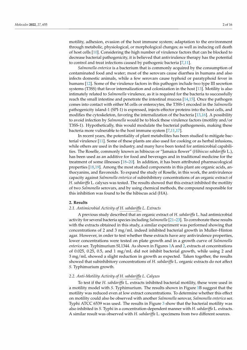
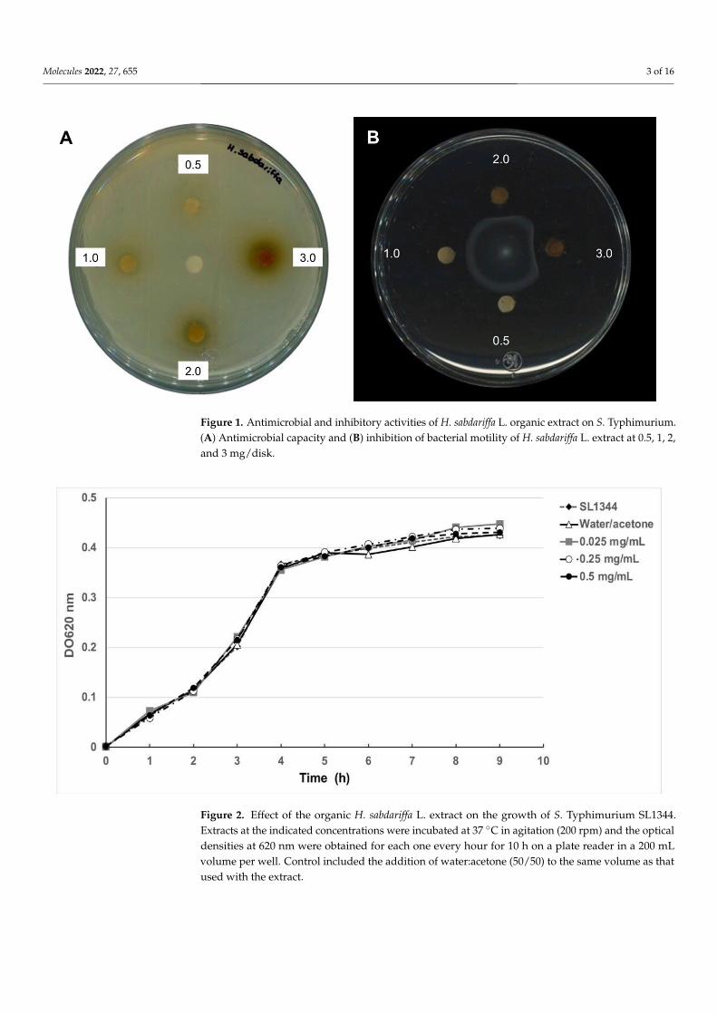
A previous study described that an organic extract of H. sabdariffa L. had antimicrobialactivity for several bacteria species including Salmonella [21–23]. To corroborate these resultswith the extracts obtained in this study, a similar experiment was performed showing thatconcentrations of 2 and 3 mg/mL indeed inhibited bacterial growth in Muller–Hintonagar. However, in order to test whether these extracts have any antivirulence properties,lower concentrations were tested on plate growth and in a growth curve of Salmonellaenterica ser. Typhimurium SL1344. As shown in Figures 1A and 2, extracts at concentrationsof 0.025, 0.25, 0.5, and 1 mg/mL did not inhibit bacterial growth, while using 2 and3 mg/mL showed a slight reduction in growth as expected. Taken together, the resultsshowed that subinhibitory concentrations of H. sabdariffa L. organic extracts do not affectS. Typhimurium growth.
2.2. Anti-Motility Activity of H. sabdariffa L. Calyxes
To test if the H. sabdariffa L. extracts inhibited bacterial motility, these were used ina motility model with S. Typhimurium. The results shown in Figure 1B suggest that themotility was reduced even at low extract concentrations. To determine whether this effecton motility could also be observed with another Salmonella serovar, Salmonella enterica ser.Typhi ATCC 6539 was used. The results in Figure 3 show that the bacterial motility wasalso inhibited in S. Typhi in a concentration-dependent manner with H. sabdariffa L. extracts.A similar result was observed with H. sabdariffa L. specimens from two different sources.
Molecules 2022, 27, 655 3 of 16Molecules 2022, 27, x FOR PEER REVIEW 3 of 16
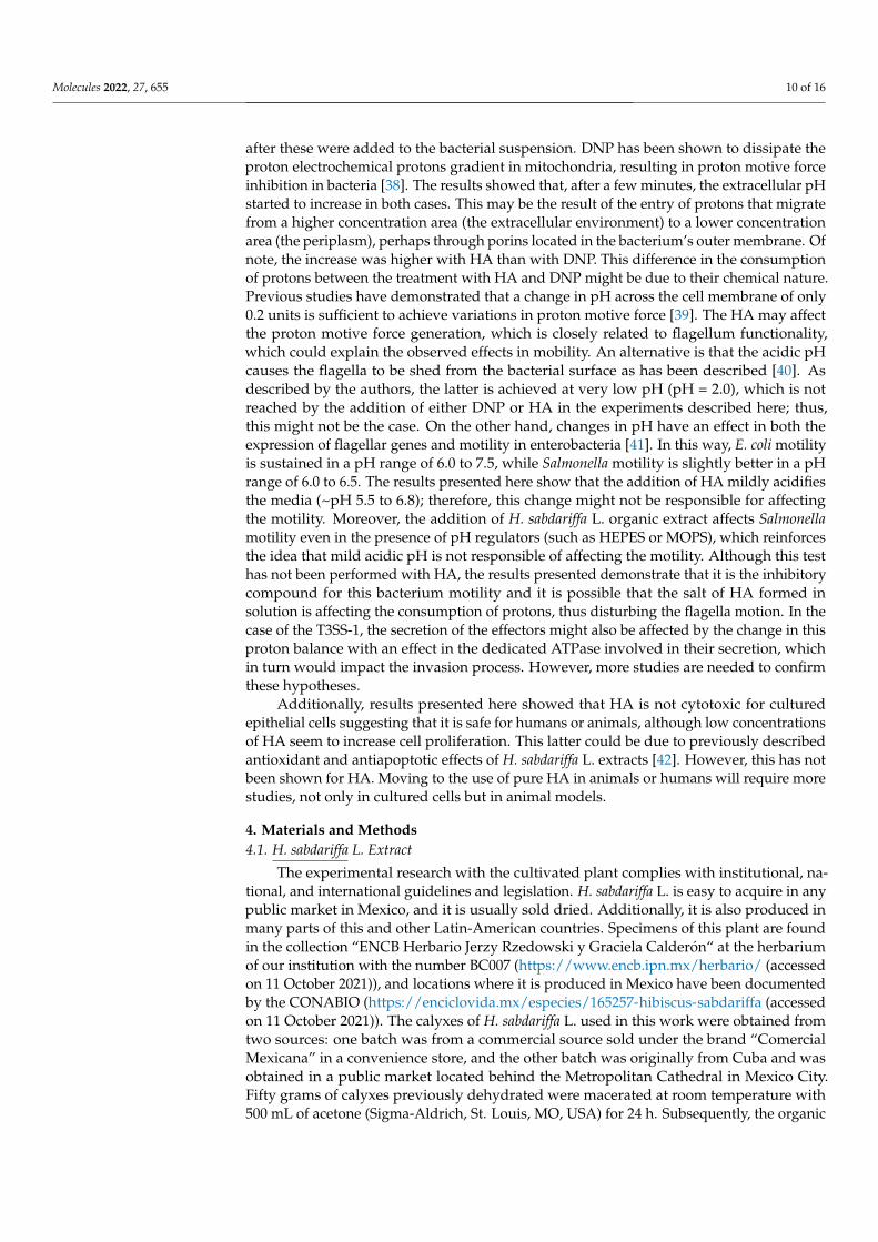
Figure 1. Antimicrobial and inhibitory activities of H. sabdariffa L. organic extract on S. Typhi-murium. (A) Antimicrobial capacity and (B) inhibition of bacterial motility of H. sabdariffa L. extract at 0.5, 1, 2, and 3 mg/disk.
Figure 2. Effect of the organic H. sabdariffa L. extract on the growth of S. Typhimurium SL1344. Extracts at the indicated concentrations were incubated at 37 °C in agitation (200 rpm) and the op-tical densities at 620 nm were obtained for each one every hour for 10 h on a plate reader in a 200 mL volume per well. Control included the addition of water:acetone (50/50) to the same volume as that used with the extract.
2.2. Anti-Motility Activity of H. sabdariffa L. Calyxes To test if the H. sabdariffa L. extracts inhibited bacterial motility, these were used in a
motility model with S. Typhimurium. The results shown in Figure 1B suggest that the motility was reduced even at low extract concentrations. To determine whether this effect on motility could also be observed with another Salmonella serovar, Salmonella enterica ser. Typhi ATCC 6539 was used. The results in Figure 3 show that the bacterial motility was also inhibited in S. Typhi in a concentration-dependent manner with H. sabdariffa L. ex-tracts. A similar result was observed with H. sabdariffa L. specimens from two different sources.
Figure 1. Antimicrobial and inhibitory activities of H. sabdariffa L. organic extract on S. Typhimurium.(A) Antimicrobial capacity and (B) inhibition of bacterial motility of H. sabdariffa L. extract at 0.5, 1, 2,and 3 mg/disk.
Molecules 2022, 27, x FOR PEER REVIEW 3 of 16
Figure 1. Antimicrobial and inhibitory activities of H. sabdariffa L. organic extract on S. Typhi-murium. (A) Antimicrobial capacity and (B) inhibition of bacterial motility of H. sabdariffa L. extract at 0.5, 1, 2, and 3 mg/disk.
Figure 2. Effect of the organic H. sabdariffa L. extract on the growth of S. Typhimurium SL1344. Extracts at the indicated concentrations were incubated at 37 °C in agitation (200 rpm) and the op-tical densities at 620 nm were obtained for each one every hour for 10 h on a plate reader in a 200 mL volume per well. Control included the addition of water:acetone (50/50) to the same volume as that used with the extract.
2.2. Anti-Motility Activity of H. sabdariffa L. Calyxes To test if the H. sabdariffa L. extracts inhibited bacterial motility, these were used in a
motility model with S. Typhimurium. The results shown in Figure 1B suggest that the motility was reduced even at low extract concentrations. To determine whether this effect on motility could also be observed with another Salmonella serovar, Salmonella enterica ser. Typhi ATCC 6539 was used. The results in Figure 3 show that the bacterial motility was also inhibited in S. Typhi in a concentration-dependent manner with H. sabdariffa L. ex-tracts. A similar result was observed with H. sabdariffa L. specimens from two different sources.
Figure 2. Effect of the organic H. sabdariffa L. extract on the growth of S. Typhimurium SL1344.Extracts at the indicated concentrations were incubated at 37 ◦C in agitation (200 rpm) and the opticaldensities at 620 nm were obtained for each one every hour for 10 h on a plate reader in a 200 mLvolume per well. Control included the addition of water:acetone (50/50) to the same volume as thatused with the extract.
Molecules 2022, 27, 655 4 of 16Molecules 2022, 27, x FOR PEER REVIEW 4 of 16
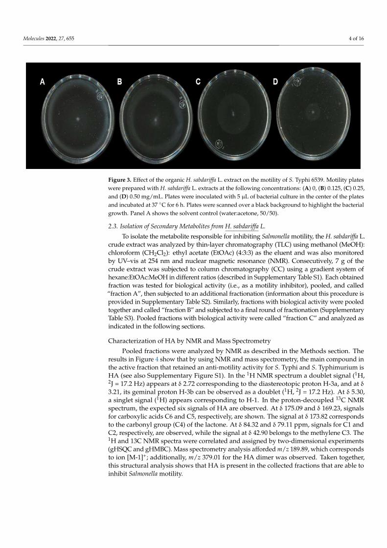
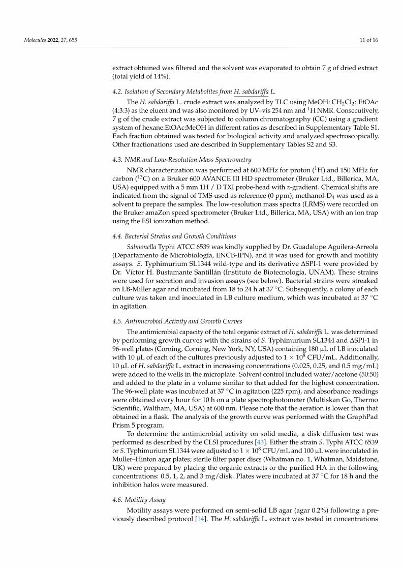
Figure 3. Effect of the organic H. sabdariffa L. extract on the motility of S. Typhi 6539. Motility plates were prepared with H. sabdariffa L. extracts at the following concentrations: (A) 0, (B) 0.125, (C) 0.25, and (D) 0.50 mg/mL. Plates were inoculated with 5 μL of bacterial culture in the center of the plates and incubated at 37 °C for 6 h. Plates were scanned over a black background to highlight the bacte-rial growth. Panel A shows the solvent control (water:acetone, 50/50).
2.3. Isolation of Secondary Metabolites from H. sabdariffa L. To isolate the metabolite responsible for inhibiting Salmonella motility, the H. sabdar-
iffa L. crude extract was analyzed by thin-layer chromatography (TLC) using methanol (MeOH): chloroform (CH2Cl2): ethyl acetate (EtOAc) (4:3:3) as the eluent and was also monitored by UV–vis at 254 nm and nuclear magnetic resonance (NMR). Consecutively, 7 g of the crude extract was subjected to column chromatography (CC) using a gradient system of hexane:EtOAc:MeOH in different ratios (described in Supplementary Table S1). Each obtained fraction was tested for biological activity (i.e., as a motility inhibitor), pooled, and called “fraction A”, then subjected to an additional fractionation (information about this procedure is provided in Supplementary Table S2). Similarly, fractions with biological activity were pooled together and called “fraction B” and subjected to a final round of fractionation (Supplementary Table S3). Pooled fractions with biological activity were called “fraction C” and analyzed as indicated in the following sections.
Characterization of HA by NMR and Mass Spectrometry Pooled fractions were analyzed by NMR as described in the Methods section. The
results in Figure 4 show that by using NMR and mass spectrometry, the main compound in the active fraction that retained an anti-motility activity for S. Typhi and S. Typhi-murium is HA (see also Supplementary Figure S1). In the 1H NMR spectrum a doublet signal (1H, 2J = 17.2 Hz) appears at δ 2.72 corresponding to the diastereotopic proton H-3a, and at δ 3.21, its geminal proton H-3b can be observed as a doublet (1H, 2J = 17.2 Hz). At δ 5.30, a singlet signal (1H) appears corresponding to H-1. In the proton-decoupled 13C NMR spectrum, the expected six signals of HA are observed. At δ 175.09 and δ 169.23, signals for carboxylic acids C6 and C5, respectively, are shown. The signal at δ 173.82 corresponds to the carbonyl group (C4) of the lactone. At δ 84.32 and δ 79.11 ppm, signals for C1 and C2, respectively, are observed, while the signal at δ 42.90 belongs to the methylene C3. The 1H and 13C NMR spectra were correlated and assigned by two-dimensional experi-ments (gHSQC and gHMBC). Mass spectrometry analysis afforded m/z 189.89, which cor-responds to ion [M-1]+; additionally, m/z 379.01 for the HA dimer was observed. Taken together, this structural analysis shows that HA is present in the collected fractions that are able to inhibit Salmonella motility.
Figure 3. Effect of the organic H. sabdariffa L. extract on the motility of S. Typhi 6539. Motility plateswere prepared with H. sabdariffa L. extracts at the following concentrations: (A) 0, (B) 0.125, (C) 0.25,and (D) 0.50 mg/mL. Plates were inoculated with 5 µL of bacterial culture in the center of the platesand incubated at 37 ◦C for 6 h. Plates were scanned over a black background to highlight the bacterialgrowth. Panel A shows the solvent control (water:acetone, 50/50).
2.3. Isolation of Secondary Metabolites from H. sabdariffa L.
To isolate the metabolite responsible for inhibiting Salmonella motility, the H. sabdariffa L.crude extract was analyzed by thin-layer chromatography (TLC) using methanol (MeOH):chloroform (CH2Cl2): ethyl acetate (EtOAc) (4:3:3) as the eluent and was also monitoredby UV–vis at 254 nm and nuclear magnetic resonance (NMR). Consecutively, 7 g of thecrude extract was subjected to column chromatography (CC) using a gradient system ofhexane:EtOAc:MeOH in different ratios (described in Supplementary Table S1). Each obtainedfraction was tested for biological activity (i.e., as a motility inhibitor), pooled, and called“fraction A”, then subjected to an additional fractionation (information about this procedure isprovided in Supplementary Table S2). Similarly, fractions with biological activity were pooledtogether and called “fraction B” and subjected to a final round of fractionation (SupplementaryTable S3). Pooled fractions with biological activity were called “fraction C” and analyzed asindicated in the following sections.
Characterization of HA by NMR and Mass Spectrometry
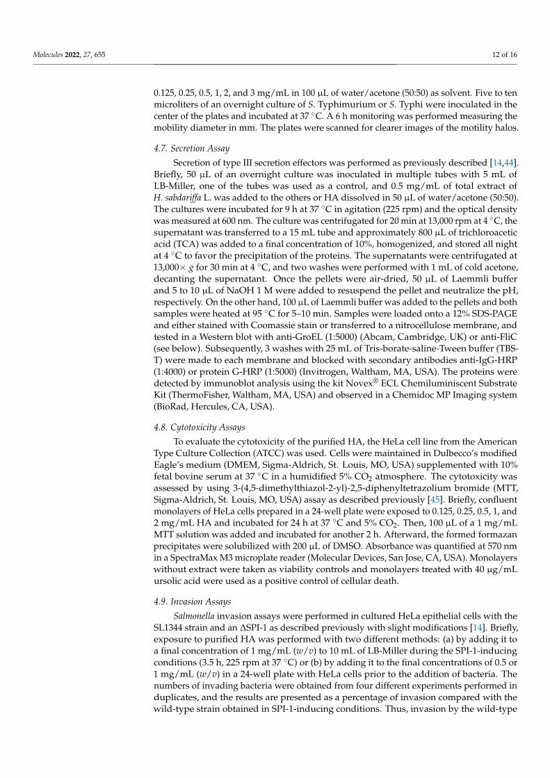
Pooled fractions were analyzed by NMR as described in the Methods section. Theresults in Figure 4 show that by using NMR and mass spectrometry, the main compound inthe active fraction that retained an anti-motility activity for S. Typhi and S. Typhimurium isHA (see also Supplementary Figure S1). In the 1H NMR spectrum a doublet signal (1H,2J = 17.2 Hz) appears at δ 2.72 corresponding to the diastereotopic proton H-3a, and at δ3.21, its geminal proton H-3b can be observed as a doublet (1H, 2J = 17.2 Hz). At δ 5.30,a singlet signal (1H) appears corresponding to H-1. In the proton-decoupled 13C NMRspectrum, the expected six signals of HA are observed. At δ 175.09 and δ 169.23, signalsfor carboxylic acids C6 and C5, respectively, are shown. The signal at δ 173.82 correspondsto the carbonyl group (C4) of the lactone. At δ 84.32 and δ 79.11 ppm, signals for C1 andC2, respectively, are observed, while the signal at δ 42.90 belongs to the methylene C3. The1H and 13C NMR spectra were correlated and assigned by two-dimensional experiments(gHSQC and gHMBC). Mass spectrometry analysis afforded m/z 189.89, which correspondsto ion [M-1]+; additionally, m/z 379.01 for the HA dimer was observed. Taken together,this structural analysis shows that HA is present in the collected fractions that are able toinhibit Salmonella motility.

Molecules 2022, 27, 655 5 of 16
Molecules 2022, 27, x FOR PEER REVIEW 5 of 16
Figure 4. NMR analyses. (A) Homonuclear single quantum correlation (gHSQC) spectrum of hibis-cus acid showing one-bond H-C correlations. (B) Heteronuclear multiple bond correlation (HMBC) spectrum showing heteronuclear long-range, 2- and 3-bond correlations. In both panels, the chemi-cal formula is shown as an inset.
Figure 4. NMR analyses. (A) Homonuclear single quantum correlation (gHSQC) spectrum ofhibiscus acid showing one-bond H-C correlations. (B) Heteronuclear multiple bond correlation(HMBC) spectrum showing heteronuclear long-range, 2- and 3-bond correlations. In both panels, thechemical formula is shown as an inset.

Molecules 2022, 27, 655 6 of 16
2.4. Antimicrobial and Anti-Motility Activities of HA
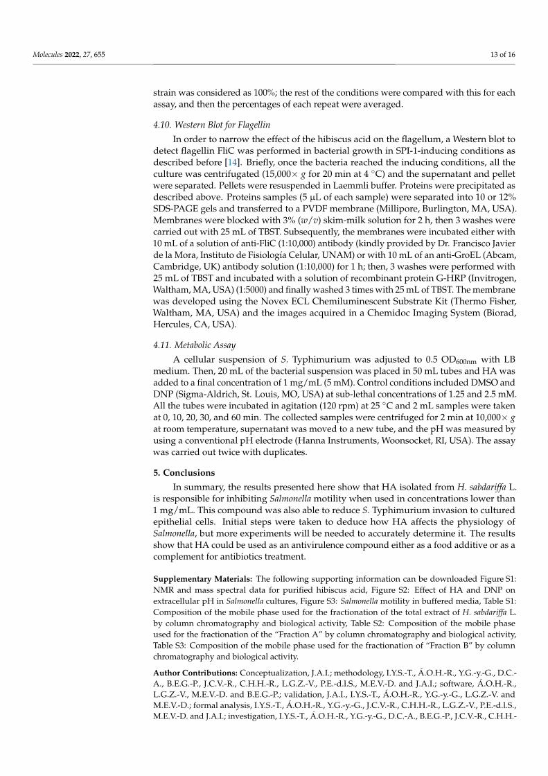
In order to corroborate that HA has antimicrobial and anti-motility activities, the puri-fied compound was tested as described above for the H. sabdariffa L. extract. The results inFigure 5 show that the purified HA inhibited growth when high concentrations (3 mg/disk)were tested, and when lower concentrations (0.5 mg/disk) were tested, the inhibitionof mobility was observed. Taken together, these results show that HA is responsible forinhibiting both bacterial growth and motility.
Molecules 2022, 27, x FOR PEER REVIEW 6 of 16
2.4. Antimicrobial and Anti-Motility Activities of HA In order to corroborate that HA has antimicrobial and anti-motility activities, the pu-
rified compound was tested as described above for the H. sabdariffa L. extract. The results in Figure 5 show that the purified HA inhibited growth when high concentrations (3 mg/disk) were tested, and when lower concentrations (0.5 mg/disk) were tested, the inhi-bition of mobility was observed. Taken together, these results show that HA is responsible for inhibiting both bacterial growth and motility.
Figure 5. Antimicrobial and inhibitory activities of hibiscus acid on S. Typhimurium. (A) Antimi-crobial capacity and (B) inhibition of motility of purified hibiscus acid at 0.5, 1, 2, and 3 mg/disk, respectively. 4% DMSO was used as solvent control in panel A, and it is shown in the center of the plate.
2.5. HA Partially Affects Flagellin Secretion To understand how HA inhibits bacterial motility biogenesis and secretion of the fla-
gellin, FliC in S. Typhimurium SL1344 was detected by Western blot. The results showed that the addition to HA to the media inhibited FliC secretion but not its synthesis (Figure 6). Taken together, these results suggest that this compound might be acting to inhibit a component of the flagella type-III secretion system (fT3SS) involved in the secretion of FliC.
Figure 6. Hibiscus acid reduces flagellin secretion. Secreted proteins were obtained from the indi-cated cultures with the addition or not of HA, and flagellin was detected by Western blots with anti-FliC antibodies. Intracellular FliC and GroEL proteins were also detected. Proteins detected in the supernatant and the pellet are shown. DMSO shows the solvent control and HA the addition of hibiscus acid (1 mg/mL).
Figure 5. Antimicrobial and inhibitory activities of hibiscus acid on S. Typhimurium. (A) Antimi-crobial capacity and (B) inhibition of motility of purified hibiscus acid at 0.5, 1, 2, and 3 mg/disk,respectively. 4% DMSO was used as solvent control in panel A, and it is shown in the center ofthe plate.
2.5. HA Partially Affects Flagellin Secretion
To understand how HA inhibits bacterial motility biogenesis and secretion of theflagellin, FliC in S. Typhimurium SL1344 was detected by Western blot. The results showedthat the addition to HA to the media inhibited FliC secretion but not its synthesis (Figure 6).Taken together, these results suggest that this compound might be acting to inhibit acomponent of the flagella type-III secretion system (fT3SS) involved in the secretion of FliC.
Molecules 2022, 27, x FOR PEER REVIEW 6 of 16
2.4. Antimicrobial and Anti-Motility Activities of HA In order to corroborate that HA has antimicrobial and anti-motility activities, the pu-
rified compound was tested as described above for the H. sabdariffa L. extract. The results in Figure 5 show that the purified HA inhibited growth when high concentrations (3 mg/disk) were tested, and when lower concentrations (0.5 mg/disk) were tested, the inhi-bition of mobility was observed. Taken together, these results show that HA is responsible for inhibiting both bacterial growth and motility.
Figure 5. Antimicrobial and inhibitory activities of hibiscus acid on S. Typhimurium. (A) Antimi-crobial capacity and (B) inhibition of motility of purified hibiscus acid at 0.5, 1, 2, and 3 mg/disk, respectively. 4% DMSO was used as solvent control in panel A, and it is shown in the center of the plate.
2.5. HA Partially Affects Flagellin Secretion To understand how HA inhibits bacterial motility biogenesis and secretion of the fla-
gellin, FliC in S. Typhimurium SL1344 was detected by Western blot. The results showed that the addition to HA to the media inhibited FliC secretion but not its synthesis (Figure 6). Taken together, these results suggest that this compound might be acting to inhibit a component of the flagella type-III secretion system (fT3SS) involved in the secretion of FliC.
Figure 6. Hibiscus acid reduces flagellin secretion. Secreted proteins were obtained from the indi-cated cultures with the addition or not of HA, and flagellin was detected by Western blots with anti-FliC antibodies. Intracellular FliC and GroEL proteins were also detected. Proteins detected in the supernatant and the pellet are shown. DMSO shows the solvent control and HA the addition of hibiscus acid (1 mg/mL).
Figure 6. Hibiscus acid reduces flagellin secretion. Secreted proteins were obtained from the indicatedcultures with the addition or not of HA, and flagellin was detected by Western blots with anti-FliCantibodies. Intracellular FliC and GroEL proteins were also detected. Proteins detected in thesupernatant and the pellet are shown. DMSO shows the solvent control and HA the addition ofhibiscus acid (1 mg/mL).
Molecules 2022, 27, 655 7 of 16
2.6. HA Partially Inhibits Secretion of T3SS Effector Proteins
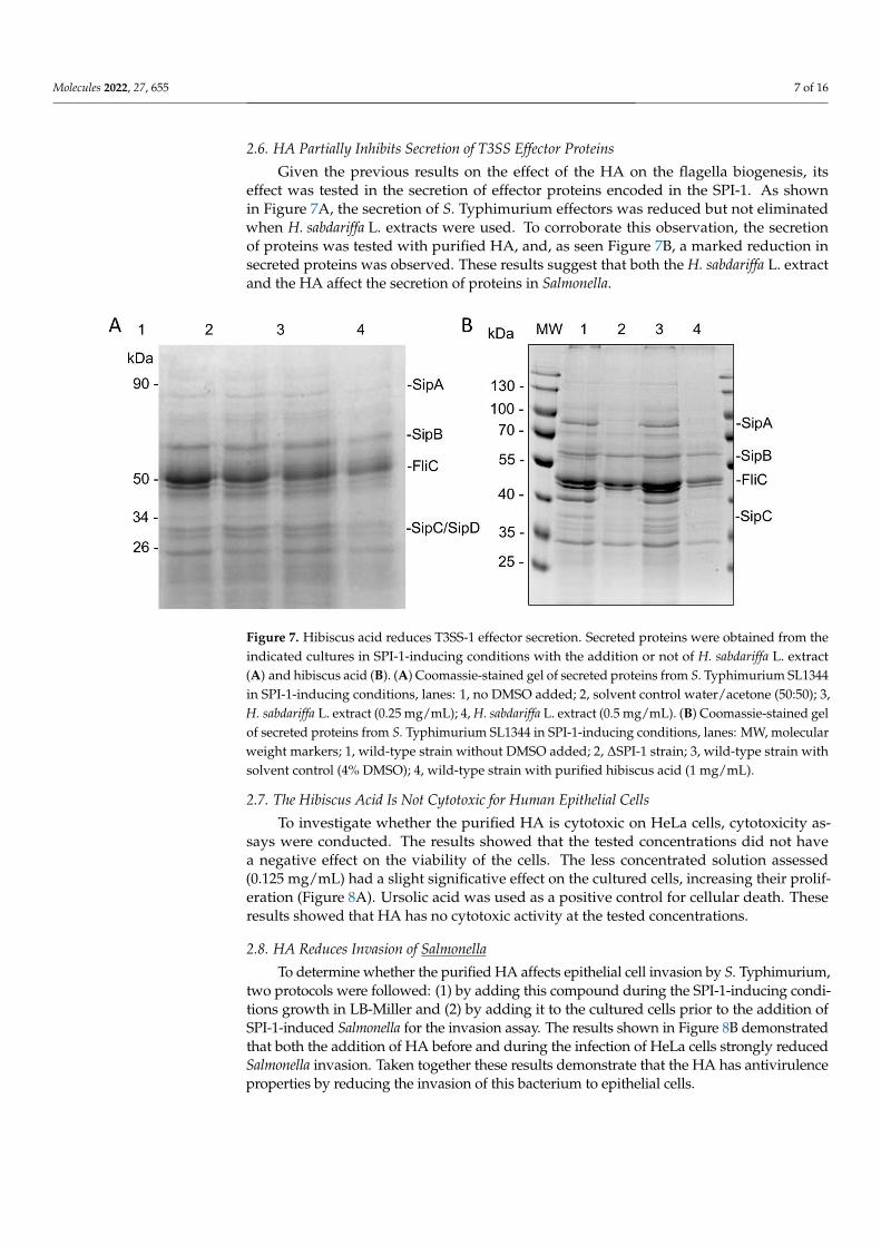
Given the previous results on the effect of the HA on the flagella biogenesis, itseffect was tested in the secretion of effector proteins encoded in the SPI-1. As shownin Figure 7A, the secretion of S. Typhimurium effectors was reduced but not eliminatedwhen H. sabdariffa L. extracts were used. To corroborate this observation, the secretionof proteins was tested with purified HA, and, as seen Figure 7B, a marked reduction insecreted proteins was observed. These results suggest that both the H. sabdariffa L. extractand the HA affect the secretion of proteins in Salmonella.
Molecules 2022, 27, x FOR PEER REVIEW 7 of 16
2.6. HA Partially Inhibits Secretion of T3SS Effector Proteins Given the previous results on the effect of the HA on the flagella biogenesis, its effect
was tested in the secretion of effector proteins encoded in the SPI-1. As shown in Figure 7A, the secretion of S. Typhimurium effectors was reduced but not eliminated when H. sabdariffa L. extracts were used. To corroborate this observation, the secretion of proteins was tested with purified HA, and, as seen Figure 7B, a marked reduction in secreted pro-teins was observed. These results suggest that both the H. sabdariffa L. extract and the HA affect the secretion of proteins in Salmonella.
Figure 7. Hibiscus acid reduces T3SS-1 effector secretion. Secreted proteins were obtained from the indicated cultures in SPI-1-inducing conditions with the addition or not of H. sabdariffa L. extract (A) and hibiscus acid (B). (A) Coomassie-stained gel of secreted proteins from S. Typhimurium SL1344 in SPI-1-inducing conditions, lanes: 1, no DMSO added; 2, solvent control water/acetone (50:50); 3, H. sabdariffa L. extract (0.25 mg/mL); 4, H. sabdariffa L. extract (0.5 mg/mL). (B) Coomassie-stained gel of secreted proteins from S. Typhimurium SL1344 in SPI-1-inducing conditions, lanes: MW, mo-lecular weight markers; 1, wild-type strain without DMSO added; 2, ΔSPI-1 strain; 3, wild-type strain with solvent control (4% DMSO); 4, wild-type strain with purified hibiscus acid (1 mg/mL).
2.7. The Hibiscus Acid Is not Cytotoxic for Human Epithelial Cells To investigate whether the purified HA is cytotoxic on HeLa cells, cytotoxicity assays
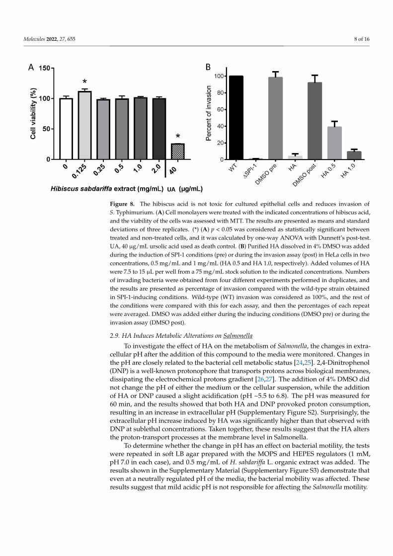
were conducted. The results showed that the tested concentrations did not have a negative effect on the viability of the cells. The less concentrated solution assessed (0.125 mg/mL) had a slight significative effect on the cultured cells, increasing their proliferation (Figure 8A). Ursolic acid was used as a positive control for cellular death. These results showed that HA has no cytotoxic activity at the tested concentrations.
2.8. HA Reduces Invasion of Salmonella To determine whether the purified HA affects epithelial cell invasion by S. Typhi-
murium, two protocols were followed: (1) by adding this compound during the SPI-1-inducing conditions growth in LB-Miller and (2) by adding it to the cultured cells prior to the addition of SPI-1-induced Salmonella for the invasion assay. The results shown in Fig-ure 8B demonstrated that both the addition of HA before and during the infection of HeLa cells strongly reduced Salmonella invasion. Taken together these results demonstrate that the HA has antivirulence properties by reducing the invasion of this bacterium to epithe-lial cells.
Figure 7. Hibiscus acid reduces T3SS-1 effector secretion. Secreted proteins were obtained from theindicated cultures in SPI-1-inducing conditions with the addition or not of H. sabdariffa L. extract(A) and hibiscus acid (B). (A) Coomassie-stained gel of secreted proteins from S. Typhimurium SL1344in SPI-1-inducing conditions, lanes: 1, no DMSO added; 2, solvent control water/acetone (50:50); 3,H. sabdariffa L. extract (0.25 mg/mL); 4, H. sabdariffa L. extract (0.5 mg/mL). (B) Coomassie-stained gelof secreted proteins from S. Typhimurium SL1344 in SPI-1-inducing conditions, lanes: MW, molecularweight markers; 1, wild-type strain without DMSO added; 2, ∆SPI-1 strain; 3, wild-type strain withsolvent control (4% DMSO); 4, wild-type strain with purified hibiscus acid (1 mg/mL).
2.7. The Hibiscus Acid Is Not Cytotoxic for Human Epithelial Cells
To investigate whether the purified HA is cytotoxic on HeLa cells, cytotoxicity as-says were conducted. The results showed that the tested concentrations did not havea negative effect on the viability of the cells. The less concentrated solution assessed(0.125 mg/mL) had a slight significative effect on the cultured cells, increasing their prolif-eration (Figure 8A). Ursolic acid was used as a positive control for cellular death. Theseresults showed that HA has no cytotoxic activity at the tested concentrations.
2.8. HA Reduces Invasion of Salmonella
To determine whether the purified HA affects epithelial cell invasion by S. Typhimurium,two protocols were followed: (1) by adding this compound during the SPI-1-inducing condi-tions growth in LB-Miller and (2) by adding it to the cultured cells prior to the addition ofSPI-1-induced Salmonella for the invasion assay. The results shown in Figure 8B demonstratedthat both the addition of HA before and during the infection of HeLa cells strongly reducedSalmonella invasion. Taken together these results demonstrate that the HA has antivirulenceproperties by reducing the invasion of this bacterium to epithelial cells.
Molecules 2022, 27, 655 8 of 16Molecules 2022, 27, x FOR PEER REVIEW 8 of 16
Figure 8. The hibiscus acid is not toxic for cultured epithelial cells and reduces invasion of S. Typhi-murium. (A) Cell monolayers were treated with the indicated concentrations of hibiscus acid, and the viability of the cells was assessed with MTT. The results are presented as means and standard deviations of three replicates. (*) (A) p < 0.05 was considered as statistically significant between treated and non-treated cells, and it was calculated by one-way ANOVA with Dunnett’s post-test. UA, 40 μg/mL ursolic acid used as death control. (B) Purified HA dissolved in 4% DMSO was added during the induction of SPI-1 conditions (pre) or during the invasion assay (post) in HeLa cells in two concentrations, 0.5 mg/mL and 1 mg/mL (HA 0.5 and HA 1.0, respectively). Added volumes of HA were 7.5 to 15 μL per well from a 75 mg/mL stock solution to the indicated concentrations. Numbers of invading bacteria were obtained from four different experiments performed in dupli-cates, and the results are presented as percentage of invasion compared with the wild-type strain obtained in SPI-1-inducing conditions. Wild-type (WT) invasion was considered as 100%, and the rest of the conditions were compared with this for each assay, and then the percentages of each repeat were averaged. DMSO was added either during the inducing conditions (DMSO pre) or dur-ing the invasion assay (DMSO post).
2.9. HA Induces Metabolic Alterations on Salmonella To investigate the effect of HA on the metabolism of Salmonella, the changes in extra-
cellular pH after the addition of this compound to the media were monitored. Changes in the pH are closely related to the bacterial cell metabolic status [24,25]. 2,4-Dinitrophenol (DNP) is a well-known protonophore that transports protons across biological mem-branes, dissipating the electrochemical protons gradient [26,27]. The addition of 4% DMSO did not change the pH of either the medium or the cellular suspension, while the addition of HA or DNP caused a slight acidification (pH ~5.5 to 6.8). The pH was meas-ured for 60 min, and the results showed that both HA and DNP provoked proton con-sumption, resulting in an increase in extracellular pH (Supplementary Figure S2). Surpris-ingly, the extracellular pH increase induced by HA was significantly higher than that ob-served with DNP at sublethal concentrations. Taken together, these results suggest that the HA alters the proton-transport processes at the membrane level in Salmonella.
To determine whether the change in pH has an effect on bacterial motility, the tests were repeated in soft LB agar prepared with the MOPS and HEPES regulators (1 mM, pH 7.0 in each case), and 0.5 mg/mL of H. sabdariffa L. organic extract was added. The results shown in the Supplementary Material (Supplementary Figure S3) demonstrate that even at a neutrally regulated pH of the media, the bacterial mobility was affected. These results suggest that mild acidic pH is not responsible for affecting the Salmonella motility.
Figure 8. The hibiscus acid is not toxic for cultured epithelial cells and reduces invasion ofS. Typhimurium. (A) Cell monolayers were treated with the indicated concentrations of hibiscus acid,and the viability of the cells was assessed with MTT. The results are presented as means and standarddeviations of three replicates. (*) (A) p < 0.05 was considered as statistically significant betweentreated and non-treated cells, and it was calculated by one-way ANOVA with Dunnett’s post-test.UA, 40 µg/mL ursolic acid used as death control. (B) Purified HA dissolved in 4% DMSO was addedduring the induction of SPI-1 conditions (pre) or during the invasion assay (post) in HeLa cells in twoconcentrations, 0.5 mg/mL and 1 mg/mL (HA 0.5 and HA 1.0, respectively). Added volumes of HAwere 7.5 to 15 µL per well from a 75 mg/mL stock solution to the indicated concentrations. Numbersof invading bacteria were obtained from four different experiments performed in duplicates, andthe results are presented as percentage of invasion compared with the wild-type strain obtainedin SPI-1-inducing conditions. Wild-type (WT) invasion was considered as 100%, and the rest ofthe conditions were compared with this for each assay, and then the percentages of each repeatwere averaged. DMSO was added either during the inducing conditions (DMSO pre) or during theinvasion assay (DMSO post).
2.9. HA Induces Metabolic Alterations on Salmonella
To investigate the effect of HA on the metabolism of Salmonella, the changes in extra-cellular pH after the addition of this compound to the media were monitored. Changes inthe pH are closely related to the bacterial cell metabolic status [24,25]. 2,4-Dinitrophenol(DNP) is a well-known protonophore that transports protons across biological membranes,dissipating the electrochemical protons gradient [26,27]. The addition of 4% DMSO didnot change the pH of either the medium or the cellular suspension, while the additionof HA or DNP caused a slight acidification (pH ~5.5 to 6.8). The pH was measured for60 min, and the results showed that both HA and DNP provoked proton consumption,resulting in an increase in extracellular pH (Supplementary Figure S2). Surprisingly, theextracellular pH increase induced by HA was significantly higher than that observed withDNP at sublethal concentrations. Taken together, these results suggest that the HA altersthe proton-transport processes at the membrane level in Salmonella.
To determine whether the change in pH has an effect on bacterial motility, the testswere repeated in soft LB agar prepared with the MOPS and HEPES regulators (1 mM,pH 7.0 in each case), and 0.5 mg/mL of H. sabdariffa L. organic extract was added. Theresults shown in the Supplementary Material (Supplementary Figure S3) demonstrate thateven at a neutrally regulated pH of the media, the bacterial mobility was affected. Theseresults suggest that mild acidic pH is not responsible for affecting the Salmonella motility.
Molecules 2022, 27, 655 9 of 16
3. Discussion
The antibacterial properties of the Roselle or hibiscus have been studied and shownto be effective against bacteria causing nosocomial infections such as methicillin-resistantStaphylococcus aureus (MRSA), Staphylococcus epidermidis, Proteus vulgaris, Klebsiella pneumo-niae, Pseudomonas aeruginosa, and Acinetobacter baumannii [20,28,29]. Here, the antivirulenceproperties of H. sabdariffa L. organic extracts against Salmonella enterica were tested, and thecompound responsible for this effect was isolated.
As mentioned above, H. sabdariffa L. extracts have antimicrobial activity; therefore,lower concentrations were tested for their ability to inhibit bacterial motility. The initialtest using lower than 1 mg/mL concentrations of the organic extract showed not to havean effect in bacterial growth of S. Typhimurium, but it affected this bacterium’s motility.Moreover, this effect was also extended to S. Typhi. Following a chemical workflowdescribed in the Methods section, a major component in the fractions that retained theability to inhibit Salmonella motility was detected, which showed to be the HA, as theobtained NMR data corresponded with those by Portillo-Torres and colleagues [22]. Despitethe fact that many studies have been carried out with the extract (either organic or aqueous)of H. sabdariffa L. [18–20,28,29], only a few studies have been focused on HA [30–34]. In afew reports, it was shown to have inhibitory activity against α-amylase and α-glucosidaseand also an anti-hypertensive effect in eukaryotes [30,32,33]. At first glance, these activitiesdo not seem to have a relationship with either the biogenesis of either the fT3SS, the T3SS-1,or the motility, which might explain the effect shown by the results described here.
In regard to its antibiotic properties, Portillo-Torres and colleagues [23] showed thatHA has activity against Escherichia coli, S. Typhimurium, P. aeruginosa, S. aureus, andVibrio cholerae. In a more recent report, the use of small concentrations of HA showed todecrease the virulence of P. aeruginosa [34]. In this report, the authors proposed that theHA interacts with LasR inhibiting the quorum sensing (QS) in this bacterium. In orderto explore the possible mechanism of action of the HA to inhibit Salmonella motility, thesecretion of flagellin and alteration in metabolism were explored. In relation to the secretionof flagellin, the results showed that the FliC synthesis was not inhibited, suggesting thatHA does not affect the regulatory network for the expression of this protein and perhapsthat of the other components of flagellum. Thus, despite the fact that the flagella synthesisin Salmonella is regulated by QS [35] and that HA inhibits this system on other bacteria [34].The results presented here suggest that the effect is on another level as the secretion of thisprotein was affected but not its synthesis, though this needs to be further studied. Ourresults suggest that HA might affect the secretion through the fT3SS secretory apparatus.Moreover, the secretion of SPI-1 T3SS effectors is also reduced and, similarly to flagellum,its synthesis is not affected when HA was added. This again implies that HA does notaffect the expression of the SPI-1 effectors but their secretion.
Both pieces of data suggest that HA might act by reducing the secretory process ofboth flagellin and SPI-1 effectors. The addition of HA either during bacterial growth or justprior to the invasion assay dramatically reduced the invasion of Salmonella. As expectedfor the initial results mentioned above, the addition of HA during the incubation perioddid not affect the bacterial growth, supporting our previous data that this amount does nothave an effect during the growth curve. In fact, adding the HA during the invasion assaysuggests that this compound could either block the secretory channel or inhibit the activityof the ATPase at the base of the T3SS [36]. Thus, some of the questions that arise are: doesHA act on the surface of the bacterium, or is the HA able to penetrate and gain access toboth the periplasm and the cytoplasm of the bacterium?
As an initial step to understanding how HA might affect the secretion of both flagellinand SPI-1 effectors, the changes in extracellular pH were measured and compared withthose induced by DNP, a well-known protonophore. pH reports the chemical activityof protons, relevant in metabolic reactions (redox), mineral dissolution, etc.; changes init also reflect the activity of enzymes and other cellular processes such as motility [37].Both, HA and DNP showed a similar effect by lowering the extracellular pH immediately
Molecules 2022, 27, 655 10 of 16
after these were added to the bacterial suspension. DNP has been shown to dissipate theproton electrochemical protons gradient in mitochondria, resulting in proton motive forceinhibition in bacteria [38]. The results showed that, after a few minutes, the extracellular pHstarted to increase in both cases. This may be the result of the entry of protons that migratefrom a higher concentration area (the extracellular environment) to a lower concentrationarea (the periplasm), perhaps through porins located in the bacterium’s outer membrane. Ofnote, the increase was higher with HA than with DNP. This difference in the consumptionof protons between the treatment with HA and DNP might be due to their chemical nature.Previous studies have demonstrated that a change in pH across the cell membrane of only0.2 units is sufficient to achieve variations in proton motive force [39]. The HA may affectthe proton motive force generation, which is closely related to flagellum functionality,which could explain the observed effects in mobility. An alternative is that the acidic pHcauses the flagella to be shed from the bacterial surface as has been described [40]. Asdescribed by the authors, the latter is achieved at very low pH (pH = 2.0), which is notreached by the addition of either DNP or HA in the experiments described here; thus,this might not be the case. On the other hand, changes in pH have an effect in both theexpression of flagellar genes and motility in enterobacteria [41]. In this way, E. coli motilityis sustained in a pH range of 6.0 to 7.5, while Salmonella motility is slightly better in a pHrange of 6.0 to 6.5. The results presented here show that the addition of HA mildly acidifiesthe media (~pH 5.5 to 6.8); therefore, this change might not be responsible for affectingthe motility. Moreover, the addition of H. sabdariffa L. organic extract affects Salmonellamotility even in the presence of pH regulators (such as HEPES or MOPS), which reinforcesthe idea that mild acidic pH is not responsible of affecting the motility. Although this testhas not been performed with HA, the results presented demonstrate that it is the inhibitorycompound for this bacterium motility and it is possible that the salt of HA formed insolution is affecting the consumption of protons, thus disturbing the flagella motion. In thecase of the T3SS-1, the secretion of the effectors might also be affected by the change in thisproton balance with an effect in the dedicated ATPase involved in their secretion, whichin turn would impact the invasion process. However, more studies are needed to confirmthese hypotheses.
Additionally, results presented here showed that HA is not cytotoxic for culturedepithelial cells suggesting that it is safe for humans or animals, although low concentrationsof HA seem to increase cell proliferation. This latter could be due to previously describedantioxidant and antiapoptotic effects of H. sabdariffa L. extracts [42]. However, this has notbeen shown for HA. Moving to the use of pure HA in animals or humans will require morestudies, not only in cultured cells but in animal models.
4. Materials and Methods4.1. H. sabdariffa L. Extract
The experimental research with the cultivated plant complies with institutional, na-tional, and international guidelines and legislation. H. sabdariffa L. is easy to acquire in anypublic market in Mexico, and it is usually sold dried. Additionally, it is also produced inmany parts of this and other Latin-American countries. Specimens of this plant are foundin the collection “ENCB Herbario Jerzy Rzedowski y Graciela Calderón“ at the herbariumof our institution with the number BC007 (https://www.encb.ipn.mx/herbario/ (accessedon 11 October 2021)), and locations where it is produced in Mexico have been documentedby the CONABIO (https://enciclovida.mx/especies/165257-hibiscus-sabdariffa (accessedon 11 October 2021)). The calyxes of H. sabdariffa L. used in this work were obtained fromtwo sources: one batch was from a commercial source sold under the brand “ComercialMexicana” in a convenience store, and the other batch was originally from Cuba and wasobtained in a public market located behind the Metropolitan Cathedral in Mexico City.Fifty grams of calyxes previously dehydrated were macerated at room temperature with500 mL of acetone (Sigma-Aldrich, St. Louis, MO, USA) for 24 h. Subsequently, the organic
Molecules 2022, 27, 655 11 of 16
extract obtained was filtered and the solvent was evaporated to obtain 7 g of dried extract(total yield of 14%).
4.2. Isolation of Secondary Metabolites from H. sabdariffa L.
The H. sabdariffa L. crude extract was analyzed by TLC using MeOH: CH2Cl2: EtOAc(4:3:3) as the eluent and was also monitored by UV–vis 254 nm and 1H NMR. Consecutively,7 g of the crude extract was subjected to column chromatography (CC) using a gradientsystem of hexane:EtOAc:MeOH in different ratios as described in Supplementary Table S1.Each fraction obtained was tested for biological activity and analyzed spectroscopically.Other fractionations used are described in Supplementary Tables S2 and S3.
4.3. NMR and Low-Resolution Mass Spectrometry
NMR characterization was performed at 600 MHz for proton (1H) and 150 MHz forcarbon (13C) on a Bruker 600 AVANCE III HD spectrometer (Bruker Ltd., Billerica, MA,USA) equipped with a 5 mm 1H / D TXI probe-head with z-gradient. Chemical shifts areindicated from the signal of TMS used as reference (0 ppm); methanol-D4 was used as asolvent to prepare the samples. The low-resolution mass spectra (LRMS) were recorded onthe Bruker amaZon speed spectrometer (Bruker Ltd., Billerica, MA, USA) with an ion trapusing the ESI ionization method.
4.4. Bacterial Strains and Growth Conditions
Salmonella Typhi ATCC 6539 was kindly supplied by Dr. Guadalupe Aguilera-Arreola(Departamento de Microbiología, ENCB-IPN), and it was used for growth and motilityassays. S. Typhimurium SL1344 wild-type and its derivative ∆SPI-1 were provided byDr. Víctor H. Bustamante Santillán (Instituto de Biotecnología, UNAM). These strainswere used for secretion and invasion assays (see below). Bacterial strains were streakedon LB-Miller agar and incubated from 18 to 24 h at 37 ◦C. Subsequently, a colony of eachculture was taken and inoculated in LB culture medium, which was incubated at 37 ◦Cin agitation.
4.5. Antimicrobial Activity and Growth Curves
The antimicrobial capacity of the total organic extract of H. sabdariffa L. was determinedby performing growth curves with the strains of S. Typhimurium SL1344 and ∆SPI-1 in96-well plates (Corning, Corning, New York, NY, USA) containing 180 µL of LB inoculatedwith 10 µL of each of the cultures previously adjusted to 1 × 10⁸ CFU/mL. Additionally,10 µL of H. sabdariffa L. extract in increasing concentrations (0.025, 0.25, and 0.5 mg/mL)were added to the wells in the microplate. Solvent control included water/acetone (50:50)and added to the plate in a volume similar to that added for the highest concentration.The 96-well plate was incubated at 37 ◦C in agitation (225 rpm), and absorbance readingswere obtained every hour for 10 h on a plate spectrophotometer (Multiskan Go, ThermoScientific, Waltham, MA, USA) at 600 nm. Please note that the aeration is lower than thatobtained in a flask. The analysis of the growth curve was performed with the GraphPadPrism 5 program.
To determine the antimicrobial activity on solid media, a disk diffusion test wasperformed as described by the CLSI procedures [43]. Either the strain S. Typhi ATCC 6539or S. Typhimurium SL1344 were adjusted to 1 × 10⁸ CFU/mL and 100 µL were inoculated inMuller–Hinton agar plates; sterile filter paper discs (Whatman no. 1, Whatman, Maidstone,UK) were prepared by placing the organic extracts or the purified HA in the followingconcentrations: 0.5, 1, 2, and 3 mg/disk. Plates were incubated at 37 ◦C for 18 h and theinhibition halos were measured.
4.6. Motility Assay
Motility assays were performed on semi-solid LB agar (agar 0.2%) following a pre-viously described protocol [14]. The H. sabdariffa L. extract was tested in concentrations
Molecules 2022, 27, 655 12 of 16
0.125, 0.25, 0.5, 1, 2, and 3 mg/mL in 100 µL of water/acetone (50:50) as solvent. Five to tenmicroliters of an overnight culture of S. Typhimurium or S. Typhi were inoculated in thecenter of the plates and incubated at 37 ◦C. A 6 h monitoring was performed measuring themobility diameter in mm. The plates were scanned for clearer images of the motility halos.
4.7. Secretion Assay
Secretion of type III secretion effectors was performed as previously described [14,44].Briefly, 50 µL of an overnight culture was inoculated in multiple tubes with 5 mL ofLB-Miller, one of the tubes was used as a control, and 0.5 mg/mL of total extract ofH. sabdariffa L. was added to the others or HA dissolved in 50 µL of water/acetone (50:50).The cultures were incubated for 9 h at 37 ◦C in agitation (225 rpm) and the optical densitywas measured at 600 nm. The culture was centrifugated for 20 min at 13,000 rpm at 4 ◦C, thesupernatant was transferred to a 15 mL tube and approximately 800 µL of trichloroaceticacid (TCA) was added to a final concentration of 10%, homogenized, and stored all nightat 4 ◦C to favor the precipitation of the proteins. The supernatants were centrifugated at13,000× g for 30 min at 4 ◦C, and two washes were performed with 1 mL of cold acetone,decanting the supernatant. Once the pellets were air-dried, 50 µL of Laemmli bufferand 5 to 10 µL of NaOH 1 M were added to resuspend the pellet and neutralize the pH,respectively. On the other hand, 100 µL of Laemmli buffer was added to the pellets and bothsamples were heated at 95 ◦C for 5–10 min. Samples were loaded onto a 12% SDS-PAGEand either stained with Coomassie stain or transferred to a nitrocellulose membrane, andtested in a Western blot with anti-GroEL (1:5000) (Abcam, Cambridge, UK) or anti-FliC(see below). Subsequently, 3 washes with 25 mL of Tris-borate-saline-Tween buffer (TBS-T) were made to each membrane and blocked with secondary antibodies anti-IgG-HRP(1:4000) or protein G-HRP (1:5000) (Invitrogen, Waltham, MA, USA). The proteins weredetected by immunoblot analysis using the kit Novex® ECL Chemiluminiscent SubstrateKit (ThermoFisher, Waltham, MA, USA) and observed in a Chemidoc MP Imaging system(BioRad, Hercules, CA, USA).
4.8. Cytotoxicity Assays
To evaluate the cytotoxicity of the purified HA, the HeLa cell line from the AmericanType Culture Collection (ATCC) was used. Cells were maintained in Dulbecco’s modifiedEagle’s medium (DMEM, Sigma-Aldrich, St. Louis, MO, USA) supplemented with 10%fetal bovine serum at 37 ◦C in a humidified 5% CO2 atmosphere. The cytotoxicity wasassessed by using 3-(4,5-dimethylthiazol-2-yl)-2,5-diphenyltetrazolium bromide (MTT,Sigma-Aldrich, St. Louis, MO, USA) assay as described previously [45]. Briefly, confluentmonolayers of HeLa cells prepared in a 24-well plate were exposed to 0.125, 0.25, 0.5, 1, and2 mg/mL HA and incubated for 24 h at 37 ◦C and 5% CO2. Then, 100 µL of a 1 mg/mLMTT solution was added and incubated for another 2 h. Afterward, the formed formazanprecipitates were solubilized with 200 µL of DMSO. Absorbance was quantified at 570 nmin a SpectraMax M3 microplate reader (Molecular Devices, San Jose, CA, USA). Monolayerswithout extract were taken as viability controls and monolayers treated with 40 µg/mLursolic acid were used as a positive control of cellular death.
4.9. Invasion Assays
Salmonella invasion assays were performed in cultured HeLa epithelial cells with theSL1344 strain and an ∆SPI-1 as described previously with slight modifications [14]. Briefly,exposure to purified HA was performed with two different methods: (a) by adding it toa final concentration of 1 mg/mL (w/v) to 10 mL of LB-Miller during the SPI-1-inducingconditions (3.5 h, 225 rpm at 37 ◦C) or (b) by adding it to the final concentrations of 0.5 or1 mg/mL (w/v) in a 24-well plate with HeLa cells prior to the addition of bacteria. Thenumbers of invading bacteria were obtained from four different experiments performed induplicates, and the results are presented as a percentage of invasion compared with thewild-type strain obtained in SPI-1-inducing conditions. Thus, invasion by the wild-type
Molecules 2022, 27, 655 13 of 16
strain was considered as 100%; the rest of the conditions were compared with this for eachassay, and then the percentages of each repeat were averaged.
4.10. Western Blot for Flagellin
In order to narrow the effect of the hibiscus acid on the flagellum, a Western blot todetect flagellin FliC was performed in bacterial growth in SPI-1-inducing conditions asdescribed before [14]. Briefly, once the bacteria reached the inducing conditions, all theculture was centrifugated (15,000× g for 20 min at 4 ◦C) and the supernatant and pelletwere separated. Pellets were resuspended in Laemmli buffer. Proteins were precipitated asdescribed above. Proteins samples (5 µL of each sample) were separated into 10 or 12%SDS-PAGE gels and transferred to a PVDF membrane (Millipore, Burlington, MA, USA).Membranes were blocked with 3% (w/v) skim-milk solution for 2 h, then 3 washes werecarried out with 25 mL of TBST. Subsequently, the membranes were incubated either with10 mL of a solution of anti-FliC (1:10,000) antibody (kindly provided by Dr. Francisco Javierde la Mora, Instituto de Fisiología Celular, UNAM) or with 10 mL of an anti-GroEL (Abcam,Cambridge, UK) antibody solution (1:10,000) for 1 h; then, 3 washes were performed with25 mL of TBST and incubated with a solution of recombinant protein G-HRP (Invitrogen,Waltham, MA, USA) (1:5000) and finally washed 3 times with 25 mL of TBST. The membranewas developed using the Novex ECL Chemiluminescent Substrate Kit (Thermo Fisher,Waltham, MA, USA) and the images acquired in a Chemidoc Imaging System (Biorad,Hercules, CA, USA).
4.11. Metabolic Assay
A cellular suspension of S. Typhimurium was adjusted to 0.5 OD600nm with LBmedium. Then, 20 mL of the bacterial suspension was placed in 50 mL tubes and HA wasadded to a final concentration of 1 mg/mL (5 mM). Control conditions included DMSO andDNP (Sigma-Aldrich, St. Louis, MO, USA) at sub-lethal concentrations of 1.25 and 2.5 mM.All the tubes were incubated in agitation (120 rpm) at 25 ◦C and 2 mL samples were takenat 0, 10, 20, 30, and 60 min. The collected samples were centrifuged for 2 min at 10,000× gat room temperature, supernatant was moved to a new tube, and the pH was measured byusing a conventional pH electrode (Hanna Instruments, Woonsocket, RI, USA). The assaywas carried out twice with duplicates.
5. Conclusions
In summary, the results presented here show that HA isolated from H. sabdariffa L.is responsible for inhibiting Salmonella motility when used in concentrations lower than1 mg/mL. This compound was also able to reduce S. Typhimurium invasion to culturedepithelial cells. Initial steps were taken to deduce how HA affects the physiology ofSalmonella, but more experiments will be needed to accurately determine it. The resultsshow that HA could be used as an antivirulence compound either as a food additive or as acomplement for antibiotics treatment.
Supplementary Materials: The following supporting information can be downloaded Figure S1:NMR and mass spectral data for purified hibiscus acid, Figure S2: Effect of HA and DNP onextracellular pH in Salmonella cultures, Figure S3: Salmonella motility in buffered media, Table S1:Composition of the mobile phase used for the fractionation of the total extract of H. sabdariffa L.by column chromatography and biological activity, Table S2: Composition of the mobile phaseused for the fractionation of the “Fraction A” by column chromatography and biological activity,Table S3: Composition of the mobile phase used for the fractionation of “Fraction B” by columnchromatography and biological activity.
Author Contributions: Conceptualization, J.A.I.; methodology, I.Y.S.-T., Á.O.H.-R., Y.G.-y.-G., D.C.-A., B.E.G.-P., J.C.V.-R., C.H.H.-R., L.G.Z.-V., P.E.-d.l.S., M.E.V.-D. and J.A.I.; software, Á.O.H.-R.,L.G.Z.-V., M.E.V.-D. and B.E.G.-P.; validation, J.A.I., I.Y.S.-T., Á.O.H.-R., Y.G.-y.-G., L.G.Z.-V. andM.E.V.-D.; formal analysis, I.Y.S.-T., Á.O.H.-R., Y.G.-y.-G., J.C.V.-R., C.H.H.-R., L.G.Z.-V., P.E.-d.l.S.,M.E.V.-D. and J.A.I.; investigation, I.Y.S.-T., Á.O.H.-R., Y.G.-y.-G., D.C.-A., B.E.G.-P., J.C.V.-R., C.H.H.-
Molecules 2022, 27, 655 14 of 16
R., L.G.Z.-V., P.E.-d.l.S., M.E.V.-D. and J.A.I.; resources, J.A.I.; data curation, I.Y.S.-T., Á.O.H.-R., Y.G.-y.-G., D.C.-A., B.E.G.-P., J.C.V.-R., C.H.H.-R., L.G.Z.-V., P.E.-d.l.S., M.E.V.-D. and J.A.I.; writing—originaldraft preparation, J.A.I.; writing—review and editing, I.Y.S.-T., Á.O.H.-R., Y.G.-y.-G., D.C.-A., B.E.G.-P., J.C.V.-R., C.H.H.-R., L.G.Z.-V., P.E.-d.l.S., M.E.V.-D. and J.A.I.; visualization, J.A.I.; supervision,Y.G.-y.-G., J.C.V.-R., C.H.H.-R., L.G.Z.-V., P.E.-d.l.S., M.E.V.-D. and J.A.I.; project administration, J.A.I.;funding acquisition, P.E.-d.l.S., M.E.V.-D., J.C.V.-R. and J.A.I. All authors have read and agreed to thepublished version of the manuscript.
Funding: This research was funded by CONSEJO NACIONAL DE CIENCIA Y TECNOLOGÍA,grant number A1-S-25438; by Secretaría de Investigación y Posgrado (IPN), grants numbers SIP-IPN 2021-0738; SIP-IPN 2021-0392; SIP-IPN 2021-0395; SIP-IPN 2021-0961; SIP-IPN 2022-0660;SIP-IPN 2022-0679.
Acknowledgments: We are in great debt to Francisco Javier de la Mora and Bertha González-Pedrajo(Instituto de Fisiología Celular, UNAM) for providing us with the anti-FliC antibody and greatadvice for motility techniques, to Miguel A. de la Cruz-Villegas (Centro Médico Nacional Siglo XXI,IMSS) for the gift of HeLa cells, and to Víctor H. Bustamante (Instituto de Biotecnología, UNAM)for providing us the Salmonella strains. Araceli Contreras-Rodriguez and Graciela González-Lugo(Departamento de Microbiología, ENCB) are thanked for facilitating the use of equipment. Maríade la Luz Arreguín is also much appreciated for giving us access to the “Herbario Jerzy Rzedowskiy Graciela Calderón” at ENCB-IPN. We also thank the members of the Laboratorio de GenéticaMicrobiana for their support through all the steps in this study. We also thank the reviewers as theircomments and suggestions were very useful.
Conflicts of Interest: The authors declare no conflict of interest. The funders had no role in the designof the study; in the collection, analyses, or interpretation of data; in the writing of the manuscript, orin the decision to publish the results.
Sample Availability: Samples of the compound are available from the authors.
References1. Mulvey, M.R.; Simor, A.E. Antimicrobial resistance in hospitals: How concerned should we be? Can. Med Assoc. J. 2009,
180, 408–415. [CrossRef]2. McEwen, S.A.; Collignon, P.J. Antimicrobial Resistance: A One Health Perspective. Microbiol. Spectr. 2018, 6. [CrossRef]3. Ferri, M.; Ranucci, E.; Romagnoli, P.; Giaccone, V. Antimicrobial resistance: A global emerging threat to public health systems.
Crit. Rev. Food Sci. Nutr. 2017, 57, 2857–2876. [CrossRef]4. Bengtsson-Palme, J.; Kristiansson, E.; Larsson, D.G.J. Environmental factors influencing the development and spread of antibiotic
resistance. FEMS Microbiol. Rev. 2018, 42, fux053. [CrossRef]5. Dhingra, S.; Rahman, N.A.A.; Peile, E.; Rahman, M.; Sartelli, M.; Hassali, M.A.; Islam, T.; Islam, S.; Haque, M. Microbial Resistance
Movements: An Overview of Global Public Health Threats Posed by Antimicrobial Resistance, and How Best to Counter. Front.Public Health 2020, 8, 535668. [CrossRef]
6. Boucher, H.W.; Talbot, G.H.; Bradley, J.S.; Edwards, J.E.; Gilbert, D.; Rice, L.B.; Scheld, M.; Spellberg, B.; Bartlett, J. Bad Bugs, NoDrugs: No ESKAPE! An Update from the Infectious Diseases Society of America. Clin. Infect. Dis. 2009, 48, 1–12. [CrossRef]
7. Rasko, D.A.; Sperandio, V. Anti-virulence strategies to combat bacteria-mediated disease. Nat. Rev. Drug Discov. 2010, 9, 117–128.[CrossRef]
8. Fernebro, J. Fighting bacterial infections—Future treatment options. Drug Resist. Updat. 2011, 14, 125–139. [CrossRef]9. Gill, E.E.; Franco, O.L.; Hancock, R.E. Antibiotic Adjuvants: Diverse Strategies for Controlling Drug-Resistant Pathogens. Chem.
Biol. Drug Des. 2015, 85, 56–78. [CrossRef]10. Sharma, A.K.; Dhasmana, N.; Dubey, N.; Kumar, N.; Gangwal, A.; Gupta, M.; Singh, Y. Bacterial Virulence Factors: Secreted for
Survival. Indian J. Microbiol. 2017, 57, 1–10. [CrossRef]11. Silva, L.N.; Zimmer, K.R.; Macedo, A.J.; Trentin, D. Plant Natural Products Targeting Bacterial Virulence Factors. Chem. Rev. 2016,
116, 9162–9236. [CrossRef]12. Lamas, A.; Miranda, J.M.; Regal, P.; Vazquez, B.; Franco, C.M.; Cepeda, A. A comprehensive review of non-enterica subspecies of
Salmonella enterica. Microbiol. Res. 2018, 206, 60–73. [CrossRef] [PubMed]13. Dos Santos, A.M.P.; Ferrari, R.G.; Conte-Junior, C.A. Virulence Factors in Salmonella Typhimurium: The Sagacity of a Bacterium.
Curr. Microbiol. 2019, 76, 762–773. [CrossRef]14. Ibarra, J.A.; Knodler, L.A.; Sturdevant, D.E.; Virtaneva, K.; Carmody, A.B.; Fischer, E.R.; Porcella, S.F.; Steele-Mortimer, O.
Induction of Salmonella pathogenicity island 1 under different growth conditions can affect Salmonella–host cell interactionsin vitro. Microbiology 2010, 156, 1120–1133. [CrossRef]
Molecules 2022, 27, 655 15 of 16
15. De Jong, H.K.; Parry, C.M.; Van Der Poll, T.; Wiersinga, W.J. Host–Pathogen Interaction in Invasive Salmonellosis. PLoS Pathog.2012, 8, e1002933. [CrossRef] [PubMed]
16. Herrero-Fresno, A.; Olsen, J.E. Salmonella Typhimurium metabolism affects virulence in the host—A mini-review. Food Microbiol.2018, 71, 98–110. [CrossRef] [PubMed]
17. Wang, C.H.; Hsieh, Y.H.; Powers, Z.M.; Kao, C.Y. Defeating Antibiotic-Resistant Bacteria: Exploring Alternative Therapies for aPost-Antibiotic Era. Int. J. Mol. Sci. 2020, 21, 1061. [CrossRef]
18. Vasudeva, N.; Sharma, S.K. Biologically Active Compounds from the Genus Hibiscus. Pharm. Biol. 2008, 46, 145–153. [CrossRef]19. Da-Costa-Rocha, I.; Bonnlaender, B.; Sievers, H.; Pischel, I.; Heinrich, M. Hibiscus sabdariffa L.—A phytochemical and pharmaco-
logical review. Food Chem. 2014, 165, 424–443. [CrossRef]20. Liu, K.-S.; Tsao, S.-M.; Yin, M.-C. In vitro antibacterial activity of roselle calyx and protocatechuic acid. Phytother. Res. 2005,
19, 942–945. [CrossRef]21. Gutiérrez-Alcántara, E.J.; Rangel-Vargas, E.; Gómez-Aldapa, C.A.; Cortes, R.N.F.; Rodriguez, M.L.R.M.; Godínez-Oviedo, A.;
Acevedo-Sandoval, O.A.; Castro-Rosas, J. Attachment of 13 Types of Foodborne Bacteria to Jalapeño and Serrano Peppers andAntibacterial Effect of Roselle Calyx Extracts, Sodium Hypochlorite, Colloidal Silver, and Acetic Acid against These FoodborneBacteria on Peppers. J. Food Prot. 2017, 80, 406–413. [CrossRef]
22. Gutiérrez-Alcántara, E.J.; Rangel-Vargas, E.; Gómez-Aldapa, C.A.; Cortes, R.N.F.; Rodriguez, M.L.R.M.; Godínez-Oviedo, A.;Cortes-López, H.; Castro-Rosas, J. Antibacterial effect of roselle extracts (Hibiscus sabadariffa), sodium hypochlorite and acetic acidagainst multidrug-resistant Salmonella strains isolated from tomatoes. Lett. Appl. Microbiol. 2015, 62, 177–184. [CrossRef]
23. Portillo-Torres, L.A.; Bernardino-Nicanor, A.; Gómez-Aldapa, C.A.; González-Montiel, S.; Rangel-Vargas, E.; Villagómez-Ibarra,J.R.; González-Cruz, L.; Cortés-López, H.; Castro-Rosas, J. Hibiscus Acid and Chromatographic Fractions from Hibiscus sabdariffaCalyces: Antimicrobial Activity against Multidrug-Resistant Pathogenic Bacteria. Antibiotics 2019, 8, 218. [CrossRef]
24. Dunlop, D.M. The use of 2:4-dinitrophenol as a metabolic stimulant. Brit. Med. J. 1934, 1, 524–527. [CrossRef]25. Sánchez-Clemente, R.; Igeño, M.I.; Población, A.G.; Guijo, M.I.; Merchán, F.; Blasco, R. Study of pH Changes in Media during
Bacterial Growth of Several Environmental Strains. Proceedings 2018, 2, 1297. [CrossRef]26. Geisler, J.G. 2,4 Dinitrophenol as Medicine. Cells 2019, 8, 280. [CrossRef] [PubMed]27. Sousa, D.; Carmo, H.; Roque Bravo, R.; Carvalho, F.; Bastos, M.D.L.; De Pinho, P.G.; Da Silva, D.D. Diet aid or aid to die: An
update on 2,4-dinitrophenol (2,4-DNP) use as a weight-loss product. Arch. Toxicol. 2020, 94, 1071–1083. [CrossRef]28. Abdallah, E.M. Antibacterial efficiency of the Sudanese Roselle (Hibiscus sabdariffa L.), a famous beverage from Sudanese folk
medicine. J. Intercult. Ethnopharmacol. 2016, 5, 186–190. [CrossRef] [PubMed]29. Baena-Santillán, E.S.; Piloni-Martini, J.; Santos-López, E.M.; Gómez-Aldapa, C.A.; Rangel-Vargas, E.; Castro-Rosas, J. Comparison
of the Antimicrobial Activity of Hibiscus sabdariffa Calyx Extracts, Six Commercial Types of Mouthwashes, and Chlorhexidineon Oral Pathogenic Bacteria, and the Effect of Hibiscus sabdariffa Extracts and Chlorhexidine on Permeability of the BacterialMembrane. J. Med. Food 2021, 24, 67–76. [CrossRef]
30. Hansawasdi, C.; Kawabata, J.; Kasai, T. Hibiscus Acid as an Inhibitor of Starch Digestion in the Caco-2 Cell Model System. Biosci.Biotechnol. Biochem. 2001, 65, 2087–2089. [CrossRef] [PubMed]
31. Zheoat, A.M.; Gray, A.I.; Igoli, J.O.; Kennedy, A.R.; Ferro, V.A. Crystal structures of hibiscus acid and hibiscus acid dimethyl esterisolated from Hibiscus sabdariffa (Malvaceae). Acta Crystallogr. E Crystallogr. Commun. 2017, 73, 1368–1371. [CrossRef]
32. Zheoat, A.M.; Gray, A.I.; Igoli, J.O.; Ferro, V.; Drummond, R.M. Hibiscus acid from Hibiscus sabdariffa (Malvaceae) has avasorelaxant effect on the rat aorta. Fitoterapia 2019, 134, 5–13. [CrossRef] [PubMed]
33. Hansawasdi, C.; Kawabata, J.; Kasai, T. Alpha-Amylase Inhibitors from Roselle (Hibiscus sabdariffa Linn.) Tea. Biosci. Biotechnol.Biochem. 2000, 64, 1041–1043. [CrossRef] [PubMed]
34. Cortes-López, H.; Castro-Rosas, J.; García-Contreras, R.; Rodríguez-Zavala, J.S.; González-Pedrajo, B.; Díaz-Guerrero, M.;Hernández-Morales, J.; Muñoz-Cazares, N.; Soto-Hernández, M.; Ruíz-Posadas, L.D.M.; et al. Antivirulence Activity of a DietaryPhytochemical: Hibiscus Acid Isolated from Hibiscus sabdariffa L. Reduces the Virulence of Pseudomonas aeruginosa in a MouseInfection Model. J. Med. Food 2021, 24, 934–943. [CrossRef] [PubMed]
35. Choi, J.; Shin, D.; Kim, M.; Park, J.; Lim, S.; Ryu, S. LsrR-Mediated Quorum Sensing Controls Invasiveness of Salmonellatyphimurium by Regulating SPI-1 and Flagella Genes. PLoS ONE 2012, 7, e37059. [CrossRef] [PubMed]
36. Diepold, A.; Wagner, S. Assembly of the bacterial type III secretion machinery. FEMS Microbiol. Rev. 2014, 38, 802–822. [CrossRef]37. Jin, Q.; Kirk, M.F. pH as a Primary Control in Environmental Microbiology: 1. Thermodynamic Perspective. Front. Environ. Sci.
2018, 6, 21. [CrossRef]38. Berg, H.; Turner, L. Torque generated by the flagellar motor of Escherichia coli. Biophys. J. 1993, 65, 2201–2216. [CrossRef]39. Hong, Y.; Brown, D.G. Variation in Bacterial ATP Level and Proton Motive Force Due to Adhesion to a Solid Surface. Appl.
Environ. Microbiol. 2020, 75, 2346–2353. [CrossRef]40. Ibrahim, G.F.; Fleet, G.H.; Lyons, M.J.; Walker, R.A. Method for the isolation of highly purified Salmonella flagellins. J. Clin.
Microbiol. 1985, 22, 1040–1044. [CrossRef]41. Maurer, L.M.; Yohannes, E.; Bondurant, S.S.; Radmacher, M.; Slonczewski, J.L. pH Regulates Genes for Flagellar Motility,
Catabolism, and Oxidative Stress in Escherichia coli K-12. J. Bacteriol. 2005, 187, 304–319. [CrossRef] [PubMed]42. Hosseini, A.; Bakhtiari, E.; Mousavi, S.H. Protective Effect of Hibiscus sabdariffa on Doxorubicin-induced Cytotoxicity in H9c2
Cardiomyoblast Cells. Iran J. Pharm. Res. 2017, 16, 708–713.
Molecules 2022, 27, 655 16 of 16
43. CLSI. Procedure for Optimizing Disk Contents (Potencies) for Disk Diffusion Testing of Antimicrobial Agents Using HarmonizedCLSI and EUCAST Criteria. In CLSI Document M23S, 1st ed.; Clinical and Laboratory Standards Institute: Wayne, PA, USA,2020; pp. 1–50.
44. Banda, M.M.; López, C.; Manzo, R.; Rico-Pérez, G.; García, P.; Rosales-Reyes, R.; De la Cruz, M.A.; Soncini, F.C.; Portillo,F.G.-D.; Bustamante, V.H. HilD and PhoP independently regulate the expression of grhD1, a novel gene required for SalmonellaTyphimurium invasion of host cells. Sci. Rep. 2018, 8, 4841, Erratum in Sci. Rep. 2018, 8, 7697. [CrossRef] [PubMed]
45. Castrejón-Jiménez, N.S.; Leyva-Paredes, K.; Baltierra-Uribe, S.L.; Castillo-Cruz, J.; Campillo-Navarro, M.; Hernández-Pérez,A.D.; Luna-Angulo, A.B.; Chacón-Salinas, R.; Coral-Vázquez, R.M.; Estrada-García, I.; et al. Ursolic and Oleanolic Acids InduceMitophagy in A549 Human Lung Cancer Cells. Molecules 2019, 24, 3444. [CrossRef] [PubMed]
Copyright © 2022 FDOKUMEN