Winter–Summer Succession of Unicellular Eukaryotes in a Meso-eutrophic Coastal System
Upload
uni-erlangenCategory
view
1download
0
A Ca2�- and voltage-modulated £agellar ion channel is a component ofthe mechanoshock response in the unicellular green alga
Spermatozopsis similis
Kerstin Hill a, Roland Hemmler a, Peter Kovermann a, Michael Calenberg b,Georg Kreimer b;*, Richard Wagner a
a Universita«t Osnabru«ck, Fachbereich Biologie/Chemie, Barbarastr. 11, D-49069 Osnabru«ck, Germanyb Universita«t zu Ko«ln, Botanisches Institut, Lehrstuhl I, Gyrhofstr. 15, D-50931 Ko«ln, Germany
Received 26 August 1999; received in revised form 9 February 2000; accepted 15 March 2000
Abstract
In flagellate green algae, behavioral responses to photo- and mechanoshock are induced by different external stimuliwithin 10^15 ms. In the accompanying changes in flagella beat, Ca2� has important regulatory roles. Although the axonemalCa2� responsive elements are well characterized, analyses of flagellar channels involved in Ca2� signalling as well as other ionchannels at the single-channel level were not yet conducted in green algae. To gain a further understanding of these importantsignaling elements in movement responses, intact flagella of Spermatozopsis similis were isolated and characterized and thesolubilized flagellar membrane proteins were reconstituted into liposomes. We observed three types of channel activity, twoof which were weakly anion and cation-selective and in the high-conductance regime typical for porin-like solute channels.The dominating channel activity was a voltage dependent, rectifying, low conductance (1= 80 pS in 50 mM KCl) cation-selective channel modulated by, and highly permeable to, Ca2� ions (SFC1: Spermatozopsis flagellar cation channel 1).Depolarizations necessary to activate SFC1 probably only occur in vivo during avoidance reactions of this alga. Ca2�-activation of SFC1 points to a direct link to Ca2�-mediated signaling pathway(s) in the flagella. Both the response tomechanoshock and SFC1 activity were inhibited by Gd3� and Ba2�, thus supporting our assumption that SFC1 represents amajor flagellar ion channel involved in this green algal avoidance reaction. ß 2000 Elsevier Science B.V. All rightsreserved.
Keywords: Flagellar calcium £ux; Flagellar ion channel; Green alga; Shock response; Voltage-modulated channel; Ca2�-modulatedchannel; Spermatozopsis similis
1. Introduction
Flagellate green algae respond to a number of dif-ferent external stimuli in subtle well-de¢ned ways.
Directed responses (e.g. to light) are often peculiarand ¢nally lead to accumulation of cells in areas bestmatching their individual needs. Depending on thequality and strength of the stimuli, also transientavoidance or shock reactions can be induced. Inthe case of photoresponses, the signal is processedwithin 15^50 ms and leads to di¡erential responsesof the same e¡ector, i.e. the £agella (for review see[1,2]).
0005-2736 / 00 / $ ^ see front matter ß 2000 Elsevier Science B.V. All rights reserved.PII: S 0 0 0 5 - 2 7 3 6 ( 0 0 ) 0 0 2 0 0 - 5
* Corresponding author. Fax: 49-221-470-5181;E-mail : [email protected]
BBAMEM 77862 15-5-00
Biochimica et Biophysica Acta 1466 (2000) 187^204www.elsevier.com/locate/bba

In unicellular green algae, such as Chlamydomo-nas, these responses involve peculiar changes in thebeating pattern of the £agella. In Chlamydomonas,the £agella normally shows an asymmetrical breaststroke-like beat [3]. A temporary change to a sym-metrical, undulating waveform, which causes the cellto move backwards, is induced upon stimulation byintense light or mechanical disturbances. Chlamydo-monas responds to both stimuli by a decrease in itsaverage swimming velocity. Spermatozopsis also re-sponds to physiological light shock by decreasing itsswimming velocity. However, during the mechano-shock response or non-physiological photoshock,the cells accelerate to velocities s 600 Wm s31. Theshock responses occur in both algae within 10^50 msand strictly depend on extracellular Ca2� [4^7]. Anal-yses of isolated £agellar apparatuses of Chlamydomo-nas and Spermatozopsis revealed that the main Ca2�-sensitive elements responsible for the change in £ag-ellar beating are components of the axoneme [8^10].In addition, the basal bodies of both algae exhibitcentrin-mediated reorientation upon changes in thefree Ca2� concentration between 1038 and 1036 M.Whereas in Chlamydomonas, the reversible reorienta-tion is small (W20³), the basal bodies of Spermato-zopsis are aligned parallel upon an increase in Ca2�
[10,11]. This probably facilitates the close alignmentof the £agella, apparently a necessary prerequisite forthe accelerations observed upon massive stimulationof the cells [6]. Additionally, Ca2� is intricately in-volved in regulating adaptation to the prevailinglight conditions as well as to the strength and signof phototactic behavior [12]. Ca2�-modulated proteinphosphorylation/dephosphorylation and GTPasespresent in the eyespot region might be involved inthese processes (e.g. [13,14]).
Recent electrophysiological analyses by the suctionpipette technique, have shown that stimulation byboth photoshock and negative pressures lead to in-ward currents of Ca2� into the £agella. Bright £ashesof light ¢rst induce photoreceptor currents in theplasma membrane region overlying the carotenoid-rich lipid globules of the eyespot, which are mainlycarried by Ca2� [15,16]. When these rhodopsin-acti-vated currents exceed a threshold, two transient Ca2�
inward currents in the £agella are triggered in an all-or-nothing manner. The ¢rst is the fast £agellar cur-
rent (FF), which is followed by a small, slow current(FS) [17]. Combined analyses of £agella movementand electrical responses on a single cell have demon-strated that a close link exists between these currentsand behavioral responses [18]. E¡ective triggering of£agellar reversal by electrical stimulation is furtherevidence that these channels are voltage-gated [19].Recent isolation of mutants in the generation of theall-or-nothing £agellar currents, suggests that thephotoshock response is mediated by a multicompo-nent mechanism clearly distinct from that used forphototaxis [20].
Currents evoked by suction in Chlamydomonas dif-fer clearly from photoshock induced FF and FS cur-rents in that they are larger and that the generationof pressure induced current spikes is inhibited byGd3� and Ba2� [7,21]. The FF and FS channels aremost probably evenly distributed over the entirelength of the £agella membrane [22], whereas thedistribution of channels opened upon mechanicalstimulation is not yet known. Mechanosensitivechannels are reported for both £agella and cellbody [7,21]. As consistent patch-clamp measurementson whole cells of £agellate green algae were not yetpossible, information about these channels at thesingle-channel level are not yet available.
Changes in intra£agellar Ca2� generally plays akey role in the regulation of £agellar motility,although the actual responses of the axonemes tothe same messengers can be opposite in di¡erent or-ganisms [1,23,24]. In Chlamydomonas, Ca2� £uxesare also intricately involved in mating, excision,and regeneration of £agella [25^27]. Thus, signalingprocesses during photo- and mechanoshock of greenalgae are not the only reason for studying the prop-erties of single Ca2�-conducting channels in £agellarmembranes. We therefore have incorporated mem-brane proteins of isolated and puri¢ed £agella ofSpermatozopsis in liposomes and studied the recon-stituted channel activities at the single-channel levelby the patch-clamp and planar bilayer techniques.Here we characterize a Ca2�-permeable cation chan-nel and combine electrophysiological measurementswith behavioral analyses of the avoidance responseof Spermatozopsis. The results strongly support in-volvement of this channel in the peculiar avoidanceresponse of this unicellular green alga.
BBAMEM 77862 15-5-00
K. Hill et al. / Biochimica et Biophysica Acta 1466 (2000) 187^204188

2. Materials and methods
2.1. Organism and culture conditions
Spermatozopsis similis Preisig et Melkonian (strainno. B 1.85) was obtained from the Sammlung vonAlgenkulturen, P£anzenphysiologisches Institut,Go«ttingen, Germany. For isolation of £agella, cul-tures were grown in a modi¢ed Waris solution in10-l £asks as described [28]. Aliquots of these masscultures or batch cultures grown in 100-ml Erlen-meyer £asks were used for photo- and mechano-shock assays.
2.2. Isolation of £agella
Usually 10^40 l of late log-phase cultures (cell den-sity: V5^7U106 cell ml31) were harvested and con-centrated to about 2 l using a Pellicon tangential £ow¢ltration system (Millipore, ¢lter HVLP, pore size0.45 Wm) at low £ow rates. Further concentrationwas achieved through centrifugation (15 min, 610^745Ug, rotor: GSA, Sorvall). The cells were resus-pended in modi¢ed Waris solution (without soil ex-tract and vitamins), pelleted as above and resus-pended in HMDS solution (10 mM HEPES, 5 mMMgSO4, 1 mM DTT, 4% sucrose, pH 7.4). All fol-lowing steps followed the method described by Wit-man [29] with the following exceptions: the ¢nal con-centration of dibucaine was reduced to 1 mM, thecells were vigorously pipetted 10^20 times in a 10-mlpolystyrene pipette to induce de£agellation, and thepuri¢ed £agella for biochemical analyses were col-lected by high-speed centrifugation (45 min,100 000Ug). The ¢nal £agella pellet was resuspendedin HMDS and processed as indicated below. Fixa-tion and processing for whole mount and transmis-sion electron microscopy were done as described [10].
2.3. Preparation of £agella liposomes
Small liposomes were obtained by dissolving 50mg/ml of puri¢ed azolectin (Sigma, type IV S; [30])in 10 mM MOPS/Tris pH 7 using the microtip of aBranson Soni¢er (Danburg/Connecticut). Liposomeswere freeze-thawed once. Flagella membranes weresolubilized in 80 mM MEGA-9, 50 mM DTT,20 mM Tricine^Tris (pH 8) for 30 min at room tem-
perature with stirring. The solution was centrifuged(30 min, 14 000 rpm, Eppendorf 5402 centrifuge).The pellet was discharged and protein concentrationswere estimated for the supernatant containing thesolubilized membrane/matrix proteins by comparingCoomassie brilliant blue stained SDS^PAGE gels ofthe solubilisate to known standard concentrations ofBSA. About 0.3 mg of protein/ml were mixed withpreformed liposomes to ¢nal concentrations of 0.1 mgprotein/10 mg azolectin and 0.1 Wg protein/10 mgazolectin. The suspension was freeze-thawed and so-nicated in a supersonic bath. After 0.5 h of incuba-tion at room temperature, the suspension was dia-lyzed for 4 h at room temperature and thenovernight against a bu¡er containing 10 mMMOPS/Tris pH 7 at 4³C and used for bilayer mea-surements.
2.4. Formation of giant proteoliposomes
Giant vesicles suitable for patch-clamp measure-ments were obtained by a modi¢ed dehydration^re-hydration procedure [31]. After thawing, 5^7 ml ofthe proteoliposomes (about 10 Wg protein/ml; 25 mg/ml lipid) were spread on a glass slide and dehydratedat 4³C for 45^60 min in a 500-ml desiccator over dryCaCl2. Afterwards, 10 ml of the electrolyte solutionto be used in the patch-clamp measurements wereadded to the partially dried sample on the slide. Toavoid evaporation, the slide was transferred to a Pet-ri dish, the bottom of which was covered with water-saturated paper. After 1 h, giant liposomes with in-corporated £agella membranes were observed. Thesegiant £agella membrane/liposome vesicles typicallyhad diameters between 20 and 50 Wm. Giant lipo-somes without fused £agella membranes, when usedas controls, did not show any single-channel activityunder the applied experimental conditions.
2.5. Electrophysiological measurements
2.5.1. Patch-clamp measurements
Giant £agella membrane/liposome vesicles wereplaced in a tissue bath mounted on an OlympusIMT-2 inverted microscope, and the vesicles wereviewed using phase contrast optics. Single-channelcurrent recordings using the patch-clamp technique
BBAMEM 77862 15-5-00
K. Hill et al. / Biochimica et Biophysica Acta 1466 (2000) 187^204 189

were performed as described [32]. Holding potentialsare always referred to the pipette. G6-seals rangingfrom 5 to 50 G6 could be achieved by slight suctiononce the pipette tip was brought into contact withthe membrane. Sealing was apparently e¡ected bythe protein content of the vesicles (i.e. the lowerthe protein concentration, the higher the probabilityof G6-seal formation). The currents were ampli¢edusing a PATCH-CLAMP L/M-EPC 7 ampli¢er (ListMedical). Current recordings were digitized at a10 kHz sampling rate using the Axon Digidata1200 system (Axon Instruments) and stored on apersonal computer. For analyses, current recordingswere ¢ltered with an 8-pole bessel ¢lter, typically at1 kHz.
2.5.2. Planar lipid bilayers
Planar lipid bilayers were produced using thepainting technique [33]. A solution of puri¢ed azo-lectin (Sigma type IV-S; 50 mg/ml) in n-decan (ana-lytical grade, Merck) was applied to a hole (100^500Wm diameter) in a Te£on septum, separating the twobath chambers (total volume V3 ml). Both cham-bers were equipped with magnetic stirrers. Bilayerformation was monitored optically and by capaci-tance measurements. The resulting bilayers had atypical capacitance of W0.5 mF/cm2 and a resistanceof s 100 G6. The noise was 1 pA (rms) at a 5-kHzbandwidth. After a stable bilayer was formed in sym-metrical solutions of 20 mM KCl, 10 mM MOPS/Tris (pH 7.0), the experimental conditions werechanged to asymmetric concentrations. Concentratedsolutions of KCl and CaCl2 were added to the cis-chamber up to ¢nal concentrations of 250 mM KCland 10 mM CaCl2. The proteoliposomes were addedto the bu¡er solution in the cis-compartment throughthe tip of a microloader (Eppendorf) so that the lipo-somes slowly £owed directly across the bilayer. Ifnecessary, the solution in the cis-chamber was stirredto promote fusion. After addition of the £agella lipo-somes (50^100 mg protein/ml) we usually observedbetween one and three active channels. The Ag/AgClelectrodes were connected to the chambers through1 M KCl-agar bridges. The electrode of the trans-compartment was directly mounted to the headstage(HS-2A x 10MG) of a current ampli¢er (GeneClamp500, Axon Instruments). Reported holding potentials
are referred to the trans compartment. The ampli¢edcurrents were typically digitized at 1^2 kHz samplingrate using the Axon Digidata 1200 system (AxonInstruments) and stored on a personal computer.For analyses, a Windows-based analysis software[34] was used in combination with Origin (MicrocalSoftware).
2.6. Mechanoshock and photoshock assay
The photoshock assay takes advantage of the re-orientation of the basal bodies in Spermatozopsisfrom an anti-parallel to a parallel con¢gurationupon photo- and mechanoshock [6,10]. All assayswere carried out in complete culture medium withsamples from log-phase cultures (cell densityW1U105 cell ml31). The photoshock assay was car-ried out as described in [10], except that the algaewere equilibrated only for 1 min in the presence orabsence of the indicated concentrations of Gd3� andthat ¢xation with Lugol's solution was done 10 safter the photoshock (700 WE m32 s31 of cool whitelight). For the mechanoshock analyses, 40 Wl culturealiquots were incubated for 1^5 min in the presenceof the indicated inhibitor concentrations. Mecha-noshock was applied by vigorously pipetting the ali-quot several times with a 20-Wl Eppendorf pipette.After the last agitation, 20 Wl of 100 mM EGTAwas rapidly added, followed by an addition of 20 Wlof Lugol's solution. Analyses of swimming paths andspeed were conducted with a commercial movementanalysis system (Medea) and the included softwarepackage.
3. Results
3.1. Flagella isolation and characterization
Spermatozopsis possess two apical inserted £agellaof unequal length with a smooth £agellar surface(V15^20 and 8^16 Wm; [35]). A necklace, presentin many cilia and £agella, was revealed by deep-etch-ing between the £agellar proper and the £agellarbase/transition regions of the basal bodies. No fur-ther particle specializations of the £agellar membranewere evident (data not shown). As Spermatozopsisnaturally lacks a cell wall, a reduced concentration
BBAMEM 77862 15-5-00
K. Hill et al. / Biochimica et Biophysica Acta 1466 (2000) 187^204190

of dibucaine (1 mM) was used to avoid damage ofthe cell bodies and consequent contamination of the£agella fraction by cell debris. However, probablybecause Spermatozopsis neither retracts nor shedsits £agella during the cell cycle, a higher degree ofmechanical stress was needed to shear o¡ the £agella.Most of the cells (V90%) were de£agellated and thecrescent cell shape was not a¡ected. About 85% of
the cells retained £agellar stumps of equal length.The point of breakage appeared to be in the transi-tion region (data not shown).
Isolated £agella appeared largely intact when ob-served by phase microscopy. However, probably dueto the length of the £agella, some broken £agellawere always observed. Cell bodies were e¡ectivelyremoved by the subsequent puri¢cation procedureand only moderate fraying of the £agella membraneswas detected. Electron microscopy shown in Fig.1A^E con¢rmed these observations. Neither in wholemount nor in thin section analyses were contaminat-ing cell debris observed. Structural preservation ofthe isolated £agella was good. Fig. 1B shows thatin some cases even the conspicuous £agellar tip ispreserved. Most of the £agella retained the £agellarmembrane (Fig. 1C^E) and the £agellar matrix, asindicated by the dense staining of the matrix space(Fig. 1C). As described for Chlamydomonas [36], the£agella matrix of Spermatozopsis appears to be con-densed due to membrane shrinkage during the isola-tion and puri¢cation procedure.
3.2. Di¡erent classes of ion channels in liposomescontaining reconstituted £agellar membraneproteins
It was not possible to obtain high-resistance sealsby direct patch-clamping of isolated £agella. Wetherefore reconstituted Mega-9 solubilized £agellamembrane proteins using a dialysis technique. Elec-trophysiological experiments (patch-clamp and pla-nar bilayers) were conducted to characterize ionchannel activity in the reconstituted £agellar mem-branes. Patch-clamp measurements on giant £agel-la-liposomes yielded, in about 50% of the attempts,high-resistance seals (s 5 G6), thus allowing for sin-gle-channel recordings from the excised patches.Based on the conductances in di¡erent symmetrical/asymmetrical bu¡er systems, three di¡erent types ofchannels were identi¢ed in the reconstituted £agellarmembrane of Spermatozopsis : two large conductancechannels, one with a weak cation selectivity and aconductance of 1= 450 pS for the fully open stateand one weak anion-selective channel with a conduc-tance of 1= 830 pS for the fully open channel, and achannel in the lower conductance regime (1= 80 pS),which was also cation-selective with a higher perme-
Fig. 1. Electron microscopy of isolated, puri¢ed £agella ofSpermatozopsis similis. (A,B) Whole mount preparations givingan overview (A) and a detail (B) of the preparations. The ar-rowhead points to the hair-point. (C^E) Thin section analysisof the £agella fraction. (C) Overview; details show a cross sec-tion (D) and an oblique longitudinal section (E). Arrows pointto the £agellar membrane. Scale bars: A and B, 5 Wm; C,1 Wm; and D and E, 200 nm.
BBAMEM 77862 15-5-00
K. Hill et al. / Biochimica et Biophysica Acta 1466 (2000) 187^204 191

ability for Ca2� than for K� ions. Here we describethe principal channel characteristics of these threedi¡erent conductances observed in reconstituted £ag-ella membranes, which were apparently identical inpatch-clamp measurements after formation of giantliposomes and in planar bilayers after fusion of theproteoliposomes.
3.3. A weak cation-selective channel permeable toCa2+
The channel activity described below was observedin about 10% (ns 100) of the excised inside-outpatches obtained from giant £agella-liposomes. Sin-gle-channel recordings in asymmetric Ca2� bu¡er(50 mM/10 mM CaCl2 ; bath/pipette) revealedburst-like openings and closures of the channel atpositive and negative potentials (Fig. 2A). Directtransitions between the fully closed and di¡erentopen states were frequently observed (Fig. 2A). Atthe given time resolution (5 kHz), these would beunlikely to occur with two di¡erent independentchannels (for discussion see [37]). Thus, the courseof the current transients indicates that the smalleramplitudes represent subconductant levels ratherthan the simultaneous activities of two or more chan-nels within the patches. A detailed analysis revealedmainly three di¡erent open-channel amplitudes (seebelow). The current^voltage relationship of the chan-nel was linear for the three main conductance states(Fig. 2B). In asymmetric Ca2�-bu¡er, the following
slope conductance values for subconductances wereobtained (Fig. 2B): 11 = 132 þ 9.9 pS, 12 = 252 þ 10.9pS and 13 = 449 þ 9.1 pS for the fully open channel.In this asymmetric Ca2�-bu¡er, we obtained a rever-sal potential of Erev =312 mV (E2�
Ca =320 mV),while in asymmetric K�-bu¡er (bath: 50 mM KCl,5 mM CaCl2, 10 mM MOPS/Tris pH 7.2, pipette:10 mM KCl, 5 mM CaCl2, 10 mM MOPS/Tris pH7.2) the determined Erev was 327 mV (E�K =341mV). In both bu¡er systems, we found, within error
C
Fig. 2. Flagella membrane proteins reconstituted into liposomesconstitute a weak cation-selective high-conductance channel per-meable to calcium. Electrophysiological properties were exam-ined by the patch-clamp technique. (A) Subconductance levelsof a single-channel obtained from an excised patch are shownat the indicated di¡erent voltages. Examples for direct transi-tions between the di¡erent subconductance levels are markedwith arrows. The following solutions were used: bath 10 mMCaCl2, 10 mM MOPS/Tris pH 7; pipette 50 mM CaCl2, 10 mMMOPS/Tris pH 7. (B) Current^voltage relationship of the threedi¡erent main conductance levels of a single channel. Experi-mental conditions are as given in (A). Linear regression of thedata revealed the following slope conductances for the threesubconductance states: 11 = 132 þ 9.9 pS, 12 = 252 þ 10.9 pS,and 13 = 449 þ 9.1 pS. Theoretical Nernst potentials for 100%selectivity are marked with arrows.
BBAMEM 77862 15-5-00
K. Hill et al. / Biochimica et Biophysica Acta 1466 (2000) 187^204192

limits, identical reversal potentials for the di¡erentopen-channel amplitudes supporting our interpreta-tion that these conductances represent subconductantstates of a single channel. The reversal potentialsmay be converted into relative permeabilities by theconstant ¢eld approach. With the GHK currentequation [38], we obtained the following values:P�K : P2�
Ca : P3Cl = 1:0.15:0.37. Therefore, the above-de-
scribed channel can be considered as a weak cation-selective, high-conductance channel, which is alsosomewhat permeable to Ca2� ions. In contrast tothe Ca2�-permeable channel described below, thischannel was not inhibited by Gd3�.
3.4. A weak anion-selective high-conductance channel
This channel activity was only observed in about5% (ns 100) of the excised inside-out patches fromgiant £agella-liposomes. However, when present, itwas the only channel which was mostly open withoutan externally applied membrane potential. We there-fore attempted to characterize this channel in planarbilayers using the osmotically induced fusion of pro-teoliposomes [39]. Fusion of proteoliposomes to thebilayer is only observed with this technique whenliposomes contain a permeant (open) channel. Withthis technique we were able to obtain the activity ofthe below described anion-selective channel in morethan 70% of the attempts in planar bilayers. Afterfusion of small £agella-liposomes to the bilayer, sin-gle-channel currents with multiple open-channel am-plitudes were observed at positive and negative mem-brane potentials (Fig. 3A). Direct transitions betweenthese conductance states occurred, showing that this
channel also exhibits subconductant states. From adetailed analysis of di¡erent independent bilayers,the following frequent conductances were obtained(see also Fig. 3B): 11 = 140 þ 20 pS (n = 8),12 = 210 þ 22 pS (n = 5), 13 = 310 þ 32 pS (n = 9),14 = 410 þ 15 pS (n = 5), 15 = 540 þ 35 pS (n = 5) and1= 830 þ 30 pS (n = 5) for the fully open channel.These results indicate that the channel exhibits com-plex voltage-dependent gating. The reversal potentialwas, within error limits, identical for all states. In
C
Fig. 3. Flagella membrane proteins reconstituted into liposomesform an anion-selective high-conductance channel. Electrophys-iological properties were examined by the bilayer technique. (A)Current traces from a bilayer containing one active channel atdi¡erent voltages as indicated. Multiple open-channel ampli-tudes are marked with dashed lines. The cis chamber contained250 mM KCl, 10 mM CaCl2, 10 mM MOPS/Tris pH 7 and thetrans compartment contained 20 mM KCl, 10 mM CaCl2,10 mM MOPS/Tris pH 7. (B) Current^voltage relationship ofthe anion-selective channel in response to a voltage sweep(vU = 20 mV/s) from Vh =3100 mV to Vh = +100 mV. Thezero current potential is 316 mV. Theoretical Nernst potentialsfor 100% selectivity are marked with arrows. The bu¡ers usedwere as in A.
BBAMEM 77862 15-5-00
K. Hill et al. / Biochimica et Biophysica Acta 1466 (2000) 187^204 193

di¡erent asymmetric bu¡er systems (bu¡er I: cis, 250mM KCl, 10 mM CaCl2, 10 mM MOPS/Tris pH 7,trans, 20 mM KCl, 10 mM CaCl2, 10 mM MOPS/Tris pH 7; bu¡er II: cis, 250 mM KCl, 250 mMCaCl2, 10 mM MOPS/Tris pH 7, trans, 250 mMKCl, 20 mM CaCl2, 10 mM MOPS/Tris pH 7; bu¡erIII: cis, 250 mM KCl, 10 mM MOPS/Tris pH 7,trans, 20 mM KCl, 10 mM MOPS/Tris pH 7) thefollowing reversal potentials were obtained:Erev(I) =319 þ 3 mV (n = 6), Erev(II) =310 þ 1.5 mV(n = 3), Erev(III) =324 þ 3 mV (n = 3). Conversion ofthese reversal potentials into relative permeabilitiesby the GHK-current equation yielded the followingvalues, P�K : P2�
Ca : P3Cl = 1:1:3. Thus the channel ex-
hibits a weak anion selectivity while being equallypermeable to Ca2� and K�. From the above results,we can consider this channel as a weak anion-selec-tive channel with a high conductance typical for por-in-like channels.
3.5. A voltage- and Ca 2+-modulated £agellar cationchannel (SFC1)
The channel described below was the most fre-quent active channel present in the excised inside-out patches from £agella-liposomes (s 60%,ns 100), indicating that it might occur with highdensity within native £agellar membranes. At normal
resting potentials observed in unicellular green algaein vivo (about 370 to 3135 mV; [40,41]) and lowpositive holding potentials, the channel was predom-inantly inactive. Activation of the channel occurredpreferentially at high positive holding potentials(Figs. 4A and 5A). Therefore, a current^voltage(I/V) relation of the open channel could be obtainedonly at high positive potentials (Fig. 4B). Expanding
C
Fig. 4. Flagella proteins reconstituted into liposomes form avoltage-modulated £agellar Ca2� channel, which we namedSFC1. Electrophysiological properties were examined using thepatch-clamp technique. (A) Single-channel recordings of an ex-cised patch at di¡erent voltages as indicated. Subconductancelevels could frequently be observed. Examples for transitionsbetween the di¡erent open states are indicated by the arrows.The following bu¡er was used for pipette and bath: 50 mMKCl, 2 mM CaCl2, 2 mM MgCl2,10 mM MOPS/Tris pH 7.2.(B) Current^voltage relationship of a channel obtained at highpositive potentials. The three main conductance levels couldalso be observed in the I/V plot. Because of the non-linear cur-rent^voltage relationship, conductances of the di¡erent stateswere determined only from the linear range of the plot at mem-brane potentials s 160 mV. The following conductances wereestimated: 11 = 22, 12 = 50 pS, and 13 = 80 pS. The measure-ments were conducted in symmetrical bu¡ers (as given in A),were the current 0 pA is expected at Vm = 0 mV. Therefore thedata were extrapolated to 0 mV by an arbitrary second-orderpolynomial.
BBAMEM 77862 15-5-00
K. Hill et al. / Biochimica et Biophysica Acta 1466 (2000) 187^204194

the time scale of the current recordings allowed forthe discrimination of at least three di¡erent open-channel amplitudes, which are also shown in theI/V relation (Fig. 5B). Some current traces point toadditional substates between the open states 2 and 3.Direct transitions between the fully closed and di¡er-ent open states were frequently observed at the usedsampling rate of 5 kHz (Fig. 4A). Since these wouldunlikely occur with two di¡erent independent chan-nels, the course of the current transients indicatesthat here also the smaller amplitudes represent sub-conductant levels rather than simultaneous activitiesof two or more channels in the patches. The ob-served rectifying I/V relation (Figs. 4B and 5A) de-viated from that predicted by the constant ¢eld ap-proach (GHK-equations) under symmetrical (Fig.4B) and asymmetrical ionic conditions (not shown).Therefore, conductances of the di¡erent states wereestimated from the linear portion of the current^volt-age relationship at membrane potentials s 160 mV(see Fig. 4B). The conductances in symmetrical bu¡-er (50 mM KCl, 2 mM CaCl2 2 mM MgCl2 10 mMMOPS/Tris, pH 7.2) were 11w22 pS, 12w50 pS and13w80 pS for the di¡erent substates. The estimatedreversal potentials in di¡erent asymmetric bu¡er so-lutions (bu¡er I: cis, 50 mM KCl, 4 mM CaCl2, 10mM MOPS/Tris pH 7.2, trans, 10 mM KCl, 4 mMCaCl2, 10 mM MOPS/Tris pH 7.2; bu¡er II, cis : 5mM KCl, 50 mM CaCl2, 10 mM MOPS/Tris pH 7.2,
trans, 5 mM KCl, 10 mM CaCl2, 10 mM MOPS/TrispH 7.2) were Erev(I) =311 þ 3 mV (n = 3) andErev(II) = 315 þ 1.5 mV (n = 3). These reversal poten-tials, when converted into relative permeabilities bythe GHK-current equation, yielded the following val-ues: P2�
Ca :P�K :P3Cl = 2:1:0.25. Due to its cation selec-
tivity with a higher permeability for Ca2� ions thanfor K� ions, we named this channel SFC1 (for Sper-matozopsis flagellar cation channel 1).
3.6. SFC1 is modulated by Ca 2+
As gating of cation channels by cytosolic Ca2�
occurs in plants and algae (e.g. [42^44]), and intra-
C
Fig. 5. SFC1 is inhibited by Gd3� and modulated by Ca2�. (A)Current recordings obtained from an excised patch containingseveral channels in response to voltage sweeps (vU = 20 mV/s)from Vh =3200 mV to Vh = +200 mV. The upper trace showsthe current in the presence of V1U1035 M Ca2� and the lowerin the presence of EGTA bu¡er (V1U1039 M Ca2�). The fol-lowing bu¡ers were used: upper trace, 50 mM KCl, 10 WMCaCl2, 10 mM MOPS/Tris pH 7 in the bath, 250 mM KCl, 10mM MOPS/Tris pH 7 in the pipette; lower trace, 50 mM KCl,1 mM EGTA, 10 mM MOPS/Tris pH 7 in the bath, 250 mMKCl, 10 mM MOPS/Tris pH 7 in the pipette. The shaded sec-tion is shown enlarged below. (B) Current^voltage relationshipobtained from an excised patch containing several channelsafter an identical voltage sweep as in A. Upper trace, control ;lower trace, in the presence of 50 WM GdCl3. The followingbu¡ers were used: 50 mM KCl, 10 WM CaCl2, 10 mM MOPS/Tris pH 7 for the bath, 250 mM KCl, 10 mM MOPS/Tris pH7 for the pipette; lower trace, same bu¡ers, but additionally 50WM GdCl3 in the bath.
BBAMEM 77862 15-5-00
K. Hill et al. / Biochimica et Biophysica Acta 1466 (2000) 187^204 195
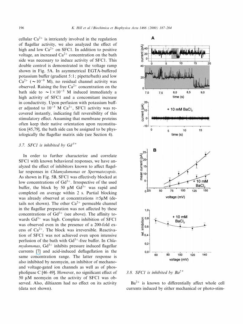
cellular Ca2� is intricately involved in the regulationof £agellar activity, we also analyzed the e¡ect ofhigh and low Ca2� on SFC1. In addition to positivevoltage, an increased Ca2� concentration on the bathside was necessary to induce activity of SFC1. Thisdouble control is demonstrated in the voltage rampshown in Fig. 5A. In asymmetrical EGTA-bu¡eredpotassium bu¡er (gradient 5:1; pipette/bath) and lowCa2� (V1039 M), no residual channel activity wasobserved. Raising the free Ca2� concentration on thebath side to V1U1035 M induced immediately ahigh activity of SFC1 and a concomitant increasein conductivity. Upon perfusion with potassium bu¡-er adjusted to 1035 M Ca2�, SFC1 activity was re-covered instantly, indicating full reversibility of thisstimulatory e¡ect. Assuming that membrane proteinsoften keep their native orientation upon reconstitu-tion [45,79], the bath side can be assigned to be phys-iologically the £agellar matrix side (see Section 4).
3.7. SFC1 is inhibited by Gd 3+
In order to further characterize and correlateSFC1 with known behavioral responses, we have an-alyzed the e¡ect of inhibitors known to a¡ect £agel-lar responses in Chlamydomonas or Spermatozopsis.As shown in Fig. 5B, SFC1 was e¡ectively blocked atlow concentrations of Gd3�. Irrespective of the usedbu¡er, the block by 50 WM Gd3� was rapid andcompleted on average within 2 s. Partial blockingwas already observed at concentrations v5WM (de-tails not shown). The other Ca2� permeable channelin the £agellar preparation was not a¡ected by theseconcentrations of Gd3� (see above). The a¤nity to-wards Gd3� was high. Complete inhibition of SFC1was observed even in the presence of a 200-fold ex-cess of Ca2�. The block was irreversible. Reactiva-tion of SFC1 was not achieved even upon intensiveperfusion of the bath with Gd3�-free bu¡er. In Chla-mydomonas, Gd3� inhibits pressure induced £agellarcurrents [7] and acid-induced de£agellation in thesame concentration range. The latter response isalso inhibited by neomycin, an inhibitor of mechano-and voltage-gated ion channels as well as of phos-pholipase C [46^49]. However, no signi¢cant e¡ect of50 WM neomycin on the activity of SFC1 was ob-served. Also, diltiazem had no e¡ect on its activity(data not shown).
3.8. SFC1 is inhibited by Ba2+
Ba2� is known to di¡erentially a¡ect whole cellcurrents induced by either mechanical or photo-stim-
BBAMEM 77862 15-5-00
K. Hill et al. / Biochimica et Biophysica Acta 1466 (2000) 187^204196

ulation in Chlamydomonas. While the £agellar cur-rent peak amplitude of FF is only slightly a¡ectedby Ba2�, the FS current is largely increased [50]. Incontrast, both the pressure-induced impulses in Chla-mydomonas and the avoidance response of Sperma-tozopsis are e¡ectively suppressed by Ba2� [6,7,21]. Incontrast to Gd3�, which inhibits the mechanoshockresponse of Spermatozopsis at low concentrations(see below), millimolar concentrations of Ba2� areneeded for complete inhibition [6]. In order to gainevidence for the possible involvement of SFC1 ineither of these responses, we have analyzed the e¡ectof Ba2� on SFC1 activity. Fig. 6 shows single-chan-nel recordings from a bilayer containing two activeSFC1 channels as judged by the typical voltage de-pendence of this single-channel activity (see also Fig.5A). In asymmetric bu¡er solutions of 250/20 mMKCl (cis/trans), the chord conductance of the fullyopen SFC1 channel was 1= 120 þ 15 pS, slightlyhigher than in symmetrical 50 mM KCl solutionson both sides of the membrane (Fig. 6A top andexpanded view, middle). In the particular measure-ment shown in Fig. 6A, the current dropped to al-most zero after addition of 10 mM Ba2� to bothcompartments (Fig. 6A, bottom trace). In di¡erentbilayers, average relative current reductions between75 and 55% were observed after addition of Ba2�
(Fig. 6B). Since no changes in the reversal potentialwere observed after addition of Ba2� to either side ofthe membrane, these results indicate that Ba2� is tosome extent equally permeable from both sides of themembrane, although the channel is blocked to a cer-
tain degree by the divalent cation. As Ba2� is also ablocker of potassium channels, we also tested thee¡ects of TEA� and apamin, inhibitors of voltage-gated K� and Na� channels and Ca2�-modulatedpotassium channels, on SFC1. However, these inhib-itors did not a¡ected SFC1 signi¢cantly (data notshown).
3.9. Gd 3+ inhibits the shock response
Spermatozopsis responds to mechanical stimula-tion and non-physiological light shock by a peculiaravoidance response. During the response, the £agellaact as a hydrodynamically coupled pair, allowing forextreme accelerations (velocities of about 60 Wm s31
to s 600 Wm s31 ) during the avoidance reactions.This response is initiated in a time window below 18ms and can be analyzed either directly by video mi-croscopy or via the concomitant Ca2�-dependent re-orientation of the basal bodies from the antiparallelto the parallel con¢guration. The shock response isinhibited by Ba2� or by decreasing the extracellular
Fig. 6. SFC1 is inhibited by Ba2�. Flagella membrane proteinsreconstituted into liposomes were examined by the bilayer tech-nique. (A) Current traces from a bilayer containing two activechannels of the SFC1-type before (top), expanded view (middle)and after addition of 10 mM BaCl2 to both bilayer compart-ments (bottom). The cis chamber contained 250 mM KCl, 10mM CaCl2, 10 mM MOPS/Tris pH 7.0 and the trans compart-ment contained 20 mM KCl, 10 mM CaCl2, 10 mM MOPS/Tris pH 7.0 (holding potential Vh = 80 mV). (B) Mean currentsof di¡erent bilayers containing active SFC1 channels in re-sponse to a voltage gate (duration 60 s) in the absence andpresence of (cis/trans) 10 mM BaCl2. Relative inhibition ofmean currents after addition of BaCl2 (n = 3) are shown in thelower ¢gure. The bu¡ers used were as in A. Mean currentswere calculated during the 1-min onset of the given holding po-tentials.6
Fig. 7. Mechanoshock-induced basal body reorientation inSpermatozopsis cells is inhibited by gadolinium. Squares, cellswere preincubated for 5 min with the indicated concentrationsof GdCl3 prior to application of a mechanical stress. Controls:triangles, cells were ¢xed without prior addition of EGTA;circles, no mechanoshock was given to the cells after the 5-minadaptation period. Mean þ S.D. of three to seven independentexperiments. Total analyzed cells, 158^372.
BBAMEM 77862 15-5-00
K. Hill et al. / Biochimica et Biophysica Acta 1466 (2000) 187^204 197

free Ca2� concentration [6,10]. The observed voltagedependence of SFC1 strongly suggested that, in vivo,it is most likely activated solely upon strong depola-rizations (i.e. during the peculiar avoidance reactionsexhibited by Spermatozopsis). In order to get a pos-sible link between these behavioral responses andSFC1, we analyzed the e¡ect of Gd3� on both re-sponses.
Dose-dependent inhibition was observed for theavoidance response irrespective of the stimulusused. Already low concentrations of this lanthanidee¡ectively inhibited the reorientation of basal bodiesinduced by mechanical agitation (Fig. 7). Half-max-imal inhibition was observed at V5^7.5 WM Gd3�.Complete suppression down to the percentage ofparallel basal bodies observed in non-stimulated cellswas observed between 10 and 20 WM Gd3�. In theseconcentrations, Gd3� had no inhibitory e¡ect on thereorientation of basal bodies when EGTA was omit-ted from the ¢xation (Fig. 7, triangles). Thus, theinhibition can be attributed to a real block of Ca2�
entry into the cells and not to a block of Ca2� bind-ing sites in centrin, which is present in the distalconnecting ¢ber responsible for the reorientation ofbasal bodies [10]. No di¡erences were found in theconcentrations of Gd3� necessary to inhibit the me-chano- or photoshock-induced reorientation of thebasal bodies (not shown). Also, the in vivo e¡ectsof Gd3� were rapid. Already after 1 min in the pres-ence of 20 WM Gd3�, the response to mechanicalagitation and to extreme light shock was completelyabolished. However, longer times (5 min) were rou-tinely used in our analyses, as an extended equilibra-tion time lead to a reduced starting number of cellsexhibiting basal bodies in the parallel con¢guration.These cells were probably already stimulated by themechanical forces administered by the necessary mix-ing after application of the Gd3� stock. Reorienta-tion of the basal bodies to the antiparallel con¢gu-ration can take 3^5 min and occurs independent ofthe restoration of the normal forward swimmingmode, which takes place after a few seconds [10].
The gadolinium e¡ect on the photoshock responsewas also analyzed at the single cell level by videomicroscopy. Here it became evident at low concen-trations (915 WM), that initially the distance the cellsjumped backwards rapidly decreased. The cells onlyexhibited a short stop response, equivalent to the
shock response exhibited upon a normal strongstep-up stimulus, prior to resuming the normal for-ward swimming mode. This behavior was compara-ble to that observed upon reducing the extracellularfree Ca2� concentration [6]. As depicted in Fig. 8, the
Fig. 8. E¡ects of gadolinium on the swimming behavior ofSpermatozopsis similis. (A) Projection of swimming tracks ofSpermatozopsis in culture medium in the absence of gadolinium.Tracks were captured under red safety light after a 5-min prein-cubation period in the assay chamber. (B) Projections of swim-ming tracks in the presence of 40 WM Gd3�. Cells were preincu-bated for 5 min with gadolinium in the assay chamber prior tothe analysis. All other conditions as in A. (C) E¡ects of in-creasing concentrations of Gd3� on the average swimmingspeed. Cells were preincubated for 5 min prior to speed analy-ses. Mean þ S.D. of three independent experiments. Total ana-lyzed cells, 90^105.
BBAMEM 77862 15-5-00
K. Hill et al. / Biochimica et Biophysica Acta 1466 (2000) 187^204198

general swimming behavior of Spermatozopsis atGd3� concentrations up to V30 WM was not a¡ectedat the used cell concentrations. Thus, unspeci¢c ef-fects on motility at the e¡ective concentrations canmost likely be excluded. However, with increasingGd3� concentrations, the swimming path of the cellsbecame less straight. At higher concentrations, anincreasing abnormal spiraling swimming behaviorwas observed and the general motility was dramati-cally a¡ected (Fig. 8B,C).
4. Discussion
Experimental evidence for the presence of voltage-gated Ca2� permeable channels in the £agella mem-brane of unicellular green algae has until now arisensolely from capacitive current measurements (e.g.[5,7,22]). In order to characterize these channel pro-teins at the single-channel level, we have applied thefollowing strategy. Intact £agella were isolated fromSpermatozopsis by adapting the Ca2�/dibucainemethod [29] to the needs of this naturally cell-wall-lacking green alga. As direct recordings from theisolated £agella were not successful, solubilized £ag-ellar membrane proteins were reconstituted into lipo-somes. Consistent with the central role of cation£uxes across £agellar membranes in ciliates [51], wemainly observed activities of cation-selective chan-nels in liposomes containing reconstituted £agellarmembrane proteins. However, a voltage-gated weakanion-selective channel with a high conductance wasalso observed in the £agellar membrane at high fre-quencies, indicating that it might also occur withhigh density in native membranes. Considering thecentral role of Ca2� £uxes in £agellar function andsignaling (see Section 1), we focussed our analyses onthe major voltage-dependent Ca2�-permeable lowconductant channel (SFC1) observed in our prepara-tions.
The SFC1 channel revealed a strongly rectifyingcurrent^voltage relationship, showing that the chan-nel was incorporated mainly unidirectional into thebilayer membrane. Assuming that the proteins of the£agellar membrane keep their native orientations, asobserved with other membrane proteins during re-constitution [45,79], we can propose that the intra-liposomal side corresponds to the £agella matrix
side. Considering further that in vivo the Ca2� con-centration in the matrix is orders of magnitudes low-er than that outside, the SFC1 channel would tran-siently catalyze Ca2� in£ux. This in£ux requires invivo transient depolarization of the £agellar mem-brane. Provided the intra-liposomal side correspondsto the matrix side, this depolarization would corre-spond in vitro to recti¢cation at higher membranepotentials. This was exactly what we observed inour measurements. Therefore, the asymmetric volt-age dependence of SFC1 further supports our pro-posal. All currently identi¢ed green algal £agellarCa2� currents induced by either photo- or mecha-noshock are inwardly rectifying and are triggeredby depolarization (see Section 1).
SFC1 activation is observed solely at positiveholding potentials in the presence of increased freeCa2� concentrations, suggesting that this channelmight be opened in vivo only upon massive depola-rizations of the £agellar membrane with a concom-itant Ca2� signal from the matrix side. This agreeswell with the strict dependence of the shock re-sponses on extracellular Ca2� and with previous sug-gestions that a larger Ca2� in£ux occurs during themechanoshock than during photoshock [6,7]. It hasbeen calculated that depolarizations v80 mV can beinduced by the photoreceptor currents in Chlamydo-monas [17,50]. Thus, assuming a similar averagesteady state free running membrane potential for£agellate green algae at low extracellular K� as de-termined for Eremosphera (385 þ 11 mV; [41]), it iswell feasible that the membrane potential could bedepolarized close to positive values upon strong stim-uli. The channels involved in the shock behavior re-sponse in an `all-or-nothing' manner and appear tobe evenly distributed along the entire £agella length[7,17,19,22]. As the dominating reconstituted activityamong the cation-selective channels in the £agella,SFC1 might well be a candidate for one of thesechannel activities observed in whole cell measure-ments. Activation of the high conductance, voltage-gated anion-selective channel present in high fre-quencies in the £agellar membrane of Spermatozopsisin response to moderate depolarizations, could fur-ther facilitate a rapid and massive membrane depo-larization. However, the detailed voltage dependenceof the reconstituted SFC1 and the anion-selectivechannel needs to be con¢rmed by measurements tak-
BBAMEM 77862 15-5-00
K. Hill et al. / Biochimica et Biophysica Acta 1466 (2000) 187^204 199

en under physiologically less disruptive conditions.Important regulatory subunits may well have beenlost during proteoliposome formation. Unfortu-nately, comparison of our SFC1 data to directpatch-clamp analyses of ciliary membranes fromother protists is not possible, as these have not yetbeen analyzed by direct patch-clamp experiments[52].
The three £agellar channels of Spermatozopsischaracterized so far have relatively large unitary con-ductances in their fully open states. Also, £agellamembranes of wild-type Chlamydomonas reinhardtiiare dominated by ion channels in this conductancerange (G. Pazour, G. Kreimer, R. Wagner and G.B.Witman, unpublished results). Di¡erent channels inboth the low and high conductance ranges are alsoknown from ciliary membranes of Paramecium (1.5^2, 16, 30 and 45 pS; [23,53], Tetrahymena (211 pS[54]; 14^20 and 354 pS [55]; 73 pS [56]) and £agellamembranes of e.g. sea urchin sperms (22, 46, 82 and148 pS [57]). Recently, the conductance of mechano-sensitive channels in Chlamydomonas has been esti-mated from whole-cell measurements to be 30^50 pS[21]. This closely matches the values observed forSFC1 (22, 50 and 80 pS). A relatively high conduc-tivity of SFC1 might be necessary for a steep increasein the matrix free Ca2� needed for induction of £ag-ellar beat reversal. Flagella Ca2�-binding capacity,however, can be quite considerable as indicated byrecent simultaneous analysis of the time course ofintraciliary free Ca2� and the early inward Ca2� cur-rent induced by a depolarizing voltage step in Stylo-nychia [58]. In this ciliate, upon a 280-ms depolariza-tion, only about 5% of the Ca2� entering the matrixwas detected as free Ca2� by the ratiometric Ca2�-sensitive dye fura-2. In other ciliates, smaller depola-rizations induce even lower intraciliary Ca2� rises toW2U1037 M [59]. Many Ca2� channels of higherplants and of mechanogated channels in plants, al-gae, fungi and animals also fall into the conductivityrange observed for SFC1 [44,48,53,60^63].
In which of the known £agella shock responsesmight SFC1 be involved? Considering its sensitivitytowards Gd3� and Ba2�, SFC1 is most likely a com-ponent of the signaling system involved in £agellarshock responses evoked by mechanical disturbanceof the cells. This conclusion is supported by the ob-servation that both the mechanoshock response of
Spermatozopsis as well as pressure-induced currentimpulses originating from the £agella of Chlamydo-monas are inhibited by Gd3� and Ba2� ([6,7], presentstudy). In contrast, £agellar currents evoked by pho-toshock are not inhibited by Ba2� (see below). Gd3�
blocks mechano- and voltage-sensitive channels andresponses in plants and animals [44,48,64]. Althoughrapid backward jumping of Spermatozopsis can alsobe induced by extreme photoshock [6,10], and Gd3�
e¡ectively suppressed this response as well as theaccompanying reorientation of basal bodies, involve-ment of SFC1 in the photoshock pathway is notlikely. Firstly, normal stop responses followed by ashort period of circling prior to resuming forwardswimming in a new direction, were still observed atGd3� concentrations su¤cient to block rapid back-wards jumping. Secondly, £agellar currents accompa-nying the photoshock response in Chlamydomonasare slightly (FF) or even dramatically prolonged(FS) by Ba2� [50], whereas the mechanoshock re-sponse and the generation of pressure-induced im-pulses is inhibited by Ba2� [6,7]. In addition, lowconcentrations of Gd3� have no e¡ect on light re-sponses in Chlamydomonas [49]. Induction of the pe-culiar avoidance response of Spermatozopsis requiresa steep Ca2� gradient and apparently much higherintra£agellar free Ca2� concentrations than reachedby a physiological photoshock [6]. In Chlamydomo-nas the pressure-induced Ca2� currents are also sig-ni¢cantly larger than those induced by photoshock[7]. Suppression of the light shock triggered responseby Gd3� can thus re£ect additional interaction ofthis lanthanide with Ca2�-in£ux channels on thecell body. This would parallel the situation in Chla-mydomonas, where Gd3�-sensitive channels are local-ized on the £agellar membrane and plasma mem-brane [7,21,49].
Ca2� in£ux in plants can occur through bothhighly Ca2�-selective channels and non-selective cat-ion channels [65^67]. SFC1, as many Ca2� channelsof higher plant plasma membranes (for review see[63,68]), exhibited only a weak selectivity for Ca2�
over K� under our measuring conditions. Also inChlamydomonas, the channels involved in generationof FF currents do not appear to be highly selectivefor Ca2� and can also be permeated by K� [16].Ca2� channels in the ciliary membrane of Tetrahy-mena species can also be permeated well by K�
BBAMEM 77862 15-5-00
K. Hill et al. / Biochimica et Biophysica Acta 1466 (2000) 187^204200

[55,69^71], whereas the two major Ca2� channels inciliary membranes of Paramecium exhibit virtuallyno permeability towards potassium [53]. However,possible selectivity/regulatory mechanisms mightwell have been lost or deactivated during our recon-stitution procedure. Therefore, the selectivity valuesreported here for SFC1 certainly re£ect only approx-imations with respect to physiological conditions.Whereas Ca2� suppressed the K� conductivity ofmajor £agellar cation channels in Tetrahymena[55,70], no signi¢cant e¡ect of high concentrationsof Ca2� on the conductivity of SFC1 for K� wasobserved. This parallels the ¢ndings for the photo-induced FF current in whole cells of Chlamydomonas[16].
SFC1 activity was only observed at increased con-centrations of free Ca2� from the assumed matrixside. Ca2� modulation of ion channels is a well-known phenomenon in di¡erent systems includingplants, yeast, algae and protists. Both channel acti-vation and inactivation have been reported [42^44,50,72^75]. Our data suggest that SFC1 activationin vivo will occur only when depolarization of the£agellar membrane is larger than that observedupon physiological photoshock and is accompaniedby an increase in intra£agellar Ca2�. Involvement ofat least one additional channel in mechanoshock sig-naling prior to activation of SFC1 is thus necessary.As SFC1 is probably not the mechanoreceptor chan-nel, activation of mechano- or stretch-activatedchannels might lead to positive de£ections in themembrane potential. Mechanically induced impulsesin Chlamydomonas probably also result from activa-tion of at least two types of channels, a mechanosen-sitive and a voltage-gated Ca2� channel [7]. AlthoughBa2�-carried £uctuating currents can be observed inthe £agella of Chlamydomonas upon application ofpressure, the amplitudes of these currents werelargely reduced and no current spikes were generated[7,21]. This indicates that either the mechanosensitivechannel is directly inhibited by Ba2� or, as Ba2�
usually does not substitute for Ca2� intracellulary,that a second Ca2�- and voltage-gated channel inthe large conductance range might be involved inspike generation. If the latter assumption holdstrue, Chlamydomonas £agella might possess a chan-nel with properties similar to SFC1.
In order to prevent increases of intra£agellar Ca2�
above critical levels (e.g. axonemes detach from basalbodies at W1034 M [10,76]) and to allow the ob-served graded responses of Spermatozopsis to di¡er-ent stimuli [6], SFC1 as well as the other channelsmust be tightly controlled and e¤ciently inactivated.The steep voltage-dependence of SFC1 suggests thatit will probably rapidly inactivate upon beginningrepolarization of the £agellar membrane. SFC1 ac-tivity can also be controlled in parallel by lowering ofthe Ca2� concentration in the £agellar matrix due tothe activity of Ca2� sequestering systems, such asCa2� ATPase pumps. In Paramecium, a complex in-terplay of di¡erent ion channels and inactivationmechanisms (e.g. voltage- and Ca2�-dependent K�
e¥ux and inactivation of Ca2� channels by Ca2�)are involved in regulating amplitude and durationof £agella responses [51]. Down-regulation of FS
probably also occurs through Ca2� in the photo-shock response of Chlamydomonas [50]. Towardsthis end more detailed future analyses of the Ca2�-dependence of SFC1 activation will be helpful. Inaddition, as K� e¥ux can be expected upon strongdepolarizations [16,40,77], the K� conductance ofSFC1 also points to a putative concomitant role in£agellar membrane repolarization. Thus, it might ad-ditionally have a similar function to Ca2�-activatedplant outward rectifying K� channels in repolarizingthe membrane during the signaling event [74,78].Such a dual function would represent the most directcontrol mechanism to prevent an increase of freeCa2� to critical levels. Clearly, more work is neededto understand how the di¡erent ion channels are in-volved in regulating duration and amplitude of Ca2�
in£ux into the £agellar matrix, which is the basis ofthe stimulus-graded movement responses observed in£agellate green algae. We hope that expansion of thesingle-channel analyses to Chlamydomonas with itsgreat variety of mutants and accessibility to geneticmanipulation will help to further unravel ion channelfunctions within the regulatory network of green al-gal £agellar signaling.
Acknowledgements
This study was supported by grants fromthe Deutsche Forschungsgemeinschaft (G.K. andR.W.).
BBAMEM 77862 15-5-00
K. Hill et al. / Biochimica et Biophysica Acta 1466 (2000) 187^204 201

References
[1] G.B. Witman, Chlamydomonas phototaxis, Trends Cell Biol.3 (1993) 403^408.
[2] G. Kreimer, Cell biology of phototaxis in £agellate algae,Int. Rev. Cytol. 148 (1994) 229^310.
[3] D. Ringo, Flagellar motion and ¢ne structure of the £agellarapparatus in Chlamydomonas, J. Cell Biol. 33 (1967) 543^571.
[4] J.A. Schmidt, R. Eckert, Calcium couples £agellar reversalto photostimulation in Chlamydomonas reinhardtii, Nature262 (1976) 713^715.
[5] H. Harz, P. Hegemann, Rhodopsin-regulated calcium cur-rents in Chlamydomonas, Nature 351 (1991) 489^491.
[6] G. Kreimer, G.B. Witman, Novel touch-induced, Ca2�-de-pendent phobic response in a £agellate green alga, Cell Mo-til. Cytoskeleton 29 (1994) 97^109.
[7] K. Yoshimura, A novel type of mechanoreception by the£agella of Chlamydomonas, J. Exp. Biol. 199 (1996) 295^302.
[8] J.S. Hyams, G.G. Borisy, Isolated £agellar apparatus ofChlamydomonas : characterization of forward swimmingand alteration of waveform and reversal of motion by cal-cium ions in vitro, J. Cell Sci. 33 (1978) 235^253.
[9] M. Bessen, R.B. Fay, G.B. Witman, Calcium control ofwaveforms in isolated £agellar axonemes of Chlamydomonas,J. Cell Biol. 86 (1980) 446^455.
[10] G.I. McFadden, D. Schulze, B. Surek, J.L. Salisbury, M.Melkonian, Basal body reorientation mediated by a Ca2�-modulated contractile protein, J. Cell Biol. 105 (1987) 903^912.
[11] M. Hayashi, T. Yagi, K. Yoshimura, R. Kamiya, Real-timeobservation of Ca2�-induced basal body reorientation inChlamydomonas, Cell Motil. Cytoskeleton 41 (1998) 49^56.
[12] N. Morel-Laurens, Calcium control of phototactic orienta-tion in Chlamydomonas reinhardtii : sign and strength of re-sponse, Photochem. Photobiol. 45 (1987) 119^128.
[13] L. Linden, G. Kreimer, Calcium modulates rapid proteinphosphorylation/dephosphorylation in isolated eyespot ap-paratuses of the green alga Spermatozopsis similis, Planta197 (1995) 343^351.
[14] M. Calenberg, U. Brohsonn, M. Zedlacher, G. Kreimer,Light- and Ca2�-modulated heterotrimeric GTPases in theeyespot apparatus of a £agellate green alga, Plant Cell 10(1998) 91^103.
[15] F.F. Litvin, O.A. Sineshchekov, V.A. Sineshchekov, Photo-receptor electric potential in the phototaxis of the alga Hae-matococcus pluvialis, Nature 271 (1978) 476^478.
[16] C. Nonnenga«sser, E.-M. Holland, H. Harz, P. Hegemann,The nature of rhodopsin-triggered photocurrents in Chlamy-domonas. II. In£uence of monovalent ions, Biophys. J. 70(1996) 932^938.
[17] H. Harz, C. Nonnenga«sser, P. Hegemann, The photorecep-tor current of the green alga Chlamydomonas, Phil Trans. R.Soc. Lond. B Biol. Sci. 338 (1992) 39^52.
[18] E.-M. Holland, H. Harz, R. Uhl, P. Hegemann, Control of
phobic responses by rhodopsin-induced photocurrents inChlamydomonas, Biophys. J. 73 (1997) 1395^1401.
[19] K. Yoshimura, C. Shingyoji, K. Takahashi, Conversion ofbeating mode in Chlamydomonas £agella induced by electricstimulation, Cell Motil. Cytoskeleton 36 (1997) 236^245.
[20] A. Matsuda, K. Yoshimura, O.A. Sineshchekov, M. Hirono,R. Kamiya, Isolation and characterization of novel Chlamy-domonas mutants that display phototaxis but not photopho-bic response, Cell Motil. Cytoskeleton 41 (1998) 353^362.
[21] K. Yoshimura, Mechanosensitive channels in the cell bodyof Chlamydomonas, J. Membr. Biol. 166 (1998) 149^155.
[22] C. Beck, R. Uhl, On the localisation of voltage sensitivecalcium channels in the £agella of Chlamydomonas, J. CellBiol. 125 (1994) 1119^1125.
[23] R.R. Preston, Y. Saimi, Calcium ions and the regulation ofmotility in Paramecium, in: R.A. Bloodgood (Ed.), Ciliaryand £agellar membranes, Plenum Press, New York, 1990,pp. 173^200.
[24] C.B. Lindemann, K.S. Kanous, A model for £agellar motil-ity, Int. Rev. Cytol. 173 (1997) 1^72.
[25] U.W. Goodenough, B. Shames, L. Small, T. Saito, R.C.Crain, M.A. Sanders, J.L. Salisbury, The role of calciumin the Chlamydomonas reinhardtii mating reaction, J. CellBiol. 121 (1993) 365^374.
[26] L.M. Quarmby, H.C. Hartzell, Two distinct, calcium-medi-ated, signal transduction pathways can trigger de£agellationin Chlamydomonas reinhardtii, J. Cell Biol. 124 (1994) 807^815.
[27] J.H. Evans, L.R. Keller, Calcium in£ux signals normal £ag-ellar RNA induction following acid shock of Chlamydomo-nas reinhardtii, Plant Mol. Biol. 33 (1997) 467^481.
[28] G. Kreimer, U. Brohsonn, M. Melkonian, Isolation andpartial characterization of the photoreceptive organelle forphototaxis of a £agellate green alga, Eur. J. Cell Biol. 55(1991) 318^327.
[29] G.B. Witman, Isolation of Chlamydomonas £agella and £ag-ellar axonemes, Methods Enzymol. 134 (1986) 280^290.
[30] N.J. Cook, C. Zeilinger, K.W. Koch, U.B. Kaup, Solubili-sation and functional reconstitution of a cGMP-dependentcation channel from bovine rod outer segments, J. Biol.Chem. 261 (1986) 17033^17039.
[31] M. Criado, B.U. Keller, A membrane fusion strategy forsingle-channel recordings of membranes usually non-accessi-ble to patch-clamp pipette electrodes, FEBS Lett. 224 (1987)172^176.
[32] O.P. Hamill, A. Martiy, E. Neher, B. Sakmann, F.J. Sig-worth, Improved patch clamp technique for high-resolutioncurrent recording from cells and cell free patches, P£u«gersArch. 391 (1981) 85^100.
[33] P. Mueller, D.O. Rudin, H. Tien, W.C. Wescott, Reconsti-tution of cell membrane structure in vitro and its transfor-mation into an excitable system, Nature 194 (1962) 979^980.
[34] M. Schwarz, A. Gross, T. Steinkamp, U.I. Flu«gge, R. Wag-ner, Ion channel properties of the reconstituted chloroplasttriosephosphate/phosphate translocator, Biol. Chem. 269(1994) 29481^29489.
BBAMEM 77862 15-5-00
K. Hill et al. / Biochimica et Biophysica Acta 1466 (2000) 187^204202

[35] H.R. Preisig, M. Melkonian, A light and electron microscop-ical study of the green £agellate Spermatozopsis similis spec.nova, Pl. Syst. Evol. 146 (1984) 57^74.
[36] G.B. Witman, K. Carlson, J. Berliner, J.-L. Rosenbaum,Chlamydomonas £agella. I. Isolation and electrophoreticanalysis of microtubules, matrix, membranes, and mastigo-nemes, J. Cell Biol. 54 (1972) 507^539.
[37] D.R. Laver, P.W. Gage, Interpretation of substates in ionchannels-unipores or multipores, Prog. Biophys. Mol. Biol.67 (1997) 99^140.
[38] G.J. Allen, D. Sanders, Calcineurin, a type 2B protein phos-phatase, modulates the Ca2�-permeable vacuolar ion chan-nel of stomatal guard cells, Plant Cell 7 (1995) 1473^1483.
[39] D.J. Woodbury, J.E. Hall, Role of channels in the fusion ofvesicles with a planar bilayer, Biophys. J. 54 (1988) 1053^1063.
[40] B. Malhotra, A.D.M. Glass, Potassium £uxes in Chlamydo-monas reinhardtii. I. Kinetics and electrical potentials, PlantPhysiol. 108 (1995) 1527^1536.
[41] C.S. Bauer, C. Plieth, U.-P. Hansen, B. Sattelmacher, W.Simonis, G. Scho«nknecht, Repetitive Ca2� spikes in a uni-cellular alga, FEBS Lett. 405 (1997) 390^393.
[42] O.M. Zherelova, A.A. Kataev, G.N. Berestovsky, Regula-tion of the calcium channels of the plasmalemma of Nitel-lopsis obtusa by intracellular Calcium, Biophysics 32 (1987)379^380.
[43] B. Schulz-Lessdorf, R. Hedrich, Protons and calcium mod-ulate SV-type channels in the vacuolar-lysosomal compart-ment-channel interaction with calmodulin inhibitors, Planta197 (1995) 655^671.
[44] B. Klu«sener, G. Boheim, H. Lia, J. Engelberth, E.W. Weiler,Gadolinium-sensitive, voltage-dependent calcium releasechannels in the endoplasmic reticulum of a higher plant me-chanoreceptor organ, EMBO J. 14 (1995) 2708^2714.
[45] J.-L. Rigaud, B. Pitard, D. Levy, Reconstitution of mem-brane proteins into liposomes: application to energy-trans-ducing membrane proteins, Biochim. Biophys. Acta 1231(1995) 223^246.
[46] E. Gabev, J. Kasianowicz, T. Abott, S. McLaughlin, Bindingof neomycin to phosphatidylinositol 4,5-bisphosphate(PIP2), Biochim. Biophys. Acta 979 (1989) 905^912.
[47] Y.G. Yueh, R.C. Crain, De£agellation of Chlamydomonasreinhardtii follows a rapid transitory accumulation of inosi-tol 1,4,5-trisphosphate and requires Ca2� entry, J. Cell Biol.123 (1993) 869^875.
[48] O.P. Hamill, D.W. McBride Jr., The pharmacology of me-chanogated membrane ion channels, Pharmacol. Rev. 48(1996) 231^252.
[49] L.M. Quambry, Ca2� in£ux activated by low pH in Chlamy-domonas, J. Gen. Physiol. 108 (1996) 351^361.
[50] E.M. Holland, F.-J. Braun, C. Nonnenga«sser, H. Harz, P.Hegemann, The nature of rhodopsin-triggered photocurrentsin Chlamydomonas. I. Kinetics and in£uence of divalent cat-ions, Biophys. J. 70 (1996) 924^931.
[51] H. Machemer, Electrophysiology, in: H.-D. Go«rtz (Ed.),Paramecium ; Springer, Berlin 1988, pp. 185^215.
[52] H. Machemer, Electrophysiology of ciliates, Methods CellBiol. 47 (1995) 419^424.
[53] B.E. Ehrlich, A. Finkelstein, M. Forte, C. Kung, Voltage-dependent calcium channels from Paramecium cilia incorpo-rated into planar lipid bilayers, Science 225 (1984) 427^428.
[54] Y. Oosawa, M. Sokabe, Cation channels from Tetrahymenacilia incorporated into planar lipid bilayers, Am. J. Physiol.244 (1985) C177^179.
[55] Y. Oosawa, M. Sokabe, M. Kasai, A cation channel for K�
and Ca2� from Tetrahymena cilia in planar lipid bilayers,Cell Struct. Funct. 13 (1988) 51^60.
[56] C. Fujiwara-Hirashima, K. Anzai, M. Takahashi, Y. Kirino,A voltage-dependent chloride channel from Tetrahymena cil-iary membrane incorporated into planar lipid bilayers, Bio-chim. Biophys. Acta 1280 (1996) 207^216.
[57] A. Lievano, J.A. Sanchez, A. Darszon, Single-channel activ-ity of bilayers derived from sea urchin sperm plasma mem-branes at the tip of a patch-clamp electrode, Dev. Biol. 112(1985) 253^257.
[58] H. Machemer, R. Bra«ucker, S. Machemer-Ro«hnisch, U. Na-gel, D.C. Neugebauer, M. Weskamp, The linking of extrinsicstimuli to behaviour: roles of cilia in ciliates, Eur. J. Protis-tol. 34 (1998) 254^261.
[59] J. Pernberg, H. Machemer, Fluorometric measurement ofintracellular free Ca2�-concentration in the ciliate Didiniumnasutum using fura-2, Cell Calcium 18 (1995) 484^494.
[60] R.W. Tsien, P.T. Ellinor, W.A. Horne, Molecular diversityof voltage-dependent Ca2� channels, Trends Phys. Sci. 12(1991) 349^349.
[61] A.M. Hetherington, A. Graziana, C. Mazars, P. Thuleau, R.Ranjeva, The biochemistry and pharmacology of plasma-membrane calcium channels in plants, Philos. Trans. R.Soc. London B 338 (1992) 91^96.
[62] A.R. Taylor, N.F.H. Manison, C. Fernandez, J. Wood, C.Brownlee, Spatial organization of calcium signaling involvedin cell volume control in the Fucus rhizoid, Plant Cell 8(1996) 2015^2031.
[63] M. Pinn¬eros, M. Tester, Calcium channels in higher plantcells : selectivity, regulation and pharmacology, J. Exp. Bot.48 (1997) 551^577.
[64] J.P. Ding, B.G. Pickard, Mechanosensory calcium-selectivecation channels in epidermal cells, Plant J. 3 (1993) 83^110.
[65] J.I. Schroeder, S. Hagiwara, Repetitive increases in cytosolicCa2� of guard cells by abscisic acid activation of nonselec-tive Ca2� permeable channels, Proc. Natl. Acad. Sci. USA87 (1990) 9305^9309.
[66] P.J. White, Characterization of a voltage-dependent cation-channel from the plasma membrane of rye (Secale cereale L.)roots in planar lipid bilayers, Planta 193 (1994) 186^193.
[67] M. Pinn¬eros, M. Tester, Characterization of a voltage-de-pendent Ca2�-selective channel from wheat roots, Planta195 (1995) 478^488.
[68] M. Tester, Plant ion channels: whole-cell and single-channelstudies, New Phytol. 114 (1990) 305^340.
[69] S. Kawahara, Y. Kirino, S. Nagao, Y. Nozawa, Ion perme-ability of ciliary membrane vesicles isolated from Tetrahyme-
BBAMEM 77862 15-5-00
K. Hill et al. / Biochimica et Biophysica Acta 1466 (2000) 187^204 203

na. Single-channel recording study on the membrane recon-stituted into a planar lipid bilayer, J. Biochem. 100 (1986)1569^1573.
[70] C. Fujiwara, K. Anzai, Y. Kirino, S. Nagao, Y. Nozawa, M.Takahashi, Cation channels from ciliary membrane of Tet-rahymena reconstituted into planar lipid bilayer. Compari-sion between the channels from the wild T. termophila andfrom its mutant which does not show ciliary reversal,J. Biochem. 104 (1988) 344^348.
[71] Y. Oosawa, Ionic currents of channels that are permeable tomonovalent and divalent cations, Biophys. J. 56 (1989)1217^1223.
[72] Y. Saimi, B. Martinac, Calcium-dependent potassium chan-nel in Paramecium studied under patch clamp, J. Membr.Biol. 112 (1989) 79^89.
[73] A. Bertl, D. Gradmann, C.L. Slayman, Calcium- and volt-age-dependent ion channels in Saccharomyces cerevisiae,Phil. Trans. R. Soc. Lond. B Biol. Sci. 338 (1992) 63^72.
[74] K. Czempinski, S. Zimmermann, T. Ehrhardt, B. Mu«ller-Ro«ber, New structure and function in plant K� channels:
KCO1, an outward recti¢er with a steep Ca2� dependency,EMBO J. 16 (1997) 2565^2575.
[75] F. Saitow, Y. Nakaoka, Y. Oosawa, A calcium-activated,large conductance and non-selective cation channel in Para-mecium cell, Biochim. Biophys. Acta 1327 (1997) 52^60.
[76] R. Kamiya, G.B. Witman, Submicromolar levels of calciumcontrol the balance of beating between the two £agella indemembranated models of Chlamydomonas, J. Cell Biol. 98(1984) 97^107.
[77] E.G. Govorunova, O.A. Sineshchekov, P. Hegemann, De-sensitization and dark recovery of the photoreceptor currentin Chlamydomonas reinhardtii, Plant Physiol. 115 (1997) 633^642.
[78] K.A. Ketchum, R.J. Poole, Cytosolic calcium regulates apotassium current in corn (Zea mays) protoplasts, J. Membr.Biol. 119 (1991) 277^288.
[79] B. Bo« lter, J. Soll, K. Hill, R. Hemmler, R. Wagner, A rec-tifying ATP-regulated solute channel in the chloroplasticouter envelope from pea, EMBO J. 18 (1999) 5505^5516.
BBAMEM 77862 15-5-00
K. Hill et al. / Biochimica et Biophysica Acta 1466 (2000) 187^204204
Copyright © 2022 FDOKUMEN