STRUCTURE OF HIBISCUS LATENT SINGAPORE VIRUS DETERMINED BY X-RAY FIBER DIFFRACTION
104
STRUCTURE OF HIBISCUS LATENT SINGAPORE VIRUS DETERMINED BY X-RAY FIBER DIFFRACTION SUNIL KUMAR TEWARY A THESIS SUBMITTED FOR THE DEGREE OF DOCTOR OF PHILOSOPHY DEPARTMENT OF BIOLOGICAL SCIENCES NATIONAL UNIVERSITY OF SINGAPROE 2010
Transcript of STRUCTURE OF HIBISCUS LATENT SINGAPORE VIRUS DETERMINED BY X-RAY FIBER DIFFRACTION

STRUCTURE OF HIBISCUS LATENT SINGAPORE VIRUS
DETERMINED BY X-RAY FIBER DIFFRACTION
SUNIL KUMAR TEWARY
A THESIS SUBMITTED FOR THE DEGREE OF
DOCTOR OF PHILOSOPHY
DEPARTMENT OF BIOLOGICAL SCIENCES
NATIONAL UNIVERSITY OF SINGAPROE
2010
STRUCTURE OF HIBISCUS LATENT SINGAPORE VIRUS
DETERMINED BY X-RAY FIBER DIFFRACTION
SUNIL KUMAR TEWARY
MSc. (Biotech.), M.Tech. (Biotech. Biochem. Engg.)
A THESIS SUBMITTED FOR THE DEGREE OF
DOCTOR OF PHILOSOPHY
DEPARTMENT OF BIOLOGICAL SCIENCES
NATIONAL UNIVERSITY OF SINGAPROE
2010
i
ACKNOWLEDGEMENTS
At the threshold of completing my doctor of philosophy (PhD) in X-ray fiber
diffraction, I feel extremely gratified for doing work under my supervisor Dr. Sek-Man
Wong and co-supervisor Dr. Kunchitpadam Swaminathan. Although, any praise will be
too small for my project supervisors, but I can definitely say, they are a perfect man with
a vast wealth of knowledge, experience and a vision for tomorrow. I express my deep
sense of gratitude and regards for not only agreeing to become my project supervisors
but also their astute guidance. They have taught me in solving various problems
encountered during my graduation work. I am also grateful to them for providing various
facilities in the lab and sparing their most precious time for me.
I would also like to extend my thanks to Dr. Gerald Stubbs and Amy Kendall for
sample preparation in his lab and data collection at ANL, Chicago, USA. I also thank
Wen Bian for providing the X-ray fiber package for data reduction and structure
refinement.
I also express my thanks to Dr. Toshiro Oda for his support and technical expertise
in solving the virus structure. I also thank him for providing facilities to work during our
trip to Spring 8, Japan. I express my deep sense of gratitude to Mdm. G. L. Loy for her
support in TEM sample preparation in the core facility. I would also like to thank Ping
Lee Chong for his logistic support.
I take this opportunity to express thanks to my molecular virology lab members Dr.
Niu Shengniao, Dr. Meng Chunying, Zhang Xin, Xie Juntao, Wen Yi, Xie Zhicheng and
Gao Ruimin. I would like to extend my special thanks to Vinod, Shiva, Veerendra,
Kuntal, Umar, Fengxia, Kanmani, Pankaj, Abhilash, Thangavellu and Manjeet for
making my 4 years of Singapore stay memorable.
ii
Last but not the least, I would like to pay gratitude to my parents, in whom I see the
Almighty, whose blessings always helped me progress in difficult situations of my life. I
would like to thank my wife Mamata for her understanding and taking care of the social
necessities of life during my graduation work and my lovely daughter Tulsi who gives
me a lot of joy. I also thank my brothers, elders, youngers and all family members for
their support during my research.
iii
TABLE OF CONTENTS
Acknowledgements i
Table of contents iii
List of publications vii
List of abbreviations viii
List of Figures x
List of Tables xii
Summary xiii
CHAPTER 1. X-RAY FIBER DIFFRACTION TECHNIQUES 1-19
1.1 INTRODUCTION 1
1.2 THEORY OF FIBER DIFFRACTION 3
1.2.1 Diffraction by a helical structure 3
1.2.2 Nature of fiber diffraction 5
1.2.3 Crystalline and non-crystalline fiber 7
1.3 STRUCTURE DETERMINATION USING FIBER DIFFRACTION 8
1.3.1 Multidimensional isomorphous replacement (MDIR) 8
1.3.2 Molecular replacement 11
1.4 REFINEMENT OF FIBER STRUCTURES 12
1.5 DIFFERENCE FOURIER AND OMIT MAP IN FIBER DIFFRACTION 13
1.6 FIBER DIFFRACTION IN MACROMOLECULAR STRUCTURE
DETERMINATION 14
1.6.1 Filamentous plant viruses 14
iv
1.6.2 Tobamovirus structure determination by fiber diffraction 16
1.6.3 Other filamentous virus structure by fiber diffraction 17
CHAPTER 2. HIBISCUS LATENT SINGAPORE VIRUS 20-30
2.1 INTRODUCTION 20
2.2 HIBISCUS LATENT SINGAPORE VIRUS (HLSV) 21
2.2.1 General characterization of HLSV 21
2.2.2 HLSV gene structure, regulation and proteins function 22
2.3 PREVIOUS STUDIES ON TOBAMOVIRUS STRUCTURES 23
2.3.1 Infection and stability of native virus capsid 23
2.3.2 Evolutionary insights from virus structures 25
2.3.3 Coat protein interaction with genomic RNA 26
2.3.4 Maturation processes of Tobamoviruses 27
2.4 RATIONALE AND OBJECTIVES 29
CHAPTER 3. MATERIALS AND METHODS 31-43
3.1 MOLECULAR BIOLOGY 31
3.1.1 Cloning the HLSV c-DNA 31
3.1.2 In vitro transcript preparation 31
3.2 VIRUS PROPAGATION AND PURIFICATION 31
3.2.1 Plant inoculation 31
3.2.2 Crude extraction of HLSV 32
3.2.3 Cesium chloride density gradient centrifugation 33
3.2.4 Slow speed centrifugation to purify 300 nm long HLSV virion 33
3.2.5 Sephacryl 1000 gel filtration 34
3.3 CHARACTERIZATION OF PURIFIED HLSV VIRION 34
3.3.1 Virus purity and concentration 34
v
3.3.2 Transmission electron microscope (TEM) 34
3.3.3 Western blot 35
3.4 ORIENTED SOL PREPARATION 35
3.5 FIBER STRUCTURE DETERMINATION 36
3.5.1 Data collection 36
3.5.2 Data processing 36
3.5.3 Layer line splitting 39
3.6 HLSV STRUCTURE DETERMINATION 40
CHAPTER 4. RESULTS AND DISCUSSION 44-74
4.1 SAMPLE PREPARATION 44
4.1.1 Crude extraction of HLSV from plant tissue 44
4.1.2 Cesium chloride density gradient centrifugation 44
4.1.3 Slow speed centrifugation to purify the long virion 44
4.1.4 Sephacryl 1000 gel filtration chromatography 47
4.1.5 Sol preparation 48
4.2 DATA COLLECTION AND STRUCTURE DETERMINATION 49
4.2.1 Fiber diffraction data collection 49
4.2.2 Structure determination 50
4.2.3 HLSV CP protein contains a kink in the LR α-helix 52
4.2.4 Nucleic acid structure 58
4.2.5 Protein-protein interaction 59
4.2.6 Protein-nucleic acid interaction 64
4.3 DISCUSSION 67
4.3.1 Protein-RNA interactions 67
4.3.2 HLSV CP protein-protein interaction 68
vi
4.3.3 Other structural features of HLSV 70
4.4 FUTURE DIRECTIONS 72
REFERENCES 75
vii
LIST OF PUBLICATION
Tewary, S. K., Oda, T., Kendall, A., Bian, W., Stubbs, G., Wong, S. M and
Swaminathana, K. (2011). Structure determination of Hibiscus latent Singapore virus by
X-ray fiber diffraction: a non-conserved His122 contributes to coat protein stability.
Journal of Molecular Biology 406: 516-526.
viii
LIST OF ABBREVIATIONS
BSMV Barley stripe mosaic virus
CGMMV Cucumber green mottle mosaic virus
CP Coat protein
CsCl Cesium chloride
EDTA Ethylene-diamine-tetra-acetic acid
FD Fiber diffraction
FT Fourier transform
HLSV Hibiscus latent Singapore virus
IR Isomorphous replacement
MD Molecular dynamics
MDIR Multidimensional isomorphous replacement
MP Movement protein
MR Molecular replacemet
OAS Origin of assembly sequence
PVX Potato virus X
RdRp RNA dependent RNA polymerase
RLS Restrained least square
RMV Ribgrass mosaic virus
RNA Ribonucleic acid
SDS-PAGE Sodium dodecylsulfate-polyacrylamide electrophoresis
SHMV Sunn hemp mosaic virus
SIR Single isomorphous replacement
TEM Transmission electron microscope
ix
TMV Tobacco mosaic virus
TRV Tobacco rattle virus
UTR Untranslated region
UV Ultra violet
VLPs Virus-like particles
x
LIST OF FIGURES
Figure 1.1 Tilt and twist of a sample 7
Figure 2.1 Genome organization of HLSV 22
Figure 3.1 Fiber diffraction data representation by WCEN 37
Figure 4.1 Cesium chloride density gradient centrifugation and Western
blot analysis of purified HLSV 45
Figure 4.2 TEM view of the HLSV after slow centrifugation 46
Figure 4.3 HLSV purification by Sephacryl 1000 gel filtration
chromatography 47
Figure 4.4 HLSV sol preparation and visualization under polarization
microscope 48
Figure 4.5 X-ray fiber diffraction frame of HLSV oriented sol 49
Figure 4.6 CP sequence alignments of HLSV, TMV, CGMMV, RMV
and SHMV 51
Figure 4.7 HLSV CP structure 52
Figure 4.8 Comparison of Tobamovirus CP structures 53
Figure 4.9 Comparison of the LR helix of Tobamoviruses 54
Figure 4.10 Superimposition of the HLSV CP (blue) with that of other
Tobamoviruses 57
Figure 4.11 Superimposition of the HLSV RNA 59
Figure 4.12 Pictorial representation of a partial HLSV virion 60
Figure 4.13 HLSV protein-protein (CP) interaction 61
Figure 4.14 Stereo view of HLSV axial intersubunit carboxyl-carboxylate
interactions at high radius region 62
xi
Figure 4.15 Stereo diagram of HLSV carboxyl-carboxylate interactions
at the low radius loop region 63
Figure 4.16 Electron density map of HLSV CP-nucleic acid interaction 65
Figure 4.17 HLSV CP-RNA interaction 66
Figure 4.18 Electron density map showing phosphate-carboxylate
interactions in the HLSV CP 66
Figure 4.19 Summary of CP-RNA interactions of HLSV (Schematic diagram) 67
Figure 4.20 Surface density diagram of low radius carboxyl-carboxylate
interactions of HLSV coat protein (CP) 70
xii
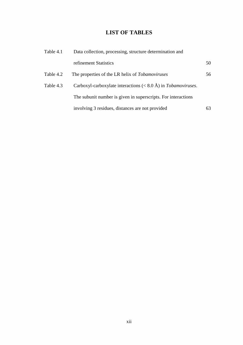
LIST OF TABLES
Table 4.1 Data collection, processing, structure determination and
refinement Statistics 50
Table 4.2 The properties of the LR helix of Tobamoviruses 56
Table 4.3 Carboxyl-carboxylate interactions (< 8.0 Å) in Tobamoviruses.
The subunit number is given in superscripts. For interactions
involving 3 residues, distances are not provided 63
xiii
SUMMARY
Hibiscus latent Singapore virus (HLSV) is a new member of the Tobamovirus family.
The HLSV genome contains a unique poly(A) tract in its 3΄-UTR which is absent in
other Tobamoviruses. The virion is composed of a monomeric coat protein (CP) of 18
kDa. We have determined the HLSV structure at 3.5 Å by X-ray fiber diffraction with
R factor of 0.096. The structure of HLSV CP resembles that of other Tobamoviruses,
with a few unique differences. In other Tobamoviruse structure, CP sequence at
position 122 contains a conserved Arg residue, while the HLSV and SHMV contain
His residue. Also, His122 is followed by another positively charged amino acid
residue Lys which is uncharged residue in other Tobamoviruses. There is a kink
observed for the first time in the LR helix of HLSV due to the presence of the unique
His122, which produces a bend in the helix in the non-Pro non-Gly bends. Also, the
adjacent Lys123 may destabilize the helix by positive charge repulsion, making the
kink more pronounced. In the HLSV structure, we are able to see Lys123 stabilizing
the phosphate 1, hence balancing the protein-nucleic acid interactions. Another
residue Arg92 from the Subunit -17 is believed to be involved in stabilizing the
remaining phosphate 2 and phosphate 3. Arg122 is believed to regulate the guanine 1
recognition during assembly for all other existing structures of the Tobamovirus.
Uniquely, His122 at this position showed a very strong salt bridge with the
neighboring Asp88 from subunit -1, hence significantly stabilizing the loop adjacent
to RR helix. The carboxyl-carboxylate interactions that drive viral disassembly are
also seem to be different in HLSV. The nucleotide recognition mechanism for virus
assembly is similar between HLSV and RMV but different from that of TMV and
CGMMV.

xiv
By solving the structure of HLSV by X-ray fiber diffraction, we will be able to have a
better understanding of the structural differences between HLSV and other
Tobamoviruses. This research may also enhance our knowledge of virus structure at
atomic details. By knowing the atomic details of this novel virus, we may be able to
use it in future as a vector to express pathogenic epitopes (to develop vaccine) and to
express economically important proteins.

1
CHAPTER 1. X-RAY FIBER DIFFRACTION TECHNIQUES
1.1 INTRODUCTION
Many biological molecules that are polymeric in nature are long, structurally
helical and have a natural tendency to form fibers. This prevents the growth of single
crystals from these polymeric fibers and even if crystals are grown, the molecular
interactions in the crystals hardly represent any biologically significant interactions in
the fibers. Conventional X-ray crystallography is therefore of very less use to fibrous
biomolecules. These macromolecular helical aggregates are too large to be studied by
nuclear magnetic resonance. Fiber diffraction (FD), however, is a powerful method
for determining three dimensional (3-D) structural details of fibrous polymers. This
technique has been used to study a wide variety of biopolymers, ranging from simple
polypeptides, polynucleotides, and polysaccharides to very complex filamentous
viruses and cytoskeletal filaments.
Fiber samples lack true 3-D crystalline structure. The key difference between
fibers and crystals is that in fibers, structural aggregates, although parallel to each
other, are randomly oriented (disordered) about the fiber axis. Consequently the
diffraction pattern is cylindrically averaged. The cylindrical averaging is the defining
property of the FD samples. The combination of cylindrical averaging and inherent
disorder makes structural analysis of the fibrous filaments difficult. Hence, FD is not
an appropriate method for studying molecules that do not naturally form filaments.
Proteins that are fibrous in nature are important structural component of biological
systems such as skin, bone, hair and tendons cannot be crystallized by the protein X-
ray crystallographic technique. These samples are suitable for study by the method of
Chapter 1. X-ray fiber diffraction techniques
2
FD. One of the most active areas of research is the study of the relationship between
structure and mechanical properties in spider dragline silk, using FD technique in
conjunction with electron microscopy, spectroscopy and molecular biology methods
(Grubb & Ji, 1999; Winkler et al., 1999). Studies of collagen fibers have been focused
on its mechanical properties, in correlation to alignment pattern of fibrils with its
different stages of development and stress (James et al., 1998; Purslow et al., 1998).
Amyloid fibrils and other related fibrous aggregates have also been studied using the
FD method (Inouye et al., 1998; Kirschner et al, 1998; Malinchik et al., 1998; Sunde
et al., 1997). They are associated with various pathological conditions, including
prion infections and Alzheimer’s disease. They are formed when soluble proteins mis-
fold to form insoluble cross-β structures. As a result of disorder in the fibrils, these
insoluble proteins are often analyzed by using the FD technique. The relevance of
these fibrils to medicine and fundamental questions of protein folding make it an
interesting for research.
The experimental set-up for FD includes orientated fiber packed in a very thin
quartz capillary tube and placed perpendicular to a collimated X-ray beam. A FD
pattern is recorded on film. Fibers show helical symmetry rather than the 3-D
symmetry assumed by crystals. The diffraction data recorded on film appears as layer
lines as opposed to spots produced by a protein crystal. This difference in the FD
pattern is due to the repeating nature of the polymer helix at a distance inversely
proportional to the filament repeat distance. By analyzing the diffraction pattern from
orientated fibers, one can find out the helical symmetry of the sample and may solve
the structure. The following section discusses the theory of FD and its applications in
research, focused mainly on filamentous viruses.
Chapter 1. X-ray fiber diffraction techniques
3
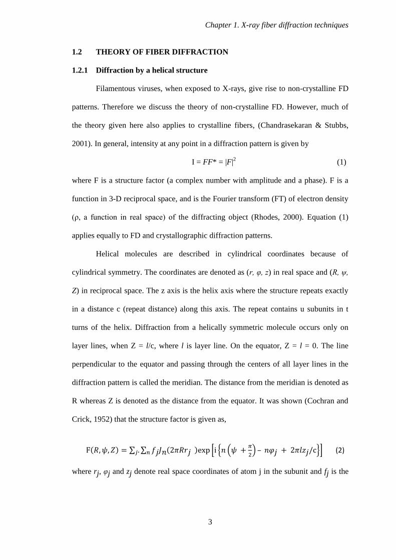
1.2 THEORY OF FIBER DIFFRACTION
1.2.1 Diffraction by a helical structure
Filamentous viruses, when exposed to X-rays, give rise to non-crystalline FD
patterns. Therefore we discuss the theory of non-crystalline FD. However, much of
the theory given here also applies to crystalline fibers, (Chandrasekaran & Stubbs,
2001). In general, intensity at any point in a diffraction pattern is given by
I = FF* = |F|2
(1)
where F is a structure factor (a complex number with amplitude and a phase). F is a
function in 3-D reciprocal space, and is the Fourier transform (FT) of electron density
(ρ, a function in real space) of the diffracting object (Rhodes, 2000). Equation (1)
applies equally to FD and crystallographic diffraction patterns.
Helical molecules are described in cylindrical coordinates because of
cylindrical symmetry. The coordinates are denoted as (r, φ, z) in real space and (R, ψ,
Z) in reciprocal space. The z axis is the helix axis where the structure repeats exactly
in a distance c (repeat distance) along this axis. The repeat contains u subunits in t
turns of the helix. Diffraction from a helically symmetric molecule occurs only on
layer lines, when Z = l/c, where l is layer line. On the equator, Z = l = 0. The line
perpendicular to the equator and passing through the centers of all layer lines in the
diffraction pattern is called the meridian. The distance from the meridian is denoted as
R whereas Z is denoted as the distance from the equator. It was shown (Cochran and
Crick, 1952) that the structure factor is given as,
( ) ∑ ∑ ( ) * , (
) – -+ (2)
where rj, φj and zj denote real space coordinates of atom j in the subunit and fj is the
Chapter 1. X-ray fiber diffraction techniques
4
atomic scattering factor of that atom. Jn is the Bessel function of the first kind of
order n. The value of n is restricted to satisfy the selection rule
l = tn + um
where t is number of turns in one repeat distance; u represents the number of subunits
in one repeat distance; n and m are integers. The number of significant terms
contributing to the structure factor is limited, since Jn(x) is generally negligible for x
less than about n-2. Cylindrical averaging can be taken into account by re-writing
equation (2) in terms of Fourier-Bessel structure factors G (Klug et al., 1958), where
( ) ∑ ( ) ( ) (3)
Gn,l(R) is independent of ψ. Then
( ) ∑ ( ) , (
)+ (4)
and it can be shown (Franklin and Klug 1955; Waser, 1955) that
( ) ∑ ( ) ( ) ∑ | ( )| (5)
The number of significant terms in the summation is limited, e.g. in the
diffraction pattern of Tobacco mosaic virus (TMV), the largest number of terms
contributing to any intensity at a resolution of 3 Å is eight. Near the meridian, a single
G term makes up the whole intensity. Equation (5) may be compared to the
corresponding crystallographic equation (1). In crystallography, the diffracted
intensity is the square of the amplitude of a single structure factor, whereas in FD the
diffracted intensity is the sum of the squares of the Fourier-Bessel structure factors.
The summation occurs because of the cylindrical averaging of the diffraction pattern,
and may be thought of as the superimposition of the diffracted intensities. The
electron density in a non-crystalline fiber may be calculated by means of a Fourier-
Bessel synthesis, an analogue of Fourier synthesis used in crystallography and
crystalline FD. The electron density at a point r, φ, z is

Chapter 1. X-ray fiber diffraction techniques
5
( ) ( ) ∑ ∑ ( ) ( )] (6)
where
( ) ∫
( ) ( ) (7)
The inner part of the equator in FD is derived from a single G term, G0,0,
whose phase is either 0 or 180°; that is, G0,0 simply has a positive or negative sign. If
the signs are known, or can be estimated, this part of the equator can be transformed
to obtain a radial density distribution.
1.2.2 Nature of fiber diffraction
Fundamental structural aggregates of a fibrous sample are regarded as
filaments, and complete diffraction from specimen as fiber. Fibers are collection of
nearly parallel filaments, which are randomly oriented about the fiber axis. These
structural aggregates might be individual virions in a fiber or an oriented solution,
individual molecules of DNA or some other chain molecule. Diffraction from a fiber
sample is confined to layer lines, because of the repeating nature of the filament helix
at spacing inversely proportional to the repeat distance c. The layer lines are
continuous and correspond to the cylindrical average of the FT of a single molecule.
In a FD experiment, individual helical fibers are not perfectly aligned and not parallel
to the fiber axis. This deviation from parallelism is called disorientation and it causes
reflections from the fibers to spread into arcs. The layer lines in reciprocal space are
perpendicular to the fiber axis in real space. The layer line that passes through the
origin in reciprocal space is called the equator or zero layer line. The direction normal
to the equator is called the meridian. The pitch of a helix is the width of one complete
turn, measured parallel to the helical axis. The number of helical turns that marks a
unique repeating section of the sample is known as helix repeat and the corresponding
Chapter 1. X-ray fiber diffraction techniques
6
distance is called repeat distance (c). In TMV, the pitch height is 23 Å with 49
subunits in three turns (u=49; t=3). So, the repeat distance c, is 69 Å. For a helical
sample, the layer line separation is proportional to the reciprocal of the pitch of the
helix. However, the pitch of a helix may not be simply the reciprocal of the layer line
separation since there might be two or more helical turns in one repeat distance.
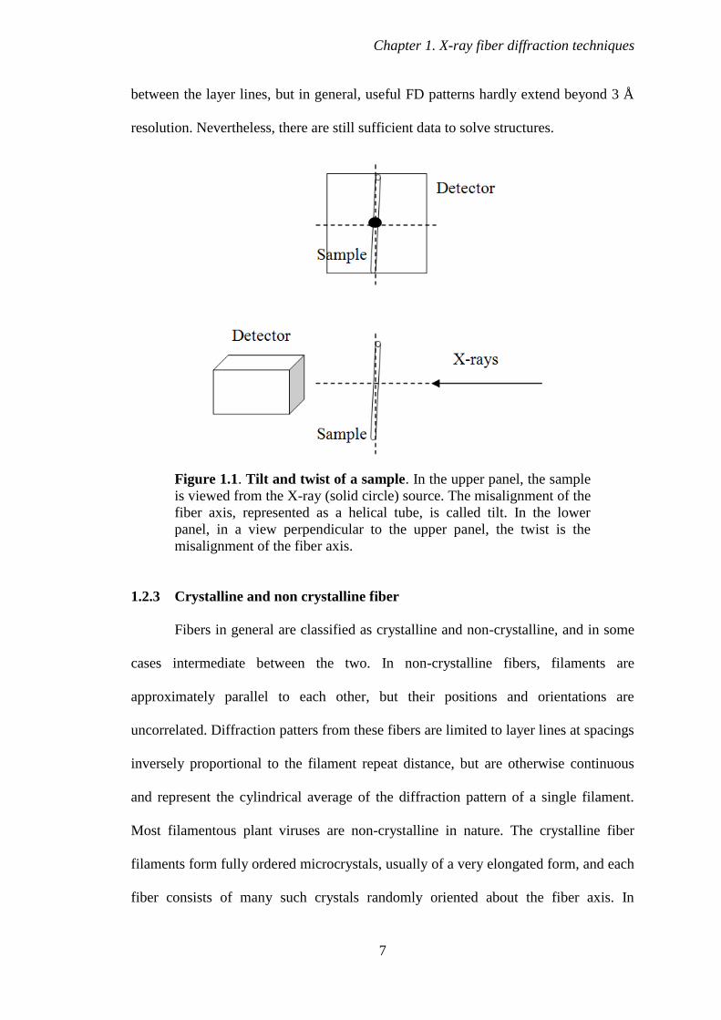
Furthermore, two other parameters, fiber specimen tilt and twist must be accurately
determined for proper data reduction. The specimen tilt is defined as the deviation of
the fiber axis from an absolute normal to the X-ray beam and the twist is the deviation
of the fiber axis from the plane of the detector, Fig. 1.1 (Kendall et al., 2007).
Fiber diffraction data contain less information than the equivalent from protein
crystals. Firstly, cylindrical averaging reduces the information content of diffraction.
The amount of information lost depends on the size and symmetry of filaments and
also on the resolution of the diffraction data. Cylindrical averaging affects high
resolution data that are away from the axis of rotation much more than that close to
the axis. In fact, for highly symmetrical helical filaments, the data near the axis of
rotation are cylindrically symmetric, and no data loss occurs due to cylindrical
averaging. For filaments of high symmetry viruses, e.g. TMV, cylindrical averaging
reduces the effective number of observable diffraction data at 3 Å resolution by a
factor of about 2.5, while for Pf1, the corresponding factor is only 1.7 (Makowski,
1982). There is no significant data loss for TMV at 15 Å and for Pf1 at 7 Å resolution.
Secondly, the resolution of the diffraction pattern is limited by disorientation of the
fiber sample. In practice, the filaments are not perfectly parallel and the mean
deviation of the filament axis from the fiber sample axis is typically 1 or 2°, even for a
well oriented fiber. As result, the layer lines fan out (overlap), running into each other
at high resolution. The severity of this effect obviously depends on the distance
Chapter 1. X-ray fiber diffraction techniques
7
between the layer lines, but in general, useful FD patterns hardly extend beyond 3 Å
resolution. Nevertheless, there are still sufficient data to solve structures.
Figure 1.1. Tilt and twist of a sample. In the upper panel, the sample
is viewed from the X-ray (solid circle) source. The misalignment of the
fiber axis, represented as a helical tube, is called tilt. In the lower
panel, in a view perpendicular to the upper panel, the twist is the
misalignment of the fiber axis.
1.2.3 Crystalline and non crystalline fiber
Fibers in general are classified as crystalline and non-crystalline, and in some
cases intermediate between the two. In non-crystalline fibers, filaments are
approximately parallel to each other, but their positions and orientations are
uncorrelated. Diffraction patters from these fibers are limited to layer lines at spacings
inversely proportional to the filament repeat distance, but are otherwise continuous
and represent the cylindrical average of the diffraction pattern of a single filament.
Most filamentous plant viruses are non-crystalline in nature. The crystalline fiber
filaments form fully ordered microcrystals, usually of a very elongated form, and each
fiber consists of many such crystals randomly oriented about the fiber axis. In

Chapter 1. X-ray fiber diffraction techniques
8
diffraction patterns from the crystalline fibers, the layer lines are sampled to from
separate reflection and the diffraction pattern represents the cylindrical average of the
diffraction pattern from a single crystal. The A form of DNA is crystalline fiber in
nature whereas the B form is non-crystalline fiber. Many filamentous bacteriophage
fibers are intermediate because these fibers are usually made by drying (McDonald et
al., 2008). The diffraction pattern from this type of fiber shows individual reflections
at the center (low resolution) of the equator, but continuous data on all other layer
lines.
1.3 STRUCTURE DETERMINATION USING FIBER DIFFRACTION
It is evident from Eqs. 5-7 that in order to determine electron density, the
observed intensities in a diffraction pattern must be separated into their component G
terms, and the phase of each G term must also be determined. Equivalently, the real
imaginary parts of each G term, contributing to each point in the diffraction pattern
must be determined. This is the FD equivalent of the phase problem in
crystallography, determining the phase for each F. In studies of filamentous viruses,
several different methods have been used for this determination.
1.3.1 Multidimensional isomorphous replacement (MDIR)
This method is an extension of the protein crystallographic method of
isomorphous replacement (IR) (Namba & Stubbs 1985; Namba & Stubbs, 1987a;
Stubbs & Diamond, 1975). If a heavy atom (i.e. an atom for which fj is significantly
greater than the other atoms in a structure) can be introduced into the molecule
without significantly perturbing the rest of the structure, the diffraction pattern from
the resulting derivative can be used together with the original (native) diffraction
Chapter 1. X-ray fiber diffraction techniques
9
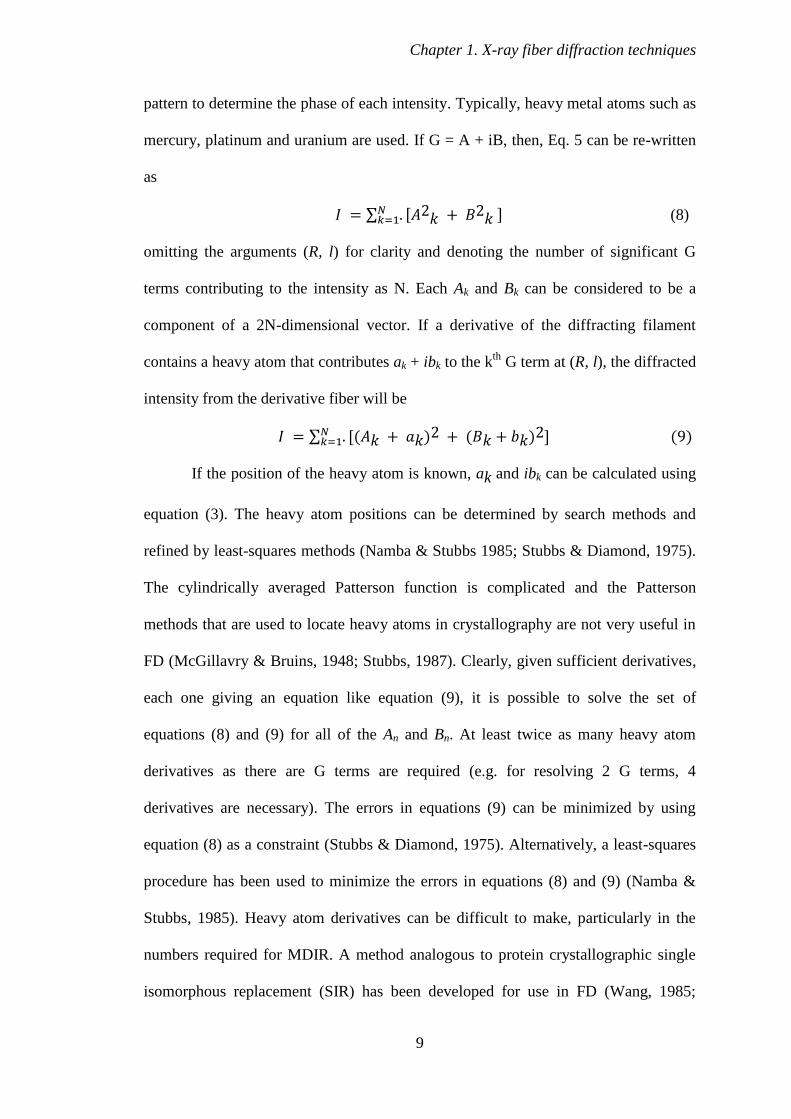
pattern to determine the phase of each intensity. Typically, heavy metal atoms such as
mercury, platinum and uranium are used. If G = A + iB, then, Eq. 5 can be re-written
as
∑ ] (8)
omitting the arguments (R, l) for clarity and denoting the number of significant G
terms contributing to the intensity as N. Each Ak and Bk can be considered to be a
component of a 2N-dimensional vector. If a derivative of the diffracting filament
contains a heavy atom that contributes ak + ibk to the kth
G term at (R, l), the diffracted
intensity from the derivative fiber will be
∑ ( ) ( ) ] (9)
If the position of the heavy atom is known, ak and ibk can be calculated using
equation (3). The heavy atom positions can be determined by search methods and
refined by least-squares methods (Namba & Stubbs 1985; Stubbs & Diamond, 1975).
The cylindrically averaged Patterson function is complicated and the Patterson
methods that are used to locate heavy atoms in crystallography are not very useful in
FD (McGillavry & Bruins, 1948; Stubbs, 1987). Clearly, given sufficient derivatives,
each one giving an equation like equation (9), it is possible to solve the set of
equations (8) and (9) for all of the An and Bn. At least twice as many heavy atom
derivatives as there are G terms are required (e.g. for resolving 2 G terms, 4
derivatives are necessary). The errors in equations (9) can be minimized by using
equation (8) as a constraint (Stubbs & Diamond, 1975). Alternatively, a least-squares
procedure has been used to minimize the errors in equations (8) and (9) (Namba &
Stubbs, 1985). Heavy atom derivatives can be difficult to make, particularly in the
numbers required for MDIR. A method analogous to protein crystallographic single
isomorphous replacement (SIR) has been developed for use in FD (Wang, 1985;
Chapter 1. X-ray fiber diffraction techniques
10
Namba & Stubbs, 1987a). It was used to determine the structure of Cucumber green
mottle mosaic virus (CGMMV) at 5 Å resolution (Lobert & Stubbs, 1990). In this
method, the magnitudes of the G terms contributing to a given intensity are either
estimated from a model of a related structure (sometimes called the ‘proportional
amplitude’ method) or they are assumed to be equal (Wang & Stubbs, 1994). The
equal-amplitude assumption is preferable, as it avoids model bias. The phases are then
determined by conventional crystallographic IR or by SIR.
Electron density maps of CGMMV, determined using the equal-amplitude
assumption, were as good as maps determined using amplitudes based on the related
TMV structure, even at 4.5 Å resolution (Lobert & Stubbs, 1990; Wang & Stubbs,
1994). But at higher resolutions the equal-amplitude assumption may not always be
sufficient to obtain a fully interpretable map (Namba & Stubbs 1987a and 1987b).
An additional source of information is in the positions of the layer lines in the
diffraction pattern. The layer line spacing is determined by the size of the helical
repeat, and when the repeat is approximate (fractional value), the G terms in each
layer line do not fall at exactly the same values of Z (where Z is the distance from the
equator in the reciprocal space), and the layer lines are said to be split (Tollin et al.,
1968). Layer lines have a finite thickness and the disorientation in the diffracting
specimen increases this thickness, so the splitting is seen only as a small shift in the
apparent position of the line. The magnitude and direction of the shift depend on the
relative magnitudes of the contributing G terms. The shifts can be measured for each
heavy atom derivative and used in the equation (Makowski, 1978),
∑ ( ) ( ) (10)
where Θ is the shift observed in the layer line position, measured as an angular
displacement about the centre of the diffraction pattern, θk is the calculated shift for

Chapter 1. X-ray fiber diffraction techniques
11
the kth
G term and q is the ratio of the magnitude of splitting in the derivative to the
magnitude of splitting in the native filament (Stubbs & Makowski, 1982). An
equation like (10) can be obtained from the native (with ak = bk = 0) and from each
derivative that is oriented sufficiently well to determine accurately. Equation (10) is
independent of equation (9) and can be combined with them to determine the values
of Ak and Bk. This method was particularly important in determining the structure of
TMV for which several extremely well oriented derivatives were available (Namba &
Stubbs, 1986).
1.3.2 Molecular replacement
The molecular replacement (MR) method is also used in fiber structure
determination when a structure closely related to the structure under investigation is
already known. A known structure can be used to estimate the phases and relative
magnitudes of the G terms. These phases and relative magnitudes can be applied to
the observed data and electron density maps can be calculated. A new model is built,
based on the generated electron density by several cycles of refinement until the
structure converges. This approach must be used with caution because the data is
cylindrically averaged due to which fewer data are available, and the phase solution is
consequently more dependent on the initial model. The map may therefore tend to
resemble the initial model regardless of the true differences in the structure. This was
a problem in earlier structure determinations. Improved methods of refinement,
particularly molecular dynamics (MD) refinement have greatly reduced the problem
of model bias (Wang & Stubbs, 1993). For example, the structure of the U2 strain of
TMV could not at first be determined by MR from the TMV structure, even though
these two virus structures are very similar. With the development of MD refinement,
Chapter 1. X-ray fiber diffraction techniques
12
the U2 model was finally solved satisfactorily (Pattanayek & Stubbs, 1992). With MD
refinement, the structure of the much difficult Ribgrass mosaic virus (RMV)
determined by MR from TMV (Wang et al., 1997) as initial model. Also the structure
of the bacteriophage Pf3, determined from the Pf1as a initial model were refined
(Welsh et al., 1998). The use of a limited number of heavy atom derivatives in this
way may be a useful corrective measurement against excessive bias toward a model
structure in molecular replacement.
1.4 REFINEMENT OF FIBER STRUCTURES
Two methods of restrained refinement have been used in FD studies of
filamentous viruses, namely, restrained least squares (RLS) and MD. The RLS
method was adapted from the protein crystallographic refinement method
(Hendrickson, 1985; Stubbs et al., 1986). RLS has been effectively used for
refinement of plant virus (Namba et al., 1989) structures. A closely related Jack-Levitt
refinement has been used to refine bacteriophage structures (Jack & Levitt, 1978).
The radius of convergence of these methods is limited and successful refinement
depends on the accuracy of the starting model. The MD is the major choice of refining
fiber structures. The most useful application of this method has been simulated
annealing (SA), in which the structure is heated to a temperature of 3000-4000 K and
then potential energy is minimized as the structure is cooled in small decrements. At
higher temperatures, energy barriers between the starting model and the structures of
the lower potential can be overcome. In this way the radius of convergence of the
refinement is increased. Simulated annealing has been used to refine both
bacteriphages and filamentous viruses (Gonzalez, 1995; Wang et al., 1997; Wang &
Stubbs 1994; Welsh et al., 1998).

Chapter 1. X-ray fiber diffraction techniques
13
1.5 DIFFERENCE FOURIER AND OMIT MAP IN FIBER DIFFRACTION
Difference Fourier syntheses have been widely used in both protein and small-
molecule crystallography to determine structures. It is also used in FD (Mandelkow et
al., 1981). This use has been limited by the difficulties peculiar to FD which arise
from the cylindrical averaging of FD patterns. Difference Fourier maps calculated
from FD data, by direct analogy with crystallographic difference maps, tend to have
high noise levels and found to be biased toward the known or model structure.
The crystallographic reflection has two components, one real and one
imaginary. But the FD layer lines may have more Bessel terms as we move to higher
resolution shell. The problem of FD is multidimensional unlike the two dimensional
problem of crystallography. The number of significant Bessel terms (N) contributing
to the diffraction intensity depends on the symmetry and dimensions of the diffracting
particle and on the values of (R, l) (see Eq. 5). In FD, e.g. TMV, 10 Å resolution data
can contribute to 1 Bessel term, but 2.9 Å data can contribute to 8 Bessel terms.
In crystallography the difference map is generally 2Fo – Fc. The fiber
equivalent of 2Fo-Fc is 6Go – 5Gc (Namba & Stubbs, 1987b). Although the difference
maps described is satisfactory for most applications, it is sometimes desirable to
minimize any possible bias towards a model structure. In recent years, omit maps
have become popular as a means to eliminate model bias. Omit maps are calculated
from observed structure factor amplitudes and calculated phases, but for phase
calculation, the part of the structure under investigation is omitted. A series of map
sections of the unit-cell can be systematically omitted (Artymiuk & Blake, 1981).
Alternatively, parts of the model structure can be omitted e.g. at a time three amino
acid residues can be omitted (Furey et al., 1986). A similar approach was taken in a
FD study where electron density corresponding to amino acid side chains suspected of
Chapter 1. X-ray fiber diffraction techniques
14
changing conformation between two different forms of TMV protein were omitted
(Mandelkow et al., 1981). In some cases omitted density may return or included
density may disappear. In other cases, however, the noise level may be too high to
allow unambiguous interpretations to be made. The size of the omitted structure has
considerable bearing on the interpretability of omit maps. This is particularly true
with FD data because the ratio of model observations to diffracted data observations
is much higher than in crystallography. For very small omissions, such as a single side
chain we may obtain satisfactory results but with larger omissions there is a
significant loss of interpretability. It therefore appears that while omit maps can be of
value to FD in answering questions about small regions of a molecule, they are not
suitable for systematic examination of complete structures.
1.6 FIBER DIFFRACTION IN MACROMOLECULAR STRUCTURE
DETERMINATION
Since the development of the theory of FD (Cochran et al., 1952a), it has been
widely used for structure determination of helical aggregate forming biomolecules,
notably synthetic polypeptides (Cochran & Crick, 1952b), deoxyribonucleic acid
(Wilkins et al., 1953), TMV (Namba et al., 1989) and collagen (Cohen & Bear, 1953;
Cowan et al., 1953).
1.6.1 Filamentous plant viruses
Filamentous plant viruses make up almost half of the plant virus genera. The
Potyvirus genus alone has been described as including almost a third of known plant
viruses (Riechmann et al., 1992) and is responsible for more than half viral crop
damage in the world. A single Potexvirus, Potato virus X (PVX) destroys world
Chapter 1. X-ray fiber diffraction techniques
15
potato crop by 20% (White et al., 1994). Filamentous plant viruses can be grouped
broadly into rigid (rod-shaped) and flexible viruses. The International Committee on
Taxonomy of Viruses currently recognizes 8 genera of rigid filamentous plant viruses
(type member, TMV) and 17 flexible species (type member, PVX) (Regenmortel et
al., 2000). All existing filamentous plant viruses are RNA viruses that contain a single
type of Coat protein (CP) encapsidating a single-stranded RNA molecule in a helical
array. In some genera, the genome is divided among two or more RNA molecules in
which the virus consists of multiple particles, typically of different lengths e.g.
Tobacco rattle virus (TRV), a member of the Tobravirus, is a bipartite virus having
particles of two lengths encapsidating the two RNA molecules that make up the TRV
genome. Some filamentous plant virus genera are morphologically similar to each
other at the electron microscopic level but most exhibit large differences in both
morphology and chemical structure. A better argument has been presented that most
filamentous plant viruses fall into one of two groups the rigid rods or the flexible
filaments (Dolja et al., 1991).
Filamentous virus studies and the development of FD methods have always
been synergistic. TMV and other Tobamoviruses have served as models for FD data
processing. It also helps in method development including the method of angular
deconvolution and phase determination (Makowski, 1978; Namba & Stubbs, 1985).
Isomorphous replacement (IR) was used earlier to obtain radial density distributions
of TMV (Caspar, 1956; Franklin, 1956). Later IR was developed to solve the multi-
dimensional phase problem in structure determination by FD (Stubbs & Diamond,
1975). Other techniques developed on TMV included layer line splitting (Franklin &
Klug, 1955). Methods of structure refinement and evaluation were also developed
using TMV. RLS (Stubbs et al., 1986), MD refinement (Wang & Stubbs, 1993), using
Chapter 1. X-ray fiber diffraction techniques
16
the likelihood function as a target (Mu & Makowski, 2000) difference Fourier
analysis (Namba & Stubbs, 1987b) and FD R factors (Millane, 1989; Stubbs, 1989)
are part of improvement in FD structural study. The filamentous bacteriophage Pf1
has been important in FD method development. Angular deconvolution was first time
applied to Pf1 structure (Makowski, 1978). Pf1 has also been important in the
development of MD refinement (Gonzalez et al., 1995).
Other filamentous bacteriophages served as model systems for the
development of background subtraction methods (Ivanova & Makowski, 1998;
Marvin et al., 1987). Techniques for making oriented sols by shearing were originally
developed for TMV (Bernal & Fankuchen, 1941; Gregory & Holmes, 1965). The use
of magnetic fields for orienting FD specimens was first time applied using Pf1
(Torbet, 1987; Torbet & Maret, 1979). Magnetic orientation is now widely used in FD
(Stubbs, 1999; Torbet, 1987).The combination of these two techniques with
centrifugation (Ivanova & Makowski, 1998) showed exceptional promise for a
number of FD systems including filamentous viruses (Oda et al., 1998; Stubbs et al.,
2000; Yamashita et al., 1998b).
1.6.2 Tobamovirus structure determination by fiber diffraction
TMV, a rod shaped virus of the genus Tobamovirus, was the first virus to be
discovered and subjected to structural studies using X-ray FD.
Powder diffraction patterns from unoriented virus solutions had been obtained earlier
(Wyckoff & Corey, 1936). Later it was shown that TMV can form highly oriented
sols where rod shaped particles are oriented to within about 1° of each other (Bawden
et al., 1936; Bernal & Fankuchen, 1941). These oriented sols yielded high quality FD
patterns with virions aligned about their long axes. Also FD patterns from flexible
Chapter 1. X-ray fiber diffraction techniques
17
virus PVX was obtained although those patterns did not exhibit such a high degree of
orientation (Tollin et al., 1980; Wilson & Tollin, 1969). The characteristic spacing of
the layer lines in the TMV diffraction pattern and the higher intensity of every third
layer line showed that the virion structure was periodically repeating every 69 Å and
with an approximate repeat at every 23 Å. It was recognized that the pattern was
typical of diffraction from a helical structure and 69 Å repeating unit of TMV must
contain 3n + 1 subunits in three turns of the helix, with n being an integer. It was
shown for TMV that n = 16. i.e. 49 subunits in three turns (Franklin & Holmes, 1958).
Using heavy atoms and IR, the equatorial diffraction patterns with and without heavy
atom were compared for the radial density distribution. The center of the virion was
hollow, along the virus axis, with a central hole of about radius of 20 Å and RNA was
located about 40 Å from the viral axis. The method was later used for FD (Stubbs &
Diamond, 1975) and used to determine the TMV structure (Holmes et al., 1975;
Namba et al., 1989).
1.6.3 Other filamentous virus structure by fiber diffraction
The filamentous bacteriophages have been classified on the basis of structure
into two classes, I and II (Marvin, 1998; Marvin et al., 1974a; Marvin et al., 1974b).
They are morphologically similar at the electron microscopic level and are members
of the Inovirus genus (family Inoviridae) (Regenmortel et al., 2000).
A major development in their structure determination was the use of strong
magnetic fields to induce the bacteriophage particles to orient parallel to each other
(Torbet and Maret, 1979). This technique allows the production of exceptionally high-
quality diffraction patterns. IR method was of little use in filamentous bacteriophage
studies because most heavy atom compounds induced structural changes in the
Chapter 1. X-ray fiber diffraction techniques
18
virions. However, the simple α-helical structure of the CP (Marvin et al., 1974b)
allows models to be built and refined against diffraction data. In this way, the
structure of Pf1 was determined at 7 Å resolution (Makowski et al., 1980). The
structures of several filamentous bacteriophages from both structural classes were
determined later at resolutions as high as 3.0 Å (Welsh et al., 2000). The Potexviruses
(type member, PVX) are flexible filamentous viruses about 5000 Å in length and 130
Å in diameter (Richardson et al., 1981; Tollin et al., 1980; White et al., 1994). The
earliest FD studies of PVX showed that the diffraction patterns of virion have a
periodicity of about 33Å (Bernal & Fankuchen, 1941). The FD patterns from
Potexviruses have been interpreted and the symmetry of these viruses are found to be
considerably more variable than rigid Tobamoviruses (Tollin et al., 1980). Recently
oriented sols of PVX and Papaya mosaic virus have been obtained using techniques
combining magnetic fields with centrifugal forces (Yamashita et al., 1988a).
Potexviruses respond well to magnetic orientation in combination with centrifugation
developed by Namba’s group. Tobraviruses are rigid rod shaped bipartite viruses
having particles of various lengths encapsidating two RNA molecules (MacFarlane,
1999; Mathis & Linthorst, 1994). Both types of particle have same CP with 230 Å
diameters. On the basis of sequence comparisons with TMV, a model for the CP
structure of Tobraviruses has been proposed (Goulden et al., 1992).
Diffraction studies have been described for oriented sols of TRV (Finch, 1965)
and Pepper ringspot virus formerly known as the Campinas strain of TRV (Tollin &
Wilson, 1971). Fiber diffraction study of a Hordeivirus, called Barley stripe mosaic
virus (BSMV) has been reported (Finch, 1965). BSMV is a rigid rod 1250 Å in length
and about 200 Å in diameter. In summary, structural study of a number of filamentous
viruses (plant viruses and bacteriophages) is possible. However, crystallization of
Chapter 1. X-ray fiber diffraction techniques
19
filamentous viruses by common approaches for viral structure determination using
protein crystallography is very difficult. In very rare cases, isolated CP can be
crystallized. The TMV CP has been crystallized and its structure was determined at
2.8 Å resolution (Bloomer et al., 1978) and later at 2.4 Å (Bhyravbhatla et al., 1998).
But the natural tendency of CP subunits is to form helical aggregates rather than
crystals and attempts to crystallize the CP of other filamentous viruses have failed. In
rare cases where crystals have been grown, protein–protein interactions in the crystal
do not correspond to biologically significant interactions. In addition, protein-nucleic
acid interactions are absent altogether (Bhyravbhatla et al., 1998). Thus, structural
studies of filamentous viruses very much rely on the FD method.
20
CHAPTER 2. HIBISCUS LATENT SINGAPORE VIRUS
2.1 INTRODUCTION
Viruses can infect animals, plants and bacteria. Since first discovery of TMV
(Beijerinck, 1898), many viruses have been discovered. Viruses are nucleoprotein
complexes with their genetic material as DNA or RNA. The genetic material of a
virus is protected by CP structure. Major category viruses are of two shapes,
filamentous and icosahedral types. Plant viruses enter the host cells by damage of the
host tissue. The cell to cell movement is regulated by movement protein (MP)
whereas the long distance movement is regulated by CP.
Using various molecular biology methods, it is now possible to explore the
genome organization and expression strategies of different viruses elaborately. This in
turn help us to develop and design methods to combat crop losses resulting from viral
epidemics in agricultural fields and their exploitation as vectors for expressing
therapeutic proteins (Hamamoto et al., 1993; Wu et al., 2003). Tobamoviruses are
rod-shaped with an approximate length of 3000 Å and a diameter of 180 Å. Its
genome is positive-sense single-stranded RNA packed in a capsid of about 2100 CP
subunits. This typical packaging forms a right handed helical virion with 49 CP
subunits in three helical turns. Among Tobamoviruses, TMV is the most widely
studied virus and it remains to be useful tools for understanding the fundamental
processes of viral infection, replication and movement. Tobamovirus genus consists of
several species, which can be classified into 2 major sub-groups based on their origin
of assembly sequence (OAS). The OAS for the subgroup I and subgroup II located in
the MP and CP, respectively. The complete genome sequences of the
Chapter 2. Hibiscus latent Singapore virus
21
various Tobamoviruses have been reported (Alonso et al., 1991; Chng et al., 1996;
Goelet et al., 1982; Hamamoto et al., 1993; Heinze et al., 2006; Ikeda et al., 1993;
Lartey et al., 1995; Meshi et al., 1981; Min et al., 2009; Rhie et al., 2007; Silver et
al., 1996; Solis & Garcia-Arenal, 1990; Song et al., 2006; Srinivasan et al., 2002; Tan
et al., 2000; Ugaki et al., 1991; Yoon et al., 2001; Yoon et al., 2002; Zhang et al.,
2008).
2.2 HIBISCUS LATENT SINGAPORE VIRUS (HLSV)
Hibiscus latent Singapore virus (HLSV) is a newly discovered member of
subgroup II Tobamovirus. It is a positive sense RNA virus comprising 6,474 nt
(Genbank Accession No. NC 008310; Srinivasan et al., 2005). The helix has a repeat
of 70.5 Å (helix pitch 23.5 Å), which is close to the 70.8 Å of CGMMV (Wang &
Stubbs, 1994). Two other viruses of subgroup II are Cucumber green mottle mosaic
virus (CGMMV) and Sun-hemp mosaic virus (SHMV). HLSV differs from CGMMV
and SHMV in containing a 77-96 poly(A) tract at the 3΄ untranslated region (UTR).
The following sections give a preview of HLSV characterization.
2.2.1 General characterization of HLSV
The single molecule genome of HLSV is packed with 2,100 CP molecules (3
nucleotides per CP). The virus is 180 Å in diameter and the CP molecules are
arranged helically to give a rigid rod shape. HLSV also makes two sub-genomic RNA
for MP and CP (Srinivasan et al., 2002). HLSV induces chlorotic local lesions in C.
quinoa and systemic infection in Nicotiana benthamiana (Srinivasan et al., 2002).
HLSV host range is relatively limited as compared to TMV. HLSV forms particles of
two length sizes, i.e. 34 nm and 307 nm during its life cycle of infection in plants
Chapter 2. Hibiscus latent Singapore virus
22
(Srinivasan et al., 2002). The longer particle packs the full length RNA genome
HLSV. The shorter particles may contain its CP sub-genomic RNA, as reported for
SHMV (Higgins et al., 1976).
2.2.2 HLSV gene structure, regulation and proteins function
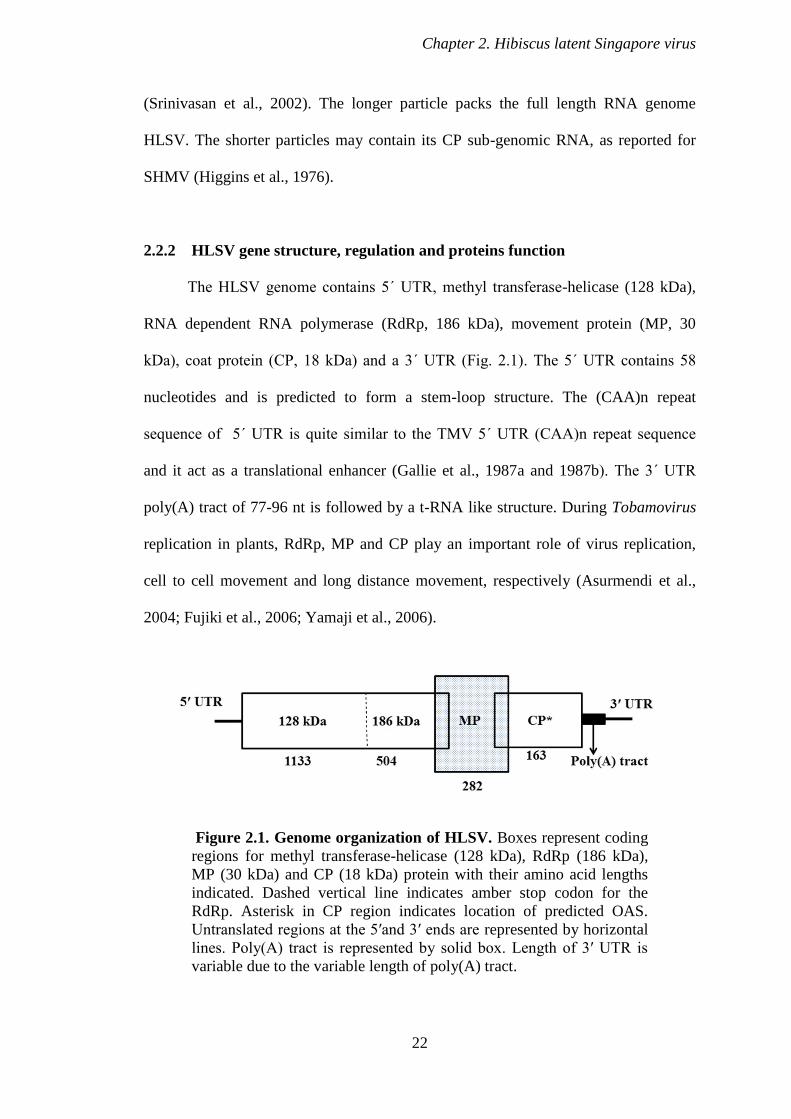
The HLSV genome contains 5΄ UTR, methyl transferase-helicase (128 kDa),
RNA dependent RNA polymerase (RdRp, 186 kDa), movement protein (MP, 30
kDa), coat protein (CP, 18 kDa) and a 3΄ UTR (Fig. 2.1). The 5΄ UTR contains 58
nucleotides and is predicted to form a stem-loop structure. The (CAA)n repeat
sequence of 5΄ UTR is quite similar to the TMV 5΄ UTR (CAA)n repeat sequence
and it act as a translational enhancer (Gallie et al., 1987a and 1987b). The 3΄ UTR
poly(A) tract of 77-96 nt is followed by a t-RNA like structure. During Tobamovirus
replication in plants, RdRp, MP and CP play an important role of virus replication,
cell to cell movement and long distance movement, respectively (Asurmendi et al.,
2004; Fujiki et al., 2006; Yamaji et al., 2006).
Figure 2.1. Genome organization of HLSV. Boxes represent coding
regions for methyl transferase-helicase (128 kDa), RdRp (186 kDa),
MP (30 kDa) and CP (18 kDa) protein with their amino acid lengths
indicated. Dashed vertical line indicates amber stop codon for the
RdRp. Asterisk in CP region indicates location of predicted OAS.
Untranslated regions at the 5′and 3′ ends are represented by horizontal
lines. Poly(A) tract is represented by solid box. Length of 3′ UTR is
variable due to the variable length of poly(A) tract.
Chapter 2. Hibiscus latent Singapore virus
23
2.3 PREVIOUS STUDIES ON TOBAMOVIRUS STRUCTURES
Several Tobamovirus structures have been reported. The structure of TMV
(Holmes et al., 1975; Namba et al., 1989), CGMMV (Wang & Stubbs, 1994), RMV
(Wang et al., 1997) and U2 strain of TMV (Pattanayek & Stubbs, 1992) have already
been solved by X-ray FD. Surprisingly, the TMV CP structure was solved at 2.4 Å
resolution by X-ray crystallography method (Bhyravbhatla et al., 1998). But for other
Tobamovirus structures X-ray crsyatllography approach was not successful. All other
Tobamovirus structures are determined by X-ray FD.
2.3.1 Infection and stability of native virus capsid
For Tobamoviruses, it has been shown that negatively charged amino acid
residues in the CP from different molecules are juxtaposed at subunit interfaces, at the
low (proximal to virus axis) and high radius regions (distal to virus axis). This creates
an electrostatic potential that is believed to drive disassembly and thus initiate the
early stages of viral infection (Caspar, 1963; Lu et al., 1996; Namba et al., 1989). The
carboxyl-carboxylate pair in the low radius region and phosphate-carboxylate pair
also appear to bind calcium (Ca2+
). Electrostatic interactions have been recognized to
be an important factor in the disassembly of helical and spherical plant viruses
(Bancroft, 1970; Caspar, 1963). On the basis of titrations of isolated TMV CP under
various conditions, anomalously titrating amino acids was suggested to exist in
subunit interfaces (Shalaby & Lauffer, 1977). It was also shown that TMV has two
sites which compete for calcium and protons ions (Gallagher & Lauffer, 1983a;
Gallagher & Lauffer, 1983b). Ca2+
ion binding gives structural stability to many
icosahedral viruses (Butler et al., 1977; Olson et al., 1983; Wada et al., 2008).

Chapter 2. Hibiscus latent Singapore virus
24
Structural details of TMV provide some interesting facts regarding molecular
basis of assembly in Tobamoviruses. A general description of the assembly process of
TMV has been described (Butler et al., 1977; Lebeurier et al., 1977). The OAS of the
RNA binds to the 20 S aggregate of the CP and elongation of the viral rod proceeds
by addition of the 20 S aggregates (Zimmern, 1977). For TMV, during infection, it is
believed that low concentration of Ca2+
and high pH (relative to extracellular
conditions) of the cell could considerably destabilize the close approach of the
negative charges in the viral subunit interfaces as described earlier (Namba et al.,
1989). However, these conditions are not sufficient for complete disassembly of virus
under in vitro conditions. It was shown that with pretreatment of TMV at pH 8.0
particles could be dissociated under in vitro conditions by a preparation containing
ribosomes (Wilson, 1984). This phenomenon, called co-translational disassembly,
was later observed also for in vivo conditions (Wilson, 1984). This mechanism
protects viral genome under unusual alkaline conditions.
In summary, the whole infection mechanism is described as; when virion
enters into a plant cell, owing to low concentration of intracellular calcium (Ca2+
) and
high pH (relative to the extracellular environment), protons and Ca2+
ions are removed
from carboxyl-carboxylate pairs and phosphate-carboxylate positions. This allows
electrostatic repulsive forces (from negative charges of the protein residues) to
destabilize the intact virus and hence, disassembly. It has been proposed that protein-
nucleic acid interactions involving first 69 nucleotides of 5΄ UTR are weaker than the
rest of the genome because of the presence of relatively less guanine bases (Douglas
& Young, 1998). So, CP subunits forming about 1.5 turns of the virus helix at the 5΄
UTR are lost easily. The first start codon is thus exposed and ribosomes bind and
move toward the 3΄ end during translation, competing with the CP and stripping the

Chapter 2. Hibiscus latent Singapore virus
25
rest of the genome, thereby beginning a new cycle of viral replication (Namba et al.,
1989).
2.3.2 Evolutionary insights from virus structures
Three major forces that drive evolution of viruses are mutation, recombination
and reassortment. The discovery of the GDD-sequence motif in a wide range of viral
polymerases shows that viruses have genes that are related (Argos, 1988; Kamer &
Argos, 1984). Koonin & Dolja (1993) and Zanotto and his team (1996) proposed that
Tobamoviruses share their RNA polymerase genes with other species of a large group
of viruses called the `α-like' virus group (Goldbach & De, 1994).
Tobamovirus CPs are also related in sequence and structure to those of other
viruses with rod-shaped and filamentous virions. The 3-D architecture of a typical
CPs of Tobamovirus consists of a bundle of four α-helices which is also a structural
fold observed in several other proteins, and shows evidence of having arisen by
duplication of a two-helix protein (McLachlan et al., 1980). In Tobamoviruses,
protein-nucleic acid interactions are more conserved than protein-protein interactions.
Firstly, among the 25 residues that are conserved in the Tobamovirus sequences, 11
are directly involved in RNA binding (Altschuh et al., 1987; Namba et al., 1989).
Secondly, because evolutionary pressures would resist viable mutations in the nucleic
acid binding site, mutations in other parts of the structure can be compensated for by
complementary mutations in spatially nearby residues.
Another phenomenon, called Caspar carboxylates interaction, is an important
feature of many viruses in disassembly process and is evolutionarily migrating among
virus species (Wang et al., 1998; Caspar, 1963; Bancroft, 1970). It is seen as
interactions between the side chains of specific amino acid residues of the CPs.
Chapter 2. Hibiscus latent Singapore virus
26
Carboxyl-carboxylate interactions are not conserved during evolution (Namba et al.,
1989; Wang & Stubbs 1994; Wang et al., 1997). The complex and variable nature of
protein-protein and protein-nucleic interactions in Tobamoviruses provides
information about the relationship between structure and function. Carboxylate
interactions allow structural features of inter-subunit interactions to be conserved,
even while amino acid sequences are changing. This feature allows virus to change
structure but retain function, is evolutionarily important and advantageous. Such
evolutionary flexibility helps virus in evading host defensive responses. For example,
host factors in tobacco are known to recognize the surface of the viral CP in many
Tobamoviruses that triggers a hypersensitive response. This response is characterized
by the death of cells close to the original site of viral infection thus preventing the
virus to spread systemically and the rest of the plant is protected (Culver et al., 1994).
2.3.3 Coat protein interaction with genomic RNA
Of several interactions that stabilize a Tobamovirus virion, protein-nucleic
acid interactions are very important. The entire genomic RNA of is encapsidated by
CP through non base specific protein-nucleic acid interactions. Such interactions are
non-directional, ionic and van der Waals forces. There are 4 α-helices in each CP, two
radial and two slewed ( Left Slewed, Right Slewed, Left Radial, Right Radial)
(Champness et al., 1976).
In TMV, nonspecific base binding is achieved by interactions between the
base surface and left radial α-helix (Namba et al., 1989). Electrostatic interactions are
considered complementary between electrostatic surfaces of the protein and the
nucleic acid. The top surface of the helical array of the CP subunits creates a
positively charged groove to accommodate the negatively charged phosphate groups
Chapter 2. Hibiscus latent Singapore virus
27
of RNA. TMV CP recognizes RNA with very high selectivity, efficiently
encapsidating only its own or closely related RNA (Hirth & Richards, 1981; Meshi et
al., 1983; Meshi et al., 1981). It was proposed that TMV assembly is initiated by a
specific RNA sequence that includes AAGAAG as part of a sequence (XXG)6
(Zimmern, 1977). It was proposed that this part of the genome has a highly base-
paired secondary structure. In TMV and U2 strain, RNA recognition is achieved by a
specific hydrogen bonding interaction between Arg122 and a guanine1 (Pattanayek &
Stubbs, 1992; Namba et al., 1989). Arg122 is conserved in most Tobamoviruses. In
CGMMV, guanine1 specificity is achieved by hydrogen bonding with Gln36 and this
may be a characteristic of a subgroup II Tobamovirus (Wang & Stubbs, 1994). In
TMV CP subunits, there is an electrostatic repulsion prevalent between Asp116 and
phosphate2. There was electron density evidence for Ca2+
ions binding in this region
shown (Namba et al., 1989). In general protein-nucleic acid interactions in a virus
structure demonstrates an important principle that virus structure must achieve a
metastable balance. All protein-nucleic acid interactions in viruses are required for
assembly and dis-assembly in response to changes in their environment (Caspar,
1963; Bancroft, 1970).
2.3.4 Maturation processes of Tobamoviruses
Structural studies of CP are advantageous in understanding the self-assembly
of a virus in terms of protein-protein and protein-nucleic acid interactions. These
interactions are responsible for nucleation and subsequent growth of the virus. TMV
CP is one of the first fibrous protein assemblies that was successfully crystallized, as a
dimer of bilayer disks or also called a four layer aggregate having 68 subunits (each
layer having 17 subunits) (Bloomer et al., 1978). This assembly shown a

Chapter 2. Hibiscus latent Singapore virus
28
sedimentation coefficient of 28 S (Bhyravbhatla et al., 1998) and the structure helped
in understanding the self-assembly of TMV.
Since then, it was proposed that bilayer disk plays a key role through an initial
RNA recognition reaction at a specific OAS, which then induces a structural
switching from the cylindrical disk to helical subunit packing array (Bloomer &
Butler, 1986; Butler & Durham, 1977; Hirth & Richards, 1981; Okada, 1986; Stubbs,
1984). A major role for the bilayer disk in the growth of TMV assembly has been
proposed (Bloomer & Butler, 1986), but this point has been controversial for some
time (Hirth & Richards, 1981; Okada, 1986; Schuster et al., 1980). Central to this
controversy has been the assumption that the bilayer disk structure, which is seen in
crystals as a dimer, also exists in solution and can undergo a direct structural change,
resulting in helical packing of subunits. It has been shown that axial inter-subunit
contacts in the bilayer disk are totally different from those in the helix (Champness et
al., 1976). At pH 7.0, the CP exists as a mixture of 4 S and 20 S sedimentation
aggregates. The CP species with a sedimentation coefficient of 20 S has been shown
to be involved in formation of large helical protein aggregate (Durham et al., 1971;
Schuster et al., 1979) as well as in the nucleation of viral assembly under in vitro
experiments (Shire et al., 1979). The 20 S aggregate is present in low ionic strength
solutions, either in equilibrium with 4 S aggregate at pH 7.0 or as metastable
aggregate at pH 6.5 (Durham et al., 1971; Butler & Klug, 1971).
The 20 S aggregate from the crystallized four-layer aggregate, was further
examined (Raghavendra et al., 1985) by circular dichorism technique. This study
revealed that the aggregation assembly observed in the crystal differs from the
structure present in the 20 S boundary in solution. As aggregates larger than 34
subunits cannot be formed in the closed cylindrical disk, the authors concluded that
Chapter 2. Hibiscus latent Singapore virus
29
two structures are not the same and that the four-layer aggregate is only one of the
many self-assembling aggregates and not the nucleating aggregate of the virus.
2.4 RATIONALE AND OBJECTIVES
During virion assembly, Tobamovirus CP recognizes a repeating GXX
sequence, which is located in the OAS of a viral RNA (Zimmern, 1977, Meshi et al.,
1981, Meshi et al., 1983, Takamstusu et al., 1983). The guanine1 recognition during
viral assembly is proposed for TMV, CGMMV and RMV (Namba et al., 1989, Wang
and Stubbs, 1994, Wang et al., 1997). Arg122 is conserved in all Tobamoviruses
except for HLSV and SHMV and it is believed to be involved in guanine1 hydrogen
bond formation during virion assembly of TMV and U2 (Namba et al., 1989,
Pattanayek and Stubbs, 1992). However, in HLSV and SHMV, the corresponding
residue (Arg122) is replaced by histidine. Although histidine is a good hydrogen
donor, it is unlikely to form a hydrogen bond with a base, since it does not have the
flexible extended side chain to reach the base (Wang and Stubbs, 1994). Nevertheless,
guanine specificity is believed to be required in SHMV (Meshi et al., 1981). What is
the role of His122 and why is it not conserved as Arg122 during evolution in SHMV
and HLSV? The recognition of the guanine1 in CGMMV and RMV is also believed
to be dictated by Arg122 of the CP similar to TMV (Wang et al., 1997). If Arg122 is
changed to His122, how do HLSV and SHMV CP recognize guanine1?
While carboxyl-carboxylate interactions in Tobamoviruses are the main
driving force for virus disassembly (Wang et al., 1998), they are not conserved during
the Tobamovirus evolution (Wang et al., 1997; Wang and Stubbs, 1994; Namba et al.,
1989). Does this mean that these interactions in HLSV would be different from other
Tobamoviruses?
Chapter 2. Hibiscus latent Singapore virus
30
By solving the structure of HLSV by X-ray FD, we will be able to have a
better understanding of the differences between HLSV and other Tobamoviruses.This
research may also enhance our knowledge of virus structure at atomic details. By
knowing the atomic details of this novel virus we may able to use it in future as a
vector to express pathogenic epitopes (to develop vaccine) and express economically
important proteins.
31
CHAPTER 3. MATERIALS AND METHODS
3.1 MOLECULAR BIOLOGY
3.1.1 Cloning the HLSV c-DNA
The pBluescript II KS(+) vector (Stratagene) was used to clone the HLSV full
length cDNA of 6474 bp (Cao Shishu, PhD. thesis, Natl. Univ. Singapore, 2007). The
bacterial strain Escherichia coli DH5α was used for plasmid propagation.
3.1.2 In vitro transcript preparation
The pHLSV construct was linearized with XhoI and cleaned up using the
Qiaquick gel extraction column following manufacturer’s protocol (Qiagen). The
mMessage mMachine kit (Ambion) was used for generating capped in vitro
transcripts. The reaction mixture was as follows: 10 μl of 2x NTP/CAP, 2 μl of 10x
reaction buffer, 1 μg linearized DNA template, 1μl GTP, 2 μl enzyme mix (T7
polymerase) and nuclease free water to make the reaction volume to 20 μl. The
mixture was incubated at 37 °C for 2 h. The transcript was checked on 1.2% agarose
gel. The transcript was cleaned using the manufacturer’s protocol.
3.2 VIRUS PROPAGATION AND PURIFICATION
3.2.1 Plant inoculation
The full length c-DNA clone of HLSV (see section 3.1.2), linearized with xhoI
at the 3' end was transcribed using mMessage mMachine® (Ambion). 10 μg of the
transcribed RNA was mixed with 50 μl of nuclease free water and inoculated
mechanically onto Nicotiana benthamiana leaves using carborundum. Later on,
Chapter 3. Materials & methods
32
HLSV-infected leaves were used as inoculums for re-inoculation to fresh plants.
Mechanical inoculation using carborundum was carried out by grinding the infected
leaves in 0.1 M borate buffer (pH 7.0), with a mortar and a pestle. Nicotiana
benthamiana seeds were grown in plant growth room under 16 h light and 8 h dark at
25 °C. Newly germinated seedlings were transplanted into new pots and grown in a
plant incubator for three weeks under the same environmental conditions. Plant
inoculation was performed mechanically as described above. Subsequently, the
inoculated leaves were rinsed with sterile water to remove buffer from the leaf
surface. The infected plant was then transferred to a plant growth room and incubated
for 4 weeks for virus multiplication. The propagation of virus from plant infection to
final virus purification took two months.
3.2.2 Crude extraction of HLSV
HLSV infected Nicotiana benthamiana leaves were harvested and stored at
-80 °C. Frozen infected leaves were homogenized using a blender in 3 volumes (w/v)
of extraction ice cold buffer (0.1 M borate buffer, pH 7.0). Homogenization was
carried out 3 times for 30 sec and between every cycle 1 min interval was given to
avoid over heating. The homogenate was centrifuged at 7,000 x g for 30 min in a
JA14 rotor (Beckman Coulter) at 4 °C. The supernatant was clarified with an equal
volume of n-butanol/chloroform (1:1) and again centrifuged at 7,000 x g for 30 min.
The supernatant was filtered using Whatman paper. The filtered supernatant was
transferred into 94 ml polyallomer thin tubes (resistant to organic chemicals) and
centrifuged at 22,000 x g for 2.5 h in a 45Ti rotor (in a Beckman Coulter L-100 xp
ultracentrifuge) to pellet the virus. The pellet was resuspended overnight in 0.1 M
borate buffer (pH 7.0) containing 5 mM EDTA.
Chapter 3. Materials & methods
33
3.2.3 Cesium chloride density gradient centrifugation
The crude virus preparation showed nucleic acid and other contamination from
the plant tissue. To overcome this we used cesium chloride density gradient
centrifugation. We Prepared 30% (w/v) CsCl solution in milli Q water with 5 mM
EDTA, pH 7.0, to prevent virus aggregation. The crude virus sample was layered on
the top of the solution in 30 ml ultra-clear centrifuge tube (with 3 mg virus per tube).
The sample was centrifuged at 40,000 x g for 16 h at 20 °C in a 70.1Ti rotor (in a
Beckman Coulter L-100 xp ultracentrifuge). The virus band was collected with a
Pasteur pipette under fluorescent light. The collected virus, containing CsCl, was
diluted 10 times using 0.1 M borate buffer containing 5 mM EDTA and transferred to
75 ml polycarbonate tubes and centrifuged at 22,000 x g for 2.5 h at 4 °C in a 45Ti
rotor. The white clean virus pellet was resuspended in 20 mM borate buffer
containing 5 mM EDTA overnight at 4 °C. The yield and quality of the virus was
tested spectrophotometrically for A260/280, using an extinction coefficient of 3.3.
3.2.4 Slow speed centrifugation to purify 300 nm long HLSV virion
To partially remove the 34 nm short particles from the long particles of 307
nm, low speed centrifugation was carried out. Initial attempts to separate the two
particles at 8,000 x g for 18 h at 4 °C failed as it led to pelleting both virus particles at
the bottom of the tube. In later attempts, the centrifugation speed was reduced to
6,000 x g for 18 h at 4 °C. The pellet was resuspended in 20 mM borate buffer (pH
7.0) containing 5 mM EDTA and the supernatant was re-centrifuged at 22,000 x g at 4
°C for 2.5 h to see the remaining visible pellet. A very small pellet was seen at the
bottom of the tube. The pellet was resuspended in 20 mM borate buffer (pH 7.0). Both
the pellet and supernatant were analyzed by electron microscopy.

Chapter 3. Materials & methods
34
3.2.5 Sephacryl 1000 gel filtration
In the next attempt, the Sephacryl S-1000 superfine (GE healthcare) size
exclusion chromatography was carried out to separate the two particles. We loaded 30
mg CsCl purified HLSV at each FPLC run, with a flow rate of 0.2 ml/min. The virus
was eluted from the column starting from 60th
to 110th
fractions. Three types of
sample pooling were carried out: 60-74, 75-85 and 86-110. The three samples
separately pooled and were again subjected to ultracentrifugation at 22,000 x g for 2.5
h at 4 °C to pellet the virus. The transparent virus pellet was resuspended in 20 mM
borate buffer (pH 7.0) containing 5 mM EDTA overnight at 4 °C. The samples were
stored at 4 °C for oriented sol preparation for X-ray FD study. Electron micrograms
were taken on carbon coated copper grid with 2% uranyl acetate. For practical reason
we use the peak fraction 75-85 ml pooled fraction for obtaining best sols as it
consisted of longer particles only.
3.3 CHARACTERIZATION OF PURIFIED HLSV VIRION
3.3.1 Virus purity and concentration
The purity of the purified HLSV sample was detected first by measuring the
A260/280. The purified virus was diluted 100 times and the absorbance was recorded at
Beckmann UV spectrophotometer.
3.3.2 Transmission electron microscope (TEM)
Copper grids (400-mesh) were prepared by covering them with formvar film a
carbon layer was coated using a carbon evaporator. Purified virus was negatively
stained with 1 μl of 10 mg/ml bacitracin (to prevent the positive staining of the highly
Chapter 3. Materials & methods
35
pure HLSV) and 1 μl 2.0% urine acetate and examined in a Jeol JEM-2010 TEM
operated at 100 kV with magnification of 30,000.
3.3.3 Western blot
Virus purity was also checked on SDS-PAGE (Sodium dodecyl sulphate-
polyacrylamide gel electrophoresis). 12 % SDS-polyacrylamide separating gel, with
5% stacking gel, was prepared. The samples were treated with 5 μl of 6x loading
buffer and boiled at 100 °C for 5 min. Electrophoresis was run at 100 V for 2.5 h. The
gel was stained by the Coomassie dye. In addition, Western blot analysis was
performed with a HLSV polyclonal antibody (primary antibody) and an anti-rabbit
antibody (secondary antibody), following the standard procedures.
3.4 ORIENTED SOL PREPARATION
For FD studies, the most effective specimen should be in a thick gel-like form,
known as sol. The sol was prepared in a 0.5 mm quartz capillary tube using previous
method (Gregory & Holmes, 1965). A virus pellet was exchanged to a suitable buffer
(5 mM EDTA, pH 7.0) and the concentration was kept at 30 mg/ml. The resuspended
virus was centrifuged at 10,000 x g for 16 h at 4 °C in a 1.5 ml microfuge tube. The
supernatant was pipetted out and the surface of the pellet was dried with the corner of
a tissue paper. The virus pellet was mounted in thin 0.7 mm quartz capillary (Charles
Supper) that was washed with 1 M HCl, reverse osmosis (RO) water and 5 mM
EDTA, each for four times. During sol preparation, the wide mouth of the capillary
was first connected with tubing (1/8”, Nalgene). The thin sealed end was broken with
a capillary cutter and the broken end was dipped into the virus pellet and the virus was
drawn to a distance of 3 cm by withdrawing air by mouth through the PVC tube. The

Chapter 3. Materials & methods
36
virus column was pipetted back and forth several times gently for giving better
orientation to the virus. Three μl 1 M KCl, which would increase the fluidity of the
sample in the capillary, was used. The column of virus was drawn to the end of the
capillary and the end was washed by alternate siphoning and blowing. This process
was continued until the end of the capillary was completely clean. The capillary was
cut at the broad end side also. The quality of the oriented sol was tested using a
polarization microscope before sealing it. Well oriented sols show complete
extinction (Yamashita et al., 1998a) in a polarizing microscope. Both ends were
sealed using an oxygen flame torch. The sealed capillary, with the oriented sol, was
kept upright in plasticine (Nalgene).
3.5 FIBER STRUCTURE DETERMINATION
3.5.1 Data collection
Unlike X-ray crystallography, in FD, two to five frames of data are enough for
structure determination. Furthermore, importance of humidity in fiber sample
preparation and significance of the chambers in controlling humidity have been
demonstrated for filamentous plant viruses, yeast prions and Potyviruses (Hull, 2001;
Wickner et al., 2007; Lo´pez-Moya & Garci´a, 1999). The oriented sols, in which the
protein was obviously fully hydrated, gave rise to the strong meridional diffracted
intensity at 4.7 Å resolution, characteristic of amyloid structures, but without
diffracted intensity on the equator (McDonald et al., 2008).
3.5.2 Data processing
In X-ray crystallography, diffraction by a crystal produces reflections with
designated lattice indices. The intensity of a reflection is related to all the atoms,
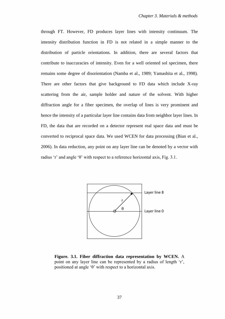
Chapter 3. Materials & methods
37
through FT. However, FD produces layer lines with intensity continuum. The
intensity distribution function in FD is not related in a simple manner to the
distribution of particle orientations. In addition, there are several factors that
contribute to inaccuracies of intensity. Even for a well oriented sol specimen, there
remains some degree of disorientation (Namba et al., 1989; Yamashita et al., 1998).
There are other factors that give background to FD data which include X-ray
scattering from the air, sample holder and nature of the solvent. With higher
diffraction angle for a fiber specimen, the overlap of lines is very prominent and
hence the intensity of a particular layer line contains data from neighbor layer lines. In
FD, the data that are recorded on a detector represent real space data and must be
converted to reciprocal space data. We used WCEN for data processing (Bian et al.,
2006). In data reduction, any point on any layer line can be denoted by a vector with
radius ‘r’ and angle ‘θ’ with respect to a reference horizontal axis, Fig. 3.1.
Figure. 3.1. Fiber diffraction data representation by WCEN. A
point on any layer line can be represented by a radius of length ‘r’,
positioned at angle ‘θ’ with respect to a horizontal axis.
r
θ
Layer line 8
Layer line 0
Chapter 3. Materials & methods
38
The resolution d (in Å) at any point on the detector in a diffraction experimental setup
is given by the equation,
d= λ/2 sin[1/2 tan-1
y/D] (11)
Where λ is the wavelength of X-rays (in Å), y is the distance of the point of interest
from the center of the detector (in mm) and D is the distance between the specimen
and detector (in mm). In WCEN, the maximum number of radius is 1200 and the
raster value (or pixel size) for the ADSC Q315 detector is set as 0.1026 mm. Thus, in
WCEN, the maximum value for y is given by,
y = 1200 x 0.1026 = 123.12 mm (12)
In the above equation (12), if y = 123.12 mm, λ = 0.9002 Å and D = 252.8 mm, then
the maximum resolution (d) for an experiment will be 2.0 Å. It is worth noting here
that even though the maximum y for an ADSC Q315 detector, according to the
manufacturer, is 3072 pixel x 51 μm per pixel = 156.672 mm, WCEN takes only upto
an effective maximum of 123.12 mm. Thus the raster value of WCEN is pre-set to
match a maximum resolution of 2.0 Å and correlated to the specimen to detector
distance of 252.8 mm.
When WCEN reads a diffraction frame, the entire diffraction image is first
augmented into 4 equivalent quadrants. Depending on a user’s choice, the 4 quadrants
are first averaged or only one quadrant (by default the first quadrant, right and up) is
taken. Also, the program is designed in a way that the effective detector half width
(from the center to the edge) is divided into 1200 radii and each radius sweeps across
the detector quadrant area from 0 to 90°. This means, the 1st quadrant area will be
marked by 91 x 1200 data points. Thus the output of WCEN is a matrix of 91 x 1200
data points and all inter layer line points should have a theoretical intensity of 0. By
the rule of inverse proportion, for our experimental setup, the relationship between a
Chapter 3. Materials & methods
39
radius number N and its corresponding resolution of diffraction is given by the
equation
Resolution = (1200 / N) x 2.0 (13)
The above equation (13) stems from the fact that the width between two adjacent radii
is conveniently expressed as 0.001 Å-1
in the reciprocal space. This helps in the
calculation of the resolution for any radius. For example, the resolution of radius 800
can be calculated as:
= (1200/800) x 2 = 3 Å
The output of WCEN was corrected for angular convolution, a process known
as deconvolution. The angular deconvolution method estimates the intensity from
partially oriented specimens to the highest possible resolution. At higher diffraction
angle noise will be large, compared to the intensity distribution at low resolution
limits. The major problem in structural studies of oriented non-crystalline specimen is
the insufficient number of data due to the cylindrical averaging of the intensities. The
angular deconvolution of data allows collection of maximum amount of information
from a given diffraction pattern. It defines the limiting resolution to which accurate
intensity information can be obtained. We used the program RAD (Makoswski, 1978)
for deconvolution. The output of RAD is further formatted by the program CORR
(Makoswski, 1978), which presents FD data in the format layer line number,
minimum radius, maximum radius (theoretically in the range 0 to the maximum
radius allowed for the resolution of the data, Eq. 13) for structure determination and
refinement.
3.5.3 Layer line splitting
Chapter 3. Materials & methods
40
Following earlier discussion in section 1.2.1, fiber particles are naturally
disoriented. Due to this anomaly layer lines normally overlap with each other. So,
each layer line may be associated with more than one Bessel term (section 1.5) and
unlike the crystallographic counterpart, a phase is multidimensional in FD (section
1.3.1). The intensity at any point on a layer line is made of Fourier-Bessel structure
factors. To solve a structure, contributions from each Bessel term must be determined
precisely. Splitting of the layer line simply means a Bessel function does not exactly
fall at the same distance from the equator (Franklin & Klug, 1955). This is generally
the case as the repeat distance in one repeat (the number of helical turns) is not
exactly repeated along a fiber specimen (Stubbs & Makowski, 1982). The degree of
layer line splitting can be determined from exact axial repeat. In practice, due to
disorientation of the particles in the specimen, splitting is not directly observed but the
effect of layer line splitting causes shifts in the apparent position of the layer line
which depend on the relative magnitude of the contributing Bessel terms. Nonetheless
the splitting is very informative and can be a source of information about phase. This
can be used to separate the contributions from the Bessel function which are
superimposed on layer lines. It has been shown that shifts in layer line positions can
be measured by angular deconvolution which can determine the contributions from
two Bessel function terms that are superimposed on a layer line (Makowski, 1978).
3.6 HLSV STRUCTURE DETERMINATION
The structure of HLSV was solved using the molecular replacement (MR)
method. Several frames (from different well oriented sols) of diffraction data at 0, 8
and 12° tilt of the specimen were collected. HLSV fiber sample diffracted X-rays to
2.8 Å. The diffraction data were processed using WCEN. Even though we could
Chapter 3. Materials & methods
41
extract data up to the 25th
layer line (2.8 Å resolution), the effective data that could be
used for refinement was limited to 3.5 Å (upto the 20th
layer line). At shells higher
than this resolution, layer lines smear/overlap heavily. Near meridian reflections were
used for refining the tilt angle whereas other parameters were refined using well
defined reflections, distributed throughout the diffraction image. The data were
transformed to the polar reciprocal space with angular transformation and further
were deconvoluted using RADFN which determines intensity along layer lines
(Makowski, 1978). The input parameters for running RADFN are: specimen to
detector distance (D) = 252.812 mm; helical repeat distance (C) = 70.51 Å; sigma G
(SIGG), the disorientation limit = 1.3°; error limit (ELIMIT) = 1; maximum number
of radii processed (NR) = 900; first radius to be processed (from the beam stop, IR1)
= 50; background type (NBKGD) = 1; the switch value to rotate the pattern by 90
degree (IFV) = 0; number of allowed bad points for a radius (NULB) = 0; reciprocal
space or film space (IFREC) = 0; detector resolution (the raster value, as defined by
WCEN) (DR) = 0.1026. The output of RADFN was further formatted by CORR for
structure determination and refinement.
A HLSV model was built based on the CGMMV structure (PDB code: 1cgm),
but, the R-factor did not improve below 18.4%. The R factor in FD is inherently lower
than that of protein X-ray crystallography because of the cylindrical averaging of
diffracted intensities (Stubbs, 1989). Usually, the R-factor for a well refined FD
structure will be in the range of 9.5%. The HLSV structure was finally determined at
3.5 Å using TMV as a model (PDB code: 2tmv). A trinucleotide (GAA) for RNA was
used as discussed previously (Namba et al., 1989; Pattanayek & Stubbs, 1992) for
other Tobamovirus structures. To model CP inter-subunit interactions, the coordinates
of 6 neighboring subunits (+1, +16, +17, -1, -16, -17) around the helical axis from the
Chapter 3. Materials & methods
42
reference subunit (labeled ‘0’ and named as the central subunit hereafter) calculated.
These subunit interactions represent and correspond to all interactions in the virion.
The blmax (Bessel function limit) value was set at 5. The initial model was refined by
energy minimization (minimize) without any FD effective energy. The idealized
model was subjected to repeated cycles of molecular dynamics simulated annealing
(SA) refinement from an initial temperature of 3000 K to a final temperature of 300 K
using the diffraction data between 10.0 and 3.5 Å resolution. The molecular dynamics
refinement was followed by temperature factor refinement (B refinement). The model
was adjusted using the recommended 6Gobs – 5Gcalc maps (Wang & Stubbs, 1994;
Namba et al., 1989). The 6Gobs – 5Gcalc maps are the FD equivalent of the Fobs-Fcalc
maps used in protein crystallography. The larger coefficients are due to cylindrical
averaging of the FD intensity and depend on the size and symmetry of the structure
and resolution of the data (Namba & Stubbs, 1987b). The structure was refined by
NIH-XPLOR (Schwieters et al, 2003). The model was fit to the map with the O
(Jones et al., 1991) and COOT (Emsley & Cowtan, 2004) programs. The refinement
process was repeated until the R-factor dropped to about 0.096.
The quality and geometry of the final model was analyzed using PROCHECK
(Laskowski et al., 1993). Also, omit maps (again, 6Gobs – 5Gcalc maps) were calculated
by omitting five residues at a time (in order to prevent model bias). The
Ramachandran plot, shows some disallowed geometry, due to the low data resolution.
All molecular diagrams were prepared with Pymol (DeLano, 2002). The coordinates
for the six surrounding were built using a program written by Swaminathan for
measuring protein-protein and protein-RNA interaction distances. We used the
HELANAL program to compare the LR helix geometry of the Tobamoviruses (Bansal
et al., 2000). Structure-based sequence alignment was made with the ESPript server
Chapter 3. Materials & methods
43
(http://espript.ibcp.fr/ESPript/ESPript/) and r.m.s.d. calculations were made with
Superpose (http://wishart.biology.ualberta.ca/SuperPose/).
44
CHAPTER 4. RESULTS AND DISCUSSION
4.1 SAMPLE PREPARATION
4.1.1 Crude extraction of HLSV from plant tissue
Adequate amount of the HLSV virus was propagated by infecting healthy N.
benthamiana plants with the full length c-DNA clone of HLSV in pBluescript KS II
(+), linearized with xhoI at the 3′ end and transcribed using mMessage mMachine.
HLSV was initially purified with the n-butanol/chloroform mixture. A260/280 of this
crude HLSV was 1.38 to 1.6, as opposed to that of 1.19 for TMV. This is attributed to
contamination by nucleic acids which could have come from the plant. We adopted
additional purification steps to eliminate contamination and to obtain more
homogeneous samples.
4.1.2 Purification using cesium chloride density gradient centrifugation
The virus was purified by the cesium chloride method. The purified virus
formed a clear white band under yellow light (Fig. 4.1A). We could also see nucleic
acids and other plant contamination as a higher band. Electron microscopy confirmed
that the purified HLSV was of long intact particles (Fig. 4.1B). We also verified its
purity by 12% SDS-PAGE and western blotting using an anti-HLSV antibody (Fig.
4.1C, D).
4.1.3 Slow speed centrifugation to purify the long virions
The crude HLSV preparation and the cesium chloride purified preparation
contained two particles, the 307 nm long virus and 34 nm long shorter particles. For
proper orientation of particles in sol, it is necessary to remove the short particles.
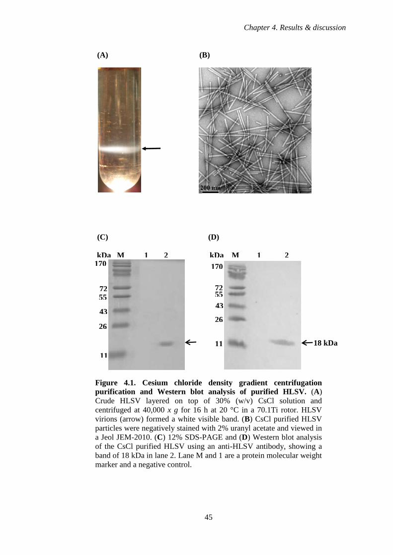
Chapter 4. Results & discussion
45
(A) (B)
(C) (D)
Figure 4.1. Cesium chloride density gradient centrifugation
purification and Western blot analysis of purified HLSV. (A)
Crude HLSV layered on top of 30% (w/v) CsCl solution and
centrifuged at 40,000 x g for 16 h at 20 °C in a 70.1Ti rotor. HLSV
virions (arrow) formed a white visible band. (B) CsCl purified HLSV
particles were negatively stained with 2% uranyl acetate and viewed in
a Jeol JEM-2010. (C) 12% SDS-PAGE and (D) Western blot analysis
of the CsCl purified HLSV using an anti-HLSV antibody, showing a
band of 18 kDa in lane 2. Lane M and 1 are a protein molecular weight
marker and a negative control.
kDa M 1 2 kDa M 1 2
72
43
26
11
55
170
11
55
43
26
72
170
18 kDa
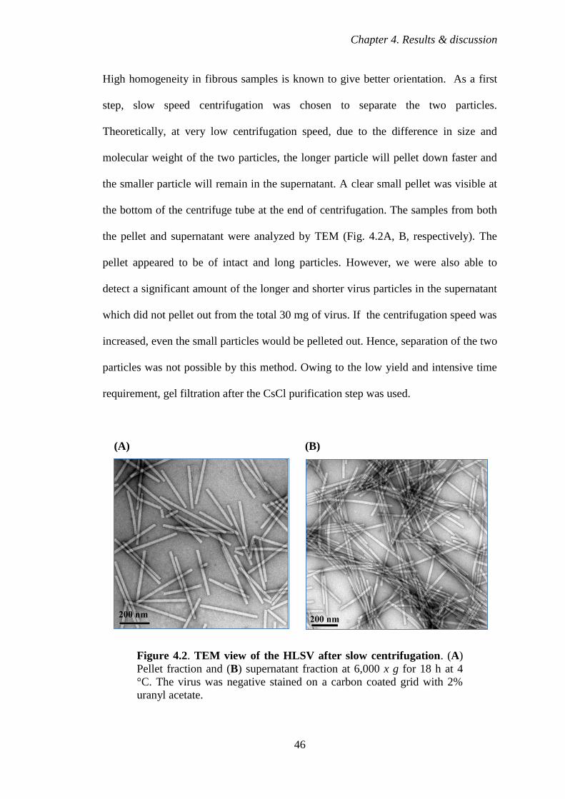
Chapter 4. Results & discussion
46
High homogeneity in fibrous samples is known to give better orientation. As a first
step, slow speed centrifugation was chosen to separate the two particles.
Theoretically, at very low centrifugation speed, due to the difference in size and
molecular weight of the two particles, the longer particle will pellet down faster and
the smaller particle will remain in the supernatant. A clear small pellet was visible at
the bottom of the centrifuge tube at the end of centrifugation. The samples from both
the pellet and supernatant were analyzed by TEM (Fig. 4.2A, B, respectively). The
pellet appeared to be of intact and long particles. However, we were also able to
detect a significant amount of the longer and shorter virus particles in the supernatant
which did not pellet out from the total 30 mg of virus. If the centrifugation speed was
increased, even the small particles would be pelleted out. Hence, separation of the two
particles was not possible by this method. Owing to the low yield and intensive time
requirement, gel filtration after the CsCl purification step was used.
(A) (B)
Figure 4.2. TEM view of the HLSV after slow centrifugation. (A)
Pellet fraction and (B) supernatant fraction at 6,000 x g for 18 h at 4
°C. The virus was negative stained on a carbon coated grid with 2%
uranyl acetate.

Chapter 4. Results & discussion
47
4.1.4 Sephacryl 1000 gel filtration chromatography
In the next attempt, gel filtration chromatography was run to remove the short
HLSV particles. The virus was eluted from the S1000 column as an asymmetrical
peak (Fig. 4.3A). The fractions (75-85 ml) that contained intact particles under TEM
(Fig. 4.3B) were pooled and later used for the oriented sol of HLSV.
(A)
140
120
100
80
60
40
20
0
(B)
Figure 4.3. HLSV purification by Sephacryl 1000 gel filtration
chromatography. (A) The chromatogram of HLSV purification
showing an asymmetric peak (B) TEM of gel filtration purified virus,
pooled from fractions 75-85 ml. 30 mg virus was loaded to the column
and purified at a flow rate of 0.2 ml/min.
20 40 60 80 100 120
Abso
rban
ce (
A2
80)
(mA
U)
Volume (ml)
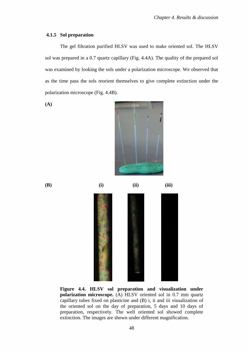
Chapter 4. Results & discussion
48
4.1.5 Sol preparation
The gel filtration purified HLSV was used to make oriented sol. The HLSV
sol was prepared in a 0.7 quartz capillary (Fig. 4.4A). The quality of the prepared sol
was examined by looking the sols under a polarization microscope. We observed that
as the time pass the sols reorient themselves to give complete extinction under the
polarization microscope (Fig. 4.4B).
(A)
(B) (i) (ii) (iii)
Figure 4.4. HLSV sol preparation and visualization under
polarization microscope. (A) HLSV oriented sol in 0.7 mm quartz
capillary tubes fixed on plasticine and (B) i, ii and iii visualization of
the oriented sol on the day of preparation, 5 days and 10 days of
preparation, respectively. The well oriented sol showed complete
extinction. The images are shown under different magnification.
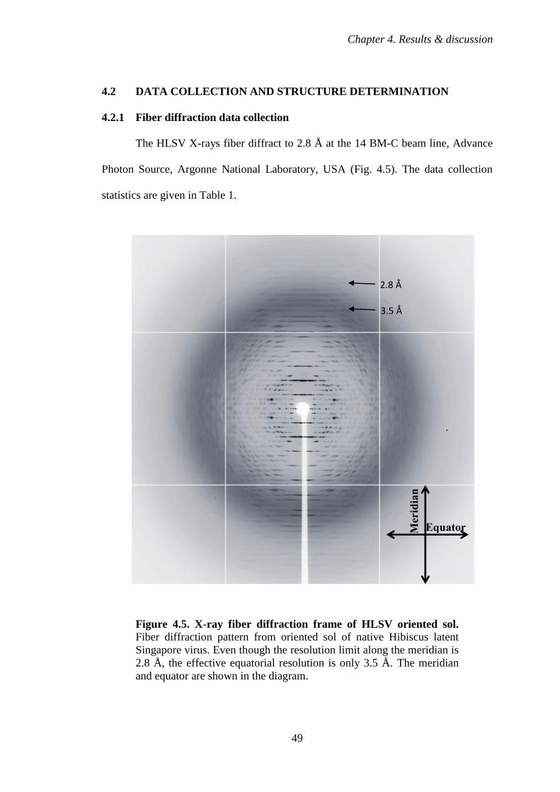
Chapter 4. Results & discussion
49
4.2 DATA COLLECTION AND STRUCTURE DETERMINATION
4.2.1 Fiber diffraction data collection
The HLSV X-rays fiber diffract to 2.8 Å at the 14 BM-C beam line, Advance
Photon Source, Argonne National Laboratory, USA (Fig. 4.5). The data collection
statistics are given in Table 1.
Figure 4.5. X-ray fiber diffraction frame of HLSV oriented sol. Fiber diffraction pattern from oriented sol of native Hibiscus latent
Singapore virus. Even though the resolution limit along the meridian is
2.8 Å, the effective equatorial resolution is only 3.5 Å. The meridian
and equator are shown in the diagram.
2.8 Å
3.5 Å
Equator Mer
idia
n
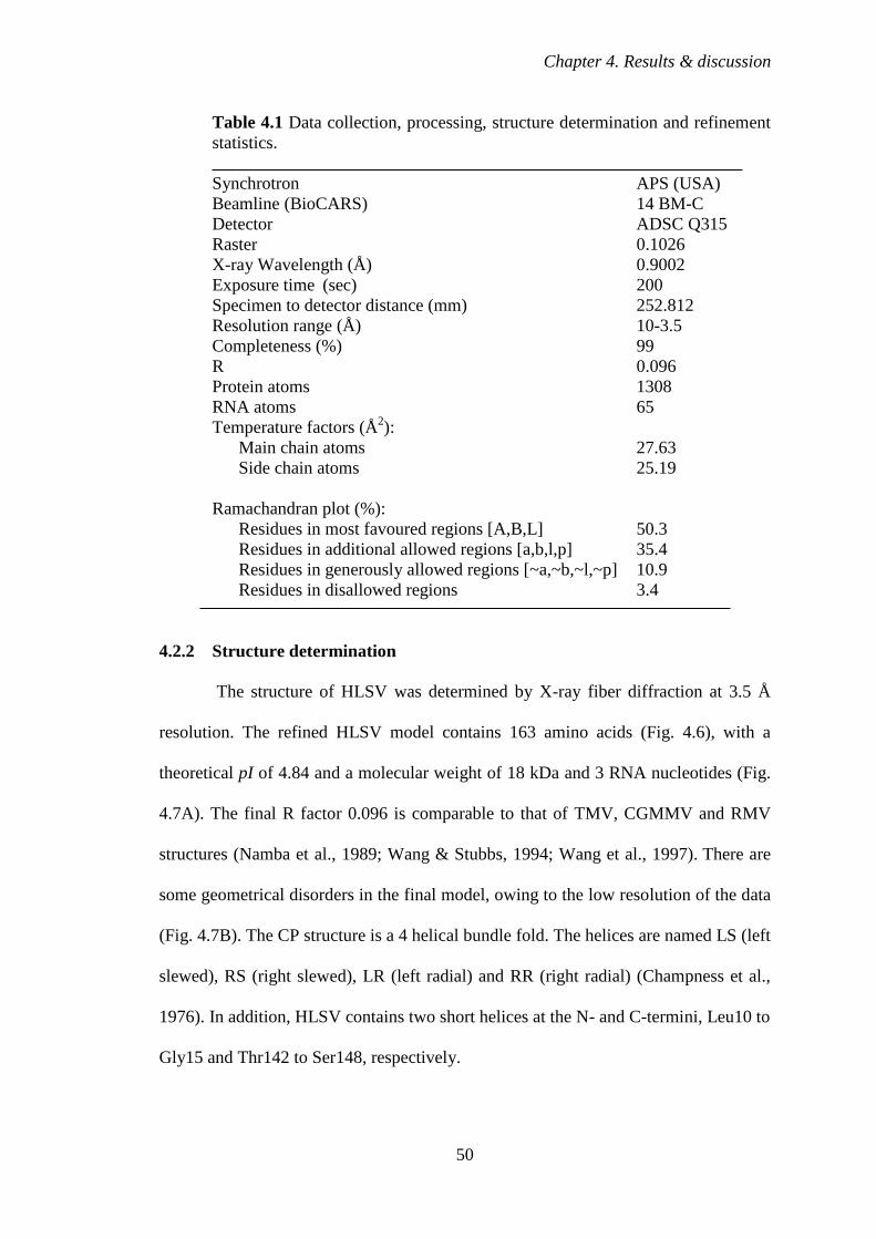
Chapter 4. Results & discussion
50
Table 4.1 Data collection, processing, structure determination and refinement
statistics.
Synchrotron APS (USA)
Beamline (BioCARS) 14 BM-C
Detector ADSC Q315
Raster 0.1026
X-ray Wavelength (Å) 0.9002
Exposure time (sec) 200
Specimen to detector distance (mm) 252.812
Resolution range (Å) 10-3.5
Completeness (%) 99
R 0.096
Protein atoms 1308
RNA atoms 65
Temperature factors (Å2):
Main chain atoms 27.63
Side chain atoms 25.19
Ramachandran plot (%):
Residues in most favoured regions [A,B,L] 50.3
Residues in additional allowed regions [a,b,l,p] 35.4
Residues in generously allowed regions [~a,~b,~l,~p] 10.9
Residues in disallowed regions 3.4
4.2.2 Structure determination
The structure of HLSV was determined by X-ray fiber diffraction at 3.5 Å
resolution. The refined HLSV model contains 163 amino acids (Fig. 4.6), with a
theoretical pI of 4.84 and a molecular weight of 18 kDa and 3 RNA nucleotides (Fig.
4.7A). The final R factor 0.096 is comparable to that of TMV, CGMMV and RMV
structures (Namba et al., 1989; Wang & Stubbs, 1994; Wang et al., 1997). There are
some geometrical disorders in the final model, owing to the low resolution of the data
(Fig. 4.7B). The CP structure is a 4 helical bundle fold. The helices are named LS (left
slewed), RS (right slewed), LR (left radial) and RR (right radial) (Champness et al.,
1976). In addition, HLSV contains two short helices at the N- and C-termini, Leu10 to
Gly15 and Thr142 to Ser148, respectively.
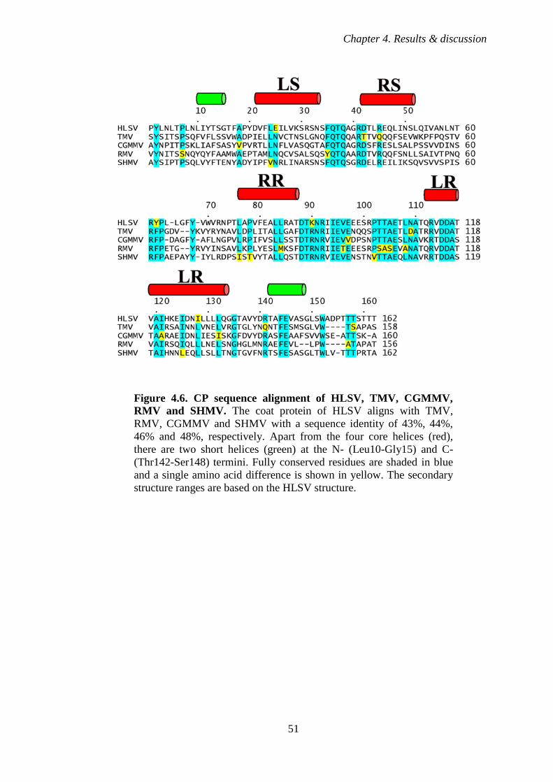
Chapter 4. Results & discussion
51
Figure 4.6. CP sequence alignment of HLSV, TMV, CGMMV,
RMV and SHMV. The coat protein of HLSV aligns with TMV,
RMV, CGMMV and SHMV with a sequence identity of 43%, 44%,
46% and 48%, respectively. Apart from the four core helices (red),
there are two short helices (green) at the N- (Leu10-Gly15) and C-
(Thr142-Ser148) termini. Fully conserved residues are shaded in blue
and a single amino acid difference is shown in yellow. The secondary
structure ranges are based on the HLSV structure.

Chapter 4. Results & discussion
52
(A) (B)
Figure 4.7. HLSV CP structure. (A) The ribbon diagram of the
central (0th
) subunit of the HLSV coat protein (CP), shown in blue.
Based on the earlier convention of Tobamovirus structure publications,
the first methionine is omitted and numbering starts from the second
residue (Pro1-Thr162). The α-helices are labeled as left and right
slewed (LS, RS) and left and right radial (LR, RR), a nomenclature
introduced by Champness et al., 1976 and the corresponding amino
acids number are marked. The 3 RNA nucleotides, GAA, are
represented as red sticks. The black circle represents the location of the
viral axis, which is perpendicular to the plane of the paper. (B) The
Ramachandran plot of the HLSV CP structure. The residues that
occupy the generously allowed and disallowed regions are labeled. The
overall statistics is comparable to that of structures at 3.5 Å resolution.
4.2.3 HLSV CP contains a kink in the LR α-helix
The CP structure of HLSV is compared to that of TMV, CGMMV and RMV,
(Fig. 4.8). The structure of HLSV CP is similar to that of TMV, CGMMV and RMV,
with some noticeable differences.
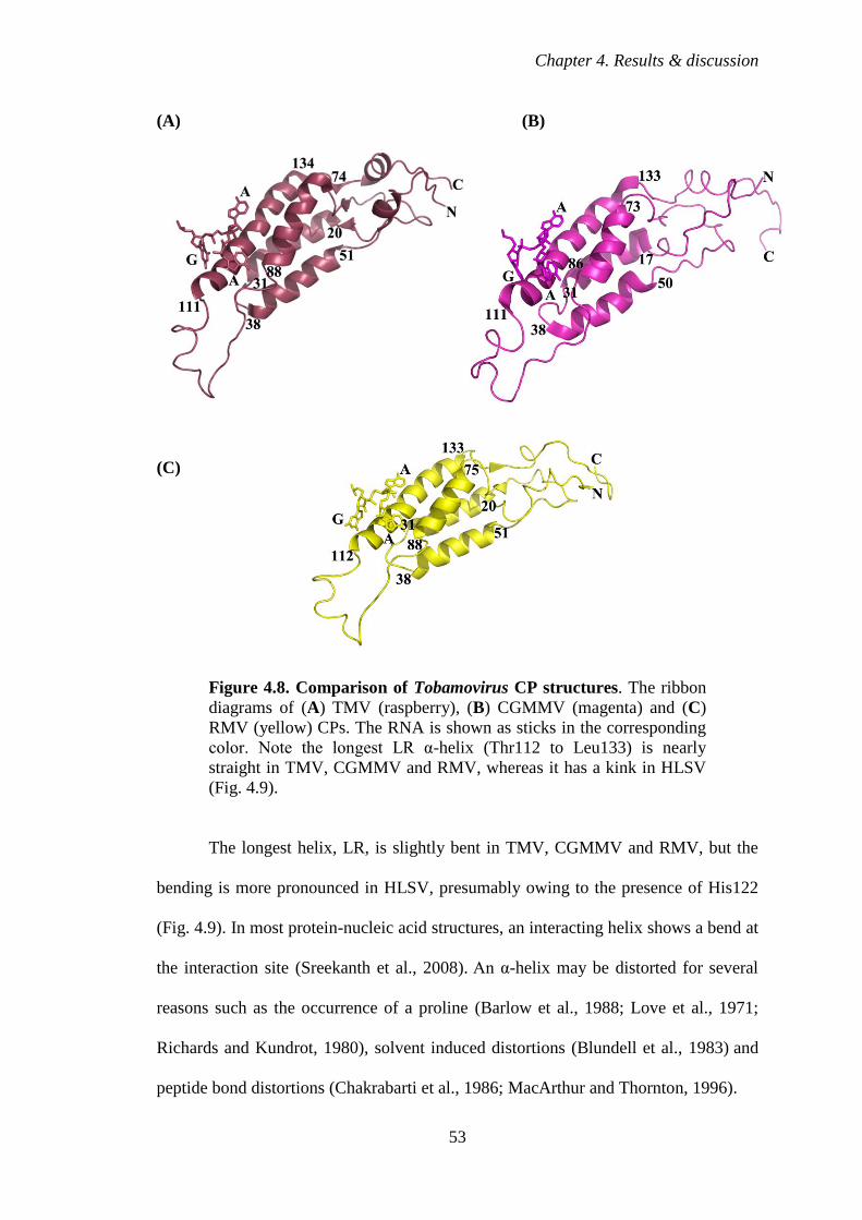
Chapter 4. Results & discussion
53
(A) (B)
(C)
Figure 4.8. Comparison of Tobamovirus CP structures. The ribbon
diagrams of (A) TMV (raspberry), (B) CGMMV (magenta) and (C)
RMV (yellow) CPs. The RNA is shown as sticks in the corresponding
color. Note the longest LR α-helix (Thr112 to Leu133) is nearly
straight in TMV, CGMMV and RMV, whereas it has a kink in HLSV
(Fig. 4.9).
The longest helix, LR, is slightly bent in TMV, CGMMV and RMV, but the
bending is more pronounced in HLSV, presumably owing to the presence of His122
(Fig. 4.9). In most protein-nucleic acid structures, an interacting helix shows a bend at
the interaction site (Sreekanth et al., 2008). An α-helix may be distorted for several
reasons such as the occurrence of a proline (Barlow et al., 1988; Love et al., 1971;
Richards and Kundrot, 1980), solvent induced distortions (Blundell et al., 1983) and
peptide bond distortions (Chakrabarti et al., 1986; MacArthur and Thornton, 1996).
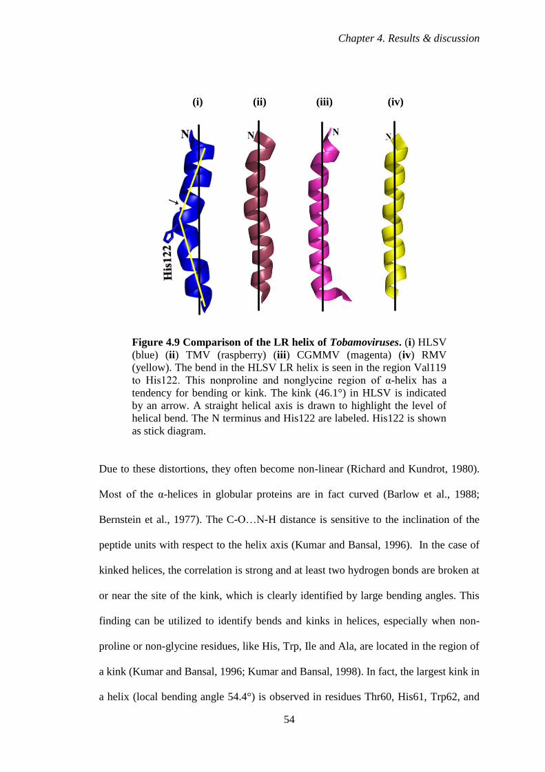
Chapter 4. Results & discussion
54
(i) (ii) (iii) (iv)
Figure 4.9 Comparison of the LR helix of Tobamoviruses. (i) HLSV
(blue) (ii) TMV (raspberry) (iii) CGMMV (magenta) (iv) RMV
(yellow). The bend in the HLSV LR helix is seen in the region Val119
to His122. This nonproline and nonglycine region of α-helix has a
tendency for bending or kink. The kink (46.1°) in HLSV is indicated
by an arrow. A straight helical axis is drawn to highlight the level of
helical bend. The N terminus and His122 are labeled. His122 is shown
as stick diagram.
Due to these distortions, they often become non-linear (Richard and Kundrot, 1980).
Most of the α-helices in globular proteins are in fact curved (Barlow et al., 1988;
Bernstein et al., 1977). The C-O…N-H distance is sensitive to the inclination of the
peptide units with respect to the helix axis (Kumar and Bansal, 1996). In the case of
kinked helices, the correlation is strong and at least two hydrogen bonds are broken at
or near the site of the kink, which is clearly identified by large bending angles. This
finding can be utilized to identify bends and kinks in helices, especially when non-
proline or non-glycine residues, like His, Trp, Ile and Ala, are located in the region of
a kink (Kumar and Bansal, 1996; Kumar and Bansal, 1998). In fact, the largest kink in
a helix (local bending angle 54.4°) is observed in residues Thr60, His61, Trp62, and
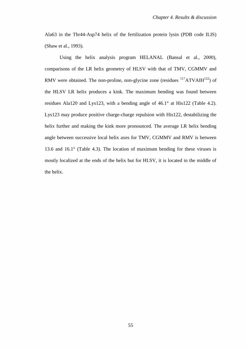
Chapter 4. Results & discussion
55
Ala63 in the Thr44-Asp74 helix of the fertilization protein lysin (PDB code ILIS)
(Shaw et al., 1993).
Using the helix analysis program HELANAL (Bansal et al., 2000),
comparisons of the LR helix geometry of HLSV with that of TMV, CGMMV and
RMV were obtained. The non-proline, non-glycine zone (residues 117
ATVAIH122
) of
the HLSV LR helix produces a kink. The maximum bending was found between
residues Ala120 and Lys123, with a bending angle of 46.1° at His122 (Table 4.2).
Lys123 may produce positive charge-charge repulsion with His122, destabilizing the
helix further and making the kink more pronounced. The average LR helix bending
angle between successive local helix axes for TMV, CGMMV and RMV is between
13.6 and 16.1° (Table 4.3). The location of maximum bending for these viruses is
mostly localized at the ends of the helix but for HLSV, it is located in the middle of
the helix.
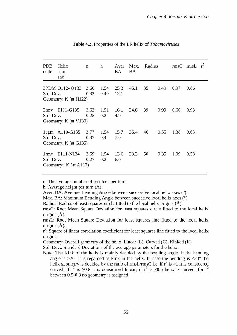
Chapter 4. Results & discussion
56
Table 4.2. Properties of the LR helix of Tobamoviruses
PDB Helix n h Aver Max. Radius rmsC rmsL r2
code start- BA BA
end
3PDM Q112- Q133 3.60 1.54 25.3 46.1 35 0.49 0.97 0.86
Std. Dev. 0.32 0.40 12.1
Geometry: K (at H122)
2tmv T111-G135 3.62 1.51 16.1 24.8 39 0.99 0.60 0.93
Std. Dev. 0.25 0.2 4.9
Geometry: K (at V130)
1cgm A110-G135 3.77 1.54 15.7 36.4 46 0.55 1.38 0.63
Std. Dev. 0.37 0.4 7.0
Geometry: K (at G135)
1rmv T111-N134 3.69 1.54 13.6 23.3 50 0.35 1.09 0.58
Std. Dev. 0.27 0.2 6.0
Geometry: K (at A117)
n: The average number of residues per turn.
h: Average height per turn (Å).
Aver. BA: Average Bending Angle between successive local helix axes (°).
Max. BA: Maximum Bending Angle between succesive local helix axes (°).
Radius: Radius of least squares circle fitted to the local helix origins (Å).
rmsC: Root Mean Square Deviation for least squares circle fitted to the local helix
origins (Å).
rmsL: Root Mean Square Deviation for least squares line fitted to the local helix
origins (Å).
r2: Square of linear correlation coefficient for least squares line fitted to the local helix
origins.
Geometry: Overall geometry of the helix, Linear (L), Curved (C), Kinked (K)
Std. Dev.: Standard Deviations of the average parameters for the helix.
Note: The Kink of the helix is mainly decided by the bending angle. If the bending
angle is >20° it is regarded as kink in the helix. In case the bending is <20° the
helix geometry is decided by the ratio of rmsL/rmsC i.e. if r2 is >1 it is considered
curved; if r2 is ≥0.8 it is considered linear; if r
2 is ≤0.5 helix is curved; for r
2
between 0.5-0.8 no geometry is assigned.
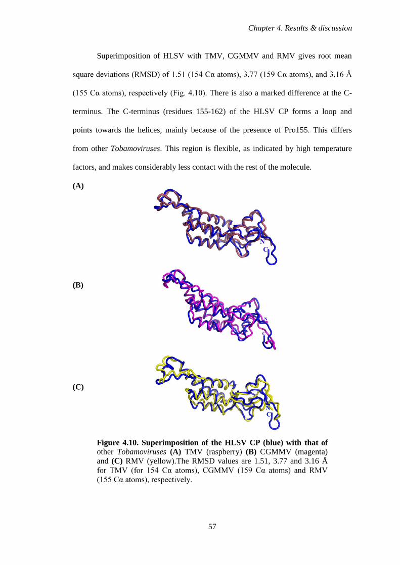
Chapter 4. Results & discussion
57
Superimposition of HLSV with TMV, CGMMV and RMV gives root mean
square deviations (RMSD) of 1.51 (154 Cα atoms), 3.77 (159 Cα atoms), and 3.16 Å
(155 Cα atoms), respectively (Fig. 4.10). There is also a marked difference at the C-
terminus. The C-terminus (residues 155-162) of the HLSV CP forms a loop and
points towards the helices, mainly because of the presence of Pro155. This differs
from other Tobamoviruses. This region is flexible, as indicated by high temperature
factors, and makes considerably less contact with the rest of the molecule.
(A)
(B)
(C)
Figure 4.10. Superimposition of the HLSV CP (blue) with that of other Tobamoviruses (A) TMV (raspberry) (B) CGMMV (magenta)
and (C) RMV (yellow).The RMSD values are 1.51, 3.77 and 3.16 Å
for TMV (for 154 Cα atoms), CGMMV (159 Cα atoms) and RMV
(155 Cα atoms), respectively.
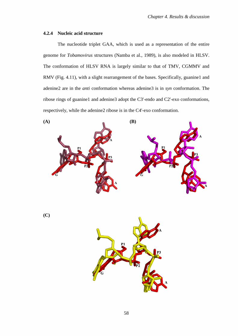
Chapter 4. Results & discussion
58
4.2.4 Nucleic acid structure
The nucleotide triplet GAA, which is used as a representation of the entire
genome for Tobamovirus structures (Namba et al., 1989), is also modeled in HLSV.
The conformation of HLSV RNA is largely similar to that of TMV, CGMMV and
RMV (Fig. 4.11), with a slight rearrangement of the bases. Specifically, guanine1 and
adenine2 are in the anti conformation whereas adenine3 is in syn conformation. The
ribose rings of guanine1 and adenine3 adopt the C3'-endo and C2'-exo conformations,
respectively, while the adenine2 ribose is in the C4'-exo conformation.
(A) (B)
(C)
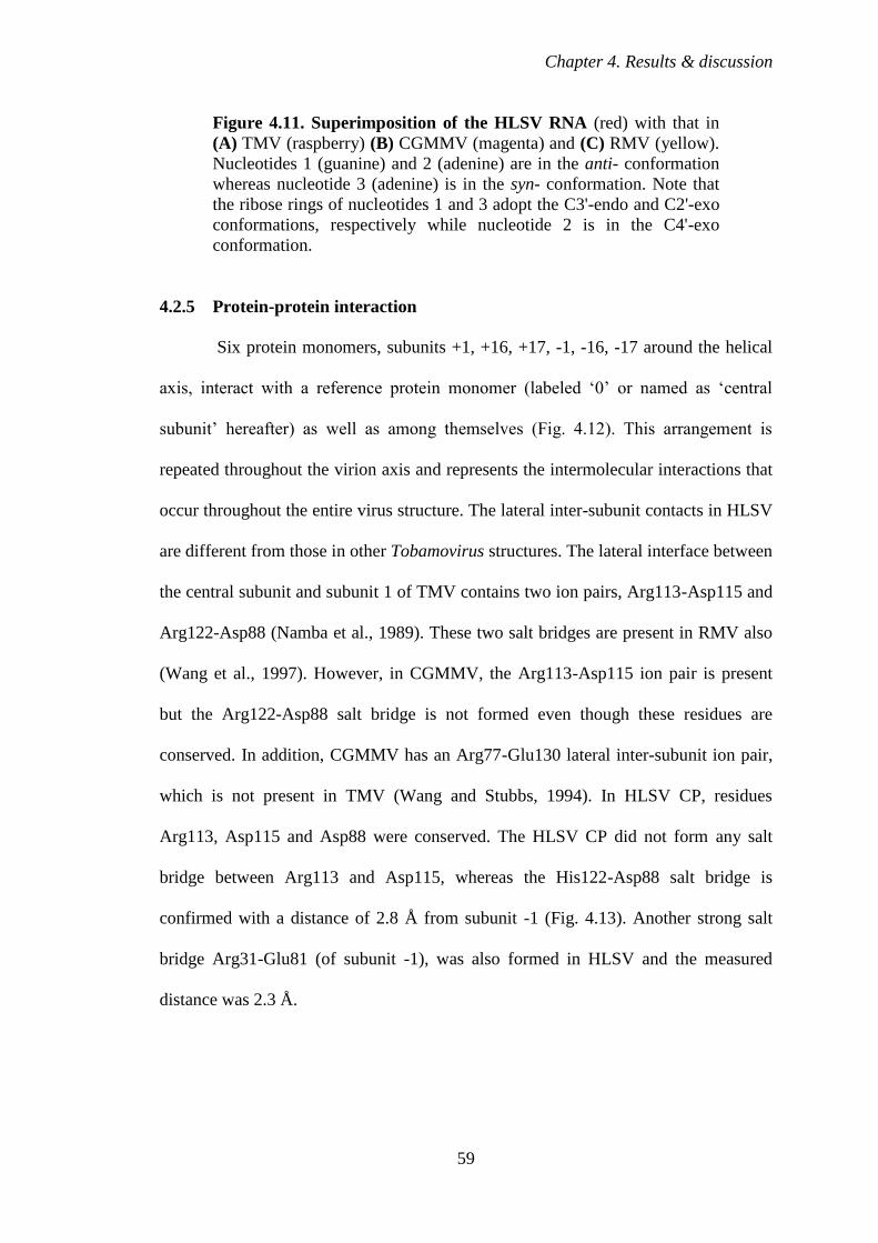
Chapter 4. Results & discussion
59
Figure 4.11. Superimposition of the HLSV RNA (red) with that in
(A) TMV (raspberry) (B) CGMMV (magenta) and (C) RMV (yellow).
Nucleotides 1 (guanine) and 2 (adenine) are in the anti- conformation
whereas nucleotide 3 (adenine) is in the syn- conformation. Note that
the ribose rings of nucleotides 1 and 3 adopt the C3'-endo and C2'-exo
conformations, respectively while nucleotide 2 is in the C4'-exo
conformation.
4.2.5 Protein-protein interaction
Six protein monomers, subunits +1, +16, +17, -1, -16, -17 around the helical
axis, interact with a reference protein monomer (labeled ‘0’ or named as ‘central
subunit’ hereafter) as well as among themselves (Fig. 4.12). This arrangement is
repeated throughout the virion axis and represents the intermolecular interactions that
occur throughout the entire virus structure. The lateral inter-subunit contacts in HLSV
are different from those in other Tobamovirus structures. The lateral interface between
the central subunit and subunit 1 of TMV contains two ion pairs, Arg113-Asp115 and
Arg122-Asp88 (Namba et al., 1989). These two salt bridges are present in RMV also
(Wang et al., 1997). However, in CGMMV, the Arg113-Asp115 ion pair is present
but the Arg122-Asp88 salt bridge is not formed even though these residues are
conserved. In addition, CGMMV has an Arg77-Glu130 lateral inter-subunit ion pair,
which is not present in TMV (Wang and Stubbs, 1994). In HLSV CP, residues
Arg113, Asp115 and Asp88 were conserved. The HLSV CP did not form any salt
bridge between Arg113 and Asp115, whereas the His122-Asp88 salt bridge is
confirmed with a distance of 2.8 Å from subunit -1 (Fig. 4.13). Another strong salt
bridge Arg31-Glu81 (of subunit -1), was also formed in HLSV and the measured
distance was 2.3 Å.
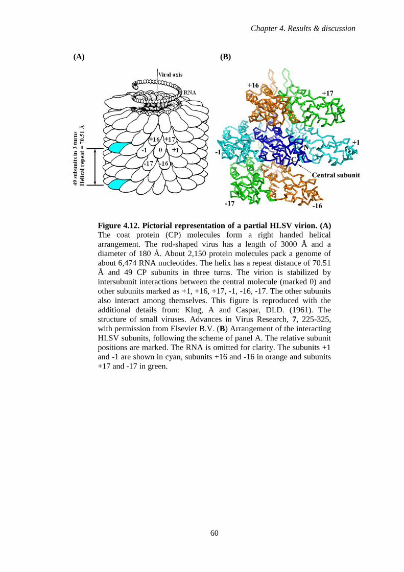
Chapter 4. Results & discussion
60
(A) (B)
Figure 4.12. Pictorial representation of a partial HLSV virion. (A)
The coat protein (CP) molecules form a right handed helical
arrangement. The rod-shaped virus has a length of 3000 Å and a
diameter of 180 Å. About 2,150 protein molecules pack a genome of
about 6,474 RNA nucleotides. The helix has a repeat distance of 70.51
Å and 49 CP subunits in three turns. The virion is stabilized by
intersubunit interactions between the central molecule (marked 0) and
other subunits marked as +1, +16, +17, -1, -16, -17. The other subunits
also interact among themselves. This figure is reproduced with the
additional details from: Klug, A and Caspar, DLD. (1961). The
structure of small viruses. Advances in Virus Research, 7, 225-325,
with permission from Elsevier B.V. (B) Arrangement of the interacting
HLSV subunits, following the scheme of panel A. The relative subunit
positions are marked. The RNA is omitted for clarity. The subunits +1
and -1 are shown in cyan, subunits +16 and -16 in orange and subunits
+17 and -17 in green.

Chapter 4. Results & discussion
61
Figure 4.13. HLSV protein-protein (CP) interaction. Hydrogen
bonding between His123 and Asp88 (of subunit -1) stabilizes the loop
adjacent to RR helix and gives significant stability to the interaction
CP at the lateral interface. The N-termini of the subunits are labeled.
Asp88 and His122 are shown as sticks. Subunits 0 and -1 are colored
blue and cyan, respectively.
Axial interactions of CP subunits are not the same among TMV, CGMMV and
RMV (Namba et al., 1989; Wang and Stubbs, 1994; Wang et al., 1997). In TMV,
there are two ion pairs in the axial interface between the central subunit and its
neighboring subunit 17 along the virion helix: Glu50-Arg134 and Glu95-Arg112
(Namba et al., 1989). CGMMV also has two axial intersubunit ion pairs: Arg122-
Asp42 and Lys134-Asp57 from subunit 17 (Wang and Stubbs, 1994). The Arg122-
Asp42 contact is also present in RMV (Wang et al., 1997; Wang and Stubbs, 1994).
In HLSV, Glu95 is conserved but amino acid residue at position 112 is a glutamine,
as in RMV. The Glu50-Arg134 ion pair of TMV is not found in HLSV. Instead, the
two axial salt bridges are Arg45-Asp126 and Asp116-Arg92 from subunit +17 and -
17, respectively. Apart from salt bridge formation, Arg 92 is involved in protein-RNA
interaction in HLSV to stabilize the virus particle.

Chapter 4. Results & discussion
62
Carboxyl-carboxylate interactions form the main driving force for virus
disassembly in TMV, CGMMV and RMV (Wang et al., 1998; Caspar, 1963). In
TMV, there are two carboxyl-carboxylate pairs: high radius, involving axial Glu50-
Asp77 (subunit 16) and low radius, involving lateral inter-subunit interactions
Glu95/Glu97/Asp109-Glu106 (subunit 1). The influence of carboxylates in viral
disassembly has been well studied in TMV (Culver et al., 1995; Lu et al., 1996). In
addition, in HLSV, intersubunit carboxyl-carboxylate interactions are also observed
(Figs. 4.14, 4.15 and Table 4.3).
Figure 4.14. Stereo view of HLSV axial intersubunit carboxyl-
carboxylate interactions at high radius region. Glu46 with Asp126
(of subunit +17 at 5.1 Å), Glu46 with Glu81 (of subunit +16 at 6.3 Å),
and Glu81 with Glu46 and Asp126. Also, some important salt bridges
are: Arg45 with Glu46 (4.0 Å) and Asp42 (3.1 Å); Arg41 with Asp42
(at 4.0 Å) which stabilizes the RS helix. The salt bridges are shown in
green dashed lines whereas carboxyl-carboxylate interactions are
shown in red dashed lines.
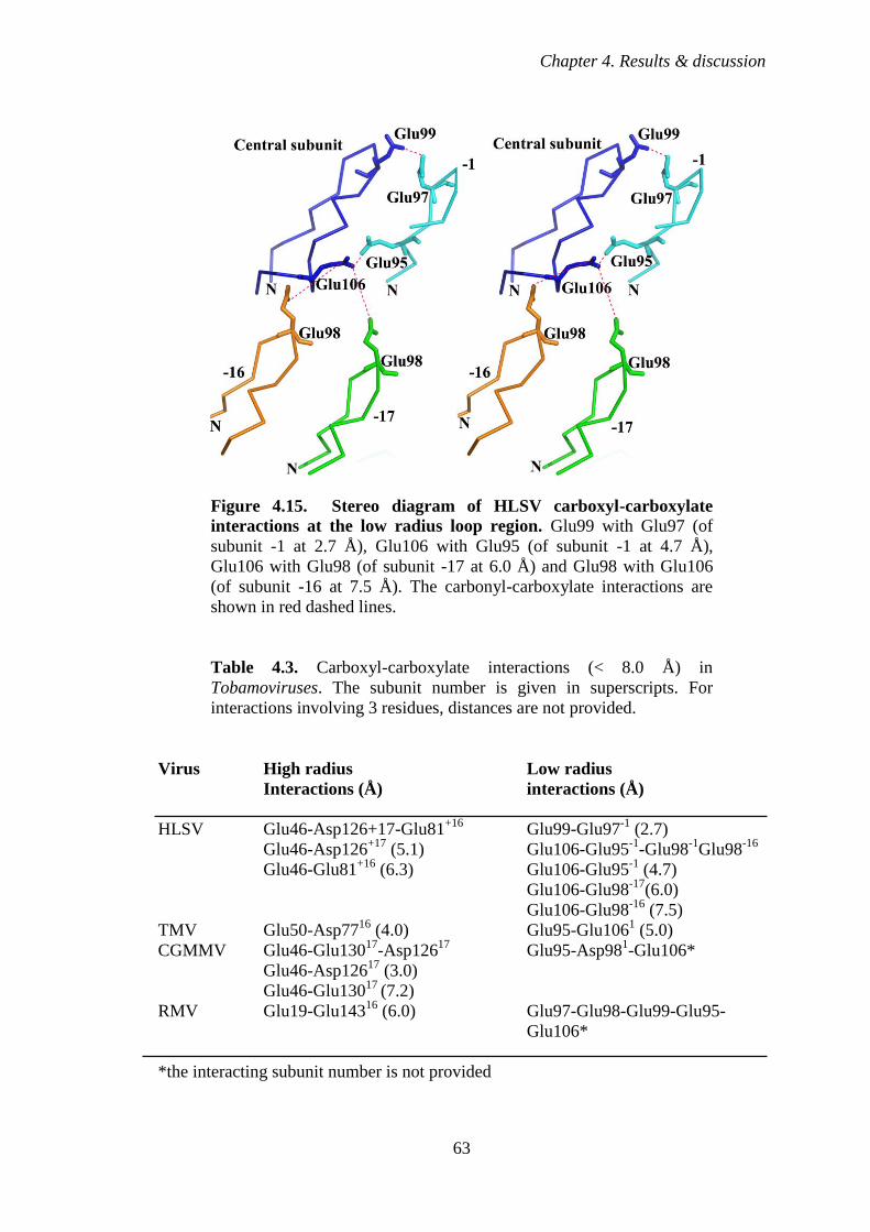
Chapter 4. Results & discussion
63
Figure 4.15. Stereo diagram of HLSV carboxyl-carboxylate
interactions at the low radius loop region. Glu99 with Glu97 (of
subunit -1 at 2.7 Å), Glu106 with Glu95 (of subunit -1 at 4.7 Å),
Glu106 with Glu98 (of subunit -17 at 6.0 Å) and Glu98 with Glu106
(of subunit -16 at 7.5 Å). The carbonyl-carboxylate interactions are
shown in red dashed lines.
Table 4.3. Carboxyl-carboxylate interactions (< 8.0 Å) in
Tobamoviruses. The subunit number is given in superscripts. For
interactions involving 3 residues, distances are not provided.
Virus High radius Low radius
Interactions (Å) interactions (Å)
HLSV Glu46-Asp126+17-Glu81+16
Glu99-Glu97-1
(2.7)
Glu46-Asp126+17
(5.1) Glu106-Glu95-1
-Glu98-1
Glu98-16
Glu46-Glu81+16
(6.3) Glu106-Glu95-1
(4.7)
Glu106-Glu98-17
(6.0)
Glu106-Glu98-16
(7.5)
TMV Glu50-Asp7716
(4.0) Glu95-Glu1061 (5.0)
CGMMV Glu46-Glu13017
-Asp12617
Glu95-Asp981-Glu106*
Glu46-Asp12617
(3.0)
Glu46-Glu13017
(7.2)
RMV Glu19-Glu14316
(6.0) Glu97-Glu98-Glu99-Glu95-
Glu106*
*the interacting subunit number is not provided
Chapter 4. Results & discussion
64
4.2.6 Protein-nucleic acid interaction
The OAS of HLSV is predicted to be located in the CP ORF (Srinivasan et al.,
2002; Srinivasan et al., 2005). It contains GAA repeats, similar to TMV, CGMMV
and RMV. The hydrogen bonding interactions between the nucleotide bases and CP at
the OAS in HLSV assembly are different from those in TMV and CGMMV, but
identical to those in RMV. In CGMMV, guanine1 forms a hydrogen bond with
Arg113. Wang and Stubbs (1994) argue that any base at this position would make a
hydrogen bond with Arg113 and particularly, a guanine at this position would make
an additional hydrogen bond with the side chain oxygen of Gln36. However, in
HLSV, Arg113 does not form any hydrogen bond. Instead, there is a hydrogen bond
between guanine1 and Asp115, which is also observed in RMV (Wang et al., 1997).
This interaction has been proposed to contribute to nucleotide base specificity. In
TMV, Arg122 plays an important role in selection and stabilization of guanine1
during virus assembly (Namba et al., 1989). The same was also proposed for
CGMMV and RMV (Wang et al., 1997). In HLSV, the residue corresponding to
Arg122 is His122 and its adjacent residue Lys123 stabilizes the phosphate of
guanine1 (Fig. 4.16). In CGMMV, the charges on the RNA phosphate groups are
neutralized by the interactions with neighboring side chains: Arg41 forms a hydrogen
bond with phosphate1, Lys112 and Asn91 form hydrogen bonds with phosphate2 and
the charge on phosphate3 is partly stabilized by the dipole of the RS helix (with the
NH groups of Gln38 and Ala39, both well placed to form hydrogen bonds with the
phosphate oxygen atoms) (Wang and Stubbs, 1994). In HLSV, the charges on
phosphate2 and phosphate3 are neutralized by Arg92 from the axial position at
neighboring subunit -17, which is positioned at a distance of 3.8 Å and 3.7 Å,
respectively (Fig. 4.17). Furthermore, Thr37 of subunit -16 makes a hydrogen bond
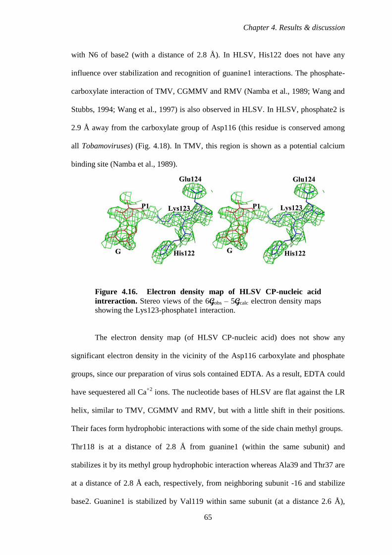
Chapter 4. Results & discussion
65
with N6 of base2 (with a distance of 2.8 Å). In HLSV, His122 does not have any
influence over stabilization and recognition of guanine1 interactions. The phosphate-
carboxylate interaction of TMV, CGMMV and RMV (Namba et al., 1989; Wang and
Stubbs, 1994; Wang et al., 1997) is also observed in HLSV. In HLSV, phosphate2 is
2.9 Å away from the carboxylate group of Asp116 (this residue is conserved among
all Tobamoviruses) (Fig. 4.18). In TMV, this region is shown as a potential calcium
binding site (Namba et al., 1989).
Figure 4.16. Electron density map of HLSV CP-nucleic acid
intreraction. Stereo views of the 6Gobs – 5Gcalc electron density maps
showing the Lys123-phosphate1 interaction.
The electron density map (of HLSV CP-nucleic acid) does not show any
significant electron density in the vicinity of the Asp116 carboxylate and phosphate
groups, since our preparation of virus sols contained EDTA. As a result, EDTA could
have sequestered all Ca+2
ions. The nucleotide bases of HLSV are flat against the LR
helix, similar to TMV, CGMMV and RMV, but with a little shift in their positions.
Their faces form hydrophobic interactions with some of the side chain methyl groups.
Thr118 is at a distance of 2.8 Å from guanine1 (within the same subunit) and
stabilizes it by its methyl group hydrophobic interaction whereas Ala39 and Thr37 are
at a distance of 2.8 Å each, respectively, from neighboring subunit -16 and stabilize
base2. Guanine1 is stabilized by Val119 within same subunit (at a distance 2.6 Å),
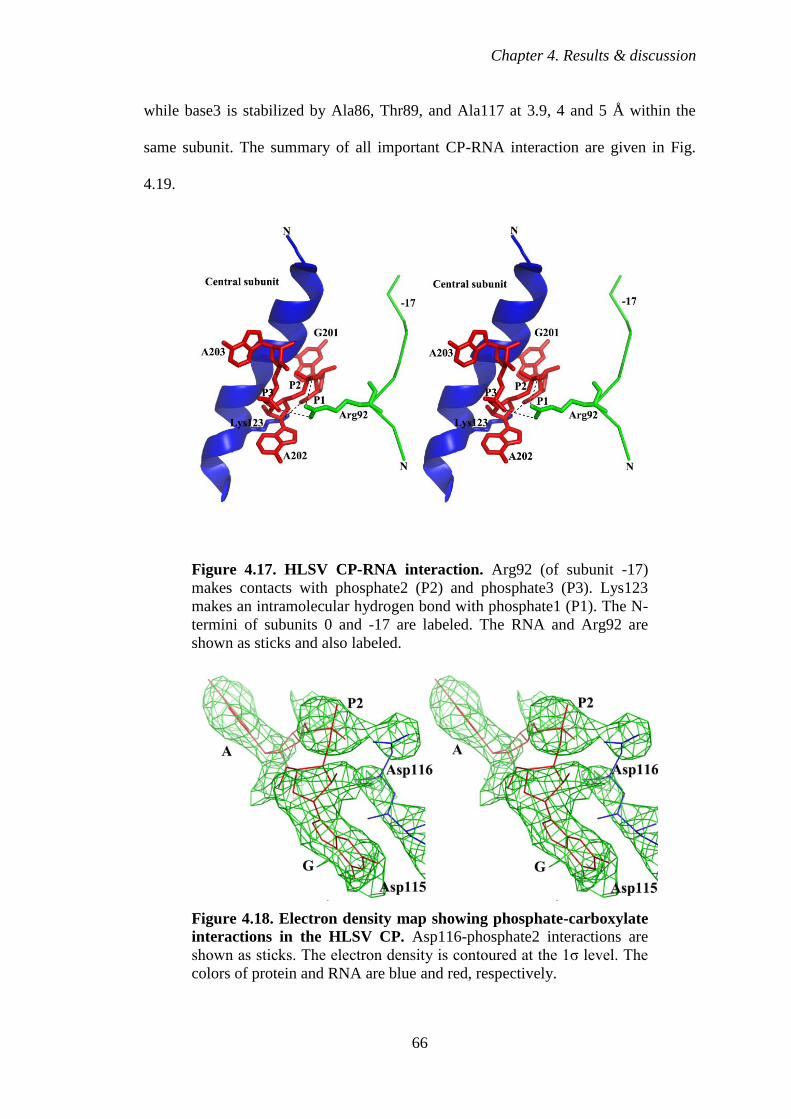
Chapter 4. Results & discussion
66
while base3 is stabilized by Ala86, Thr89, and Ala117 at 3.9, 4 and 5 Å within the
same subunit. The summary of all important CP-RNA interaction are given in Fig.
4.19.
Figure 4.17. HLSV CP-RNA interaction. Arg92 (of subunit -17)
makes contacts with phosphate2 (P2) and phosphate3 (P3). Lys123
makes an intramolecular hydrogen bond with phosphate1 (P1). The N-
termini of subunits 0 and -17 are labeled. The RNA and Arg92 are
shown as sticks and also labeled.
Figure 4.18. Electron density map showing phosphate-carboxylate
interactions in the HLSV CP. Asp116-phosphate2 interactions are
shown as sticks. The electron density is contoured at the 1σ level. The
colors of protein and RNA are blue and red, respectively.
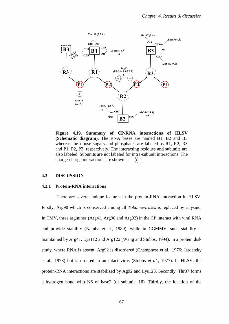
Chapter 4. Results & discussion
67
Figure 4.19. Summary of CP-RNA interactions of HLSV
(Schematic diagram). The RNA bases are named B1, B2 and B3
whereas the ribose sugars and phosphates are labeled as R1, R2, R3
and P1, P2, P3, respectively. The interacting residues and subunits are
also labeled. Subunits are not labeled for intra-subunit interactions. The
charge-charge interactions are shown as
4.3 DISCUSSION
4.3.1 Protein-RNA interactions
There are several unique features in the protein-RNA interaction in HLSV.
Firstly, Arg90 which is conserved among all Tobamoviruses is replaced by a lysine.
In TMV, three arginines (Arg41, Arg90 and Arg92) in the CP interact with viral RNA
and provide stability (Namba et al., 1989), while in CGMMV, such stability is
maintained by Arg41, Lys112 and Arg122 (Wang and Stubbs, 1994). In a protein disk
study, where RNA is absent, Arg92 is disordered (Champness et al., 1976; Jardetzky
et al., 1978) but is ordered in an intact virus (Stubbs et al., 1977). In HLSV, the
protein-RNA interactions are stabilized by Ag92 and Lys123. Secondly, Thr37 forms
a hydrogen bond with N6 of base2 (of subunit -16). Thirdly, the location of the
.
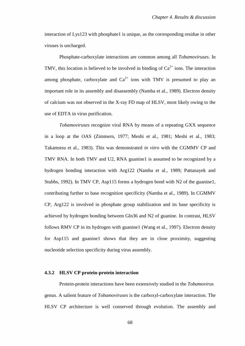
Chapter 4. Results & discussion
68
interaction of Lys123 with phosphate1 is unique, as the corresponding residue in other
viruses is uncharged.
Phosphate-carboxylate interactions are common among all Tobamoviruses. In
TMV, this location is believed to be involved in binding of Ca2+
ions. The interaction
among phosphate, carboxylate and Ca2+
ions with TMV is presumed to play an
important role in its assembly and disassembly (Namba et al., 1989). Electron density
of calcium was not observed in the X-ray FD map of HLSV, most likely owing to the
use of EDTA in virus purification.
Tobamoviruses recognize viral RNA by means of a repeating GXX sequence
in a loop at the OAS (Zimmern, 1977; Meshi et al., 1981; Meshi et al., 1983;
Takamstsu et al., 1983). This was demonstrated in vitro with the CGMMV CP and
TMV RNA. In both TMV and U2, RNA guanine1 is assumed to be recognized by a
hydrogen bonding interaction with Arg122 (Namba et al., 1989; Pattanayek and
Stubbs, 1992). In TMV CP, Asp115 forms a hydrogen bond with N2 of the guanine1,
contributing further to base recognition specificity (Namba et al., 1989). In CGMMV
CP, Arg122 is involved in phosphate group stabilization and its base specificity is
achieved by hydrogen bonding between Gln36 and N2 of guanine. In contrast, HLSV
follows RMV CP in its hydrogen with guanine1 (Wang et al., 1997). Electron density
for Asp115 and guanine1 shows that they are in close proximity, suggesting
nucleotide selection specificity during virus assembly.
4.3.2 HLSV CP protein-protein interaction
Protein-protein interactions have been extensively studied in the Tobamovirus
genus. A salient feature of Tobamoviruses is the carboxyl-carboxylate interaction. The
HLSV CP architecture is well conserved through evolution. The assembly and
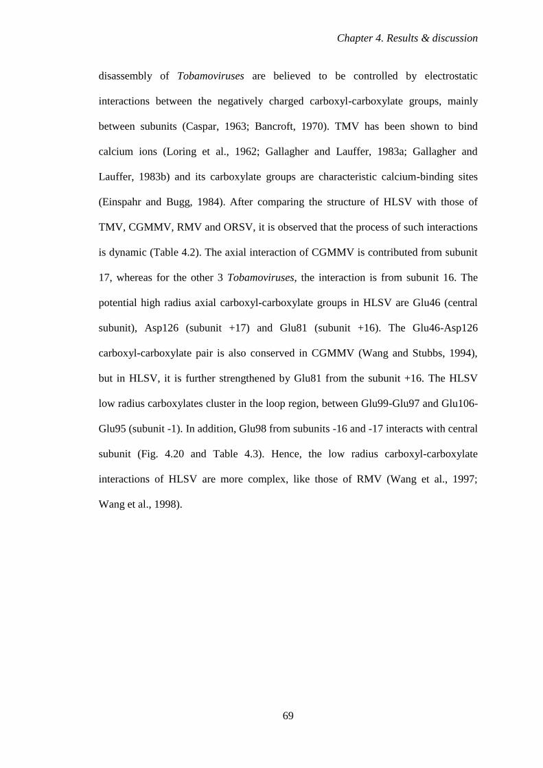
Chapter 4. Results & discussion
69
disassembly of Tobamoviruses are believed to be controlled by electrostatic
interactions between the negatively charged carboxyl-carboxylate groups, mainly
between subunits (Caspar, 1963; Bancroft, 1970). TMV has been shown to bind
calcium ions (Loring et al., 1962; Gallagher and Lauffer, 1983a; Gallagher and
Lauffer, 1983b) and its carboxylate groups are characteristic calcium-binding sites
(Einspahr and Bugg, 1984). After comparing the structure of HLSV with those of
TMV, CGMMV, RMV and ORSV, it is observed that the process of such interactions
is dynamic (Table 4.2). The axial interaction of CGMMV is contributed from subunit
17, whereas for the other 3 Tobamoviruses, the interaction is from subunit 16. The
potential high radius axial carboxyl-carboxylate groups in HLSV are Glu46 (central
subunit), Asp126 (subunit +17) and Glu81 (subunit +16). The Glu46-Asp126
carboxyl-carboxylate pair is also conserved in CGMMV (Wang and Stubbs, 1994),
but in HLSV, it is further strengthened by Glu81 from the subunit +16. The HLSV
low radius carboxylates cluster in the loop region, between Glu99-Glu97 and Glu106-
Glu95 (subunit -1). In addition, Glu98 from subunits -16 and -17 interacts with central
subunit (Fig. 4.20 and Table 4.3). Hence, the low radius carboxyl-carboxylate
interactions of HLSV are more complex, like those of RMV (Wang et al., 1997;
Wang et al., 1998).
Chapter 4. Results & discussion
70
Figure 4.20. Surface density diagram of low radius carboxyl-
carboxylate interactions of HLSV coat protein (CP). HLSV CP
shows carboxyl-carboxylate interactions between the central and
subunits -1, -16 and -17, as in Fig. 4.12. The red and blue colors
represent a charge distribution of -55.5 and +55.5 KT/e, respectively.
4.3.3 Other structural features of HLSV
In the CGMMV CP, its residue 87 is a threonine and it contributes to the
shortening of the RR α-helix significantly. In HLSV, Thr87 breaks the corresponding
helix. Its Thr75 further shortens the RR helix, which is the shortest when compared to
the TMV, CGMMV and RMV structures. In addition, the kink in the LR helix of
HLSV is increased by His122. This is a major structural difference.
Understanding the recognition of GAA by the CP is very important to
appreciate RNA specificity and virus assembly. Asp115 and Arg122 form guanine
specific hydrogen bonds in TMV and U2, but not in CGMMV (Namba et al., 1989;
Pattanayek & Stubbs, 1992). The structural basis of RNA guanine1 recognition is less
obvious in CGMMV (Wang and Stubbs, 1994) and RMV. Arg122 is believed to be
the key player in base recognition during virus assembly both in CGMMV and RMV
(Wang et al., 1997). The considerable flexibility of the arginine side chain would
Chapter 4. Results & discussion
71
easily allow it to extend during assembly process (Wang and Stubbs 1994). This
otherwise conserved Arg122 in Tobamoviruses is changed to His122 in HLSV and
SHMV. The presence of two consecutive positively charged amino acids at this
position is unique to HLSV. His122 is involved in a salt bridge and Lys123 is
involved in a base independent phosphate interaction. In TMV and CGMMV,
guanine1 selection during assembly is achieved by hydrogen bonding with viral CP
Arg122 and Gln36, respectively (Namba et al., 1989; Wang and Stubbs, 1994).
However, guanine1 selection in HLSV is achieved by Asp115, as in RMV (Wang et
al., 1997). More studies should be undertaken to understand the importance of
guanine1 recognition and viral assembly.
The four residues at positions 25, 62, 90 and 129 are conserved in all other
Tobamoviruses, except for HLSV (Fig. 4.6). Glu25 in the HLSV CP makes an
intramolecular salt bridge with Lys29 and stabilizes the RS α-helix. Also, Tyr62
functions only as a hydrophobic residue and fails to execute its second role as a
charge stabilizer, as it is not a part of any helix and no charge residues are seen in its
vicinity. Lys90 and Ile128 replace Arg and Leu in other Tobamoviruses, respectively.
Lys90 is at a distance of 5.9 Å from guanine1 and probably it could be one of the
hydrogen bonding candidates during viral assembly before attaining a fixed
conformation in a virion, similar to the proposed method for Arg122 in CGMMV and
RMV (Wang et al., 1997). This is only a speculation and needs validation with
mutational experiments. Ile128 is part of the LR helix and it is likely to assume the
same role of Leu128 in other viruses.
In conclusion, the kink at the LR helix of HLSV is a unique feature among
Tobamoviruses. Tobamovirus protein-nucleic acid base specificity interaction during
virus assembly is proposed to be achieved by viral CP Arg122 residue (Namba et al.,

Chapter 4. Results & discussion
72
1989; Wang et al., 1997). This position is occupied by a histidine in HLSV which is
not close to any of the RNA bases. However, it interacts with Asp88 of the
neighboring lateral subunit 1 and stabilizes the less ordered loop through a salt bridge.
Furthermore, Lys123 in HLSV is close to phosphate1, suggesting its possible
involvement with base recognition. However, the exact nature and mechanism of
RNA recognition by Tobamoviruses are yet to be determined.
The intersubunit carboxyl-carboxylate interactions of HLSV partially
resemble those of CGMMV, but remain distinct from other Tobamoviruses. The
carboxyl-carboxylate interactions include involvement of both subunit +16 and +17 in
HLSV. This is another difference observed, as the high radius interactions are usually
confined to the axial subunit, either 16 or 17 in other Tobamoviruses. This unique
axial contribution shows more intermolecular carboxyl-carboxylate interactions
occurring in the HLSV CP. The low radius inter-subunit interactions in other
Tobamoviruses are confined only to the lateral inter-subunit direction but in HLSV
they extend in both lateral and axial directions.
4.4 FUTURE DIRECTIONS
Viruses are nucleoprotein complexes. Due to their small size and rapid
replication rate, they are very useful in biotechnological applications. A virus CP has
the capacity to pack other molecules, apart from nucleic acids, so that it could be
exploited as a potential drug packaging protein nano cage. (Douglas & Young, 1998;
Ren et al., 2006). These viruses like particles (VLPs) can be used as a novel delivery
platform for drugs, for example Doxorubicin, an anti-cancer drug (Ren et al., 2007).
Gallie and his team are able to pack multiply dispersed copies of RNA
containing the OAS to form pseudovirus like particles (Gallie et al., 1987a). In an in

Chapter 4. Results & discussion
73
vitro study, a short OAS, located in the MP gene, was shown to nucleate
encapsidation of the 6395 nucleotide long genome by the TMV CP (Butler, 1984;
Lomonossoff & Wilson, 1985). Agrobacterium tumefaciens was used to incorporate
the TMV OAS and was found to assemble into a stable 'pseudovirus' like particle in
vivo during systemic infection by TMV (as a helper). These results may further be
extended for packaging foreign genes into a virus. As a route to protect, accumulate
and recover a specific mRNA in vivo, in transgenic plant cells, this new method may
be useful in developmental and plant molecular biology (Sleat et al., 1988; Sleat et al.,
1986).
To control the mosquito Aedes, trypsin-modulating oostatic factor was
expressed successfully on the virion of TMV as a potential larvicide (Borovsky et al.,
2006). Similar approach can be adopted with our structural details of the HLSV and
may be used as a biocontrol in future. TMV has also been used in the development of
vaccines. The Canine oral papillomavirus L2 protein, displayed on the surface of the
TMV CP along with streptavidin, was found to be more immunogenic than uncoupled
antigen when tested in mice (Sleat et al., 1986). TMV was also used to display protein
A, which was densely packed as nanoparticles (>2,100 copies per viral particle) on its
virion. This can be a new approach as an immunoadsorbent to purify monoclonal
antibodies (mAb) with less cost (Werner et al., 2006). With the structural details of
HLSV we can also adopt similar approach and carry out more experiments to see its
suitability as an immunoadsorbent. Furthermore, a new TMV based vector has been
used to produce systemically an angiotensin-I converting enzyme inhibitor in
transgenic tobacco and tomato (Hamamoto et al., 1993). In another study two
epitopes of the foot and mouth disease virus are successfully expressed by a TMV
based vector (Wu et al., 2003). This study can be useful in developing vaccines
Chapter 4. Results & discussion
74
against many pathogenic viruses and bacteria. The full length clone of CGMMV was
used to express Hepatitis B surface antigen (Ooi et al., 2006). The study showed
threefold increase in the level of anti HBsAg immunoglobulin, suggesting the possible
application of new chimeric virus as an effective Hepatitis B vaccine. HLSV can also
be exploited in similar studies.
We believe that with the current structure of HLSV, we can do more functional
studies to better understand the virus structure function relation. We can ask some
basic questions like for example what will happen to virus if the we mutate the
His122 of the CP? Will the kink still exist if we mutant His122? What kind of amino
acid substitution will increase the degree of the bent? Will the virus LR helix remain
structurally the same? Will the virus be as infective as the wild type? Can the
knowledge be used in designing nano-molecule for anti-cancer drug delivery?
References
75
REFERENCES
Alonso, E., Garcia-Luque, I., De-la Cruz, A., Wicke, B., Avila-Rincon, M. J., Serra,
M. T., Castresana, C. & Diaz-Ruiz, J. R. (1991). Nucleotide sequence of the
genomic RNA of Pepper mild mottle virus, a resistance-breaking Tobamovirus in
pepper. J. Gen. Virol. 72, 2875-2884.
Altschuh. D., Lesk, A. M., Bloomer, A. C. & Klug, A. (1987). Correlations of co-
ordinated amino acid substitutions with function in viruses related to TMV. J.
Mol. Biol. 193, 693-707.
Argos, P. (1988). A sequence motif in many polymerases. Nucl. Acids Res. 16, 9909-
9916.
Artymiuk, P. J. & Blake, C. C. F. (1981). Refinement of human lysozyme at 1.5 Å
resolution analysis of non-bonded and hydrogen-bond interactions. J. Mol. Biol.
152, 737-762.
Asurmendi, S., Berg, R. H., Koo, J. C. & Beachy, R. N. (2004). Coat protein regulates
formation of replication complexes during TMV infection. Proc. Natl. Acad. Sci.
USA. 101, 1415-1420.
Bancroft, J.R. (1970). The self-assembly of spherical plant viruses. Adv. Virus Res.
16, 99-134.
Bansal, M., Kumar, S. & Velavan, R. (2000). HELANAL: a program to characterize
helix geometry in proteins. J. Biomol. Struct. Dyn. 17, 811–819.
Barlow, D. & Thornton, J. M. (1988). Helix geometry in protein. J. Mol. Biol. 201,
601-619.
Bawden, F. C., Pirie, N. W., Bernal, J. D. & Fankuchen, I. (1936). Liquid crystalline
substances from virus-infected plants. Nature 138, 1051-1052.
Beijerinck, M. W. (1898). Concerning a contagium vivum fluidium as a cause of the
spot-disease of tobacco leaves. Reprint from: Phytopathology Classics, Number 7.
1942.
Bernal, J. D. & Fankuchen, I. (1941). X-ray and crystallographic studies of plant virus
preparations. III. J. Gen. Physiol. 25, 147-165.
Bernstein, F. C., Koetzle, T. F., Williams, G. J. B., Jr-Meyer, E. F., Brice, M. D.,
Rodgers, J. R., Kennard, O., Shimanouchi T. & Tasumi, M. (1977). The protein
data bank: A computer-based archival file for macromolecular structures. J. Mol.
Biol. 112, 535-542.
Bhyravbhatla, B., Watowich, S. & Caspar, D. L. D. (1998). Refined atomic model of
the four-layer aggregate of the TMV coat protein at 2.4 Å resolution. Biophys. J.
74, 604-615.
References
76
Bian, W., Wang, H., McCullough, I. & Stubbs, G. (2006). WCEN: a computer
program for initial processing of fiber diffraction patterns J. Appl. Crystallogr. 39,
752-756.
Bloomer, A. C. & Butler, P. J. G. (1986). The Plant Viruses (Van Regenmortel, M.H.
V., Fraenkel-Conrat, H., Eds.) Vol. 2, pp 19-57, Plenum, New York.
Bloomer, A. C., Champness, J. N., Bricogne, G., Staden, R. & Klug, A. (1978).
Protein disk of TMV at 2.8 Å resolution showing the interactions within and
between subunits. Nature 276, 362-368.
Blundell, T. L., Barlow, D., Barkakoti N. & Thornton, J. M., 1983. Solvent-induced
distortions and the curvature of α-helices. Nature 306, 281-283.
Borovsky, D., Rabindran, S., Dawson, W. O., Powell, C. A., Iannotti, D. A., Morris,
T. J., Shabanowitz, J., Hunt, F. D., Debondt, H. L. & DeLoof, A. (2006).
Expression of aedes trypsin-modulating oostatic factor on the virion of TMV: a
potential larvicide 2006. Proc. Natl. Acad. Sci. USA. 103, 18963-18968.
Butler, P. J. G. (1984). The current picture of the structure and assembly of TMV. J.
Gen. Virol. 65, 253-279.
Butler, P. J. G. & Durham, A. C. H. (1977). TMV protein aggregation and the virus
assembly. Adv. Protein Chem. 31, 187-251.
Butler, P. J. G., Finch, J. T. & Zimmern, D. (1977). Configuration of TMV RNA
during virus assembly. Nature 265, 217-219.
Butler, P. J. G. & Klug, A. (1971). Assembly of the particle of TMV from RNA and
disks of protein. Nature New Biol. 229, 47-50.
Caspar, D. L. D. (1956). Structure of TMV: radial density distribution in the TMV
Particle. Nature 177, 928.
Caspar, D. L. D. (1963). Assembly and stability of the Tobacco mosiac virus Particle.
Adv. Protein Chem. 18, 37-121.
Chakarabarti, P., Bernard, M. & Rees, D. C. (1986). Peptide bond distortions and the
curvature of α-helices. Biopolymers 25, 1087-1093.
Champness, J. N., Bloomer, A. C., Bricogne, G., Butler, P. J. G. & Klug, A. (1976).
The structure of the protein disk of TMV to 5 Å resolution. Nature 259, 20-24.
Chandrasekaran, R. & Stubbs, G. (2001). Fibre diffraction in international tables for
crystallography, Vol. F: Crystallography of biological macromolecules (Rossman,
M.G. and Arnold, E., eds.), Kluwer Academic Publishers, The Netherlands, 444-
450.
Chng, C. G., Wong, S. M., Mahtani, P. H., Loh, C. S., Goh, C. J., Kao, M. C. C.,
Chung, M. C. M. & Watanabe, Y. (1996). The complete sequence of a Singapore
References
77
isolate of Odontoglossum ringspot virus and comparison with other
Tobamoviruses. Gene 171, 155-161.
Cochran, W. & Crick, F. H. C. (1952a). Evidence for the Pauling–corey α-helix in
synthetic polypeptides. Nature 169, 234-235.
Cochran, W., Crick, F. H. C. & Vand, V. (1952b). The structure of synthetic
polypeptides. 1. The transform of atoms on a helix. Acta Crystallogr. 5, 581-586.
Cohen C. & Bear, R. S. (1953). Helical polypeptide chain configuration in collagen. J.
Amer. Chem. Soc. 75, 2783-2784.
Cowan, P., North, A. C. W. & Randall, J. T. (1953). Nature and structure of collagen.
pp 241. London: Butterworths.
Culver, J. N., Dawson, W. O., Plonk, K. & Stubbs, G. (1995). Site-directed
mutagenesis confirms the involvement of carboxylate groups in the disassembly
of TMV. Virology 206, 724-730.
Culver, J. N., Stubbs, G. & Dawson, W. O. (1994). Structure-function relationship
between Tobacco mosaic virus coat protein and hypersensitivity in Nicotiana
sylvestris. J. Mol. Biol. 242, 130-138.
DeLano, W. L. (2002). The PyMOL Molecular Graphics System.
http://www.pymol.org.
Dolja, V. V., Boyko, V. P., Agranovsky, A. A. & Koonin, E. V, (1991). Phylogeny of
capsid proteins of rod-shaped and filamentous RNA plant viruses: two families
with distinct patterns of sequence and probably structure conservation. Virology
184, 79-86.
Douglas, T. & Young, M. (1998). Host-guest encapsulation of materials by assembled
virus protein cages. Nature 393, 152-155.
Durham, A. C. H., Finch, J. T. & Klug, A. (1971). States of aggregation of TMV
protein. Nature New Biol. 229, 37-42.
Einspahr, H. & Bugg, C. E. (1984). Metal ions in biological systems: calcium and its
role in biology (Sigel, H., ed.), Vol 17, 51-97, Dekker, New York.
Emsley, P. & Cowtan, K. (2004). Coot: model-building tools for molecular graphics.
Acta Crystallogr. Biol. Crystallogr. D 60, 2126-2132.
Finch, J. T. (1965). Preliminary X-ray diffraction studies on Tobacco rattle and barley
stripe mosaic viruses. J. Mol. Biol. 12, 612-619.
Franklin, R. E. & Klug, A. (1955). The splitting of layer lines in X-ray fibre diagrams
of helical structures: application to TMV. Acta Crystallogr. 8, 777-780.
References
78
Franklin, R. E. (1956). Structure of TMV: location of the ribonucleic acid in the TMV
Particle. Nature 177, 928-930.
Franklin, R. E. & Holmes K. C. (1958). TMV: application of the method of
isomorphous replacement to the determination of the helical parameters and radial
density distribution. Acta Crystallogr. 11, 213-220.
Fujiki, M., Kawakami, S., Kim, R. W. & Beachy, R. N. (2006). Domains of TMV
movement protein essential for its membrane association. J. Gen. Virol. 87, 2699-
2707.
Furey, W. F., Robbins, A. H., Clancy, L. L., Winge, D. R., Wang, B. C. & Stout C. D.
(1986). Crystal structure of Cd, Zn metallothionein. Science 231, 704-710.
Gallagher, W. H. & Lauffer, M. A. (1983a). Calcium ion binding by TMV. J. Mol.
Biol. 170, 905-919.
Gallagher, W. H. & Lauffer, M. A. (1983b). Calcium ion binding by TMV. J. Mol.
Biol. 170, 921-929.
Gallie, D. R., Plaskitt, K. A., Michael, T. & Wilson, A. (1987a). The effect of
multiple dispersed copies of the origin of assembly sequence from TMV RNA on
the morphology of pseudovirus particles assembled in vitro. Virology 158, 473-
476.
Gallie, D. R., Sleat, D. E., Watts, J. W., Turner, P. C. & Wilson, T. M. (1987b). The 5'
leader sequence of TMV RNA enhances the expression of foreign gene transcripts
in vitro and in vivo. Nucleic Acids Res. 15, 3257-3273.
Goelet, P., Lomonossoff, G. P., Butler, P. J., Akam, M. E., Gait, M. J. & Karn, J.
(1982). Nucleotide sequence of Tobacco mosaic virus RNA. Proc Natl. Acad. Sci.
USA. 79, 5818-5822.
Goldbach, R. & De, H. P. (1994). RNA viral supergroups and the evolution of RNA
viruses. In The evolutionary biology of viruses (ed. S. Morse) pp. 161-184. New
York: Raven Press.
Gonzalez, A., Nave, C. & Marvin, D. A. (1995). Pf1 filamentous bacteriophage:
refinement of a molecular model by simulated annealing using 3.3 Å resolution X-
ray fibre diffraction data. Acta Crystallogr. D 51, 792-804.
Goulden, M. G., Davies, J. W., Wood, K. R. & Lomonossoff, G. P. (1992). Structure
of Tobraviral particles: a model suggested from sequence conservation in
Tobraviral and Tobamoviral coat proteins. J. Mol. Biol. 227, 1-8.
Gregory, J. & Holmes, K. C. (1965). Methods of preparing orientated Tobacco mosaic
virus sols for X-ray diffraction. J. Mol. Biol. 13, 796-801.
Grubb, D. T. & Ji, G. (1999). Molecular chain orientation in supercontracted and re-
extended spider silk. Int. J. Biol. Macromol. 24, 203-210.
References
79
Hamamoto, H., Sugiyama, Y., Nakagawa, N., Hashida, E., Matsunaga, Y., Takemoto,
S., Watanabe, Y. & Okada, Y. (1993). A new Tobacco mosaic virus vector and its
use for the systemic production of angiotensin-I-converting enzyme inhibitor in
transgenic tobacco and tomato. Nature Biotech. 11, 930-932.
Heinze, C., Lesemann, D. E., Ilmberger, N., Willingmann, P. & Adam, G. (2006). The
phylogenetic structure of the cluster of tobamovirus species serologically related
to ribgrass mosaic virus (RMV) and the sequence of streptocarpus flower break
virus (SFBV). Arch. Virol. 151, 763-774.
Hendrickson, W. A. (1985). Stereochemically restrained refinement of
macromolecular structures. Methods Enzymol. 115, 252-270.
Higgins, T. J. V., Goodwin, P. B. & Whitfield, P. R. (1976). Occurence of short
particles in beans infested with cowpea strain of TMV. II. Evidence that short
particles contain the cistron for coat-protein. Virology 71, 486-497.
Hirth, L. & Richards, K. E. (1981). Tobacco mosaic virus: model for structure and
function of a simple virus. Adv. Virus Res. 26, 145-199.
Holmes, K. C., Stubbs, G. J., Mandelkow, E. & Gallwitz, U. (1975). Structure of
Tobacco mosaic virus at 6.7 Å resolution. Nature 254, 192-196.
Hull, R. (2001). Matthews’ Plant Virology, 4th ed. San Diego: Academic Press.
Ikeda, R., Watanabe, E., Watanabe, Y. & Okada, Y. (1993). Nucleotide sequence of
Tobamovirus Ob which can spread systemically in N gene tobacco. J. Gen. Virol.
74, 1939-1944.
Inouye, H. & Kirschner, D. A. (1998). Polypeptide chain folding in the hydrophobic
core of hamster scrapie prion: analysis by X-ray diffraction. J. Struct. Biol. 122,
247-255.
Ivanova, M. I. & Makowski, L. (1998). Iterative low-pass filtering for estimation of
the background in fiber diffraction patterns. Acta Crystallogr. A 54, 626-631.
Jack, A. & Levitt, M. (1978). Refinement of large structures by simultaneous
minimization of energy and R factor. Acta Crystallogr. A 34, 931-935.
James, V. J., McConnell, J. F. & Amemiya, Y. (1998). Molecular structural changes
in human fetal tissue during the early stages of embryogenesis. Biochim. Biophys.
Acta 1379, 282-288.
Jardetzky, O., Akasaka, K., Vogel, D., Morris, S. & Holmes, K. C. (1978). Unusual
segmental flexibility in a region of Tobacco mosaic virus coat protein. Nature
273, 564-566.
References
80
Jones, T. A., Zou, J. Y., Cowan, S. W. & Kjeldgaard, M. (1991). Improved methods
for building protein models in electron density maps and the location of errors in
these models. Acta Crystallogr. A 47, 110-119.
Kamer, G. & Argos, P. (1984). Primary structural comparisons of RNA-dependent
polymerases from plant, animal and bacterial viruses. Nucl. Acids Res. 12, 7269-
7282.
Kendall, A., McDonald, M. & Stubbs, G. (2007). Precise determination of the helical
repeat of Tobacco mosaic virus. Virology 369, 226-227.
Kirschner, D. A., Elliot-Bryant, R., Szumowski, K. E., Gonnerman, W. A., Kindy, M.
S., Sipe, J. D. & Cathcart, E. S. (1998). In vitro amyloid fibril formation by
synthetic peptides corresponding to the amino terminus of apo SAA isoforms
from amyloid-susceptible and amyloid-resistant mice. J. Struct. Biol. 124, 88-98.
Klug, A., Crick, F. H. C. & Wyckoff, H. W. (1958). Diffraction by helical structures.
Acta Crystallogr. 11, 199-213.
Koonin, E. V. & Dolja, V. V. (1993). Evolution and taxonomy of positive-strand
RNA viruses: implications of comparative analysis of amino acid sequences. Crit.
Rev. Biochem. Mol. Biol. 28, 375-430.
Kumar, S. & Bansal, M. (1996). Structural and sequence characteristics of long a
helices in globular proteins. Biophy. J. 71, 1574-1586.
Kumar, S. & Bansal, M. (1998). Geometrical and sequence characteristics of α-
helices in Globular Proteins. Biophy J. 75, 1935-1944.
Lartey, R. T., Voss, T. C. & Melcher, U. (1995). Completion of a c-DNA sequence
from a Tobamovirus pathogenic to crucifers. Gene 166, 331-332.
Laskowski, R. A., McArthur, M. W., Moss, D. S. & Thornton, J. M. (1993).
PROCHECK: a program to check the stereochemical quality of protein structures.
J. App. Crystallogr. 26, 283-291.
Lebeurier, G., Nicolaieff, A. & Richards, K. E. (1977). Inside-out model for self-
assembly of Tobacco mosaic virus. Proc. Natl. Acad. Sci. USA. 74, 149-153.
Lo´pez-Moya, J. J. & Garci´a, J. A. (1999). Encyclopedia of virology, edited by R.
Webster & A. Granoff, pp. 1369-1375. London: Academic Press.
Lobert, S. & Stubbs, G. (1990). Fiber diffraction analysis of Cucumber green mottle
mosaic virus using limited numbers of heavy atom derivatives. Acta Crystallogr.
A 46, 993-997.
Lomonossoff, G. P. & Wilson, T. M. A. (1985). In Davies, J. W. (ed), Mol. Plant
Virol. CRC Press, Florida, Vol. 1, pp 43-83.
References
81
Loring, H. S., Fujimoto, Y. & Tu, A. T. (1962). Tobacco mosaic virus a calcium-
magnesium coordination complex. Virology 16, 30-40.
Love, W. F., Klock, P. A., Lattman, E. E., Padhan, E. A., Ward K. B. J. &
Hendrickson, W. A. (1971). Cold Spring Harbor Symp., Quan. Biol. 36, 349-357.
Lu, B., Stubbs, G. & Culver, J. N. (1996). Carboxylate interactions involved in the
disassembly of Tobacco mosaic Tobamovirus. Virology 225, 11-20.
MacArthur, W. M. & Thornton, J. M. (1996). Deviations from planarity of the peptide
bond in peptides and proteins. J. Mol. Biol. 264, 1180-1195.
MacFarlane, S. A. (1999). Molecular biology of the Tobraviruses. J. Gen. Virol. 80,
2799-2807.
Makowski, L. (1978). Processing of X-ray diffraction data from partially oriented
specimens. J. Appl. Crystallogr. 11, 273-83.
Makowski, L. (1982). The use of continuous diffraction data as a phase constraint. II.
application to fiber diffraction data. Appl. Crystallogr. 15, 546-557.
Makowski, L., Caspar, D. L. D. & Marvin, D. A. (1980). Filamentous bacteriophage
Pf1 structure determined at 7 Å resolution by refinement of models for the α-
helical subunit. J. Mol. Biol. 140, 149-181.
Malinchik, S. B., Inouye, H., Szumowski, K. E. & Kirschner, D. A. (1998). Structural
analysis of Alzheimer’s beta (1-40) amyloid: protofilament assembly of tubular
fibrils. Biophys. J. 74, 537-545.
Mandelkow, E., Stubbs, G. & Warren, S. (1981). Structures of the helical aggregates
of Tobacco mosaic virus protein. J. Mol. Biol. 152, 375-386.
Marvin, D. A. (1998). Filamentous phage structure, infection and assembly. Curr.
Opin. Struct. Biol. 8, 150-158.
Marvin, D. A., Bryan, R. K. & Nave, C. (1987). Pf1 Inovirus: electron density
distribution calculated by a maximum entropy algorithm from native fibre
diffraction data to 3 Å resolution and single isomorphous replacement data to 5 Å
resolution. J. Mol. Biol. 193, 315-343.
Marvin, D. A., Pigram, W. J., Wiseman, R. L., Wachtel, E. J. & Marvin, F. J. (1974a).
Filamentous bacterial viruses: XII. Molecular architecture of the class I (fd, If1,
IKe) virion. J. Mol. Biol. 88, 581-582.
Marvin, D. A., Wiseman, R. L. & Wachtel, E. J. (1974b). Filamentous bacterial
viruses: XI. Molecular architecture of the class II (Pf1, Xf) virion. J. Mol. Biol.
82, 121-130.
Mathis, A. & Linthorst, H. J. M. (1994). Encyclopedia of Virology vol 2, ed R G
Webster and A Ganoff (London: Academic) pp 1442-1446.
References
82
McDonald, M., Kendall, A., Tanaka, M., Weissman, J. S. & Stubbs, G. (2008).
Enclosed chambers for humidity control and sample containment in fiber
diffraction. J. Appl. Cryst. 41, 206-209.
McGillavry, C. H. & Bruins, E. M. (1948). On the Patterson transforms of fibre
diagrams. Acta Crystallogr. 1, 156-158.
McLachlan, A. D., Bloomer, A. C. & Butler, P. J. G. (1980). Structural repeats and
evolution of Tobacco mosaic virus coat protein and RNA. J. Mol. Biol. 136, 203-
224.
Meshi, T., Kiyama, R., Ohno, T. & Okada, Y. (1983). Nucleotide sequence of the coat
protein cistron and 3′ noncoding region of Cucumber green mottle mosaic virus
(watermelon strain) RNA. Virology 127, 54-64.
Meshi, T., Ohno, T., Iba, H. & Okada, Y. (1981). Nucleotide sequence of a cloned
cDNA copy of TMV (cowpea strain) RNA, including the assembly origin, the
coat protein cistron, and the 3′ non-coding region. Mol. Gen. Genet. 184, 20-25.
Millane, R. P. (1989). R factors in X-ray fiber diffraction. II. Largest likely R factors.
Acta Crystallogr. A 45, 573-576.
Min, B. E., Song, Y. S. & Ryu, K.H. (2009). Complete sequence and genome
structure of cactus mild mottle virus. Arch. Virol. 154, 1371-1374.
Mu, X. Q. & Makowski, L. (2000). The likelihood function in fiber diffraction. Acta
Crystallogr. A 56, 168-177.
Namba, K., Pattanayek, R. & Stubbs, G. (1989). Visualization of protein-nucleic acid
interactions in a virus: refined structure of intact Tobacco mosaic virus at 2.9 Å
resolution by X-ray fiber diffraction. J. Mol. Biol. 208, 307-325.
Namba, K. & Stubbs, G. (1985). Solving the phase problem in fiber diffraction.
Application to Tobacco mosaic virus at 3.6 Å resolution. Acta Crystallogr. A 41,
252-262.
Namba, K. & Stubbs, G. (1986). Structure of Tobacco mosaic virus at 3.6 Å
resolution: implications for assembly. Science 231, 1401-1406.
Namba, K. & Stubbs, G. (1987a). Isomorphous replacement in fiber diffraction using
limited numbers of heavy-atom derivatives. Acta Crystallogr. A 43, 64-69.
Namba, K. & Stubbs, G. (1987b). Difference Fourier syntheses in fiber diffraction.
Acta Crystallogr. A 43, 533-539.
Oda, T., Makino, K., Yamashita, I., Namba, K. & Ma´eda, Y. (1998). Effect of the
length and effective diameter of F-actin on the filament orientation in liquid
crystalline sols measured by x-Ray fiber diffraction. Biophys. J. 75, 2672-2681.
References
83
Okada, Y. (1986). Molecular assembly of Tobacco mosaic virus in vitro. Adv.
Biophys. 22, 95-149.
Olson, A. J., Bricogne, G. & Harrison, S. C. (1983). Structure of Tomato busy stunt
virus IV. The virus particle at 2.9 Å resolution. J. Mol. Biol. 171, 61-93.
Ooi, A., Tan, S., Mohamedc, R., Rahmand, N. A. & Othmana, R. Y. (2006). The full-
length clone of Cucumber green mottle mosaic virus and its application as an
expression system for hepatitis B surface antigen. J. Biotech. 121, 471-481.
Pattanayek, R. & Stubbs, G. (1992). Structure of the U2 strain of Tobacco mosaic
virus refined at 3.5 Å resolution using X-ray fiber diffraction. J. Mol. Biol. 228,
516-528.
Purslow, P. P., Wess, T. J. & Hukins, D. W. L. (1998). Collagen orientation and
molecular spacing during creep and stress-relaxation in soft connective tissues. J.
Exp. Biol. 201, 135-142.
Raghavendra, K., Adams, M. L. & Schuster, T. M. (1985). Tobacco mosaic virus
protein aggregates in solution: structural comparison of 20S aggregates with those
near conditions for disk crystallization. Biochemistry 24, 3298-3304.
Regenmortel, M. H. V. V., Fauquet, C. M., Bishop, D. H. L., Carstens, E. B., Estes,
M. K., Lemon, S. M., Maniloff, J., Mayo, M. A., Mcgeoch, D. J., Pringle, C. R. &
Wickner, R. B. (ed) (2000). Virus taxonomy: seventh report of the Int. committee
on taxonomy of viruses (San Diego: Academic)
Ren, Y., Wong, S. M. & Lim, L. Y. (2006). In vitro-reassembled plant virus-like
particles for loading of polyacids. J. Gen. Virol. 87, 2749-2754.
Ren, Y., Wong, S. M. & Lim, L. Y. (2007). Folic acid-conjugated protein cages of a
plant virus: novel delivery platform for doxorubicin. Bioconjugate Chem. 18,
836-843.
Rhie, M. J., Min, B. E., Hong, J. S., Song, Y. S. & Ryu, K. H. (2007). Complete
genome sequence supports bell pepper mottle virus as a species of the genus
Tobamovirus. Arch. Virol. 152, 1401-1407.
Rhodes, G. (2000). Crystallography made crystal clear 2nd edn (San Diego:
Academic).
Richards, F. M. & Kundrot, C. E. (1980). Identification of structural motifs from
protein coordinate data: secondary structure and first level super-secondary
structure. Proteins 3, 71-84.
Richardson, J. F., Tollin, P. & Bancroft, J. B. (1981). The architecture of the
Potexviruses. Virology 112, 34-39.
Riechmann, J. L., La´ın, S. & Garcia, J. A. (1992). Highlights and prospects of
Potyvirus molecular biology. J. Gen. Virol. 73, 1-16.
References
84
Schuster, T., Scheele, R., Adams, M., Shire, S., Steckert, J. & Potschka, M. (1980).
Studies on the mechanism of assembly of Tobacco mosaic virus. Biophys. J. 32,
313-329.
Schuster, T. M., Scheele, R. B. & Khairallah, L. H. (1979). Mechanism of self-
assembly of Tobacco mosaic virus protein. I. Nucleation-controlled kinetics of
polymerization. J. Mol. Biol. 127, 461-468.
Schwieters, C. D., Kuszewski, J. J., Tjandra, N. & Clore, G. M. (2003). The Xplor-
NIH NMR molecular structure determination package. J. Mag. Res. 160, 65-73.
Shalaby. R. A. F. & Lauffer, M. A. (1977). Hydrogen ion uptake upon Tobacco
mosaic virus protein polymerization. J. Mol. Biol. 116, 709-725.
Shaw, A., McRee, D. E., Vacquir, V. D. & Stout, C. D. (1993). The crystal structure
of lysin, a fertilization protein. Science 262, 1864-1867.
Shire, S. J., Steckert, J. J. & Schuster, T. M. (1979). Mechanism of self-assembly of
Tobacco mosaic virus protein. II. Characterization of the metastable
polymerization nucleus and the initial stages of helix formation. J. Mol. Biol. 127,
487-506.
Silver, S., Quan, S. & Deom, C. M. (1996). Completion of the nucleotide sequence of
sunn-hemp mosaic virus: a Tobamovirus pathogenic to legumes. Virus Genes 13,
83-85.
Sleat, D. E., Gallie, D. R., Watts, J. W., Deom, C. M., Turaer, P. C., Beachy, R. N. &
Wilson, T. M. A. (1988). Selective recovery of foreign gene transcripts as virus-
like particles in TMV-infected transgenic tobaccos. Nucl. Acids Res. 16, 3127-
3140.
Sleat, D. E., Turner, P. C., Finch, J. T., Butler, P. J. G. & Wilson, T. M. A. (1986).
Packaging of recombinant RNA molecules into pseudovirus particles directed by
the origin-of-assembly sequence from Tobacco mosaic virus RNA. Virology 155,
299-308.
Solis, I. & Garcia-Arenal, F. (1990). The complete nucleotide sequence of the
genomic RNA of the Tobamovirus Tobacco mild green mosaic virus. Virology
177, 553-558.
Song, Y. S., Min, B. E., Hong, J. S., Rhie M. J, Kim, M. J & Ryu, K.H. (2006).
Molecular evidence supporting the confirmation of maracuja mosaic virus as a
species of the genus Tobamovirus and production of an infectious cDNA
transcript (2006). Arch. Virol. 12, 2337-48
Sreekanth, R., Pattabhi, V. & Rajan, S. S. (2008). Characterization of alpha helices
interacting with nucleic acids. Compt. Bio. Chem. 32, 378–381.
References
85
Srinivasan, K. G., Min, B. E., Ryu, K. H., Adkins, S. & Wong, S. M. (2005).
Determination of complete nucleotide sequence of Hibiscus latent Singapore
virus: evidence for the presence of an internal poly(A) trac. Arch. Virol. 150, 153-
166.
Srinivasan, K. G., Narendrakumar, R. & Wong, S. M. (2002). Hibiscus virus S is a
new subgroup II Tobamovirus: evidence from its unique coat protein and
movement protein sequences. Arch. Virol. 147, 1585-1598.
Stubbs, G. (1984). In Biological Macromolecules and Assemblies, vol. 1. Virus
Structures (Jurnak, F. A. & McPherson, A., eds), pp 149-202, Wiley, New York.
Stubbs, G. (1987). The Patterson function in fiber diffraction in Patterson and
Pattersons (Glusker, J.P., Patterson, E.K., and Rossi, M., eds.), OUP, New York,
548-557.
Stubbs, G. (1989). The probability distributions of X-ray intensities in fiber
diffraction: largest likely values for fiber diffraction R factors. Acta Crystallogr. A
45, 254-258.
Stubbs, G. (1999). Developments in fiber diffraction. Curr. Opin. Struct. Biol. 9, 615-
619.
Stubbs, G. & Diamond, R. (1975). The phase problem for cylindrically averaged
diffraction patterns. Solution by isomorphous replacement and application to
Tobacco mosaic virus. Acta Crystallogr. A 31, 709-718.
Stubbs, G., Ferrell, G., Reams, M. & Fletcher, N. (2000). Fibre diffraction and
diversity in filamentous plant virus structure. Fib. Diffract. Rev. 9, 24-28.
Stubbs, G. & Makowski, L. (1982). Coordinated use of isomorphous replacement and
layer-line splitting in the phasing of fiber diffraction data. Acta Crystallogr. A 38,
417-425.
Stubbs, G., Namba, K. & Makowski, L. (1986). Application of restrained least-
squares refinement to fiber diffraction from macromolecular assemblies. Biophys.
J. 49, 58-60.
Stubbs, G. & Stauffachher, C. (1989). Structure of the RNA in Tobacco mosaic virus.
Acta Crystallogr. A 31, 709-718.
Stubbs, G., Warren, S. & Holmes, K. (1977). Structure of RNA and RNA binding site
in Tobacco mosaic virus from 4 Å map calculated from X-ray fiber diagram.
Nature. 267, 216-221.
Sunde, M., Serpell, L. C., Bartlam, M., Fraser, P. E., Pepys, M. B. & Blake, C. C.
(1997). Common core structure of amyloid fibrils by synchrotron X-ray
diffraction. J. Mol. Biol. 273, 729-739.
References
86
Takamatsu, N., Ohno, T., Meshi, T. & Okada, Y. (1983). Molecular cloning and
nucleotide sequence of the 30 K and the coat protein cistron of TMV (tomato
strain) genomic RNA. Nucl. Acids Res. 11, 3767-3778.
Tan, S. H., Nishiguchi, M., Murata, M. & Motoyoshi, F. (2000). The genome
structure of Kyuri green mottle mosaic Tobamovirus and its comparison with that
of Cucumber green mottle mosaic Tobamovirus. Arch. Virol. 145, 1067-1079.
Tollin, P., Wilson, H. R. & Young, D. W. (1968). X-ray diffraction evidence of the
helical structure of narcissus mosaic virus. J. Mol. Biol. 34, 189-192.
Tollin, P. & Wilson, H. R. (1971). Observations on the structure of the campinas
strain of Tobacco rattle virus. J. Gen. Virol. 13, 433-440.
Tollin, P., Wilson, H. R. & Bancroft, J. B. (1980). Further observations on the
structure of particles of Potato virus x. J. Gen. Virol. 49, 407-410.
Torbet, J. (1987). Using magnetic orientation to study structure and assembly. Trends
Biochem. Sci. 12, 327-330.
Torbet, J. & Maret, G. (1979). Fibres of highly oriented Pf1 bacteriophage produced
in a strong magnetic field. J. Mol. Biol. 134, 843-845.
Ugaki, M., Tomiyama, M., Kakutani, T., Hidaka, S., Kiguchi, T., Nagata, R., Sato, T.,
Motoyoshi, F. & Nishiguchi, M. (1991). The complete nucleotide sequence of
Cucumber green mottle mosaic virus (SH strain) genomic RNA. J. Gen. Virol. 72,
1487-1495.
Wada, Y., Tanaka, H., Yamashita, E., Kubo, C., Ichiki-Uehara, T., Nakazono-
Nagaoka, E., Omurab, T. & Tsukihara, T. (2008). The structure of Melon necrotic
spot virus determined at 2.8 Å resolution. Acta Crystallogr. F 64, 8-13.
Wang, B. C. (1985). Resolution of phase ambiguity in macromolecular
crystallography. Methods Enzymol. 115, 90-112.
Wang, H., Culver, J. N. & Stubbs, G. (1997). Structure of Ribgrass mosaic virus at
2.9 Å resolution evolution and taxonomy of Tobamoviruses. J. Mol. Biol. 269,
769-779.
Wang, H., Planchart, A. & Stubbs, G. (1998). Caspar carboxylates: the structural basis
of Tobamovirus disassembly. Biophy. J. 74, 633-638.
Wang, H. & Stubbs, G. (1993). Molecular dynamics in refinement against fiber
diffraction data. Acta Crystallogr. A 49, 504-513.
Wang, H. & Stubbs, G. (1994). Structure determination of Cucumber green mottle
mosaic virus by X-ray fiber diffraction. Significance for the evolution of
Tobamoviruses. J. Mol. Biol. 239, 371-384.

References
87
Waser, J. (1955). Fourier transforms and scattering intensities of tubular objects. Acta
Crystallogr. 8, 142-150.
Welsh, L. C., Symmons, M. F. & Marvin, D. A. (2000). The molecular structure and
structural transition of the α-helical capsid in filamentous bacteriophage Pf1. Acta
Crystallogr. D 56, 137-150.
Welsh, L. C., Symmons, M. F., Sturtevant, J. M., Marvin, D. A. & Perham, R. N.
(1998). Structure of the capsid of pf3 filamentous phage determined from X-ray
fiber diffraction data at 3.1 Å resolution. J. Mol. Biol. 283, 155-177.
Werner, S., Marillonnet, S., Hause, G., Klimyuk, V. & Gleba, Y. (2006).
Immunoabsorbent nanoparticles based on a Tobamovirus displaying protein A.
Proc. Natl. Acad. Sci. U S A. 103, 17678-17683.
White, K. A., Rouleau, M., Bancroft, J. B. & Mackie, G. A. (1994). Potexviruses. In
Encyclopedia of Virology (R. G. Webster and A. Ganoff, Eds.), Vol. 2, pp. 1142-
1147. Academic Press, London.
Wickner, R. B., Edskes, H. K., Shewmaker, F. & Nakayashiki, T. (2007). Prions of
fungi: inherited structures and biological roles. Nat. Rev. Microbiol. 5, 611-618.
Wilkins, M. H. F., Seeds, W. E., Strokes, A. R. & Wilson, H. R. (1953). Helical
Structure of crystalline deoxy-pentose nucleic Acid. Nature 171, 759-762.
Wilson, H. R. & Tollin, P. (1969). Some observations on the structure of Potato virus
X. J. Gen. Virol. 5, 151-154.
Wilson, T. M. A. (1984). Cotranslational disassembly of Tobacco mosaic virus in
vitro. Virology 137, 255-265.
Winkler, S., Szela, S., Avtges, P., Valluzi, R., Kirschner, D. A. & Kaplan, D. (1999).
Designing recombinant spider silk proteins to control assembly. Int. J. Biol.
Macromol. 24, 265-270.
Wu, L., Jiang, L., Zhou, Z., Fan, J., Zhang, Q., Zhu, H., Han, Q. & Xu, Z. (2003).
Expression of Foot and mouth disease virus epitopes in tobacco by a Tobacco
mosaic virus based vector. Vaccine 21, 4390-4398.
Wyckoff, R. W. G. & Corey, R. B. (1936). X-ray diffraction pattern of crystalline
Tobacco mosaic virus proteins. J. Biol. Chem. 116, 51-55.
Yamaji, Y., Kobayashi, T., Hamada, K., Sakurai, K., Yoshii, A., Suzuki, M., Namba,
S. & Hibi, T. (2006). In vivo interaction between Tobacco mosaic virus RNA-
dependent RNA polymerase and host translation elongation factor 1A. Virology
347, 100-108.
Yamashita, I., Hasegawa, K., Suzuki, H., Vonderviszt, F., Mimori-Kiyosue, Y. &
Namba, K. (1998a). Structure and switching of bacterial flagellar filaments
studied by X-ray fiber diffraction. Nature Struct. Biol. 5, 125-132.
References
88
Yamashita, I., Suzuki, H. & Namba, K. (1998b). Multiple-step method for making
exceptionally well-oriented liquid-crystalline sols of macromolecular assemblies.
J. Mol. Biol. 278, 609-615.
Yoon, J. Y., Min, B. E., Choi, S. H. & Ryu, K. H. (2001). Completion of nucleotide
sequence and generation of highly infectious transcripts to cucurbits from full-
length cDNA clone of Kyuri green mottle mosaic virus. Arch. Virol. 146, 2085-
96.
Yoon, J. Y., Min, B. E., Choi, J. K. & Ryu, K., H. (2002). Genome Structure and
Production of Biologically Active In Vitro Transcripts of Cucurbit-Infecting
Zucchini green mottle mosaic virus. Phytopathology 92, 56-63.
Zanotto, P. M., de, A., Gibbs, M. J., Gould, E. A. & Holmes, E. C. (1996). A re-
evaluation of the higher taxonomy of viruses based on RNA polymerases. J. Virol.
70, 6083-6096.
Zhang, Z. C., Lei, C. Y., Zhang, L. F., Yang, X. X, Chen, R. & Zhang, D, S. (2008).
The complete nucleotide sequence of a novel Tobamovirus, rehmannia mosaic
virus. Arch. Virol. 153, 595-599.
Zimmern. D. (1977). The nucleotide sequence at the origin for assembly on Tobacco
mosaic virus RNA. Cell 11, 463-482.