The management of the patient with respiratory insufficiency
Upload
independentCategory
view
1download
0
CORRESPONDENCE
Reactive multiple keratoacanthoma in a patientwith chronic renal insufficiency
SIR, We read with great interest the article ‘Reactive multiplekeratoacanthoma in a patient with chronic renal insuffi-ciency’ by Karakas et al. in which the authors describedmultiple keratoacanthomas at sites of previous injury foundon a 40-year-old woman undergoing haemodialysis.1 Theauthors describe a single biopsy from one of this patient’slesions that showed histopathological changes of a keratoa-canthoma. We would like to suggest that in this clinicalsetting additional biopsies might provide evidence of aperforating dermatosis in some of the other lesions aspreviously reported by Rapini et al.2 In that study, skinbiopsies from four patients demonstrated evidence of trans-epidermal elimination of both collagen and elastic fibres; thusRapini et al. coined the term ‘acquired perforating dermatosis’(APD) to encompass a variety of perforating diseases thathave been associated with renal disease and diabetes melli-tus.2 They suggested these varying pathological findings aremanifestations of a single disease process or different stages oflesion development. We provide further evidence to supportRapini’s conclusions.
We performed multiple biopsies from a 45-year-old whitewoman with a 10-year history of insulin-dependent diabetes,hypertension, and end-stage renal disease, secondary todiabetic nephropathy, treated with haemodialysis for3 months. She presented with mildly pruritic lesions on herleft forearm and face that had been present for several weeks.Physical examination revealed a cluster of rough papules onthe left forearm (Fig. 1) with several scattered papules on herright cheek (Fig. 2), forehead, trunk and lower extremities.Several of these lesions had a clinical appearance suggestiveof keratoacanthoma.
Histopathology of a lesion from her left forearm and rightcheek showed large keratotic plugs which formed curvedchannels into and through an acanthotic epidermis. In-creased numbers of thickened, degenerated elastic fibres werepresent at the dermal–epidermal junction. A Verhoeff–vanGieson stain showed transepidermal migration of abnormal,thickened elastic fibres within the epidermis inside narrowperforating channels. Bacterial and fungal cultures werenegative. These findings were interpreted as being mostconsistent with elastosis perforans serpiginosa.
One month later, the patient complained of development ofnew lesions on her trunk and lower extremities. Again,several of these lesions had the clinical appearance ofkeratoacanthomas. Histopathology of lesions from her rightcheek, left hip and left knee showed irregular epidermalacanthosis extending downward into the dermis formingendophytic lesions containing keratotic plugs and basophiliccellular debris. Eosinophilic collagen fibres were presentwithin narrow perforating channels in the epidermis. Thesefeatures were interpreted as being most consistent with areactive perforating collagenosis. Additional biopsies from theleft arm and from another lesion of the left knee showedsimilar endophytic lesions, but with epidermal rete ridgeattenuation or disruption beneath the keratotic plug andassociated granulation tissue and inflammation in the sub-jacent dermis. These changes were most consistent withhyperkeratosis follicularis et parafollicularis in cutem pene-trans (Kyrle’s disease).
The aetiology of APD is unknown; however, the patho-genesis may involve xerosis and chronic irritation secondaryto pruritus leading to epithelial hyperplasia and eventually adisorder of perforation.2
Treatment modalities include ultraviolet B irradiation,photochemotherapy, topical and systemic retinoids, liquidnitrogen therapy and topical keratolytics such as salicylicacid.2,3
Figure 2. Scattered centrally umbilicated papules on the right cheek.
Figure 1. Multiple centrally umbilicated papules and nodules on the
left forearm.
British Journal of Dermatology 2003; 148: 1270–1290.
1270 � 2003 British Association of Dermatologists

In summary, we have demonstrated that APD encompassesa variety of perforating disorders and further support that, atleast in the setting of renal failure and diabetes, thesedisorders are all manifestations of one disease process.Perhaps this conclusion could also be drawn in the articleby Karakas et al.1 if the histopathology of multiple lesionswere explored.
S . H s u *
A . N . H i l l *
N . C o n r a d *
A . C . W o o d *
J . A . R e e d *,�
Departments of *Dermatologyand �Pathology, Baylor Collegeof Medicine, One Baylor Plaza, FB800, Houston, TX 77030, U.S.A.Correspondence: Sylvia Hsu,E-mail: [email protected]
References
1 Karakas M, Homan S, Baba M et al. Reactive multiple keratoa-
canthoma in a patient with chronic renal insufficiency. Br J Der-
matol 2001; 145: 846–7.
2 Rapini RP, Herbert AA, Drucker CR. Acquired perforating derma-
tosis. Evidence for combined transepidermal elimination of both
collagen and elastic fibers. Arch Dermatol 1989; 125: 1074–8.
3 Wolff-Schreiner EC. et al. Wolff, K, Austen, KF, Goldsmith, LA,
Katz, SI, Fitzpatrick, TB., eds), 5th edn. New York: McGraw-Hill,
1999: 635–6.
A multiparametric approach is essential to definedifferent clinicopathological entities withinpseudopelade of Brocq
SIR, We read with interest the recent article by Amato et al.1
and would like to comment. Firstly, we ought to rememberthe origins of the word pseudopelade which means ‘pseudoalopecia areata’.2 This descriptive term was initially used forthis characteristic multifocal scarring alopecia as often noinflammation is apparent on the scalp, thus resemblingalopecia areata. We believe there is a genuine group ofpatients with pseudopelade who have no demonstrable scalpinflammation from the outset of the disease, who may befollowed for many years and never exhibit any inflammation,have non-specific features on histology (no features suggest-ive of lichen planus or discoid lupus erythematosus) andnegative (or just the presence of IgM) immunofluorescence. Itis these patients Braun-Falco et al.3 were alluding to who havetrue pseudopelade and hence the term should be retained.
The second point we feel should be made is that the scalpbiopsy technique is extremely important with regard to thehistology obtained and no mention of this was made by theauthors. In the past we have found punch biopsies, orientatedalong the direction of the hair follicle, which are sectionedboth longitudinally and horizontally provide excellent histol-ogy of the entire follicle.4 However, despite using a goodtechnique it should be remembered that scalp biopsies can benotoriously difficult to interpret and some immunofluores-cence results may be dependent on the expertise available inthe local laboratory. We have studied patients with typicallichen planus and discoid lupus who have unequivocal
disease proven on skin biopsy at other sites (with corres-ponding positive direct immunofluorescence) and found thatbiopsies from affected areas of the scalp in these patientsusually failed to show diagnostic histology with negativeimmunofluorescence.4,5 It seems evident that the scalpbehaves differently to the rest of the skin.
A . S a l i m
R . D a w b e r
Department of Dermatology,Churchill Hospital, Old Road,Headington, Oxford, OX3 7lJ, U.K.Correspondence: Dr A. Salim.E-mail: [email protected]
References
1 Amato L, Massi D, Berti S et al. A multiparametric approach is
essential to define different clinicopathological entities within
pseudopelade of Brocq. Br J Dermatol 2002; 146: 532–3.
2 Kossard S, Lee M-S, Wilkinson B. Postmenopausal frontal fibrosing
alopecia: a frontal variant of lichen planopilaris. J Am Acad Der-
matol 1997; 36: 59–66.
3 Braun-Falco O, Imai S, Schmoeckel C et al. Pseudopelade of Brocq.
Dermatologica 1986; 172: 18–23.
4 Nayar M, Schomberg K, Dawber RPR, Millard PR. A clinicopath-
ological study of scarring alopecia. Br J Dermatol 1993; 128: 533–
6.
5 Wilson CL, Burge SM, Dean D, Dawber RPR. Scarring alopecia in
discoid lupus erythematosus. Br J Dermatol 1992; 126: 307–14.
A multiparametric approach is essential to definedifferent clinicopathological entities withinpseudopelade of Brocq: reply from authors
SIR, We thank Drs Salim and Dawber for their comments onour paper.1 We agree that there exists a group of patientswith pseudopelade who have no demonstrable scalp inflam-mation from the onset or during the course of the disease.Nevertheless, this possibility is very difficult to evaluate bothby the patient and the dermatologist, and it should beconsidered a rare event. In fact in most patients a modest andtransient erythema can be demonstrated at the periphery ofpseudopelade cicatricial lesions, especially in early lesions.2 Inaddition, the cases presented by Braun-Falco et al.3 werecharacterized clinically by mild inflammation at onset orduring evolution of the disease (in 12 of 15 patients affectedby pseudopelade).
Concerning the scalp biopsy technique, we agree that this isa very important point and in our studies we obtained punchbiopsies orientated along the direction of the hair follicle thatwere sectioned only longitudinally. In our opinion it isequally important to take biopsies (1) at the border betweenthe alopecic lesion and apparently normal scalp; and (2) onearly lesions, so that the initial inflammatory phase can bedetected.
In our experience we found very few cases of lichenplanopilaris and discoid lupus erythematosus localized on thescalp, showing non-specific histology and negative immuno-fluorescence, but in our opinion this absence of positivemarkers is not due to a peculiar biological behaviour of the
C O R R E S P O N D E N C E 1 2 7 1
� 2003 British Association of Dermatologists, British Journal of Dermatology, 148, 1270–1290

scalp compared with the rest of the skin, but rather to thedifficulty of obtaining a biopsy from an early scalp lesion. Infact the lack of specific histology and immunofluorescence isstrictly related to the long-lasting nature of lesions and theirsclero-atrophic evolution, which induces the disappearance ofimmunological precipitate and inflammatory cells. Our inter-pretation is supported by Whiting,4 who demonstrated apositive immunofluorescence in 16 of 21 patients affected bydiscoid lupus erythematosus of the scalp and in 9 of 21patients affected by lichen planopilaris of the scalp, and asignificant lymphocytic infiltration in no more than 80% ofcases in both diseases.
L . A m a t o
S . M o r e t t i
P . F a b b r i
II Dermatology Clinic, Department ofDermatological Sciences, Universityof Florence, Via della Pergola 60,50121 Firenze, ItalyCorrespondence: Prof Paolo Fabbri.E-mail: [email protected]
References
1 Amato L, Massi D, Berti S et al. A multiparametric approach is
essential to define different clinicopathological entities within
pseudopelade of Brocq. Br J Dermatol 2002; 146: 532–3.
2 Dawber RPR, De Berker D, Wojnarowska F. Disorders of hair.
Cicatricial alopecia. In: Textbook of Dermatology (Champion DH,
Burton JL, Burns DA, Breathnach SM, eds), 6th edn. Oxford:
Blackwell Science Ltd, 1998; 2930–40.
3 Braun-Falco O, Imai S, Schmoeckel C et al. Pseudopelade of Brocq.
Dermatologica 1986; 172: 18–23.
4 Whiting DA. Cicatricial alopecia: clinico-pathological findings and
treatment. Clin Dermatol 2001; 19: 211–15.
Why digital follow-up of dermoscopically equivocalpigmented lesions should be discouraged
SIR, Dermoscopy allows us to distinguish melanoma fromnonmelanoma better than naked eye examination alone.However, not even dermoscopy gives 100% diagnosticaccuracy, and in some cases the exact classification of amelanoma is hampered by the lack of well-characterizedfeatures of malignancy (featureless melanomas).1,2 Therefore,some lesions have to be regarded as dermoscopically equivo-cal, and their management must be based on additionalinformation (history of growth, clinical features, ‘ugly duck-ling’ sign).3 The recent availability of instruments for digitaldermoscopy (stereomicroscope connected to a videocameraand a personal computer) may influence the management ofsuch equivocal lesions, supporting a wait-and-see approachdepending on digital follow-up.
We would like to open a discussion about the risk ⁄ benefitratio of this procedure. In our opinion, as this ‘new’management of lesions may appear attractive, in particularfor less expert observers, physicians should be aware that itmay eventually be associated with a real risk of liability formedical malpractice.
The following points of concern should be kept in mindduring practice:
1 Every lesion appropriately selected for dermoscopy may bea melanoma. Thus, not excising a lesion with equivocalfeatures on dermoscopy is a diagnostic hazard.
2 Diagnostic simulation has suggested that the availability ofdigital follow-up is associated with a significant reductionof sensitivity of melanoma diagnosis, with a number ofmelanomas left unexcised.4 The fact that the diagnosticsensitivity can be recovered at the time of comparison withbaseline images is of little reassurance, because patientsmay not present for follow-up.
3 The frequency of leaving a melanoma unexcised whensubmitting equivocal lesions to follow-up does not appearnegligible (at least three cases observed by our group inrecent months). In such cases, the examination of baselinedigital images—probably already equivocal—may revealmedicolegal responsibilities on the part of the clinicianswho decided the course of lesion management.
4 Digital follow-up of atypical lesions by dermatologists issimilar to total body photography of patients with atypicalnaevi, recommended in the past in high-risk individualssubmitted to follow-up.5 However, these two proceduresdiffer greatly. Total body photography is advised once thepresence of a specific equivocal ⁄ suspicious lesion has beenexcluded (alternative procedure: follow-up examinationwithout comparison with baseline photographs). In con-trast, digital follow-up is prescribed for selected lesions thatshow equivocal features. In this case the alternativeprocedure—markedly relevant in terms of lesion manage-ment—is the excision of the lesion.The crucial point is that some may paradoxically justify this
procedure with the aim of minimizing the risk of false-negative diagnosis by dermoscopy. Nevertheless, this point ofview is not convincing: assuming an average of one melan-oma diagnosed for every 100 subjects screened by adermatologist (current ratio 1 : 70 after lesion triage by thefamily doctor and 1 : 250 in open-access pigmented lesionclinics)6 and assuming that dermoscopy has a 90% sensitivityin melanoma diagnosis,3 we should expect one melanomamissed in every 1000 subjects screened. The habit ofperforming digital follow-up in all lesions examined willproduce an extraordinary overload for pigmented lesionclinics, as most subjects referred to pigmented lesion clinicshave atypical naevi and some thousands of lesion imageswould thus need to be stored and retrieved. Combined clinicaland dermoscopic evaluation are probably sufficient to min-imize the risk of leaving a melanoma unexcised.3
Finally, what about follow-up of benign naevi with the aimof timely detection of those that progress to melanoma? Theaverage incidence of melanoma in Caucasians is about 10 in100 000 per year: assuming that roughly 30% of melanomasdevelop on a pre-existing naevus, we should follow up all thenaevi of 10 000 individuals for a 10-year period to detectthree naevus-associated melanomas. As no one can establishwhich naevi are at higher risk of progression, all naevi,
1 2 7 2 C O R R E S P O N D E N C E
� 2003 British Association of Dermatologists, British Journal of Dermatology, 148, 1270–1290
including trivial lesions, would need to be followed up. Werecently reported a 30-year-old woman with multiple atypicalnaevi who at an interval of 10 years from initial examinationdeveloped a melanoma on a melanocytic lesion that hadinitially appeared to be a banal Clark’s naevus, whereasneither malignant progression nor major changes were seenin large and atypical naevi present at baseline.7
In conclusion, according to the presently available data,the only strategy which significantly improves melanomaprevention is thorough skin self-examination8 associated withspecialist follow-up of individuals at risk, and it is probablethat the use of dermoscopy by clinicians will result in a betterselection of lesions submitted to excision. Convincing dataabout the advantages, if any, of digital follow-up of lesionsover the classic specialized clinical examination—withoutcomparison with baseline images—should be provided beforerecommending this procedure in clinical practice.
P . C a r l i
V . d e G i o r g i
B . G i a n n o t t i
Department of Dermatology,University of Florence,Via degli Alfani 37,505121 Florence, Italy.Corresponding author: Paolo CarliE-mail: [email protected]
References
1 Carli P, Massi D, De Giorgi V, Giannotti B. Clinically and der-
moscopically featureless melanoma: when prevention fails. J Am
Acad Dermatol 2002; 46: 957–9.
2 Ferrara G, Argenziano G, Soyer HP et al. Dermoscopic and histo-
pathologic diagnosis of equivocal melanocytic skin lesions: an
interdisciplinary study on 107 cases. Cancer 2002; 95: 1094–100.
3 Carli P, De Giorgi V, Giannotti B. Dermoscopy and early diagnosis
of melanoma: the light and the dark. Arch Dermatol 2001; 137:
1641–4.
4 Kittler H, Binder M. Risks and benefits of sequential imaging of
melanocytic skin lesions in patients with multiple atypical nevi.
Arch Dermatol 2001; 137: 1590–5.
5 Nehal KS, Oliveria SA, Marghoob AA et al. Use of and beliefs about
baseline photography in the management of patients with
pigmented lesions: a survey of dermatology residency programmes
in the United States. Melanoma Res 2002; 12: 161–7.
6 Holme SA, Varma S, Chowdury MMU, Roberts DL. Audit of a
melanoma screening day in the U.K.: clinical results, participant
satisfaction and perceived value. Br J Dermatol 2001; 145: 784–8.
7 Carli P, Massi D, De Giorgi V et al. A 10-year-old in situ melanoma?
Arch Dermatol 2002; 138: 980–1.
8 Berwick M, Beggs CB, Fine JA et al. Screening for cutaneous mel-
anoma by skin self-examination. J Natl Cancer Inst 1996; 88: 17–
23.
Mucosal involvement in classic Kaposi’s sarcoma
SIR, Kaposi’s sarcoma (KS) is a multicentric angioproliferativecondition that mainly affects the skin but can also involve themucosa and internal organs. The four variants of KS (classic,African, post-transplant and epidemic) differ not only incourse and prognosis, but also in terms of the sites frequently
involved: classic KS most frequently affects the lower limbs;the African variant can often be seen in the internal organs,particularly in the lymph nodes; and post-transplant andAIDS patients show internal organ or mucosal lesions oftenpreceding skin lesions.
From 1977 to 2001, 279 patients with classic KS werecollected prospectively. These comprised 227 men and52 women, age range 39–101 years, all from northernSardinia. A database was set up with each patient’s age andsex, how long they had had the disease, original localizationof lesions, and distribution of lesions at the time of firstexamination. For each patient a skin biopsy was performed toconfirm the clinical diagnosis. Patients with AIDS-associatedKS or a history of organ transplant were excluded.
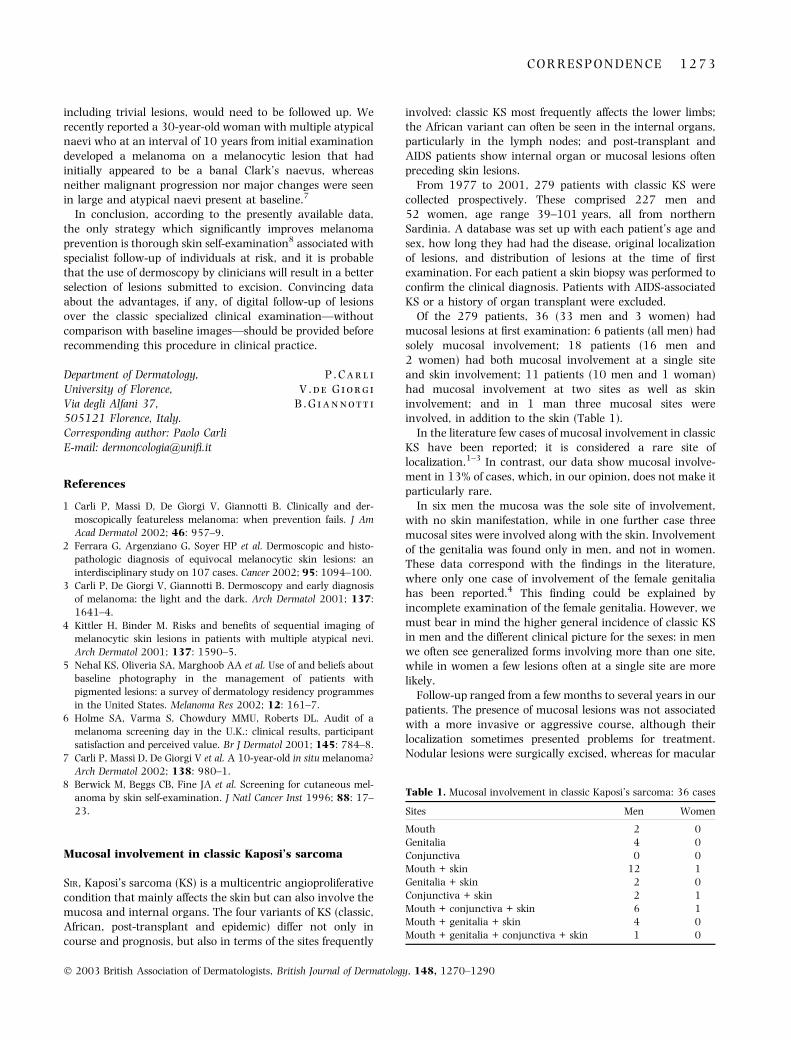
Of the 279 patients, 36 (33 men and 3 women) hadmucosal lesions at first examination: 6 patients (all men) hadsolely mucosal involvement; 18 patients (16 men and2 women) had both mucosal involvement at a single siteand skin involvement; 11 patients (10 men and 1 woman)had mucosal involvement at two sites as well as skininvolvement; and in 1 man three mucosal sites wereinvolved, in addition to the skin (Table 1).
In the literature few cases of mucosal involvement in classicKS have been reported; it is considered a rare site oflocalization.1–3 In contrast, our data show mucosal involve-ment in 13% of cases, which, in our opinion, does not make itparticularly rare.
In six men the mucosa was the sole site of involvement,with no skin manifestation, while in one further case threemucosal sites were involved along with the skin. Involvementof the genitalia was found only in men, and not in women.These data correspond with the findings in the literature,where only one case of involvement of the female genitaliahas been reported.4 This finding could be explained byincomplete examination of the female genitalia. However, wemust bear in mind the higher general incidence of classic KSin men and the different clinical picture for the sexes: in menwe often see generalized forms involving more than one site,while in women a few lesions often at a single site are morelikely.
Follow-up ranged from a few months to several years in ourpatients. The presence of mucosal lesions was not associatedwith a more invasive or aggressive course, although theirlocalization sometimes presented problems for treatment.Nodular lesions were surgically excised, whereas for macular
Table 1. Mucosal involvement in classic Kaposi’s sarcoma: 36 cases
Sites Men Women
Mouth 2 0
Genitalia 4 0
Conjunctiva 0 0
Mouth + skin 12 1
Genitalia + skin 2 0
Conjunctiva + skin 2 1
Mouth + conjunctiva + skin 6 1
Mouth + genitalia + skin 4 0
Mouth + genitalia + conjunctiva + skin 1 0
C O R R E S P O N D E N C E 1 2 7 3
� 2003 British Association of Dermatologists, British Journal of Dermatology, 148, 1270–1290

lesions or lesions in delicate sites such as the conjunctiva,systemic therapy using antiblastic drugs was the preferredtreatment modality.5
In conclusion, we feel that the mucosa, with the exceptionof the female genitalia, cannot be considered to be a rare sitefor classic KS but rather should be taken into account in theoverall evaluation of the clinical picture in patients with thiscondition.
F . C o t t o n i
M . V . M a s a l a
P . P i r a s
M . A . M o n t e s u
D . C e r i m e l e
Department of Dermatology,University of Sassari,Viale San Pietro 43 d,07100 Sassari, ItalyCorrespondence: Francesca Cottoni,via Mazzini 19, 07100 Sassari, ItalyE-mail: [email protected]
References
1 Jaimovich L, Calb I, Kaminssky A. Kaposi’s sarcoma of the con-
junctiva. J Am Acad Dermatol 1986; 14: 589–92.
2 Searles GE, Markman S, Yazdi M. Primary oral Kaposi’s sarcoma of
the hard palate. Dermatology 1990; 23: 518–19.
3 Kavak A, Akman RY, Alper M, Buyukbabani N. Penile Kaposi’s
sarcoma in a human immunodeficiency virus-negative patient. Br J
Dermatol 2001; 144: 207–8.
4 Stratigos JD, Potouridou I, Katoulis AC et al. Classic Kaposi’s sar-
coma in Greece: a clinico-epidemiological profile. Int J Dermatol
1997; 36: 735–40.
5 Masia IM, Satta R, Rosella M et al. Terapia del sarcoma di Kaposi
classico. G Ital Dermatol Venereol 2000; 135: 569–78.
Photodynamic therapy of sebaceous hyperplasiawith topical 5-aminolaevulinic acid and slide projector
SIR, Sebaceous hyperplasia (SH) is a manifestation of photo-ageing, and can be a cosmetic concern in elderly persons.However, SH has not been a major object for treatment,probably because no satisfactory methods have been devel-oped. Photodynamic therapy (PDT) with the combined use of5-aminolaevulinic acid (ALA) and visible light is nowsuccessfully used in the treatment of superficial nonmel-anoma skin cancers including actinic keratosis, Bowen’sdisease and superficial basal cell carcinoma.1,2 The advan-tages of this PDT modality include the absence of prolongedsystemic photosensitivity and the preferential accumulationof the intermediate metabolite protoporphyrin IX (PpIX) intumour cells after topical application of ALA. Topicallyapplied ALA is rapidly cleared from the tissue and thereforecauses only a brief period of photosensitivity. It was recentlydemonstrated that PpIX accumulates not only in the malig-nant cell but also in the pilosebaceous units,3,4 which can betarget tissues in ALA-PDT. Thus, we tried PDT for thetreatment of SH.
A 61-year-old Japanese man presented with a 10-yearhistory of multiple asymptomatic, slowly enlarging lesionson the face. The lesions were soft, brownish-yellow papules,1Æ5–4Æ0 mm in diameter (Fig. 1a). The diagnosis of senileSH was made and confirmed histologically. The patientsought a treatment for the larger lesions without the risks ofsurgical excision and with good cosmetic outcome. Aftergiving informed consent, he was treated with PDT. Twenty
Figure 1. (a) Clinical presentation of sebaceous hyperplasia before 5-aminolaevulinic acid-photodynamic therapy (PDT). Papules with arrows
were treated. (b) Oedema and erythema were induced on the PDT-treated lesions 24 h after irradiation. (c) Papules resolved after three PDT
courses. A larger lesion (arrow) became smaller, but did not completely resolve.
1 2 7 4 C O R R E S P O N D E N C E
� 2003 British Association of Dermatologists, British Journal of Dermatology, 148, 1270–1290

percent ALA (A 7793; Sigma, St Louis, MO, U.S.A.) in anoil-in-water emulsion was topically applied on the papulesand occluded with a light-shielded dressing for 4 h. Onepapule was excised for a fluorescence microscopic study. Thelesions were then exposed through a red glass filter, at adistance of 5 cm from the lens, to illumination from a slideprojector (Twin Cabin Auto; Cabin Industry Co., Tokyo,Japan) equipped with a 300-W halogen bulb. Physicalmeasurements of the irradiance of the slide projector lampwere not available. The R-62 glass filter (Toshiba Glass Co.,Tokyo, Japan) transmits wavelengths longer than 620 nm.Six small (2Æ5 · 2Æ5 mm) and two large (3Æ5 · 3Æ0 mm)papules on the face were irradiated continuously for 15 or20 min without anaesthesia. Although a slightly burningsensation developed during and immediately after lightexposure, the PDT was easily tolerated. Within 1 h afterexposure, erythema and oedema appeared in the treatedarea, especially on papules, peaked at 24 h (Fig. 1b), andgradually subsided by 4 days with scaling and residualbrownish pigmentation. This treatment regimen was repea-ted three times in total at 1-week intervals. The treatedlesions regressed with flattening, and decreased in size witheach PDT session. Small papules nearly disappeared, show-ing an umbilication (Fig. 1c). Large papules became smaller,but did not completely resolve. Hyperpigmentation subsidedby 10 days after the final PDT. Photosensitivity to naturalsunlight did not follow PDT. There has been no recurrenceof the lesions in the 12 months after therapy.
Fluorescence microscopy demonstrated that topicallyapplied ALA penetrated well into the hyperplastic sebaceousgland, revealing intense red fluorescence in frozen sections ofthe lesion (Fig. 2).
Lesions of SH represent benign hyperplasia of the sebaceousglands, possibly caused by chronic exposure to sunlight.Plewing and Kligman compared the size of sebaceous glandson the cheek in three different age groups, and showed agradual increase in size from young adults to elderlysubjects.5 Furthermore, they demonstrated that the turnoverof sebaceous glands in the elderly is slower than in youngadults, and that sebocytes migrate slowly out of the acini,suggesting that sebum production declines with age despitehyperplasia of the sebaceous glands.5,6 SH was also producedexperimentally in hairless mice by repeated exposure toultraviolet radiation.7 In contrast to chronological ageing,photoageing is now preventable and is partially reversiblewith the help of recent advances in photodermatology andcosmetology. Rejuvenation of the skin has been performed byvarious procedures, including surgery, laser therapy, derm-abrasion, chemical peeling and topical retinoids. However, SHdoes not seem to respond to these modalities.
PDT combines the administration of a photosensitizer andthe subsequent exposure to light to induce cell destruction viageneration of reactive oxygen intermediates. The therapeuticeffect of PDT on malignant tumours is partly due to prefer-ential accumulation of photosensitizing chemicals in tumourcells.8 Several photosensitizing agents have been employed forPDT. Among them, ALA is now most commonly used for skin
diseases. Although ALA has no photosensitizing ability itself,it is metabolized in the skin to the potent photosensitizer, PpIX,after topical application. Red light around 630 nm, corres-ponding to one of the absorption maxima of the chemical, isused for irradiation in ALA-PDT. This wavelength canpenetrate deep enough into the skin. Rapid clearance ofALA from the skin can decrease the chance of adverse effectssuch as persistent photosensitivity, which is often inducedafter systemic administration of photosensitizers.
Divaris et al. demonstrated the selective accumulation ofALA in sebaceous glands after intraperitoneal injection intoalbino mice.3 Subsequent exposure to photoactivating lightproduced destruction of sebaceous glands and hair follicles. Itis likely that not only malignant cells but also normalsebaceous glands are sensitive to ALA-PDT. After topicalapplication of ALA to the human skin, greater production ofPpIX was detected in sebaceous glands and hair follicles thanin adjacent tissues.4 Based on these observations, topicalALA-PDT was used for the treatment of naevus sebaceus9 andacne vulgaris4,10 with long-lasting benefit. From a similarviewpoint, we attempted to treat senile sebaceous glandhyperplasia with topical ALA-PDT. A satisfactory cosmetic
Figure 2. Fluorescence microscopy showed red fluorescence of top-
ically applied 5-aminolaevulinic acid in the hyperplastic sebaceous
glands (original magnification, ·100).
C O R R E S P O N D E N C E 1 2 7 5
� 2003 British Association of Dermatologists, British Journal of Dermatology, 148, 1270–1290

result was obtained, although larger lesions did not com-pletely resolve. Improvements in the ALA-PDT technique maylead to improved efficacy. As demonstrated in the presentstudy, topically applied ALA can penetrate well into thehyperplastic sebaceous gland. We used a slide projector as thelight source for PDT. This light source is advantageous in thatit is inexpensive, easily available, and has a large irradiationfield. Furthermore, infrared emissions may contribute to thetherapeutic effect. However, light sources that are nowcommercially available for topical PDT are probably moreeffective.
In conclusion, topical ALA-PDT may be a choice in thetreatment of SH. Cosmetic results are satisfactory for smalllesions and the procedure is well tolerated. Although largepapules were resistant to the present therapeutic procedure,early treatment may prevent lesion growth. Rejuvenation ofphotoaged skin will be of increasing concern in the futurebecause of increasing longevity and the possible depletion ofthe ozone layer.
T . H o r i o
O . H o r i o *
H . M i y a u c h i -
H A S H I M O T O
M . O h n u k i
T . I s e i
Departments of Dermatology and*Plastic Surgery, Kansai MedicalUniversity, 10-15 Fumizono,Moriguchi,Osaka 570-8507, Japan.E-mail: [email protected]
References
1 Fritsch C, Goerz G, Ruzicka T. Photodynamic therapy in derma-
tology. Arch Dermatol 1998; 134: 207–14.
2 Morton CA, Brown SB, Collins S et al. Guidelines for topical
photodynamic therapy: report of a workshop of the British Pho-
todermatology Group. Br J Dermatol 2002; 146: 552–67.
3 Divaris DXG, Kennedy JC, Poittier RH. Phototoxic damage to
sebaceous glands and hair follicles of mice after systemic admin-
istration of 5-aminolevulinic acid correlates with localized proto-
porphyrin fluorescence. Am J Pathol 1990; 136: 891–7.
4 Hongcharu W, Taylor CR, Chang Y et al. Topical ALA-photody-
namic therapy for the treatment of acne vulgaris. J Invest Dermatol
2000; 115: 183–92.
5 Plewing G, Kligman AM. Proliferative activity of the sebaceous
glands of the aged. J Invest Dermatol 1978; 70: 314–17.
6 Luderschmidt C, Plewig G. Circumscribed sebaceous gland hyper-
plasia: autoradiographic and histoplanimetric studies. J Invest
Dermatol 1978; 70: 207–9.
7 Bissett DL, Hannon DP, Orr TV. An animal model of solar-aged
skin: histological, physical, and visible changes in UV-irradiated
hairless mouse skin. Photochem Photobiol 1987; 46: 367–78.
8 Klinteberg C, Enejder AMK, Wang I et al. Kinetic fluorescence
studies of 5-aminolaevulinic acid-induced protoporphyrin IX
accumulation in basal cell carcinomas. J Photochem Photobiol
B 1999; 49: 120–8.
9 Dierickx CC, Goldenhersh M, Dwyer P et al. Photodynamic
therapy for nevus sebaceus with topical 5-aminolevulinic acid.
Arch Dermatol 1999; 135: 637–40.
10 Itoh Y, Ninomiya Y, Tajima S et al. Photodynamic therapy for
acne vulgaris with topical 5-aminolevulinic acid. Arch Dermatol
2000; 136: 1093–5.
Absence of human herpesvirus-8 in glomeruloidhaemangiomas associated with POEMS syndromeand Castleman’s disease
SIR, POEMS syndrome is considered to be a form of plasma celldyscrasia presenting as osteosclerotic myeloma or multicen-tric angiofollicular lymph node hyperplasia ( ¼ Castleman’sdisease) and evokes a large variety of symptoms includingpolyneuropathy, organomegaly (lymphadenopathy, spleno-megaly or hepatomegaly), endocrinopathy (frequently ashypothyroidism or hypogonadism), monoclonal gammopathyand protean skin changes. Synonyms are Crow–Fukasesyndrome, Takatsuki syndrome and PEP syndrome (poly-neuropathy, endocrinopathy, plasma cell dyscrasia).1 Skinsigns in POEMS syndrome are usually nonspecific and includegeneralized hyperpigmentation, hypertrichosis and diffusesclerodermiform skin thickening.2 Glomeruloid haemangio-mas, however, are regarded to be highly specific for POEMSsyndrome. To date, no case of glomeruloid haemangioma hasbeen reported outside POEMS syndrome.3
A 36-year-old man presented with multiple dome-shaped,firm purple-red angiomas measuring up to 2 cm in diameteron the upper back and abdomen. They had appeared suddenly1 year previously within a 2-month period and had remainedunchanged and asymptomatic thereafter (Fig. 1A). The pa-tient was in poor general condition. He had lost 10 kg ofweight over the past 2 years, and he had a disablingsensorimotor polyneuropathy of the lower extremities, he-patosplenomegaly, ascites, pleural effusions, generalizedlymphadenopathy and multiple endocrine dysfunctions (pri-mary hypothyroidism, hypogonadotrophic hypogonadismwith sexual impotence, hyperparathyroidism, vitamin D defi-ciency and pathological oral glucose tolerance). Skin biopsy ofa haemangioma revealed endothelial cell proliferation withectatic vascular spaces lined by a single layer of flat endothelialcells, filled with red blood cells and interspersed stromal cellsresulting in structures reminiscent of renal glomeruli (Fig. 1B).In immunohistochemistry, two distinct endothelial cell popu-lations were observed: one was predominantly CD31-positiveand formed sinusoidal to slit-like vascular channels, and theother type was CD34-positive, exhibited a plumper morphol-ogy, and outlined small round to oval vessels within theglomeruloid haemangioma.4 Macrophage-like KP1 (CD68)-positive cells were interspersed in the stroma.
The rare histological finding of glomeruloid haemangiomaswas the clue to establish a unifying diagnosis of POEMSsyndrome accounting for the polyneuropathy, hepatospleno-megaly (organomegaly), endocrine and skin symptoms.However, there was no monoclonal gammopathy on serumelectrophoresis. As for an underlying lymphoproliferativedisorder, inguinal lymph node biopsy revealed Castleman’sdisease of the hyaline-vascular type. Treatment with meth-ylprednisone at an initial dose of 40 mg daily (0Æ5 mg kg)1)was followed by rapid improvement of ascites and polyneur-opathy. Five years after diagnosis monoclonal IgG jparaproteins were detected for the first time in the urine,while the patient continued to be in clinical remission under a
1 2 7 6 C O R R E S P O N D E N C E
� 2003 British Association of Dermatologists, British Journal of Dermatology, 148, 1270–1290

low dose of alternate day corticosteroids and hormonalsubstitution.
Glomeruloid haemangiomas are believed to be a reactiveendothelial proliferation in response to angiogenic stimuli,rather than a true neoplasm.3 Their pathogenesis is obscure.Overproduction of proinflammatory cytokines such as interleu-kin (IL)-1 and IL-6 (which was twofold increased in our patientat 6 pg mL)1; normal < 3) and vascular endothelial growthfactor (VEGF) has been suggested to account for the numerousmanifestations of POEMS syndrome.5, 6 Human herpesvirus-8(HHV-8) has been implicated in angiogenic processes such asKaposi’s sarcoma and it was recently identified in multicentricCastleman’s disease associated with POEMS syndrome.7
A homologue to human IL-6 is present in the HHV-8 genomeand viral IL-6 is suspected to induce angiogenesis and haema-topoiesis.8 Furthermore, HHV-8-transformed endothelial cellsexpress VEGF.9 Taking these observations into account, wehypothesized that HHV-8 may play a role in the development ofthis unique type of haemangioma.
By immunofluorescence assay, no circulating anti-HHV-8antibodies were detected to the latency-associated nuclearantigen or to viral structural antigens. Likewise, enzyme-linked immunosorbent assays for antibodies to the recom-binant structural proteins encoded by the HHV-8 genes 65and K8.1 were negative. The presence of HHV-8 DNA wasexamined by nested polymerase chain reaction (PCR) ampli-fication from a snap-frozen biopsy of glomeruloid haemangi-oma, paraffin-embedded sections of a Castleman lymph node,and peripheral blood mononuclear cells. The 194-bp DNAfragment selected for amplification was derived from the DNApolymerase gene with primers within the positions 13601–13794 (GenBank U75698). As positive control we cloned theproduct of first-round PCR of DNA derived from HHV-8-infected JSC-1 cells (kindly provided by Dr Richard Ambinder,Boston, MA, U.S.A.) into a pCR� 2.1 TOPO vector system(Invitrogen, Life Technologie, Lofer, Australia). In agaroseelectrophoresis, no HHV-8-specific bands of the nested PCRwere detected. A second set of PCR primers derived fromKaposi’s sarcoma-associated herpesvirus gene 26 for a minorcapsid protein was also negative.10 Furthermore, indirectimmunofluorescence assays carried out on sections of glom-eruloid haemangioma using a rat monoclonal antibody tolatent nuclear antigen (LNA-1, ORF-73; Advanced Biotech-nologies Inc., Columbia, MO, USA) were negative.
Our case documents that HHV-8 infection seems not to bein all instances a prerequisite for the development ofglomeruloid haemangioma, and that other factors, such aselevated serum levels of IL-6, play a role in the development ofthis unique type of haemangioma in POEMS syndrome.
Acknowledgments
We thank Professor Dr Thomas F. Schulz, Departmentof Virology, Hannover Medical School, for helpful discus-sion of the manuscript. The work was supported by a grantof the Fonds zur Forderung der Forschung an denUniversitatskliniken Innsbruck awarded to N.S. and C.L.
G . O b e r m o s e r
C . L a r c h e r *
J . A . S h e l d o n�N . S e p p
B . Z e l g e r
Department of Dermatology,University Hospital of Innsbruck,Anichstrasse 35, 6020 Innsbruckand *Institute of Hygiene and SocialMedicine, Leopold-FranzensUniversity of Innsbruck, Fritz-PreglStrasse 3, 6020 Innsbruck, Austria,and �Department of Virology,Hannover Medical School,Carl-Neuberg-Strasse 1, 30625Hannover, Germany.E-mail: [email protected]
Figure 1. (A) Multiple dome-shaped angiomas on the back. (B)
Glomeruloid haemangioma in skin biopsy (haematoxylin and eosin;
original magnification, ·100).
C O R R E S P O N D E N C E 1 2 7 7
� 2003 British Association of Dermatologists, British Journal of Dermatology, 148, 1270–1290

References
1 Nakanishi T, Sobue I, Toyokura Y et al. The Crow–Fukase
syndrome: a study of 102 cases in Japan. Neurology 1984; 34:
712–20.
2 Ishikawa O, Nihei Y, Ishikawa H. The skin changes of POEMS
syndrome. Br J Dermatol 1987; 117: 523–6.
3 Tsai CY, Lai CH, Chan HL, Kuo TT. Glomeruloid hemangioma—a
specific cutaneous marker of POEMS syndrome. Int J Dermatol
2001; 40: 403–6.
4 Kishimoto S, Takenaka H, Shibagaki R et al. Glomeruloid
hemangioma in POEMS syndrome shows two different immuno-
phenotypic endothelial cells. J Cutan Pathol 2000; 27: 87–92.
5 Gherardi RK, Belec L, Fromont G et al. Elevated levels of inter-
leukin-1 beta (IL-1 beta) and IL-6 in serum and increased pro-
duction of IL-1 beta mRNA in lymph nodes of patients with
polyneuropathy, organomegaly, endocrinopathy, M protein, and
skin changes (POEMS) syndrome. Blood 1994; 83: 2587–93.
6 Watanabe O, Arimura K, Kitajima I et al. Greatly raised vascular
endothelial growth factor (VEGF) in POEMS syndrome. Lancet
1996; 347: 702.
7 Belec L, Mohamed AS, Authier FJ et al. Human herpesvirus 8
infection in patients with POEMS syndrome-associated multicen-
tric Castleman’s disease. Blood 1999; 93: 3643–53.
8 Aoki Y, Jaffe ES, Chang Y et al. Angiogenesis and hematopoiesis
induced by Kaposi’s sarcoma-associated herpesvirus-encoded
interleukin-6. Blood 1999; 93: 4034–43.
9 Masood R, Cesarman E, Smith DL et al. Human herpesvirus-
8-transformed endothelial cells have functionally activated vas-
cular endothelial growth factor ⁄ vascular endothelial growth
factor receptor. Am J Pathol 2002; 160: 23–9.
10 Boshoff C, Whitby D, Hatziioannou T et al. Kaposi’s-sarcoma-
associated herpesvirus in HIV-negative Kaposi’s sarcoma. Lancet
1995; 345: 1043–4.
Size matters? Yes, but the terminology doesn’t
SIR, Ferguson and Williams1 make a valid point aboutdefinitions of papules and nodules, a topic previously aired inthe Archives of Dermatology (as referenced in their article), butrigid definitions remove any sense of dynamics from theclinical scenario, and do not always improve things from areal-life practical perspective. The 0Æ5 cm or 1 cm cut-offbetween papules and nodules in the examples cited is asartificial as assuming that a 0Æ99 mm thick melanoma willbehave any differently from one of 1Æ01 mm thickness, simplyby virtue of falling into a different arbitrary category.Furthermore, other authors have used even wider ranges todistinguish between lesions of papular or nodular morphol-ogy: Jackson uses up to 1 cm for a papule, over 1 cm for anodule, and over 2 cm for a ‘node or subcutaneous nodule’,2
although it is unclear why a subcutaneous lesion needs to belarger in order to acquire ‘nodule’ status.
How should we describe a lesion that is between 0Æ5 and1 cm? Is it a large papule or a small nodule? And whathappens when the papule grows in size and becomes anodule: does the change in terminology alter the diagnosis?Many nodules may evolve from a lesion that starts its life as apapule, although many papules will never grow into nodules
and the converse (nodules turning into papules) rarelyoccurs. What about huge nodules of 5, 10 or 20 cm? Theirsize clearly creates issues about therapy, e.g. surgicalexcision, but they are not specifically catered for withinpresent definitions. It is clear that it is actually much better todocument the measured or approximate size in the case ofsolitary lesions: at least the description then has clinical valuein documenting any progression or otherwise, and it conveysa much more useful message.
Similarly, in the case of multiple lesions, what do weterm an eruption consisting of lesions around 0Æ5 or 1 cm,such as lesions in neurofibromatosis: is it a papulonodulardisease? (Not many authors use the term papulonodular,although some do.2) What size delineates lesions that aretermed micronodular? Why do we not just acknowledgethat the distinction between papules and nodules isarbitrary, as argued elsewhere,3 and describe an eruptionas consisting of lesions ‘mainly of about x cm’, or‘between x and y cm’, thereby conveying a useful clinicalmessage?
Ferguson and Williams surveyed selected current studenttextbooks for their paper. A trip back in time to some previousstandard textbooks, all of which predate use of centimetres inthe U.K., documents a more pragmatic approach by manyauthors, although not all descriptions concur with either the0Æ5 or 1-cm limits currently in vogue. Percival, for example,describes a nodule as being ‘a dermic lesion (that) … may beas large as a pea’4 (and therefore hopefully smaller than the1 cm lower limit, if not the 0Æ5-cm variant). McKennadescribes a papule as a circumscribed lesion elevated abovethe surface of the skin and neither large, deep-seated, norcompletely spherical; a nodule or tubercle (slightly strangely)is ‘a large papule, more deeply set in the skin than anordinary nodule’5 (‘ordinary nodule’ is not defined). Sutton, alittle later, describes papules as ‘small, variously shaped,circumscribed, solid elevations’, while tubercles (the word‘nodule’ is not used) are ‘bean- to pea-sized’ and tumours are‘pea- to egg-sized’.6 Borrie, in Roxburgh’s Common SkinDiseases, succinctly describes papules as small superficialraised abnormalities and nodules as larger and deeper raisedlesions.7 And really, that says it all. Papules are small,nodules are larger; both terms basically describe the samemorphology, although a palpable deeper component fits bestwith a nodule (microscopically, either may have a deepercomponent), and the best way to record them is to documentthe actual size.
So yes, size does matter, but the terminology probably doesnot. I would rather that our trainees learn how to describethe shape, colour, surface texture and evolution of a lesionthan worry about whether it fulfils the arbitrary definition ofa papule or a nodule.
N . H . C o xDepartment of Dermatology,Cumberland Infirmary,Carlisle CA2 7HY, U.K.E-mail: [email protected]
1 2 7 8 C O R R E S P O N D E N C E
� 2003 British Association of Dermatologists, British Journal of Dermatology, 148, 1270–1290

References
1 Ferguson AD, Williams HC. Size matters. Br J Dermatol 2002; 147:
1269–70.
2 Jackson R. Morphological Diagnosis of Skin Disease. Ottawa: Manti-
core Publishers, 1998.
3 Lawrence CM, Cox NH. Physical Signs in Dermatology, 2nd edn.
London: Mosby, 2002; 3.
4 Percival GH. An Introduction to Dermatology, 11th edn. Edinburgh:
E. & S.Livingstone Ltd; 1947: 11–12.
5 McKenna RW. Diseases of the Skin. London: Bailliere, Tindall and
Cox, 1923; 7.
6 Sutton RL. Diseases of the Skin, 8th edn. London: Henry Kimpton,
1931; 66–7.
7 Borrie P. Roxburgh’s Common Skin Diseases, 12th edn. London:
H.K.Lewis & Co. Ltd, 1964; 19.
What matters is simplicity and standardization: replyfrom authors
SIR, Enforcing a categorical definition upon something whichalters in a continuous fashion, such as blood pressure, age,P-values and round bumps in the skin, is, by definition anarbitrary one which will always result in ‘disputes’ as towhere the cut-off should be and what to call something that isvery near the cut-off. With experience, the limitations of sucharbitrary definitions become apparent. Thus, we are in fullagreement with Cox when he points out that the distinctionbetween a papule and nodule is artificial, and that the use ofsize in definitions may oversimplify things. As a result, in theclinical situation, morphological definitions of individuallesions are rarely perfect.
Indeed, it is probably because of this that so manydefinitions have been given for common terms.1 We alsoagree with Cox that squabbling over the size of a papule andgiving medical students long and complicated lists to learn isnot going to help them very much, and is not as important asrecognizing the early cardinal signs of a malignant melan-oma.
But what is the alternative to such arbitrary definitions?We maintain that one either does one thing or the other.Either we should drop all our dermatology terms becausetheir arbitrary nature makes them just too difficult to copewith, in which case we could resort to laymen’s terms such as‘small sticking-out spot’ or ‘biggish lump’ (the size of whichwould vary according to the eye of the beholder). Or, weshould accept that terms for skin lesions are useful incommunication, in which case we should make an attempt atjust one standard international definition. In our view theworse scenario is to carry on muddling away on theassumption that all physicians over the world implicitlyunderstand what we mean by terms that have a multitude ofdefinitions—a situation akin to the Tower of Babel, or shouldwe say the Tower of Papule.
The purpose of defining dermatological terms is to improvecommunication so that a dermatologist sending an e-mailfrom Addis Ababa, Angora or Abertillery can describe thesame patient’s skin eruption reasonably similarly and accu-
rately and in a way that conveys increased predictive abilityto the person receiving that information. Indeed, the hypo-thesis that some terms may not increase our predictive abilitycould be readily tested by a suitable experiment along theselines.
In the meantime, the fact that even the most basic termsused in dermatology still lack a standard definition remainsan embarrassment to us when we try and teach such terms toour medical students. Few would argue that accuratedescription is a key skill in dermatology. Some simple agreeddefinitions are needed to allow medical students to do this.Ideally, given that most medical students spend only a weekor two in a dermatology department, these definitions need tobe simple enough to learn and apply quickly. Standardizationis important to ensure that the enthusiastic student whoconsults more than one text is enlightened and not confusedor, worse still, be put off from pursuing a discipline thatcannot agree on a definition of something as simple as apimple.
A . D . F e r g u s o n
H . C . W i l l i a m s *
Department of Dermatology,Royal Hallamshire Hospital,Sheffield S10 2JF, U.K.*Department of Dermatology,Queen’s Medical Centre,Nottingham NG7 2UH, U.K.Correspondence: A.D.Ferguson.E-mail: [email protected]
Reference
1 Ferguson AD, Williams HC. Size matters. Br J Dermatol 2002; 147:
1269–70.
In-column electrocoagulation for lymphaticmalformation of the tongue
SIR, A 17-year-old Caucasian male was referred to our clinicbecause of a congenital lymphatic malformation on themiddle third of the tongue. The lesion had slowly grown sincechildhood. Two years previously the patient underwentpartial surgical excision; however, the lesion recurred6 months later. One year after recurrence, the patientunderwent CO2 laser treatment, with partial reduction inlesion size. Finally, 2 years later, when the patient consultedour clinic the lesion showed an important hypertrophyassociated with bleeding and discomfort at swallowing(Fig. 1). The histopathological examination showed largedilated vascular spaces with scarce erythrocytes, and prote-inaceous material as well as dilated venules, findings corres-ponding to a lymphatic malformation. Our challenge was tofind a conservative, efficient treatment with low morbiditythat could lead to better results than those obtained withother surgical techniques.
The size of the lesion dissuaded us from using surgicalexcision that would result in partial mutilation of the tongue.In addition, the patient rejected both excisional surgery and
C O R R E S P O N D E N C E 1 2 7 9
� 2003 British Association of Dermatologists, British Journal of Dermatology, 148, 1270–1290

CO2 laser, which had already failed. Pulsed dye laser was notrecommended due to its limited penetration (1Æ2 mm). Othertreatments such as selective embolization, cryotherapy andtransfixation were judged of limited use in this case. Micro-embolization does not achieve complete elimination of thelymphatic component of lymphatic malformation, whilecryotherapy and transfixation might be difficult to accomplishin this anatomical region.
Treatment of small and medium vessel vascular malforma-tion by means of electrocoagulation is a well-known tech-nique in dermatology. However, the patient herein presentedhad a large lesion in a difficult region that required a specialelectrode design in order to treat the lesion completely andeasily without substantial risk to other anatomical structuresin the oral cavity.
Five 14-gauge metallic needles (Venflon, Medicut, Labor-atorios Omeda, Barcelona, Spain) covered with a noncon-ductive plastic coat were modified in order to perform theprocedure. The plastic coat was cut distally with theintention that the exposed portion of the needle (10 mm)
worked as an electrode when inserted into the lesion. Asmall window was proximally opened in the plastic coat toassure direct contact to the electrosurgery apparatus elec-trode (Fig. 2).
Electrocoagulation of the lymphatic malformation wasperformed under general anaesthesia. Oral forceps were usedin order to open the mouth wide. Then traction to the tonguewas done with an assistant’s help, a manoeuvre that madepossible a better approach to the lesion. Five 14-gaugeneedles, prepared as described above, were inserted evenlyand about 20 mm deep throughout the lesion (Fig. 3).Sequential electrocoagulation was done with a high-frequency electrosurgery unit (Electrosurgical generator Force2; ValleyLab Inc., Boulder, CO, U.S.A.) (Fig. 4). We used themonopolar electrode output in coagulation mode. The gener-ator was set at 10 W and power was gradually increased untiltissue desiccation was observed. Power generally fluctuatedbetween 20 W and 30 W. The electrosurgery unit electrodereached the needle through the proximal notch in the plasticcoat. Then current was released until a yellowish-greycolouration was obtained locally, a visual indicator of necro-
Figure 2. Electrode for electrocoagulation: a proximal notch in the
plastic coat allows contact with the electrosurgery unit electrode.
Figure 1. A 17-year-old patient with macroglosia secondary to
lymphatic malformation of the tongue.
1 2 8 0 C O R R E S P O N D E N C E
� 2003 British Association of Dermatologists, British Journal of Dermatology, 148, 1270–1290

sis. When the lymphatic component of the lesion disappearedand an eschar (Fig. 5) was evident the needle was withdrawn.The same procedure as described above was performed withall five inserted needles. The above described technique isreferred as ‘in-column electrocoagulation’. Mild oedema of thetongue without respiratory ⁄ digestive tract obstruction wasobserved 24 h postoperatively. Neither bleeding nor infectionwas documented. Post-operative instructions included pred-nisone at a dose of 1 mg kg)1 day)1 for 3 days, acetamino-phen 750 mg three times daily., liquid ⁄ bland diet and gargleswith 10 p ⁄ v oxygenated water diluted in tempered water aftermeals. On the third day postoperatively, streptodornase10 000 U ⁄ streptokinase 2Æ500 U (Varidase, Wyeth-Lederle,Madrid, Spain) orally, four times daily was introduced andmaintained for 15 days. Local ulceration, painful swallowing,halitosis and functional dyslexia disappeared 3 weeks postop-eratively. Healing has been normal and the final scar isacceptable (Fig. 6). Moreover, deglutition and speech arenormal after a 12-month follow-up.
Progression of lymphatic malformation can lead to macro-glossia, which may cause obstruction to the airway and
digestive tract. Even though both are potentially fatalcomplications they are rarely observed. More commoncomplications include bleeding, ulceration, painful degluti-tion, and functional speech disability or associated inflam-mation ⁄ oedema that may lead to asphyctic complications.
Immediate surgical1 treatment or embolization2,3 is man-datory in case of profuse persistent haemorrhage, ocularocclusion, respiratory obstruction, central nervous systemcomplications, interference with nutrition, and functionalimpairment related to the effect of the lesion on the developingfacial skeleton and teeth;2 however, these therapeutic approa-ches are not exempted from important complications.1
Patients having secondary macroglossia but with no risk tolife may undergo more conservative procedures such ascryotherapy, transfixation, excisional surgery or electrocoag-ulation of the lesion.
The method described here is a simple, safe and cost-effective procedure. Metallic needles are good conductors ofelectricity; moreover, they are flexible, inexpensive and are
Figure 4. The current passes through the needle up to the uncovered
end, which works as an internal electrode that leads to limited and
deep coagulation.
Figure 3. Multiple electrodes are inserted evenly and about 20 mm
deep throughout the lingual lesion.
C O R R E S P O N D E N C E 1 2 8 1
� 2003 British Association of Dermatologists, British Journal of Dermatology, 148, 1270–1290

available almost everywhere. Needle flexibility allows thesurgeon to adjust it to almost any localization and depth. Theneedle is provided with a plastic coat that covers itproximally, avoiding accidental burns to nearby structures;however, the distal portion of the needle is uncovered,allowing deep and limited coagulation of the lesion. Finally,another advantage of sterilized disposable needles is that onecan prepare as many electrodes as are needed, allowing for arapid treatment of large lesions.
Acknowledgment
The authors thank Harmut Schwarze MD for kindly review-ing and editing the manuscript.
A . V i l a l t a
G . A . M o r e n o - A r i a s
J . M . M a s c a r o
Department of Dermatology,Hospital Clınic,University of Barcelona, Villarroel170, 08036, Barcelona, Spain.CorrespondenceE-mail: [email protected]
References
1 Bureau Y, Delaire J, Barriere H et al. [Hemolymphangioma of the
tongue. Results of surgical treatment]. Bull Soc Fr Dermatol Syph-
iligr 1966; 73: 422–3.
2 Burrows PE, Lasjaunias PL, Ter Brugge KG, Flodmark O. Urgent
and emergent embolization of lesions of the head and neck in
children: indications and results. Pediatrics 1987; 80: 386–94.
3 Laufer J, Girsault M. [Hemolymphangioma of the tongue treated
with combination embolization-surgery. Apropos of a case]. Rev
Stomatol Chir Maxillofac 1986; 87: 184–7.
Is there really no clear association between low serumferritin and chronic diffuse telogen hair loss?
SIR, It was no surprise that Dr Sinclair’s study, based on amodest increase (personal communication) in serum ferritin,found no reduction of hair shedding.1
His conclusion that there is no clear association betweenlow serum ferritin and chronic diffuse telogen hair loss isbased on the assumption that a serum ferritin of > 20 lg L)1
Figure 5. Clinical aspect immediately postelectrocoagulation. Figure 6. Final clinical result after only one session of electrocoag-
ulation (12-month follow-up).
1 2 8 2 C O R R E S P O N D E N C E
� 2003 British Association of Dermatologists, British Journal of Dermatology, 148, 1270–1290
provides an estimate of adequate iron stores. However, wehave challenged this position and the derivation of theso-called ‘normal’ female range for serum ferritin andhaemoglobin.2 Our hypothesis is that these ranges have beenderived from populations containing a high proportion ofiron-deficient women. This idea received support from a large-scale (n ¼ 12 828) study undertaken by Waalen et al.,3
who demonstrated iron deficiency in 38% of San Diegowomen on re-analysis by Rushton et al.4 Thus populationsused to provide female reference ranges were assumed,incorrectly, to be normal, leading to the normalization ofabnormality and aberrant reference ranges.
We appreciate the confusion that might exist with thedefinition of ‘normal’ serum ferritin in females that actuallyhave inadequate iron stores, especially where the only overtsymptom is persistent increased hair shedding (chronic telogeneffluvium (CTE)). This position is further complicated by the factthat some women with iron deficiency anaemia do not shedhair excessively. However, we have shown that around 30% ofwomen in the U.K., U.S.A. and Japan complain of CTE.5
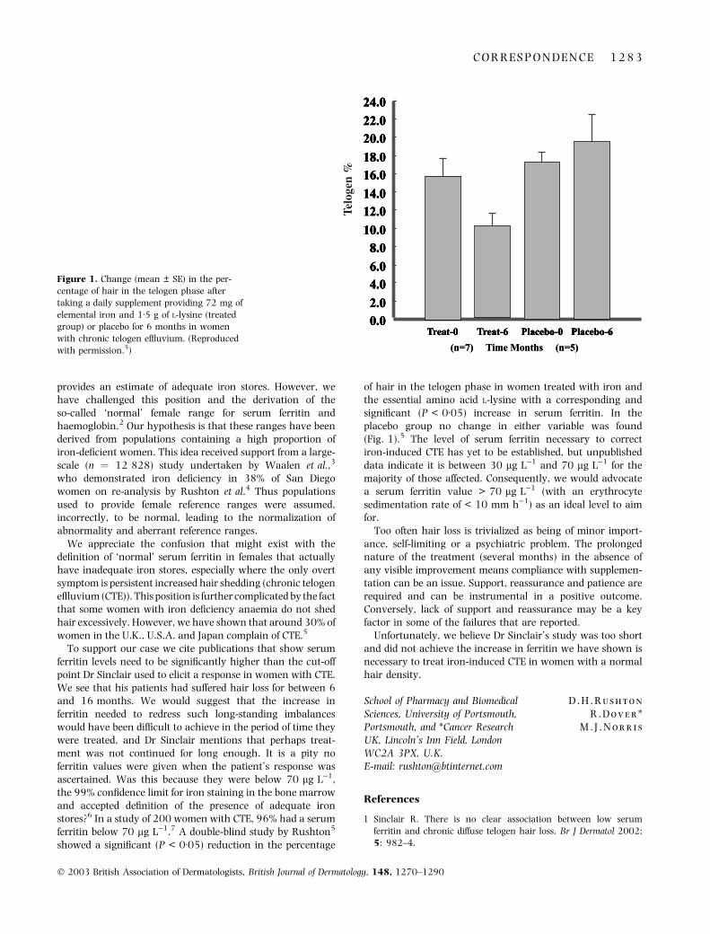
To support our case we cite publications that show serumferritin levels need to be significantly higher than the cut-offpoint Dr Sinclair used to elicit a response in women with CTE.We see that his patients had suffered hair loss for between 6and 16 months. We would suggest that the increase inferritin needed to redress such long-standing imbalanceswould have been difficult to achieve in the period of time theywere treated, and Dr Sinclair mentions that perhaps treat-ment was not continued for long enough. It is a pity noferritin values were given when the patient’s response wasascertained. Was this because they were below 70 lg L)1,the 99% confidence limit for iron staining in the bone marrowand accepted definition of the presence of adequate ironstores?6 In a study of 200 women with CTE, 96% had a serumferritin below 70 lg L)1.7 A double-blind study by Rushton5
showed a significant (P < 0Æ05) reduction in the percentage
of hair in the telogen phase in women treated with iron andthe essential amino acid L-lysine with a corresponding andsignificant (P < 0Æ05) increase in serum ferritin. In theplacebo group no change in either variable was found(Fig. 1).5 The level of serum ferritin necessary to correctiron-induced CTE has yet to be established, but unpublisheddata indicate it is between 30 lg L)1 and 70 lg L)1 for themajority of those affected. Consequently, we would advocatea serum ferritin value > 70 lg L)1 (with an erythrocytesedimentation rate of < 10 mm h)1) as an ideal level to aimfor.
Too often hair loss is trivialized as being of minor import-ance, self-limiting or a psychiatric problem. The prolongednature of the treatment (several months) in the absence ofany visible improvement means compliance with supplemen-tation can be an issue. Support, reassurance and patience arerequired and can be instrumental in a positive outcome.Conversely, lack of support and reassurance may be a keyfactor in some of the failures that are reported.
Unfortunately, we believe Dr Sinclair’s study was too shortand did not achieve the increase in ferritin we have shown isnecessary to treat iron-induced CTE in women with a normalhair density.
D . H . R u s h t o n
R . D o v e r *
M . J . N o r r i s
School of Pharmacy and BiomedicalSciences, University of Portsmouth,Portsmouth, and *Cancer ResearchUK, Lincoln’s Inn Field, LondonWC2A 3PX, U.K.E-mail: [email protected]
References
1 Sinclair R. There is no clear association between low serum
ferritin and chronic diffuse telogen hair loss. Br J Dermatol 2002;
5: 982–4.
Figure 1. Change (mean ± SE) in the per-
centage of hair in the telogen phase after
taking a daily supplement providing 72 mg of
elemental iron and 1Æ5 g of L-lysine (treated
group) or placebo for 6 months in women
with chronic telogen effluvium. (Reproduced
with permission.5)
C O R R E S P O N D E N C E 1 2 8 3
� 2003 British Association of Dermatologists, British Journal of Dermatology, 148, 1270–1290

2 Rushton DH, Dover R, Sainsbury AW et al. Why should women
have lower reference limits for haemoglobin and ferritin concen-
tration than men? Br Med J 2001; 322: 1355–7.
3 Waalen J, Felitti V, Beutler E. Haemoglobin and ferritin concen-
trations in men and women: cross sectional study. Br Med J 2002;
325: 137.
4 Rushton DH, Dover R, Sainsbury AW et al. Iron deficiency is neg-
lected in women’s health. Br Med J 2002; 325: 1176–7 (Letter).
5 Rushton DH. Nutritional factors and hair loss. Clin Exp Dermatol
2002; 27: 396–404.
6 Puolakka J. Serum ferritin in the evaluation of iron status in
young healthy women. Acta Obstet Gynecol Scand Suppl 1980; 95:
35–41.
7 Rushton DH, Norris MJ, Dover R, Busuttil N. Causes of hair loss
and the developments in hair rejuvenation. Int J Cosmet Sci 2002;
24: 17–23.
There is no clear association between low serumferritin and chronic diffuse telogen hair loss: replyfrom authors
SIR, I am aware of Dr Rushton’s opinion regarding thereference ranges for serum ferritin, and indeed I cited three ofhis papers. I am not an expert on serum ferritin levels and theintricacies of how the reference range is determined, but I donote that haematologists have not adopted his position. Infact, many haematology laboratories in Australia havelowered the reference range from 20 lg L)1 to 7 lg L)1, inspite of Dr Rushton’s published protestations.
Regardless of the ‘true’ serum ferritin level for women, themajor point of my paper is not contested. As more than half ofthe women who presented to me with chronic diffuse hair lossand a low serum ferritin were found to have androgeneticalopecia on scalp biopsy, physicians should be cautious ofattributing hair loss too readily to low serum ferritin levels.This is particularly important as hair shedding can fluctuatein the early stages of androgenetic alopecia.
I reiterate that ‘if low serum ferritin levels cause reversiblehair loss, then iron replacement should lead to cessation ofhair loss and ⁄ or regrowth’. If Dr Rushton is correct in hisassertion that low ferritin levels cause hair loss, he should beable to prove it in a randomized controlled trial withmeaningful endpoints and publish it. Using accepted ferritinlevels and a standard course of iron replacement therapy, myresults did not show any association between low serumferritin and chronic diffuse telogen hair loss.
R . S i n c l a i rUniversity of Melbourne, Departmentof Dermatology, St Vincent’sHospital, 41 Victoria Parade, Fitzroy,Victoria 3065, AustraliaE-mail: [email protected]
Importance of trial design in studies using high-doseintravenous immunoglobulin
SIR, We read with interest the recent randomized controlledtrial treating adults with atopic dermatitis (AD) with intrave-nous immunoglobulin (IVIg) by Paul and Dubertret.1 This is
the first randomized controlled study using high-dose IVIg(hdIVIg) in AD and the findings are particularly important fortwo reasons: (i) AD is a very common disorder with a smallsubset of patients suffering from severe treatment-resistantdisease who urgently need treatment advances, and (ii) there iscurrently a world plasma shortage making IVIg not only anexpensive resource but also a precious one. A randomizedcontrolled trial is thus an opportunity to offer clear guidance asto the most appropriate use of hdIVIg in AD.
We have some concerns about the hypothesis being tested,the design of the trial, its analysis and the conclusions drawn.The authors aimed to test whether hdIVIg is effective in adultswith severe AD, stating that only dramatic efficacy could justifythe use of hdIVIg in view of its cost and potential risk oftreatment and that the study was intentionally powered todetect only large treatment effects. It is not clear whether‘dramatic and large’ imply rapidity of response in view of theshort follow-up time or an overall improvement in severityscoring of atopic dermatitis (SCORAD) values. Only a smallsubset of the most severe AD patients who have failed to respondto multiple other treatments and ⁄ or suffer from unacceptableside-effects are going to be considered for hdIVIg. This group ofpatients has the highest morbidity from their disease and will bethe most frequent users of outpatient, inpatient and drugbudget resources. Against this background it is vital to testthe efficacy of hdIVIg in a way most likely to make the nullhypothesis of no efficacy robust as the cost–benefit analysisis not the same as it might be for AD patients as a whole.
This raises the question of trial design where two mainpoints arise. Firstly, the half-life of IgG is 11–17 days andwhen used at immunomodulatory doses (most commonly2 g kg)1 month)1) the literature is clear that it may take2–4 months to establish evidence of benefit for a wide rangeof dermatological conditions including dermatomyositis,bullous disorders and AD.2–6 In AD in particular the longerstudy duration may reduce the effects of natural fluctuationsin disease severity. It is also becoming evident in somedermatological conditions that it may be possible gradually towithdraw both the hdIVIg and other treatments whenpatients are in remission, altering the long-term cost–benefitanalysis.4
Secondly, analysis of the use of hdIVIg in dermatologicalconditions including AD suggest that it is more effective whenused as adjunctive therapy2,6,7 even where the existingtreatment alone is not able to control the disease. The publishedstudies of hdIVIg in adult AD show that adjunctive therapy waseffective in 59% and monotherapy in 0%, although interest-ingly this was not the case for children.6 This is supportedexperimentally by the finding that in steroid sensitive asthma,IVIg therapy acted synergistically with dexamethasone insuppressing lymphocyte activation and significantly improvedglucocorticoid receptor binding affinity after 3 and 6 months oftherapy.8
In the analysis, no difference in disease severity was observedbetween the groups at day 30; however, a statisticallysignificant improvement of 22% in the SCORAD index wasnoted at 60 days in the cohort as a whole. This is a confusing
1 2 8 4 C O R R E S P N D E N C E
� 2003 British Association of Dermatologists, British Journal of Dermatology, 148, 1270–1290

mixed message and the analysis of the whole cohort might havebeen more appropriately performed at day 30 following hdIVIgfor both groups rather than when some patients had their lasthdIVIg 30 days earlier and some 60 days earlier. The graph inthe study suggests that this may have yielded an even greaterreduction in SCORAD values, although it is not possible to bedefinitive without the raw data. Given the single dose of hdIVIgmonotherapy, small study size and short duration of the study,it is not possible to draw conclusions for or against the use ofhdIVIg in refractory AD.
Surely the question a randomized, controlled, evaluator-blinded trial such as this needs to address is: is there evidencethat we can make these patients better, not, is there adramatic improvement after a single dose of hdIVIg mono-therapy at 30 days. Randomization is no substitute forappropriate trial design and an opportunity has been lost toclarify the situation for or against hdIVIg in severe adult AD.
S . J o l l e s
J . H u g h e s *
Division of Infection and Immunity,National Institute for MedicalResearch, London,and *Department of Dermatology,Hillingdon and Amersham Hospitals,U.K.E-mail: [email protected]
References
1 Paul C, Dubertret L. A randomized controlled evaluator-blinded
trial of intravenous immunoglobulin in adults with severe atopic
dermatitis. Br J Dermatol 2002; 147: 518–22.
2 Dalakas MC, Illa I, Dambrosia JM et al. A controlled trial of high-
dose intravenous immune globulin infusions as treatment for
dermatomyositis. N Engl J Med 1993; 329: 1993–2000.
3 Ahmed AR. Intravenous immunoglobulin therapy for patients with
bullous pemphigoid unresponsive to conventional immunosuppressive
treatment. J Am Acad Dermatol 2001; 45: 825–35.
4 Engineer L, Bhol KC, Ahmed AR. Analysis of current data on the
use of intravenous immunoglobulins in management of pemphigus
vulgaris. J Am Acad Dermatol 2000; 43: 1049–57.
5 Jolles S. High-dose intravenous immunoglobulin (hdIVIg) in the
treatment of autoimmune blistering disorders. Clin Exp Immunol
2002; 129: 385–9.
6 Jolles S. A review of high-dose intravenous immunoglobulin
treatment for atopic dermatitis. Clin Exp Dermatol 2002; 27: 3–7.
7 Jolles S, Hughes J, Whittaker S. Dermatological uses of high-dose
intravenous immunoglobulin. Arch Dermatol 1998; 134: 80–6.
8 Spahn JD, Leung DY, Chan MT et al. Mechanisms of glucocorticoid
reduction in asthmatic subjects treated with intravenous im-
munoglobulin. J Allergy Clin Immunol 1999; 103: 421–6.
Importance of trial design in studies using high-doseintravenous immunoglobulin: reply from authors
SIR, Drs Jolles and Hughes express reservations about thehypothesis and design of our controlled trial,1 the conclusionof which is at variance with the one of open observations theyand others have reported. Their main concerns are theduration of observation and the use of immunoglobulins as a
monotherapy. Regarding rapidity of response to intravenousimmunoblobulin (IVIg), there are a number of immune-mediated diseases where clinical effect is evident within daysafter a single course of IVIg treatment, including Kawasakidisease, thrombocytopenic purpura and toxic epidermalnecrolysis. Moreover, established pharmacological interven-tions in atopic dermatitis (AD), such as topical corticosteroids,tacrolimus, pimecrolimus, oral ciclosporin and phototherapyhave been shown to be effective within 2–4 weeks. Patientsin our study were assessed 30 (primary endpoint) and60 days after IVIg infusion. We believe the duration ofobservation in our study is adequate to detect a clinicallyrelevant effect taking into account both the disease and thetreatment under consideration.
Concerning the improvement in severity scoring of atopicdermatitis (SCORAD), it is suggested that ‘the analysis of thewhole cohort might have been more appropriately per-formed at day 30 following IVIg for both groups’. Actuallythis is exactly what was done in the exploratory analysispresented in the paper on the observed changes in SCORADrelative to the baseline values for the entire cohort 30 and60 days after IVIg infusion. The mean 22% improvement inseverity score observed 60 days after IVIg infusion is clearlyinferior to what can be expected with available second linetherapies in severe adult AD.2 Given the high cost and thepotential safety issues with IVIg, alternative therapeuticoptions have a better cost-effectiveness profile for patientswith severe AD.
We agree with the authors that the utility of IVIg as‘adjunctive therapy’ in AD is an interesting hypothesis tobe tested. This was not the hypothesis tested in our trial,as it is standard practice first to demonstrate effectivenessof a therapeutic agent as a monotherapy before investi-gating its use in combination. The concept of an ‘adjunc-tive therapy’ that would be effective only when used incombination with standard therapy has not received a lotof attention in AD. However, extreme caution should beexercised and rigorous methodology should apply for theevaluation of such ‘adjunctive therapies’, as it is especiallydifficult to distinguish the contribution of each individualagent from the effect observed in uncontrolled observa-tions.
In summary, we believe that the results from our trial ruleout a dramatic therapeutic effect of IVIg used as a mono-therapy. The efficacy and safety of IVIg as an ‘adjunctivetherapy’ in AD remains to be evaluated. This evaluationshould be carried out in a carefully randomized clinical trial.As we commonly ‘shut our eyes to observations which do notagree with the conclusions we wish to reach’,3 this trialshould preferably be evaluator-blinded.
C . P a u l
L . D u b e r t r e t *
Department of Dermatology,Mulhouse General Hospital,68100 Mulhouse, and*Department of Dermatology,Saint Louis University Hospital,75010 Paris, France.
C O R R E S P O N D E N C E 1 2 8 5
� 2003 British Association of Dermatologists, British Journal of Dermatology, 148, 1270–1290

References
1 Paul C, Dubertret L. A randomized controlled evaluator-blinded
trial of intravenous immunoglobulin in adults with severe atopic
dermatitis. Br J Dermatol 2002; 147: 518–22.
2 Hoare C, Li Wan Po A, Williams H. Systematic review of treatments
for atopic eczema. Health Technol Assess 2000; 4: 1–191.
3 Asher R. cited by Williams HC. Inflammatory skin diseases. I.
Atopic dermatitis. In: The Challenge of Dermato-Epidemiology (Wil-
liams HC, Strachan DP, eds). Boca Raton: CRC Press, 1997: 126.
Ulceration of the penis due to Absidia corymbifera
SIR, Ulcerations may appear on the genitalia in a variety ofboth infectious and noninfectious conditions. The vastdiversity of aetiologies, coupled with the often overlappingmorphological features, make the diagnosis of genital ulcer-ation a challenging endeavour. We describe here the first caseof genital ulcer revealing a systemic absidomycosis in apatient with leukaemia. Absidia corymbifera (class Zygomyc-etes, order mucorales, family Mucoraceae) is a filamentousfungus isolated from soil and decaying vegetation. In animmunocompetent host, the spores are destroyed by polynu-clear neutrophils and macrophages. In an immunocompro-mised patient, infection by this fungus may be fatal.
A 36-year-old farmer suffered from an acute myeloblasticleukaemia with a low differentiation type. During the firstconsolidation schedule with cytarabine and idarubicin assoc-iated with heparin and itraconazole, an aplasia occurred. Onthe eighth day of the aplasia (white blood count at500 mm)1, neutrophils at 45 mm)1), the patient presentedwith fever and facial cellulitis with black nasal secretion. Thenext day, he showed an oedema of the penis with grey-blackplaques of the glans penis and the prepuce (Fig. 1); theplaques rapidly increased in size over a 12–24-h period toform a necrotic and painful ulceration. The cranial scannershowed a maxillary, frontal and sphenoidal pansinusitis.Direct examination and microbiological cultures of nasalsecretion were sterile. Biopsy specimens of the glans peniswere obtained for microbiological culture; before the histo-pathological examination, an infection with A. corymbiferawas found (Fig. 2a,b). Two days later, a severe pneumoniadeveloped; direct examination and mycological culture of thesaliva and the bronchoalveolar fluid confirmed the presenceof A. corymbifera. Aspergillus-, cryptoccocus-serology andinvestigations for circulate soluble antigens were all negative.
These observations led us to begin an early intravenous(1Æ5 mg kg)1 day)1) and aerosol antifungal treatment withamphotericin B associated with hyperbaric oxygenotherapy.The superficial surgical debridement of the glans penisulceration associated with topic amphotericin B allowedcomplete healing cicatrization after a 2-week treatment.According to the patient, there has been no alteration insexual function. On the 28th day, with the end of the aplasia,pathological respiratory signs disappeared, but the expectora-tion of blood clots led us to perform another thoracic scan,which showed a tissular necrosis in the vicinity of the rightpulmonary artery. We then performed a surgical resection of
Figure 1. Necrotic ulceration of the glans penis and the prepuce.
Figure 2. Histological section from the glans penis ulceration
showing: (a) intravascular thrombus and surrounding tissue infarc-
tion, oedema and a few polynuclear cells (haematoxylin and eosin
stain, original magnification · 10); (b) interstitial tissue of glans penis
invaded by mucorales-type filaments: broad, nonseptate hyphae with
irregular branching at right angles (Grocott stain, original magnifi-
cation · 400).
1 2 8 6 C O R R E S P O N D E N C E
� 2003 British Association of Dermatologists, British Journal of Dermatology, 148, 1270–1290

the lung because of the risk of massive haemoptysis. A few dayslater, we also decided to carry out a sinusal surgical drainage.Treatment with amphotericin B was stopped after a totalcumulative dose of 2 g delivered in 1 month. Regression of thesigns was obtained without residual cutaneous signs. One yearlater the patient was still alive without any recurrence.
We here report the first case of genital ulcer due toA. corymbifera during the aplastic phase by antineoplasticchemotherapy in a patient with leukaemia. A. corymbifera is asaprophytic organism found in the upper and lower respiratorysystems, digestive and genitourinary tracts, where the sporesare destroyed by the polynuclear neutrophils and macrophages.
Most mucormycoses in leukaemic patients are caused byRhizopus species, but several cases of A. corymbifera infectionshave been observed in bone marrow or solid-organ transplantrecipients as well as in patients with cancer or haematologicalmalignancies. In such patients, the widespread use ofchemotherapeutic agents, immunosuppressive therapy(including prednisone) and broad-spectrum antibiotics prob-ably play a contributory role in the development of fungalinfections. An absolute neutrophil count <1000 cells mL)1
for at least 1 week constitutes another major risk factor.1
The infectious manifestation spectrum of absidomycosisincludes cutaneous and subcutaneous, rhinocerebral, pul-monary and gastrointestinal forms of disease. Disseminatedabsidomycoses are more frequent in patients with respiratoryillness associated with immunosuppression. Secondary local-izations are frequently observed in the brain, spleen, kidney,liver, skin and digestive tract.
In the case reported here, the early genital lesion observedbefore the development of pneumopathy did not lead us tosuspect a haematogenic dissemination; the shallow genital lesionwas probably related to a hand- or instrument-borne infection orcontamination from nasal secretions; the necrotic aspect of thepenile ulceration resulted from the multiple thrombosis of theterminal vessel vascularization due to the spore ability to adhereto type IV collagen and laminin.2 Inflammatory reaction was lowbecause of the medullar aplasia of our patient. Fungus exam-ination at higher magnification showed broad, nonseptatehyphae with irregular branching at right angles.
In patients with haematological malignancies and no sign ofsexually transmitted diseases or systemic diseases, acuteoccurrence of genital ulcerative lesions strongly suggests aninfectious aetiology (herpes simplex virus, Epstein–Barr virus,cytomegalovirus, bacteroides) or an iatrogenic one (all-transretinoic,3 Foscarnet4). Genital ulcerative lesions associatedwith tumour cell infiltration have been described in acutemyelogenous leukaemia,5 natural killer cell lymphoma,6
primary cutaneous anaplastic lymphoma7 and histiocytosis X.8
Treatment of absidomycosis infection includes control ofthe underlying disease, surgical debridement, hyperbaricoxygen therapy and amphotericin B.1
In the case of medullar aplasia, the spontaneous or growthfactor-induced resolution of aplasia together with the inter-ruption of the chemotherapy are clearly strategies to revert toa sufficient number of neutrophils and platelets. The excisionof the necrotic tissues is ruled by the macroscopic aspect and
the histological examination to check for the absence ofresidual infection. By reducing the tissular acidosis, hyper-baric oxygen treatment may promote the proliferation of theMucorales; moreover, it may also induce a tissular hyperoxia,which would increase the function of the neutrophils andstimulate neovascularization as well as collagen production,and consequently favour healing after surgical procedures.
Antifungal treatment using amphotericin B (1 mgkg)1 day)1) is clearly important. Increasing doses over afew days is not the correct therapeutic schema. A total dose of2 or 3 g can be used when the patient presents with a verysevere infection and a rather good tolerance of the drug.Liposomal amphotericin B is also useful. Topical and inhaledamphotericin B can also be proposed because of nondiffusionof venous amphotericin B in the case of necrosis.
To our knowledge, we report the first case of genital ulcerdue to A. corymbifera during the aplastic phase by antineo-plastic chemotherapy in a patient with leukaemia. Therecovery was obtained by the end of the aplasia phase,together with global medical and surgical management.
A . K a r a m
J - C . I a n o t t o *
J - P . M e t g e s�J - R . E v e i l l a r d *
J - P . L e r o y�A - M . L e F l o h i c §
C . B e r t h o u *
Departments of Dermatology,*Haematology, �Oncology,�Pathology and §Mycology, HospitalUniversity, 5 Avenue Foch, 29200Brest, France.E-mail: [email protected]
References
1 Ribes JA, Vanover-Sams CL, Baker DJ. Zygomycetes in human
disease. Clin Microbiol Rev 2000; 13: 236–301.
2 Bouchara JP, Oumeziane NA, Lissitzky JC et al. Attachment of
spores of the human pathogenic fungus Rhizopus oryzae to
extracellular matrix components. Eur J Cell Biol 1996; 70: 76–83.
3 Charles KS, Kanaa M, Winfield DA, Reilly JT. Scrotal ulceration
during all-trans retinoic (ATRA) therapy for acute promyelocytic
leukaemia. Clin Lab Haematol 2000; 22: 171–4.
4 Wald A, Mattson D, Schubert MA. Photo quiz. Foscarnet-induced
genital ulcers. Clin Infect Dis 1999; 28: 139.
5 Zax RH, Kulp-Shorten CL, Callen JP. Leukemia cutis presenting as a
scrotal ulcer. J Am Acad Dermatol 1989; 21: (2pt2) 410–13.
6 Au WY, Chan AC, Kwong YL. Scrotal skin ulcer in a patient with a
previous tonsillectomy because of natural killer cell lymphoma. Am
J Dermatopathol 1998; 20: 582–5.
7 Cribier B, Lipsker D, Grosshans E et al. Genital ulceration revealing
a primary cutaneous anaplastic lymphoma. Genitourin Med 1997;
73: 325.
8 Wong KK, Lin HP, Looi LM. Histiocytosis X and vulvar ulceration.
Int J Gynaecol Obstet 1992; 39: 131–4.
A case of port-wine stain associated with ipsilateralpneumosinus dilatans
SIR, Port-wine stain (PWS) is the 19th century term for themost common type of capillary malformation. It is charac-terized pathologically by ectasia of superficial dermal capil-
C O R R E S P O N D E N C E 1 2 8 7
� 2003 British Association of Dermatologists, British Journal of Dermatology, 148, 1270–1290

laries and clinically by persistent macular erythema.1,2 PWShas an incidence of 0Æ3% in neonates.3 Because of the greatlydiminished density of perivascular nerves in the cutaneoussuperficial vascular plexus in PWS, it has been suggested thatdiminished neural influence on vascular tone may be thecause of PWS.4 On the face, the surface of a PWS thickensslowly and progressively throughout life. In contrast, on thelimbs and trunk, the PWS often fades somewhat over theyears. There may be ocular problems such as choroidalangioma and glaucoma in patients with facial PWS. To thebest of our knowledge, we present the first case of PWSassociated with ipsilateral pneumosinus dilatans (PSD).
A 21-year-old man presented to the Department ofDermatology of GATA Haydarpasa Teaching Hospital, Istan-bul, Turkey, for evaluation of the treatment of his PWS. Hehad a unilateral PWS on the left side of his face (Fig. 1a).There was no other PWS on his limbs or trunk, and heshowed no ocular or neurological abnormalities. When weperformed craniography to see if there was any corticalcalcification (to rule out Sturge–Weber syndrome), wenoticed an excessively pneumotized left frontal sinus (notshown). There was no calcification. Computed tomography(CT) revealed a marked enlargement of the left frontal sinus(Fig. 1b), which contained only air. The left maxillary andethmoidal sinuses were also dilated, but to a lesser extent.There was also hypertrophy of the left inferior concha andnasal septum deviation to the right. The dilatation of thesinuses did not cause any pain or discomfort.
This craniofacial malformation was first described by Meyesin 1898, and the association of external capillary haeman-giomas with intracranial malformations (vascular or non-vascular) was first described by Pascual-Castroviejo in 1978.5
PWS is a light-pink to deep-red macule that covers a smallor extensive area of the body in a geographical pattern or adermatomal distribution. The face is the most frequentlyaffected site and the lesions are mostly unilateral with a sharpmidline cut-off. There are associated eye and brain abnor-malities in 8–15% of patients with PWS.6
PSD is a rare condition and may be defined as an abnormalexpansion of part of the paranasal sinus, containing air only,which can cause prominent facial disfigurement and nasalobstruction, despite the fact that the sinus itself may beentirely functional. There are fewer than 70 cases reported inthe medical literature. The age at presentation varies fromchildhood to old age.7,8 It primarily involves the frontalsinuses, and the precise cause and pathogenesis are stillunknown. Some possible mechanisms have been proposed,but air trapping in the sinus has been postulated as themost probable mechanism.8 Some intracranial anomaliesassociated with PSD have been reported.9 No association ofPSD with PWS has previously been reported.
We consider that the coexistence of PWS and PSD in ourpatient is not merely coincidental, because our patient hadvarying degrees of PSD of the frontal, ethmoidal andmaxillary sinuses just beneath the area affected with PWS.This observation may help to determine one of the possiblecauses of PSD.
(a)
(b)
Figure 1. (a) Unilateral port-wine stain on the face. (b) Computed
tomographic scan showing enlargement of frontal sinuses.
1 2 8 8 C O R R E S P O N D E N C E
� 2003 British Association of Dermatologists, British Journal of Dermatology, 148, 1270–1290

Almost all individuals with facial PWS are investigated forSturge–Weber syndrome by craniography or CT. It issuggested that these patients should also be investigated forPSD. Early diagnosis and surgical treatment of PSD, especiallyin children, may prevent any cosmetic disturbances andclinical symptoms such as local pain, ocular alterations,anosmia and headache.
B . D o g a n
O . T a s k a p a n
Y . H a r m a n y e r i
A . G u n g o r *
C . B a s e k i m�
Departments of Dermatology,*Otorhinolaryngology and�Radiology, GATA HaydarpasaTeaching Hospital, Istanbul, TurkeyE-mail: [email protected]
References
1 Atherton DJ. Naevi and other developmental defects. In: Textbook of
Dermatology (Champion RH, Burton JL, Burns DA, Breathnach SM,
eds), 6th edn. Oxford: Blackwell Science, 1998; 568–81.
2 Grevelink SV, Mulliken JB. Vascular anomalies. In: Dermatology in
General Medicine (Freedberg IM, Eisen AZ, Wolff K, Austen KF,
Goldsmith LA, Katz SI et al. eds), 5th edn. New York: McGraw-Hill,
1999; 1175–94.
3 Jacobs AH, Walton RG. The incidence of birthmarks in the neo-
nate. Pediatrics 1976; 58: 218–22.
4 Lanigan SW, Cotterill JA. Reduced vasoactive responses in port
wine stains. Br J Dermatol 1987; 123: 861–2.
5 Pascual-Castroviejo I. Vascular and nonvascular intracranial
malformation associated with external capillary hemangiomas.
Neuroradiology 1978; 16: 82–4.
6 Tallman B, Tan OT, Morelli JG et al. Location of port wine stains
and the likelihood of ophthalmic and ⁄ or central nervous system
complications. Pediatrics 1991; 87: 323–7.
7 Tellado MG, Mendez R, Lopez-Cedrun JL et al. Pneumosinus
dilatans of the frontal and ethmoidal sinuses: case report.
J Craniomaxillofac Surg 2002; 30: 62–4.
8 Mauri M, de Oliveira CO, Franche G. Pneumosinus dilatans of the
maxillary sinus. Case report. Ann Otol Rhinol Laryngol 2000; 109:
278–80.
9 Hsu WM, Wang AG. Pneumosinus dilatans associated with orbital
neurilemmoma. Zhonghua Yi Xue Za Zhi 2002; 65: 218–24.
Book review
Handbook of systemic drug treatment in dermatology.SH.WAKELIN. London: Manson Publishing. 256 pages. ISBN 1-840 76-013-3
Therapy for skin disease, I think more than other special-ties, requires that we use drugs frequently off label for rareindications. Such treatment is often anecdotal and findinggood, helpful, practical advice requires a trip to the library,rummaging through filing cabinets or tomes such as ‘‘theRook book’’ – no more. In this pocket sized handbook, clearlydesigned to slip into the white coat, is a succinct summary ofall that vital information that is missing from Mim’smagazine, the British National Formulary and their overseascounterparts of office handbooks. Dermatology specific appli-cations of drugs like sulphapyridine, fumaric acid esters, andintravenous immunoglobulins are discussed alongside met-hotrexate, cyclosporin, interferons, psoralens, clofazamineand thalidomide. Drugs are easy to find as they are inalphabetical order except that antifungals are groupedtogether in ‘‘A’’ as are antibiotics and anti-androgens.
A well standardised, succinct bullet point style belies the factthat this is a multi-author collaboration. The small print ondrugs can run to large volumes but here the authors keep tothe point of need to know information. Focussing on indica-
tions, monitoring therapy, drug interactions and adverseevents and their management, use in pregnancy, childhood,renal and liver disease are all covered. Clear guidance is givenon all aspects of therapy without ever being controversial.Assessment of thio purine methyl transferase (TPMT)statusprior to using azathioprine is advocated while monitoring pro-collagen 3 peptide in methotrexate sits on the fence.
For those training in dermatology this is a book you mustread. If you have to sit an exit exam it would be one to readthe night before the examination. For those experiencedpractitioners who already know their stuff, I guarantee youwill learn something new here and painlessly.
The challenge for this type of book will be to keep it up todate. Inevitably, already, another edition is needed. Stanazo-lol has a chapter but is no longer manufactured, there is asection on nicotinomide which I find unobtainable. Newtherapies such as Alefacept and Inflimab will need to beadded. It would be useful to have an appendix of suppliers forthe more unusual drugs. My only plea would be not to let thisgrow beyond the pocket (in size and cost) as, for this firstedition, small is beautiful.
A n t h o n y D . O r m e r o d
B O O K R E V I E W 1 2 8 9
� 2003 British Association of Dermatologists, British Journal of Dermatology, 148, 1270–1290

News and Notices
Advances in anti-ageing: revealing new techniques andpuncturing false claims
This meeting will be held on 27 September 2003, at ImperialCollege, School of Medicine, Charing Cross Campus, London,UK. The program will include an international guest facultyand is designed to provide the latest advances in Anti-ageingand Rejuvination. The meeting is hosted by Drs Percival,Davies and Metaxotos.
For further information please contact: Institute of Cos-metic and Reconstructive Surgery; tel: +44 20 8735 6060;fax. +44 20 8004 0801; e-mail: [email protected].
Contact Dermatitis: State-of-the-art issuesThursday 19 July 2003–Saturday 21 July 2002The Hotel Hershey, Hershey, Pennsylvania, USA
Sponsored by: The American Contact Dermatitis Society,The Pennsylvania State University, Rohn and Haas CompanyContact person: Tracy Miller, CME Coordinator, Office ofContinuing Education, Penn State College of Medicine, POBox 851, MC A220, Hershey, PA, 17033, USA. Tel: +1 (717)531-7965; fax: +1 (717) 531-5604; e-mail: [email protected] by: AMA and AAD
NSC Dermatology Update 2003 – updates by worldexperts27–29 June 2003, Singapore
For further information please contact: Mrs Alice Chew,National Skin Centre, 1 Mandalay Road, Singapore 308205.Tel: +65 63508404; fax: +65 62533225; e-mail: [email protected]
1 2 9 0 N E W S A N D N O T I C E S
� 2003 British Association of Dermatologists, British Journal of Dermatology, 148, 1270–1290
Copyright © 2022 FDOKUMEN