Renal growth hormone—Insulin-like growth factor-I system in acute renal failure
Upload
independentCategory
view
2download
0
Renal Dysfunction in Heart Failure
Robert T. Cole, MDa,*, Amirali Masoumi, MDa,Filippos Triposkiadis, MDb, Gregory Giamouzis, MDb,Vasiliki Georgiopoulou, MDa, Andreas Kalogeropoulos, MD, PhDa,Javed Butler, MD, MPHa
INTRODUCTION
Renal dysfunction is common in patientswith heart failure (HF), with a prevalence rangingfrom20%to57% inpatientswith chronic, stableHF1–9 and30%to67% in large registriesof patients admittedwith acutely decompensatedHF (ADHF).10–12 In addition,worseningrenal function (WRF) occurs in 18% to 40% of patients during hospitalization forADHF,13–19 an important subset of patients with generally guarded prognosis. The inter-play between the heart and the kidney in patients with HF is a complex relationship, anda complete understanding of the bidirectional interactions between these 2 organsremains elusive. While attempts have been made to better define and categorize theseinteractions, to date these definitions lack clinical utility. In its simplest form, the so-calledcardiorenal syndrome (CRS)hasbeendescribedasacomplexdisorderof theheartand kidneys whereby acute or chronic dysfunction in one organ may result in acute or
Conflict of interest: None.Funding sources: None.a Emory University, Atlanta, GA, USA; b Larissa University Hospital, Larissa, Greece* Corresponding author. Emory University Hospital, Center for Heart Failure Therapy, 1365Clifton Road Northeast, Suite AT 430, Atlanta, GA 30322.E-mail address: [email protected]
KEYWORDS
! Chronic kidney disease ! Heart failure ! Worsening renal function! Cardiorenal syndrome
KEY POINTS
! Both chronic kidney disease and worsening renal function are associated with worseoutcomes, but our understanding of the complex bidirectional interactions between theheart and kidney remains poor.
! When addressing these interactions, one must consider the impact of intrinsic renaldisease resulting from medical comorbidities on outcomes of patients with heart failure.
! Worsening renal function in heart failure is the result of a complex, multifactorial processthat includes: RAAS and SNS activation, hemodynamic aberrations, pharmacologicalinterventions, and inflammation/cytokine activation..
! The development of novel renal biomarkers will enable earlier detection of WRF andsomeday allow for the administration of reno-protective strategies.
Med Clin N Am 96 (2012) 955–974http://dx.doi.org/10.1016/j.mcna.2012.07.005 medical.theclinics.com0025-7125/12/$ – see front matter ! 2012 Elsevier Inc. All rights reserved.

chronic dysfunction in the other.20 The syndrome has been further broken down into 1 of5 categories, based on the acute or chronic nature of the disease course and whether ornot the primary precipitant of dysfunction is the heart, the kidney, or a third independentprocess affecting both the heart and kidneys (Table 1).20
Yet such categorization does little to shed light on the underlying pathophysiology ofthe CRS as it pertains to the patient with HF. In truth, many factors must be consideredin the development of a comprehensive construct of renal dysfunction in HF. Onemustfirst consider the importance of intrinsic renal disease and the adverse effects ofcommon medical comorbidities on kidney function in HF patients. Diabetes mellitus,hypertension, and renovascular disease are common in the HF population, and canlead to significant intrinsic, chronic kidney disease (CKD). Next, it is important torecognize the significance of WRF in HF and the many factors that can predisposeto it, including: (1) the deleterious acute and chronic effects of activation of therenin-angiotensin-aldosterone system (RAAS) and sympathetic nervous system(SNS), (2) the direct effects of hemodynamic aberrations, (3) the effects of pharmaco-logic interventions (eg, diuretics, angiotensin-converting enzyme [ACE] inhibitors), and(4) the role of inflammation and cytokine activation. All of these factors likely playcritical roles to varying degrees in the individual HF patient, perhaps accounting forthe heterogeneous presentations of patients with WRF in HF. Although the pathophys-iology may not be entirely clear, most HF patients with CKD or WRF have a worseprognosis than those without renal involvement.9,13,16,21–27 In the near future, thedevelopment of novel biomarkers of renal dysfunction may enable earlier and moreaccurate detection of renal damage in HF, and pave the way for the administrationof future renoprotective strategies.
Table 1Cardiorenal syndrome (CRS) subtypes
Cardiorenal Subtype Description Examples/Etiology
CRS Type 1 (acute CRS) Rapid worsening of cardiacfunction leading to acutekidney injury
Acute MI with cardiogenicshock, ADSHF, acutevalvular insufficiency
CRS Type 2 (chronic CRS) Chronic abnormalities incardiac function leadingto chronic kidney disease
Chronic inflammation,long-term RAAS and SNSactivation, chronichypoperfusion
CRS Type 3 (acuterenocardiac syndrome)
Acute worsening of renalfunction leading tocardiac dysfunction (HF,arrhythmia, and so forth)
Uremia causing impairedcontractility,hyperkalemia causingarrhythmias, volumeoverload causingpulmonary edema
CRS Type 4 (chronicrenocardiac syndrome)
Chronic worsening of renalfunction leading toworsening cardiacfunction
CKD leading to LVH,coronary disease andcalcification, diastolicdysfunction, and so forth
CRS Type 5 Acute or chronic systemicdisease leading to bothcardiac and renaldysfunction
Diabetes mellitus,amyloidosis, sepsis,vasculitis
Abbreviations:ADSHF, acute decompensated systolic heart failure; CKD, chronic kidney disease; HF,heart failure; LVH, left ventricular hypertrophy; MI, myocardial infarction; RAAS, renin-angiotensin-aldosterone system; SNS, sympathetic nervous system.
Cole et al956

CHRONIC KIDNEY DISEASE IN HEART FAILUREDefinition
To fully understand the potential adverse effects of CKD in HF, one must first defineCKD. According to the National Kidney Foundation (NKF) practice guidelines, glomer-ular filtration rate (GFR) is the best measure of renal function.28 Of importance, the NKFpractice guidelines do not recommend the use of serum creatinine (sCr) concentrationas the sole measure of renal function,28 as this value can be greatly affected by an indi-vidual’s age, sex, race, muscle mass, and diet. Direct assessment of GFR requiresmeasuring the renal clearance of a nontoxic exogenous marker such as inulin, whichis freely filtered without any tubular secretion or reabsorption. Unfortunately, sucha method is cumbersome and impractical for use in routine clinical practice. In lieuof directly measuring GFR, several formulas have been developed that reliably esti-mate GFR with relative accuracy. Of note, all of these formulas incorporate the sCrand some combination of age, sex, body size, and race, all factors that affect GFRto varying degrees. The formulas most frequently used in clinical practice includethe Cockcroft-Gault (C-G),29 Modification of Diet in Renal Disease (MDRD),30 andChronic Kidney Disease Epidemiology Collaboration (CKD-EPI)31 equations(Table 2). Based on the estimated GFR (eGFR), individual patients can be classifiedinto 1 of 5 categories of CKD (Table 3).It is important to understand potential pitfalls in the various formulas for GFR,
especially as they pertain to the HF population. Although the equations for estimatingGFR are all relatively similar, the MDRD may be more precise in patients with lowerGFR,32 whereas the C-G equation is more precise in those with milder forms ofCKD.29 Unfortunately, both the MDRD and C-G formulas may misclassify the degreeof CKD in up to 30% of patients and may be off by as much as 13.5 mL/min and 15.1mL/min in their GFR estimations, respectively.32 Of importance, all of these formulastend to overestimate GFR in the setting of severe renal disease.33,34 Finally, the MDRDequation was derived from a relatively young population (mean age 50" 12 years) withestablished CKD and excluded older patients.30 It therefore may be inaccurate in theHF population, which comprises mostly patients older than 65 years.35,36
Table 2Clinically used formulas for estimating glomerular filtration rate (GFR)
Equation/Formula
Cockcroft-Gault (mL/min) Male: [(140 # age) $ (weight)]/72 $ sCrFemale: GFR $ 0.85BSA corrected: GFRcg $ (1.73/BSA) (5 mL/min/1.73 m2)
MDRD (mL/min/1.73 m2) Male: 170 $ (sCr)#0$999 $ (age)#0$176 $ (sU)#0$170 $ (sAlb)10$318
Black male: MDRD $ 1.180Female: MDRD $ 0.76Black female: MDRD $ 0.762 $ 1.180
CKD-EPI (mL/min/1.73 m2) Male: 141 $ minimum (sCr/0.9, 1)#0$411 $ max (sCr/0.9, 1)#1$209
$ 0.993Age
Black male: CKD-EPI $ 1.159Female: 141 $ minimum (sCr/0.7, 1)#0$329 $ max (sCr/0.7, 1)#1$209
$ 0.993Age $ 1.018Black female: CKD-EPI (female) $ 1.159
Abbreviations: BSA, body surface area; CKD-EPI, Chronic Kidney Disease Epidemiology Collabora-tion; MDRD, Modification of Diet in Renal Diseases; Alb, serum albumin; sCr, serum creatinine; sU,serum uric acid.
Renal Dysfunction in Heart Failure 957

Epidemiology and Prognosis
Retrospective analysis of several clinical trials has shown that the prevalence of CKDranges between 20% and 57% in chronic, stable HF populations.3–9 Of importance,many of these analyses included only patients with at least moderate renal dysfunc-tion, defined as an eGFR of less than 60 mL/min (CKD stage III). Therefore patientswith milder forms of CKD may not have been included, and represent a large popula-tion at risk for worse outcomes. A meta-analysis of 16 studies and more than 80,000patients revealed that approximately 51% of outpatients with HF have some degree ofrenal dysfunction (eGFR <90 mL/min, sCr >1.0 mg/dL).25
While HF pathophysiology may contribute to the development of CKD, concomitantcomorbidities also play an important role. For example, data from the FraminghamHeart Study show that nearly 60%of patients with newly diagnosedHF had preexistinghypertension (HTN), and 25% were being treated for diabetes mellitus at the time ofdiagnosis, both important risk factors for CKD.37 In addition, the median age at timeof HF diagnosis was 78 years. It is known that GFR decreases by as much as 0.75mL/min annually after the age of 30 years,38,39 and this decline may accelerate in theelderly.40 Finally, the presence of atheromatous renovascular disease (ARD) is increas-ingly recognized as a cause of renal dysfunction,41 and ARD is reported to account for15%of end-stage renal disease (ESRD) in the elderly.42,43 One analysis found that 30%of HF patients have some degree of ARD when assessed by angiography.44 ARD istherefore likely an important, often overlooked, source of renal dysfunction in HF.Whatever the cause of CKD in HF, its presence is associated with a worse prognosis
and poor outcomes. A retrospective cohort study of more than 600 recently dis-charged HF patients revealed that the presence of CKD (sCr >1.5 mg/dL inmen, >1.4 mg/dL in women) was associated with a 43% increase in the relative riskof death.26 Similarly, a large meta-analysis showed that any degree of renal dysfunc-tion (eGFR <90 mL/min) was associated with a 48% increase in the relative risk ofdeath25; those with moderate to severe renal dysfunction had an 81% increasedrisk. Many other studies have shown similarly poor survival in HF patients withCKD4,9,27 as well as higher rates of readmission for HF.45–49
WORSENING RENAL FUNCTION IN HEART FAILUREDefinition, Epidemiology, and Prognosis
WRF in HF is common in patients with ADHF and complicates 18% to 40% of admis-sions.13–19 Despite the association betweenWRF and worse clinical outcomes, a stan-dard definition has not been adopted. The most commonly used definition in moststudies is an increase in the sCr of greater than 0.3 mg/dL,11,13,14,50,51 but othersuse a value of greater than 0.5 mg/dL,15,21 greater than 0.2 mg/dL,19 or a decreasein eGFR by 25%.16 Regardless of the definition, development of WRF during hospital-ization for ADHF is associated with poor outcomes in most, but not all studies. Several
Table 3Stages of chronic kidney disease (CKD)
CKD Stage Description GFR (mL/min/1.73 m2)
Stage 1 Kidney damage with preserved GFR %90
Stage 2 Kidney damage with mildly decreased GFR 60–89
Stage 3 Moderately reduced GFR 30–59
Stage 4 Severely reduced GFR 15–29
Stage 5 Kidney failure/end-stage renal disease <15 (or dialysis)
Cole et al958

studies have shown that WRF is associated with an increased risk of in-hospitalmortality and prolonged length of stay.11,14,15,21,52 Krumholz and colleagues17 foundthat an increase in sCr by greater than 0.3 mg/dL resulted in an increase in lengthof stay by 2.3 days, an increase in the cost by $1758, and an increase in in-hospitalmortality odds by 2.72 times. Other studies have also shown that even minimalchanges in renal function (increased sCr >0.1 mg/dL) are associated with worseoutcomes,15,25 although greater degrees of WRF result in higher rates of death.19
WRF is also associated with postdischarge mortality, including reductions in 60-day16 and 1-year survival.18 However, Nohria and colleagues50 did not find a correla-tion between WRF and outcomes in the ESCAPE (Evaluation Study of CongestiveHeart Failure and Pulmonary Artery Catheterization Effectiveness) trial. Instead, theyfound that admission and discharge renal dysfunction better predicted mortality andrehospitalization.Many investigators have attempted to identify risk factors for the development of
WRF in HF. Several risk factors have been identified to date, including the presenceof baseline renal dysfunction (admission sCr),13,14,17,21,53 diabetes mellitus,14,21,53
hypertension,14,17,53 pulmonary edema,13,17 low serum sodium,21 male gender,17 dia-stolic dysfunction by echocardiography,21 and the presence of atrial fibrillation.13
Although these factors may predispose patients to the development of WRF, theydo little to shed light on the etiology of the disease process. In truth, the etiology ofWRF is a complex, multifactorial process that is incompletely understood (Fig. 1).
Role of Neurohormonal Activation in Worsening Renal Function
The kidney plays a fundamental role in the adaptive responses in HF. As a response torenal underperfusion, the activation of the RAAS initially maintains circulating bloodvolume (by increasing sodium reabsorption) as well as GFR (through angiotensinII–mediated renal efferent arteriolar constriction).54,55 However, prolonged RAAS acti-vation leads to volume overload, congestion, worsening HF, cardiac fibrosis, and
Fig. 1. The complex bidirectional relationship between heart failure and renal disease.
Renal Dysfunction in Heart Failure 959

adverse myocardial remodeling.56–60 RAAS inhibition is therefore the cornerstone oflong-term HF therapy. In addition to the harmful effects of RAAS activation on theheart, there is significant evidence that angiotensin II leads to progressive fibrosis ofthe kidney by activating fibroblasts and increasing extracellular matrix deposition,and through its effects as a proinflammatory cytokine (Fig. 2).61 The use of agentsthat antagonize RAAS activation can prevent fibrosis and inflammatory cell infiltra-tion62–64 and prevent WRF.The SNS is closely linked to RAAS activation in the kidneys and plays a significant
role in renal physiology. Renal dysfunction, like HF, is associated with sympatheticoveractivity, and the level of activity is an independent predictor of death in patientswith CKD.65,66 The renal sympathetic nerves modulate many functions of the kidneythrough their innervation of the tubules, the afferent and efferent vessels, and the jux-taglomerular granular cells.67 Sympathetic overactivity as found in HF and CKDultimately leads to WRF through multiple mechanisms. The stimulation of a1-adren-ergic receptors in vascular smooth muscle results in increased renal vascular resis-tance68 and preferential efferent arteriolar constriction, thus serving to increase thefiltration fraction at the expense of renal blood flow.69 Stimulation of b1-adrenergicreceptors of the juxtaglomerular cells results in the release of renin70 and therefore
Fig. 2. The pathophysiology of angiotensin II and renal fibrosis. Angiotensin II leads to renalfibrosis through direct effects on renal cells and through activation of inflammation. CTGF,connective tissue growth factor; ECM, extracellular matrix; IL, interleukin; MCP, monocytechemotactic protein; OPN, osteopontin; PAI, plasminogen activator inhibitor; PDGF,platelet-derived growth factor; RANTES, regulated upon activation, normal T-cell expressed,and secreted; TGF, transforming growth factor; TNF, tumor necrosis factor; VCAM, vascularcell adhesion molecule. (Reprinted fromMezzano SA, Ruiz-Ortega M, Egido J. Angiotensin IIand renal fibrosis. Hypertension 2001;38:635–8.)
Cole et al960

downstreamRAAS activation, further worsening both HF andWRF. Of importance, theuse of carvedilol (a1/b1-receptor blocker) has been shown to reduce renal vascularresistance, increase renal blood flow, and decrease tubular atrophy and interstitialfibrosis.71 Considering these data, it is clear that the fundamental pathophysiologyleading to progressive HF, RAAS and SNS activation, is also a cause of WRF, andhighlights the importance of the bidirectional relationship of these 2 organs.
Hemodynamics and Worsening Renal Function
The historical concept that WRF in ADHF is a direct result of reduced cardiac outputand “underperfusion” is an oversimplification. Several reports have failed to showa correlation between lower ejection fraction (EF) and WRF.10,72–74 While reductionin cardiac output may play a role in WRF, especially at extremes,75 the pathophysi-ology is more complex. The maintenance of an adequate renal perfusion pressure(RPP) is certainly affected by alterations in forward flow, but recent data suggestthat alterations in congestive forces (central venous pressure, intra-abdominal pres-sure, and therefore elevated renal vein pressure) may play a more critical hemody-namic role in WRF.76–79 Mullens and colleagues78 reported that the 2 strongestpredictors of WRF were a higher CVP on admission (18 " 7 mm Hg vs 12 " 6 mmHg, P <.001) and a higher CVP after therapy (11 " 8 mm Hg vs 8 " 5 mm Hg,P <.04). Of note, there was no difference in cardiac index (CI) in patients with or withoutWRF, suggesting that lower CI did not play a role in WRF in this population. In a reportof less-sick HF patients, Guglin and colleagues79 showed that elevated CVP is asso-ciated with higher sCr and lower GFR, whereas there was no association between CIand renal function. In a heterogeneous population of patients undergoing right heartcatheterization, higher CVP was associated with WRF as well as mortality.77 Ofinterest, the relationship between CVP and estimated GFR was most pronounced inthose patients with normal CI, again suggesting that congestive forces play a morecritical role in the development of WRF.Elevation in intra-abdominal pressure (IAP), as may be seen in a variety of surgical
emergencies and the abdominal compartment syndrome, has been linked toWRF.51,52 Considering that many patients with ADHF have significant visceral edemaand ascites, it is feasible to hypothesize that they may have significant elevations inIAP and therefore impaired renal function. Mullens and colleagues76 measured IAPin a cohort of patients with ADHF requiring right heart catheterization and tailoredtherapy, and showed a high prevalence of elevated IAP (>8 mm Hg) that was associ-ated with worse renal function (Fig. 3). In addition, reductions in IAP with therapy wereassociated with improvements in renal function. There was no correlation withimprovement in renal function and other hemodynamic variables.Not all studies support the role of hemodynamic alterations as a cause of WRF in
ADHF. Data from the ESCAPE trial showed no correlation between baseline hemody-namics or changes in hemodynamics during hospitalization and WRF.50 Similarly,Testani and colleagues80 found no difference in baseline, final, or change in hemody-namics when comparing patients with WRF during hospitalization and those withimproved renal function (IRF) during hospitalization. Of note, both sets of patients(WRF and IRF) had worse outcomes than those with stable renal function.
Role of Pharmacotherapy in Worsening Renal Function
Diuretic therapyCongestion is the hallmark of HF81 and diuretics remain the mainstay of therapy.Despite their role, diuretics have not been proved to improve outcomes in randomizedcontrolled trials. In fact, some data suggest that use of loop diuretics is associated with
Renal Dysfunction in Heart Failure 961

increased risk of arrhythmic death,82 hospitalization,83 and long-term mortality.83–85
These adverse effects are thought to result fromsecondary neurohormonal activation86
and diuretic-induced electrolyte depletion.82 In addition, escalating doses of loopdiuretics in patients with ADHF has been linked toWRF. In a nested case-control studyof 382 ADHF patients, Butler and colleagues53 showed that higher doses of loopdiuretics were associated with an increased risk of WRF independent of the amountof fluid loss. Similarly, in another study of 318 patients with ADHF, daily furosemidedose was a predictor of WRF and subsequent poor prognosis.87 Hasselblad andcolleagues88 reviewed data from the ESCAPE trial and also found that higher diureticdose (especially >300mg/d) was an independent risk factor for mortality after adjustingfor several variables. Of note, however, they did not find a significant correlationbetween change in sCr andmaximal diuretic dose. Themechanismswhereby diureticsprecipitateWRFare likelymorecomplex than simpledepletionof circulating volume.Byincreasing thesodium load to thedistal tubule, diureticsmayprecipitate increases in therelease of adenosine from the juxtaglomerular cells. Elevations in intrarenal adenosinein turn may lead to increased sodium reabsorption in the proximal tubule and constric-tion of the renal afferent arteriole, which reduces GFR.89
While it is clear that many patients requiring higher doses of diuretics during hospi-talization for ADHF have an increased risk of WRF and therefore a worse prognosis, itis not clear if higher doses or WRF are a cause of worse outcomes. These patients maysimply represent a subset of more advanced disease. To further complicate thepicture, there are data to suggest that aggressive diuresis that results in WRF is asso-ciated with improved survival.90 A retrospective analysis of the ESCAPE trial showedthat aggressive diuresis resulting in hemoconcentration was associated with WRF;however, these patients had lower mortality at 180 days (Fig. 4).90 Also, in the recentDOSE (Diuretic Optimization Strategies Evaluation) trial assessing optimal loopdiuretic dosing strategies, higher bolus doses of diuretics were associated with
Fig. 3. Serum creatinine and intra-abdominal pressure. Patients admitted with intra-abdominal pressure greater than 8 mm Hg had higher creatinine levels on admission foracutely decompensated heart failure. (Reprinted from Mullens W, Abrahams Z, Skouri HN,et al. Elevated intra-abdominal pressure in acute decompensated heart failure: a potentialcontributor to worsening renal function? J Am Coll Cardiol 2008;51:300–6.)
Cole et al962

greater diuresis, greater weight loss, improvement in dyspnea, and fewer seriousadverse events compared with lower bolus doses, despite higher rates of transientWRF.91 This transient WRF resolved by discharge and there was no difference in renalfunction at 60 days. Thus, in these 2 studies aggressive diuresis resulting in transientWRF was associated with improved outcomes.
ACE inhibitionACE inhibition is a key component of HF therapy. The use of these agents results inreduced mortality, improved symptoms, and reduction in HF hospitalizations.1,2,92
Use of ACE inhibitors is associated with an expected increase in sCr of up to 30%,especially in patients with a baseline sCr greater than 1.4 mg/dL.93 This increase isthe physiologic result of renal efferent arteriole dilation and subsequent decrease inGFR, and the value usually stabilizes within the first 2 months of treatment.93 Thecontinuation of ACE inhibition in these patients leads to long-term preservation of renalfunction, likely a result of inhibiting the proinflammatory and profibrotic effects ofangiotensin II on the kidney.62–64,94 Unfortunately, many patients are taken off thisessential therapy in response to increases in sCr, despite their well-documentedlong-term benefits. Not all patients started on ACE inhibitors experience an increasein sCr, and an improvement in sCr in 24% of patients with ACE-inhibitor therapyhas been reported.95 There is clearly a subset of patients with HF who have difficultytolerating ACE inhibition, including those with low blood pressure, higher doses ofdiuretics, volume contraction, and hyponatremia.95,96 These patients may be moredependent on neurohormonal activation to maintain renal perfusion, and the inhibitionof angiotensin II may result in marked WRF and hypotension.97 Strategies to combatthis issue include reduction in diuretic dose, reduction in ACE-inhibitor dose, ordiscontinuation of ACE inhibitors altogether in a few cases.97
Role of Inflammation
HF is associated with the activation of systemic inflammation and the upregulation ofseveral inflammatory cytokines.98 Of importance, these biomarkers correlate with HF
Fig. 4. Hemoconcentration and outcomes in heart failure. Patients who experienced hemo-concentration after dieresis had better long-term survival after discharge. (Reprinted fromTestani JM, Chen J, McCauley BD, et al. Potential effects of aggressive decongestion duringthe treatment of decompensated heart failure on renal function and survival. Circulation2010;122:265–2.)
Renal Dysfunction in Heart Failure 963

severity andpooroutcomes.For example, tumornecrosis factora (TNF-a) and interleukin(IL)-6 levels are increased in HF, and are associated with increased mortality and wors-ening New York Heart Association class.99–101 Elevated C-reactive protein (CRP) levelsindependently predict death102,103 and readmission for worsening HF.103 There areseveral theories as towhyHF is associatedwith inflammation, including: (1) RAAS activa-tion anddirect angiotensin II–inducedexpressionofTNF-aand IL-694,104,105; (2) SNSacti-vation leading to b-adrenergic–induced expression of inflammatory cytokines106,107; (3)venous congestion leading to endothelial activation and release of proinflammatorymediators108,109; and (4) venous congestion leading to translocation of intestinal gram-negative endotoxin (lipopolysaccharide) and resultant imflammation.110–112 Circulatingcytokines lead to the infiltration of inflammatory cells into the renal interstitium, resultingin tubular injury, fibrosis, and WRF.98,113–115 Both TNF-a and IL-1 induce production offree radicals in the mesangial cells,116 which can result in significant glomerular damage.In addition, both TNF-a and reactive oxygen species have been shown to inhibit renalsodium excretion and lead to worsening volume expansion,117,118 which then causesfurther activation of SNS67,119 and RAAS.120,121
MARKERS OF RENAL FUNCTION AND INJURY
As previously discussed, sCr levels are affected by muscle mass, which can besubstantially reduced in the setting of cardiac cachexia. To combat this, manyformulas (MDRD, C-G, CKD-EPI) have been developed to account for body size,age, and gender, among other variables. However, these formulas only estimatethe ability of the kidney to filter the blood; they do not assess glomerular perme-ability, tubular function, or other actions of the kidney such as erythropoietin andproduction of vitamin D.122 In addition, these measures are slow to detect kidneyinjury and may lag injury by several days, making them clinically less useful. Newerserum and urinary biomarkers may offer advantages over sCr and sCr-basedformulas to detect WRF and renal injury in HF, and may provide this informationin a timely manner (Table 4).
Blood Urea Nitrogen
Blood urea nitrogen (BUN) has long been measured clinically, but only recently has itscorrelation with HF prognosis been recognized. In the ADHERE registry, BUN was thebest predictor of in-hospital mortality (BUN %43 mg/dL).123 In another study, BUNremained the most sensitive predictor of 1-year mortality.124 Finally, a retrospectivestudy of the OPTIME-HF registry showed that changes in BUN during hospitalizationare an independent predictor of 60-day mortality (BUN increase of 10 mg/dL overbaseline).16 While BUN levels are affected by changes in renal function, they arealso influenced by dietary protein intake, catabolism, and tubular reabsorption. There-fore, despite its potential for use as a prognostic marker, BUN is an inaccurate markerof true renal function.
Cystatin C
Cystatin C is a low molecular weight protein produced by all nucleated cells.125 It isfreely filtered in the glomerulus, completely reabsorbed, and degraded in thetubules.126 Cystatin C is unaffected by muscle mass or turnover, and therefore is anideal measure of glomerular filtration.126,127 It is a more reliable predictor of GFRthan sCr,128,129 although this has not been assessed in HF. In ADHF, cystatin C levelswere an independent predictor of mortality, even in the presence of normal sCr.130
Cole et al964

Neutrophil Gelatinase-Associated Lipocalin
Neutrophil gelatinase-associated lipocalin (NGAL) is a low molecular weight proteinfound in neutrophils, and plays a role in iron transport and sequestration.125 In normalpatients it can be found at low levels in both serum and urine. Because it may beelevated in the setting of inflammation, plasma NGAL is less specific than urinaryNGAL in the detection of acute kidney injury.131 Because NGAL is freely filtered bythe glomerulus and fully reabsorbed, its presence in the urine is a marker of injury tothe tubule or interstitium, making it a potentially useful clinical marker of renal injury.Both plasma and urinary NGAL levels have been shown to have excellent sensitivityand specificity in identifying acute kidney injury,132 and the increase in NGAL occursmore than 24 hours before the increase in sCr. Aghel and colleagues133 have shownthat an elevated serum NGAL levels at the time of admission for ADHF is a strongpredictor of WRF.
Kidney Injury Molecule 1
Kidney injury molecule 1 (KIM-1) is a transmembrane glycoprotein that is not found inthe urine normally.122 However, with acute tubular necrosis, the proximal tubuleepithelial cells increase expression of KIM-1, and KIM-1 in the urine is associatedwith a 12-fold increased risk of acute tubular necrosis.134 Of importance, the increase
Table 4Beyond serum creatinine and estimated GFR: biomarkers of renal dysfunction
Marker Pros and Cons
BUN PRO: Correlates well with prognosis, inexpensive, and easy to measureCON: Greatly affected by protein intake, catabolism, and tubular
reabsorption / poor measure of true renal function
Cystatin C PRO: excellent marker of GFR (better than sCr); not affected by intake,catabolism, and so forth; good marker of prognosis in CHF
CON: more costly than sCr; clinicians unfamiliar with use and normals/abnormals
NGAL PRO: excellent sensitivity and specificity to detect AKI; levels increase >24 hbefore sCr increases in response to injury
CON: Plasma NGAL levels increase in settings of inflammation, making themless specific than urinary NGAL levels
KIM-1 PRO: Levels are elevated even with minimal GFR reductions; associated withdeath or HF hospitalization independent of GFR; increases 24 h before sCrin response to renal injury
CON: Very few studies in HF at this time
NAG PRO: Excellent predictor of AKI; levels are elevated even in the setting ofminimally reduced GFR; associated with risk of death or HF hospitalization
CON: Very few studies in HF at this time
FABP PRO: Presence in the urine is sensitive and specific for AKI and predicts theneed for renal replacement therapy and death
CON: No data on ability to predict WRF in CHF
Albuminuria PRO: Inexpensive, easy to measure; correlates with worse prognosis in HFCON: can be found in other disease states (DM, HTN), therefore low
specificity
Abbreviations: AKI, acute kidney injury; BUN, blood urea nitrogen; DM, diabetes mellitus; FABP,fatty acid–binding protein; HTN, hypertension; KIM-1, kidney injury molecule 1; NAG, N-acetyl-b-D-glucosaminidase; NGAL, neutrophil gelatinase-associated lipocalin.
Renal Dysfunction in Heart Failure 965

in KIM-1 levels occurs a full 24 hours before an increase in sCr.135 There are minimaldata on the use of KIM-1 in HF; however, Damman and colleagues136 have shown thatKIM-1 is elevated in stable HF patients with only mildly reduced GFR, suggestingongoing tubular damage in these patients. These investigators also found thatelevated levels were associated with an increased risk of death and hospitalizationfor HF, independent of GFR.136
N-Acetyl-b-D-Glucosaminidase
N-Acetyl-b-D-glucosaminidase (NAG) is a brush-border lysosomal enzyme that is shedfrom the proximal tubule cells in response to renal injury.122,125 Its presence in theurine is an excellent predictor of acute kidney injury.137–139 Similar to KIM-1, elevatedNAG levels were found in HF patients with only mildly reduced GFR, and these eleva-tions were associated with increased risk of death and hospitalization for HF, indepen-dent of GFR.136
Fatty Acid–Binding Protein
Fatty acid–binding proteins (FABPs) are proteins that bind selectively to free fattyacids and are expressed in a tissue-specific pattern.122 FABP-1 and FABP-3 are foundin the proximal and distal tubules, where they play a role in energy metabolism.140
Their presence in urine is a sensitive and specific marker of acute kidney injury, andpredicts the need for renal replacement therapy and death.141 There are currentlyno data on the ability of FABP to predict WRF.
Albuminuria
Albumin is not filtered by the glomerulus under normal circumstances, and its pres-ence in urine suggests a disruption of the basement membrane, which may be seenin a variety of diseases including diabetic and hypertensive kidney disease.142,143
Albuminuria may be found in up to 32% of patients with HF,144 and is thought to resultfrom poor renal perfusion and increased congestion. Several studies have shown thatthe presence of albuminuria in HF is associated with increased mortality, even in thepresence of normal GFR.145,146
SUMMARY
Renal dysfunction is a common, important comorbidity in patients with both chronicand acute HF. Both CKD and WRF are associated with worse outcomes, but ourunderstanding of the complex bidirectional interactions between the heart and kidneyremains poor. When addressing these interactions, one must consider the impact ofintrinsic renal disease resulting from medical comorbidities on HF outcomes. In addi-tion, WRF may result from any number of important processes, including RAAS andSNS activation, hemodynamics aberrations, pharmacotherapy, and inflammation.Understanding the role of each of these factors and their interplay is essential in fullyunderstanding how to improve outcomes in patients with renal dysfunction and HF. Itis hoped that the continued development of novel biomarkers of renal function willallow earlier diagnosis of WRF and ultimately allow earlier interventions that targetrenal protection.
REFERENCES
1. Effect of enalapril on survival in patients with reduced left ventricular ejectionfractions and congestive heart failure. The SOLVD investigators. N Engl JMed 1991;325:293–302.
Cole et al966

2. Effect of enalapril on mortality and the development of heart failure in asymp-tomatic patients with reduced left ventricular ejection fractions. The SOLVDinvestigators. N Engl J Med 1992;327:685–91.
3. Cleland JG, Carubelli V, Castiello T, et al. Renal dysfunction in acute and chronicheart failure:prevalence, incidenceandprognosis.Heart Fail Rev2012;17:133–49.
4. Dries DL, Exner DV, Domanski MJ, et al. The prognostic implications of renalinsufficiency in asymptomatic and symptomatic patients with left ventricularsystolic dysfunction. J Am Coll Cardiol 2000;35:681–9.
5. McMurray JJ, Ostergren J, Swedberg K, et al. Effects of candesartan in patientswith chronic heart failure and reduced left-ventricular systolic function takingangiotensin-converting-enzyme inhibitors: the CHARM-ADDED trial. Lancet2003;362:767–71.
6. Solomon SD, Wang D, Finn P, et al. Effect of candesartan on cause-specificmortality in heart failure patients: the Candesartan in Heart Failure Assessmentof Reduction in Mortality and Morbidity (CHARM) program. Circulation 2004;110:2180–3.
7. Yusuf S, Pfeffer MA, Swedberg K, et al. Effects of candesartan in patients withchronic heart failure and preserved left-ventricular ejection fraction: theCHARM-PRESERVED trial. Lancet 2003;362:777–81.
8. Zannad F, McMurray JJ, Krum H, et al. Eplerenone in patients with systolic heartfailure and mild symptoms. N Engl J Med 2011;364:11–21.
9. de Silva R, Nikitin NP, Witte KK, et al. Incidence of renal dysfunction over 6months in patients with chronic heart failure due to left ventricular systolicdysfunction: contributing factors and relationship to prognosis. Eur Heart J2006;27:569–81.
10. Heywood JT, Fonarow GC, Costanzo MR, et al. High prevalence of renaldysfunction and its impact on outcome in 118,465 patients hospitalized withacute decompensated heart failure: a report from the ADHERE database.J Card Fail 2007;13:422–30.
11. Fonarow GC, Abraham WT, Albert NM, et al. Influence of a performance-improvement initiative on quality of care for patients hospitalized with heartfailure: results of the Organized Program to Initiate Lifesaving Treatment inHospitalized Patients with Heart Failure (OPTIMIZE-HF). Arch Intern Med2007;167:1493–502.
12. Cleland JG, Swedberg K, Follath F, et al. The Euroheart Failure Survey Pro-gramme—a survey on the quality of care among patients with heart failure inEurope. Part 1: patient characteristics and diagnosis. Eur Heart J 2003;24:442–63.
13. Cowie MR, Komajda M, Murray-Thomas T, et al. Prevalence and impact of wors-ening renal function in patients hospitalized with decompensated heart failure:results of the Prospective Outcomes study in Heart Failure (POSH). Eur HeartJ 2006;27:1216–22.
14. Forman DE, Butler J, Wang Y, et al. Incidence, predictors at admission, andimpact of worsening renal function among patients hospitalized with heart failure.J Am Coll Cardiol 2004;43:61–7.
15. Gottlieb SS, Abraham W, Butler J, et al. The prognostic importance of differentdefinitions of worsening renal function in congestive heart failure. J Card Fail2002;8:136–41.
16. Klein L, Massie BM, Leimberger JD, et al. Admission or changes in renal func-tion during hospitalization for worsening heart failure predict postdischargesurvival: results from the Outcomes of a Prospective Trial of Intravenous
Renal Dysfunction in Heart Failure 967

Milrinone for Exacerbations of Chronic Heart Failure (OPTIME-CHF). Circ HeartFail 2008;1:25–33.
17. Krumholz HM, Chen YT, Vaccarino V, et al. Correlates and impact on outcomesof worsening renal function in patients > or 5 65 years of age with heart failure.Am J Cardiol 2000;85:1110–3.
18. Kociol RD, Greiner MA, Hammill BG, et al. Long-term outcomes of Medicarebeneficiaries with worsening renal function during hospitalization for heartfailure. Am J Cardiol 2010;105:1786–93.
19. Damman K, Navis G, Voors AA, et al. Worsening renal function and prognosis inheart failure: systematic review and meta-analysis. J Card Fail 2007;13:599–608.
20. Ronco C, McCullough PA, Anker SD, et al. Cardiorenal syndromes: an executivesummary from the consensus conference of the Acute Dialysis Quality Initiative(ADQI). Contrib Nephrol 2010;165:54–67.
21. Chittineni H, Miyawaki N, Gulipelli S, et al. Risk for acute renal failure in patientshospitalized for decompensated congestive heart failure. Am J Nephrol 2007;27:55–62.
22. Giamouzis G, Butler J, Triposkiadis F. Renal function in advanced heart failure.Congest Heart Fail 2011;17:180–8.
23. Giamouzis G, Kalogeropoulos A, Georgiopoulou V, et al. Hospitalizationepidemic in patients with heart failure: risk factors, risk prediction, knowledgegaps, and future directions. J Card Fail 2011;17:54–75.
24. Cruz DN, Gheorghiade M, Palazzuoli A, et al. Epidemiology and outcome of thecardio-renal syndrome. Heart Fail Rev 2011;16:531–42.
25. Smith GL, Lichtman JH, Bracken MB, et al. Renal impairment and outcomes inheart failure: systematic review and meta-analysis. J Am Coll Cardiol 2006;47:1987–96.
26. McClellan WM, Flanders WD, Langston RD, et al. Anemia and renal insufficiencyare independent risk factors for death among patients with congestive heartfailure admitted to community hospitals: a population-based study. J Am SocNephrol 2002;13:1928–36.
27. McAlister FA, Ezekowitz J, Tonelli M, et al. Renal insufficiency and heart failure:prognostic and therapeutic implications from a prospective cohort study. Circu-lation 2004;109:1004–9.
28. National Kidney Foundation. K/DOQI clinical practice guidelines for chronickidney disease: evaluation, classification, and stratification. Am J Kidney Dis2002;39:S1–266.
29. Cockcroft DW, Gault MH. Prediction of creatinine clearance from serum creati-nine. Nephron 1976;16:31–41.
30. Levey AS, Bosch JP, Lewis JB, et al. A more accurate method to estimateglomerular filtration rate from serum creatinine: a new prediction equation.Modification of diet in renal disease study group. Ann Intern Med 1999;130:461–70.
31. Levey AS, Stevens LA, Schmid CH, et al. A new equation to estimate glomerularfiltration rate. Ann Intern Med 2009;150:604–12.
32. Froissart M, Rossert J, Jacquot C, et al. Predictive performance of the modifica-tion of diet in renal disease and Cockcroft-Gault equations for estimating renalfunction. J Am Soc Nephrol 2005;16:763–73.
33. Smilde TD, van Veldhuisen DJ, Navis G, et al. Drawbacks and prognostic valueof formulas estimating renal function in patients with chronic heart failure andsystolic dysfunction. Circulation 2006;114:1572–80.
Cole et al968

34. Zamora E, Lupon J, Vila J, et al. Estimated glomerular filtration rate andprognosis in heart failure: value of the Modification of Diet in Renal DiseaseStudy-4, Chronic Kidney Disease Epidemiology Collaboration, and Cockcroft-Gault formulas. J Am Coll Cardiol 2012;59:1709–15.
35. Rich MW. Heart failure in the 21st century: a cardiogeriatric syndrome. J GerontolA Biol Sci Med Sci 2001;56:M88–96.
36. Croft JB, Giles WH, Pollard RA, et al. Heart failure survival among older adults inthe united states: a poor prognosis for an emerging epidemic in the Medicarepopulation. Arch Intern Med 1999;159:505–10.
37. Lee DS, Gona P, Vasan RS, et al. Relation of disease pathogenesis and riskfactors to heart failure with preserved or reduced ejection fraction: insightsfrom the Framingham Heart Study of the National Heart, Lung, and Blood Insti-tute. Circulation 2009;119:3070–7.
38. Lindeman RD, Tobin J, Shock NW. Longitudinal studies on the rate of decline inrenal function with age. J Am Geriatr Soc 1985;33:278–85.
39. Perrone RD, Madias NE, Levey AS. Serum creatinine as an index of renal func-tion: new insights into old concepts. Clin Chem 1992;38:1933–53.
40. Fehrman-Ekholm I, Skeppholm L. Renal function in the elderly (>70 years old)measured by means of iohexol clearance, serum creatinine, serum urea andestimated clearance. Scand J Urol Nephrol 2004;38:73–7.
41. Chrysochou C, Kalra PA. Current management of atherosclerotic renovasculardisease—what havewe learned fromASTRAL?NephronClin Pract 2010;115:c73–81.
42. vanAmpting JM,PenneEL,BeekFJ, et al. Prevalence of atherosclerotic renal arterystenosis in patients starting dialysis. Nephrol Dial Transplant 2003;18:1147–51.
43. Mailloux LU, Napolitano B, Bellucci AG, et al. Renal vascular disease causingend-stage renal disease, incidence, clinical correlates, and outcomes: a 20-year clinical experience. Am J Kidney Dis 1994;24:622–9.
44. Olin JW,Melia M, Young JR, et al. Prevalence of atherosclerotic renal artery stenosisin patients with atherosclerosis elsewhere. Am J Med 1990;88:46N–51N.
45. Valle R, Aspromonte N, Carbonieri E, et al. Fall in readmission rate for heartfailure after implementation of B-type natriuretic peptide testing for dischargedecision: a retrospective study. Int J Cardiol 2008;126:400–6.
46. McClellan WM, Langston RD, Presley R. Medicare patients with cardiovas-cular disease have a high prevalence of chronic kidney disease anda high rate of progression to end-stage renal disease. J Am Soc Nephrol2004;15:1912–9.
47. Philbin EF, DiSalvo TG. Prediction of hospital readmission for heart failure: devel-opment of a simple risk score based on administrative data. J Am Coll Cardiol1999;33:1560–6.
48. Yamokoski LM, Hasselblad V, Moser DK, et al. Prediction of rehospitalizationand death in severe heart failure by physicians and nurses of the ESCAPE trial.J Card Fail 2007;13:8–13.
49. KeenanPS,NormandSL, LinZ, et al. Anadministrative claimsmeasure suitable forprofiling hospital performance on the basis of 30-day all-cause readmission ratesamong patients with heart failure. Circ Cardiovasc Qual Outcomes 2008;1:29–37.
50. Nohria A, Hasselblad V, Stebbins A, et al. Cardiorenal interactions: insights fromthe ESCAPE trial. J Am Coll Cardiol 2008;51:1268–74.
51. Doty JM, Saggi BH, Blocher CR, et al. Effects of increased renal parenchymalpressure on renal function. J Trauma 2000;48:874–7.
52. Malbrain ML, Deeren D, De Potter TJ. Intra-abdominal hypertension in thecritically ill: it is time to pay attention. Curr Opin Crit Care 2005;11:156–71.
Renal Dysfunction in Heart Failure 969

53. Butler J, Forman DE, Abraham WT, et al. Relationship between heart failuretreatment and development of worsening renal function among hospitalizedpatients. Am Heart J 2004;147:331–8.
54. Packer M. The neurohormonal hypothesis: a theory to explain the mechanism ofdisease progression in heart failure. J Am Coll Cardiol 1992;20:248–54.
55. PackerM.Why do the kidneys release renin in patientswith congestive heart failure?Anephrocentric viewof converting-enzyme inhibition. EurHeart J 1990;11(SupplD):44–52.
56. Brecher P. Angiotensin II and cardiac fibrosis. Trends Cardiovasc Med 1996;6:193–8.
57. Brilla CG, Pick R, Tan LB, et al. Remodeling of the rat right and left ventricles inexperimental hypertension. Circ Res 1990;67:1355–64.
58. McEwan PE, Gray GA, Sherry L, et al. Differential effects of angiotensin ii oncardiac cell proliferation and intramyocardial perivascular fibrosis in vivo. Circu-lation 1998;98:2765–73.
59. Lijnen P, Petrov V. Induction of cardiac fibrosis by aldosterone. J Mol Cell Cardiol2000;32:865–79.
60. Lijnen PJ, Petrov VV, Fagard RH. Induction of cardiac fibrosis by angiotensin II.Methods Find Exp Clin Pharmacol 2000;22:709–23.
61. Mezzano SA, Ruiz-Ortega M, Egido J. Angiotensin II and renal fibrosis. Hyper-tension 2001;38:635–8.
62. Ruiz-Ortega M, Gonzalez S, Seron D, et al. ACE inhibition reduces proteinuria,glomerular lesions and extracellular matrix production in a normotensive ratmodel of immune complex nephritis. Kidney Int 1995;48:1778–91.
63. Ruiz-Ortega M, Lorenzo O, Suzuki Y, et al. Proinflammatory actions of angioten-sins. Curr Opin Nephrol Hypertens 2001;10:321–9.
64. Ruiz-Ortega M, Lorenzo O, Ruperez M, et al. Systemic infusion of angiotensin iiinto normal rats activates nuclear factor-kappaB and AP-1 in the kidney: role ofAT(1) and AT(2) receptors. Am J Pathol 2001;158:1743–56.
65. Converse RL Jr, Jacobsen TN, Toto RD, et al. Sympathetic overactivity inpatients with chronic renal failure. N Engl J Med 1992;327:1912–8.
66. Zoccali C, Mallamaci F, Parlongo S, et al. Plasma norepinephrine predictssurvival and incident cardiovascular events in patients with end-stage renaldisease. Circulation 2002;105:1354–9.
67. DiBona GF. Nervous kidney. Interaction between renal sympathetic nerves andthe renin-angiotensin system in the control of renal function. Hypertension 2000;36:1083–8.
68. Salomonsson M, Brannstrom K, Arendshorst WJ. Alpha(1)-adrenoceptorsubtypes in rat renal resistance vessels: in vivo and in vitro studies. Am J PhysiolRenal Physiol 2000;278:F138–47.
69. Bakris GL, Hart P, Ritz E. Beta blockers in the management of chronic kidneydisease. Kidney Int 2006;70:1905–13.
70. Osborn JL, DiBona GF, Thames MD. Beta-1 receptor mediation of renin secre-tion elicited by low-frequency renal nerve stimulation. J Pharmacol Exp Ther1981;216:265–9.
71. Jovanovic D, Jovovic D, Mihailovic-Stanojevic N, et al. Influence of carvedilol onchronic renal failure progression in spontaneously hypertensive rats with adria-mycin nephropathy. Clin Nephrol 2005;63:446–53.
72. Akhter MW, Aronson D, Bitar F, et al. Effect of elevated admission serum creat-inine and its worsening on outcome in hospitalized patients with decompen-sated heart failure. Am J Cardiol 2004;94:957–60.
Cole et al970
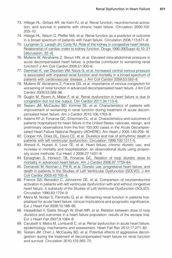
73. Hillege HL, Girbes AR, de Kam PJ, et al. Renal function, neurohormonal activa-tion, and survival in patients with chronic heart failure. Circulation 2000;102:203–10.
74. Hillege HL, Nitsch D, Pfeffer MA, et al. Renal function as a predictor of outcomein a broad spectrum of patients with heart failure. Circulation 2006;113:671–8.
75. Ljungman S, Laragh JH, Cody RJ. Role of the kidney in congestive heart failure.Relationship of cardiac index to kidney function. Drugs 1990;39(Suppl 4):10–21[discussion: 22–4].
76. Mullens W, Abrahams Z, Skouri HN, et al. Elevated intra-abdominal pressure inacute decompensated heart failure: a potential contributor to worsening renalfunction? J Am Coll Cardiol 2008;51:300–6.
77. Damman K, van Deursen VM, Navis G, et al. Increased central venous pressureis associated with impaired renal function and mortality in a broad spectrum ofpatients with cardiovascular disease. J Am Coll Cardiol 2009;53:582–8.
78. Mullens W, Abrahams Z, Francis GS, et al. Importance of venous congestion forworsening of renal function in advanced decompensated heart failure. J Am CollCardiol 2009;53:589–96.
79. Guglin M, Rivero A, Matar F, et al. Renal dysfunction in heart failure is due tocongestion but not low output. Clin Cardiol 2011;34:113–6.
80. Testani JM, McCauley BD, Kimmel SE, et al. Characteristics of patients withimprovement or worsening in renal function during treatment of acute decom-pensated heart failure. Am J Cardiol 2010;106:1763–9.
81. Adams KF Jr, Fonarow GC, Emerman CL, et al. Characteristics and outcomes ofpatients hospitalized for heart failure in the United States: rationale, design, andpreliminary observations from the first 100,000 cases in the Acute Decompen-sated Heart Failure National Registry (ADHERE). Am Heart J 2005;149:209–16.
82. Cooper HA, Dries DL, Davis CE, et al. Diuretics and risk of arrhythmic death inpatients with left ventricular dysfunction. Circulation 1999;100:1311–5.
83. Ahmed A, Husain A, Love TE, et al. Heart failure, chronic diuretic use, andincrease in mortality and hospitalization: an observational study using propen-sity score methods. Eur Heart J 2006;27:1431–9.
84. Eshaghian S, Horwich TB, Fonarow GC. Relation of loop diuretic dose tomortality in advanced heart failure. Am J Cardiol 2006;97:1759–64.
85. Domanski M, Norman J, Pitt B, et al. Diuretic use, progressive heart failure, anddeath in patients in the Studies of Left Ventricular Dysfunction (SOLVD). J AmColl Cardiol 2003;42:705–8.
86. Francis GS, Benedict C, Johnstone DE, et al. Comparison of neuroendocrineactivation in patients with left ventricular dysfunction with and without congestiveheart failure. A substudy of the Studies of Left Ventricular Dysfunction (SOLVD).Circulation 1990;82:1724–9.
87. Metra M, Nodari S, Parrinello G, et al. Worsening renal function in patients hos-pitalised for acute heart failure: clinical implications and prognostic significance.Eur J Heart Fail 2008;10:188–95.
88. Hasselblad V, Gattis Stough W, Shah MR, et al. Relation between dose of loopdiuretics and outcomes in a heart failure population: results of the escape trial.Eur J Heart Fail 2007;9:1064–9.
89. Carubelli V, Metra M, Lombardi C, et al. Renal dysfunction in acute heart failure:epidemiology, mechanisms and assessment. Heart Fail Rev 2012;17:271–82.
90. Testani JM, Chen J, McCauley BD, et al. Potential effects of aggressive decon-gestion during the treatment of decompensated heart failure on renal functionand survival. Circulation 2010;122:265–72.
Renal Dysfunction in Heart Failure 971

91. Felker GM, Lee KL, Bull DA, et al. Diuretic strategies in patients with acute de-compensated heart failure. N Engl J Med 2011;364:797–805.
92. Effects of enalapril on mortality in severe congestive heart failure. Results of theCooperative North Scandinavian Enalapril Survival Study (CONSENSUS). TheCONSENSUS Trial Study Group. N Engl J Med 1987;316:1429–35.
93. Bakris GL, Weir MR. Angiotensin-converting enzyme inhibitor-associated eleva-tions in serum creatinine: is this a cause for concern? Arch Intern Med 2000;160:685–93.
94. Ruiz-Ortega M, Ruperez M, Lorenzo O, et al. Angiotensin II regulates thesynthesis of proinflammatory cytokines and chemokines in the kidney. KidneyInt Suppl 2002;82:S12–22.
95. Ljungman S, Kjekshus J, Swedberg K. Renal function in severe congestive heartfailure during treatment with enalapril (the Cooperative North Scandinavian Ena-lapril Survival Study [CONSENSUS] trial). Am J Cardiol 1992;70:479–87.
96. Oster JR, Materson BJ. Renal and electrolyte complications of congestive heartfailure and effects of therapy with angiotensin-converting enzyme inhibitors.Arch Intern Med 1992;152:704–10.
97. Valika AA, Gheorghiade M. Ace inhibitor therapy for heart failure in patients withimpaired renal function: a review of the literature. Heart Fail Rev 2012. [Epubahead of print].
98. Colombo PC, Ganda A, Lin J, et al. Inflammatory activation: cardiac, renal, andcardio-renal interactions in patients with the cardiorenal syndrome. Heart FailRev 2012;17:177–90.
99. Ferrari R, Bachetti T, Confortini R, et al. Tumor necrosis factor soluble receptorsin patients with various degrees of congestive heart failure. Circulation 1995;92:1479–86.
100. Testa M, Yeh M, Lee P, et al. Circulating levels of cytokines and their endoge-nous modulators in patients with mild to severe congestive heart failure due tocoronary artery disease or hypertension. J Am Coll Cardiol 1996;28:964–71.
101. Maeda K, Tsutamoto T, Wada A, et al. High levels of plasma brain natriureticpeptide and interleukin-6 after optimized treatment for heart failure are indepen-dent risk factors for morbidity and mortality in patients with congestive heartfailure. J Am Coll Cardiol 2000;36:1587–93.
102. Kozdag G, Ertas G, Kilic T, et al. Elevated level of high-sensitivity C-reactiveprotein is important in determining prognosis in chronic heart failure. Med SciMonit 2010;16:CR156–61.
103. Alonso-Martinez JL, Llorente-Diez B, Echegaray-Agara M, et al. C-reactiveprotein as a predictor of improvement and readmission in heart failure. Eur JHeart Fail 2002;4:331–6.
104. Kalra D, Sivasubramanian N, Mann DL. Angiotensin ii induces tumor necrosisfactor biosynthesis in the adult mammalian heart through a protein kinaseC-dependent pathway. Circulation 2002;105:2198–205.
105. Moriyama T, Fujibayashi M, Fujiwara Y, et al. Angiotensin II stimulatesinterleukin-6 release from cultured mouse mesangial cells. J Am Soc Nephrol1995;6:95–101.
106. Murray DR, Prabhu SD, Chandrasekar B. Chronic beta-adrenergic stimulationinduces myocardial proinflammatory cytokine expression. Circulation 2000;101:2338–41.
107. Prabhu SD, Chandrasekar B, Murray DR, et al. Beta-adrenergic blockade indeveloping heart failure: effects on myocardial inflammatory cytokines, nitricoxide, and remodeling. Circulation 2000;101:2103–9.
Cole et al972

108. Colombo PC, Banchs JE, Celaj S, et al. Endothelial cell activation in patients withdecompensated heart failure. Circulation 2005;111:58–62.
109. Colombo PC, Rastogi S, Onat D, et al. Activation of endothelial cells in conduitveins of dogs with heart failure and veins of normal dogs after vascular stretchby acute volume loading. J Card Fail 2009;15:457–63.
110. Anker SD, Egerer KR, Volk HD, et al. Elevated soluble CD14 receptors andaltered cytokines in chronic heart failure. Am J Cardiol 1997;79:1426–30.
111. Niebauer J, Volk HD, Kemp M, et al. Endotoxin and immune activation in chronicheart failure: a prospective cohort study. Lancet 1999;353:1838–42.
112. Peschel T, Schonauer M, Thiele H, et al. Invasive assessment of bacterial endo-toxin and inflammatory cytokines in patients with acute heart failure. Eur J HeartFail 2003;5:609–14.
113. Yhee JY, Yu CH, Kim JH, et al. Effects of T lymphocytes, interleukin-1, andinterleukin-6 on renal fibrosis in canine end-stage renal disease. J Vet DiagnInvest 2008;20:585–92.
114. Szeto CC, Kwan BC, Chow KM, et al. Endotoxemia is related to systemic inflam-mation and atherosclerosis in peritoneal dialysis patients. Clin J Am Soc Nephrol2008;3:431–6.
115. Schwedler SB, Guderian F, Dammrich J, et al. Tubular staining of modifiedC-reactive protein in diabetic chronic kidney disease. Nephrol Dial Transplant2003;18:2300–7.
116. Radeke HH, Meier B, Topley N, et al. Interleukin 1-alpha and tumor necrosisfactor-alpha induce oxygen radical production in mesangial cells. Kidney Int1990;37:767–75.
117. DiPetrillo K, Coutermarsh B, Gesek FA. Urinary tumor necrosis factor contributesto sodium retention and renal hypertrophy during diabetes. Am J Physiol RenalPhysiol 2003;284:F113–21.
118. Garvin JL, Ortiz PA. The role of reactive oxygen species in the regulation oftubular function. Acta Physiol Scand 2003;179:225–32.
119. Taddei S, Favilla S, Duranti P, et al. Vascular renin-angiotensin system andneurotransmission in hypertensive persons. Hypertension 1991;18:266–77.
120. Fiksen-Olsen MJ, Strick DM, Hawley H, et al. Renal effects of angiotensin II inhi-bition during increases in renal venous pressure. Hypertension 1992;19:II137–41.
121. Kastner PR, Hall JE, Guyton AC. Renal hemodynamic responses to increasedrenal venous pressure: role of angiotensin II. Am J Physiol 1982;243:F260–4.
122. Damman K, Voors AA, Navis G, et al. Current and novel renal biomarkers inheart failure. Heart Fail Rev 2012;17:241–50.
123. Fonarow GC, Adams KF Jr, Abraham WT, et al. Risk stratification for in-hospitalmortality in acutely decompensated heart failure: classification and regressiontree analysis. JAMA 2005;293:572–80.
124. Aronson D, Mittleman MA, Burger AJ. Elevated blood urea nitrogen level asa predictor of mortality in patients admitted for decompensated heart failure.Am J Med 2004;116:466–73.
125. Comnick M, Ishani A. Renal biomarkers of kidney injury in cardiorenalsyndrome. Curr Heart Fail Rep 2011;8:99–105.
126. Laterza OF, Price CP, Scott MG. Cystatin C: an improved estimator of glomerularfiltration rate? Clin Chem 2002;48:699–707.
127. Newman DJ, Thakkar H, Edwards RG, et al. Serum cystatin C measured byautomated immunoassay: a more sensitive marker of changes in GFR thanserum creatinine. Kidney Int 1995;47:312–8.
Renal Dysfunction in Heart Failure 973

128. Hoek FJ, Kemperman FA, Krediet RT. A comparison between cystatin C, plasmacreatinine and the Cockcroft and Gault formula for the estimation of glomerularfiltration rate. Nephrol Dial Transplant 2003;18:2024–31.
129. Tidman M, Sjostrom P, Jones I. A comparison of GFR estimating formulae basedupon S-cystatin C and S-creatinine and a combination of the two. Nephrol DialTransplant 2008;23:154–60.
130. Lassus J, Harjola VP, Sund R, et al. Prognostic value of cystatin C in acute heartfailure in relation to other markers of renal function and NT-proBNP. Eur Heart J2007;28:1841–7.
131. Schmidt-Ott KM, Mori K, Li JY, et al. Dual action of neutrophil gelatinase-associated lipocalin. J Am Soc Nephrol 2007;18:407–13.
132. Mishra J, Dent C, Tarabishi R, et al. Neutrophil gelatinase-associated lipocalin(NGAL) as a biomarker for acute renal injury after cardiac surgery. Lancet2005;365:1231–8.
133. Aghel A, Shrestha K, Mullens W, et al. Serum neutrophil gelatinase-associatedlipocalin (NGAL) in predicting worsening renal function in acute decompen-sated heart failure. J Card Fail 2010;16:49–54.
134. Han WK, Bailly V, Abichandani R, et al. Kidney injury molecule-1 (KIM-1): a novelbiomarker for human renal proximal tubule injury. Kidney Int 2002;62:237–44.
135. Han WK, Waikar SS, Johnson A, et al. Urinary biomarkers in the early diagnosisof acute kidney injury. Kidney Int 2008;73:863–9.
136. Damman K, Van Veldhuisen DJ, Navis G, et al. Tubular damage in chronicsystolic heart failure is associated with reduced survival independent of glomer-ular filtration rate. Heart 2010;96:1297–302.
137. Westhuyzen J, Endre ZH, Reece G, et al. Measurement of tubular enzymuriafacilitates early detection of acute renal impairment in the intensive care unit.Nephrol Dial Transplant 2003;18:543–51.
138. Bazzi C, Petrini C, Rizza V, et al. Urinary N-acetyl-beta-glucosaminidase excre-tion is a marker of tubular cell dysfunction and a predictor of outcome in primaryglomerulonephritis. Nephrol Dial Transplant 2002;17:1890–6.
139. Liangos O, Perianayagam MC, Vaidya VS, et al. Urinary N-acetyl-beta-(D)-glu-cosaminidase activity and kidney injury molecule-1 level are associated withadverse outcomes in acute renal failure. J Am Soc Nephrol 2007;18:904–12.
140. Maatman RG, Van Kuppevelt TH, Veerkamp JH. Two types of fatty acid-bindingprotein in human kidney. Isolation, characterization and localization. Biochem J1991;273(Pt 3):759–66.
141. FergusonMA,VaidyaVS,WaikarSS, et al.Urinary liver-type fatty acid-bindingproteinpredicts adverse outcomes in acute kidney injury. Kidney Int 2010;77:708–14.
142. Agewall S, Wikstrand J, Ljungman S, et al. Usefulness of microalbuminuria inpredicting cardiovascular mortality in treated hypertensive men with and withoutdiabetes mellitus. Risk Factor Intervention Study Group. Am J Cardiol 1997;80:164–9.
143. Mogensen CE. Microalbuminuria predicts clinical proteinuria and early mortalityin maturity-onset diabetes. N Engl J Med 1984;310:356–60.
144. van de Wal RM, Asselbergs FW, Plokker HW, et al. High prevalence of microal-buminuria in chronic heart failure patients. J Card Fail 2005;11:602–6.
145. Jackson CE, Solomon SD, Gerstein HC, et al. Albuminuria in chronic heartfailure: prevalence and prognostic importance. Lancet 2009;374:543–50.
146. Masson S, Latini R, Milani V, et al. Prevalence and prognostic value of elevatedurinary albumin excretion in patients with chronic heart failure: data from theGISSI-heart failure trial. Circ Heart Fail 2010;3:65–72.
Cole et al974
Copyright © 2022 FDOKUMEN