The role of HUCB derived stem cells therapy in repair of renal damage and improvement of renal...
9
J. Afr. Ass. Physiol. Sci. 1 (1): 27 - 35 Journal of African Association of Physiological Sciences http://www.jaaps.aapsnet.org Research Article The role of HUCB derived stem cells therapy in repair of renal damage and improvement of renal function in cisplatin induced acute renal failure in rats 1 Taddesse A. A, 2 Mohamed M. I., 3 Rahman A. A & 2 El-Wazir Y. M 1 Department of Physiology, Hawassa University , Addis Ababa, Ethiopia 2,4 Physiology Department, Faculty of Medicine, Suez Canal University, Ismailia, Egypt 3 Histology Department, Faculty of Medicine, Suez Canal University, Ismailia, Egypt Keywords: Acute renal failure; CD 34 + cells; Umbilical cord blood; Mesenchymal stem cells. ABSTRACT Acute renal failure (ARF) is a common clinical problem with increasing incidence, serious consequences, unsatisfactory therapeutic options and enormous financial burden to society. The aim is to investigate the role of human umbilical cord blood (HUCB) derived mesenchymal (MSCs) and CD34 + hematopoietic stem cell therapy in repair of renal damage and improvement of renal function in cisplatin-induced ARF model. Forty four rats were divided into 4 equal groups. ARF was induced in 3 groups using cisplatin and was confirmed by an increase in serum urea and creatinine levels after 5 days. On the same day, 2 groups were injected via the tail vein by either MSCs (1x10 6 cells/rat) or CD34 + hematopoietic cells (5 x10 5 cells/rat). The third group received intravenous injection of phosphate buffer saline and served as positive control, while the last group was normal control. Renal functions were followed up every 4 days. Thirty-three days after initiation of cisplantin injection, rats were sacrificed, kidneys were extracted for histopathological and immunohistochemical examination for detection of human specific anti-vimentin monoclonal mouse anti-body to investigate homing of HUCB stem cells into the damaged renal tissue. Treatment with MSCs and CD34 + cells significantly decreased both serum urea and creatinine induced by cisplatin administration with concomitant improvement in the degree of necrotic and degenerative changes. There was no significant difference in these parameters between MSCs and CD 34 + stem cells treated groups. There was positive reaction for human specific anti-vimentin in 88.9% of animals in MSCs treated rats versus 87.5% in CD34 + cells treated rats. HUCB derived CD 34 + and MSCs accelerate regeneration of renal tubular epithelial cells and lead to reduction of progressive renal injury in cisplatin-induced acute renal failure rats. © Copyright 2013 African Association of Physiological Sciences -ISSN: 2315-9987. All rights reserved ______________________________________________________________________________________________________ INTRODUCTION 1 The clinical condition of ARF is a common cause of morbidity and mortality and no satisfactory management is currently available (Bellomo et al., *Address for correspondence: Email: [email protected] 2004). In the early 2000s, pluripotent bone marrow– derived stem cells were thought to contribute directly to regeneration of the kidney (Kale et al., 2003). Togel et al., (2005) demonstrated that administration of bone marrow derived stem cell (BMSC) significantly improved renal function in the ischemia/reperfusion model. Also, Bi et al., (2007) demonstrated that transfer of BMSC into the peritoneal cavity accelerates recovery from cisplatin-induced ARF in mice. There are many problems with bone marrow cell transplantation, such as the limitation of cell number, the donor’s physical responsibility and the prevention of rejection (Nonome et al., 2005). Human umbilical
Transcript of The role of HUCB derived stem cells therapy in repair of renal damage and improvement of renal...

J. Afr. Ass. Physiol. Sci. 1 (1): 27 - 35
Journal of African Association of Physiological Sciences http://www.jaaps.aapsnet.org
Research Article The role of HUCB derived stem cells therapy in repair of renal damage and improvement of renal function in cisplatin induced acute renal failure in rats 1Taddesse A. A, 2Mohamed M. I., 3Rahman A. A & 2El-Wazir Y. M 1Department of Physiology, Hawassa University , Addis Ababa, Ethiopia 2,4Physiology Department, Faculty of Medicine, Suez Canal University, Ismailia, Egypt 3Histology Department, Faculty of Medicine, Suez Canal University, Ismailia, Egypt
Keywords: Acute renal failure; CD 34+ cells; Umbilical cord blood; Mesenchymal stem cells.
ABSTRACT Acute renal failure (ARF) is a common clinical problem with increasing incidence, serious consequences, unsatisfactory therapeutic options and enormous financial burden to society. The aim is to investigate the role of human umbilical cord blood (HUCB) derived mesenchymal (MSCs) and CD34+ hematopoietic stem cell therapy in repair of renal damage and improvement of renal function in cisplatin-induced ARF model. Forty four rats were divided into 4 equal groups. ARF was induced in 3 groups using cisplatin and was confirmed by an increase in serum urea and creatinine levels after 5 days. On the same day, 2 groups were injected via the tail vein by either MSCs (1x10⁶ cells/rat) or CD34+ hematopoietic cells (5 x10⁵ cells/rat). The third group received intravenous injection of phosphate buffer saline and served as positive control, while the last group was normal control. Renal functions were followed up every 4 days. Thirty-three days after initiation of cisplantin injection, rats were sacrificed, kidneys were extracted for histopathological and immunohistochemical examination for detection of human specific anti-vimentin monoclonal mouse anti-body to investigate homing of HUCB stem cells into the damaged renal tissue. Treatment with MSCs and CD34+ cells significantly decreased both serum urea and creatinine induced by cisplatin administration with concomitant improvement in the degree of necrotic and degenerative changes. There was no significant difference in these parameters between MSCs and CD 34+ stem cells treated groups. There was positive reaction for human specific anti-vimentin in 88.9% of animals in MSCs treated rats versus 87.5% in CD34+ cells treated rats. HUCB derived CD 34+ and MSCs accelerate regeneration of renal tubular epithelial cells and lead to reduction of progressive renal injury in cisplatin-induced acute renal failure rats.
© Copyright 2013 African Association of Physiological Sciences -ISSN: 2315-9987. All rights reserved
______________________________________________________________________________________________________ INTRODUCTION 1 The clinical condition of ARF is a common cause of morbidity and mortality and no satisfactory management is currently available (Bellomo et al., *Address for correspondence: Email: [email protected]
2004). In the early 2000s, pluripotent bone marrow–derived stem cells were thought to contribute directly to regeneration of the kidney (Kale et al., 2003). Togel et al., (2005) demonstrated that administration of bone marrow derived stem cell (BMSC) significantly improved renal function in the ischemia/reperfusion model. Also, Bi et al., (2007) demonstrated that transfer of BMSC into the peritoneal cavity accelerates recovery from cisplatin-induced ARF in mice. There are many problems with bone marrow cell transplantation, such as the limitation of cell number, the donor’s physical responsibility and the prevention of rejection (Nonome et al., 2005). Human umbilical
Stem cell therapy for renal damage
28 J. Afr. Ass. Physiol. Sci. 1 (1): Taddesse, Mohammed, Rahaman and El-Wazir
cord blood (HUCB), however, can be collected without any harm to the newborn infant and has been found to be multipotent and cause fewer episodes of rejection because of the relative immaturity of its cells (Lewis 2002). Umbilical cord blood (UCB) contains circulating stem /progenitor cells and its cellular contents are known to be quite distinct (Kakinuma et al., 2003). HUCB cells have also been reported to differentiate into a variety of cell types such as cartilage, muscle (Erices et al., 2000), bone and nerve cells (Goodwin et al., 2001) and hepatocytes (Kakinuma et al., 2003). To the best of our knowledge, the application of HUCB derived CD34+ cells in treating ARF has not been reported. However, the application of HUCB MSCs in treating ARF has been reported in few studies (Cao et al., 2010 and Morigi et al., 2010). In a previous study, we compared the effects of HUCB progenitor cells and mononuclear cells (MNCs) on glycerol induced ARF and found that, HUCB CD34+ cells and HUCB MNCs improved renal function of nephrotoxic kidney with superiority to the HUCB MNCs (Mohamed et al., 2011). Therefore, the applications of HUCB stem cells should be further expanded and may serve as an excellent alternative to BMSC for cell transplantation in ARF. In this study we will investigate the role of HUCB MSCs and CD34+ hematopoietic stem cells therapy in repair of renal damage and improvement of renal function in cisplatin-induced ARF. Also, we will compare the regenerative effect of MSCs and CD34+ hematopoietic stem cells to suggest the better cell type. MATERIALS AND METHODS Animals: Forty four female Wistar rats, aged 8-12 weeks with average weight of 148.6 ± 19.5 gm were randomly selected in this study. All rats were housed in small cages at room temperature. They were maintained at a 12-h day and night cycle. They were acclimatized for one week and kept with free access to standard pellet animal diet and tap water. Rats were randomly divided into four equal groups (11 animals each): group I (GI) Negative control group: without any intervention, group II (GII) Positive control group: was subjected to induction of ARF with intraperitoneal cisplatin 7 mg/kg (Vaziri et al., 1994) then received intravenous injection of phosphate buffer saline, groups III (GIII) MSC group, and IV (GIV) CD34+ group: were subjected to induction of renal failure as in group II, then to
transplantation of either HUCB MSCs in a dose of 1x10⁶ cells/ rat, or to HUCB CD34+ cells in a dose of 5x10⁵ cells/ rat after five days through the tail vein after an alcohol swab (Cao et al., 2010). Study design: Renal function was assessed by measuring serum urea and creatinine levels. Blood samples were obtained from retro-orbital sinuses of the rats before induction of ARF (basal level), 5 days after induction to confirm its occurrence and then measured every 4 days afterwards until the animals were sacrificed 33 days after induction of ARF by decapitation under ether anaesthesia. Kidneys were isolated and prepared for histopathological and immunohistochemical examination. Measurement of Serum Urea and Creatinine Levels: Blood samples were centrifuged at 3000 rpm for 10 min and the supernatant was stored at -70°C for later determination of serum urea and creatinine which were done by vitro Scient colorimetric kits (Vitro Scient, Inshas Industrial Zone, Belbis, Sharkia, Egypt) according to manufacturer’s manual. Collection of Umbilical Cord Blood (UCB) Samples: HUCB samples were obtained at the end of full-term vaginal deliveries of male babies after clamping and cutting of the cord. Ten HUCB samples were collected while the placenta was still in utero (Dhot et al., 2003). The umbilical vein was cleansed with alcohol followed by betadine and pierced with a sterile needle, and then UCB was collected into sterile collection tubes containing 5 ml of citrate phosphate dextrose adenine-1 anticoagulant. UCB samples were stored at 4°C and processed within 24 hours of collection. Separation of Human Umbilical Cord Blood CD34+ Cells (HUCB CD34+ Cells): To isolate MNCs, each UCB unit was diluted 1:1 with phosphate buffer saline (PBS)/2mM EDTA. UCB was centrifuged at 1500 rpm for 35 min at 25°C on a layer of Ficoll-Hypaque solution (Sigma chemical co., USA). The mononuclear cell layer was obtained by a micropipette. The cells were incubated for 20 min at 4°C and washed with PBS. CD34+ cells were separated and purified by immunomagnetic separation technique (Milteny, 1990) (Dynal beads Oslo Norway) according to the manufacturer’s instructions obtaining purity of approximately 97% for CD34+ cells. Upon mixing and incubation, CD34+ cells bind to Dynabeads M-450 CD34 and the rosetted cells are isolated from the suspension by using a DYNAL Magnetic Particle
Stem cell therapy for renal damage
29 J. Afr. Ass. Physiol. Sci. 1 (1): Taddesse, Mohammed, Rahaman and El-Wazir
Concentrator (DYNAL MPC, Oslo Norway). A subsequent incubation with DETACHaBEAD CD34 gently detaches isolated cells from the beads. A DYNAL MPC is then used to separate the purified, positively selected CD34+ cells from the released Dynabeads M-450 CD34. Isolation of Human Umbilical Cord Blood Mesenchymal Stem cells (HUCB MSCs): MSCs were recovered from HUCB by their tendency to adhere tightly to plastic culture dishes (Pereira et al., 1995). Filtered HUCB cells were plated in Dulbecco's Modified Eagle Medium (DMEM) plus 10% fetal calf serum (FCS) and penicillin-streptomycin (100 U/ml to 0.1 mg/ml) and allowed to adhere for 24 h. Non adherent cells were then removed. Medium was changed regularly every 3 days; after 2 to 3 weeks, adherent cells were detached by trypsin (0.25%; Lonza Biproducts, Belgium), washed with PBS, and used for the in vivo experiments. Culture and subculture of MSCs were carried out according to Erices et al., (2000) (Lonza Biproducts, Belgium) MSCs cultures were monitored periodically for cell detachment by observing the cells under the inverted microscope. Optimally cultured cells were used after 2 weeks of growth. Histopathology: After scarification, the two kidneys of each animal were removed, fixed in 10% neutral buffered formalin saline for 24 hours. Specimens were processed to paraffin sections, 5 µ thick, and then stained with Hematoxylin and Eosin (H & E) stain for kidneys general architecture evaluation, PAS stain for evaluation of the basement membranes & tubular brush borders and Immuonohistochemistry (IHC) using human specific anti-vimentin monoclonal mouse anti-body for evaluation of transplanted stem cells homing. Twelve serial sections were taken from each block, four sections for each stain (H & E, PAS and IHC). Morphological analysis was performed to determine the kidney architecture, necrotic and degenerative changes. In addition, the extent of regeneration was also looked at in all slides. Statistical Analysis: All statistical analysis was performed using the
statistical software package SPSS 15.0 for Windows® (SPSS Inc., Chicago, IL, USA). The results were expressed as mean ± SD. Analysis of variance (ANOVA) for non-parametric data was used to
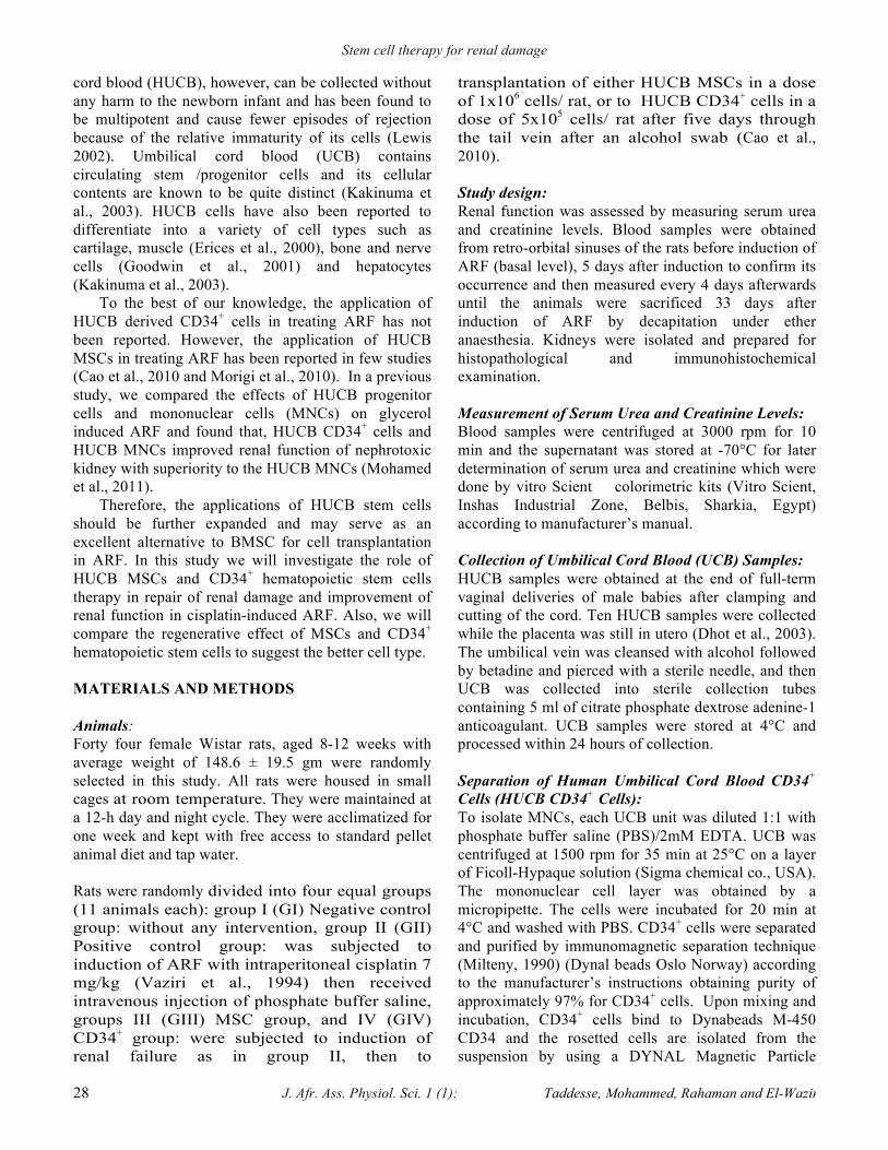
elucidate differences between all groups. Post hoc range tests were used to test the difference between each pair of means. Statistical significance level was defined as P < 0.05. Frequency and percentage were done for qualitative data. Ethical Consideration: The study was approved by the ethical committee of the Faculty of Medicine, Suez Canal University. In brief, the purpose of the study was explained and a written consent was taken from the study participants (cord blood donors). The used methods were safe and had no harm to mothers or neonates. The samples were used only in this study and then discarded. . Animals were handled gently, housed in suitable environmental conditions, and allowed free access to water and rat chow. All animals were anesthetized before sacrifice. RESULTS Blood Chemistry: In day 5, both serum creatinine (Table 1) and serum urea (Table 2) showed a significant increment in GII, GIII, and GIV when compared to GI. In GII only, this increment progressively increased until day 25, while it decreased in both GIII and GIV starting from day 9. Afterwards, serum creatinine level showed slight fluctuations and a comparable level was detected in the last sample at day 33. Table 1: Creatinine levels in all the study groups through the study. Day GI GII GIII GIV P
value 0 0.92±.07 0.80±.16 0.82±.169 0.83±.34 NS
5 0.87±.11 4.24±2.12" 4.02±.91" 3.64±.73" .000
9 0.74±.11 4. 18±.16" 2.3±1.65 1.89±1.11* .020
13 0.67±.08 5.66±1.24" 3.42±1.93 3.61±1.06 .016
17 0.67±.10 6.07±1.13" 2.66±1.72* 3.03±1.12* .004
21 0.98±.12 7.39±.33" 2.26±1.15* 2.476±.80* .000
25 0.65±.07 8.23±.75 2.53±.69* 1.86±1.19* .000
29 0.85±.10 6.73±.56" 2.43±.71* 2.4±.39* .000
33 0.65±.06 7.76±.33" 2.005±.75* 1.94±.63* .000
G1: Negative control, G2: Positive control, G3: MSCs Treated group, G4: CD 34+ Treated group. " Represents a statistically significant difference between the given group and group 1. * Represents a statistically significant difference between the given group and group 2.

Stem cell therapy for renal damage
30 J. Afr. Ass. Physiol. Sci. 1 (1): Taddesse, Mohammed, Rahaman and El-Wazir
Table 2: Urea levels in all the study groups through the study
Days GI GII GIII GIV P value 0 18.5 ±2.24 17.5±2.24 19.36±2.24 18.94±2.13 NS 5 18.75±1.43 65.6±35.98" 92.7±35.98" 76.07±14.03" .001 9 16.87±1.17 65.2±8.79" 30.45±7.18* 30.2±10.48* .000 13 15.67±2.34 83.43±19.17" 35.67±13.18* 32.25±14.91* .001 17 17.88±2.20 94.54±5.02" 37.677±15.41* 43.34±9.79* .000 21 19.87±1.42 84.23±4.43" 36.5±8.28" * 31.74±9.75* .000 25 15.31±2.20 108.76±4.41" 35.89±13.58* 23.457±9.34* .000 29 16.64±2.28 82.34±8.18" 26.33±8.89* 51.265±20.11* .001 33 19.23±1.40 93.21±7.90" 61.53±26.95* 32.14±10.90* .000
G1: Negative control, G2: Positive control, G3: MSCs Treated group, G4: CD 34+ Treated group. " Represents a statistically significant difference between the given group and group 1. * Represents a statistically significant difference between the given group and group 2.
Fig.1: Pattern of serum creatinine levels in all the study groups. GI: negative control group, GII: positive control group, GIII: MSCs treated group and GIV: CD34+ treated group
Fig. 2: Pattern of serum urea levels in all the study groups. GI: negative control group, GII: positive control group, GIII: MSCs treated group and GIV: CD34+ treated group
0
5
10
0 5 9 13 17 21 25 29 33
mg/dl
Crea+nine GI GII GIII GIV
Time (Days)
0 20 40 60 80
100 120
0 5 9 13 17 21 25 29 33
mg/dl
Urea GI
GII
GIII
GIV
Time (Days)
Stem cell therapy for renal damage
31 J. Afr. Ass. Physiol. Sci. 1 (1): Taddesse, Mohammed, Rahaman and El-Wazir
Statistically significant difference was observed on days 13, 17, 21, 25, 29 and 33 between positive control and HUCB MSCs groups. HUCB CD34+ group showed a statistically significant difference on days 9, 17, 21, 25, 29 and 33 as compared to positive control group. Regarding serum urea, statistically significant differences were observed at all days between positive control and both treatment groups. There was no statistically significant difference between HUCB CD34+ and HUCB MSCs treated groups for both serum creatinine and serum urea levels. The pattern of renal function tests in the time linear graphs are depicted in Figs. 1 & 2. Histopathology and immunohistochemistry: Histopathological examination revealed a significant difference between treated groups (GIII and GIV) and the positive control group (GII). Approximately 80 % of rats treated with either MSC or CD 34+ regained a normal nephronal architecture.
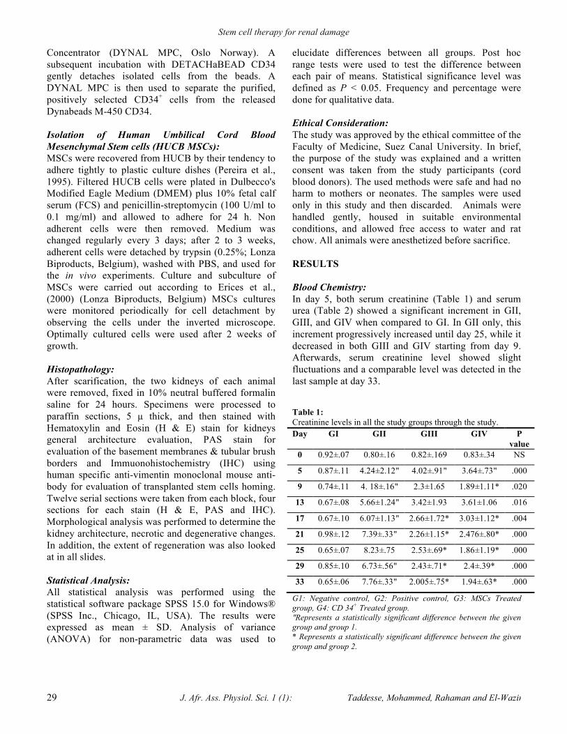
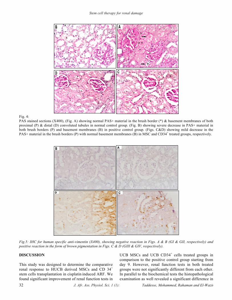
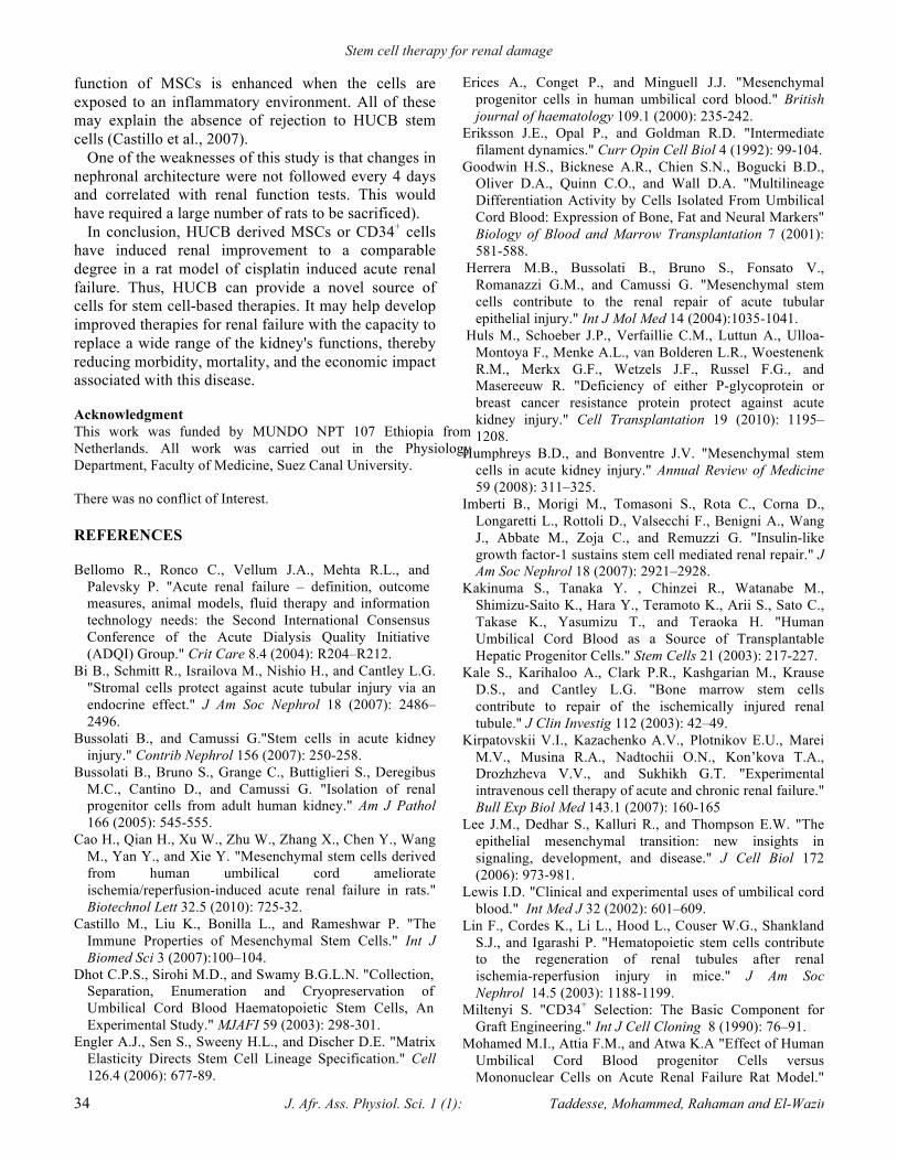
Kidneys of the control group (GI) showed normal renal architecture of both renal glomeruli, proximal and distal renal tubules (Fig. 3A & 4A) and negative reaction for human specific anti-vimentin (Fig. 5A). Group II showed necrotic changes of the tubular epithelium (Fig. 3B) with severe loss of PAS+ material (Fig. 4B) in 75% of the animals and negative reaction for human specific anti-vimentin (Fig. 5B). Group III showed improvement of the proximal and distal tubular epithelium in 88.9% of the animals (Fig. 3C), mild decrease in PAS+ material (Fig. 4C) and positive reaction for human specific anti-vimentin in 88.9% of animals (Fig. 5C). Group IV showed improvement of the proximal and distal tubular epithelium in 87.5% of the animals (Fig. 3D), mild decrease in PAS+ material (Fig. 4D) and positive reaction for human specific anti-vimentin in 87.5% of animals (Fig. 5D).
Fig. 3: H&E (X400) stained sections: (Fig. A) showing renal glomerulus (G), Proximal (P) & distal (D) convoluted tubules in normal control group. (Fig. B) showing necrotic changes in the form of nuclear pyknosis (P), karyolysis (K) & cytoplasmic vacuolation (V) in positive control group. (Figs. C & D) showing improvement of the tubular epithelium in MSC and CD34+ treated groups, respectively.
Stem cell therapy for renal damage
32 J. Afr. Ass. Physiol. Sci. 1 (1): Taddesse, Mohammed, Rahaman and El-Wazir
Fig. 4: PAS stained sections (X400), (Fig. A) showing normal PAS+ material in the brush border (*) & basement membranes of both proximal (P) & distal (D) convoluted tubules in normal control group. (Fig. B) showing severe decrease in PAS+ material in both brush borders (P) and basement membranes (B) in positive control group. (Figs. C&D) showing mild decrease in the PAS+ material in the brush borders (P) with normal basement membranes (B) in MSC and CD34+ treated groups, respectively.
Fig.5: IHC for human specific anti-vimentin (X400), showing negative reaction in Figs. A & B (GI & GII, respectively) and positive reaction in the form of brown pigmentation in Figs. C & D (GIII & GIV, respectively).
DISCUSSION This study was designed to determine the comparative renal response to HUCB derived MSCs and CD 34+ stem cells transplantation in cisplatin induced ARF. We found significant improvement of renal function tests in
UCB MSCs and UCB CD34+ cells treated groups in comparison to the positive control group starting from day 9. However, renal function tests in both treated groups were not significantly different from each other. In parallel to the biochemical tests the histopathological examination as well revealed a significant difference in

Stem cell therapy for renal damage
33 J. Afr. Ass. Physiol. Sci. 1 (1): Taddesse, Mohammed, Rahaman and El-Wazir
regaining a normal nephronal architecture in both treated groups and they were reactive to the human specific anti-vimentin antibody, which provides evidence of homing of the transplanted cells in the kidney tissue of affected rats. Our results are in agreement with those of Cao et al., (2010), who reported that the administration of UCB MSC led to improved renal function 16 h after reperfusion compared to that in control animals, as well as the histological changes showing recovery of tissues in UCB MSC treated rats. Also, Morigie et al., (2010) observed that infusion of UCB-MSCs in immunodeficient mice with cisplatin-induced acute kidney injury ameliorated both renal function and tubular cell injury, and prolonged survival. These results also confirm what we had reported in a previous study that renal function in glycerol induced ARF in rats improved after 4 days of HUCB CD43+ therapy and was restored after 2 weeks of therapy (Mohamed et al., 2011). In this study, the improvement of the rats' renal function started 9 days after injection of UCB derived CD 34+ and MSCs, which was similar to the results of Kirpatovskii et al., (2007) who found similar improvement after 2 weeks of kidney cells or bone marrow MSCs injection. They also found that kidney function remained normal during the remainder of the study period and this was also evident in our study) In our study, full recovery wasn't achieved in all cases. This is in contrast to the results of Morigi et al., (2004) who found that, injection of MSCs protected cisplatin-treated animals from renal function impairment and severe tubular injury and Tögel et al., (2005) who demonstrated full recovery of ischemic ARF model within three days of MSCs administrations. This discrepancy may be explained by the intracarotid administration of MSCs to rats either immediately or 24 hours after renal ischemia in the study of Tögel et al., (2005) and the early transplantation of cells in the study of Morigi et al., (2004) as they transplanted the cells one day after cisplatin injection. The current study’s striking finding that HUCB-derived stem cells significantly improved nephronal and proximal tubular regeneration 28 days after transplantation is comparable to the results of Lin et al., (2003) who confirmed the nephronal regeneration in rats with renal ischemia/reperfusion injury treated with BMSCs. Imberti et al., (2007) also detected improvement in renal structure and significant increase in tubular cell proliferation at 4 and 11 days post-MSC treatment. MSC differentiation to tubular epithelial cells and promotion of epithelial proliferation were also reported in mice with glycerol induced ARF after MSC therapy (Herrera et al., 2004). Recently, Morigi et al.,
(2008) observed that, human bone marrow derived MSCs treatment could prevent acute kidney injury induced by cisplatin and prolong survival in an immunodeficient mouse model. Although Tögel et al., (2005) reported that MSCs had not differentiated into tubular epithelium or renal endothelium, suggesting that the observed beneficial effects had primarily been mediated via complex paracrine actions and not by stem cell-derived de novo generation of renal parenchyma, Bussolati and Camussi (2007) confirmed that the administered stem cells (SCs) may modify the microenvironment by inducing dedifferentiation and proliferation of tubular cells surviving from injury or by allowing expansion of resident SCs. This was confirmed in our study by the finding that the transplanted HUBC CD34+ cells and HUBC MSCs were identified by the positive reaction to human specific vimentin antibody. Vimentin is the most ubiquituos intermediate filament protein and the first to be expressed during cell differentiation. All primitive cell types express vimentin (Eriksson et al., 1992). It is thus used as a marker for neural stem/progenitor cells in adult brain (Walder et al., 2003). Recent studies show that renal tissue–derived CD133 positive cells express vimentin before they fully differentiate into epithelial cells (Bussolati et al., 2005). The anti-Human Vimentin Antibody was screened on a human mesenchymal stem cell line and on human fibroblast cells by immunocytochemistry (Lee et al., 2006). The exact mechanism through which MSCs mitigate renal damage is not fully elucidated. One possible explanation is that MSCs produce cytokines and growth factors that promote anti-inflammatory, immunosuppressive, anti-apoptotic and proliferative effects (Humphreys and Bonventre, 2008). Recently, Huls et al., (2010) revealed that, bone marrow derived MSCs may have the capacity to migrate to the injured kidney and contribute to tubule epithelium regeneration and renal function repair without fusing with resident tubular cells. Finally, Togel and Westenfelder (2011) proposed that MSCs must provide paracrine and/or endocrine factors that explain their positive effects on kidney repair following injury. Theoretically MSCs have a better capacity for self-renewal while maintaining their multipotency (Engler et al., 2006). They are also a better candidate for renal repair, because nephrons are of mesenchymal origin (Rayan et al., 2005) which is proved in our study. MSCs have a better immunomodulatory effect by virtue of avoiding allorecognition, interference with T cell function and generation of local immunosuppressive microenvironment by secreting cytokines. Ryan et al., (2005) also concluded that the immunomodulatory
Stem cell therapy for renal damage
34 J. Afr. Ass. Physiol. Sci. 1 (1): Taddesse, Mohammed, Rahaman and El-Wazir
function of MSCs is enhanced when the cells are exposed to an inflammatory environment. All of these may explain the absence of rejection to HUCB stem cells (Castillo et al., 2007). One of the weaknesses of this study is that changes in nephronal architecture were not followed every 4 days and correlated with renal function tests. This would have required a large number of rats to be sacrificed). In conclusion, HUCB derived MSCs or CD34+ cells have induced renal improvement to a comparable degree in a rat model of cisplatin induced acute renal failure. Thus, HUCB can provide a novel source of cells for stem cell-based therapies. It may help develop improved therapies for renal failure with the capacity to replace a wide range of the kidney's functions, thereby reducing morbidity, mortality, and the economic impact associated with this disease. Acknowledgment This work was funded by MUNDO NPT 107 Ethiopia from Netherlands. All work was carried out in the Physiology Department, Faculty of Medicine, Suez Canal University. There was no conflict of Interest. REFERENCES Bellomo R., Ronco C., Vellum J.A., Mehta R.L., and
Palevsky P. "Acute renal failure – definition, outcome measures, animal models, fluid therapy and information technology needs: the Second International Consensus Conference of the Acute Dialysis Quality Initiative (ADQI) Group." Crit Care 8.4 (2004): R204–R212.
Bi B., Schmitt R., Israilova M., Nishio H., and Cantley L.G. "Stromal cells protect against acute tubular injury via an endocrine effect." J Am Soc Nephrol 18 (2007): 2486–2496.
Bussolati B., and Camussi G."Stem cells in acute kidney injury." Contrib Nephrol 156 (2007): 250-258.
Bussolati B., Bruno S., Grange C., Buttiglieri S., Deregibus M.C., Cantino D., and Camussi G. "Isolation of renal progenitor cells from adult human kidney." Am J Pathol 166 (2005): 545-555.
Cao H., Qian H., Xu W., Zhu W., Zhang X., Chen Y., Wang M., Yan Y., and Xie Y. "Mesenchymal stem cells derived from human umbilical cord ameliorate ischemia/reperfusion-induced acute renal failure in rats." Biotechnol Lett 32.5 (2010): 725-32.
Castillo M., Liu K., Bonilla L., and Rameshwar P. "The Immune Properties of Mesenchymal Stem Cells." Int J Biomed Sci 3 (2007):100–104.
Dhot C.P.S., Sirohi M.D., and Swamy B.G.L.N. "Collection, Separation, Enumeration and Cryopreservation of Umbilical Cord Blood Haematopoietic Stem Cells, An Experimental Study." MJAFI 59 (2003): 298-301.
Engler A.J., Sen S., Sweeny H.L., and Discher D.E. "Matrix Elasticity Directs Stem Cell Lineage Specification." Cell 126.4 (2006): 677-89.
Erices A., Conget P., and Minguell J.J. "Mesenchymal progenitor cells in human umbilical cord blood." British journal of haematology 109.1 (2000): 235-242.
Eriksson J.E., Opal P., and Goldman R.D. "Intermediate filament dynamics." Curr Opin Cell Biol 4 (1992): 99-104.
Goodwin H.S., Bicknese A.R., Chien S.N., Bogucki B.D., Oliver D.A., Quinn C.O., and Wall D.A. "Multilineage Differentiation Activity by Cells Isolated From Umbilical Cord Blood: Expression of Bone, Fat and Neural Markers" Biology of Blood and Marrow Transplantation 7 (2001): 581-588.
Herrera M.B., Bussolati B., Bruno S., Fonsato V., Romanazzi G.M., and Camussi G. "Mesenchymal stem cells contribute to the renal repair of acute tubular epithelial injury." Int J Mol Med 14 (2004):1035-1041.
Huls M., Schoeber J.P., Verfaillie C.M., Luttun A., Ulloa-Montoya F., Menke A.L., van Bolderen L.R., Woestenenk R.M., Merkx G.F., Wetzels J.F., Russel F.G., and Masereeuw R. "Deficiency of either P-glycoprotein or breast cancer resistance protein protect against acute kidney injury." Cell Transplantation 19 (2010): 1195–1208.
Humphreys B.D., and Bonventre J.V. "Mesenchymal stem cells in acute kidney injury." Annual Review of Medicine 59 (2008): 311–325.
Imberti B., Morigi M., Tomasoni S., Rota C., Corna D., Longaretti L., Rottoli D., Valsecchi F., Benigni A., Wang J., Abbate M., Zoja C., and Remuzzi G. "Insulin-like growth factor-1 sustains stem cell mediated renal repair." J Am Soc Nephrol 18 (2007): 2921–2928.
Kakinuma S., Tanaka Y. , Chinzei R., Watanabe M., Shimizu-Saito K., Hara Y., Teramoto K., Arii S., Sato C., Takase K., Yasumizu T., and Teraoka H. "Human Umbilical Cord Blood as a Source of Transplantable Hepatic Progenitor Cells." Stem Cells 21 (2003): 217-227.
Kale S., Karihaloo A., Clark P.R., Kashgarian M., Krause D.S., and Cantley L.G. "Bone marrow stem cells contribute to repair of the ischemically injured renal tubule." J Clin Investig 112 (2003): 42–49.
Kirpatovskii V.I., Kazachenko A.V., Plotnikov E.U., Marei
M.V., Musina R.A., Nadtochii O.N., Kon’kova T.A., Drozhzheva V.V., and Sukhikh G.T. "Experimental intravenous cell therapy of acute and chronic renal failure." Bull Exp Biol Med 143.1 (2007): 160-165
Lee J.M., Dedhar S., Kalluri R., and Thompson E.W. "The epithelial mesenchymal transition: new insights in signaling, development, and disease." J Cell Biol 172 (2006): 973-981.
Lewis I.D. "Clinical and experimental uses of umbilical cord blood." Int Med J 32 (2002): 601–609.
Lin F., Cordes K., Li L., Hood L., Couser W.G., Shankland S.J., and Igarashi P. "Hematopoietic stem cells contribute to the regeneration of renal tubules after renal ischemia-reperfusion injury in mice." J Am Soc Nephrol 14.5 (2003): 1188-1199.
Miltenyi S. "CD34+ Selection: The Basic Component for Graft Engineering." Int J Cell Cloning 8 (1990): 76–91.
Mohamed M.I., Attia F.M., and Atwa K.A "Effect of Human Umbilical Cord Blood progenitor Cells versus Mononuclear Cells on Acute Renal Failure Rat Model."
Stem cell therapy for renal damage
35 J. Afr. Ass. Physiol. Sci. 1 (1): Taddesse, Mohammed, Rahaman and El-Wazir
Current stem cell research & amp; therapy 6. 4 (2011): 362-367.
Morigi M., Imberti B., Zoja C., Corna D., Tomasoni S., Abbate M., Rottoli D., Angioletti S., Benigni A., Perico N., Alison M., and Remuzzi G. "Mesenchymal stem cells are renotropic, helping to repair the kidney and improve function in acute renal failure." J Am Soc Nephrol 15 (2004):1794-1804.
Morigi M., Introna M., Imberti B., Corna D., Abbate M., Rota C., Rottoli D., Benigni A., Perico N., Zoja C., Rambaldi A., Remuzzi A., and Remuzzi G. "Human bone marrow-mesenchymal stem cells accelerate recovery of acute renal injury and prolong survival in mice." Stem Cells 26 (2008): 2075–2082.
Morigi M., Rota C., Montemurro T., Montelatici E., Lo Cicero V., Imberti B., Abbate M., Zoja C., Cassis P., Longaretti L., Rebulla P., Introna M., Capelli C., Benigni A., Remuzzi G., and Lazzari L. "Life-sparing effect of human cord blood-mesenchymal stem cells in experimental acute kidney injury." Stem Cells 28.3 (2010): 513-522.
Nonome K., LiX .K., Takahare T., Kitazawa Y., Funeshima N., Yata Y., Xue F., Kanayama M., Shinno E., Kuwae C., Saito S., Watanabe A., and Sugiyama T. "Human umbilical cord blood-derived cells differentiate into hepatocyte-like cells in the Fas-mediated liver injury model." Am J Physiol Gastrointest Liver Physiol 289.6 (2005): G1091-G1099.
Pereira R.F., Halford K.W., O'Harat M.D., Leepert D.B., Sokolov B.P., Pollardt M.D., Bagasrat O., and Prockop D.J. "Cultured adherent cells from marrow can serve as long-lasting precursor cells for bone, cartilage, and lung in irradiated mice." Proc Natl Acad Sci USA , 92 (1995): 4857-4861.
Ryan J.M., Barry F.P., Murphy J.M., and Mahon B.P. "Mesenchymal stem cells avoid allogeneic rejection." J Inflamm (Lond) 2 (2005): 8.
Tögel F., Hu Z., Weiss K., Isaac J., Lange C., and Westenfelder C. "Administered mesenchymal stem cells protect against ischemic acute renal failure through differentiation-independent mechanisms." Am J Physiol Renal Physiol 289 (2005): F31-F42.
Togel F., and Westenfelder C. "The role of multipotent marrow stromal cells (MSCs) in tissue regeneration." Organogenesis 7 (2011): 96–100.
Vaziri N.D., Zhou X.J., and Liao S.Y. "Erythropoietin enhances recovery from cisplatin-induced acute renal failure." AJP - Renal Physiology 266.3 (1994): 360-366.
Walder S., Zhang F., and Ferretti P. "Up-regulation of neural stem cell markers suggests the occurrence of dedifferentiation in regenerating spinal cord." Dev Genes Evol 213 (2003): 625-30.
.
.