Cisplatin-DNA adduct formation in patients treated with cisplatin-based chemoradiation: lack of...
7
Cancer Chemother Pharmacol (2008) 61:1075–1081 DOI 10.1007/s00280-007-0545-1 123 SHORT COMMUNICATION Cisplatin-DNA adduct formation in patients treated with cisplatin-based chemoradiation: lack of correlation between normal tissues and primary tumor F. J. P. Hoebers · D. Pluim · A. A. M. Hart · M. Verheij · A. J. M. Balm · G. Fons · C. R. N. Rasch · J. H. M. Schellens · L. J. A. Stalpers · H. Bartelink · A. C. Begg Received: 12 February 2007 / Accepted: 8 June 2007 / Published online: 18 July 2007 © Springer-Verlag 2007 Abstract Purpose In this study, the formation of cisplatin-DNA adducts after concurrent cisplatin-radiation and the rela- tionship between adduct-formation in primary tumor tissue and normal tissue were investigated. Methods Three intravenous cisplatin-regimens, given concurrently with radiation, were studied: daily low-dose (6 mg/m 2 ) cisplatin, weekly 40 mg/m 2 , three-weekly 100 mg/m 2 . A 32 P-postlabeling technique was used to quan- tify adducts in normal tissue [white blood cells (WBC) and buccal cells] and tumor. Results Normal tissue samples for adduct determination were obtained from 63 patients and tumor biopsies from 23 of these patients. Linear relationships and high correlations were observed between the levels of two guanosine- and adenosine–guanosine-adducts in normal and tumor tissue. Adduct levels in tumors were two to Wve times higher than those in WBC (P < 0.001). No signiWcant correlations were found between adduct levels in normal tissues and primary tumor biopsies, nor between WBC and buccal cells. Conclusions In concurrent chemoradiotherapy schedules, cisplatin adduct levels in tumors were signiWcantly higher than in normal tissues (WBC). No evidence of a correlation was found between adduct levels in normal tissues and pri- mary tumor biopsies. This lack of correlation may, to some extent, explain the inconsistencies in the literature regard- ing whether or not cisplatin-DNA adducts can be used as a predictive test in anticancer platinum therapy. Keywords Chemoradiation · Cisplatin · DNA adducts · Head and neck cancer · Cervical cancer F. J. P. Hoebers (&) · A. A. M. Hart · M. Verheij · C. R. N. Rasch · H. Bartelink Department of Radiotherapy, The Netherlands Cancer Institute/Antoni van Leeuwenhoek Hospital, Plesmanlaan 121, 1066 CX Amsterdam, The Netherlands e-mail: [email protected] D. Pluim · A. C. Begg Department of Experimental Therapy, The Netherlands Cancer Institute/Antoni van Leeuwenhoek Hospital, Plesmanlaan 121, 1066 CX Amsterdam, The Netherlands A. J. M. Balm Department of Head and Neck Oncology and Surgery, The Netherlands Cancer Institute/Antoni van Leeuwenhoek Hospital, Plesmanlaan 121, 1066 CX Amsterdam, The Netherlands A. J. M. Balm Department of Otorhinolaryngology, Academic Medical Center, Meibergdreef 9, 1105 AZ Amsterdam, The Netherlands J. H. M. Schellens Department of Medical Oncology, The Netherlands Cancer Institute/Antoni van Leeuwenhoek Hospital, Plesmanlaan 121, 1066 CX Amsterdam, The Netherlands J. H. M. Schellens Faculty of Pharmaceutical Sciences, Utrecht University, Utrecht, The Netherlands G. Fons Department of Gynecologic Surgery, The Netherlands Cancer Institute/Antoni van Leeuwenhoek Hospital, Plesmanlaan 121, 1066 CX Amsterdam, The Netherlands L. J. A. Stalpers Department of Radiotherapy, Academic Medical Center, Meibergdreef 9, 1105 AZ Amsterdam, The Netherlands
Transcript of Cisplatin-DNA adduct formation in patients treated with cisplatin-based chemoradiation: lack of...

Cancer Chemother Pharmacol (2008) 61:1075–1081
DOI 10.1007/s00280-007-0545-1SHORT COMMUNICATION
Cisplatin-DNA adduct formation in patients treated with cisplatin-based chemoradiation: lack of correlation between normal tissues and primary tumor
F. J. P. Hoebers · D. Pluim · A. A. M. Hart · M. Verheij · A. J. M. Balm · G. Fons · C. R. N. Rasch · J. H. M. Schellens · L. J. A. Stalpers · H. Bartelink · A. C. Begg
Received: 12 February 2007 / Accepted: 8 June 2007 / Published online: 18 July 2007© Springer-Verlag 2007
AbstractPurpose In this study, the formation of cisplatin-DNAadducts after concurrent cisplatin-radiation and the rela-tionship between adduct-formation in primary tumor tissueand normal tissue were investigated.Methods Three intravenous cisplatin-regimens, givenconcurrently with radiation, were studied: daily low-dose(6 mg/m2) cisplatin, weekly 40 mg/m2, three-weekly100 mg/m2. A 32P-postlabeling technique was used to quan-tify adducts in normal tissue [white blood cells (WBC) andbuccal cells] and tumor.Results Normal tissue samples for adduct determinationwere obtained from 63 patients and tumor biopsies from 23of these patients. Linear relationships and high correlationswere observed between the levels of two guanosine- andadenosine–guanosine-adducts in normal and tumor tissue.
Adduct levels in tumors were two to Wve times higher thanthose in WBC (P < 0.001). No signiWcant correlations werefound between adduct levels in normal tissues and primarytumor biopsies, nor between WBC and buccal cells.Conclusions In concurrent chemoradiotherapy schedules,cisplatin adduct levels in tumors were signiWcantly higherthan in normal tissues (WBC). No evidence of a correlationwas found between adduct levels in normal tissues and pri-mary tumor biopsies. This lack of correlation may, to someextent, explain the inconsistencies in the literature regard-ing whether or not cisplatin-DNA adducts can be used as apredictive test in anticancer platinum therapy.
Keywords Chemoradiation · Cisplatin · DNA adducts · Head and neck cancer · Cervical cancer
F. J. P. Hoebers (&) · A. A. M. Hart · M. Verheij · C. R. N. Rasch · H. BartelinkDepartment of Radiotherapy, The Netherlands Cancer Institute/Antoni van Leeuwenhoek Hospital, Plesmanlaan 121, 1066 CX Amsterdam, The Netherlandse-mail: [email protected]
D. Pluim · A. C. BeggDepartment of Experimental Therapy, The Netherlands Cancer Institute/Antoni van Leeuwenhoek Hospital, Plesmanlaan 121, 1066 CX Amsterdam, The Netherlands
A. J. M. BalmDepartment of Head and Neck Oncology and Surgery, The Netherlands Cancer Institute/Antoni van Leeuwenhoek Hospital, Plesmanlaan 121, 1066 CX Amsterdam, The Netherlands
A. J. M. BalmDepartment of Otorhinolaryngology, Academic Medical Center, Meibergdreef 9, 1105 AZ Amsterdam, The Netherlands
J. H. M. SchellensDepartment of Medical Oncology, The Netherlands Cancer Institute/Antoni van Leeuwenhoek Hospital, Plesmanlaan 121, 1066 CX Amsterdam, The Netherlands
J. H. M. SchellensFaculty of Pharmaceutical Sciences, Utrecht University, Utrecht, The Netherlands
G. FonsDepartment of Gynecologic Surgery, The Netherlands Cancer Institute/Antoni van Leeuwenhoek Hospital, Plesmanlaan 121, 1066 CX Amsterdam, The Netherlands
L. J. A. StalpersDepartment of Radiotherapy, Academic Medical Center, Meibergdreef 9, 1105 AZ Amsterdam, The Netherlands
123
1076 Cancer Chemother Pharmacol (2008) 61:1075–1081
Introduction
Concurrent chemoradiotherapy is more eVective than radio-therapy (RT) alone, both in in vitro studies [1, 2] as well asin clinical studies in many diVerent tumor types, includingadvanced head and neck squamous cell carcinoma(HNSCC) and cervical cancer, leading to improvements inlocoregional control and/or survival [11, 13, 21, 28]. In ametaanalysis on concurrent chemoradiation in HNSCC, theaddition of concurrent single agent cisplatin to RT was themost eVective treatment regime with the largest improve-ment on overall survival [5]. Concurrent cisplatin-basedchemoradiation is now considered standard care inadvanced-stage HNSCC and cervical cancer.
In addition to the increased eYcacy of the combinedtreatment, it was shown that the concurrent regimens areaccompanied by higher acute toxicity rates compared toradiation alone [11, 21], with more severe mucositis andgastrointestinal toxicity.
Since a substantial number of patients treated with con-current chemoradiation still fail to respond to this toxictreatment, there is a need for an accurate predictive assay,based on which patients that are likely to respond to thetherapy can be selected. This strategy may also provide atool to individualize and tailor treatment, based on evalua-tion of the predictive assay, early during therapy.
One potential predictive marker is the formation of cis-platin-DNA adducts, which are formed when cisplatinreacts with the cellular DNA by binding to nucleotides. Themajority of adducts are either intrastrand adducts with cis-platin bound between two guanosine (GG) nucleotides oradenosine–guanosine (AG) nucleotides [8]. Cisplatin-DNAadducts can be measured in tumor and normal tissue. Thelevel of adducts has been shown to correlate with cyto-toxicity in vitro [34], and with response to therapy inpatients [3, 17, 32, 35]. In most of these studies [3, 32, 35],adduct measurements were performed in the normal tissue,with the assumption that normal tissue can be used as surro-gate marker for tumor.
In our institute, study protocols with cisplatin-DNAadduct measurements are ongoing in patients with HNSCCand cervical cancer, who are all treated with concurrent cis-platin-based chemoradiation. The objectives of the currentstudy are (1) to investigate the two major forms of cis-platin-DNA adducts (GG and AG adducts) after diVerentschedules of cisplatin given concurrently with radiation and(2) to explore relationships between adducts in primarytumor and normal tissue. We speciWcally wanted to investi-gate whether the level of adducts in tumors are reXected bythose in normal tissues. In studies focused on the predictivevalue of cisplatin-DNA adduct levels, this would thenjustify the use of more easily obtained normal tissues as asurrogate for tumor samples.
Patients and methods
Concurrent chemoradiation protocols
This study on adduct formation was approved by the medi-cal ethical committee of the participating hospitals. Themain eligibility criteria were: patients scheduled for cis-platin chemoradiation, no previous treatment with cisplatin,and informed consent. Eligible patients were informedabout the nature of the protocol and after written informedconsent they were entered in the study. Patients wererecruited from one of the following regimens.
In advanced-stage HNSCC patients, two diVerentcisplatin-based concurrent chemoradiation protocols(RADPLAT) were used. The RADPLAT 100 schedule,which is the most commonly administered schedule inHNSCC [11], consisted of cisplatin given intravenously(IV) at a dose of 100 mg/m2, as a 30 min infusion, 1–2 hbefore RT at days 1, 22, and 43 of treatment. This treatmentwas part of a randomized trial on IV vs. intra-arterialchemoradiation. In RADPLAT daily LD, low-dose (LD) cis-platin was given as a 1–2 min IV infusion at a dose of 6 mg/m2 daily, for a total number of 20 doses, 1–2 h prior to RT.This treatment was shown to be an eVective alternative inHNSCC [6, 19]. Patients ineligible for or refusing the ran-domized trial on intra-arterial chemoradiation were treatedwith RADPLAT daily LD, since this treatment could begiven on an outpatient basis. The RT target volumes for allschedules included the primary tumor and the bilateral neckat a dose of 46 Gy in 23 fractions. A boost was given to themacroscopic tumor extensions at the primary tumor site andlymph node metastases at a dose of 24 Gy in 12 fractions,resulting in a total dose of 70 Gy in 35 fractions.
In patients with advanced-stage squamous cell cervicalcancer, concurrent chemoradiation (CERVIX 40) consistedof weekly administration of cisplatin IV as a 4-h infusion ata dose of 40 mg/m2, followed by RT within 1–2 h. The totalnumber of doses was 5–6, depending on external beam RTschedule and the number of intracavitary brachytherapyapplications. The total radiation dose was usually 46 Gy tothe cervical tumor, uterus, and pelvic lymph nodes, with aboost to the cervix tumor and other involved regions, to atotal dose of 60–74 Gy, depending on treated volume andwhether or not intracavitary brachytherapy was given.
Cisplatin-DNA adducts
Before and after chemotherapy, normal tissue samples[white blood cells (WBC) and buccal cells] were collected.In patients with an accessible primary tumor, a biopsy of thetumor was also taken. To avoid harvesting necrotic tissue,the biopsy was taken at the viable peripheral rim of thetumor. Samples were obtained at diVerent times, due to
123

Cancer Chemother Pharmacol (2008) 61:1075–1081 1077
logistic reasons: for the patients in the RADPLAT 100study, this was done 23 h after the end of administration ofthe Wrst cisplatin infusion (given on day 1 of treatment). Inthe patients in the CERVIX 40 study, samples were taken20 h after the end of administration of the Wrst weekly cis-platin infusion (given on day 1 of treatment). For thepatients in the RADPLAT daily LD group, samples weretaken 1 h after the 5th dose on day 5 of treatment. WBCwere isolated from whole blood samples according to a pre-viously published protocol [24]. Buccal cells were collectedin phosphate-buVered saline by scraping the bilateral buccalmucosa using a cotton swab. In HNSCC patients, the buccalmucosa could be located within the RT treatment Welds,depending on the tumor site. The harvested cells were cen-trifuged (5 min at 4°C, 1,000 rpm) and resuspended in aTris–EDTA buVer and stored at ¡80°C until analysis.Tumor biopsies were taken and immediately frozen at¡80°C until analysis. QuantiWcation of GG- and AG-intra-strand adducts was performed by a 32P-postlabeling tech-nique as previously described [29]. Internal standardizationwas incorporated in the present analysis method, by adding300 fmol of TT nucleotides to each sample. From previouswork, the reproducibility of the assay is known by analysisof duplicate specimens within the same experiment (within-run reproducibility) and by analysis of duplicate specimensin separate experiments (between-run reproducibility) [29].The reproducibility was described for WBC and tumor sam-ples and was within 10% for the within-run precision andbetween 2–20% for the between-run precision. It was alsodetermined for buccal cells in the same way and similarreproducibility was obtained. The concentration of DNApresent in the samples was measured spectrophotometri-cally at 260 nm with the Nanodrop ND-1000 (NanodropTechnologies Inc, Wilmington, DE, USA). The cisplatin-DNA adduct levels were expressed as fmol/�g DNA. Thelower limit of quantiWcation for the Pt-GG and Pt-AGadducts was 0.087 and 0.053 fmol/�g DNA, respectively.
Statistical analysis
Analysis of data was performed in SPSS software (version11.5, SPSS, Inc.). For quantitative comparison of numericaldata between groups, the Student’s t-test was applied. ThePearson correlation coeYcient and Spearman’s rank corre-lation coeYcient were calculated for analysis of correla-tions between diVerent samples (WBC, buccal cells, andprimary tumor) on an intra-patient level.
Results
Samples for cisplatin-DNA adduct determination wereobtained from 63 patients: 27 from RADPLAT daily LD,
15 from CERVIX 40, and 21 from RADPLAT 100. WBCsamples were taken from 61 patients, buccal cells from 25,and tumor biopsies from 23 of these patients. The reasonsfor the missing data for the normal tissue samples were: nocollection of samples due to logistics or (in minority ofcases) not suYcient volume for analysis. The reason formissing primary tumor biopsy data were: tumor not acces-sible for direct outpatient-based biopsy (in HNSCCpatients) or refusal (in cervix cancer patients).
In WBC, all but three of the 60 available baseline sam-ples were below the LLQ for the GG adducts and all buttwo below the LLQ for the AG adducts. This was proba-bly due to some background signal inherent in the postla-beling method, since all patients had not been treatedbefore with platinum chemotherapy. The yield of DNA,obtained from the buccal cell samples was rather low,ranging from 1–10 �g. Baseline samples of buccal cellswere available from 16 of 25 patients, of whom posttreat-ment samples were also available. The baseline values ofGG adducts in buccal cells ranged from 0.067 to0.745 fmol/�g DNA (mean 0.282, SD 0.19) and baselinevalues of AG adducts in buccal the cells ranged from0.087 to 1.538 fmol/�g DNA (mean 0.398, SD 0.38). Allbut two of the baseline GG-adduct values were above theLLQ and all the baseline AG-adduct values were abovethe LLQ. This was probably due to the low DNA quanti-ties obtained from the buccal cell samples. The diVerencein adduct levels from baseline to post-infusion values wassigniWcant for the GG adducts, but not for the AGadducts. This implies that for measuring low quantities ofAG adducts in the low amounts of buccal cell DNA avail-able, we reached the limits of quantiWcation with thispostlabeling method.
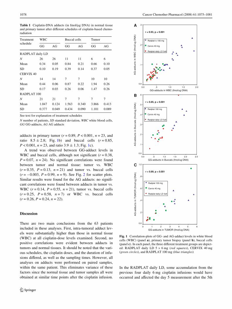
In Table 1, the results are presented for the GG- andAG-adduct levels in normal tissue and primary tumor forthe three diVerent cisplatin-chemoradiation regimes.Adduct levels in primary tumor were two to Wve timeshigher than those in WBC for all three treatment regimesfor both GG- and AG-adduct formation (Student t-test,P < 0.001 for both adduct types). For the comparison of theadduct levels from the three diVerent treatment protocols,the data from the RADPLAT daily LD were omitted, sincethe daily administration schedule and sampling time (1 hafter the infusion) were diVerent from the other two regi-mens. The adduct levels in the RADPLAT 100 schedulewere statistically signiWcantly higher than those after theCERVIX 40 schedule for both tumors (Student t-test,P = 0.01) and normal tissues (Student t-test, P < 0.01).
A highly signiWcant linear correlation (Pearson correla-tion, r = 0.93, P <0.001, n = 61) was observed between thelevel of GG and AG adducts in WBC (see Fig. 1a), with amean ratio of GG/AG adducts of 7.6 § 2.1 SD. Similarlinear relationships and ratios were found for GG and AG
123
1078 Cancer Chemother Pharmacol (2008) 61:1075–1081
adducts in primary tumor (r = 0.89, P < 0.001, n = 23, andratio 8.5 § 2.8; Fig. 1b) and buccal cells (r = 0.85,P < 0.001, n = 23, and ratio 3.9 § 1.3; Fig. 1c).
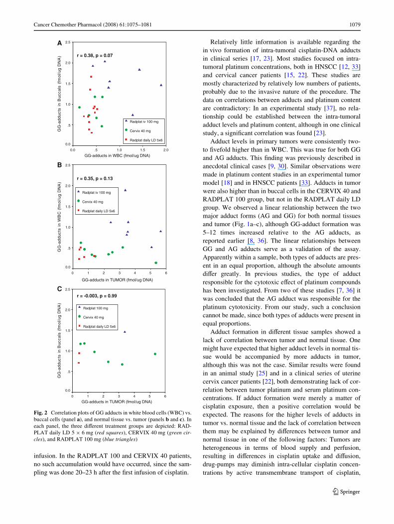
A trend was observed between GG-adduct levels inWBC and buccal cells, although not signiWcant (r = 0.38,P = 0.07, n = 24). No signiWcant correlations were foundbetween tumor and normal tissue: tumor vs. WBC(r = 0.35, P = 0.13, n = 21) and tumor vs. buccal cells(r = ¡0.003, P = 0.99, n = 9). See Fig. 2 for scatter plots.Similar results were found for the AG adducts: no signiW-cant correlations were found between adducts in tumor vs.WBC (r = 0.14, P = 0.55, n = 21), tumor vs. buccal cells(r = 0.25, P = 0.58, n = 7) or WBC vs. buccal cells(r = 0.26, P = 0.24, n = 22).
Discussion
There are two main conclusions from the 63 patientsincluded in these analyses. First, intra-tumoral adduct lev-els were substantially higher than those in normal tissue(WBC) at all cisplatin-dose levels examined. Second, nopositive correlations were evident between adducts intumors and normal tissues. It should be noted that the vari-ous schedules, the cisplatin doses, and the duration of infu-sions diVered, as well as the sampling times. However, allanalyses on adducts were performed on paired samples,within the same patient. This eliminates variance of thesefactors since the normal tissue and tumor samples all wereobtained at similar time points after the cisplatin infusion.
In the RADPLAT daily LD, some accumulation from theprevious four daily 6 mg cisplatin infusions would haveoccurred and aVected the day 5 measurement after the 5th
Table 1 Cisplatin-DNA adducts (in fmol/�g DNA) in normal tissueand primary tumor after diVerent schedules of cisplatin-based chemo-radiation
See text for explanation of treatment schedules
N number of patients, SD standard deviation, WBC white blood cells,GG GG-adducts, AG AG adducts
Treatment schedule
WBC Buccal cells Tumor
GG AG GG AG GG AG
RADPLAT daily LD
N 26 26 11 11 6 6
Mean 0.34 0.05 0.84 0.21 0.66 0.10
SD 0.10 0.19 0.39 0.14 0.37 0.05
CERVIX 40
N 14 14 7 7 10 10
Mean 0.44 0.06 0.87 0.22 1.94 0.26
SD 0.17 0.03 0.26 0.06 1.47 0.26
RADPLAT 100
N 21 21 7 7 7 7
Mean 1.047 0.124 1.563 0.340 3.866 0.413
SD 0.377 0.049 0.434 0.090 1.101 0.089
Fig. 1 Correlation-plots of GG- and AG-adduct levels in white bloodcells (WBC) (panel a), primary tumor biopsy (panel b), buccal cells(panel c). In each panel, the three diVerent treatment groups are depict-ed: RADPLAT daily LD 5 £ 6 mg (red squares), CERVIX 40 mg(green circles), and RADPLAT 100 mg (blue triangles)
Radplat 100 mg
Cervix 40 mg
Radplat daily LD 5x6
Radplat iv 100 mg
Cervix 40 mg
Radplat daily LD 5x6
GG-adducts in WBC (fmol/ug DNA)2.01.51.0.50.0
)A
ND
gu/l
omf(
CB
Wni
stcud
da -G
A
.3
.2
.1
0.0
r = 0.93, p < 0.001
GG-adducts in Buccals (fmol/ug DNA)2.52.01.51.0.50.0
)A
ND
gu/lomf(
s la ccu
Bni
stcudda-
GA
.6
.5
.4
.3
.2
.1
0.0
r = 0.85, p < 0.001
GG-adducts in TUMOR (fmol/ug DNA)6543210
)A
ND
gu/lom f(
RO
MU
Tni
stcud
da-G
A
1.0
.8
.6
.4
.2
0.0
Radplat 100 mg
Cervix 40 mg
Radplat daily LD 5x6
r = 0.89, p < 0.001
A
B
C
123
Cancer Chemother Pharmacol (2008) 61:1075–1081 1079
infusion. In the RADPLAT 100 and CERVIX 40 patients,no such accumulation would have occurred, since the sam-pling was done 20–23 h after the Wrst infusion of cisplatin.
Relatively little information is available regarding thein vivo formation of intra-tumoral cisplatin-DNA adductsin clinical series [17, 23]. Most studies focused on intra-tumoral platinum concentrations, both in HNSCC [12, 33]and cervical cancer patients [15, 22]. These studies aremostly characterized by relatively low numbers of patients,probably due to the invasive nature of the procedure. Thedata on correlations between adducts and platinum contentare contradictory: In an experimental study [37], no rela-tionship could be established between the intra-tumoraladduct levels and platinum content, although in one clinicalstudy, a signiWcant correlation was found [23].
Adduct levels in primary tumors were consistently two-to Wvefold higher than in WBC. This was true for both GGand AG adducts. This Wnding was previously described inanecdotal clinical cases [9, 30]. Similar observations weremade in platinum content studies in an experimental tumormodel [18] and in HNSCC patients [33]. Adducts in tumorwere also higher than in buccal cells in the CERVIX 40 andRADPLAT 100 group, but not in the RADPLAT daily LDgroup. We observed a linear relationship between the twomajor adduct forms (AG and GG) for both normal tissuesand tumor (Fig. 1a–c), although GG-adduct formation was5–12 times increased relative to the AG adducts, asreported earlier [8, 36]. The linear relationships betweenGG and AG adducts serve as a validation of the assay.Apparently within a sample, both types of adducts are pres-ent in an equal proportion, although the absolute amountsdiVer greatly. In previous studies, the type of adductresponsible for the cytotoxic eVect of platinum compoundshas been investigated. From two of these studies [7, 36] itwas concluded that the AG adduct was responsible for theplatinum cytotoxicity. From our study, such a conclusioncannot be made, since both types of adducts were present inequal proportions.
Adduct formation in diVerent tissue samples showed alack of correlation between tumor and normal tissue. Onemight have expected that higher adduct levels in normal tis-sue would be accompanied by more adducts in tumor,although this was not the case. Similar results were foundin an animal study [25] and in a clinical series of uterinecervix cancer patients [22], both demonstrating lack of cor-relation between tumor platinum and serum platinum con-centrations. If adduct formation were merely a matter ofcisplatin exposure, then a positive correlation would beexpected. The reasons for the higher levels of adducts intumor vs. normal tissue and the lack of correlation betweenthem may be explained by diVerences between tumor andnormal tissue in one of the following factors: Tumors areheterogeneous in terms of blood supply and perfusion,resulting in diVerences in cisplatin uptake and diVusion,drug-pumps may diminish intra-cellular cisplatin concen-trations by active transmembrane transport of cisplatin,
Fig. 2 Correlation plots of GG adducts in white blood cells (WBC) vs.buccal cells (panel a), and normal tissue vs. tumor (panels b and c). Ineach panel, the three diVerent treatment groups are depicted: RAD-PLAT daily LD 5 £ 6 mg (red squares), CERVIX 40 mg (green cir-cles), and RADPLAT 100 mg (blue triangles)
GG-adducts in WBC (fmol/ug DNA)2.01.51.0.50.0
)A
ND
gu/lomf(
slacc
uB
nist
cudd
a-G
G
2.5
2.0
1.5
1.0
.5
0.0
Radplat iv 100 mg
Cervix 40 mg
Radplat daily LD 5x6
r = 0.38, p = 0.07
GG-adducts in TUMOR (fmol/ug DNA)
6543210
)A
ND
gu/lomf(
CB
Wni
stcud
da-G
G
2.5
2.0
1.5
1.0
.5
0.0
Radplat iv 100 mg
Cervix 40 mg
Radplat daily LD 5x6
r = 0.35, p = 0.13
GG-adducts in TUMOR (fmol/ug DNA)6543210
)A
ND
gu/lomf(
slaccuB
nistcudda-
GG
2.5
2.0
1.5
1.0
.5
0.0
Radplat 100 mg
Cervix 40 mg
Radplat daily LD 5x6
r = -0.003, p = 0.99
A
B
C
123
1080 Cancer Chemother Pharmacol (2008) 61:1075–1081
prohibiting adducts to be formed, and tumor cells may haveless eVective capacities to repair damage from cytotoxicagents.
Adducts are formed rapidly and in a dose-dependentfashion within 1–2 h after cisplatin exposure, with a grad-ual decrease (repair) within the next 20–24 h [20, 32, 36].The persistence of cisplatin-DNA adducts may therefore beregarded as a measure of repair and possibly be used as pre-dictive assay. We therefore chose to measure adducts20–23 h after the end of chemotherapy infusion. Ideally,more frequent measurements would have generated moreinformation on the rate of adduct formation, repair, andtotal exposure to adducts (like an AUC analysis) [24].However, obtaining repeated biopsies is not feasible in clin-ical practice.
The results from the buccal cell samples need to be inter-preted with caution, especially the AG-adduct levels, sinceuncertainties remain. With the low quantities of AG-adducts in the low amounts of buccal cell DNA we couldextract, we reached the limits of quantiWcation of the post-labeling method.
The rationale for measuring cisplatin-DNA adducts isthat it could be used as a predictive assay: higher levels ofadducts would predict favorable treatment outcome. Manystudies have been performed for this purpose, investigatingadducts in normal tissue (WBC and buccal cells) [3, 4, 10,14, 26, 31, 32, 35]. These study designs are based on theassumption that normal tissue can be used as a surrogatemarker for tumor tissue with respect to cisplatin-DNAadduct formation. In our present study, however, we did notWnd such a correlation. This might explain why the resultson the role of adduct formation to predictive outcome areheterogeneous and contradictory, as illustrated below.
Several studies on adduct formation in WBC showedthat the level of adducts was positively correlated withresponse to chemotherapy in patients with advanced dis-ease in a variety of tumor sites [31, 32], while othersshowed no correlation [4, 26]. One study showed a positivecorrelation in one tumor site (ovarian cancer), but not in theother (breast cancer) [14], and in another study the level ofadduct formation showed a negative association with sur-vival for day-5 adducts, while there was no diVerence forday-1 adducts [10]. In studies on adduct levels in buccalcells, a positive correlation was found between adducts andeither disease response [3] or better survival in non-smallcell lung cancer (NSCLC) [35]. We recently showed thatadduct formation in primary tumor appeared to be associ-ated with better progression-free survival in HNSCC [17].
DiVerences were observed between the levels of intra-tumoral adducts for the three chemoradiation schedules,with lower adducts after lower dosages of cisplatin. Basedon this, however, one cannot predict that the schedules withless adducts will result in less cytotoxicity, since not only
the cisplatin dose, but also timing and schedule of cisplatinadministration are crucial determinants of eYcacy [1].These factors may contribute to diVerences in the formationand rate of repair of adducts, resulting in diVerent expo-sures to cisplatin-DNA adducts. These diVerences make itdiYcult to extrapolate from the observed adduct values intumor and normal tissues to a prediction of superiority ofone schedule over another.
In future studies, immunohistochemistry on repair pro-teins like ERCC1 [27] or gene expression proWling studieson platinum resistance [16] could be used to improve theprediction of cisplatin sensitivity and prediction of therapyresponse.
In conclusion, we have demonstrated that in concurrentchemoradiotherapy schedules, cisplatin adduct levels intumors were signiWcantly higher than in normal tissues(WBC). No evidence of a correlation was found betweenadduct levels in normal tissues and primary tumor biopsies.This lack of correlation may, to some extent, explain theinconsistencies in the literature regarding whether or notcisplatin-DNA adducts can be used as predictive test inanticancer therapy.
ConXict of interest: none declared.
References
1. Bartelink H, Kallman RF, Rapacchietta D, Hart GA (1986) Ther-apeutic enhancement in mice by clinically relevant dose and frac-tionation schedules of cis-diamminedichloroplatinum (II) andirradiation. Radiother Oncol 6:61–74
2. Begg AC, van der Kolk PJ, Dewit L, Bartelink H (1986) Radiosen-sitization by cisplatin of RIF1 tumour cells in vitro. Int J RadiatBiol Relat Stud Phys Chem Med 50:871–884
3. Blommaert FA, Michael C, Terheggen PM, et al (1993) Drug-induced DNA modiWcation in buccal cells of cancer patientsreceiving carboplatin and cisplatin combination chemotherapy, asdetermined by an immunocytochemical method: interindividualvariation and correlation with disease response. Cancer Res53:5669–5675
4. Bonetti A, Apostoli P, Zaninelli M, et al (1996) Inductivelycoupled plasma mass spectroscopy quantitation of platinum-DNAadducts in peripheral blood leukocytes of patients receiving cispl.Clin Cancer Res 2:1829–1835
5. Bourhis J, Amand C, Pignon J-P on behalf of the MACH-NCCollaborative Group (2004) Update of MACH-NC (Meta-Analy-sis of Chemotherapy in Head & Neck Cancer) database focused onconcomitant chemoradiotherapy. J Clin Oncol 22 No 14S (July 15Suppl):ASCO Annual Meeting Proceedings (Post-MeetingEdition). 5505
6. Brizel DM, Albers ME, Fisher SR, et al (1998) Hyperfractionatedirradiation with or without concurrent chemotherapy for locallyadvanced head and neck cancer. N Engl J Med 338:1798–1804
7. Fichtinger-Schepman AM, Dijk-Knijnenburg HC, van der Velde-Visser SD, Berends F, Baan RA (1995) Cispl. Carcinogenesis16:2447–2453
8. Fichtinger-Schepman AM, van Oosterom AT, Lohman PH,Berends F (1987) cis-Diamminedichloroplatinum(II)-inducedDNA adducts in peripheral leukocytes from seven cancer patients:
123

Cancer Chemother Pharmacol (2008) 61:1075–1081 1081
quantitative immunochemical detection of the adduct inductionand removal after a single dose of cis-diamminedichloroplati-num(II). Cancer Res 47:3000–3004
9. Fichtinger-Schepman AM, van der Velde-Visser SD, Dijk-Knijn-enburg HC, et al (1990) Kinetics of the formation and removal ofcisplatin-DNA adducts in blood cells and tumor tissue of cancerpatients receiving chemotherapy: comparison with in vitro adductformation. Cancer Res 50:7887–7894
10. Fisch MJ, Howard KL, Einhorn LH, Sledge GW (1996) Relation-ship between platinum-DNA adducts in leukocytes of patientswith advanced germ cell cancer and survival. Clin Cancer Res2:1063–1066
11. Forastiere AA, Goepfert H, Maor M, et al (2003) Concurrentchemotherapy and radiotherapy for organ preservation inadvanced laryngeal cancer. N Engl J Med 349:2091–2098
12. Gouyette A, Apchin A, Foka M, Richard JM (1986) Pharmacoki-netics of intra-arterial and intravenous cisplatin in head and neckcancer patients. Eur J Cancer Clin Oncol 22:257–263
13. Green JA, Kirwan JM, Tierney JF, et al (2001) Survival and recur-rence after concomitant chemotherapy and radiotherapy for cancerof the uterine cervix: a systematic review and meta-analysis.Lancet 358:781–786
14. Gupta-Burt S, Shamkhani H, Reed E, et al (1993) Relationship be-tween patient response in ovarian and breast cancer and platinumdrug-DNA adduct formation. Cancer Epidemiol Biomarkers Prev2:229–234
15. Hecquet B, Vennin P, Fournier C, Poissonnier B (1987) Evalua-tion of the pharmacological beneWt and determination of the inXu-encing factors of intraarterial cis-diamminedichloroplatinumadministration in patients with uterine cervical cancer. Cancer Res47:6134–6137
16. Helleman J, Jansen MP, Span PN, et al (2006) Molecular proWlingof platinum resistant ovarian cancer. Int J Cancer 118:1963–1971
17. Hoebers FJ, Pluim D, Verheij M, et al (2006) Prediction of treat-ment outcome by cisplatin-DNA adduct formation in patients withstage III/IV head and neck squamous cell carcinoma, treated byconcurrent cisplatin-radiation (RADPLAT). Int J Cancer119:750–756
18. Jakowatz JG, Ginn GE, Snyder LM, DieVenbach KW, Wile AG(1991) Increased cisplatin tissue levels with prolonged arterialinfusion in the rat. Cancer 67:2828–2832
19. Jeremic B, Shibamoto Y, Milicic B, et al (2000) Hyperfractionatedradiation therapy with or without concurrent low-dose daily cis-platin in locally advanced squamous cell carcinoma of the headand neck: a prospective randomized trial. J Clin Oncol 18:1458–1464
20. Johnsson A, Olsson C, Nygren O, et al (1995) Pharmacokineticsand tissue distribution of cisplatin in nude mice: platinum levelsand cisplatin-DNA adducts. Cancer Chemother Pharmacol 37:23–31
21. Keys HM, Bundy BN, Stehman FB, et al (1999) Cisplatin, radia-tion, and adjuvant hysterectomy compared with radiation andadjuvant hysterectomy for bulky stage IB cervical carcinoma. NEngl J Med 340:1154–1161
22. Lagrange JL, Bondiau PY, Tessier E, et al (1996) Tumoral plati-num concentrations in patients treated with repeated low-dosecisplatin as a radiosensitizer. Int J Cancer 68:452–456
23. Los G, Blommaert FA, Barton R, et al (1995) Selective intra-arte-rial infusion of high-dose cisplatin in patients with advanced headand neck cancer results in high tumor platinum concentrations andcisplatin-DNA adduct formation. Cancer Chemother Pharmacol37:150–154
24. Ma J, Verweij J, Planting AS, et al (1995) Current sample han-dling methods for measurement of platinum-DNA adducts in leu-cocytes in man lead to discrepant results in DNA adduct levels andDNA repair. Br J Cancer 71:512–517
25. Moses BL, Chan DW, Hruban RH, Forastiere A, Richtsmeier WJ(1993) Comparison of intra-arterial and intravenous infusion ofcisplatin for head and neck squamous cell carcinoma in a modiWedrat model. Arch Otolaryngol Head Neck Surg 119:612–617
26. Motzer RJ, Reed E, Perera F, et al (1994) Platinum-DNA adductsassayed in leukocytes of patients with germ cell tumors measuredby atomic absorbance spectrometry and enzyme-linked immuno-sorbent assay. Cancer 73:2843–2852
27. Olaussen K, Dunant A, Fouret P, et al (2006) DNA repair byERCC1 in non-small-cell lung cancer and cisplatin-based adjuvantchemotherapy. N Engl J Med 355:983–991
28. Pignon JP, Bourhis J, Domenge C, Designe L (2000) Chemother-apy added to locoregional treatment for head and neck squamous-cell carcinoma: three meta-analyses of updated individual data.MACH-NC Collaborative Group. Meta-Analysis of Chemother-apy on Head and Neck Cancer. Lancet 355:949–955
29. Pluim D, Maliepaard M, van Waardenburg RC, Beijnen JH, Schel-lens JH (1999) 32P-postlabeling assay for the quantiWcation of themajor platinum-DNA adducts. Anal Biochem 275:30–38
30. Reed E, Ozols RF, Tarone R, Yuspa SH, Poirier MC (1987) Plati-num-DNA adducts in leukocyte DNA correlate with disease re-sponse in ovarian cancer patients receiving platinum-basedchemotherapy. Proc Natl Acad Sci USA 84:5024–5028
31. Reed E, Ozols RF, Tarone R, Yuspa SH, Poirier MC (1988) Themeasurement of cisplatin-DNA adduct levels in testicular cancerpatients. Carcinogenesis 9:1909–1911
32. Schellens JH, Ma J, Planting AS, et al (1996) Relationship be-tween the exposure to cisplatin, DNA-adduct formation in leuco-cytes and tumour response in patients with solid tumours. Br JCancer 73:1569–1575
33. Tegeder I, Brautigam L, Seegel M, et al (2003) Cisplatin tumorconcentrations after intra-arterial cisplatin infusion or emboliza-tion in patients with oral cancer. Clin Pharmacol Ther 73:417–426
34. Terheggen PM, Emondt JY, Floot BG, et al (1990) Correlation be-tween cell killing by cis-diamminedichloroplatinum(II) in sixmammalian cell lines and binding of a cis-diamminedichloroplat-inum(II)-DNA antiserum. Cancer Res 50:3556–3561
35. van de Vaart PJ, Belderbos J, de Jong D, et al (2000) DNA-adductlevels as a predictor of outcome for NSCLC patients receivingdaily cisplatin and radiotherapy. Int J Cancer 89:160–166
36. Welters MJ, Fichtinger-Schepman AM, Baan RA, et al (1999)Pharmacodynamics of cisplatin in human head and neck cancer:correlation between platinum content, DNA adduct levels anddrug sensitivity in vitro and in vivo. Br J Cancer 79:82–88
37. Zamboni WC, Gervais AC, Egorin MJ, et al (2002) Inter- andintratumoral disposition of platinum in solid tumors after adminis-tration of cisplatin. Clin Cancer Res 8:2992–2999
123