Preventive effect of tert-butylhydroquinone on cisplatin-induced nephrotoxicity in rats
8
This article appeared in a journal published by Elsevier. The attached copy is furnished to the author for internal non-commercial research and education use, including for instruction at the authors institution and sharing with colleagues. Other uses, including reproduction and distribution, or selling or licensing copies, or posting to personal, institutional or third party websites are prohibited. In most cases authors are permitted to post their version of the article (e.g. in Word or Tex form) to their personal website or institutional repository. Authors requiring further information regarding Elsevier’s archiving and manuscript policies are encouraged to visit: http://www.elsevier.com/copyright
-
Upload
independent -
Category
Documents
-
view
6 -
download
0
Transcript of Preventive effect of tert-butylhydroquinone on cisplatin-induced nephrotoxicity in rats

This article appeared in a journal published by Elsevier. The attachedcopy is furnished to the author for internal non-commercial researchand education use, including for instruction at the authors institution
and sharing with colleagues.
Other uses, including reproduction and distribution, or selling orlicensing copies, or posting to personal, institutional or third party
websites are prohibited.
In most cases authors are permitted to post their version of thearticle (e.g. in Word or Tex form) to their personal website orinstitutional repository. Authors requiring further information
regarding Elsevier’s archiving and manuscript policies areencouraged to visit:
http://www.elsevier.com/copyright

Author's personal copy
Preventive effect of tert-butylhydroquinone on cisplatin-induced nephrotoxicityin rats
Jazmin M. Pérez-Rojas a,b,⇑, Carlos Enrique Guerrero-Beltrán a, Cristino Cruz c, Dolores J. Sánchez-González d,Claudia M. Martínez-Martínez d, José Pedraza-Chaverri a,⇑a Facultad de Química, Departamento de Biología, Universidad Nacional Autónoma de México, Mexico City, Mexicob Subdirección de Investigación Básica, Instituto Nacional de Cancerología (INCan), Mexico City, Mexicoc Instituto Nacional de Ciencias Médicas y Nutrición Salvador Zubirán, Mexico City, Mexicod Departamento de Biología Celular, Escuela Médico Militar, Universidad del Ejército y Fuerza Aérea, Mexico City, Mexico
a r t i c l e i n f o
Article history:Received 15 February 2010Accepted 7 July 2011Available online 23 July 2011
Keywords:tert-ButylhydroquinoneCisplatinOxidative stressKidney damage
a b s t r a c t
Cis-diamminedichloroplatinum II (CDDP)-induced nephrotoxicity is associated with the overproductionof reactive oxygen species. tert-Butylhydroquinone (tBHQ) is a compound widely used as food antioxi-dant. The purpose of this study was to investigate the ability of tBHQ to prevent the nephrotoxic effectof CDDP in rats as well as the mechanisms involved. Thirty-six Wistar rats divided in the following groupswere used: control, tBHQ (12.5 mg/kg), CDDP (7.5 mg/kg) and tBHQ + CDDP. Twenty-four h urine wascollected at the beginning and at the end of the experiment and the rats were sacrificed 72 h afterCDDP-administration. Histological studies were performed and markers of renal function and oxida-tive/nitrosative stress were measured. In addition, the activity of the following antioxidant enzymeswas measured: glutathione peroxidase (GPx), superoxide dismutase (SOD), glutathione reductase (GR)and glutathione-S-transferase (GST). CDDP-induced renal dysfunction, structural damage and oxida-tive/nitrosative were prevented by tBHQ. In addition, tBHQ completely prevented the CDDP-induced fallin GPx and GST activities. In conclusion, the present study indicates that the antioxidant activity of tBHQis associated with its nephroprotective effect against CDDP-induced acute kidney injury in rats.
� 2011 Elsevier Ltd. All rights reserved.
1. Introduction
Cis-diamminedichloroplatinum II (CDDP) is an important che-motherapeutic agent that has been widely used for its potent cyto-toxic effects upon a variety of tumor types including testicular,ovarian, and cervical carcinoma (Fontanelli et al., 1992; Loehrerand Einhorn, 1984; Ozols, 1995; Panici et al., 1991). However,the administration of CDDP is associated with serious side effectsincluding nephrotoxicity and neurotoxic alterations (Gandaraet al., 1991). CDDP causes tubular injury, mainly at proximal tubu-lar level, through multiple mechanisms including hypoxia, oxida-tive stress, inflammation and apoptosis (Chirino et al., 2008a;Francescato et al., 2007; Yao et al., 2007). Despite its side effects,CDDP remains the drug of preference in chemotherapy, mainly
because of its efficacy and low cost. That is why it is very importantto counteract CDDP side effects to improve quality of life of pa-tients who are under this treatment regimen.
Oxidative stress is one of the most important factors involved inCDDP-induced toxicity. Some researchers have been used naturalextracts, isolated compounds and new drugs to block oxidativestress by several ways with the purpose to prevent CDDP-inducedrenal damage (Ali and Al Moundhri, 2006; Chirino and Pedraza-Chaverri, 2009). In this context, it is important to employcompounds which are used for human beings, as is the case oftert-Butylhydroquinone (tBHQ), because they will be easily addedin therapy and also will be cheaper for the patient.
tBHQ is a synthetic phenolic antioxidant (Kim et al., 2009) thatwas approved for human use by both Food and Agriculture Organi-zation and World Health Organization (1999). Several groups haveused this compound because of its antioxidant properties. Favreauand Pickett (1991) and Rushmore and Pickett (1990) demonstratedthat the treatment with tBHQ induced many genes associated withresistance against oxidative stress and prevented hydrogen perox-ide (H2O2)-induced apoptosis in neuroblastoma IMR32 cells. Also,Hara and colleagues (2003) reported that tBHQ has a neuroprotec-tive effect against 6-hydroxydopamine induced oxidative stress inthe human neuroblastoma cell line SH-SY5Y. However, it is
0278-6915/$ - see front matter � 2011 Elsevier Ltd. All rights reserved.doi:10.1016/j.fct.2011.07.008
⇑ Corresponding authors. Address: Laboratorio de Farmacología, Subdirección deInvestigación Básica, Instituto Nacional de Cancerología (INCan), Av. San Fernando22, Tlalpan 14000, Apartado Postal 22026, Mexico City, Mexico. Tel.: +52 55 56280400x303 (J.M. Pérez-Rojas). Facultad de Química, Edificio F, Universidad NacionalAutónoma de México, Mexico City, México. Tel./fax: +52 55 5622 3878(J. Pedraza-Chaverri).
E-mail addresses: [email protected] (J.M. Pérez-Rojas), [email protected] (J. Pedraza-Chaverri).
Food and Chemical Toxicology 49 (2011) 2631–2637
Contents lists available at ScienceDirect
Food and Chemical Toxicology
journal homepage: www.elsevier .com/locate / foodchemtox

Author's personal copy
unknown if tBHQ may protect the kidney from the CDDP-inducedrenal damage.
For the above reasons, in the present study, we investigatedwhether the antioxidant properties of tBHQ may attenuate acute
renal failure, oxidative stress, and reduction in antioxidant en-zymes activities induced by CDDP in rats.
2. Methods
2.1. Reagents
The Amplex Red� H2O2/peroxidase assay kit used to measureurinary excretion of H2O2, CDDP (Cat. No. P-4394) and tBHQ (Cat.No. 112941) were purchased from Sigma–Aldrich (St. Louis, MO,USA). Mayer’s Hematoxylin (Lillie’s Modification) (Cat. No. S3309)was from DAKO Corporation (Carpinteria, CA, USA). All otherchemicals used were of the highest quality available and obtainedfrom commercial sources.
2.2. Experimental design
Thirty-six male Wistar rats were obtained from Harlan (MexicoCity) with a body weight of 286 ± 19 (mean ± SD), and were ran-domly divided into four groups: (1) control (C) (n = 9), rats received
Table 1Activity of CAT, SOD and GR in renal cortex.
CAT (k/mgprotein)
SOD (U/mgprotein)
GR (U/mgprotein)
Ct 0.253 ± 0.008 31.11 ± 3.4 0.082 ± 0.012tBHQ 0.214 ± 0.024 28.90 ± 2.1 0.081 ± 0.007CDDP 0.178 ± 0.018a 31.66 ± 1.5 0.086 ± 0.004tBHQ + CDDP 0.109 ± 0.016a,b 34.70 ± 1.9 0.077 ± 0.004
CAT, catalase; SOD, superoxide dismutase; GR, glutathione reductase; Ct, control;tBHQ, tert-butylhydroquinone; CDDP, cisplatin. Values are mean ± SEM of 6–8 rats/group.
a P < 0.05 vs. CT.b P < 0.05 vs. CDDP.
Fig. 1. Protective effect of tBHQ against CDDP-induced renal dysfunction in rats. (A)Serum creatinine, (B) blood urea nitrogen (BUN) and (C) creatinine clearance incontrol; tBHQ, tert-butylhydroquinone; CDDP, cisplatin and tBHQ + CDDP groups.Values are means ± SEM. n = 4–7. ªP < 0.001 vs. Control; bP < 0.05 vs. CDDP.
Fig. 2. Protective effect of tBHQ against CDDP-induced tubular injury in rats. (A)Urinary volume, (B) urinary excretion of N-acetyl-b-D-glucosaminidase (NAG) and(C) total protein excretion in control; tBHQ, tert-butylhydroquinone; CDDP,cisplatin and tBHQ + CDDP groups. Values are means ± SEM. n = 5–8. ªP < 0.05 vs.Control; bP < 0.05 vs. CDDP.
2632 J.M. Pérez-Rojas et al. / Food and Chemical Toxicology 49 (2011) 2631–2637

Author's personal copy
a single intraperitoneal injection of vehicle (saline solution). (2)tBHQ (n = 9), one day before vehicle injection, rats received tBHQ(12.5 mg/kg, i.p.) divided into three identical doses each 8 h. (3)CDDP (n = 9), rats were injected with a single intraperitoneal injec-tion of CDDP (7.5 mg/kg), which was previously dissolved in salinesolution (1.5 mg/mL). (4) tBHQ + CDDP (n = 9), one day beforeCDDP injection (7.5 mg/kg, i.p.), rats received tBHQ (12.5 mg/kg,i.p.) divided into three identical doses each 8 h. Before startingthe experiment and before the sacrifice, the rats were placed intometabolic cages to collect 24-h urine. In addition blood sampleswere obtained at the end of the experiment.
Rats were placed in rooms at 22 �C with 12:12-h light–darkcycle and free access to water. Three days after CDDP administra-tion rats were sacrificed by decapitation and blood was collected.A midline laparotomy was made; the right kidney was excised, fro-zen in liquid nitrogen, and kept at �80 �C until the determinationswere performed. The left kidney was placed in 10% formalin bufferfor histopathological evaluation. All procedures followed were inaccordance with our institutional guidelines.
The dose of tBHQ was chosen after the performance of previ-ous experiments using the following doses: 12.5 mg/kg one daybefore CDDP administration, 12.5 mg/kg for 5 days prior of CDDPadministration and 25 mg/kg body weight 5 days prior to theinjection of CDDP. The following parameters were evaluated:serum creatinine, blood urea nitrogen (BUN), creatinine clear-ance (CCr), and urinary excretion of total protein and N-acetyl-b-D-glucosaminidase (NAG). tBHQ (12.5 mg/kg) given one dayprevious to drug administration, showed the highest renoprotec-tive effect against CDDP-induced nephrotoxicity (data not show).Based on the above data, that dose was chosen to perform thepresent study.
2.3. Analytical methods
Renal function was evaluated by measuring BUN, serum creati-nine, creatinine clearance and urinary excretion of total proteinand NAG by methods previously described (Chirino et al., 2008a).
Fig. 3. Protective effect of tBHQ against CDDP-induced renal histological alteration in rats. (A) Histological profiles were detected by H&E staining in rats treated with salinesolution (Control); tBHQ, tert-butylhydroquinone; CDDP, cisplatin and tBHQ + CDDP. CDDP induced necrosis (asterisks), vacuolization (arrowheads) and tubular casts (arrow).(B) Histological damage expressed as percentage of tubular area. x400 magnification. Values are means ± SEM. n = 4. ªP < 0.0002 vs. CDDP.
J.M. Pérez-Rojas et al. / Food and Chemical Toxicology 49 (2011) 2631–2637 2633

Author's personal copy
2.4. Histological studies
Kidney sections were fixed in 10% neutral buffered formalinsolution and embedded in paraffin (Pérez-Rojas et al., 2009). Sec-tions of 3 lm of thickness were obtained and stained with hema-toxiline-eosin (H&E). A quantitative histological damage wasdetermined by using a Leica Qwin Image Analyzer (Cambridge,England). The histological profiles of thirty proximal tubules ran-domly selected per rat were recorded. The percentage of tubuleswith histopathological alterations like swelling, cytoplasmic vacu-olization, desquamation, or necrosis was obtained. Total surfacearea of tubular cells in square microns was determined, the surfacearea occupied by vacuoles was measured, and the percentage ofthis affected area was calculated.
2.5. Oxidative stress markers
Urinary excretion of H2O2 was measured with the Amplex Red�
method (Pérez-Rojas et al., 2009). Malondialdehyde (MDA) and 4-hydroxynonenal (4-HNE) were measured in the renal cortex usinga standard curve of trimethoxypropane (Gerard-Monnier et al.,1998). The content of protein carbonyl groups, an index of oxidizedproteins, was measured using 2,4-dinitrophenylhydrazine (Maldo-nado et al., 2003).
2.6. Immunohistochemical studies
Immunohistochemical studies of 4-HNE, a marker of lipid per-oxidation, and of 3-nitro-L-tyrosine (3-NT), a marker of nitrosative
stress, were performed as previously described (Chirino et al.,2008a; Pérez-Rojas et al., 2009).
2.7. Activity of antioxidant enzymes
Glutathione peroxidase (GPx) activity was measured by the dis-appearance of NADPH at 340 nm in a coupled assay containingH2O2, GSH and glutathione reductase (GR) (Maldonado et al.,2003). GR activity was measured by the disappearance of NADPHat 340 nm in a reaction mixture containing oxidized glutathioneas a substrate (Maldonado et al., 2003). Total superoxide dismutase(SOD) was measured at 560 nm using the superoxide anion gener-ator system xanthine/xhanthine oxidase and nitroblue tetrazoliumas the indicator molecule (Maldonado et al., 2003). Catalase (CAT)activity was assayed at 240 nm by a method based on the disap-pearance of H2O2 (Maldonado et al., 2003). Glutathione-S-transfer-ase (GST) activity was assayed at 340 nm in a mixture containingglutathione (GSH) and 1-chloro-2,4-dinitrobenzene (CDNB) as pre-viously described (Chirino et al., 2008a).
2.8. Statistical analysis
Results were expressed as mean ± SE. Data were analyzed byone-way ANOVA followed by student Newman-Keuls test. Non-paired t-test was used to compare the quantitative histologicaldamage. GraphPad Prism 2.01 software (San Diego, CA, USA) wasused. A p-value < 0.05 was considered statistically significant.
Fig. 4. Protective effect of tBHQ against CDDP-induced increase in renal immunostaining of 3-NT and 4-HNE in rats. (A) Representative microphotographs of 3-NT. (B)Percentage area of 3-NT abundance. (C) Representative microphotographs of 4-HNE. (D) Percentage area of 4-HNE abundance. tBHQ, tert-butylhydroquinone; CDDP, cisplatin.Values are mean ± SEM. n = 4. ªP < 0.001 vs. Ct; bP < 0.01 vs. CDDP.
2634 J.M. Pérez-Rojas et al. / Food and Chemical Toxicology 49 (2011) 2631–2637
Author's personal copy
3. Results
Acute nephrotoxicity was evident by renal function impairmentand histopathological kidney lesion. On day 3 post-injection,CDDP-injected rats showed increased plasma levels of creatinineand BUN and decreased creatinine clearance; these changes wereprevented by tBHQ administration (Fig. 1). In addition, CDDP-induced increase in urine volume and urinary excretion of NAGand protein was attenuated by tBHQ treatment (Fig. 2). In controlrats, tBHQ administration alone had no effect on renal function.Additionally, we also studied renal structure. Histological evalua-tion using H&E staining is shown in Fig. 3. We observed that thecontrol and tBHQ groups showed normal architecture. WhereasCDDP treated rats showed extensive damage and most of corticaltubules showed vacuolization, tubular casts and necrosis; in con-trast, tBHQ treated rats showed lesser tissue damage with few epi-thelial tubular cells affected. Histological damage expressed aspercentage of tubular area with histopathological alterations inCDDP and a tBHQ + CDDP groups is shown in Figure 3B. It is ob-served that tBHQ ameliorates CDDP-induced deterioration of renalstructural damage.
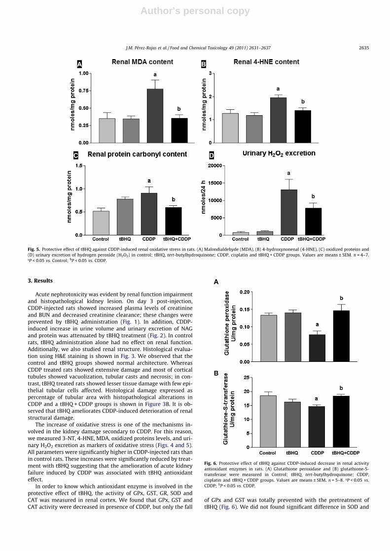
The increase of oxidative stress is one of the mechanisms in-volved in the kidney damage secondary to CDDP. For this reason,we measured 3-NT, 4-HNE, MDA, oxidized proteins levels, and uri-nary H2O2 excretion as markers of oxidative stress (Figs. 4 and 5).All parameters were significantly higher in CDDP-injected rats thanin control rats. These increases were significantly reduced by treat-ment with tBHQ suggesting that the amelioration of acute kidneyfailure induced by CDDP was associated with tBHQ antioxidanteffect.
In order to know which antioxidant enzyme is involved in theprotective effect of tBHQ, the activity of GPx, GST, GR, SOD andCAT was measured in renal cortex. We found that GPx, GST andCAT activity were decreased in presence of CDDP, but only the fall
of GPx and GST was totally prevented with the pretreatment oftBHQ (Fig. 6). We did not found significant difference in SOD and
Fig. 5. Protective effect of tBHQ against CDDP-induced renal oxidative stress in rats. (A) Malondialdehyde (MDA), (B) 4-hydroxynonenal (4-HNE), (C) oxidized proteins and(D) urinary excretion of hydrogen peroxide (H2O2) in control; tBHQ, tert-butylhydroquinone; CDDP, cisplatin and tBHQ + CDDP groups. Values are means ± SEM. n = 4–7.ªP < 0.05 vs. Control; bP < 0.05 vs. CDDP.
Fig. 6. Protective effect of tBHQ against CDDP-induced decrease in renal activityantioxidant enzymes in rats. (A) Glutathione peroxidase and (B) glutathione-S-transferase were measured in Control; tBHQ, tert-butylhydroquinone; CDDP,cisplatin and tBHQ + CDDP groups. Values are means ± SEM. n = 5–8. ªP < 0.05 vs.CDDP; bP < 0.05 vs. CDDP.
J.M. Pérez-Rojas et al. / Food and Chemical Toxicology 49 (2011) 2631–2637 2635

Author's personal copy
GR in any of the experimental groups although tBHQ exacerbatedthe decrease of CAT activity in presence of CDDP (Table 1).
4. Discussion
The pathogenesis of renal injury induced by CDDP is associatedmainly with oxidative/nitrosative stress (Pabla and Dong, 2008;Chirino and Pedraza-Chaverri, 2009; Yao et al., 2007). In this study,we demonstrated that tBHQ has a renoprotective effect on CDDP-induced nephrotoxicity in rats by attenuating oxidative and nitro-sative stress of renal tubular epithelial cells. We demonstrated thattBHQ prevents the increase of reactive oxygen (ROS) and nitrogen(RNS) species which is associated with the improving of the func-tional and structural alterations. This effect can be related to thefact that tBHQ is an antioxidant compound and can be enhancingthe antioxidative capacity of renal cells.
Unbound CDDP, which is freely filtered by the glomerulus andtaken up into renal tubular cells through a transport-mediated pro-cess, and, by a pathway involving c-glutamyl transpeptidase andcysteine-S-conjugate b-lyase is partially metabolized into toxicspecies which cause direct cytotoxicity involving ROS/RNS (Town-send et al., 2003; Chirino et al., 2004; Yao et al., 2007). tBHQ is anaromatic organic compound of phenolic type that has been used asfood additive because of it inhibits autopolymerization of organicperoxides. In this study we utilized the antioxidant tBHQ to coun-teract the CDDP-induced oxidant stress and toxicity. In fact, it wasclearly demonstrated that tBHQ totally prevents the increase in re-nal content of protein carbonyl, 3-NT, MDA, 4-HNE and in the uri-nary H2O2 excretion (Fig. 5). The enzymatic activity of severalantioxidant enzymes also revealed the participation of oxidativestress in the CDDP-induced damage, as well as, the possible mech-anism of tBHQ protection. We found that tBHQ totally preventedthe CDDP-induced decrease in the activity of GPx and GST. In con-trast the CDDP-induced decrease in CAT activity was exacerbatedby tBHQ (Fig. 6 and Table 1).
Several reports show the renoprotective effects of natural anti-oxidants against CDDP-induced damage including a-mangostin(Pérez-Rojas et al., 2009), green tea (Khan et al., 2009), grape seed(Yousef et al., 2009), and sulforaphane (Guerrero-Beltrán et al.,
2010). In this study we used tBHQ, a synthetic compound with di-rect antioxidant properties. This compound is also an indirect anti-oxidant, which exerts their action by modulating transcriptionfactors governing the expression of antioxidant genes (Dinkova-Kostova and Talalay, 2008; Kraft et al., 2004).
The protective effect of tBHQ has been explored in several re-ports. Tasset and colleagues (2010) showed the protective effectof tBHQ against quinolinic acid-induced toxicity in striatal slices.On the other hand, Koh and colleagues (2009) demonstrated thatpretreatment with tBHQ attenuated the lipopolysaccharide-in-duced microglial activation and neuronal death. Moreover, Lavoieet al. (2009) found that the preventive effect of tBHQ against celldeath was associated with increased glutathione levels in astro-cytes and to a lesser extent in neurons. As mentioned above, mostof the studies with tBHQ have been performed in brain tissue; thisis the first study made in a model of renal damage induced by ROS.
The deterioration of renal function induced by CDDP was evi-dent by the decrease in creatinine clearance and by the increasein BUN, serum creatinine, urine volume, and by the urinary excre-tion of NAG and protein (Figs. 1 and 2). tBHQ was able to totallyprevent the CDDP-induced changes in renal function (Figs. 1 and2).
We assume that the renoprotective effect of tBHQ is not inter-fering with the antitumoral activity of CDDP for several reasons:(1) the distribution of tBHQ is biphasic, consisting of a rapid com-ponent with a half-life which ranged from 0.6 to 1.4 h, and a slowercomponent with a half-life that ranged from 16 to 69 h. Whereashalf-life of CDDP is 0.30 h (Wang et al., 2007). (2) Despite tBHQis also eliminated by urine as CDDP; tBHQ needs to be conjugated,so it is eliminated as tBHQ-glucuronide, tBHQ-sulfate and other po-lar unidentified conjugates making unlikely its combination withCDDP (Ikeda et al., 1998). (3) Moreover other polyphenolic antiox-idants structurally more complex such a-mangostin (Pérez-Rojaset al., 2009) and resveratrol (Wang et al., 2009) have no effect onpharmacokinetics and not compromise the antitumor activity ofCDDP.
In summary, Fig. 7 shows a possible mechanism of the renopro-tective effect of tBHQ against CDDP. By passive diffusion CDDPcrosses plasma membrane, once in the cytoplasm, CDDP impairsmitochondrial activity which enhances ROS production. The
Fig. 7. Schema showing how tBHQ may protect of CDDP-induced nephrotoxicity. CDDP, cisplatin; tBHQ, tert-butylhydroquinone; iNOS, inducible nitric oxide synthase; RNS,reactive nitrogen species; ROS, reactive oxygen species; GST, glutathione-S-transferase; GSH, glutathione; GPx, glutathione peroxidase; Keap1/Nrf2, Kelch-like ECH-associated protein 1/nuclear factor-erythroid 2-related factor 2; ARE, antioxidant response elements; DNA, deoxyribonucleic acid; HO-1, heme oxygenase-1; NQO1, NADPHquinone oxidoreductase.
2636 J.M. Pérez-Rojas et al. / Food and Chemical Toxicology 49 (2011) 2631–2637

Author's personal copy
decrease in antioxidants enzymes may further enhance increasesROS production (Pérez-Rojas et al., 2009). CDDP also enhances ni-tric oxide production. In fact the inhibition of inducible nitric oxidesynthase has beneficial effect in CDDP-induce nephrotoxicity(Chirino et al., 2008b). While into the nucleus, CDDP form adductsbetween nitrogen of nitrogen bases and a hydroxyl group substi-tuted by chloride of CDDP molecule and forming interstrand andintrastrand crosslink that inhibit cell division (this is the mecha-nism by which this compound is used as cancer chemotherapy)(Townsend et al., 2003). On the other hand, tBHQ by its chemicalstructure easily crosses the plasma membrane. In the cytoplasmthis phenolic antioxidant acts as scavenger of several ROS includ-ing superoxide anion and hydroxyl radicals. Also, tBHQ is a classicmolecule that induces phase 2 enzymes (indirect antioxidant)(Dinkova-Kostova and Talalay, 2008), tBHQ can dissociate Keap1/Nrf2 (Kelch-like ECH-associated protein 1/nuclear factor-erythroid2-related factor 2) complex. Once free, Nrf2 is translocated into thenucleus and join with antioxidant response elements (AREs) induc-ing cytoprotective proteins such as GPx and GST. So, tBHQ also mayprotect the kidney by this way.
5. Conclusion
Our study corroborates that oxidative stress is involved in themechanism of kidney injury in CDDP-induced nephrotoxicity. Weshowed that tBHQ has a renoprotective effect against oxidativestress in the kidneys of CDDP-treated rats. That protection wasdue mainly to the direct antioxidant effect of tBHQ and to theimprovement in the activity of the antioxidant enzymes GPx andGST and this was reflected in the recovery of renal function. So,tBHQ may be useful in the clinical setting for the prevention of kid-ney failure induced by CDDP during cancer chemotherapy.
Conflict of Interest
The authors declare that there are no conflicts of interest.
Acknowledgements
The authors thank the financial support of CONACYT 129838and DGAPA 201910. Jazmin M. Pérez-Rojas received a postdoctoralfellowship from UNAM through DGAPA.
References
Ali, B.H., Al Moundhri, M.S., 2006. Agents ameliorating or augmenting thenephrotoxicity of cisplatin and other platinum compounds: a review of somerecent research. Food Chem. Toxicol. 44, 1173–1183.
Guerrero-Beltrán, C.E., Calderón-Oliver, M., Tapia, E., Medina-Campos, O.N.,Sánchez-González, D.J., Martínez-Martínez, C.M., Ortiz-Vega, K.M., Franco, M.,Pedraza-Chaverri, J., 2010. Sulforaphane protects against cisplatin-inducednephrotoxicity. Toxicol Lett. 192, 278–285.
Chirino, Y.I., Pedraza-Chaverri, J., 2009. Role of oxidative and nitrosative stress incisplatin-induced nephrotoxicity. Exp. Toxicol. Pathol. 61, 223–242.
Chirino, Y.I., Sánchez-González, D.J., Martínez-Martínez, C.M., Cruz, C., Pedraza-Chaverri, J., 2008a. Protective effects of apocynin against cisplatin-inducedoxidative stress and nephrotoxicity. Toxicology 12, 18–23.
Chirino, Y.I., Hernandez-Pando, R., Pedraza-Chaverri, J., 2004. Peroxynitritedecomposition catalyst ameliorates renal damage and protein nitration incisplatin induced nephrotoxicity in rats. BMC Pharmacol. 4, 20.
Chirino, Y.I., Trujillo, J., Sanchez-Gonzalez, D.J., Martinez-Martinez, C.M., Cruz, C.,Bobadilla, N.A., Pedraza-Chaverri, J., 2008b. Selective iNOS inhibition reducesrenal damage induced by cisplatin. Toxicol. Lett. 176, 48–57.
Dinkova-Kostova, A.T., Talalay, P., 2008. Direct and indirect antioxidant propertiesof inducers of cytoprotective proteins. Mol. Nutr. Food Res. 52 (Suppl. 1), S128–S138.
Favreau, L.V., Pickett, C.B., 1991. Transcriptional regulation of the ratNAD(P)H:quinone reductase gene. Identification of regulatory elementscontrolling basal level expression and inducible expression by planararomatic compounds and phenolic antioxidants. J. Biol. Chem. 266, 4556–4561.
Fontanelli, R., Spatti, G., Raspagliesi, F., Zunino, F., Di Re, F., 1992. A preoperativesingle course of high-dose cisplatin and bleomycin with glutathione protectionin bulky stage IB/II carcinoma of the cervix. Ann. Oncol. 3, 1121–1171.
Francescato, H.D., Costa, R.S., Scavone, C., Coimbra, T.M., 2007. Parthenolide reducescisplatin-induced renal damage. Toxicology 230, 64–75.
Food and Agriculture Organization of the United Nations/World HealthOrganization, 1999. Evaluation of Certain Food Additives and Contaminants(49th Report of the Joint FAO/WHO Expert Committee on Food Additives).World Health Organ. Tech. Rep. Ser. 884, i–viii, 1–96.
Gandara, D.R., Perez, E.A., Weibe, V., De Gregório, M.W., 1991. Cisplatinchemoprotection and rescue: pharmacological modulation of toxicity. Semin.Oncol. 18, 49–55.
Gerard-Monnier, D., Erdelmeier, I., Regnard, K., Moze-Henry, N., Yadan, J.C.,Chaudiere, J., 1998. Reactions of 1-methyl-2-phenylindole withmalondialdehyde and 4-hydroxyalkenals. Analytical applications to acolorimetric assay of lipid peroxidation. Chem. Res. Toxicol. 11, 1176–1183.
Hara, H., Ohta, M., Ohta, K., Kuno, S., Adachi, T., 2003. Increase of antioxidativepotential by tert-butylhydroquinone protects against cell death associated with6-hydroxydopamine-induced oxidative stress in neuroblastoma SH-SY5Y cells.Brain Res. Mol. Brain Res. 119, 125–131.
Ikeda, G.J., Sapienza, P.P., Ross, I.A., 1998. Distribution and excretion of radiolabelledtert-butylhydroquinone in Fischer 344 rats. Food Chem. Toxicol. 36,907–914.
Khan, S.A., Priyamvada, S., Khan, W., Khan, S., Farooq, N., Yusufi, A.N., 2009. Studieson the protective effect of green tea against cisplatin induced nephrotoxicity.Pharmacol. Res. 60, 382–391.
Kim, J.I., Lee, J.H., Choi, D.S., Won, B.M., Jung, M.Y., Park, J., 2009. Kinetic study of thequenching reaction of singlet oxygen by common synthetic antioxidants (tert-butylhydroxyanisol, tert-di-butylhydroxytoluene, and tert-butylhydroquinone)as compared with alpha-tocopherol. J. Food Sci. 74, C362–C369.
Kraft, A.D., Johnson, D.A., Johnson, J.A., 2004. Nuclear factor E2-related factor 2-dependent antioxidant response element activation by tert-butylhydroquinoneand sulforaphane occurring preferentially in astrocytes conditions neuronsagainst oxidative insult. J. Neurosci. 24, 1101–1112.
Koh, K., Cha, Y., Kim, S., Kim, J., 2009. TBHQ inhibits LPS-induced microglialactivation via Nrf2-mediated suppression of p38 phosphorylation. Biochem.Biophys. Res. Commun. 380, 449–453.
Lavoie, S., Chen, Y., Dalton, T.P., Gysin, R., Cuénod, M., Steullet, P., Do, K.Q., 2009.Curcumin, quercetin, and tBHQ modulate glutathione levels in astrocytes andneurons: importance of the glutamate cysteine ligase modifier subunit. J.Neurochem. 108, 1410–1422.
Loehrer, P.J., Einhorn, L.H., 1984. Drug five years later. Cisplatin. Ann. Int. Med. 100,704–713.
Maldonado, P.D., Barrera, D., Rivero, I., Mata, R., Medina-Campos, O.N., Hernandez-Pando, R., Pedraza-Chaverrí, J., 2003. Antioxidant S-allylcysteine preventsgentamicin-induced oxidative stress and renal damage. Free Radic. Biol. Med.35, 317–324.
Ozols, R.F., 1995. Current status of chemotherapy for ovarian cancer. Semin. Oncol.22, 61–66.
Pabla, N., Dong, Z., 2008. Cisplatin nephrotoxicity: mechanisms renoprotectivestrategies. Kidney Int. 73, 994–1007.
Panici, P.B., Greggi, S., Scambia, G., Ragusa, G., Baiocchi, G., Battaglia, F., Coronetta, F.,Mancuso, S., 1991. High-dose cisplatin and bleomycin neoadjuvantchemotherapy plus radical surgery in locally advanced cervical carcinoma: apreliminary report. Gynecol. Oncol. 41, 212–216.
Pérez-Rojas, J.M., Cruz, C., García-López, P., Sánchez-González, D.J., Martínez-Martínez, C.M., Ceballos, G., Espinosa, M., Meléndez-Zajgla, J., Pedraza-Chaverri,J., 2009. Renoprotection by a-mangostin is related to the attenuation in renaloxidative/nitrosative stress induced by cisplatin nephrotoxicity. Free Radic. Res.43, 1122–1132.
Rushmore, T.H., Pickett, C.B., 1990. Transcriptional regulation of the rat glutathioneS-transferase Ya subunit gene. Characterization of a xenobiotic-responsiveelement controlling inducible expression by phenolic antioxidants. J. Biol.Chem. 265, 14648–14653.
Tasset, I., Pérez-De La Cruz, V., Elinos-Calderón, D., Carrillo-Mora, P., González-Herrera, I.G., Luna-López, A., Konigsberg, M., Pedraza-Chaverrí, J., Maldonado,P.D., Ali, S.F., Túnez, I., Santamaría, A., 2010. Protective effect of tert-butylhydroquinone on the quinolinic-acid-Induced toxicity in rat striatalslices: role of the Nrf2-antioxidant response element pathway. Neurosignals18, 24–31.
Townsend, D.M., Deng, M., Zhang, L., Lapus, M.G., Hanigan, M.H., 2003. Metabolismof cisplatin to a nephrotoxin in proximal tubule cells. J. Am. Soc. Nephrol. 14, 1–10.
Wang, J., He, D., Zhang, Q., Han, Y., Jin, S., Qi, F., 2009. Resveratrol protects againstcisplatin-induced cardiotoxicity by alleviating oxidative damage. CancerBiother. Radiopharm. 24, 675–680.
Wang, X., Au-Yeung, S.C., Ho, Y.P., 2007. Pharmacokinetics and tissue distribution ofnovel traditional Chinese medicine-platinum anticancer agents in rats. J. Inorg.Biochem. 101, 909–917.
Yao, X., Panichpisal, K., Kurtzman, N., Nugent, K., 2007. Cisplatin nephrotoxicity: areview. Am. J. Med. Sci. 334, 115–124.
Yousef, M.I., Saad, A.A., El-Shennawy, L.K., 2009. Protective effect of grape seedproanthocyanidin extract against oxidative stress induced by cisplatin in rats.Food Chem. Toxicol. 47, 1176–1183.
J.M. Pérez-Rojas et al. / Food and Chemical Toxicology 49 (2011) 2631–2637 2637