Ellagic acid protects against cisplatin-induced nephrotoxicity in rats: a dose-dependent study
12
Abstract. – BACKGROUND: The anticancer- drug cisplatin (CP) causes nephrotoxicity through different mechanisms, including generation of free radicals. Ellagic acid (EA) is a polyphenolic antiox- idant found in fruits and nuts. AIM: This study aimed to investigate the abili- ty of different doses of EA to ameliorate CP nephrotoxicity in rats. MATERIALS AND METHODS: Animals were randomly divided into six groups and treated with saline; CP alone (6 mg/kg); two doses of EA, both alone (10 and 30 mg/kg) or with CP. RESULTS: Treatment with CP alone reduced body weight, water intake, urine output, and renal total antioxidant and reduced glutathione (GSH) concentrations (p < 0.01). In addition, it increased relative kidney weight, plasma creatinine, and blood urea nitrogen (BUN) concentrations (p < 0.01). However, a dose of 30 mg/kg EA mitigated most of the CP-induced actions, but no effect was seen for the 10 mg/kg dose. Histopathologically, rats given CP+EA30 showed < 25% necrotic le- sions in the renal cortical area compared with > 60% in rats treated with CP alone. Molecular analy- sis showed that clusterin (Clu) mRNA and protein were expressed in all treated groups, meanwhile kidney injury molecule-1 (Kim-1) mRNA and pro- tein were only expressed in the CP and CP+EA treated rats. CONCLUSIONS: EA (30 mg/kg) ameliorated most of the physiological, histological, and bio- chemical markers of CP nephrotoxicity.The mol- ecular findings in this work did not completely tally with the conventional method used. The overexpression of the molecular markers may be related to the EA induced repair mechanism. Key Words: Ellagic acid, Cisplatin, Nephrotoxicity, Rats. Introduction Cisplatin (CP) or cis-diamminedichloroplat- inum II is an inorganic platinum-based antineo- European Review for Medical and Pharmacological Sciences Ellagic acid protects against cisplatin-induced nephrotoxicity in rats: a dose-dependent study N. AL-KHARUSI, H.A. BABIKER*, S. AL-SALAM**, M.I. WALY § , A. NEMMAR***, I. AL-LAWATI*, J. YASIN****, S. BEEGAM*, B.H. ALI* Department of Biochemistry and *Department of Pharmacology, College of Medicine and Health Sciences, § Department of Food Science and Nutrition, College of Agricultural and Marine Sciences, Sultan Qaboos University, Al Khod, Oman **Department of Pathology and ***Department of Physiology, ****Department of Internal Medicine; College of Medicine, UAE University, Al Ain, United Arab of Emirates Corresponding Author: Mostafa I. Waly, Ph.D.; e-mail: [email protected] 299 plastic drug used to treat solid tumors such as head and neck, testicular, ovarian, small cell and non-small cell cancers 1 . However, its effective- ness against tumors is limited by its neurotoxici- ty, ototoxicity, nephrotoxicity and bone marrow suppression. The nephrotoxic effect of CP is con- sidered to be one of its major limitations, where it arises in 20-30% of the treated patients, one third of which end up with irreversible kidney damage 2 . Different mechanisms have been reported de- scribing the pathogenesis of CP nephrotoxicity, including the production of nephrotoxic metabo- lites, vascular injury, inflammation, generation of free radicals and apoptotic pathways 1-4 . In gener- al, CP damages the S3 segment of the proximal tubules, where the expression of copper trans- porter receptor1 (Ctr1) and organic cation recep- tor2 (Oct2) is high. These two receptors actively transport CP into the kidney tubules contributing to the fact that the kidney has the highest concen- tration of CP compared to any other organ in the body 4,5 . The accumulated CP enters the cell and causes disturbance to the redox status either by reducing the endogenous antioxidant, such as GSH and NADPH, or by damaging the mito- chondrial inner membrane which releases free radicals such as reactive oxygen species (ROS) and reactive nitrogen species (RNS). The re- leased ROS and RNS induce intrinsic cell death leading to apoptosis 6,7 . Ellagic acid (EA) is a natural polyphenolic compound found in nuts and a wide range of veg- etables and fruits 8 . EA has an antioxidant 9 , anti- cancer 10 , antimutagenic 11 , and anti-inflammatory activity 12,13 . The antioxidant effect of EA on CP- induced cytotoxicity was evaluated by different studies. For example, it was reported that EA (2 mg/kg) can ameliorate CP-induced testicular dam- 2013;17: 299-310 art. 1.2683
-
Upload
independent -
Category
Documents
-
view
0 -
download
0
Transcript of Ellagic acid protects against cisplatin-induced nephrotoxicity in rats: a dose-dependent study

Abstract. – BACKGROUND: The anticancer-drug cisplatin (CP) causes nephrotoxicity throughdifferent mechanisms, including generation of freeradicals. Ellagic acid (EA) is a polyphenolic antiox-idant found in fruits and nuts.
AIM: This study aimed to investigate the abili-ty of different doses of EA to ameliorate CPnephrotoxicity in rats.
MATERIALS AND METHODS: Animals wererandomly divided into six groups and treatedwith saline; CP alone (6 mg/kg); two doses of EA,both alone (10 and 30 mg/kg) or with CP.
RESULTS: Treatment with CP alone reducedbody weight, water intake, urine output, and renaltotal antioxidant and reduced glutathione (GSH)concentrations (p < 0.01). In addition, it increasedrelative kidney weight, plasma creatinine, andblood urea nitrogen (BUN) concentrations (p <0.01). However, a dose of 30 mg/kg EA mitigatedmost of the CP-induced actions, but no effect wasseen for the 10 mg/kg dose. Histopathologically,rats given CP+EA30 showed < 25% necrotic le-sions in the renal cortical area compared with >60% in rats treated with CP alone. Molecular analy-sis showed that clusterin (Clu) mRNA and proteinwere expressed in all treated groups, meanwhilekidney injury molecule-1 (Kim-1) mRNA and pro-tein were only expressed in the CP and CP+EAtreated rats.
CONCLUSIONS: EA (30 mg/kg) amelioratedmost of the physiological, histological, and bio-chemical markers of CP nephrotoxicity. The mol-ecular findings in this work did not completelytally with the conventional method used. Theoverexpression of the molecular markers may berelated to the EA induced repair mechanism.
Key Words:Ellagic acid, Cisplatin, Nephrotoxicity, Rats.
Introduction
Cisplatin (CP) or cis-diamminedichloroplat-inum II is an inorganic platinum-based antineo-
European Review for Medical and Pharmacological Sciences
Ellagic acid protects against cisplatin-inducednephrotoxicity in rats: a dose-dependent study
N. AL-KHARUSI, H.A. BABIKER*, S. AL-SALAM**, M.I. WALY§, A. NEMMAR***,I. AL-LAWATI*, J. YASIN****, S. BEEGAM*, B.H. ALI*
Department of Biochemistry and *Department of Pharmacology, College of Medicine and HealthSciences, §Department of Food Science and Nutrition, College of Agricultural and Marine Sciences,Sultan Qaboos University, Al Khod, Oman**Department of Pathology and ***Department of Physiology, ****Department of InternalMedicine; College of Medicine, UAE University, Al Ain, United Arab of Emirates
Corresponding Author: Mostafa I. Waly, Ph.D.; e-mail: [email protected] 299
plastic drug used to treat solid tumors such ashead and neck, testicular, ovarian, small cell andnon-small cell cancers1. However, its effective-ness against tumors is limited by its neurotoxici-ty, ototoxicity, nephrotoxicity and bone marrowsuppression. The nephrotoxic effect of CP is con-sidered to be one of its major limitations, whereit arises in 20-30% of the treated patients, onethird of which end up with irreversible kidneydamage2.Different mechanisms have been reported de-
scribing the pathogenesis of CP nephrotoxicity,including the production of nephrotoxic metabo-lites, vascular injury, inflammation, generation offree radicals and apoptotic pathways1-4. In gener-al, CP damages the S3 segment of the proximaltubules, where the expression of copper trans-porter receptor1 (Ctr1) and organic cation recep-tor2 (Oct2) is high. These two receptors activelytransport CP into the kidney tubules contributingto the fact that the kidney has the highest concen-tration of CP compared to any other organ in thebody4,5. The accumulated CP enters the cell andcauses disturbance to the redox status either byreducing the endogenous antioxidant, such asGSH and NADPH, or by damaging the mito-chondrial inner membrane which releases freeradicals such as reactive oxygen species (ROS)and reactive nitrogen species (RNS). The re-leased ROS and RNS induce intrinsic cell deathleading to apoptosis6,7.Ellagic acid (EA) is a natural polyphenolic
compound found in nuts and a wide range of veg-etables and fruits8. EA has an antioxidant9, anti-cancer10, antimutagenic11, and anti-inflammatoryactivity12,13. The antioxidant effect of EA on CP-induced cytotoxicity was evaluated by differentstudies. For example, it was reported that EA (2mg/kg) can ameliorate CP-induced testicular dam-
2013;17: 299-310
art. 1.2683

N. Al-Kharusi, H. Babiker, S. Al-Salam, M.I. Waly, A. Nemmar, I. Al-Lawati, J. Yasin, S. Beegan, B.H. Ali
for 9 consecutive days, and a single CP injectionon the fifth day. All injections were given in-traperitoneally (i.p.).The experiment lasted for 10 days. On day nine,
the rats were kept in metabolic cages for 24hrurine collection and volume measurement. Theamount of water intake was also recorded. On day10, the rats were dosed with 0.2 ml of (100mg/ml) ketamine and 0.1ml of (20 mg/ml) xy-lazine, to facilitate blood collection in heparinizedtubes from the inferior vena cava (5 ml). Theblood samples were centrifuged at 900 xg for10min at 4°C and plasma was then separated andstored at –80°C pending analysis. The kidneyswere also collected, cleaned of fats, and weighedimmediately; two small portions there from weretaken for histology (stored in 10% neutral bufferedformalin) and for molecular analysis (stored inRNA later solution (Ambion) at 4°C for 24hr, andthen transferred to –80°C). The rest of the kidneywas then wrapped in foil, frozen immediately inliquid nitrogen and transferred to a –80°C freezer.
Cisplatin Concentration MeasurementThe concentration of CP in the kidney was
measured as platinum by dissolving a piece of re-nal tissue (0.5 g) of CP treated rats into 15.7 M ni-tric acid (3 ml) and 11.5 M hydrochloric acid (2ml). The samples were then kept in a water bath at100°C for 2 hr. Finally, platinum concentrationwas estimated using an inductively coupled plas-ma (ICP) machine, as described previously17.
HistopathologyThe kidney samples stored in 10% formalin
were processed through increasing concentrationof ethanol for dehydration, cleared in xylene andblocked in paraffin wax. Haematoxylin and eosinstaining was performed on 5 µm thick sectionsby a histopathologist who was unaware of eitherthe treatments or the arrangement of the groups.The necrotic lesion of the proximal tubules wasscored as described previously18. The scoring wason a scale 0-4 where 0 indicates no necrosis; 1indicates a few focal necrotic spots; 2 indicates50 % necrosis; 3 indicates 60% necrosis; and 4indicates necrotic lesions in almost all the area.The size of necrosis was then estimated and pre-sented as mean ± SD.Staining for apoptosis was performed using a
signal stain cleaved caspase-3 immunohisto-chemical detection Kit (Cell Signaling Technolo-gy, Boston, MA, USA). This kit was used to de-tect the activation of caspase-3 using an avidin-
age by preventing lipid peroxidation and reducingapoptosis through scavenging ROS14. In a differentstudy, the effect of EA at a single dose (10 mg/kg)on CP-induced nephrotoxicity in rats was investi-gated and it was found that EA can correct creati-nine and blood urea nitrogen (BUN) concentra-tions, elevate the non-enzymatic antioxidant re-duced glutathione (GSH), and reduce the necroticlesions in the renal tubular cells15.The aim of this study was to verify and extend
the previous work of Atessahin et al, in which asingle dose of EA was used15, by using two dosesof EA (10 and 30 mg/kg) in CP-induced nephro-toxicity. We assessed the impact of EA by exam-ining an array of biochemical, histopathologicaland molecular markers.
Materials and Methods
AnimalsFemale Sprague-Dawley (SD) rats, weighing
250 g ± 20 g, were used in this investigation. Therats were housed in individual polypropylenecages and were provided with standard laboratorychow diet containing normal sodium (Oman FlourMills, Muscat, Oman) and normal tap water ad li-bitum. Rats were housed under standard condi-tions of temperature (22±2°C), humidity (60%)and a 12hr light: dark cycle. The study was ap-proved by Sultan Qaboos University Animal Ethi-cal Committee, and was conducted in accordanceto international laws and policies (EEC Councildirectives 86/609, OJL 358, 1 December, 12,1987; NIH Guide for the Care and Use of Labora-tory Animals, NIH Publication No. 85-23, 1985).
Experimental DesignThe CP dose used in this experiment (6
mg/kg) was similar to that previouslydescribed16. The rats were randomly divided intosix groups with a minimum of six rats in eachgroup. The first group (control) received corn oilorally by gavage for 9 consecutive days, and asingle injection of normal saline (0.9% NaCl) onthe fifth day. The second group received corn oilorally for 9 consecutive days, and a single CP in-jection (6 mg/kg) on the fifth day. Groups 3 and4 received different doses of EA alone (10 mg/kgand 30 mg/kg) dissolved in corn oil orally for 9consecutive days, and a single injection of nor-mal saline (0.9% NaCl) on the fifth day. Groups5 and 6 received the same doses of EA at (10mg/kg and 30 mg/kg) dissolved in corn oil orally
300

Protein Antibody Type Concentration Source
Kim-1 Rat TIM-1/KIM-1/HAVCR Primary 1:1000 R & D SystemAffinity Purified PolyclonalAntibody, Goat IgGRabbit Anti-Goat IgG HRP Secondary 1:5000 R & D SystemAffinity Purified PAb, Rabbit IgG
Clusterin Clusterin (CLU) Mouse anti-Rat Primary 1:500 LifeSpanBioScienceMonoclonal AntibodyGoat Anti-Mouse IgG HRP Secondary 1:2000 R & D SystemAffinity Purified PAb, Goat IgG
β-Actin Rabit Anti-Human Monoclonal IgG Primary 1:500 Cell SignalingGoat Anti-RabitIgG HRP G Secondary 1:2000 Cell SignalingAffinity Purified PAb, Goat Ig
Table I. Primary and secondary antibodies used for detection of kidney injury molecule-1 (Kim-1), Clusterin (Clu) and β-actinproteins using Western Blot
biotin immunoperoxidase method to detect intra-cellular cleaved caspase-3 protein. Staining wasperformed on 5 µm paraffin sections from theright kidney by a standard technique using rabbitanti-cleaved caspase3 (clone Asp175, 1:50)19.Known positive control sections for apoptosiswere used. For a negative control, primary anti-body was replaced with normal rabbit serum.
Biochemical Markers
OsmolalityUrine osmolality was determined by using
freezing point depression (–70°C) on a FreezingPoint Osmometer machine (Ultrasound Technol-ogy, Teltow, Germany). The machine determinesthe osmotic concentration of the biological fluidby super cooling the sample to a crystallizedstate, which causes the release of heat of fusionof the sample’s water. The released heat of fusionwarms up the sample to an equilibrium state, be-tween its solid and ice status. The equilibriumstate differs according to solute concentration inthe sample where samples with high solute con-centration require more energy to reach the equi-librium state20.
Plasma Urea and CreatinineHuman GmbH kit (Wiesbaden, Germany) was
used to measure plasma creatinine based on theJaffé reaction, and urea based ona modifiedBerthelot reaction.
Antioxidant Analysis (TAS, GSH)The medullary region was removed from the kid-
ney and a part of the cortical area (0.5 g) wasweighed and homogenized in phosphate buffered
saline (PBS) buffer (5 ml). The homogenate wasthen centrifuged at 6000 x g for 1hr at 4°C. The su-pernatant was then collected and stored at –80°C.Total antioxidant concentration was measured
spectrophotometrically in the kidney ho-mogenate, as described previously using TotalAntioxidant Status Assay Kit (Calbiochem,Darmstadt, Germany)21. Reduced glutathione(GSH) was also measured spectrophotometrical-ly in the kidney homogenate using a GlutathioneAssay Kit (BioVision, Milpitas, CA, USA)22,23.Total protein concentration was estimated in thehomogenate using the Lowry et al method.
Molecular Markers
Western Blot For Detection ofKim-1 and Clu ProteinsThe expression of clusterin (Clu) and kidney
injury molecule-1 (Kim-1) proteins in the proxi-mal tubules of the kidney were detected throughWestern blot. β-Actin was used to normalize theobtained data. The cortical area of the kidneywas homogenized in 1 ml (T-PER) Tissue ProteinExtraction Reagent and Halt Protease CocktailInhibitor (1X) (ThermoScientific, Rockford, IL,USA), and centrifuged at 10,000 × g for 5 min at4°C. Total protein concentration was estimatedusing Bradford’s method24. About 10 µg of pro-tein was loaded onto a 10% acrylamide gel forelectrophoresis. The protein was then blotted andtransferred to a nitrocellulose membrane for 1 hr;the membrane was then blocked with 5% low fatmilk for 2hr at room temperature, and probedwith the appropriate primary antibody overnightat 4°C (Table I). Membranes used for Kim-1 andβ-Actin detection were then washed four times,
301
Ellagic acid protects against cisplatin-induced nephrotoxicity in rats: a dose-dependent study

302
for 10 min each. However, the membrane usedfor Clu detection was washed two times, for 5min each. The membranes were then probed withsecondary antibody (Table I) for 2hr and washed,as described above. Finally, the protein bandswere detected by ECL Advance Western BlottingDetection reagent (Amersham, Bucking-hamshire, UK) and visualized using CHEMI GE-NIUS Bio Imaging System (Syngene, Cam-bridge, UK).
Expression Levels ofKim-1 and Clu genesReverse transcriptase (RT) followed by real-
time PCR was used for the quantification assayof Kim-1 and clusterin mRNA expression. Theprimers and probes used in qPCR are listed inTable II. RNA was extracted from 30 mg renaltissue using RNeasy Mini kit (Qiagen, Milan,Italy). The tissue was homogenized using a nee-dle and syringe, and RNA extraction was per-formed as described by the manufacturer (Qia-gen, Milan, Italy). A NanoDrop1000 Spectropho-tometer (Thermo Fisher Scientific, Wilmington,DE 19801, USA) was used to estimate the quan-tity of extracted RNA, and 0.25 µg of the RNAwas converted to cDNA by using a High Capaci-ty cDNA Reverse Transcription kit (AppliedBiosystem, Monza, Italy). For real time PCR, 1µl of the cDNA was used in a total reaction vol-ume of 20 µl, which contained 0.35 µM forwardprimer, 0.35 µM reverse primer, 100 µM probeand 1x Taqman Universal PCR master mix (ABI,Monza, Italy). The reaction took place in an ABI7500 fast real time machine on standard mode.The reaction conditions were as follows: 10minat 95°C to activate the polymerase enzyme; 40cycles of denaturation at 95°C for 15s, then an-nealing and extension at 60°C for 1min. A com-
parative CT method was employed, and GAPDHwas used as the endogenous control to obtain therelative quantification (RQ).
Statistical AnalysisStatistical analysis was performed using Mi-
crosoft excel sheet and GraphPad Prism 4. Dataare expressed as mean ± SEM. Paired t-test wasused to compare water intake and urine outputbefore and after treatment, and the ANOVA testwas used to compare the six groups for the otherbiochemical and molecular parameters. Statisti-cal significance was considered at p < 0.05. In re-al time RT-PCR, relative quantification (RQ) wasdone automatically by the ABI machine using theCT value (2-∆∆CT), as described by the manufactur-er (Applied Biosystems, Warrington, UK).
Results
CP Distribution in the Rats KidneyPlatinum concentration was measured in rats
treated with either CP alone or CP and EA at dif-ferent doses. There was no significant differencein the platinum concentration between the groups(Table III).
N. Al-Kharusi, H. Babiker, S. Al-Salam, M.I. Waly, A. Nemmar, I. Al-Lawati, J. Yasin, S. Beegan, B.H. Ali
Gene Type Sequence Product Size
Kim-1 Primer-F 5’-GAGTTCATTAGAGCCATTTCCACTCC-3’ (26) 146bpPrimer-R 5’-GAAAGCCTGTGTCCTGCTCTCTCT-3’ (24)Probe 5’-AACTCACCCACTGAGCTCTGAATTAGGTGCAG-3’ (32)
Clusterin Primer-F 5’-GAGAGGCTGACCCAGCAGTACAA-3’ (23) 124bpPrimer-R 5’-TGAGGTTAGCCAGCTGGGACA-3’ (21)Probe 5’-CTCCAGTCCAAGATGCTCAACACCTCATCC-3’ (30)
GAPDH Primer-F 5’-TCCTGCACCACCAACTGCTT-3’ (20) 125bpPrimer-R 5’-GAGGGGCCATCCACAGTCTT-3’ (20)Probe 5’-CACTCATGACCACAGTCCATGCCATCAC-3’ (28)
Table II. Sequence of primers and probes used in real-time PCR for quantification of kidney injury molecule-1 (Kim-1) andclusterin mRNA expression.
No. Treatment Pt (mg/l)*
1 CP 0.286 ± 0.012 CP + EA10 0.297 ± 0.013 CP + EA30 0.325 ± 0.01
Table III. Platinum (Pt) concentration in kidneys of ratstreated with either cisplatin (CP) alone or CP combined withdifferent doses of EA.
*Values in the table are mean ± SEM from at least 6 rats.There were no significant differences between the groups.

Kidney Function and Biochemical MarkersCP-treated rats had significantly reduced body
weight, while the kidney size of the rats in-creased significantly (Table IV). Treatment withEA alone at different doses did not affect eitherthe body or kidney weight. However, all ratstreated with CP+EA lost weight and the kidneyweight increased, but the weight loss inCP+EA30 was not significant compared with thecontrol rats.Paired t-test was used to compare the amount of
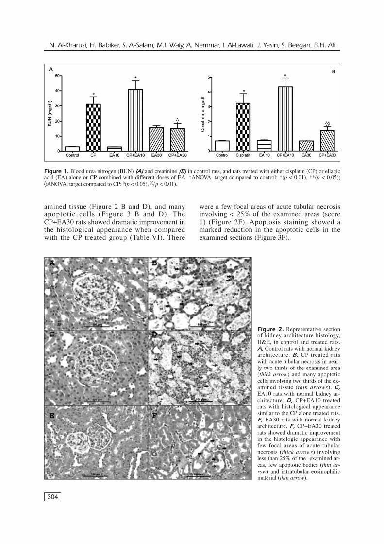
water intake before and after treatment. There wasno significant difference in the amount of waterconsumed by the controls and rats treated with EAalone before and after treatment (Table V). Howev-er, water consumption decreased sharply followingCP and CP+EA10 treatment (p < 0.01), while it in-creased dramatically following CP+EA30 treat-ment (p < 0.05). The osmolality of urine decreasedsignificantly in all CP treated rats (Table V).The creatinine (Cr) and BUN concentrations
increased significantly in rats treated with CP
and CP+EA10 (p < 0.01) (Figure 1 A and B).However, Cr and BUN concentrations in ratstreated with CP+EA30 showed no significant dif-ference when compared with the control rats, butthere was a significant difference in Cr concen-tration (p < 0.01) and BUN (p < 0.05) betweenthis group and the CP alone treated rats. TheBUN concentration of EA30 treated rats washigher than that of the control rats, but the in-crease was not significant.
HistopathologyRats treated with saline (control) and with
EA alone at different doses showed normal kid-ney architecture and histology, and a score of 0(Figure 2 A, C and E). There were no apoptoticbodies found in these groups (Figure 3 A, C andE). However, CP and CP+EA10 treated ratsshowed diffuse acute tubular necrosis in > 60%of the examined tissue areas, with a score of 3(Table VI), and showed tubular distention withnecrotic material involving two thirds of the ex-
303
Ellagic acid protects against cisplatin-induced nephrotoxicity in rats: a dose-dependent study
Change in Relative kidneyGroup N Initial weight* Final weight* body weight (%) weight*
Saline (control) 6 247 ± 7 259 ± 8 4.8 0.71 ± 0.04CP 6 242 ± 5 212 ± 5 -12.1** 1.12 ± 0.12**EA10 7 244 ± 6 256 ± 6 5.2 0.68 ± 0.04CP+EA10 7 250 ± 6 221 ± 7 -11.7** 0.94 ± 0.07**EA30 6 241 ± 9 250 ± 7 3.8 0.72 ± 0.02CP+EA30 9 244 ± 7 242 ± 6 -2.8◊ 0.94 ± 0.05**
Table IV. The effect of saline, cisplatin (CP) or CP plus ellagic acid (EA) given at doses of 10 mg/Kg and 30 mg/Kg on bodyweight and relative kidney weight of rats.
*Values in the table are mean ± SEM from at least 6 rats. **ANOVA significantly different from control rats, significant at p <0.05. ◊ANOVA significantly different from CP rats, significant at p < 0.05.
Water intake* Urine Output*
Before After Before After UrineTreatment treatment treatment treatment treatment osmolality*
Saline (control) 17 ± 1 15 ± 1 6 ± 0.6 6 ± 1.0 2119 ± 205CP 17 ± 2 2 ± 0.4◊ 6 ± 0.5 3 ± 0.8◊ 1098 ± 143**EA10 19 ± 2 17 ± 2 9 ± 0.7 9 ±1.4 2431 ± 330CP+EA10 20 ± 3 3 ± 2◊ 7 ± 1.0 2 ± 0.5◊ 1038 ± 199**EA30 15 ± 2 18 ± 3 7 ± 0.5 9 ± 2.2 1959 ± 233CP+EA30 19 ± 1 31 ± 4◊ 10 ± 1.1 23 ± 3.2◊ 793 ± 123**
Table V. The effect of saline, cisplatin (CP) or CP plus ellagic acid (EA) given at doses of 10 mg/Kg and 30 mg/Kg on waterintake, urine output and urine osmolality in control rats, and rats treated with either cisplatin (CP) or ellagic acid (EA) alone orCP combined with different doses of EA.
*Values in the table are mean ± SEM from at least 6 rats. **ANOVA significantly different from control rats, significant at p <0.05. ◊Paired t-test, significant at (p < 0.05).
304
amined tissue (Figure 2 B and D), and manyapoptotic cells (Figure 3 B and D). TheCP+EA30 rats showed dramatic improvement inthe histological appearance when comparedwith the CP treated group (Table VI). There
were a few focal areas of acute tubular necrosisinvolving < 25% of the examined areas (score1) (Figure 2F). Apoptosis staining showed amarked reduction in the apoptotic cells in theexamined sections (Figure 3F).
N. Al-Kharusi, H. Babiker, S. Al-Salam, M.I. Waly, A. Nemmar, I. Al-Lawati, J. Yasin, S. Beegan, B.H. Ali
Figure 1. Blood urea nitrogen (BUN) (A) and creatinine (B) in control rats, and rats treated with either cisplatin (CP) or ellagicacid (EA) alone or CP combined with different doses of EA. *ANOVA, target compared to control: *(p < 0.01), **(p < 0.05);◊ANOVA, target compared to CP: ◊(p < 0.05), ◊◊(p < 0.01).
A B
* *
**
◊◊◊◊◊◊
Figure 2. Representative sectionof kidney architecture histology,H&E, in control and treated rats.A, Control rats with normal kidneyarchitecture. B, CP treated ratswith acute tubular necrosis in near-ly two thirds of the examined area(thick arrow) and many apoptoticcells involving two thirds of the ex-amined tissue (thin arrows). C,EA10 rats with normal kidney ar-chitecture. D, CP+EA10 treatedrats with histological appearancesimilar to the CP alone treated rats.E, EA30 rats with normal kidneyarchitecture. F, CP+EA30 treatedrats showed dramatic improvementin the histologic appearance withfew focal areas of acute tubularnecrosis (thick arrows) involvingless than 25% of the examined ar-eas, few apoptotic bodies (thin ar-row) and intratubular eosinophilicmaterial (thin arrow).

Total Antioxidant Status (TAS)Both total antioxidant and GSH concentrations
decreased significantly following CP treatment.However, the concentrations of total antioxidant andGSH were similar to those in the control when CPwas combined with 30 mg/kg of EA (Table VII).
Molecular Markers
Kim-1 and Clusterin ProteinsWestern blot was performed to examine the
expression level of Kim-1 and Clu proteins in allrats. β-Actin, a house keeping gene, was used to
305
Ellagic acid protects against cisplatin-induced nephrotoxicity in rats: a dose-dependent study
Figure 3. Representative sectionof kidney architecture histologyand apoptotic body formation,Streptavidin-biotin immunoperoxi-dase method, in control and treatedrats. A, Control rats with normalkidney architecture with no apop-totic bodies. B, CP treated ratswith acute tubular necrosis in near-ly two thirds of examined tissue ar-eas with tubular distention, necrot-ic material and many apoptotic cellshowing brown cytoplasmic stain-ing (thick arrows). C, EA10 ratswith normal kidney architecture.D, CP+EA10 treated rats with his-tological appearance similar to theCP treated rats. E, EA30 rats withnormal kidney architecture. F,CP+EA30 treated rats showed dra-matic improvement in the histolog-ic appearance with few focal areasof acute tubular necrosis and fewapoptotic bodies (thick arrow).
Necrosis No Treatment Score (%)*
1 Saline 0 02 CP 3 63.2 ± 2.73 EA10 0 04 CP + EA10 3 62.7 ± 2.45 EA30 0 06 CP + EA30 1 17.5 ± 3.4
Table VI.Microscopic scoring of kidney section for necrosisin control rats, and treated rats either with cisplatin (CP) orellagic acid (EA) alone or CP combined with different dosesof EA.
*Values in the table are mean ± SD, from at least 6 rats.
Treatment N TAS* GSH*
Saline (control) 6 2.85 ± 0.01 68 ± 2CP 6 2.59 ± 0.06** 49 ± 2EA30 6 2.74 ± 0.06 62 ± 4CP + EA30 9 2.74 ± 0.02 66 ± 2
Table VII. The effect of saline, cisplatin (CP) or CP plus el-lagic acid (EA) given at doses of 10 mg/Kg and 30 mg/Kg ontotal antioxidant (TAS) and reduced glutathione (GSH) con-centrations in controls, rats treated cisplatin (CP) or ellagicacid (EA30) alone, or CP combined with 30 mg/Kg EA.
*Values in the table are mean ± SEM from at least 6 rats.**ANOVA significantly different from control rats. Signifi-cant at p < 0.05.

306
normalize the target proteins. The expression ofβ-actin protein was similar in the controls anddifferent treated rats (Figure 4 A). A similar pat-tern was seen for Clu protein; there was no ap-parent variation in levels of expression in alltreatment groups, including the control (Figure 4B). However, the Kim-1 protein was detected inthe CP alone and CP+EA treated rats, regardlessof the concentration of EA dose (Figure 4 C).
mRNA ExpressionThe level of mRNA expression of Clu and
Kim-1 genes was examined using a Taqman geneexpression mix (ABI) and normalized with thatof the housekeeping gene glyceraldehydes 3-phosphate dehydrogenase (GAPDH). The expres-sion levels of both genes were not affected bydifferent doses of EA alone (Figure 5). However,Clu and Kim-1 expression levels increased signif-icantly when either CP alone or CP+EA at differ-ent doses were used (p < 0.001). The expressionlevels of Clu and Kim-1 in CP+EA30 were high-er than those in CP alone, but the increase wasnot statistically significant (Figure 5).
Discussion
A significant loss in body weight (BW) wasobserved in rats treated with CP. This reductionmay be due to either tubular injury, which affectswater reabsorption leading to dehydration andloss of BW16, or to CP cytotoxic effects on thegastrointestinal tract, which affects rats’ eatingbehavior25, alters gastrointestinal rhythm, and de-lays gastric emptying, which subsequently causes
BW loss. Rats treated with either CP alone or CPtogether with different doses of EA showed sig-nificant increase in relative kidney weight(RKW). The increase in RKW is suggested to bean indication of kidney damage caused by an in-crease in the glomerular volume and cellular de-generative changes, including cytoplasmic vac-uolization of the proximal tubular cells, and tubu-lar dilation26.Histopathological examination revealed acute
tubular necrosis and many apoptotic cells in 60%of the examined sections of the cortical area ofthe kidney in rats treated with CP. Similar resultswere obtained in rats treated with CP combinedwith EA at doses of 10 mg/kg. However, less than25% of the tubular necrosis was observed whenCP was combined with 30 mg/kg EA.Histopathological examination of the kidney is
N. Al-Kharusi, H. Babiker, S. Al-Salam, M.I. Waly, A. Nemmar, I. Al-Lawati, J. Yasin, S. Beegan, B.H. Ali
Figure 4. Kidney protein expression detected by westernblot in control rats, rats treated with either cisplatin (CP) orellagic acid (EA) alone or CP combined with different dosesof EA. Panel A: expression of β-Actin; Panel B: expressionof Clusterin; Panel C: expression of Kim-1.
Figure 5. Clusterin (A) and kidney injury molecule-1 (Kim-1) (B)mRNA expression in control rats, and rats treated with either cis-platin (CP) or ellagic acid (EA) alone or CP combined with different doses of EA. Data are shown as kidney Kim-1 and Clu mRNAexpression relative to expression of the house keeping gene GAPDH. *ANOVA, target compared to control: *(p < 0.001).
A B
* *
*
*
*
*

considered to be the golden standard method todetect renal injury27. It has been reported that CPcan induce epithelial cell atrophy and degenera-tion, loss of brush border, tubular dilation, necro-sis and apoptosis, leading to severe renal injurythrough increased expression of intercellular ad-hesion molecule 1 (ICAM-1) in renal proximaltubules, macrophage infiltration, nuclear factor-kappa B (NF-κB) activation and ROS produc-tion28-30. However, Atessahin et al15 reported thatEA, at a dose of 10 mg/kg, ameliorated thehistopathological changes induced by CP in thekidney. They reported that EA at 10 mg/kg pro-tected the kidney from tubular necrosis, degener-ation, desquamation and tubular dilatation. Thisfinding differs from our results where a dose of10 mg/kg of EA was found to provide no salutaryeffect on the histological architecture of the kid-ney. Ameliorative effects were only seen when adose of 30 mg/kg was used, and the histologicalimprovement of the kidney at this dose of EAwas consistent with that seen in the biochemicalparameters.Rats treated with CP showed significant reduc-
tion in water intake and urine output post CPtreatment. However, it has been reported that CPadministration damages the renal tubule and dis-torts its ability to reabsorb water and causepolyuria, which can also lead to polydipsia31. Re-cent studies reported that CP administration leadsto increase urine output16,32. These findings differfrom those found in the present study. The signif-icant reduction in urine volume may be due to ei-ther significant reduction in water intake or to se-vere kidney injury. However, the reduction inurine volume was also accompanied by low urineosmolality (Table V). The reduction in urine os-molality is thought to be caused by distort reab-sorption of water by the kidney cells, as men-tioned above31. Administration of EA at a dose of30 mg/kg prior to and post CP treatment in-creased water intake and urine output, whichsuggests improved kidney function. Additional conventional biochemical markers
used in the present study to detect kidney injurywere plasma creatinine (Cr) and BUN. In the pre-sent study, plasma Cr and BUN increased morethan 4- and 10-fold, respectively, following eitherCP treatment or CP combined with 10 mg/kgEA. However, Cr and BUN concentrations de-creased by at least half (from 3.3 mg/dl to 1.4mg/dl and from 31 mg/dl to 16 mg/dl, respective-ly) when 30 mg/kg EA was administered prior toand post CP treatment.
The total antioxidant and GSH concentrationswere reduced significantly in CP treated rats. Thelevel of cellular antioxidants, such as GSH, cata-lase and SOD, decreases after CP treatment6. Theintroduction of an exogenous antioxidant isthought to help the endogenous antioxidant sys-tem in scavenging the ROS produced during animbalance in redox status induced by CP7. In thepresent study, the combined treatment of CP 30mg/kg seems to maintain the total antioxidantand GSH concentrations at a level similar tothose in the control rats. It was reported thatwhen 10 mg/kg AE is combined with CP treat-ment, the level of GSH is improved15. However,in our investigation, the improvement was ob-served at 30 mg/kg EA. The reason for this dis-crepancy is not certain, but it may indicate thatthe low dose of EA was not sufficient to amelio-rate the negative effect of CP treatment and cor-rect the redox imbalance in the cell. The discrep-ancy may also be due to the difference in thegender of animals used in the two experiments.Female SD rats were used in our study, wheremale SD rats were used in the other report15. Oth-er parameters used beside the antioxidant assayshowed that EA at doses of 10 mg/kg did not im-prove either the kidney architecture or function.The effect of graded dosed of EA (30, 60 and 90mg/kg) on alcohol induced oxidative stress wasinvestigated and it was reported that low and highdoses of EA did not have an effect on improvingthe imbalance in redox status8. It was suggestedthat a high dose of EA (90 mg/kg) was not effec-tive due to non-specific binding to non-ROS mol-ecules8. In addition, it has been reported that an-tioxidants, such as polyphenols, when adminis-tered at high concentration, may induce ROS inthe cell, cause DNA double strand break, apopto-sis and cytotoxicity33. The effective dose found inthe alcohol induced oxidative stress study was 60mg/kg instead of 30 mg/kg in the current study.The discrepancy may be due to the difference inthe studied organ and the cytotoxic agent used8.At a dose of 30 mg/kg, EA treatment im-
proved most of the biochemical parameters andthe kidney histology. The consistency of EA atthis dose in improving the kidney status may bedue to either the ability to scavenge the ROS asantioxidant and protect the kidney from oxidativestress or EA may affect CP uptake by the kidneycells. The latter possibility is unlikely, is CP con-centration was not significantly different betweenkidneys of rats treated with CP and those treatedwith CP+EA. It is known that the kidneys accu-
307
Ellagic acid protects against cisplatin-induced nephrotoxicity in rats: a dose-dependent study

308
mulate CP more than other organs5, resulting innecrosis in the terminal portion of the proximalrenal tubules and apoptosis in the distalnephron16. In this work, EA was found not to al-ter significantly the concentration of CP in therenal cortex of CP-treated rats. This strongly im-plies that the nephroprotective action of EA isnot mediated thorough a reduction in the accu-mulation of the drug in the renal cortex, and fur-ther suggests that the antitumor activity of CPmay not be adversely affected by the concomi-tant administration of EA.Biochemical markers such as Cr and BUN ele-
vate significantly following administration ofnephrotoxic drugs. However, these markers,though reliable for detection of renal injury, lackthe sensitivity and specificity for early kidneydamage detection34. Therefore, highly sensitivemolecular markers, such as Kim-1 and Clu genesand their proteins, have been deployed to detectearly signs of CP-induced nephrotoxicity and ac-cess the ameliorative effect of EA. In the presentstudy, Clu protein was detected in all treatedgroups, including the control. This was surpris-ing as Clu is not expected to be expressed in highquantities in normal kidneys35. However, the pro-tein is expressed in all tissues and organs and ithas a role in cell remodeling and repair, lipid re-cycling and cell aggregation36. The method weused for detection of Clu (Western blot) is not aquantitative assay and the level seen in untreatedcontrol animals probably reflects normal cellularlevels. On the contrary, Kim-1 protein was detect-ed in all CP treated rats, but not in either the un-treated control rats or rats treated with EA alone.Kim-1 can be found in different tissues in rats,but when a nephrotoxic drug such as CP is intro-duced, its expression is up-regulated in the kid-ney only37. It was reported that Kim-1 and Cluare very sensitive (95%) to early kidney injurydetection and their expression persists until kid-ney recovery34. In the current study, thehistopathological analysis showed that there wasless than 25% damage in the kidney of rats treat-ed with CP and 30 mg/kg EA. This agrees withthe restoration of levels of the conventional bio-chemical markers. However, Clu and Kim-1 werestill elevated, and their levels were higher in ratstreated with CP plus 30 mg/kg EA comparedwith those treated with either CP alone or CPplus 10 mg/kg EA. This could be due to reflec-tion of the role of both Clu and Kim-1 in cell re-modeling and repair at the dose of 30 mg/kg EAin the regenerative area after kidney injury38,39.
EA may have driven the process of repair (elevat-ing remodeling and repair molecules such as Cluand Kim-1) in a dose dependent manner. In addi-tion, it has been postulated that Kim-1 can be in-volved in cell debris removal of damaged renaltissue and participates in epithelial cell growthand differentiation38. Therefore, we hypothesizethat the increased level of Kim-1 and Clu in thekidney tissue of rats treated with CP and 30mg/kg EA is due to kidney repair encouraged byEA administration and regulated by tissue repairproteins, including Kim-1 and Clu. Since thestudy was conducted for 10 days only and the re-covery state of the kidney was not complete, itwas expected that both Clu and Kim-1 would behigh. Thus, the levels of Kim-1 expression are inagreement with the histopathological results andlevels of biochemical markers at the dose of 30mg/kg EA compared with 10 mg/kg EA.
Conclusions
This work has shown that EA, at a dose of 30mg/kg (but not 10 mg/kg), had an ameliorativeeffect against CP nephrotoxicity.
––––––––––––––––––––Acknowledgements
This work was supported by the College of Medicineand Health Sciences, SQU. We thank Prof. Anvar Kaci-mov (College of Agricultural and Marine Sciences) forallowing us to measure platinum in his Laboratory, andthe Staff of the Animal House for looking after the rats.
References
1) MILLER RP, TADAGAVADI RK, RAMESH G, REEVES WB.Mechanisms of cisplatin nephrotoxicity. Toxins2010; 2: 2490-2518.
2) ALI BH, AL MOUNDHRI MS. Agents ameliorating oraugmenting the nephrotoxicity of cisplatin andother platinum compounds: a review of some re-cent research. Food Chem Toxicol 2006; 44:1173-1183.
3) HANIGAN MH, DEVARAJAN P. Cisplatin nephrotoxicity:molecular mechanisms. Cancer Ther 2003; 1: 47-61.
4) PABLA N, MURPHY RF, LIU K, DONG Z. The coppertransporter Ctr1 contributes to cisplatin uptakeby renal tubular cells during cisplatin nephrotox-icity. Am J Physiol Renal Physiol 2009; 296:F505-511.
N. Al-Kharusi, H. Babiker, S. Al-Salam, M.I. Waly, A. Nemmar, I. Al-Lawati, J. Yasin, S. Beegan, B.H. Ali

5) YONEZAWA A, INUI K. Organic cation transporterOCT/SLC22A and H(+)/organic cation antiporterMATE/SLC47A are key molecules for nephrotoxi-city of platinum agents. Biochem Pharmacol2011; 81: 563-568.
6) SANTOS NA, CATAO CS, MARTINS NM, CURTI C, BIANCHIML, SANTOS AC. Cisplatin-induced nephrotoxicity isassociated with oxidative stress, redox state un-balance, impairment of energetic metabolism andapoptosis in rat kidney mitochondria. Arch Toxicol2007; 81: 495-504.
7) CHIRINO YI, PEDRAZA-CHAVERRI J. Role of oxidativeand nitrosative stress in cisplatin-induced nephro-toxicity. Exp Toxicol Pathol 2009; 61: 223-242.
8) DEVIPRIYA N, SRINIVASAN M, SUDHEER AR, MENON VP.Effect of ellagic acid, a natural polyphenol, on al-cohol-induced prooxidant and antioxidant imbal-ance: a drug dose dependent study. SingaporeMed J 2007; 48: 311-318.
9) KANNAN MM, QUINE SD. Ellagic acid amelioratesisoproterenol induced oxidative stress: Evidencefrom electrocardiological, biochemical and his-tological study. Eur J Pharmacol 2011; 659: 45-52.
10) UMESALMA S, SUDHANDIRAN G. Ellagic acid preventsrat colon carcinogenesis induced by 1, 2 dimethylhydrazine through inhibition of AKT-phosphoinosi-tide-3 kinase pathway. Eur J Pharmacol 2011;660: 249-258.
11) ROMERT L, JANSSON T, CURVALL M, JENSSEN D. Screen-ing for agents inhibiting the mutagenicity of ex-tracts and constituents of tobacco products. MutatRes 1994; 322: 97-110.
12) KASSIM M, ACHOUI M, MUSTAFA MR, MOHD MA, YU-SOFF KM . Ellagic acid, phenolic acids, andflavonoids in Malaysian honey extracts demon-strate in vitro anti-inflammatory activity. Nutr Res2010; 30: 650-659.
13) ROSILLO MA, SANCHEZ-HIDALGO M, CARDENO A, DE LA
LASTRA CA. Protective effect of ellagic acid, a natur-al polyphenolic compound, in a murine model ofCrohn's disease. Biochem Pharmacol 2011; 82:737-745.
14) TURK G, CERIBASI AO, SAHNA E, ATESSAHIN A. Ly-copene and ellagic acid prevent testicular apopto-sis induced by cisplatin. Phytomedicine 2011; 18:356-361.
15) ATESSAHIN A, CERIBASI AO, YUCE A, BULMUS O, CIKIMG. Role of ellagic acid against cisplatin-inducednephrotoxicity and oxidative stress in rats. BasicClin Pharmacol Toxicol 2007; 100: 121-126.
16) ALI BH, AL-MOUNDHRI M, TAGELDIN M, AL HUSSEINI IS,MANSOUR MA, NEMMAR A, TANIRA MO. Ontogenicaspects of cisplatin-induced nephrotoxicity in rats.Food Chem Toxicol 2008; 46: 3355-3359.
17) BATISTA BL, GROTTO D, RODRIGUES JL, SOUZA VC, BAR-BOSA F, JR. Determination of trace elements in bio-logical samples by inductively coupled plasmamass spectrometry with tetramethylammoniumhydroxide solubilization at room temperature.Anal Chim Acta 2009; 646: 23-29.
18) MOHAN IK, KHAN M, SHOBHA JC, NAIDU MU, PRAYAGA, KUPPUSAMY P, KUTALA VK. Protection against cis-platin-induced nephrotoxicity by Spirulina in rats.Cancer Chemother Pharmacol 2006; 58: 802-808.
19) VIELHAUER V, STAVRAKIS G, MAYADAS TN. Renal cell-expressed TNF receptor 2, not receptor 1, is es-sential for the development of glomerulonephritis.J Clin Invest 2005; 115: 1199-1209.
20) ER TK. Effect of sample volume for the measure-ment of osmolality by using the Advanced 3250osmometer. Am J Emerg Med 2008; 26: 1060-1061.
21) MILLER NJ, RICE-EVANS C, DAVIES MJ, GOPINATHAN V,MILNER A. A novel method for measuring antioxi-dant capacity and its application to monitoring theantioxidant status in premature neonates. Clin Sci(Lond) 1993; 84: 407-412.
22) GUNTHERBERG H, ROST J. The true oxidized glu-tathione content of red blood cells obtained bynew enzymic and paper chromatographic meth-ods. Anal Biochem 1966; 15: 205-210.
23) GRIFFITH OW. Determination of glutathione andglutathione disulfide using glutathione reductaseand 2-vinylpyridine. Anal Biochem 1980; 106:207-212.
24) BRADFORD MM. A rapid and sensitive method forthe quantitation of microgram quantities of proteinutilizing the principle of protein-dye binding. AnalBiochem 1976; 72: 248-254.
25) VERA G, CHIARLONE A, MARTIN MI, ABALO R. Alteredfeeding behaviour induced by long-term cis-platin in rats. Auton Neurosci 2006; 126-127:81-92.
26) SAAD SY, NAJJAR TA, AL-SOHAIBANI MO. The effect ofrebamipide on cisplatin-induced nephrotoxicity inrats. Pharmacol Res 2000; 42: 81-86.
27) HAASE M, MERTENS PR. Urinary biomarkers–silverbullets to faster drug development and nephronprotection. Nephrol Dial Transplant 2010; 25:3167-3169.
28) SUNG MJ, KIM DH, JUNG YJ, KANG KP, LEE AS, LEE S,KIM W, DAVAATSEREN M, HWANG JT, KIM HJ, KIM MS,KWON DY, PARK SK. Genistein protects the kidneyfrom cisplatin-induced injury. Kidney Int 2008; 74:1538-1547.
29) AN Y, XIN H, YAN W, ZHOU X. Amelioration of cis-platin-induced nephrotoxicity by pravastatin inmice. Exp Toxicol Pathol 2011; 63: 215-219.
30) AHMAD ST, SULTANA S. Tannic acid mitigates cis-platin-induced nephrotoxicity in mice. Human ExpToxicol 2012; 31: 145-156.
31) WONG NL, WALKER VR, WONG EF, SUTTON RA. Mech-anism of polyuria after cisplatin therapy. Nephron1993; 65: 623-627.
32) ABDELRAHMAN AM, AL SALAM S, ALMAHRUQI AS, ALHUSSENI IS, MANSOUR MA, ALI BH. N-acetylcysteineimproves renal hemodynamics in rats with cis-platin-induced nephrotoxicity. J Appli Toxicol 2010;30: 15-21.
309
Ellagic acid protects against cisplatin-induced nephrotoxicity in rats: a dose-dependent study

310
33) BOUAYED J, BOHN T. Exogenous antioxidants–Dou-ble-edged swords in cellular redox state: Healthbeneficial effects at physiologic doses versusdeleterious effects at high doses. Oxid Med CellLongev 2010; 3: 228-237.
34) OZER JS, DIETERLE F, TROTH S, PERENTES E, CORDIER A,VERDES P, STAEDTLER F, MAHL A, GRENET O, ROTH DR,WAHL D, LEGAY F, HOLDER D, ERDOS Z, VLASAKOVA K,JIN H, YU Y, MUNIAPPA N, FOREST T, CLOUSE HK,REYNOLDS S, BAILEY WJ, THUDIUM DT, TOPPER MJ,SKOPEK TR, SINA JF, GLAAB WE, VONDERSCHER J, MAUR-ER G, CHIBOUT SD, SISTARE FD, GERHOLD DL. A PANelof urinary biomarkers to monitor reversibility of re-nal injury and a serum marker with improved po-tential to assess renal function. Nat Biotechnol2010; 28: 486-494.
35) ROSENBERG ME, SILKENSEN J. Clusterin: physiologicand pathophysiologic considerations. Int JBiochem Cell Biol 1995; 27: 633-645.
36) DIETERLE F, PERENTES E, CORDIER A, ROTH DR,VERDES P, GRENET O, PANTANO S, MOULIN P, WAHL
D, MAHL A, END P, STAEDTLER F, LEGAY F, CARL K,LAURIE D, CHIBOUT SD, VONDERSCHER J, MAURER G.
Urinary clusterin, cystatin C, beta2-microglobu-lin and total protein as markers to detect drug-induced kidney injury. Nat Biotechnol 2010; 28:463-469.
37) VAIDYA VS, OZER JS, DIETERLE F, COLLINGS FB, RAMIREZV, TROTH S, MUNIAPPA N, THUDIUM D, GERHOLD D,HOLDER DJ, BOBADILLA NA, MARRER E, PERENTES E,CORDIER A, VONDERSCHER J, MAURER G, GOERING PL,SISTARE FD, BONVENTRE JV. Kidney injury molecule-1outperforms traditional biomarkers of kidney in-jury in preclinical biomarker qualification studies.Nat Biotechnol 2010; 28: 478-485.
38) MALYSZKO J, KOC-ZORAWSKA E, MALYSZKO JS, MYSLIWIEC
M. Kidney injury molecule-1 correlates with kid-ney function in renal allograft recipients. Trans-plantat Proc 2010; 42: 3957-3959.
39) HARPUR E, ENNULAT D, HOFFMAN D, BETTON G, GAUTI-ER JC, RIEFKE B, BOUNOUS D, SCHUSTER K, BEUSHAUSENS, GUFFROY M, SHAW M, LOCK E, PETTIT S; HESI COM-MITTEE ON BIOMARKERS OF NEPHROTOXICITY. Biologicalqualification of biomarkers of chemical-inducedrenal toxicity in two strains of male rat. Toxicol Sci2011; 122: 235-252.
N. Al-Kharusi, H. Babiker, S. Al-Salam, M.I. Waly, A. Nemmar, I. Al-Lawati, J. Yasin, S. Beegan, B.H. Ali