Effect of Coenzyme-Q10 on Doxorubicin-Induced Nephrotoxicity in Rats
Upload
independentCategory
view
0download
0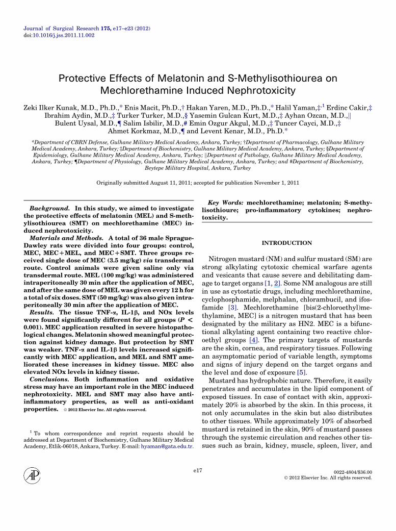
Journal of Surgical Research 175, e17–e23 (2012)doi:10.1016/j.jss.2011.11.002
Protective Effects of Melatonin and S-Methylisothiourea on
Mechlorethamine Induced Nephrotoxicity
Zeki Ilker Kunak, M.D., Ph.D.,* Enis Macit, Ph.D.,† Hakan Yaren, M.D., Ph.D.,* Halil Yaman,‡,1 Erdinc Cakir,‡Ibrahim Aydin, M.D.,‡ Turker Turker, M.D.,§ Yasemin Gulcan Kurt, M.D.,‡ Ayhan Ozcan, M.D.,k
Bulent Uysal, M.D.,{ Salim Isbilir, M.D.,# Emin Ozgur Akgul, M.D.,‡ Tuncer Cayci, M.D.,‡Ahmet Korkmaz, M.D.,{ and Levent Kenar, M.D., Ph.D.*
*Department of CBRN Defense, Gulhane Military Medical Academy, Ankara, Turkey; †Department of Pharmacology, Gulhane MilitaryMedical Academy, Ankara, Turkey; ‡Department of Biochemistry, GulhaneMilitary Medical Academy, Ankara, Turkey; §Department ofEpidemiology, Gulhane Military Medical Academy, Ankara, Turkey; kDepartment of Pathology, Gulhane Military Medical Academy,Ankara, Turkey; {Department of Physiology, Gulhane Military Medical Academy, Ankara, Turkey; and #Department of Biochemistry,
Beytepe Military Hospital, Ankara, Turkey
Originally submitted August 11, 2011; accepted for publication November 1, 2011
Background. In this study, we aimed to investigatethe protective effects of melatonin (MEL) and S-meth-ylisothiourea (SMT) on mechlorethamine (MEC) in-duced nephrotoxicity.Materials and Methods. A total of 36 male Sprague-
Dawley rats were divided into four groups: control,MEC, MECDMEL, and MECDSMT. Three groups re-ceived single dose of MEC (3.5 mg/kg) via transdermalroute. Control animals were given saline only viatransdermal route. MEL (100 mg/kg) was administeredintraperitoneally 30 min after the application of MEC,andafter the samedose ofMELwas given every 12 h fora total of six doses. SMT (50mg/kg)was also given intra-peritoneally 30 min after the application of MEC.Results. The tissue TNF-a, IL-1b, and NOx levels
were found significantly different for all groups (P <0.001). MEC application resulted in severe histopatho-logical changes. Melatonin showedmeaningful protec-tion against kidney damage. But protection by SMTwas weaker. TNF-a and IL-1b levels increased signifi-cantly with MEC application, and MEL and SMT ame-liorated these increases in kidney tissue. MEC alsoelevated NOx levels in kidney tissue.Conclusions. Both inflammation and oxidative
stress may have an important role in the MEC inducednephrotoxicity. MEL and SMT may also have anti-inflammatory properties, as well as anti-oxidantproperties. � 2012 Elsevier Inc. All rights reserved.
1 To whom correspondence and reprint requests should beaddressed at Department of Biochemistry, Gulhane Military MedicalAcademy,Etlik-06018, Ankara, Turkey. E-mail: [email protected].
e17
Key Words: mechlorethamine; melatonin; S-methy-lisothioure; pro-inflammatory cytokines; nephro-toxicity.
INTRODUCTION
Nitrogen mustard (NM) and sulfur mustard (SM) arestrong alkylating cytotoxic chemical warfare agentsand vesicants that cause severe and debilitating dam-age to target organs [1, 2]. Some NM analogous are stillin use as cytostatic drugs, including mechlorethamine,cyclophosphamide, melphalan, chlorambucil, and ifos-famide [3]. Mechlorethamine [bis(2-chloroethyl)me-thylamine, MEC] is a nitrogen mustard that has beendesignated by the military as HN2. MEC is a bifunc-tional alkylating agent containing two reactive chlor-oethyl groups [4]. The primary targets of mustardsare the skin, cornea, and respiratory tissues. Followingan asymptomatic period of variable length, symptomsand signs of injury depend on the target organs andthe level and dose of exposure [5].
Mustard has hydrophobic nature. Therefore, it easilypenetrates and accumulates in the lipid component ofexposed tissues. In case of contact with skin, approxi-mately 20% is absorbed by the skin. In this process, itnot only accumulates in the skin but also distributesto other tissues. While approximately 10% of absorbedmustard is retained in the skin, 90% of mustard passesthrough the systemic circulation and reaches other tis-sues such as brain, kidney, muscle, spleen, liver, and
0022-4804/$36.00� 2012 Elsevier Inc. All rights reserved.
JOURNAL OF SURGICAL RESEARCH: VOL. 175, NO. 1, JUNE 1, 2012e18
bone marrow [6]. Following acute mustard exposure,analyses have show that it accumulates at a high ratein thigh fat, brain, abdominal skin, kidney, and muscletissues [7].
In addition to the alkylating action of mustard, theinjuries of mustard and analogs are mediated by theformation and action of reactive oxygen species (ROS)[8]. Alkylating agents such asMEC can also induce glu-tathione depletion [9]. The depletion of glutathioneleads to accumulation of ROS and reactive nitrogen spe-cies (RNS) within cells, inactivation of sulfhydryl-containing enzymes, loss of calcium homeostasis, lipidperoxidation, cellular membrane breakdown, and ulti-mately cell death [10]. MEC also induces the secretionof pro-inflammatory cytokines, including tumor necro-sis factor-alpha (TNF-a), interleukin (IL)-1, IL-6, IL-8,and IL-15 from differentiated human respiratory epi-thelial cells [11]. On the other hand, inflammation char-acterized by accumulation of granulocytes andmacrophages at sites of injury has a significant role inSM and analogs induced tissue toxicity [12, 13]. In addi-tion, inflammatory cells generate additional ROS thatcontribute to oxidative stress in the SM induced skin in-jury [14].
More evidences suggest that the mechanisms under-lying mustard toxicity include DNA alkylation, gluta-thione depletion, activation of proteases, production offree radicals, and free radical-mediated oxidativestress, inflammation, activation of pro-inflammatorycytokines such as TNF-a, IL-1b, IL-6, IL-8, inducible ni-tric oxide synthase (iNOS) activation and poly(ADP-ribose) polymerase activation [15, 16].
Melatonin (N-acetyl-5-methoxytryptamine, MEL) isthe major product of the pineal gland that has a signifi-cant role in modulation of sleep, sexual behavior, im-mune function, and circadian rhythm. Additionally, itis a potent ROS-scavenger [17–19]. MEL and its metab-olites have also powerful anti-inflammatory propertiesand have proven to be highly effective in a variety of dis-orders linked to oxidative stress and inflammation inexperimental animals [17].
Nitric oxide (NO) is an unstable nitrogen radical thatis generated in different cell types by the concomitantconversion of L-arginine into citrulline through theenzyme NO synthase (NOS). The iNOS induced byendotoxins and cytokines is continuously active and re-leases NO. NO reacts with superoxide anion and ulti-mately form peroxynitrite, a potent reactive oxidant,which leads to direct damage to mitochondrial enzymeandDNA [20]. S-methylisothiourea (SMT) is not a selec-tive iNOS inhibitor and reduces plasma NO levels. Butit is 10- to 20-fold more potent than NG methyl-L-arginine or other iNOS inhibitors known [21].
In this study, we aimed to investigate the protectiveeffects of MEL and SMT on MEC induced nephrotoxi-
city and to determine the levels of TNF-a, IL-1b andnitrite plus nitrate (NOx) in kidney tissues.
MATERIALS AND METHODS
This project was approved by the Experimental Ethics Committeeof Gulhane Military Medical Academy, Ankara, Turkey, and the Na-tional Institute of Health’s Guide for the Care and Use of LaboratoryAnimals was followed.
Animal and Experimental Protocol
A total of 36 male Sprague-Dawley rats (Health sciences Institute,Gulhane Military Medical Academy, Ankara, Turkey), with bodyweights of 220–280 g, were included in this study. They were givenfood and water ad libitum. All animals received humane care in com-pliance with the institution’s guidelines, as outlined in the Guide forthe Care and Use of Laboratory Animals, published by the NationalInstitutes of Health.
They were randomly assigned into four groups containing nine ratseach: control, MEC, MECþMEL, and MECþSMT. All animals exceptthe control groupwere given, via transdermal route, a dose of 3.5mg/kgMEC (dissolved in 500 mL saline). Control animals were given 2 mLsaline only via transdermal route. MEL (100 mg/kg) was adminis-tered intraperitoneally 30min after the application of MEC, and afterthe same dose of MEL was given every 12 h for a total of six doses.SMT (50 mg/kg) was also given intraperitoneally 30 min after the ap-plication of MEC. All surviving animals were killed on d 3 of experi-ment via decapitation, the abdomen was opened, and then kidneyswere harvested for analysis. One kidney was fixed in 4% bufferedformaldehyde for histopathologic evaluation and the other waswashed with saline to remove residual blood, placed in tubes, frozenwith liquid nitrogen, and stored at �80�C.
Tissue Preparation
The frozen kidney tissues were homogenized in phosphate buffersolution (pH 7.4) by means of a homogenizator (Heidolph Diax 900;Heidolph Elektro GmbH, Kelheim, Germany) on ice cube. Homoge-nates were centrifuged at 14,000 rpm (7530 3 g) in 4�C for 10 min.The supernatants were used for all analysis. The protein content ofkidney homogenates was measured by the method defined by Lowryet al. [22] with bovine serum albumin as the standard.
Biochemical Analyses
Tissue TNF-a, IL-1b andNOx concentrations weremeasured in du-plicate by commercial TNF-a and IL-1b rat ELISA kits (Bender Med-Systems, Vienna, Austria), and NOx colorimetric assay kit (CaymanChemical, Ann Arbor, MI). According to the manufacturer, the sensi-tivities of the assays for TNF-a, IL-1b and NOx were 4.3, 4.4 pg/mL,and 2.5 mmol/L, respectively. TNF-a, IL-1b concentrations were ex-pressed as ng/g protein and NOx concentration was expressed asmol/g protein.
Histopathologic Evaluation
For kidney histopathology analysis, the kidney tissues were pro-cessed for light microscopy. This processing consisted of specimens’fixation in 4% buffered neutral formalin solution for 24 h, embeddingin paraffin wax, slicing sections at 4–5 mm of thickness, and stainingthem with hematoxylin and eosin (H&E). Histopathologic examina-tion was blindly evaluated by a pathologist. All sections of kidneysamples were examined for characteristic histologic changes, includ-ing glomerular, tubular (vacuolization, necrosis, and regeneration),interstitial, and vascular alterations.
III)
Py
(IIversu
sIV
)Py
(III
versu
sIV
)
1.000
0.007
1.000
0.057
0.016
0.030
um).
KUNAK ET AL.: MEL AND SMT IN MEC INDUCED NEPHROTOXICITY e19
Statistical Analysis
All the statistical analyses were performed by using SPSS 13.0 soft-ware (SPSS Inc., Chicago, IL). Distributions were evaluated by usingone-sample Kolmogorov-Smirnov test to determine whether the con-tinuous variables are normally distributed or not. Then, we usedKruskal-Wallis test for the ones that were not normally distributed.For the pair-wise comparisons, we used Bonferroni corrected MannWhitney U test with respect to distribution. We performed a post-hoc power analysis by using Power and Sample Size Calculator ver.3.0 (Vanderbilt University, Nashville, TN). The results were ex-pressed as median (minimum- maximum). A P value < 0.05 was con-sidered statistically significant for overall comparisons.
TABLE
1
BiochemicalResu
ltsin
theallExperim
enta
lGroups
Parameters
Groups
P*
(overall)
Py
(Iversu
sII)
Py
(Iversu
sIII)
Py
(Iversu
sIV
)Py
(IIversu
sCon
trol
(I)
MEC
(II)
MECþM
EL
(III)
MECþS
MT
(IV)
TNF-a
(ng/g
prot)
5.3
(3.6�7
.5)
27.7
(12.2–63.3)
9.5
(3.2–14.3)
24.8
(13.7–31.2)
<0.001
0.009
0.042
0.005
0.013
IL-1b(ng/g
prot)
5.3
(2.6�7
.9)
25.2
(13.4–56.1)
9.3
(2.5–16.7)
21.6
(11.1–42.1)
<0.001
0.009
0.042
0.005
0.013
NOx(m
ol/g
prot)
1.3
(0.6�2
.6)
18.8
(12.9–34.4)
4.7
(2.8–8.1)
1.0
(0.7–4.4)
<0.001
0.009
0.002
1.000
0.009
TNF-a
¼tumor
necrosisfactor-alpha;IL
-1b¼
interleu
kin-1
beta;NOx¼
nitrate/nitrite.Alldata
wereex
pressed
asmed
ian(m
inim
um-m
axim
*Kru
skalWallis
test.
y Bon
ferron
icorrectedMannWhitney
UTest.
RESULTS
Three rats in the MEC group and two rats in theMECþSMT group are died until the end of experiment.The kidney TNF-a, IL-1b, and NOx concentrations areshown in Table 1. The tissue TNF-a IL-1b and NOxlevels were found significantly different for all groups(P < 0.001).
The control group had the lowest kidney TNF-a levels[5.3 (3.6–7.5) ng/g protein], and these concentrationswere significantly different compared with the MEC[27.7 (12.2–63.3) ng/g protein], the MECþMEL [9.5(3.2–14.3) ng/g protein], and the MECþSMT [24.8(13.7–31.2) ng/g protein] groups (P ¼ 0.009, P ¼ 0.042,and P ¼ 0.005, respectively) (Fig. 1). TNF-a levels inthe MEC group were significantly higher than theMECþMEL and the MECþSMT groups, respectively(P ¼ 0.013 and P ¼ 1.000). Additionally, in theMECþMEL group, TNF-a concentrations were signifi-cantly lower than the MECþSMT group (P ¼ 0.007).However, there was no difference between the MECand the MECþSMT groups in terms of TNF-aconcentrations.
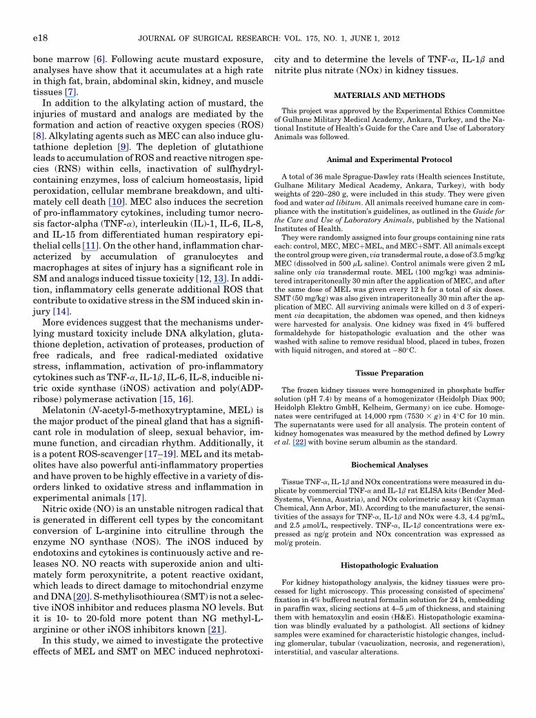
The concentrations of IL-1b in the control group [5.3(2.6–7.9) ng/g protein] were significantly different com-pared with theMEC [25.2 (13.4–56.1) ng/g protein], theMECþMEL [9.3 (2.5–16.7) ng/g protein] and theMECþSMT [21.6 (11.1–42.1) ng/g protein] groups(P ¼ 0.009, P ¼ 0.042, and P ¼ 0.005, respectively)(Fig. 2). On the other hand, in the MEC group, IL-1blevels were significantly higher than the MECþMELgroup (P ¼ 0.013). But there was no difference betweenthe MEC and the MECþSMT groups, the MECþMELand the MECþSMT groups in terms of IL-1bconcentrations.
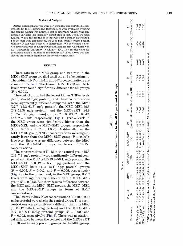
The lowest kidney NOx concentrations [1.3 (0.6–2.6)mol/g protein] were also in the control group. These con-centrations were significantly different than the MEC[18.8 (12.9–34.4) mol/g protein] and the MECþMEL[4.7 (2.8–8.1) mol/g protein] groups (P ¼ 0.009 andP ¼ 0.002, respectively) (Fig. 3). There was no statisti-cal difference between the control and the MECþSMT[1.0 (0.7–4.4) mol/g protein] groups. In the MEC group,
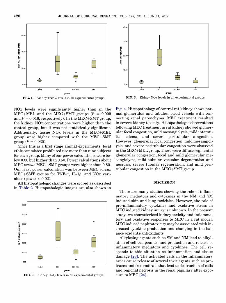
FIG. 3. Kidney NOx levels in all experimental groups.FIG. 1. Kidney TNF-a levels in all experimental groups.
JOURNAL OF SURGICAL RESEARCH: VOL. 175, NO. 1, JUNE 1, 2012e20
NOx levels were significantly higher than in theMECþMEL and the MECþSMT groups (P ¼ 0.009and P ¼ 0.016, respectively). In the MECþSMT group,the kidney NOx concentrations were higher than thecontrol group, but it was not statistically significant.Additionally, tissue NOx levels in the MECþMELgroup were higher compared with the MECþSMTgroup (P ¼ 0.030).
Since this is a first stage animal experiments, localethic committee prohibited use more than nine subjectsfor each group.Many of our power calculations were be-low 0.80 but higher than 0.50. Power calculations aboutMEC versusMECþSMT groups were higher than 0.80.Our least power calculation was between MEC versusMECþSMT groups for TNF-a, IL-1b, and NOx vari-ables (power < 0.02).
All histopathologic changes were scored as describedin Table 2. Histopathologic images are also shown in
FIG. 2. Kidney IL-1b levels in all experimental groups.
Fig. 4. Histopathology of control rat kidney shows nor-mal glomerulus and tubules, blood vessels with con-necting renal parenchyma. MEC treatment resultedin severe kidney toxicity. Histopathologic observationsfollowingMEC treatment in rat kidney showed glomer-ular focal congestion, mild mesangiolysis, mild intersti-tial edema, and severe peritubular congestion.However, glomerular focal congestion, mild mesangiol-ysis, and severe peritubular congestion were observedin theMECþMEL group. There were diffuse segmentalglomerular congestion, focal and mild glomerular me-sangiolysis, mild tubular vacuolar degeneration andnecrosis, severe tubular regeneration, and mild peri-tubular congestion in the MECþSMT group.
DISCUSSION
There are many studies showing the role of inflam-matory mediators and cytokines in the NM and SMinduced skin and lung toxicities. However, the role ofpro-inflammatory cytokines and oxidative stress inMEC induced kidney injury is unknown. In the presentstudy, we characterized kidney toxicity and inflamma-tory and oxidative responses to MEC in a rat model.MEC induced nephrotoxicitymay be associated with in-creased cytokine production and changing in the bal-ance oxidants/antioxidants.
Alkylating agents such as SM and NM lead to alkyl-ation of cell compounds, and production and release ofinflammatory mediators and cytokines. The cell re-sponds to this situation as inflammation and tissuedamage [23]. The activated cells in the inflammatoryareas cause release of several toxic agents such as pro-teases and free radicals that lead to destruction of cellsand regional necrosis in the renal papillary after expo-sure to MEC [24].
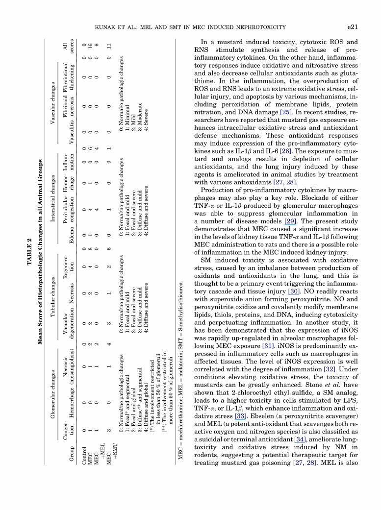
TABLE
2
MeanScoreofHisto
path
ologic
Changesin
allAnim
alGroups
Group
Glomerularch
anges
Tubularch
anges
Interstitialch
anges
Vascularch
anges
All
scores
Con
ges-
tion
Hem
orrh
age
Necrosis
(mesangiolisis)
Vacu
olar
deg
eneration
Necrosis
Reg
enera-
tion
Edem
aPeritubular
congestion
Hem
or-
rhage
Inflam-
mation
Vasculitis
Fibrinoid
necrosis
Fibrointimal
thicken
ing
Con
trol
00
00
00
00
00
00
00
00
00
MEC
10
12
22
48
14
10
60
00
016
MEC þM
EL
10
12
00
00
04
00
40
00
06
MEC þSMT
30
14
31
26
01
00
10
00
011
0:Normal/nopatholog
icch
anges
1:Focal*
andsegmen
tal
2:Focalandglobal
3:Diffuse**
andsegmen
tal
4:Diffuse
andglobal
(*)Theinvolvem
entrestricted
inless
than50%
ofglomeruli
(**)
Theinvolvem
entrestricted
inmorethan50%
ofglomeruli
0:Normal/nopatholog
icch
anges
1:Focalandmild
2:Focalandsevere
3:Diffuse
andmild
4:Diffuse
andsevere
0:Normal/nopatholog
icch
anges
1:Focalandmild
2:Focalandsevere
3:Diffuse
andmild
4:Diffuse
andsevere
0:Normal/opatholog
icch
anges
1:Minim
al
2:Mild
3:Mod
erate
4:Sev
ere
MEC¼
mechlorethamine;
MEL¼
melatonin;SMT¼
S-m
ethylisothiourea.
KUNAK ET AL.: MEL AND SMT IN MEC INDUCED NEPHROTOXICITY e21
In a mustard induced toxicity, cytotoxic ROS andRNS stimulate synthesis and release of pro-inflammatory cytokines. On the other hand, inflamma-tory responses induce oxidative and nitrosative stressand also decrease cellular antioxidants such as gluta-thione. In the inflammation, the overproduction ofROS and RNS leads to an extreme oxidative stress, cel-lular injury, and apoptosis by various mechanisms, in-cluding peroxidation of membrane lipids, proteinnitration, and DNA damage [25]. In recent studies, re-searchers have reported that mustard gas exposure en-hances intracellular oxidative stress and antioxidantdefense mechanisms. These antioxidant responsesmay induce expression of the pro-inflammatory cyto-kines such as IL-1b and IL-6 [26]. The exposure to mus-tard and analogs results in depletion of cellularantioxidants, and the lung injury induced by theseagents is ameliorated in animal studies by treatmentwith various antioxidants [27, 28].
Production of pro-inflammatory cytokines by macro-phages may also play a key role. Blockade of eitherTNF-a or IL-1b produced by glomerular macrophageswas able to suppress glomerular inflammation ina number of disease models [29]. The present studydemonstrates that MEC caused a significant increasein the levels of kidney tissue TNF-a and IL-1b followingMEC administration to rats and there is a possible roleof inflammation in the MEC induced kidney injury.
SM induced toxicity is associated with oxidativestress, caused by an imbalance between production ofoxidants and antioxidants in the lung, and this isthought to be a primary event triggering the inflamma-tory cascade and tissue injury [30]. NO readily reactswith superoxide anion forming peroxynitrite. NO andperoxynitrite oxidize and covalently modify membranelipids, thiols, proteins, and DNA, inducing cytotoxicityand perpetuating inflammation. In another study, ithas been demonstrated that the expression of iNOSwas rapidly up-regulated in alveolar macrophages fol-lowing MEC exposure [31]. iNOS is predominantly ex-pressed in inflammatory cells such as macrophages inaffected tissues. The level of iNOS expression is wellcorrelated with the degree of inflammation [32]. Underconditions elevating oxidative stress, the toxicity ofmustards can be greatly enhanced. Stone et al. haveshown that 2-chloroethyl ethyl sulfide, a SM analog,leads to a higher toxicity in cells stimulated by LPS,TNF-a, or IL-1b, which enhance inflammation and oxi-dative stress [33]. Ebselen (a peroxynitrite scavenger)andMEL (a potent anti-oxidant that scavenges both re-active oxygen and nitrogen species) is also classified asa suicidal or terminal antioxidant [34], ameliorate lung-toxicity and oxidative stress induced by NM inrodents, suggesting a potential therapeutic target fortreating mustard gas poisoning [27, 28]. MEL is also
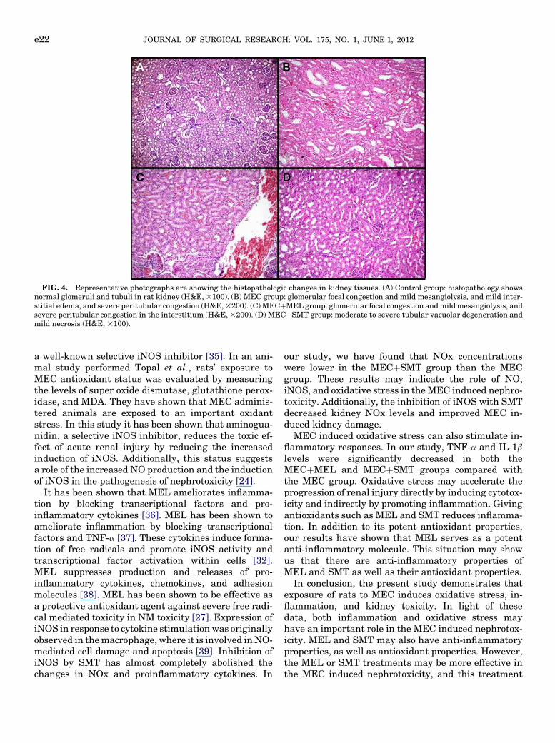
FIG. 4. Representative photographs are showing the histopathologic changes in kidney tissues. (A) Control group: histopathology showsnormal glomeruli and tubuli in rat kidney (H&E,3100). (B) MEC group: glomerular focal congestion and mild mesangiolysis, and mild inter-stitial edema, and severe peritubular congestion (H&E,3200). (C)MECþMEL group: glomerular focal congestion andmildmesangiolysis, andsevere peritubular congestion in the interstitium (H&E,3200). (D) MECþSMT group: moderate to severe tubular vacuolar degeneration andmild necrosis (H&E, 3100).
JOURNAL OF SURGICAL RESEARCH: VOL. 175, NO. 1, JUNE 1, 2012e22
a well-known selective iNOS inhibitor [35]. In an ani-mal study performed Topal et al., rats’ exposure toMEC antioxidant status was evaluated by measuringthe levels of super oxide dismutase, glutathione perox-idase, and MDA. They have shown that MEC adminis-tered animals are exposed to an important oxidantstress. In this study it has been shown that aminogua-nidin, a selective iNOS inhibitor, reduces the toxic ef-fect of acute renal injury by reducing the increasedinduction of iNOS. Additionally, this status suggestsa role of the increased NO production and the inductionof iNOS in the pathogenesis of nephrotoxicity [24].
It has been shown that MEL ameliorates inflamma-tion by blocking transcriptional factors and pro-inflammatory cytokines [36]. MEL has been shown toameliorate inflammation by blocking transcriptionalfactors and TNF-a [37]. These cytokines induce forma-tion of free radicals and promote iNOS activity andtranscriptional factor activation within cells [32].MEL suppresses production and releases of pro-inflammatory cytokines, chemokines, and adhesionmolecules [38]. MEL has been shown to be effective asa protective antioxidant agent against severe free radi-cal mediated toxicity in NM toxicity [27]. Expression ofiNOS in response to cytokine stimulationwas originallyobserved in themacrophage, where it is involved inNO-mediated cell damage and apoptosis [39]. Inhibition ofiNOS by SMT has almost completely abolished thechanges in NOx and proinflammatory cytokines. In
our study, we have found that NOx concentrationswere lower in the MECþSMT group than the MECgroup. These results may indicate the role of NO,iNOS, and oxidative stress in theMEC induced nephro-toxicity. Additionally, the inhibition of iNOS with SMTdecreased kidney NOx levels and improved MEC in-duced kidney damage.
MEC induced oxidative stress can also stimulate in-flammatory responses. In our study, TNF-a and IL-1blevels were significantly decreased in both theMECþMEL and MECþSMT groups compared withthe MEC group. Oxidative stress may accelerate theprogression of renal injury directly by inducing cytotox-icity and indirectly by promoting inflammation. Givingantioxidants such as MEL and SMT reduces inflamma-tion. In addition to its potent antioxidant properties,our results have shown that MEL serves as a potentanti-inflammatory molecule. This situation may showus that there are anti-inflammatory properties ofMEL and SMT as well as their antioxidant properties.
In conclusion, the present study demonstrates thatexposure of rats to MEC induces oxidative stress, in-flammation, and kidney toxicity. In light of thesedata, both inflammation and oxidative stress mayhave an important role in the MEC induced nephrotox-icity. MEL and SMT may also have anti-inflammatoryproperties, as well as antioxidant properties. However,the MEL or SMT treatments may be more effective inthe MEC induced nephrotoxicity, and this treatment
KUNAK ET AL.: MEL AND SMT IN MEC INDUCED NEPHROTOXICITY e23
approachmay be an alternative and effective therapeu-tic strategy for mitigating the toxicity of MEC. Furtherstudies are needed to show these specified effects.
REFERENCES
1. ShakarjianMP,HeckDE,Gray JP, et al.Mechanismsmediatingthe vesicant actions of sulfur mustard after cutaneous exposure.Toxicol Sci 2010;114:5.
2. Wormser U. Toxicology of mustard gas. Trends Pharmacol Sci1991;12:164.
3. Lerch S, K€upfer A, Idle JR, et al. Cerebral formation in situ ofS-carboxymethylcysteine after ifosfamide administration tomice: A further clue to the mechanism of ifosfamide encephalop-athy. Toxicol Lett 2006;161:188.
4. Hardej D, Billack B. Ebselen protects brain, skin, lung and bloodcells from mechlorethamine toxicity. Toxicol Ind Health 2007;23:209.
5. Papirmeister B, Feister AJ, Robinson SI, Ford RD, Eds. MedicalDefense Against Mustard Gas: Toxic Mechanisms and Pharma-cological Implications. 1st edition. Florida: CRC Press, Florida,1991.
6. Somani SM, Babu SR. Toxicodynamics of sulfur mustard. Int JClin Pharmacol Ther Toxicol 1989;27:419.
7. DraschG,KretschmerE,KauertG, et al. Concentrations ofmus-tard gas [bis(2-chloroethyl)sulfide] in the tissues of a victim ofa vesicant exposure. J Forensic Sci 1987;32:1788.
8. Banin E,Morad Y, Berenshtein E, et al. Injury induced by chem-ical warfare agents: Characterization and treatment of oculartissues exposed to nitrogen mustard. Invest Ophthalmol VisSci 2003;44:2966.
9. Langford AM,HobbsMJ,Upshall DG, et al. The effect of sulphurmustard on glutathione levels in rat lung slices and the influenceof treatment with arylthiols and cysteine esters. Hum Exp Tox-icol 1996;15:619.
10. Naghii MR. Sulfur mustard intoxication, oxidative stress, andantioxidants. Mil Med 2002;167:573.
11. Karacsonyi C, Shanmugam N, Kagan E. A clinically relevantin vitromodel for evaluating the effects of aerosolized vesicants.Toxicol Lett 2009;185:38.
12. Guignabert C, Taysse L, Calvet JH, et al. Effect of doxycycline onsulfur mustard induced respiratory lesions in guinea pigs. Am JPhysiol Lung Cell Mol Physiol 2005;289:L67.
13. Paromov V, Suntres Z, Smith M, et al. Sulfur mustard toxicityfollowing dermal exposure: Role of oxidative stress, and antiox-idant therapy. J Burns Wounds 2007;7:e7.
14. Dr€oge W. Free radicals in the physiological control of cell func-tion. Physiol Rev 2002;82:47.
15. Weinberger B, Laskin JD, Sunil VR, et al. Sulfur mustard in-duced pulmonary injury: Therapeutic approaches to mitigatingtoxicity. Pulm Pharmacol Ther 2011;24:92.
16. Kehe K, Raithel K, Kreppel H, et al. Inhibition of poly(ADP-ribose) polymerase (PARP) influences the mode of sulfur mus-tard (SM) induced cell death in HaCaT cells. Arch Toxicol2008;82:461.
17. Reiter RJ, Tan DX, Maldonado MD. Melatonin as an antioxidant:Physiology versus pharmacology. J Pineal Res 2005;39:215.
18. Reiter RJ, Tan DX, Osuna C, et al. Actions of melatonin in thereduction of oxidative stress. A review. J Biomed Sci 2000;7:444.
19. Ozler M, Korkmaz A, Uysal B, et al. Effects of topical melatoninand vitamin E in a rat ischemic wound model. J Exp Integr Med2011;1:123.
20. Sims NR, Anderson MF. Mitochondrial contributions to tissuedamage in stroke. Neurochem Int 2002;40:511.
21. ArunaDevi R, Ramteke VD, Kumar S, et al. Neuroprotective ef-fect of s-methylisothiourea in transient focal cerebral ischemiain rat. Nitric Oxide 2010;22:1.
22. LowryOH,RosebroughNJ, Farr AL, et al. Proteinmeasurementwith the folin phenol reagent. J Biol Chem 1951;193:265.
23. Gao X, Ray R, Xiao Y, et al. Inhibition of sulfur mustard inducedcytotoxicity and inflammation by the macrolide antibiotic roxi-thromycin in human respiratory epithelial cells. BMC Cell Biol2007;8:17.
24. Topal T, UcarM, Gocgeldi E, et al. Investigation of acute toxicityof a chemical warfare agent in kidneys. TAFPrevMedBull 2007;6:227 [Article in Turkish].
25. Beckman JS, KoppenolWH.Nitric oxide, superoxide, and perox-ynitrite: The good, the bad, and ugly. Am J Physiol 1996;271:C1424.
26. Pal A, Tewari-Singh N, Gu M, et al. Sulfur mustard analog in-duces oxidative stress and activates signaling cascades in theskin of SKH-1 hairless mice. Free Radic Biol Med 2009;47:1640.
27. Ucar M, Korkmaz A, Reiter RJ, et al. Melatonin alleviates lungdamage induced by the chemical warfare agent nitrogen mus-tard. Toxicol Lett 2007;173:124.
28. Yaren H, Mollaoglu H, Kurt B, et al. Lung toxicity of nitrogenmustard may be mediated by nitric oxide and peroxynitrite inrats. Res Vet Sci 2007;83:116.
29. Kluth DC, Rees AJ. New approaches to modify glomerular in-flammation. J Nephrol 1999;12:66.
30. Korkmaz A, Yaren H, Topal T, et al. Molecular targets againstmustard toxicity: Implication of cell surface receptors, peroxyni-trite production, and PARP activation. Arch Toxicol 2006;80:662.
31. Sunil VR, Patel KJ, Shen J, et al. Functional and inflammatoryalterations in the lung following exposure of rats to nitrogenmustard. Toxicol Appl Pharmacol 2011;250:10.
32. Korkmaz A, Reiter RJ, Topal T, et al. Melatonin: An establishedantioxidant worthy of use in clinical trials. Mol Med 2009;15:43.
33. StoneWL,QuiM, SmithM. Lipopolysaccharide enhances the cy-totoxicity of 2-chloroethyl ethyl sulfide. BMCCell Biol. 2003;4:1.
34. Korkmaz A, Manchester C. Reactive nitrogen species; devastat-ing intracellular players and melatonin as a defender. J Exp In-tegr Med 2011;1:63.
35. CuzzocreaS,MazzonE,Serraino I, etal.Melatonin reducesdinitro-benzene sulfonic acid induced colitis. J Pineal Res 2001;30:1.
36. Wang H, Wei W, Shen YX, et al. Protective effect of melatoninagainst liver injury in mice induced by bacillus Calmette-Guerin plus lipopolysaccharide. World J Gastroenterol 2004;10:2690.
37. Li JH, Yu JP, Yu HG, et al. Melatonin reduces inflammatory in-jury through inhibitingNF-kB activation in rats with colitis.Me-diators Inflamm 2005;2005:185.
38. Reiter RJ, Calvo JR, KarbownikM, et al. Melatonin and its rela-tion to the immune system and inflammation. Ann N Y Acad Sci2000;917:376.
39. Sagoo P, Chan G, Larkin DF, et al. Inflammatory cytokines in-duce apoptosis of corneal endothelium through nitric oxide. In-vest Ophthalmol Vis Sci 2004;45:3964.
Copyright © 2022 FDOKUMEN