Protective effect of green tea extract on gentamicin-induced nephrotoxicity and oxidative damage in...
21
INTERNATIONAL RESEARCH JOURNAL OF CHEMISTRY (IRJC) ISSN 2321 – 2845(Online), 2321 – 3299 (Print) http://irjc.petsd.org Page | 52 PROTECTIVE EFFECT OF GREEN TEA EXTRACT ON GENTAMICIN INDUCED GASTRO- AND HEPATOTOXICITY IN RATS SARA A KHAN 1,2 , SHUBHA PRIYAMVADA 1 , SHEEBA KHAN 1 , Md WASIM KHAN 1,3 , E.R. AGHARIA 2 AND Ahad N.K.YUSUFI 1* 1 DEPARTMENT OF BIOCHEMISTRY, FACULTY OF LIFE SCIENCES, ALIGARH MUSLIM UNIVERSITY, ALIGARH-202002, UP, INDIA. 2 SVKMS MITHIBAI COLLEGE, BHAKTI VEDANTA MARG, VILE PARLE (W), MUMBAI-400 056, INDIA. 3 DST-INSPIRE FACULTY, CELL BIOLOGY & PHYSIOLOGY DIVISION, CSIR-INDIAN INSTITUTE OF CHEMICAL BIOLOGY, 4, RAJA S.C. MULLICK ROAD, KOLKATA-700032, INDIA. Email: [email protected] ABSTRACT: Activity of glucose-6-phosphate dehydrogenase increased whereas that of malic enzyme decreased. However, GT given to GM rats improved the overall metabolism, enhanced antioxidant defense and energy metabolism. The BBM enzymes were differentially altered by GT. KEY WORD: Antioxidants; Green tea; Gentamicin; Carbohydrate metabolism, Intestine; Liver. INTRODUCTION: A number of environmental variables, including certain chemicals, metal ions, and antibiotics, were shown to alter the structure and function of various tissues [1-6]. Gentamicin (GM) is frequently used in the treatment of severe infections of the abdomen and urinary tract including bacteremia and endocarditis [7]. However, nephrotoxicity and ototoxicity limit its long-term clinical use [8]. GM also accumulates in other tissues, including intestine, liver, heart, and ear causing multiple adverse effects in these tissues [3, 4, 9-12]. Aminoglycoside antibiotics were also shown to alter mucosal SJIF 2012: 2.545 UIF 2013: 1.075 Received on: 6 th December 2014 Revised on: 10 th December 2014 Accepted on: 11 th December 2014 Published on: 1 st January 2015 Volume No. Online & Print 8 (2015) Page No. 52 to 72 IRJC is an international open access print & e journal, peer reviewed, worldwide abstract listed, published quarterly with ISSN, Free- membership, downloads and access.
-
Upload
independent -
Category
Documents
-
view
0 -
download
0
Transcript of Protective effect of green tea extract on gentamicin-induced nephrotoxicity and oxidative damage in...
INTERNATIONAL RESEARCH JOURNAL OF CHEMISTRY (IRJC) ISSN 2321 – 2845(Online), 2321 – 3299 (Print)
http://irjc.petsd.org Page | 52
PROTECTIVE EFFECT OF GREEN TEA EXTRACT ON
GENTAMICIN INDUCED GASTRO- AND
HEPATOTOXICITY IN RATS
SARA A KHAN1,2, SHUBHA PRIYAMVADA1, SHEEBA KHAN1,
Md WASIM KHAN1,3, E.R. AGHARIA2 AND Ahad N.K.YUSUFI1*
1DEPARTMENT OF BIOCHEMISTRY, FACULTY OF LIFE SCIENCES,
ALIGARH MUSLIM UNIVERSITY, ALIGARH-202002, UP, INDIA.
2 SVKMS MITHIBAI COLLEGE, BHAKTI VEDANTA MARG, VILE
PARLE (W), MUMBAI-400 056, INDIA.
3DST-INSPIRE FACULTY, CELL BIOLOGY & PHYSIOLOGY
DIVISION, CSIR-INDIAN INSTITUTE OF CHEMICAL BIOLOGY, 4,
RAJA S.C. MULLICK ROAD, KOLKATA-700032, INDIA.
Email: [email protected]
ABSTRACT:
Activity of glucose-6-phosphate dehydrogenase increased whereas
that of malic enzyme decreased. However, GT given to GM rats
improved the overall metabolism, enhanced antioxidant defense and
energy metabolism. The BBM enzymes were differentially altered by
GT.
KEY WORD: Antioxidants; Green tea; Gentamicin; Carbohydrate
metabolism, Intestine; Liver.
INTRODUCTION:
A number of environmental variables, including certain chemicals,
metal ions, and antibiotics, were shown to alter the structure and
function of various tissues [1-6]. Gentamicin (GM) is frequently used
in the treatment of severe infections of the abdomen and urinary
tract including bacteremia and endocarditis [7]. However,
nephrotoxicity and ototoxicity limit its long-term clinical use [8]. GM
also accumulates in other tissues, including intestine, liver, heart, and
ear causing multiple adverse effects in these tissues [3, 4, 9-12].
Aminoglycoside antibiotics were also shown to alter mucosal
SJIF 2012: 2.545
UIF 2013: 1.075
Received on:
6th December 2014
Revised on:
10th December 2014
Accepted on:
11th December 2014
Published on:
1st January 2015
Volume No.
Online & Print
8 (2015)
Page No.
52 to 72
IRJC is an
international open
access print & e
journal, peer reviewed,
worldwide abstract
listed, published
quarterly with ISSN,
Free- membership,
downloads and access.
INTERNATIONAL RESEARCH JOURNAL OF CHEMISTRY (IRJC) ISSN 2321 – 2845(Online), 2321 – 3299 (Print)
http://irjc.petsd.org Page | 53
morphology and absorption of nutrients [13-15]. They are known to interact with the
intestinal epithelium, its innervations causing alterations in intestinal motility which
contribute to antibiotic-associated diarrhea and colitis [16]. GM was shown to inhibit the
growth of majority of E. coli and bactericides fragile strains isolated from gastro-intestinal
tract [17]. GM also produced multiple adverse effects in the liver and induced oxidative stress
[2].
In the past decade, the concept of disease prevention via naturally occurring substances has
gained much interest. Since ancient times, green tea (GT) consumption has been shown to
improve health [18]. GT polyphenols, especially catechins, are well known antioxidative
agents and have been held responsible for most of the beneficial health effects ascribed to GT
[19]. It is well established that gastrointestinal (GI) tract plays an important role in the
absorption, metabolism, and conjugation of GT polyphenols [20]. GT polyphenols were found
to inhibit the growth of stomach cancer and gastritis [21]. GT is found to activate intracellular
antioxidant, inhibits pro-carcinogen formation and cancer cell proliferation in the GI tract
[22]. GT catechins improve gut flora by selectively increasing the growth of bifidobacteria and
lactobacilli in the gut wall while decreasing levels of potential pathogens [23] and also
prevent atrophy of the intestinal mucosa [24]. The health promoting properties of GT have
the potential to prevent GI diseases [25]. GT has been shown to reduce inflammation
associated with Crohn’s disease and ulcerative colitis, a type of inflammatory Bowl disease
(IBD) [24, 26-27]. GT is effective in reducing cholesterol and lipid absorption in the GI tract
[28]. It also appears to protect liver from damaging effects of toxic substances and also against
the development of liver tumors [25].
Recently we have shown that GT consumption enhanced cellular energy metabolism and
antioxidant defense mechanism in the liver, kidney, and small intestine [29] and prevented
GM and CP-induced nephrotoxicity and oxidative damage in the rat kidney [5, 30-32].
Considering the potential clinical use GM and the numerous health benefits of GT, we now
hypothesize that GT would prevent GM-induced gastrotoxicity and hepatotoxicity. The results
obtained indicate that GM administration caused selective alterations in the activities of
various enzymes of carbohydrate metabolism, BBM and oxidative stress and increased lipid
peroxidation. However, GT consumption markedly reversed GM-induced alterations by
improving energy metabolism, BBM integrity and by enhancing antioxidant defense
mechanism. The present results suggest that GT consumption can be an option for long-term
clinical use of GM without any toxic side effects.
INTERNATIONAL RESEARCH JOURNAL OF CHEMISTRY (IRJC) ISSN 2321 – 2845(Online), 2321 – 3299 (Print)
http://irjc.petsd.org Page | 54
MATERIALS AND METHOD:
Chemicals and drugs-
Green tea (Lipton / Kangra-brand) was purchased from commercial sources (Jain Pan House,
New Delhi, India), Gentamicin (Nicholas, Mumbai, India) from the source indicated in
parentheses. All other chemicals used were of analytical grade and were purchased either
from Sigma Chemical Co. (St Louis, MO, USA) or Sisco Research Laboratory, Mumbai, India.
Green Tea Extract-
Green tea extract (GTE) was prepared by adding green tea (30 g) to 500 ml of boiling water,
steeped for 15-20 min. Infusion was cooled to room temperature and then filtered. The tea
leaves were extracted a second time with 500 ml of boiling water and filtered, and the two
filtrates were combined to obtain 3% green tea extract (3 g tea leaves/100 ml H2O). The
resulting clear solution is similar to tea brews consumed by humans. A known amount of
green tea extract (2 X 125 mL= 250 mL/day) was provided in two servings to GT consuming
rats for 25 days and was found to be sufficient. According to the manufacturer’s information,
the antioxidant content was 95 mg/g of GT.
Experimental design-
The animal experiments were conducted according to the guidelines of Committee for
Purpose of Control and Supervision of Experiments on Animals (CPCSEA), Ministry of
Environment and Forests, Government of India. Adult male Wistar rats (8 rats/group)
weighing 150-180 g fed with standard rat chow (Aashirwaad Industries, Chandigarh, India)
and water ad libitum were conditioned for one week before the start of the experiment.
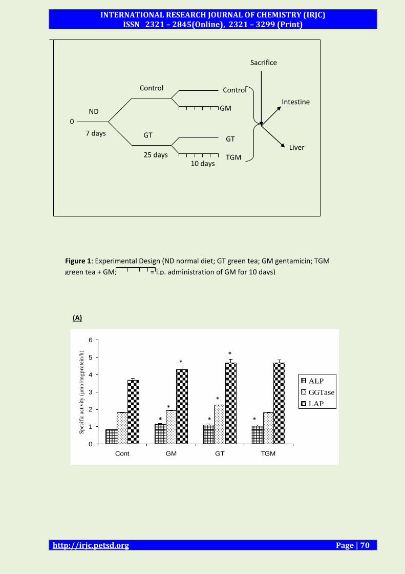
Initially two groups of rats entered the study after acclimatization (Fig 1).
They were fed on a normal rat chow diet with one group consuming water (control) and the
other consuming GT extract (3% w/v) in drinking water for 25 days as described above. After
15 days the rats were subdivided into two groups each (8 rats/group) and continued to
receive their respective drinking fluids. Nephrotoxicity was induced by intraperitoneal
administration of GM (80 mg/kg bwt/day), in 0.9% saline daily for 10 days to one of the sub-
group designated as GM and TGM. The other sub-group from each group received an
equivalent volume of normal saline for the same period. The rats were sacrificed 24 hours
after the last injection under light ether anesthesia. Blood samples were collected and the
intestines and liver were removed and processed for the preparation of homogenate and
brush-border membrane vesicles (BBMVs) as described below. All the preparations and
analyses of various parameters were carried out simultaneously under similar experimental
INTERNATIONAL RESEARCH JOURNAL OF CHEMISTRY (IRJC) ISSN 2321 – 2845(Online), 2321 – 3299 (Print)
http://irjc.petsd.org Page | 55
conditions to avoid any day-to-day variations. Body weights of rats were recorded at the start
and completion of the experimental procedure.
Preparation of homogenates:-
After the completion of the experiment, liver and intestine were extracted. The intestines
were washed by flushing them with ice cold buffered saline (1mM Tris-HCl, 9g/L NaCl, pH
7.4). The liver was put in Tris buffered saline (TBS). The washed intestines (from control and
treated groups) were slit in the middle and the entire mucosa was gently scraped with a glass
slide and weighed. A 6.5% homogenate of this mucosa was prepared in 50 mM mannitol, pH
7.0, in a glass Teflon homogenizer (Remi motors, Mumbai, India) with 5 complete strokes. The
homogenate was then subjected to high speed Ultra-Turrex homogenizer (Type T-25, Janke &
Kunkel GMBH & Co. KG. Staufen) for 3 pulses of 30 s each with an interval of 30 s between
each stroke.
Preparation of brush border membrane-
The intestinal BBMV was prepared as described by Kessler et al. [33], using differential
precipitation by CaCl2. Mucosa scraped from 4-5 washed intestines was used for each BBM
preparation. Briefly, the mucosal scrapings were collected in a beaker containing 50 mM
mannitol, 5 mM Tris-HCl, pH 7.5. The mucosal homogenate was diluted with the above
mentioned Tris-mannitol buffer (15 ml/g tissue) and further homogenized using Ultra-Turrex
T25 homogenizer with three pulses of 30s each with 30s interval between each pulse.
Aliquots of mucosal homogenate were saved and quickly frozen for further analysis. CaCl2 was
added to the filtrate, to a final concentration of 10 mM and was kept for 20 min on ice, with
intermittent stirring. The homogenate were then centrifuged at 2000 g (5000 rpm) for 10 min
in a Beckman J2-M1 refrigerated centrifuge using a JA-17 rotor. The pellet was discarded and
the supernatant was recentrifuged at 35000 g (17000 rpm) for 30 min. The pellet was
resuspended in a small volume (1-2 ml) of 50 mM sodium maleate buffer, pH 6.8, with four
complete passes by a loose fitting Dounce homogenizer (Wheaton, USA) and centrifuged at
35,000 g (17,000 rpm) for 30 min in 15 ml corex glass tube using JA-20 rotor. The white outer
fluffy portion of the pellet was resuspended carefully in a small volume of the above
mentioned buffer, leaving the dark brown centre of the pellet undisturbed (mitochondrial
contamination). The suspension thus obtained was homogenized by hand held Douncer. The
BBM suspension was quickly frozen in small aliquots and used for enzyme analysis. In each
experiment, tissues from three to six animals (control and experimental) were pooled to
obtain a sufficient amount of starting material. All the steps involved were strictly carried out
at 0-4º C unless otherwise specified.
INTERNATIONAL RESEARCH JOURNAL OF CHEMISTRY (IRJC) ISSN 2321 – 2845(Online), 2321 – 3299 (Print)
http://irjc.petsd.org Page | 56
Assay of carbohydrate metabolism enzymes:
The activities of the enzymes involving oxidation of NADH or reduction of NADP were
determined spectrophotometrically on Cintra 5 fixed for 340 nm using 3 ml of assay in a 1-cm
cuvette at room temperature (28-30 °C). The enzyme assays of lactate dehydrogenase (LDH,
E.C.1.1.1.27), malate dehydrogenase (MDH, E.C.1.1.1.37), malic enzyme (ME, E.C.1.1.1.40),
glucose-6-phosphate dehydrogenase (G6PDH, E.C.1.1.1.49), glucose-6-phosphatase (G6Pase,
E.C.3.1.3.3) and fructose-1, 6-bisphosphatase (FBPase, E.C.3.1.3.11) activities were studied as
described by Khundmiri et al [34]. Hexokinase was estimated by the method of Crane and Sols
[35] and the remaining glucose was measured by method of Nelson-Somogyi [36].
2.7 Assay of brush border membrane marker enzymes and lysosomal marker enzymes:
The activities of alkaline phosphatase (ALP), leucine amino peptidase (LAP), γ- glutamyl
transpeptidase (GGTase) and acid phosphatase (ACPase) were determined as described by
Farooq et al [3].
Assay of enzymes involved in free radical scavenging:
Superoxide dismutase (SOD, E.C.1.15.1.1) was assayed by the method of Marklund [37].
Catalase (CAT, E.C.1.11.1. 6) activity was assayed by the method of Giri et al [38].
2.9 Lipid peroxidation and Total –SH group estimation:
Total SH groups were determined by the method of Sedlak and Lindsay [39]. Lipid
peroxidation (LPO) by the method of Ohkawa et al [40].
Statistical analyses-
All data are expressed as Mean ± SEM for at least 4-5 different preparations. Statistical
evaluation was conducted by one-way ANOVA and by unpaired student’s t test using SPSS 7.5
software. A probability level of p<0.05 was selected as indicating statistical significance. Most
of the changes between various groups were compared with control values for better
understanding and clarity. However, specific differences and statistical significance between
other groups were evaluated separately e.g. GM vs. TGM.
RESULT:
To address the hypothesis whether green tea (GT) extract would prevent GM induced
intestinal and hepatic toxicity and oxidative damage, the effect of green tea was determined in
detail on the biomarkers enzymes of oxidative damage, brush border membrane and
carbohydrate metabolism in intestinal and liver tissues. In general, there was no significant
difference in body weight and food intake between GM administered and control rats.
INTERNATIONAL RESEARCH JOURNAL OF CHEMISTRY (IRJC) ISSN 2321 – 2845(Online), 2321 – 3299 (Print)
http://irjc.petsd.org Page | 57
However a slight loss of body weight (~10%) was recorded in rats consuming GT alone and in
combination with GM (Table 1).
Effect of GT on GM induced alterations in biomarker enzymes of BBM and lysosomes:-
To assess the structural integrity of various organelles e.g. plasma membrane (BBM) and
lysosomes, the effect of GM and GT was determined on biomarker enzymes of BBM and
lysosomes in the intestinal and liver homogenates and isolated BBM preparations from
intestine.
a) Effect of GM and GT on biomarkers of BBM and lysosomes in intestinal homogenates:
The activities of alkaline phosphatase (ALP), γ-glutamyl transpeptidase (GGTase), leucine
aminopeptidase (LAP) and acid phosphatase (ACPase) were determined under different
experimental conditions in the homogenates of intestine and liver (Table 2). The activities of
these enzymes along with sucrase (a biomarker of intestinal BBM) significantly increased in
intestinal homogenate as reported earlier [7]. GM treatment caused small increase in BBM
enzymes in liver homogenates. GT consumption, however affected these enzymes
differentially in different tissues (Table 2). The activities of ALP (+29%), GGTase (+25%), LAP
(+27%) and sucrase (+44%) significantly increased by GT in the intestine. However, except
the decrease of GGTase (-24%) activity, ALP and LAP activities were not altered in the liver by
GT (Figure 2). Since GT caused increase in all BBM enzyme activities in the intestine, its
consumption by GM rats resulted in further improvement of BBM enzyme activities in
intestinal homogenates. The activities of GGTase and LAP were also higher in liver
homogenate in TGM compared to GM rats. It appeared from the results that GT produces an
overall improvement in BBM enzyme activities in GM treated rats in both the tissues. The
activity of ACPase (a lysosomal enzyme) variably decreased by GM treatment in both liver and
intestine. In contrast, GT increased ACPase activity in the intestine. As a result, ACPase activity
was significantly higher in tissues in TGM compared to GM rats
b) Effect of green tea (GT) on gentamicin (GM) induced alterations on brush border
membrane enzymes in isolated BBM preparation:
The effect of GM/GT on BBM marker enzymes was further analyzed in BBM preparations
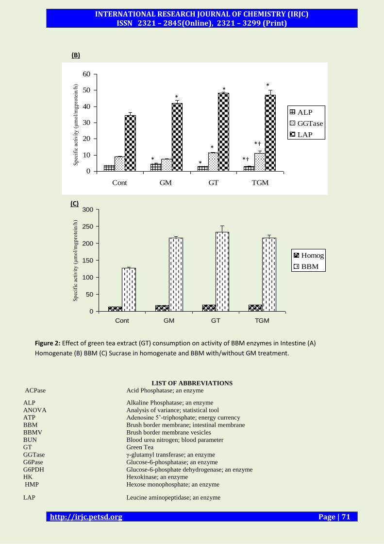
isolated from intestine. The data summarized in Table 3, (Figure 2) showed a similar activity
pattern of BBM enzymes as observed in intestinal homogenate, however the magnitude of
effects was different. The activities of ALP (+28%), LAP (+21%) and sucrase (+69%)
increased to a much greater extent whereas GGTase (-18%) decreased by GM treatment in
intestinal BBM (Table 3, Figure 2). Except ALP, (which was decreased), the activity of GGTase
(+24%), LAP (+39%) and sucrase (+82%) profoundly increased by GT ingestion. The
INTERNATIONAL RESEARCH JOURNAL OF CHEMISTRY (IRJC) ISSN 2321 – 2845(Online), 2321 – 3299 (Print)
http://irjc.petsd.org Page | 58
activities of BBM enzymes except ALP were significantly enhanced upon drinking GT by GM
rats as compared to GM alone rats.
Effect of green tea on GM induced alterations on the enzymes of carbohydrate metabolism
in rat intestinal and hepatic tissues:
The effect of GM, GT, and TGM was determined on the activities of various enzymes of
carbohydrate metabolism in intestine and liver (Tables 4 and 5). GM treatment to control rats
significantly increased hexokinase activity in the intestine (+17%) and liver (+20%). The
activity of LDH (a marker enzyme of anaerobic glycolysis), however profoundly increased in
the intestine (+124%) whereas decreased in the liver (-25%). The effect of GM was
differentially observed on MDH activity (an enzyme of TCA cycle) in the different tissues
studied. GM caused significant decline in MDH activity in the intestine (-40%) whereas
increased in the liver (+32%). As previously reported [34], GT consumption significantly
enhanced metabolic activities in almost all tissues studied. The activity of HK, LDH, and MDH
significantly increased in the intestine and liver by GT. When GM treatment was extended to
GT drinking rats, GM induced decrease in most metabolic enzyme activities, was not only
prevented but they remained significantly higher in TGM compared to either control or GM
treated rats (Table 4). The activity of LDH was especially found to be profoundly enhanced in
the intestine. The activity of MDH, however significantly increased in the liver by GT given to
GM rats.
The effect of GM and GT were also observed on enzymes of gluconeogenesis. GM significantly
increased the activity of glucose-6-phosphatase (G6Pase) and fructose 1, 6, bisphosphatase
(FBPase) in the intestine and to some extent in the liver (Table 5). GT significantly enhanced
the activity of both G6Pase and FBPase in the intestine but lowered in the liver. As a result GT
consumption by GM treated rats resulted in significant increase of both G6Pase and FBPase.
The effect of GT-extract alone and with GM was also determined on the activities of glucose-6-
phosphate dehydrogenase (G6PDH, HMP-shunt) and malic enzyme (ME), source of NADPH
needed in reductive anabolic reactions (Table 5). GM treatment to control rats significantly
increased G6PDH activity in the intestine (+46%) whereas decreased in the liver (-38%). The
activity of ME however, significantly lowered by GM treatment in both the tissues (Table 5).
The activities of G6PDH and ME however were differentially altered by GT. The activity of
G6PDH profoundly decreased in the intestine (-60%) and liver (-21%). ME activity on the
other hand was lowered by GT in both the tissues. GT consumption by GM treated rats was
able to restore G6PDH activity and was significantly higher in the liver in TGM compared to
INTERNATIONAL RESEARCH JOURNAL OF CHEMISTRY (IRJC) ISSN 2321 – 2845(Online), 2321 – 3299 (Print)
http://irjc.petsd.org Page | 59
control or GM rats. The activity of ME was also improved in TGM compared to GM or control
rats.
Effect of GT on GM induced alterations in antioxidant defense parameters in rat intestinal
and hepatic tissues:-
GM induced gastro- and hepatotoxicity can also be determined, at least in part, by
perturbation of antioxidant defense mechanism (Table 6). GM caused significant decrease in
the activity of superoxide dismutase (SOD) in the intestine (-27%) and liver (-39%). The
activity of catalase, also significantly but differentially, declined in both the rat tissues studied.
It was decreased to greater extent in the intestine (-53%) followed by liver (-25%). GM
induced decrease in SOD and catalase activities was associated with increased production of
malondialdehyde (MDA); an end product of lipid peroxidation (LPO) in all the tissues. This
was accompanied by decreased levels of total SH. Taken together, these results indicated that
GM elicited production of free radicals and suppression of antioxidant enzymes caused
significant damage to rat tissues. In contrast, GT significantly enhanced the activity of either
SOD or catalase or both in both the rat tissues. LPO was lowered in the liver (-8%). GT, by
virtue of its known antioxidant properties prevented GM elicited adverse effects on various
parameters of oxidative damage. The activities of both SOD and catalase were also
significantly increased in the liver and intestine in TGM compared to GM treated rats. The
levels of total SH and LPO were also improved indicating a marked protection by GT against
GM induced oxidative damage in both intestine and liver.
DISCUSSION: Digestion and absorption of nutrients are the major functions of the intestinal mucosa. Liver
also contributes significantly to these processes. Thus, any damage to these organs by
environmental contaminants, including aminoglycoside antibiotics, might lead to
malabsorption of food components. Gentamicin (GM) is an effective and widely used
aminoglycoside antibiotic against severe bacterial infections [7]. Although nephrotoxicity is
one of the most important side effect and therapeutical limitation [41], GM was also shown to
cause gastrotoxicity and hepatotoxicity after prolonged clinical use [3]. Several strategies and
mechanisms were utilized to prevent GM-induced nephrotoxicity, however, there have been
no reports regarding protection against GM-induced gastro- and hepatotoxicity. Tea, the
second most widely consumed beverage worldwide since ancient times, is known for its
beneficial health effects. In particular, green tea (GT) has been shown to retard various forms
of cancers due to its antimutagenic and anticarcinogenic properties [18, 22]. It was also found
INTERNATIONAL RESEARCH JOURNAL OF CHEMISTRY (IRJC) ISSN 2321 – 2845(Online), 2321 – 3299 (Print)
http://irjc.petsd.org Page | 60
to be cardioprotective, neuroprotective, antidiabetic, and antibacterial besides other health
benefits [19, 42]. GT has been shown to inhibit growth of certain cancers of GI tract and liver
and improve gut flora [21, 23, 29]. Recently, we have reported that GT was able to protect
against GM-induced nephrotoxity and oxidative stress in the rat kidney [5]. In the present
studies, we have tested our hypothesis that GT consumption would ameliorate GM-induced
toxic and other adverse effects in the intestine and liver.
GM was administered to control and GT-consuming rats and the activities of various enzymes
of carbohydrate metabolism, brush border membrane, mitochondria and lysosome and
parameters of oxidative stress were determined to understand detailed biochemical
events/cellular response/ mechanism of GM toxicity in the intestine and liver and its possible
protection by GT consumption. As reported earlier [3], the activities of various enzymes
involved in glycolysis, TCA cycle, gluconeogenesis, and HMP shunt pathway were
differentially altered in the intestine and liver by GM treatment. GM caused significant
increase in HK, LDH, G6Pase, FBPase, and G6PDH but decreased MDH and ME activities in the
intestine. However, in the liver, an opposite GM effect was observed where the decrease in
LDH activity was associated with increased MDH and HK activity but decreased G6PDH and
ME activities. Although the actual rates of glycolysis or TCA cycle were not determined, the
results indicate a shift in energy metabolism as an adaptive cellular response both in the
intestine and liver. A marked increase in LDH, and to some extent hexokinase, associated with
decreased MDH activity by GM may indicate that anaerobic glycolysis become major source
of energy in the intestine as reported earlier [1,3] due to GM-induced mitochondrial
dysfunction as reported for kidney [5]. At the same time GM induced upregulation of G6Pase
and FBPase appeared to provide sufficient glucose from other metabolites by gluconeogenesis
to support higher LDH activities in the intestine. Conversely, the increase in MDH and HK
activities associated with decreased LDH activity in the liver by GM indicates that oxidative
metabolism remained the predominant source of energy in the liver.
In addition, the effect of GM was also observed on the activity of G6PDH (HMP-shunt
pathway) and NADP-malic enzyme (ME) in the intestine and liver. The activity of G6PDH,
however, profoundly increased in the intestine, whereas decreased in the liver. While the
activity of NADP –ME, significantly but variably declined in both the tissues. Increased G6PDH
activity, at least, in the intestine seems to increase glucose oxidation by an alternative HMP-
shunt pathway. It appears to be an important cellular response as increased supply of NADPH
would be utilized in reducing anabolic pathways especially lipid biosynthesis (Membrane
lipids, cholesterol, fatty acids/phospholipids) required during repairing the membranes and
INTERNATIONAL RESEARCH JOURNAL OF CHEMISTRY (IRJC) ISSN 2321 – 2845(Online), 2321 – 3299 (Print)
http://irjc.petsd.org Page | 61
in maintaining high GSH levels required under oxidative stress and inflammatory conditions
to enhance antioxidant defense mechanism [43]. In contrast to GM, GT consumption caused
selective alterations in the activities of various enzymes involved in carbohydrate metabolism
in different rat tissues. The activity of HK, LDH (glycolysis), MDH (TCA cycle), G6Pase and
FBPase (gluconeogenesis) significantly increased in the intestine but decreased in the liver as
reported earlier [30]. GT administration to GM-treated rats resulted in overall improvement
of carbohydrate metabolism in the intestine and liver. The activities of LDH and
gluconeogenic enzymes in GT fed-GM treated rat intestine remained higher whereas the GM
induced reduction in MDH activity was prevented by GT consumption. It appears that GT
might have lowered number of damaged mitochondria or affected macromolecules or may
have increased number of normally active organelles or macromolecules. Thus, GT given to
GM treated rats appears to maintain anaerobic glycolysis in the intestine and oxidative
metabolism in the liver, respectively, to meet energy requirements.
Intestinal microvillus membrane is highly specialized to perform transport and enzymatic
functions essential for normal digestion and absorption. Alterations in the membrane
organization or damage to the structure by toxic insult can affect both these functions. The
hydrolytic enzymes such as GGTase, LAP, lactase and sucrase etc., are involved in the terminal
digestion of proteins and carbohydrates and subsequent absorption of amino acids and
glucose. For example, sucrase splits sucrose into glucose and fructose just before their
absorption. Alkaline phosphatase (ALP) converts organic phosphate to inorganic phosphate
needed for phosphorylation of metabolites and ATP formation. Since BBM and other
intracellular organelles such as mitochondria and lysosomes are known GM target [3-4], the
structural/functional integrity of the tissue was assessed by the status of their respective
biomarker enzymes. GM exerted differential effects on ALP, GGTase, LAP (BBM enzymes) in
the intestine and liver. As reported earlier [3], GM significantly increased the activities of ALP
and LAP in intestinal homogenates and BBM preparations. The activity of sucrase, an
intestinal BBM marker enzyme, was also profoundly increased. The activity of ALP, GGTase
and LAP only slightly increased in the liver. In contrast to GM, GT consumption, however,
significantly increased the activities of BBM enzymes in the homogenate and BBM of the
intestine indicating an overall improvement in mucosal BBM integrity. GT consumption given
along with GM treatment caused profound increase in the activities of BBM enzymes GGTase,
LAP and sucrase with the exception of ALP in TGM compared to GM alone in intestinal BBM.
The results convincingly demonstrate that GT consumption not only prevented GM-elicited
INTERNATIONAL RESEARCH JOURNAL OF CHEMISTRY (IRJC) ISSN 2321 – 2845(Online), 2321 – 3299 (Print)
http://irjc.petsd.org Page | 62
decrease in many of the enzyme activities but they remained significantly higher in TGM
compared to control and much more higher compared to GM-treated rats.
A relationship between oxidative stress and drug induced nephrotoxicity has been well
documented in many experimental animal studies [43-44]. Oxygen-derived free radical (ROS)
mediated damage has been implicated in the pathophysiology of certain gastrointestinal
diseases [45]. Cellular membranes including those of plasma membranes, mitrochondia,
microsomes etc are the main targets of ROS which results in peroxidation of membrane lipids
and proteins causing alterations in their structure and functions [45-47]. However, oxidative
stress can occur as a result of either increased ROS generation and/or decreased activities of
the antioxidant enzymes SOD, catalase and GSH-peroxidase. These enzymes protect the cell
against cytotoxic ROS. GM has been shown to enhance lipid peroxidation (LPO) and decrease
SOD, catalase and GSH-peroxidase activities in the kidney and heart [4-5, 10, 48]. In
agreement with previous observations [3], GM significantly increased LPO and decreased the
activities of SOD and catalase in the liver and intestine indicating GM-induced oxidative
damage. The severity of the damage appeared to be more prominent in the intestine than in
the liver. In recent years, the concept of chemoprevention by naturally occurring dietary
substances has been strengthened. Green tea provides a dietary source of biologically active
compounds (polyphenols) that help prevent a wide variety of diseases [18]. The present
results show that GT significantly enhanced antioxidant defense mechanism albeit
differentially in different tissues. The activity of SOD significantly decreased in the intestine
and liver. However, catalase activity profoundly increased in both intestine and liver
associated with lowering of LPO. Thus, GT consumption offered a significant protection
against GM-induced oxidative damage either by SOD-mediated or catalase mediated
mechanism or involving both mechanisms simultaneously.
The results of present study show that long-term GM administration induces generation of
free radicals that causes oxidative damage to cellular organelles, macromolecules and
especially to mitochondria and plasma membrane. It suppresses the enzymes of the
antioxidant defense mechanism and enhances lipid peroxidation leading to tissue injury to
both liver and intestine. It significantly decreased the activity of MDH but profoundly
increased the activity of the enzymes of anaerobic glycolysis (LDH) and gluconeogenesis in
the intestine in order to increase energy dependency on anaerobic glycolysis due to
mitochondrial dysfunction. However, GM significantly increased the MDH activity in liver to
boost oxidative metabolism in a tissue specific manner. In contrast, GT consumption reduces
GM induced oxidative stress and its adverse effects by virtue of its antioxidant properties
INTERNATIONAL RESEARCH JOURNAL OF CHEMISTRY (IRJC) ISSN 2321 – 2845(Online), 2321 – 3299 (Print)
http://irjc.petsd.org Page | 63
thus, improving the structural integrity of various organelles/macromolecules and eventually
enhances nutrition and energy metabolism in the intestine and liver as was reported for
kidney [5, 49]. Taken together, we propose that GT may maximize the long-term clinical use of
GM in the treatment of life threatening bacterial infections without harmful side effects.
Moreover the inherent antibacterial properties of GT may exhibit a synergistic/additive
beneficial effect.
ACKNOWLEDGEMENT: Council of Scientific and Industrial Research (CSIR), New Delhi, India is acknowledged for the
award of Junior Research Fellowship (JRF)/ Senior Research Fellowship to SAK, Indian
Council of Medical Research (ICMR), New Delhi, India for the award of JRF/SRF to SP and WK..
Financial support to the department from University Grant Commission (UGC-DRF),
Department of Science and Technology (DST-FIST) and a research grant (58/21/2001-BMS)
from ICMR to ANKY is also gratefully acknowledged.
REFRENCES: 1. Farooq N, Yusufi ANK, Mahmood R. Effect of fasting on enzymes of carbohydrate
metabolism and brush border membrane in rat intestine. Nutr Res. 24 (2004) 407-416.
2. Farooq N et al. Influence of Ramadan type fasting on enzymes of carbohydrate metabolism and brush border membrane in small intestine and liver of rat used as a model. Br J Nutr. 96
(2006) 1087-1094.
3. Farooq N, Priyamvada S, Khan F, Yusufi ANK. Time dependent effect of gentamicin on enzymes of carbohydrate metabolism and terminal digestion in rat intestine. Hum Exp Toxicol.; 26 (2007) 587-593.
4. Banday AA, Farooq N, Priyamvada S, Yusufi ANK, Khan F. Time dependent effects of gentamicin on the enzymes of carbohydrate metabolism, brush border membrane and oxidative stress in rat kidney tissues. Life Sci. 82(9) (2008) 450-459.
5. Khan SA, Priyamvada S, Khan S, Khan MW and Yusufi ANK. Protective effect of green tea extract on gentamicin-induced nephrotoxicity and oxidative stress in kidney and other rat tissues. Pharmacol Res. 59(4) (2009a) 254-262.
6. Khan S, Priyamvada S., Khan, SA, Khan MW, Farooq N, Khan F and Yusufi ANK. Effect of trichloroethylene (TCE) toxicity on the enzymes of carbohydrate metabolism, BBM, and oxidative stress in kidney and other rat tissues. Food Chem Toxicol 47 (2009b) 1562-1568.
7. Nagai J, Takano M. Molecular aspects of renal handling of aminoglycosides and strategies for preventing the nephrotoxicity. Drug Metab Pharmacokin 19 (2004) 159-170.
INTERNATIONAL RESEARCH JOURNAL OF CHEMISTRY (IRJC) ISSN 2321 – 2845(Online), 2321 – 3299 (Print)
http://irjc.petsd.org Page | 64
8. Tulkens PM. Nephrotoxicity of aminoglycoside antibiotics. Toxicol. Lett. 46 (1999) 727-737.
9. Kohn S, Fradis M, Robinson E, Iancu TC. Hepatotoxicity of combined treatment with cisplatin and Gentamicin in the guinea pig. Ultrastruct. Pathol. 29 (2005) 129-137.
10. Ozturk HS, Kavutcu M, Kacmaz M, Canbolat O, Durak I. The effects of Gentamicin on the activities of glutathione peroxidase and superoxide dismutase enzymes and malondialdehyde levels in heart tissues of guinea pigs. Curr. Med. Res. Opin. 14 (1997) 47-52.
11. Soberon L, Bowman RL, Pastoriza-Munoz E, Kaloyanides GJ. Comparative nephrotoxicities of gentamicin, netilmicine and tobramycin in the rat. J Pharmaco Exp Ther 210 (1979) 334-343.
12. Parker, DS. Manipulation of the functional activity of the gut by dietary and other means (antibiotics/probiotics) in ruminants. J Nutr 120 (1990) 639-648.
13. Jacabson ED, Prier JT, Faloon WW. Malabsorptive syndrome induced by neomycin. Morphologic alterations in the jejunal mucosa. J Lab Clin Med 56 (1960) 245-50.
14. Faloon WW, Paes IC, Woolfolk D, Nankin H, Wallace K, Haro EN. Effect of neomycin and kanamycin upon intestinal absorption. Annals New York Acad Sci 132: (1966) 879-87.
15. Paes IC, Searl P, Rubert MW, Faloon WW. Intestinal lactase deficiency and saccharide malabsorption during oral neomycin administration. Gastroenterol.53 (1967) :49-58.
16. Goldhill JM, Rosek, Perry WH. Effects of antibiotics on epithelial ion transport in the rabbit distal colon in vitro. J Pharm Pharmacol. 48: (1996) 651-656.
17. Thadepalli, H, Lou, MA, Prabhala, RH, Mandal,AK. Human intestine tissue antibiotic concentrations, Clindamycin, gentamicin and mezlocillin. Ann Surg. 56 (1990):655-658.
18. Khan N, Mukhtar H. Tea polypohenols for health promotion. Life Sci.2007; 81:519-533.
19. Alschuler L. Green tea: Healing tonic. Am J Natur Med. 5 (1998) 28-31.
20. Spencer JPE. Metabolism of tea flavanoids in the gastrointestinal tract. J Nutr. 133: (2003) 3255S-3261S.
21. Setiawan VW, Zhang ZF, Yu GP et al. Protective effect of green tea on the risks of chronic gastritis and stomach cancer. Int J Cancer 92 (2001) 600-604.
22. Mukhtar H, Ahmad N. Tea polyphenols: prevention of cancer and optimizing health. Am J Clin Nutr 71(6) (2000) 1698S-1702S.
23. Goto K, Kanaya S, Nishikawa T. Green tea catechins improve the gut flora. Ann Long-Term Care 6 (1998) 1-7.
24. Safar S, Abdeen S, Dashti H, Khoursheed M, Al-Sayer H, Mathew T, Al Badar A. Effect of green tea in the prevention and reversal of fasting-induced intestinal mucosal damage. Nutr. 19: (2003) 536-540.
25. Suganuma M, Okabe S, Kai Y, Sueoka E, Fujiki H. Green tea and cancer chemoprevention. Mutat Res. 428 (1999) 339-344.
INTERNATIONAL RESEARCH JOURNAL OF CHEMISTRY (IRJC) ISSN 2321 – 2845(Online), 2321 – 3299 (Print)
http://irjc.petsd.org Page | 65
26. Alic M. Green Tea for remission maintenance in Crohn’s disease? Am J Gastroenterol. 94 (1999) 1710.
27. Sano T and Sasako M. Green tea and gastric cancer. N. Engl. J. Med. 344 (200):675-676.
28. Koo MWL, Cho CH. Pharmacological effects of green tea on the gastrointestinal system. Eur J Pharmacol. 500 (2004) -185.
29. Luper S. A review of plants used in the treatment of liver disease: part two. Alt Med Rev. 4(3): (1999) 178-188.
30. Khan SA, Priyamvada S, Arivarasu N.A., Khan S and Yusufi A.N.K. Influence of green tea on enzymes of carbohydrate metabolism, antioxiodant defense and plasma membrane in rat tissues. Nutr. 23 (2007) 687-695.
31. Khan SA, Priyamvada S, Khan MW, Khan S, Farooq N, and Yusufi ANK. Studies on the protective effect of green tea against cisplatin induced nephrotoxicity. Pharmacol Res. 60: (2009c) 382-391.
32. Khan SA, Priyamvada S, Yusufi ANK. Protective effect of green tea extract on gentamicin- and cisplatin-induced nephrotoxicity. Chapter 52 in ‘Tea in Health and Disease prevention’, 2012, Elsevier Publication.
33. Kessler M, Acuto O, Storelli C, Murer H , Muller M, Semenza G. A modified procedure for the rapid preparation of efficiently transporting vesicles from small intestinal brush border membranes. Their use in investigating some properties of D-glucose and choline transport systems. Biochim Biophys Acta. 50: (1978) 136-154.
34. Khundmiri SJ, Asghar M, Banday AA, Khan F, Salim S, Levi M, Yusufi ANK. Effect of ischemia and reperfusion on sodium dependent phosphate transport in renal brush border membranes. Biochem Biophys Acta 1716 (2005) 19-28.
35. Crane RK, Sols A. The association of particulate fractions of brain and other tissue homogenates. J Biol Chem. 203: (1953) 273-292.
36. Nelson NA. photometric adaptation of the Somogyi method for the determination of glucose. J Biol Chem. 153, (1944) 375-381.
37. Marklund S, Marklund G. Involvement of the superoxide anion radical in the auto oxidation of pyrogallol and a convenient assay for superoxide dismutase. European J Biochem. 47, (1974) 469-474.
38. Giri U, Iqbal M, Athar M. Porphyrin - mediated photosensitization has a weak tumor promoting activity in mouse skin: possible role of in-situ generated reactive oxygen species. Carcinogenesis. 17, (1996) 2023-2028.
39. Sedlak J, Lindsay RH. Estimation of total protein bound and non protein bound SH groups in tissue with Ellman’s reagent. Anals of Biochemistry. 25 (1968) 192-20.
40. Ohkawa H, Ohishi N, Yagi K. Assay for lipid peroxides in animal tissues by thiobarbituric acid reaction. Anals of Biochemistry 95 (1979) 351-358.
41. Ishikawa Y, Inui K, Hori R. Gentamicin binding to brush border and basolateral membranes isolated from rat kidney cortex. J Pharm Dynam.;8 (1985) 931-941.
INTERNATIONAL RESEARCH JOURNAL OF CHEMISTRY (IRJC) ISSN 2321 – 2845(Online), 2321 – 3299 (Print)
http://irjc.petsd.org Page | 66
42. Liao S. The medicinal action of androgens and green tea epigallocatechin gallate. Hong Kong Med. 7 (2001) 369-374.
43. Walker PD, Barri Y, Shah SV. Oxidant mechanisms on gentamicin nephrotoxicity. Renal Fail. 21 (1999) 433-442.
44. Devipriya S and Shyamaldevim CS. Protective effect of quercetin in cisplatin-induced cell injury in the rat kidney. Indian J Pharmacol. 31(1999) 422.
45. Nalini S, Ibrahim SA, and Balasubramanian. Effect of oxidant exposure on money intestinal brush-border membrane. Biochim Biophys Acta.. 1147 (1993) 169-176.
46. Szabo C, Cuzzocrea S, Zingarelli B, Connor M, Salzman AL. Endothelial dysfunction in a rat model of endotoxic shock. J Clin Invest. 100 (1997) 723-725.
47. Naqshbandi A, Rizwan S, Khan MW, and Khan F. Dietary flax seed oil supplementation ameliorates the effect of cisplatin on brush border membrane enzymes and antioxidant system in rat intestine. Human Exp Toxicol. 32(4) (2013) 385-394.
48. Priyamvada S, Priyadarshini M, Arivarasu NA, Khan S, Khan SA, Khan MW and Yusufi ANK. Studies on the protective effect of dietary fish oil on gentamicin-induced nephrotoxicity and oxidative damage in rat kidney. Prost Leukot Essent Fatty Acids. 78(6) (2008) 369-81.
49. Khan MW, Arivarasu NA, Priyamvada S, Khan SA, Khan S, Yusufi ANK. Protective effect of ω-3 polyunsaturated fatty acids (PUFA) on sodium nitrite induced nephrotoxicity and oxidative damage in rat kidney. J Funct Foods, 2013.
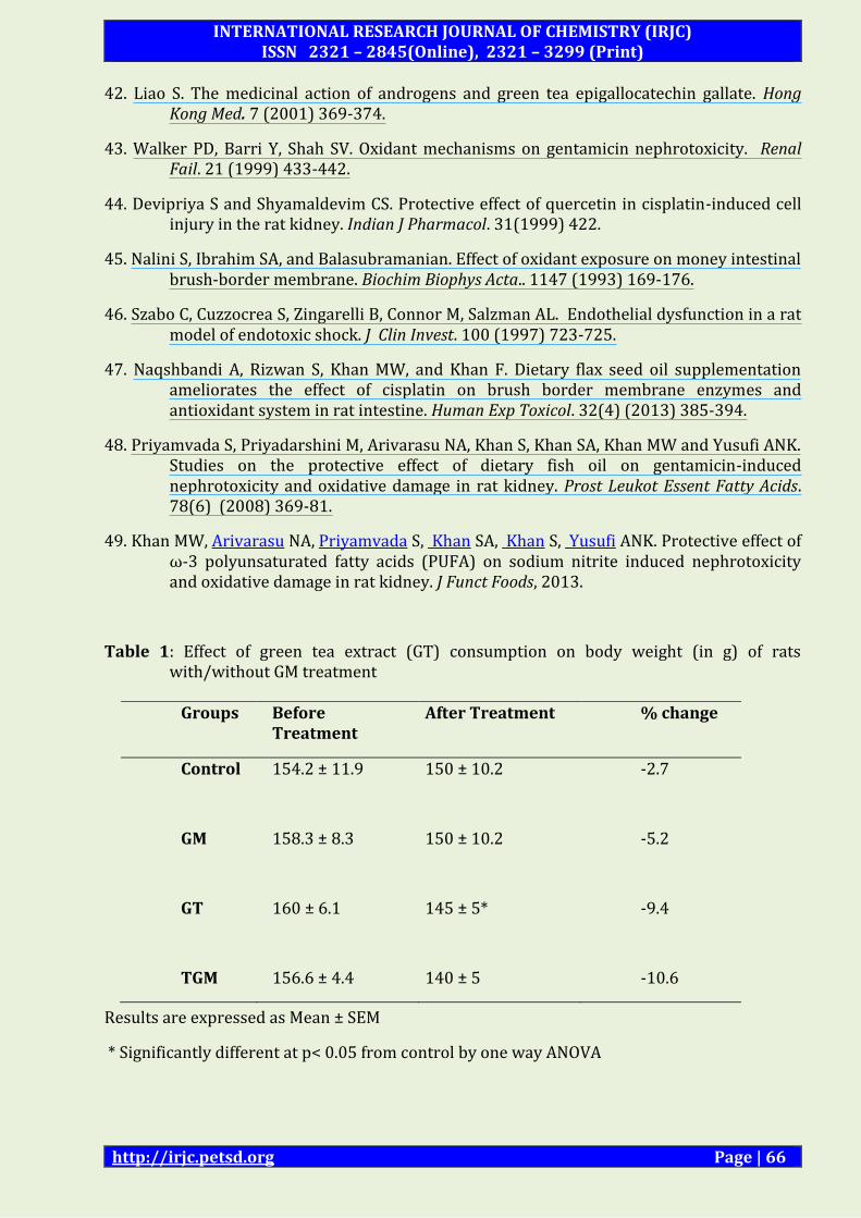
Table 1: Effect of green tea extract (GT) consumption on body weight (in g) of rats with/without GM treatment
Results are expressed as Mean ± SEM
* Significantly different at p< 0.05 from control by one way ANOVA
Groups Before Treatment
After Treatment % change
Control 154.2 ± 11.9 150 ± 10.2 -2.7
GM
158.3 ± 8.3
150 ± 10.2
-5.2
GT
TGM
160 ± 6.1
156.6 ± 4.4
145 ± 5*
140 ± 5
-9.4
-10.6
INTERNATIONAL RESEARCH JOURNAL OF CHEMISTRY (IRJC) ISSN 2321 – 2845(Online), 2321 – 3299 (Print)
http://irjc.petsd.org Page | 67
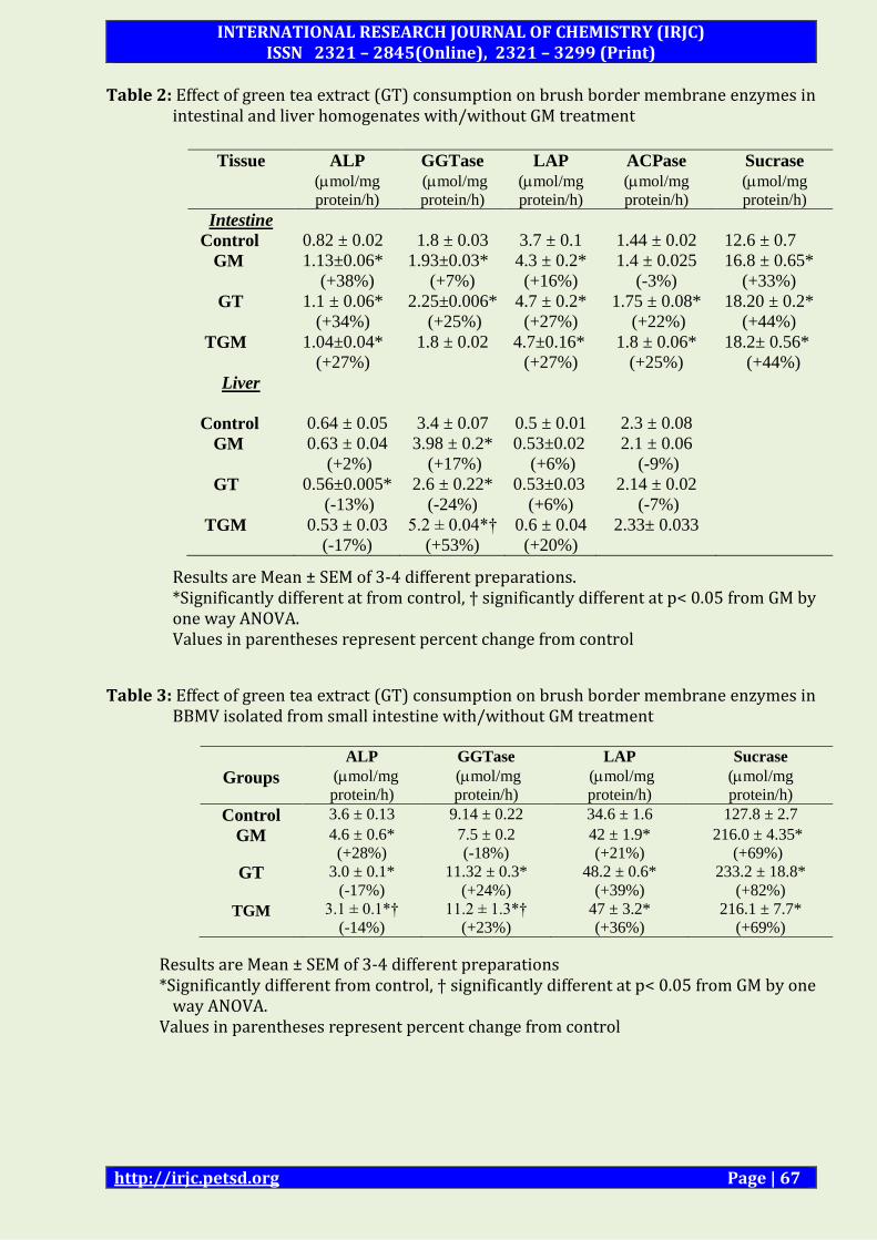
Table 2: Effect of green tea extract (GT) consumption on brush border membrane enzymes in intestinal and liver homogenates with/without GM treatment
Results are Mean ± SEM of 3-4 different preparations. *Significantly different at from control, † significantly different at p< 0.05 from GM by one way ANOVA. Values in parentheses represent percent change from control
Table 3: Effect of green tea extract (GT) consumption on brush border membrane enzymes in BBMV isolated from small intestine with/without GM treatment
Results are Mean ± SEM of 3-4 different preparations *Significantly different from control, † significantly different at p< 0.05 from GM by one
way ANOVA. Values in parentheses represent percent change from control
Tissue ALP
(mol/mg
protein/h)
GGTase
(mol/mg
protein/h)
LAP
(mol/mg
protein/h)
ACPase
(mol/mg
protein/h)
Sucrase
(mol/mg
protein/h)
Intestine
Control 0.82 ± 0.02 1.8 ± 0.03 3.7 ± 0.1 1.44 ± 0.02 12.6 ± 0.7
GM 1.13±0.06*
(+38%)
1.93±0.03*
(+7%)
4.3 ± 0.2*
(+16%)
1.4 ± 0.025
(-3%)
16.8 ± 0.65*
(+33%)
GT
TGM
1.1 ± 0.06*
(+34%)
1.04±0.04*
(+27%)
2.25±0.006*
(+25%)
1.8 ± 0.02
4.7 ± 0.2*
(+27%)
4.7±0.16*
(+27%)
1.75 ± 0.08*
(+22%)
1.8 ± 0.06*
(+25%)
18.20 ± 0.2*
(+44%)
18.2± 0.56*
(+44%)
Liver
Control 0.64 ± 0.05 3.4 ± 0.07 0.5 ± 0.01 2.3 ± 0.08
GM 0.63 ± 0.04
(+2%)
3.98 ± 0.2*
(+17%)
0.53±0.02
(+6%)
2.1 ± 0.06
(-9%)
GT
TGM
0.56±0.005*
(-13%)
0.53 ± 0.03
(-17%)
2.6 ± 0.22*
(-24%)
5.2 ± 0.04*†
(+53%)
0.53±0.03
(+6%)
0.6 ± 0.04
(+20%)
2.14 ± 0.02
(-7%)
2.33± 0.033
Groups
ALP
(mol/mg
protein/h)
GGTase
(mol/mg
protein/h)
LAP
(mol/mg
protein/h)
Sucrase
(mol/mg
protein/h)
Control 3.6 ± 0.13 9.14 ± 0.22 34.6 ± 1.6 127.8 ± 2.7
GM 4.6 ± 0.6*
(+28%)
7.5 ± 0.2
(-18%)
42 ± 1.9*
(+21%)
216.0 ± 4.35*
(+69%)
GT
TGM
3.0 ± 0.1*
(-17%)
3.1 ± 0.1*†
(-14%)
11.32 ± 0.3*
(+24%)
11.2 ± 1.3*†
(+23%)
48.2 ± 0.6*
(+39%)
47 ± 3.2*
(+36%)
233.2 ± 18.8*
(+82%)
216.1 ± 7.7*
(+69%)
INTERNATIONAL RESEARCH JOURNAL OF CHEMISTRY (IRJC) ISSN 2321 – 2845(Online), 2321 – 3299 (Print)
http://irjc.petsd.org Page | 68
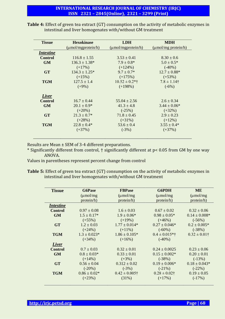
Table 4: Effect of green tea extract (GT) consumption on the activity of metabolic enzymes in intestinal and liver homogenates with/without GM treatment
Results are Mean ± SEM of 3-4 different preparations. * Significantly different from control, † significantly different at p< 0.05 from GM by one way
ANOVA. Values in parentheses represent percent change from control Table 5: Effect of green tea extract (GT) consumption on the activity of metabolic enzymes in
intestinal and liver homogenates with/without GM treatment
Tissue Hexokinase
(mol/mgprotein/h)
LDH
(mol/mgprotein/h)
MDH
(mol/mg protein/h)
Intestine
Control 116.8 ± 1.55 3.53 ± 0.41 8.30 ± 0.6
GM 136.3 ± 1.38*
(+17%)
7.9 ± 0.8*
(+124%)
5.0 ± 0.5*
(-40%)
GT
TGM
134.3 ± 1.25*
(+15%)
127.5 ± 1.4
(+9%)
9.7 ± 0.7*
(+175%)
10.52 ± 0.2*†
(+198%)
12.7 ± 0.88*
(+53%)
7.8 ± 1.14†
(-6%)
Liver
Control 16.7 ± 0.44 55.04 ± 2.56 2.6 ± 0.34
GM 20.1 ± 0.9*
(+20%)
41.3 ± 4.8
(-25%)
3.44 ± 0.06*
(+32%)
GT
TGM
21.3 ± 0.7*
(+28%)
22.8 ± 0.4*
(+37%)
71.8 ± 0.45
(+31%)
53.6 ± 0.4
(-3%)
2.9 ± 0.23
(+12%)
3.55 ± 0.4*
(+37%)
Tissue G6Pase
(mol/mg
protein/h)
FBPase
(mol/mg
protein/h)
G6PDH
(mol/mg
protein/h)
ME
(mol/mg
protein/h)
Intestine
Control 0.97 ± 0.08 1.6 ± 0.03 0.67 ± 0.02 0.32 ± 0.06
GM 1.5 ± 0.17*
(+55%)
1.9 ± 0.06*
(+19%)
0.98 ± 0.05*
(+46%)
0.14 ± 0.008*
(-56%)
GT
TGM
1.2 ± 0.03
(+24%)
1.3 ± 0.023*
(+34%)
1.77 ± 0.014*
(+11%)
1.86 ± 0.105*
(+16%)
0.27 ± 0.046*
(-60%)
0.4 ± 0.015*†
(-40%)
0.2 ± 0.005*
(-38%)
0.32 ± 0.01†
Liver
Control 0.7 ± 0.03 0.32 ± 0.01 0.24 ± 0.0025 0.23 ± 0.06
GM 0.8 ± 0.03*
(+14%)
0.33 ± 0.01
(+3%)
0.15 ± 0.002*
(-38%)
0.20 ± 0.01
(-13%)
GT
TGM
0.56 ± 0.04
(-20%)
0.86 ± 0.02*
(+23%)
0.312 ± 0.02
(-3%)
0.42 ± 0.005†
(31%)
0.19 ± 0.006*
(-21%)
0.28 ± 0.02†
(+17%)
0.18 ± 0.043*
(-22%)
0.19 ± 0.05
(-17%)
INTERNATIONAL RESEARCH JOURNAL OF CHEMISTRY (IRJC) ISSN 2321 – 2845(Online), 2321 – 3299 (Print)
http://irjc.petsd.org Page | 69
Results are Mean ± SEM of 3-4 different preparations. * Significantly different from control, † significantly different at p< 0.05 from GM by one way
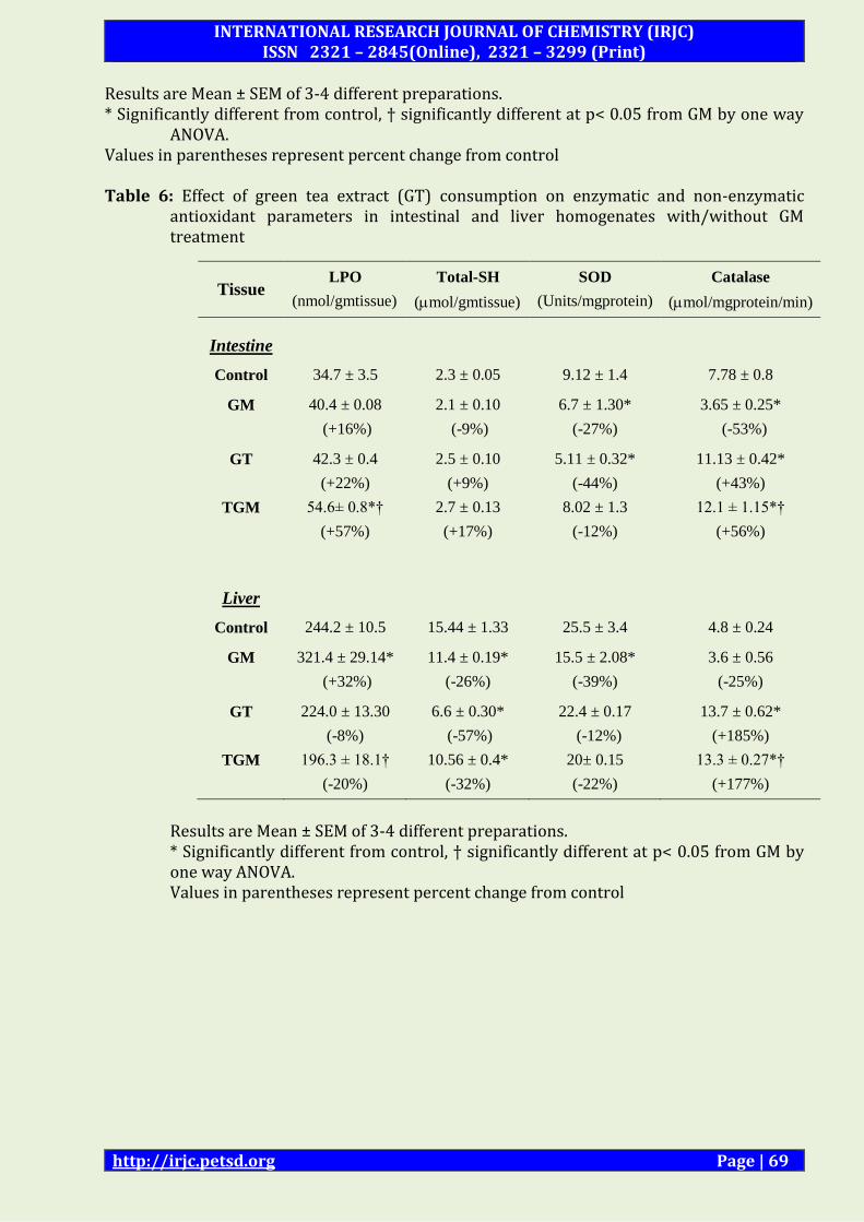
ANOVA. Values in parentheses represent percent change from control Table 6: Effect of green tea extract (GT) consumption on enzymatic and non-enzymatic
antioxidant parameters in intestinal and liver homogenates with/without GM treatment
Results are Mean ± SEM of 3-4 different preparations. * Significantly different from control, † significantly different at p< 0.05 from GM by one way ANOVA. Values in parentheses represent percent change from control
Tissue LPO
(nmol/gmtissue)
Total-SH
(mol/gmtissue)
SOD
(Units/mgprotein)
Catalase
(mol/mgprotein/min)
Intestine
Control 34.7 ± 3.5 2.3 ± 0.05 9.12 ± 1.4 7.78 ± 0.8
GM 40.4 ± 0.08
(+16%)
2.1 ± 0.10
(-9%)
6.7 ± 1.30*
(-27%)
3.65 ± 0.25*
(-53%)
GT
TGM
42.3 ± 0.4
(+22%)
54.6± 0.8*†
(+57%)
2.5 ± 0.10
(+9%)
2.7 ± 0.13
(+17%)
5.11 ± 0.32*
(-44%)
8.02 ± 1.3
(-12%)
11.13 ± 0.42*
(+43%)
12.1 ± 1.15*†
(+56%)
Liver
Control 244.2 ± 10.5 15.44 ± 1.33 25.5 ± 3.4 4.8 ± 0.24
GM 321.4 ± 29.14*
(+32%)
11.4 ± 0.19*
(-26%)
15.5 ± 2.08*
(-39%)
3.6 ± 0.56
(-25%)
GT
TGM
224.0 ± 13.30
(-8%)
196.3 ± 18.1†
(-20%)
6.6 ± 0.30*
(-57%)
10.56 ± 0.4*
(-32%)
22.4 ± 0.17
(-12%)
20± 0.15
(-22%)
13.7 ± 0.62*
(+185%)
13.3 ± 0.27*†
(+177%)
INTERNATIONAL RESEARCH JOURNAL OF CHEMISTRY (IRJC) ISSN 2321 – 2845(Online), 2321 – 3299 (Print)
http://irjc.petsd.org Page | 70
0
ND
7 days
Control
GT
25 days
Control
GM
GT
TGM 10 days
Sacrifice
Intestine
Liver
Figure 1: Experimental Design (ND normal diet; GT green tea; GM gentamicin; TGM
green tea + GM; = i.p. administration of GM for 10 days)
0
1
2
3
4
5
6
Cont GM GT TGM
Sp
ecif
ic a
ctiv
ity
(μ
mo
l/m
gp
rote
in/h
)
ALP
GGTase
LAP
(A)
*
*
*
*
*
*
*
INTERNATIONAL RESEARCH JOURNAL OF CHEMISTRY (IRJC) ISSN 2321 – 2845(Online), 2321 – 3299 (Print)
http://irjc.petsd.org Page | 71
LIST OF ABBREVIATIONS
ACPase Acid Phosphatase; an enzyme
ALP Alkaline Phosphatase; an enzyme
ANOVA
ATP
Analysis of variance; statistical tool
Adenosine 5’-triphosphate; energy currency
BBM Brush border membrane; intestinal membrane
BBMV Brush border membrane vesicles
BUN
GT
Blood urea nitrogen; blood parameter
Green Tea
GGTase γ-glutamyl transferase; an enzyme
G6Pase Glucose-6-phosphatase; an enzyme
G6PDH Glucose-6-phosphate dehydrogenase; an enzyme
HK Hexokinase; an enzyme
HMP Hexose monophosphate; an enzyme
LAP Leucine aminopeptidase; an enzyme
0
10
20
30
40
50
60
Cont GM GT TGM
Sp
ecif
ic a
ctiv
ity
(μ
mo
l/m
gp
rote
in/h
)
ALP
GGTase
LAP
(B)
*
*
*
*
*
*†
*†
*
0
50
100
150
200
250
300
Cont GM GT TGM
Sp
ecif
ic a
ctiv
ity
(μ
mo
l/m
gp
rote
in/h
)
Homog
BBM
Figure 2: Effect of green tea extract (GT) consumption on activity of BBM enzymes in Intestine (A)
Homogenate (B) BBM (C) Sucrase in homogenate and BBM with/without GM treatment.
(C)
INTERNATIONAL RESEARCH JOURNAL OF CHEMISTRY (IRJC) ISSN 2321 – 2845(Online), 2321 – 3299 (Print)
http://irjc.petsd.org Page | 72
LDH Lactate dehydrogenase; an enzyme
LPO Lipid peroxidation; an enzyme
MDH Malate dehydrogenase; an enzyme
ME Malic enzyme; an enzyme
NADPH
NADP+
NADH
Nicotinamide adenine dinucleotide phosphate (reduced);
reducing equivalent
Nicotinamide adenine dinucleotide phosphate
Nicotinamide adenine dinucleotide reduced
Pi
ROS
Inorganic phosphate
Reactive oxygen species
SOD
SH
Superoxide dismutase; an enzyme
Sulphydryl groups
TCA cycle
Tricarboxylic acid cycle