Placental regulation of peptide hormone and growth factor activity by proMBP
Upload
independentCategory
view
3download
0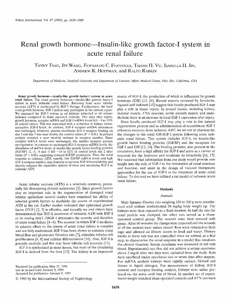
Kidney International, Vol. 47 (1995), pp. 1658—1668
Renal growth hormone—Insulin-like growth factor-I system inacute renal failure
TANNY TSAO, JIN WANG, FERNANDO C. FERVENZA, THANH H. Vu, ISABELLA H. JIN,ANDREW R. HOFFMAN, and RALPH RABKIN
Departments of Medicine, Stanford University and Department of Veterans Affairs Medical Center, Palo Alto, California, USA
Renal growth hormone—insulin-like growth factor-I system in acuterenal failure. The renal growth hormone—insulin-like growth factor-Isystem in acute ischemic renal failure. Recovery from acute tubularnecrosis (ATN) is accelerated by IGF-I therapy. Furthermore, the localrenal growth hormone-IGF-I system may participate in the natural repair.We examined the IGF-I system in rat kidneys subjected to 60 minuteischemia compared to sham operated controls. Two days after injury,growth hormone receptor mRNA and IGF-I mRNA levels fell —9 to 33%of control values. This was associated with a reduction in kidney immu-noreactive IGF-I levels. In contrast, IGF-I receptor mRNA abundancewas unchanged. However, plasma membrane IGF-I receptor binding onday 2 and day 7 was near double the control values (P < 0.01). Scatchardanalysis revealed a near twofold increase in receptor number. Sincereceptor mRNA levels were unchanged, this implies receptor proteinup-regulation. In contrast to unchanged IGF-I receptor mRNA levels, theabundance of mRNA levels of insulin-like growth factor binding proteins(IGFBP) -2, -3, -4 and -5 fell —14 to 62% of control levels day 2 afterinjury (P < 0.05), suggesting reduced IGFBP production. Thus, the renalresponse to ischemic ATN, namely, low IGFBP mRNA levels and highIGF-I receptor number, may function to increase IGF-I bioavailability andthereby enhance the reparative actions of local and circulating IGF-I inischemic ATN.
Acute tubular necrosis (ATN) is a relatively common, poten-tially life threatening clinical occurrence [11. Since growth factorsplay an important role in the regeneration of damaged renaltubular epithelium several studies have examined the ability ofselected growth factors to modulate the course of experimentalATN in the rat. Earlier studies indicated that epidermal growthfactor (EGF) [2, 3] is effective, and recently we and others havedemonstrated that IGF-I treatment of ischemic ATN with IGF-Ior its analog des(1—3)IGF-I attenuates the severity and durationof acute renal failure [4—61. The manner in which IGF-I mediatesits salutary effect on the course of acute renal failure is complexand not fully understood. IGF-I has been shown to enhance renalblood flow and glomerular filtration rate [7], stimulate tubular cellproliferation [8, 91 and cellular differentiation [10]. Also, IGF-I ispotently anabolic and this may favor tubular cell recovery [11].
IGF-I is synthesized in most tissues, but most of the circulatingIGF-I is derived from the liver [121. The kidney is an important
Received for publication May 23, 1994and in revised form January 9, 1995Accepted for publication January 9, 1995
© 1995 by the International Society of Nephrology
source of IGF-I, the production of which is influenced by growthhormone (Gil) [13, 14]. Recent reports reviewed by Jennische,Isgaard and Isaksson [15] suggest that locally produced IGF-I mayplay a role in tissue repair. In several tissues, including kidney,skeletal muscle, CNS neurons, aortic smooth muscle and endo-thelium there is an increase in local IGF-I expression after injury.
Since locally produced IGF-I may play a role in the naturalregenerative process and as administration of recombinant IGF-Ienhances recovery from ischemic ARF, we set out to characterizethe changes in the renal GH-IGF-I system following acute isch-ernie renal failure. This system includes IGF-I, six insulin-likegrowth factor binding proteins (IGFBP) and the receptors forIGF-I and GH [12, 14]. The binding proteins, also present in thecirculation, have a high affinity for IGF-I and serve as a carrier orreservoir for the hormone and modulate its bioactivity [16, 17].We reasoned that information from our study would provide newinsight into the role of IGF-I in the restoration of renal structureand function, and assist in the design of rational therapeuticapproaches for the use of IGF-I in the treatment of acute renalfailure. To this end we have utilized a rat model of ischemic acuterenal failure.
Methods
Animals
Male Sprague-Dawley rats weighing 250 to 300 g were anesthe-tized with sodium methohexital 50 mg/kg body weight i.p. Thekidneys were then exposed via a flank incision. In half the rats therenal pedicle was clamped; the other rats served as a sham-operated control group. The wounds were then covered withgauze. After 60 minutes the clamps were removed and the woundsof all the animals were suture closed. Rats were returned to theircage and allowed ad libitum access to food and water. Dietaryintake in these rats was not controlled since we wished, as a firststep, to characterize the renal response in a model that simulatesthe clinical situation. Serum creatinine was measured in tail veinblood. Experimental rats that did not achieve a serum creatinineabove 2 mg/dl after two days were excluded from the study. Ratswere sacrificed under anesthesia two or seven days after surgery.For rnRNA analysis kidneys were rapidly excised, blotted andfrozen in liquid nitrogen. For measurement of kidney IGF-Icontent and receptor binding analysis, kidneys were saline per-fused via the aorta until free of blood. In another set of experi-ments weight matched sham operated controls and ATN rats were
1658
Tsao et at: Insulin-like growth factor-I 1659
pair fed to control for any response due to the reduced dietaryintake that accompanies uremia. These rats were studied two daysafter injury.
Preparation of "crude" kidney plasma membranesTissues from one and a half whole kidneys were minced, added
to cold 8% wt/wt sucrose buffer and then disrupted with 15 gentlestrokes in a dounce homogenizer. The homogenate was centri-fuged at 1000 g for 10 minutes and the supernate collected. Thepellet was disrupted with 10 strokes in a homogenizer containing10 ml sucrose buffer and then centrifuged at 1000 g for 10 minutes.The supernates were pooled and spun at 9,500 g for 10 minutes.The supernate and soft light upper pellet were removed from thecentral dark pellet and then mixed prior to centrifugation at47,000 g for 20 minutes. The soft light fluffy upper pellet wasremoved and resuspended in 1 ml calcium free Krebs-RingersHepes (KRH) buffer and washed twice and stored at —80°C untiluse. All steps were carried out at 4°C.
IGF-I binding assaysRecombinant hIGF-1, a gift from Genentech Inc. (South San
Francisco, CA, USA), was radiolabeled with 1251-Na (< oneiodine atom/molecule) by a chloramine T method [181. Thebinding of 1251-IGF-I to crude kidney plasma membranes wasdetermined as follows. Calcium-free KRH buffer containing 0.5%BSA with or without various concentrations of unlabeled IGF-Iand other cold peptide hormones were added to the same buffercontaining 10_Il M '251-IGF-I and 2 mi bacitracin and 5 mN-ethylmaleimide. The membrane suspension containing 60 tg ofprotein was then added and incubated at 4°C overnight. Mem-brane-bound and free 1251-IGF-I were separated by centrifugationat 12,000 g for 15 minutes and the supernatants aspirated. Themembrane pellets were washed once with the KRH buffer and thetips of the centrifuge tubes excised. '251-radioactivity was deter-mined in a gamma counter. Nonspecific binding was defined asthe counts of '251-IGF-I remaining associated with membranes inthe presence of 10_6 M of unlabeled IGF-1, and is subtracted fromtotal binding to yield specific binding. With 1251-IGF-I non-specific binding averaged 1.9 0.18%. The data from competitioncurves were subjected to Scatchard analysis to determine bindingaffinity and receptor number [19]. 1251-IGF-I degradation in thebinding assays was assessed as the appearance of radioactivitysoluble in 10% trichloroacetic acid (TCA) and averaged 4.3% oftotal counts.
Cross linking of '251-IGF-I receptor'251-IGF4 was cross linked to kidney membranes essentially as
described by Pillion, Haskell and Meezan [201. Aliquots of crudemembrane preparations equivalent to 200 jg of protein wereincubated overnight at 4°C with 10_b M radiolabeled I-IGF-I inthe absence or presence of i0- M unlabeled IGF-I or insulin. Themembranes were washed twice in BSA-free Krebs-Ringers Hepesbuffer (KRH), pH 7.5. Cross linking of the alpha1 subunit of IGF-1receptor was allowed in KRH in the presence of 0.25 mMdisuccinimidyl suberate at 4°C for 15 minutes. The cross linkingwas terminated by the addition of 10 ifiM Tris-HCI with I ifiMEDTA, pH 7.5. The pellets were washed twice and were resus-pended in 20 p.! of sample buffer in the presence of 1% 2-mer-captoethanol as a reducing agent, heated at 95°C for 90 seconds,and subjected to 5 to 15% gradient SDS-polyacrylamide gel
electrophoresis. The radioactivity in the gel was analyzed byautoradiography. Molecular weights were determined by compar-ison with prestained markers from BioRad (Richmond, CA,USA).
Western ligand blot
Plasma membranes were prepared from saline perfused blood-less whole kidneys as described earlier. Membrane protein, 60 p.g,pooled from five rats was electrophoresed in 12% polyacrylamidegel. Separated proteins were electroblotted onto nitrocellulosefilters with a BioRad Semi-Dry Transfer Cell and then incubatedwith a mixture of 1251-IGF-I and '251-IGF-II overnight at 4°C andvisualized by autoradiography [21]. Cytosolic protein (10,000 gsupernate) was also examined in the same manner, but no signalwas detected with 100 p.g cytosolic protein.
Estimation of serum and kidney concentrations of IGF-Iusing RIA
IGF-1 was extracted from serum and bloodless saline perfusedkidneys before measurement by radioimmunoassay. Before har-vesting, kidneys were briefly perfused with saline in vivo to renderthem blood free. The extraction method used was based on thatdescribed by D'Ercole, Stiles and Underwood [22] and acid-separates IGF-I from its binding proteins. This procedure givesvalues comparable to that obtained following high pressure liquidchromatography separation [23]. A quarter of a kidney from eachrat was homogenized in 1 M acetic acid and the acid extractassayed for IGF-I and corrected for recoveries. Serum IGF-I wasextracted with acid ethanol. IGF-I was measured with a radioim-munoassay kit (Nichols Institute, San Juan Capistrano, CA,USA). Recovery of recombinant IGF-I added to control and ATNkidney homogenates averaged 85 1 and 95 1.7%, respectively,and to serum 88 3.2 and 94 3.4%, respectively.
Preparation of RNATotal kidney RNA was isolated using the single step method of
Chomczynski and Sacchi [24]. A quarter of a kidney from each ratwas homogenized, and RNA was extracted using acid guani-dinium thiocyanate-phenol-chloroform and then spectrophoto-metrically quantified by determining absorbance at 260 nm. Theintegrity of RNA was checked by inspecting the fluorescence ofethidium bromide stained I 8S and 28S rRNA bands separated byagarose gel electrophoresis.
Solution hybridization/RNase protection assay
Protection assays were performed as previously described [25].Total RNA (20 p.g) was hybridized with 100,000 cpm of a-32PCTP labeled antisense IGF-I or IGF-I receptor riboprobes and5,000 cpm of antisense GAPDH riboprobe for 14 hours at 55°C in80% formamide/400 mivi NaCl buffer. After hybridization, themixture was digested with ribonuclease A (10 p.g/ml) and ribonu-clease Ti (100 U/mI) at 37°C for 30 minutes and then proteinaseK (200 p.g/ml) at 37°C for 30 minutes, Protected hybrids weresize-separated on a 5% polyacrylamide/8 M urea denaturing geland analyzed by autoradiography.
Northern blot analysis
Total RNA was size-separated by agarose gel electrophoresis.Briefly, RNA was denatured with 0.55 M formaldehyde. Twelve
1660 Tsao et al: Insulin-like growth factor-I
micrograms of RNA was loaded in each lane and electrophoresedon a denaturing agarose gel containing 0.55 M formaldehyde.RNA was transferred by capillary action onto nitrocellulose filtersand then fixed to the filters by UV cross linking (UV 1800Stratalinker; Stratgene, La Jolla, CA, USA). The filters were thenprehybridized in the hybridization solution (50% formamide, 1.5X SSPE, 2.5 x Denharts solution, 50 jg salmon sperm DNA,0.2% SDS) at 42°C for two hours. The mRNA levels of IGFBP-1,-2, -3, -4, and -5 were detected by hybridizing radiolabeled cDNAprobes to the mRNA. Probes, —2.5 million cpmlml, were hybrid-ized to the respective mRNAs on Northern blots in a freshhybridization solution at 42°C overnight. The filters were thenwashed and exposed to Kodak XAR films (Kodak, Rochester,NY, USA) at — 70°C for one to five days. Where noted the filterswere then reprobed with a hGAPDH eDNA probe. The mRNAabundance of each IGFBP was analyzed by densitometry (Ultra-scan XL, Pharmacia LKB Biotechnology, Inc., Alameda, CA,USA) and corrected for either the levels of the GAPDH mRNAreadings or the I 8S mRNA readings obtained from photographicnegatives of the nitrocellulose filters. In preliminary experimentswe found that GAPDH mRNA levels in kidneys two and sevendays post-ischemia did not differ from that measured in sham-operated control kidneys. A linear relationship was present be-tween the optical density (OD) of the photographic negative ofthe nitrocellulose 18S mRNA signal and the total RNA contentup to 30 jg (r = 0.99).
Polymerase chain reaction (PCR) assayRelative levels of renal GH receptor mRNA were assayed by
using a gene Amp RNA-PCR kit (Perkin Elmer Cetus, Norwalk,CT, USA) [26]. Total RNA was reverse transcribed by usingrandom hexamers. The oligonucleotide primers for the GHreceptor were designed to amplify a 321 base pair DNA fragment.The fragment included 97 bases in exon 5 and 145 bases in exon7. Both exons are in the extracellular domain of the rat GH cDNAsequence [27]. GH receptor mRNA was amplified by 28 PCRcycles and as an internal amplification control, 18S rRNA wasamplified in the same assay tube for the last 10 cycles by addingspecific 18S rRNA primers. The 18S rRNA primers amplified 140bases spanning nucleotide positions 107 to 246 in the humancDNA sequence [28]. PCR products were separated on a 5%polyacrylamide!8 M urea gel and analyzed by quantitative autora-diography.
Preparation of radiolabeled probesFor the solution hybridization/RNase protection assay, labeled
antisense IGF-I, IGF-I receptor and GAPDH riboprobes weresynthesized using 32P-labeled CTP as previously described [25].The IGF-I riboprobe, a 376 bp Sau3A-EcoRI fragment wasdesigned to detect both the Ea and Eb IGF-I mRNAs [29]. TheIGF-I receptor riboprobe contained 280 bases complementary tothe IGF-I receptor mRNA. For Northern blot analysis the eDNAprobes were labeled with 50 tCi [32P] dCTP (3000 Ci/mmol,Amersham, Arlington Heights, IL, USA) by a random primermethod (DNA labeling kit 70200; United States BiochemicalCorporation, Cleveland, OH, USA). Radiolabeled cDNA probeswere purified by centrifugation through a G-50 Sephadex column.
The cDNA probes for the five IGFBPs studied were derivedfrom rat cDNAs cloned into recombinant plasmid vectors (pBlue-scriptSK+) which were gifts of Dr. S. Shimasaki (Whittier Insti-
tute, La Jolla, CA, USA). The probes were obtained by synthe-sizing the corresponding cDNA by PCR using the cloned cDNAas a template. The molecular characteristics of the rat cDNAshave recently been reviewed by Shimasaki and Ling [30]. TheIGFBP-1 probe comprised 407 bp corresponding to the codingregion spanning nucleotide position 486 to 892 in the rat IGFBP-1cDNA sequence. The IGFBP-2 probe consisted of 342 bp span-fling nucleotide position 670 to 1011. The IGFBP-3 probe com-prised 636 bp spanning nucleotide positions 196 to 831. TheIGFBP-4 probe comprised 384 bp spanning nucleotide position457 to 840. The IGFBP-5 probe comprised 259 bp spanningnucleotide position 856 to 1114.
Data analysisDensitometry readings of the mRNAs of interest were cor-
rected for the corresponding GAPDH mRNA or 18S RNAreading. For IGF-I we quantified the Ea IGF-I mRNA transcriptsonly since it accounted for >95% of the IGF-1 mRNA signal.Results are expressed as the mean SEM. The mean of thecontrols was assigned a value of unity and all results wereexpressed in relation to this value. Differences between thecontrol and experimental groups were evaluated using Student'stwo-tailed unpaired t-test. P values of <0.05 were consideredsignificant.
Results
Ad libitum fed rats
Following bilateral renal pedicle clamping the serum creatininerose, reaching a maximum after 24 hours. After the second day theserum creatinine, which averaged 4.2 0.32 mg/dl, declined (Fig.1). By the seventh day serum creatinine levels had fallen consid-erably but were still more than double the baseline values (1.00.4 vs. 0.4 0.04 mg/dl). Changes in body weight differedsignificantly (P < 0.01) between the two groups after the experi-mental intervention (Table 1). Two days post-injury ATN rats lost27 g while the body weight of the controls had not changed. On
** **5.
4,
C. 3
a)
0E2a)Cl)
1—
0-
*
**
I I I I
0 1 2 3 5 7
Time, days
Fig. 1. Serum creatinine following a sham operation (•, controls) or 60minutes of renal ischemia with acute tubular necrosis (EI A TN). Results arethe mean SEM from 3 to 6 ad libitum fed rats per time point. *p < 0.05,
< 0.01.
Tsao et al: Insulin-like growth factor-I 1661
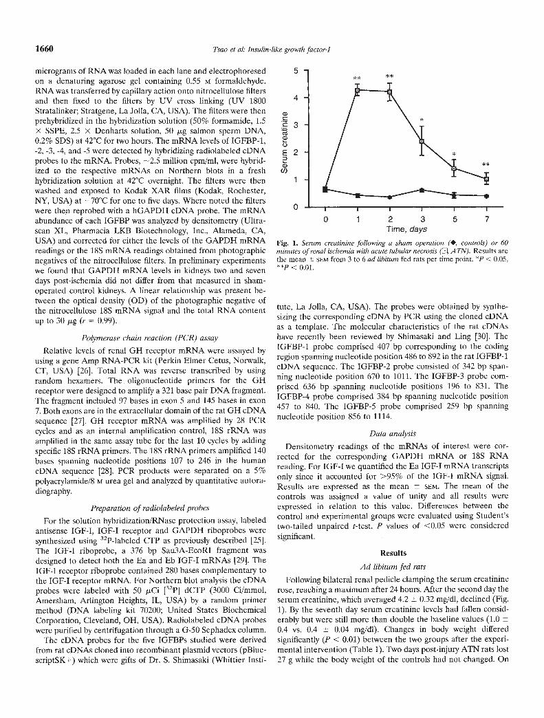
Table 1. Body weight changes () and serum creatinine levels in rats with ischemic acute tubular necrosis (ATN), and in ad libitum or pair-fedsham operated controls
Day2 Day7Control ATh Control ATh
Ad-libitum dietBodyweight,g 0± 2.1(N= 15) —27 1.4(N= 11) 25 10(N= 8) —17 10,5(N= 8)
Serum creatinine mg/dl 0.36 0.04 (N = 20) 4.19 0.32 (N = 20)Pair-fed diet
Body weight, g 25 1.6 (N = 5) 24 3.2 (N 5)Serum creatinine mg/dl 0.38 0.02 (N = 5) 3.68 0.02 (N = 5)Data are SEM.ap < 0.01 vs. control
20
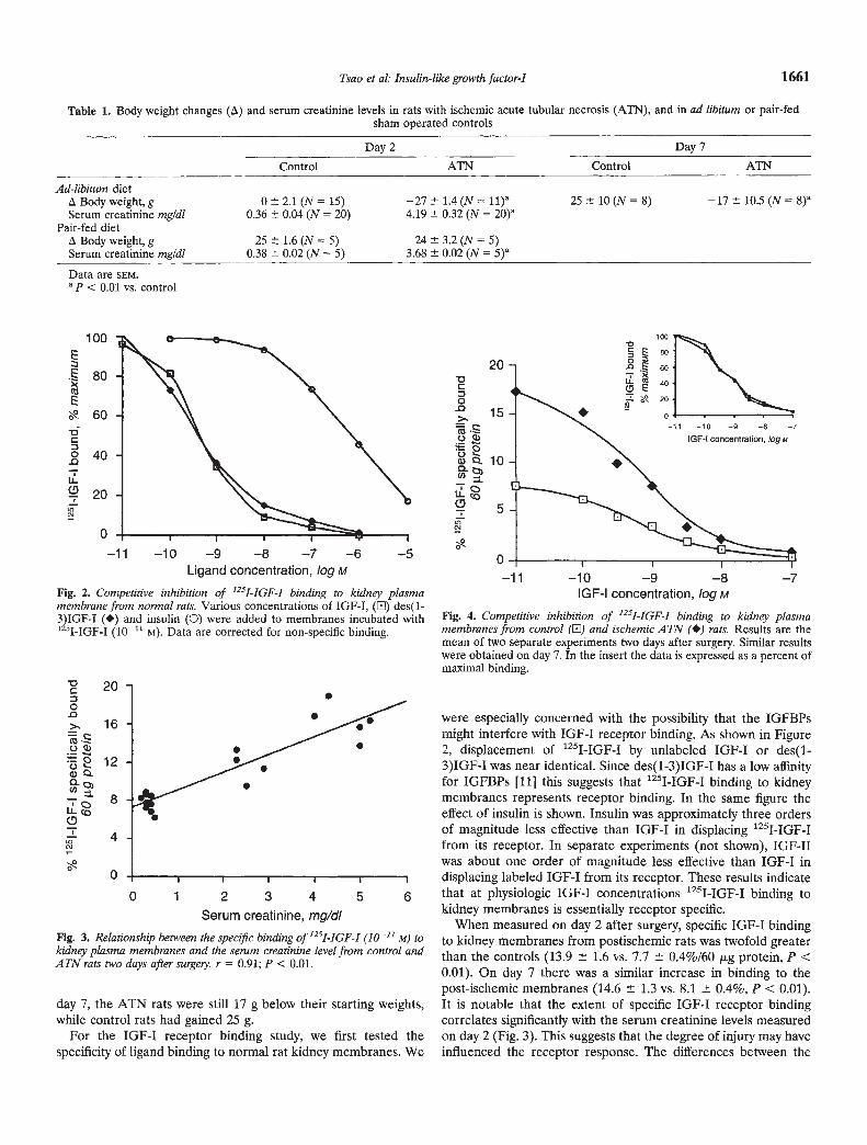
were especially concerned with the possibility that the IGFBPsmight interfere with IGF-I receptor binding. As shown in Figure2, displacement of 1251-IGF-I by unlabeled IGF-I or des(1-3)IGF-I was near identical. Since des(1-3)IGF-I has a low affinityfor IGFBPs [11] this suggests that 1251-IGF-I binding to kidneymembranes represents receptor binding. In the same figure theeffect of insulin is shown. Insulin was approximately three ordersof magnitude less effective than IGF-I in displacing 1251-IGF-Ifrom its receptor. In separate experiments (not shown), IGF-IIwas about one order of magnitude less effective than IGF-I indisplacing labeled IGF-I from its receptor. These results indicate
0 1 2 3 4 5 6 that at physiologic IGF-I concentrations '251-IGF-I binding tokidney membranes is essentially receptor specific.
When measured on day 2 after surgery, specific IGF-I bindingto kidney membranes from postischemic rats was twofold greaterthan the controls (13.9 1.6 vs. 7.7 0.4%/60 g protein, P <0.01). On day 7 there was a similar increase in binding to thepost-ischemic membranes (14.6 1.3 vs. 8.1 0.4%, P < 0.01).It is notable that the extent of specific IGF-I receptor bindingcorrelates significantly with the serum creatinine levels measuredon day 2 (Fig. 3). This suggests that the degree of injury may haveinfluenced the receptor response. The differences between the
100
80
60
40
20
0
0.0EE
E
0.0
(1,
LU
Fig. 2. Competitive inhibition of '251-IGF-I binding to kidney plasmamembrane from normal rats. Various concentrations of IGF-I, (El) des(1-
3IGF-I (•) and insulin (0) were added to membranes incubated with1 51-IGF-I (10—11 M). Data are corrected for non-specific binding.
20
0- 15>
IF:0
Ligand concentration, log M—11
—11 —10 —9 —8 —7
IGF-I concentration, log M
—11 —10 —9 —8 —7 —6 —5
—10 —9 —8 —7
IGF-I concentration, log M
.
Fig. 4. Competitive inhibition of '251-IGF-I binding to kidney plasmamembranes from control (El) and ischemic A TN (•) rats. Results are themean of two separate experiments two days after surgery. Similar resultswere obtained on day 7. In the insert the data is expressed as a percent ofmaximal binding.
16
12
8
4
0
S
S
.
0C0.0>.i .OQ)
QL
U)CU
Serum creatinine, mg/dl
Fig. 3. Relationship between the specific binding of '251-IGF-I (10_21 M) tokidney plasma membranes and the sensm creatinine level from control andATN rats two days after suigely. r = 0.91; P < 0.01.
day 7, the ATN rats were still 17 g below their starting weights,while control rats had gained 25 g.
For the IGF-I receptor binding study, we first tested thespecificity of ligand binding to normal rat kidney membranes. We
1662 Tsao et al: Insulin-like growth factor-I
Control ATN
— 205
— 116.5
— 80
— 49.5
— 32.5
IGF-IInsulin
— + — — +— — + — — +
control and ATN groups is readily apparent from the displace-ment curves shown in Figure 4. When the data were expressed asa percent of maximum 1251-IGF-I bound, the displacement curveswere superimposable (Fig. 4 insert). These curves were used todetermine the concentration of recombinant IGF-I at which1251-.IGF4 binding was 50% of maximum, a measure of relativereceptor affinity. For control and experimental membranes fromday 2 and 7 this averaged 2 and 1.4 nM, respectively. Thesefindings suggest that receptor affinity was similar in these twogroups. Scatchard analysis of the high affinity binding componentsrevealed that receptor affinity (0.4 nM) was the same in bothgroups, while receptor number was increased more than twofoldfollowing ATN compared to the controls (125 vs. 54 pM/60 gmembrane protein).
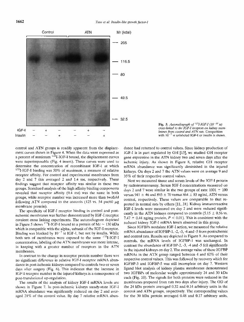
The specificity of IGF-I receptor binding in control and post-ischemic membranes was further demonstrated by IGF-I receptorcovalent cross linking experiments. The autoradiogram depictedin Figure 5 shows 1251-IGF-I bound to a protein of Mr — 130 kDawhich is compatible with the alpha1 subunit of the IGF-I receptor.Binding was blocked by iO M IGF-I, but not by insulin. Whileboth sets of membranes were exposed to the same '251-IGF-Iconcentration, labeling of the ATN membranes was more intense,in keeping with a greater number of receptors in the ATNmembranes.
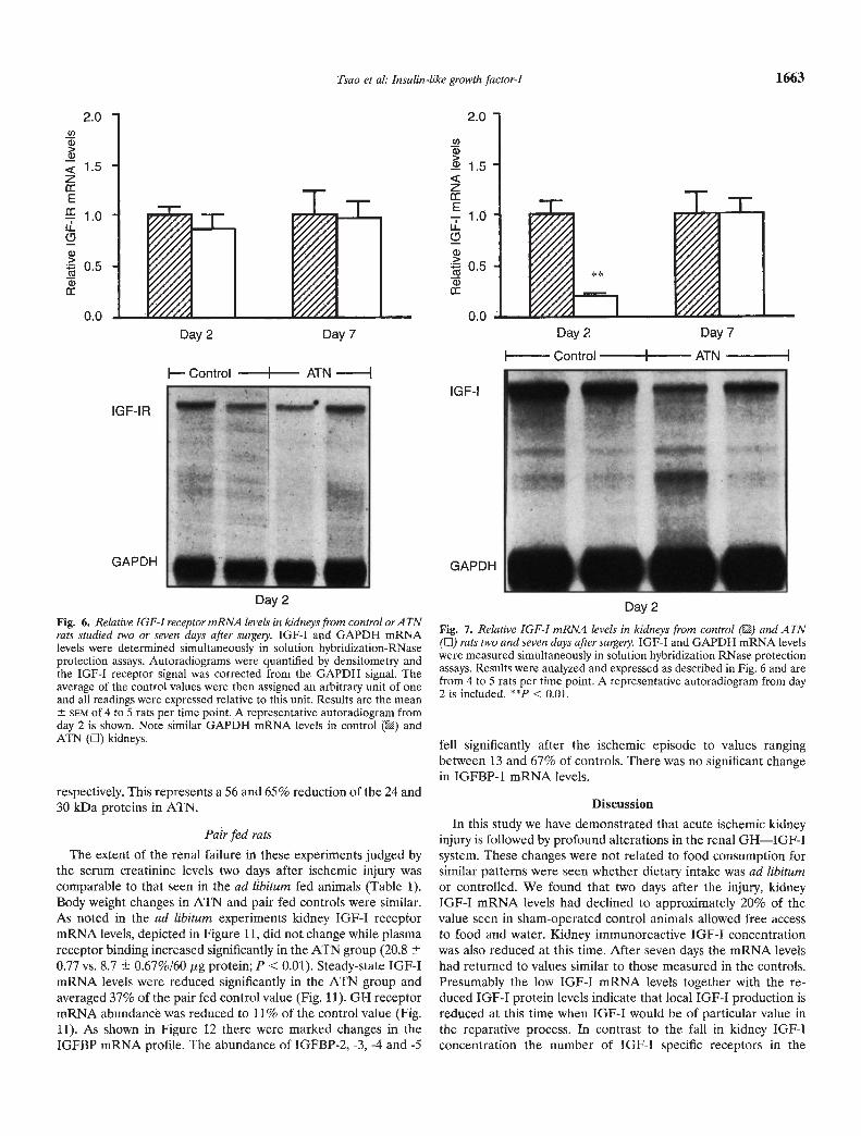
In contrast to the change in receptor protein number there wasno significant difference in relative IGF-I receptor mRNA abun-dance in post-ischemic kidneys compared to controls two or sevendays after surgery (Fig. 6). This indicates that the increase inIGF-I receptor number in the injured kidneys is a consequence ofpost-translational up-regulation.
The results of the analysis of kidney IGF-I mRNA levels areshown in Figure 7. In post-ischemic kidneys steady-state IGF-ImRNA abundance was significantly reduced on day 2 and aver-aged 21% of the control value. By day 7 relative mRNA abun-
Fig. 5. Autoradiographof 1251-IGF-I (10'° M)cross-linked to the IGF-I receptorN on kidney mem-branes from control and A TN rats. Competitionwith iO— M unlabeled IGF-I or insulin is shown.
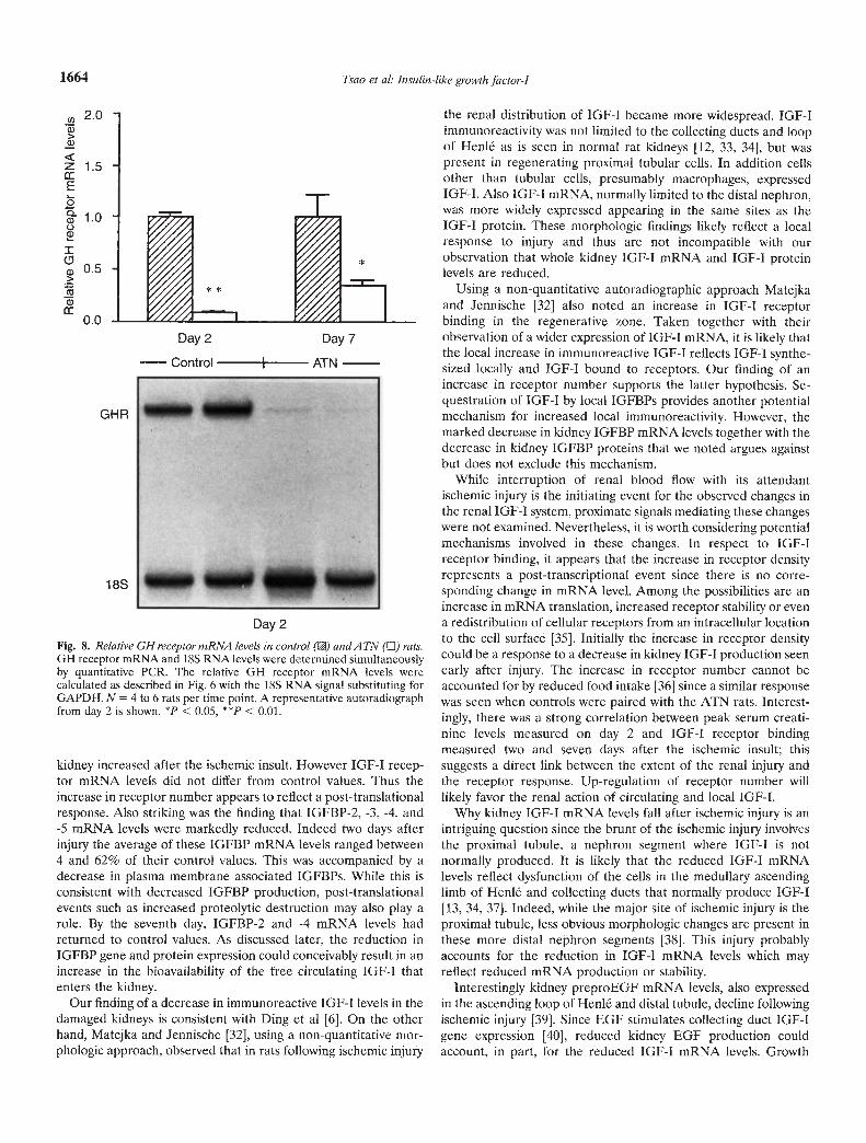
dance had returned to control values. Since kidney production ofIGF-I is in part regulated by Gil [13], we studied GH receptorgene expression in the ATN kidney two and seven days after theischemic injury. As shown in Figure 8, relative GH receptormRNA abundance was significantly diminished in the injuredkidneys. On days 2 and 7 the ATN values were on average 9 and35% of their respective control values.
Next we measured tissue and serum levels of the IGF-I proteinby radioimmunoassay. Serum IGF-I concentrations measured ondays 2 and 7 were similar in the two groups of rats: 1001 100versus 941 46 and 893 70 versus 864 85 ng/ml, ATN versuscontrol, respectively. These values are comparable to that re-ported in normal rats by others [11, 311. Kidney immunoreactiveIGF-I levels were measured on day 2 and were reduced signifi-cantly in the ATN kidneys compared to controls (5.15 0.56 vs.7.47 0.44 ng/mg protein, P < 0.01). This is consistent with thereduced kidney IGF-I mRNA levels observed in this group.
Since IGFBPs modulate IGF-I action, we measured the relativemRNA abundance of IGFBP-1, -2, -3, -4 and -5 from postischemicand control rats. Results are depicted in Figure 9. As compared tocontrols, the mRNA levels of IGFBP-1 was unchanged. Incontrast the abundance of IGFBP-2, -3, -4 and -5 fell significantlyin the injured kidneys on day 2. The average value of these IGFBPmRNAs in the ATN group ranged between 4 and 62% of theirrespective control values. This was followed by recovery which forIGFBP-3 and IGFBP-5 was still incomplete on day 7. Westernligand blot analysis of kidney plasma membranes demonstratedtwo IGFBPs of molecular weight approximately 24 and 30 kDaeach (Fig. 10). The signals for both proteins were reduced in themembranes prepared from rats two days after injury. The OD ofthe 24 kDa protein averaged 0.32 and 0.14 arbitrary units in thecontrol and ATN groups, respectively. The corresponding valuesfor the 30 kDa protein averaged 0.48 and 0.17 arbitrary units,
Mr (kdal)
2.0U)a)>
< 1.5zE
1.0IL0
0.5
0.0
Day 2
Fig. 6. Relative IGF-I receptor mRWA levels in kidneys from control orATWrats studied two or seven days after suigely. IGF-I and GAPDH mRNAlevels were determined simultaneously in solution hybridization-RNaseprotection assays. Autoradiograms were quantified by densitometry andthe IGF-l receptor signal was corrected from the GAPDH signal. Theaverage of the control values were then assigned an arbitrary unit of oneand all readings were expressed relative to this unit. Results are the mean
SEM of 4 to 5 rats per time point. A representative autoradiogram fromday 2 is shown. Note similar GAPDH mRNA levels in control () andATN (LI) kidneys.
respectively. This represents a 56 and 65% reduction of the 24 and30 kDa proteins in ATN.
Pair fed rats
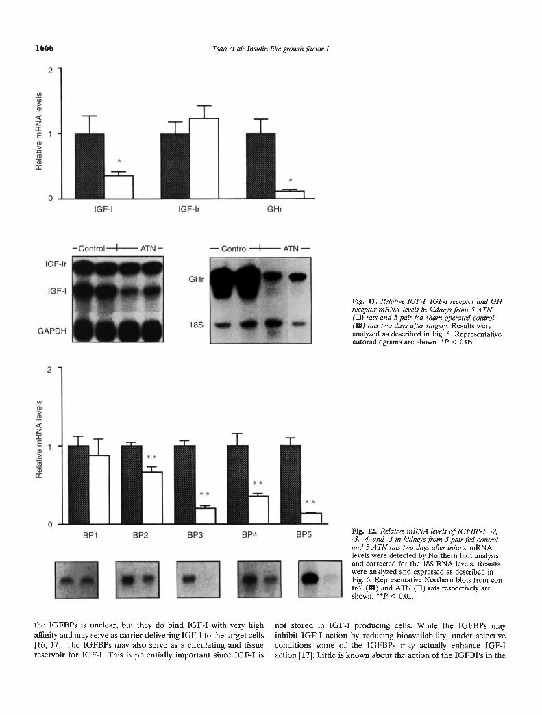
The extent of the renal failure in these experiments judged bythe serum creatinine levels two days after ischemic injury wascomparable to that seen in the ad libitum fed animals (Table 1).Body weight changes in ATN and pair fed controls were similar.As noted in the ad libitum experiments kidney IGF-I receptormRNA levels, depicted in Figure 11, did not change while plasmareceptor binding increased significantly in the ATN group (20.80.77 vs. 8.7 0.67%160 jsg protein; P < 0.01). Steady-state IGF-ImRNA levels were reduced significantly in the ATN group andaveraged 37% of the pair fed control value (Fig. 11). GM receptormRNA abundance was reduced to 11% of the control value (Fig.11). As shown in Figure 12 there were marked changes in theIGFBP mRNA profile. The abundance of IGFBP-2, -3, -4 and -5
(I)ci
zE
IL0a)>
a)
Day 2
Fig. 7. Relative IGF-I mRNA levels in kidneys from control () and ATN(LI) rats two and seven days after surgery. IGF-I and GAPDH mRNA levelswere measured simultaneously in solution hybridization RNase protectionassays. Results were analyzed and expressed as described in Fig. 6 and arefrom 4 to 5 rats per time point. A representative autoradiogram from day2 is included. < 0.01.
fell significantly after the ischemic episode to values rangingbetween 13 and 67% of controls. There was no significant changein IGFBP-1 mRNA levels.
Discussion
In this study we have demonstrated that acute ischemic kidneyinjury is followed by profound alterations in the renal GH—IGF-Isystem. These changes were not related to food consumption forsimilar patterns were seen whether dietary intake was ad libitumor controlled. We found that two days after the injury, kidneyIGF-I mRNA levels had declined to approximately 20% of thevalue seen in sham-operated control animals allowed free accessto food and water. Kidney immunoreactive IGF-I concentrationwas also reduced at this time. After seven days the mRNA levelshad returned to values similar to those measured in the controls.Presumably the low IGF-I mRNA levels together with the re-duced IGF-I protein levels indicate that local IGF-I production isreduced at this time when IGF-I would be of particular value inthe reparative process. In contrast to the fall in kidney IGF-lconcentration the number of IGF-I specific receptors in the
Day2 Day7
I— Control I ATN
Tsao et al: Insulin-like growth factor-I 1663
2.0
1.5
1.0
0.5
0.0
IGF-l
GAPDH
Day2 Day7
I Control I ATN
IGF-IR
GAPDH
—
H
- -ya a
a
1664 Tsao et al: Insulin-like growth factor-I
, 2.0a)>a)
Z 1.5
E00.a)0a)
0.5
0.0
GHR
1 8S
Day 2
Fig. 8. Relative GH receptor mRNA levels in control (B) andA TN (LI) rats.GH receptor mRNA and 18S RNA levels were determined simultaneouslyby quantitative PCR. The relative GH receptor mRNA levels werecalculated as described in Fig. 6 with the 18S RNA signal substituting forGAPDH. N = 4 to 6 rats per time point. A representative autoradiographfrom day 2 is shown. *p < 0.05, **p < 0.01.
kidney increased after the ischemic insult. However IGF-I recep-tor mRNA levels did not differ from control values. Thus theincrease in receptor number appears to reflect a post-translationalresponse. Also striking was the finding that IGFBP-2, -3, -4, and-5 mRNA levels were markedly reduced. Indeed two days afterinjury the average of these IGFBP mRNA levels ranged between4 and 62% of their control values. This was accompanied by adecrease in plasma membrane associated IGFBPs. While this isconsistent with decreased IGFBP production, post-translationalevents such as increased proteolytic destruction may also play arole. By the seventh day, IGFBP-2 and -4 mRNA levels hadreturned to control values. As discussed later, the reduction inIGFBP gene and protein expression could conceivably result in anincrease in the bioavailability of the free circulating IGF-I thatenters the kidney.
Our finding of a decrease in immunoreactive IGF-I levels in thedamaged kidneys is consistent with Ding et a! [6]. On the otherhand, Matejka and Jennische [32], using a non-quantitative mor-phologic approach, observed that in rats following ischemic injury
the renal distribution of IGF-I became more widespread. IGF-Iimmunoreactivity was not limited to the collecting ducts and loopof Henlé as is seen in normal rat kidneys [12, 33, 341, but waspresent in regenerating proximal tubular cells. In addition cellsother than tubular cells, presumably macrophages, expressedIGF-I. Also IGF-I mRNA, normally limited to the distal nephron,was more widely expressed appearing in the same sites as theIGF-I prOtein. These morphologic findings likely reflect a localresponse to injury and thus are not incompatible with ourobservation that whole kidney IGF-I mRNA and IGF-I proteinlevels are reduced.
Using a non-quantitative autoradiographic approach Matejkaand Jennische [321 also noted an increase in IGF-l receptorbinding in the regenerative zone. Taken together with theirobservation of a wider expression of IGF-I mRNA, it is likely thatthe local increase in immunoreactive IGF-1 reflects IGF-I synthe-sized locally and IGF-I bound to receptors. Our finding of anincrease in receptor number supports the latter hypothesis. Se-questration of IGF-I by local IGFBPs provides another potentialmechanism for increased local immunoreactivity. However, themarked decrease in kidney IGFBP mRNA levels together with thedecrease in kidney IGFBP proteins that we noted argues againstbut does not exclude this mechanism.
While interruption of renal blood flow with its attendantischemic injury is the initiating event for the observed changes inthe renal IGF-I system, proximate signals mediating these changeswere not examined. Nevertheless, it is worth considering potentialmechanisms involved in these changes. In respect to IGF-Ireceptor binding, it appears that the increase in receptor densityrepresents a post-transcriptional event since there is no corre-sponding change in mRNA level. Among the possibilities are anincrease in mRNA translation, increased receptor stability or evena redistribution of cellular receptors from an intracellular locationto the cell surface [35]. Initially the increase in receptor densitycould be a response to a decrease in kidney IGF-I production seenearly after injury. The increase in receptor number cannot beaccounted for by reduced food intake [36] since a similar responsewas seen when controls were paired with the ATN rats. Interest-ingly, there was a strong correlation between peak serum creati-nine levels measured on day 2 and IGF-1 receptor bindingmeasured two and seven days after the ischemic insult; thissuggests a direct link between the extent of the renal injury andthe receptor response. Up-regulation of receptor number willlikely favor the renal action of circulating and local IGF-I.
Why kidney IGF-I mRNA levels fall after ischemic injury is anintriguing question since the brunt of the ischemic injury involvesthe proximal tubule, a nephron segment where IGF-I is notnormally produced. It is likely that the reduced IGF-I mRNAlevels reflect dysfunction of the cells in the medullary ascendinglimb of Henlé and collecting ducts that normally produce IGF-I[13, 34, 37]. Indeed, while the major site of ischemic injury is theproximal tubule, less obvious morphologic changes are present inthese more distal nephron segments [38]. This injury probablyaccounts for the reduction in IGF-I mRNA levels which mayreflect reduced mRNA production or stability.
Interestingly kidney preproEGF mRNA levels, also expressedin the ascending loop of Henlé and distal tubule, decline followingischemic injury [39]. Since EGF stimulates collecting duct IGF-Igene expression [40], reduced kidney EGF production couldaccount, in part, for the reduced IGF-I mRNA levels. Growth
Day2 Day7— Control I ATN

BP3 mRNAppControl pp
ATN
2d 7d
BP5 mRNA
Control
2d 7d
U)
>a)
zE
a)
BP1 BP2 BP3 BP4 BP5
Day 7
hormone is another potent regulator of kidney IGF-I production[13]. We found that kidney GH receptor mRNA levels arereduced in ischemic ATN, and thus propose that reduced GHsignaling may also be a factor accounting for the low kidney IGF-ImRNA levels. This likely explains the failure of GH therapy toinfluence the course of acute ischemic renal failure in rats [51.Note that the GH receptor mRNA levels were measured with aprimer set that amplified a portion of the rat cDNA whichencodes most of the extracellular domain of the receptor which isidentical to the corresponding part of the serum GH-bindingprotein [27]. In the rat there is an alternative splicing event duringthe processing of the complete GH receptor mRNA. The fulllength transcript forms the complete receptor while the truncatedmRNA encodes the GH-binding protein. This protein has thesame extracellular domain as the GH receptor but lacks ahydrophobic transmembrane and an intracellular domain. As thefull length GH receptor transcript is predominant in the ratkidney [41], we conclude that the 35 to 91% reduction in mRNAlevels, detected in our assay, reflects a true lowering of the fulllength transcript.
The IGFBPs are important, but poorly understood, compo-nents of the renal IGF-I system [14]. Six IGFBPs have beenidentified and all are expressed in the kidney [30]. Of thesebinding proteins, the site of renal production has only beenidentified for IGFBP-1 which is localized to the medullary thickascending loop of Henlé and collecting ducts [34, 371. Again asdiscussed above in the context of IGF-I gene expression, it is notentirely clear why IGFBP-I mRNA levels fall when the ischemicinjusy is predominantly proximal tubular. The function of each of
2
7
/ ii I**I_BP1 BP2 BP3 BP4 BP5
Day 2
i.iIi
0.
2
0
Cl)
a)>a)
zEa)>Ca
a)
Tsao et al: Insulin-like growth factor-I 1665
/
Fig. 9. Relative mRNA levels of IGFBP-I, -2, -3,-4, and -5 in kidneys from control () or ATN(LI) rats. mRNA levels were detected by North-ern blot analysis and corrected for the GAPDHmRNA levels. Results were analyzed and ex-pressed as described in Fig. 6 and are from 3 to5 rats per time point. Representative Northernblots are shown. *P < < 0.01.
Control ATN
I I I I
Mr(kD)
49.5 —
32.5 —
27.5 —
12.5 —
Fig. 10. Western ligand blot of plasma membrane-associated IGF bindingproteins. Membranes were prepared from saline perfused control andATN kidneys two days after the ischemic insult. Membranes were pooledfrom 5 rats from each group and assayed in duplicate.
IGF-lr
— Control I ATN — — Control I ATN —
GHr
1 8S
IGF-lr
GAPDH
1666 Tsao et al: Insulin-like growth factor-I
Fig. 11. Relative IGF-I, IGF-I receptor and GHreceptor mRNA levels in kidneys from 5 ATN(Li) rats and 5 pair-fed sham operated control(VA) rats two days after swyely. Results wereanalyzed as described in Fig. 6. Representativeautoradiograms are shown. < 0.05.
FIg. 12. Relative mRNA levels of IGFBP-1, -2,-3, -4, and -5 in kidneys from 5 pair-fed controland 5 A TN rats two days after injwy. mRNAlevels were detected by Northern blot analysisand corrected for the 18S RNA levels. Resultswere analyzed and expressed as described inFig. 6. Representative Northern blots from con-trol (VA) and ATN (El) rats respectively areshown. < 0.01.
the IGFBPs is unclear, but they do bind IGF-I with very high not stored in IGF-I producing cells. While the IGFBPs mayaffinity and may serve as carrier delivering IGF-I to the target cells inhibit IGF-I action by reducing bioavailability, under selective[16, 171. The IGFBPs may also serve as a circulating and tissue conditions some of the IGFBPs may actually enhance IGF-Ireservoir for IGF-I. This is potentially important since IGF-I is action [17]. Little is known about the action of the IGFBPs in the
Rel
ativ
e m
RN
A le
vels
0
N)
w
-U
w
-u
N)
03
-D
Co 03
-U
03
-u
01
* * I-
I
Tsao et a!: Insulin-like growth factor-I 1667
kidney. However, in preliminary studies with cultured rabbitproximal tubular cells we have found that the IGFBPs producedby these cells and as well as recombinant human IGFBP-3 inhibitIGF-I action [42]. Taking together what little is known about theactions of IGFBPs we offer for consideration the possibility thatthe overall effect of reduced IGFBP mRNA and protein levels inischemic ATN may result in an increase in the bioavailability ofIGF-I.
While this study offers fresh insight into the response of therenal GH—IGF-I axis to acute ischemic injury, it should berecognized that the study was conducted at the whole kidney leveland thus reflects net changes in the kidney so that local changesmay be masked. Nevertheless, when interpreting these resultsimportant general conclusions can be made which will serve as thebasis for further mechanistic studies into the local response toacute tubular cell injury. Of particular interest are the observa-tions that whole kidney mRNA levels for IGF-I and also severalIGFBPs are reduced, that kidney plasma membrane associatedIGFBPs are reduced and that kidney IGF-I receptor density isincreased. Taken together we propose that the low kidney IGF-ImRNA levels in combination with the reduced kidney IGF-Iprotein levels indicate an overall decrease in kidney IGF-I pro-duction. This IGF-I deficiency will not favor tissue repair. On theother hand, we offer for consideration the suggestion that reducedkidney IGFBP gene expression with reduced IGFBP proteinlevels together with an increase in IGF-I receptor number mightserve to offset the lowered kidney IGF-I production and alsoenhance the reparative actions of circulating endogenous orexogenous administered hormone.
Acknowledgments
This study was supported by the Department of Veterans AffairsResearch Service and NIH Grants DK-3604 and DK-32342. J. Wang wassupported by NIH Training Grant DK-07357 and F.C. Fervenza by CNPq(Brazil). The authors wish to thank Dr. Timothy A. Stewart, Ph.D. ofGenentech, Inc., South San Francisco, CA, for helpful discussions.
Reprint requests to Ralph Rabkin, M.D., Veterans Affairs Medical Center(154L), 3801 Miranda Avenue, Palo Alto, California 94304 USA.
References
1. SHUSTERMAN N, STROM BL, MURRAY TG, MORRISON G, WEST SL,MAISLIN G: Risk factors and outcome of hospital-acquired acute renalfailure. Am J Med 83:65—71, 1987
2. HUMES, HD, Cw.sLINsKI DA, COIMBRA TM, MESSANA JM, GALVAO C:Epidermal growth factor enhances renal tubule cell regeneration andrepair and accelerates the recovery of renal function in postischemicacute renal failure. J Clin Invest 84:1757—1761, 1989
3. NORMAN J, TSAU Y-K, BACAY A, FINE LG: Epidermal growth factoraccelerates functional recovery from ischaemic acute tubular necrosisin the rat: Role of the epidermal growth factor receptor. Clin Sci78:445—450, 1990
4. CLARK R, MORTENSEN D, RABKIN R: Recovery from acute ischemicrenal failure is accelerated by des(1-3)insulin-like growth factor in therat. Clin Sci 86:709—714, 1994
5. MILLER 5, MARTIN DR, KISSANE J, HAMMERMAN MR: Insulin-likegrowth factor I accelerates recovery from ischemic acute tubularnecrosis in the rat. Proc NatlAcad Sci USA 89:11876—11880, 1992
6. DING H, KOPPLE JD, COHEN A, HIRSCHBERG R: Recombinant humaninsulin-like growth factor I accelerates recovery and reduces catabo-lism in rats with ischemic acute renal failure. J Clin Invest 91:2281—2287, 1993
7. HIRSCHBERG R, KOPPLE J: Evidence that insulin-like growth factor Iincreases renal plasma flow and glomerular filtration rate in fastedrats. J Clin Invest 83:326—330, 1989
8. FlUMES HD, BEAIS TF, CIESLINSK DA, SANCHEZ 10, PAGE TP: Effects
of transforming growth factor-f3, transforming growth factor, andother growth factors on renal proximal tubule cells. Lab Invest64:538—545, 1991
9. MEFILS 0, IRZYNJEC T, RITZ E, EDEN 5, KovACs G, KlAUs G, FLOEGEJ, MAn. G: Effects of rhGH and rhIGF-1 on renal growth andmorphology. Kidney Int 44:1251—1258, 1993
10. ROGERS SA, RYAN G, HAMMERMAN MR: Insulin-like growth factors Iand II are produced in the metanephros and are required for growthand development in vitro. J Cell Biol 113:1447—1453, 1991
11. MARTIN AA, THOMAS FM, OWENS PC, KNOWLES SE, BAII..&RD FJ,READ LC: IGF-I and its variant, des-(1-3)IGF-I, enhance growth inrats with reduced renal mass. Am J Physiol 261:F626—F633, 1991
12. HAMMERMAN M: The growth hormone-insulin-like growth factor axisin kidney. Am J Physiol 257:F503—F514, 1989
13. ROGERS SA, MILLER SB, HAMMERMAN MR: Growth hormone stim-ulates IGF-I gene expression in isolated rat renal collecting duct. AmJPhysiol 259:F474—F479, 1990
14. ZUMKELLER W, SCHOFIELD PN: The role of insulin-like growth factorsand IGF-binding proteins in the physiological and pathological pro-cesses of the kidney. Virchows Archiv B Cell Pathol 62:207—220, 1992
15. JENNISCHE E, ISGAARD J, ISAKSSON OGP: Local expression of insulin-like growth factors during tissue growth and differentiation, in TheInsulin-like Growth Factors Structure and Biological Functions, editedby PN SCHOFIELD, Oxford, Oxford University Press, 1992, pp 221—239
16. BAXTER RC: Physiological roles of IGF binding proteins, in ModernConcepts of Insulin-Like Growth Factors, edited by EM SPENCER, NewYork, Elsevier, 1991, pp 371—380
17. HOLLY JMP: Insulin-like growth factor binding proteins in diabeticand non-diabetic states, in Growth Hormone and Insulin-Like GrowthFactor I, edited by A FLYVBJERG, H ORSK0v, KGMM ALBERTI, NewYork, John Wiley, 1993, pp 47—76
18. RABKIN R, PETERSEN J, MAMELOK R: Binding and degradation ofinsulin by isolated renal brush border membranes. Diabetes 31:618—623, 1982
19. SCATCHARD G: The attraction of proteins for small molecules andions. Ann IVYAcad Sci 51:660—672, 1949
20. PILLION DJ, HASKELL JF, MEEZAN E: Distinct receptors for insulin-like growth factor I in rat renal glomeruli and tubules. Am J Physiol255:E504—E512, 1988
21. HOSSENSLOPP P, SEURIN D, SEGOVIA-QUINSON B, HARDOUIN 5, BIN-oux M: Analysis of serum insulin-like growth factor binding proteinsusing Western ligand blotting; use of the method for titration of thebinding proteins and competitive binding studies. Anal Biochem154:138—143, 1986
22. D'ERCOLE AJ, STILES AD, UNDERWOOD LE: Tissue concentrations ofsomatomedin C: Further evidence for multiple sites of synthesis andparacrine or autocrine mechanisms of action. Proc NatlAcad Sci USA81:935—939, 1984
23. CRAWFORD BA, MARTIN JL, HOWE Ci, HANDELSMAN Di, BAXTERRC: Comparison of extraction methods for insulin-like growth factor-Iin rat serum. J Endocrinol 134:169—176, 1992
24. CHOMCZYNSKI P, SACCHI N: Single-step method of RNA isolation byacid guanidinium thiocyanate-phenol-chloroform extraction. AnalBiochem 162:156—159, 1987
25. GUIDICE LC, DSUPIN BA, JIN IH, Vu TH, HOFFMAN AR: Differentialexpression of messenger ribonucleic acids encoding insulin-likegrowth factors and their receptors in human uterine endometrium anddecidua. J Clin Endocrinol Metab 76:1115—1122, 1993
26. AUSUBEL FM: The Polymerase Chain Reaction, in Cun-ent Protocols inMolecular Biology (vol 2), Green-Wiley Interscience USA, 1993, pp15.31—15.35
27. BAUMBACH WR, HORNER DL, LOGAN iS: The growth hormone-binding protein in rat serum is an alternatively spliced form of thegrowth hormone receptor. Gene Develop 3:1199—1205, 1989
28. TS0 JY, SUN X-H, K.&o T-H, REECE KS, Wu R: Isolation andcharacterization of rat and human glyceraldehyde-3-phosphate dehy-drogenase cDNAs: Genomic complexity and molecular evolution ofthe gene. Nuci Acid Res 13:2485—2502, 1985
29. LOWE WL JR, LASKY SR, LEROITH D, ROBERTS CT iR: Distribution
1668 Tsao et a!: Insulin-like growth factor-I
and regulation of rat insulin-like growth factor I messenger ribonu-cleic acids encoding alternative carboxyterminal E-peptides: Evidenceof differential processing and regulation in liver. Mol Endocrinol2:528—535, 1988
30. SHIMASAKI S, LING N: Identification and molecular characterization ofinsulin-like growth factor binding proteins (IGFBP-1, -2, -3, -4, -5 and-6). Prog Growth Factor Res 3:243—266, 1991
31. ROGERS SA, MILLER SB, HAMMERMAN MR: Enhanced renal IGF-Iexpression following partial kidney infarction. Am J Physiol 264:F963—F967, 1993
32. MATEJKA G, JENNIscHE B: IGF-I binding and IGF-I mRNA expres-sion in the post-ischemic regenerating rat kidney. Kidney Int 42:1113—1123, 1992
33. MATEJKA GL, ERIKSSON PS, CARLSSON B, JENNISCHE B: Distributionof IGF-I mRNA and IGF-1 binding sites in the rat kidney. Histochem-ist,y 97:172—180, 1992
34. KOBAYASHI S, CLEMM0N DR, VENKATACHALAM MA: Colocalizationof insulin-like growth factor-binding protein with insulin-like growthfactor I. Am J Physiol 261:F22—F28, 1991
35. RECFILER MM: The nature and regulation of the receptors forinsulin-like growth factors. Ann Rev Physiol 47:425—442, 1985
36. LOWE WL JR, Aiito M, WERNER H, ROBERTS CT JR, LnRoim D:
Regulation by fasting of rat insulin-like growth factor I and itsreceptor. J Clin Invest 84:619—626, 1989
37. CHIN E, ZHOU J, BONnY C: Anatomical relationships in the patternsof insulin-like growth factor (IGF)-I, IGF binding protein-I, andIGF-I receptor gene expression in the rat kidney. Endocnnol 130:3237—3246, 1992
38. DoooR RB, BENNETr V, MANDEL LI: Degradation of spectrin andankyrin in the ischemic rat kidney. Am .1 Physiol 264:C1003—C1013,1993
39. SAFIRSTEIN R, PRICE PM, SAGGI SJ, HARRIS RC: Changes in geneexpression after temporaey renal ischemia. Kidney mt 37:1515—1521,1990
40. ROGERS SA, MILLER SB, HAMMERMAN MR: Insulin-like growth factorI gene expression in isolated rat renal collecting duct is stimulated byepidennal growth factor. J Gun Invest 87:347—351, 1991
41. TSIONG TS, HERRINGTON AC: Tissue distribution, characterizationand regulation of messenger ribonucleic acid for growth hormonereceptor and serum binding protein in the rat. Endocrinology 129:1628—1634, 1991
42. YAP J, TSAO T, STEWART T, MOHLER M, CooK J, RABKIN R:Insulin-like growth factor binding protein secretion by rabbit proximaltubular cells. (abstract) JASN 3:483, 1992
Copyright © 2022 FDOKUMEN