Isolation, characterization and comparison of Atlantic and Chinook salmon growth hormone 1 and 2
8
Comp. Biochem. Physiol. Vol. 116B, No. 3, pp. 315–322, 1997 ISSN 0305-0491/ 97/$17.00 Copyright 1997 Elsevier Science Inc. PII S0305-0491(96)00246-5 Isolation, Characterization and Comparison of a Novel Crustacean Toxin with a Mammalian Toxin from the Venom of the Scorpion Centruroides noxius Hoffmann Consuelo Garcı ´a, 1 Baltazar Becerril, 1 Barbara Selisko, 1 Muriel Delepierre 2 and Lourival D. Possani 1, * 1 Department of Molecular Recognition and Structural Biology, Institute of Biotechnology, National Autonomous University of Mexico, Avenida Universidad, 2001, Apartado Postal 510-3 Cuernavaca, Morelos 62271 Mexico, and 2 Laboratory of Nuclear Magnetic Resonance (CNRS URA 1129) Pasteur Institute, 28 rue du Dr. Roux, 75015 Paris, France ABSTRACT. A novel crustacean-specific toxin, Cn5, containing 66 amino acid residues was isolated from the venom of the scorpion Centruroides noxius Hoffmann. It is stabilized by four disulfide bridges, formed between Cys12-Cys65, Cys16-Cys41, Cys25-Cys46 and Cys29-Cys48. Toxicity tests revealed that Cn5 is a toxin that affects arthropods but not mammals. However, at high concentrations, Cn5 does displace the mammal-specific toxin Cn2 from rat brain synaptosomes. The concentration of Cn5 that produces half-maximal inhibition (IC 50 ) was estimated to be 100 μM. Sequence comparison of Cn5 with toxin Cn2, a mammal-specific toxin from the same scorpion, showed the presence of two sequence stretches, at positions 30 to 38 and 49 to 58, where the majority of the differences are concentrated. On the three-dimensional structure of Cn5 it is demonstrated that these two sequence stretches form a continuous surface region near the site thought to bind to the sodium channel. We assume that this region might be implicated in determining species specificity. comp biochem physiol 116B;3:315–322, 1997. 1997 Elsevier Science Inc. KEY WORDS. Amino acid sequence, Centruroides noxius, crustacean-specific toxin, scorpion toxins, sodium channel, species specificity, three-dimensional modelling INTRODUCTION All known Na 1 -channel specific scorpion toxins are com- posed of 61–70 amino acid residues, stabilized by four disul- Scorpion toxins have been shown to affect ion permeability fide bridges (3,12–16). According to toxicity tests, binding of excitable cells (1). Four different families of peptides have experiments or electrophysiological experiments, they can been described, which specifically recognize membrane- be divided into toxins that are specifically toxic to mammals bound proteins, Na 1 -channels (1,2,3), K 1 -channels (4,5), or arthropods. The term ‘‘specific,’’ however, does not mean Cl 2 -channels (6) and Ca 21 -channels (7). Despite the fact ‘‘exclusively effective’’ but refers to low toxicity or affinity that the primary structures of these proteins can be quite in one group and/or higher toxicity or affinity in the other. different (8), their three-dimensional structure is extremely There are many reports of toxins that show effects towards similar. Except for the Ca 21 -channels specific scorpion tox- both mammals and arthropods (17–20). ins, whose structure is still unknown (7), all these toxins Mammal-specific toxins can be grouped into α-toxins were shown to contain a core structure, formed by an α- binding to site 3 of mammal Na 1 -channels, slowing down helix and a three-stranded β-sheet that are connected by their inactivation process, and β-toxins binding to site 4 of two disulfide bridges formed between two conserved se- the channel, affecting the activation of the channel quence stretches, Cys-X-X-X-Cys and Cys-X-Cys, where X (21,22). Displacement experiments on excitatory tissues is a variable amino acid (9–11). showed that the two binding sites are not related (21,23). Comparing structural features, α- and β-toxins vary in the Address reprint requests to: Lourival D. Possani, Institute of Biotechnol- size of two loop regions, the J-loop and the B-loop (24). ogy—UNAM, Avenida Universidad, 2001, Apartado Postal 510-3, Cuer- Arthropod-specific toxins have been tested so far mainly navaca, 62271 MEXICO, Tel (25273) 171209. Fax (25273) 172388; E- mail: [email protected] in insects (13,17–20), e.g., blowfly larvae, crickets, cock- Abbreviations –Cn, Centruroides noxius Hoffmann; CM-Cellulose, car- roaches, and some of them in crustaceans (13,17,25), e.g., boxymethyl-cellulose; RC-toxin, toxin reduced and carboxymethylated; crayfishes and cochinillas. Insect α-toxins show the classic 1 H-NMR, proton nuclear magnetic resonance. Received 25 June 1996; accepted 6 September 1996. α-toxin effect on insect sodium channels (17,26). This type
-
Upload
independent -
Category
Documents
-
view
1 -
download
0
Transcript of Isolation, characterization and comparison of Atlantic and Chinook salmon growth hormone 1 and 2

Comp. Biochem. Physiol. Vol. 116B, No. 3, pp. 315–322, 1997 ISSN 0305-0491/97/$17.00Copyright 1997 Elsevier Science Inc. PII S0305-0491(96)00246-5
Isolation, Characterization and Comparison of aNovel Crustacean Toxin with a Mammalian Toxinfrom the Venom of the Scorpion Centruroides noxius
HoffmannConsuelo Garcıa,1 Baltazar Becerril,1 Barbara Selisko,1 Muriel Delepierre2
and Lourival D. Possani1,*1Department of Molecular Recognition and Structural Biology, Institute of Biotechnology, National
Autonomous University of Mexico, Avenida Universidad, 2001, Apartado Postal 510-3 Cuernavaca, Morelos
62271 Mexico, and2Laboratory of Nuclear Magnetic Resonance (CNRS URA 1129) Pasteur Institute,
28 rue du Dr. Roux, 75015 Paris, France
ABSTRACT. A novel crustacean-specific toxin, Cn5, containing 66 amino acid residues was isolated from thevenom of the scorpion Centruroides noxius Hoffmann. It is stabilized by four disulfide bridges, formed betweenCys12-Cys65, Cys16-Cys41, Cys25-Cys46 and Cys29-Cys48. Toxicity tests revealed that Cn5 is a toxin thataffects arthropods but not mammals. However, at high concentrations, Cn5 does displace the mammal-specifictoxin Cn2 from rat brain synaptosomes. The concentration of Cn5 that produces half-maximal inhibition (IC50)was estimated to be 100 µM. Sequence comparison of Cn5 with toxin Cn2, a mammal-specific toxin from thesame scorpion, showed the presence of two sequence stretches, at positions 30 to 38 and 49 to 58, where themajority of the differences are concentrated. On the three-dimensional structure of Cn5 it is demonstrated thatthese two sequence stretches form a continuous surface region near the site thought to bind to the sodiumchannel. We assume that this region might be implicated in determining species specificity. comp biochem
physiol 116B;3:315–322, 1997. 1997 Elsevier Science Inc.
KEY WORDS. Amino acid sequence, Centruroides noxius, crustacean-specific toxin, scorpion toxins, sodiumchannel, species specificity, three-dimensional modelling
INTRODUCTION All known Na1-channel specific scorpion toxins are com-posed of 61–70 amino acid residues, stabilized by four disul-
Scorpion toxins have been shown to affect ion permeabilityfide bridges (3,12–16). According to toxicity tests, binding
of excitable cells (1). Four different families of peptides haveexperiments or electrophysiological experiments, they can
been described, which specifically recognize membrane-be divided into toxins that are specifically toxic to mammals
bound proteins, Na1-channels (1,2,3), K1-channels (4,5),or arthropods. The term ‘‘specific,’’ however, does not mean
Cl2-channels (6) and Ca21-channels (7). Despite the fact‘‘exclusively effective’’ but refers to low toxicity or affinitythat the primary structures of these proteins can be quitein one group and/or higher toxicity or affinity in the other.
different (8), their three-dimensional structure is extremelyThere are many reports of toxins that show effects towards
similar. Except for the Ca21-channels specific scorpion tox-both mammals and arthropods (17–20).
ins, whose structure is still unknown (7), all these toxinsMammal-specific toxins can be grouped into α-toxins
were shown to contain a core structure, formed by an α-binding to site 3 of mammal Na1-channels, slowing down
helix and a three-stranded β-sheet that are connected bytheir inactivation process, and β-toxins binding to site 4 of
two disulfide bridges formed between two conserved se-the channel, affecting the activation of the channel
quence stretches, Cys-X-X-X-Cys and Cys-X-Cys, where X(21,22). Displacement experiments on excitatory tissues
is a variable amino acid (9–11).showed that the two binding sites are not related (21,23).Comparing structural features, α- and β-toxins vary in the
Address reprint requests to: Lourival D. Possani, Institute of Biotechnol- size of two loop regions, the J-loop and the B-loop (24).ogy—UNAM, Avenida Universidad, 2001, Apartado Postal 510-3, Cuer-
Arthropod-specific toxins have been tested so far mainlynavaca, 62271 MEXICO, Tel (25273) 171209. Fax (25273) 172388; E-mail: [email protected] in insects (13,17–20), e.g., blowfly larvae, crickets, cock-
Abbreviations–Cn, Centruroides noxius Hoffmann; CM-Cellulose, car- roaches, and some of them in crustaceans (13,17,25), e.g.,boxymethyl-cellulose; RC-toxin, toxin reduced and carboxymethylated;
crayfishes and cochinillas. Insect α-toxins show the classic1H-NMR, proton nuclear magnetic resonance.Received 25 June 1996; accepted 6 September 1996. α-toxin effect on insect sodium channels (17,26). This type

316 C. Garcıa et al.
of toxins are believed to bind to receptor site 3 (26). Toxin Reagents for reduction and alkylation were obtained fromSigma (St Louis, MO). Deionized, double distilled waterTsγ (also called Ts VII) from the New-World scorpion Ti-
tyus serrulatus is a β-toxin that shows toxic effects in insects was used throughout.and in mammals (18,27). β-Toxins like Tsγ are thought tobind to the receptor site 4 on insect sodium channels (26).
Separation ProceduresAn aspect which distinguishes toxins against insect sodiumchannels from their vertebrate counterpart is that the for- Soluble venom from the scorpion Centruroides noxius Hoff-
mann was separated by chromatographic procedures as ear-mer can be further divided into excitatory and depressanttoxins, causing a contractory or flaccid paralysis, respec- lier described (8,32). Briefly, the first purification step con-
sisted in a gel filtration step using a Sephadex G-50 column.tively (28). Both toxin types displace each other (28–30),but their receptor sites are not identical (31). For further purification the material was applied onto a car-
boxymethyl cellulose (CM-Cellulose) column, equilibratedThe question why scorpion toxin molecules with theirsimilar three-dimensional fold show marked differences in and developed in the presence of 20 mM ammonium ace-
tate buffer, pH 4.7, with a continuous salt gradient from 0their functional characteristics, i.e., specific affinity to so-dium channels of a particular animal phyla, still remains to 0.5 M NaCl. The third and last purification step was
conducted on a Waters (Millipore Co., Milford, MA, USA)unclear. Rather small differences, i.e., single amino acidmodifications within the critical binding region of the toxin high performance liquid chromatography (HPLC) system
using a C4 reverse phase column (Vydac, Hisperia, CA). Forto its receptor, seem to be one possible explanation. Thusfar, there has been little information published concerning details refer to corresponding figure legend. The homogene-
ity of the resulting components were assessed by three inde-the identification of certain regions or residues that mightdetermine the specificity of scorpion toxins. pendent means: HPLC separation on a C18 reverse phase
microbore column (2 mm I.D.) using the Waters systemIn the present communication three aspects of a novelcrustacean-specific toxin, Cn5, from the scorpion Centruro- (Millipore Co., Milford, MA, U.S.A.), polyacrylamide gel
electrophoresis in acidic conditions (33) or in presence ofides noxius, are presented: 1) the isolation, the determina-tion of the primary structure and the position of the disulfide sodium dodecyl sulfate (34) and microsequencing as de-
scribed below.bridges, 2) its species specificity and ability to compete withthe mammal-specific toxin Cn2 for binding to rat brain syn-aptosomes and 3) a sequence comparison of Cn5 with mam-
Bio-assaysmal-specific toxins of the genus Centruroides resulting in theidentification of a continuous region on the surface of the Lethality tests were performed in three distinct species of
animals, following approved protocols by the Animal Caretoxin molecule, which might be responsible for their speci-ficity. Commission of the Institute of Biotechnology. Adult albino
mice (strain CD1, average body weight 20 g) were used totest the various chromatographic fractions including the pu-
MATERIALS AND METHODS rified toxin. A given amount of protein dissolved in 200 µlSource of Venom phosphate-buffered saline (PBS) was injected intraperitone-Venom from scorpions of the species Centruroides noxius ally. Bioassays with insects and crustaceans were performedHoffmann was obtained by electrical stimulation of the an- using crickets (Achaeta spp.) and freshwater crayfishesaesthetized animals. The venom was resuspended in water (Cambarellus montezumae spp.), respectively, where up toand centrifuged for 15 min at 10,000 3 g. Subsequently, it 180 µg toxin dissolved in 20 µl water were injected. Aswas freeze dried and kept at 220°C. control experiments test animals were injected with pure
PBS or water.
Chemical and ReagentsPrimary Structure Determination of Cn5Only analytical grade chemicals were used throughout this
work. Reagents for sequencing were all obtained from The amino acid sequence of toxin Cn5 was obtained bydirect sequencing the native toxin, the reduced and carbox-MilliGen/Biosearch, Division of Millipore (Burlington,
MA). Acetonitrile and trifluoracetic acid were from Pierce ymethylated toxin (RC-toxin) as well as fragments gener-ated by enzymatic cleavage of RC-toxin. The reduction of(Rockford, IL). Trypsin and chymotrypsin were sequence
grade enzymes from Boehringer Mannheim (Mannheim, the toxin with dithiothreitol, the alkylation with iodoaceticacid and the enzymatic cleavage with trypsin, chymotrypsinGermany). Protease V8 from Staphylococcus aureus was
kindly provided by Dr. Brian Martin (NIMH, Bethesda, and protease V8 were performed as described (35,36). Thepeptides produced by enzymatic hydrolysis were separatedMD). Sephadex G-50 (medium-grade) was purchased from
Pharmacia Fine Chemicals (Uppsala, Sweden), carboxy- by HPLC as described (37). Microsequence determinationwas carried out with a Milligen ProSequencer model 6600methyl-cellulose (CM-32) from Whatman (Clifton, NJ).
Crustacean Toxin 317
(Division of Millipore Co, Milford, MA, U.S.A.) using pro-tocols with the peptide either adsorbed or covalentlyattached to Sequelon membranes (Aryl amine or DITC)as previously described (35–37).
Amino acid analysis was performed on samples hy-drolyzed in 6 N HCl with 0.5% phenol at 110°C in evacu-ated, sealed tubes as described (38).
Determination of Disulfide Bridges
Aliquots (usually 100 µg protein) of native Cn5 were di-gested with trypsin, chymotrypsin and endopeptidase V8,or a combination of these enzymes. The corresponding pep-tides were separated by HPLC and sequenced using the pro-tocol described above.
Toxin Binding to Rat Brain Synaptosomes
The purification of rat brain synaptosomes (fraction P3) andbinding-displacement assays were performed as described(39). Briefly, rat brain membranes were added to a bufferedsolution (140 mM NaCl, 5 mM KCl, 2.5 mM CaCl2, 1.8
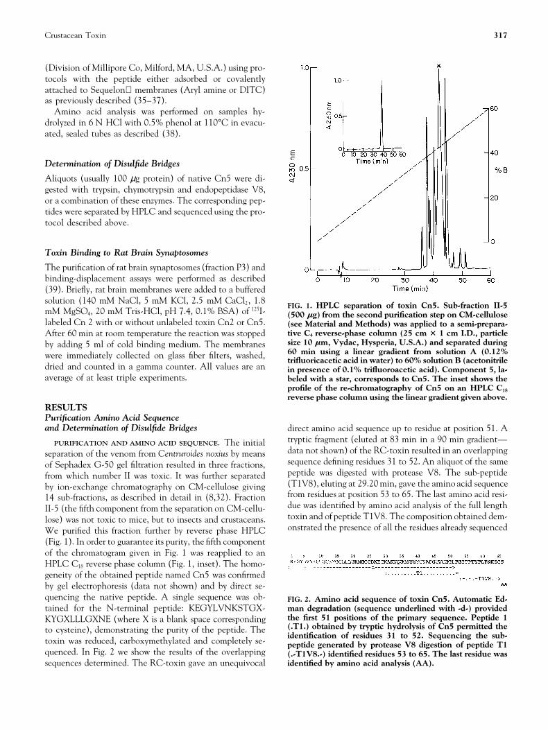
FIG. 1. HPLC separation of toxin Cn5. Sub-fraction II-5mM MgSO4, 20 mM Tris-HCl, pH 7.4, 0.1% BSA) of 125I- (500 mg) from the second purification step on CM-celluloselabeled Cn 2 with or without unlabeled toxin Cn2 or Cn5. (see Material and Methods) was applied to a semi-prepara-
tive C4 reverse-phase column (25 cm 3 1 cm I.D., particleAfter 60 min at room temperature the reaction was stoppedsize 10 mm, Vydac, Hysperia, U.S.A.) and separated duringby adding 5 ml of cold binding medium. The membranes60 min using a linear gradient from solution A (0.12%were immediately collected on glass fiber filters, washed, trifluoricacetic acid in water) to 60% solution B (acetonitrile
dried and counted in a gamma counter. All values are an in presence of 0.1% trifluoroacetic acid). Component 5, la-average of at least triple experiments. beled with a star, corresponds to Cn5. The inset shows the
profile of the re-chromatography of Cn5 on an HPLC C18
reverse phase column using the linear gradient given above.RESULTSPurification Amino Acid Sequenceand Determination of Disulfide Bridges direct amino acid sequence up to residue at position 51. A
tryptic fragment (eluted at 83 min in a 90 min gradient—PURIFICATION AND AMINO ACID SEQUENCE. The initialdata not shown) of the RC-toxin resulted in an overlappingseparation of the venom from Centruroides noxius by meanssequence defining residues 31 to 52. An aliquot of the sameof Sephadex G-50 gel filtration resulted in three fractions,peptide was digested with protease V8. The sub-peptidefrom which number II was toxic. It was further separated(T1V8), eluting at 29.20 min, gave the amino acid sequenceby ion-exchange chromatography on CM-cellulose givingfrom residues at position 53 to 65. The last amino acid resi-14 sub-fractions, as described in detail in (8,32). Fractiondue was identified by amino acid analysis of the full lengthII-5 (the fifth component from the separation on CM-cellu-toxin and of peptide T1V8. The composition obtained dem-lose) was not toxic to mice, but to insects and crustaceans.onstrated the presence of all the residues already sequencedWe purified this fraction further by reverse phase HPLC
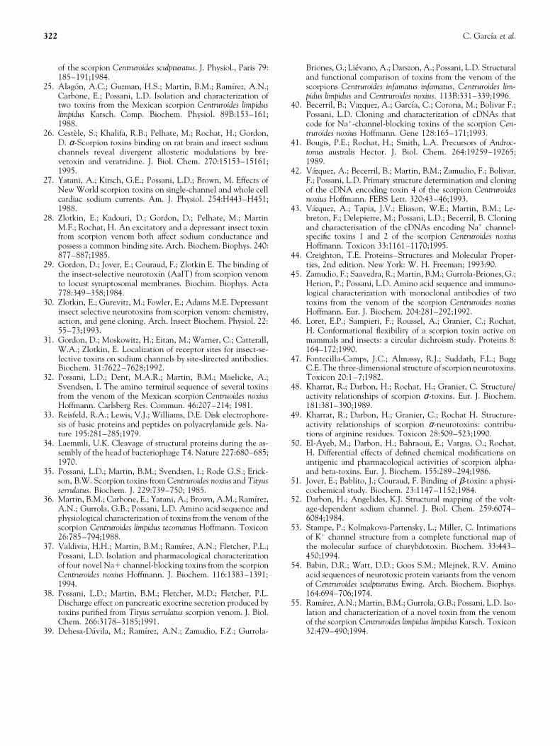
(Fig. 1). In order to guarantee its purity, the fifth componentof the chromatogram given in Fig. 1 was reapplied to anHPLC C18 reverse phase column (Fig. 1, inset). The homo-geneity of the obtained peptide named Cn5 was confirmedby gel electrophoresis (data not shown) and by direct se-quencing the native peptide. A single sequence was ob- FIG. 2. Amino acid sequence of toxin Cn5. Automatic Ed-
man degradation (sequence underlined with -d-) providedtained for the N-terminal peptide: KEGYLVNKSTGX-the first 51 positions of the primary sequence. Peptide 1KYGXLLLGXNE (where X is a blank space corresponding(.T1.) obtained by tryptic hydrolysis of Cn5 permitted theto cysteine), demonstrating the purity of the peptide. Theidentification of residues 31 to 52. Sequencing the sub-
toxin was reduced, carboxymethylated and completely se- peptide generated by protease V8 digestion of peptide T1quenced. In Fig. 2 we show the results of the overlapping (.-T1V8.-) identified residues 53 to 65. The last residue was
identified by amino acid analysis (AA).sequences determined. The RC-toxin gave an unequivocal

318 C. Garcıa et al.
TABLE 1. Amino acid sequence of heteropolymeric peptidescorresponding to the disulfide bridges of Cn 5
Elution HPLC Amino acid sequence Corresponding(min) heteropolymer disulfide bridge
1 2 3 4 528.75 Gly- Tyr-CYS-Tyr-Ala Cys16-Cys41
Gly- Xxx- Leu1 2 3 4 5 6
31.90 Asn- Glu-Gly-CYS-Asp-Lys Cys25-Cys46Ala- Phe- Gly-CYS
1 2 3 436.14 Ser-Thr-Gly-CYS Cys12-Cys65
Ser-Xxx Cys16-Cys41Asn-Gln-Gly-Gly . . . (Tyr)
1 2 336.62 Glu-CYS-Lys Cys29-Cys48
Xxx-Glu-Gly-Leu-Pro
The given amino acid sequences were obtained by microsequencing pep-tides purified by HPLC after the cleavage of Cn5 with trypsin and chymo-trypsin. The numbers on top of the amino acid mean positions from thesequencer; Xxx means a blank position corresponding to cysteine, whileCYS refers to the position of a cystine (a component that absorbs at 300nm corresponds to this amino acid). Tyrosine between parentheses wassurmised based on the amino acid sequence and enzymatic cleavage speci-ficity.
plus one additional serine. We concluded that the last posi-tion, amino acid 66, was occupied by a serine. These find-ings were confirmed by the amino acid sequence deducedfrom the nucleotide sequence of the cDNA clone CngtIIencoding toxin Cn5 (40).
FIG. 3. Displacement of 125I-Cn2 binding to rat brain synap-tosome membranes by Cn5. Rat brain synaptosome mem-DETERMINATION OF THE DISULFIDE BRIDGES. Several ali-branes (90 mg/200 ml per assay) were incubated with 300quots of native Cn5 were hydrolyzed with trypsin followedpM of 125I-Cn2 at room temperature for 1 hr, in the presenceby an additional cleavage with chymotrypsin. The resulting of increasing concentration of native Cn2 (s) or Cn5 (d),
peptides were separated by reverse phase HPLC and se- then filtered and counted as described in Materials andquenced. Over forty different fractions were sequenced (not Methods. One hundred percent binding was obtained in ab-
sence of Cn5. All values are means of triplicates.shown), some of which gave two amino acids per step, indi-cating the presence of two peptides connected by a disulfidebridge (dimeric peptide). Table 1 shows four sequences thatallowed us to propose how the disulfide bridges are arranged and crayfishes, the onset of toxicity lies at doses of less than
20 µg per g body weight. Nevertheless, applying 20 µg inin the native toxin. The dimer eluting at 28.75 min gavesequences consistent with the disulfide bridge Cys16-Cys41. crickets we observed a fast recuperation (after 30 min)
whereas the toxic effect in crayfishes was stronger andSimilarly, the peptide-pair eluting at 31.90 min corre-sponded to peptide fragments around the disulfide bridge longer lasting (more than 6 hr). The onset of lethality (at
least one test animal died of intoxication) in crickets wasCys25-Cys46. The oligomer eluting at 36.14 gave three se-quences from which the disulfide bridge Cys12-Cys65 was detected at 180 µg per g body weight whereas in crayfishes
at considerably lower doses, at 30 µg per g body weight. Inidentified, since the remaining peptide of the trimeric se-quence corresponded to the pair of Cys16 and Cys41, al- crayfishes a preliminary LD50 was determined using three
groups of six test animals applying doses of 25, 30 and 35ready identified. Finally, the peptide eluting at 36.62 mingave a dimeric sequence compatible with the disulfide µg per g body weight (results not shown). From this data a
LD50 of 28.5 µg per g body weight was estimated.bridge Cys29-Cys48.
Species Specificity Binding and Displacement Assays
In order to obtain additional information on the specificityLethality tests in mice showed that Cn5 is not toxic up toa quantity of 570 µg per 20 g average body weight. In con- of toxin Cn5, its binding properties to rat brain synapto-
somes were studied using the mammal-specific toxin Cn2trast, in arthropods the injection of Cn5 yielded toxic ef-fects as excitability, paralysis and death. In both crickets as a comparison. Figure 3 shows the results of binding and

Crustacean Toxin 319
displacement experiments accomplished with 125I-Cn2 andCn5. Toxin Cn5 displaced labeled Cn2 with a differenceof four orders of magnitude with respect to the displacementby non-labeled Cn2. The 50% inhibition constants IC50
were determined to be 16 nM and 100 µM for Cn2 andCn5, respectively.
DISCUSSION
In this communication we report the amino acid sequenceof an unknown toxin of Centruroides noxius Hoffmannwhich we named Cn5. The amino acid sequence corre-sponds to a mature peptide derived from a precursor of FIG. 4. Sequence comparison of arthropod-specific (A) and
mammal-specific (B) toxins of the genus Centruroides. Gapswhich the cDNA was reported earlier (40). The determina-were introduced to maximize similarities. Below each toxintion of the full amino acid sequence by peptide chemistryfamily the consensus sequence is shown which representsresulted to be important, because the comparison with thethe sequence with the most representative amino acid. X
amino acid sequence deduced from the cDNA shows that stands for a variable residue with no obvious preference.the toxin is synthesized in vivo as a pre-pro-peptide. The Cn1 (43), Cn2 and Cn3 (43,44), Cn4 (42) and Cn10 (B. Se-
lisko, C. Garcia, B. Becerril, M. Delepierre, L.D. Possani,precursor includes a signal peptide of 19 amino acids andin prep.) are from Centruroides noxius, CssII from Cen-two additional basic residues in the C-terminal. The pro-truroides suffusus suffusus (see review 8), CsEv1, CsEv2 andcessing of the C-terminal was reported earlier for other scor-CsEv3 are from Centruroides sculpturatus Ewing (54), Cll1
pion toxins from Old-World as well as from New-World (13), Cll1m (55) and Cll2 (39) from Centruroides limpidusscorpions (40–43). The C-terminal of the mature peptide limpidus, Cii1 from Centruroides infamatus infamatus (39)
and Clt1 from Centruroides limpidus tecomanus (36). Thecan include a free or an amidated carboxyl group. Amida-vertical bars indicate identity of the respective residues.tion takes place when in the precursor peptide a glycine
follows the last residue of the resulting mature peptide. Theglycine is processed leaving the preceding residue amidated(44). Since the cDNA of Cn5 does not contain a triplet stimulated extraction (8). Thus, the maximum doses we ap-
plied per mouse is approximately 10 times higher than thecoding for a glycine after the last residue, Ser66, of the ma-ture peptide, we assume that Ser66 is not amidated. amount of venom produced by a single scorpion in a given
time. In contrast to the absence of toxic effects in mice, Cn5With 66 amino acids Cn5 belongs to the group of long-chain toxins specific for sodium channels. A sequence com- shows toxic effects applying 20 µg/g average body weight of
arthropods (crickets and crayfishes). Therefore, Cn5 can beparison of toxin Cn5 with other sodium-channel-affectingtoxins of the genus Centruroides, shown in Fig. 4, reveals considered a toxin with high specificity towards arthropods.
The comparison of its activity in insects versus crustaceansthat Cn5 shares higher similarity with a consensus sequenceof toxins specific for arthropods (panel A) than with a con- shows that it is more effective in crustaceans.
In order to investigate the extent and the molecular basesensus sequence of toxins against mammals (panel B). Cn5shares typical characteristics of arthropod-specific toxins of the species specificity of Cn5 further, we compared it with
mammal-specific toxins of the genus Centruroides, especiallylike the presence of four prolines at positions 52, 56, 59and 61 in the C-terminal. Moreover, the C-terminal is less with toxin Cn2 of Centruroides noxius (45). Cn2 has been
characterized, functionally as a β-toxin (39). Therefore, ithydrophobic in its character than its counterpart in mam-mal-specific toxins. is expected to bind to site 4 at vertebrate sodium channels.
Lethality tests, conducted with Cn2 in mice, showed anThe position of the disulfide bridges, Cys12-Cys65,Cys16-Cys41, Cys25-Cys46 and Cys29-Cys48, corresponds LD50 of 0.4 µg/20 g mouse (45) whereas in crustaceans it
did not show any toxic effect up to 25 µg/g body weightto the typical pattern found in the majority of sodium-chan-nel-binding toxins with the exception of excitatory insect- (S. Serrano and L. D. Possani, unpublished results).
The displacement of 125I-Cn2 by Cn5 and Cn2 itselfspecific toxins from old-world scorpions (16). Having a longJ-loop (residues 16 to 24) and a short B-loop (residues 42 showed that, against our expectation, Cn5 was able to dis-
place Cn2 from its binding sites on rat brain synaptosomes.to 45), Cn5 belongs structurally to the group of β-toxins.The biological assays support the above observation of The high amount of Cn5, necessary to displace Cn2, reflects
the differences in species specificity of these toxins. BothCn5 being an arthropod-specific toxin. It did not show anysymptoms of toxicity towards mammals using up to 570 µg/ toxins belong structurally to the β-toxin class. This indi-
cates that the displacement shown might be due to competi-20 g average body weight mouse. In this respect, it is impor-tant to note, that a scorpion of the species Centruroides nox- tion for the same site. Since Cn2 binds to site 4 of the Na1-
channel, we expect Cn5 to bind to site 4 as well. The lowius can provide up to 50 µg venon per single electrically

320 C. Garcıa et al.
affinity of Cn5 for the mammalian Na1-channel might bethe cause of the absence of a toxic response observed inmammals even at high doses. Wheeler et al. (23) using threearthropod-specific toxins: CsEv1, CsEv2 and CsEv3, fromCentruroides sculpturatus Ewing, in binding and displace-ment experiments found no competition among the arthro-pod-specific toxins and those of two mammal-specific tox-ins: Androctonus australis toxin II (an α-toxin), andCentruroides suffusus suffusus II (a typical β-toxin). This ap-parent discrepancy to the results of our study is very likelydue to the limited concentration range used by Wheeler etal. (23). While they used concentrations up to 1 µM, wetested concentrations up to 100 µM, which was the esti-mated IC50 . Recently, Gordon et al. (20) proposed the exis-tence of a functional macro-receptor site 3 at mammalianand insect sodium channels, where homologous α-toxinsbind to various overlapping receptor sites, causing the physi-ological effect of α-toxins but with different efficacies. Itmight well be that something similar is occurring in the caseof β-toxins, i.e., that Cn5 binds to a macro-receptor site 4on the mammal sodium channel, but not exactly to thesame site as Cn2. In this case the displacement could becaused by an allosteric effect or by steric hindrance of thetwo toxins binding to overlapping sites.
The sequence comparison of toxin Cn5 with a consensussequence of mammal-specific toxins including Cn2 is shownin Fig. 4 (panel B) reveals that the amino acid differencesare concentrated mainly in two sequence stretches, betweenresidues 30 to 38 and 49 to 58. We assume that these twostretches are involved in the definition of the sequencespecificity. In order to localize the position of residues 30–
FIG. 5. Region of variable residues (30–38, 49–58, colored38 and 49–58 within the three-dimensional structure, wein dark grey) identified by comparison of arthropod-specificused the structure of Cn5 determined by NMR spectroscopytoxin Cn5 and mammal-specific toxin Cn2. A space-filled(F. Lebreton, C. Garcia, L. D. Possani, M. Delepierre, in presentation of the NMR-structure of Cn5 (F. Lebreton, C.
preparation). A solid-sphere representation of the model is Garcia, L.D. Possani, M. Delepierre, in prep.) is shown pre-shown in Fig. 5. The side facing the viewer presents the β- senting the ‘‘front’’ of the molecule in the upper figure and
the ‘‘top’’ in the lower figure.sheet structure. Following the proposal of Fontecilla-Campset al. (12) we will call this side the ‘‘face’’ of the molecule.The α-helix, in turn, is situated on the top-back of themodel. Figure 5 demonstrates that the sequence stretches backbone. In contrast, in Cn2 these residues are occupied
by Tyr and Ile, respectively.30–38 and 49–58 (shown in black) form a continuous sur-face region at the top and left side of the molecule. In terms The possible involvement of the identified surface region
in the definition of the species specificity is further sup-of secondary structure, residues 30–38 comprise the turn(residues 30–35) between the α-helix and the second β- ported by its being situated at the side of the toxin that is
thought to bind to the channel. For α- as well as for β-strand and the first residues of this β-strand. Residues 49–58 are involved in the third (middle) β-strand (residues 49 toxins exists evidence that the flat surface of the toxin, fac-
ing the viewer in Figure 5, contains the channel-bindingand 50) and the beginning of the C-terminal (residues 51–58) which does not adopt a particular secondary structure. site (47–52). Thereby, special importance is given to the
aromatic cluster, formed by residues 4, 40, 42, 47 and 58Loret et al. (46), after analyzing the secondary structure ele-ments of three toxins that show differences in their speci- in mammal-specific toxins from New-World scorpions. In
arthropod-specific toxins the aromatic cluster is extendedficity, also proposed the C-terminal as being involved in thedetermination of the species specificity. In this respect, it by Tyr38. The surface region, that we propose here to be
specificity-determining, is situated in the vicinity of the aro-is interesting to note, that Cn5 as other arthropod-specifictoxins (see Fig. 4) contains two prolines at positions 52 and matic cluster. In fact, residues 38 and 58 are part of it. The
maximum distance between residues 30–38 and 49–58, and56. They are expected to confer a certain rigidity to the

Crustacean Toxin 321
8. Possani, L.D. Structure of scorpion toxins. In Handbook ofthe corresponding nearest residue of the aromatic cluster inNatural Toxins, Vol. 2. New York: Marcel Dekker, Inc.; 1984:Cn5 is 14.6 A. Considering that the dimensions of the bind-513–550.
ing site of Charybdotoxin, a K1-channel-binding toxin of 9. Kobayashi, Y.; Takashima, H.; Tamaoki, H.; Kiogoku, Y.;similar structure, are 10 3 15 A (53), it is reasonable to Lambert, P.; Kuroda, H.; Chino, N.; Watanabe, T.X.; Kimura,
T.; Sakakibara, S.; Moroder, L. The Cystine-Stabilized α-He-suppose that all these residues can be situated within or inlix: A Common Structural Motif of Ion-Channel Blockingthe immediate vicinity of the binding site and therefore beNeurotoxic Peptides. Biopolymers 31:1213–1220;1991.of influence. However, the distance given is the direct dis-
10. Menez, A.; Bontems, F.; Roumestand, C.; Gilquin, B.; Toma,tance between the Cα atoms, it does not consider the ‘‘ef- F. Structural basis for functional diversity of animal toxins.fective’’ distance described by the surface of the molecule. Proc. Roy. Soc. Edinburgh 99B:83–103;1992.
11. Bontems, F.; Roumestand, C.; Gilquin, B.; Menez, A.; Toma,The visual inspection of the three-dimensional structureF. Refined structure of Charybdotoxin: common motifs inprovides more information in that respect. It revealed thatscorpion toxins and insect defensins. Science 254:1521–1523;residues 31, 33 and 34 are oriented rather to the back of1991.
the molecule whereas residues 51 to 56 to the left side. The 12. Fontecilla-Camps, J.C.; Almassy, R.J.; Suddath, F.L.; Watt,question remains if these particular residues could still in- D.D.; Bugg, C.E. Three-dimensional structure of a protein
from scorpion venom: A new structural class of neurotoxins.fluence the binding specificity of the toxins.Proc. Natl. Acad. Sci. USA 77:6496–6500;1980.In conclusion, our comparative studies indicate that the
13. Lebreton, F.; Delepierre, M.; Ramırez, A.N.; Balderas, C.; Pos-surface region comprising residues 30–38 and 49–58 mightsani, L.D. Primary and NMR three-dimensional structure de-
be responsible for the species specificity of a toxin. These termination of a novel crustacean toxin from the venom ofresidues present therefore interesting candidates for muta- the scorpion Centruroides limpidus limpidus Karsch. Biochem.
33:11135–11149;1994.tion analyses of Cn5. As especially promising candidates we14. Lee, W.; Moore, C.H.; Watt, D.D.; Rama Krishna, N. Solu-propose Pro52 and Pro56 as well as the residues that are
tion structure of the variant-3 neurotoxin from Centruroidespart of (residues 38 and 58) or in close vicinity to the aro-sculpturatus Ewing. Eur. J. Biochem. 218:89–95;1994.
matic cluster (residues 30, 32, 35, 36, 37, 49, 50, 57). 15. Housset, D.; Habersetzer-Rochat, C.; Astier, J.-P.; Fontecilla-Camps J.C. Crystal structure of toxin II from the scorpion An-
This work was supported partially by grants from the Howard Hughes droctonus australis Hector refined at 1.3 A resolution. J. Mol.Medical Institute (75191-527104) and the European Commission Biol. 238:88–103;1994.(CI1*-CT 94-0045) to L.D.P., from CONACyT-Mexico (IN- 16. Darbon, H.; Weber, C.; Braun, W. Two-dimensional 1H Nu-4734) and DGAPA-UNAM (IN-205893) to B.B. and a scholarship clear Magnetic Resonance study of AaH IT, an anti-insectfrom the Alexander-von-Humboldt Foundation, Germany to B.S. We toxin from the scorpion Androctonus australis Hector. Sequen-would like to thank Fernando Zamudio, Timoteo Olamendi, Fredy Co- tial resonance assignments and folding of the polypeptideronas and Cipriano Balderas for their technical assistance. chain. Biochem. 30:1836–1845;1991.
17. Eitan, M.; Fowler, E.; Herrmann, R.; Duval, A.; Pelhate, M.;Zlotkin, E. A scorpion venom neurotoxin paralytic to insectsthat affects sodium current inactivation: purification, primaryReferences
1. Catterall, W. Neurotoxins that act on voltage-sensitive so- structure and mode of action. Biochem. 29:5941–5947;1990.18. De Lima, M.E.; Martin, M.F.; Diniz, C.R.; Rochat, H. Tityusdium channels in excitable membranes. Annu. Rev. Pharma-
col. Toxicol. 20:15–43;1980. serrulatus toxin VII bears pharmacological properties of bothβ-toxin and insect toxin from scorpion venoms. Biochem. Bi-2. Rochat, H.; Bernard, P.; Couraud, F. Scorpion toxins: chemis-
try and mode of action. In Ceccarelli, B.; Clementi, F. (Eds). ophys. Research Commun. 139:296–302;1986.19. Loret, E.P.; Martin-Eauclaire, M.-F.; Mansuelle, P.; Sampieri,Advances in Cytopharmacology, Vol. 3B. New York: Raven
Press; 1979:325–334. F.; Granier, C.; Rochat H. An anti-insect toxin purified fromthe scorpion Androctonus australis Hector also acts on the α-3. Lazdunski, M.; Frelin, C.; Barhanin, J.; Lombet, A.; Meiri, H.;
Pauron, D.; Romey, G.; Schmid, A.; Schweitz, H.; Vigne, P.; and β-sites of the mammalian sodium channel: sequence andcircular dichroism study. Biochem. 30:633–640;1991.Vijverberg, H.P.M. Polypeptide toxins as tools to study volt-
age-sensitive sodium channels. Ann. N. Y. Acad. Sci. 479: 20. Gordon, D.; Martin-Eauclaire, M.F.; Cestele, S.; Kopeyan,E.C.; Khalifa, R.B.; Pelhate, M.; Rochat, H. Scorpion toxins204–220;1986.
4. Carbone, E.; Wanke, E.; Prestipino, G.; Possani, L.D.; Mae- affecting sodium current inactivation bind to distinct homolo-gous receptor sites on rat brain and insect sodium channels.licke, A. Selective blockage of voltage-dependent K1 chan-
nels by a novel scorpion toxin. Nature 296:90–91;1982. J. Biol. Chem. 271:8034–8045;1996.21. Jover, E.; Couraud, F.; Rochat, H. Two types of scorpion neu-5. Miller, C.; Moczydlowski, E.; Latorre, R.; Phillips M. Cha-
rybdotoxin, a protein inhibitor of single Ca21-activated K1 rotoxins characterized by their binding to two separate recep-tor sites on rat brain synaptosomes. Biochem. Biophys. Res.channels from mammalian skeletal muscle. Nature 313:316–
318;1985. Commun. 64:1607–1614;1980.22. Catterall, W.A. Structure and function of voltage-gated so-6. DeBin, J.A.; Maggio, J.E.; Strichartz, G.R. Purification and
characterization of chlorotoxin, a chloride channel ligand dium and calcium channels. Current Opinion Neurobiol. 1:5–13;1991.from the venom of the scorpion. Cell. Physiol. 264:C361–
C369;1993. 23. Wheeler, K.P.; Watt, D.D.; Lazdunski, M. Classification of Nachannel receptors specific for various scorpion toxins. Pflugers7. Valdivia, H.H.; Kirby, M.S.; Lederer, W.J.; Coronado, R.
Scorpion toxins targeted against the sarcoplasmic reticulum Arch. 97:164–165;1983.24. Meves, H.; Simard, J.M.; Watt, D.D. Biochemical and electro-Ca (21)-release channel of skeletal and cardiac muscle. Proc.
Natl. Acad. Sci. USA 89:12185–12189;1992. physiological characteristics of toxins isolated from the venom
322 C. Garcıa et al.
of the scorpion Centruroides sculpturatus. J. Physiol., Paris 79: Briones, G.; Lievano, A.; Darszon, A.; Possani, L.D. Structuraland functional comparison of toxins from the venom of the185–191;1984.
25. Alagon, A.C.; Guzman, H.S.; Martin, B.M.; Ramırez, A.N.; scorpions Centruroides infamatus infamatus, Centruroides lim-pidus limpidus and Centruroides noxius. 113B:331–339;1996.Carbone, E.; Possani, L.D. Isolation and characterization of
two toxins from the Mexican scorpion Centruroides limpidus 40. Becerril, B.; Vazquez, A.; Garcıa, C.; Corona, M.; Bolivar F.;Possani, L.D. Cloning and characterization of cDNAs thatlimpidus Karsch. Comp. Biochem. Physiol. 89B:153–161;
1988. code for Na1-channel-blocking toxins of the scorpion Cen-truroides noxius Hoffmann. Gene 128:165–171;1993.26. Cestele, S.; Khalifa, R.B.; Pelhate, M.; Rochat, H.; Gordon,
D. α-Scorpion toxins binding on rat brain and insect sodium 41. Bougis, P.E.; Rochat, H.; Smith, L.A. Precursors of Androc-tonus australis Hector. J. Biol. Chem. 264:19259–19265;channels reveal divergent allosteric modulations by bre-
vetoxin and veratridine. J. Biol. Chem. 270:15153–15161; 1989.42. Vazquez, A.; Becerril, B.; Martin, B.M.; Zamudio, F.; Bolivar,1995.
27. Yatani, A.; Kirsch, G.E.; Possani, L.D.; Brown, M. Effects of F.; Possani, L.D. Primary structure determination and cloningof the cDNA encoding toxin 4 of the scorpion CentruroidesNew World scorpion toxins on single-channel and whole cell
cardiac sodium currents. Am. J. Physiol. 254:H443–H451; noxius Hoffmann. FEBS Lett. 320:43–46;1993.43. Vazquez, A.; Tapia, J.V.; Eliason, W.E.; Martin, B.M.; Le-1988.
28. Zlotkin, E.; Kadouri, D.; Gordon, D.; Pelhate, M.; Martin breton, F.; Delepierre, M.; Possani, L.D.; Becerril, B. Cloningand characterisation of the cDNAs encoding Na1 channel-M.F.; Rochat, H. An excitatory and a depressant insect toxin
from scorpion venom both affect sodium conductance and specific toxins 1 and 2 of the scorpion Centruroides noxiusHoffmann. Toxicon 33:1161–1170;1995.possess a common binding site. Arch. Biochem. Biophys. 240:
877–887;1985. 44. Creighton, T.E. Proteins–Structures and Molecular Proper-ties, 2nd edition. New York: W. H. Freeman; 1993:90.29. Gordon, D.; Jover, E.; Couraud, F.; Zlotkin E. The binding of
the insect-selective neurotoxin (AaIT) from scorpion venom 45. Zamudio, F.; Saavedra, R.; Martin, B.M.; Gurrola-Briones, G.;Herion, P.; Possani, L.D. Amino acid sequence and immuno-to locust synaptosomal membranes. Biochim. Biophys. Acta
778:349–358;1984. logical characterization with monoclonal antibodies of twotoxins from the venom of the scorpion Centruroides noxius30. Zlotkin, E.; Gurevitz, M.; Fowler, E.; Adams M.E. Depressant
insect selective neurotoxins from scorpion venom: chemistry, Hoffmann. Eur. J. Biochem. 204:281–292;1992.46. Loret, E.P.; Sampieri, F.; Roussel, A.; Granier, C.; Rochat,action, and gene cloning. Arch. Insect Biochem. Physiol. 22:
55–73;1993. H. Conformational flexibility of a scorpion toxin active onmammals and insects: a circular dichroism study. Proteins 8:31. Gordon, D.; Moskowitz, H.; Eitan, M.; Warner, C.; Catterall,
W.A.; Zlotkin, E. Localization of receptor sites for insect-se- 164–172;1990.47. Fontecilla-Camps, J.C.; Almassy, R.J.; Suddath, F.L.; Bugglective toxins on sodium channels by site-directed antibodies.
Biochem. 31:7622–7628;1992. C.E. The three-dimensional structure of scorpion neurotoxins.Toxicon 20:1–7;1982.32. Possani, L.D.; Dent, M.A.R.; Martin, B.M.; Maelicke, A.;
Svendsen, I. The amino terminal sequence of several toxins 48. Kharrat, R.; Darbon, H.; Rochat, H.; Granier, C. Structure/activity relationships of scorpion α-toxins. Eur. J. Biochem.from the venom of the Mexican scorpion Centruoides noxius
Hoffmann. Carlsberg Res. Commun. 46:207–214; 1981. 181:381–390;1989.49. Kharrat, R.; Darbon, H.; Granier, C.; Rochat H. Structure-33. Reisfeld, R.A.; Lewis, V.J.; Williams, D.E. Disk electrophore-
sis of basic proteins and peptides on polyacrylamide gels. Na- activity relationships of scorpion α-neurotoxins: contribu-tions of arginine residues. Toxicon 28:509–523;1990.ture 195:281–285;1979.
34. Laemmli, U.K. Cleavage of structural proteins during the as- 50. El-Ayeb, M.; Darbon, H.; Bahraoui, E.; Vargas, O.; Rochat,H. Differential effects of defined chemical modifications onsembly of the head of bacteriophage T4. Nature 227:680–685;
1970. antigenic and pharmacological activities of scorpion alpha-and beta-toxins. Eur. J. Biochem. 155:289–294;1986.35. Possani, L.D.; Martin, B.M.; Svendsen, I.; Rode G.S.; Erick-
son, B.W. Scorpion toxins from Centruroides noxius and Tityus 51. Jover, E.; Bablito, J.; Couraud, F. Binding of β-toxin: a physi-cochemical study. Biochem. 23:1147–1152;1984.serrulatus. Biochem. J. 229:739–750; 1985.
36. Martin, B.M.; Carbone, E.; Yatani, A.; Brown, A.M.; Ramırez, 52. Darbon, H.; Angelides, K.J. Structural mapping of the volt-age-dependent sodium channel. J. Biol. Chem. 259:6074–A.N.; Gurrola, G.B.; Possani, L.D. Amino acid sequence and
physiological characterization of toxins from the venom of the 6084;1984.53. Stampe, P.; Kolmakova-Partensky, L.; Miller, C. Intimationsscorpion Centruroides limpidus tecomanus Hoffmann. Toxicon
26:785–794;1988. of K1 channel structure from a complete functional map ofthe molecular surface of charybdotoxin. Biochem. 33:443–37. Valdivia, H.H.; Martin, B.M.; Ramırez, A.N.; Fletcher, P.L.;
Possani, L.D. Isolation and pharmacological characterization 450;1994.54. Babin, D.R.; Watt, D.D.; Goos S.M.; Mlejnek, R.V. Aminoof four novel Na1 channel-blocking toxins from the scorpion
Centruroides noxius Hoffmann. J. Biochem. 116:1383–1391; acid sequences of neurotoxic protein variants from the venomof Centruroides sculpturatus Ewing. Arch. Biochem. Biophys.1994.
38. Possani, L.D.; Martin, B.M.; Fletcher, M.D.; Fletcher, P.L. 164:694–706;1974.55. Ramırez, A.N.; Martin, B.M.; Gurrola, G.B.; Possani, L.D. Iso-Discharge effect on pancreatic exocrine secretion produced by
toxins purified from Tityus serrulatus scorpion venom. J. Biol. lation and characterization of a novel toxin from the venomof the scorpion Centruroides limpidus limpidus Karsch. ToxiconChem. 266:3178–3185;1991.
39. Dehesa-Davila, M.; Ramırez, A.N.; Zamudio, F.Z.; Gurrola- 32:479–490;1994.