Diverse Functions of Autophagy in Liver Physiology ... - MDPI
88
Int. J. Mol. Sci. 2019, 20, 300; doi:10.3390/ijms20020300 www.mdpi.com/journal/ijms Review Diverse Functions of Autophagy in Liver Physiology and Liver Diseases Po-Yuan Ke 1,2,3 1 Department of Biochemistry & Molecular Biology and Graduate Institute of Biomedical Sciences, College of Medicine, Chang Gung University, Taoyuan 33302, Taiwan; [email protected]; Tel.: +886-3-211-8800 (ext. 5115); Fax: +886-3-211-8700 2 Liver Research Center, Chang Gung Memorial Hospital, Taoyuan 33305, Taiwan 3 Division of Allergy, Immunology, and Rheumatology, Chang Gung Memorial Hospital, Taoyuan 33305, Taiwan Received: 1 December 2018; Accepted: 8 January 2019; Published: 13 January 2019 Abstract: Autophagy is a catabolic process by which eukaryotic cells eliminate cytosolic materials through vacuole-mediated sequestration and subsequent delivery to lysosomes for degradation, thus maintaining cellular homeostasis and the integrity of organelles. Autophagy has emerged as playing a critical role in the regulation of liver physiology and the balancing of liver metabolism. Conversely, numerous recent studies have indicated that autophagy may disease-dependently participate in the pathogenesis of liver diseases, such as liver hepatitis, steatosis, fibrosis, cirrhosis, and hepatocellular carcinoma. This review summarizes the current knowledge on the functions of autophagy in hepatic metabolism and the contribution of autophagy to the pathophysiology of liver-related diseases. Moreover, the impacts of autophagy modulation on the amelioration of the development and progression of liver diseases are also discussed. Keywords: autophagy; selective autophagy; liver; liver disease; hepatitis; steatosis; fibrosis; cirrhosis; hepatocellular carcinoma 1. Introduction Autophagy is an evolutionarily conserved process that catabolizes intracellular components through lysosomes to recycle nutrients for supplying energy and regenerating organelles [1,2]. Several types of stress and damage stimuli, such as the deprivation of nutrients, the damage of organelles, the unfolding and aggregation of proteins, and tissue injury have been shown to induce autophagy [3,4]. Interference with the precise and appropriate process of autophagy may contribute to the pathogeneses of various human diseases, such as liver-associated diseases, neurodegenerative diseases, cancer, and infectious diseases [5,6]. In the past few decades, the homeostatic role of autophagy has emerged in the regulation of liver physiology through promoting the degradations of macromolecules and organelles to support the balance of energy as well as the metabolism and regeneration of organelles [7–9]. Additionally, autophagy has been indicated as a disease-associated factor that is modulated in the liver cells of people with liver-related diseases, and it contributes to the development and progression of various liver diseases, including hepatitis, steatosis, fibrosis, cirrhosis, and hepatocellular carcinoma [7,10–12]. Autophagy protects liver cells against injury and cell death by eliminating the damaged organelles and proteins that are introduced in those with liver- associated diseases. Conversely, autophagy could also act as an alternative pathway that promotes the development and progression of liver diseases. Most importantly, the modulation of autophagy has been extensively proved to alter the occurrence and outcome of liver-related diseases, implying that it represents a novel therapeutic target for the design of new and effective therapies to prevent
-
Upload
khangminh22 -
Category
Documents
-
view
0 -
download
0
Transcript of Diverse Functions of Autophagy in Liver Physiology ... - MDPI

Int. J. Mol. Sci. 2019, 20, 300; doi:10.3390/ijms20020300 www.mdpi.com/journal/ijms
Review
Diverse Functions of Autophagy in Liver Physiology
and Liver Diseases
Po-Yuan Ke 1,2,3
1 Department of Biochemistry & Molecular Biology and Graduate Institute of Biomedical Sciences,
College of Medicine, Chang Gung University, Taoyuan 33302, Taiwan; [email protected];
Tel.: +886-3-211-8800 (ext. 5115); Fax: +886-3-211-8700 2 Liver Research Center, Chang Gung Memorial Hospital, Taoyuan 33305, Taiwan 3 Division of Allergy, Immunology, and Rheumatology, Chang Gung Memorial Hospital, Taoyuan 33305,
Taiwan
Received: 1 December 2018; Accepted: 8 January 2019; Published: 13 January 2019
Abstract: Autophagy is a catabolic process by which eukaryotic cells eliminate cytosolic materials
through vacuole-mediated sequestration and subsequent delivery to lysosomes for degradation,
thus maintaining cellular homeostasis and the integrity of organelles. Autophagy has emerged as
playing a critical role in the regulation of liver physiology and the balancing of liver metabolism.
Conversely, numerous recent studies have indicated that autophagy may disease-dependently
participate in the pathogenesis of liver diseases, such as liver hepatitis, steatosis, fibrosis, cirrhosis,
and hepatocellular carcinoma. This review summarizes the current knowledge on the functions of
autophagy in hepatic metabolism and the contribution of autophagy to the pathophysiology of
liver-related diseases. Moreover, the impacts of autophagy modulation on the amelioration of the
development and progression of liver diseases are also discussed.
Keywords: autophagy; selective autophagy; liver; liver disease; hepatitis; steatosis; fibrosis;
cirrhosis; hepatocellular carcinoma
1. Introduction
Autophagy is an evolutionarily conserved process that catabolizes intracellular components
through lysosomes to recycle nutrients for supplying energy and regenerating organelles [1,2].
Several types of stress and damage stimuli, such as the deprivation of nutrients, the damage of
organelles, the unfolding and aggregation of proteins, and tissue injury have been shown to induce
autophagy [3,4]. Interference with the precise and appropriate process of autophagy may contribute
to the pathogeneses of various human diseases, such as liver-associated diseases, neurodegenerative
diseases, cancer, and infectious diseases [5,6]. In the past few decades, the homeostatic role of
autophagy has emerged in the regulation of liver physiology through promoting the degradations of
macromolecules and organelles to support the balance of energy as well as the metabolism and
regeneration of organelles [7–9]. Additionally, autophagy has been indicated as a disease-associated
factor that is modulated in the liver cells of people with liver-related diseases, and it contributes to
the development and progression of various liver diseases, including hepatitis, steatosis, fibrosis,
cirrhosis, and hepatocellular carcinoma [7,10–12]. Autophagy protects liver cells against injury and
cell death by eliminating the damaged organelles and proteins that are introduced in those with liver-
associated diseases. Conversely, autophagy could also act as an alternative pathway that promotes
the development and progression of liver diseases. Most importantly, the modulation of autophagy
has been extensively proved to alter the occurrence and outcome of liver-related diseases, implying
that it represents a novel therapeutic target for the design of new and effective therapies to prevent

Int. J. Mol. Sci. 2019, 20, 300 2 of 88
and treat liver diseases. In this paper, we summarize the current knowledge on the functional role of
autophagy in liver physiology and address how autophagy is regulated by liver-associated diseases
to become involved in the prevention or promotion of disease occurrence and pathogenesis.
2. Overview of Autophagy
The term autophagy is derived from the Greek words for auto (“self”) and phagy (“eating”). The
concept of autophagy was initially devised from the observation of vesicle-like dense bodies that
encompass cytoplasmic organelles, such as mitochondria and endoplasmic reticulum (ER), in
differentiated kidney tissue in mice and glucagon-perfused rat hepatocytes viewed using
transmission electron microscopy (TEM) [13–16]. These double-membraned dense bodies were
shown to be associated with the lysosome-mediated degradative process [13–16]. Subsequently, this
process was termed “autophagy” by de Duve, the 1974 Nobel Laureate in Physiology or Medicine, at
the Ciba Symposium on Lysosome in 1963 [17,18]. In the late 1960s, several studies, through
morphological and biochemical characterization, revealed that glucagon induces the formation of
autophagic vacuoles, which are influenced by lysosomes and lysosomal enzymes [19,20]. Despite the
effects of glucagon, the deprivation of amino acids and growth factors was indicated to trigger
autophagy [21–24]. From the 1970s to the 1990s, numerous studies demonstrated that the induction
of autophagy enhanced the degradation of long-lived proteins, leading to a decrease in amino acid
levels [24–26]. Additionally, the molecular signaling underlying autophagy initiation and the
autophagy inhibitors generated from these findings, such as 3-methyladenine (3-MA) and okadaic
acid, have been identified and characterized [24,27–33]. The membrane source of support for a
phagophore for the emergence of autophagic vacuoles was first described in the late 1980s [34] and
was further characterized in the 1990s [35–41]. The comprehensive isolation and molecular cloning
of autophagy-related genes (ATGs) were initiated by Ohsumi, who was the 2016 Nobel Laureate in
Physiology or Medicine for work on the genetic screening of temperature-sensitive, autophagy-
defective mutants in Saccharomyces cerevisiae [42–44]. Ohsumi identified 15 autophagy-defective
mutants that can be respectively complemented by the corresponding ATGs, which function in the
entire process of yeast autophagy and degradation [43]. Analogous to yeast, the functional ATGs
involved in autophagy in humans and other eukaryotes were also identified and characterized [45–
49]. To date, approximately 40 ATGs have been identified [49–51], most of which have been
evolutionarily conserved among almost all eukaryotes. Furthermore, the nomenclature for ATGs
across different species of eukaryotes has been unified [45–49].
2.1. Three Modes of Autophagy
Three types of autophagy have been defined according to the mechanism used for the delivery
of the intracellular components to lysosomes for degradation: microautophagy, chaperone-mediated
autophagy (CMA), and macroautophagy (Figure 1) [1,2]. Microautophagy was defined in
mammalian cells through TEM observation of a lysosomal membrane rearranged to have a
protrusion and arm-like structure to wrap the cytoplasmic portion into the lumen of the lysosome for
decomposition (Figure 1) [17,52,53]. Microautophagy not only randomly engulfs the intracellular
materials to instigate degradation (so-called nonselective microautophagy) but also selectively
eliminates specific organelles (defined as selective microautophagy) in yeast cells [54,55]. Although
core ATG proteins and the endosomal sorting complexes required for transport (ESCRT) machinery
are required for microautophagy [56–60], information about how microautophagy is precisely
induced and the detailed molecular mechanisms underlying the process of microautophagy remain
limited. Similarly, the functional role of microautophagy in human health and diseases is also largely
unknown and requires further investigations. CMA is characterized by a selective elimination
process in which the degradative substrates that contain the pentapeptide “Lys-Phe-Glu-Arg-Gln”
(KFERQ) motifs are specifically recognized by a cytosolic chaperone, namely, the heat-shock cognate
protein of 70 kDa (HSC70); these motifs are transported into the lysosomal lumen through the
lysosomal membrane protein 2A (LAMP2A)-mediated docking process (Figure 1) [61,62]. Multiple
types of stress have been shown to induce CMA, such as nutrient starvation, DNA damage, hypoxia,
Int. J. Mol. Sci. 2019, 20, 300 3 of 88
oxidative stress, and metabolic imbalance [63–68]. Crucially, CMA plays a role in the replenishment
of amino acids and ATP in cells that have undergone prolonged starvation [64,69], the regulation of
lipid metabolism [70,71], the reprogramming of gene transcription [72–74], the activation of immune
responses [75,76], the control of cell cycle progression [68,77], and the control of ageing [78,79].
Accordingly, the malfunctioning of CMA has emerged as a contributor to numerous human diseases,
such as tumorigenesis [80–83], neurodegenerative disorders [84–89], liver diseases [90,91], and
lysosomal storage disorders [92]. In macroautophagy (hereafter referred to as autophagy), the
membrane rearrangement process leads to the formation of an autophagosome, a double-
membranous vacuole that sequestrates the cytoplasmic components and delivers them to lysosomes
for degradation (Figure 1) [2,93]. Several types of stress, such as the starvation of nutrients, damage
of organelles, aggregation of proteins, and invasion of pathogens, have been shown to induce
autophagy [3,4]. In the past decade, autophagy has emerged as a “double-edged sword” in the
pathogenesis of a variety of human diseases, including neurodegenerative diseases [94–97], cancer
[98,99], cardiovascular diseases [100–102], ageing [94,99–104], infectious diseases [105,106], and
metabolic disorders [98,107–110]. Therefore, targeting autophagy could be a feasible strategy for
treating human diseases.
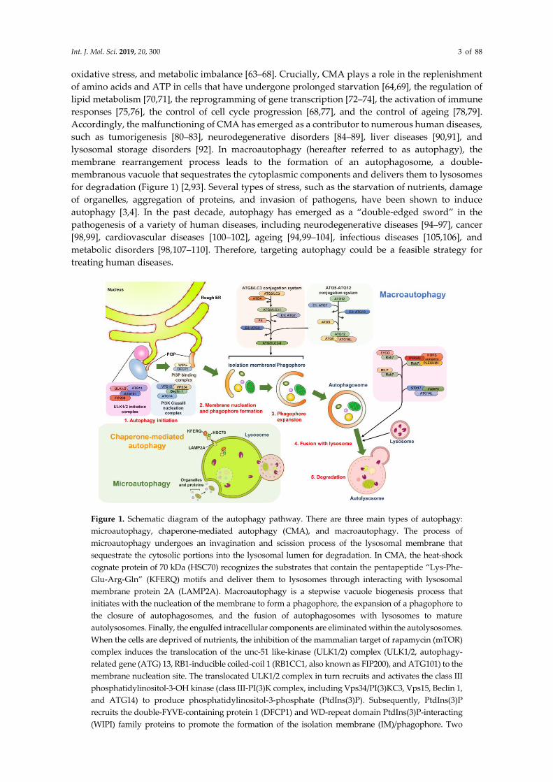
Figure 1. Schematic diagram of the autophagy pathway. There are three main types of autophagy:
microautophagy, chaperone-mediated autophagy (CMA), and macroautophagy. The process of
microautophagy undergoes an invagination and scission process of the lysosomal membrane that
sequestrate the cytosolic portions into the lysosomal lumen for degradation. In CMA, the heat-shock
cognate protein of 70 kDa (HSC70) recognizes the substrates that contain the pentapeptide “Lys-Phe-
Glu-Arg-Gln” (KFERQ) motifs and deliver them to lysosomes through interacting with lysosomal
membrane protein 2A (LAMP2A). Macroautophagy is a stepwise vacuole biogenesis process that
initiates with the nucleation of the membrane to form a phagophore, the expansion of a phagophore to
the closure of autophagosomes, and the fusion of autophagosomes with lysosomes to mature
autolysosomes. Finally, the engulfed intracellular components are eliminated within the autolysosomes.
When the cells are deprived of nutrients, the inhibition of the mammalian target of rapamycin (mTOR)
complex induces the translocation of the unc-51 like-kinase (ULK1/2) complex (ULK1/2, autophagy-
related gene (ATG) 13, RB1-inducible coiled-coil 1 (RB1CC1, also known as FIP200), and ATG101) to the
membrane nucleation site. The translocated ULK1/2 complex in turn recruits and activates the class III
phosphatidylinositol-3-OH kinase (class III-PI(3)K complex, including Vps34/PI(3)KC3, Vps15, Beclin 1,
and ATG14) to produce phosphatidylinositol-3-phosphate (PtdIns(3)P). Subsequently, PtdIns(3)P
recruits the double-FYVE-containing protein 1 (DFCP1) and WD-repeat domain PtdIns(3)P-interacting
(WIPI) family proteins to promote the formation of the isolation membrane (IM)/phagophore. Two

Int. J. Mol. Sci. 2019, 20, 300 4 of 88
ubiquitin-like (UBL) conjugation systems underlie the expansion and elongation of the phagophore to
form mature autophagosomes. The ubiquitin conjugation enzyme 1 (E1) ATG7 activates ATG12 through
a thioester bonding with ATG12, and then ATG12 is transferred to ATG10 enzyme 2 (E2). ATG12 is
finally conjugated to ATG5, forming an ATG5-ATG12 complex, which in turn interacts with ATG16L to
form an ATG12-ATG5-ATG16L complex. To successfully conjugate phosphatidylethanolamine (PE) to
ATG8/LC3 family proteins, the ATG8/LC3 family proteins are cleaved by the cysteine protease ATG4 to
expose the C-terminal glycine residues, generating the ATG8/LC3-I. Then, ATG8/LC3-I is covalently
linked to PE to form the lipidated form of LC3 (ATG8/LC3-II) via an enzyme cascade of ATG7 E1 and
ATG3 E2. The fusion of autophagosomes and lysosomes relies on the interactions between the small
GTPase Ras-related protein 7 (Rab7) and cytoskeleton-associated factors, the FYVE and coiled-coil
domain-containing 1 (FYCO1) and Rab-interacting lysosomal protein (RILP). Additionally, UV
radiation resistance-associated (UVRAG), the homotypic fusion and protein sorting (HOPS) complex,
pleckstrin homology domain-containing protein family member 1 (PLEKHM1), and protein complex
containing syntaxin 17 (STX17), vesicle-associated protein 8 (VAMP8), and synaptosome-associated
protein 29 (SNAP29) are involved in the maturation process of autolysosomes.
2.2. Stepwise Process of Vacuole Biogenesis for Autophagy
Autophagy undergoes a stepwise process for vacuole biogenesis that involves the initial
nucleation and elongation of the isolation membrane (IM)/phagophore, the closure of autophagosomes,
and the fusion of autophagosomes with lysosomes to form autolysosomes (Figure 1) [111–113].
Numerous organelles [112–114], including the ER [115,116], Golgi apparatus [117], mitochondria [118],
recycling endosome [119,120], plasma membrane [121], and mitochondria-associated ER membrane
(MAM) [122] support the membrane source for the emergence of the IM/phagophore. At the initial
stage, the IM/phagophore that originates from a particular membrane structure, which is often derived
from the ER, expands to form a double-membraned and enclosed autophagosome (Figure 1) [114,123–
125]. Subsequently, mature autophagosomes fuse with the lysosome to generate autolysosomes in
which the interior materials are degraded by lysosomal proteases (Figure 1) [124,126–128].
Most ATGs (also known as core ATGs) and the signaling molecules and vesicle-trafficking
factors involved in other cellular pathways are coordinately required for the completion of the entire
autophagic process (Figure 1) [93,129,130]. The starvation of nutrients in cells leads to the suppression
of the mammalian target of rapamycin (mTOR), a serine/threonine protein kinase required for
controlling cell growth [131,132]. The repression of mTOR results in the translocation of the unc-51
like-kinase (ULK) complex (contains ULK1/2, ATG13, RB1-inducible coiled-coil 1 (RB1CC1, also
known as FIP200) and ATG101( from the cytosol to a certain domain reconstituted from the ER
(Figure 1) [133,134]. This translocation of the ULK complex in turn recruits the class III
phosphatidylinositol-3-OH kinase (PI(3)K) complex (class III-PI(3)K, including Vps34/PI(3)KC3,
Vps15, Beclin 1, and ATG14) to the ER membrane-derived domain (Figure 1) [130,135,136] and
enhances the activity of the PI(3)K complex through the phosphorylation of Vps34/PI(3)KC3 [137].
PI(3)K in turn produces phosphatidylinositol-3-phosphate (PtdIns(3)P), leading to the recruitment of
double-FYVE-containing protein 1 (DFCP1) and WD-repeat domain PtdIns(3)P-interacting (WIPI,
the mammalian orthologue of ATG18) family proteins to promote the organization of an ER-
associated omegasome structure (also termed IM/phagophore) (Figure 1) [130,135,136,138,139].
Moreover, two multi-spanning membrane proteins, namely ATG9a and vacuole membrane protein
1 (VMP1), are critical to the initial biogenesis of autophagosomes. The ATG9a-enriched vesicles that
are trafficked from the trans-Golgi network (TGN) to the ER deliver the lipid bilayers required for
autophagosome formation [140–142]. ER-associated VMP1 interacts with Beclin 1 of the PI(3)K
complex, thereby facilitating the generation of PtdIns(3)P required for the assembly of
IM/phagophore [143–145]. The subsequent expansion and enclosure of the IM/phagophore into a
mature autophagosome requires two ubiquitin-like (UBL) conjugation systems (Figure 1) [146–149].
The ATG5-ATG12 conjugate is formed by the ATG7 (E1) and ATG10 (E2) enzymatic cascade (Figure
1). This conjugate then forms a trimeric complex with ATG16L (an ATG12-ATG5-ATG16L complex)
[146,147,150–152]. The other conjugation is that of the phosphatidylethanolamine (PE)-conjugated
ATG8 family proteins (including the microtubule-associated protein 1 light chain 3 (LC3) and

Int. J. Mol. Sci. 2019, 20, 300 5 of 88
gamma-aminobutyric acid receptor-associated protein (GABARAP) subfamilies). After protein
translation, the C-terminal region of ATG8/LC3 family proteins are immediately processed by ATG4
family proteases to form ATG8/LC3-I. Then, ATG7 enzyme 1 (E1) and ATG3 enzyme 2 (E2) confer
the conjugation of the ATG8/LC3-I to generate PE-ATG8/LC3, sometimes called ATG8-LC3-II (also
known as lipidated ATG8-LC3) (Figure 1) [153–155]. PE-ATG8/LC3 participates in the elongation of
the autophagosomal membrane [156] and the tethering and membrane fusion of autophagic vacuoles
[149]. Notably, ATG5-ATG12 may act as an E3-like enzyme to promote the lipidation of ATG8/LC3
[157,158], thereby promoting the formation of autophagosomes. Additionally, the sphingolipid
microdomains, so-called lipid rafts, were indicated to play roles in the morphogenesis of autophagic
vacuoles [159]. The fluorescence resonance energy transfer (FRET) and co-immunoprecipitation (co-
IP) studies showed that ganglioside GD3, a paradigmatic raft constituent, interacts with PI3P and
LC3-II on the immature autophagosomal membrane [159]. Also, the interactions between GD3 and
WIPI/ATG18 family proteins as well as autophagy and Beclin 1 regulator 1 (AMBRA1) were shown
in MAM raft-like microdomains [160]. Downregulation of GD3 level by gene knockdown of
ST8SIA1/GD3 synthase and alteration of sphingolipid metabolism by fumonisin B1 was
demonstrated to inhibit autophagic process [159] and interfere with the interaction of AMBRA1 with
calnexin at MAM [160], implying that MAM-associated lipid rafts function in the biogenesis of
autophagosomes.
The mature autophagosome fuses with a lysosome, forming an autolysosome in which acidic
proteases degrade the sequestrated materials to recycle their nutrients. The autophagosome–
lysosome fusion process relies on the multilayered actions of protein–protein interactions,
microtubule-mediated transport, and membrane fusion events [123,125,126,128]. The actions of the
microtubule ensures the precise transport of the autophagosome to the lysosome for fusion
[128,161,162]. The small GTPase Ras-related protein 7 (Rab7) located on the autophagosomal
membrane interacts with FYVE and coiled-coil domain-containing 1 (FYCO1) and Rab-interacting
lysosomal protein (RILP), two effectors that are respectively linked to kinesin and dynactin in
microtubules [163–167], enabling the movements of the autophagosomes on microtubules (Figure 1).
Apart from microtubules, the histone deacetylase 6 (HDAC6)-induced remodeling of F-actin and the
formation of the F-actin network also promote autophagosome–lysosome fusion in the quality
control autophagy-mediated removal of aggregated proteins rather than starvation-induced
autophagy [168]. In addition to bridging the transport of autophagosomes on microtubules, Rab7
located on late endosomes and lysosomes stimulates autophagosome–lysosome fusion through
recruiting several effectors of this action, including the pleckstrin homology domain-containing
protein family member 1 (PLEKHM1) and the homotypic fusion and protein sorting (HOPS) complex
(Figure 1) [153,154]. PLEKHM1 contains an LC3-interacting motif that can bind to ATG8 family
proteins located on the autophagosomal membrane and concomitantly interacts with Rab7 as well as
the HOPS complex, thereby facilitating the fusion of autophagosomes and lysosomes (Figure 1) [169].
Additionally, the PI(3)K complex-associated UV radiation resistance-associated gene (UVRAG) binds
to the HOPS complex via Vps16 to induce Rab7 GTPas activity and trigger autophagosome–lysosome
fusion [136,170,171]. Notably, the binding of Rubicon to the PI(3)K protein complex reciprocally
interferes with the fusion of autophagosomes and lysosomes [136]. Another protein complex,
containing ATG14L, syntaxin 17 (STX17), synaptosome-associated protein 29 (SNAP29), and vesicle-
associated membrane protein 8 (VAMP8), also stimulates autophagosome–lysosome fusion,
presumably through the membrane tethering and fusion process (Figure 1) [172,173]. Recently, ATG8
family proteins were shown to be mainly active during autophagosome–lysosome fusion rather than
autophagosome biogenesis at the initial stage of autophagy by recruiting PLEKHM1 in PTEN-
induced putative kinase 1 (PINK1)/Parkinson’s disease protein (Parkin)-mediated autophagic
clearance of mitochondria (so called mitophagy) and starvation autophagy [174]. In addition to acting
at the biogenesis of autophagosomes [159], lipid rafts have emerged as a regulator of autolysosome
maturation [159,160]. The interaction between GD3 and lysosome-associated membrane protein-1
(LAMP1) in the autolysosomal membrane as demonstrated by FRET, co-IP, and TEM assays
Int. J. Mol. Sci. 2019, 20, 300 6 of 88
indicated that GD3-enriched lipid rafts could induce membrane remodeling to promote the
morphogenesis of autolysosomes and increase autophagic flux [160].
After degradation within autolysosomes, the nutrient-fed-reactivation of mTOR suppresses
autophagy initiation and concomitantly initiates autophagic lysosome reformation (ALR), thereby
terminating autophagy [175]. Related studies have implied that spinster (spin), a lysosomal efflux
permease, is required for ALR formation [176]. Recently, the Cullin 3-Kelch-like protein 20 (KLHL20)
ubiquitin ligase was also shown to participate in autophagy termination by promoting the turnover
of the ULK1 and Vps34 complexes [177]. Nevertheless, the detailed molecular mechanism underlying
the biogenesis of autophagic vacuoles within the entire autophagy process is not comprehensively
understood and further investigations are required.
2.3. Selective Autophagy and Cargo Recognition
Autophagy has been considered to be a bulky and nonselective degradative process; however,
a growing body of literature has indicated that autophagy may selectively sequestrate specific cargos,
including organelles and proteins, to induce degradation. This is termed “selective autophagy” [178–
180]. The concept of selective autophagy was first described in 1973, in a study that showed that a
diabetogenic dose of alloxan or streptozotocin induces selective autophagy to degrade β-granules in
intermediate cells in the pancreas of rats [181]. At the initial stage of selective autophagy, the specific
cargo receptors recognize the degradative cargos that are tagged through polyubiquitination or
additional adaptor proteins and then deliver them into the autophagosome through the interaction
of cargo receptors with ATG8 family proteins located on the autophagosomal membrane [182–185].
Numerous cargo receptors of selective autophagy have been identified and characterized, including
the neighbor of BRCA1 (NBR1), calcium-binding and coiled-coil domain-containing protein 2
(Calcoco2, also known as NDP52), p62/sequestosome 1 (SQSTM1), and optineurin (OPTN), all of
which contain LC3-interacting regions (LIRs) to bind ATG8 family proteins, thus engulfing the cargos
into autophagosomes (Figure 2) [184–186]. In addition to eliminating degradative substrates through
LIR-containing cargo receptors, the potential ATG8-interacting motifs (AIMs) and GABARAP-
interacting motifs (GIMs) have been recently found to regulate selective autophagy within ATGs and
other cellular proteins [187–190]. For example, the Saccharomyces cerevisiae ATG19 was shown to
directly interact with ATG5 through AIMs, and that interaction recruits the ATG5-ATG12-ATG16L
trimeric complex, thus enhancing the lipidation of ATG8/LC3 to promote the local biogenesis of
autophagosomes to sequestrate the cargos [191].
Regarding the maintenance of the organelle integrity in eukaryotic cells, selective autophagy
plays a homeostatic role in the selective elimination of damaged organelles, termed organellophagy
[178,180,192], which provides the recycled nutrients for the regeneration of mitochondria,
peroxisomes, the ER, lipid droplets (LDs), ribosomes, lysosomes, and nuclei (Figure 2). Numerous
stimuli, such as hypoxia [193,194], the accumulation of reactive oxygen species (ROS) [195–197], and
mitochondrial depolarization [198–200], can result in the fission, depolarization, and damage of
mitochondria. Mitochondrial damage triggers selective autophagy to degrade the harmful
mitochondria in a process known as mitophagy [201,202]. Mitophagy is often initiated without
adequate cleavage of PINK1 by presenilin-associated rhomboid-like protein (PARL) within the inner
mitochondrial membrane of damaged mitochondria, thus suppressing the degradation of PINK1
[203,204]. This outcome in turn leads to the accumulation of PINK1 on the outer mitochondrial
membrane, thereby phosphorylating ubiquitin at serine 65 and then recruiting the ubiquitin enzyme
3 (E3) ligase Parkin [198–200,205–207]. Subsequently, Parkin ubiquitinates the mitochondrial proteins
onto the outer mitochondrial membrane [198–200,205,208], thus recruiting specific cargo receptors,
such as Calcoco2/NDP52 and OPTN, for the removal of mitochondria through autophagy (Figure 2)
[202,209]. The translocation of these cargo receptors also induces the local concentration of
phagophore-organization effectors, including DFCP1 and WIPI/ATG18 family proteins, for
autophagosome maturation proximal to the damaged mitochondria [209]. Additionally, TANK
binding kinase 1 (TBK1) participates in the cargo recognition process of mitophagy by
phosphorylating p62/SQSTM1 at serine residue 403 and OPTN at serine residues 177, 473, and 513

Int. J. Mol. Sci. 2019, 20, 300 7 of 88
[210–212]. Despite the PINK1/Parkin-induced ubiquitination of damaged mitochondria, several outer
mitochondrial membrane proteins, including FUN14 domain-containing 1 (FUNDC1),
BCL2/adenovirus E1B 19 kDa protein-interacting protein 3 (BNIP3), BCL2/adenovirus E1B 19 kDa
protein-interacting protein 3-like (BNIP3L), and yeast ATG32, also activate mitophagy in a ubiquitin-
independent manner (Figure 2) [213–217]. Recently, numerous studies have identified novel cargo
receptors for mitophagy, such as prohibitin 2 (PHB2) and Toll-interacting protein (Tollip) (Figure 2)
[218,219]. In contrast, the deubiquitination (DUB) of mitochondrial proteins onto the outer membrane
of mitochondria by DUB enzymes USP30 and USP35 antagonizes mitophagy [220,221].
Figure 2. Different modes of selective autophagy. Degradation of selective autophagy involves the
specific interactions between the ATG8/LC3-interacting regions (LIRs) within cargo receptors and
ATG8/LC3 located onto autophagosomal membrane. The ubiquitination of degradative cargos or
associated ligand proteins is often required for recognition by cargo receptors. Selective autophagy
has been shown to eliminate different kinds of organelles and proteins, including damaged
mitochondria (mitophagy), injured lysosomes (lysophagy), damaged peroxisomes (pexophagy),
stressed ER (ER-phagy), and infectious pathogens (xenophagy), through specific cargo receptors in
yeast and mammalian cells as indicated. Also, lipid droplets (LDs) ferritin, nuclei, ribosomes, and
protein aggregates are also degraded by selective autophagy via the identified cargo receptors and
other unknown adaptor proteins.
Selective autophagy promotes the turnover of other intracellular organelles. The specific cargo
receptors that confer the elimination of these organelles are also identified and characterized. To
degrade oxidized and damaged peroxisomes through pexophagy, yeast ATG36 and mammalian
NBR1 and p62/SQSTM1 are required to target the degradative peroxisomes to autophagosomes
(Figure 2) [222–225]. Numerous kinases, such as yeast Hrr25 and mammalian ataxia-telangiectasia-
mutated (ATM), induce the phosphorylation of these two cargo receptors, thus promoting the
delivery of peroxisomes to the autophagosomal membrane [226,227]. The polyubiquitination of
several peroxisomal (PEX) membrane proteins, such as PEX5 and the 70-kDa PEX membrane protein
(PMP70), facilitates the recognition of damaged peroxisomes by cargo receptors [227,228].
The targeting of a stressed ER to degradation through ER-phagy involves the biological activities
of ATG39, ATG11, and ATG40 [229] in yeast cells; the family with sequence similarity 134, member
B (FAM134B) (Figure 2); and reticulon family proteins in mammals [230,231]. ATG39 and ATG11 also
participate in the selective degradation of yeast nuclei, termed nucleophagy (Figure 2) [229]. The
clearance of protein aggregates by selective autophagy is achieved through the p62/SQSTM1- and
HDAC6-mediated recognition of Lys63 (K63)-linked poly-ubiquitination of aggregated proteins
[168,232–234]. Moreover, NBR1 and autophagy-linked FYVE (ALFY) could cooperate with
p62/SQSTM1 to degrade protein aggregates through selective autophagy (Figure 2) [235–238]. The

Int. J. Mol. Sci. 2019, 20, 300 8 of 88
injured lysosomes have recently been reported to be removed by lysophagy, which begins with the
recruitment of galectin-3 and LC3 onto lysosomal membranes, which are subsequently recognized
by p62/SQSTM1 and delivered to the autophagosome for degradation (Figure 2) [239,240]. Similarly,
selective autophagy has emerged as playing a pivotal role in the clearance of ribosomes, termed
ribophagy (Figure 2) [241,242], and in the catabolism of LDs for maintaining metabolic homeostasis
(Figure 2) [243,244].
In addition to organellophagy, the cargo receptors of selective autophagy can eliminate specific
proteins and invading pathogens. The nuclear receptor coactivator 4 (NCOA4) has been recently
shown to interact with ATG8 family proteins and to target ferritin heavy and light chains for
autophagic degradation, thus modulating the intracellular level of iron (Figure 2) [245,246]. The
turnover of ferritin through selective autophagy, termed ferritinophagy, has been implicated in the
regulation of erythropoiesis and DNA replication in blood cells [247,248]. The elimination of
infectious pathogens by xenophagy represents the host’s first-line defense in restricting microbial
infections [249–251]. Pexophagy involves the engulfment of invading pathogens by p62/SQSTM1-,
Calcoco2/NDP52-, and OPTN-mediated recognition processes and delivery to the autophagosome
for degradation (Figure 2) [106,252,253]. The phosphorylations of p62/SQSTM1 (at serine residues 349
and 403) and OPTN (at serine 177) promote the clearance of infecting pathogens through pexophagy
[253–256]. Taken together, selective autophagy not only maintains cellular homeostasis by removing
damaged organelles but also acts as a host defensive mechanism to counteract pathogen infection.
2.4. Autophagy as an Alternative Cell-Death Pathway
Autophagy (“self-eating”) has been considered a stress-responsive, survival mechanism to protect
cells against apoptosis (“self-killing”, type I cell death) [257–259]. Autophagy is often activated by the
inhibition of apoptosis. For instance, simultaneous gene knockout of BAX and BAK, two BCL2 family
proteins involved in cell apoptosis in mice was shown to activate autophagy to counteract etoposide
(an inhibitor of topoisomerase-2)-induced cell death [260]. Reciprocally, apoptosis can be activated by
inhibiting autophagy. Interference with autophagy by gene silencing and pharmacological inhibitors in
nutrient-starved cells was shown to trigger cell apoptosis [261]. The specific gene knockout of ATG5 in
neuron cells and T cells in mice was demonstrated to increase apoptotic cell death [262,263]. However,
autophagy confers an alternative route to promote cell death, known as type II cell death under some
specific cellular conditions [264,265]. For instance, human immunodeficiency virus (HIV) infection
leads to autophagy activation to trigger apoptotic cell death of CD4/CXCR4-expressing T cells [266].
The inhibition of HIV Env-induced autophagy by gene knockdown and pharmacological inhibitors was
demonstrated to interfere with cell apoptotic death [266]. In spite of apoptosis, autophagy was also
indicated to promote necrotic cell death [267]. Autophagy was demonstrated to be activated by caspase
inhibition to promote cell death through the accumulation of ROS and degradation of catalases [267].
This caspase inhibition-induced cell death could be reversed by interference with autophagy by siRNAs
against ATGs and autophagy inhibitors [267]. Notably, the enhancement of cellular autophagy by the
Tat-Beclin1 peptide was specifically demonstrated to trigger the “autosis” cell death pathway, which is
mediated by the Na+, K+-ATPase pump and is characterized by the convolution of nuceli at the early-
stage and focal swelling of the perinuclear space at the late-stage [268,269]. Besides the autophagy-
inducing Tat-Beclin1 peptide, starvation and in vivo cerebral hypoxia-ischemia were also shown to
induce autotic cell death [268,269]. These studies together indicate that autophagy not only adapts to
stresses to avoid cell death but also induces diverse types of cell death pathways to kill cells when cells
no longer circumvent certain stimuli.
3. Regulation and Functional Roles of Autophagy in Liver Physiology
3.1. The Leading Discovery of Autophagy in Liver Tissue
Hepatocytes in liver tissue were initially revealed to contain autophagic vacuoles. In the early
1960s, Ashford et al. first demonstrated that glucagon perfusion in rats can induce the formation of
polymorphic dense bodies in liver cells (Table 1) [14]. These dense bodies were shown to sequestrate

Int. J. Mol. Sci. 2019, 20, 300 9 of 88
the fragmented and morphologically abnormal mitochondria, which were associated with autolysis
triggered by the glucagon-related protein catabolic process (Table 1) [14]. Similarly, the treatment of
rat livers with the detergent Triton also led to the formation of dense bodies (known as cytolysomes)
that exhibit two patterns: one consists of double-membraned vacuoles containing mitochondria and
ER membrane fragments and the other consists of single-membrane vesicles in which the engulfed
materials are degraded (Table 1) [15]. Soon thereafter, glucagon was revealed to be as an activator of
autophagy in liver cells (Table 1) [19,20,270], which were standardized for monitoring autophagy. De
Duve and Deter first observed that glucagon administration triggers an increase in lysosomal size,
which could be related to the formation of autophagic vacuoles in the rat livers (Table 1) [19]. After
the biochemical fractionation of lysosomes, their study further revealed that glucagon induction
upregulates acidic phosphatase as well as cathepsin D in lysosomes and also increases the fragility
of lysosomes in rat liver (Table 1) [19]. In a subsequent study that combined biochemical fractionation
and TEM, Deter and colleagues revealed by a morphological quantification that glucagon-induced
autophagic vacuoles represent a substantial portion of lysosomes in liver homogenates (Table 1) [20].
Their study implied that hepatic lysosomes are involved in the biogenesis of autophagic vacuoles
and thus provide the main source of acidic proteases for the degradation of sequestrated interior
materials (Table 1) [20]. Moreover, two types of glucagon-triggered autophagic vacuoles in the liver
were further specified: type I vacuoles are predominantly double-membraned vacuoles that contain
the ER, ribosomes, and ground cytoplasm and type II vacuoles are larger than type I vacuoles and
are composed of a single limiting membrane, in which the sequestrated ER and cytoplasm are broken
down (Table 1) [270]. Taken together, these studies not only indicate that hepatic autophagy may
present a novel degradative process that eliminates the intracellular components in the liver but also
provide evidence that lysosomes participate in autophagy to support proteolytic enzymes.
3.2. The Role of Autophagy in Balancing Metabolism and Sensing Stresses in the Liver
The regulation of autophagy in liver physiology and the modulation of autophagy by liver injury
were discovered in the early 1970s [271–274]. The study by Pfeifer first revealed the role of autophagy
in the decomposition of glycogen in liver atrophy [273]. Long-term starvation has been shown to induce
hepatic autophagy, correlating with cell atrophy in rat livers (Table 1) [274,275]. These studies suggested
that hepatic autophagy may detect malnutrition in the liver as well as liver damage, instantly
supporting the refueling of nutrients through degradation. Accordingly, the formation of autophagic
vacuoles was shown to be energy-dependent and correlated with the rate of protein synthesis (Table 1)
[276,277], alteration of metabolites (Table 1) [278–280], and interference with cytoskeleton organization
(Table 1) [281]. In the late 1970s, stress and amino acid deprivation were demonstrated to trigger
autophagy in hepatic cells (Table 1) [21,25,280]. This autophagic proteolytic effect induced by the
deprivation of nutrients in hepatocytes can be inhibited by the refeeding of nutrients and autophagy
inhibitors (Table 1) [24,30,282], indicating that the status of nutrient supplies plays a detrimental role in
autophagy activation in the liver. These studies collectively imply that autophagy acts as a regulator
that senses changes in the metabolism and alterations of energy in the liver.
The induction of hepatocellular necrosis by dimethylnitrosamine (DMNA) can increase the
number and size of autophagic vacuoles in the period beyond the onset of cell necrosis (Table 1)
[271,272], suggesting that autophagy might be activated to counteract cell death in the liver. At the
same time, numerous studies have shown that the smooth membrane of the ER can contribute to the
membranous structure that supports autophagosome biogenesis in liver cells [283–287], leading to a
new paradigm for understanding the membrane resource for developing autophagosomal
membranes. Collectively, these studies indicate that hepatic autophagy could be activated by
numerous stimuli, such as nutrient starvation, metabolism imbalance, and liver injury, to promote
the maintenance of metabolic homeostasis.

Int. J. Mol. Sci. 2019, 20, 300 10 of 88
Table 1. Summary of autophagy in liver physiology.
Experimental Model Characteristics of Autophagy Function of Autophagy References
Rat liver
(Perfusion of glucagon)
Electron micrograph of polymorphic dense
bodies
Sequestration of deformed mitochondria that is
associated with glucagon-related catabolic process [14]
Rat liver
(Intravenous injection of Triton
WR-1339)
Electron micrograph of polymorphic dense
bodies that includes single- and double-
membraned vesicles (termed cytolysomes)
Degradation and degeneration of mitochondria in
Triton-treated hepatic cells [15]
Rat liver
(Intravenous injection of
glucagon)
Biochemical fractionation of lysosomes and
autophagic vacuoles
Association with lysosomes and enrichment of
lysosomal acidic enzymes within autophagic vacuoles
for protein degradation in the glucagon-stimulated
liver cells
[19]
Rat liver
(Intravenous injection of
glucagon)
1. Biochemical fractionation of lysosomes and
autophagic vacuoles
2. Electron micrograph of autophagic vacuoles
that engulf organelles
1. Increased number of autophagic vacuoles by
glucagon stimulation
2. Increased lysosomal proteases in glucagon-treated
hepatic cells
3. Degradation of intracellular organelles in hepatic
cells treated by glucagon
[20]
Rat liver
(Intravenous injection of
glucagon)
1. Biochemical fractionation of lysosomes and
autophagic vacuoles
2. Electron micrograph of single- and double-
membranous autophagic vacuoles that
sequestrate organelles
1. Increased formation of autophagic vacuoles by
glucagon-treated liver cells
2. Increased lysosomal proteases in autophagic
vacuoles in hepatic cells treated by glucagon
3. Degradation of intracellular organelles in glucagon-
treated liver cells
[270]
Rat liver
(Intraperitoneal injection of
dimethylnitrosamine (DMNA))
1. Electron micrograph of autophagic vacuoles
2. Increased autophagic vacuoles when the
onset of necrosis is detected
1. Degradation of organelles by autophagic vacuoles in
DMNA-treated liver cells
2. Associated with the occurrence of hepatocellular
necrosis
[272]
Rat liver
(Long-term starvation)
Electron micrograph of autophagic vacuoles
that engulf organelles
1. Degradation of organelles by autophagic vacuoles in
hepatocytes of liver atrophy induced by starvation
2. Decomposition of glycogen in the starved liver cells
of liver atrophy
[273–275]
Rat liver Electron micrograph of autophagic vacuoles The correlation of autophagic vacuoles formation with
the rate of protein synthesis and the level of energy [276–278]

Int. J. Mol. Sci. 2019, 20, 300 11 of 88
(Intraperitoneal injection of
glucagon and cycloheximide)
Mouse liver
(Intravenous injection of lysine
acetylsalicylate
Electron micrograph of single- and multiple-
membranous autophagic vacuoles
The engulfment of intracellular components within
autophagic vacuoles that may protect the lysine
acetylsalicylate-treated liver cells against injury
[279]
Rat liver
(Hypothermia)
Electron micrograph of autophagic vacuoles
that sequestrate enlarged mitochondria and
disorganized endoplasmic reticulum (ER)
The elimination of intracellular organelles by
hypothermia-induced autophagic vacuoles in
hepatocytes
[280]
Rat liver
(Intraperitoneal injection of
vinblastine)
Electron micrograph of autophagic vacuoles
that engulf organelles
Degradation of intracellular organelles within
autophagic vacuoles [281]
Rat liver
(Perfusion of amino acids-
deprived medium)
Electron micrograph of autophagic vacuoles
that engulf organelles
Maintenance of intracellular amino acids in
hepatocytes and recycling of nutrients [21]
Rat liver
(Perfusion of amino acids-
deprived medium)
Electron micrograph of autophagic vacuoles
that engulf organelles
Decreased the intracellular amounts of glucogenic
amino acids by autophagy [25]
1. Rat isolated hepatocytes
2. Mouse liver
(Starvation and refeeding)
1. Electron micrograph of autophagic vacuoles
that engulf organelles
2. Protein degradation of endogenous proteins
Degradation of intracellular organelles and
endogenous proteins, which is inhibited by methylated
adenosine derivatives and refeeding
[24,30,282]
Rat liver
1. Electron micrograph of autophagic vacuoles
that engulf organelles
2. Isolation of autophagic vacuoles
3. Inhibited formation of autophagic vacuoles
by insulin
Degradation of intracellular organelles and
endogenous proteins, which is inhibited by insulin, 3-
methyladenine (3-MA), vinblastine, and amino acids
[22,23,287–
294]
Newborn rat hepatocytes
Electron micrograph of autophagic vacuoles
that is closely related to the degradation of
fetal-type glycogen
Degradation of fetal-type glycogen in the neonatal
period [295]
1. Rat liver
(Perfusion of orotic acid)
2. Isolated rat hepatocytes
Autophagic degradation of RNA and proteins in liver,
which is inhibited by chloroquine and amino acids [296]
Rat liver
Electron micrograph and biochemical
fractionation of lysosomes and autophagic
vacuoles
Degradation of proteasomes by lysosomes and
autophagic vacuoles [297]

Int. J. Mol. Sci. 2019, 20, 300 12 of 88
Rat liver Restriction of ischemic liver injury by inhibition of
autophagy [298]
1. Rat liver
2. Isolated rat hepatocytes
Enhancement of cell survival of carcinogen-treated
hepatocytes by reduced autophagy [299]
Primary rat hepatocytes Electron micrograph of autophagic vacuoles Increased the intracellular iron pool by autophagic
turnover of ferritin and iron-containing proteins [300]
Liver specimens of patients Electron micrograph of autophagic vacuoles Promotion of cell death in anorexia nervosa livers of
patients by starvation-induced autophagy [301]
Rat hepatoma H4IIE cells
Support of amino acids from autophagic proteolysis of
endogenous proteins to the regulation of translational
effectors
[302]
Wild type and liver-specific
knockout of ATG7 mice
1. Electron micrograph of autophagic vacuoles
2. Detection of lipidation of ATG8/LC3
Maintenance of blood glucose and amino acids levels
by hepatic autophagy [303]
Primary human and mouse
hepatic stellate cells
1. Analysis of autophagic flux by the
fluorescent signal of mRFP-GFP-LC3
2. Detection of changes in autophagic flux by
interference with autolysosome maturation
Involvement of enhanced autophagic flux in the
activation of hepatic stellate cells [304]
Rat liver Degradation of cathepsin family enzymes (B,
H, and L)
Reduced lysosomal proteolysis by suppression of
autophagy in regenerating liver [305]
Rat liver 1. Immunofluorescence analysis of ATG8/LC3
2. Electron micrograph of autophagic vacuoles
Involvement of autophagy in the degeneration of
hepatocytes of liver grafts [306]
Wild type and liver-specific Tet-
off-LAMP2A transgenic mice
Electron micrograph of CMA-mediated
autophagic process
The maintenance of liver function and protection
against liver damage by hepatic CMA [79]
Mouse hepatocytes
(In vivo and in vitro ischemia
and reperfusion)
1. Detection of the lipidation of ATG8/LC3
2. Analysis of autophagic flux by fluorescence
signal of AdmCherry-GFP-LC3
3. Detection of the processing and expression
of cathepsin D
Amelioration of liver damage and restoration of
mitochondrial function in liver after ischemia and
reperfusion
[307]
1. Human hepatoma, Huh7 cells
2. Wild type and keratin 8-
overexpressing mice
1. Detection of the lipidation of ATG8/LC3
2. Colocalization of p62/SQSTM1 with keratin
8 and GFP-LC3 with HSP70
3. Electron micrograph of autophagic vacuoles
Elimination of components of MDBs by rapamycin-
induced autophagy [308,309]
1. Wild type and liver-specific
knockout of ATG7 mice
1. Electron micrograph of autophagic vacuoles
that engulf LDs
1. Involvement of ATG8/LC3 conjugation system in the
biogenesis of LDs
[243,244,310
,311]

Int. J. Mol. Sci. 2019, 20, 300 13 of 88
2. Rat hepatocyte RALA255-10G
3. Primary rat hepatocytes
4. Human hepatoma, Huh7 and
HepG2 cells
2. Detection of the lipidation of ATG8/LC3
3. Colocalization of ATG8/LC3 with LDs-
associated proteins
4. Biochemical fractionation of autophagic
vacuoles that contain LDs-related proteins
2. Promotion of LDs catabolism by hepatic autophagy,
i.e., “lipophagy”
3. Degradation of lipid by hepatic autophagy
4. Degradation of Apolipoprotein B located around
LDs
1. Wild type and liver specific
knockout of ATG7, VHL, HIF-
1α, and HIF-2α mice
2. Rat liver
(Antilipolytic agents treatment)
3. Isolated rat hepatocytes
1. Detection of GFP-LC3-labeled autophagic
vacuoles that sequestrate peroxisomes
2. Electron micrograph of autophagic vacuoles
that engulf peroxisomes
1. Selective engulfment of excess peroxisomes by
autophagy (termed “pexophagy”)
2. Degradation of peroxisomes by HIF-2α-mediated
selective autophagy
3. Decreased peroxisomal enzyme activities by
enhanced degradation of peroxisomes by autophagy,
which is inhibited by 3-MA
[312–318]
1. Primary rat hepatocytes
2. Isolated hepatocytes from
wild type and GFP-LC3-
transgenic mice
3. Wild type and Vps34/PI-
3KC3D761A mice
4. Rat liver
1. Electron micrograph of autophagic vacuoles
that engulf mitochondria
2. Translocation of mitochondria into acidic
organelles
3. Engulfment of mitochondria by GFP-LC3-
labeled autophagic vacuoles in the nutrient
starved- and glucagon-treated hepatocytes
4. Mildly inhibited hepatic autophagy by
inactivation of Vps34/PI-3KC3
1. The remodeling of mitochondria by mitochondrial
autophagy (termed “mitophagy”)
2. The degradation of mitochondria by hepatic
mitophagy
3. Restricted mitochondrial respiration and
gluconeogenesis by inactive Vps34/PI-3KC3-interfered
mitophagy
4. Accumulated injured mitochondria by age-related
failure of mitophagy
[319–323]
1. Mouse hepatoma, Hepa1-6
cells
2. Wild type,
ATG5−/−, and ATG7−/− mouse
embryonic cells (MEF)
3. Ob/ob mice and high fat diet
(HFD)-induced mice
4. Wild type and liver-specific
knockout of ATG7 mice
1. Downregulation of hepatic autophagy by
obesity in ob/ob mice and HFD-mice
2. Electron micrograph of autophagic vacuoles
that contains ER
1. Induced insulin resistance and ER stress by obesity-
inhibited hepatic autophagy
2. Selective modulation of UPR by hepatic autophagy
[321,324]

Int. J. Mol. Sci. 2019, 20, 300 14 of 88
3.3. Turnover of Macromolecules through Autophagy in the Liver
In the late 1970s, autophagy was first shown to degrade glycogen and to participate in the selective
elimination of organelles in the liver (Table 1) [23]. In line with this study, biochemical and morphological
studies have, together, demonstrated that hepatic autophagy plays a major role in protein degradation and
the degeneration of organelles through the formation of autolysosomes (Table 1) [23,25,287–294]. The
functional roles of autophagic degradation in the liver were implicated in the turnover track of intracellular
macromolecules, such as the degradation of fetal-type glycogen in the neonatal period (Table 1) [295], the
destruction of damaged organelles by virus infection (Table 1) [325], the selective degradation of RNA and
proteins through the deprivation of amino acids (Table 1) [296], and the elimination of the ubiquitin–
proteasomal pathway through long-term starvation (Table 1) [297]. Additionally, hepatic autophagy is
involved in multiple cell surveillance mechanisms, including the regulation of ischemic liver injury (Table
1) [298], the growth suppression of carcinogen-treated hepatocytes (Table 1) [299], the modulation of the
iron pool and sensitivity to oxidative stress (Table 1) [300], and the regulation of cell death in the damaged
livers of patients with anorexia nervosa (Table 1) [301]. Conversely, autophagy plays critical roles in the
integration of metabolic pathways by regulating the supply of amino acids for effective translation in
hepatoma cells (Table 1) [302], the balancing of blood glucose and amino acid levels (Table 1) [303], and the
activation of hepatic stellate cells (Table 1) [304]. Moreover, autophagy participates in the regulation of
lysosomal proteolysis in liver regeneration (Table 1) [305], the degeneration of transplanted livers in rats
(Table 1) [306], the maintenance of hepatic function in the aged liver (Table 1) [79], and the suppression of
age-dependent ischemia in injured livers (Table 1) [307]. Taken together, these results indicate that
autophagy acts as a protector in physiologically balancing liver metabolism and maintaining liver function
and growth.
3.4. Selective Degradation of Organelles through Autophagy in the Liver
In the past few decades, numerous studies have indicated that autophagy participates in the catabolism
of intracellular compartments in the liver, including Mallory–Denk bodies (MDBs) [308,309,326], LDs
[70,71,243,244,310,311,327,328], peroxisomes [312–318,329,330], mitochondria [320–323], and the ER
[324,331] (Table 1). A biochemical fractionation study indicated that a considerable portion of several types
of organelles was sequestrated within autophagic vacuoles in rat hepatocytes (Table 1) [332,333], implying
the functional roles of autophagy in the elimination of intracellular organelles in the liver. MDBs are
cytosolic hyaline inclusions that were discovered in the hepatocytes of patients with alcoholic hepatitis in
1911 by Mallory [334] and further characterized in mouse livers by Denk in the late 1970s [335,336]. Several
intracellular components are enclosed in MDBs, including keratins, chaperones, protein degradation
machinery that contains ubiquitin and p62/SQSTM1, and phosphoproteins [337]. MDBs have been observed
in various liver diseases, such as alcoholic steatohepatitis, nonalcoholic steatohepatitis (NASH),
nonalcoholic fatty liver disease (NAFLD), and hepatocellular carcinoma (HCC) [337–339]. Harada et al. first
demonstrated that rapamycin-induced autophagy may mediate the turnover of bortezomib-induced MDBs
in in vitro cell cultures and in vivo mouse models [308,309], supporting autophagy′s role in the clearance of
cytoplasmic inclusions.
LDs are the primary organelles that store neutral lipids, including cholesterol ester and triglycerides
(TG), and serve as a reservoir for energy, particularly for the liver [340–342]. The aberrant accumulation of
lipids in LDs has been evinced in numerous metabolic disorders in the liver, such as hepatic steatosis,
NASH, and NAFLD, leading to global health burdens in modern society (Table 1) [328,340,342,343]. The
role of autophagy in LD dynamics was originally defined in the analysis by Fujimoto et al. of apolipoprotein
B (ApoB) degradation (Table 1) [311]. By combining biochemical fractionation and microscope-based
approaches, the authors posited that autophagy may promote the degradation of ApoB, which specifically
occurs around the surface of LDs in hepatocytes (Table 1) [311]. Subsequently, Singh et al. showed that
interference with autophagy by the knockdown of the ATG5 gene expression increased TG accumulation
and inhibited the β-oxidation of free fatty acids (FFAs) and degradation of TG in hepatocytes (Table 1) [244].
Their TEM-based ultrastructural study further indicated that LDs are delivered into autophagic vacuoles
Int. J. Mol. Sci. 2019, 20, 300 15 of 88
for degradation, which is enhanced by nutrient starvation (Table 1) [244]. Their study first uncovered the
role of autophagy in the catabolism of LDs, (e.g., “lipophagy”). Another study further confirmed that
starvation upregulated lysosomal lipase activity in the autophagic fraction of the liver to promote lipid
degradation (Table 1) [310]. In contrast, ATG7 deficiency in mouse hepatocytes was shown to impede the
formation of LDs (Table 1) [243]. The specific localization of lipidated-LC3 onto the surface of LDs in starved
mouse hepatocytes suggested that the ATG8/LC3-lipidation process might be involved in the biogenesis of
hepatic LDs (Table 1) [243]. In line with this study, another report demonstrated that mammalian ATG2
plays a crucial role in the morphogenesis and dynamics of LDs (Table 1) [344]. In addition, autophagy was
shown to inhibit ethanol-induced steatosis in mouse livers and to protect liver cells from ethanol-triggered
hepatotoxicity (Table 1) [345]. In addition to macroautophagy, CMA was recently indicated to promote the
degradation of LD-associated proteins perilipin 2 (PLIN2) and perilipin 3 (PLIN3) to control LDs biogenesis
[70]. Moreover, the 5′-AMP-activated protein kinase (AMPK)-induced phosphorylation of PLIN2 was
shown to promote its interaction with HSC70, a chaperone of CMA, and thus facilitate the degradation of
PLIN2, thereby recruiting lysosomes and cytosolic lipases to catabolize LDs [71]. More importantly, this
specific form of LD degradation through autophagy, the so-called lipophagy, was reported to participate in
thyroid hormone-induced LD catabolism (Table 1) [346,347].
The sequestration of peroxisomes within autophagic vacuoles was initially observed in a study
showing that antilipolytic agent-treated rat livers that contained enhanced autophagic vacuoles engulfed
peroxisomes and downregulated the activities of peroxisomal enzymes (Table 1) [312,315], suggesting that
autophagy participates in peroxisome degradation. Analogously, autophagic vacuoles have also been
reported to sequestrate peroxisomes in the hepatocytes of patients with chronic hepatitis B virus (HBV) who
received transplantation and immunosuppressive therapy (Table 1) [329]. Subsequently, amino acid
deprivation-induced autophagy was suggested to selectively degrade peroxisomes in hepatocytes isolated
from clofibrate-treated rats, and this degradation was completely inhibited by the administration of 3-MA
(Table 1) [316]. Furthermore, several other studies have demonstrated that autophagy is involved in the
elimination of excess peroxisomes, thus prohibiting the uncontrolled proliferation of peroxisomes in the
liver [36,38,313,314,317,330]. Moreover, the selective degradation of peroxisome autophagy, termed
“pexophagy”, may regulate peroxisome proliferator-activated receptor α (PPARα) target genes and the β-
oxidation of FFAs to prevent hepatic steatosis and tumorigenesis in the liver (Table 1) [348] and acute liver
failure induced by inflammation (Table 1) [349].
Mitochondria and the ER are the intracellular organelles that were originally detected in autophagic
vacuoles in the late 1950s and early 1960s [13–16]. The concepts underpinning the degradation of
mitochondria through autophagy was derived from the observation that the rate of mitochondria removal
was selectively and positively correlated with the formation of autophagic vacuoles in rat livers [23,287].
Subsequently, studies have shown that autophagic vacuoles contain the mitochondrial enzymes of the liver
[37,38,350–352], further indicating that hepatic autophagy selectively eliminates mitochondria. The
autophagic degradation of mitochondria in hepatocytes was further demonstrated to introduce
mitochondrial injury, thus promoting the pathogenesis of alpha (1)-antitrypsin (α1-AT) deficiency-induced
liver injury that was highly associated with chronic liver diseases (Table 1) [353–355]. Komatsu et al. first
demonstrated that the genetic knockout of ATG7 in mice interfered with autophagosome biogenesis in
livers in which deformed mitochondria had accumulated (Table 1) [356], suggesting that hepatic autophagy
plays a major role in mitochondria degradation. The targeting of mitochondria to autophagic degradation
was enhanced in the livers of aged mice (Table 1) [321]. Moreover, the impairment of hepatic autophagy
was involved in mitochondrial dysfunction in ischemia/reperfusion (I/R)-triggered mouse liver injuries
(Table 1) [357]. By contrast, the autophagy-mediated degradation of mitochondria was related to acute liver
cell damage in patients with anorexia nervosa (Table 1) [301]. The selective degradation of autophagy,
termed “mitophagy”, was demonstrated to underlie mitochondrial remodeling in rat hepatocytes [323].
Mitophagy reduces ethanol-induced toxicity in mouse livers (Table 1) [345], regulates interferon (IFN)-
mediated antiviral responses (Table 1) [358], protects liver cells against acetaminophen-induced
hepatotoxicity (Table 1) [359], rescues liver function in efavirenz-induced mitochondrial dysfunction (Table
1) [360], suppresses the development of HCC (Table 1) [361], and prevents liver damage in patients with

Int. J. Mol. Sci. 2019, 20, 300 16 of 88
NAFLD (Table 1) [362,363]. Overall, autophagy not only plays a crucial role in the balance of diverse
metabolic pathways but also promotes the elimination of damaged organelles and protects liver cells from
injury, thereby maintaining cellular homeostasis.
4. Autophagy: A Friend or Foe in Liver Diseases?
4.1. Liver Injury
The correlation of autophagy and liver injury was first described in studies showing that DMNA-
induced liver damage activates the formation of autophagic vacuoles (Table 2) [272]. Subsequently, the
formation of autophagic vacuoles was detected in the injured liver cells of mice treated with lysine
acetylsalicylate (Table 2) [279], in the livers of mice exposed to acute stressors (Table 2) [364], and in the
injured hepatocytes of rats infected with lethal Escherichia coli (Table 2) [365]. Numerous physiological and
pathological stimuli in the rat liver were implicated in the elevated autophagy-mediated protein
degradation (Table 2) [366]. Alpha (1)-antitrypsin deficiency has been considered as a major cause of liver
injury in patients with chronic hepatitis and HCC (Table 2) [367–369]. Autophagy has been extensively
demonstrated to be activated in the injured liver of α1-AT-deficient mice and may participate in the disposal
of mutant α1-ATZ aggregated proteins (Table 2) [353,354,370]. The role of autophagy in the clearance of α1-
ATZ mutated proteins was further proven by genetic studies showing that the gene knockout of ATG5 in
mice leads to an increased abundance of insoluble α1-ATZ [371] and that the deficiency of ATG6 and ATG14
inhibits α1-ATZ degradation in yeast cells (Table 2) [372]. Conversely, the induction of autophagy by
rapamycin may reduce intrahepatic α1-ATZ aggregation and related liver injury in mice (Table 2) [373].
These studies collectively indicate that autophagic degradation plays a pivotal role in the elimination of α1-
ATZ aggregates in the cytoplasm to prevent these aggregated proteins from impairing the ubiquitin–
proteasomal pathway and to protect liver cells from organelle damage and cell death (Table 2) [355,374–
376].
Evidence for the physiological significance of autophagy in the clearance of the cytoplasmic inclusion
body was uncovered by the study of Komatsu et al. on ATG gene knockout in mice experiments (Table 2)
[356,377]. The genetic deletion of ATG7 in mice resulted in the accumulations of ubiquitin- and
p62/SQSTM1-containing protein aggregates and abnormal mitochondria in liver cells and caused liver
injury (Table 2) [356], implying that autophagy protects liver cells from damage by promoting the clearance
of aggregate-prone proteins. In addition, the gene knockout of p62/SQSTM1 in mice livers represses the
accumulation of aggregated proteins in such livers and attenuates liver injury, indicating that autophagy
prohibits damage of the liver through the p62/SQSTM1-mediated disposal of cytoplasmic inclusion proteins
(Table 2) [377]. Moreover, the accumulated p62/SQSTM1 through autophagy deficiency was shown to
interact with Kelch-like ECH-associated protein 1 (Keap1) and interfere with the Cullin3-Kepa1 ubiquitin
E3 ligase-mediated proteasomal degradation of nuclear factor erythroid 2-related factor 2 (Nrf2), thereby
stabilizing and translocating Nrf2 into the nucleus to transcriptionally activate antioxidant genes
expressions (Table 2) [378]. Liver dysfunction in autophagy-deficient mice was further exacerbated by an
additional knockout of Keap1 (Table 2) [378]. The upregulation of the p62/SQSTM1-containing aggregate
and induction of Nrf2-targeted genes were detected in a major group of HCC cell lines (Table 2) [379]. The
induction of liver injury through autophagy deficiency may be associated with the upregulation of
oxidation stress, as demonstrated by the high levels of oxidative stress-inducible proteins detected in mouse
livers lacking the ATG7 gene expression (Table 2) [380]. Moreover, a reduction of oxidative damage by
hepatic autophagy represses ischemic liver injury (Table 2) [381]. These aforementioned studies indicate
that autophagic degradation in the liver eliminates aggregate-prone proteins to prevent liver injury.
Furthermore, the deregulation of autophagy may induce liver damage and progressive liver diseases.
However, autophagy was shown to be activated in chemotherapy-injured livers to limit the necrotic
cell death of hepatocytes (Table 2) [382,383]. Moreover, it was implicated in the repression of age-dependent
ischemia and reperfusion-induced liver injury in mice (Table 2) [307]. Autophagy was also suggested to
reduce acute ethanol-induced hepatotoxicity in mouse livers by promoting damage to mitochondria

Int. J. Mol. Sci. 2019, 20, 300 17 of 88
through mitophagy (Table 2) [345,384]. Sepsis and lipopolysaccharide (LPS)-induced autophagy via heme
oxygenase-1 (HO-1) signaling also protects hepatocytes from death (Table 2) [385]. Moreover, autophagy is
also involved in the inhibition of lipotoxicity in the hepatocytes of injured livers (Table 2) [386], in the
enhancement of cytotoxicity in polyethyleneimine (PEI)-triggered liver damage (Table 2) [387], and in the
protection of livers against acetaminophen (APAP)-induced hepatotoxicity (Table 2) [359,388]. These results
collectively suggest a protective role of autophagy in the suppression of liver injury caused by various
stimuli. By contrast, other studies have shown the opposite effect of autophagy in liver dysfunction, such
as its contribution to cell death during liver graft dysfunction (Table 2) [389] and its involvement in liver
cell death in patients with anorexia nervosa (Table 2) [268,301].
Numerous signaling pathways have been shown to activate autophagy during liver injury, such as
insulin-like growth factor-1 (IGF-1) signaling [390]; gene transfer of transcription factor EB (TFEB) activity
[391]; caspase 1 activation [392]; activation of NACHT, LRR, and PYD domains-containing protein 3
(NLRP3) inflammasome [393]; suppression of protein kinase C (PKC) downstream signaling [394]; and ER
stress [395–397] (Table 2). However, the NAD-dependent deacetylase sirtuin-1 (Sirt1)-dependent
downregulation of circulating high mobility group protein B1 (HMGB1) [398,399], activation of PPARα
[349], nuclear receptor binding factor 2 (NRBF2)-mediated activation of the PI(3)K complex [400], AMPK
activation [401–403], hypoxia-inducing factor-1α (HIF-1α) [404], retinoic acid receptor α (RARα) [405], HO-
1 signaling [406,407], suppression of c-jun-N-terminal kinase (JNK) [408], nicotinic acid adenine
dinucleotide phosphate (NAADP)-mediated calcium signaling [409], and Krüppel-like factor 6 (KLF6)-
mediated transcription [410] were shown to participate in the autophagy-mediated protection against liver
injury (Table 2). Nevertheless, these studies collectively indicate that autophagy plays a critical role in
protecting liver cells against different types of liver injury. Furthermore, they demonstrate that autophagy
represents a potential target for the development of new therapeutic agents for treating liver diseases.

Int. J. Mol. Sci. 2019, 20, 300 18 of 88
Table 2. Summary of autophagy in liver injury.
Experimental Model Characteristics of Autophagy Function of Autophagy References
Rat liver
(Intraperitoneal injection of
DMNA)
1. Electron micrograph of autophagic vacuoles
2. Increased autophagic vacuoles when the onset of
necrosis is detected
1. Degradation of organelles by autophagic vacuoles in
DMNA-treated liver cells
2. Associated with the occurrence of hepatocellular
necrosis
[272]
Mouse liver
(Intravenous injection of lysine
acetylsalicylate
Electron micrograph of single- and multiple-
membranous autophagic vacuoles
The engulfment of intracellular components within
autophagic vacuoles that may protect the lysine
acetylsalicylate-treated liver cells against injury
[279]
Rat liver
(Stressors: fasting, cortisol
injection, reserpine injection,
restraint, spinal cord
transection, etc.)
Electron micrograph of single- and multiple-
membranous autophagic vacuoles Protection of liver cells against multiple stress responses [364]
Rat liver
(Lethal Escherichia coli) Electron micrograph of autophagic vacuoles
Association of hepatic autophagy in Escherichia coli-
induced liver injury [365]
Rat hepatocytes
(Calcium ionophore,
microtubule active agents, and
hepatotoxins)
Detection of autophagic degradation of endogenous
proteins Decreased autophagic degradation by liver injury [366]
1. Liver specimens of normal
and α1-AT-deficeint patients
2. Wild type and PiZ (α1-AT-
deficeint) mice
1. Electron micrograph of autophagic vacuoles that
engulf mitochondria
2. Colocalization of α1-AT and cathepsin D in fasted
mouse liver
3. Enhanced the formation of autophagic vacuoles in
hepatocytes of fasting mouse liver
4. Sequestration of ER-retained and mutated α1-ATZ
aggregated protein by autophagic vacuoles
1. Induced mitochondrial injury and mitochondrial
autophagy in α1-AT-deficient livers
2. Suppressed autophagy activation of deficiency of α1-AT
in liver
3. Autophagic elimination of α1-ATZ aggregated protein
4. Reduced the inclusion bodies of α1-ATZ aggregated
protein and liver injury by rapamycin-induced hepatic
autophagy
[353–355,371–
373]
1. Wild type and liver-specific
knockout of ATG7 mice
2. Hepatocellular carcinoma
cell lines
1. Impaired autophagy by ATG7-deficicent mice by
electron microscopy analysis and detection of LC3
lipidation
2. Degradation of p62/SQSTM1 by autophagy
1. Induced hepatomegaly and the swelling of hepatic cells
deficient of ATG7
2. Accumulated peroxisomes and deformed mitochondria
in the liver cells of ATG7-deficient mice
3. Accumulated ubiquitin- and p62/SQSTM1 containing
inclusion bodies in the liver cells of ATG7-deficient mice
4. Suppression of liver dysfunction in ATG7-deficient mice
by additional knockout of p62/SQSTM1
[356,377–381]

Int. J. Mol. Sci. 2019, 20, 300 19 of 88
5. Formation of p62/SQSTM1- and Keap1-postivie
inclusion bodies in ATG7-deficient hepatocytes
6. Induction of Nrf2-dependent transcriptions of
antioxidant genes in ATG7-deficient livers by
p62/SQSTM1-Keap1 interaction
7. Amelioration of liver dysfunction in ATG7-deficient
livers by additional ablation of Nrf2
8. Exacerbation of liver injury in ATG7-deficient livers by
additional knockout of Nrf2
9. Induction of hepatocellular carcinoma in the mice livers
loss of ATG7 through activation of Nrf2
Liver specimens of liver-
related diseases patients
1. Electron micrograph of autophagic vacuoles in liver
after ischemia and reperfusion
2. Enhanced lipidation of ATG8/LC3
3. Increased of the formation of autophagic vacuoles in
the ischemia and reperfusion-injured liver with ischemic
preconditioning
Association of increased autophagy with the
chemotherapy-injured liver after ischemic preconditioning [382,383]
Mouse hepatocytes
(In vivo and in vitro ischemia
and reperfusion)
1. Detection of the lipidation of ATG8/LC3
2. Analysis of autophagic flux by fluorescence signal of
AdmCherry-GFP-LC3
3. Detections of the processing and expression of
cathepsin D
Amelioration of liver damage and restoration of
mitochondrial function in liver after ischemia and
reperfusion
[307]
1. Wild type and GFP-LC3-
transgenic mice
2. Isolated primary mouse
hepatocytes
1. Detection of lipidation of ATG8/LC3 and p62/SQSTM1
degradation
2. Electron micrograph of autophagic vacuoles that
engulf mitochondria
3. The expression of GFP-LC3-labeled punctate structure
1. Induction of autophagy in livers of ethanol-treated mice
2. Enhanced cell apoptosis of hepatocytes and liver injury
by inhibition of autophagy
3. Activation of mitophagy to modulate ethanol-induced
liver steatosis
[345,384]
Primary mouse hepatocytes
(LPS; Cecal ligation and
performation)
1. Electron micrograph of autophagic vacuoles
2. Detection of lipidation of ATG8/LC3
3. Immunofluorescence analysis of ATG8/LC3
1. Induction of hepatic autophagy through HO-1
upregulation in the hepatocytes treated with LPS and cecal
ligation of performation
2. Enhanced cell apoptosis in in the hepatocytes treated
with LPS and cecal ligation of performation by interfered
with autophagy
[385]
1. Human hepatoma, HepG2
cells
2. Mouse liver
1. Electron micrograph of autophagic vacuoles
2. Detection of lipidation of ATG8/LC3
3. Immunofluorescence analysis of GFP-LC3-labeled
punctate structure
Protection of fatty acids-induced lipotoxicity and liver
injury by autophagy [386]

Int. J. Mol. Sci. 2019, 20, 300 20 of 88
Chang liver cells
(PEI treatment)
1. Electron micrograph of autophagic vacuoles
2. Immunofluorescence analysis of ATG8/LC3
Enhancement of the PEI-induced cytotoxicity in liver cells
by autophagy [387]
Wild type and liver-specific
knockout of ATG7 mice
(APAP treatment)
1. Electron micrograph of autophagic vacuoles
2. Detection of lipidation of ATG8/LC3
1. Induction of hepatic autophagy by APAP treatment
2. Induced cell apoptosis and liver injury in the ATG7-
deficient mouse livers treated with APAP
3. Enhanced mitochondrial damage, ROS production, and
depolarization of mitochondria in the ATG7-deficient
mouse livers treated with APAP
[388]
Rat liver
(Cold ischemia/warm
reperfusion; liver
transplantation)
1. Electron micrograph of autophagic vacuoles
2. Immunofluorescence analysis of ATG8/LC3
1. Association of hepatic autophagy with cold
ischemia/warm reperfusion-induced cell death of liver
injury
2. Suppressed liver injury by inhibition of autophagy
3. Participation of autophagy in the cell death of
dysfunctional liver graft
[389]
Liver specimens of patients Electron micrograph of autophagic vacuoles Promotion of cell death in anorexia nervosa livers of
patients by starvation-induced autophagy [301]
1. Wild type and IGF-1
deficient mice
2. Human hepatoma, HepG2
cells
Immunofluorescence analysis of GFP-LC3-labeled
punctate structure
1. Suppressed autophagy in senescent livers
2. Rescued hepatic autophagy by loss of IGF-1
3. Amelioration of ageing-induced liver injury by IGF-1
deficiency-induced autophagy activation
[390]
1. Wild type and PiZ (α1-AT-
deficeint) mice
2. Wild type and ATG7−/− MEF
1. Detections of lipidation of ATG8/LC3 and p62/SQSTM1
degradation
2. Immunofluorescence analysis of GFP-LC3-labeled
punctate structure
3. Electron micrograph of autophagic vacuoles that
eliminates α1-ATZ inclusion bodies
1. Enhanced autophagic flux by liver-directed gene
transfer of TFEB
2. Increased autophagic degradation of mutated α1-ATZ
polymer by liver-directed gene transfer of TFEB
3. Decreased the α1-ATZ-induced liver injury by TFEB-
enhanced autophagy
[391]
Isolated hepatocytes from wild
type and caspase 1−/− mice
1. Immunofluorescence analysis of GFP-LC3-labeled
punctate structure
2. Detection of lipidation of ATG8/LC3
1. Decreased autophagic flux in caspase 1−/− hepatocytes
after hypoxia/reoxygenation
2. Activation of autophagy through caspase 1-dependent
upregulation of Beclin 1
3. Selective elimination of mitochondria by caspase 1-
mediated autophagy activation
4. Reduced liver injury and oxidative stress after
hypoxia/reoxygenation
by caspase 1-mediated autophagy
[392]
Isolated mouse hepatocytes
and Kupffer cells Detection of lipidation of ATG8/LC3
1. Enhanced the activation of NLRP3 inflammasome after
LPS challenge by deficiency of Kir6.2/K-ATP channel [393]

Int. J. Mol. Sci. 2019, 20, 300 21 of 88
2. Aggravated ER stress and autophagy in the Kir6.2-
deficicency-induced NLRP3 inflammasome activation
Mouse liver Detections of lipidation of ATG8/LC3 and p62/SQSTM1
degradation
1. Induction of APAP-induced hepatic necrosis by protein
kinase C
2. Suppressed APAP-induced hepatic cytotoxicity by PKC
inhibitors
3. Repressed APAP-induced hepatic cytotoxicity by
activated autophagy
[394]
Isolated mouse hepatic stellate
cells
Detections of lipidation of ATG8/LC3 and p62/SQSTM1
degradation
1. Decreased hepatic stellate cell activation and autophagy
by blocking of inositol-requiring enzyme 1 (IRE1α)
2. Induced the oxidative stress and Nrf2-dependent
antioxidant response by IRE1α blocking-repressed
autophagy
[395]
Rat liver
(Ischemia/reperfusion) Detections of lipidation of ATG8/LC3
1. Alleviated ischemia/reperfusion-induced liver injury by
tunicamycin-preconditioning ER stress
2. The repressed ischemia/reperfusion-induced liver injury
by IRE1 and glucose-regulated protein 78
3. Prevented live apoptosis after ischemia/reperfusion by
activated autophagy
[396]
Mouse liver
(LPS/D-galactosamine (GalN)) Detections of lipidation of ATG8/LC3
Induced ER stress and autophagy at the early stage of
LPS/GalN-induced liver injury [397]
Mouse liver
(Ischemia/reperfusion; fasting) Detections of lipidation of ATG8/LC3
1. Protection of hepatic injury after ischemia/reperfusion
by one-day fasting
2. Reduced circulating HMGB1 and cytoplasmic
translocation of HMGB1 and activated autophagy in liver
injury by fasting
3. Suppressed protection of liver injury by inhibition of
autophagy
4. Prevented HMGB1 translocation and elevated
circulating HMGB1 by Sirt1 inhibition
[398]
Wild type and liver-specific
knockout of ATG7 mice
Detections of lipidation of ATG8/LC3 and p62/SQSTM1
degradation
1. Induced HMGB1 release from hepatocytes by loss of
autophagy
2. Promoted ductular reaction by HMGB1 in autophagy-
deficient livers
3. Prevented ductular reaction by inhibition of HMGB1
4. HMGB1 and receptor for advanced glycation end
product promote tumorigenesis in autophagy-deficient
livers
[399]

Int. J. Mol. Sci. 2019, 20, 300 22 of 88
Mouse liver
(Ischemia/reperfusion; fasting)
Detections of lipidation of ATG8/LC3 and p62/SQSTM1
degradation
1. Protection of acute liver failure by PPARα-activated
autophagy
2. Attenuated inflammatory response by PPARα-activated
autophagy
[349]
Wild type and NRBF2
knockout mice
1. Detections of lipidation of ATG8/LC3 and p62/SQSTM1
degradation
2. Immunofluorescence analysis of mcherry-GFP-LC3B
1. Association of NRBF2 with PI(3)K complex, which is
required for autophagy initiation
2. Impaired autophagic degradation by NRBF2
knockdown
3. Induced hepatic necrosis in NRBF2-deficient mice
4. Prevented ER stress-mediated cytotoxicity and liver
injury by NRBF2-modulated autophagy
[400]
Mouse liver
(Cecal ligation and puncture)
Immunofluorescence analysis of ATG8/LC3
1. Limited sepsis-induced liver injury by AMPK activation
2. Decrease cytokine induction by AMPK activation
3. Increased liver injury by repressed AMPK-inhibited
autophagy
4. Attenuated mitochondrial dysfunction by AMPK-
mediated autophagy
5. Suppressed inflammasome and apoptosis and
protection against fulminant hepatitis by AMPK-mediated
autophagy
[401–403]
Wild type and liver specific
HIF-1β knockout mice
1. Detection of lipidation of ATG8/LC3
2. Immunofluorescence analysis of GFP-LC3
3. Electron micrograph of autophagic vacuoles
1. Increased BNIP3 and BNIP3L expressions by HIF-1β in
liver cells after acute ethanol treatment
2. Limited liver injury and steatosis by deficiency of HIF-
1β
3. Protection of liver injury by FoxO3-activated autophagy
in HIF-1β-knockout hepatocytes
[404]
Mouse liver
(Ischemia/reperfusion)
Detections of lipidation of ATG8/LC3 and p62/SQSTM1
degradation
1. Amelioration of liver injury after ischemia/reperfusion
by ATRA
2. Reduced hepatic apoptosis and inflammation after
ischemia/reperfusion by ATRA
3. Promoted autophagy through Foxo3a/phosphor-
Akt/Foxo1 pathway by ATRA
[405]
Mouse liver
(Ischemia/reperfusion)
1. Detection of lipidation of ATG8/LC3
2. Immunofluorescence analysis of ATG8/LC3
1. Impaired autophagy through HO-1/calpain 2-signaling
in liver steatosis
2. Increased sensitivity of liver injury after
ischemia/reperfusion by impaired autophagy
[406]

Int. J. Mol. Sci. 2019, 20, 300 23 of 88
Mouse liver
(Ischemia/reperfusion)
1. Detections of lipidation of ATG8/LC3 and p62/SQSTM1
degradation
2. Electron micrograph of autophagic vacuoles
3. Immunofluorescence analysis of ATG8/LC3
1. Protection of liver injury after ischemia/reperfusion by
1-2-cyano3-,12-dioxooleana-1,9(11)-dien-28-oyl-imidazole-
mediated autophagy activation
2. Protection of mitochondrial dysfunction and excessive
ROS during liver injury after ischemia/reperfusion
3. Reduced liver inflammation and apoptosis after
ischemia/reperfusion by CDDC-Im-enhanced autophagy
4. Involvement of Nrf2/HO-1 pathway in the autophagy-
mediated protection of liver injury after
ischemia/reperfusion
[407]
Mouse liver
(LPS/D-GalN)
1. Detections of lipidation of ATG8/LC3 and p62/SQSTM1
degradation
2. Immunofluorescence analysis of mRFP-GFP-LC3
1. Attenuated LPS/D-GalN- and ConA-induced acute liver
failure by FK866
2. Amelioration of acute liver failure through FK866-
induced autophagy
3. Reduced liver injury by rapamycin-induced autophagy
4. Activation of FK866-induced autophagy through
suppressed JNK signaling
5. Alleviated hepatotoxicity by FK866-increased
autophagy
[408]
Wild type and Cd38-/- mice
(LPS/D-GalN)
1. Detections of lipidation of ATG8/LC3 and p62/SQSTM1
degradation
2. Immunofluorescence analysis of mCherry-GFP-LC3
3. Electron micrograph of autophagic vacuoles
1. Impaired autophagy in Cd38-/- hepatocytes after LPS/D-
GallN-induced liver injury
2. Protection of LPS/D-GalN-induced liver injury by CD38
3. Promoted autophagy by NAADP in the protection of
LPS/D-GalN-induced liver injury by CD38
[409]
Wild type and liver specific
KLF6 knockout mice
1. Detections of lipidation of ATG8/LC3 and p62/SQSTM1
degradation
2. Electron micrograph of autophagic vacuoles
3. Immunofluorescence analysis of mRFP-GFP-LC3
1. Induced KLF6 by acute human liver injury
2. Attenuated liver regeneration and autophagy activation
by KLF6 in liver after partial hepatectomy
3. Induced autophagy by KLF6 through the binding to
promoter regions of Beclin 1 and ATG7
[410]
Int. J. Mol. Sci. 2019, 20, 300 24 of 88
4.2. Steatosis and Fatty Liver Diseases
Autophagy promotes LD catabolism (Table 3) [70,71,244,310,311,346,347], and the components of the
autophagic machinery were shown to participate in the biogenesis of LDs (Table 3) [243,344]. These studies,
thus, imply that hepatic autophagy has a homeostatic role in the regulation of lipid metabolism to prevent
liver steatosis. Moreover, it is also a therapeutic target for developing novel therapies for curing fatty liver
diseases [11,12,328,411]. An earlier report showed that interference with autophagic degradation in the fatty
livers of rats was correlated with tissue necrosis and the limitation of mitochondrial injury (Table 3) [412].
Ding et al. provided the first evidence that the repression of autophagy in the liver by pharmacological
inhibitors and RNA interference resulted in the accumulation of LDs and induced apoptosis of hepatocytes
of mice (Table 3) [345], suggesting that autophagy attenuates the formation of alcoholic fatty liver. In line
with this finding, another study demonstrated that the cytochrome P450 2E1 (CYP2E1) mediated the
upregulation of oxidative stress-suppressed autophagy, thus leading to lipid accumulation in cultured liver
cells (Table 3) [413]. The inhibition of autophagy by a thymidine analog was shown to lead to lipid
accumulation, increased ROS, and hepatic dysfunction (Table 3) [414]. Autophagy was also demonstrated
to be activated by exendin-4 to combat dysfunctional ER stress and lipid accumulation in unsaturated fatty
acid-induced NAFLD mice (Table 3) [386,415], indicating that autophagy prevents steatosis under NAFLD
conditions. The ablation of starvation-induced autophagy by the liver-specific deletion of Vps34, the kinase
of the PI(3)K complex in mice, led to the development of hepatic steatosis and hepatomegaly (Table 3) [416].
Additionally, the tumor suppressing p73-mediated transcription of ATG5 promotes autophagy activation
to regulate lipid metabolism in hepatocytes (Table 3) [417]. The deletion of the acyl-CoA-dependent
lysocardiolipin acyltransferase (ALCAT1), an enzyme required for mitochondrial bioenergetics, was proved
to promote autophagosome formation and to prevent NAFLD and related metabolic disorders in mice
(Table 3) [418]. Transcriptional factor 3 (TFE3) induced lipophagy to alleviate hepatic steatosis (Table 3)
[419]. The sterol regulatory element-binding proteins (SREBPs)-patatin-like phospholipase domain-
containing enzyme 8 (PNPLA8) axis was demonstrated to activate autophagy to decrease hepatic steatosis
in mice with NAFLD (Table 3) [420]. Interference with the ApoB synthesis has been indicated to induce ER
stress to trigger autophagy, thus preventing hepatic steatosis in mice (Table 3) [421]. Rubicon was shown to
repress the entire autophagic process to induce lipid accumulation and to trigger cell apoptosis in mice with
NAFLD (Table 3) [422]. Saturated fatty acids-induced sirtuin 3 (SIRT3) impaired autophagy to contribute to
lipotoxicity in hepatocytes (Table 3) [423]. Recently, the suppression of transcription factor EB (TFEB)-
mediated lysosome biogenesis and autophagy was demonstrated to promote chronic ethanol uptake-
induced hepatic steatosis and liver injury (Table 3) [424].
The enhancement of autophagy using pharmacological approaches has been shown to alleviate liver
steatosis and injury in alcoholic and nonalcoholic fatty livers in mice (Table 3) [425,426]. The gluscose-6-
phosphatase (G6Pase) deficiency-induced von Gieke’s disease (GSDIa), a common glycogen storage
disorder, has been shown to impair autophagy, leading to hepatic steatosis in those with NAFLD (Table 3)
[427]. Furthermore, the pharmacological elevation of autophagy indicated that it could reverse lipid
accumulation and liver damage (Table 3) [427]. Trehalose, a naturally occurring disaccharide, was shown
to inhibit solute carrier 2A to activate autophagy, thus preventing hepatic steatosis (Table 3) [428]. The
activation of autophagy by rapamycin was shown to attenuate ethanol-LPS-induced hepatic steatosis and
injury (Table 3) [429]. Taken together, these studies evince that the induction of autophagy through
pharmacological approaches represents a feasible therapeutic approach for treating fatty liver diseases. In
spite of ethanol- and NAFLD-induced hepatic steatosis, autophagy is known to protect hepatocytes from
hepatitis C virus (HCV)-induced liver steatosis in patients with chronic HCV (Table 3) [430]. In addition to
autophagy, the specific deletion of CMA in mice leads to hepatic glycogen and induced liver steatosis,
implying the role of age-dependent CMA in balancing liver metabolism by promoting lipid catabolism [69].
Parkin-mediated mitophagy was reported to protect against alcohol-induced mitochondrial dysfunction,
hepatic steatosis, and liver injury in mice (Table 3) [431]. However, a recent study demonstrated that the
p62/SQSTM1-mediated recruitment of Cullin-Kepa1-Rbx ubiquitin E3 ligase ubiquitinates mitochondria
and induces mitophagy to alleviate liver injury in mice with NAFLD (Table 3) [362]. The liver-specific

Int. J. Mol. Sci. 2019, 20, 300 25 of 88
deletion of FIP200/RB1CC1 was reported to suppress lipid accumulation, to decrease lipogenic gene
expression, and to exacerbate LPS and endotoxin-induced liver injury in an NAFLD mouse model (Table 3)
[432].
In contrast to the role of autophagy in attenuating liver steatosis and injury, a series of studies has
shown that autophagy also promotes the pathogenesis of steatohepatitis diseases. Autophagy in Kupffer
cells was shown to be suppressed by hepatic steatosis, and that enhances an inflammatory response to
endotoxins (Table 3) [433]. Additionally, chronic alcohol intake was reported to suppress hepatic autophagy
and to promote liver steatosis as well as inflammation, which can be reversed by mitochondrial aldehyde
dehydrogenase (ALDH2), a detoxification enzyme of ethanol metabolite acetaldehyde (Table 3) [434].
However, in a recent study, mitophagy and ER-phagy independently participated in the progression of
NAFLD (Table 3) [363]. In addition, obesity-induced hepatic steatosis inhibited autophagic proteolysis by
interfering with the fusion of autophagosomes with lysosomes (Table 3) [435]. Moreover, hepatic steatosis
in the livers of patients with NAFLD and in a murine model of NAFLD were shown to impair autophagic
flux, which is associated with elevated ER stress and cell apoptosis (Table 3) [436]. Another study reported
that the suppressed expressions of cathepsin family enzymes increased p62/SQSTM1 level in patients with
NAFLD with autophagic dysfunction and hepatic inflammasome (Table 3) [437]. These results suggest that
hepatic steatosis could interfere with the autophagic process to promote disease progression in patients
with fatty liver diseases.

Int. J. Mol. Sci. 2019, 20, 300 26 of 88
Table 3. Summary of autophagy in steatosis and fatty liver diseases.
Experimental Model Characteristics of Autophagy Function of Autophagy References
1. Wild type and liver-
specific knockout of
ATG7 mice
2. Rat hepatocyte
RALA255-10G
3. Primary rat
hepatocytes
4. Human hepatoma,
Huh7 and HepG2 cells
1. Electron micrograph of autophagic vacuoles
that engulf LDs
2. Detection of the lipidation of ATG8/LC3
3. Colocalization of ATG8/LC3 with LDs-
associated proteins
4. Biochemical fractionation of autophagic
vacuoles that contain LDs-related proteins
1. Involvement of ATG8/LC3 conjugation system in the
biogenesis of LDs
2. Promotion of LDs catabolism by hepatic autophagy,
i.e., “lipophagy”
3. Degradation of lipid by hepatic autophagy
4. Degradation of ApoB located around LDs
[243,244,310,311]
1. Human hepatoma cell
lines, HepG2, Huh7, and
Hep3B
2. Mouse liver
1. Detection of the lipidation of ATG8/LC3
2. Immunofluorescence analysis of LC3
3. Electron micrograph of autophagic vacuoles
that engulf LDs
4. Immunofluorescence analysis of mRFP-GFP-
LC3
1. Activated autophagy by thyroid hormone
2. Enhanced autophagic flux by thyroid hormone
3. Induced lipophagy by thyroid hormone in vitro and in
vivo
4. Induced fatty acids β-oxidation and ketosis by thyroid
hormone in hepatic cells
5. Association of the thyroid hormone-induced
lipophagy to C19orf80
[346,347]
Rat liver
(Carbohydrate-rich diet;
hypothermic
reconditioning)
Detection of the lipidation of ATG8/LC3
1. Limited mitochondrial defects in liver steatosis by
hypothermic reconditioning
2. Restored autophagy in liver steatosis by hypothermic
reconditioning
3. Stimulated cell apoptosis in liver steatosis by
hypothermic reconditioning
[412]
1. Wild type and GFP-
LC3-transgenic mice
2. Isolated primary
mouse hepatocytes
1. Detections of lipidation of ATG8/LC3 and
p62/SQSTM1 degradation
2. Electron micrograph of autophagic vacuoles
that engulf mitochondria
3. Detection of GFP-LC3-labeled punctate
structure
1. Induction of autophagy in hepatocytes of mice livers
of ethanol-induced steatosis
2. Enhanced cell apoptosis of hepatocytes and liver
injury by inhibition of autophagy
3. Activation of mitophagy to modulate ethanol-induced
liver steatosis
[345,384]
Human hepatoma cell
line, HepG2 E47 cells
1. Detections of lipidation of ATG8/LC3 and
p62/SQSTM1 degradation 1. Induced autophagy by ethanol-treated liver cells [413]

Int. J. Mol. Sci. 2019, 20, 300 27 of 88
2. Immunofluorescence analysis of LC3 2. Increased CYP2E1 activity, lipid peroxidation, and
ROS formation by ethanol treatment
3. Increased ethanol-induced LDs accumulation by
interference with autophagy
1. Human hepatoma cell
line, HepG2 and Huh7
cells
2. Primary human
hepatocytes
1. Electron micrograph of autophagic vacuoles
2. Detection of the lipidation of ATG8/LC3
3. Immunofluorescence analysis of LC3
1. Inhibited autophagy by treatment of thymidine
analogues
2. Associated of autophagy to increased ROS production,
lipid accumulation, and hepatic dysfunction
[414]
Primary human
hepatocytes
1. Detection of the lipidation of ATG8/LC3
2. Electron micrograph of autophagic vacuoles
1. Reduced hepatic steatosis and enhanced survivability
by exendin-4 (GLP analog)
2. Mitigated fatty acids-induced ER stress by exendin-4
3. Reduced hepatic steatosis by exendin-4-induced
autophagy
[415]
1. Human hepatoma cell
line, HepG2 cells
2. Mouse liver
1. Detection of GFP-LC3-labeled punctate
structure
2. Detection of the lipidation of ATG8/LC3
3. Electron micrograph of autophagic vacuoles
1. Protection of fatty acids-induced lipotoxicity and liver
injury by autophagy
2. Protection of obesity-induced liver steatosis by
autophagy
[386]
Wild type and liver-
specific knockout of
Vps34/PI(3)KC3 mice
1. Detections of lipidation of ATG8/LC3 and
p62/SQSTM1 degradation
2. Electron micrograph of autophagic vacuoles
3. Immunofluorescence analysis of mRFP-GFP-
LC3
1. Blocked autophagic flux by knockout of
Vps34/PI(3)KC3
2. Induced hepatomegaly and liver steatosis by knockout
of Vps34/PI(3)KC3
[416]
1. Wild type and p73
knockout mice
2. Human hepatoma cell
line, HepG2 cells
1. Detections of lipidation of ATG8/LC3 and
p62/SQSTM1 degradation
2. Immunohistochemistry analysis of
p62/SQSTM1
1. Accumulated LDs formation in liver cells by p73
deficiency
2. Impaired autophagy in p73-deficient liver cells
3. Repressed autophagy through downregulating ATGs
gene expressions in p73-deficient liver cells
[417]
Wild type and ALCAT1
knockout mice
1. Detections of lipidation of ATG8/LC3 and
p62/SQSTM1 degradation
2. Electron micrograph of autophagic vacuoles
that contain mitochondria
3. Colocalization analysis of p62/SQSTM1 and
LC3
1. Prevented the onset of high fat diet-induced NAFLD
by ablation of ALCAT1
2. Upregulated ALCAT1 expression in the pathogenesis
of NAFLD
3. Attenuated hepatic lipogenesis and fibrosis by
ALCAT1 deficiency
[418]

Int. J. Mol. Sci. 2019, 20, 300 28 of 88
4. Prevented mitochondrial dysfunction in ALCAT1-
deficient liver
5. Promoted autophagic degradation of mitochondria by
deletion of ALCAT1
6. Induced mitochondrial fission, oxidative stress, lipid
peroxidation, mtDNA depletion, and mtDNA mutation
by overexpression of ALCAT1
Human hepatocyte, L02
cells
1. Detections of lipidation of ATG8/LC3 and
p62/SQSTM1 degradation
2. Detection of GFP-LC3-labeled punctate
structure
1. Impaired autophagic flux by free fatty acids-induced
hepatic steatosis
2. Involvement of TFE3 in the dysfunctional hepatic lipid
metabolism
3. Enhanced autophagic flux by overexpression of TFE3
4. Increased autophagic degradation of LDs by
overexpression of TFE3
[419]
Mouse liver Detection of the lipidation of ATG8/LC3
1. Attenuated high fat diet-induced hepatic steatosis by
lovastatin
2. Induced autophagy in liver cells by statin
3. Reduced lipid accumulation in hepatocytes of high fat
diet-induced steatosis liver through PNPLA8-induced
autophagy
[420]
Wild type and apobec-1
knockout mice
1. Electron micrograph of autophagic vacuoles
that contain ER
2. Detections of lipidation of ATG8/LC3 and
p62/SQSTM1 degradation
3. Colocalization analysis of LC3 and calnexin
1. Induced ER stress and autophagy by inhibition of
ApoB gene expression
2. Decreased very low density lipoprotein secretion and
plasma cholesterol by inhibition of ApoB gene
expression
3. Activated autophagic degradation of ER by loss of
ApoB expression
4. Prevented hepatic steatosis by defected ApoB
expression-induced ER-phagy
[421]
1. Human hepatoma cell
line, HepG2 cells
2. Wild type and liver-
specific knockout of
Rubicon
1. Detections of lipidation of ATG8/LC3 and
p62/SQSTM1 degradation
2. Electron micrograph of autophagic vacuoles
that contain LDs
1. Suppressed autophagy and induced cell apoptosis in
liver cells by saturated fatty acid, palmitate treatment
2. Activated gene transcription of Rubicon and
accelerated lipoapoptosis in liver cells by palmitate
[422]

Int. J. Mol. Sci. 2019, 20, 300 29 of 88
3. Restrained autophagy and induced apoptosis by high
fat diet-induced elevation of Rubicon
4. Amelioration of high fat diet-induced liver steatosis
and injury by liver specific knockout of Rubicon
Wild type and SIRT3
knockout mice
1. Detections of lipidation of ATG8/LC3 and
p62/SQSTM1 degradation
2. Immunofluorescence analysis of mRFP-GFP-
LC3
1. Enhanced autophagy in liver cells of SIRT3-deficient
mice
2. Upregulation of SIRT3 in liver cells by saturated fatty
acid (SFA)-rich high fat diet
3. Aggravated liver damage and lipotoxicity induced by
SFA-induced SIRT3
4. Increased palmitate-induced lipotoxicity by SIRT3-
suppressed autophagy
5. The suppression of autophagy by SIRT3-mediated
repression of AMPK
[423]
1. Wild type and GFP-
LC3 transgenic mice
2. Wild type and liver
specific knockout of
TFE3 mice
Detections of lipidation of ATG8/LC3 and
p62/SQSTM1 degradation
1. Impaired lysosomal and autolysosomal functions in
mice livers by Gao-binge alcohol
2. Induced alcoholic hepatitis by Gao-binge alcohol-
induced impairment of TFEB
3. Increased mTOR lysosomal translocation and
activated mTORC1 in mice livers by Gao-binge alcohol
4. Exacerbation of Gao-binge-induced liver injury in
mouse liver by acute knockdown of TFEB and TFE3
[424]
1. Wild type and GFP-
LC3 transgenic mice
2. Isolated mouse
hepatocytes
1. Detection of GFP-LC3-labeled punctate
structure
2. Detection of the lipidation of ATG8/LC3
1. Promoted autophagy in liver cells by carbamazepine
treatment
2. Alleviated acute ethanol-induced liver injury by
carbamazepine treatment
3. Reduced chronic ethanol-induced liver injury by
enhanced autophagy
4. Improved fatty liver condition in high fat diet-fed
mice by enhanced autophagy
[425]
Wild type and
G6Pase−/− mice
1. Detection of the lipidation of ATG8/LC3
2. Immunofluorescence analysis of LC3
3. Immunofluorescence analysis of mRFP-GFP-
LC3
1. Decreased autophagosome formation by loss of
G6Pase
2. Reduced autophagic flux by G6Pase deficiency
[427]

Int. J. Mol. Sci. 2019, 20, 300 30 of 88
3. Inhibited AMPK and activated mTOR signaling by
loss of G6Pase
4. Enhanced lipid accumulation and decreased β-
oxidation by G6Pase deficiency-mediated repression of
autophagy
5. Reduced lipid accumulation by restored autophagy in
G6Pase-knockout cells
Mouse liver 1. Detection of the lipidation of ATG8/LC3
2. Immunofluorescence analysis of LC3
1. Enhanced sensitivity of liver to LPS-induced
steatohepatitis by alcohol
2. Exacerbated LPS-induced ROS production and NF-kB
activation by alcohol
3. Reduced LPS-induced autophagy by alcohol
4. Decrease ethanol and LPS-induced liver injury by
rapamycin-induced autophagy
[429]
1. Wild type and
ATG16L1HM mice
2. Primary mouse
hepatocytes
Detection of the lipidation of ATG8/LC3
1. Induced AMPK and ULK1 activation by trehalose
2. Enhanced autophagic flux by trehalose
3. Reduced triglyceride accumulation by trehalose
4. Prevented NAFLD and dyslipidemia by feeding mice
trehalose
[428]
Human hepatoma,
Huh7 cells
1. Detection of the lipidation of ATG8/LC3
2. Immunofluorescence analysis of LC3
3. Detection of GFP-LC3- and RFP-LC3-labeled
punctate structure
4. Colocalization of RFP-LC3 with LDs
1. Inverse correlation of autophagy level to
microvesicular steatosis in HCV patients
2. Targeted LDs to degradation by autophagy in HCV
replicon cells
3. Increased cholesterol level by impaired autophagy in
HCV-infected cells
[430]
Wild type and Parkin
knockout mice
1. Detections of lipidation of ATG8/LC3 and
p62/SQSTM1 degradation
2. Electron micrograph of autophagic vacuoles
that contain LDs and mitochondria
1. Increased liver injury in Parkin knockout cells after
alcohol treatment
2. Enhanced hepatic steatosis and injury in Parkin
knockout cells after acute-binge alcohol treatment
3. Reduced mitophagy in Parkin knockout cells after
alcohol treatment
4. Increased lipid peroxidation in Parkin knockout cells
after alcohol treatment
[431]

Int. J. Mol. Sci. 2019, 20, 300 31 of 88
Wild type and Drp,
Parkin, p62/SQSTM1,
Nrf2, and Opa1-
knockout mice
1. Reestablishment of mitochondrial size in hepatocytes
by simultaneous loss of Drp1 and Opa1
2. Restored the integrity and function of liver by
simultaneous loss of Drp1 and Opa1
3. Restored the Parkin-independent mitophagy by
simultaneous loss of Drp1 and Opa1
4. Promoted mitochondrial ubiquitination by
p62/SQSTM1-Keap1-Rbx axis
5. Aggregated mitochondrial damage and liver injury by
p62/SQSTM1-induced mitochondrial ubiquitination
6. Rescued liver damage in NAFLD mice by Opa1
knockout-mediated turnover of mitophagy intermediate
[362]
Wild type and liver-
specific knockout of
ATG7 mice
Detection of the lipidation of ATG8/LC3
1. Induced autophagy by LPS in Kupffer cells from high
fat diet-fed mice
2. Produced TNF-α by loss of autophagy in Kupffer cells
from high fat diet-fed mice
3. Activated toll-like receptor 4 in Kupffer cells from
high fat diet-fed mice
[433]
Wild type and liver-
specific knockout of
FIP200/RB1CC1 mice
1. Detections of lipidation of ATG8/LC3 and
p62/SQSTM1 degradation
2. Electron micrograph of autophagic vacuoles
1. Impaired autophagy in the liver of FIP200/RB1CC1
knockout mice
2. Prevented starvation-induced triglyceride
accumulation in liver cells by chronic autophagy
inhibition
3. Reduced high fat diet-induced hepatic steatosis by
FIP200/RB1CC1 deficiency
4. Induced progressive liver injury, fibrosis, and
inflammation by FIP200/RB1CC1 deficiency
[432]
1. Wild type and
ALDH2 transgenic mice
2. Human hepatoma,
HepG2 cells
1. Detections of lipidation of ATG8/LC3 and
p62/SQSTM1 degradation
1. Eliminated alcohol intake-induced hepatic steatosis by
ALDH2
2. Attenuated chronic alcohol intake-induced hepatic fat
metabolic disturbance by ALDH2
3. Attenuated chronic alcohol-induced inflammatory
response by ALDH2
[434]

Int. J. Mol. Sci. 2019, 20, 300 32 of 88
4. Restoration of chronic alcohol-induced suppression of
hepatic autophagy by ALDH2
Human hepatoma,
HepG2 cells
1. Detections of lipidation of ATG8/LC3 and
p62/SQSTM1 degradation
2. Detection of GFP-LC3-labeled punctate
structure
1. Induced reticulophagy in liver cells by oleic acid (OA)
2. Aggravated lipotoxicity-induced apoptosis in OA-
treated cells by inhibition of reticulophagy
3. Decreased hepatocyte apoptosis in OA-treated cells by
inhibition of mitophagy
[363]
Wild type and ob/ob
mice
1. Detections of lipidation of ATG8/LC3 and
p62/SQSTM1 degradation
2. Detection of GFP-LC3-labeled punctate
structure
1. Suppressed autophagic proteolysis by hepatic
steatosis
2. Interfered with acidification and clearance of
autolysosome by hepatic steatosis
3. Suppressed cathepsin activity of autolysosome by
hepatic steatosis
[435]
1. Mouse liver
2. Liver specimens of
NAFLD patients
1. Detections of lipidation of ATG8/LC3 and
p62/SQSTM1 degradation
2. Electron micrograph of autophagic vacuoles
3. Immunofluorescence analysis of LC3
1. Impaired hepatic autophagy in the liver cells of NASH
patients
2. Increased ER stress and accumulated p62/SQSTM1 in
livers of mice fed with high fat diet and methionine
choline deficient diet
3. Induction of hepatic autophagy by short-term
treatment of palmitic acid
4. Prevented ER stress in human stellate cells treated
with palmitic acid
[436]
Liver specimens of
NAFLD patients
Electron micrograph of autophagic vacuoles
1. Accumulated autophagic vacuoles and p62/SQSTM1
aggregate in hepatocytes from NAFLD patients
2. Suppressed cathepsin expression in hepatocytes from
NAFLD patients
3. Correlation of p62/SQSTM1 aggregate with increased
hepatic inflammation in NAFLD patients
[437]

Int. J. Mol. Sci. 2019, 20, 300 33 of 88
4.3. Liver Cancer
The relevance of autophagy in the tumorigenesis of liver cancer was first suggested in an
ultrastructural microscopy study published in the late 1970s [438]. By using TEM to investigate changes in
the subcellular organelles of liver tissues in the different disease progression stages of patients with liver
cancer, Hruban’s study revealed that an increased formation of autophagic vacuoles was associated with
early-stage carcinogenesis (Table 4) [438]. Subsequently, another study demonstrated that the induction of
carcinogenesis in rat hepatocytes mitigated amino acid deprivation-induced autophagic responsiveness and
concomitantly allowed carcinogen-treated cells to survive for a longer period, suggesting that autophagy
impaired by carcinogens might benefit the cell growth of carcinogen-altered hepatocytes to promote
tumorigenesis (Table 4) [439]. Also, autophagic degradation of cytoplasmic constituents was shown to be
negatively regulated by the growth of rat hepatoma cells (Table 4) [440–442], implying that downregulated
autophagy may selectively promote cell growth from normal cells to transformed cancer cells (Table 4)
[443].
Moreover, the deregulation of autophagy was reported to be synergistically associated with altered
apoptotic activity, tumor malignancy, and poor prognosis in HCC (Table 4) [444,445]. The activation of
autophagy by the knockdown of HIF-2α was demonstrated to attenuate cell apoptosis to promote the cell
survival of hepatocellular tumor spheroids (Table 4) [446]. The dysregulation of autophagy through the
downregulation of the Beclin 1 expression was reported to be involved in HAb18G/CD147, a
transmembrane glycoprotein-induced biomarker of tumorigenesis in human hepatoma cells (Table 4) [447].
Notably, autophagy was shown to suppress hepatocarcinogenesis at the dysplastic stage but promoted
tumor growth in the tumor-forming stage (Table 4) [448]. However, in one study, autophagy was reported
to maintain mitochondrial integrity and to protect cells against oxidative stress in the early stages of tumor
development to prevent hepatocarcinogenesis but was reported to downregulate tumor suppressors to
promote the development of HCC (Table 4) [449]. Autophagy was activated by the long noncoding RNAs
(lncRNAs)/has-miR-30b-5p axis to promote hepatocarcinogenesis (Table 4) [450]. Autophagy was also
reported to be involved in the promotion of HCC cell invasion by regulating the epithelial–mesenchymal
transition (EMT) (Table 4) [451,452]. Furthermore, it supports intracellular ATP in the cell proliferation of
liver cancer cells through activating mitochondrial β-oxidation (Table 4) [453,454].
The induction of autophagy by concanavalin A (ConA) was demonstrated to inhibit tumor nodule
formation in mouse livers and to prolong survival (Table 4) [455–457], indicating that the modulation of
autophagy is a potential target for designing anticancer therapies for liver cancer. Similarly, the induction
of autophagy by different chemotherapy drugs, naturally isolated compounds, and photodynamic therapy
has been suggested to inhibit hepatoma cell growth and induce cell death (Table 4) [383,458–467].
Furthermore, the activation of autophagy was reported to mediate the transforming growth factor-β (TGF-
β)-induced growth inhibition and cellular apoptosis of HCC [468], the microtubule-associated protein 1S
(MAP1S)-mediated suppression of hepatocarcinogenesis [469], the HDAC6-associated induction of liver
cancer cell death [470], the inactivated HDAC1-induced cell death of liver cancer cells [471], the oroxylin A-
triggered HCC cell death [472], the MLN4924-induced repression of liver cancer cell growth [473], and the
repressed regulator of cullins 1 (ROC1)-mediated inhibition of liver cancer cell growth [474] (Table 4). In
addition, autophagy was demonstrated to be induced by Hedgehog inhibition to reduce the liver cancer
cell growth induced by several stimuli (Table 4) [475].
Numerous studies have also suggested that autophagy facilitates cell death in human HCC cells treated
with a group of anticancer molecules (Table 4) [476–485]. By contrast, interference with hepatic autophagy
can enhance antitumor drug- and biologically active compound-triggered deaths of HCC cells (Table 4)
[486–494], suggesting that the inhibition of autophagy could be combined with chemotherapy drugs to
instigate the death of liver cancer cells. Furthermore, the inhibition of autophagy reportedly represses the
development of hepatoblastoma and HCC [495,496], reduces the viability of HCC under hypoxia [497], and
induces cell apoptosis in HCC cells by a proteasome inhibitor [498] (Table 4). Autophagy may also
contribute to the chemoresistance of HCC (Table 4) [499–507]. The suppression of autophagy was found to
enhance the susceptibility of liver cancer cells toward sorafenib-induced cell death (Table 4) [508].

Int. J. Mol. Sci. 2019, 20, 300 34 of 88
Moreover, autophagy was activated by sorafenib and premexetred-combined chemotherapy to enhance the
killing of liver cancer cells though also inducing intrinsic cell apoptosis [509]. By contrast, autophagy was
reciprocally shown to suppress the chemotherapy-induced apoptosis of HCC cells (Table 4) [510].
Nevertheless, these studies collectively highlight that the modulation of autophagy can be feasibly adapted
to form a new therapeutic strategy to cure liver cancers.
The suppressive role of autophagy in the tumorigenesis of liver cancer in in vivo animal models was
demonstrated by the studies of Komatsu et al. and Mizushima et al. (Table 4) [379,511]. Komatsu et al.
revealed that the liver-specific knockout of ATG7 in mice leads to the development of multiple tumors in
the liver, which is accompanied by the accumulation of p62/SQSTM1-induced Nrf2 and downstream
transcriptional activation of antioxidant genes (Table 4) [379]. The upregulated p62/SQSTM1-induced Nrf2
activation through Keap1 degradation by autophagy deficiency was analogously detected in the liver
tissues of human hepatocellular carcinoma (Table 4) [379]. Similarly, the mosaic deletion of ATG5 in mice
was shown to cause multiple liver tumors in which ubiquitin and p62/SQSTM1 were accumulated.
Furthermore, oxidative stress and genomic damage responses were elevated (Table 4) [511]. The
progression of liver cancer in mice with partial ATG5 deletions was suppressed by the simultaneous
knockout of p62/SQSTM1 (Table 4) [511]. Taken together, these results indicate that autophagy is required
for the repression of tumorigenesis in liver cancers. A recent study further demonstrated that in the tumor
regions of liver tissues in HCV-positive patients with HCC, a high phosphorylation level of p62/SQSTM1
at serine 349 promotes glucoronate pathways and glutathione synthesis through Nrf2-downstream
transcription (Table 4) [255,512]. This phospho-p62/SQSTM1-Nrf2 axis-dependent metabolic
reprogramming is involved in the survival and chemoresistance of HCC cells and is, thus, a therapeutic
target for designing anticancer drugs with improved efficacy (Table 4) [512]. Analogously, p62/SQSTM1
was also induced by diethylnitrosamine (DEN) to enhance carcinogenic activity through Nrf2-induced
antioxidant responses and mTORC1 signaling (Table 4) [513]. The accumulated p62/SQSTM1-positive
aggregate in the liver tissues contributed to disease progression in NASH and HCC and was correlated with
the recurrence of HCC after curative ablation (Table 4) [513]. These results indicate that p62/SQSTM1 could
serve as a prognostic biomarker for HCC recurrence after curative ablation (Table 4) [513,514]. Recently, the
hepatocyte-specific ablation of ATG7 in mice was shown to promote liver size, fibrosis, the expansion of
progenitor cells, and hepatocarcinogenesis, as accompanied by the accumulation of the yes-associated
protein (YAP), an effector of the Hippo signaling pathway (Table 4) [515], suggesting that autophagy acts
to prevent the progression of liver-related diseases by targeting YAP to induce degradation.
Recently, mitochondrial fission was found to be frequently upregulated in the liver tissues of patient
with HCC and related to poor prognosis (Table 4) [516]. Elevated mitochondrial fission activates autophagy
to promote the survival of HCC cells through increased ROS production in in vitro and in vivo models
(Table 4) [516]. The autophagic degradation of HIF-1α was reported to be repressed by α-2 adrenergic
receptors (ADRB2) signaling and to promote the survival, disease progression, and sorafenib resistance in
patients with HCC (Table 4) [517]. The nutrition deprivation-mediated activation of ketone catabolism and
ketolysis in HCC cells was shown to suppress AMPK to inhibit the excess activation of autophagy, thereby
promoting tumor growth (Table 4) [518]. Conversely, the p300/CBP-associated factor (PCAF) was reported
to promote autophagy to inhibit the growth of HCC cells (Table 4) [519]. Moreover, mitophagy was recently
shown to remove p53 to maintain hepatic cancer stem cells (CSCs), thus eliciting hepatocarcinogenesis
(Table 4) [520]. These studies strengthen the scenario that autophagy is involved in multiple biological
pathways and contributes to the tumorigenesis of HCC.
In addition to HCC, autophagy was indicated to function in the development of cholangiocarcinoma.
Autophagy was first shown to be activated to participate in vitamin K2 (a well-known antitumor agent for
treating malignant leukemia and HCC)-induced cell growth inhibition in cholangiocellular carcinoma cell
lines (Table 4) [521]. In addition to vitamin K2, autophagy was reported to be induced in cholangiocellular
carcinoma cell lines and mouse xenografts treated with decitabine, an inhibitor of DNA methyltransferase
that was shown to suppress tumor development of various cancer types (Table 4) [522]. Activated
autophagy was detected in tumor cells from a xenograft tumor model in nude mice and in specimens from
clinical cholangiocellular patients (Table 4) [523]. Interference with autophagy was demonstrated to induce

Int. J. Mol. Sci. 2019, 20, 300 35 of 88
cell apoptosis in cholangiocellular cell lines, to suppress cholangiocellular tumor development in a mice
xenograft model, and to enhance chemotherapy sensitivity of cisplatin-treated cholangiocellular cells [523].
Moreover, a lower Beclin 1 expression was shown to be associated with lymph node metastasis and poor
survival rates of intrahepatic cholangiocellular carcinoma (IHCC) patients (Table 4) [524,525]. Analogously,
autophagy was also activated in KrasG12D mutations and p53 deletions (Kras/p53)-induced IHCC in mice
(Table 4) [526,527]. The blockade of autophagy by CQ inhibited the tumor cell growth in Kras/p53 IHCC
(Table 4) [526,527]. Similarly, CQ was shown to increase the chemosensitivity of cisplatin-treated human
cholangiocarcinoma (CCA) cells (Table 4) [528] and to enhance cell apoptosis of human CCA cells through
ER stress (Table 4) [529]. In addition, the expressions of HIF-1α, BNIP3, and PI(3)KC3 were positively
correlated with lymph node metastasis and poor survival rate in cholangiocellular carcinoma patients [530].
The expressions of LC3B, Beclin 1, and p62/SQSTM1 was shown to be increased at the early stage of
multistep cholangiocarcinogenesis in hepatolithiasis [531]. Autophagy was also induced by 5-fluorouracil
(5-FU)-treated CCA cells (Table 4) [532]. The 5-FU-activated autophagy was inhibited by capsaicin (the
major pungent ingredient found in hot red chili peppers that was shown to repress cell growth of malignant
tumors) (Table 4) [532], suggesting that autophagy may participate in the multidrug resistance of
chemotherapy in CCA. Treatments with the sphingosine kinase 2 inhibitor, ABC294640, in CCA cells was
shown to induce autophagy, and the inhibition of autophagy by CQ and BAF-A1 potentiated ABC294640-
induced cell cytotoxicity and apoptosis (Table 4) [533]. The suppressed autophagic flux was indicated to
contribute to oblongifolin C (a natural, small molecule that induces cell apoptosis in cervical cancer)-
induced cell apoptosis of human CCA cells (Table 4) [534] and to salinomycin-inhibited tumor cell growth
in CCA mouse xenograft model (Table 4). Additionally, p53 was demonstrated to activate autophagy in
CCA cells, and the repression of p53 was reported to enhance the chemosensitivity in nutrient-deprived
CCA cells through downregulating autophagy (Table 4) [535]. In line with this study, compound C (a
pharmacological inhibitor of AMPK) treatment in human CCA cells was shown to induce p53-dependent
autophagy to protect cells from apoptosis (Table 4) [536]. On the contrary, autophagy was indicated to
participate in microRNA (miR)-124-induced cell death in human CCA cells (Table 4) [537] and in
dihydroartemisinin-triggered cell death of human CCA cells through the death-associated protein kinase
(DAPK)–Beclin 1 pathway (Table 4) [538]. Collectively, these studies indicate that autophagy may
participate in the development of IHCC, and the modulation of autophagy by pharmacological inhibitors
could serve as a therapeutic option to treat IHCC.

Int. J. Mol. Sci. 2019, 20, 300 36 of 88
Table 4. Summary of autophagy in liver cancer.
Experimental Model Characteristics of Autophagy Function of Autophagy References
Rat liver
(Hepatectomy; DEN; amino acid
deprivation)
Degradation of endogenous proteins
1. Less responsiveness of carcinogen-treated rat to amino acid
deprivation
2. Increased survival rate of carcinogen-treated rat for long-
term deprivation of amino acid
[439]
Rat liver 1. Degradation of endogenous proteins
2. Electron micrograph of autophagic vacuoles
1. Failed to increase protein degradation rate in tumor cells by
glucagon, cyclic AMP, and nutrient deprivation
2. No association of the enhanced proteolysis in tumor cells
with any increase in the intracellular activity levels of
lysosomal cathepsin enzymes
3. Negative modulation of autophagy by growth rate of tumor
cells
[440–443]
1. Liver specimens of HCC
patients
2. Human HCC cell lines
1. Electron micrograph of autophagic vacuoles
2. Detection of GFP-LC3-labeled punctate
structure
1. Suppressed autophagic activity in most of HCC cell lines,
which correlates with the malignant phenotype of HCC
2. Association of autophagy defect with poor diagnosis of Bcl-
XL+ HCC
3. Synergized autophagy defect and altered apoptosis in
facilitation of tumor malignant differentiation
[444,445]
Huma hepatoma, HepG2 cells 1. Electron micrograph of autophagic vacuoles
2. Colocalization of mitoTracker and lysoTracker
1. Compensatory increase of HIF-1α in HIF-2α knockdown cells
2. Enhanced cell viability and growth in HIF-1α and HIF-2α
knockdown tumor spheroids
3. Reduced caspase-3 activity and dysregulated Bcl-XL and Bax
expressions in HIF-1α and HIF-2α knockdown tumor spheroids
4. Increased autophagy in HIF-2α knockdown tumor spheroids
5. Reversed survival advantages in HIF-2α knockdown tumor
spheroids by knockdown of HIF-1α
[446]
Human hepatoma, SMMC7721
cells
1. Electron micrograph of autophagic vacuoles
2. Immunofluorescence analysis of LC3
1. Inhibited starvation-induced autophagy by HAb18G/CD147
in hepatoma cells
2. Enhanced cell survival of hepatoma cells by inhibited
starvation-induced autophagy
3. Downregulation of Beclin 1 expression by HAb18G/CD147
[447]
1. Mouse liver
(DEN)
2. Human HCC cell lines
Electron micrograph of autophagic vacuoles that
contain mitochondria
1. Synergistic inhibition of mTOR signaling by RAD001 and
BEN235 in HCC cells
2. Synergistic inhibition of HCC progression by RAD001 and
BEN235 in HCC cells
[539]

Int. J. Mol. Sci. 2019, 20, 300 37 of 88
3. Synergistic activation of autophagy by RAD001 and BEN235
in HCC cells
4. Induced mitophagy by RAD001 and BEN235 in liver tumors
Rat liver
(DEN; CQ)
1. Detections of lipidation of ATG8/LC3 and
p62/SQSTM1 degradation
2. Electron micrograph of autophagic vacuoles
1. Promoted tumor development in the dysplastic stage (Ds) of
HCC by inhibition of autophagy
2. Suppressed tumor development in the tumor formation stage
(Ts) of HCC by inhibition of autophagy
3. Promoted cell proliferation, DNA damage and inflammation
in the Ds of HCC development c tumor development in the Ds
of HCC by inhibition of autophagy
4. Increased ROS production in the Ds of HCC development c
tumor development in the Ds of HCC by inhibition of
autophagy
5. Reduced tumor cell proliferation and survival in the Ts of
HCC by inhibition of autophagy
[448]
Wild type and liver-specific
knockout of ATG5 mice
1. Detections of lipidation of ATG8/LC3 and
p62/SQSTM1 degradation
2. Electron micrograph of autophagic vacuoles
that contain mitochondria
1. Induced hepatocarcinogenesis by loss of ATG5
2. Increased oxidative stress and DNA damage in the livers of
ATG5 knockout mice
3. Inhibited hepatocarcinogenesis in the livers of ATG5
knockout mice by N-acetylcysteine
4. Induction of tumor suppressors by loss of ATG5
[449]
1. Liver specimens of HCC
patients
2. Human HCC cell lines
1. Detections of lipidation of ATG8/LC3 and
p62/SQSTM1 degradation
2. Immunofluorescence analysis of mRFP-GFP-
LC3
1. Promoted autophagy by HNF1A-AS1-miR30b axis through
regulating ATG5 in HCC cells
2. Promoted tumor growth and apoptosis by HNF1A-AS1-
mediated sponging has-miR-30b-5p
[450]
1. Human HCC cell lines
2. Wild type and liver-specific
knockout of ATG7 mice
1. Detections of lipidation of ATG8/LC3 and
p62/SQSTM1 degradation
2. Immunofluorescence analysis of GFP-LC3
3. Colocalization of GFP-LC3 and Snail
1. Promoted invasion of HCC cells by autophagy-induced EMT
2. Induced autophagy-mediated EMT through TGF-β/Smad3
3. Rescued EMT and invasion of autophagy-deficient HCC cells
by TGF-β
[451]
Wild type and liver-specific
knockout of ATG7 mice
1. Detections of lipidation of ATG8/LC3 and
p62/SQSTM1 degradation
2. Immunofluorescence analysis of GFP-LC3
3. Colocalization of GFP-LC3 and Snail
1. Decreased levels of epithelial genes and increased
mesenchymal markers levels by liver specific knockout of
ATG7
2. Autophagic degradation of Snail through p62/SQSTM1
3. Impaired autophagic flux by TGF-β
4. Inhibited TGF-β-induced EMT through autophagy-mediated
Snail degradation
[452]
Liver specimens of HCC patients 1. Detections of lipidation of ATG8/LC3 and
p62/SQSTM1 degradation
1. Upregulated LC3 expression and autophagy activation in
liver specimens of HCC [453]

Int. J. Mol. Sci. 2019, 20, 300 38 of 88
2. Immunofluorescence analysis of GFP-LC3
3. Electron micrograph of autophagic vacuoles
2. Association of LC3 with HIF-1α and HCC recurrence
3. Association of high expression of LC3 with tumor size and
poor HCC prognosis
4. Maintenance of intracellular ATP and activation of
mitochondrial β-oxidation by autophagic removal of damaged
mitochondria in HCC development
Human hepatoma, HepG2 cells
1. Detections of lipidation of ATG8/LC3 and
p62/SQSTM1 degradation
2. Immunofluorescence analysis of LC3
3. Electron micrograph of autophagic vacuoles
1. Induced autophagy by multi-kinase inhibitor, linifanib in
HCC cells
2. Inhibition of autophagy by linifanib through PDGFR-β
signaling
3. Potentiation of anti-tumor effects of linifanib by inhibition of
autophagy in vivo and in vitro
[454]
1. Human and mouse hepatoma
cell lines
2. Mouse liver
1. Detection of lipidation of ATG8/LC3
2. Immunofluorescence analysis of GFP-LC3
3. Electron micrograph of autophagic vacuoles
1. Induced cell death and inhibited cell growth of hepatoma
cells by ConA
2. Induced autophagic cell death of hepatoma cells by ConA
3. Inhibited tumor nodule formation by ConA in vivo
[455–457]
1. Liver specimens of HCC
patients
2. Human and mouse hepatoma
cell lines
1. Detections of lipidation of ATG8/LC3 and
p62/SQSTM1 degradation
2. Immunofluorescence analysis of LC3
3. Electron micrograph of autophagic vacuoles
Inhibited cell growth and induced cell death of HCC cells by
chemotherapy- and photodynamic therapy-induced autophagy
[383,458–
467]
1. Human hepatoma cells
2. Liver specimens of HCC
patients
3. Mouse liver
1. Detections of lipidation of ATG8/LC3 and
p62/SQSTM1 degradation
2. Immunofluorescence analysis of GFP-LC3
3. Electron micrograph of autophagic vacuoles
1. Inhibited cell growth and induced cell apoptosis by TGF-β-
induced autophagy
2. Suppressed genome instability and hepatocarcinogenesis by
MAP1S-medaited autophagy activation
3. Induced autophagic cell death in liver cancer by HDAC6
4. Induced autophagic cell death in liver cancer by inactivation
of HDAC1
5. Induced cell death and growth inhibition of HCC cells by
oroxylin A-induced autophagy
6. Induced apoptosis and growth inhibition of HCC cells by
MLN4924-induced autophagy
7. Induced cell death and growth inhibition of HCC cells by
ROC1 knockdown-induced autophagy
8. Induced apoptosis and growth inhibition of HCC cells by
Hedgehog inhibition-induced autophagy
[468–475]
1. Human hepatoma cells
2. Liver specimens of HCC
patients
1. Detections of lipidation of ATG8/LC3 and
p62/SQSTM1 degradation
2. Immunofluorescence analysis of GFP-LC3
1. Induced autophagic cell death in HCC by OSU-03012-
induced autophagy [476–485]
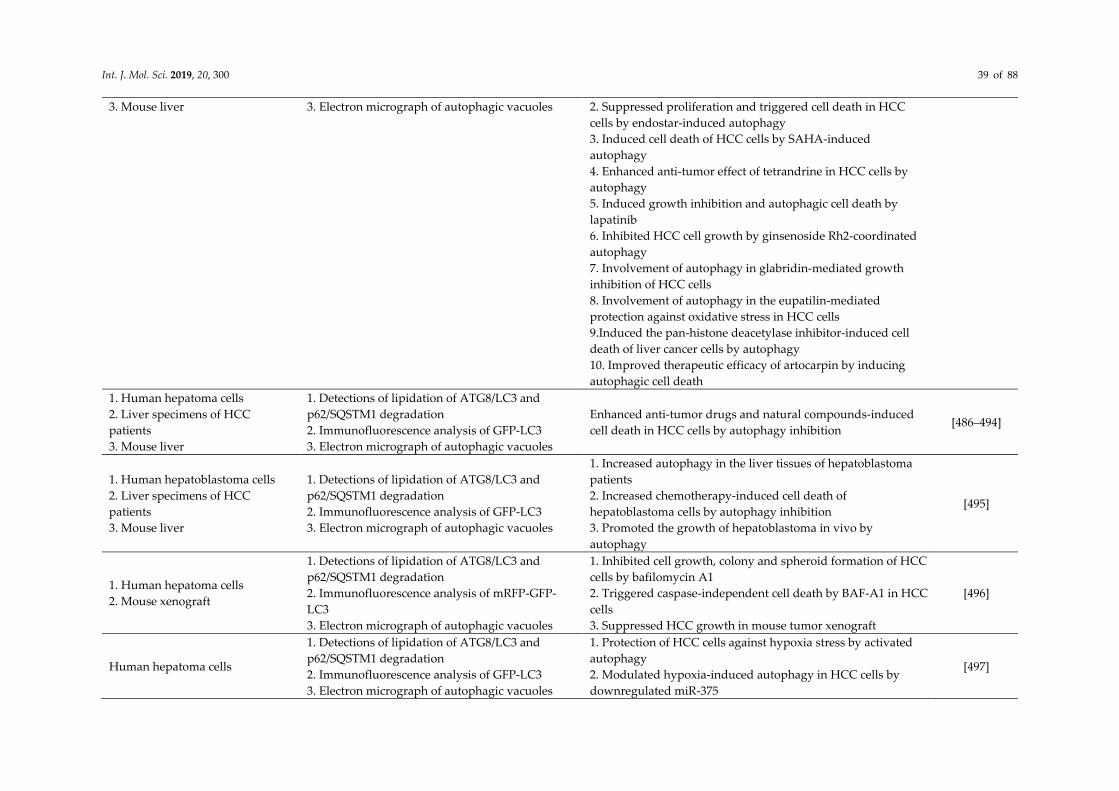
Int. J. Mol. Sci. 2019, 20, 300 39 of 88
3. Mouse liver 3. Electron micrograph of autophagic vacuoles 2. Suppressed proliferation and triggered cell death in HCC
cells by endostar-induced autophagy
3. Induced cell death of HCC cells by SAHA-induced
autophagy
4. Enhanced anti-tumor effect of tetrandrine in HCC cells by
autophagy
5. Induced growth inhibition and autophagic cell death by
lapatinib
6. Inhibited HCC cell growth by ginsenoside Rh2-coordinated
autophagy
7. Involvement of autophagy in glabridin-mediated growth
inhibition of HCC cells
8. Involvement of autophagy in the eupatilin-mediated
protection against oxidative stress in HCC cells
9.Induced the pan-histone deacetylase inhibitor-induced cell
death of liver cancer cells by autophagy
10. Improved therapeutic efficacy of artocarpin by inducing
autophagic cell death
1. Human hepatoma cells
2. Liver specimens of HCC
patients
3. Mouse liver
1. Detections of lipidation of ATG8/LC3 and
p62/SQSTM1 degradation
2. Immunofluorescence analysis of GFP-LC3
3. Electron micrograph of autophagic vacuoles
Enhanced anti-tumor drugs and natural compounds-induced
cell death in HCC cells by autophagy inhibition [486–494]
1. Human hepatoblastoma cells
2. Liver specimens of HCC
patients
3. Mouse liver
1. Detections of lipidation of ATG8/LC3 and
p62/SQSTM1 degradation
2. Immunofluorescence analysis of GFP-LC3
3. Electron micrograph of autophagic vacuoles
1. Increased autophagy in the liver tissues of hepatoblastoma
patients
2. Increased chemotherapy-induced cell death of
hepatoblastoma cells by autophagy inhibition
3. Promoted the growth of hepatoblastoma in vivo by
autophagy
[495]
1. Human hepatoma cells
2. Mouse xenograft
1. Detections of lipidation of ATG8/LC3 and
p62/SQSTM1 degradation
2. Immunofluorescence analysis of mRFP-GFP-
LC3
3. Electron micrograph of autophagic vacuoles
1. Inhibited cell growth, colony and spheroid formation of HCC
cells by bafilomycin A1
2. Triggered caspase-independent cell death by BAF-A1 in HCC
cells
3. Suppressed HCC growth in mouse tumor xenograft
[496]
Human hepatoma cells
1. Detections of lipidation of ATG8/LC3 and
p62/SQSTM1 degradation
2. Immunofluorescence analysis of GFP-LC3
3. Electron micrograph of autophagic vacuoles
1. Protection of HCC cells against hypoxia stress by activated
autophagy
2. Modulated hypoxia-induced autophagy in HCC cells by
downregulated miR-375
[497]

Int. J. Mol. Sci. 2019, 20, 300 40 of 88
3. Inhibited hypoxia-induced autophagy and autophagic flux in
HCC cells by miR-375
4. Resensitization of HCC cells to hypoxia stress by miR-375-
inhibted mitophagy
1. Human hepatoma cells
2. Mouse xenograft
1. Detection of lipidation of ATG8/LC3 2.
Immunofluorescence analysis of GFP-LC3
3. Electron micrograph of autophagic vacuoles
1. Enhanced proteasome inhibitors-induced apoptosis in HCC
cells by autophagy inhibition
2. Enhanced suppression of HCC growth by autophagy and
proteasome inhibition
[498]
1. Human hepatoblastoma cells
2. Liver specimens of HCC
patients
3. Mouse liver
1. Detections of lipidation of ATG8/LC3 and
p62/SQSTM1 degradation
2. Immunofluorescence analysis of GFP-LC3
3. Electron micrograph of autophagic vacuoles
1. Increased chemoresistance of HCC cells by hypoxia-induced
autophagy
2. Protection of HCC cells against DNA damage-inducing
chemotherapeutic agents by autophagy
3. Increased tolerance of oxaliplatin treatment in HCC cells by
autophagy
4. Increased cell survival of sorafenib-treated HCC cells by
autophagy
5. Involvement of autophagy in the drug resistance of cisplatin
in HCC cells
6. Protection of nilotinib-induced HCC against cellular
apoptosis by autophagy
7. Attenuated chemosensitivity of anti-tumor reagents in HCCs
by lncRNA HULC-triggered autophagy
[495–507]
1. Human hepatoma cells
2. Mouse xenograft
1. Detection of lipidation of ATG8/LC3
2. Immunofluorescence analysis of LC3
3. Electron micrograph of autophagic vacuoles
1. Inhibited ER stress by sorafenib in HCC cells
2. Induced ER stress-induced cellular apoptosis in HCC cells by
sorafenib
3. Induced autophagosome formation by sorafenib-induced ER
stress
4. Protection of HCC cells against cell death by ER stress-
induced autophagy
5. Enhanced sorafenib-induced tumor formation by autophagy
inhibition
[508]
Human hepatoma cells
1. Detections of lipidation of ATG8/LC3 and
p62/SQSTM1 degradation
2. Immunofluorescence analysis of GFP-LC3
3. Electron micrograph of autophagic vacuoles
1. Induced autophagy in HCC cells by low glucose and hypoxia
2. Downregulated Bad and Bim expressions in HCC cells by
autophagy
3. Inhibited chemotherapy-induced apoptosis in HCC cells by
autophagy
4. Increase chemotherapy-induced cell death by autophagy
inhibition
[510]

Int. J. Mol. Sci. 2019, 20, 300 41 of 88
1. Wild type and liver-specific
knockout of ATG7 mice
2. Hepatocellular carcinoma cell
lines
1. Impaired autophagy by ATG7-deficicent mice
by electron microscopy analysis and detection of
LC3 lipidation
2. Degradation of p62/SQSTM1 by autophagy
1. Induced hepatomegaly and the swelling of hepatic cells
deficient of ATG7
2. Accumulated peroxisomes and deformed mitochondria in
the liver cells of ATG7-deficient mice
3. Accumulated ubiquitin- and p62/SQSTM1 containing
inclusion bodies in the liver cells of ATG7-deficient mice
4. Suppression of liver dysfunction in ATG7-deficient mice by
additional knockout of p62/SQSTM1
5. Formation of p62/SQSTM1- and Keap1-postivie inclusion
bodies in ATG7-deficient hepatocytes
6. Induction of Nrf2-dependent transcriptions of antioxidant
genes in ATG7-deficient livers by p62/SQSTM1-Keap1
interaction
7. Amelioration of liver dysfunction in ATG7-deficient livers by
additional ablation of Nrf2
8. Exacerbation of liver injury in ATG7-deficient livers by
additional knockout of Nrf2
9. Induction of hepatocellular carcinoma in the mice livers loss
of ATG7 through activation of Nrf2
[356,377–
381]
1. Wild type and mosaic knockout
of ATG5 mice
2. Wild type and liver-specific
knockout of ATG7 mice
Electron micrograph of autophagic vacuoles
1. Spontaneous occurrence of multiple liver tumors by mosaic
deletion of ATG5
2. Induction of oxidative stress and DNA damage response in
ATG5-deficienct mouse liver
3. Partial suppressed tumor progression in ATG7-deficient mice
liver by additional ablation of p62/SQSTM1
[511]
1. Liver specimens of HCC
patients
2. Human hepatoma cells
3. Wild type and liver-specific
knockout of ATG7 mice
Detections of lipidation of ATG8/LC3 and
p62/SQSTM1 degradation
1. Altered metabolic profiling in HCC cells by phosphor-
p62/SQSTM1 (at serine 349)
2. Altered metabolic profiling in autophagy-deficient mouse
liver
3. Specific accumulation of phosphor-p62/SQSTM1 (at serine
349) in HCV-positive HCC
4. Suppressed proliferation and anti-cancer agent tolerance of
HCC by inhibition of phosphorylated p62/SQSTM1-dependent
Nrf2 activation
[512]
1. Liver specimens of HCC
patients
2. Human hepatoma cells
Detections of lipidation of ATG8/LC3 and
p62/SQSTM1 degradation
1. Enhanced DEN-induced carcinogenesis in mouse liver by
inducing p62/SQSTM1
2. Requirement of p62/SQSTM1 for the initiation of HCC in
mice liver
[513,514]

Int. J. Mol. Sci. 2019, 20, 300 42 of 88
3. Wild type and liver-specific
knockout of p62/SQSTM1 mice
3. Acceleration of NASH to HCC progression by p62/SQSTM1
4. Induction of HCC formation by ectopic expression of
p62/SQSTM1
5. Correlation of p62/SQSTM1 accumulation to HCC recurrence
Wild type, liver-specific knockout
of ATG7, Nrf2 knockout, YAP
knockout, ATG7/Nrf2 double
knockout, ATG7/YAP double
knockout mice
1. Detections of lipidation of ATG8/LC3 and
p62/SQSTM1 degradation
2. Immunofluorescence analysis of GFP-LC3
1. Maintenance of hepatic organ size and differentiation and
hepatic Hippo tumor suppressor pathway by autophagy
2. Degradation of YAP by autophagy
3. Attenuated hepatomegaly and hepatocarcinogenesis by loss
of YAP
4. Inhibited the ATG7 deficiency-induced enlargement of liver
size, fibrosis, progenitor cell expansion, hepatocarcinogenesis
by additional ablation of YAP
[515]
1. Liver specimens of HCC
patients
2. Human hepatoma cells
1. Detections of lipidation of ATG8/LC3 and
p62/SQSTM1 degradation
2. Immunofluorescence analysis of GFP-LC3
3. Electron micrograph of autophagic vacuoles
1. Upregulated mitochondria fission in HCC cells.
2. Correlation of increased mitochondria fission to poor
prognosis in HCC patients
3. Promoted mitochondrial function and survival of HCC cells
by mitochondrial fission in vivo and in vitro
4. Inhibited mitochondrial apoptosis by increased
mitochondrial fission-mediated autophagy
[516]
1. Mouse liver
(DEN)
2. Human hepatoma cells
1. Detection of lipidation of ATG8/LC3
2. Immunofluorescence analysis of GFP-LC3
3. Electron micrograph of autophagic vacuoles
1. Promoted DEN-induced hepatocarcinogenesis by adrenaline
2. Sustained proliferation and survival of HCC cells by ADRB2
3. Negative modulation of autophagy by ADRB2
4. Inhibited autophagy by ADRB2 through interfering with
PI(3)K complex formation
5. Stabilization of HIF-1α by ADRB2-suppressed autophagic
degradation
6. Promoted HCC xenografts in vivo by ADRB2
7. Inhibited anti-tumor effect of sorafenib by ADRB-2-repressed
autophagy
[517]
1. Human hepatoma cells
2. Liver specimens of HCC
patients
1. Detection of lipidation of ATG8/LC3
2. Immunofluorescence analysis of GFP-LC3
1. Reactivated ketolysis and facilitated cell proliferation by
nutrient starvation in HCC cells
2. Repressed AMPK activation by ketolysis in nutrient starved
HCC cells
3. Protected HCC cells from excessive autophagy and enhanced
tumor growth by suppressed AMPK
[518]
1. Human hepatoma cells
2. Mouse xenograft
1. Detection of lipidation of ATG8/LC3
2. Immunofluorescence analysis of LC3
1. Promoted autophagy by PCAF in HCC cells
2. Induced cell death and suppressed tumor growth by PCAF-
activated autophagy in vivo and in vitro
[519]

Int. J. Mol. Sci. 2019, 20, 300 43 of 88
Human hepatoma cells Detections of lipidation of ATG8/LC3 and
p62/SQSTM1 degradation
1. Positive regulation of hepatic cancer stem cells by autophagy
2. Requirement of p53 for autophagy to regulate hepatic cancer
stem cells
3. Phosphorylation of p53 at serine-392 by PINK1
4. Removal of phosphorylated p53 by mitophagy
[520]
Human cholangiocellular
carcinoma cell lines
1. Electron micrograph of autophagic vacuoles
2. Immunofluorescence analysis of acidic
organelles
Participation in Vitamin K2-mediated growth inhibition by
autophagy [521]
1. Human cholangiocellular
carcinoma cell lines
2. Mouse xenograft
3. Cholangiocellular carcinoma
patient specimens
1. Electron micrograph of autophagic vacuoles
2. Immunofluorescence analysis of GFP-LC3
3. Detection of lipidation of ATG8/LC3
4. Immunohistochemistry analysis of Beclin 1
5. Immunohistochemistry analysis of HIF-1α,
BNIP3, and PI(3)KC3
1. Induced autophagy in cholangiocellular carcinoma cells of
mice xenograft model and clinical specimens of
cholangiocellular carcinoma patients
2. Induction of apoptosis in cholangiocellular carcinoma cell
lines, repression of tumor formation in cholangiocellular
carcinoma xenograft mice, and enhancement of
chemosensitivity of cisplatin in cholangiocellular carcinoma
cells
3. Positive correlation between low Beclin 1 expression and
lymph node metastasis as well as poor survival of
cholangiocellular carcinoma patients
4. Positive correlation between high HIF-1α, BNIP3, and
PI(3)KC3 expressions and lymph node metastasis as well as
poor survival of cholangiocellular carcinoma patients
[523–
525,530]
1. The KrasG12D mutation and p53
deletion-induced intrahepatic
cholangiocellular carcinoma
(IHCC)
1. Immunofluorescence analysis of GFP-LC3
2. Detection of lipidation of ATG8/LC3 and
p62/SQSTM1
Requirement of induced autophagy for cell growth in IHCC [526,527]
1. Human cholangiocellular
carcinoma cell lines
2. Mouse xenograft
Immunofluorescence analysis of GFP-LC3 Participation in decitabine-induced growth suppression by
autophagy [522]
1. Human cholangiocarcinoma cell
lines
2. Mouse xenograft
1. Detection of lipidation of ATG8/LC3
2. Immunofluorescence analysis of acidic
lysosomes
Participation in multi-drug resistance of chemotherapy by
autophagy [532]
Human cholangiocarcinoma cell
lines
1. Electron micrograph of autophagic vacuoles
2. Detection of lipidation of ATG8/LC3
Participation in cell survival signaling of ABC294640-treated
human cholangiocarcinoma cells [533]
1. Human cholangiocarcinoma cell
lines
2. Mouse xenograft
Detection of lipidation of ATG8/LC3 and
p62/SQSTM1
1. Contribution to cell apoptosis in oblongifolin C-treated
human cholangiocarcinoma cells by inhibited autophagic flux [534,540]

Int. J. Mol. Sci. 2019, 20, 300 44 of 88
2. Contribution to cell apoptosis in salinomycin-repressed
tumor cell growth in cholangiocarcinoma mouse xenograft
model
Human cholangiocarcinoma cell
line
Detection of lipidation of ATG8/LC3 and
p62/SQSTM1
1. Increased sensitivity to cisplatin in human
cholangiocarcinoma cells by CQ-suppressed autophagy
2. Enhanced cell apoptosis of human cholangiocarcinoma cells
by CQ-suppressed autophagy and ER stress
[528,529]
Human cholangiocarcinoma cell
line
1. Electron micrograph of autophagic vacuoles
2. Detection of lipidation of ATG8/LC3
1. Promotion on drug resistance of chemotherapy in human
cholangiocarcinoma cells
2. Protection human cholangiocarcinoma cells from compound
C-induced cell apoptosis
[535,536]
Human cholangiocarcinoma cell
line
1. Detection of lipidation of ATG8/LC3 and
p62/SQSTM1
2. Immunofluorescence analysis of GFP-LC3
1. Promotion on miR-124-induced cell death in human
cholangiocarcinoma cells
2. Promotion on dihydroartemisinin-induced cell death in
human cholangiocarcinoma cells through DAPK1-Beclin
pathway
[537,538]

Int. J. Mol. Sci. 2019, 20, 300 45 of 88
4.4. Viral Hepatitis
In the past decade, hepatic autophagy has been extensively shown to participate in the liver cells
infected with hepatitis viruses, including HBV and HCV. In the late 2000s, the HCV-H77 (genotype 1a)
infection was first shown to activate the formation of autophagic vacuoles in immortalized human
hepatocytes (IHH) (Table 5) [541]. Furthermore, in the replication of a full-length HCV-JFH1 (genotype 2a)
viral RNA in human HCC, Huh7 cells were found to induce an incomplete autophagic process that
enhanced the accumulation of autolysosomes and interfered with autophagic flux and degradation (Table
5) [542]. HCV-JFH1-induced autophagosome formation was further demonstrated to be required for viral
RNA replication (Table 5) [542]. Moreover, ATG5 was shown to interact with the nonstructural (NS)
proteins of HCV, NS4B (a viral protein that reconstitutes a membranous web for the replication of viral
RNA), and NS5B (a RNA polymerase that replicates viral RNA) to promote HCV viral replication (Table 5)
[543]. The HCV-induced autophagosomal membrane was reported to provide a membraned platform that
contains HCV NS5A, NS5B, and viral RNA for replication (Table 5) [544–546]. These studies imply that
HCV activates the autophagic process to organize a membranous structure for replicating viral RNA.
However, several contradictory studies have indicated that HCV-induced autophagic vacuoles do not
colocalize with viral proteins or viral RNA (Table 5) [547–551], suggesting that the viral-triggered biogenesis
of autophagic vacuoles may not be necessary for viral RNA replication.
The infection by a cell culture-derived HCV-JFH1 virus (HCVcc) into Huh7.5.1 cells, a clone derived
from Huh7 cells that is highly permissive to HCV growth, is known to promote the translation of incoming
viral RNA, thus establishing a virus infection (Table 5) [548]. Another study showed that a HCVcc-JFH1
infection activates an entire autophagic process throughout the maturation of autolysosomes to support
viral RNA replication by suppressing the HCV pathogen associated molecular pattern-mediated innate
antiviral responses (Table 5) [548,549]. Similarly, the genetical silencing of ATGs, including Beclin and
ATG7, was found to suppress HCV viral infectivity and to upregulate interferon-stimulated gene (ISG)
expression in HCV H77-infected IHH cells (Table 5) [552]. Moreover, a recent study revealed that HCV may
activate autophagy to degrade the tumor necrosis factor receptor (TNFR)-associated factor 6 (TRAF6)
through p62/SQSTM1, thus alleviating innate host immunity (Table 5) [553]. These results together indicate
that HCV may induce hepatic autophagy to repress innate antiviral immunity to promote the HCV-
infecting life cycle in liver cells (Table 5) [548,549,552,554]. Moreover, the HCV infection was shown to
participate in the assembly of infectious particles rather than to affect the replication of intracellular viral
RNA (Table 5) [555]. Further studies have demonstrated that autophagy mediates the egress of HCV virion
through the CD63-associated exosome pathway (Table 5) [556] and the regulation of apolipoprotein E
(ApoE) transport (Table 5) [557]. Additionally, HCV-activated autophagy may depend on different HCV
genotypes (Table 5) [558]. The replication of HCV-Con1 (genotype 1b) was reported to impede the fusion
of autophagosomes with lysosomes, thus protecting infected liver cells from cell death (Table 5) [558].
Numerous studies have demonstrated that the ER stress-associated unfolded protein response (UPR)
is required for HCV-induced autophagy (Table 5) [542,548,549,554], presumably through the DNA damage-
inducible transcript 3 protein (DDIT3, also known as CHOP)-mediated transcription of LC3B and ATG5, to
activate autophagy (Table 5) [559]. Furthermore, UPR is required for interference with protein kinase B
(PKB)-tuberous sclerosis (TSC)-mTOR complex 1 (mTORC1) signaling (Table 5) [560]. STX17, an autophagy
regulator implicated in the biogenesis of autophagic vacuoles, may regulate HCV virion egress (Table 5)
[561] and promote viral RNA replication (Table 5) [562]. Recently, the alternative splicing of ATG10 was
shown to regulate HCV replication through affecting autophagic flux (Table 5) [563,564].
Apart from bulk and nonselective autophagy, HCV was shown to activate autophagy to selectively
degrade intracellular organelles, such as LDs and mitochondria (Table 5) [430,565]. The level of lipidated LC3
was inversely correlated with the clinical parameters related to hepatic steatosis in the liver samples of patients
with chronic HCV (Table 5) [430]. The HCV-activated autophagic vacuoles were found to engulf LDs in HCV-
replicon and HCV-infected cells (Table 5) [430]. In addition, interference with autophagy by pharmacological
inhibitors and gene knockdown was demonstrated to significantly enhance the intracellular levels of
cholesterol (Table 5) [430], suggesting that the inhibition of autophagic degradation of LDs may result in the

Int. J. Mol. Sci. 2019, 20, 300 46 of 88
occurrence of hepatic steatosis in chronically HCV-infected patients. These results indicate that HCV could
induce autophagy to catabolize LDs to protect HCV-infected cells against excess LDs accumulation. Moreover,
HCV infection has been shown to trigger the formation of mitophagosomes that degrade mitochondria via
the PINK1–Parkin axis in infected cells, thereby promoting HCV viral RNA replication (Table 5) [565,566].
Another study indicated that HCV-induced mitochondrial fission is required for the elimination of
mitochondria in infected cells (Table 5) [567]. These studies argue that HCV may activate autophagic
degradation to clear the mitochondria with viral-induced damage to attenuate the cell apoptosis of infected
liver cells (Table 5) [567]. Oppositely, another study reported that HCV capsids interact with Parkin to impede
the translocation of Parkin from the cytoplasm to mitochondria to inhibit mitophagy and to sustain HCV-
induced mitochondrial damage and injury in infected liver cells (Table 5) [568]. Recently, HCV viral RNA was
shown to induce selective autophagy, possibly promoting the elimination of ubiquitinated protein aggregates
(Table 5) [569]. However, CMA was also reported to be activated by HCV and implicated in the degradation
of IFN-alpha receptor-1 (IFNAR1), which is stimulated by FFAs (Table 5) [570]. Another study demonstrated
that HCV NS5A interacts with HSC70 to promote the degradation of hepatocyte nuclear factor 1α (HNF1α)
(Table 5) [571]. Recently, IFN-β-inducible SCOTIN was shown to recruit HCV NS5A into autolysosomes for
degradation, restricting the HCV infection (Table 5) [572]. These studies collectively suggest that HCV
subverts autophagy to antagonize viral RNA replication and the harmful host responses in infected liver cells.
The induction of autophagy by HBV was first discovered through observing the autophagic degradation
of peroxisomes in the liver tissues of patients with chronic HBV who received kidney transplantations and
immunosuppressive therapy [329]. Levine et al. showed that the heterozygous deletion of Beclin 1
downregulates autophagy in liver cells, thus promoting HBV-associated premalignant lesions [573]. The HBV
infection was further revealed to induce the early-stage formation of autophagic vacuoles in Huh7.5.1 cells
through increasing PI(3)K enzyme activity to promote HBV DNA replication (Table 5) [574,575]. The role of
autophagy in HBV DNA replication was further confirmed by a study that revealed that the ablation of ATG5
inhibits autophagy to reduce the HBV gene expression in liver cells and in sera (Table 5) [576]. HBV viral
proteins, such as HBV x protein (HBx) and HBV small surface protein (SHB), activate autophagy in liver cells
through enhancing the Beclin 1 expression and inducing UPR (Table 5) [577,578], thus promoting HBV
replication. Recently, HBV was shown to subvert the ATG5-ATG12-ATG16L complex rather than the lipidated
ATG8/LC3-mediated autophagosome biogenesis to support the assembly and stability of the HBV
nucleocapsid, suggesting that HBV exploits nondegradative autophagy to facilitate HBV propagation (Table
5) [579]. Moreover, HBV can promote autophagy through the miR-192-3p-XIAP axis to benefit HBV replication
in vitro and in vivo (Table 5) [580]. In addition to these findings that autophagy promotes HBV growth,
Epigallocatechin-3-gallate (EGCG), a major polyphenol in green tea, was demonstrated to inhibit HBV-
induced autophagy through increasing lysosomal acidification, thus repressing HBV growth (Table 5) [581].
In spite of genomic DNA replication, HBV-activated autophagy may participate in the secretion of infectious
virions in infected cells (Table 5) [582].
HBx-induced autophagy was reported to inhibit mitochondrial apoptosis to promote the survival of HBV
genomic DNA-transfected cells (Table 5) [583]. Also, autophagy and ER-associated degradation (ERAD) were
shown to promote the degradation of HBV enveloped proteins for the establishment of chronic HBV infection
(Table 5) [584]. Additionally, autophagy was negatively correlated with an oncogenic miR-244, which was
degraded through the autophagic process in the liver tissues of HBV-associated patients with HCC and HBx-
transgenic mice (Table 5) [585]. Reduced ATG5 expression and elevated miR-244 level were significantly
correlated with HBV infection and a poor survival rate (Table 5) [585], suggesting that autophagy promotes
the degradation of miR-244 to repress HBV-associated tumorigenesis. HBx was shown to activate autophagy
through the PI(3)K/Akt-mTOR pathway [586], death-associated protein kinase [587], the JNK2-mediated
regulation of Beclin 1/Bcl-2 interaction [588], and the upregulation of HMGB1 [589] (Table 5). By contrast, the
transfection of HBV genomic DNA and overexpression of HBx in hepatoma cells interfere with autophagic
degradation by impairing the maturation of lysosomes (Table 5) [590]. Additionally, accumulated
p62/SQSTM1 and defected cathepsin D were positively related to the liver tissues of people with chronic HBV
infection and HBV-associated liver cancer (Table 5) [590], suggesting that HBV’s inhibition of autophagy could

Int. J. Mol. Sci. 2019, 20, 300 47 of 88
result in the development of HCC. Taken together, these results indicate that HBV may modulate autophagy
to promote viral growth and may participate in the development of HBV-related liver cancer.

Int. J. Mol. Sci. 2019, 20, 300 48 of 88
Table 5. Summary of autophagy in viral hepatitis.
Experimental Model Characteristics of Autophagy Function of Autophagy References
1. Immortalized human
hepatocytes (IHH)
(HCV infection)
2. Human hepatoma, Huh7.5 cells
(HCV infection)
1. Electron micrograph of autophagic
vacuoles
2. Immunofluorescence analysis of GFP-LC3
Unknown [541]
Human hepatoma, Huh7.5 cells
(Transfection of HCV viral RNA)
1. Detection of lipidation of ATG8/LC3
2. Immunofluorescence analysis of GFP-LC3
3. Electron micrograph of autophagic
vacuoles
Promotion on viral RNA replication [542]
Human hepatoma, Huh7 cells
(HCV infection)
1. Detection of lipidation of ATG8/LC3
2. Immunofluorescence analysis of GFP-LC3
1. Promotion on viral RNA replication
2. Support on the translation of viral RNA [548]
Human hepatoma, Huh7 cells
(HCV infection)
1. Detection of lipidation of ATG8/LC3
2. Immunofluorescence analysis of GFP-LC3
3. Electron micrograph of autophagic
vacuoles
1. Promotion on viral RNA replication
2. Suppression of innate antiviral immunity [549,591]
1. IHH
(HCV infection)
2. Human hepatoma, Huh7.5 cells
(HCV infection)
Colocalization of autophagosomes and
lysosomes
1. Promotion on viral RNA replication
2. Suppression of innate antiviral immunity
3. Protection the infected cells from cell death
[552]
Human hepatoma, Huh7 cells
(HCV infection)
Detections of lipidation of ATG8/LC3 and
p62/SQSTM1 degradation
1. Promotion on viral RNA replication
2. Suppression of innate antiviral [553]
Human hepatoma, Huh7 and
Huh7.5.1 cells
(HCV infection; transfection of
HCV replicon RNA)
Detection of lipidation of ATG8/LC3 1. Promotion on viral RNA replication
2. Support on the organization of replication complex for viral RNA [544]
Human hepatoma, Huh7 cells
(HCV infection)
1. Promotion on viral RNA replication
2. Support on the organization of replication complex for viral RNA [543]
Human hepatoma, Huh7.5.1
(Transfection of HCV replicon
RNA)
Electron micrograph of autophagic vacuoles 1. Promotion on viral RNA replication
2. Support on the organization of replication complex for viral RNA [545]
Human hepatoma, Huh7 and
Huh7.5.1 cells
(HCV infection; transfection of
HCV replicon RNA)
Immunofluorescence analysis of GFP-LC3 1. Promotion on viral RNA replication
2. Support on the recruitment of lipid rafts for viral RNA replication [546]
Human hepatoma, Huh7.5.1 cells Immunofluorescence analysis of GFP-LC3 Promotion on the release of viral particles [555]

Int. J. Mol. Sci. 2019, 20, 300 49 of 88
(HCV infection)
1. IHH
(HCV infection)
2. Human hepatoma, Huh7.5 cells
(HCV infection)
Colocalization of autophagosomes and
lysosomes Promotion on the release of viral particles [556]
Human hepatoma, Huh7 and
Huh7.5.1 cells
(HCV infection; transfection of
HCV replicon RNA)
Immunofluorescence analysis of GFP-LC3
Promotion on the release of viral particles [557]
Human hepatoma, Huh7 cells
(Transfection of HCV replicon
RNA)
1. Colocalization of autophagosomes and
lysosomes
2. Detection of lipidation of ATG8/LC3
Counteracting the viral-induced cell death [558]
Human hepatoma, Huh7.5 cells
(HCV infection; transfection of
HCV replicon RNA)
Detection of lipidation of ATG8/LC3 Unknown [592]
Human hepatoma, Huh7.5 cells
(HCV infection) Detection of lipidation of ATG8/LC3 Promotion on viral RNA replication [593]
Human hepatoma, Huh7 cells
(Transfection of HCV replicon
RNA)
Detection of lipidation of ATG8/LC3 Unknown [559]
Human hepatoma, Huh7 cells
(HCV infection; transfection of
HCV replicon RNA)
1. Detection of lipidation of ATG8/LC3
2. Detection of CMA Promotion on HNF1α degradation [571]
Human hepatoma, Huh7 and
Huh7.5.1 cells
(HCV infection; transfection of
HCV replicon RNA)
1. Detection of lipidation of ATG8/LC3
2. Immunofluorescence analysis of LC3 Facilitation on virion release [561]
Human hepatoma, Huh7.5.1 cells
(HCV infection; transfection of
HCV replicon RNA)
1. Detection of lipidation of ATG8/LC3
2. Electron micrograph of autophagic
vacuoles
1. Promotion on viral RNA replication
2. Support on the organization of replication complex for viral RNA [571]
Human hepatoma, HepG2 cells
(Transfection of HCV replicon
RNA)
1. Detection of lipidation of ATG8/LC3
2. Immunofluorescence analysis of LC3 Promotion on the degradation of replicon RNA [563,564]
1. Human hepatoma, Huh7 cells
(HCV infection; transfection of
HCV replicon RNA)
1. Detection of lipidation of ATG8/LC3
2. Immunofluorescence analysis of RFP-LC3 Promotion on LDs catabolism [430]

Int. J. Mol. Sci. 2019, 20, 300 50 of 88
2. Liver specimens of HCV-infected
patients
Human hepatoma, Huh7.5.1 cells
(HCV infection; transfection of
HCV replicon RNA)
1. Detection of lipidation of ATG8/LC3
2. Colocalization of autophagosome and
mitochondria
1. Promotion on mitochondria degradation
2. Protection of infected cells from apoptosis
3. Establishment of viral persistence
[565,567]
Human hepatoma, Huh7 cells
(HCV infection; transfection of
HCV viral protein)
1. Detection of lipidation of ATG8/LC3
2. Colocalization of autophagosome and
mitochondria
Sustained mitochondrial injury [568]
Human hepatoma, Huh7 cells
(HCV infection; transfection of
HCV replicon RNA)
1. Detection of lipidation of ATG8/LC3
2. Colocalization of autophagosome and
ubiquitinated proteins
Unknown [571]
Human hepatoma, Huh7 and
Huh7.5.1 cells
(HCV infection)
1. Detection of lipidation of ATG8/LC3
2. Detection of CMA Suppression of innate antiviral immunity [570]
Human hepatoma, Huh7 cells
(HCV infection)
1. Detection of lipidation of ATG8/LC3
2. Colocalization of autophagosome and
HCV NS5A
1. Promotion on viral RNA replication
2. Repression of innate antiviral immunity [572]
1. Human hepatoma, Huh7.5.1
cells
(HBV genomic DNA transfection)
2. Wild type and liver specific
knockout of ATG5
1. Detection of lipidation of ATG8/LC3
2. Immunofluorescence analysis of GFP-LC3
3. Detections of lipidation of ATG8/LC3 and
p62/SQSTM1 degradation
1. Promotion on HBV DNA replication
2. Induction of autophagy by HBV via HBx [574–576]
Human hepatoma cell lines
(HBx transfection; transfection of
HBV genomic DNA)
1. Electron micrograph of autophagic
vacuoles
2. Immunofluorescence analysis of GFP-LC3
1. Transcriptional activation of Beclin 1 by HBx
2. Activation autophagy by transfection of HBV genomic DNA [577]
Human hepatoma, Huh7 cells
(SHBs transfection; transfection of
HBV genomic DNA)
1. Electron micrograph of autophagic
vacuoles
2. Immunofluorescence analysis of GFP-LC3
3. Detections of lipidation of ATG8/LC3 and
p62/SQSTM1 degradation
1. Requirement of SHBs for autophagy activation by HBV
2. Induction of autophagy by SHBs via ER stress
3. Requirement of autophagy for HBV envelopment
[578]
Human hepatoma, Huh7 cells
(Transfection of HBV genomic
DNA)
1. Detection of lipidation of ATG8/LC3
2. Immunofluorescence analysis of GFP-LC3
1. Impaired HBV formation and release by silencing of ATG5,
ATG12, ATG16L1
2. Altered intracellular distribution of HBV by silencing of ATG12
[579]

Int. J. Mol. Sci. 2019, 20, 300 51 of 88
3. Requirement of ATG5-ATG12-ATG16L1 complex for HBV viral
maturation
1. Human hepatoma, Huh7,
HepG2, and HepG2.2.15 cells
2. Primary human hepatocytes
(Transfection of HBV genomic
DNA)
1. Detection of lipidation of ATG8/LC3
2. Immunofluorescence analysis of GFP-LC3
1. Activated autophagy by HBV through miR-192-2p-XIAP axis
2. Inhibited HBV replication by miR-192-2p-mediated NF-κB
signaling and autophagy inhibition
[580]
Human hepatoma, HepG2.2.15
cells
(Transfection of HBV genomic
DNA)
Detections of lipidation of ATG8/LC3 and
p62/SQSTM1 degradation
1. Repressed HBV replication by EGCG
2. Suppressed HBV-induced incomplete autophagy by EGCG [581]
Human hepatoma, HepG2.2.15
cells
(Transfection of HBV genomic
DNA)
1. Electron micrograph of autophagic
vacuoles
2. Immunofluorescence analysis of GFP-LC3
3. Detection of lipidation of ATG8/LC3
1. Colocalization of HBV components with multivesicular bodies
(MVB) and autophagosomes
2. Facilitation of HBV secretion by an activation of the endolysosomal
and autophagic pathway
[582]
1. Human hepatic cells lines, L02
and Chang cells
2. Human hepatoma, HepG2.2.15
cells
(HBx transfection)
1. Detection of lipidation of ATG8/LC3
2. Immunofluorescence analysis of GFP-LC3
Reduced starvation-induced cell death by HBx via autophagy
activation and inhibition of mitochondrial apoptotic pathway [583]
Human hepatoma, Huh7 and
HepG2 cells
(Transfection of HBV genomic
DNA)
1. Activation of ERAD by HBV replication
2.Upregulations of ER degradation-enhancing mannosidase-like
proteins (EDEMs) by HBV replication
[584]
1. Liver specimens of HBV-
associated HCC patients
2. Human hepatoma, Hep3B cells
1. Detection of lipidation of ATG8/LC3
2. Immunofluorescence analysis of LC3
3. Electron micrograph of autophagic
vacuoles
1. Inverse correlation of autophagy level with miR-224 expression in
HBV-associated HCC patients
2. Degradation of miR-224 by autophagic pathway
3. Suppressed tumorigenesis of HBV-associated HCC through
degradation of miR-244
[585]
Human hepatoma, HepG2.2.15
cells cells
(HBx transfection)
1. Electron micrograph of autophagic
vacuoles
2. Detection of lipidation of ATG8/LC3
Induced autophagy by HBx through PI(3)K/Akt-mTOR pathway [586]
1. Human hepatic cells line, Chang
cells
2. Human hepatoma, HepG2.2.15
cells
(HBx transfection)
1. Detection of lipidation of ATG8/LC3
2. Immunofluorescence analysis of GFP-LC3
Induced autophagy by HBx through activating death-associated
kinase [587]

Int. J. Mol. Sci. 2019, 20, 300 52 of 88
1. Human hepatoma, HepG2 cells
2. Primary human hepatocytes
(HBx transfection)
Detections of lipidation of ATG8/LC3 and
p62/SQSTM1 degradation
Induced autophagy by HBx through ROS-mediated JNK regulation
of Beclin 1/Bcl-2 interaction [588]
1. Human hepatic cells line, L02
cells
2. Human hepatoma, Huh7,
HepG2, HepG2.2.15 cells
(HBx transfection)
1. Detections of lipidation of ATG8/LC3 and
p62/SQSTM1 degradation
2. Electron micrograph of autophagic
vacuoles
3. Immunofluorescence analysis of LC3
Induced autophagy by HBx through interacting with HMGB1 [589]
Human hepatoma, Huh7 cells
(HBx transfection)
1. Detections of lipidation of ATG8/LC3 and
p62/SQSTM1 degradation
2. Electron micrograph of autophagic
vacuoles
3. Immunofluorescence analysis of GFP-LC3
Inhibited autophagic degradation by HBx through impairing
lysosomal maturation [590]

Int. J. Mol. Sci. 2019, 20, 300 53 of 88
4.5. Fibrosis and Cirrhosis
Autophagy was previously shown to eliminate the mutant α1-ATZ aggregated proteins in liver cells
[353,354,370]. The enhancement of autophagy by carbamazepine (CBZ) was reported to reduce the
accumulation of α1-ATZ and the inhibited hepatic fibrosis in a α1-AT-deficient mouse model (Table 6) [594]
and to reduce the death of hepatic cells in fibrinogen-storage-related diseases (Table 6) [595]. Furthermore,
activation of autophagy by gene transfer of the autophagy regulator, TFEB, was demonstrated to enhance
the removal of α1-ATZ inclusion bodies and to reverse α1-ATZ-induced liver damage in a α1-AT-deficient
mouse model (Table 6) [391]. The induction of MAP1s-mediated autophagy by spermidine was found to
prevent liver fibrosis and HCC development in mice (Table 6) [596]. Moreover, in a tetrachloride (CCl4)-
induced fibrosis mouse model, human palatine tonsil-derived mesenchymal stem cells (T-MSCs) that
differentiated into hepatocyte-like cells were found to ameliorate hepatic fibrosis in an autophagy-
dependent manner (Table 6) [597]. Interference with autophagic degradation by chloroquine was reported
to improve CCl4-induced liver fibrosis through inhibiting the activation of hepatic stellate cells (Table 6)
[598]. The impairment of autophagy by dihydroceramide promoted the disease progression of hepatic
steatosis to liver fibrosis (Table 5) [599]. Similarly, impaired autophagy-mediated inhibition of YAP
degradation also resulted in liver fibrosis (Table 6) [515]. Autophagy was shown to protect liver sinusoidal
endothelial cells (LSECs) from oxidative stress to ameliorate liver fibrosis (Table 6) [600]. These studies
collectively indicate that autophagy prevents the development of liver fibrosis.
HIF-1α, ROS-JNK1/2-dependent activation, and the X-box binding protein 1 (XBP1) signaling of UPR
were required for the autophagy-mediated activation of hepatic stellate cells (Table 6) [601–603]. Moreover,
autophagy deficiency was reported to induce cell death in hepatocytes, leading to liver inflammation,
hepatic fibrosis, and tumorigenesis by substantially activating Nrf2 (Table 6) [604]. Taken together, these
studies indicate that autophagic degradation plays a critical role in preventing liver fibrosis. In addition,
macrophages derived from mice with an ATG5 deletion was found to secrete high levels of ROS-induced
interleukin 1A (IL-1A) and IL-1B, which were correlated with the enhanced accumulation of matrix and
fibrogenic cells in CCl4-induced fibrosis livers and upregulated profibrogenic genes expressions in the
cocultured hepatic myofibroblasts (Table 6) [605], suggesting that the activation of autophagy in
macrophages may repress liver fibrosis. These studies imply that autophagy regulates multiple biological
processes to mitigate the occurrence of hepatic fibrosis.
By contrast, one study has shown that autophagy promotes hepatic fibrogenesis by activating hepatic
stellate cells in the CCl4 or thioacetamide (TAA)-induced liver fibrosis mouse model (Table 6) [606].
Similarly, dimethyl α-ketoglutarate (DMKG) was reported to inhibit CCl4-induced hepatic fibrosis through
reducing autophagy (Table 6) [607]. Furthermore, quercetin attenuated bile duct ligation (BDL) and CCl4-
induced liver fibrosis in mice through inhibiting hepatic stellate cell activation and autophagy (Table 6)
[608]. Analogously, 3-MA was reported to ameliorate hepatic fibrosis through impairing autophagy
initiation in hepatic stellate cells (Table 6) [609]. The genetic deletion of Golgin A2 (GOLAG2), a cis-Golgi
protein, reportedly activates autophagy and promotes liver fibrosis in mice (Table 6) [610]. Furthermore,
through activating autophagy, β-arrestin 1 (β-arr1) has been implicated in improving the compensatory
proliferation of hepatocytes and the growth of hepatic stellate cells with a consequence in liver fibrosis
(Table 6) [611]. These studies indicate autophagy’s promoting role in liver fibrosis development.
Unsurprisingly, autophagy was also correlated with cirrhosis in the liver samples of patients with
chronic liver disease [612]. The aggregation of p62/SQSTM1 through defective autophagy was found to be
increased in the damaged bile ducts of primary biliary cirrhosis (PBC) patients (Table 6) [613]. The elevated
expressions of mitochondrial antigens, including pyruvate dehydrogenase complex-E2 component (PDC-
E2) and cytochrome c oxidase, subunit I (CCO), was further shown to be associated with the autoimmune
pathogenesis of bile duct lesions in patients with PBC (Table 6) [614]. The increased level of LC3B, the
colocalization of LC3B with LAMP1, and the elevated expressions of LAMP2 and cathepsin D were detected
in the liver samples of patients with cirrhosis (Table 6) [615]. The impairment of autophagy by CQ was
reported to be attenuated the CCl4-induced ductular reaction and fibrosis (Table 6) [615]. By contrast,
autophagy was reported to contribute to the formation of hepatocyte growth factor (HGF)-directed

Int. J. Mol. Sci. 2019, 20, 300 54 of 88
Axin2+CD90+ CSCs to promote hepatocarcinogenesis in cirrhotic livers (Table 6) [616]. These studies
together constitute the notion that autophagy may regulate the occurrence of liver cirrhosis.

Int. J. Mol. Sci. 2019, 20, 300 55 of 88
Table 6. Summary of autophagy in fibrosis and cirrhosis.
Experimental Model Characteristics of Autophagy Function of Autophagy References
1. HTO/Z cell line
2. Wild type and PiZ (α1-AT-
deficient) mice
3. Wild type and GFP-LC3
transgenic mice
1. Detection of lipidation of ATG8/LC3
2. Immunofluorescence analysis of
GFP-LC3
1. Promoted degradation of α1-ATZ by autophagy-enhancing
drug, CBZ
2. Reduced α1-ATZ-induced hepatic fibrosis by CBZ
[594]
Liver specimens of fibrinogen
storage disease
Electron micrograph of autophagic
vacuoles
Diminished hepatocellular death in fibrinogen storage diseases
by CBZ [595]
Mouse liver
(CCl4)
Detections of lipidation of ATG8/LC3
and p62/SQSTM1 degradation
Prevented liver fibrosis in HCC by activating MAP1S-mediated
autophagy [596]
Mouse liver
(CCl4) Immunofluorescence analysis of LC3
Amelioration of liver fibrosis by T-MSCs via activating
autophagy [597]
Rat liver
(CCl4)
Detections of lipidation of ATG8/LC3
and p62/SQSTM1 degradation
Improved liver fibrosis by chloroquine through inhibiting
activation of hepatic stellate cells [598]
1. Human hepatic cells line,
Chang cells
2. Human liver stellate cell
line, LX-2 cells
1. Electron micrograph of autophagic
vacuoles
2. Detection of lipidation of ATG8/LC3
3. Immunofluorescence analysis of LC3
1. Impaired autophagic flux by dihydroceramide
2. Increased triglyceride storage in LDs and upregulated
expressions of fibrosis markers by dihydroceramide
3. Restoration of autophagic flux and reversed
dihydroceramide-induced fibrogenesis by rapamycin
[599]
1. Rat liver
(CCl4)
2. LSECs from wild type and
ATG7 knockout mice
1. Detection of lipidation of ATG8/LC3
2. Immunofluorescence analysis of LC3
1. Upregulated autophagy during LSECs capillarization in vivo
and in vitro
2. Maintenance of LSECs homeostasis by autophagy
3. Association of loss of LSECs autophagy with insufficient
antioxidant response
4. Promoted liver fibrosis by impaired endothelial autophagy
[600]
1. Human liver stellate cell
line, LX-2 cells
2. Rat liver
(CCl4)
3. Primary human and mouse
hepatic stellate cells
1. Detection of lipidation of ATG8/LC3
2. Immunofluorescence analysis of LC3
3. Electron micrograph of autophagic
vacuoles
1. Involvement of HIF-1α in the regulation of autophagy-
mediated activation of hepatic stellate cells
2. Requirement of ROS-JNK1/2-depedent autophagy activation
for the induction of anti-inflammation in liver fibrosis
3. Induced fibrogenic activity in hepatic stellate cells by XBP1
through autophagy
[603]

Int. J. Mol. Sci. 2019, 20, 300 56 of 88
Wild type and liver specific
knockout of ATG5 mice and
Nrf2 knockout mice
1. Detections of lipidation of ATG8/LC3
and p62/SQSTM1 degradation
2. Electron micrograph of autophagic
vacuoles
1. Increase apoptosis in ATG5-deficcient hepatocytes by
disruption of the homeostasis of pro-and anti-apoptotic
proteins
2. Induced liver fibrosis by loss of ATG5
3. Suppressed ATG5 deficiency-induced liver injury,
inflammation, fibrosis, and tumor development by additional
ablation of Nrf2
[404]
1. Wild type and ATG5
knockout mice
2. Kupffer cells isolated from
Wild type and ATG5 knockout
mice
(CCl4)
Immunofluorescence analysis of LC3
1. Enhanced the recruitment of inflammatory cells and hepatic
inflammation in ATG5-deficient mice after CCl4-induced liver
fibrosis
2. Promoted cellular apoptosis of hepatocytes in ATG5-
deficeint mice after CCl4-inducecd liver damage
3. Enhanced fibrogenic properties of hepatic myofibroblast in
autophagy-deficient macrophages via IL1-dependent pathway
[605]
1. Rat liver
(CCl4)
2. Hepatic stellate cell, HSC-T6
cells
1. Immunofluorescence analysis of LC3
2. Electron micrograph of autophagic
vacuoles
3. Detection of lipidation of ATG8/LC3
Reduced CCl4-induced liver fibrosis by DMKG through
inhibition of autophagy in hepatic stellate cells in vivo [607]
Mouse liver
(CCL4)
1. Electron micrograph of autophagic
vacuoles
2. Detections of lipidation of ATG8/LC3
and p62/SQSTM1 degradation
Prevented hepatic fibrosis by Quercetin through reducing
hepatic stellate cell activation and inhibiting autophagy [608]
1. Human liver stellate cell
line, LX-2 cells
2. Primary hepatic stellate cells
Detection of lipidation of ATG8/LC3 Amelioration of liver fibrosis by 3-MA through inhibition of
autophagy and hepatic stellate cell activation [609]
1. Human hepatic, Chang cells
2. Wild type and GOLAG2
knockout mice
1. Detections of lipidation of ATG8/LC3
and p62/SQSTM1 degradation
2. Electron micrograph of autophagic
vacuoles
3. Immunofluorescence analysis of LC3
1. Induced autophagy by knockout of GOLAG2
2. Promoted liver fibrosis by loss of GOLAG2 in vivo [610]
1. Human liver stellate cell
line, LX-2 cells
2. Primary hepatic stellate cells
Detection of lipidation of ATG8/LC3
1. Enhanced hepatocyte compensatory proliferation and
hepatic stellate cell growth by β-arr1 via activating autophagy
2. Promoted liver fibrosis by β-arr1 via autophagy-mediated
Snail signaling
[611]
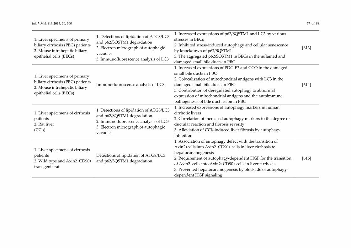
Int. J. Mol. Sci. 2019, 20, 300 57 of 88
1. Liver specimens of primary
biliary cirrhosis (PBC) patients
2. Mouse intrahepatic biliary
epithelial cells (BECs)
1. Detections of lipidation of ATG8/LC3
and p62/SQSTM1 degradation
2. Electron micrograph of autophagic
vacuoles
3. Immunofluorescence analysis of LC3
1. Increased expressions of p62/SQSTM1 and LC3 by various
stresses in BECs
2. Inhibited stress-induced autophagy and cellular senescence
by knockdown of p62/SQSTM1
3. The aggregated p62/SQSTM1 in BECs in the inflamed and
damaged small bile ducts in PBC
[613]
1. Liver specimens of primary
biliary cirrhosis (PBC) patients
2. Mouse intrahepatic biliary
epithelial cells (BECs)
Immunofluorescence analysis of LC3
1. Increased expressions of PDC-E2 and CCO in the damaged
small bile ducts in PBC
2. Colocalization of mitochondrial antigens with LC3 in the
damaged small bile ducts in PBC
3. Contribution of deregulated autophagy to abnormal
expression of mitochondrial antigens and the autoimmune
pathogenesis of bile duct lesion in PBC
[614]
1. Liver specimens of cirrhosis
patients
2. Rat liver
(CCl4)
1. Detections of lipidation of ATG8/LC3
and p62/SQSTM1 degradation
2. Immunofluorescence analysis of LC3
3. Electron micrograph of autophagic
vacuoles
1. Increased expressions of autophagy markers in human
cirrhotic livers
2. Correlation of increased autophagy markers to the degree of
ductular reaction and fibrosis severity
3. Alleviation of CCl4-induced liver fibrosis by autophagy
inhibition
1. Liver specimens of cirrhosis
patients
2. Wild type and Axin2+CD90+
transgenic rat
Detections of lipidation of ATG8/LC3
and p62/SQSTM1 degradation
1. Association of autophagy defect with the transition of
Axin2+cells into Axin2+CD90+ cells in liver cirrhosis to
hepatocarcinogenesis
2. Requirement of autophagy-dependent HGF for the transition
of Axin2+cells into Axin2+CD90+ cells in liver cirrhosis
3. Prevented hepatocarcinogenesis by blockade of autophagy-
dependent HGF signaling
[616]

Int. J. Mol. Sci. 2019, 20, 300 58 of 88
5. Conclusions and Future Directions
Autophagy plays a critical role in the maintenance of cellular homeostasis and the elimination of
unwanted intracellular materials, such as proteins and organelles, in liver physiology. Autophagy has been
extensively shown to be regulated in liver cells in multiple liver diseases. The activation of autophagy has
mostly been conceptualized as a protector of liver cells against injury. Furthermore, it has been suggested
to prevent the development of liver-associated diseases through the autophagic degradation of aggregate-
prone proteins and damaged organelles. In accordance with the protective roles of autophagy, the
enhancement of autophagy in liver cells is considered a treatment strategy for ameliorating the
development of liver diseases. By contrast, autophagy has also been implicated in promoting liver damage-
induced cell death and the development of liver-related diseases, suggesting that interference with
autophagy may represent a novel route to mitigate the progression of liver diseases. Hence, the
physiological significance of autophagy in liver diseases is still debated and is largely hampered by
discrepancies between studies. Our current understanding of hepatic autophagy in liver diseases seems to
be context-dependent and disease stage-dependent. Nevertheless, further investigations are required to
comprehensively dissect the detailed role of autophagy in the induction and progression of liver-related
diseases. Moreover, an improved understanding of the clinical relevance of autophagy in the different
stages of liver disease progression is required. Furthermore, the innovative and integrative system-based
biology approach should be employed to identify distinct aspects of autophagy that can be targeted and
used for the development of a feasible treatment strategy to effectively cure liver diseases in clinical settings.
Author Contributions: Conceptualization, original draft preparation, review, and editing: Po-Yuan Ke.
Funding: This study was supported by research grants from the National Health Research Institute (NHRI-EX103-
10322SC, NHRI-EX104-10322SC, NHRI-EX105-10322SC, and NHRI-EX106-10322SC), Miaoli; the Ministry of Science
and Technology (MOST 102-2320-B-182-037-MY3 and MOST 105-2628-B-182-001-MY3), Taipei; and Chang Gung
Memorial Hospital (CMRPD1C0211, CMRPD1D0021, CMRPD1D0022, CMRPD1D0023, CMRPD1G0281,
CRRPD1F0031, CMRPG3F0571, and CMRPD1H0681), Taoyuan, Taiwan.
Acknowledgments: The author apologizes to the colleagues whose work could not be cited due to space limitations.
The author thanks Bert Tan for suggestions on the manuscript preparation.
Conflicts of Interest: The author has no conflicts of interest to declare.
References
1. Mizushima, N. A brief history of autophagy from cell biology to physiology and disease. Nat. Cell Biol. 2018, 20,
521–527, doi:10.1038/s41556-018-0092-5.
2. Galluzzi, L.; Baehrecke, E.H.; Ballabio, A.; Boya, P.; Bravo-San Pedro, J.M.; Cecconi, F.; Choi, A.M.; Chu, C.T.;
Codogno, P.; Colombo, M.I.; et al. Molecular definitions of autophagy and related processes. EMBO J. 2017, 36,
1811–1836, doi:10.15252/embj.201796697.
3. Yang, Z.; Klionsky, D.J. Eaten alive: A history of macroautophagy. Nat. Cell Biol. 2010, 12, 814–822,
doi:10.1038/ncb0910-814.
4. Kroemer, G.; Marino, G.; Levine, B. Autophagy and the integrated stress response. Mol. Cell 2010, 40, 280–293,
doi:10.1016/j.molcel.2010.09.023.
5. Levine, B.; Packer, M.; Codogno, P. Development of autophagy inducers in clinical medicine. J. Clin. Investig. 2015,
125, 14–24, doi:10.1172/JCI73938.
6. Choi, A.M.; Ryter, S.W.; Levine, B. Autophagy in human health and disease. N. Engl. J. Med. 2013, 368, 651–662,
doi:10.1056/NEJMra1205406.
7. Ueno, T.; Komatsu, M. Autophagy in the liver: Functions in health and disease. Nat. Rev. Gastroenterol. Hepatol.
2017, 14, 170–184, doi:10.1038/nrgastro.2016.185.
8. Yin, X.M.; Ding, W.X.; Gao, W. Autophagy in the liver. Hepatology 2008, 47, 1773–1785, doi:10.1002/hep.22146.
9. Khambu, B.; Yan, S.; Huda, N.; Liu, G.; Yin, X.M. Homeostatic Role of Autophagy in Hepatocytes. Semin. Liver Dis.
2018, 38, 308–319, doi:10.1055/s-0038-1669939.
10. Wang, K. Autophagy and apoptosis in liver injury. Cell Cycle 2015, 14, 1631–1642,
Int. J. Mol. Sci. 2019, 20, 300 59 of 88
doi:10.1080/15384101.2015.1038685.
11. Czaja, M.J. Functions of autophagy in hepatic and pancreatic physiology and disease. Gastroenterology 2011, 140,
1895–1908, doi:10.1053/j.gastro.2011.04.038.
12. Rautou, P.E.; Mansouri, A.; Lebrec, D.; Durand, F.; Valla, D.; Moreau, R. Autophagy in liver diseases. J. Hepatol.
2010, 53, 1123–1134, doi:10.1016/j.jhep.2010.07.006.
13. Clark, S.L., Jr. Cellular differentiation in the kidneys of newborn mice studies with the electron microscope. J.
Biophys. Biochem. Cytol. 1957, 3, 349–362.
14. Ashford, T.P.; Porter, K.R. Cytoplasmic components in hepatic cell lysosomes. J. Cell Biol. 1962, 12, 198–202.
15. Novikoff, A.B.; Essner, E. Cytolysomes and mitochondrial degeneration. J. Cell Biol. 1962, 15, 140–146.
16. Novikoff, A.B. The proximal tubule cell in experimental hydronephrosis. J. Biophys. Biochem. Cytol. 1959, 6, 136–
138.
17. De Duve, C.; Wattiaux, R. Functions of lysosomes. Annu. Rev. Physiol. 1966, 28, 435–492,
doi:10.1146/annurev.ph.28.030166.002251.
18. Klionsky, D.J. Autophagy revisited: A conversation with Christian de Duve. Autophagy 2008, 4, 740–743.
19. Deter, R.L.; De Duve, C. Influence of glucagon, an inducer of cellular autophagy, on some physical properties of
rat liver lysosomes. J. Cell Biol. 1967, 33, 437–449.
20. Deter, R.L.; Baudhuin, P.; De Duve, C. Participation of lysosomes in cellular autophagy induced in rat liver by
glucagon. J. Cell Biol. 1967, 35, C11–C16.
21. Mortimore, G.E.; Schworer, C.M. Induction of autophagy by amino-acid deprivation in perfused rat liver. Nature
1977, 270, 174–176.
22. Pfeifer, U. Inhibition by insulin of the physiological autophagic breakdown of cell organelles. Acta Biol. Med. Ger.
1977, 36, 1691–1694.
23. Pfeifer, U. Inhibition by insulin of the formation of autophagic vacuoles in rat liver. A morphometric approach to
the kinetics of intracellular degradation by autophagy. J. Cell Biol. 1978, 78, 152–167.
24. Kovacs, A.L.; Molnar, K.; Seglen, P.O. Inhibition of autophagic sequestration and endogenous protein degradation
in isolated rat hepatocytes by methylated adenosine derivatives. FEBS Lett. 1981, 134, 194–196.
25. Schworer, C.M.; Mortimore, G.E. Glucagon-induced autophagy and proteolysis in rat liver: Mediation by selective
deprivation of intracellular amino acids. Proc. Natl. Acad. Sci. USA 1979, 76, 3169–3173.
26. Schworer, C.M.; Shiffer, K.A.; Mortimore, G.E. Quantitative relationship between autophagy and proteolysis
during graded amino acid deprivation in perfused rat liver. J. Biol. Chem. 1981, 256, 7652–7658.
27. Blankson, H.; Holen, I.; Seglen, P.O. Disruption of the cytokeratin cytoskeleton and inhibition of hepatocytic
autophagy by okadaic acid. Exp. Cell Res. 1995, 218, 522–530, doi:10.1006/excr.1995.1187.
28. Holen, I.; Gordon, P.B.; Seglen, P.O. Protein kinase-dependent effects of okadaic acid on hepatocytic autophagy
and cytoskeletal integrity. Biochem. J. 1992, 284 Pt 3, 633–636.
29. Holen, I.; Stromhaug, P.E.; Gordon, P.B.; Fengsrud, M.; Berg, T.O.; Seglen, P.O. Inhibition of autophagy and
multiple steps in asialoglycoprotein endocytosis by inhibitors of tyrosine protein kinases (tyrphostins). J. Biol.
Chem. 1995, 270, 12823–12831.
30. Seglen, P.O.; Gordon, P.B. 3-Methyladenine: Specific inhibitor of autophagic/lysosomal protein degradation in
isolated rat hepatocytes. Proc. Natl. Acad. Sci. USA 1982, 79, 1889–1892.
31. Kovacs, A.L.; Seglen, P.O. Inhibition of hepatocytic protein degradation by methylaminopurines and inhibitors of
protein synthesis. Biochim. Biophys. Acta 1981, 676, 213–220.
32. Gordon, P.B.; Kovacs, A.L.; Seglen, P.O. Temperature dependence of protein degradation, autophagic
sequestration and mitochondrial sugar uptake in rat hepatocytes. Biochim. Biophys. Acta 1987, 929, 128–133.
33. Plomp, P.J.; Wolvetang, E.J.; Groen, A.K.; Meijer, A.J.; Gordon, P.B.; Seglen, P.O. Energy dependence of autophagic
protein degradation in isolated rat hepatocytes. Eur. J. Biochem. 1987, 164, 197–203.
34. Gordon, P.B.; Seglen, P.O. Prelysosomal convergence of autophagic and endocytic pathways. Biochem. Biophys. Res.
Commun. 1988, 151, 40–47.
35. Yamamoto, A.; Masaki, R.; Tashiro, Y. Characterization of the isolation membranes and the limiting membranes of
autophagosomes in rat hepatocytes by lectin cytochemistry. J. Histochem. Cytochem. 1990, 38, 573–580,
doi:10.1177/38.4.2319125.
36. Yokota, S. Formation of autophagosomes during degradation of excess peroxisomes induced by administration of
dioctyl phthalate. Eur. J. Cell Biol. 1993, 61, 67–80.

Int. J. Mol. Sci. 2019, 20, 300 60 of 88
37. Yokota, S.; Himeno, M.; Roth, J.; Brada, D.; Kato, K. Formation of autophagosomes during degradation of excess
peroxisomes induced by di-(2-ethylhexyl)phthalate treatment. II. Immunocytochemical analysis of early and late
autophagosomes. Eur. J. Cell Biol. 1993, 62, 372–383.
38. Yokota, S.; Himeno, M.; Kato, K. Formation of autophagosomes during degradation of excess peroxisomes induced
by di-(2-ethylhexyl)-phthalate treatment. III. Fusion of early autophagosomes with lysosomal compartments. Eur.
J. Cell Biol. 1995, 66, 15–24.
39. Liou, W.; Geuze, H.J.; Geelen, M.J.; Slot, J.W. The autophagic and endocytic pathways converge at the nascent
autophagic vacuoles. J. Cell Biol. 1997, 136, 61–70.
40. Fengsrud, M.; Roos, N.; Berg, T.; Liou, W.; Slot, J.W.; Seglen, P.O. Ultrastructural and immunocytochemical
characterization of autophagic vacuoles in isolated hepatocytes: Effects of vinblastine and asparagine on vacuole
distributions. Exp. Cell Res. 1995, 221, 504–519, doi:10.1006/excr.1995.1402.
41. Furuno, K.; Ishikawa, T.; Akasaki, K.; Lee, S.; Nishimura, Y.; Tsuji, H.; Himeno, M.; Kato, K. Immunocytochemical
study of the surrounding envelope of autophagic vacuoles in cultured rat hepatocytes. Exp. Cell Res. 1990, 189,
261–268.
42. Takeshige, K.; Baba, M.; Tsuboi, S.; Noda, T.; Ohsumi, Y. Autophagy in yeast demonstrated with proteinase-
deficient mutants and conditions for its induction. J. Cell Biol. 1992, 119, 301–311.
43. Tsukada, M.; Ohsumi, Y. Isolation and characterization of autophagy-defective mutants of Saccharomyces
cerevisiae. FEBS Lett. 1993, 333, 169–174.
44. Noda, T.; Matsuura, A.; Wada, Y.; Ohsumi, Y. Novel system for monitoring autophagy in the yeast Saccharomyces
cerevisiae. Biochem. Biophys. Res. Commun. 1995, 210, 126–132, doi:10.1006/bbrc.1995.1636.
45. Harding, T.M.; Hefner-Gravink, A.; Thumm, M.; Klionsky, D.J. Genetic and phenotypic overlap between
autophagy and the cytoplasm to vacuole protein targeting pathway. J. Biol. Chem. 1996, 271, 17621–17624.
46. Schlumpberger, M.; Schaeffeler, E.; Straub, M.; Bredschneider, M.; Wolf, D.H.; Thumm, M. AUT1, a gene essential
for autophagocytosis in the yeast Saccharomyces cerevisiae. J. Bacteriol. 1997, 179, 1068–1076.
47. Straub, M.; Bredschneider, M.; Thumm, M. AUT3, a serine/threonine kinase gene, is essential for autophagocytosis
in Saccharomyces cerevisiae. J. Bacteriol. 1997, 179, 3875–3883.
48. Lang, T.; Schaeffeler, E.; Bernreuther, D.; Bredschneider, M.; Wolf, D.H.; Thumm, M. Aut2p and Aut7p, two novel
microtubule-associated proteins are essential for delivery of autophagic vesicles to the vacuole. EMBO J. 1998, 17,
3597–3607, doi:10.1093/emboj/17.13.3597.
49. Klionsky, D.J.; Cregg, J.M.; Dunn, W.A., Jr.; Emr, S.D.; Sakai, Y.; Sandoval, I.V.; Sibirny, A.; Subramani, S.; Thumm,
M.; Veenhuis, M.; et al. A unified nomenclature for yeast autophagy-related genes. Dev. Cell 2003, 5, 539–545.
50. Klionsky, D.J.; Codogno, P.; Cuervo, A.M.; Deretic, V.; Elazar, Z.; Fueyo-Margareto, J.; Gewirtz, D.A.; Kroemer, G.;
Levine, B.; Mizushima, N.; et al. A comprehensive glossary of autophagy-related molecules and processes.
Autophagy 2010, 6, 438–448, doi:10.4161/auto.6.4.12244.
51. Klionsky, D.J.; Baehrecke, E.H.; Brumell, J.H.; Chu, C.T.; Codogno, P.; Cuervo, A.M.; Debnath, J.; Deretic, V.; Elazar,
Z.; Eskelinen, E.L.; et al. A comprehensive glossary of autophagy-related molecules and processes (2nd edition).
Autophagy 2011, 7, 1273–1294, doi:10.4161/auto.7.11.17661.
52. Ahlberg, J.; Marzella, L.; Glaumann, H. Uptake and degradation of proteins by isolated rat liver lysosomes.
Suggestion of a microautophagic pathway of proteolysis. Lab. Investig. 1982, 47, 523–532.
53. Sakai, M.; Araki, N.; Ogawa, K. Lysosomal movements during heterophagy and autophagy: With special reference
to nematolysosome and wrapping lysosome. J. Electron Microsc. Tech. 1989, 12, 101–131,
doi:10.1002/jemt.1060120206.
54. Mijaljica, D.; Prescott, M.; Devenish, R.J. Microautophagy in mammalian cells: Revisiting a 40-year-old conundrum.
Autophagy 2011, 7, 673–682.
55. Oku, M.; Sakai, Y. Three Distinct Types of Microautophagy Based on Membrane Dynamics and Molecular
Machineries. Bioessays 2018, 40, e1800008, doi:10.1002/bies.201800008.
56. Sattler, T.; Mayer, A. Cell-free reconstitution of microautophagic vacuole invagination and vesicle formation. J.
Cell Biol. 2000, 151, 529–538.
57. Krick, R.; Muehe, Y.; Prick, T.; Bremer, S.; Schlotterhose, P.; Eskelinen, E.L.; Millen, J.; Goldfarb, D.S.; Thumm, M.
Piecemeal microautophagy of the nucleus requires the core macroautophagy genes. Mol. Biol. Cell 2008, 19, 4492–
4505, doi:10.1091/mbc.E08-04-0363.
58. Wang, C.W.; Miao, Y.H.; Chang, Y.S. A sterol-enriched vacuolar microdomain mediates stationary phase

Int. J. Mol. Sci. 2019, 20, 300 61 of 88
lipophagy in budding yeast. J. Cell Biol. 2014, 206, 357–366, doi:10.1083/jcb.201404115.
59. Oku, M.; Maeda, Y.; Kagohashi, Y.; Kondo, T.; Yamada, M.; Fujimoto, T.; Sakai, Y. Evidence for ESCRT- and
clathrin-dependent microautophagy. J. Cell Biol. 2017, 216, 3263–3274, doi:10.1083/jcb.201611029.
60. Zhu, L.; Jorgensen, J.R.; Li, M.; Chuang, Y.S.; Emr, S.D. ESCRTs function directly on the lysosome membrane to
downregulate ubiquitinated lysosomal membrane proteins. eLife 2017, 6, doi:10.7554/eLife.26403.
61. Kaushik, S.; Cuervo, A.M. The coming of age of chaperone-mediated autophagy. Nat. Rev. Mol. Cell Biol. 2018, 19,
365–381, doi:10.1038/s41580-018-0001-6.
62. Tekirdag, K.; Cuervo, A.M. Chaperone-mediated autophagy and endosomal microautophagy: Joint by a
chaperone. J. Biol. Chem. 2018, 293, 5414–5424, doi:10.1074/jbc.R117.818237.
63. Park, C.; Suh, Y.; Cuervo, A.M. Regulated degradation of Chk1 by chaperone-mediated autophagy in response to
DNA damage. Nat. Commun. 2015, 6, 6823, doi:10.1038/ncomms7823.
64. Cuervo, A.M.; Knecht, E.; Terlecky, S.R.; Dice, J.F. Activation of a selective pathway of lysosomal proteolysis in rat
liver by prolonged starvation. Am. J. Physiol. 1995, 269, C1200–C1208, doi:10.1152/ajpcell.1995.269.5.C1200.
65. Kiffin, R.; Christian, C.; Knecht, E.; Cuervo, A.M. Activation of chaperone-mediated autophagy during oxidative
stress. Mol. Biol. Cell 2004, 15, 4829–4840, doi:10.1091/mbc.e04-06-0477.
66. Dohi, E.; Tanaka, S.; Seki, T.; Miyagi, T.; Hide, I.; Takahashi, T.; Matsumoto, M.; Sakai, N. Hypoxic stress activates
chaperone-mediated autophagy and modulates neuronal cell survival. Neurochem. Int. 2012, 60, 431–442,
doi:10.1016/j.neuint.2012.01.020.
67. Finn, P.F.; Dice, J.F. Ketone bodies stimulate chaperone-mediated autophagy. J. Biol. Chem. 2005, 280, 25864–25870,
doi:10.1074/jbc.M502456200.
68. Ferreira, J.V.; Fofo, H.; Bejarano, E.; Bento, C.F.; Ramalho, J.S.; Girao, H.; Pereira, P. STUB1/CHIP is required for
HIF1A degradation by chaperone-mediated autophagy. Autophagy 2013, 9, 1349–1366, doi:10.4161/auto.25190.
69. Schneider, J.L.; Suh, Y.; Cuervo, A.M. Deficient chaperone-mediated autophagy in liver leads to metabolic
dysregulation. Cell Metab. 2014, 20, 417–432, doi:10.1016/j.cmet.2014.06.009.
70. Kaushik, S.; Cuervo, A.M. Degradation of lipid droplet-associated proteins by chaperone-mediated autophagy
facilitates lipolysis. Nat. Cell Biol. 2015, 17, 759–770, doi:10.1038/ncb3166.
71. Kaushik, S.; Cuervo, A.M. AMPK-dependent phosphorylation of lipid droplet protein PLIN2 triggers its
degradation by CMA. Autophagy 2016, 12, 432–438, doi:10.1080/15548627.2015.1124226.
72. Cuervo, A.M.; Hu, W.; Lim, B.; Dice, J.F. IkappaB is a substrate for a selective pathway of lysosomal proteolysis.
Mol. Biol. Cell 1998, 9, 1995–2010.
73. Yang, Q.; She, H.; Gearing, M.; Colla, E.; Lee, M.; Shacka, J.J.; Mao, Z. Regulation of neuronal survival factor MEF2D
by chaperone-mediated autophagy. Science 2009, 323, 124–127, doi:10.1126/science.1166088.
74. Zhang, L.; Sun, Y.; Fei, M.; Tan, C.; Wu, J.; Zheng, J.; Tang, J.; Sun, W.; Lv, Z.; Bao, J.; et al. Disruption of chaperone-
mediated autophagy-dependent degradation of MEF2A by oxidative stress-induced lysosome destabilization.
Autophagy 2014, 10, 1015–1035, doi:10.4161/auto.28477.
75. Valdor, R.; Mocholi, E.; Botbol, Y.; Guerrero-Ros, I.; Chandra, D.; Koga, H.; Gravekamp, C.; Cuervo, A.M.; Macian,
F. Chaperone-mediated autophagy regulates T cell responses through targeted degradation of negative regulators
of T cell activation. Nat. Immunol. 2014, 15, 1046–1054, doi:10.1038/ni.3003.
76. Hu, M.M.; Yang, Q.; Xie, X.Q.; Liao, C.Y.; Lin, H.; Liu, T.T.; Yin, L.; Shu, H.B. Sumoylation Promotes the Stability
of the DNA Sensor cGAS and the Adaptor STING to Regulate the Kinetics of Response to DNA Virus. Immunity
2016, 45, 555–569, doi:10.1016/j.immuni.2016.08.014.
77. Hubbi, M.E.; Gilkes, D.M.; Hu, H.; Kshitiz; Ahmed, I.; Semenza, G.L. Cyclin-dependent kinases regulate lysosomal
degradation of hypoxia-inducible factor 1alpha to promote cell-cycle progression. Proc. Natl. Acad. Sci. USA 2014,
111, E3325–E3334, doi:10.1073/pnas.1412840111.
78. Cuervo, A.M.; Dice, J.F. Age-related decline in chaperone-mediated autophagy. J. Biol. Chem. 2000, 275, 31505–
31513, doi:10.1074/jbc.M002102200.
79. Zhang, C.; Cuervo, A.M. Restoration of chaperone-mediated autophagy in aging liver improves cellular
maintenance and hepatic function. Nat. Med. 2008, 14, 959–965, doi:10.1038/nm.1851.
80. Lv, L.; Li, D.; Zhao, D.; Lin, R.; Chu, Y.; Zhang, H.; Zha, Z.; Liu, Y.; Li, Z.; Xu, Y.; et al. Acetylation targets the M2
isoform of pyruvate kinase for degradation through chaperone-mediated autophagy and promotes tumor growth.
Mol. Cell 2011, 42, 719–730, doi:10.1016/j.molcel.2011.04.025.
81. Saha, T. LAMP2A overexpression in breast tumors promotes cancer cell survival via chaperone-mediated

Int. J. Mol. Sci. 2019, 20, 300 62 of 88
autophagy. Autophagy 2012, 8, 1643–1656, doi:10.4161/auto.21654.
82. Chava, S.; Lee, C.; Aydin, Y.; Chandra, P.K.; Dash, A.; Chedid, M.; Thung, S.N.; Moroz, K.; Wu, T.; Nayak, N.C.; et
al. Chaperone-mediated autophagy compensates for impaired macroautophagy in the cirrhotic liver to promote
hepatocellular carcinoma. Oncotarget 2017, 8, 40019–40036, doi:10.18632/oncotarget.16685.
83. Xilouri, M.; Brekk, O.R.; Landeck, N.; Pitychoutis, P.M.; Papasilekas, T.; Papadopoulou-Daifoti, Z.; Kirik, D.;
Stefanis, L. Boosting chaperone-mediated autophagy in vivo mitigates alpha-synuclein-induced
neurodegeneration. Brain 2013, 136, 2130–2146, doi:10.1093/brain/awt131.
84. Wang, B.; Cai, Z.; Tao, K.; Zeng, W.; Lu, F.; Yang, R.; Feng, D.; Gao, G.; Yang, Q. Essential control of mitochondrial
morphology and function by chaperone-mediated autophagy through degradation of PARK7. Autophagy 2016, 12,
1215–1228, doi:10.1080/15548627.2016.1179401.
85. Wang, Y.; Martinez-Vicente, M.; Kruger, U.; Kaushik, S.; Wong, E.; Mandelkow, E.M.; Cuervo, A.M.; Mandelkow,
E. Tau fragmentation, aggregation and clearance: The dual role of lysosomal processing. Hum. Mol. Genet. 2009, 18,
4153–4170, doi:10.1093/hmg/ddp367.
86. Park, J.S.; Kim, D.H.; Yoon, S.Y. Regulation of amyloid precursor protein processing by its KFERQ motif. BMB Rep.
2016, 49, 337–342.
87. Huang, C.C.; Bose, J.K.; Majumder, P.; Lee, K.H.; Huang, J.T.; Huang, J.K.; Shen, C.K. Metabolism and mis-
metabolism of the neuropathological signature protein TDP-43. J. Cell Sci. 2014, 127, 3024–3038,
doi:10.1242/jcs.136150.
88. Bauer, P.O.; Goswami, A.; Wong, H.K.; Okuno, M.; Kurosawa, M.; Yamada, M.; Miyazaki, H.; Matsumoto, G.; Kino,
Y.; Nagai, Y.; et al. Harnessing chaperone-mediated autophagy for the selective degradation of mutant huntingtin
protein. Nat. Biotechnol. 2010, 28, 256–263, doi:10.1038/nbt.1608.
89. Koga, H.; Martinez-Vicente, M.; Arias, E.; Kaushik, S.; Sulzer, D.; Cuervo, A.M. Constitutive upregulation of
chaperone-mediated autophagy in Huntington's disease. J. Neurosci. 2011, 31, 18492–18505,
doi:10.1523/JNEUROSCI.3219-11.2011.
90. Li, Y.; Lu, L.; Luo, N.; Wang, Y.Q.; Gao, H.M. Inhibition of PI3K/AKt/mTOR signaling pathway protects against d-
galactosamine/lipopolysaccharide-induced acute liver failure by chaperone-mediated autophagy in rats. Biomed.
Pharmacother. 2017, 92, 544–553, doi:10.1016/j.biopha.2017.05.037.
91. Das, S.; Seth, R.K.; Kumar, A.; Kadiiska, M.B.; Michelotti, G.; Diehl, A.M.; Chatterjee, S. Purinergic receptor X7 is
a key modulator of metabolic oxidative stress-mediated autophagy and inflammation in experimental
nonalcoholic steatohepatitis. Am. J. Physiol. Gastrointest. Liver Physiol. 2013, 305, G950–G963,
doi:10.1152/ajpgi.00235.2013.
92. Venugopal, B.; Mesires, N.T.; Kennedy, J.C.; Curcio-Morelli, C.; Laplante, J.M.; Dice, J.F.; Slaugenhaupt, S.A.
Chaperone-mediated autophagy is defective in mucolipidosis type IV. J. Cell Physiol. 2009, 219, 344–353,
doi:10.1002/jcp.21676.
93. Feng, Y.; He, D.; Yao, Z.; Klionsky, D.J. The machinery of macroautophagy. Cell Res. 2014, 24, 24–41,
doi:10.1038/cr.2013.168.
94. Madeo, F.; Zimmermann, A.; Maiuri, M.C.; Kroemer, G. Essential role for autophagy in life span extension. J. Clin.
Investig. 2015, 125, 85–93, doi:10.1172/JCI73946.
95. Menzies, F.M.; Fleming, A.; Caricasole, A.; Bento, C.F.; Andrews, S.P.; Ashkenazi, A.; Fullgrabe, J.; Jackson, A.;
Jimenez Sanchez, M.; Karabiyik, C.; et al. Autophagy and Neurodegeneration: Pathogenic Mechanisms and
Therapeutic Opportunities. Neuron 2017, 93, 1015–1034, doi:10.1016/j.neuron.2017.01.022.
96. Harris, H.; Rubinsztein, D.C. Control of autophagy as a therapy for neurodegenerative disease. Nat. Rev. Neurol.
2011, 8, 108–117, doi:10.1038/nrneurol.2011.200.
97. Boland, B.; Yu, W.H.; Corti, O.; Mollereau, B.; Henriques, A.; Bezard, E.; Pastores, G.M.; Rubinsztein, D.C.; Nixon,
R.A.; Duchen, M.R.; et al. Promoting the clearance of neurotoxic proteins in neurodegenerative disorders of ageing.
Nat. Rev. Drug Discov. 2018, 17, 660–688, doi:10.1038/nrd.2018.109.
98. Galluzzi, L.; Pietrocola, F.; Levine, B.; Kroemer, G. Metabolic control of autophagy. Cell 2014, 159, 1263–1276,
doi:10.1016/j.cell.2014.11.006.
99. Doherty, J.; Baehrecke, E.H. Life, death and autophagy. Nat. Cell Biol. 2018, 20, 1110–1117, doi:10.1038/s41556-018-
0201-5.
100. Abdellatif, M.; Sedej, S.; Carmona-Gutierrez, D.; Madeo, F.; Kroemer, G. Autophagy in Cardiovascular Aging. Circ.
Res. 2018, 123, 803–824, doi:10.1161/CIRCRESAHA.118.312208.

Int. J. Mol. Sci. 2019, 20, 300 63 of 88
101. Ren, J.; Sowers, J.R.; Zhang, Y. Metabolic Stress, Autophagy, and Cardiovascular Aging: From Pathophysiology to
Therapeutics. Trends Endocrinol. Metab. 2018, 29, 699–711, doi:10.1016/j.tem.2018.08.001.
102. Picca, A.; Mankowski, R.T.; Burman, J.L.; Donisi, L.; Kim, J.S.; Marzetti, E.; Leeuwenburgh, C. Mitochondrial
quality control mechanisms as molecular targets in cardiac ageing. Nat. Rev. Cardiol. 2018, 15, 543–554,
doi:10.1038/s41569-018-0059-z.
103. Bagherniya, M.; Butler, A.E.; Barreto, G.E.; Sahebkar, A. The effect of fasting or calorie restriction on autophagy
induction: A review of the literature. Ageing Res. Rev. 2018, 47, 183–197, doi:10.1016/j.arr.2018.08.004.
104. Rubinsztein, D.C.; Marino, G.; Kroemer, G. Autophagy and aging. Cell 2011, 146, 682–695,
doi:10.1016/j.cell.2011.07.030.
105. Deretic, V.; Levine, B. Autophagy balances inflammation in innate immunity. Autophagy 2018, 14, 243–251,
doi:10.1080/15548627.2017.1402992.
106. Gomes, L.C.; Dikic, I. Autophagy in antimicrobial immunity. Mol. Cell 2014, 54, 224–233,
doi:10.1016/j.molcel.2014.03.009.
107. Zhang, Y.; Sowers, J.R.; Ren, J. Targeting autophagy in obesity: From pathophysiology to management. Nat. Rev.
Endocrinol. 2018, 14, 356–376, doi:10.1038/s41574-018-0009-1.
108. Kim, K.H.; Lee, M.S. Autophagy—A key player in cellular and body metabolism. Nat. Rev. Endocrinol. 2014, 10,
322–337, doi:10.1038/nrendo.2014.35.
109. Kroemer, G. Autophagy: A druggable process that is deregulated in aging and human disease. J. Clin. Investig.
2015, 125, 1–4, doi:10.1172/JCI78652.
110. Green, D.R.; Galluzzi, L.; Kroemer, G. Cell biology. Metabolic control of cell death. Science 2014, 345, 1250256,
doi:10.1126/science.1250256.
111. Carlsson, S.R.; Simonsen, A. Membrane dynamics in autophagosome biogenesis. J. Cell Sci. 2015, 128, 193–205,
doi:10.1242/jcs.141036.
112. Lamb, C.A.; Yoshimori, T.; Tooze, S.A. The autophagosome: Origins unknown, biogenesis complex. Nat. Rev. Mol.
Cell Biol. 2013, 14, 759–774, doi:10.1038/nrm3696.
113. Shibutani, S.T.; Yoshimori, T. A current perspective of autophagosome biogenesis. Cell Res. 2014, 24, 58–68,
doi:10.1038/cr.2013.159.
114. Mercer, T.J.; Gubas, A.; Tooze, S.A. A molecular perspective of mammalian autophagosome biogenesis. J. Biol.
Chem. 2018, 293, 5386–5395, doi:10.1074/jbc.R117.810366.
115. Hayashi-Nishino, M.; Fujita, N.; Noda, T.; Yamaguchi, A.; Yoshimori, T.; Yamamoto, A. A subdomain of the
endoplasmic reticulum forms a cradle for autophagosome formation. Nat. Cell Biol. 2009, 11, 1433–1437,
doi:10.1038/ncb1991.
116. Yla-Anttila, P.; Vihinen, H.; Jokitalo, E.; Eskelinen, E.L. 3D tomography reveals connections between the
phagophore and endoplasmic reticulum. Autophagy 2009, 5, 1180–1185, doi:10274[pii].
117. Yen, W.L.; Shintani, T.; Nair, U.; Cao, Y.; Richardson, B.C.; Li, Z.; Hughson, F.M.; Baba, M.; Klionsky, D.J. The
conserved oligomeric Golgi complex is involved in double-membrane vesicle formation during autophagy. J. Cell
Biol. 2010, 188, 101–114, doi:10.1083/jcb.200904075.
118. Reggiori, F.; Shintani, T.; Nair, U.; Klionsky, D.J. Atg9 cycles between mitochondria and the pre-autophagosomal
structure in yeasts. Autophagy 2005, 1, 101–109.
119. Puri, C.; Vicinanza, M.; Ashkenazi, A.; Gratian, M.J.; Zhang, Q.; Bento, C.F.; Renna, M.; Menzies, F.M.; Rubinsztein,
D.C. The RAB11A-Positive Compartment Is a Primary Platform for Autophagosome Assembly Mediated by WIPI2
Recognition of PI3P-RAB11A. Dev. Cell 2018, 45, 114–131, doi:10.1016/j.devcel.2018.03.008.
120. Knaevelsrud, H.; Soreng, K.; Raiborg, C.; Haberg, K.; Rasmuson, F.; Brech, A.; Liestol, K.; Rusten, T.E.; Stenmark,
H.; Neufeld, T.P.; et al. Membrane remodeling by the PX-BAR protein SNX18 promotes autophagosome formation.
J. Cell Biol. 2013, 202, 331–349, doi:10.1083/jcb.201205129.
121. Ravikumar, B.; Moreau, K.; Jahreiss, L.; Puri, C.; Rubinsztein, D.C. Plasma membrane contributes to the formation
of pre-autophagosomal structures. Nat. Cell Biol. 2010, 12, 747–757, doi:10.1038/ncb2078.
122. Hamasaki, M.; Furuta, N.; Matsuda, A.; Nezu, A.; Yamamoto, A.; Fujita, N.; Oomori, H.; Noda, T.; Haraguchi, T.;
Hiraoka, Y.; et al. Autophagosomes form at ER-mitochondria contact sites. Nature 2013, 495, 389–393,
doi:10.1038/nature11910.
123. Reggiori, F.; Ungermann, C. Autophagosome Maturation and Fusion. J. Mol. Biol. 2017, 429, 486–496,
doi:10.1016/j.jmb.2017.01.002.

Int. J. Mol. Sci. 2019, 20, 300 64 of 88
124. Zhao, Y.G.; Zhang, H. Formation and maturation of autophagosomes in higher eukaryotes: A social network. Curr.
Opin. Cell Biol. 2018, 53, 29–36, doi:10.1016/j.ceb.2018.04.003.
125. Yu, S.; Melia, T.J. The coordination of membrane fission and fusion at the end of autophagosome maturation. Curr.
Opin. Cell Biol. 2017, 47, 92–98, doi:10.1016/j.ceb.2017.03.010.
126. Kriegenburg, F.; Ungermann, C.; Reggiori, F. Coordination of Autophagosome-Lysosome Fusion by Atg8 Family
Members. Curr. Biol. 2018, 28, R512–R518, doi:10.1016/j.cub.2018.02.034.
127. Yu, L.; Chen, Y.; Tooze, S.A. Autophagy pathway: Cellular and molecular mechanisms. Autophagy 2018, 14, 207–
215, doi:10.1080/15548627.2017.1378838.
128. Nakamura, S.; Yoshimori, T. New insights into autophagosome-lysosome fusion. J. Cell Sci. 2017, 130, 1209–1216,
doi:10.1242/jcs.196352.
129. Mizushima, N.; Yoshimori, T.; Ohsumi, Y. The role of Atg proteins in autophagosome formation. Annu. Rev. Cell
Dev. Biol. 2011, 27, 107–132, doi:10.1146/annurev-cellbio-092910-154005.
130. Itakura, E.; Mizushima, N. Characterization of autophagosome formation site by a hierarchical analysis of
mammalian Atg proteins. Autophagy 2010, 6, 764–776.
131. Saxton, R.A.; Sabatini, D.M. mTOR Signaling in Growth, Metabolism, and Disease. Cell 2017, 169, 361–371,
doi:10.1016/j.cell.2017.03.035.
132. Wolfson, R.L.; Sabatini, D.M. The Dawn of the Age of Amino Acid Sensors for the mTORC1 Pathway. Cell Metab.
2017, 26, 301–309, doi:10.1016/j.cmet.2017.07.001.
133. Jewell, J.L.; Russell, R.C.; Guan, K.L. Amino acid signalling upstream of mTOR. Nat. Rev. Mol. Cell Biol. 2013, 14,
133–139, doi:10.1038/nrm3522.
134. Mizushima, N. The role of the Atg1/ULK1 complex in autophagy regulation. Curr. Opin. Cell Biol. 2010, 22, 132–
139, doi:10.1016/j.ceb.2009.12.004.
135. Matsunaga, K.; Morita, E.; Saitoh, T.; Akira, S.; Ktistakis, N.T.; Izumi, T.; Noda, T.; Yoshimori, T. Autophagy
requires endoplasmic reticulum targeting of the PI3-kinase complex via Atg14L. J. Cell Biol. 2010, 190, 511–521,
doi:10.1083/jcb.200911141.
136. Matsunaga, K.; Saitoh, T.; Tabata, K.; Omori, H.; Satoh, T.; Kurotori, N.; Maejima, I.; Shirahama-Noda, K.; Ichimura,
T.; Isobe, T.; et al. Two Beclin 1-binding proteins, Atg14L and Rubicon, reciprocally regulate autophagy at different
stages. Nat. Cell Biol. 2009, 11, 385–396, doi:10.1038/ncb1846.
137. Russell, R.C.; Tian, Y.; Yuan, H.; Park, H.W.; Chang, Y.Y.; Kim, J.; Kim, H.; Neufeld, T.P.; Dillin, A.; Guan, K.L.
ULK1 induces autophagy by phosphorylating Beclin-1 and activating VPS34 lipid kinase. Nat. Cell Biol. 2013, 15,
741–750, doi:10.1038/ncb2757.
138. Axe, E.L.; Walker, S.A.; Manifava, M.; Chandra, P.; Roderick, H.L.; Habermann, A.; Griffiths, G.; Ktistakis, N.T.
Autophagosome formation from membrane compartments enriched in phosphatidylinositol 3-phosphate and
dynamically connected to the endoplasmic reticulum. J. Cell Biol. 2008, 182, 685–701.
139. Polson, H.E.; de Lartigue, J.; Rigden, D.J.; Reedijk, M.; Urbe, S.; Clague, M.J.; Tooze, S.A. Mammalian Atg18 (WIPI2)
localizes to omegasome-anchored phagophores and positively regulates LC3 lipidation. Autophagy 2010, 6, 506–
522, doi:10.4161/auto.6.4.11863.
140. Yamamoto, H.; Kakuta, S.; Watanabe, T.M.; Kitamura, A.; Sekito, T.; Kondo-Kakuta, C.; Ichikawa, R.; Kinjo, M.;
Ohsumi, Y. Atg9 vesicles are an important membrane source during early steps of autophagosome formation. J.
Cell Biol. 2012, 198, 219–233, doi:10.1083/jcb.201202061.
141. Orsi, A.; Razi, M.; Dooley, H.C.; Robinson, D.; Weston, A.E.; Collinson, L.M.; Tooze, S.A. Dynamic and transient
interactions of Atg9 with autophagosomes, but not membrane integration, are required for autophagy. Mol. Biol.
Cell 2012, 23, 1860–1873, doi:10.1091/mbc.E11-09-0746.
142. Mari, M.; Griffith, J.; Rieter, E.; Krishnappa, L.; Klionsky, D.J.; Reggiori, F. An Atg9-containing compartment that
functions in the early steps of autophagosome biogenesis. J. Cell Biol. 2010, 190, 1005–1022,
doi:10.1083/jcb.200912089.
143. Molejon, M.I.; Ropolo, A.; Re, A.L.; Boggio, V.; Vaccaro, M.I. The VMP1-Beclin 1 interaction regulates autophagy
induction. Sci. Rep. 2013, 3, 1055, doi:10.1038/srep01055.
144. Ropolo, A.; Grasso, D.; Pardo, R.; Sacchetti, M.L.; Archange, C.; Lo Re, A.; Seux, M.; Nowak, J.; Gonzalez, C.D.;
Iovanna, J.L.; et al. The pancreatitis-induced vacuole membrane protein 1 triggers autophagy in mammalian cells.
J. Biol. Chem. 2007, 282, 37124–37133, doi:10.1074/jbc.M706956200.
145. Zhao, Y.G.; Chen, Y.; Miao, G.; Zhao, H.; Qu, W.; Li, D.; Wang, Z.; Liu, N.; Li, L.; Chen, S.; et al. The ER-Localized
Int. J. Mol. Sci. 2019, 20, 300 65 of 88
Transmembrane Protein EPG-3/VMP1 Regulates SERCA Activity to Control ER-Isolation Membrane Contacts for
Autophagosome Formation. Mol. Cell 2017, 67, 974–989, doi:10.1016/j.molcel.2017.08.005.
146. Mizushima, N.; Sugita, H.; Yoshimori, T.; Ohsumi, Y. A new protein conjugation system in human. The counterpart
of the yeast Apg12p conjugation system essential for autophagy. J. Biol. Chem. 1998, 273, 33889–33892.
147. Mizushima, N.; Noda, T.; Yoshimori, T.; Tanaka, Y.; Ishii, T.; George, M.D.; Klionsky, D.J.; Ohsumi, M.; Ohsumi, Y.
A protein conjugation system essential for autophagy. Nature 1998, 395, 395–398, doi:10.1038/26506.
148. Suzuki, K.; Kirisako, T.; Kamada, Y.; Mizushima, N.; Noda, T.; Ohsumi, Y. The pre-autophagosomal structure
organized by concerted functions of APG genes is essential for autophagosome formation. EMBO J. 2001, 20, 5971–
5981, doi:10.1093/emboj/20.21.5971.
149. Nakatogawa, H.; Ichimura, Y.; Ohsumi, Y. Atg8, a ubiquitin-like protein required for autophagosome formation,
mediates membrane tethering and hemifusion. Cell 2007, 130, 165–178, doi:10.1016/j.cell.2007.05.021.
150. Fujita, N.; Itoh, T.; Omori, H.; Fukuda, M.; Noda, T.; Yoshimori, T. The Atg16L complex specifies the site of LC3
lipidation for membrane biogenesis in autophagy. Mol. Biol. Cell 2008, 19, 2092–2100.
151. Mizushima, N.; Noda, T.; Ohsumi, Y. Apg16p is required for the function of the Apg12p-Apg5p conjugate in the
yeast autophagy pathway. EMBO J. 1999, 18, 3888–3896, doi:10.1093/emboj/18.14.3888.
152. Kuma, A.; Mizushima, N.; Ishihara, N.; Ohsumi, Y. Formation of the approximately 350-kDa Apg12-Apg5.Apg16
multimeric complex, mediated by Apg16 oligomerization, is essential for autophagy in yeast. J. Biol. Chem. 2002,
277, 18619–18625, doi:10.1074/jbc.M111889200.
153. Kabeya, Y.; Mizushima, N.; Ueno, T.; Yamamoto, A.; Kirisako, T.; Noda, T.; Kominami, E.; Ohsumi, Y.; Yoshimori,
T. LC3, a mammalian homologue of yeast Apg8p, is localized in autophagosome membranes after processing.
EMBO J. 2000, 19, 5720–5728, doi:10.1093/emboj/19.21.5720.
154. Kabeya, Y.; Mizushima, N.; Yamamoto, A.; Oshitani-Okamoto, S.; Ohsumi, Y.; Yoshimori, T. LC3, GABARAP and
GATE16 localize to autophagosomal membrane depending on form-II formation. J. Cell Sci. 2004, 117, 2805–2812,
doi:10.1242/jcs.01131.
155. Ichimura, Y.; Kirisako, T.; Takao, T.; Satomi, Y.; Shimonishi, Y.; Ishihara, N.; Mizushima, N.; Tanida, I.; Kominami,
E.; Ohsumi, M.; et al. A ubiquitin-like system mediates protein lipidation. Nature 2000, 408, 488–492,
doi:10.1038/35044114.
156. Xie, Z.; Nair, U.; Klionsky, D.J. Atg8 controls phagophore expansion during autophagosome formation. Mol. Biol.
Cell 2008, 19, 3290–3298, doi:10.1091/mbc.E07-12-1292.
157. Hanada, T.; Noda, N.N.; Satomi, Y.; Ichimura, Y.; Fujioka, Y.; Takao, T.; Inagaki, F.; Ohsumi, Y. The Atg12-Atg5
conjugate has a novel E3-like activity for protein lipidation in autophagy. J. Biol. Chem. 2007, 282, 37298–37302,
doi:10.1074/jbc.C700195200.
158. Romanov, J.; Walczak, M.; Ibiricu, I.; Schuchner, S.; Ogris, E.; Kraft, C.; Martens, S. Mechanism and functions of
membrane binding by the Atg5-Atg12/Atg16 complex during autophagosome formation. EMBO J. 2012, 31, 4304–
4317, doi:10.1038/emboj.2012.278.
159. Matarrese, P.; Garofalo, T.; Manganelli, V.; Gambardella, L.; Marconi, M.; Grasso, M.; Tinari, A.; Misasi, R.; Malorni,
W.; Sorice, M. Evidence for the involvement of GD3 ganglioside in autophagosome formation and maturation.
Autophagy 2014, 10, 750–765, doi:10.4161/auto.27959.
160. Garofalo, T.; Matarrese, P.; Manganelli, V.; Marconi, M.; Tinari, A.; Gambardella, L.; Faggioni, A.; Misasi, R.; Sorice,
M.; Malorni, W. Evidence for the involvement of lipid rafts localized at the ER-mitochondria associated
membranes in autophagosome formation. Autophagy 2016, 12, 917–935, doi:10.1080/15548627.2016.1160971.
161. Kimura, S.; Noda, T.; Yoshimori, T. Dynein-dependent movement of autophagosomes mediates efficient
encounters with lysosomes. Cell Struct. Funct. 2008, 33, 109–122.
162. Cardoso, C.M.; Groth-Pedersen, L.; Hoyer-Hansen, M.; Kirkegaard, T.; Corcelle, E.; Andersen, J.S.; Jaattela, M.;
Nylandsted, J. Depletion of kinesin 5B affects lysosomal distribution and stability and induces peri-nuclear
accumulation of autophagosomes in cancer cells. PLoS ONE 2009, 4, e4424, doi:10.1371/journal.pone.0004424.
163. Jager, S.; Bucci, C.; Tanida, I.; Ueno, T.; Kominami, E.; Saftig, P.; Eskelinen, E.L. Role for Rab7 in maturation of late
autophagic vacuoles. J. Cell Sci. 2004, 117, 4837–4848, doi:10.1242/jcs.01370.
164. Gutierrez, M.G.; Munafo, D.B.; Beron, W.; Colombo, M.I. Rab7 is required for the normal progression of the
autophagic pathway in mammalian cells. J. Cell Sci. 2004, 117, 2687–2697, doi:10.1242/jcs.01114.
165. Pankiv, S.; Johansen, T. FYCO1: Linking autophagosomes to microtubule plus end-directing molecular motors.
Autophagy 2010, 6, 550–552, doi:10.4161/auto.6.4.11670.
Int. J. Mol. Sci. 2019, 20, 300 66 of 88
166. Pankiv, S.; Alemu, E.A.; Brech, A.; Bruun, J.A.; Lamark, T.; Overvatn, A.; Bjorkoy, G.; Johansen, T. FYCO1 is a Rab7
effector that binds to LC3 and PI3P to mediate microtubule plus end-directed vesicle transport. J. Cell Biol. 2010,
188, 253–269, doi:10.1083/jcb.200907015.
167. Jordens, I.; Fernandez-Borja, M.; Marsman, M.; Dusseljee, S.; Janssen, L.; Calafat, J.; Janssen, H.; Wubbolts, R.;
Neefjes, J. The Rab7 effector protein RILP controls lysosomal transport by inducing the recruitment of dynein-
dynactin motors. Curr. Biol. 2001, 11, 1680–1685.
168. Lee, J.Y.; Koga, H.; Kawaguchi, Y.; Tang, W.; Wong, E.; Gao, Y.S.; Pandey, U.B.; Kaushik, S.; Tresse, E.; Lu, J.; et al.
HDAC6 controls autophagosome maturation essential for ubiquitin-selective quality-control autophagy. EMBO J.
2010, 29, 969–980, doi:10.1038/emboj.2009.405.
169. McEwan, D.G.; Popovic, D.; Gubas, A.; Terawaki, S.; Suzuki, H.; Stadel, D.; Coxon, F.P.; Miranda de Stegmann, D.;
Bhogaraju, S.; Maddi, K.; et al. PLEKHM1 regulates autophagosome-lysosome fusion through HOPS complex and
LC3/GABARAP proteins. Mol. Cell 2015, 57, 39–54, doi:10.1016/j.molcel.2014.11.006.
170. Liang, C.; Lee, J.S.; Inn, K.S.; Gack, M.U.; Li, Q.; Roberts, E.A.; Vergne, I.; Deretic, V.; Feng, P.; Akazawa, C.; et al.
Beclin1-binding UVRAG targets the class C Vps complex to coordinate autophagosome maturation and endocytic
trafficking. Nat. Cell Biol. 2008, 10, 776–787, doi:10.1038/ncb1740.
171. Zhong, Y.; Wang, Q.J.; Li, X.; Yan, Y.; Backer, J.M.; Chait, B.T.; Heintz, N.; Yue, Z. Distinct regulation of autophagic
activity by Atg14L and Rubicon associated with Beclin 1-phosphatidylinositol-3-kinase complex. Nat. Cell Biol.
2009, 11, 468–476, doi:10.1038/ncb1854.
172. Diao, J.; Li, L.; Lai, Y.; Zhong, Q. In Vitro Reconstitution of Autophagosome-Lysosome Fusion. Methods Enzymol.
2017, 587, 365–376, doi:10.1016/bs.mie.2016.09.065.
173. Diao, J.; Liu, R.; Rong, Y.; Zhao, M.; Zhang, J.; Lai, Y.; Zhou, Q.; Wilz, L.M.; Li, J.; Vivona, S.; et al. ATG14 promotes
membrane tethering and fusion of autophagosomes to endolysosomes. Nature 2015, 520, 563–566,
doi:10.1038/nature14147.
174. Nguyen, T.N.; Padman, B.S.; Usher, J.; Oorschot, V.; Ramm, G.; Lazarou, M. Atg8 family LC3/GABARAP proteins
are crucial for autophagosome-lysosome fusion but not autophagosome formation during PINK1/Parkin
mitophagy and starvation. J. Cell Biol. 2016, 215, 857–874, doi:10.1083/jcb.201607039.
175. Yu, L.; McPhee, C.K.; Zheng, L.; Mardones, G.A.; Rong, Y.; Peng, J.; Mi, N.; Zhao, Y.; Liu, Z.; Wan, F.; et al.
Termination of autophagy and reformation of lysosomes regulated by mTOR. Nature 2010, 465, 942–946,
doi:10.1038/nature09076.
176. Rong, Y.; McPhee, C.K.; Deng, S.; Huang, L.; Chen, L.; Liu, M.; Tracy, K.; Baehrecke, E.H.; Yu, L.; Lenardo, M.J.
Spinster is required for autophagic lysosome reformation and mTOR reactivation following starvation. Proc. Natl.
Acad. Sci. USA 2011, 108, 7826–7831, doi:10.1073/pnas.1013800108.
177. Liu, C.C.; Lin, Y.C.; Chen, Y.H.; Chen, C.M.; Pang, L.Y.; Chen, H.A.; Wu, P.R.; Lin, M.Y.; Jiang, S.T.; Tsai, T.F.; et al.
Cul3-KLHL20 Ubiquitin Ligase Governs the Turnover of ULK1 and VPS34 Complexes to Control Autophagy
Termination. Mol. Cell 2016, 61, 84–97, doi:10.1016/j.molcel.2015.11.001.
178. Gatica, D.; Lahiri, V.; Klionsky, D.J. Cargo recognition and degradation by selective autophagy. Nat. Cell Biol. 2018,
20, 233–242, doi:10.1038/s41556-018-0037-z.
179. Zaffagnini, G.; Martens, S. Mechanisms of Selective Autophagy. J. Mol. Biol. 2016, 428, 1714–1724,
doi:10.1016/j.jmb.2016.02.004.
180. Anding, A.L.; Baehrecke, E.H. Cleaning House: Selective Autophagy of Organelles. Dev. Cell 2017, 41, 10–22,
doi:10.1016/j.devcel.2017.02.016.
181. Melmed, R.N.; Benitez, C.J.; Holt, S.J. Intermediate cells of the pancreas. 3. Selective autophagy and destruction of
beta-granules in intermediate cells of the rat pancreas induced by alloxan and streptozotocin. J. Cell Sci. 1973, 13,
297–315.
182. Stolz, A.; Ernst, A.; Dikic, I. Cargo recognition and trafficking in selective autophagy. Nat. Cell Biol. 2014, 16, 495–
501, doi:10.1038/ncb2979.
183. Rogov, V.; Dotsch, V.; Johansen, T.; Kirkin, V. Interactions between autophagy receptors and ubiquitin-like
proteins form the molecular basis for selective autophagy. Mol. Cell 2014, 53, 167–178,
doi:10.1016/j.molcel.2013.12.014.
184. Birgisdottir, A.B.; Lamark, T.; Johansen, T. The LIR motif—Crucial for selective autophagy. J. Cell Sci. 2013, 126,
3237–3247, doi:10.1242/jcs.126128.
185. Johansen, T.; Lamark, T. Selective autophagy mediated by autophagic adapter proteins. Autophagy 2011, 7, 279–

Int. J. Mol. Sci. 2019, 20, 300 67 of 88
296.
186. Kirkin, V.; McEwan, D.G.; Novak, I.; Dikic, I. A role for ubiquitin in selective autophagy. Mol. Cell 2009, 34, 259–
269, doi:10.1016/j.molcel.2009.04.026.
187. Rogov, V.V.; Stolz, A.; Ravichandran, A.C.; Rios-Szwed, D.O.; Suzuki, H.; Kniss, A.; Lohr, F.; Wakatsuki, S.; Dotsch,
V.; Dikic, I.; et al. Structural and functional analysis of the GABARAP interaction motif (GIM). EMBO Rep. 2017,
18, 1382–1396, doi:10.15252/embr.201643587.
188. Fracchiolla, D.; Sawa-Makarska, J.; Martens, S. Beyond Atg8 binding: The role of AIM/LIR motifs in autophagy.
Autophagy 2017, 13, 978–979, doi:10.1080/15548627.2016.1277311.
189. Schaaf, M.B.; Keulers, T.G.; Vooijs, M.A.; Rouschop, K.M. LC3/GABARAP family proteins: Autophagy-(un)related
functions. FASEB J. 2016, 30, 3961–3978, doi:10.1096/fj.201600698R.
190. Jacomin, A.C.; Samavedam, S.; Promponas, V.; Nezis, I.P. iLIR database: A web resource for LIR motif-containing
proteins in eukaryotes. Autophagy 2016, 12, 1945–1953, doi:10.1080/15548627.2016.1207016.
191. Fracchiolla, D.; Sawa-Makarska, J.; Zens, B.; Ruiter, A.; Zaffagnini, G.; Brezovich, A.; Romanov, J.; Runggatscher,
K.; Kraft, C.; Zagrovic, B.; et al. Mechanism of cargo-directed Atg8 conjugation during selective autophagy. eLife
2016, 5, doi:10.7554/eLife.18544.
192. Okamoto, K. Organellophagy: Eliminating cellular building blocks via selective autophagy. J. Cell Biol. 2014, 205,
435–445, doi:10.1083/jcb.201402054.
193. Bellot, G.; Garcia-Medina, R.; Gounon, P.; Chiche, J.; Roux, D.; Pouyssegur, J.; Mazure, N.M. Hypoxia-induced
autophagy is mediated through hypoxia-inducible factor induction of BNIP3 and BNIP3L via their BH3 domains.
Mol. Cell Biol. 2009, 29, 2570–2581, doi:10.1128/MCB.00166-09.
194. Zhang, H.; Bosch-Marce, M.; Shimoda, L.A.; Tan, Y.S.; Baek, J.H.; Wesley, J.B.; Gonzalez, F.J.; Semenza, G.L.
Mitochondrial autophagy is an HIF-1-dependent adaptive metabolic response to hypoxia. J. Biol. Chem. 2008, 283,
10892–10903, doi:10.1074/jbc.M800102200.
195. Frank, M.; Duvezin-Caubet, S.; Koob, S.; Occhipinti, A.; Jagasia, R.; Petcherski, A.; Ruonala, M.O.; Priault, M.; Salin,
B.; Reichert, A.S. Mitophagy is triggered by mild oxidative stress in a mitochondrial fission dependent manner.
Biochim. Biophys. Acta 2012, 1823, 2297–2310, doi:10.1016/j.bbamcr.2012.08.007.
196. Xiao, B.; Goh, J.Y.; Xiao, L.; Xian, H.; Lim, K.L.; Liou, Y.C. Reactive oxygen species trigger Parkin/PINK1 pathway-
dependent mitophagy by inducing mitochondrial recruitment of Parkin. J. Biol. Chem. 2017, 292, 16697–16708,
doi:10.1074/jbc.M117.787739.
197. Xiao, B.; Deng, X.; Lim, G.G.Y.; Xie, S.; Zhou, Z.D.; Lim, K.L.; Tan, E.K. Superoxide drives progression of
Parkin/PINK1-dependent mitophagy following translocation of Parkin to mitochondria. Cell Death Dis. 2017, 8,
e3097, doi:10.1038/cddis.2017.463.
198. Narendra, D.; Tanaka, A.; Suen, D.F.; Youle, R.J. Parkin is recruited selectively to impaired mitochondria and
promotes their autophagy. J. Cell Biol. 2008, 183, 795–803, doi:10.1083/jcb.200809125.
199. Matsuda, N.; Sato, S.; Shiba, K.; Okatsu, K.; Saisho, K.; Gautier, C.A.; Sou, Y.S.; Saiki, S.; Kawajiri, S.; Sato, F.; et al.
PINK1 stabilized by mitochondrial depolarization recruits Parkin to damaged mitochondria and activates latent
Parkin for mitophagy. J. Cell Biol. 2010, 189, 211–221, doi:10.1083/jcb.200910140.
200. Narendra, D.P.; Jin, S.M.; Tanaka, A.; Suen, D.F.; Gautier, C.A.; Shen, J.; Cookson, M.R.; Youle, R.J. PINK1 is
selectively stabilized on impaired mitochondria to activate Parkin. PLoS Biol. 2010, 8, e1000298,
doi:10.1371/journal.pbio.1000298.
201. Ashrafi, G.; Schwarz, T.L. The pathways of mitophagy for quality control and clearance of mitochondria. Cell Death
Differ. 2013, 20, 31–42, doi:10.1038/cdd.2012.81.
202. Pickles, S.; Vigie, P.; Youle, R.J. Mitophagy and Quality Control Mechanisms in Mitochondrial Maintenance. Curr.
Biol. 2018, 28, R170–R185, doi:10.1016/j.cub.2018.01.004.
203. Jin, S.M.; Lazarou, M.; Wang, C.; Kane, L.A.; Narendra, D.P.; Youle, R.J. Mitochondrial membrane potential
regulates PINK1 import and proteolytic destabilization by PARL. J. Cell Biol. 2010, 191, 933–942,
doi:10.1083/jcb.201008084.
204. Meissner, C.; Lorenz, H.; Weihofen, A.; Selkoe, D.J.; Lemberg, M.K. The mitochondrial intramembrane protease
PARL cleaves human Pink1 to regulate Pink1 trafficking. J. Neurochem. 2011, 117, 856–867, doi:10.1111/j.1471-
4159.2011.07253.x.
205. Vives-Bauza, C.; Zhou, C.; Huang, Y.; Cui, M.; de Vries, R.L.; Kim, J.; May, J.; Tocilescu, M.A.; Liu, W.; Ko, H.S.; et
al. PINK1-dependent recruitment of Parkin to mitochondria in mitophagy. Proc. Natl. Acad. Sci. USA 2010, 107,

Int. J. Mol. Sci. 2019, 20, 300 68 of 88
378–383, doi:10.1073/pnas.0911187107.
206. Koyano, F.; Okatsu, K.; Kosako, H.; Tamura, Y.; Go, E.; Kimura, M.; Kimura, Y.; Tsuchiya, H.; Yoshihara, H.;
Hirokawa, T.; et al. Ubiquitin is phosphorylated by PINK1 to activate parkin. Nature 2014, 510, 162–166,
doi:10.1038/nature13392.
207. Kane, L.A.; Lazarou, M.; Fogel, A.I.; Li, Y.; Yamano, K.; Sarraf, S.A.; Banerjee, S.; Youle, R.J. PINK1 phosphorylates
ubiquitin to activate Parkin E3 ubiquitin ligase activity. J. Cell Biol. 2014, 205, 143–153, doi:10.1083/jcb.201402104.
208. Yang, J.Y.; Yang, W.Y. Bit-by-bit autophagic removal of parkin-labelled mitochondria. Nat. Commun. 2013, 4, 2428,
doi:10.1038/ncomms3428.
209. Lazarou, M.; Sliter, D.A.; Kane, L.A.; Sarraf, S.A.; Wang, C.; Burman, J.L.; Sideris, D.P.; Fogel, A.I.; Youle, R.J. The
ubiquitin kinase PINK1 recruits autophagy receptors to induce mitophagy. Nature 2015, 524, 309–314,
doi:10.1038/nature14893.
210. Heo, J.M.; Ordureau, A.; Paulo, J.A.; Rinehart, J.; Harper, J.W. The PINK1-PARKIN Mitochondrial Ubiquitylation
Pathway Drives a Program of OPTN/NDP52 Recruitment and TBK1 Activation to Promote Mitophagy. Mol. Cell
2015, 60, 7–20, doi:10.1016/j.molcel.2015.08.016.
211. Richter, B.; Sliter, D.A.; Herhaus, L.; Stolz, A.; Wang, C.; Beli, P.; Zaffagnini, G.; Wild, P.; Martens, S.; Wagner, S.A.;
et al. Phosphorylation of OPTN by TBK1 enhances its binding to Ub chains and promotes selective autophagy of
damaged mitochondria. Proc. Natl. Acad. Sci. USA 2016, 113, 4039–4044, doi:10.1073/pnas.1523926113.
212. Matsumoto, G.; Shimogori, T.; Hattori, N.; Nukina, N. TBK1 controls autophagosomal engulfment of
polyubiquitinated mitochondria through p62/SQSTM1 phosphorylation. Hum. Mol. Genet. 2015, 24, 4429–4442,
doi:10.1093/hmg/ddv179.
213. Ding, W.X.; Ni, H.M.; Li, M.; Liao, Y.; Chen, X.; Stolz, D.B.; Dorn, G.W., 2nd; Yin, X.M. Nix is critical to two distinct
phases of mitophagy, reactive oxygen species-mediated autophagy induction and Parkin-ubiquitin-p62-mediated
mitochondrial priming. J. Biol. Chem. 2010, 285, 27879–27890, doi:10.1074/jbc.M110.119537.
214. Novak, I.; Kirkin, V.; McEwan, D.G.; Zhang, J.; Wild, P.; Rozenknop, A.; Rogov, V.; Lohr, F.; Popovic, D.; Occhipinti,
A.; et al. Nix is a selective autophagy receptor for mitochondrial clearance. EMBO Rep. 2010, 11, 45–51,
doi:10.1038/embor.2009.256.
215. Quinsay, M.N.; Thomas, R.L.; Lee, Y.; Gustafsson, A.B. Bnip3-mediated mitochondrial autophagy is independent
of the mitochondrial permeability transition pore. Autophagy 2010, 6, 855–862.
216. Liu, L.; Feng, D.; Chen, G.; Chen, M.; Zheng, Q.; Song, P.; Ma, Q.; Zhu, C.; Wang, R.; Qi, W.; et al. Mitochondrial
outer-membrane protein FUNDC1 mediates hypoxia-induced mitophagy in mammalian cells. Nat. Cell Biol. 2012,
14, 177–185, doi:10.1038/ncb2422.
217. Wu, W.; Tian, W.; Hu, Z.; Chen, G.; Huang, L.; Li, W.; Zhang, X.; Xue, P.; Zhou, C.; Liu, L.; et al. ULK1 translocates
to mitochondria and phosphorylates FUNDC1 to regulate mitophagy. EMBO Rep. 2014, 15, 566–575,
doi:10.1002/embr.201438501.
218. Lu, K.; Psakhye, I.; Jentsch, S. Autophagic clearance of polyQ proteins mediated by ubiquitin-Atg8 adaptors of the
conserved CUET protein family. Cell 2014, 158, 549–563, doi:10.1016/j.cell.2014.05.048.
219. Wei, Y.; Chiang, W.C.; Sumpter, R., Jr.; Mishra, P.; Levine, B. Prohibitin 2 Is an Inner Mitochondrial Membrane
Mitophagy Receptor. Cell 2017, 168, 224–238, doi:10.1016/j.cell.2016.11.042.
220. Bingol, B.; Tea, J.S.; Phu, L.; Reichelt, M.; Bakalarski, C.E.; Song, Q.; Foreman, O.; Kirkpatrick, D.S.; Sheng, M. The
mitochondrial deubiquitinase USP30 opposes parkin-mediated mitophagy. Nature 2014, 510, 370–375,
doi:10.1038/nature13418.
221. Wang, Y.; Serricchio, M.; Jauregui, M.; Shanbhag, R.; Stoltz, T.; Di Paolo, C.T.; Kim, P.K.; McQuibban, G.A.
Deubiquitinating enzymes regulate PARK2-mediated mitophagy. Autophagy 2015, 11, 595–606,
doi:10.1080/15548627.2015.1034408.
222. Motley, A.M.; Nuttall, J.M.; Hettema, E.H. Pex3-anchored Atg36 tags peroxisomes for degradation in
Saccharomyces cerevisiae. EMBO J. 2012, 31, 2852–2868, doi:10.1038/emboj.2012.151.
223. Kim, P.K.; Hailey, D.W.; Mullen, R.T.; Lippincott-Schwartz, J. Ubiquitin signals autophagic degradation of
cytosolic proteins and peroxisomes. Proc. Natl. Acad. Sci. USA 2008, 105, 20567–20574, doi:10.1073/pnas.0810611105.
224. Deosaran, E.; Larsen, K.B.; Hua, R.; Sargent, G.; Wang, Y.; Kim, S.; Lamark, T.; Jauregui, M.; Law, K.; Lippincott-
Schwartz, J.; et al. NBR1 acts as an autophagy receptor for peroxisomes. J. Cell Sci. 2013, 126, 939–952,
doi:10.1242/jcs.114819.
225. Shibata, M.; Oikawa, K.; Yoshimoto, K.; Kondo, M.; Mano, S.; Yamada, K.; Hayashi, M.; Sakamoto, W.; Ohsumi, Y.;

Int. J. Mol. Sci. 2019, 20, 300 69 of 88
Nishimura, M. Highly oxidized peroxisomes are selectively degraded via autophagy in Arabidopsis. Plant. Cell
2013, 25, 4967–4983, doi:10.1105/tpc.113.116947.
226. Tanaka, C.; Tan, L.J.; Mochida, K.; Kirisako, H.; Koizumi, M.; Asai, E.; Sakoh-Nakatogawa, M.; Ohsumi, Y.;
Nakatogawa, H. Hrr25 triggers selective autophagy-related pathways by phosphorylating receptor proteins. J. Cell
Biol. 2014, 207, 91–105, doi:10.1083/jcb.201402128.
227. Zhang, J.; Tripathi, D.N.; Jing, J.; Alexander, A.; Kim, J.; Powell, R.T.; Dere, R.; Tait-Mulder, J.; Lee, J.H.; Paull, T.T.;
et al. ATM functions at the peroxisome to induce pexophagy in response to ROS. Nat. Cell Biol. 2015, 17, 1259–1269,
doi:10.1038/ncb3230.
228. Sargent, G.; van Zutphen, T.; Shatseva, T.; Zhang, L.; Di Giovanni, V.; Bandsma, R.; Kim, P.K. PEX2 is the E3
ubiquitin ligase required for pexophagy during starvation. J. Cell Biol. 2016, 214, 677–690, doi:10.1083/jcb.201511034.
229. Mochida, K.; Oikawa, Y.; Kimura, Y.; Kirisako, H.; Hirano, H.; Ohsumi, Y.; Nakatogawa, H. Receptor-mediated
selective autophagy degrades the endoplasmic reticulum and the nucleus. Nature 2015, 522, 359–362,
doi:10.1038/nature14506.
230. Khaminets, A.; Heinrich, T.; Mari, M.; Grumati, P.; Huebner, A.K.; Akutsu, M.; Liebmann, L.; Stolz, A.; Nietzsche,
S.; Koch, N.; et al. Regulation of endoplasmic reticulum turnover by selective autophagy. Nature 2015, 522, 354–
358, doi:10.1038/nature14498.
231. Grumati, P.; Morozzi, G.; Holper, S.; Mari, M.; Harwardt, M.I.; Yan, R.; Muller, S.; Reggiori, F.; Heilemann, M.;
Dikic, I. Full length RTN3 regulates turnover of tubular endoplasmic reticulum via selective autophagy. eLife 2017,
6, doi:10.7554/eLife.25555.
232. Seibenhener, M.L.; Babu, J.R.; Geetha, T.; Wong, H.C.; Krishna, N.R.; Wooten, M.W. Sequestosome 1/p62 is a
polyubiquitin chain binding protein involved in ubiquitin proteasome degradation. Mol. Cell Biol. 2004, 24, 8055–
8068, doi:10.1128/MCB.24.18.8055-8068.2004.
233. Olzmann, J.A.; Chin, L.S. Parkin-mediated K63-linked polyubiquitination: A signal for targeting misfolded
proteins to the aggresome-autophagy pathway. Autophagy 2008, 4, 85–87.
234. Olzmann, J.A.; Li, L.; Chudaev, M.V.; Chen, J.; Perez, F.A.; Palmiter, R.D.; Chin, L.S. Parkin-mediated K63-linked
polyubiquitination targets misfolded DJ-1 to aggresomes via binding to HDAC6. J. Cell Biol. 2007, 178, 1025–1038,
doi:10.1083/jcb.200611128.
235. Kirkin, V.; Lamark, T.; Sou, Y.S.; Bjorkoy, G.; Nunn, J.L.; Bruun, J.A.; Shvets, E.; McEwan, D.G.; Clausen, T.H.; Wild,
P.; et al. A role for NBR1 in autophagosomal degradation of ubiquitinated substrates. Mol. Cell 2009, 33, 505–516,
doi:10.1016/j.molcel.2009.01.020.
236. Filimonenko, M.; Isakson, P.; Finley, K.D.; Anderson, M.; Jeong, H.; Melia, T.J.; Bartlett, B.J.; Myers, K.M.; Birkeland,
H.C.; Lamark, T.; et al. The selective macroautophagic degradation of aggregated proteins requires the PI3P-
binding protein Alfy. Mol. Cell 2010, 38, 265–279, doi:10.1016/j.molcel.2010.04.007.
237. Clausen, T.H.; Lamark, T.; Isakson, P.; Finley, K.; Larsen, K.B.; Brech, A.; Overvatn, A.; Stenmark, H.; Bjorkoy, G.;
Simonsen, A.; et al. p62/SQSTM1 and ALFY interact to facilitate the formation of p62 bodies/ALIS and their
degradation by autophagy. Autophagy 2010, 6, 330–344.
238. Simonsen, A.; Birkeland, H.C.; Gillooly, D.J.; Mizushima, N.; Kuma, A.; Yoshimori, T.; Slagsvold, T.; Brech, A.;
Stenmark, H. Alfy, a novel FYVE-domain-containing protein associated with protein granules and autophagic
membranes. J. Cell Sci. 2004, 117, 4239–4251, doi:10.1242/jcs.01287.
239. Maejima, I.; Takahashi, A.; Omori, H.; Kimura, T.; Takabatake, Y.; Saitoh, T.; Yamamoto, A.; Hamasaki, M.; Noda,
T.; Isaka, Y.; et al. Autophagy sequesters damaged lysosomes to control lysosomal biogenesis and kidney injury.
EMBO J. 2013, 32, 2336–2347, doi:10.1038/emboj.2013.171.
240. Hung, Y.H.; Chen, L.M.; Yang, J.Y.; Yang, W.Y. Spatiotemporally controlled induction of autophagy-mediated
lysosome turnover. Nat. Commun. 2013, 4, 2111, doi:10.1038/ncomms3111.
241. Wyant, G.A.; Abu-Remaileh, M.; Frenkel, E.M.; Laqtom, N.N.; Dharamdasani, V.; Lewis, C.A.; Chan, S.H.; Heinze,
I.; Ori, A.; Sabatini, D.M. NUFIP1 is a ribosome receptor for starvation-induced ribophagy. Science 2018, 360, 751–
758, doi:10.1126/science.aar2663.
242. An, H.; Harper, J.W. Systematic analysis of ribophagy in human cells reveals bystander flux during selective
autophagy. Nat. Cell Biol. 2018, 20, 135–143, doi:10.1038/s41556-017-0007-x.
243. Shibata, M.; Yoshimura, K.; Furuya, N.; Koike, M.; Ueno, T.; Komatsu, M.; Arai, H.; Tanaka, K.; Kominami, E.;
Uchiyama, Y. The MAP1-LC3 conjugation system is involved in lipid droplet formation. Biochem. Biophys. Res.
Commun. 2009, 382, 419–423, doi:10.1016/j.bbrc.2009.03.039.

Int. J. Mol. Sci. 2019, 20, 300 70 of 88
244. Singh, R.; Kaushik, S.; Wang, Y.; Xiang, Y.; Novak, I.; Komatsu, M.; Tanaka, K.; Cuervo, A.M.; Czaja, M.J.
Autophagy regulates lipid metabolism. Nature 2009, 458, 1131–1135, doi:10.1038/nature07976.
245. Mancias, J.D.; Wang, X.; Gygi, S.P.; Harper, J.W.; Kimmelman, A.C. Quantitative proteomics identifies NCOA4 as
the cargo receptor mediating ferritinophagy. Nature 2014, 509, 105–109, doi:10.1038/nature13148.
246. Hou, W.; Xie, Y.; Song, X.; Sun, X.; Lotze, M.T.; Zeh, H.J., 3rd; Kang, R.; Tang, D. Autophagy promotes ferroptosis
by degradation of ferritin. Autophagy 2016, 12, 1425–1428, doi:10.1080/15548627.2016.1187366.
247. Mancias, J.D.; Pontano Vaites, L.; Nissim, S.; Biancur, D.E.; Kim, A.J.; Wang, X.; Liu, Y.; Goessling, W.; Kimmelman,
A.C.; Harper, J.W. Ferritinophagy via NCOA4 is required for erythropoiesis and is regulated by iron dependent
HERC2-mediated proteolysis. eLife 2015, 4, doi:10.7554/eLife.10308.
248. Bellelli, R.; Federico, G.; Matte, A.; Colecchia, D.; Iolascon, A.; Chiariello, M.; Santoro, M.; De Franceschi, L.;
Carlomagno, F. NCOA4 Deficiency Impairs Systemic Iron Homeostasis. Cell Rep. 2016, 14, 411–421,
doi:10.1016/j.celrep.2015.12.065.
249. Orvedahl, A.; MacPherson, S.; Sumpter, R., Jr.; Talloczy, Z.; Zou, Z.; Levine, B. Autophagy protects against Sindbis
virus infection of the central nervous system. Cell Host Microbe 2010, 7, 115–127, doi:10.1016/j.chom.2010.01.007.
250. Liu, Y.; Schiff, M.; Czymmek, K.; Talloczy, Z.; Levine, B.; Dinesh-Kumar, S.P. Autophagy regulates programmed
cell death during the plant innate immune response. Cell 2005, 121, 567–577.
251. Liang, X.H.; Kleeman, L.K.; Jiang, H.H.; Gordon, G.; Goldman, J.E.; Berry, G.; Herman, B.; Levine, B. Protection
against fatal Sindbis virus encephalitis by beclin, a novel Bcl-2-interacting protein. J. Virol. 1998, 72, 8586–8596.
252. Verlhac, P.; Gregoire, I.P.; Azocar, O.; Petkova, D.S.; Baguet, J.; Viret, C.; Faure, M. Autophagy receptor NDP52
regulates pathogen-containing autophagosome maturation. Cell Host Microbe 2015, 17, 515–525,
doi:10.1016/j.chom.2015.02.008.
253. Wild, P.; Farhan, H.; McEwan, D.G.; Wagner, S.; Rogov, V.V.; Brady, N.R.; Richter, B.; Korac, J.; Waidmann, O.;
Choudhary, C.; et al. Phosphorylation of the autophagy receptor optineurin restricts Salmonella growth. Science
2011, 333, 228–233, doi:10.1126/science.1205405.
254. Pilli, M.; Arko-Mensah, J.; Ponpuak, M.; Roberts, E.; Master, S.; Mandell, M.A.; Dupont, N.; Ornatowski, W.; Jiang,
S.; Bradfute, S.B.; et al. TBK-1 promotes autophagy-mediated antimicrobial defense by controlling autophagosome
maturation. Immunity 2012, 37, 223–234, doi:10.1016/j.immuni.2012.04.015.
255. Ichimura, Y.; Waguri, S.; Sou, Y.S.; Kageyama, S.; Hasegawa, J.; Ishimura, R.; Saito, T.; Yang, Y.; Kouno, T.;
Fukutomi, T.; et al. Phosphorylation of p62 activates the Keap1-Nrf2 pathway during selective autophagy. Mol.
Cell 2013, 51, 618–631, doi:10.1016/j.molcel.2013.08.003.
256. Ishimura, R.; Tanaka, K.; Komatsu, M. Dissection of the role of p62/Sqstm1 in activation of Nrf2 during xenophagy.
FEBS Lett. 2014, 588, 822–828, doi:10.1016/j.febslet.2014.01.045.
257. Maiuri, M.C.; Zalckvar, E.; Kimchi, A.; Kroemer, G. Self-eating and self-killing: Crosstalk between autophagy and
apoptosis. Nat. Rev. Mol. Cell Biol. 2007, 8, 741–752, doi:10.1038/nrm2239.
258. Eisenberg-Lerner, A.; Bialik, S.; Simon, H.U.; Kimchi, A. Life and death partners: Apoptosis, autophagy and the
cross-talk between them. Cell Death Differ. 2009, 16, 966–975, doi:10.1038/cdd.2009.33.
259. Nikoletopoulou, V.; Markaki, M.; Palikaras, K.; Tavernarakis, N. Crosstalk between apoptosis, necrosis and
autophagy. Biochim. Biophys. Acta 2013, 1833, 3448–3459, doi:10.1016/j.bbamcr.2013.06.001.
260. Shimizu, S.; Kanaseki, T.; Mizushima, N.; Mizuta, T.; Arakawa-Kobayashi, S.; Thompson, C.B.; Tsujimoto, Y. Role
of Bcl-2 family proteins in a non-apoptotic programmed cell death dependent on autophagy genes. Nat. Cell Biol.
2004, 6, 1221–1228, doi:10.1038/ncb1192.
261. Boya, P.; Gonzalez-Polo, R.A.; Casares, N.; Perfettini, J.L.; Dessen, P.; Larochette, N.; Metivier, D.; Meley, D.;
Souquere, S.; Yoshimori, T.; et al. Inhibition of macroautophagy triggers apoptosis. Mol. Cell Biol. 2005, 25, 1025–
1040, doi:10.1128/MCB.25.3.1025-1040.2005.
262. Hara, T.; Nakamura, K.; Matsui, M.; Yamamoto, A.; Nakahara, Y.; Suzuki-Migishima, R.; Yokoyama, M.; Mishima,
K.; Saito, I.; Okano, H.; et al. Suppression of basal autophagy in neural cells causes neurodegenerative disease in
mice. Nature 2006, 441, 885–889, doi:10.1038/nature04724.
263. Pua, H.H.; Dzhagalov, I.; Chuck, M.; Mizushima, N.; He, Y.W. A critical role for the autophagy gene Atg5 in T cell
survival and proliferation. J. Exp. Med. 2007, 204, 25–31, doi:10.1084/jem.20061303.
264. Baehrecke, E.H. Autophagy: Dual roles in life and death? Nat. Rev. Mol. Cell Biol. 2005, 6, 505–510,
doi:10.1038/nrm1666.
265. Levine, B.; Yuan, J. Autophagy in cell death: An innocent convict? J. Clin. Investg. 2005, 115, 2679–2688,

Int. J. Mol. Sci. 2019, 20, 300 71 of 88
doi:10.1172/JCI26390.
266. Espert, L.; Denizot, M.; Grimaldi, M.; Robert-Hebmann, V.; Gay, B.; Varbanov, M.; Codogno, P.; Biard-Piechaczyk,
M. Autophagy is involved in T cell death after binding of HIV-1 envelope proteins to CXCR4. J. Clin. Investig. 2006,
116, 2161–2172, doi:10.1172/JCI26185.
267. Yu, L.; Wan, F.; Dutta, S.; Welsh, S.; Liu, Z.; Freundt, E.; Baehrecke, E.H.; Lenardo, M. Autophagic programmed
cell death by selective catalase degradation. Proc. Natl. Acad. Sci. USA 2006, 103, 4952–4957,
doi:10.1073/pnas.0511288103.
268. Liu, Y.; Shoji-Kawata, S.; Sumpter, R.M., Jr.; Wei, Y.; Ginet, V.; Zhang, L.; Posner, B.; Tran, K.A.; Green, D.R.; Xavier,
R.J.; et al. Autosis is a Na+, K+-ATPase-regulated form of cell death triggered by autophagy-inducing peptides,
starvation, and hypoxia-ischemia. Proc. Natl. Acad. Sci. USA 2013, 110, 20364–20371, doi:10.1073/pnas.1319661110.
269. Liu, Y.; Levine, B. Autosis and autophagic cell death: The dark side of autophagy. Cell Death Differ. 2015, 22, 367–
376, doi:10.1038/cdd.2014.143.
270. Deter, R.L. Quantitative characterization of dense body, autophagic vacuole, and acid phosphatase-bearing
particle populations during the early phases of glucagon-induced autophagy in rat liver. J. Cell Biol. 1971, 48, 473–
489.
271. Grasso, P.; Gangolli, S.D.; Wright, M.; Hendy, R. Liver enlargement and autophagy in rats. J. Pathol. 1972, 107, Pxvi.
272. Hendy, R.; Grasso, P. Autophagy in acute liver damage produced in the rat by dimethylnitrosamine. Chem. Biol.
Interact. 1972, 5, 401–413.
273. Pfeifer, U. Cellular autophagy: Glycogen segregation in early stages of a partial liver atrophy. Virchows Arch. B Cell
Pathol. 1970, 5, 242–253.
274. Pfeifer, U. Cellular autophagy and cell atrophy in the rat liver during long-term starvation. A quantitative
morphological study with regard to diurnal variations. Virchows Arch. B Cell Pathol. 1973, 12, 195–211.
275. Krustev, L.P. Cell autophagy of the liver in starvation and undernutrition. Bibl. Nutr. Dieta 1976, 145-154.
276. Shelburne, J.D.; Arstila, A.U.; Trump, B.F. Studies on cellular autophagocytosis. The relationship of
autophagocytosis to protein synthesis and to energy metabolism in rat liver and flounder kidney tubules in vitro.
Am. J. Pathol. 1973, 73, 641–670.
277. Rumpelt, H.J.; Weisbach, T. Effect of cycloheximide on glucagon-induced autophagy. Quantitative examinations
on hepatocytes in the rat. Am. J. Pathol. 1978, 91, 49–56.
278. Shelburne, J.D.; Arstila, A.U.; Trump, B.F. Studies on cellular autophagocytosis. Cyclic AMP- and dibutyryl cyclic
AMP-stimulated autophagy in rat liver. Am. J. Pathol. 1973, 72, 521–540.
279. Aguas, A.P.; Soares, J.O.; Nunes, J.F. Autophagy in mouse hepatocytes induced by lysine acetylsalicylate.
Experientia 1978, 34, 1618–1619.
280. Salas, M.; Tuchweber, B.; Kourounakis, P.; Selye, H. Temperature-dependence of stress-induced hepatic autophagy.
Experientia 1977, 33, 612–614.
281. Arstila, A.U.; Nuuja, I.J.; Trump, B.F. Studies on cellular autophagocytosis. Vinblastine-induced autophagy in the
rat liver. Exp. Cell Res. 1974, 87, 249–252.
282. Mortimore, G.E.; Hutson, N.J.; Surmacz, C.A. Quantitative correlation between proteolysis and macro- and
microautophagy in mouse hepatocytes during starvation and refeeding. Proc. Natl. Acad. Sci. USA 1983, 80, 2179–
2183.
283. Novikoff, A.B.; Shin, W.Y. Endoplasmic reticulum and autophagy in rat hepatocytes. Proc. Natl. Acad. Sci. USA
1978, 75, 5039–5042.
284. Hirsimaki, P.; Reunanen, H. Studies on vinblastine-induced autophagocytosis in mouse liver. II. Origin of
membranes and acquisition of acid phosphatase. Histochemistry 1980, 67, 139–153.
285. Reunanen, H.; Hirsimaki, P. Studies on vinblastine-induced autophagocytosis in mouse liver. IV. Origin of
membranes. Histochemistry 1983, 79, 59–67.
286. Reunanen, H.; Punnonen, E.L.; Hirsimaki, P. Studies on vinblastine-induced autophagocytosis in mouse liver. V.
A cytochemical study on the origin of membranes. Histochemistry 1985, 83, 513–517.
287. Marzella, L.; Ahlberg, J.; Glaumann, H. Isolation of autophagic vacuoles from rat liver: Morphological and
biochemical characterization. J. Cell Biol. 1982, 93, 144–154.
288. Deter, R.L. Analog modeling of glucagon-induced autophagy in rat liver. I. Conceptual and mathematical model
of telolysosome-autophagosome-autolysosome interaction. Exp. Cell Res. 1975, 94, 122–126.
289. Deter, R.L. Analog modeling of glucagon-induced autophagy in rat liver. II. Evaluation of iron labeling as a means

Int. J. Mol. Sci. 2019, 20, 300 72 of 88
for identifying telolysosome, autophagosome and autolysosome populations. Exp. Cell Res. 1975, 94, 127–139.
290. Hopgood, M.F.; Clark, M.G.; Ballard, F.J. Protein degradation in hepatocyte monolayers. Effects of glucagon,
adenosine 3′:5′-cyclic monophosphate and insulin. Biochem. J. 1980, 186, 71–79.
291. Marzella, L.; Sandberg, P.O.; Glaumann, H. Autophagic degradation in rat liver after vinblastine treatment. Exp.
Cell Res. 1980, 128, 291–301.
292. Schworer, C.M.; Cox, J.R.; Mortimore, G.E. Alteration of lysosomal density by sequestered glycogen during
deprivation-induced autophagy in rat liver. Biochem. Biophys. Res. Commun. 1979, 87, 163–170.
293. Searle, J.; Lawson, T.A.; Abbott, P.J.; Harmon, B.; Kerr, J.F. An electron-microscope study of the mode of cell death
induced by cancer-chemotherapeutic agents in populations of proliferating normal and neoplastic cells. J. Pathol.
1975, 116, 129–138, doi:10.1002/path.1711160302.
294. Seglen, P.O.; Gordon, P.B.; Poli, A. Amino acid inhibition of the autophagic/lysosomal pathway of protein
degradation in isolated rat hepatocytes. Biochim. Biophys. Acta 1980, 630, 103–118.
295. Iwamasa, T.; Tsuru, T.; Hamada, T.; Takeuchi, T. Physicochemical and ultrastructural studies on glycogenosomes
in newborn rat hepatocytes. Pathol. Res. Pract. 1980, 167, 363–373, doi:10.1016/S0344-0338(80)80065-3.
296. Lardeux, B.R.; Mortimore, G.E. Amino acid and hormonal control of macromolecular turnover in perfused rat liver.
Evidence for selective autophagy. J. Biol. Chem. 1987, 262, 14514–14519.
297. Cuervo, A.M.; Palmer, A.; Rivett, A.J.; Knecht, E. Degradation of proteasomes by lysosomes in rat liver. Eur. J.
Biochem. 1995, 227, 792–800.
298. Schneider, P.D.; Gorschboth, C.M. Limiting ischemic liver injury by interfering with lysosomal autophagy. J. Surg
Res. 1983, 34, 550–554.
299. Schwarze, P.E.; Seglen, P.O. Reduced autophagic activity, improved protein balance and enhanced in vitro
survival of hepatocytes isolated from carcinogen-treated rats. Exp. Cell Res. 1985, 157, 15–28.
300. Ollinger, K.; Roberg, K. Nutrient deprivation of cultured rat hepatocytes increases the desferrioxamine-available
iron pool and augments the sensitivity to hydrogen peroxide. J. Biol. Chem. 1997, 272, 23707–23711.
301. Rautou, P.E.; Cazals-Hatem, D.; Moreau, R.; Francoz, C.; Feldmann, G.; Lebrec, D.; Ogier-Denis, E.; Bedossa, P.;
Valla, D.; Durand, F. Acute liver cell damage in patients with anorexia nervosa: A possible role of starvation-
induced hepatocyte autophagy. Gastroenterology 2008, 135, 840–848, doi:10.1053/j.gastro.2008.05.055.
302. Shigemitsu, K.; Tsujishita, Y.; Hara, K.; Nanahoshi, M.; Avruch, J.; Yonezawa, K. Regulation of translational
effectors by amino acid and mammalian target of rapamycin signaling pathways. Possible involvement of
autophagy in cultured hepatoma cells. J. Biol. Chem. 1999, 274, 1058–1065.
303. Ezaki, J.; Matsumoto, N.; Takeda-Ezaki, M.; Komatsu, M.; Takahashi, K.; Hiraoka, Y.; Taka, H.; Fujimura, T.;
Takehana, K.; Yoshida, M.; et al. Liver autophagy contributes to the maintenance of blood glucose and amino acid
levels. Autophagy 2011, 7, 727–736.
304. Thoen, L.F.; Guimaraes, E.L.; Dolle, L.; Mannaerts, I.; Najimi, M.; Sokal, E.; van Grunsven, L.A. A role for
autophagy during hepatic stellate cell activation. J. Hepatol. 2011, 55, 1353–1360, doi:10.1016/j.jhep.2011.07.010.
305. Watanabe, K.; Ishidoh, K.; Ueno, T.; Sato, N.; Kominami, E. Suppression of lysosomal proteolysis at three different
steps in regenerating rat liver. J. Biochem. 1998, 124, 947–956.
306. Lu, Z.; Dono, K.; Gotoh, K.; Shibata, M.; Koike, M.; Marubashi, S.; Miyamoto, A.; Takeda, Y.; Nagano, H.; Umeshita,
K.; et al. Participation of autophagy in the degeneration process of rat hepatocytes after transplantation following
prolonged cold preservation. Arch. Histol. Cytol. 2005, 68, 71–80.
307. Wang, J.H.; Ahn, I.S.; Fischer, T.D.; Byeon, J.I.; Dunn, W.A., Jr.; Behrns, K.E.; Leeuwenburgh, C.; Kim, J.S.
Autophagy suppresses age-dependent ischemia and reperfusion injury in livers of mice. Gastroenterology 2011, 141,
2188–2199, doi:10.1053/j.gastro.2011.08.005.
308. Harada, M. Autophagy is involved in the elimination of intracellular inclusions, Mallory-Denk bodies, in
hepatocytes. Med. Mol. Morphol. 2010, 43, 13–18, doi:10.1007/s00795-009-0476-5.
309. Harada, M.; Hanada, S.; Toivola, D.M.; Ghori, N.; Omary, M.B. Autophagy activation by rapamycin eliminates
mouse Mallory-Denk bodies and blocks their proteasome inhibitor-mediated formation. Hepatology 2008, 47, 2026–
2035, doi:10.1002/hep.22294.
310. Skop, V.; Cahova, M.; Papackova, Z.; Palenickova, E.; Dankova, H.; Baranowski, M.; Zabielski, P.; Zdychova, J.;
Zidkova, J.; Kazdova, L. Autophagy-lysosomal pathway is involved in lipid degradation in rat liver. Physiol. Res.
2012, 61, 287–297.
311. Ohsaki, Y.; Cheng, J.; Fujita, A.; Tokumoto, T.; Fujimoto, T. Cytoplasmic lipid droplets are sites of convergence of

Int. J. Mol. Sci. 2019, 20, 300 73 of 88
proteasomal and autophagic degradation of apolipoprotein B. Mol. Biol. Cell 2006, 17, 2674–2683,
doi:10.1091/mbc.e05-07-0659.
312. Bergamini, E.; De Tata, V.; Cubeddu, T.L.; Masiello, P.; Pollera, M. Increased degradation in rat liver induced by
antilipolytic agents: A model for studying autophagy and protein degradation in liver? Exp. Mol. Pathol. 1987, 46,
114–122.
313. Iwata, J.; Ezaki, J.; Komatsu, M.; Yokota, S.; Ueno, T.; Tanida, I.; Chiba, T.; Tanaka, K.; Kominami, E. Excess
peroxisomes are degraded by autophagic machinery in mammals. J. Biol. Chem. 2006, 281, 4035–4041,
doi:10.1074/jbc.M512283200.
314. Kondo, K.; Makita, T. Inhibition of peroxisomal degradation by 3-methyladenine (3MA) in primary cultures of rat
hepatocytes. Anat. Rec. 1997, 247, 449–454.
315. Locci Cubeddu, T.; Masiello, P.; Pollera, M.; Bergamini, E. Effects of antilipolytic agents on rat liver peroxisomes
and peroxisomal oxidative activities. Biochim. Biophys. Acta 1985, 839, 96–104.
316. Luiken, J.J.; van den Berg, M.; Heikoop, J.C.; Meijer, A.J. Autophagic degradation of peroxisomes in isolated rat
hepatocytes. FEBS Lett. 1992, 304, 93–97.
317. Nardacci, R.; Sartori, C.; Stefanini, S. Selective autophagy of clofibrate-induced rat liver peroxisomes.
Cytochemistry and immunocytochemistry on tissue specimens and on fractions obtained by Nycodenz density
gradient centrifugation. Cell. Mol. Biol. (Noisy-le-Grand) 2000, 46, 1277–1290.
318. Walter, K.M.; Schonenberger, M.J.; Trotzmuller, M.; Horn, M.; Elsasser, H.P.; Moser, A.B.; Lucas, M.S.; Schwarz,
T.; Gerber, P.A.; Faust, P.L.; et al. Hif-2alpha promotes degradation of mammalian peroxisomes by selective
autophagy. Cell Metab. 2014, 20, 882–897, doi:10.1016/j.cmet.2014.09.017.
319. Donati, A.; Taddei, M.; Cavallini, G.; Bergamini, E. Stimulation of macroautophagy can rescue older cells from 8-
OHdG mtDNA accumulation: A safe and easy way to meet goals in the SENS agenda. Rejuvenation Res. 2006, 9,
408–412, doi:10.1089/rej.2006.9.408.
320. Bilanges, B.; Alliouachene, S.; Pearce, W.; Morelli, D.; Szabadkai, G.; Chung, Y.L.; Chicanne, G.; Valet, C.; Hill, J.M.;
Voshol, P.J.; et al. Vps34 PI 3-kinase inactivation enhances insulin sensitivity through reprogramming of
mitochondrial metabolism. Nat. Commun. 2017, 8, 1804, doi:10.1038/s41467-017-01969-4.
321. Cavallini, G.; Donati, A.; Taddei, M.; Bergamini, E. Evidence for selective mitochondrial autophagy and failure in
aging. Autophagy 2007, 3, 26–27.
322. Kim, I.; Lemasters, J.J. Mitochondrial degradation by autophagy (mitophagy) in GFP-LC3 transgenic hepatocytes
during nutrient deprivation. Am. J. Physiol. Cell Physiol. 2011, 300, C308–C317, doi:10.1152/ajpcell.00056.2010.
323. Rodriguez-Enriquez, S.; Kai, Y.; Maldonado, E.; Currin, R.T.; Lemasters, J.J. Roles of mitophagy and the
mitochondrial permeability transition in remodeling of cultured rat hepatocytes. Autophagy 2009, 5, 1099–1106.
324. Yang, L.; Li, P.; Fu, S.; Calay, E.S.; Hotamisligil, G.S. Defective hepatic autophagy in obesity promotes ER stress
and causes insulin resistance. Cell Metab. 2010, 11, 467–478, doi:10.1016/j.cmet.2010.04.005.
325. Woodfin, B.M.; Davis, L.E. Liver autophagy in the influenza B virus model of Reye’s syndrome in mice. J. Cell
Biochem. 1986, 31, 271–275, doi:10.1002/jcb.240310404.
326. Strnad, P.; Zatloukal, K.; Stumptner, C.; Kulaksiz, H.; Denk, H. Mallory-Denk-bodies: Lessons from keratin-
containing hepatic inclusion bodies. Biochim. Biophys. Acta 2008, 1782, 764–774, doi:10.1016/j.bbadis.2008.08.008.
327. Dong, H.; Czaja, M.J. Regulation of lipid droplets by autophagy. Trends Endocrinol. Metab. 2011, 22, 234–240,
doi:10.1016/j.tem.2011.02.003.
328. Martinez-Lopez, N.; Singh, R. Autophagy and Lipid Droplets in the Liver. Annu. Rev. Nutr. 2015, 35, 215–237,
doi:10.1146/annurev-nutr-071813-105336.
329. Reinke, P.; David, H.; Uerlings, I.; Decker, T. Pathology of hepatic peroxisomes in chronic hepatitis B and
immunosuppression. Exp. Pathol. 1988, 34, 71–77.
330. Yokota, S.; Dariush Fahimi, H. Degradation of excess peroxisomes in mammalian liver cells by autophagy and
other mechanisms. Histochem. Cell Biol. 2009, 131, 455–458, doi:10.1007/s00418-009-0564-6.
331. Kwanten, W.J.; Vandewynckel, Y.P.; Martinet, W.; De Winter, B.Y.; Michielsen, P.P.; Van Hoof, V.O.; Driessen, A.;
Timmermans, J.P.; Bedossa, P.; Van Vlierberghe, H.; et al. Hepatocellular autophagy modulates the unfolded
protein response and fasting-induced steatosis in mice. Am. J. Physiol. Gastrointest. Liver Physiol. 2016, 311, G599–
G609, doi:10.1152/ajpgi.00418.2015.
332. Stromhaug, P.E.; Berg, T.O.; Fengsrud, M.; Seglen, P.O. Purification and characterization of autophagosomes from
rat hepatocytes. Biochem. J. 1998, 335 Pt 2, 217–224.

Int. J. Mol. Sci. 2019, 20, 300 74 of 88
333. Seglen, P.O.; Brinchmann, M.F. Purification of autophagosomes from rat hepatocytes. Autophagy 2010, 6, 542–547,
doi:10.4161/auto.6.4.11272.
334. Mallory, F.B. Chronic Passive Congestion of the Liver. J. Med. Res. 1911, 24, 455–462, 453.
335. Denk, H.; Gschnait, F.; Wolff, K. Hepatocellar hyalin (Mallory bodies) in long term griseofulvin-treated mice: A new
experimental model for the study of hyalin formation. Lab. Investig. 1975, 32, 773–776.
336. Denk, H.; Eckerstorfer, R. Colchicine-induced Mallory body formation in the mouse. Lab. Investig. 1977, 36, 563–
565.
337. Zatloukal, K.; French, S.W.; Stumptner, C.; Strnad, P.; Harada, M.; Toivola, D.M.; Cadrin, M.; Omary, M.B. From
Mallory to Mallory-Denk bodies: What, how and why? Exp. Cell Res. 2007, 313, 2033–2049,
doi:10.1016/j.yexcr.2007.04.024.
338. Rakoski, M.O.; Brown, M.B.; Fontana, R.J.; Bonkovsky, H.L.; Brunt, E.M.; Goodman, Z.D.; Lok, A.S.; Omary, M.B.;
Group, H.-C.T. Mallory-Denk bodies are associated with outcomes and histologic features in patients with chronic
hepatitis C. Clin. Gastroenterol. Hepatol. 2011, 9, 902–909, doi:10.1016/j.cgh.2011.07.006.
339. Basaranoglu, M.; Turhan, N.; Sonsuz, A.; Basaranoglu, G. Mallory-Denk Bodies in chronic hepatitis. World J.
Gastroenterol. 2011, 17, 2172–2177, doi:10.3748/wjg.v17.i17.2172.
340. Gluchowski, N.L.; Becuwe, M.; Walther, T.C.; Farese, R.V., Jr. Lipid droplets and liver disease: From basic biology
to clinical implications. Nat. Rev. Gastroenterol. Hepatol. 2017, 14, 343–355, doi:10.1038/nrgastro.2017.32.
341. Walther, T.C.; Chung, J.; Farese, R.V., Jr. Lipid Droplet Biogenesis. Annu. Rev. Cell Dev. Biol. 2017, 33, 491–510,
doi:10.1146/annurev-cellbio-100616-060608.
342. Walther, T.C.; Farese, R.V., Jr. Lipid droplets and cellular lipid metabolism. Annu. Rev. Biochem. 2012, 81, 687–714,
doi:10.1146/annurev-biochem-061009-102430.
343. Mashek, D.G.; Khan, S.A.; Sathyanarayan, A.; Ploeger, J.M.; Franklin, M.P. Hepatic lipid droplet biology: Getting
to the root of fatty liver. Hepatology 2015, 62, 964–967, doi:10.1002/hep.27839.
344. Velikkakath, A.K.; Nishimura, T.; Oita, E.; Ishihara, N.; Mizushima, N. Mammalian Atg2 proteins are essential for
autophagosome formation and important for regulation of size and distribution of lipid droplets. Mol. Biol. Cell
2012, 23, 896–909, doi:10.1091/mbc.E11-09-0785.
345. Ding, W.X.; Li, M.; Chen, X.; Ni, H.M.; Lin, C.W.; Gao, W.; Lu, B.; Stolz, D.B.; Clemens, D.L.; Yin, X.M. Autophagy
reduces acute ethanol-induced hepatotoxicity and steatosis in mice. Gastroenterology 2010, 139, 1740–1752,
doi:10.1053/j.gastro.2010.07.041.
346. Sinha, R.A.; You, S.H.; Zhou, J.; Siddique, M.M.; Bay, B.H.; Zhu, X.; Privalsky, M.L.; Cheng, S.Y.; Stevens, R.D.;
Summers, S.A.; et al. Thyroid hormone stimulates hepatic lipid catabolism via activation of autophagy. J. Clin.
Investig. 2012, 122, 2428–2438, doi:10.1172/JCI60580.
347. Tseng, Y.H.; Ke, P.Y.; Liao, C.J.; Wu, S.M.; Chi, H.C.; Tsai, C.Y.; Chen, C.Y.; Lin, Y.H.; Lin, K.H. Chromosome 19
open reading frame 80 is upregulated by thyroid hormone and modulates autophagy and lipid metabolism.
Autophagy 2014, 10, 20–31, doi:10.4161/auto.26126.
348. Inokuchi-Shimizu, S.; Park, E.J.; Roh, Y.S.; Yang, L.; Zhang, B.; Song, J.; Liang, S.; Pimienta, M.; Taniguchi, K.; Wu,
X.; et al. TAK1-mediated autophagy and fatty acid oxidation prevent hepatosteatosis and tumorigenesis. J. Clin.
Investig. 2014, 124, 3566–3578, doi:10.1172/JCI74068.
349. Jiao, M.; Ren, F.; Zhou, L.; Zhang, X.; Zhang, L.; Wen, T.; Wei, L.; Wang, X.; Shi, H.; Bai, L.; et al. Peroxisome
proliferator-activated receptor alpha activation attenuates the inflammatory response to protect the liver from
acute failure by promoting the autophagy pathway. Cell Death Dis. 2014, 5, e1397, doi:10.1038/cddis.2014.361.
350. Vargas, J.L.; Roche, E.; Knecht, E.; Grisolia, S. Differences in the half-lives of some mitochondrial rat liver enzymes
may derive partially from hepatocyte heterogeneity. FEBS Lett. 1987, 224, 182–186.
351. Jorda, A.; Perez-Pastor, E.; Portoles, M. Effect of streptozotocin-diabetes on rat liver mitochondrial adenosine
triphosphatase turnover. Biochem. J. 1988, 251, 621–624.
352. Knecht, E.; Martinez-Ramon, A.; Grisolia, S. Autophagy of mitochondria in rat liver assessed by immunogold
procedures. J. Histochem. Cytochem. 1988, 36, 1433–1440, doi:10.1177/36.11.3171166.
353. Teckman, J.H.; An, J.K.; Blomenkamp, K.; Schmidt, B.; Perlmutter, D. Mitochondrial autophagy and injury in the
liver in alpha 1-antitrypsin deficiency. Am. J. Physiol. Gastrointest. Liver Physiol. 2004, 286, G851–G862,
doi:10.1152/ajpgi.00175.2003.
354. Teckman, J.H.; An, J.K.; Loethen, S.; Perlmutter, D.H. Fasting in alpha1-antitrypsin deficient liver: Constitutive
[correction of consultative] activation of autophagy. Am. J. Physiol. Gastrointest. Liver Physiol. 2002, 283, G1156–

Int. J. Mol. Sci. 2019, 20, 300 75 of 88
G1165, doi:10.1152/ajpgi.00041.2002.
355. Perlmutter, D.H. Liver injury in alpha1-antitrypsin deficiency: An aggregated protein induces mitochondrial
injury. J. Clin. Investig. 2002, 110, 1579–1583, doi:10.1172/JCI16787.
356. Komatsu, M.; Waguri, S.; Ueno, T.; Iwata, J.; Murata, S.; Tanida, I.; Ezaki, J.; Mizushima, N.; Ohsumi, Y.; Uchiyama,
Y.; et al. Impairment of starvation-induced and constitutive autophagy in Atg7-deficient mice. J. Cell Biol. 2005, 169,
425–434, doi:10.1083/jcb.200412022.
357. Kim, J.S.; Nitta, T.; Mohuczy, D.; O’Malley, K.A.; Moldawer, L.L.; Dunn, W.A., Jr.; Behrns, K.E. Impaired
autophagy: A mechanism of mitochondrial dysfunction in anoxic rat hepatocytes. Hepatology 2008, 47, 1725–1736,
doi:10.1002/hep.22187.
358. Desai, M.M.; Gong, B.; Chan, T.; Davey, R.A.; Soong, L.; Kolokoltsov, A.A.; Sun, J. Differential, type I interferon-
mediated autophagic trafficking of hepatitis C virus proteins in mouse liver. Gastroenterology 2011, 141, 674–685,
doi:10.1053/j.gastro.2011.04.060.
359. Ni, H.M.; Bockus, A.; Boggess, N.; Jaeschke, H.; Ding, W.X. Activation of autophagy protects against
acetaminophen-induced hepatotoxicity. Hepatology 2012, 55, 222–232, doi:10.1002/hep.24690.
360. Apostolova, N.; Gomez-Sucerquia, L.J.; Gortat, A.; Blas-Garcia, A.; Esplugues, J.V. Autophagy as a rescue
mechanism in efavirenz-induced mitochondrial dysfunction: A lesson from hepatic cells. Autophagy 2011, 7, 1402–
1404, doi:10.4161/auto.7.11.17653.
361. Li, W.; Li, Y.; Siraj, S.; Jin, H.; Fan, Y.; Yang, X.; Huang, X.; Wang, X.; Wang, J.; Liu, L.; et al. FUNDC1-mediated
mitophagy suppresses hepatocarcinogenesis by inhibition of inflammasome activation. Hepatology 2018,
doi:10.1002/hep.30191.
362. Yamada, T.; Murata, D.; Adachi, Y.; Itoh, K.; Kameoka, S.; Igarashi, A.; Kato, T.; Araki, Y.; Huganir, R.L.; Dawson,
T.M.; et al. Mitochondrial Stasis Reveals p62-Mediated Ubiquitination in Parkin-Independent Mitophagy and
Mitigates Nonalcoholic Fatty Liver Disease. Cell Metab. 2018, 28, 588–604, doi:10.1016/j.cmet.2018.06.014.
363. Pang, L.; Liu, K.; Liu, D.; Lv, F.; Zang, Y.; Xie, F.; Yin, J.; Shi, Y.; Wang, Y.; Chen, D. Differential effects of
reticulophagy and mitophagy on nonalcoholic fatty liver disease. Cell Death Dis. 2018, 9, 90, doi:10.1038/s41419-
017-0136-y.
364. Salas, M.; Tuchweber, B.; Kourounakis, P. Liver ultrastructure during acute stress. Pathol. Res. Pract. 1980, 167, 217–
233, doi:10.1016/S0344-0338(80)80052-5.
365. Sato, T.; Tanaka, J.; Kono, Y.; Jones, R.T.; Cowley, R.A.; Trump, B.F. Hepatic cellular injury following lethal
Escherichia coli bacteremia in rats. Lab. Investig. 1982, 47, 304–310.
366. Yu, Q.C.; Marzella, L. Response of autophagic protein degradation to physiologic and pathologic stimuli in rat
hepatocyte monolayer cultures. Lab. Investig. 1988, 58, 643–652.
367. Greene, C.M.; Marciniak, S.J.; Teckman, J.; Ferrarotti, I.; Brantly, M.L.; Lomas, D.A.; Stoller, J.K.; McElvaney, N.G.
alpha1-Antitrypsin deficiency. Nat. Rev. Dis. Primers 2016, 2, 16051, doi:10.1038/nrdp.2016.51.
368. Lomas, D.A.; Hurst, J.R.; Gooptu, B. Update on alpha-1 antitrypsin deficiency: New therapies. J. Hepatol. 2016, 65,
413–424, doi:10.1016/j.jhep.2016.03.010.
369. Rudnick, D.A.; Perlmutter, D.H. Alpha-1-antitrypsin deficiency: A new paradigm for hepatocellular carcinoma in
genetic liver disease. Hepatology 2005, 42, 514–521, doi:10.1002/hep.20815.
370. Teckman, J.H.; Perlmutter, D.H. Retention of mutant alpha(1)-antitrypsin Z in endoplasmic reticulum is associated
with an autophagic response. Am. J. Physiol. Gastrointest. Liver Physiol. 2000, 279, G961–G974,
doi:10.1152/ajpgi.2000.279.5.G961.
371. Kamimoto, T.; Shoji, S.; Hidvegi, T.; Mizushima, N.; Umebayashi, K.; Perlmutter, D.H.; Yoshimori, T. Intracellular
inclusions containing mutant alpha1-antitrypsin Z are propagated in the absence of autophagic activity. J. Biol.
Chem. 2006, 281, 4467–4476, doi:10.1074/jbc.M509409200.
372. Kruse, K.B.; Brodsky, J.L.; McCracken, A.A. Characterization of an ERAD gene as VPS30/ATG6 reveals two
alternative and functionally distinct protein quality control pathways: One for soluble Z variant of human alpha-
1 proteinase inhibitor (A1PiZ) and another for aggregates of A1PiZ. Mol. Biol. Cell 2006, 17, 203–212,
doi:10.1091/mbc.e04-09-0779.
373. Kaushal, S.; Annamali, M.; Blomenkamp, K.; Rudnick, D.; Halloran, D.; Brunt, E.M.; Teckman, J.H. Rapamycin
reduces intrahepatic alpha-1-antitrypsin mutant Z protein polymers and liver injury in a mouse model. Exp. Biol.
Med. (Maywood) 2010, 235, 700–709, doi:10.1258/ebm.2010.009297.
374. Perlmutter, D.H. The role of autophagy in alpha-1-antitrypsin deficiency: A specific cellular response in genetic

Int. J. Mol. Sci. 2019, 20, 300 76 of 88
diseases associated with aggregation-prone proteins. Autophagy 2006, 2, 258–263.
375. Perlmutter, D.H. Autophagic disposal of the aggregation-prone protein that causes liver inflammation and
carcinogenesis in alpha-1-antitrypsin deficiency. Cell Death Differ. 2009, 16, 39–45, doi:10.1038/cdd.2008.103.
376. Lindblad, D.; Blomenkamp, K.; Teckman, J. Alpha-1-antitrypsin mutant Z protein content in individual
hepatocytes correlates with cell death in a mouse model. Hepatology 2007, 46, 1228–1235, doi:10.1002/hep.21822.
377. Komatsu, M.; Waguri, S.; Koike, M.; Sou, Y.S.; Ueno, T.; Hara, T.; Mizushima, N.; Iwata, J.; Ezaki, J.; Murata, S.; et
al. Homeostatic levels of p62 control cytoplasmic inclusion body formation in autophagy-deficient mice. Cell 2007,
131, 1149–1163, doi:10.1016/j.cell.2007.10.035.
378. Komatsu, M.; Kurokawa, H.; Waguri, S.; Taguchi, K.; Kobayashi, A.; Ichimura, Y.; Sou, Y.S.; Ueno, I.; Sakamoto, A.;
Tong, K.I.; et al. The selective autophagy substrate p62 activates the stress responsive transcription factor Nrf2
through inactivation of Keap1. Nat. Cell Biol. 2010, 12, 213–223, doi:10.1038/ncb2021.
379. Inami, Y.; Waguri, S.; Sakamoto, A.; Kouno, T.; Nakada, K.; Hino, O.; Watanabe, S.; Ando, J.; Iwadate, M.;
Yamamoto, M.; et al. Persistent activation of Nrf2 through p62 in hepatocellular carcinoma cells. J. Cell Biol. 2011,
193, 275–284, doi:10.1083/jcb.201102031.
380. Matsumoto, N.; Ezaki, J.; Komatsu, M.; Takahashi, K.; Mineki, R.; Taka, H.; Kikkawa, M.; Fujimura, T.; Takeda-
Ezaki, M.; Ueno, T.; et al. Comprehensive proteomics analysis of autophagy-deficient mouse liver. Biochem. Biophys.
Res. Commun. 2008, 368, 643–649, doi:10.1016/j.bbrc.2008.01.112.
381. Sun, K.; Xie, X.; Liu, Y.; Han, Z.; Zhao, X.; Cai, N.; Zhang, S.; Song, J.; Wei, L. Autophagy lessens ischemic liver
injury by reducing oxidative damage. Cell Biosci. 2013, 3, 26, doi:10.1186/2045-3701-3-26.
382. Esposti, D.D.; Domart, M.C.; Sebagh, M.; Harper, F.; Pierron, G.; Brenner, C.; Lemoine, A. Autophagy is induced
by ischemic preconditioning in human livers formerly treated by chemotherapy to limit necrosis. Autophagy 2010,
6, 172–174.
383. Domart, M.C.; Esposti, D.D.; Sebagh, M.; Olaya, N.; Harper, F.; Pierron, G.; Franc, B.; Tanabe, K.K.; Debuire, B.;
Azoulay, D.; et al. Concurrent induction of necrosis, apoptosis, and autophagy in ischemic preconditioned human
livers formerly treated by chemotherapy. J. Hepatol. 2009, 51, 881–889, doi:10.1016/j.jhep.2009.06.028.
384. Ding, W.X.; Li, M.; Yin, X.M. Selective taste of ethanol-induced autophagy for mitochondria and lipid droplets.
Autophagy 2011, 7, 248–249.
385. Carchman, E.H.; Rao, J.; Loughran, P.A.; Rosengart, M.R.; Zuckerbraun, B.S. Heme oxygenase-1-mediated
autophagy protects against hepatocyte cell death and hepatic injury from infection/sepsis in mice. Hepatology 2011,
53, 2053–2062, doi:10.1002/hep.24324.
386. Mei, S.; Ni, H.M.; Manley, S.; Bockus, A.; Kassel, K.M.; Luyendyk, J.P.; Copple, B.L.; Ding, W.X. Differential roles
of unsaturated and saturated fatty acids on autophagy and apoptosis in hepatocytes. J. Pharmacol. Exp. Ther. 2011,
339, 487–498, doi:10.1124/jpet.111.184341.
387. Gao, X.; Yao, L.; Song, Q.; Zhu, L.; Xia, Z.; Xia, H.; Jiang, X.; Chen, J.; Chen, H. The association of autophagy with
polyethylenimine-induced cytotoxicity in nephritic and hepatic cell lines. Biomaterials 2011, 32, 8613–8625,
doi:10.1016/j.biomaterials.2011.07.047.
388. Igusa, Y.; Yamashina, S.; Izumi, K.; Inami, Y.; Fukada, H.; Komatsu, M.; Tanaka, K.; Ikejima, K.; Watanabe, S. Loss
of autophagy promotes murine acetaminophen hepatotoxicity. J. Gastroenterol. 2012, 47, 433–443,
doi:10.1007/s00535-011-0500-0.
389. Gotoh, K.; Lu, Z.; Morita, M.; Shibata, M.; Koike, M.; Waguri, S.; Dono, K.; Doki, Y.; Kominami, E.; Sugioka, A.; et
al. Participation of autophagy in the initiation of graft dysfunction after rat liver transplantation. Autophagy 2009,
5, 351–360.
390. Xu, X.; Hueckstaedt, L.K.; Ren, J. Deficiency of insulin-like growth factor 1 attenuates aging-induced changes in
hepatic function: Role of autophagy. J. Hepatol. 2013, 59, 308–317, doi:10.1016/j.jhep.2013.03.037.
391. Pastore, N.; Blomenkamp, K.; Annunziata, F.; Piccolo, P.; Mithbaokar, P.; Maria Sepe, R.; Vetrini, F.; Palmer, D.;
Ng, P.; Polishchuk, E.; et al. Gene transfer of master autophagy regulator TFEB results in clearance of toxic protein
and correction of hepatic disease in alpha-1-anti-trypsin deficiency. EMBO Mol. Med. 2013, 5, 397–412,
doi:10.1002/emmm.201202046.
392. Sun, Q.; Gao, W.; Loughran, P.; Shapiro, R.; Fan, J.; Billiar, T.R.; Scott, M.J. Caspase 1 activation is protective against
hepatocyte cell death by up-regulating beclin 1 protein and mitochondrial autophagy in the setting of redox stress.
J. Biol. Chem. 2013, 288, 15947–15958, doi:10.1074/jbc.M112.426791.
393. Du, R.H.; Tan, J.; Yan, N.; Wang, L.; Qiao, C.; Ding, J.H.; Lu, M.; Hu, G. Kir6.2 knockout aggravates

Int. J. Mol. Sci. 2019, 20, 300 77 of 88
lipopolysaccharide-induced mouse liver injury via enhancing NLRP3 inflammasome activation. J. Gastroenterol.
2014, 49, 727–736, doi:10.1007/s00535-013-0823-0.
394. Saberi, B.; Ybanez, M.D.; Johnson, H.S.; Gaarde, W.A.; Han, D.; Kaplowitz, N. Protein kinase C (PKC) participates
in acetaminophen hepatotoxicity through c-jun-N-terminal kinase (JNK)-dependent and -independent signaling
pathways. Hepatology 2014, 59, 1543–1554, doi:10.1002/hep.26625.
395. Hernandez-Gea, V.; Hilscher, M.; Rozenfeld, R.; Lim, M.P.; Nieto, N.; Werner, S.; Devi, L.A.; Friedman, S.L.
Endoplasmic reticulum stress induces fibrogenic activity in hepatic stellate cells through autophagy. J. Hepatol.
2013, 59, 98–104, doi:10.1016/j.jhep.2013.02.016.
396. Liu, D.; Liu, X.; Zhou, T.; Yao, W.; Zhao, J.; Zheng, Z.; Jiang, W.; Wang, F.; Aikhionbare, F.O.; Hill, D.L.; et al. IRE1-
RACK1 axis orchestrates ER stress preconditioning-elicited cytoprotection from ischemia/reperfusion injury in
liver. J. Mol. Cell Biol. 2016, 8, 144–156, doi:10.1093/jmcb/mjv066.
397. Shi, T.; Song, W.; Xu, R. Autophagy and ER stress in LPS/GalNinduced acute liver injury. Mol. Med. Rep. 2017, 16,
7001–7005, doi:10.3892/mmr.2017.7409.
398. Rickenbacher, A.; Jang, J.H.; Limani, P.; Ungethum, U.; Lehmann, K.; Oberkofler, C.E.; Weber, A.; Graf, R.; Humar,
B.; Clavien, P.A. Fasting protects liver from ischemic injury through Sirt1-mediated downregulation of circulating
HMGB1 in mice. J. Hepatol. 2014, 61, 301–308, doi:10.1016/j.jhep.2014.04.010.
399. Khambu, B.; Huda, N.; Chen, X.; Antoine, D.J.; Li, Y.; Dai, G.; Kohler, U.A.; Zong, W.X.; Waguri, S.; Werner, S.; et
al. HMGB1 promotes ductular reaction and tumorigenesis in autophagy-deficient livers. J. Clin. Investig. 2018, 128,
2419–2435, doi:10.1172/JCI91814.
400. Lu, J.; He, L.; Behrends, C.; Araki, M.; Araki, K.; Jun Wang, Q.; Catanzaro, J.M.; Friedman, S.L.; Zong, W.X.; Fiel,
M.I.; et al. NRBF2 regulates autophagy and prevents liver injury by modulating Atg14L-linked
phosphatidylinositol-3 kinase III activity. Nat. Commun. 2014, 5, 3920, doi:10.1038/ncomms4920.
401. Escobar, D.A.; Botero-Quintero, A.M.; Kautza, B.C.; Luciano, J.; Loughran, P.; Darwiche, S.; Rosengart, M.R.;
Zuckerbraun, B.S.; Gomez, H. Adenosine monophosphate-activated protein kinase activation protects against
sepsis-induced organ injury and inflammation. J. Surg. Res. 2015, 194, 262–272, doi:10.1016/j.jss.2014.10.009.
402. Xing, W.; Yang, L.; Peng, Y.; Wang, Q.; Gao, M.; Yang, M.; Xiao, X. Ginsenoside Rg3 attenuates sepsis-induced
injury and mitochondrial dysfunction in liver via AMPK-mediated autophagy flux. Biosci. Rep. 2017, 37,
doi:10.1042/BSR20170934.
403. Li, J.; Chen, B.; Zhong, L.; Gao, F.; Zhu, H.; Wang, F. AMP-activated protein kinase agonist N6-(3-
hydroxyphenyl)adenosine protects against fulminant hepatitis by suppressing inflammation and apoptosis. Cell
Death Dis. 2018, 9, 37, doi:10.1038/s41419-017-0118-0.
404. Ni, H.M.; Bhakta, A.; Wang, S.; Li, Z.; Manley, S.; Huang, H.; Copple, B.; Ding, W.X. Role of hypoxia inducing
factor-1beta in alcohol-induced autophagy, steatosis and liver injury in mice. PLoS ONE 2014, 9, e115849,
doi:10.1371/journal.pone.0115849.
405. Zhong, C.; Pu, L.Y.; Fang, M.M.; Gu, Z.; Rao, J.H.; Wang, X.H. Retinoic acid receptor alpha promotes autophagy
to alleviate liver ischemia and reperfusion injury. World J. Gastroenterol. 2015, 21, 12381–12391,
doi:10.3748/wjg.v21.i43.12381.
406. Liu, A.; Guo, E.; Yang, J.; Li, R.; Yang, Y.; Liu, S.; Hu, J.; Jiang, X.; Dirsch, O.; Dahmen, U.; et al. Ischemic
preconditioning attenuates ischemia/reperfusion injury in rat steatotic liver: Role of heme oxygenase-1-mediated
autophagy. Oncotarget 2016, 7, 78372–78386, doi:10.18632/oncotarget.13281.
407. Xu, D.; Chen, L.; Chen, X.; Wen, Y.; Yu, C.; Yao, J.; Wu, H.; Wang, X.; Xia, Q.; Kong, X. The triterpenoid CDDO-
imidazolide ameliorates mouse liver ischemia-reperfusion injury through activating the Nrf2/HO-1 pathway
enhanced autophagy. Cell Death Dis. 2017, 8, e2983, doi:10.1038/cddis.2017.386.
408. Guo, E.; Li, R.; Yang, J.; Zhang, J.; Li, A.; Yang, Y.; Liu, S.; Liu, A.; Jiang, X. FK866 attenuates acute hepatic failure
through c-jun-N-terminal kinase (JNK)-dependent autophagy. Sci. Rep. 2017, 7, 2206, doi:10.1038/s41598-017-
02318-7.
409. Rah, S.Y.; Lee, Y.H.; Kim, U.H. NAADP-mediated Ca(2+) signaling promotes autophagy and protects against LPS-
induced liver injury. FASEB J. 2017, 31, 3126–3137, doi:10.1096/fj.201601290R.
410. Sydor, S.; Manka, P.; Best, J.; Jafoui, S.; Sowa, J.P.; Zoubek, M.E.; Hernandez-Gea, V.; Cubero, F.J.; Kalsch, J.; Vetter,
D.; et al. Kruppel-like factor 6 is a transcriptional activator of autophagy in acute liver injury. Sci. Rep. 2017, 7, 8119,
doi:10.1038/s41598-017-08680-w.
411. Czaja, M.J.; Ding, W.X.; Donohue, T.M., Jr.; Friedman, S.L.; Kim, J.S.; Komatsu, M.; Lemasters, J.J.; Lemoine, A.;

Int. J. Mol. Sci. 2019, 20, 300 78 of 88
Lin, J.D.; Ou, J.H.; et al. Functions of autophagy in normal and diseased liver. Autophagy 2013, 9, 1131–1158,
doi:10.4161/auto.25063.
412. Minor, T.; Stegemann, J.; Hirner, A.; Koetting, M. Impaired autophagic clearance after cold preservation of fatty
livers correlates with tissue necrosis upon reperfusion and is reversed by hypothermic reconditioning. Liver
Transpl. 2009, 15, 798–805, doi:10.1002/lt.21751.
413. Wu, D.; Wang, X.; Zhou, R.; Cederbaum, A. CYP2E1 enhances ethanol-induced lipid accumulation but impairs
autophagy in HepG2 E47 cells. Biochem. Biophys. Res. Commun. 2010, 402, 116–122, doi:10.1016/j.bbrc.2010.09.127.
414. Stankov, M.V.; Panayotova-Dimitrova, D.; Leverkus, M.; Vondran, F.W.; Bauerfeind, R.; Binz, A.; Behrens, G.M.
Autophagy inhibition due to thymidine analogues as novel mechanism leading to hepatocyte dysfunction and
lipid accumulation. AIDS 2012, 26, 1995–2006, doi:10.1097/QAD.0b013e32835804f9.
415. Sharma, S.; Mells, J.E.; Fu, P.P.; Saxena, N.K.; Anania, F.A. GLP-1 analogs reduce hepatocyte steatosis and improve
survival by enhancing the unfolded protein response and promoting macroautophagy. PLoS ONE 2011, 6, e25269,
doi:10.1371/journal.pone.0025269.
416. Jaber, N.; Dou, Z.; Chen, J.S.; Catanzaro, J.; Jiang, Y.P.; Ballou, L.M.; Selinger, E.; Ouyang, X.; Lin, R.Z.; Zhang, J.;
et al. Class III PI3K Vps34 plays an essential role in autophagy and in heart and liver function. Proc. Natl. Acad. Sci.
USA 2012, 109, 2003–2008, doi:10.1073/pnas.1112848109.
417. He, Z.; Liu, H.; Agostini, M.; Yousefi, S.; Perren, A.; Tschan, M.P.; Mak, T.W.; Melino, G.; Simon, H.U. p73 regulates
autophagy and hepatocellular lipid metabolism through a transcriptional activation of the ATG5 gene. Cell Death
Differ. 2013, 20, 1415–1424, doi:10.1038/cdd.2013.104.
418. Wang, L.; Liu, X.; Nie, J.; Zhang, J.; Kimball, S.R.; Zhang, H.; Zhang, W.J.; Jefferson, L.S.; Cheng, Z.; Ji, Q.; et al.
ALCAT1 controls mitochondrial etiology of fatty liver diseases, linking defective mitophagy to steatosis.
Hepatology 2015, 61, 486–496, doi:10.1002/hep.27420.
419. Xiong, J.; Wang, K.; He, J.; Zhang, G.; Zhang, D.; Chen, F. TFE3 Alleviates Hepatic Steatosis through Autophagy-
Induced Lipophagy and PGC1alpha-Mediated Fatty Acid beta-Oxidation. Int. J. Mol. Sci. 2016, 17, 387,
doi:10.3390/ijms17030387.
420. Kim, K.Y.; Jang, H.J.; Yang, Y.R.; Park, K.I.; Seo, J.; Shin, I.W.; Jeon, T.I.; Ahn, S.C.; Suh, P.G.; Osborne, T.F.; et al.
SREBP-2/PNPLA8 axis improves non-alcoholic fatty liver disease through activation of autophagy. Sci. Rep. 2016,
6, 35732, doi:10.1038/srep35732.
421. Conlon, D.M.; Thomas, T.; Fedotova, T.; Hernandez-Ono, A.; Di Paolo, G.; Chan, R.B.; Ruggles, K.; Gibeley, S.; Liu,
J.; Ginsberg, H.N. Inhibition of apolipoprotein B synthesis stimulates endoplasmic reticulum autophagy that
prevents steatosis. J. Clin. Investig. 2016, 126, 3852–3867, doi:10.1172/JCI86028.
422. Tanaka, S.; Hikita, H.; Tatsumi, T.; Sakamori, R.; Nozaki, Y.; Sakane, S.; Shiode, Y.; Nakabori, T.; Saito, Y.; Hiramatsu,
N.; et al. Rubicon inhibits autophagy and accelerates hepatocyte apoptosis and lipid accumulation in nonalcoholic
fatty liver disease in mice. Hepatology 2016, 64, 1994–2014, doi:10.1002/hep.28820.
423. Li, S.; Dou, X.; Ning, H.; Song, Q.; Wei, W.; Zhang, X.; Shen, C.; Li, J.; Sun, C.; Song, Z. Sirtuin 3 acts as a negative
regulator of autophagy dictating hepatocyte susceptibility to lipotoxicity. Hepatology 2017, 66, 936–952,
doi:10.1002/hep.29229.
424. Chao, X.; Wang, S.; Zhao, K.; Li, Y.; Williams, J.A.; Li, T.; Chavan, H.; Krishnamurthy, P.; He, X.C.; Li, L.; et al.
Impaired TFEB-Mediated Lysosome Biogenesis and Autophagy Promote Chronic Ethanol-Induced Liver Injury
and Steatosis in Mice. Gastroenterology 2018, 155, 865–879, doi:10.1053/j.gastro.2018.05.027.
425. Lin, C.W.; Zhang, H.; Li, M.; Xiong, X.; Chen, X.; Chen, X.; Dong, X.C.; Yin, X.M. Pharmacological promotion of
autophagy alleviates steatosis and injury in alcoholic and non-alcoholic fatty liver conditions in mice. J. Hepatol.
2013, 58, 993–999, doi:10.1016/j.jhep.2013.01.011.
426. Schulze, R.J.; Drizyte, K.; Casey, C.A.; McNiven, M.A. Hepatic Lipophagy: New Insights into Autophagic
Catabolism of Lipid Droplets in the Liver. Hepatol. Commun. 2017, 1, 359–369, doi:10.1002/hep4.1056.
427. Farah, B.L.; Landau, D.J.; Sinha, R.A.; Brooks, E.D.; Wu, Y.; Fung, S.Y.S.; Tanaka, T.; Hirayama, M.; Bay, B.H.;
Koeberl, D.D.; et al. Induction of autophagy improves hepatic lipid metabolism in glucose-6-phosphatase
deficiency. J. Hepatol. 2016, 64, 370–379, doi:10.1016/j.jhep.2015.10.008.
428. DeBosch, B.J.; Heitmeier, M.R.; Mayer, A.L.; Higgins, C.B.; Crowley, J.R.; Kraft, T.E.; Chi, M.; Newberry, E.P.; Chen,
Z.; Finck, B.N.; et al. Trehalose inhibits solute carrier 2A (SLC2A) proteins to induce autophagy and prevent hepatic
steatosis. Sci. Signal. 2016, 9, ra21, doi:10.1126/scisignal.aac5472.
429. Kong, X.; Yang, Y.; Ren, L.; Shao, T.; Li, F.; Zhao, C.; Liu, L.; Zhang, H.; McClain, C.J.; Feng, W. Activation of

Int. J. Mol. Sci. 2019, 20, 300 79 of 88
autophagy attenuates EtOH-LPS-induced hepatic steatosis and injury through MD2 associated TLR4 signaling.
Sci. Rep. 2017, 7, 9292, doi:10.1038/s41598-017-09045-z.
430. Vescovo, T.; Romagnoli, A.; Perdomo, A.B.; Corazzari, M.; Ciccosanti, F.; Alonzi, T.; Nardacci, R.; Ippolito, G.;
Tripodi, M.; Garcia-Monzon, C.; et al. Autophagy protects cells from HCV-induced defects in lipid metabolism.
Gastroenterology 2012, 142, 644–653, doi:10.1053/j.gastro.2011.11.033.
431. Williams, J.A.; Ni, H.M.; Ding, Y.; Ding, W.X. Parkin regulates mitophagy and mitochondrial function to protect
against alcohol-induced liver injury and steatosis in mice. Am. J. Physiol. Gastrointest. Liver Physiol. 2015, 309, G324–
G340, doi:10.1152/ajpgi.00108.2015.
432. Ma, D.; Molusky, M.M.; Song, J.; Hu, C.R.; Fang, F.; Rui, C.; Mathew, A.V.; Pennathur, S.; Liu, F.; Cheng, J.X.; et al.
Autophagy deficiency by hepatic FIP200 deletion uncouples steatosis from liver injury in NAFLD. Mol. Endocrinol.
2013, 27, 1643–1654, doi:10.1210/me.2013-1153.
433. Fukada, H.; Yamashina, S.; Izumi, K.; Komatsu, M.; Tanaka, K.; Ikejima, K.; Watanabe, S. Suppression of autophagy
sensitizes Kupffer cells to endotoxin. Hepatol. Res. 2012, 42, 1112–1118, doi:10.1111/j.1872-034X.2012.01024.x.
434. Guo, R.; Xu, X.; Babcock, S.A.; Zhang, Y.; Ren, J. Aldehyde dedydrogenase-2 plays a beneficial role in ameliorating
chronic alcohol-induced hepatic steatosis and inflammation through regulation of autophagy. J. Hepatol. 2015, 62,
647–656, doi:10.1016/j.jhep.2014.10.009.
435. Inami, Y.; Yamashina, S.; Izumi, K.; Ueno, T.; Tanida, I.; Ikejima, K.; Watanabe, S. Hepatic steatosis inhibits
autophagic proteolysis via impairment of autophagosomal acidification and cathepsin expression. Biochem.
Biophys. Res. Commun. 2011, 412, 618–625, doi:10.1016/j.bbrc.2011.08.012.
436. Gonzalez-Rodriguez, A.; Mayoral, R.; Agra, N.; Valdecantos, M.P.; Pardo, V.; Miquilena-Colina, M.E.; Vargas-
Castrillon, J.; Lo Iacono, O.; Corazzari, M.; Fimia, G.M.; et al. Impaired autophagic flux is associated with increased
endoplasmic reticulum stress during the development of NAFLD. Cell Death Dis. 2014, 5, e1179,
doi:10.1038/cddis.2014.162.
437. Fukuo, Y.; Yamashina, S.; Sonoue, H.; Arakawa, A.; Nakadera, E.; Aoyama, T.; Uchiyama, A.; Kon, K.; Ikejima, K.;
Watanabe, S. Abnormality of autophagic function and cathepsin expression in the liver from patients with non-
alcoholic fatty liver disease. Hepatol. Res. 2014, 44, 1026–1036, doi:10.1111/hepr.12282.
438. Hruban, Z. Ultrastructure of hepatocellular tumors. J. Toxicol. Environ. Health 1979, 5, 403–433.
439. Seglen, P.O.; Schwarze, P.E.; Saeter, G. Changes in cellular ploidy and autophagic responsiveness during rat liver
carcinogenesis. Toxicol. Pathol. 1986, 14, 342–348, doi:10.1177/019262338601400309.
440. Tessitore, L.; Bonelli, G.; Cecchini, G.; Autelli, R.; Amenta, J.S.; Baccino, F.M. Regulation of protein turnover versus
growth state. Studies on the mechanism(s) of initiation of acidic vacuolar proteolysis in cells of stationary ascites
hepatoma. Biochem. J. 1988, 251, 483–490.
441. Pfeifer, U.; Tessitore, L.; Bonelli, G.; Baccino, F.M. Regulation of protein turnover versus growth state. III. Growth
cessation is associated with activation of autophagy in Yoshida ascites hepatoma AH-340. Virchows Arch. B Cell
Pathol. Incl. Mol. Pathol. 1988, 55, 363–369.
442. Ahlberg, J.; Yucel, T.; Eriksson, L.; Glaumann, H. Characterization of the proteolytic compartment in rat hepatocyte
nodules. Virchows Arch. B Cell Pathol. Incl. Mol. Pathol. 1987, 53, 79–88.
443. Kisen, G.O.; Tessitore, L.; Costelli, P.; Gordon, P.B.; Schwarze, P.E.; Baccino, F.M.; Seglen, P.O. Reduced autophagic
activity in primary rat hepatocellular carcinoma and ascites hepatoma cells. Carcinogenesis 1993, 14, 2501–2505.
444. Ding, Z.B.; Shi, Y.H.; Zhou, J.; Qiu, S.J.; Xu, Y.; Dai, Z.; Shi, G.M.; Wang, X.Y.; Ke, A.W.; Wu, B.; et al. Association
of autophagy defect with a malignant phenotype and poor prognosis of hepatocellular carcinoma. Cancer Res. 2008,
68, 9167–9175, doi:10.1158/0008-5472.CAN-08-1573.
445. Shi, Y.H.; Ding, Z.B.; Zhou, J.; Qiu, S.J.; Fan, J. Prognostic significance of Beclin 1-dependent apoptotic activity in
hepatocellular carcinoma. Autophagy 2009, 5, 380–382.
446. Menrad, H.; Werno, C.; Schmid, T.; Copanaki, E.; Deller, T.; Dehne, N.; Brune, B. Roles of hypoxia-inducible factor-
1alpha (HIF-1alpha) versus HIF-2alpha in the survival of hepatocellular tumor spheroids. Hepatology 2010, 51,
2183–2192, doi:10.1002/hep.23597.
447. Gou, X.; Ru, Q.; Zhang, H.; Chen, Y.; Li, L.; Yang, H.; Xing, J.; Chen, Z. HAb18G/CD147 inhibits starvation-induced
autophagy in human hepatoma cell SMMC7721 with an involvement of Beclin 1 down-regulation. Cancer Sci. 2009,
100, 837–843, doi:10.1111/j.1349-7006.2009.01113.x.
448. Sun, K.; Guo, X.L.; Zhao, Q.D.; Jing, Y.Y.; Kou, X.R.; Xie, X.Q.; Zhou, Y.; Cai, N.; Gao, L.; Zhao, X.; et al. Paradoxical
role of autophagy in the dysplastic and tumor-forming stages of hepatocarcinoma development in rats. Cell Death

Int. J. Mol. Sci. 2019, 20, 300 80 of 88
Dis. 2013, 4, e501, doi:10.1038/cddis.2013.35.
449. Tian, Y.; Kuo, C.F.; Sir, D.; Wang, L.; Govindarajan, S.; Petrovic, L.M.; Ou, J.H. Autophagy inhibits oxidative stress
and tumor suppressors to exert its dual effect on hepatocarcinogenesis. Cell Death Differ. 2015, 22, 1025–1034,
doi:10.1038/cdd.2014.201.
450. Liu, Z.; Wei, X.; Zhang, A.; Li, C.; Bai, J.; Dong, J. Long non-coding RNA HNF1A-AS1 functioned as an oncogene
and autophagy promoter in hepatocellular carcinoma through sponging hsa-miR-30b-5p. Biochem. Biophys. Res.
Commun. 2016, 473, 1268–1275, doi:10.1016/j.bbrc.2016.04.054.
451. Li, J.; Yang, B.; Zhou, Q.; Wu, Y.; Shang, D.; Guo, Y.; Song, Z.; Zheng, Q.; Xiong, J. Autophagy promotes
hepatocellular carcinoma cell invasion through activation of epithelial-mesenchymal transition. Carcinogenesis
2013, 34, 1343–1351, doi:10.1093/carcin/bgt063.
452. Grassi, G.; Di Caprio, G.; Santangelo, L.; Fimia, G.M.; Cozzolino, A.M.; Komatsu, M.; Ippolito, G.; Tripodi, M.;
Alonzi, T. Autophagy regulates hepatocyte identity and epithelial-to-mesenchymal and mesenchymal-to-
epithelial transitions promoting Snail degradation. Cell Death Dis. 2015, 6, e1880, doi:10.1038/cddis.2015.249.
453. Toshima, T.; Shirabe, K.; Matsumoto, Y.; Yoshiya, S.; Ikegami, T.; Yoshizumi, T.; Soejima, Y.; Ikeda, T.; Maehara, Y.
Autophagy enhances hepatocellular carcinoma progression by activation of mitochondrial beta-oxidation. J.
Gastroenterol. 2014, 49, 907–916, doi:10.1007/s00535-013-0835-9.
454. Pan, H.; Wang, Z.; Jiang, L.; Sui, X.; You, L.; Shou, J.; Jing, Z.; Xie, J.; Ge, W.; Cai, X.; et al. Autophagy inhibition
sensitizes hepatocellular carcinoma to the multikinase inhibitor linifanib. Sci. Rep. 2014, 4, 6683,
doi:10.1038/srep06683.
455. Chang, C.P.; Yang, M.C.; Liu, H.S.; Lin, Y.S.; Lei, H.Y. Concanavalin A induces autophagy in hepatoma cells and
has a therapeutic effect in a murine in situ hepatoma model. Hepatology 2007, 45, 286–296, doi:10.1002/hep.21509.
456. Lei, H.Y.; Chang, C.P. Induction of autophagy by concanavalin A and its application in anti-tumor therapy.
Autophagy 2007, 3, 402–404.
457. Chang, C.P.; Yang, M.C.; Lei, H.Y. Concanavalin A/IFN-gamma triggers autophagy-related necrotic hepatocyte
death through IRGM1-mediated lysosomal membrane disruption. PLoS ONE 2011, 6, e28323,
doi:10.1371/journal.pone.0028323.
458. Qian, H.; Yang, Y. Alterations of cellular organelles in human liver-derived hepatoma G2 cells induced by
adriamycin. Anticancer Drugs 2009, 20, 779–786, doi:10.1097/CAD.0b013e32832f4e6f.
459. Yang, F.; Gao, Y.H.; Wu, K.W.; Deng, R.; Li, D.D.; Wei, Z.X.; Jiang, S.; Wu, X.Q.; Feng, G.K.; Li, H.J.; et al. A novel
sesquiterpene Hirsutanol A induces autophagical cell death in human hepatocellular carcinoma cells by increasing
reactive oxygen species. Chin. J. Cancer 2010, 29, 655–660.
460. Xie, S.Q.; Li, Q.; Zhang, Y.H.; Wang, J.H.; Mei, Z.H.; Zhao, J.; Wang, C.J. NPC-16, a novel naphthalimide-polyamine
conjugate, induced apoptosis and autophagy in human hepatoma HepG2 cells and Bel-7402 cells. Apoptosis 2011,
16, 27–34, doi:10.1007/s10495-010-0537-1.
461. Zhang, J.Q.; Li, Y.M.; Liu, T.; He, W.T.; Chen, Y.T.; Chen, X.H.; Li, X.; Zhou, W.C.; Yi, J.F.; Ren, Z.J. Antitumor effect
of matrine in human hepatoma G2 cells by inducing apoptosis and autophagy. World J. Gastroenterol. 2010, 16,
4281–4290.
462. Wang, N.; Feng, Y.; Zhu, M.; Tsang, C.M.; Man, K.; Tong, Y.; Tsao, S.W. Berberine induces autophagic cell death
and mitochondrial apoptosis in liver cancer cells: The cellular mechanism. J. Cell Biochem. 2010, 111, 1426–1436,
doi:10.1002/jcb.22869.
463. Wang, N.; Pan, W.; Zhu, M.; Zhang, M.; Hao, X.; Liang, G.; Feng, Y. Fangchinoline induces autophagic cell death
via p53/sestrin2/AMPK signalling in human hepatocellular carcinoma cells. Br. J. Pharmacol. 2011, 164, 731–742,
doi:10.1111/j.1476-5381.2011.01349.x.
464. Hou, Q.; Tang, X.; Liu, H.; Tang, J.; Yang, Y.; Jing, X.; Xiao, Q.; Wang, W.; Gou, X.; Wang, Z. Berberine induces cell
death in human hepatoma cells in vitro by downregulating CD147. Cancer Sci. 2011, 102, 1287–1292,
doi:10.1111/j.1349-7006.2011.01933.x.
465. Qian, H.; Yang, Y.; Wang, X. Curcumin enhanced adriamycin-induced human liver-derived Hepatoma G2 cell
death through activation of mitochondria-mediated apoptosis and autophagy. Eur. J. Pharm. Sci. 2011, 43, 125–131,
doi:10.1016/j.ejps.2011.04.002.
466. Andrzejak, M.; Price, M.; Kessel, D.H. Apoptotic and autophagic responses to photodynamic therapy in 1c1c7
murine hepatoma cells. Autophagy 2011, 7, 979–984.
467. Hu, M.; Huang, H.; Zhao, R.; Li, P.; Li, M.; Miao, H.; Chen, N.; Chen, M. AZD8055 induces cell death associated
Int. J. Mol. Sci. 2019, 20, 300 81 of 88
with autophagy and activation of AMPK in hepatocellular carcinoma. Oncol. Rep. 2014, 31, 649–656,
doi:10.3892/or.2013.2890.
468. Kiyono, K.; Suzuki, H.I.; Matsuyama, H.; Morishita, Y.; Komuro, A.; Kano, M.R.; Sugimoto, K.; Miyazono, K.
Autophagy is activated by TGF-β and potentiates TGF-β-mediated growth inhibition in human hepatocellular
carcinoma cells. Cancer Res. 2009, 69, 8844–8852, doi:10.1158/0008-5472.CAN-08-4401.
469. Xie, R.; Wang, F.; McKeehan, W.L.; Liu, L. Autophagy enhanced by microtubule- and mitochondrion-associated
MAP1S suppresses genome instability and hepatocarcinogenesis. Cancer Res. 2011, 71, 7537–7546,
doi:10.1158/0008-5472.CAN-11-2170.
470. Jung, K.H.; Noh, J.H.; Kim, J.K.; Eun, J.W.; Bae, H.J.; Chang, Y.G.; Kim, M.G.; Park, W.S.; Lee, J.Y.; Lee, S.Y.; et al.
Histone deacetylase 6 functions as a tumor suppressor by activating c-Jun NH2-terminal kinase-mediated beclin
1-dependent autophagic cell death in liver cancer. Hepatology 2012, 56, 644–657, doi:10.1002/hep.25699.
471. Xie, H.J.; Noh, J.H.; Kim, J.K.; Jung, K.H.; Eun, J.W.; Bae, H.J.; Kim, M.G.; Chang, Y.G.; Lee, J.Y.; Park, H.; et al.
HDAC1 inactivation induces mitotic defect and caspase-independent autophagic cell death in liver cancer. PLoS
ONE 2012, 7, e34265, doi:10.1371/journal.pone.0034265.
472. Zou, M.; Lu, N.; Hu, C.; Liu, W.; Sun, Y.; Wang, X.; You, Q.; Gu, C.; Xi, T.; Guo, Q. Beclin 1-mediated autophagy in
hepatocellular carcinoma cells: Implication in anticancer efficiency of oroxylin A via inhibition of mTOR signaling.
Cell Signal. 2012, 24, 1722–1732, doi:10.1016/j.cellsig.2012.04.009.
473. Luo, Z.; Yu, G.; Lee, H.W.; Li, L.; Wang, L.; Yang, D.; Pan, Y.; Ding, C.; Qian, J.; Wu, L.; et al. The Nedd8-activating
enzyme inhibitor MLN4924 induces autophagy and apoptosis to suppress liver cancer cell growth. Cancer Res.
2012, 72, 3360–3371, doi:10.1158/0008-5472.CAN-12-0388.
474. Yang, D.; Li, L.; Liu, H.; Wu, L.; Luo, Z.; Li, H.; Zheng, S.; Gao, H.; Chu, Y.; Sun, Y.; et al. Induction of autophagy
and senescence by knockdown of ROC1 E3 ubiquitin ligase to suppress the growth of liver cancer cells. Cell Death
Differ. 2013, 20, 235–247, doi:10.1038/cdd.2012.113.
475. Wang, Y.; Han, C.; Lu, L.; Magliato, S.; Wu, T. Hedgehog signaling pathway regulates autophagy in human
hepatocellular carcinoma cells. Hepatology 2013, 58, 995–1010, doi:10.1002/hep.26394.
476. Gao, M.; Yeh, P.Y.; Lu, Y.S.; Hsu, C.H.; Chen, K.F.; Lee, W.C.; Feng, W.C.; Chen, C.S.; Kuo, M.L.; Cheng, A.L. OSU-
03012, a novel celecoxib derivative, induces reactive oxygen species-related autophagy in hepatocellular
carcinoma. Cancer Res. 2008, 68, 9348–9357, doi:10.1158/0008-5472.CAN-08-1642.
477. Wu, G.; Zhang, R.; Ren, J.; Sun, Y. Autophagic cell death of human hepatoma cells induced by endostar, a
recombinant human endostatin. Cancer Biother. Radiopharm. 2008, 23, 735–740, doi:10.1089/cbr.2008.0518.
478. Liu, Y.L.; Yang, P.M.; Shun, C.T.; Wu, M.S.; Weng, J.R.; Chen, C.C. Autophagy potentiates the anti-cancer effects of
the histone deacetylase inhibitors in hepatocellular carcinoma. Autophagy 2010, 6, 1057–1065.
479. Gong, K.; Chen, C.; Zhan, Y.; Chen, Y.; Huang, Z.; Li, W. Autophagy-related gene 7 (ATG7) and reactive oxygen
species/extracellular signal-regulated kinase regulate tetrandrine-induced autophagy in human hepatocellular
carcinoma. J. Biol. Chem. 2012, 287, 35576–35588, doi:10.1074/jbc.M112.370585.
480. Chen, Y.J.; Chi, C.W.; Su, W.C.; Huang, H.L. Lapatinib induces autophagic cell death and inhibits growth of human
hepatocellular carcinoma. Oncotarget 2014, 5, 4845–4854, doi:10.18632/oncotarget.2045.
481. Yang, Z.; Zhao, T.; Liu, H.; Zhang, L. Ginsenoside Rh2 inhibits hepatocellular carcinoma through beta-catenin and
autophagy. Sci. Rep. 2016, 6, 19383, doi:10.1038/srep19383.
482. Hsieh, M.J.; Chen, M.K.; Chen, C.J.; Hsieh, M.C.; Lo, Y.S.; Chuang, Y.C.; Chiou, H.L.; Yang, S.F. Glabridin induces
apoptosis and autophagy through JNK1/2 pathway in human hepatoma cells. Phytomedicine 2016, 23, 359–366,
doi:10.1016/j.phymed.2016.01.005.
483. Jegal, K.H.; Ko, H.L.; Park, S.M.; Byun, S.H.; Kang, K.W.; Cho, I.J.; Kim, S.C. Eupatilin induces Sestrin2-dependent
autophagy to prevent oxidative stress. Apoptosis 2016, 21, 642–656, doi:10.1007/s10495-016-1233-6.
484. Di Fazio, P.; Waldegger, P.; Jabari, S.; Lingelbach, S.; Montalbano, R.; Ocker, M.; Slater, E.P.; Bartsch, D.K.; Illig, R.;
Neureiter, D.; et al. Autophagy-related cell death by pan-histone deacetylase inhibition in liver cancer. Oncotarget
2016, 7, 28998–29010, doi:10.18632/oncotarget.8585.
485. Tzeng, C.W.; Tzeng, W.S.; Lin, L.T.; Lee, C.W.; Yen, F.L.; Lin, C.C. Enhanced autophagic activity of artocarpin in
human hepatocellular carcinoma cells through improving its solubility by a nanoparticle system. Phytomedicine
2016, 23, 528–540, doi:10.1016/j.phymed.2016.02.010.
486. Longo, L.; Platini, F.; Scardino, A.; Alabiso, O.; Vasapollo, G.; Tessitore, L. Autophagy inhibition enhances
anthocyanin-induced apoptosis in hepatocellular carcinoma. Mol. Cancer Ther. 2008, 7, 2476–2485,
Int. J. Mol. Sci. 2019, 20, 300 82 of 88
doi:10.1158/1535-7163.MCT-08-0361.
487. Ko, H.; Kim, Y.J.; Park, J.S.; Park, J.H.; Yang, H.O. Autophagy inhibition enhances apoptosis induced by
ginsenoside Rk1 in hepatocellular carcinoma cells. Biosci. Biotechnol. Biochem. 2009, 73, 2183–2189.
488. Shi, M.; Wang, H.N.; Xie, S.T.; Luo, Y.; Sun, C.Y.; Chen, X.L.; Zhang, Y.Z. Antimicrobial peptaibols, novel
suppressors of tumor cells, targeted calcium-mediated apoptosis and autophagy in human hepatocellular
carcinoma cells. Mol. Cancer 2010, 9, 26, doi:10.1186/1476-4598-9-26.
489. Altmeyer, A.; Jung, A.C.; Ignat, M.; Benzina, S.; Denis, J.M.; Gueulette, J.; Noel, G.; Mutter, D.; Bischoff, P.
Pharmacological enhancement of autophagy induced in a hepatocellular carcinoma cell line by high-LET radiation.
Anticancer Res. 2010, 30, 303–310.
490. Ganapathy-Kanniappan, S.; Geschwind, J.F.; Kunjithapatham, R.; Buijs, M.; Syed, L.H.; Rao, P.P.; Ota, S.; Kwak,
B.K.; Loffroy, R.; Vali, M. 3-Bromopyruvate induces endoplasmic reticulum stress, overcomes autophagy and
causes apoptosis in human HCC cell lines. Anticancer Res. 2010, 30, 923–935.
491. Xie, B.S.; Zhao, H.C.; Yao, S.K.; Zhuo, D.X.; Jin, B.; Lv, D.C.; Wu, C.L.; Ma, D.L.; Gao, C.; Shu, X.M.; et al. Autophagy
inhibition enhances etoposide-induced cell death in human hepatoma G2 cells. Int. J. Mol. Med. 2011, 27, 599–606,
doi:10.3892/ijmm.2011.607.
492. Jung, H.J.; Seo, I.; Casciello, F.; Jacquelin, S.; Lane, S.W.; Suh, S.I.; Suh, M.H.; Lee, J.S.; Baek, W.K. The anticancer
effect of chaetocin is enhanced by inhibition of autophagy. Cell Death Dis. 2016, 7, e2098, doi:10.1038/cddis.2016.15.
493. Liu, W.; Yu, G.; Yu, W.; Ye, X.; Jin, Y.; Shrestha, A.; Yang, Q.; Sun, H. Autophagy Inhibits Apoptosis Induced by
agrocybe aegerita Lectin in Hepatocellular Carcinoma. Anticancer Agents Med. Chem. 2017, 17, 221–229.
494. Chen, X.; Tan, M.; Xie, Z.; Feng, B.; Zhao, Z.; Yang, K.; Hu, C.; Liao, N.; Wang, T.; Chen, D.; et al. Inhibiting ROS-
STAT3-dependent autophagy enhanced capsaicin-induced apoptosis in human hepatocellular carcinoma cells.
Free Radic. Res. 2016, 50, 744–755, doi:10.3109/10715762.2016.1173689.
495. Chang, Y.; Chen, L.; Liu, Y.; Hu, L.; Li, L.; Tu, Q.; Wang, R.; Wu, M.; Yang, J.; Wang, H. Inhibition of autophagy
may suppress the development of hepatoblastoma. FEBS J. 2011, 278, 4811–4823, doi:10.1111/j.1742-
4658.2011.08381.x.
496. Yan, Y.; Jiang, K.; Liu, P.; Zhang, X.; Dong, X.; Gao, J.; Liu, Q.; Barr, M.P.; Zhang, Q.; Hou, X.; et al. Bafilomycin A1
induces caspase-independent cell death in hepatocellular carcinoma cells via targeting of autophagy and MAPK
pathways. Sci. Rep. 2016, 6, 37052, doi:10.1038/srep37052.
497. Chang, Y.; Yan, W.; He, X.; Zhang, L.; Li, C.; Huang, H.; Nace, G.; Geller, D.A.; Lin, J.; Tsung, A. miR-375 inhibits
autophagy and reduces viability of hepatocellular carcinoma cells under hypoxic conditions. Gastroenterology 2012,
143, 177–187, doi:10.1053/j.gastro.2012.04.009.
498. Hui, B.; Shi, Y.H.; Ding, Z.B.; Zhou, J.; Gu, C.Y.; Peng, Y.F.; Yang, H.; Liu, W.R.; Shi, G.M.; Fan, J. Proteasome
inhibitor interacts synergistically with autophagy inhibitor to suppress proliferation and induce apoptosis in
hepatocellular carcinoma. Cancer 2012, 118, 5560–5571, doi:10.1002/cncr.27586.
499. Song, J.; Qu, Z.; Guo, X.; Zhao, Q.; Zhao, X.; Gao, L.; Sun, K.; Shen, F.; Wu, M.; Wei, L. Hypoxia-induced autophagy
contributes to the chemoresistance of hepatocellular carcinoma cells. Autophagy 2009, 5, 1131–1144.
500. Chen, L.H.; Loong, C.C.; Su, T.L.; Lee, Y.J.; Chu, P.M.; Tsai, M.L.; Tsai, P.H.; Tu, P.H.; Chi, C.W.; Lee, H.C.; et al.
Autophagy inhibition enhances apoptosis triggered by BO-1051, an N-mustard derivative, and involves the ATM
signaling pathway. Biochem. Pharmacol. 2011, 81, 594–605, doi:10.1016/j.bcp.2010.12.011.
501. Ding, Z.B.; Hui, B.; Shi, Y.H.; Zhou, J.; Peng, Y.F.; Gu, C.Y.; Yang, H.; Shi, G.M.; Ke, A.W.; Wang, X.Y.; et al.
Autophagy activation in hepatocellular carcinoma contributes to the tolerance of oxaliplatin via reactive oxygen
species modulation. Clin. Cancer Res. 2011, 17, 6229–6238, doi:10.1158/1078-0432.CCR-11-0816.
502. Shimizu, S.; Takehara, T.; Hikita, H.; Kodama, T.; Tsunematsu, H.; Miyagi, T.; Hosui, A.; Ishida, H.; Tatsumi, T.;
Kanto, T.; et al. Inhibition of autophagy potentiates the antitumor effect of the multikinase inhibitor sorafenib in
hepatocellular carcinoma. Int. J. Cancer 2012, 131, 548–557, doi:10.1002/ijc.26374.
503. Du, H.; Yang, W.; Chen, L.; Shi, M.; Seewoo, V.; Wang, J.; Lin, A.; Liu, Z.; Qiu, W. Role of autophagy in resistance
to oxaliplatin in hepatocellular carcinoma cells. Oncol. Rep. 2012, 27, 143–150, doi:10.3892/or.2011.1464.
504. Xu, N.; Zhang, J.; Shen, C.; Luo, Y.; Xia, L.; Xue, F.; Xia, Q. Cisplatin-induced downregulation of miR-199a-5p
increases drug resistance by activating autophagy in HCC cell. Biochem. Biophys. Res. Commun. 2012, 423, 826–831,
doi:10.1016/j.bbrc.2012.06.048.
505. Yu, H.C.; Lin, C.S.; Tai, W.T.; Liu, C.Y.; Shiau, C.W.; Chen, K.F. Nilotinib induces autophagy in hepatocellular
carcinoma through AMPK activation. J. Biol. Chem. 2013, 288, 18249–18259, doi:10.1074/jbc.M112.446385.

Int. J. Mol. Sci. 2019, 20, 300 83 of 88
506. Xiong, H.; Ni, Z.; He, J.; Jiang, S.; Li, X.; He, J.; Gong, W.; Zheng, L.; Chen, S.; Li, B.; et al. LncRNA HULC triggers
autophagy via stabilizing Sirt1 and attenuates the chemosensitivity of HCC cells. Oncogene 2017, 36, 3528–3540,
doi:10.1038/onc.2016.521.
507. Wu, B.; Cui, J.; Yang, X.M.; Liu, Z.Y.; Song, F.; Li, L.; Jiang, J.L.; Chen, Z.N. Cytoplasmic fragment of CD147
generated by regulated intramembrane proteolysis contributes to HCC by promoting autophagy. Cell Death Dis.
2017, 8, e2925, doi:10.1038/cddis.2017.251.
508. Shi, Y.H.; Ding, Z.B.; Zhou, J.; Hui, B.; Shi, G.M.; Ke, A.W.; Wang, X.Y.; Dai, Z.; Peng, Y.F.; Gu, C.Y.; et al. Targeting
autophagy enhances sorafenib lethality for hepatocellular carcinoma via ER stress-related apoptosis. Autophagy
2011, 7, 1159–1172, doi:10.4161/auto.7.10.16818.
509. Bareford, M.D.; Park, M.A.; Yacoub, A.; Hamed, H.A.; Tang, Y.; Cruickshanks, N.; Eulitt, P.; Hubbard, N.; Tye, G.;
Burow, M.E.; et al. Sorafenib enhances pemetrexed cytotoxicity through an autophagy-dependent mechanism in
cancer cells. Cancer Res. 2011, 71, 4955–4967, doi:10.1158/0008-5472.CAN-11-0898.
510. Zhou, Y.; Sun, K.; Ma, Y.; Yang, H.; Zhang, Y.; Kong, X.; Wei, L. Autophagy inhibits chemotherapy-induced
apoptosis through downregulating Bad and Bim in hepatocellular carcinoma cells. Sci. Rep. 2014, 4, 5382,
doi:10.1038/srep05382.
511. Takamura, A.; Komatsu, M.; Hara, T.; Sakamoto, A.; Kishi, C.; Waguri, S.; Eishi, Y.; Hino, O.; Tanaka, K.; Mizushima,
N. Autophagy-deficient mice develop multiple liver tumors. Genes Dev. 2011, 25, 795–800, doi:10.1101/gad.2016211.
512. Saito, T.; Ichimura, Y.; Taguchi, K.; Suzuki, T.; Mizushima, T.; Takagi, K.; Hirose, Y.; Nagahashi, M.; Iso, T.;
Fukutomi, T.; et al. p62/Sqstm1 promotes malignancy of HCV-positive hepatocellular carcinoma through Nrf2-
dependent metabolic reprogramming. Nat. Commun. 2016, 7, 12030, doi:10.1038/ncomms12030.
513. Umemura, A.; He, F.; Taniguchi, K.; Nakagawa, H.; Yamachika, S.; Font-Burgada, J.; Zhong, Z.; Subramaniam, S.;
Raghunandan, S.; Duran, A.; et al. p62, Upregulated during Preneoplasia, Induces Hepatocellular Carcinogenesis
by Maintaining Survival of Stressed HCC-Initiating Cells. Cancer Cell 2016, 29, 935–948,
doi:10.1016/j.ccell.2016.04.006.
514. Taniguchi, K.; Yamachika, S.; He, F.; Karin, M. p62/SQSTM1-Dr. Jekyll and Mr. Hyde that prevents oxidative stress
but promotes liver cancer. FEBS Lett. 2016, 590, 2375–2397, doi:10.1002/1873-3468.12301.
515. Lee, Y.A.; Noon, L.A.; Akat, K.M.; Ybanez, M.D.; Lee, T.F.; Berres, M.L.; Fujiwara, N.; Goossens, N.; Chou, H.I.;
Parvin-Nejad, F.P.; et al. Autophagy is a gatekeeper of hepatic differentiation and carcinogenesis by controlling
the degradation of Yap. Nat. Commun. 2018, 9, 4962, doi:10.1038/s41467-018-07338-z.
516. Huang, Q.; Zhan, L.; Cao, H.; Li, J.; Lyu, Y.; Guo, X.; Zhang, J.; Ji, L.; Ren, T.; An, J.; et al. Increased mitochondrial
fission promotes autophagy and hepatocellular carcinoma cell survival through the ROS-modulated coordinated
regulation of the NFKB and TP53 pathways. Autophagy 2016, 12, 999–1014, doi:10.1080/15548627.2016.1166318.
517. Wu, F.Q.; Fang, T.; Yu, L.X.; Lv, G.S.; Lv, H.W.; Liang, D.; Li, T.; Wang, C.Z.; Tan, Y.X.; Ding, J.; et al. ADRB2
signaling promotes HCC progression and sorafenib resistance by inhibiting autophagic degradation of HIF1alpha.
J. Hepatol. 2016, 65, 314–324, doi:10.1016/j.jhep.2016.04.019.
518. Huang, D.; Li, T.; Wang, L.; Zhang, L.; Yan, R.; Li, K.; Xing, S.; Wu, G.; Hu, L.; Jia, W.; et al. Hepatocellular carcinoma
redirects to ketolysis for progression under nutrition deprivation stress. Cell Res. 2016, 26, 1112–1130,
doi:10.1038/cr.2016.109.
519. Jia, Y.L.; Xu, M.; Dou, C.W.; Liu, Z.K.; Xue, Y.M.; Yao, B.W.; Ding, L.L.; Tu, K.S.; Zheng, X.; Liu, Q.G. P300/CBP-
associated factor (PCAF) inhibits the growth of hepatocellular carcinoma by promoting cell autophagy. Cell Death
Dis. 2016, 7, e2400, doi:10.1038/cddis.2016.247.
520. Liu, K.; Lee, J.; Kim, J.Y.; Wang, L.; Tian, Y.; Chan, S.T.; Cho, C.; Machida, K.; Chen, D.; Ou, J.J. Mitophagy Controls
the Activities of Tumor Suppressor p53 to Regulate Hepatic Cancer Stem Cells. Mol. Cell 2017, 68, 281–292 e285,
doi:10.1016/j.molcel.2017.09.022.
521. Enomoto, M.; Tsuchida, A.; Miyazawa, K.; Yokoyama, T.; Kawakita, H.; Tokita, H.; Naito, M.; Itoh, M.; Ohyashiki,
K.; Aoki, T. Vitamin K2-induced cell growth inhibition via autophagy formation in cholangiocellular carcinoma
cell lines. Int. J. Mol. Med. 2007, 20, 801–808.
522. Wang, B.; Li, H.; Yang, R.; Zhou, S.; Zou, S. Decitabine inhibits the cell growth of cholangiocarcinoma in cultured
cell lines and mouse xenografts. Oncol. Lett. 2014, 8, 1919–1924, doi:10.3892/ol.2014.2499.
523. Hou, Y.J.; Dong, L.W.; Tan, Y.X.; Yang, G.Z.; Pan, Y.F.; Li, Z.; Tang, L.; Wang, M.; Wang, Q.; Wang, H.Y. Inhibition
of active autophagy induces apoptosis and increases chemosensitivity in cholangiocarcinoma. Lab. Investig. 2011,
91, 1146–1157, doi:10.1038/labinvest.2011.97.

Int. J. Mol. Sci. 2019, 20, 300 84 of 88
524. Dong, L.W.; Hou, Y.J.; Tan, Y.X.; Tang, L.; Pan, Y.F.; Wang, M.; Wang, H.Y. Prognostic significance of Beclin 1 in
intrahepatic cholangiocellular carcinoma. Autophagy 2011, 7, 1222–1229, doi:10.4161/auto.7.10.16610.
525. Wang, T.T.; Cao, Q.H.; Chen, M.Y.; Xia, Q.; Fan, X.J.; Ma, X.K.; Lin, Q.; Jia, C.C.; Dong, M.; Ruan, D.Y.; et al. Beclin
1 deficiency correlated with lymph node metastasis, predicts a distinct outcome in intrahepatic and extrahepatic
cholangiocarcinoma. PLoS ONE 2013, 8, e80317, doi:10.1371/journal.pone.0080317.
526. O'Dell, M.R.; Huang, J.L.; Whitney-Miller, C.L.; Deshpande, V.; Rothberg, P.; Grose, V.; Rossi, R.M.; Zhu, A.X.;
Land, H.; Bardeesy, N.; et al. Kras(G12D) and p53 mutation cause primary intrahepatic cholangiocarcinoma.
Cancer Res. 2012, 72, 1557–1567, doi:10.1158/0008-5472.CAN-11-3596.
527. Huang, J.L.; Hezel, A.F. Autophagy in intra-hepatic cholangiocarcinoma. Autophagy 2012, 8, 1148–1149,
doi:10.4161/auto.20647.
528. Qu, X.; Sheng, J.; Shen, L.; Su, J.; Xu, Y.; Xie, Q.; Wu, Y.; Zhang, X.; Sun, L. Autophagy inhibitor chloroquine
increases sensitivity to cisplatin in QBC939 cholangiocarcinoma cells by mitochondrial ROS. PLoS ONE 2017, 12,
e0173712, doi:10.1371/journal.pone.0173712.
529. Jia, B.; Xue, Y.; Yan, X.; Li, J.; Wu, Y.; Guo, R.; Zhang, J.; Zhang, L.; Li, Y.; Liu, Y.; et al. Autophagy inhibitor
chloroquine induces apoptosis of cholangiocarcinoma cells via endoplasmic reticulum stress. Oncol. Lett. 2018, 16,
3509–3516, doi:10.3892/ol.2018.9131.
530. Thongchot, S.; Yongvanit, P.; Loilome, W.; Seubwai, W.; Phunicom, K.; Tassaneeyakul, W.; Pairojkul, C.; Promkotra,
W.; Techasen, A.; Namwat, N. High expression of HIF-1alpha, BNIP3 and PI3KC3: Hypoxia-induced autophagy
predicts cholangiocarcinoma survival and metastasis. Asian Pac. J. Cancer Prev. 2014, 15, 5873–5878.
531. Sasaki, M.; Nitta, T.; Sato, Y.; Nakanuma, Y. Autophagy may occur at an early stage of cholangiocarcinogenesis via
biliary intraepithelial neoplasia. Hum. Pathol. 2015, 46, 202–209, doi:10.1016/j.humpath.2014.09.016.
532. Hong, Z.F.; Zhao, W.X.; Yin, Z.Y.; Xie, C.R.; Xu, Y.P.; Chi, X.Q.; Zhang, S.; Wang, X.M. Capsaicin Enhances the
Drug Sensitivity of Cholangiocarcinoma through the Inhibition of Chemotherapeutic-Induced Autophagy. PLoS
ONE 2015, 10, e0121538, doi:10.1371/journal.pone.0121538.
533. Ding, X.; Chaiteerakij, R.; Moser, C.D.; Shaleh, H.; Boakye, J.; Chen, G.; Ndzengue, A.; Li, Y.; Zhou, Y.; Huang, S.;
et al. Antitumor effect of the novel sphingosine kinase 2 inhibitor ABC294640 is enhanced by inhibition of
autophagy and by sorafenib in human cholangiocarcinoma cells. Oncotarget 2016, 7, 20080–20092,
doi:10.18632/oncotarget.7914.
534. Zhang, A.; He, W.; Shi, H.; Huang, X.; Ji, G. Natural compound oblongifolin C inhibits autophagic flux, and
induces apoptosis and mitochondrial dysfunction in human cholangiocarcinoma QBC939 cells. Mol. Med. Rep.
2016, 14, 3179–3183, doi:10.3892/mmr.2016.5591.
535. Hu, F.; Guo, X.L.; Zhang, S.S.; Zhao, Q.D.; Li, R.; Xu, Q.; Wei, L.X. Suppression of p53 potentiates chemosensitivity
in nutrient-deprived cholangiocarcinoma cells via inhibition of autophagy. Oncol. Lett. 2017, 14, 1959–1966,
doi:10.3892/ol.2017.6449.
536. Zhao, X.; Luo, G.; Cheng, Y.; Yu, W.; Chen, R.; Xiao, B.; Xiang, Y.; Feng, C.; Fu, W.; Duan, C.; et al. Compound C
induces protective autophagy in human cholangiocarcinoma cells via Akt/mTOR-independent pathway. J. Cell
Biochem. 2018, 119, 5538–5550, doi:10.1002/jcb.26723.
537. Ma, J.; Weng, L.; Wang, Z.; Jia, Y.; Liu, B.; Wu, S.; Cao, Y.; Sun, X.; Yin, X.; Shang, M.; et al. MiR-124 induces
autophagy-related cell death in cholangiocarcinoma cells through direct targeting of the EZH2-STAT3 signaling
axis. Exp. Cell Res. 2018, 366, 103–113, doi:10.1016/j.yexcr.2018.02.037.
538. Thongchot, S.; Vidoni, C.; Ferraresi, A.; Loilome, W.; Yongvanit, P.; Namwat, N.; Isidoro, C. Dihydroartemisinin
induces apoptosis and autophagy-dependent cell death in cholangiocarcinoma through a DAPK1-BECLIN1
pathway. Mol. Carcinog. 2018, 57, 1735–1750, doi:10.1002/mc.22893.
539. Thomas, H.E.; Mercer, C.A.; Carnevalli, L.S.; Park, J.; Andersen, J.B.; Conner, E.A.; Tanaka, K.; Matsutani, T.;
Iwanami, A.; Aronow, B.J.; et al. mTOR inhibitors synergize on regression, reversal of gene expression, and
autophagy in hepatocellular carcinoma. Sci. Transl. Med. 2012, 4, 139ra184, doi:10.1126/scitranslmed.3003923.
540. Klose, J.; Guerlevik, E.; Trostel, T.; Kuhnel, F.; Schmidt, T.; Schneider, M.; Ulrich, A. Salinomycin inhibits
cholangiocarcinoma growth by inhibition of autophagic flux. Oncotarget 2018, 9, 3619–3630,
doi:10.18632/oncotarget.23339.
541. Ait-Goughoulte, M.; Kanda, T.; Meyer, K.; Ryerse, J.S.; Ray, R.B.; Ray, R. Hepatitis C virus genotype 1a growth
and induction of autophagy. J. Virol. 2008, 82, 2241–2249, doi:10.1128/JVI.02093-07.
542. Sir, D.; Chen, W.L.; Choi, J.; Wakita, T.; Yen, T.S.; Ou, J.H. Induction of incomplete autophagic response by hepatitis

Int. J. Mol. Sci. 2019, 20, 300 85 of 88
C virus via the unfolded protein response. Hepatology 2008, 48, 1054–1061, doi:10.1002/hep.22464.
543. Guevin, C.; Manna, D.; Belanger, C.; Konan, K.V.; Mak, P.; Labonte, P. Autophagy protein ATG5 interacts
transiently with the hepatitis C virus RNA polymerase (NS5B) early during infection. Virology 2010, 405, 1–7,
doi:10.1016/j.virol.2010.05.032.
544. Sir, D.; Kuo, C.F.; Tian, Y.; Liu, H.M.; Huang, E.J.; Jung, J.U.; Machida, K.; Ou, J.H. Replication of hepatitis C virus
RNA on autophagosomal membranes. J. Biol. Chem. 2012, 287, 18036–18043, doi:10.1074/jbc.M111.320085.
545. Ferraris, P.; Blanchard, E.; Roingeard, P. Ultrastructural and biochemical analyses of hepatitis C virus-associated
host cell membranes. J. Gen. Virol. 2010, 91, 2230–2237, doi:10.1099/vir.0.022186-0.
546. Kim, J.Y.; Wang, L.; Lee, J.; Ou, J.J. Hepatitis C Virus Induces the Localization of Lipid Rafts to Autophagosomes
for Its RNA Replication. J. Virol. 2017, 91, doi:10.1128/JVI.00541-17.
547. Mohl, B.P.; Tedbury, P.R.; Griffin, S.; Harris, M. Hepatitis C virus-induced autophagy is independent of the
unfolded protein response. J. Virol. 2012, 86, 10724–10732, doi:10.1128/JVI.01667-12.
548. Dreux, M.; Gastaminza, P.; Wieland, S.F.; Chisari, F.V. The autophagy machinery is required to initiate hepatitis C
virus replication. Proc. Natl. Acad. Sci. USA 2009, 106, 14046–14051.
549. Ke, P.Y.; Chen, S.S. Activation of the unfolded protein response and autophagy after hepatitis C virus infection
suppresses innate antiviral immunity in vitro. J. Clin. Investig. 2011, 121, 37–56, doi:10.1172/JCI41474.
550. Romero-Brey, I.; Merz, A.; Chiramel, A.; Lee, J.Y.; Chlanda, P.; Haselman, U.; Santarella-Mellwig, R.; Habermann,
A.; Hoppe, S.; Kallis, S.; et al. Three-dimensional architecture and biogenesis of membrane structures associated
with hepatitis C virus replication. PLoS Pathog. 2012, 8, e1003056, doi:10.1371/journal.ppat.1003056.
551. Paul, D.; Hoppe, S.; Saher, G.; Krijnse-Locker, J.; Bartenschlager, R. Morphological and biochemical
characterization of the membranous hepatitis C virus replication compartment. J. Virol. 2013, 87, 10612–10627,
doi:10.1128/JVI.01370-13.
552. Shrivastava, S.; Raychoudhuri, A.; Steele, R.; Ray, R.; Ray, R.B. Knockdown of autophagy enhances the innate
immune response in hepatitis C virus-infected hepatocytes. Hepatology 2011, 53, 406–414, doi:10.1002/hep.24073.
553. Chan, S.T.; Lee, J.; Narula, M.; Ou, J.J. Suppression of Host Innate Immune Response by Hepatitis C Virus via
Induction of Autophagic Degradation of TRAF6. J. Virol. 2016, 90, 10928–10935, doi:10.1128/JVI.01365-16.
554. Estrabaud, E.; De Muynck, S.; Asselah, T. Activation of unfolded protein response and autophagy during HCV
infection modulates innate immune response. J. Hepatol. 2011, 55, 1150–1153, doi:10.1016/j.jhep.2011.04.025.
555. Tanida, I.; Fukasawa, M.; Ueno, T.; Kominami, E.; Wakita, T.; Hanada, K. Knockdown of autophagy-related gene
decreases the production of infectious hepatitis C virus particles. Autophagy 2009, 5, 937–945, doi:9243 [pii].
556. Shrivastava, S.; Devhare, P.; Sujijantarat, N.; Steele, R.; Kwon, Y.C.; Ray, R.; Ray, R.B. Knockdown of Autophagy
Inhibits Infectious Hepatitis C Virus Release by the Exosomal Pathway. J. Virol. 2016, 90, 1387–1396,
doi:10.1128/JVI.02383-15.
557. Kim, J.Y.; Ou, J.J. Regulation of Apolipoprotein E Trafficking by Hepatitis C Virus-induced Autophagy. J. Virol.
2018, doi:10.1128/JVI.00211-18.
558. Taguwa, S.; Kambara, H.; Fujita, N.; Noda, T.; Yoshimori, T.; Koike, K.; Moriishi, K.; Matsuura, Y. Dysfunction of
autophagy participates in vacuole formation and cell death in cells replicating hepatitis C virus. J. Virol. 2011, 85,
13185–13194, doi:10.1128/JVI.06099-11.
559. Wang, J.; Kang, R.; Huang, H.; Xi, X.; Wang, B.; Wang, J.; Zhao, Z. Hepatitis C virus core protein activates
autophagy through EIF2AK3 and ATF6 UPR pathway-mediated MAP1LC3B and ATG12 expression. Autophagy
2014, 10, 766–784, doi:10.4161/auto.27954.
560. Huang, H.; Kang, R.; Wang, J.; Luo, G.; Yang, W.; Zhao, Z. Hepatitis C virus inhibits AKT-tuberous sclerosis
complex (TSC), the mechanistic target of rapamycin (MTOR) pathway, through endoplasmic reticulum stress to
induce autophagy. Autophagy 2013, 9, 175–195, doi:10.4161/auto.22791.
561. Ren, H.; Elgner, F.; Jiang, B.; Himmelsbach, K.; Medvedev, R.; Ploen, D.; Hildt, E. The Autophagosomal SNARE
Protein Syntaxin 17 Is an Essential Factor for the Hepatitis C Virus Life Cycle. J. Virol. 2016, 90, 5989–6000,
doi:10.1128/JVI.00551-16.
562. Wang, L.; Kim, J.Y.; Liu, H.M.; Lai, M.M.C.; Ou, J.J. HCV-induced autophagosomes are generated via homotypic
fusion of phagophores that mediate HCV RNA replication. PLoS Pathog. 2017, 13, e1006609,
doi:10.1371/journal.ppat.1006609.
563. Zhang, M.Q.; Li, J.R.; Peng, Z.G.; Zhang, J.P. Differential Effects of Autophagy-Related 10 Protein on HCV
Replication and Autophagy Flux Are Mediated by Its Cysteine(44) and Cysteine(135). Front. Immunol. 2018, 9, 2176,

Int. J. Mol. Sci. 2019, 20, 300 86 of 88
doi:10.3389/fimmu.2018.02176.
564. Zhao, Q.; Hu, Z.Y.; Zhang, J.P.; Jiang, J.D.; Ma, Y.Y.; Li, J.R.; Peng, Z.G.; Chen, J.H. Dual Roles of Two Isoforms of
Autophagy-related Gene ATG10 in HCV-Subgenomic replicon Mediated Autophagy Flux and Innate Immunity.
Sci. Rep. 2017, 7, 11250, doi:10.1038/s41598-017-11105-3.
565. Kim, S.J.; Syed, G.H.; Siddiqui, A. Hepatitis C virus induces the mitochondrial translocation of Parkin and
subsequent mitophagy. PLoS Pathog. 2013, 9, e1003285, doi:10.1371/journal.ppat.1003285.
566. Chu, V.C.; Bhattacharya, S.; Nomoto, A.; Lin, J.; Zaidi, S.K.; Oberley, T.D.; Weinman, S.A.; Azhar, S.; Huang, T.T.
Persistent expression of hepatitis C virus non-structural proteins leads to increased autophagy and mitochondrial
injury in human hepatoma cells. PLoS ONE 2011, 6, e28551, doi:10.1371/journal.pone.0028551.
567. Kim, S.J.; Syed, G.H.; Khan, M.; Chiu, W.W.; Sohail, M.A.; Gish, R.G.; Siddiqui, A. Hepatitis C virus triggers
mitochondrial fission and attenuates apoptosis to promote viral persistence. Proc. Natl. Acad. Sci. USA 2014, 111,
6413–6418, doi:10.1073/pnas.1321114111.
568. Hara, Y.; Yanatori, I.; Ikeda, M.; Kiyokage, E.; Nishina, S.; Tomiyama, Y.; Toida, K.; Kishi, F.; Kato, N.; Imamura,
M.; et al. Hepatitis C virus core protein suppresses mitophagy by interacting with parkin in the context of
mitochondrial depolarization. Am. J. Pathol. 2014, 184, 3026–3039, doi:10.1016/j.ajpath.2014.07.024.
569. Mori, H.; Fukuhara, T.; Ono, C.; Tamura, T.; Sato, A.; Fauzyah, Y.; Wada, M.; Okamoto, T.; Noda, T.; Yoshimori, T.;
et al. Induction of selective autophagy in cells replicating hepatitis C virus genome. J. Gen. Virol. 2018,
doi:10.1099/jgv.0.001161.
570. Kurt, R.; Chandra, P.K.; Aboulnasr, F.; Panigrahi, R.; Ferraris, P.; Aydin, Y.; Reiss, K.; Wu, T.; Balart, L.A.; Dash, S.
Chaperone-Mediated Autophagy Targets IFNAR1 for Lysosomal Degradation in Free Fatty Acid Treated HCV
Cell Culture. PLoS ONE 2015, 10, e0125962, doi:10.1371/journal.pone.0125962.
571. Matsui, C.; Deng, L.; Minami, N.; Abe, T.; Koike, K.; Shoji, I. Hepatitis C Virus NS5A Protein Promotes the
Lysosomal Degradation of Hepatocyte Nuclear Factor 1alpha via Chaperone-Mediated Autophagy. J. Virol. 2018,
92, doi:10.1128/JVI.00639-18.
572. Kim, N.; Kim, M.J.; Sung, P.S.; Bae, Y.C.; Shin, E.C.; Yoo, J.Y. Interferon-inducible protein SCOTIN interferes with
HCV replication through the autolysosomal degradation of NS5A. Nat. Commun. 2016, 7, 10631,
doi:10.1038/ncomms10631.
573. Qu, X.; Yu, J.; Bhagat, G.; Furuya, N.; Hibshoosh, H.; Troxel, A.; Rosen, J.; Eskelinen, E.L.; Mizushima, N.; Ohsumi,
Y.; et al. Promotion of tumorigenesis by heterozygous disruption of the beclin 1 autophagy gene. J. Clin. Investig.
2003, 112, 1809–1820, doi:10.1172/JCI20039.
574. Sir, D.; Ann, D.K.; Ou, J.H. Autophagy by hepatitis B virus and for hepatitis B virus. Autophagy 2010, 6, 548–549,
doi:10.4161/auto.6.4.11669.
575. Sir, D.; Tian, Y.; Chen, W.L.; Ann, D.K.; Yen, T.S.; Ou, J.H. The early autophagic pathway is activated by hepatitis
B virus and required for viral DNA replication. Proc. Natl. Acad. Sci. USA 2010, 107, 4383–4388,
doi:10.1073/pnas.0911373107.
576. Tian, Y.; Sir, D.; Kuo, C.F.; Ann, D.K.; Ou, J.H. Autophagy required for hepatitis B virus replication in transgenic
mice. J. Virol. 2011, 85, 13453–13456, doi:10.1128/JVI.06064-11.
577. Tang, H.; Da, L.; Mao, Y.; Li, Y.; Li, D.; Xu, Z.; Li, F.; Wang, Y.; Tiollais, P.; Li, T.; et al. Hepatitis B virus X protein
sensitizes cells to starvation-induced autophagy via up-regulation of beclin 1 expression. Hepatology 2009, 49, 60–
71, doi:10.1002/hep.22581.
578. Li, J.; Liu, Y.; Wang, Z.; Liu, K.; Wang, Y.; Liu, J.; Ding, H.; Yuan, Z. Subversion of cellular autophagy machinery
by hepatitis B virus for viral envelopment. J. Virol. 2011, 85, 6319–6333, doi:10.1128/JVI.02627-10.
579. Doring, T.; Zeyen, L.; Bartusch, C.; Prange, R. Hepatitis B Virus Subverts the Autophagy Elongation Complex Atg5-
12/16L1 and Does Not Require Atg8/LC3 Lipidation for Viral Maturation. J. Virol. 2018, 92, doi:10.1128/JVI.01513-
17.
580. Wang, J.; Chen, J.; Liu, Y.; Zeng, X.; Wei, M.; Wu, S.; Xiong, Q.; Song, F.; Yuan, X.; Xiao, Y.; et al. Hepatitis B Virus
Induces Autophagy to Promote its Replication by the Axis of miR-192-3p-XIAP via NF-kappaB Signaling.
Hepatology 2018, 10.1002/hep.30248, doi:10.1002/hep.30248.
581. Zhong, L.; Hu, J.; Shu, W.; Gao, B.; Xiong, S. Epigallocatechin-3-gallate opposes HBV-induced incomplete
autophagy by enhancing lysosomal acidification, which is unfavorable for HBV replication. Cell Death Dis. 2015,
6, e1770, doi:10.1038/cddis.2015.136.
582. Inoue, J.; Krueger, E.W.; Chen, J.; Cao, H.; Ninomiya, M.; McNiven, M.A. HBV secretion is regulated through the

Int. J. Mol. Sci. 2019, 20, 300 87 of 88
activation of endocytic and autophagic compartments mediated by Rab7 stimulation. J. Cell Sci. 2015, 128, 1696–
1706, doi:10.1242/jcs.158097.
583. Mao, Y.; Da, L.; Tang, H.; Yang, J.; Lei, Y.; Tiollais, P.; Li, T.; Zhao, M. Hepatitis B virus X protein reduces starvation-
induced cell death through activation of autophagy and inhibition of mitochondrial apoptotic pathway. Biochem.
Biophys. Res. Commun. 2011, 415, 68–74, doi:10.1016/j.bbrc.2011.10.013.
584. Lazar, C.; Macovei, A.; Petrescu, S.; Branza-Nichita, N. Activation of ERAD pathway by human hepatitis B virus
modulates viral and subviral particle production. PLoS ONE 2012, 7, e34169, doi:10.1371/journal.pone.0034169.
585. Lan, S.H.; Wu, S.Y.; Zuchini, R.; Lin, X.Z.; Su, I.J.; Tsai, T.F.; Lin, Y.J.; Wu, C.T.; Liu, H.S. Autophagy suppresses
tumorigenesis of hepatitis B virus-associated hepatocellular carcinoma through degradation of microRNA-224.
Hepatology 2014, 59, 505–517, doi:10.1002/hep.26659.
586. Wang, P.; Guo, Q.S.; Wang, Z.W.; Qian, H.X. HBx induces HepG-2 cells autophagy through PI3K/Akt-mTOR
pathway. Mol. Cell Biochem. 2013, 372, 161–168, doi:10.1007/s11010-012-1457-x.
587. Zhang, H.T.; Chen, G.G.; Hu, B.G.; Zhang, Z.Y.; Yun, J.P.; He, M.L.; Lai, P.B. Hepatitis B virus x protein induces
autophagy via activating death-associated protein kinase. J. Viral Hepat. 2014, 21, 642–649, doi:10.1111/jvh.12191.
588. Zhong, L.; Shu, W.; Dai, W.; Gao, B.; Xiong, S. Reactive Oxygen Species-Mediated c-Jun NH2-Terminal Kinase
Activation Contributes to Hepatitis B Virus X Protein-Induced Autophagy via Regulation of the Beclin-1/Bcl-2
Interaction. J. Virol. 2017, 91, doi:10.1128/JVI.00001-17.
589. Fu, S.; Wang, J.; Hu, X.; Zhou, R.R.; Fu, Y.; Tang, D.; Kang, R.; Huang, Y.; Sun, L.; Li, N.; et al. Crosstalk between
hepatitis B virus X and high-mobility group box 1 facilitates autophagy in hepatocytes. Mol. Oncol. 2018, 12, 322–
338, doi:10.1002/1878-0261.12165.
590. Liu, B.; Fang, M.; Hu, Y.; Huang, B.; Li, N.; Chang, C.; Huang, R.; Xu, X.; Yang, Z.; Chen, Z.; et al. Hepatitis B virus
X protein inhibits autophagic degradation by impairing lysosomal maturation. Autophagy 2014, 10, 416–430,
doi:10.4161/auto.27286.
591. Ke, P.Y.; Chen, S.S. Autophagy: A novel guardian of HCV against innate immune response. Autophagy 2011, 7, 533–
535.
592. Su, W.C.; Chao, T.C.; Huang, Y.L.; Weng, S.C.; Jeng, K.S.; Lai, M.M. Rab5 and class III phosphoinositide 3-kinase
Vps34 are involved in hepatitis C virus NS4B-induced autophagy. J. Virol. 2011, 85, 10561–10571.
593. Gregoire, I.P.; Richetta, C.; Meyniel-Schicklin, L.; Borel, S.; Pradezynski, F.; Diaz, O.; Deloire, A.; Azocar, O.; Baguet,
J.; Le Breton, M.; et al. IRGM is a common target of RNA viruses that subvert the autophagy network. PLoS Pathog.
2011, 7, e1002422, doi:10.1371/journal.ppat.1002422.
594. Hidvegi, T.; Ewing, M.; Hale, P.; Dippold, C.; Beckett, C.; Kemp, C.; Maurice, N.; Mukherjee, A.; Goldbach, C.;
Watkins, S.; et al. An autophagy-enhancing drug promotes degradation of mutant alpha1-antitrypsin Z and
reduces hepatic fibrosis. Science 2010, 329, 229–232, doi:10.1126/science.1190354.
595. Puls, F.; Goldschmidt, I.; Bantel, H.; Agne, C.; Brocker, V.; Dammrich, M.; Lehmann, U.; Berrang, J.; Pfister, E.D.;
Kreipe, H.H.; et al. Autophagy-enhancing drug carbamazepine diminishes hepatocellular death in fibrinogen
storage disease. J. Hepatol. 2013, 59, 626–630, doi:10.1016/j.jhep.2013.05.018.
596. Yue, F.; Li, W.; Zou, J.; Jiang, X.; Xu, G.; Huang, H.; Liu, L. Spermidine Prolongs Lifespan and Prevents Liver
Fibrosis and Hepatocellular Carcinoma by Activating MAP1S-Mediated Autophagy. Cancer Res. 2017, 77, 2938–
2951, doi:10.1158/0008-5472.CAN-16-3462.
597. Park, M.; Kim, Y.H.; Woo, S.Y.; Lee, H.J.; Yu, Y.; Kim, H.S.; Park, Y.S.; Jo, I.; Park, J.W.; Jung, S.C.; et al. Tonsil-
derived mesenchymal stem cells ameliorate CCl4-induced liver fibrosis in mice via autophagy activation. Sci. Rep.
2015, 5, 8616, doi:10.1038/srep08616.
598. He, W.; Wang, B.; Yang, J.; Zhuang, Y.; Wang, L.; Huang, X.; Chen, J. Chloroquine improved carbon tetrachloride-
induced liver fibrosis through its inhibition of the activation of hepatic stellate cells: Role of autophagy. Biol. Pharm.
Bull. 2014, 37, 1505–1509.
599. Lee, A.Y.; Lee, J.W.; Kim, J.E.; Mock, H.J.; Park, S.; Kim, S.; Hong, S.H.; Kim, J.Y.; Park, E.J.; Kang, K.S.; et al.
Dihydroceramide is a key metabolite that regulates autophagy and promotes fibrosis in hepatic steatosis model.
Biochem. Biophys. Res. Commun. 2017, 494, 460–469, doi:10.1016/j.bbrc.2017.10.110.
600. Ruart, M.; Chavarria, L.; Camprecios, G.; Suarez-Herrera, N.; Montironi, C.; Guixe-Muntet, S.; Bosch, J.; Friedman,
S.L.; Garcia-Pagan, J.C.; Hernandez-Gea, V. Impaired endothelial autophagy promotes liver fibrosis by
aggravating the oxidative stress response during acute liver injury. J. Hepatol. 2018, doi:10.1016/j.jhep.2018.10.015.
601. Deng, J.; Huang, Q.; Wang, Y.; Shen, P.; Guan, F.; Li, J.; Huang, H.; Shi, C. Hypoxia-inducible factor-1alpha

Int. J. Mol. Sci. 2019, 20, 300 88 of 88
regulates autophagy to activate hepatic stellate cells. Biochem. Biophys. Res. Commun. 2014, 454, 328–334,
doi:10.1016/j.bbrc.2014.10.076.
602. Zhang, Z.; Guo, M.; Zhao, S.; Shao, J.; Zheng, S. ROS-JNK1/2-dependent activation of autophagy is required for
the induction of anti-inflammatory effect of dihydroartemisinin in liver fibrosis. Free Radic. Biol. Med. 2016, 101,
272–283, doi:10.1016/j.freeradbiomed.2016.10.498.
603. Kim, R.S.; Hasegawa, D.; Goossens, N.; Tsuchida, T.; Athwal, V.; Sun, X.; Robinson, C.L.; Bhattacharya, D.; Chou,
H.I.; Zhang, D.Y.; et al. The XBP1 Arm of the Unfolded Protein Response Induces Fibrogenic Activity in Hepatic
Stellate Cells Through Autophagy. Sci. Rep. 2016, 6, 39342, doi:10.1038/srep39342.
604. Ni, H.M.; Woolbright, B.L.; Williams, J.; Copple, B.; Cui, W.; Luyendyk, J.P.; Jaeschke, H.; Ding, W.X. Nrf2
promotes the development of fibrosis and tumorigenesis in mice with defective hepatic autophagy. J. Hepatol. 2014,
61, 617–625, doi:10.1016/j.jhep.2014.04.043.
605. Lodder, J.; Denaes, T.; Chobert, M.N.; Wan, J.; El-Benna, J.; Pawlotsky, J.M.; Lotersztajn, S.; Teixeira-Clerc, F.
Macrophage autophagy protects against liver fibrosis in mice. Autophagy 2015, 11, 1280–1292,
doi:10.1080/15548627.2015.1058473.
606. Testerink, N.; Ajat, M.; Houweling, M.; Brouwers, J.F.; Pully, V.V.; van Manen, H.J.; Otto, C.; Helms, J.B.;
Vaandrager, A.B. Replacement of retinyl esters by polyunsaturated triacylglycerol species in lipid droplets of
hepatic stellate cells during activation. PLoS ONE 2012, 7, e34945, doi:10.1371/journal.pone.0034945.
607. Zhao, J.; Peng, L.; Cui, R.; Guo, X.; Yan, M. Dimethyl alpha-ketoglutarate reduces CCl4-induced liver fibrosis
through inhibition of autophagy in hepatic stellate cells. Biochem. Biophys. Res. Commun. 2016, 481, 90–96,
doi:10.1016/j.bbrc.2016.11.010.
608. Wu, L.; Zhang, Q.; Mo, W.; Feng, J.; Li, S.; Li, J.; Liu, T.; Xu, S.; Wang, W.; Lu, X.; et al. Quercetin prevents hepatic
fibrosis by inhibiting hepatic stellate cell activation and reducing autophagy via the TGF-beta1/Smads and
PI3K/Akt pathways. Sci. Rep. 2017, 7, 9289, doi:10.1038/s41598-017-09673-5.
609. Wang, B.; Yang, H.; Fan, Y.; Yang, Y.; Cao, W.; Jia, Y.; Cao, Y.; Sun, K.; Pang, Z.; Du, H. 3-Methyladenine ameliorates
liver fibrosis through autophagy regulated by the NF-kappaB signaling pathways on hepatic stellate cell.
Oncotarget 2017, 8, 107603–107611, doi:10.18632/oncotarget.22539.
610. Park, S.; Kim, S.; Kim, M.J.; Hong, Y.; Lee, A.Y.; Lee, H.; Tran, Q.; Kim, M.; Cho, H.; Park, J.; et al. GOLGA2 loss
causes fibrosis with autophagy in the mouse lung and liver. Biochem. Biophys. Res. Commun. 2018, 495, 594–600,
doi:10.1016/j.bbrc.2017.11.049.
611. Tan, S.; Lu, Y.; Xu, M.; Huang, X.; Liu, H.; Jiang, J.; Wu, B. beta-Arrestin1 enhances liver fibrosis through
autophagy-mediated Snail signaling. FASEB J. 2018, doi:10.1096/fj.201800828RR.
612. Kotsafti, A.; Farinati, F.; Cardin, R.; Cillo, U.; Nitti, D.; Bortolami, M. Autophagy and apoptosis-related genes in
chronic liver disease and hepatocellular carcinoma. BMC Gastroenterol. 2012, 12, 118, doi:10.1186/1471-230X-12-118.
613. Sasaki, M.; Miyakoshi, M.; Sato, Y.; Nakanuma, Y. A possible involvement of p62/sequestosome-1 in the process
of biliary epithelial autophagy and senescence in primary biliary cirrhosis. Liver Int. 2012, 32, 487–499,
doi:10.1111/j.1478-3231.2011.02656.x.
614. Sasaki, M.; Miyakoshi, M.; Sato, Y.; Nakanuma, Y. Increased expression of mitochondrial proteins associated with
autophagy in biliary epithelial lesions in primary biliary cirrhosis. Liver Int. 2013, 33, 312–320, doi:10.1111/liv.12049.
615. Hung, T.M.; Yuan, R.H.; Huang, W.P.; Chen, Y.H.; Lin, Y.C.; Lin, C.W.; Lai, H.S.; Lee, P.H. Increased Autophagy
Markers Are Associated with Ductular Reaction during the Development of Cirrhosis. Am. J. Pathol. 2015, 185,
2454–2467, doi:10.1016/j.ajpath.2015.05.010.
616. Li, J.; Hu, S.B.; Wang, L.Y.; Zhang, X.; Zhou, X.; Yang, B.; Li, J.H.; Xiong, J.; Liu, N.; Li, Y.; et al. Autophagy-
dependent generation of Axin2+ cancer stem-like cells promotes hepatocarcinogenesis in liver cirrhosis. Oncogene
2017, 36, 6725–6737, doi:10.1038/onc.2017.272.
© 2019 by the authors. Licensee MDPI, Basel, Switzerland. This article is an open access article
distributed under the terms and conditions of the Creative Commons Attribution (CC BY)
license (http://creativecommons.org/licenses/by/4.0/).