Apoptotic Cells Induce Migration of Phagocytes via Caspase3-Mediated Release of a Lipid Attraction...
14
Cell, Vol. 113, 717–730, June 13, 2003, Copyright 2003 by Cell Press Apoptotic Cells Induce Migration of Phagocytes via Caspase-3-Mediated Release of a Lipid Attraction Signal engulfed as an intact cell corpse. If cells die by necrosis, or when apoptotic cells fail to be cleared and undergo postapoptotic (secondary) necrosis, the intracellular contents are released and thus cause harmful inflamma- tory responses. Therefore, apoptosis has to be comple- Kirsten Lauber, 1 Erwin Bohn, 2 Stefan Martin Kro ¨ ber, 3 Yi-jin Xiao, 4 Sibylle G. Blumenthal, 1 Ralph K. Lindemann, 1 Patrizia Marini, 5 Carolin Wiedig, 2 Anke Zobywalski, 6 Shairaz Baksh, 7 Yan Xu, 4 Ingo B. Autenrieth, 2 Klaus Schulze-Osthoff, 8 mented by a timely removal of the apoptotic cell (Rosen Claus Belka, 5 Gernot Stuhler, 6 and Casciola-Rosen, 1999). and Sebastian Wesselborg 1, * The clearance of apoptotic cells by phagocytes or 1 Department of Internal Medicine I neighboring cells contains two central elements: (1) rec- 2 Institute of Medical Microbiology ognition and (2) subsequent engulfment of the apoptotic 3 Institute of Pathology cell. This process is initiated by the display of so-called University of Tu ¨ bingen “eat-me” signals on the surface of apoptotic cells. The Germany most important eat-me signal appears to be the display 4 Department of Cancer Biology and Department of phosphatidylserine (PS) on the surface of the dying of Gynecology and Obstetrics cell. PS, in turn, is recognized by a recently cloned PS Cleveland Clinic Foundation receptor whose blockade abrogates the engulfment of Cleveland, Ohio 44195 apoptotic cells by macrophages (Fadok et al., 2000; 5 Department of Radiation Oncology Savill and Fadok, 2000). 6 Department of Internal Medicine II Recognition and phagocytosis on the part of the en- University of Tu ¨ bingen gulfing cell is mediated by two partially redundant sig- Germany naling pathways that were first identified in C. elegans: 7 Department of Medicine (1) the CED-2/CED-5/CED-10/CED-12-pathway with the Beth Israel Deaconess Hospital respective mammalian homologous Crk II/DOCK-180/ Harvard Medical School Rac1/ELMO-pathway (Gumienny et al., 2001; Reddien Boston, Massachusetts 02115 and Horvitz, 2000) and (2) the signaling module com- 8 Institute of Molecular Medicine prised of CED-1, CED-6, and CED-7, where CED-1 and University of Du ¨ sseldorf CED-7 are membrane proteins either directly or indi- Germany rectly involved in corpse recognition, while CED-6 repre- sents an adaptor protein (Hengartner, 2001). In a whole organism, it is likely that recognition and Summary engulfment alone might not suffice in order to guarantee the efficient removal of apoptotic cells, if the dying cell Efficient engulfment of the intact cell corpse is a criti- and the phagocyte are not next to each other (as they cal end point of apoptosis, required to prevent second- are in C. elegans). Whether in mammals migration of ary necrosis and inflammation. The presentation of professional scavengers to the apoptotic cell might pre- “eat-me” signals on the dying cell is an important part cede the subsequent recognition and engulfment was of this process of recognition and engulfment by pro- not known. Interestingly, all genes within the CED-2/ fessional phagocytes. Here, we present evidence that CED-5/CED-10/CED-12 signaling cassette have been apoptotic cells secrete chemotactic factor(s) that reported to be involved in cell migration (Gumienny et stimulate the attraction of monocytic cells and primary al., 2001; Reddien and Horvitz, 2000). This prompted macrophages. The activation of caspase-3 in the apo- us to investigate whether apoptotic cells might display ptotic cell was found to be required for the release of attraction signals in addition to eat-me signals in order this chemotactic factor(s). The putative chemoattrac- to induce the migration of phagocytes to the site of tant was identified as the phospholipid, lysophosphati- apoptotic cell death. dylcholine. Further analysis showed that lysophospha- Here, we present evidence that apoptotic cells secrete tidylcholine was released from apoptotic cells due to a chemotactic signal that induces attraction of mono- the caspase-3 mediated activation of the calcium- cytic cells in a caspase-3-dependent fashion. Further independent phospholipase A 2 . These data suggest studies identified the phospholipid lysophosphatidyl- that in addition to eat-me signals, apoptotic cells dis- choline (LPC) as a potential candidate mediating the play attraction signals to ensure the efficient removal attraction of monocytic cells to apoptotic cells. In addi- of apoptotic cells and prevent postapoptotic necrosis. tion, we identified the calcium-independent phospholi- pase A 2 as one potential player in the caspase-3-medi- Introduction ated generation of LPC. Thus, apoptotic cells most likely Under physiologic conditions, unwanted cells die by instigate their disposal by displaying attraction signals apoptosis, and the dying cells are quickly and efficiently that induce the recruitment of phagocytes and subse- quent recognition and engulfment of the apoptotic re- mains. * Correspondence: [email protected]
-
Upload
independent -
Category
Documents
-
view
2 -
download
0
Transcript of Apoptotic Cells Induce Migration of Phagocytes via Caspase3-Mediated Release of a Lipid Attraction...
Cell, Vol. 113, 717–730, June 13, 2003, Copyright 2003 by Cell Press
Apoptotic Cells Induce Migration of Phagocytesvia Caspase-3-Mediated Releaseof a Lipid Attraction Signal
engulfed as an intact cell corpse. If cells die by necrosis,or when apoptotic cells fail to be cleared and undergopostapoptotic (secondary) necrosis, the intracellularcontents are released and thus cause harmful inflamma-tory responses. Therefore, apoptosis has to be comple-
Kirsten Lauber,1 Erwin Bohn,2
Stefan Martin Krober,3 Yi-jin Xiao,4
Sibylle G. Blumenthal,1 Ralph K. Lindemann,1
Patrizia Marini,5 Carolin Wiedig,2
Anke Zobywalski,6 Shairaz Baksh,7 Yan Xu,4
Ingo B. Autenrieth,2 Klaus Schulze-Osthoff,8 mented by a timely removal of the apoptotic cell (RosenClaus Belka,5 Gernot Stuhler,6 and Casciola-Rosen, 1999).and Sebastian Wesselborg1,* The clearance of apoptotic cells by phagocytes or1Department of Internal Medicine I neighboring cells contains two central elements: (1) rec-2 Institute of Medical Microbiology ognition and (2) subsequent engulfment of the apoptotic3 Institute of Pathology cell. This process is initiated by the display of so-calledUniversity of Tubingen “eat-me” signals on the surface of apoptotic cells. TheGermany most important eat-me signal appears to be the display4 Department of Cancer Biology and Department of phosphatidylserine (PS) on the surface of the dying
of Gynecology and Obstetrics cell. PS, in turn, is recognized by a recently cloned PSCleveland Clinic Foundation receptor whose blockade abrogates the engulfment ofCleveland, Ohio 44195 apoptotic cells by macrophages (Fadok et al., 2000;5 Department of Radiation Oncology Savill and Fadok, 2000).6 Department of Internal Medicine II Recognition and phagocytosis on the part of the en-University of Tubingen gulfing cell is mediated by two partially redundant sig-Germany naling pathways that were first identified in C. elegans:7 Department of Medicine (1) the CED-2/CED-5/CED-10/CED-12-pathway with theBeth Israel Deaconess Hospital respective mammalian homologous Crk II/DOCK-180/Harvard Medical School Rac1/ELMO-pathway (Gumienny et al., 2001; ReddienBoston, Massachusetts 02115 and Horvitz, 2000) and (2) the signaling module com-8 Institute of Molecular Medicine
prised of CED-1, CED-6, and CED-7, where CED-1 andUniversity of Dusseldorf
CED-7 are membrane proteins either directly or indi-Germany
rectly involved in corpse recognition, while CED-6 repre-sents an adaptor protein (Hengartner, 2001).
In a whole organism, it is likely that recognition andSummaryengulfment alone might not suffice in order to guaranteethe efficient removal of apoptotic cells, if the dying cellEfficient engulfment of the intact cell corpse is a criti-and the phagocyte are not next to each other (as theycal end point of apoptosis, required to prevent second-are in C. elegans). Whether in mammals migration ofary necrosis and inflammation. The presentation ofprofessional scavengers to the apoptotic cell might pre-“eat-me” signals on the dying cell is an important partcede the subsequent recognition and engulfment wasof this process of recognition and engulfment by pro-not known. Interestingly, all genes within the CED-2/fessional phagocytes. Here, we present evidence thatCED-5/CED-10/CED-12 signaling cassette have beenapoptotic cells secrete chemotactic factor(s) thatreported to be involved in cell migration (Gumienny etstimulate the attraction of monocytic cells and primaryal., 2001; Reddien and Horvitz, 2000). This promptedmacrophages. The activation of caspase-3 in the apo-us to investigate whether apoptotic cells might displayptotic cell was found to be required for the release ofattraction signals in addition to eat-me signals in orderthis chemotactic factor(s). The putative chemoattrac-to induce the migration of phagocytes to the site oftant was identified as the phospholipid, lysophosphati-apoptotic cell death.dylcholine. Further analysis showed that lysophospha-
Here, we present evidence that apoptotic cells secretetidylcholine was released from apoptotic cells due toa chemotactic signal that induces attraction of mono-the caspase-3 mediated activation of the calcium-cytic cells in a caspase-3-dependent fashion. Furtherindependent phospholipase A2. These data suggeststudies identified the phospholipid lysophosphatidyl-that in addition to eat-me signals, apoptotic cells dis-choline (LPC) as a potential candidate mediating theplay attraction signals to ensure the efficient removalattraction of monocytic cells to apoptotic cells. In addi-of apoptotic cells and prevent postapoptotic necrosis.tion, we identified the calcium-independent phospholi-pase A2 as one potential player in the caspase-3-medi-Introductionated generation of LPC. Thus, apoptotic cells most likely
Under physiologic conditions, unwanted cells die by instigate their disposal by displaying attraction signalsapoptosis, and the dying cells are quickly and efficiently that induce the recruitment of phagocytes and subse-
quent recognition and engulfment of the apoptotic re-mains.* Correspondence: [email protected]
Cell718
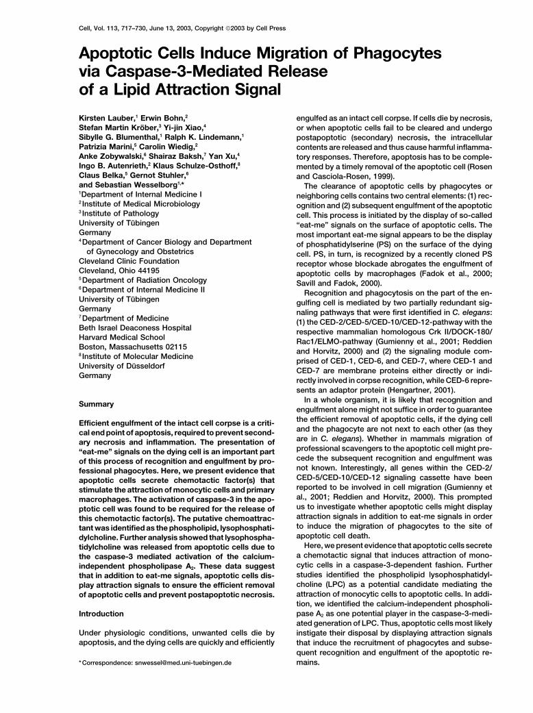
Figure 1. Attraction of Monocytic Cells to Supernatants of Apoptotic Cells
The different tumor cell lines HT29, Cos7, MCF7, and L929 were left untreated or induced to undergo apoptosis by UV irradiation (10 mJ/cm2). After 13 hr, cell-free supernatants were prepared and migration of THP-1, Mono Mac 6 cells, or primary human macrophages wasdetermined as described in Experimental Procedures.
Results tion of chemotactic activity in the supernatants ofMCF7casp3 cells was due to caspase activation, sinceaddition of the broad-spectrum caspase inhibitor zVAD-Supernatants of Different Apoptotic Cell Lines
Attract Monocytic THP-1, Mono Mac 6 Cells, fmk to MCF7casp3 cells completely abolished the migra-tion activity (Figure 2B). In order to exclude that theand Primary Macrophages
To investigate the involvement of chemotaxis in phago- absence of chemotactic activity of caspase-3 deficientcells was due to a decreased apoptosis induction, wecytic clearance of apoptotic cells we tested superna-
tants of apoptotic cells for their ability to induce migra- analyzed the cleavage of caspase-3 and -7 and of thecaspase substrate poly(ADP-ribose) polymerase (PARP).tion of monocytic cells. Therefore, we induced apoptosis
via UV irradiation in different cell lines such as the human In addition, we measured apoptosis via the inductionof cell shrinkage, since analysis of DNA fragmentationcolorectal adenocarcinoma cell line HT29, human breast
carcinoma MCF7 cells, the African green monkey kidney cannot be determined in caspase-3-deficient cells (Jan-icke et al., 1998). As shown in Figure 2C, the caspasecell line COS7, and murine fibroblast L929 cells. After
13 hr, supernatants were collected and tested for their substrate PARP was cleaved to a similar extent inMCF7vector and MCF7casp3 cells, though the processing ofability to induce the transmigration of the human mono-
cytic cell lines THP-1, Mono Mac 6, and primary human procaspase-7 was slightly decreased in MCF7vector cells.Caspase-3 was only moderately cleaved in MCF7casp3macrophages. As shown in Figure 1, supernatants of
almost all UV-irradiated apoptotic cell lines were able cells and, as expected, it was completely absent in cas-pase-3-deficient cells. Addition of zVAD-fmk blocked theto induce monocyte migration. In the case of L929 cells,
supernatants of unstimulated control cells also induced processing of all caspases and of PARP. Induction ofapoptosis was mediated to a similar extent in MCF7vectortransmigration of THP-1, which was further enhanced
upon apoptosis induction. Interestingly, supernatants of and MCF7casp3 cells (Figure 2D). Thus, activation of cas-pase-3 seemed to be crucial for the release of the che-apoptotic human breast carcinoma MCF7 cells insti-
gated only minimal transmigration activity of the mono- motactic factor by apoptotic cells, but not for the induc-tion of apoptosis.cytic cell lines and primary macrophages.
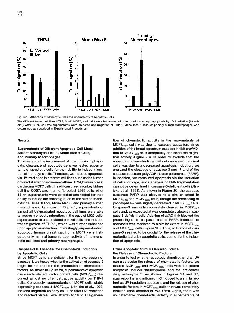
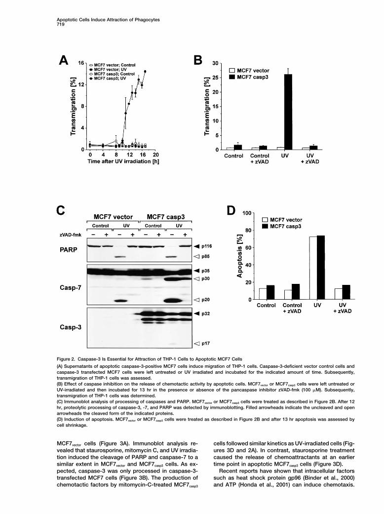
Caspase-3 Is Essential for Chemotaxis Induction Other Apoptotic Stimuli Can also Inducethe Release of Chemotactic Factorsby Apoptotic Cells
Since MCF7 cells are deficient for the expression of In order to test whether apoptotic stimuli other than UVcan also evoke the release of chemotactic factors, wecaspase-3, we tested whether the activation of caspase-3
might be required for the production of chemotactic treated MCF7vector and MCF7casp3 cells with the potentapoptosis inducer staurosporine and the anticancerfactors. As shown in Figure 2A, supernatants of apoptotic
caspase-3-deficient vector control cells (MCF7vector) dis- drug mitomycin C. As shown in Figures 3A and 3C,staurosporine and mitomycin C induced to a similar ex-played almost no chemoattractive activity on THP-1
cells. Conversely, supernatants of MCF7 cells stably tent as UV irradiation apoptosis and the release of che-motactic factors in MCF7casp3 cells that was completelyexpressing caspase-3 (MCF7casp3) (Janicke et al., 1998)
induced migration as early as 11 hr after UV irradiation blocked upon addition of zVAD-fmk. Again, there wasno detectable chemotactic activity in supernatants ofand reached plateau level after 15 to 16 hr. The genera-
Apoptotic Cells Induce Attraction of Phagocytes719
Figure 2. Caspase-3 Is Essential for Attraction of THP-1 Cells to Apoptotic MCF7 Cells
(A) Supernatants of apoptotic caspase-3-positive MCF7 cells induce migration of THP-1 cells. Caspase-3-deficient vector control cells andcaspase-3 transfected MCF7 cells were left untreated or UV irradiated and incubated for the indicated amount of time. Subsequently,transmigration of THP-1 cells was assessed.(B) Effect of caspase inhibition on the release of chemotactic activity by apoptotic cells. MCF7vector or MCF7casp3 cells were left untreated orUV-irradiated and then incubated for 13 hr in the presence or absence of the pancaspase inhibitor zVAD-fmk (100 �M). Subsequently,transmigration of THP-1 cells was determined.(C) Immunoblot analysis of processing of caspases and PARP. MCF7vector or MCF7casp3 cells were treated as described in Figure 2B. After 12hr, proteolytic processing of caspase-3, -7, and PARP was detected by immunoblotting. Filled arrowheads indicate the uncleaved and openarrowheads the cleaved form of the indicated proteins.(D) Induction of apoptosis. MCF7vector or MCF7casp3 cells were treated as described in Figure 2B and after 13 hr apoptosis was assessed bycell shrinkage.
MCF7vector cells (Figure 3A). Immunoblot analysis re- cells followed similar kinetics as UV-irradiated cells (Fig-ures 3D and 2A). In contrast, staurosporine treatmentvealed that staurosporine, mitomycin C, and UV irradia-
tion induced the cleavage of PARP and caspase-7 to a caused the release of chemoattractants at an earliertime point in apoptotic MCF7casp3 cells (Figure 3D).similar extent in MCF7vector and MCF7casp3 cells. As ex-
pected, caspase-3 was only processed in caspase-3- Recent reports have shown that intracellular factorssuch as heat shock protein gp96 (Binder et al., 2000)transfected MCF7 cells (Figure 3B). The production of
chemotactic factors by mitomycin-C-treated MCF7casp3 and ATP (Honda et al., 2001) can induce chemotaxis.
Cell720
Figure 3. Induction of Apoptosis by Staurosporine and Mitomycin C Mediates the Release of the Chemotactic Factor
(A) Effect of apoptosis induction via UV-irradiation, mitomycin C, and staurosporine on the release of chemotactic activity from MCF7 cells.MCF7vector or MCF7casp3 cells were UV irradiated (UV), incubated with medium (Control), mitomycin C (25 �g/ml; Mito), or staurosporine (2.5�M; Stauro) and incubated with or without 100 �M zVAD-fmk for 13 hr. Cell-free supernatants were prepared and filtered on Sephadex G15columns and applied to migration assay with THP-1 cells.(B) Immunoblot analysis of processing of caspases and PARP. MCF7vector or MCF7casp3 cells were UV irradiated (10 hr), incubated with medium(5 hr), mitomycin C (10 hr), or staurosporine (5 hr) in the absence or presence of 100 �M zVAD-fmk.
Apoptotic Cells Induce Attraction of Phagocytes721
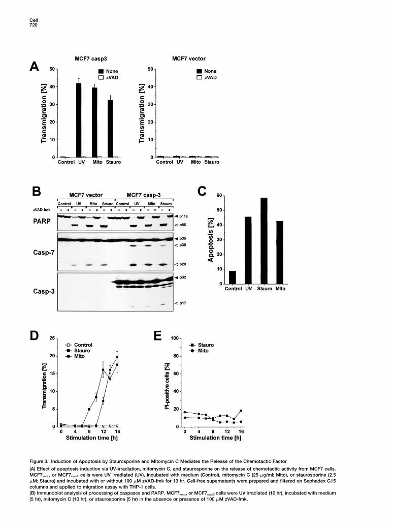
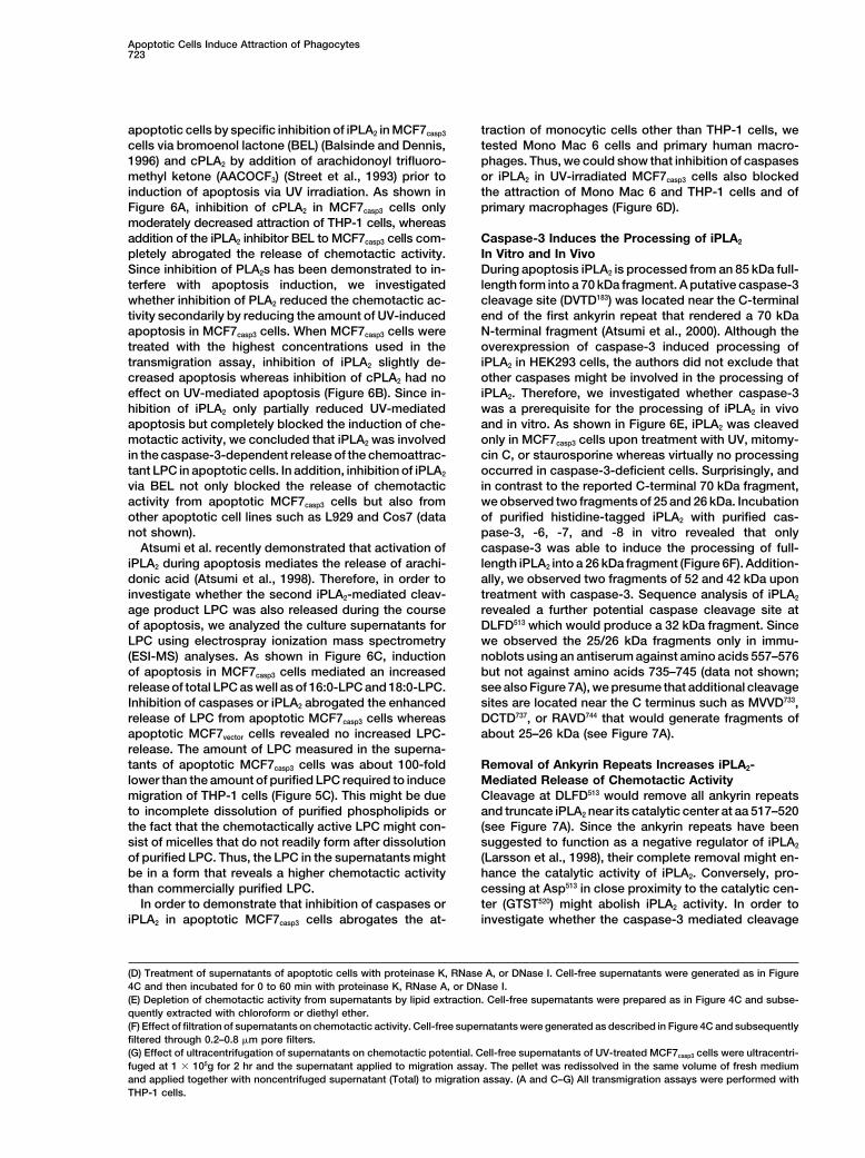
Since caspase-3 is the major caspase involved in the Chemotaxis Induction by Apoptotic Cells IsMediated via Lysophosphatidylcholinedegradation of a number of vital substrates, it was con-
ceivable that apoptotic stimuli might induce a faster and Next, we investigated whether different biologically ac-tive phospholipids could neutralize the chemotactic ac-more pronounced disintegration of caspase-3 positive
MCF7 cells. However, using the exclusion of propidium tivity of supernatants from apoptotic cells in the transmi-gration assay. To this end, we added phosphatidylserineiodide as a parameter for membrane integrity we could
demonstrate that the membrane of the apoptotic cells (PS), phosphatidylinositol (PI), phosphatidylcholine (PC),lysophosphatidylcholine (LPC), lysophosphatic acid (LPA),treated with staurosporine, mitomycin C (Figure 3E) or
UV (data not shown) was intact at all time points tested platelet-activating factor (PAF), or sphingosine-1-phos-phate (S1P) to THP-1 cells on the upper side of thefor migration activity. Thus, apoptotic cells release a
chemotactic factor at a time point when the plasma transmigration membrane to test whether these phos-pholipids could neutralize the chemotactic activity ofmembrane is still intact and thus before commitment to
postapoptotic necrosis. supernatants of apoptotic cells in the lower chamber.Among these phospholipids, LPA, LPC, S1P, and PAFhave been shown to induce chemotaxis in different cell
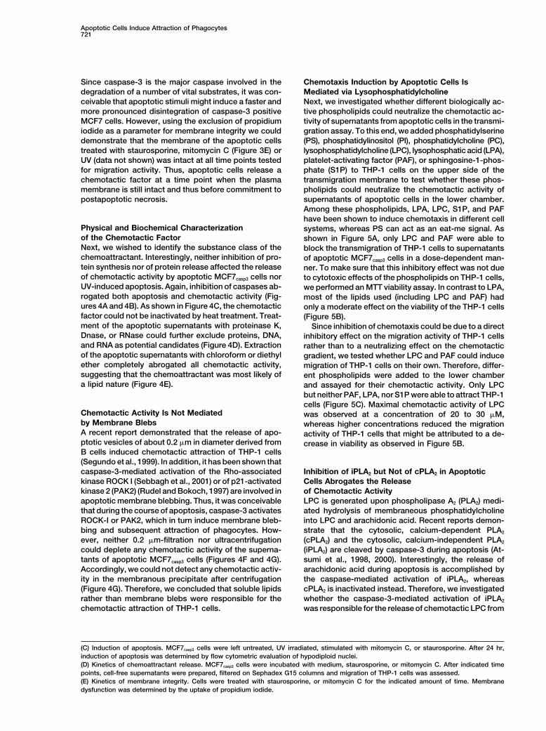
Physical and Biochemical Characterization systems, whereas PS can act as an eat-me signal. Asof the Chemotactic Factor shown in Figure 5A, only LPC and PAF were able toNext, we wished to identify the substance class of the block the transmigration of THP-1 cells to supernatantschemoattractant. Interestingly, neither inhibition of pro- of apoptotic MCF7casp3 cells in a dose-dependent man-tein synthesis nor of protein release affected the release ner. To make sure that this inhibitory effect was not dueof chemotactic activity by apoptotic MCF7casp3 cells nor to cytotoxic effects of the phospholipids on THP-1 cells,UV-induced apoptosis. Again, inhibition of caspases ab- we performed an MTT viability assay. In contrast to LPA,rogated both apoptosis and chemotactic activity (Fig- most of the lipids used (including LPC and PAF) hadures 4A and 4B). As shown in Figure 4C, the chemotactic only a moderate effect on the viability of the THP-1 cellsfactor could not be inactivated by heat treatment. Treat- (Figure 5B).ment of the apoptotic supernatants with proteinase K, Since inhibition of chemotaxis could be due to a directDnase, or RNase could further exclude proteins, DNA, inhibitory effect on the migration activity of THP-1 cellsand RNA as potential candidates (Figure 4D). Extraction rather than to a neutralizing effect on the chemotacticof the apoptotic supernatants with chloroform or diethyl gradient, we tested whether LPC and PAF could induceether completely abrogated all chemotactic activity, migration of THP-1 cells on their own. Therefore, differ-suggesting that the chemoattractant was most likely of ent phospholipids were added to the lower chambera lipid nature (Figure 4E). and assayed for their chemotactic activity. Only LPC
but neither PAF, LPA, nor S1P were able to attract THP-1cells (Figure 5C). Maximal chemotactic activity of LPC
Chemotactic Activity Is Not Mediated was observed at a concentration of 20 to 30 �M,by Membrane Blebs whereas higher concentrations reduced the migrationA recent report demonstrated that the release of apo- activity of THP-1 cells that might be attributed to a de-ptotic vesicles of about 0.2 �m in diameter derived from crease in viability as observed in Figure 5B.B cells induced chemotactic attraction of THP-1 cells(Segundo et al., 1999). In addition, it has been shown thatcaspase-3-mediated activation of the Rho-associated Inhibition of iPLA2 but Not of cPLA2 in Apoptotic
Cells Abrogates the Releasekinase ROCK I (Sebbagh et al., 2001) or of p21-activatedkinase 2 (PAK2) (Rudel and Bokoch, 1997) are involved in of Chemotactic Activity
LPC is generated upon phospholipase A2 (PLA2) medi-apoptotic membrane blebbing. Thus, it was conceivablethat during the course of apoptosis, caspase-3 activates ated hydrolysis of membraneous phosphatidylcholine
into LPC and arachidonic acid. Recent reports demon-ROCK-I or PAK2, which in turn induce membrane bleb-bing and subsequent attraction of phagocytes. How- strate that the cytosolic, calcium-dependent PLA2
(cPLA2) and the cytosolic, calcium-independent PLA2ever, neither 0.2 �m-filtration nor ultracentrifugationcould deplete any chemotactic activity of the superna- (iPLA2) are cleaved by caspase-3 during apoptosis (At-
sumi et al., 1998, 2000). Interestingly, the release oftants of apoptotic MCF7casp3 cells (Figures 4F and 4G).Accordingly, we could not detect any chemotactic activ- arachidonic acid during apoptosis is accomplished by
the caspase-mediated activation of iPLA2, whereasity in the membranous precipitate after centrifugation(Figure 4G). Therefore, we concluded that soluble lipids cPLA2 is inactivated instead. Therefore, we investigated
whether the caspase-3-mediated activation of iPLA2rather than membrane blebs were responsible for thechemotactic attraction of THP-1 cells. was responsible for the release of chemotactic LPC from
(C) Induction of apoptosis. MCF7casp3 cells were left untreated, UV irradiated, stimulated with mitomycin C, or staurosporine. After 24 hr,induction of apoptosis was determined by flow cytometric evaluation of hypodiploid nuclei.(D) Kinetics of chemoattractant release. MCF7casp3 cells were incubated with medium, staurosporine, or mitomycin C. After indicated timepoints, cell-free supernatants were prepared, filtered on Sephadex G15 columns and migration of THP-1 cells was assessed.(E) Kinetics of membrane integrity. Cells were treated with staurosporine, or mitomycin C for the indicated amount of time. Membranedysfunction was determined by the uptake of propidium iodide.
Cell722
Figure 4. Physical and Biochemical Characterization of Chemoattractant
(A) Effect of the inhibition of protein synthesis, protein secretion, and caspases on the release of chemoattractant from apoptotic cells.MCF7casp3 cells were UV irradiated and then incubated with or without actinomycin D (100 ng/ml), cycloheximide (1 �g/ml), brefeldin A (1�g/ml), or zVAD-fmk (100 �M). After 13 hr, cell-free supernatants were prepared and Sephadex G15 filtered.(B) Apoptosis induction. MCF7casp3 cells were treated as in Figure 4A and induction of apoptosis was measured after 24 hr by flow cytometricevaluation of hypodiploid nuclei.(C) Heat treatment of supernatants of apoptotic cells. MCF7casp3 cells were untreated or UV irradiated. After 13 hr, cell-free supernatants wereprepared and heated for 10 min at 50�C, 70�C, or 90�C, or left at room temperature.
Apoptotic Cells Induce Attraction of Phagocytes723
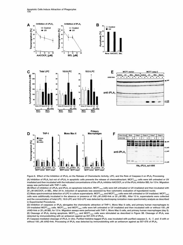
apoptotic cells by specific inhibition of iPLA2 in MCF7casp3 traction of monocytic cells other than THP-1 cells, wetested Mono Mac 6 cells and primary human macro-cells via bromoenol lactone (BEL) (Balsinde and Dennis,
1996) and cPLA2 by addition of arachidonoyl trifluoro- phages. Thus, we could show that inhibition of caspasesor iPLA2 in UV-irradiated MCF7casp3 cells also blockedmethyl ketone (AACOCF3) (Street et al., 1993) prior to
induction of apoptosis via UV irradiation. As shown in the attraction of Mono Mac 6 and THP-1 cells and ofprimary macrophages (Figure 6D).Figure 6A, inhibition of cPLA2 in MCF7casp3 cells only
moderately decreased attraction of THP-1 cells, whereasaddition of the iPLA2 inhibitor BEL to MCF7casp3 cells com- Caspase-3 Induces the Processing of iPLA2
In Vitro and In Vivopletely abrogated the release of chemotactic activity.Since inhibition of PLA2s has been demonstrated to in- During apoptosis iPLA2 is processed from an 85 kDa full-
length form into a 70 kDa fragment. A putative caspase-3terfere with apoptosis induction, we investigatedwhether inhibition of PLA2 reduced the chemotactic ac- cleavage site (DVTD183) was located near the C-terminal
end of the first ankyrin repeat that rendered a 70 kDativity secondarily by reducing the amount of UV-inducedapoptosis in MCF7casp3 cells. When MCF7casp3 cells were N-terminal fragment (Atsumi et al., 2000). Although the
overexpression of caspase-3 induced processing oftreated with the highest concentrations used in thetransmigration assay, inhibition of iPLA2 slightly de- iPLA2 in HEK293 cells, the authors did not exclude that
other caspases might be involved in the processing ofcreased apoptosis whereas inhibition of cPLA2 had noeffect on UV-mediated apoptosis (Figure 6B). Since in- iPLA2. Therefore, we investigated whether caspase-3
was a prerequisite for the processing of iPLA2 in vivohibition of iPLA2 only partially reduced UV-mediatedapoptosis but completely blocked the induction of che- and in vitro. As shown in Figure 6E, iPLA2 was cleaved
only in MCF7casp3 cells upon treatment with UV, mitomy-motactic activity, we concluded that iPLA2 was involvedin the caspase-3-dependent release of the chemoattrac- cin C, or staurosporine whereas virtually no processing
occurred in caspase-3-deficient cells. Surprisingly, andtant LPC in apoptotic cells. In addition, inhibition of iPLA2
via BEL not only blocked the release of chemotactic in contrast to the reported C-terminal 70 kDa fragment,we observed two fragments of 25 and 26 kDa. Incubationactivity from apoptotic MCF7casp3 cells but also from
other apoptotic cell lines such as L929 and Cos7 (data of purified histidine-tagged iPLA2 with purified cas-pase-3, -6, -7, and -8 in vitro revealed that onlynot shown).
Atsumi et al. recently demonstrated that activation of caspase-3 was able to induce the processing of full-length iPLA2 into a 26 kDa fragment (Figure 6F). Addition-iPLA2 during apoptosis mediates the release of arachi-
donic acid (Atsumi et al., 1998). Therefore, in order to ally, we observed two fragments of 52 and 42 kDa upontreatment with caspase-3. Sequence analysis of iPLA2investigate whether the second iPLA2-mediated cleav-
age product LPC was also released during the course revealed a further potential caspase cleavage site atDLFD513 which would produce a 32 kDa fragment. Sinceof apoptosis, we analyzed the culture supernatants for
LPC using electrospray ionization mass spectrometry we observed the 25/26 kDa fragments only in immu-noblots using an antiserum against amino acids 557–576(ESI-MS) analyses. As shown in Figure 6C, induction
of apoptosis in MCF7casp3 cells mediated an increased but not against amino acids 735–745 (data not shown;see also Figure 7A), we presume that additional cleavagerelease of total LPC as well as of 16:0-LPC and 18:0-LPC.
Inhibition of caspases or iPLA2 abrogated the enhanced sites are located near the C terminus such as MVVD733,DCTD737, or RAVD744 that would generate fragments ofrelease of LPC from apoptotic MCF7casp3 cells whereas
apoptotic MCF7vector cells revealed no increased LPC- about 25–26 kDa (see Figure 7A).release. The amount of LPC measured in the superna-tants of apoptotic MCF7casp3 cells was about 100-fold Removal of Ankyrin Repeats Increases iPLA2-
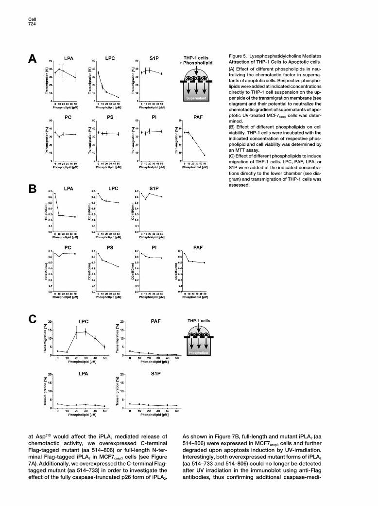
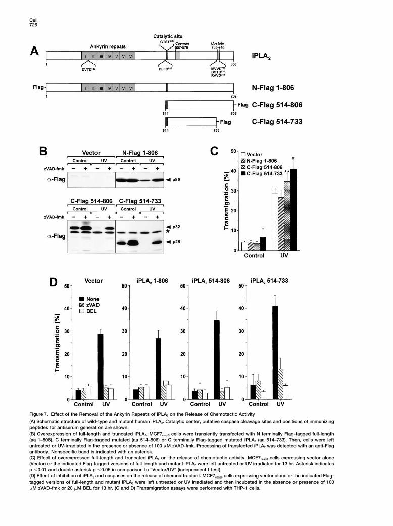
Mediated Release of Chemotactic Activitylower than the amount of purified LPC required to inducemigration of THP-1 cells (Figure 5C). This might be due Cleavage at DLFD513 would remove all ankyrin repeats
and truncate iPLA2 near its catalytic center at aa 517–520to incomplete dissolution of purified phospholipids orthe fact that the chemotactically active LPC might con- (see Figure 7A). Since the ankyrin repeats have been
suggested to function as a negative regulator of iPLA2sist of micelles that do not readily form after dissolutionof purified LPC. Thus, the LPC in the supernatants might (Larsson et al., 1998), their complete removal might en-
hance the catalytic activity of iPLA2. Conversely, pro-be in a form that reveals a higher chemotactic activitythan commercially purified LPC. cessing at Asp513 in close proximity to the catalytic cen-
ter (GTST520) might abolish iPLA2 activity. In order toIn order to demonstrate that inhibition of caspases oriPLA2 in apoptotic MCF7casp3 cells abrogates the at- investigate whether the caspase-3 mediated cleavage
(D) Treatment of supernatants of apoptotic cells with proteinase K, RNase A, or DNase I. Cell-free supernatants were generated as in Figure4C and then incubated for 0 to 60 min with proteinase K, RNase A, or DNase I.(E) Depletion of chemotactic activity from supernatants by lipid extraction. Cell-free supernatants were prepared as in Figure 4C and subse-quently extracted with chloroform or diethyl ether.(F) Effect of filtration of supernatants on chemotactic activity. Cell-free supernatants were generated as described in Figure 4C and subsequentlyfiltered through 0.2–0.8 �m pore filters.(G) Effect of ultracentrifugation of supernatants on chemotactic potential. Cell-free supernatants of UV-treated MCF7casp3 cells were ultracentri-fuged at 1 � 105g for 2 hr and the supernatant applied to migration assay. The pellet was redissolved in the same volume of fresh mediumand applied together with noncentrifuged supernatant (Total) to migration assay. (A and C–G) All transmigration assays were performed withTHP-1 cells.
Cell724
Figure 5. Lysophosphatidylcholine MediatesAttraction of THP-1 Cells to Apoptotic cells
(A) Effect of different phospholipids in neu-tralizing the chemotactic factor in superna-tants of apoptotic cells. Respective phospho-lipids were added at indicated concentrationsdirectly to THP-1 cell suspension on the up-per side of the transmigration membrane (seediagram) and their potential to neutralize thechemotactic gradient of supernatants of apo-ptotic UV-treated MCF7casp3 cells was deter-mined.(B) Effect of different phospholipids on cellviability. THP-1 cells were incubated with theindicated concentration of respective phos-pholipid and cell viability was determined byan MTT assay.(C) Effect of different phospholipids to inducemigration of THP-1 cells. LPC, PAF, LPA, orS1P were added at the indicated concentra-tions directly to the lower chamber (see dia-gram) and transmigration of THP-1 cells wasassessed.
at Asp513 would affect the iPLA2 mediated release of As shown in Figure 7B, full-length and mutant iPLA2 (aa514–806) were expressed in MCF7casp3 cells and furtherchemotactic activity, we overexpressed C-terminal
Flag-tagged mutant (aa 514–806) or full-length N-ter- degraded upon apoptosis induction by UV-irradiation.Interestingly, both overexpressed mutant forms of iPLA2minal Flag-tagged iPLA2 in MCF7casp3 cells (see Figure
7A). Additionally, we overexpressed the C-terminal Flag- (aa 514–733 and 514–806) could no longer be detectedafter UV irradiation in the immunoblot using anti-Flagtagged mutant (aa 514–733) in order to investigate the
effect of the fully caspase-truncated p26 form of iPLA2. antibodies, thus confirming additional caspase-medi-
Apoptotic Cells Induce Attraction of Phagocytes725
Figure 6. Effect of the Inhibition of iPLA2 on the Release of Chemotactic Activity, LPC, and the Role of Caspase-3 on iPLA2 Processing
(A) Inhibition of iPLA2 but not of cPLA2 in apoptotic cells prevents the release of chemoattractant. MCF7casp3 cells were left untreated or UVirradiated and then incubated with the indicated concentrations of the cPLA2 inhibitor AACOCF3 or of the iPLA2 inhibitor BEL for 13 hr. Migrationassay was performed with THP-1 cells.(B) Effect of inhibition of cPLA2 and iPLA2 on apoptosis induction. MCF7casp3 cells were left untreated or UV-irradiated and then incubated with20 �M AACOCF3 or BEL. After 24 hr, induction of apoptosis was assessed by flow cytometric evaluation of hypodiploid nuclei.(C) Mass spectrometrical detection of LPC in culture supernatants. MCF7vector and MCF7casp3 cells were left untreated or UV irradiated. MCF7casp3
cells were additionally incubated in the absence or presence of 100 �M zVAD-fmk or 20 �M BEL. After 13 hr, supernatants were collectedand the concentration of total LPC, 16:0-LPC and 18:0-LPC was detected by electrospray ionization mass spectrometry analysis as describedin Experimental Procedures.(D) Inhibition of caspases or iPLA2 abrogates the chemotactic attraction of THP-1, Mono Mac 6 cells, and primary human macrophages toUV-irradiated MCF7casp3 cells. MCF7vector and MCF7casp3 cells were left untreated or UV irradiated and then incubated with or without 100 �MzVAD-fmk or 20 �M BEL for 13 hr. Migration assays were performed with THP-1, Mono Mac 6 cells, and primary human macrophages (day 5).(E) Cleavage of iPLA2 during apoptosis. MCF7vector and MCF7casp3 cells were stimulated as described in Figure 3B. Cleavage of iPLA2 wasdetected by immunoblotting with an antiserum against aa 557–576 of iPLA2.(F) Caspase-mediated cleavage of iPLA2 in vitro. Purified histidine-tagged iPLA2 was incubated with purified caspase-3, -6, -7, and -8 with orwithout 100 �M zVAD-fmk. Processing of iPLA2 was detected by immunoblotting with an antiserum against aa 557–576 of iPLA2.
Cell726
Figure 7. Effect of the Removal of the Ankyrin Repeats of iPLA2 on the Release of Chemotactic Activity
(A) Schematic structure of wild-type and mutant human iPLA2. Catalytic center, putative caspase cleavage sites and positions of immunizingpeptides for antiserum generation are shown.(B) Overexpression of full-length and truncated iPLA2. MCF7casp3 cells were transiently transfected with N terminally Flag-tagged full-length(aa 1–806), C terminally Flag-tagged mutated (aa 514–806) or C terminally Flag-tagged mutated iPLA2 (aa 514–733). Then, cells were leftuntreated or UV-irradiated in the presence or absence of 100 �M zVAD-fmk. Processing of transfected iPLA2 was detected with an anti-Flagantibody. Nonspecific band is indicated with an asterisk.(C) Effect of overexpressed full-length and truncated iPLA2 on the release of chemotactic activity. MCF7casp3 cells expressing vector alone(Vector) or the indicated Flag-tagged versions of full-length and mutant iPLA2 were left untreated or UV irradiated for 13 hr. Asterisk indicatesp �0.01 and double asterisk p �0.05 in comparison to “Vector/UV” (independent t test).(D) Effect of inhibition of iPLA2 and caspases on the release of chemoattractant. MCF7casp3 cells expressing vector alone or the indicated Flag-tagged versions of full-length and mutant iPLA2 were left untreated or UV irradiated and then incubated in the absence or presence of 100�M zVAD-fmk or 20 �M BEL for 13 hr. (C and D) Transmigration assays were performed with THP-1 cells.
Apoptotic Cells Induce Attraction of Phagocytes727
ated cleavage of iPLA2 at the C terminus (Figure 7B). represent a prerequisite for its efficient removal by pro-fessional scavengers.When we analyzed the effect of native and both trun-
In the present study, we investigated this hypothesiscated forms of iPLA2 on the release of chemotactic activ-and observed that supernatants of apoptotic cells in-ity, we observed that overexpression of full-length iPLA2
duced migration of monocytic THP-1 cells, Mono Macdid not further augment the chemotactic activity in su-6 cells, and primary macrophages. The major releasepernatants of apoptotic MCF7casp3 cells, whereas overex-of the chemotactic factor was mediated via caspase-3pression of both truncated forms of iPLA2 increased thesince supernatants of apoptotic caspase-3-deficientrelease of chemotactic activity (Figure 7C). Interestingly,MCF7 cells, in contrast to caspase-3 expressing MCF7overexpression of the fully caspase-truncated p26 formcells, showed almost no attraction of THP-1 cells. In(aa 514–733) displayed an even more pronounced effectaddition to the attraction of monocytic cells in vitro,than the partially truncated p32-form (aa 514–806) ofsupernatants of apoptotic MCF7casp3 cells appeared toiPLA2. However, overexpression of either form of mutantinduce also the attraction of macrophages upon intra-iPLA2 (aa 514–806 and aa 514–733) could not induceperitoneal or subcutaneous injection in vivo (see Supple-significant attraction in supernatants of nonapoptoticmental Figures S1, and S2 at URL above).MCF7casp3 cells (Figure 7C). As in previous experiments,
Biochemical characterization of the supernatants ofinhibition of iPLA2 by addition of BEL completely abro-apoptotic cells revealed that the putative chemoattrac-gated the release of chemotactic activity in all apoptotictant was not a protein, DNA, or RNA but most likely aMCF7casp3 cells overexpressing different constructs oflipid instead. Potential chemotactic lipid factors wereiPLA2 (Figure 7D). Remarkably, caspase inhibition by thearachidonic acid and its derivates as well as apoptoticaddition of zVAD-fmk only partially blocked the releasemembrane blebs and phospholipids. However, we couldof chemotactic activity from apoptotic MCF7casp3 cellsrule out arachidonic acid and most of its derivates (pros-overexpressing the truncated p26 form. This was con-taglandins, leukotrienes, and thromboxanes), becausesistently observed in several experiments but neverthey are not extractable with diethyl ether or chloroform.found in cells overexpressing the empty vector alone,Apoptotic vesicles could be excluded since neither fil-full-length iPLA2, or mutant iPLA2 (aa 514–806). The activ-tration nor ultracentrifugation of the supernatants ofity retained in the presence of zVAD corresponded toapoptotic cells abrogated the attraction of monocyticthe increase of chemotactic activity in p26-transfectedTHP-1 cells. Therefore, we focused on phospholipidscells compared to vector-transfected cells. Since thesuch as LPA, LPC, S1P, and PAF that have been showncompletely truncated p26 form (aa 514–733) does notto induce chemotaxis. Of all the phospholipids testedrequire further processing, this might explain why apo-only LPC and PAF could neutralize the chemotactic ca-ptotic MCF7casp3 cells overexpressing the p26 fragmentpacity in supernatants of apoptotic cells when addedstill released chemotactic activity in the presence ofdirectly to the responding THP-1 cells. However, onlyzVAD-fmk.LPC could induce migration on its own whereas PAFWe also tested the ability of the apoptotic superna-was ineffective. Therefore, LPC that has been knowntants to recruit macrophages in two in vivo model sys-for a long time as a chemoattractant for monocytestems including a peritonitis and a subcutaneous infiltra-(Hoffman et al., 1982) seemed to be a crucial factor fortion model. To this end, supernatants of apoptotic andthe attraction of monocytes to apoptotic cells.nonapoptotic cells were injected into the peritoneum or
LPC is generated via PLA2-mediated processing ofsubcutaneously, and then histologically evaluated in athe membranous phosphatidylcholine into LPC and ara-time course for their effect on macrophage infiltration.chidonic acid. Mammalian PLA2 isoenzymes are subdi-Supporting our previous results, in both models therevided into four major families, including cytosolic, cal-was a considerably more pronounced (2-fold and higher)cium-dependent (cPLA2), secretory (sPLA2), cytosolic,recruitment of macrophages after injection of the super-calcium-independent PLA2 (iPLA2), and PAF acetyl hy-
natants from UV-treated MCF-7casp3 cells compared todrolases (Murakami et al., 2000). It was shown that pro-
supernatants from untreated cells (see Supplementalcessing by caspases inactivates cPLA2, whereas iPLA2Figures S1 and S2 available at http://www.cell.com/cgi/ is activated instead (Atsumi et al., 2000) and mediates
content/full/113/6/717/DC1). The chemotactic activity the release of arachidonic acid from apoptotic cells (At-was comparable to the effects seen after injection of sumi et al., 1998). In addition to arachidonic acid, wepurified LPC or proteose peptone and LPS that were could now demonstrate that the second iPLA2-mediatedused as positive controls of potent proinflammatory cleavage product LPC was also released from apoptoticstimuli. Thus, these data underline a biological relevance cells.of our findings and suggest that attraction signals could Because ankyrin repeats function as a negative regu-play a role in macrophage recruitment in vivo. lator of iPLA2 (Larsson et al., 1998) their caspase medi-
ated removal might contribute to the activation of iPLA2.Discussion Using a partially truncated p32-form (aa 514–806) or the
completely truncated p26 form (aa 514–733) of iPLA2 weTo date relatively little is known about what triggers could show that the cleavage of iPLA2 near the catalyticmigration of professional phagocytes to the site of apo- site and the complete removal of ankyrin repeats didptotic cell death. One can envision that the sole display not affect the release of chemotactic activity but ratherof eat-me signals might not suffice in order to guarantee increased the attraction of THP-1 cells. Nevertheless, ita timely removal of the apoptotic cell before commit- is conceivable that caspases might activate iPLA2 in ament to postapoptotic necrosis. Therefore, the display rather indirect way. Thus, iPLA2 might be retained within
the cytosol via an anchoring protein that obstructs theof long-range attraction signals by the dying cell might
Cell728
access of iPLA2 to its membrane-associated substrate flammatory responses and autoimmune diseases (Ro-phosphatidylcholine. Caspase-mediated processing of sen and Casciola-Rosen, 1999). Usually, internalizationiPLA2 might release iPLA2 from its inhibitor, thus increas- of an apoptotic cell by macrophages is accompanieding its access to endogenous substrate. Conversely, by the expression of anti-inflammatory cytokines suchcaspases might directly cleave the putative inhibitor and as transforming growth factor-� (Fadok et al., 1998).thereby release iPLA2. Accordingly, iPLA2 has been re- Conversely, uptake of necrotic cells by specializedported to exist as a multimeric complex of 270–350 kDa phagocytes as the dendritic cells (DCs) has rather anin the cell that might contain a putative cytosolic inhibitor adverse effect in activation of the DC to express costi-(Larsson et al., 1998). The observation that overexpres- mulatory molecules that are necessary for T cell activa-sion of the completely truncated p26 form of iPLA2 does tion and induction of an inflammatory response (Gallucinot induce significant chemotactic activity in nonapo- et al., 1999; Sauter et al., 2000). Therefore, one mightptotic cells, yet increases the release of chemotactic speculate that if apoptotic cells are not engulfed in timeactivity in cells undergoing apoptosis indicates that cas- by phagocytes, postapoptotic necrosis occurs, initiatingpases might exert a direct and an indirect effect on the inflammation and autoimmunity instead of self-tol-activation of iPLA2. erance.
Whether or not under physiologic conditions iPLA2 Taken together, we conclude that the clearance ofand LPC will be the sole elements required for inducing apoptotic cells is a complex multistep mechanism. Dur-the attraction of phagocytes to the apoptotic cell has to ing the course of apoptosis cells display “eat-me” sig-be proven. It is noteworthy that the perturbed membrane nals such as PS and attraction signals such as LPC.asymmetry of cells undergoing apoptosis enables the Attraction signals initiate the migration of professionalaccess of secretory PLA2 (sPLA2) (Atsumi et al., 1997; phagocytes to the apoptotic cell. Phagocytes eventuallyFourcade et al., 1995; Murakami et al., 2000). Therefore, bind to the dying cell and due to the lack of exposedsPLA2 might also contribute to the generation of LPC “detachment” signals, they are not repulsed (Brown etvia hydrolysis of phosphatidylcholine from the outer al., 2002). Display of eat-me signals ensures the recogni-membrane leaflet of apoptotic cells. Conversely, iPLA2 tion and subsequent engulfment of the apoptotic cell.and LPC might not only be involved in attraction of Further studies will be required to understand how de-phagocytes but also contribute to the recognition of fects in exposing attraction or eat-me signals on the partapoptotic cells. In this context, a recent report demon- of the apoptotic cell as well as defects in chemotaxis,strated that the iPLA2-mediated generation of cell- recognition, and engulfment on the part of the phago-bound LPC might opsonize the dying cell for phagocyto- cyte might contribute to the generation of pathologicalsis by binding of natural IgM antibodies to LPC and conditions such as autoimmunity.subsequent complement activation (Kim et al., 2002). Asin the case of phosphatidylserine regarding recognition Experimental Proceduresand engulfment, there might as well exist factors other
Cellsthan LPC that contribute to the display of attractionMCF7 and THP-1 cells were cultured in RPMI-1640 and HT29, COS7signals by apoptotic cells.and L929 cells in DMEM supplemented with 10% fetal calf serum.Current reports show that a family of G protein-cou-Mono Mac 6 cells were kindly provided by Dr. Ziegler-Heitbrock.pled receptors (GPRs) mediates the binding and signalMCF7 cells stably transfected with caspase-3 and the caspase-3-
transduction of extracellular phospholipids. Thus, LPA deficient vector control cells were a kind gift of R.U. Janicke andand S1P bind to receptors of the Edg subfamily of GPRs, A.G. Porter (Janicke et al., 1998). Human monocyte-derived macro-while the PAF receptor as well as the receptors for LPC phages were generated from peripheral blood mononuclear cells of
healthy donors. Monocytes were obtained using a negative-selec-and SPC also constitute members of the GPR group.tion-based magnetic cell separation system (Miltenyi Biotec). Mono-SPC has been shown to bind to the ovarian cancer GPRcytes were then cultured in macrophage-serum-free medium (In-(OGR1) and to GPR4 (Xu et al., 2000; Zhu et al., 2001).vitrogen) supplemented with 2.5% autologous serum and 25 ng/mlLPC has recently been demonstrated to bind to thehuman GM-CSF (Schering-Plough) in teflon dishes for 5 days yield-
orphan GPR G2A and, with lower affinity than SPC, to ing about 80% of CD14 and CD206 double-positive macrophages.GPR4 (Kabarowski et al., 2001; Zhu et al., 2001). Trig-gering of G2A via LPC-induced migration in addition to Induction of Apoptosis; Production and Treatmentan increase in intracellular calcium concentration, G2A- of Chemoattractive Cell Culture Supernatantsreceptor internalization and ERK protein kinase activa- 2 � 106 cells cultured in 6-well plates were UV-irradiated with 10
mJ/cm2 with the UV Stratalinker 2400 (Stratagene). Alternatively,tion (Kabarowski et al., 2001). Furthermore, G2A-defi-apoptosis was induced with staurosporine (2.5 �M) or mitomycin Ccient mice have been reported to develop a late-onset(25 �g/ml) for the indicated time. Supernatants were prepared bymultiorgan inflammation with striking similarity to thecentrifugation at 1 � 104g for 5 min and stored at �70�C. In experi-
human autoimmune disease systemic lupus erythema- ments where different inhibitors, mitomycin C, or staurosporine weretosus (Le et al., 2001). In this context it is of interest to used, the cell-free supernatants were additionally passed throughnote that THP-1 cells, Mono Mac 6 cells, primary human a 2 ml gel bed of Sephadex G15. For heat-inactivation, supernatantsmonocytes, and macrophages expressed the known were heated at 50�C, 70�C, 90�C or left at room temperature for 10
min. Enzyme digestion of supernatants was carried out by addingLPC receptors G2A and GPR4 (see Supplemental Figure50 �g/ml proteinase K, 50 �g/ml RNase A, or 8 �g/ml DNase I andS3 available at http://www.cell.com/cgi/content/full/incubating for 0 to 60 min at 37�C. Chloroform and diethyl ether113/6/717/DC1).extraction was performed with 1 volume of chloroform or diethyl
In general, autoimmunity is thought to be the result ether, and was repeated twice. Filtration of the culture supernatantsof the inefficient elimination of autoreactive lymphocytes was carried out with Millex sterile filters of 0.2 to 0.8 �m pore size.by apoptosis. However, there is evidence that inefficient Ultracentrifugation of the cell culture supernatants was performed
at 1 � 105g for 2 hr. The resulting supernatant was transferred to aremoval of apoptotic cells might also contribute to proin-
Apoptotic Cells Induce Attraction of Phagocytes729
fresh tube and the pellet was redissolved in the same volume of AcknowledgmentsRPMI-1640 medium.
The authors wish to thank Drs. V.M. Dixit, R.U. Janicke, A.G. Porter,and H.W. Ziegler-Heitbrock for providing valuable cell lines and
Transmigration Assay reagents and K. Ravichandran, E. Brugnera, and C. Grimsley forTransmigration assays were performed with 8 �m pore size helpful discussions. This work was supported by grants from theChemoTX plates (Neuroprobe). Three-hundred microliters of re- Federal Ministry of Education, Science, Research and Technologyspective culture supernatant was placed into the lower chamber of (Fo. 01KS9602), Interdisciplinary Center of Clinical Research Tubin-ChemoTX plate and 50 �l of calcein-stained THP-1 or Mono Mac 6 gen (IZKF), German Bundesministerium fur Bildung und Forschungcells (2 � 106/ml) added on top of the filter-membrane and the assay (Hep-Net), Deutsche Forschungsgemeinschaft (WE-1801/1), Landes-was incubated for 90 min at 37�C. Migrated cells were lysed and forschungsschwerpunktprogramm of the Ministry of Science, Re-green fluorescence was analyzed using an excitation wavelength search, and Arts of the Land Baden-Wurttemberg to S.W. and fromof 480 nm and an emission wavelength of 508 nm. Transmigration grants by the Mildred Scheel Stiftung (10-1970 Be-III, 10-1764 Be-I)was assessed in percentage of total cells deployed and mean to C.B. and P.M.values � SD from triplicate experiments are given.
Received: September 20, 2002Revised: May 7, 2003
Measurement of Apoptosis and Cell Viability Accepted: May 8, 2003For determination of apoptosis, the leakage of fragmented DNA Published: June 12, 2003from apoptotic nuclei was measured by flow cytometry as described(Engels et al., 2000). Alternatively, apoptosis induction was mea- Referencessured by cell shrinkage of trypsinized MCF7 cells by flow cytometricanalysis using the FSC profile as a parameter for cell size. Membrane Atsumi, G., Murakami, M., Kojima, K., Hadano, A., Tajima, M., andpermeability of adherent MCF7 cells was assessed after trypsiniza- Kudo, I. (2000). Distinct roles of two intracellular phospholipase A2stion by the uptake of propidium iodide (2 �g/ml) into nonfixed cells in fatty acid release in the cell death pathway. J. Biol. Chem. 275,and subsequent flow cytometric analysis using FSC/FL2 profile. For 18248–18258.determination of cell viability, 1 � 105 THP-1 cells were incubated
Atsumi, G., Murakami, M., Tajima, M., Shimbara, S., Hara, N., andwith the respective phospholipids for 2 hr at 37�C. Subsequently,Kudo, I. (1997). The perturbed membrane of cells undergoing apo-cells were incubated for 5 hr at 37�C in the presence of MTT (450ptosis is susceptible to type II secretory phospholipase A2 to liberate�g/ml). Resulting formazan crystals were dissolved in 4% SDS andarachidonic acid. Biochim. Biophys. Acta 1349, 43–54.measured at 550 nm. Results were shown as mean values of dupli-Atsumi, G., Tajima, M., Hadano, A., Nakatani, Y., Murakami, M., andcates.Kudo, I. (1998). Fas-induced arachidonic acid release is mediatedby Ca2�-independent phospholipase A2 but not cytosolic phospholi-
In Vitro Cleavage of iPLA2 pase A2, which undergoes proteolytic inactivation. J. Biol. Chem.For in vitro processing of purified iPLA2 500 ng of histidine-tagged 273, 13870–13877.full-length iPLA2 was incubated with 5 �g of purified caspase-3, Balsinde, J., and Dennis, E.A. (1996). Distinct roles in signal trans-caspase-6, caspase-7, or caspase-8 in the presence or absence of duction for each of the phospholipase A2 enzymes present in P388D1100 �M zVAD-fmk in a total volume of 50 �l of a buffer containing macrophages. J. Biol. Chem. 271, 6758–6765.50 mM HEPES (pH 7.3), 100 mM NaCl, 10% sucrose, 0.1% CHAPS,
Binder, R.J., Anderson, K.M., Basu, S., and Srivastava, P.K. (2000).and 10 mM DTT at 37�C for 4 hr.Cutting edge: heat shock protein gp96 induces maturation and mi-gration of CD11c� cells in vivo. J. Immunol. 165, 6029–6035.
Brown, S., Heinisch, I., Ross, E., Shaw, K., Buckley, C.D., and Savill,Western Blot AnalysisWestern blot analysis was performed as described (Engels et al., J. (2002). Apoptosis disables CD31-mediated cell detachment from
phagocytes promoting binding and engulfment. Nature 418,2000) with monoclonal antibodies against PARP (Qbiogene-Alexis),caspase-7, caspase-3 (BD Biosciences), or Flag (Eastman Kodak). 200–203.iPLA2 was detected with a rabbit antiserum generated by immuniza- Engels, I.H., Stepczynska, A., Stroh, C., Lauber, K., Berg, C.,tion with a peptide spanning amino acids 557–576 of iPLA2 (Cayman Schwenzer, R., Wajant, H., Janicke, R.U., Porter, A.G., Belka, C., etChemicals) that detected the 85 kDa full-length form and the 25/ al. (2000). Caspase-8/FLICE functions as an executioner caspase26 kDa fragments. Alternatively, iPLA2 was detected with a rabbit in anticancer drug-induced apoptosis. Oncogene 19, 4563–4573.antiserum against amino acids 735–745 (Upstate Biotechnology) Fadok, V.A., Bratton, D.L., Konowal, A., Freed, P.W., Westcott, J.Y.,that detected only the p85 form of iPLA2. and Henson, P.M. (1998). Macrophages that have ingested apo-
ptotic cells in vitro inhibit proinflammatory cytokine productionthrough autocrine/paracrine mechanisms involving TGF-beta,Expression of Caspases and iPLA2PGE2, and PAF. J. Clin. Invest. 101, 890–898.E. coli BL21 (DE3) expression clones of histidine-tagged humanFadok, V.A., Bratton, D.L., Rose, D.M., Pearson, A., Ezekewitz, R.A.,caspase-3 and caspase-6 in the plasmid pET23 were a generousand Henson, P.M. (2000). A receptor for phosphatidylserine-specificgift from V.M. Dixit (Genentech, South San Francisco, CA). Histidine-clearance of apoptotic cells. Nature 405, 85–90.tagged human caspase-7, caspase-8, and human full-length iPLA2
were cloned into pET15b according to standard procedures and Fourcade, O., Simon, M.F., Viode, C., Rugani, N., Leballe, F., Ragab,purified as described (Lauber et al., 2001). N terminally Flag-tagged A., Fournie, B., Sarda, L., and Chap, H. (1995). Secretory phospholi-full-length iPLA2 (aa 1–806) and C terminally Flag-tagged truncated pase A2 generates the novel lipid mediator lysophosphatidic acid iniPLA2 (aa 514–806) were cloned into pEGFP-N1 (without fusion to membrane microvesicles shed from activated cells. Cell 80,EGFP). 3 � 105 MCF7casp3 cells per well were seeded into 6-well 919–927.plates and incubated for 8 hr. Subsequently, cells were transfected Galluci, S., Lolkema, M., and Matzinger, P. (1999). Natural adjuvants:with 1 �g of expression vector per well using FuGENE TM 6 (Roche). endogenous activators of dendritic cells. Nat. Med. 5, 1249–1255.
Gumienny, T.L., Brugnera, E., Tosello-Trampont, A.C., Kinchen,J.M., Haney, L.B., Nishiwaki, K., Walk, S.F., Nemergut, M.E., Macara,Electrospray Ionization Mass SpectrometryI.G., Francis, R., et al. (2001). CED-12/ELMO, a novel member of the(ESI-MS) AnalysesCrkII/Dock180/Rac pathway, is required for phagocytosis and cellLipids were prepared from serum-free culture supernatants andmigration. Cell 107, 27–41.analyzed by mass spectrometry using a Quattro Ultima triple quad-
rupole ESI-MS (Micromass Inc.) as described previously (Xiao et al., Hengartner, M.O. (2001). Apoptosis: corralling the corpses. Cell 104,325–328.2000, 2001).
Cell730
Hoffman, R.D., Kligerman, M., Sundt, T.M., Anderson, N.D., and comparison of the lysophospholipid contents in malignant vs non-malignant ascitic fluids. Anal. Biochem. 290, 302–313.Shin, H.S. (1982). Stereospecific chemoattraction of lymphoblastic
cells by gradients of lysophosphatidylcholine. Proc. Natl. Acad. Sci. Xu, Y., Zhu, K., Hong, G., Wu, W., Baudhuin, L.M., Xiao, Y.-I., andUSA 79, 3285–3289. Damron, D.S. (2000). Sphingosylphosphorylcholine is a ligand for
ovarian cancer G-protein-coupled receptor 1. Nat. Cell Biol. 2,Honda, S., Sasaki, Y., Ohsawa, K., Imai, Y., Nakamura, Y., Inoue,261–267.K., and Kohsaka, S. (2001). Extracellular ATP or ADP induce chemo-
taxis of cultured microglia through Gi/o-coupled P2Y receptors. J. Zhu, K., Baudhuin, L.M., Hong, G., Williams, F.S., Cristina, K.L.,Neurosci. 21, 1975–1982. Kabarowski, J.H., Witte, O.N., and Xu, Y. (2001). Sphingosylphos-
phorylcholine and lysophosphatidylcholine are ligands for the GJanicke, R.U., Sprengart, M.L., Wati, M.R., and Porter, A.G. (1998).protein-coupled receptor GPR4. J. Biol. Chem. 276, 41325–41335.Caspase-3 is required for DNA fragmentation and morphological
changes associated with apoptosis. J. Biol. Chem. 273, 9357–9360.
Kabarowski, J.H.S., Zhu, K., Le, L.Q., Witte, O.N., and Xu, Y. (2001).Lysophosphatidylcholine as a ligand for the immunoregulatory re-ceptor G2A. Science 293, 702–705.
Kim, K.S., Gershov, D., Ma, X., Brot, N., and Elkon, K.B. (2002). I-PLA2
activation during apoptosis promotes the exposure of membranelysophosphatidylcholine leading to binding by natural immunoglob-ulin M antibodies and complement activation. J. Exp. Med. 196,655–665.
Larsson, P.K., Claesson, H.E., and Kennedy, B.P. (1998). Multiplesplice variants of the human calcium-independent phospholipaseA2 and their effect on enzyme activity. J. Biol. Chem. 273, 207–214.
Lauber, K., Appel, H.A., Schlosser, S.F., Gregor, M., Schulze Osthoff,K., and Wesselborg, S. (2001). The adapter protein apoptotic prote-ase-activating factor-1 (Apaf-1) is proteolytically processed duringapoptosis. J. Biol. Chem. 276, 29772–29781.
Le, Q.L., Kabarowski, J.H.S., Weng, Z., Satterthwaite, A.B., Harvill,E.T., Jensen, E.R., Miller, J.F., and Witte, O.N. (2001). Mice lackingthe orphan G protein-coupled receptor G2A develop a late-onsetautoimmune syndrome. Immunity 14, 561–571.
Murakami, M., Nakatani, Y., Kuwata, H., and Kudo, I. (2000). Cellularcomponents that functionally interact with signaling phospholipaseA2s. Biochim. Biophys. Acta 1488, 159–166.
Reddien, P.W., and Horvitz, H.R. (2000). CED-2/CrkII and CED-10/Rac control phagocytosis and cell migration in Caenorhabditis ele-gans. Nat. Cell Biol. 2, 131–136.
Rosen, A., and Casciola-Rosen, L. (1999). Autoantigens as sub-strates for apoptotic proteases: implications for the pathogenesisof systemic autoimmune disease. Cell Death Differ. 6, 6–12.
Rudel, T., and Bokoch, G.M. (1997). Membrane and morphologicalchanges in apoptotic cells regulated by caspase-mediated activa-tion of PAK2. Science 276, 1571–1574.
Sauter, B., Albert, M.L., Francisco, L., Larsson, M., Somersan, S.,and Bhardwaj, N. (2000). Consequences of cell death: exposure tonecrotic tumor cells, but not primary tissue cells or apoptotic cells,induces the maturation of immunostimulatory dendritic cells. J. Exp.Med. 191, 423–434.
Savill, J., and Fadok, V. (2000). Corpse clearance defines the mean-ing of cell death. Nature 407, 784–788.
Sebbagh, M., Renvoize, C., Hamelin, J., Riche, N., Bertoglio, J., andBreard, J. (2001). Caspase-3-mediated cleavage of ROCK I inducesMLC phosphorylation and apoptotic membrane blebbing. Nat. CellBiol. 3, 346–352.
Segundo, C., Medina, F., Rodriguez, C., Martinez-Palencia, R.,Leyva-Cobian, F., and Brieva, J.A. (1999). Surface molecule lossand bleb formation by human germinal center B cells undergoingapoptosis: role of apoptotic blebs in monocyte chemotaxis. Blood94, 1012–1020.
Street, I.P., Lin, H.K., Laliberte, F., Ghomashchi, F., Wang, Z., Perrier,H., Tremblay, N.M., Huang, Z., Weech, P.K., and Gelb, M.H. (1993).Slow- and tight-binding inhibitors of the 85-kDa human phospholi-pase A2. Biochemistry 32, 5935–5940.
Xiao, Y., Chen, Y., Kennedy, A.W., Belinson, J., and Xu, Y. (2000).Evaluation of plasma lysophospholipids for diagnostic significanceusing electrospray ionization mass spectrometry (ESI-MS) analyses.Ann. N Y Acad. Sci. 905, 242–259.
Xiao, Y.J., Schwartz, B., Washington, M., Kennedy, A., Webster,K., Belinson, J., and Xu, Y. (2001). Electrospray ionization massspectrometry analysis of lysophospholipids in human ascitic fluids: