Febrile seizures impair memory and cAMP response-element binding protein activation
Upload
khangminh22Category
view
4download
0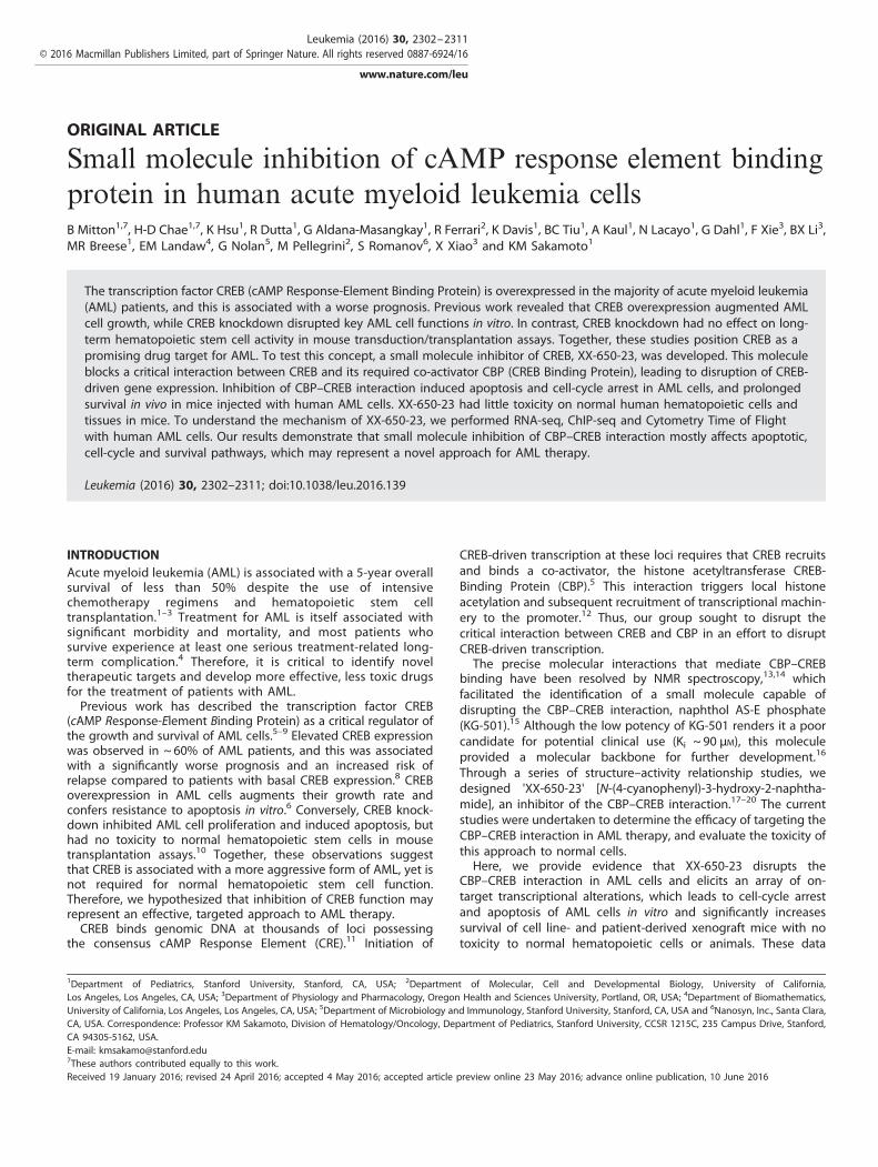
ORIGINAL ARTICLE
Small molecule inhibition of cAMP response element bindingprotein in human acute myeloid leukemia cellsB Mitton1,7, H-D Chae1,7, K Hsu1, R Dutta1, G Aldana-Masangkay1, R Ferrari2, K Davis1, BC Tiu1, A Kaul1, N Lacayo1, G Dahl1, F Xie3, BX Li3,MR Breese1, EM Landaw4, G Nolan5, M Pellegrini2, S Romanov6, X Xiao3 and KM Sakamoto1
The transcription factor CREB (cAMP Response-Element Binding Protein) is overexpressed in the majority of acute myeloid leukemia(AML) patients, and this is associated with a worse prognosis. Previous work revealed that CREB overexpression augmented AMLcell growth, while CREB knockdown disrupted key AML cell functions in vitro. In contrast, CREB knockdown had no effect on long-term hematopoietic stem cell activity in mouse transduction/transplantation assays. Together, these studies position CREB as apromising drug target for AML. To test this concept, a small molecule inhibitor of CREB, XX-650-23, was developed. This moleculeblocks a critical interaction between CREB and its required co-activator CBP (CREB Binding Protein), leading to disruption of CREB-driven gene expression. Inhibition of CBP–CREB interaction induced apoptosis and cell-cycle arrest in AML cells, and prolongedsurvival in vivo in mice injected with human AML cells. XX-650-23 had little toxicity on normal human hematopoietic cells andtissues in mice. To understand the mechanism of XX-650-23, we performed RNA-seq, ChIP-seq and Cytometry Time of Flightwith human AML cells. Our results demonstrate that small molecule inhibition of CBP–CREB interaction mostly affects apoptotic,cell-cycle and survival pathways, which may represent a novel approach for AML therapy.
Leukemia (2016) 30, 2302–2311; doi:10.1038/leu.2016.139
INTRODUCTIONAcute myeloid leukemia (AML) is associated with a 5-year overallsurvival of less than 50% despite the use of intensivechemotherapy regimens and hematopoietic stem celltransplantation.1–3 Treatment for AML is itself associated withsignificant morbidity and mortality, and most patients whosurvive experience at least one serious treatment-related long-term complication.4 Therefore, it is critical to identify noveltherapeutic targets and develop more effective, less toxic drugsfor the treatment of patients with AML.Previous work has described the transcription factor CREB
(cAMP Response-Element Binding Protein) as a critical regulator ofthe growth and survival of AML cells.5–9 Elevated CREB expressionwas observed in ~ 60% of AML patients, and this was associatedwith a significantly worse prognosis and an increased risk ofrelapse compared to patients with basal CREB expression.8 CREBoverexpression in AML cells augments their growth rate andconfers resistance to apoptosis in vitro.6 Conversely, CREB knock-down inhibited AML cell proliferation and induced apoptosis, buthad no toxicity to normal hematopoietic stem cells in mousetransplantation assays.10 Together, these observations suggestthat CREB is associated with a more aggressive form of AML, yet isnot required for normal hematopoietic stem cell function.Therefore, we hypothesized that inhibition of CREB function mayrepresent an effective, targeted approach to AML therapy.CREB binds genomic DNA at thousands of loci possessing
the consensus cAMP Response Element (CRE).11 Initiation of
CREB-driven transcription at these loci requires that CREB recruitsand binds a co-activator, the histone acetyltransferase CREB-Binding Protein (CBP).5 This interaction triggers local histoneacetylation and subsequent recruitment of transcriptional machin-ery to the promoter.12 Thus, our group sought to disrupt thecritical interaction between CREB and CBP in an effort to disruptCREB-driven transcription.The precise molecular interactions that mediate CBP–CREB
binding have been resolved by NMR spectroscopy,13,14 whichfacilitated the identification of a small molecule capable ofdisrupting the CBP–CREB interaction, naphthol AS-E phosphate(KG-501).15 Although the low potency of KG-501 renders it a poorcandidate for potential clinical use (Ki ~ 90 μM), this moleculeprovided a molecular backbone for further development.16
Through a series of structure–activity relationship studies, wedesigned 'XX-650-23' [N-(4-cyanophenyl)-3-hydroxy-2-naphtha-mide], an inhibitor of the CBP–CREB interaction.17–20 The currentstudies were undertaken to determine the efficacy of targeting theCBP–CREB interaction in AML therapy, and evaluate the toxicity ofthis approach to normal cells.Here, we provide evidence that XX-650-23 disrupts the
CBP–CREB interaction in AML cells and elicits an array of on-target transcriptional alterations, which leads to cell-cycle arrestand apoptosis of AML cells in vitro and significantly increasessurvival of cell line- and patient-derived xenograft mice with notoxicity to normal hematopoietic cells or animals. These data
1Department of Pediatrics, Stanford University, Stanford, CA, USA; 2Department of Molecular, Cell and Developmental Biology, University of California,Los Angeles, Los Angeles, CA, USA; 3Department of Physiology and Pharmacology, Oregon Health and Sciences University, Portland, OR, USA; 4Department of Biomathematics,University of California, Los Angeles, Los Angeles, CA, USA; 5Department of Microbiology and Immunology, Stanford University, Stanford, CA, USA and 6Nanosyn, Inc., Santa Clara,CA, USA. Correspondence: Professor KM Sakamoto, Division of Hematology/Oncology, Department of Pediatrics, Stanford University, CCSR 1215C, 235 Campus Drive, Stanford,CA 94305-5162, USA.E-mail: [email protected] authors contributed equally to this work.Received 19 January 2016; revised 24 April 2016; accepted 4 May 2016; accepted article preview online 23 May 2016; advance online publication, 10 June 2016
Leukemia (2016) 30, 2302–2311© 2016 Macmillan Publishers Limited, part of Springer Nature. All rights reserved 0887-6924/16
www.nature.com/leu
provide 'proof-of-principle' that CREB inhibition represents apotential approach for AML treatment.
MATERIALS AND METHODSProtein purification and biacoreKIX domain mutants were created by standard cloning and mutagenesismethods in the pGEX4T3 vector (GE Healthcare Life Sciences, Pittsburgh,PA, USA). GST-KIX and its mutants were purified with the B-PER GST FusionProtein Spin Purification Kit (Thermo Scientific/Pierce, Grand Island, NY,USA). Surface plasmon resonance analysis was performed on a GE Biacore3000 surface plasmon resonance instrument in collaboration with theStanford Protein and Nucleic Acid (PAN) Facility.
AML cell lines and patient samplesKG-1, HL-60, MOLM-13, MV-4-11 and U937 cell lines were obtained fromATCC, and low-passage stocks were used and cultured for less than3 months. Cells were regularly tested for Mycoplasma and growthcharacteristics, though no further authentication has been performed bythe authors. Cells were plated at a density of 2–4× 10⁵ cells/ml, and treatedwith various doses of XX-650-23. Cell counts and viability were determinedusing the Vi-CELL XR Cell Viability Analyzer (Beckman Coulter, Brea, CA,USA). HL-60 and KG-1 cells overexpressing CREB were generated usinglentiviral gene delivery with subsequent cells sorting for GFP. CREBknockdown was achieved by infecting cells with a lentivirus expressing theshRNA sequence 5′-GCAAATGACAGTTCAAGCCC-3′. For chemotherapycombination experiments, combination index values were calculatedusing median effects analysis on Calcusyn software as described.21 Humanpatient bone marrow samples were cultured in DMEM plus 20% FBS and1× PSG, supplemented with recombinant GM-CSF (20 ng/ml), G-CSF(20 ng/ml), SCF (50 ng/ml), IL-3 (20 ng/ml) and IL-6 (10 ng/ml). Cells(1 × 10⁵ cells/ml) were cultured with XX-650-23 for up to 72 h. All samplescontained 485% AML blasts and were not sorted before performingexperiments. Flow-cytometry analyses were performed on a DxP10 flowcytometer (Cytek, Fremont, CA, USA). All antibodies were purchased fromBD Biosciences (San Jose, CA, USA). Bone marrow from AML patients wascollected through voluntary patient participation at University of California,Los Angeles (Los Angeles, CA, USA) and Stanford University (Palo Alto, CA,USA) in compliance with the Institutional Review Board regulationsof each institution. Informed consent was obtained from all humansubjects, and all research was conducted in accordance withthe statements set forth in the declaration of Helsinki and the DataProtection Directive.
Luciferase assaysKG-1 cell lines were created to express luciferase in a CREB-dependent ornon-CREB-dependent manner using lentiviral gene delivery. Cells weresorted for mCherry expression. Luciferase activity was measured ona spectrophotometer using the Promega Luciferase Activity Kit (Promega,Madison, WI, USA) as per manufacturer’s instructions. The split-Renillaluciferase complementation assay has been described previously.20 In thisassay, the KID and KIX domains were fused to the N- and C- terminalregions of Renilla luciferase, respectively. Once KIX binds phosphorylatedKID, the Renilla luciferase regions were brought together, resulting inluciferase activity.
Cell-cycle analysisKG-1 cells were synchronized at prometaphase using a modified thymidineplus nocodazole block.22 Briefly, KG-1 cells were treated with 2 mM
thymidine for 30 h, washed with PBS and released from G1/S block in freshmedia for 4 h. The cells were incubated with 300 nM nocodazole (Sigma,St Louis, MO, USA) for 13 h. XX-650-23 or DMSO was added 3 h beforerelease. The synchronized cells were washed with PBS and released fromthe mitotic block in fresh media containing XX-650-23 or DMSO. To analyzeDNA content by flow cytometry, cells were harvested, fixed in 70% ice-coldethanol for at least 1 h at − 20 °C, and then stained with propidium iodide.Cells were analyzed on an FACS Calibur flow cytometer (BD Biosciences).Cell-cycle distribution was determined using FlowJo software (TreeStar,Ashland, OR, USA).
Chromatin immunoprecipitation and high-throughput sequencing(RNA-Seq and ChIP-Seq)For Chip-Seq experiments, KG-1 cells were treated with 5 μM XX-650-23 orDMSO for 6 h. Cells were crosslinked with 1% formaldehyde at roomtemperature for 10 min and then incubated with 0.125 mM glycine for5 min. After crosslinking, chromatin was digested by Micrococcal nucleaseand then sonicated using SimpleChIP Plus Enzymatic Chromatin IP Kit (CellSignaling, Danvers, MA, USA) following the manufacturer’s protocol.Chromatin immunoprecipitations were carried out with anti-CREB antibody(17-600, Millipore, Billerica, MA, USA), anti-Acetyl-Histone H3 (8173, Lys27)(D5E4) (Cell Signaling Technology) or a control IgG (Santa CruzBiotechnology, Santa Cruz, CA, USA). The captured immunocomplexescontaining bound transcriptional DNA fragments were eluted, withrecovered DNA fragments used for PCR amplification. For RNA-Seqexperiments, KG-1 cells were treated with XX-650-23 (5 μM) or 0.1% DMSOfor 12 h. Sample libraries were run using the Illumina sequencing platform.Two hundred million reads were collected on two biological replicates ofthe experiment. Libraries were prepared using the Illumina Truseq RNAsamples prep kit as per manufacturer’s instructions. Fastq files werealigned using TopHat.23 Aligned BAM files were used for CuffDiffcalculation of differentially expressed genes.24 CummeRbund R packagewas used to infer QC of the RNA-seq. RNA-seq and CREB ChIP-Seq datawere deposited in the NCBI GEO database (GEO submission GSE74928).ChIP-seq analysis was performed as previously described.25 BETA analysiswas used by inferring differentially expressed genes between DMSO- andXX-650-23-treated cells based on an fdr of ⩽ 0.01 and a gene distance of100 kb (Galaxy/cistrome). For analysis of RNA-seq data as target genesof known transcription factors, a curated list of all human target genes wasextracted from the TRANSFAC Pro database. DNA sequences enriched onChIP-Seq were defined as previously published.26
RT-PCRTotal RNA was isolated using the Aurum RNA Isolation Mini Kit, and theiScript cDNA Synthesis Kit was used to prepare samples for qPCR (Bio-Rad,Hercules, CA, USA). Data were analyzed using the Livak method.27 Datawere normalized to 7SL scRNA expression values.
Western blot analysisCell lysates were resolved on SDS-PAGE gels, and then transferred ontoPVDF membranes (Millipore). Listing of primary antibodies is given inSupplementary Table 1. Secondary antibodies were used at a 1:2500dilution and purchased from Thermo Scientific/Pierce. Membranes werevisualized with an enhanced chemiluminescence system. Representativeblot of at least three different experiments is shown.
Hematopoietic cell colony assaysNormal human bone marrow cells were resuspended in methylcellulose(2 × 10⁴ cells/ml, Miltenyi Biotec, Auburn, CA, USA) containing cytokines(GM-CSF, G-CSF, IL-3, IL-6, SCF, erythropoietin), and cultured for 2 weeks.Colonies were scored on the basis of morphology.
Caspase-3 activityThe ApoTarget Caspase-3 Protease Activity Kit (Invitrogen, Grand Island,NY, USA) was used as per manufacturer’s instructions.
Multiparameter single cell mass cytometry (CyTOF)Bone marrow from primary AML patients was cultured as above andtreated for 48 h with 2 μM XX-650-23. These were stained for viability usingcisplatin as previously described.28 Cells were fixed with 1.6% PFA for10 min and washed with cell staining media. Fc receptor block wasperformed using Human TruStain FcX (Biolegend, San Diego, CA, USA).Cells were stained for surface proteins at room temperature for 30 min,and then cells were washed twice with cell staining media andpermeabilized with methanol pre-cooled to 4°C for 10 min. Cells werethen washed twice and stained for intracellular proteins for 30 min at roomtemperature. Cells were washed and stained with 1 ml of 2000× iridiumDNA intercalator (diluted 1:5000 in PBS with 1.6% PFA; DVS Sciences,Sunnyvale, CA, USA) overnight at 4 °C. Data were acquired using internalmetal isotope bead standards as previously described.29 Cell events wereacquired on a CyTOF I (DVS Sciences/Fluidigm). All antibodies were alsopurchased from DVS Sciences/Fluidigm. Each patient sample was
Small molecule inhibition of CREB suppresses AML growthB Mitton et al
2303
© 2016 Macmillan Publishers Limited, part of Springer Nature. Leukemia (2016) 2302 – 2311

individually normalized to the internal bead standards before analysis. Toremove dead cells and debris, cells were gated based on cell length andDNA content as described.30
AML xenograft models of AMLFor AML xenograft mouse experiments, HL-60 cells (2 × 10⁶) expressingfirefly luciferase and GFP, or cryopreserved human AML patient cells(5 × 10⁶), were injected through the tail vein into NOD.Cg-Prkdcscid
Il2rgtm1Wjl/SzJ (NSG) mice (5–8 weeks old). AML cell-injected mice wererandomly divided into two groups. Mice were then treated with XX-650-23or 10% DMSO, injected intravenously (i.v.) daily, until death or an end pointwas reached (moribundity) in accordance with the animal care institutionalguidelines. The sample size of AML xenograft mice experiments wasdetermined based on our previous studies as well as the publishedpapers.31,32 Blinding was not used for xenograft mice experiments. Allmouse experiments were subjected to institutional approval by StanfordUniversity Institutional Animal Care and Use Committee. Leukemiaprogression in mice at the indicated time points was monitored usingan in vivo IVIS 100 bioluminescence/optical imaging system (XenogenCorporation, Alameda, CA, USA). Analysis was performed on Living ImageIn Vivo imaging software (Perkin-Elmer, Waltham, MA, USA).
Statistical analysisUnless noted, all experiments were performed in triplicate, and unpairedtwo-tailed Student’s t-test was used to assess experimental mean valuesfor statistically significant differences using Prism software (v5.04)(Graphpad, La Jolla, CA, USA), with P-values of o0.05 deemed statisticallysignificant. Kaplan–Meier plots and statistical significance of differences in
mice survival experiments were calculated using a Log-rank (Mantel-Cox)test with Prism. Synergy calculations were performed using CalcuSynsoftware. IC50s were calculated using Prism.
RESULTSXX-650-23 is a competitive inhibitor of CREB binding to CBPThe region of CBP critical for binding Ser133-phosphorylated-CREB istermed the Kinase-Inducible Acceptor domain or ‘KIX’ domain, andspans CBP amino acids 586–666. The small molecule KG-501(Naphthol AS-E phosphate) was reported to disrupt CREB–CBPbinding by interacting with key amino acids in the KIX domain,particularly residues Arg-600 to Valine-608.15 On the basis of thisinformation and subsequent structure–activity relationship studies,our group developed an optimized molecule targeted to this domaindesigned to disrupt the CREB–CBP interaction, termed 'XX-650-23'(Figure 1a). Earlier structure–activity relationship studies revealedXX-648-48 as an inactive yet similarly structured compound(N-methyl-(4-chlorophenyl)-3-hydroxy-naphthamide).20
Binding of XX-650-23 to the KIX domain and two KIX domainmutants in which residues critical for CREB binding were altered orremoved (Arg-600 mutated to Alanine or deletion of amino acids586–602) was determined by Surface Plasmon Resonance(Biacore) analysis (Figure 1b). Mutation of Arginine-600 to Alaninereduced binding of XX-650-23 by ~ 45%, while a KIX domainmutant lacking amino acids 586–602 reduced binding by ~ 70%.
Figure 1. XX-650-23 binds to the KIX domain of CBP and blocks CREB-dependent gene transcription. (a) The structure of XX-650-23 and itsinactive analog XX-648-48. (b) The native human KIX domain and two mutant proteins were expressed as a fusion protein with GST andsubjected to Biacore analysis to assess XX-650-23 binding characteristics. Mutation of Arginine-600 to Alanine reduced binding of XX-650-23by ~ 45% at a concentration of 5 μM, while a KIX mutant lacking amino acids 586–602 reduced binding by ~ 70%. (c) Binding model of XX-650-23 to CBP KIX domain. (d) Split-Renilla luciferase assays with 293T cells treated with forskolin demonstrated that XX-650-23 is a direct inhibitorof CREB–CBP binding, with an IC50 of approximately 3.2 μM. Data are presented as mean± s.d., n= 3. (e) Two KG-1 cell lines were generated inwhich luciferase was expressed either under the control of a CREB-driven promoter (CRE) or a CMV-driven promoter (CMV). These two celllines were each treated with a range of XX-650-23 or XX-648-48 concentrations for 6 h. Luciferase activity was significantly decreasedfollowing XX-650-23 treatment at concentrations of 3, 10 and 30 μM. No statistical difference in luciferase activity was detected followingXX-648-48 treatment. Data are presented as mean± s.e.m., n= 3. *Po0.05, t-test. (f) XX-650-23 inhibits CREB/CBP association. HEK293 cellswere transfected with a plasmid expressing CREB. Cells were treated with XX-650-23 (XX, 5 μM, lane 3) or DMSO vehicle (d) for 1 h. Total lysatesof transfected HEK293 cells were immunoprecipitated using anti-CBP antibody. XX-650-23 (XX, 5 μM) was added during anti-CBPimmunoprecipitation process (lane 1). Immunoprecipitates (CBP-IP) and total lysates were analyzed by immunoblotting for phospho-CREB (p-CREB) and CBP. Binding activities were calculated by the relative protein amounts of p-CREB bound to CBP normalized against CBP amounts inCBP-IP products.
Small molecule inhibition of CREB suppresses AML growthB Mitton et al
2304
Leukemia (2016) 2302 – 2311 © 2016 Macmillan Publishers Limited, part of Springer Nature.
A hypothetical binding model between XX-650-23 and CBP KIXdomain is shown (Figure 1c). This binding model suggests that themajor interactions between XX-650-23 and the KIX domain arehydrophobic. The aniline ring of XX-650-23 is predicted to projectinto a hydrophobic pocket of KIX where Leucine-141 of CREB, anamino acid essential for stable CREB binding, normally docks.XX-650-23 inhibited the interaction between CREB and CBP with
an IC50 of 3.20 ± 0.43 μM in a split-Renilla luciferase complementa-tion assay (Figure 1d).20 To determine whether XX-650-23 couldspecifically decrease CREB-driven reporter gene expression in AMLcells, KG-1 AML cell lines expressing luciferase under the control ofa CREB-driven promoter (two CRE elements placed upstream of anattenuated CMV promoter) or a CMV-driven promoter withoutCREs were treated with XX-650-23. CREB-driven luciferase activitywas reduced by XX-650-23 in a dose-dependent manner, whereasCMV-driven luciferase expression was unchanged followingtreatment (Figure 1e). The inactive analog XX-648-48 had nosignificant effect on luciferase activity in either AML cell line. Finally,we demonstrated that treatment of HEK293 lysates orcells with XX-650-23 prevented binding of CREB to CBP (Figure 1f).Thus, XX-650-23 inhibits CREB function through disruption ofCBP–CREB interaction.
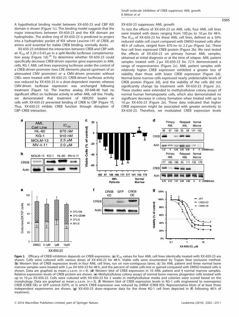
XX-650-23 suppresses AML growthTo test the effects of XX-650-23 on AML cells, four AML cell lineswere treated with doses ranging from 100 pM to 10 μM for 48 h.The IC50 of XX-650-23 for these AML cell lines, defined as a 50%reduced viable cell count compared with DMSO-treated cells after48 h of culture, ranged from 870 nM to 2.3 μM (Figure 2a). Thesefour cell lines expressed CREB protein (Figure 2b). We next testedthe effects of XX-650-23 on primary human AML samples,obtained at initial diagnosis or at the time of relapse. AML patientsamples treated with 2 μM XX-650-23 for 72 h demonstrated arange of responsiveness (Figure 2c). AML patient samples withrelatively higher CREB expression exhibited a greater loss ofviability than those with lower CREB expression (Figure 2d).Normal bone marrow cells expressed nearly undetectable levels ofCREB protein (Figure 2d), and the viability of the cells did notsignificantly change by treatment with XX-650-23 (Figure 2c).These studies were extended to methylcellulose colony assays ofnormal human hematopoietic cells, which also demonstrated nosignificant decrease in colony formation when treated with up to10 μM XX-650-23 (Figure 2e). These data indicated that higherCREB expression might be associated with greater sensitivity toXX-650-23. Therefore, we modulated CREB expression levels
Figure 2. Efficacy of CREB inhibition depends on CREB expression. (a) IC50 values for four AML cell lines identically treated with XX-650-23 areshown. Cells were cultured with various doses of XX-650-23 for 48 h. Viable cells were enumerated by Trypan blue exclusion method.(b) Western blot of CREB expression levels in four AML cell lines, run on non-contiguous lanes. (c) Six AML patient and three normal bonemarrow samples were treated with 2 μM XX-650-23 for 48 h, and the percent of viable cells lost or gained compared with DMSO-treated cells isshown. Data are graphed as mean± s.e.m. (n= 4). (d) Western blot of CREB expression in 10 AML patient and 4 normal marrow samples.Relative expression levels of CREB protein are shown. (e) Methylcellulose colony assays of normal bone marrow progenitor cells treated withup to 10 μM XX-650-23. Cells were cultured with XX-650-23 for 2 weeks in methylcellulose media and colonies were scored based on themorphology. Data are graphed as mean± s.e.m. (n= 3). (f) Western blot of CREB expression levels in KG-1 cells engineered to overexpressCREB (CREB OE) or GFP control (GFP), or in which CREB expression was reduced by shRNA (CREB KD). Representative blots of at least threeindependent experiments are shown. (g) XX-650-23 dose–response data for the three KG-1 cell lines depicted in (f) following 48 h oftreatment.
Small molecule inhibition of CREB suppresses AML growthB Mitton et al
2305
© 2016 Macmillan Publishers Limited, part of Springer Nature. Leukemia (2016) 2302 – 2311
to determine whether we could change sensitivity of cells toXX-650-23. CREB was overexpressed, or knocked down by shRNA,in KG-1 cells (Figure 2f). CREB knockdown in KG-1 cells decreasedtheir sensitivity to XX-650-23, while CREB overexpression increasedtheir sensitivity (IC50: CREB KD, 3.43 μM; GFP, 1.53 μM; CREB OE,0.99 μM) (Figure 2g). Experiments were also performed to examinethe efficacy of combining XX-650-23 with cytarabine or daunorubicin,standard drugs used in AML therapy. The combination of XX-650-23with cytarabine or daunorubicin in KG-1 cells showed a synergisticeffect against KG-1 cell viability (Supplementary Figure 1).
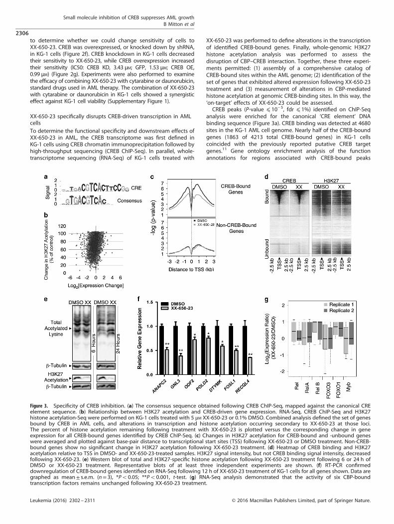
XX-650-23 specifically disrupts CREB-driven transcription in AMLcellsTo determine the functional specificity and downstream effects ofXX-650-23 in AML, the CREB transcriptome was first defined inKG-1 cells using CREB chromatin immunoprecipitation followed byhigh-throughput sequencing (CREB ChIP-Seq). In parallel, whole-transcriptome sequencing (RNA-Seq) of KG-1 cells treated with
XX-650-23 was performed to define alterations in the transcriptionof identified CREB-bound genes. Finally, whole-genomic H3K27histone acetylation analysis was performed to assess thedisruption of CBP–CREB interaction. Together, these three experi-ments permitted: (1) assembly of a comprehensive catalog ofCREB-bound sites within the AML genome; (2) identification of theset of genes that exhibited altered expression following XX-650-23treatment and (3) measurement of alterations in CBP-mediatedhistone acetylation at genomic CREB-binding sites. In this way, the‘on-target’ effects of XX-650-23 could be assessed.CREB peaks (P-value ⩽ 10− 5, fdr ⩽ 1%) identified on ChIP-Seq
analysis were enriched for the canonical ‘CRE element’ DNAbinding sequence (Figure 3a). CREB binding was detected at 4680sites in the KG-1 AML cell genome. Nearly half of the CREB-boundgenes (1863 of 4213 total CREB-bound genes) in KG-1 cellscoincided with the previously reported putative CREB targetgenes.11 Gene ontology enrichment analysis of the functionannotations for regions associated with CREB-bound peaks
Figure 3. Specificity of CREB inhibition. (a) The consensus sequence obtained following CREB ChIP-Seq, mapped against the canonical CREelement sequence. (b) Relationship between H3K27 acetylation and CREB-driven gene expression. RNA-Seq, CREB ChIP-Seq and H3K27histone acetylation-Seq were performed on KG-1 cells treated with 5 μM XX-650-23 or 0.1% DMSO. Combined analysis defined the set of genesbound by CREB in AML cells, and alterations in transcription and histone acetylation occurring secondary to XX-650-23 at those loci.The percent of histone acetylation remaining following treatment with XX-650-23 is plotted versus the corresponding change in geneexpression for all CREB-bound genes identified by CREB ChIP-Seq. (c) Changes in H3K27 acetylation for CREB-bound and -unbound geneswere averaged and plotted against base-pair distance to transcriptional start sites (TSS) following XX-650-23 or DMSO treatment. Non-CREB-bound genes show no significant change in H3K27 acetylation following XX-650-23 treatment. (d) Heatmap of CREB binding and H3K27acetylation relative to TSS in DMSO- and XX-650-23-treated samples. H3K27 signal intensity, but not CREB binding signal intensity, decreasedfollowing XX-650-23. (e) Western blot of total and H3K27-specific histone acetylation following XX-650-23 treatment following 6 or 24 h ofDMSO or XX-650-23 treatment. Representative blots of at least three independent experiments are shown. (f) RT-PCR confirmeddownregulation of CREB-bound genes identified on RNA-Seq following 12 h of XX-650-23 treatment of KG-1 cells for all genes shown. Data aregraphed as mean± s.e.m. (n= 3), *Po0.05; **Po0.001, t-test. (g) RNA-Seq analysis demonstrated that the activity of six CBP-boundtranscription factors remains unchanged following XX-650-23 treatment.
Small molecule inhibition of CREB suppresses AML growthB Mitton et al
2306
Leukemia (2016) 2302 – 2311 © 2016 Macmillan Publishers Limited, part of Springer Nature.
(Supplementary Table 2) showed that CREB-bound peaks wereenriched in clusters of genes regulating cell cycle and apoptosisas well as translation and mRNA processing. Over 90% of occupiedCREB-binding sites were within 500 bp of a transcription start site(Supplementary Figures 2A and B). Of these CREB-bound 4213 genes,2787 CREB-bound genes exhibited reduced expression followingXX-650-23 treatment and 602 genes exhibited greater than 50%reduced expression. Majority of H3K27 peaks in CREB-bound genes(4052 out of 5282 total peaks) exhibited decreased H3K27 histoneacetylation following treatment with XX-650-23 (66.5±17% of control,n=4052 peaks; Figure 3b and Supplementary Table 3). Histogramanalysis of H3K27 acetylation of all CREB-bound genomic loci alsoshowed a relative reduction in overall H3K27 acetylation, representedas a median shift to the left (Supplementary Figure 2C). Moreover,reductions in H3K27 acetylation were almost exclusively within 1–3 kbof these CREB-bound promoter sites. On the contrary, the genes thatdid not show any significant binding site for CREB showed no globaldetectable changes in acetylation (Figure 3c). CREB genomic bindingdid not change following XX-650-23 treatment (Figure 3d andSupplementary Figure 2D). Western blot analysis confirmed that
XX-650-23 caused a specific decrease in H3K27 acetylation and is not ageneral inhibitor of acetyltransferase activity (Figure 3e).To validate the RNA-Seq data set, qPCR of XX-650-23 target
genes with CREB-bound promoters that exhibited significant(450%) transcriptional downregulation and reduced H3K27acetylation was performed in two AML cell lines (KG-1 andHL-60) following treatment with XX-650-23 (Figure 3f andSupplementary Figure 2E). XX-650-23 consistently reduced thesetarget genes expression in both these cell lines in a statisticallysignificant manner, although we expect that these genes areregulated by other transcription factors in addition to CREB.To assess for potential ‘off-target’ effects of XX-650-23, the RNA-Seq gene expression profiles of other transcription factors thatbind CBP, including Rel, RelA, RelB, Foxo3, Foxo1 and Myb, wereanalyzed. XX-650-23 evoked no significant change in theexpression of these CBP-binding transcription factor targetgenes (Figure 3g). These results were confirmed for a set of Myb-driven genes, SP3, FPR1, PRODH and SLC34A2 in both KG-1 andHL-60 cells (Supplementary Figure 2F).33–36 Thus, these resultsprovide evidence that XX-650-23 disrupts the interaction of
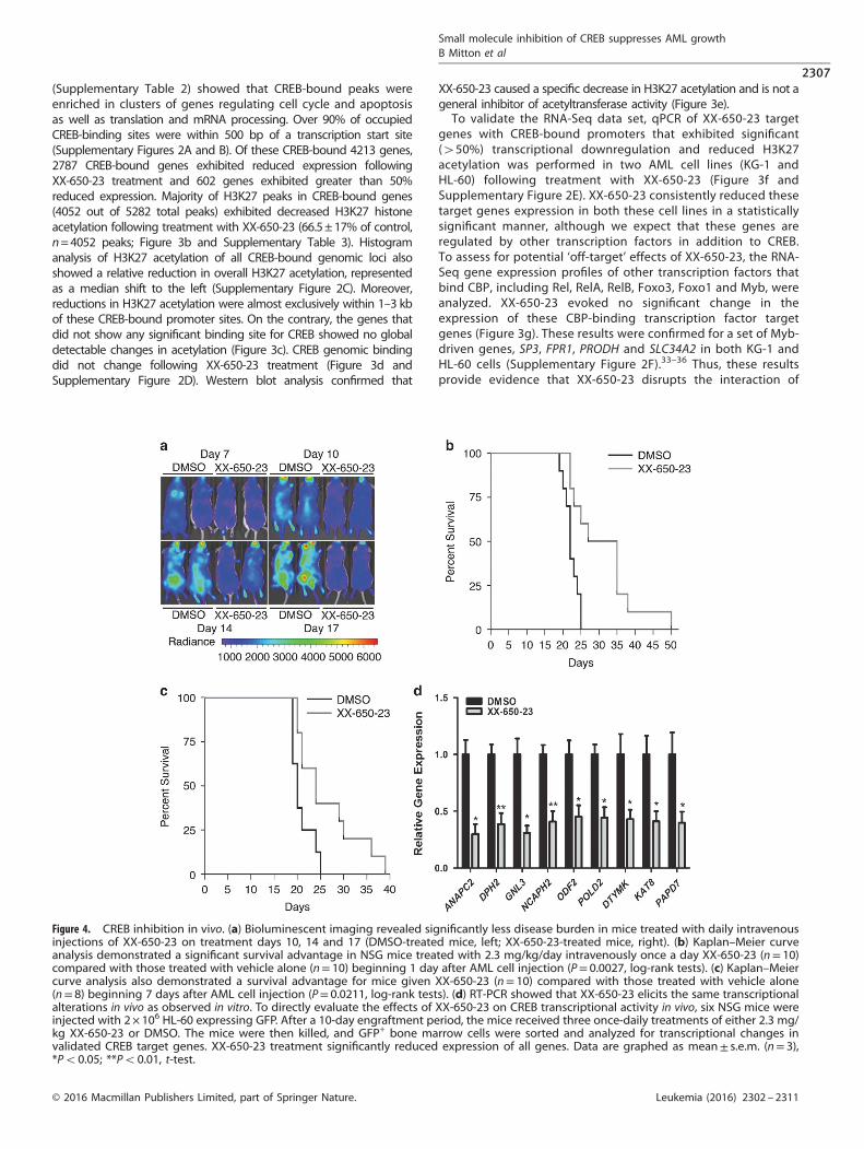
Figure 4. CREB inhibition in vivo. (a) Bioluminescent imaging revealed significantly less disease burden in mice treated with daily intravenousinjections of XX-650-23 on treatment days 10, 14 and 17 (DMSO-treated mice, left; XX-650-23-treated mice, right). (b) Kaplan–Meier curveanalysis demonstrated a significant survival advantage in NSG mice treated with 2.3 mg/kg/day intravenously once a day XX-650-23 (n= 10)compared with those treated with vehicle alone (n= 10) beginning 1 day after AML cell injection (P= 0.0027, log-rank tests). (c) Kaplan–Meiercurve analysis also demonstrated a survival advantage for mice given XX-650-23 (n= 10) compared with those treated with vehicle alone(n= 8) beginning 7 days after AML cell injection (P= 0.0211, log-rank tests). (d) RT-PCR showed that XX-650-23 elicits the same transcriptionalalterations in vivo as observed in vitro. To directly evaluate the effects of XX-650-23 on CREB transcriptional activity in vivo, six NSG mice wereinjected with 2× 10⁶ HL-60 expressing GFP. After a 10-day engraftment period, the mice received three once-daily treatments of either 2.3 mg/kg XX-650-23 or DMSO. The mice were then killed, and GFP+ bone marrow cells were sorted and analyzed for transcriptional changes invalidated CREB target genes. XX-650-23 treatment significantly reduced expression of all genes. Data are graphed as mean± s.e.m. (n= 3),*Po0.05; **Po0.01, t-test.
Small molecule inhibition of CREB suppresses AML growthB Mitton et al
2307
© 2016 Macmillan Publishers Limited, part of Springer Nature. Leukemia (2016) 2302 – 2311
CREB to the KIX domain of CBP but not other KIX bindingproteins in AML cells.
XX-650-23 prolongs survival in mouse models of AML withouttoxicityTo examine the efficacy of XX-650-23 in an in vivo model of AML,NSG mice were tail-vein injected with HL-60 AML cells expressingFirefly luciferase and GFP. Mice received XX-650-23 (2.3 mg/kg,intravenously) once-daily starting the day after cell injection(immediate treatment groups), or starting 7 days after AML cellinjection (delayed treatment groups). Bioluminescence imagingperformed during treatment revealed less disease burden inXX-650-23-treated mice compared with control (average radiancemeasurements, given in p/s/cm2/sr: 5.7 × 10⁵ for the control groupversus 2.8 × 10⁵ for the XX-650-23-treated group at day 17)(Figure 4a). XX-650-23 significantly prolonged the mediansurvival in Kaplan–Meier analysis in both the immediate (mediansurvival, 22 days versus 31 days, P= 0.0027, long-rank test;mean survival 22.3 days versus 31.2 days) and delayed treatmentgroups (median survival, 20 days versus 24 days, P= 0.0211, long-rank test; mean survival 20.9 versus 26.4 days) (Figures 4b and c).We also assessed the efficacy of XX-650-23 in a mouse xenograft
model using primary AML cell sample (patient sample #186). Micetreated with XX-650-23 demonstrated a significant survivaladvantage compared with DMSO-treated mice (SupplementaryFigure 3A). Next, we evaluated the effects of XX-650-23 on CREBtranscriptional activity in AML cells in vivo. This experimentrecapitulated in vitro findings (Figure 4d).Pharmacokinetic studies showed that the half-life of XX-650-23
in plasma is approximately 4.3 h (Supplementary Figure 3B).XX-650-23-treated mice demonstrated normal complete bloodcounts, liver function tests and kidney function tests, comparedwith age-matched NSG mice given no treatment (SupplementaryFigure 3C). Histology demonstrated no microscopic evidenceof vital organ damage (Supplementary Figure 3D).
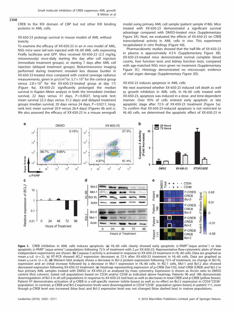
XX-650-23 induces apoptosis in AML cellsWe next examined whether XX-650-23 induced cell death as wellas growth inhibition in AML cells. In HL-60 cells treated withXX-650-23, apoptosis was induced in a dose- and time-dependentmanner. Over 95% of cells entered early apoptotic or lateapoptotic stage after 72 h of XX-650-23 treatment (Figure 5a).To confirm that XX-650-23-induced apoptosis is not restricted toHL-60 cells, we determined the apoptotic effect of XX-650-23 in
Figure 5. CREB inhibition in AML cells induces apoptosis. (a) HL-60 cells clearly showed early apoptotic (c-PARP+/aqua amine−) or lateapoptotic (c-PARP+/aqua amine+) populations following 72 h of treatment with 2 μM XX-650-23. Representative flow-cytometric plots of threeindependent experiments are shown. (b) Caspase-3 activity is activated in response to XX-650-23 treatment in HL-60 cells. Data are graphed asmean± s.d. (n= 2). (c) RT-PCR showed BCL2 expression decreases at 72 h after XX-650-23 treatment in HL-60 cells. Data are graphed asmean± s.e.m. (n= 3). (d) Western blot analysis shows a decrease in Bcl-2 protein expression following 72 h of treatment, no change in Bcl-XLexpression and an initial increase followed by a decrease in Mcl-1 expression in HL-60 cells. In KG-1 cells, Mcl-1 and Bcl-2 also showeddecreased expression following XX-650-23 treatment. (e) Heatmap representing expression of p-CREB (Ser133), total CREB (CREB) and Bcl-2 infour primary AML samples treated with DMSO or XX-650-23 as analyzed by mass cytometry. Expression is shown as Arcsin ratio to DMSOcontrol (first column). Gated cell populations based on CD34 and/or CD38 as indicated above heatmap. Patients 96 and 186 demonstratedownregulation of Bcl-2 in all cell populations in response to XX-650-23 (red box) as well as decreases in total CREB and p-CREB (yellow boxes).Patient 97 demonstrates activation of p-CREB in a cell-specific manner (white boxes) as well as no effect on Bcl-2 expression in CD34+CD38−
population. In contrast, p-CREB and Bcl-2 expression levels were downregulated in CD34+CD38− population (green boxes) in patient 111, eventhough p-CREB level was increased (blue box) and Bcl-2 expression level was not changed (blue dashed box) in mature populations.
Small molecule inhibition of CREB suppresses AML growthB Mitton et al
2308
Leukemia (2016) 2302 – 2311 © 2016 Macmillan Publishers Limited, part of Springer Nature.
additional AML cell lines (KG-1, MV-4-11 and U937 cells). Apoptosiswas observed in all these cell lines with XX-650-23 treatment(Supplementary Figure 4). XX-650-23 elicited apoptosis throughthe intrinsic apoptosis pathway, with activation of Caspase-3(Figure 5b) and detectable Caspase-9 cleavage (data not shown).The balance between pro- and anti-apoptotic protein expressioncontributes to cell fate decisions.37 CREB regulates the expressionof several anti-apoptotic proteins, including BCL2, BCL2L1 andMCL1.38,39 The transcription of BCL2 decreased following 72 h of2 μM XX-650-23 treatment (Figure 5c), and this was verified bywestern blot analysis (Figure 5d). In parallel, the expression ofMcl-1 initially increased, then decreased at 72 h, while Bcl-XLexpression remained constant. Bcl-2 inhibitor ABT-737 treatmentinduced apoptosis, similar to XX-650-23 (Supplementary Figures5A and B).40 KG-1 cells also underwent apoptosis followingtreatment with XX-650-23, and similarly exhibited decreased Bcl-2expression alongside a pronounced decrease in Mcl-1 (Figure 5d).Bcl-2 levels were influenced by CREB expression in these AML celllines (Supplementary Figure 5C). Together, these data suggest thatdownregulation of Bcl-2 is involved in XX-650-23-inducedapoptosis in AML cell lines.We examined the relationship between Bcl-2, total CREB and
p-CREB in primary AML patient samples, especially in the CD34+
CD38− subpopulation containing the putative ‘leukemia initiating’cells (see CyTOF gating strategy, Supplementary Figure 5D).41,42
XX-650-23 treatment significantly reduced Bcl-2 expression levels
in all cell subpopulations in XX-650-23 hyper-responsive primaryAML cells with higher CREB expression (#96 and #186) (Figure 5e,within red outlined box). This occurred alongside downregulationof total CREB as well as reduced phosphorylation at Serine 133, anactivation mark of CREB (Figure 5e, within yellow outlined box). Incontrast, in patient sample 97, which expressed less CREB atbaseline and showed less sensitivity to XX-650-23 (Figures 2c andd), CREB phosphorylation was increased in the CD34+CD38−
population without significant Bcl-2 downregulation (Figure 5e,within white outlined boxes). However, p-CREB and Bcl-2expression levels were downregulated in CD34+CD38− population(within green outlined boxes) in patient 111, even thoughphosphorylation of CREB was increased (Figure 5e, within blueoutlined box) without significant Bcl-2 downregulation (Figure 5e,within blue dashed box) in the more mature AML cell populations.These data suggest that Bcl-2 downregulation in CD34+CD38−
population may determine the sensitivity to the drug. The relativeactivation of AKT and ERK was associated with increased ordecreased phosphorylation of CREB (Supplementary Figure 6A).HL-60 cells also showed an increase in levels of phosphorylatedbut not total CREB following 24 h of XX-650-23 treatment(Supplementary Figure 6B), and this effect can be blocked byvalidated small molecule inhibitors of the ERK and RSK kinases(Supplementary Figure 6B), in keeping with previous workdescribing ERK/RSK-mediated phosphorylation of CREB down-stream of the GM-CSF receptor in AML cells.43–45 Blockade of these
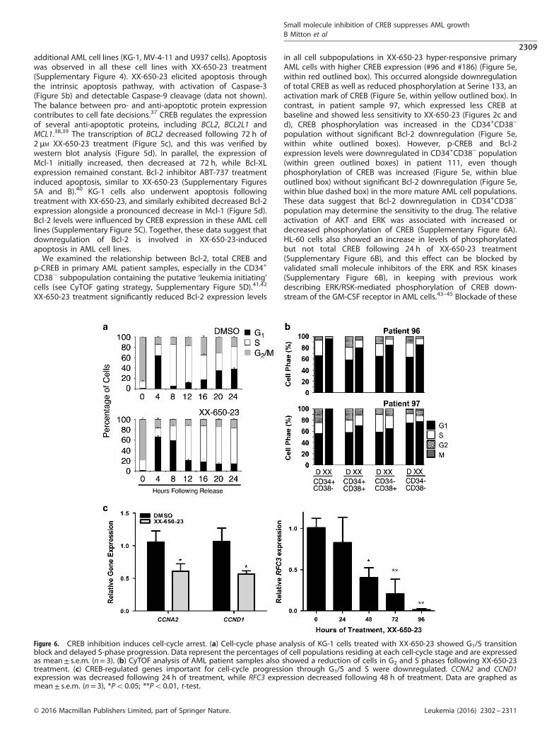
Figure 6. CREB inhibition induces cell-cycle arrest. (a) Cell-cycle phase analysis of KG-1 cells treated with XX-650-23 showed G1/S transitionblock and delayed S-phase progression. Data represent the percentages of cell populations residing at each cell-cycle stage and are expressedas mean± s.e.m. (n= 3). (b) CyTOF analysis of AML patient samples also showed a reduction of cells in G2 and S phases following XX-650-23treatment. (c) CREB-regulated genes important for cell-cycle progression through G1/S and S were downregulated. CCNA2 and CCND1expression was decreased following 24 h of treatment, while RFC3 expression decreased following 48 h of treatment. Data are graphed asmean± s.e.m. (n= 3), *Po0.05; **Po0.01, t-test.
Small molecule inhibition of CREB suppresses AML growthB Mitton et al
2309
© 2016 Macmillan Publishers Limited, part of Springer Nature. Leukemia (2016) 2302 – 2311
kinases increased the efficacy of XX-650-23 (SupplementaryFigure 6C).
XX-650-23 induces AML cell-cycle arrest at the G1/S transitionCREB regulates the cell cycle by virtue of transcriptionallyregulating the expression of key cell-cycle genes. Followingrelease from prometaphase, cell-cycle progression was followedby DNA contents for 24 h in the presence or absence of XX-650-23in KG-1 cells. XX-650-23 treatment caused aberrant cell-cycleprogression at the G1/S transition and through S phase (% cells inG1 phase for DMSO-treated cells: 61.18 ± 0.97% and 4.92 ± 0.33%at 4 and 8 h, respectively, versus XX-650-23-treated cells:66.20 ± 1.83% and 55.41 ± 0.59% (mean± s.e.m., n= 3) at 4 and8 h following release, respectively) (Figure 6a and SupplementaryFigure 7A). CyTOF cell-cycle analysis of AML patient bone marrowsamples revealed the same perturbation in the G1/S transitionfollowing XX-650-23 treatment (Figure 6b). The percent of cells inboth the S and G2/M phases was reduced, and that of G1 wasincreased.A number of previously described mediators of the G1/S
transition and S-phase progression were downregulated followingXX-650-23 treatment, including a set of CREB-bound cyclins,CCNA2 and CCND1, and FOSL1 (Figures 3f and 6c).46–48
We analyzed RNA expression and H3K27 acetylation of CREB-bound genes to identify potential target genes of XX-650-23 incell-cycle perturbation. CREB-bound cell-cycle regulators includingFOSL1, CDK2, CCNB1, CDC25C and RAD54L show decreased RNAexpression and H3K27 acetylation following XX-650-23 treatment(Supplementary Figure 7B). Transcriptional alterations of a subsetof cell-cycle regulators were confirmed by qPCR in two AML celllines (Supplementary Figure 7C). In addition, our group haspreviously reported that Replication Factor C3 (RFC3), a member ofthe PCNA DNA replication complex, is downregulated followingCREB knockdown in AML cells, which is associated with G1/Stransition arrest.49 Levels of RFC3 also decreased following XX-650-23 treatment (Figure 6c).
DISCUSSIONSuccessful treatment of AML remains a major clinical challenge,and progress in our ability to treat AML patients will depend onthe development of effective approaches associated with minimaltoxicity. Here, we provide the first evidence that a novel smallmolecule that inhibits the transcription factor CREB may offera unique, effective and non-toxic approach to treat AML patients.CREB is an ideal target for AML therapy, given that CREBoverexpression is associated with a worse prognosis and knock-down of CREB does not affect normal HSC activity. Previous workfacilitated the synthesis of a novel inhibitor of CREB function,XX-650-23, which our data establish as a competitive inhibitorof CBP–CREB interaction. We offer this study as ‘proof-of-principle’that CREB inhibition is a viable approach to AML therapy.Developing ‘targeted therapy’ for AML connotes both that the
agent is targeted to a specific function or molecule within AMLcells, and that the agent’s toxicity is specific to AML cells. On thebasis of our data, we postulate that the specificity with whichXX-650-23 disrupts the CREB–CBP interaction defines its selectivetoxicity to AML cells. Biochemical evidence supports that XX-650-23 binds CBP and physically disrupts the CREB–CBP interaction.Our whole-transcriptome and genome acetylation studies provideevidence that the CREB–CBP interaction is specifically disrupted.The almost exclusive reduction in H3K27 acetylation at CREB-bound genomic loci reflects the absence of CBP docking at, andonly at, these sites. Furthermore, the activity of other transcriptionfactors that also bind the CBP KIX domain was not affected. Thesedata provide evidence that XX-650-23 exerts ‘on-target’ effects.Within the set of genes bound by CREB, the transcription of
hundreds of genes was significantly (450%) downregulated. It isnot surprising that we did not observe a significant reduction intranscription of all CREB-bound genes, as expression of mosteukaryotic genes is determined by input from unique setsof diverse transcription factors. Our data demonstrate that keyCREB-bound factors that regulate the cell cycle and apoptosis aredownregulated, and sufficient to cause AML cell-cycle arrest andapoptosis in vitro and in vivo.Targeting the activity of specific transcription factors for the
treatment of leukemia has begun to show promise in a number ofpre-clinical studies. The interaction of CBP with β- and γ-cateninhas recently been targeted using a small molecule, and thisstrategy was effective against both primary and relapsed ALL.31
Another group recently demonstrated the efficacy of targeting themutant fusion transcription factor CBPβ-SMMHC, which drives inv(16)+ AML,32 and the critical interaction between menin and MLLfusion proteins, which drives subtypes of both AML and ALL, hasalso been successfully targeted.50 The data presented heresimilarly provide 'proof-of-principle' that CREB can be targetedfor the treatment of AML, and lay the groundwork for advancingthis strategy to the clinical arena.
CONFLICT OF INTERESTGN has personal financial interest in the company DVS Sciences, the manufacturerthat produced some of the reagents and instrumentation used in this manuscript.The remaining authors declare no conflict of interest.
ACKNOWLEDGEMENTSWe thank Kai Fu and Jing Lu for analysis of ChIP-seq and RNA-seq data. In vivo serummeasurements of XX-650-23 were performed in collaboration with LudmilaAlexandrova, PhD, at the Stanford University Mass Spectrometry (SUMS) core facility.Surface Plasmon Resonance (Biacore) analysis was performed in collaboration withMichael Eckart, PhD, at the Stanford Protein and Nucleic Acid (PAN) facility. In vivoanimal imaging was performed in collaboration with the Stanford Small AnimalImaging (SCI3) facility, directed by Timothy Doyle, PhD. This research was supportedby NIH R01 HL75826, Maxfield Foundation, SPARK program and Child HealthResearch Institute Lucile Packard Foundation for Children’s Health, Leukemia andLymphoma Society of America (SLP-8009-15), Pediatric Cancer Research Foundation,Hyundai Hope on Wheels (#02500CA) (KMS), NIH R01 GM087305 (XX), and by theAmerican Cancer Society Greeley & Seattle Gala/Friends of Rob Kinas PostdoctoralFellowship, Bear Necessities and Jane C Ventura Charitable Trust, Harvey CohenEndowment, and the Stanford University Dean’s Post-Doctoral Fellowshipprogram (BM).
AUTHOR CONTRIBUTIONSBM and HDC designed research, performed research, analyzed data and wrotethe paper. KH, RD, BCT, AK, FX, BXL and BXL performed research. GAD, RF, KDperformed research and analyzed data. GD, GN, MRB, EML, GN, MP and XXanalyzed data. NL collected and provided primary cells from AML patients. KMSdesigned research, analyzed data and wrote the paper.
REFERENCES1 Tallman MS, Gilliland DG, Rowe JM. Drug therapy for acute myeloid leukemia.
Blood 2005; 106: 1154–1163.2 Ferrara F, Schiffer CA. Acute myeloid leukaemia in adults. Lancet 2013; 381:
484–495.3 Creutzig U, van den Heuvel-Eibrink MM, Gibson B, Dworzak MN, Adachi S, de Bont E
et al. Diagnosis and management of acute myeloid leukemia in children andadolescents: recommendations from an international expert panel. Blood 2012;120: 3187–3205.
4 Schultz KA, Chen L, Chen Z, Kawashima T, Oeffinger KC, Woods WG et al. Healthconditions and quality of life in survivors of childhood acute myeloid leukemiacomparing post remission chemotherapy to BMT: a report from the children'soncology group. Pediatr Blood Cancer 2014; 61: 729–736.
5 Conkright MD, Montminy M. CREB: the unindicted cancer co-conspirator. TrendsCell Biol 2005; 15: 457–459.
Small molecule inhibition of CREB suppresses AML growthB Mitton et al
2310
Leukemia (2016) 2302 – 2311 © 2016 Macmillan Publishers Limited, part of Springer Nature.
6 Pigazzi M, Ricotti E, Germano G, Faggian D, Arico M, Basso G. cAMP responseelement binding protein (CREB) overexpression CREB has been described ascritical for leukemia progression. Haematologica 2007; 92: 1435–1437.
7 Pigazzi M, Manara E, Bresolin S, Tregnago C, Beghin A, Baron E et al.MicroRNA-34b promoter hypermethylation induces CREB overexpression andcontributes to myeloid transformation. Haematologica 2013; 98: 602–610.
8 Shankar DB, Cheng JC, Kinjo K, Federman N, Moore TB, Gill et al. The role of CREBas a proto-oncogene in hematopoiesis and in acute myeloid leukemia. Cancer Cell2005; 7: 351–362.
9 Shankar DB, Cheng JC, Sakamoto KM. Role of cyclic AMP response elementbinding protein in human leukemias. Cancer 2005; 104: 1819–1824.
10 Cheng JC, Kinjo K, Judelson DR, Chang J, Wu WS, Schmid I et al. CREB is a criticalregulator of normal hematopoiesis and leukemogenesis. Blood 2008; 111:1182–1192.
11 Zhang X, Odom DT, Koo SH, Conkright MD, Canettieri G, Best J et al. Genome-wide analysis of cAMP-response element binding protein occupancy, phosphor-ylation, and target gene activation in human tissues. Proc Natl Acad Sci USA 2005;102: 4459–4464.
12 Vo N, Goodman RH. CREB-binding protein and p300 in transcriptional regulation.J Biol Chem 2001; 276: 13505–13508.
13 Radhakrishnan I, Perez-Alvarado GC, Parker D, Dyson HJ, Montminy MR, Wright PE.Structural analyses of CREB-CBP transcriptional activator-coactivator complexesby NMR spectroscopy: implications for mapping the boundaries of structuraldomains. J Mol Biol 1999; 287: 859–865.
14 Radhakrishnan I, Perez-Alvarado GC, Parker D, Dyson HJ, Montminy MR, Wright PE.Solution structure of the KIX domain of CBP bound to the transactivation domainof CREB: a model for activator:coactivator interactions. Cell 1997; 91: 741–752.
15 Best JL, Amezcua CA, Mayr B, Flechner L, Murawsky CM, Emerson B et al. Iden-tification of small-molecule antagonists that inhibit an activator: coactivatorinteraction. Proc Natl Acad Sci USA 2004; 101: 17622–17627.
16 Uttarkar S, Dukare S, Bopp B, Goblirsch M, Jose J, Klempnauer KH. Naphthol AS-Ephosphate inhibits the activity of the transcription factor Myb by blocking theinteraction with the KIX domain of the coactivator p300. Mol Cancer Ther 2015; 14:1276–1285.
17 Jiang M, Li BX, Xie F, Delaney F, Xiao X. Design, synthesis, and biologicalevaluation of conformationally constrained analogues of naphthol AS-E as inhi-bitors of CREB-mediated gene transcription. J Med Chem 2012; 55: 4020–4024.
18 Li BX, Yamanaka K, Xiao X. Structure-activity relationship studies of naphthol AS-Eand its derivatives as anticancer agents by inhibiting CREB-mediated gene tran-scription. Bioorg Med Chem 2012; 20: 6811–6820.
19 Xie F, Li BX, Broussard C, Xiao X. Identification, synthesis and evaluation ofsubstituted benzofurazans as inhibitors of CREB-mediated gene transcription.Bioorg Med Chem Lett 2013; 23: 5371–5375.
20 Li BX, Xiao X. Discovery of a small-molecule inhibitor of the KIX-KID interaction.Chembiochem 2009; 10: 2721–2724.
21 Bijnsdorp IV, Giovannetti E, Peters GJ. Analysis of drug interactions. Methods MolBiol 2011; 731: 421–434.
22 Whitfield ML, Sherlock G, Saldanha AJ, Murray JI, Ball CA, Alexander KE et al.Identification of genes periodically expressed in the human cell cycle and theirexpression in tumors. Mol Biol Cell 2002; 13: 1977–2000.
23 Trapnell C, Pachter L, Salzberg SL. TopHat: discovering splice junctions withRNA-Seq. Bioinformatics 2009; 25: 1105–1111.
24 Trapnell C, Hendrickson DG, Sauvageau M, Goff L, Rinn JL, Pachter L. Differentialanalysis of gene regulation at transcript resolution with RNA-seq. Nat Biotechnol2013; 31: 46–53.
25 Ferrari R, Su T, Li B, Bonora G, Oberai A, Chan Y et al. Reorganization of the hostepigenome by a viral oncogene. Genome Res 2012; 22: 1212–1221.
26 Thomas-Chollier M, Herrmann C, Defrance M, Sand O, Thieffry D, van Helden J.RSAT peak-motifs: motif analysis in full-size ChIP-seq datasets. Nucleic Acids Res2012; 40: e31.
27 Livak KJ, Schmittgen TD. Analysis of relative gene expression data using real-timequantitative PCR and the 2(-Delta Delta C(T)) Method. Methods 2001; 25: 402–408.
28 Fienberg HG, Simonds EF, Fantl WJ, Nolan GP, Bodenmiller B. A platinum-basedcovalent viability reagent for single-cell mass cytometry. Cytometry A 2012; 81:467–475.
29 Finck R, Simonds EF, Jager A, Krishnaswamy S, Sachs K, Fantl W et al. Normalization ofmass cytometry data with bead standards. Cytometry A 2013; 83: 483–494.
30 Bendall SC, Simonds EF, Qiu P, Amir el AD, Krutzik PO, Finck R et al. Single-cellmass cytometry of differential immune and drug responses across a humanhematopoietic continuum. Science 2011; 332: 687–696.
31 Gang EJ, Hsieh YT, Pham J, Zhao Y, Nguyen C, Huantes S et al. Small-moleculeinhibition of CBP/catenin interactions eliminates drug-resistant clones in acutelymphoblastic leukemia. Oncogene 2014; 33: 2169–2178.
32 Illendula A, Pulikkan JA, Zong H, Grembecka J, Xue L, Sen S et al. Chemicalbiology. A small-molecule inhibitor of the aberrant transcription factor CBFbeta-SMMHC delays leukemia in mice. Science 2015; 347: 779–784.
33 Tapias A, Ciudad CJ, Noe V. Transcriptional regulation of the 5'-flanking regionof the human transcription factor Sp3 gene by NF-1, c-Myb, B-Myb, AP-1 and E2F.Biochim Biophys Acta 2008; 1779: 318–329.
34 Xu H, Inouye M, Hines ER, Collins JF, Ghishan FK. Transcriptional regulation of thehuman NaPi-IIb cotransporter by EGF in Caco-2 cells involves c-myb. Am J PhysiolCell Physiol 2003; 284: C1262–C1271.
35 Pattabiraman DR, Gonda TJ. Role and potential for therapeutic targeting of MYBin leukemia. Leukemia 2013; 27: 269–277.
36 Miettinen HM. Regulation of human formyl peptide receptor 1 synthesis: roleof single nucleotide polymorphisms, transcription factors, and inflammatorymediators. PLoS One 2011; 6: e28712.
37 Portt L, Norman G, Clapp C, Greenwood M, Greenwood MT. Anti-apoptosis andcell survival: a review. Biochim Biophys Acta 2011; 1813: 238–259.
38 Wang JM, Chao JR, Chen W, Kuo ML, Yen JJ, Yang-Yen HF. The antiapoptotic genemcl-1 is up-regulated by the phosphatidylinositol 3-kinase/Akt signaling pathwaythrough a transcription factor complex containing CREB. Mol Cell Biol 1999; 19:6195–6206.
39 Eliseev RA, Vanwinkle B, Rosier RN, Gunter TE. Diazoxide-mediatedpreconditioning against apoptosis involves activation of cAMP-responseelement-binding protein (CREB) and NFkappaB. J Biol Chem 2004; 279:46748–46754.
40 Konopleva M, Contractor R, Tsao T, Samudio I, Ruvolo PP, Kitada S et al.Mechanisms of apoptosis sensitivity and resistance to the BH3 mimetic ABT-737in acute myeloid leukemia. Cancer Cell 2006; 10: 375–388.
41 Bonnet D, Dick JE. Human acute myeloid leukemia is organized as a hierarchy thatoriginates from a primitive hematopoietic cell. Nat Med 1997; 3: 730–737.
42 Majeti R, Weissman IL. Human acute myelogenous leukemia stem cells revisited:there's more than meets the eye. Cancer Cell 2011; 19: 9–10.
43 Kwon EM, Raines MA, Blenis J, Sakamoto KM. Granulocyte-macrophage colony-stimulating factor stimulation results in phosphorylation of cAMP responseelement-binding protein through activation of pp90RSK. Blood 2000; 95:2552–2558.
44 Sakamoto KM, Fraser JK, Lee HJ, Lehman E, Gasson JC. Granulocyte-macrophagecolony-stimulating factor and interleukin-3 signaling pathways converge on theCREB-binding site in the human egr-1 promoter. Mol Cell Biol 1994; 14:5975–5985.
45 Wong A, Sakamoto KM. Granulocyte-macrophage colony-stimulating factorinduces the transcriptional activation of egr-1 through a protein kinase A-inde-pendent signaling pathway. J Biol Chem 1995; 270: 30271–30273.
46 Boulon S, Dantonel JC, Binet V, Vie A, Blanchard JM, Hipskind RA et al.Oct-1 potentiates CREB-driven cyclin D1 promoter activation via a phospho-CREB-and CREB binding protein-independent mechanism. Mol Cell Biol 2002; 22:7769–7779.
47 Desdouets C, Matesic G, Molina CA, Foulkes NS, Sassone-Corsi P, Brechot C et al.Cell cycle regulation of cyclin A gene expression by the cyclic AMP-responsivetranscription factors CREB and CREM. Mol Cell Biol 1995; 15: 3301–3309.
48 Burch PM, Yuan Z, Loonen A, Heintz NH. An extracellular signal-regulated kinase1- and 2-dependent program of chromatin trafficking of c-Fos and Fra-1 isrequired for cyclin D1 expression during cell cycle reentry. Mol Cell Biol 2004; 24:4696–4709.
49 Chae HD, Mitton B, Lacayo NJ, Sakamoto KM. Replication factor C3 is a CREBtarget gene that regulates cell cycle progression through the modulation ofchromatin loading of PCNA. Leukemia 2015; 29: 1379–1389.
50 Grembecka J, He S, Shi A, Purohit T, Muntean AG, Sorenson RJ et al. Menin-MLLinhibitors reverse oncogenic activity of MLL fusion proteins in leukemia. Nat ChemBiol 2012; 8: 277–284.
Supplementary Information accompanies this paper on the Leukemia website (http://www.nature.com/leu)
Small molecule inhibition of CREB suppresses AML growthB Mitton et al
2311
© 2016 Macmillan Publishers Limited, part of Springer Nature. Leukemia (2016) 2302 – 2311
Copyright © 2022 FDOKUMEN