Functional Consequences of Deletions of the N Terminus of the E Subunit of the Chloroplast ATP...
8
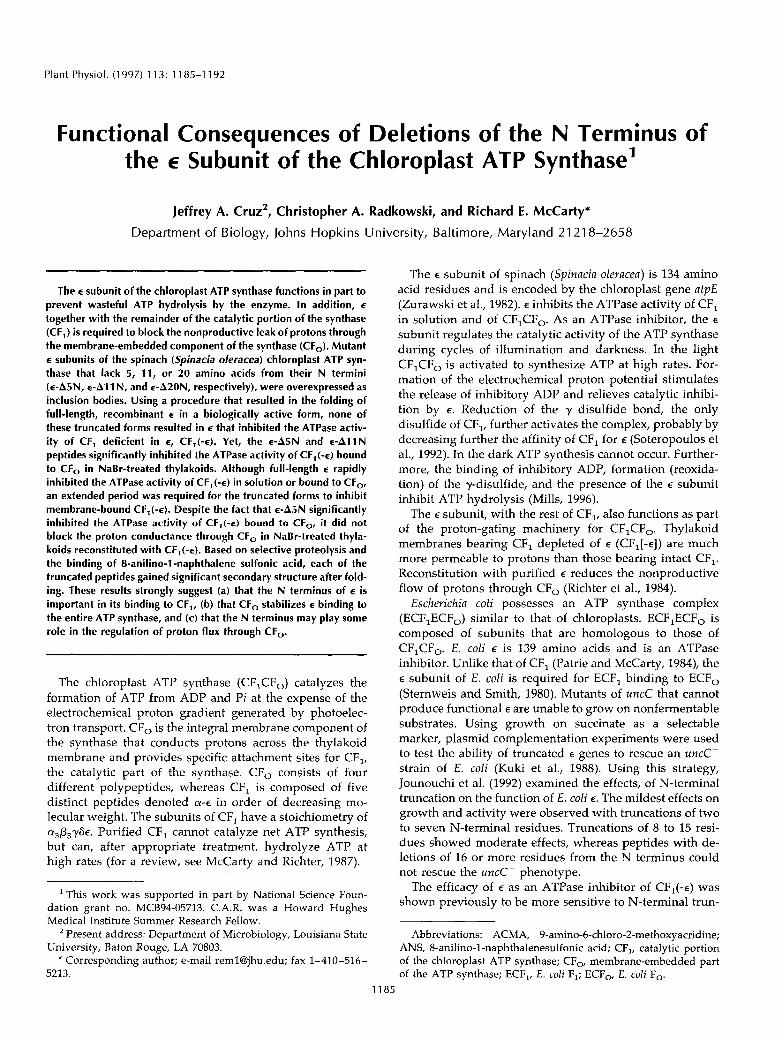
Plant Physiol. (1 997) 11 3: 11 85-1 192 Functional Consequences of Deletions of the N Terminus of the E Subunit of the Chloroplast ATP Synthase' Jeffrey A. Cruz', Christopher A. Radkowski, and Richard E. McCarty* Department of Biology, Johns Hopkins University, Baltimore, Maryland 21 21 8-2658 The E subunit of the chloroplast ATP synthase functions in part to prevent wasteful ATP hydrolysis by the enzyme. In addition, E together with the remainder of the catalytic portion of the synthase (CF,) is required to block the nonproductive leak of protons through the membrane-embedded component of the synthase (CF,). Mutant E subunits of the spinach (Spinacia oleracea) chloroplast ATP syn- thase that lack 5, 11, or 20 amino acids from their N termini (E-A~N, €-A1 1 N, and E-A~ON, respectively), were overexpressed as inclusion bodies. Using a procedure that resulted in the folding of full-length, recombinant E in a biologically active form, none of these truncated forms resulted in E that inhibited the ATPase activ- ity of CF, deficient in E, CF,(-E). Yet, the eA5N and E-AllN peptides significantly inhibited the ATPase activity of CF,(-E) bound to CF, in NaBr-treated thylakoids. Although full-length E rapidly inhibited the ATPase activity of CF,(-E) in solution or bound to CF, an extended period was required for the truncated forms to inhibit membrane-bound CF,(-e). Despite the fact that E-A~N significantly inhibited the ATPase activity of CF,(-E) bound to CF,, it did not block the proton conductance through CF, in NaBr-treated thyla- koids reconstituted with CF,(-E). Based on selective proteolysis and the binding of 8-anilino-1-naphthalene sulfonic acid, each of the truncated peptides gained significant secondary structure after fold- ing. These results strongly suggest (a) that the N terminus of E is important in its binding to CF,, (b) that CF, stabilizes E binding to the entire ATP synthase, and (c) that the N terminus may play some role in the regulation of proton flux through CF,. The chloroplast ATP synthase (CF,CF,) catalyzes the formation of ATP from ADP and Pi at the expense of the electrochemical proton gradient generated by photoelec- tron transport. CF, is the integral membrane component of the synthase that conducts protons across the thylakoid membrane and provides specific attachment sites for CF,, the catalytic part of the synthase. CF, consists of four different polypeptides, whereas CF, is composed of five distinct peptides denoted a-E in order of decreasing mo- lecular weight. The subunits of CF, have a stoichiometry of a,p,yS~. Purified CF, cannot catalyze net ATP synthesis, but can, after appropriate treatment, hydrolyze ATP at high rates (for a review, see McCarty and Richter, 1987). This work was supported in part by National Science Foun- dation grant no. MCB94-05713. C.A.R. was a Howard Hughes Medica1 Institute Summer Research Fellow. Present address: Department of Microbiology, Louisiana State University, Baton Rouge, LA 70803. * Corresponding author; e-mail remlqhuedu; fax 1-410-516- 5213. The E subunit of spinach (Spinacia oleracea) is 134 amino acid residues and is encoded by the chloroplast gene atpE (Zurawski et al., 1982). E inhibits the ATPase activity of CF, in solution and of CF,CF,. As an ATPase inhibitor, the E subunit regulates the catalytic activity of the ATP synthase during cycles of illumination and darkness. In the light CF,CF, is activated to synthesize ATP at high rates. For- mation of the electrochemical proton potential stimulates the release of inhibitory ADP and relieves catalytic inhibi- tion by E. Reduction of the y disulfide bond, the only disulfide of CF,, further activates the complex, probably by decreasing further the affinity of CF, for E (Soteropoulos et al., 1992). In the dark ATP synthesis cannot occur. Further- more, the binding of inhibitory ADP, formation (reoxida- tion) of the y-disulfide, and the presence of the E subunit inhibit ATP hydrolysis (Mills, 1996). The E subunit, with the rest of CF,, also functions as part of the proton-gating machinery for CF,CF,. Thylakoid membranes bearing CF, depleted of E (CF,[-E]) are much more permeable to protons than those bearing intact CF,. Reconstitution with purified E reduces the nonproductive flow of protons through CF, (Richter et al., 1984). Escherichia coli possesses an ATP synthase complex (ECF,ECF,) similar to that of chloroplasts. ECF,ECF, is composed of subunits that are homologous to those of CF,CF,. E. coli E is 139 amino acids and is an ATPase inhibitor. Unlike that of CF, (Patrie and McCarty, 1984), the E subunit of E. coli is required for ECF, binding to ECF, (Sternweis and Smith, 1980). Mutants of uncC that cannot produce functional E are unable to grow on nonfermentable substrates. Using growth on succinate as a selectable marker, plasmid complementation experiments were used to test the ability of truncated E genes to rescue an uncC- strain of E. coli (Kuki et al., 1988). Using this strategy, Jounouchi et al. (1992) examined the effects, of N-terminal truncation on the function of E. coli E. The mildest effects on growth and activity were observed with truncations of two to seven N-terminal residues. Truncations of 8 to 15 resi- dues showed moderate effects, whereas peptides with de- letions of 16 or more residues from the N terminus could not rescue the uncC- phenotype. The efficacy of E as an ATPase inhibitor of CF,(-E) was shown previously to be more sensitive to N-terminal trun- Abbreviations: ACMA, 9-amino-6-chloro-2-methoxyacridine; ANS, 8-anilino-1-naphthalenesulfonic acid; CF,, catalytic portion of the chloroplast ATP synthase; C F , membrane-embedded part of the ATP synthase; ECF,, E. coli F,; E C F , E. coli F,. 1185
-
Upload
michiganstate -
Category
Documents
-
view
2 -
download
0
Transcript of Functional Consequences of Deletions of the N Terminus of the E Subunit of the Chloroplast ATP...
Plant Physiol. (1 997) 1 1 3: 11 85-1 192
Functional Consequences of Deletions of the N Terminus of the E Subunit of the Chloroplast ATP Synthase'
Jeffrey A. Cruz', Christopher A. Radkowski, and Richard E. McCarty*
Department of Biology, Johns Hopkins University, Baltimore, Maryland 21 21 8-2658
The E subunit of the chloroplast ATP synthase functions in part to prevent wasteful ATP hydrolysis by the enzyme. In addition, E
together with the remainder of the catalytic portion of the synthase (CF,) i s required to block the nonproductive leak of protons through the membrane-embedded component of the synthase (CF,). Mutant E subunits of the spinach (Spinacia oleracea) chloroplast ATP syn- thase that lack 5, 11, or 20 amino acids from their N termini ( E - A ~ N , €-A1 1 N, and E-A~ON, respectively), were overexpressed as inclusion bodies. Using a procedure that resulted in the folding of full-length, recombinant E in a biologically active form, none of these truncated forms resulted in E that inhibited the ATPase activ- i ty of CF, deficient in E, CF,(-E). Yet, the e A 5 N and E - A l l N peptides significantly inhibited the ATPase activity of CF,(-E) bound to CF, in NaBr-treated thylakoids. Although full-length E rapidly inhibited the ATPase activity of CF,(-E) in solution or bound to CF,, an extended period was required for the truncated forms to inhibit membrane-bound CF,(-e). Despite the fact that E - A ~ N significantly inhibited the ATPase activity of CF,(-E) bound to CF,, it did not block the proton conductance through CF, in NaBr-treated thyla- koids reconstituted with CF,(-E). Based on selective proteolysis and the binding of 8-anilino-1 -naphthalene sulfonic acid, each of the truncated peptides gained significant secondary structure after fold- ing. These results strongly suggest (a) that the N terminus of E is important i n i t s binding to CF,, (b) that CF, stabilizes E binding to the entire ATP synthase, and (c) that the N terminus may play some role i n the regulation of proton flux through CF,.
The chloroplast ATP synthase (CF,CF,) catalyzes the formation of ATP from ADP and Pi at the expense of the electrochemical proton gradient generated by photoelec- tron transport. CF, is the integral membrane component of the synthase that conducts protons across the thylakoid membrane and provides specific attachment sites for CF,, the catalytic part of the synthase. CF, consists of four different polypeptides, whereas CF, is composed of five distinct peptides denoted a-E in order of decreasing mo- lecular weight. The subunits of CF, have a stoichiometry of a,p,yS~. Purified CF, cannot catalyze net ATP synthesis, but can, after appropriate treatment, hydrolyze ATP at high rates (for a review, see McCarty and Richter, 1987).
This work was supported in part by National Science Foun- dation grant no. MCB94-05713. C.A.R. was a Howard Hughes Medica1 Institute Summer Research Fellow.
Present address: Department of Microbiology, Louisiana State University, Baton Rouge, LA 70803.
* Corresponding author; e-mail remlqhuedu; fax 1-410-516- 5213.
The E subunit of spinach (Spinacia oleracea) is 134 amino acid residues and is encoded by the chloroplast gene atpE (Zurawski et al., 1982). E inhibits the ATPase activity of CF, in solution and of CF,CF,. As an ATPase inhibitor, the E
subunit regulates the catalytic activity of the ATP synthase during cycles of illumination and darkness. In the light CF,CF, is activated to synthesize ATP at high rates. For- mation of the electrochemical proton potential stimulates the release of inhibitory ADP and relieves catalytic inhibi- tion by E. Reduction of the y disulfide bond, the only disulfide of CF,, further activates the complex, probably by decreasing further the affinity of CF, for E (Soteropoulos et al., 1992). In the dark ATP synthesis cannot occur. Further- more, the binding of inhibitory ADP, formation (reoxida- tion) of the y-disulfide, and the presence of the E subunit inhibit ATP hydrolysis (Mills, 1996).
The E subunit, with the rest of CF,, also functions as part of the proton-gating machinery for CF,CF,. Thylakoid membranes bearing CF, depleted of E (CF,[-E]) are much more permeable to protons than those bearing intact CF,. Reconstitution with purified E reduces the nonproductive flow of protons through CF, (Richter et al., 1984).
Escherichia coli possesses an ATP synthase complex (ECF,ECF,) similar to that of chloroplasts. ECF,ECF, is composed of subunits that are homologous to those of CF,CF,. E. coli E is 139 amino acids and is an ATPase inhibitor. Unlike that of CF, (Patrie and McCarty, 1984), the E subunit of E. coli is required for ECF, binding to ECF, (Sternweis and Smith, 1980). Mutants of uncC that cannot produce functional E are unable to grow on nonfermentable substrates. Using growth on succinate as a selectable marker, plasmid complementation experiments were used to test the ability of truncated E genes to rescue an uncC- strain of E. coli (Kuki et al., 1988). Using this strategy, Jounouchi et al. (1992) examined the effects, of N-terminal truncation on the function of E. coli E . The mildest effects on growth and activity were observed with truncations of two to seven N-terminal residues. Truncations of 8 to 15 resi- dues showed moderate effects, whereas peptides with de- letions of 16 or more residues from the N terminus could not rescue the uncC- phenotype.
The efficacy of E as an ATPase inhibitor of CF,(-E) was shown previously to be more sensitive to N-terminal trun-
Abbreviations: ACMA, 9-amino-6-chloro-2-methoxyacridine; ANS, 8-anilino-1-naphthalenesulfonic acid; CF,, catalytic portion of the chloroplast ATP synthase; C F , membrane-embedded part of the ATP synthase; ECF,, E. coli F,; E C F , E. coli F,.
1185

1186 Cruz et ai. Plant Physiol. Vol. 11 3, 1997
cation than to C-terminal truncation (Cruz et al., 1995). Variants with 45 residues deleted from the C terminus (E-445C) showed similar decreases in inhibitor potency as those with 11 residues removed from the N terminus (E- 411N). These studies suggested that the loss of activity results from a loss of tight binding affinity of the truncated variants to CF1(-e) and supported the possibility that a region of the N terminus is essential for the structure and function of E. Here we report that even short N-terminal truncations abolish the ability of E to inhibit the ATPase activity of CF*(-E) in a solution. In contrast, two of the truncated forms can significantly inhibit the ATPase activ- ity of CF1(-e) bound to CF, in thylakoid membranes.
MATERIALS AND METHODS
Hot Tub DNA polymerase was purchased from Perkin- Elmer. Dideoxynucleotide stock solutions were purchased from United States Biochemical. Other standard molecular biology reagents were purchased from Gibco-BRL. Restric- tion enzymes and T4 DNA ligase were purchased from Stratagene. ANS was purchased from Research Organics (Cleveland, OH). ACMA was purchased from Sigma.
Preparation of CF,, CF,(-e), and Purified E Subunit
CF, was prepared as described by Soteropoulos et al. (1992) with the following modification: QAE chromatogra- phy was done using Toyopearl QAE-550 ion-exchange resin (Toso Haas/Supelco, Bellefonte, PA) with a bed vol- ume of approximately 250 mL. The column was equili- brated with 1.8 L of 20 mM Hepes-NaOH (pH 7.3), 1 mM EDTA, and 0.5 mM ATP. After loading of approximately 500 mg of protein the column was eluted with a 500-mL linear gradient of ammonium sulfate (20-72.5 mM) in the equilibration buffer and then with 300 mL of equilibration buffer containing 125 mM ammonium sulfate. CF, was eluted with an equilibration buffer containing 250 mM ammonium sulfate. CF,(-E) and the purified E subunit were prepared as described by Cruz et al. (1995). CF, and CF1(-e) were stored as ammonium sulfate precipitates at 4°C and were desalted in 5 mM Tris-HC1 (pH 8.0) using the method of Penefsky (1977) prior to use. The concentration of CF1(-e) was determined by the method of Lowry (1951) using BSA as a standard. The eluted E was stored at 4°C in the buffer used to extract it from CF,. The concentration of E was determined by the method of Bradford (1976) using BSA as a standard. Absorbance values for E were corrected as described by Soteropoulos et al. (1992).
Preparation of E-Deficient Thylakoid Membranes
Thylakoids deficient in CF, were prepared by treatment with 2 M NaBr, as described by Nelson and Eytan (1979) with the following modification: For the 10 mM Tricine- NaOH (pH 8.0) wash, the partially resuspended mem- branes were homogenized and filtered through a layer of 930 grade Pellon interfacing (purchased at a local fabric store) to remove the particles that were resistant to com- plete resuspension. For assays of the ability of E to inhibit
the CaZf-ATPase activity of membrane-bound CF1(+), NaBr-treated membranes were reconstituted with CF1(+), as described by Cruz et al. (1995). For proton permeability studies (ACMA fluorescence quenching studies) and time- course experiments, a second method that gave more con- sistent results was developed. In a typical experiment, NaBr-treated thylakoids equivalent to 600 pg of chloro- phyll were mixed with 3 mg of CF,(-E) and 150 pL of 0.1 M ATP, pH 7.0. The volume of the mixture was adjusted to 1.45 mL with 5 mM Tricine (pH 8.0). After 5 min at room temperature, 0.5 M MgC1, was added to a final concentra- tion of 15 mM MgCl,, and the mixture was kept at room temperature for 5 min. An equal volume of cold-buffered SUC solution (0.4 M SUC, 20 mM Tricine-NaOH [pH 8.01, 10 mM NaC1,2 mM ATP, and 7 mM MgC1,) was added. After 5 min on ice, the membranes were collected by centrifuga- tion for 5 min at 27,200g and 4°C. The supernatant was removed by aspiration and the membrane pellet was gently resuspended in 3 mL of cold STN buffer (0.4 M SUC, 20 mM Tricine-NaOH [pH 8.01, and 10 mM NaC1) and 5 mM MgCl,. The membranes were centrifuged (as above) and the pellet was resuspended in a 500 pL of cold STN and 5 mM MgC1, (or with 20 mM Tricine-NaOH [pH 8.01 and 5 mM MgC1, where noted). Chlorophyll concentrations of the suspensions were determined (Arnon, 1949) and the sus- pensions were diluted with STN and 5 mM MgCl, (or with 20 mM Tricine-NaOH [pH 8.01 and 5 mM MgCl,) to 5 pg chlorophyll 95 pLP1 just prior to reconstitution with re- combinant E. Stored in the dark and on ice, membranes prepared in this manner consistently retained strong ACMA fluorescence quenching activity for up to 4 h after reconstitution with E-C~S, and reasonable activity was ob- served even 13 to 18 h after reconstitution.
Construction of pETel 5 and pETel 6
Deletions encompassing the first 5 codons and the first 20 codons of the a t p E coding region were generated by PCR, using pETe2 (Cruz et al., 1995) as the template. For- ward primers 5’-CCGCATATGGTACTGACTCCGAATC- GAAGT-3’ and 5’-CCGCATATGATCATTTTATCTACT- AATAGTGGCC-3’ supplied the start codon as part of an NdeI site to the truncated gene for both the E-45Nt and ~-A20Nt truncations, respectively. The reverse primer 5’- GCTAGTTATTGCTCAGCGG-3’ was designed to hybrid- ize with a region on the sense strand that was approxi- mately 45 bases 3’ of the a t p E stop codon. The PCR product was digested with NdeI and BamHI and subcloned into pET3c (Novagen, Inc., Madison, WI). Truncations were verified by DNA sequencing.
Protein Expression and Preparation of Recombinant E
Plasmids used for expression are listed in Table I. The transformation of the BL21/ DE3 Esckerickia coli expression host, the induction of expression, and the purification of recombinant E were performed as described by Cruz et al. (1995). For some preparations Nonidet P-40 (Sigma) was replaced with IPEGAL (Sigma) at the same concentration (v/v).

N-Terminal Deletions of the E Subuni t of the Chloroplast ATP Synthase 1187
Table 1. Plasmids and gene products Listed below are the plasmids used and the notation for each
inclusion body preparation. For truncation mutants (A) the number indicates the number of residues deleted followed by C or N desig- nating C-terminal or N-terminal truncation, respectively. Where rel- evant, the mutation introduced is given. Plasmids marked with an asterisk have the Cys-6 to Ser-6 substitution in addition to the mu- tation listed. The first four plasmids were described by Cruz et al. (1 995).
Plasmid Mutation Cene Product
pETe2 Cys-6 to Ser-6 E - C ~ S
pET&7* Gln-89 to stop E - A ~ ~ C ~ E T E I 2 Arg-I 2 to Met-1 &-AI 1 N
pET&6* Arg-125 to stop &-A1 OC
pETEl3 Cys-6 to Met-1 E - A ~ N p E T ~ 1 4 Glu-21 to Met-1 E - A ~ O N
Solubilization and Folding
After thawing at room temperature, the inclusion bodies (approximately 2 4 mg of protein) were centrifuged for 5 min at 16,OOOg. The pellets were resuspended in 0.5 to 1 mL of deionized water and were centrifuged, as described above. The supernatants were aspirated as completely as possible. The pellets were dissolved in 100 to 200 pL of 25 mM Tris-HC1 (pH 8.0) and 8 M urea. A small amount of insoluble matter was pelleted by brief centrifugation. The protein concentration was measured by the method of Bradford (1976) using BSA as a standard. A chemical blank containing an amount of urea equivalent to the amount present in the samples was used to correct for the back- ground absorbance by urea. The method of Bradford un- derestimates the concentration of E by 43% (Richter et al., 1985); corrected concentrations were used. The dilution buffer consisted of 5 volumes of 50 mM Tris-HC1 (pH &O), 40% ethanol, 3 volumes of glycerol, and 1 volume of deion- ized water. Solubilized inclusion bodies were rapidly di- luted ( 1 : l O ) into the chilled dilution buffer.
Competition for Binding to CF,(-E) in Solution
Urea-solubilized inclusion bodies of E-AlOC, E - A ~ ~ C , E-A~N, E-AllN, and E-A~ON were folded as described above such that a 7.5-to-1 ratio of E to CF1(-e) would result when 160 pL of the folded E solution was added to 20 pg of CF'(-E) in 320 pL of 5 mM Tris-HC1 (pH 8.0). Urea- solubilized eC6S was folded to a concentration that would give a 3-to-1 ratio upon reconstitution. After folding, each of the truncation mutants was mixed with an equal volume of folded E-C~S or an equal volume of the control buffer, consisting of 9 volumes of the dilution buffer and 1 volume of 25 mM Tris-HCl (pH 8.0) and 8 M urea. Aliquots (160 pL) of each mixture were reconstituted with 20 pg of CF1(-e) in 320 pL. After at least 5 min at room temperature, Caz+- ATPase activity was measured. Molecular masses of 14.1 kD and 12.4 kD were calculated for eA5N and E-A~ON, respectively .
Competition for Binding to e-Deficient Membranes
eA45C and E-AllN were folded so that the ratio of E to CF1(-e), after reconstitution using the protocol outlined above, was either 15 to 1 or 60 to 1. Urea-solubilized eC6S was folded to a concentration that would give a 3-to-1 ratio. At each concentration the truncation mutants (at both con- centrations) were mixed with an equal volume of E-C~S or with the control buffer (prepared as described above). For every 95 p L of thylakoids reconstituted with CF,(-E) in STN, 5 pL of an E sample was added. After storage for approximately 12 h at 4"C, Ca2+-ATPase activity was measured.
Time-Course Experiment with CF,(-e) in a Solution
CF'(-E) was equilibrated with 20 mM Tricine (pH 8.0) and 5 mM MgCl, by the method of Penefsky (1977). ATP was added to a final concentration of 1 mM. After 5 min at room temperature, CF'(-E) was reequilibrated with 20 mM Tricine-NaOH (pH 8.0) and 5 mM MgCl,, and the protein concentration was determined. The preloading of CF1(-c) with Mg2+-ATP in this manner stabilizes it against cold inactivation (Hightower and McCarty, 1996). CF1(-e) was diluted to a concentration of 52.6 p g mL-' with 20 mM Tricine-NaOH (pH 8.0) and 5 mM MgCl,, and 1.8 mL aliquots were mixed with 0.2 mL of folded E-C~S, E-A~N, or the control buffer. The e-C6S and eA5N were folded to concentrations of 0.37 mg mL-' and 0.35 mg mL-', respec- tively. The solutions were stored at approximately 4°C for the duration of the experiment. Aliquots (100 pL) of each sample were assayed for Mg2+-ATPase activity 0.25, 3.5, 16, and 40 h after reconstitution.
Time-Course Experiment with e-Deficient Membranes
NaBr-treated thylakoids reconstituted with CF'(-E) were prepared as described above and resuspended in 20 mM Tricine-NaOH (pH 8.0) and 5 mM MgCl,. Membranes were diluted to a concentration of 5 p g chlorophyll 95 pL-l in the same buffer, and 1.8-mL aliquots of the membrane suspension were mixed with 0.2 mL of E-C~S, E-A~N, or the control buffer. The eC6S and eA5N were folded to con- centrations of 0.44 mg mL-' and 0.42 mg mL-I, respec- tively. The mixtures were stored at approximately 4°C for the duration of the experiment. Aliquots (100 pL) were assayed for MgZf-ATPase activity 0.25, 3.5, 16, and 40 h after reconstitution.
Time Course of ACMA Fluorescence Quenching
NaBr-treated thylakoids reconstituted with CF1(-e) were prepared as described above and resuspended in STN and 5 mM MgC1,. Membranes were diluted to a concentration of 5 pg chlorophyll95 pL-' in the same buffer, and 900-pL aliquots of the membrane suspension were mixed with 100 pL of E-C~S, E-A~N, or control buffer. The eC6S and eA5N were folded to concentrations of 0.44 mg mL-' and 0.42 mg mL-'. The solution was stored at approximately 4°C for the duration of the experiment.,Aliquots (100 pL) from each were assayed for the quenching of ACMA fluores-

1188 Cruz et al. Plant Physiol. Vol. 113, 1997
cence, as described below, 5 min, 3.5 h, and 13 h after theaddition of e.
ATPase Activity Assays
For measurement of Ca2+-ATPase activity, samples wereincubated at 37°C in the presence of 50 HIM Tris-HCl (pH8.0), 5 mM ATP, and 5 mM CaCl2 and in a final volume of1 mL. Mg2+-ATPase activity was measured by incubationof the samples in 50 mM Tris-HCl (pH 8.0), 2 mM MgCl2,100 mM Na2SO3, and 4 mM ATP. Reactions were stoppedafter 10 min by the addition of 1 mL 0.5 N TCA and theconcentration of Pi was determined (Taussky and Shorr,1953).
Proton Permeability
Aliquots (100 juL) of the reconstituted thylakoids, equiv-alent to 5 /Mg of chlorophyll, were assayed for pH gradientformation in 50 mM Tricine-NaOH (pH 8.0), 50 mM NaCl,50 fiM N-methylphenazonium methosulfate salt, 0.75 /XMACMA, and 2 mM ascorbate (pH 6.0) in a total volume of 1mL. ACMA fluorescence quenching was determined asdescribed previously (Cruz et al., 1995).
Trypsin Digests
Purified e was diluted to a final concentration of 0.3 mgmL"1 with 25 mM Tris-HCl (pH 8.0), 20% ethanol, and 30%glycerol. Urea-solubilized, recombinant e-variants werefolded (as described above) or diluted with Tris-urea toconcentrations between 0.2 and 0.3 mg mL"1. Aliquots of200 fiL of purified e and the folded e-variants were mixedwith 180 fjL of the dilution buffer and 20 /xL of 0.1 M CaCl2.For the zero time control, an aliquot of 60 /J.L was drawnfrom each sample and mixed with approximately 0.6 /uL oftrypsin (1 mg mL"1 in 1 mN HC1) and 4 jiL of PMSF (30 mgmL"1 in 100% ethanol) and was flash-frozen in dry-iceethanol. Trypsin in 3.4-/xL aliquots was added to the re-maining 340 /U.L of the samples. After 15, 30, 60, and 90 minat room temperature, 60-/J.L aliquots were withdrawn fromeach sample, mixed with 4 /xL of PMSF, and flash-frozen asdescribed above. For unfolded samples, the 180 /^L ofdilution buffer was substituted with Tris-urea and the di-gests were repeated as described above. Digests were an-alyzed by SDS-Page using a Tricine buffer system as de-scribed by Schagger and von Jagow (1987).
ANS Fluorescence Assays
Fluorescence measurements were conducted at roomtemperature in an RF-5000 spectrofluorimeter (ShimadzuScientific, Columbia, MD) using an excitation wavelengthof 360 nm and an emission wavelength range of 400 to 600nm. The excitation and emission band widths were 5 nm.
All e samples were solubilized and diluted in 25 mMTris-HCl (pH 8.0) and 8 M urea to a concentration of 250JUM. e samples in the buffered 8 M urea solution werediluted 10-fold into the ethanol-glycerol dilution buffer asdescribed in "Solubilization and Folding." To 0.95 mL of 50HIM Tris-HCl (pH 8.0) was added 50 ;u,L of the e solution,
and the fluorescence was measured at 0, 4, 8,12,16, 20, and24 fiM ANS. The values were corrected for the fluorescenceof ANS at each concentration in the same buffer and in theabsence of e. For unfolded e samples the urea-solubilizedprotein was diluted to 25 fiM using the buffered 8 M urea.Fifty microliters of the 25 fiM e sample was added to 950 fiLof the buffered 8 M urea, and the samples were titratedwith ANS as described above. The values were correctedfor ANS fluorescence at each concentration in the ab-sence of e.
Miscellaneous Methods
Primers were synthesized in a 381A DNA synthesizer(Applied Biosystems). Unless noted otherwise, SDS-PAGE(18% acrylamide and 0.48% bisacrylamide) was performedaccording to the method of Fling and Gregerson (1986).Immunoblotting was as described by Soteropoulos et al.(1992).
RESULTS
The three N-terminal truncation mutants that we gener-ated had 5, 11, and 20 amino acids removed. These mutantes are referred to as e-A5N, e-AHN, and e-A20N, respec-tively, where A represents deletion and N represents the Nterminus. The plasmids and corresponding proteins areshown in Table I. Two C-terminal truncation mutants arealso given.
Overexpression of spinach chloroplast e in E. coli resultsin its accumulation in inclusion bodies (Cruz et al., 1995).As was the case for overexpressed, full-length e,N-terminally truncated forms of e are relatively pure (Fig.1). The truncated peptides migrate distances consistentwith the extent of truncation. Each of the truncated esreacts strongly with the anti-e serum on immunoblots (notshown). The major contaminant in each case is a proteinthat has higher electrophoretic mobility on SDS-PAGE thanthe full-length proteins. These smaller-molecular-weightpeptides also cross-react with a polyclonal e antiserum (notshown) and likely result from either early termination oftranscription or translation or limited, site-specific proteol-
1 2 3 4 5
Figure 1. SDS-PAGE of overexpressed e forms. Lane 1, CF,; lane 2,e-C6S; lane 3, 6-A5N; lane 4, e-A11 N; and lane 5, e-A20N. About 20fig of CF, and 5 fig of each of the e forms were used.

N-Terminal Deletions of the E Subunit of the Chloroplast ATP Synthase
Buffer
A20N
A l l N
A5N
A45C
1189
’ 7 -
ysis. These shorter polypeptides may not bind tightly to CF,. For example, CF,(-E) was incubated in a solution with E-C~S, prepared in a biologically active form from the inclusion bodies. When purified, the reconstituted CF, con- tained only full-length E (J. Cruz, unpublished data). There- fore, the presence of these shorter e polypeptides should not affect reconstitution significantly.
We reported (Cruz et al., 1995) that eA11N and E - A ~ ~ C are poor inhibitors of the ATPase activity of CF1(+), either in solution or bound to CF, in NaBr-treated thylakoids. eA5N and E-A~ON also fail to inhibit significantly the ATPase activity of CF](-E) in solution, even at a ratio of 7.5 to 1 (mo1 E to mo1 CFJ-E]) (Fig. 2). Moreover, none of the truncated forms prevented inhibition by E-C~S (Fig. 3). CF,(-E) was incubated with the truncated ES at a 3.75 to 1 E/CF,(-E) molar ratio. The ability of E-C~S to inhibit at a molar ratio of 1.5 to 1 was unimpaired. These results sug- gest that the truncated forms of E do not bind tightly to CF,(-E) in solution.
In addition, E-AllN and eA45C were tested for the abil- ity to block the binding of E-C~S to CF1(-e) reconstituted with CF,. Despite the fact that both peptides were used at concentrations yielding molar ratios of E:CF](-E) of 7.5 to 1 and 30 to 1, neither prevented the binding of eC6S at a 1.5 to 1 molar ratio (data not shown). A variation of this experiment in which E-C~S was added after an overnight incubation of the membranes with the truncated peptides gave essentially the same results. In a11 cases, the truncated peptides alone did not inhibit more than 35% of the Ca2+- ATPase activity of CF,-bound CF1(+), whereas reconstitu- tions with E-C6S, in the presence or absence of the trun- cated peptides, strongly and consistently inhibited ca‘+- ATPase activity (80-90% inhibition). E-A~N and E-AllN significantly inhibited the ATPase activity of CF1(-e) bound to CF, in thylakoid membranes (Fig. 4). At a molar ratio of E/CF,(-E) of 7.5:1, a ratio at which none of the E-AN mutant proteins inhibited the ATPase of CF1(-e) in solution, eA5N inhibited nearly 60% of the ATPase activity and e-AllN,
O 0 1 2 3 4 5 6 7 8
Molar Ratio E/CF~(-E)
Figure 2. lnhibition of the ATPase activity of CF,(-c) in solution. CF,(-E) was incubated with the E variants after a 1 O-fold dilution from 8 M urea solutions as described in “Materials and Methods.” CaZc- ATPase activity was assayed with 5 y g of CF,(-E). The maximal activity ranged from 8 to 12 ymol Pi formed min-’ mg-’ CF,(-E). O, E-C~S; H, E-A5N; +, E-A1 1 N; and A, E-A2ON.
AlOCI-, , , , , , I O 20 40 60 80 100
’% Inhibition
Figure 3. Effect of truncated forms of E on the ability of 4 6 s to inhibit the ATPase activity of soluble CF,. Each of the truncated E
forms were solubilized and diluted into the ethanol/glycerol buffer (Buffer). Twenty micrograms of CF, was incubated with the mutant ES
at a 3.75 to 1 molar ratio. Where indicated (dark bars) E -C~S was added to a 1.5 to 1 molar ratio. White bars give the inhibition by the truncated E in the absence of eC6S.
35%. E-A~ON failed to inhibit at this concentration and had just a marginal effect at a molar ratio of E/CF~(-E) of 30 to 1.
Membrane ATPase inhibition studies were usually per- formed in conjunction with ACMA fluorescence quenching assays. Following the ACMA assay, reconstituted samples were stored overnight prior to assay of ATPase activity. In contrast to the membrane studies, samples of CF1(-e) re- constituted in solution with E were assayed for ATPase activity shortly after reconstitution. Although purified E
and E-C~S bind to CF1(-e) quickly, this is not the case for E-A~N (Table 11). eC6S strongly inhibited the ATPase of CF](-E) in solution and on membranes within 15 min of reconstitution, but eA5N did not. With longer incubation times, eA5N inhibited the Mg*+-ATPase of membrane- bound, but not soluble, CF](-E).
100
80
f i
.$ 60 2 r E:
se 40 ‘I
20
O O 5 10 15 20 25 30 35
Molar Ratio E/CF~(-E)
Figure 4. lnhibition of the ATPase activity of CF,(-E) bound to CF, thylakoid membranes. NaBr-treated thylakoids reconstituted with CF,(-E) were incubated with the forms of E shown overnight at 4°C as described in “Materials and Methods.” The Ca2+ -ATPase activity of aliquots containing 5 y g of chlorophyll was determined. C6S, E-C~S; A5, E-A5N; A1 1, €-A1 1 N; and A20, E-A~ON.

1190 Cruz et al. Plant Physiol. Vol. 11 3 , 1997
Table 11. Time course for development o f the inhibition o f the ATPase activity o f CFI(&) in solution and bound to CF, by E - C ~ S and E - A ~ N
Soluble CF,(-E) (95 pg) was incubated at 4°C with folded E - C ~ S and E - A ~ N (75 and 70 pg, respectively), in a final volume of 2.0 mL and MgZf-ATPase activity of aliquots was measured in the presence of 100 mM Na,SO, at the times indicated after addition of the E
samples. NaBr-treated thylakoids were reconstituted with CF,(-E) and aliquots equivalent to 95 yg of chlorophyll were incubated at 4°C with folded E - C ~ S (88 pg) or E - A ~ N (84 pg) in a total volume of 2.0 mL. MgZ'-ATPase activity was determined at the times indicated after the addition of the E samples. Buffer controls were run for both exoer i ments.
lnhibition of ATPase Activity
CF,(-&) Membrane- Time after E Addition Bound CF,(-&) Solution
h %
0.25 87 0 77 0 3.5 86 4 81 20 16 86 O 85 45 40 87 0 84 44
In most cases, the ATPase activity CF, was measured in the presence of Ca'+. Mg2+ is an inhibitor of Ca2+-ATP hydrolysis. In the presence of oxyanions. such as sulfite, CF,(-E) either in solution or bound to CF, catalyzes rapid ATP hydrolysis in the presence of Mg2+. Mg2+ is present in the buffer in which NaBr-treated thylakoids reconstituted with CF1(-e) is resuspended. Therefore, sulfite Mg2-ATPase activity was determined for membrane-bound CF(I-~). Both Mg2+- and Ca2+-ATPase activities of CF,(-E) in solu- tion are inhibited by E (Soteropoulos et al., 1992).
After an overnight incubation following reconstitution, E-A~N could inhibit 40% or more of the Mg2+-ATPase activity of membrane-bound CF,(-E) (Table 11). Yet, within the same period, E-A~N did not confer to CF,(-E) the ability to block proton conductance by CF, (Table 111). Significant ACMA fluorescence quenching was detected in the sample reconstituted with E-C~S 13 h after reconstitution, but no quenching was detected in the sample reconstituted with
The truncated forms of E may not fold properly after dilution from 8 M urea to 0.8 M urea. This possibility was tested by the binding of ANS and by the susceptibility of
E-A~N.
the recombinant E to proteolysis. ANS binds weakly to denatured proteins, but much more tightly to hydrophobic pockets in folded or folding proteins. The quantum yield of ANS fluorescence is increased on binding and the emission spectrum is blue-shifted. ANS fluorescence has been used to study protein folding kinetics (Semisotnov et al., 1991) and dissociation of CF, (Hightower and McCarty, 1996).
As expected, the fluorescence of ANS in 8 M urea solu- tion was not enhanced by the addition of any of the mutant E polypeptides (not shown). Each of the truncated E S tested did, however, increase ANS fluorescence after dilution into folding buffer (Fig. 5). The ANS titration curves show a similar dependence on ANS concentration, but the extent of the change appears to be higher for E-A~N and E-AllN than for E-C6S. As might be expected, eA45C binds less ANS than E-C~S.
The sensitivity of denatured proteins to proteolysis is much greater than that of folded proteins. The accessibility of the protease to potential cleavage sites is impeded by the structure of the folded protein. Selective proteolysis can thus be used to determine if the recombinant E folds in a manner similar to the native protein. As shown in Figure 6, purified E from spinach CF, and E-C~S are much more slowly determined if the recombinant E folds in a manner similar to that of the native protein. As shown in Figure 6, E purified from CF, are much more slowly digested by trypsin in the absence of 8 M urea than in its presence. Moreover, the pattern of the peptides generated by prote- olysis was very similar for the two proteins. Proteolysis of E-AllN and E - A ~ ~ C was also carried out. In both cases, 8 M
urea accelerated proteolysis, and the pattern of polypep- tide fragments generated by trypsinolysis resembled that of E-C6S.
DlSCUSSlON
The functional importance of the N terminus of the E
subunit of CF, is clearly illustrated by our results. Deletion of as few as five residues from the N terminus of E abol- ishes its ability to inhibit the ATPase activity of CF1(-e) in solution. Because the truncated form does not compete with full-length recombinant E for binding to CF,(-E) in solution, the truncation must dramatically lower the affin- ity of E for CF,(-E). A rough estimate of 2 X 1OP1' M for the dissociation constant of E from CF, was obtained (Sotero- poulos et al., 1992) from the dilution experiments. E-A~N
Table 111. E - A ~ N fails to restore ApH formation to NaBr-treated thylakoids containing CF ,(-E)
NaBr-treated thylakoids were reconstituted with CF,(-E) and reconstituted with folded E - C ~ S or E - A ~ N , essentially as described in the Table I I legend. ApH formation in the light was determined by monitoring the quenching of ACMA fluorescence at the times shown. Where indicated, DCCD (N,N'-dicyclohexylcarbodiimide) was added to 50 FM. DCCD blocks proton conductance by CF,.
Quenching of ACMA Fluorescence
Sample 0.1 h 3.5 h 13 h
-DCCD +DCCD -DCCD +DCCD -DCCD +DCCD
N o E 3 82 2 55 1 53 E - C ~ S 49 70 52 67 27 31 E-A5 N 4 77 3 62 1 33

N-Terminal Deletions of the e Subunit of the Chloroplast ATP Synthase 1191
100
80
i 60
40
E20
10 15[ANSI - |iM
20 25
Figure 5. Binding of ANS to e forms in solution, e samples (1.25 JJ.M)in 50 ITIM Tris-HCI (pH 8.0) were prepared as described in "Materialsand Methods" and ANS fluorescence was determined at the ANSconcentrations shown. The values are corrected for the fluorescenceof ANS in the absence of protein. •, e-A5N; *, e-A11N;», e-A20N;A, e-C6S; and T, e-A45C.
likely has a considerably lower K0 and it may be possibleto determine its binding affinity directly through the use offluorescence resonance energy transfer methods. This ap-proach was used to determine the KD for interaction ofthioredoxin and CFj (Dann and McCarty, 1992).
Perturbations to protein structure are a major concern inany site-directed mutagenesis study in which biochemicaland/or phenotypical analyses are made. This considerationassumes an even greater significance if, as is the case for allof the mutant es so far isolated, the protein is insoluble afterexpression in E. coli and appears in inclusion bodies. Themutated proteins must be dissolved in a denaturant (e.g. 8 Murea) and then transferred to conditions that promote fold-ing of the e polypeptide chain into a form that one hopesresembles native e. The dramatic loss of the ability of recom-binant e to inhibit the ATPase activity of CFi(-e) in solutioncould be a result of improper folding. For example, Wilkenset al. (1995) reported on an NMR structure of the £. coli e inwhich the first 20 residues are a significant part of a )3"sandwich." If the € subunit of CFj has a similar structure,deletion of even a few residues from the N terminus couldprevent formation of this j3 structure.
Two lines of evidence suggest that the truncated variantsof e can fold. Both ANS binding, as determined by en-hancement of ANS fluorescence, and limited proteolysissuggest that the gross structure of the recombinant es doesnot differ greatly from that of e-C6S. It is, however, notclear why e-A5N and e-AHN appear to enhance ANS flu-orescence to an extent greater than that of e-C6S. Onepossible explanation is that the truncations could decreaseCoomassie blue binding, resulting in an underestimation ofthe protein concentration by the Bradford assay (1976).
Given the strong possibility that the truncated forms of ehave structures similar to that of full-length e, the decreasein inhibition with truncation may result from either a lossof high-affinity binding or of a functional domain. Becauselarge excesses of truncated es do not prevent the binding of
e-C6S to CF,(-e) on membranes or in solution, truncationclearly decreases e binding to CF-,(-e).
The idea that N-terminal truncation affects binding isfurther supported by the observation that e-A5N slowlyinhibits the ATPase activity membrane-bound CF^-e).Moreover, the maximum Mg2+-ATPase inhibition bye-A5N reaches a lower value (up to 45%) than that by e-C6S(>95%). This decreased extent of inhibition by e-A5N mayresult from dissociation of e after dilution into the assaymedium. Aggregation of e-A5N during the prolonged in-cubation period could also explain the decrease in theextent of inhibition. Finally, it is possible that even whenthe e binding site of CFj(-e) is saturated with e-A5N, onlypartial inhibition is obtained.
The fact that e-A5N and e-AHN inhibit the ATPase ac-tivity of CFj(-e) bound to CF0 suggests that these truncatedforms of e can interact with CF, and that these interactionsare strengthened by CF0. The idea that e may interact withCF0 is not novel. Soteropoulos et al. (1992) showed thatupon dilution e can dissociate freely from CFl in solution,but not from the fully assembled ATP synthase on thyla-koid membranes. Wetzel and McCarty (1993) observed thatextraction of the e subunit from the purified ATP synthasemay partially deplete the complex of subunit III as well,suggesting a possible interaction between these subunits.Zhang and Fillingame (1995) showed that a Cys introducedat position 31 of E. coli e can be cross-linked to the extrinsicloop of subunit c of ECF0.
CF, blocks proton conductance through CFO (McCartyand Racker 1966), but CF^-e) does not. Binding of purifiede to NaBr-treated membranes reconstituted with CF^-e)restores the ability to block the proton leak to CF, (Richteret al., 1984). It is surprising to note that e-A5N does not
A=native e FOLDED UNFOLDED0 IS 30 60 90 min 0 15 30 60 90 min
B-A11N CF, FOLDED UNFOLDED1 0 15 30 60 90 min 0 15 30 60 90 min
Figure 6. Trypsin digestion of €. Purified native e (0.15 mg ml"1) andrecombinant es (0.1 to 0.15 mg ml"1) in either the ethanol/glycerolbuffer or 25 mM Tris-HCI (pH 8.0) and 8 M urea were incubated forthe times shown with 10 fig ml"1 trypsin. SDS-PAGE was performedas described by Schagger and von Jagow (1987).

1192 Cruz et al. Plant Physiol. Vol. 11 3 , 1997
restore proton impermeability a t all, even though E - A ~ N inhibits ATPase activity. E - C ~ S ratios of e/CFI(-;) that gave partial inhibition of the reconstituted thylakoid membranes yielded partial restoration of proton impermeability. Thus, in addition to its role in binding, the N terminus of E may be involved i n blocking proton conductance through CF,.
Because the E subunit of CF, is not required for the strong binding of CF, to CF,, certain experimental oppor- tunities are possible that are not accessible i n the E. coli system. The E subuni t of the bacterium is required for the binding of ECF, to ECF, (Sternweis, 1978). In addition, the affinity of ECF, for its E subunit appears significantly lower than that for E binding to CF,. Thus, although the bacterial system is undoubtedly that of choice for a detailed genetic study, the chloroplast ATP synthase is preferable for bio- chemical studies. For example, we should be able to exploit the unique factors of the chloroplast ATP synthase as a means to determine which of the CF, subunits may pro- mote the binding of E to CF,.
In summary, our results confirm and extend the evidence obtained for E. coli (Jounouchi e t al., 1992; Skakoon and Dunn, 1993), which suggest a critica1 role for the N termi- nus of the E subunit to its function as a part of the ATP synthase. From o u r results, we may conclude that the N terminus of the E subunit of CF, is almost crucial for its interactions with the rest of CF,. Furthermore, the ability of E - A ~ N to inhibit CF,(-E) bound t o CF, a n d its inability to inhibit CF1(-e) i n solution clearly suggest that direct inter- actions between E and CF, allow more stable binding to CF,. Finally, these results also suggest that the N termi- nus is important for regulating proton conductance through CF;.
ACKNOWLEDCMENT
We thank Dr. Victor Corces and the members of his laboratory for allowing us to use reagents and equipment.
Received October 2, 1996; accepted December 20, 1996. Copyright Clearance Center: 0032-0889/97/ 113/ 1185/08.
LITERATURE CITED
Arnon DI (1949) Copper enzymes in isolated chloroplasts; poly- phenoxidase in Beta vulgaris. Plant Physiol24 1-15
Bradford MM (1976) A rapid and sensitive method for the quan- titization of microgram quantities of protein utilizing the prin- ciple of protein-dye binding. Anal Biochem 7 2 248-254
Cruz JA, Harfe B, Radkowski CA, Dann MS, McCarty RE (1995) Molecular dissection of the E subunit of the chloroplast ATP synthase of spinach. Plant Physiol 109: 1379-1388
Dann MS, McCarty RE (1992) Characterization the activation of membrane-bound and soluble CF, by thioredoxin. Plant Physiol
Fling SP, Gregerson DS (1986) Peptide and protein molecular weight determination by electrophoresis using a high-molarity Tris buffer system without urea. Anal Biochem 155: 83-88
Hightower KE, McCarty RE (1996) Influence of nucleotides on the cold stability of chloroplast coupling factor 1. Biochemistry 35:
Jounouchi M, Takeyama M, Noumi T, Moriyama Y, Maeda M, Futai M (1992) Role of the amino terminal region of the E subunit
99: 153-160
10051-10057
of Eschevichia coli H+-ATPase (F,F,). Arch Biochem Biophys 2 9 2
Kuki M, Noumi T, Maeda M, Amemura A, Futai M (1988) Func- tional domains of E subunit of Escherichia coli H+-ATPase (F,F,). J Biol Chem 263: 17437-17442
Lowry OH, Rosebrough NJ, Farr AL, Randall RJ (1951) Protein measurement with Folin phenol reagent. J Biol Chem 193:
McCarty RE, Racker E (1966) Effects of a coupling factor and its antiserum on photophosphorylation and hydrogen ion trans- port. Brookhaven Symp Biol 19: 202-214
McCarty RE, Richter ML (1987) Roles of the E and y subunits in the regulation of the chloroplast H+-ATPase. In Y Mukohata, MF Morales, S Fleischer, eds, Perspectives of Biological Energy Transduction. Academic Press, New York, pp 369-375
Mills JD (1996) The regulation of chloroplast ATP synthase CF,- CF,. l n DR Ort, CF Yocum, eds, Oxygenic Photosynthesis: The Light Reactions. Kluwer Academic Publishers, Dordrecht, The Netherlands, pp 469485
Nelson N, Eytan E (1979) Approach to the membrane sector of the chloroplast coupling device. In Y Mukahata, L Packer, eds, Cation Fluxes Across Biomembranes. Academic Press, New York, pp 409415
Patrie WJ, McCarty RE (1984) Specific binding of coupling factor 1 lacking the S and E subunits to thylakoids. J Biol Chem 259:
Penefsky HS (1977) Reversible binding of Pi by beef heart mito- chondrial adenosine triphosphatase. J Biol Chem 252: 2891-2899
Richter ML, Patrie WJ, McCarty RE (1984) Preparation of the E
subunit and E subunit-deficient chloroplast coupling factor 1 in reconstitutively active forms. J Biol Chem 259: 7371-7373
Richter ML, Snyder 8, McCarty RE, Hammes GG (1985) Binding stoichiometry and structural mapping of the E polypeptide of chloroplast coupling factor I. Biochemistry 2 4 5755-5763
Schagger H, von Jagow G (1987) Tricine-sodium dodecyl sulfate- polyacrylamide gel electrophoresis for the separation of proteins in the range from 1 to 100 kDa. Anal Biochem 166: 368-379
Semisotnov GV, Rodionova NA, Razglyaev 01, Uversky VN, Gripas AF, Gilmanshin RI (1991) Study of the "molten globule" intermediate state in protein folding by a hydrophobic fluores- cent probe. Biopolymers 31: 119-128
Skakoon EN, Dunn SD (1993) Orientation of the E subunit in Eschevichia coli ATP synthase. Arch Biochem Biophys 302: 279-284
Soteropoulos P, Siiss K-H, McCarty RE (1992) Modifications of the y subunit of chloroplast coupling factor 1 alter the interac- tions with the inhibitory E subunit. J Biol Chem 267: 10348-10354
Sternweis PC (1978) The E subunit of Escherichia coli coupling factor 1 is required for its binding to the cytoplasmic membrane. J Biol Chem 253: 3123-3128
Sternweis PC, Smith JB (1980) Characterization of the inhibitory ( E ) subunit of the proton-translocating adenosine triphosphatase from Escherichia coli. Biochemistry 19: 526-531
Taussky HH, Shorr E (1953) Microcolorimetric method for the determination of inorganic phosphorous. J Biol Chem 202: 675-685
Wetzel CM, McCarty RE (1993) Aspects of subunit interactions in the chloroplast ATP synthase (11). Plant Physiol 1 0 2 251-259
Wilkens S, Dahlquist FW, McIntosh LP, Donaldson LW, Capaldi RA (1995) Structural features of the E subunit of the Escherichia coli ATP synthase determined by NMR spectroscopy. Nature Stsuct Biol 2: 961-967
Zhang Y, Fillingame RH (1995) Subunits coupling H+ transport and ATP synthesis in the Escherichia coli ATP synthase. J Biol Chem 270: 24609-24614
Zurawski G, Bottomley W, Whitfeld PR (1982) Structures of the genes for the 0 and E subunits of spinach chloroplast ATPase indicate a dicistronic mRNA and an overlapping translation stop/start signal. Proc Natl Acad Sci USA 79: 6260-6264
87-94
265-275
11121-11128