The Hypervariable Amino-Terminus of P1 Protease Modulates Potyviral Replication and Host Defense...
16
The Hypervariable Amino-Terminus of P1 Protease Modulates Potyviral Replication and Host Defense Responses Fabio Pasin, Carmen Simo ´n-Mateo, Juan Antonio Garcı´a* Departamento de Gene ´ tica Molecular de Plantas, Centro Nacional de Biotecnologı ´a (CNB-CSIC), Madrid, Spain Abstract The replication of many RNA viruses involves the translation of polyproteins, whose processing by endopeptidases is a critical step for the release of functional subunits. P1 is the first protease encoded in plant potyvirus genomes; once activated by an as-yet-unknown host factor, it acts in cis on its own C-terminal end, hydrolyzing the P1-HCPro junction. Earlier research suggests that P1 cooperates with HCPro to inhibit host RNA silencing defenses. Using Plum pox virus as a model, we show that although P1 does not have a major direct role in RNA silencing suppression, it can indeed modulate HCPro function by its self-cleavage activity. To study P1 protease regulation, we used bioinformatic analysis and in vitro activity experiments to map the core C-terminal catalytic domain. We present evidence that the hypervariable region that precedes the protease domain is predicted as intrinsically disordered, and that it behaves as a negative regulator of P1 proteolytic activity in in vitro cleavage assays. In viral infections, removal of the P1 protease antagonistic regulator is associated with greater symptom severity, induction of salicylate-dependent pathogenesis-related proteins, and reduced viral loads. We suggest that fine modulation of a viral protease activity has evolved to keep viral amplification below host- detrimental levels, and thus to maintain higher long-term replicative capacity. Citation: Pasin F, Simo ´ n-Mateo C, Garcı ´a JA (2014) The Hypervariable Amino-Terminus of P1 Protease Modulates Potyviral Replication and Host Defense Responses. PLoS Pathog 10(3): e1003985. doi:10.1371/journal.ppat.1003985 Editor: Aiming Wang, Agriculture and Agri-Food Canada, Canada Received August 10, 2013; Accepted January 23, 2014; Published March 6, 2014 Copyright: ß 2014 Pasin et al. This is an open-access article distributed under the terms of the Creative Commons Attribution License, which permits unrestricted use, distribution, and reproduction in any medium, provided the original author and source are credited. Funding: This work was funded by grants BIO2010-18541 from the Spanish ‘‘Ministerio de Ciencia e Innovacio ´ n’’, and KBBE-204429 from the European Union. FP is financed by a La Caixa PhD fellowship. The funders had no role in study design, data collection and analysis, decision to publish, or preparation of the manuscript. Competing Interests: The authors have declared that no competing interests exist. * E-mail: [email protected] Introduction Viruses are obligate parasite pathogens that hijack host factors to assure their own survival and propagation. Due to the limited coding capacity of their genome, viruses undertake distinct translational strategies [1]; one of most widely employed by RNA viruses involves polyprotein synthesis [2]. A full-length polyprotein is theoretically derived from a single translational event, compromising the timely expression of the individual viral cistrons. To overcome this possible drawback and successfully regulate replication, assembly and spreading stages, various post- translational mechanisms have evolved to modulate the spatial- temporal availability of functional viral subunits. For instance, it is not uncommon that the same polyprotein is hydrolyzed by several endopeptidases; cleavage kinetics are thus linked to enzyme processivity and, in trans-acting proteases, to the different affinity with the specific cleavage sites [3,4]. Activation of viral proteases might depend on the availability of defined cell- or pathogen-encoded cofactors [5–7] and structural rearrangements that modulate substrate accessibility, as shown for the hepatitis C virus NS3 protease domain [8]. Zymogen activation and allostery are key regulatory mechanisms of trypsin-like proteases [9], a group of enzymes that is widespread in positive strand RNA viruses [10] and that includes P1 proteins of potyviruses [11]. Members of the genus Potyvirus (family Potyviridae) belong to the picorna-like supergroup and represent one of the largest groups of plant-infecting RNA viruses [12,13]. Their single-stranded RNA genome is <10 kb in size and encodes a large polyprotein comprising (from N- to C-terminus) P1, HCPro, P3, 6K1, CI, 6K2, VPg, NIa-Pro, Nlb, and the coat protein (CP). An additional protein, P3N-PIPO, is originated by a frameshift in the P3 cistron [14,15]. The cysteine protease NIa-Pro processes the C-terminal part of the polyprotein by seven cleavage events, while P1 and HCPro are responsible for their own release by a cis cleavage at their respective C-termini [16–18]. Once released, the mature P1 and HCPro C-terminal extremities are thought to be trapped in the active cleft, leading to autoinhibition of trans cleavage activity [18,19]. Located at the beginning of the polyprotein, P1 was the last potyviral endopeptidase identified; inactivating mutations of its catalytic domain preclude virus viability [20], making P1 an attractive target for the development of antiviral tools. In contrast to the two other genome-encoded proteases, P1 relies on a still unidentified host factor for its activation [18]. Computational analysis of P1 potyviral proteins showed its great variability both in length and in amino acid sequence, and its diversification in potyviral species was thus associated with host specialization [21]. Although P1 involvement in the definition of virus host range was highlighted [22,23], its specific contribution to potyviral infection is still unclear. Many functions were attributed to P1, such PLOS Pathogens | www.plospathogens.org 1 March 2014 | Volume 10 | Issue 3 | e1003985
Transcript of The Hypervariable Amino-Terminus of P1 Protease Modulates Potyviral Replication and Host Defense...
The Hypervariable Amino-Terminus of P1 ProteaseModulates Potyviral Replication and Host DefenseResponsesFabio Pasin, Carmen Simon-Mateo, Juan Antonio Garcıa*
Departamento de Genetica Molecular de Plantas, Centro Nacional de Biotecnologıa (CNB-CSIC), Madrid, Spain
Abstract
The replication of many RNA viruses involves the translation of polyproteins, whose processing by endopeptidases is acritical step for the release of functional subunits. P1 is the first protease encoded in plant potyvirus genomes; onceactivated by an as-yet-unknown host factor, it acts in cis on its own C-terminal end, hydrolyzing the P1-HCPro junction.Earlier research suggests that P1 cooperates with HCPro to inhibit host RNA silencing defenses. Using Plum pox virus as amodel, we show that although P1 does not have a major direct role in RNA silencing suppression, it can indeed modulateHCPro function by its self-cleavage activity. To study P1 protease regulation, we used bioinformatic analysis and in vitroactivity experiments to map the core C-terminal catalytic domain. We present evidence that the hypervariable region thatprecedes the protease domain is predicted as intrinsically disordered, and that it behaves as a negative regulator of P1proteolytic activity in in vitro cleavage assays. In viral infections, removal of the P1 protease antagonistic regulator isassociated with greater symptom severity, induction of salicylate-dependent pathogenesis-related proteins, and reducedviral loads. We suggest that fine modulation of a viral protease activity has evolved to keep viral amplification below host-detrimental levels, and thus to maintain higher long-term replicative capacity.
Citation: Pasin F, Simon-Mateo C, Garcıa JA (2014) The Hypervariable Amino-Terminus of P1 Protease Modulates Potyviral Replication and Host DefenseResponses. PLoS Pathog 10(3): e1003985. doi:10.1371/journal.ppat.1003985
Editor: Aiming Wang, Agriculture and Agri-Food Canada, Canada
Received August 10, 2013; Accepted January 23, 2014; Published March 6, 2014
Copyright: � 2014 Pasin et al. This is an open-access article distributed under the terms of the Creative Commons Attribution License, which permitsunrestricted use, distribution, and reproduction in any medium, provided the original author and source are credited.
Funding: This work was funded by grants BIO2010-18541 from the Spanish ‘‘Ministerio de Ciencia e Innovacion’’, and KBBE-204429 from the European Union. FPis financed by a La Caixa PhD fellowship. The funders had no role in study design, data collection and analysis, decision to publish, or preparation of themanuscript.
Competing Interests: The authors have declared that no competing interests exist.
* E-mail: [email protected]
Introduction
Viruses are obligate parasite pathogens that hijack host factors
to assure their own survival and propagation. Due to the limited
coding capacity of their genome, viruses undertake distinct
translational strategies [1]; one of most widely employed by
RNA viruses involves polyprotein synthesis [2]. A full-length
polyprotein is theoretically derived from a single translational
event, compromising the timely expression of the individual viral
cistrons. To overcome this possible drawback and successfully
regulate replication, assembly and spreading stages, various post-
translational mechanisms have evolved to modulate the spatial-
temporal availability of functional viral subunits. For instance, it
is not uncommon that the same polyprotein is hydrolyzed by
several endopeptidases; cleavage kinetics are thus linked to
enzyme processivity and, in trans-acting proteases, to the different
affinity with the specific cleavage sites [3,4]. Activation of viral
proteases might depend on the availability of defined cell- or
pathogen-encoded cofactors [5–7] and structural rearrangements
that modulate substrate accessibility, as shown for the hepatitis C
virus NS3 protease domain [8]. Zymogen activation and
allostery are key regulatory mechanisms of trypsin-like proteases
[9], a group of enzymes that is widespread in positive strand
RNA viruses [10] and that includes P1 proteins of potyviruses
[11].
Members of the genus Potyvirus (family Potyviridae) belong to the
picorna-like supergroup and represent one of the largest groups of
plant-infecting RNA viruses [12,13]. Their single-stranded RNA
genome is <10 kb in size and encodes a large polyprotein
comprising (from N- to C-terminus) P1, HCPro, P3, 6K1, CI,
6K2, VPg, NIa-Pro, Nlb, and the coat protein (CP). An additional
protein, P3N-PIPO, is originated by a frameshift in the P3 cistron
[14,15]. The cysteine protease NIa-Pro processes the C-terminal
part of the polyprotein by seven cleavage events, while P1 and
HCPro are responsible for their own release by a cis cleavage at
their respective C-termini [16–18]. Once released, the mature P1
and HCPro C-terminal extremities are thought to be trapped in
the active cleft, leading to autoinhibition of trans cleavage activity
[18,19]. Located at the beginning of the polyprotein, P1 was the
last potyviral endopeptidase identified; inactivating mutations of its
catalytic domain preclude virus viability [20], making P1 an
attractive target for the development of antiviral tools. In contrast
to the two other genome-encoded proteases, P1 relies on a still
unidentified host factor for its activation [18]. Computational
analysis of P1 potyviral proteins showed its great variability both in
length and in amino acid sequence, and its diversification in
potyviral species was thus associated with host specialization [21].
Although P1 involvement in the definition of virus host range
was highlighted [22,23], its specific contribution to potyviral
infection is still unclear. Many functions were attributed to P1, such
PLOS Pathogens | www.plospathogens.org 1 March 2014 | Volume 10 | Issue 3 | e1003985
as cell-to-cell movement, systemic spread, and viral genome
replication enhancement [reviewed in 24]; P1 was later shown to
strengthen the RNA silencing suppressor activity of HCPro [25–28].
Here we used Plum pox virus (PPV) as a model system to study
potyviral P1. Our results indicate that P1 and its protease domain
have a positive role in potyvirus infection, independent of silencing
suppression. Moreover, the in vitro and in planta data presented here
suggest that host-dependent regulation of P1 self-cleavage activity
has evolved to modulate the efficiency of viral infection and escape
plant defense responses.
Results
P1 deficiency in PPV infection is not complemented byArabidopsis plants with defective RNA silencing pathways
Genetic rescue of a Turnip mosaic potyvirus (TuMV) defective in
RNA silencing suppression activity was reported in the
Arabidopsis thaliana triple mutant line dcl2-1 dcl3-1 dcl4-2 (dcl2/
3/4) [29]. A PPV isolate adapted to Nicotiana clevelandii and able
to infect herbaceous hosts [30], as well as its infectious cDNA
clone encoding a green fluorescent protein (GFP) reporter gene,
were described [31]. To study the P1 contribution in silencing
suppression, we generated a viral clone that lacks the entire P1
sequence (DP1, Figure 1A), which we used to challenge
Arabidopsis mutant plants with defective antiviral silencing
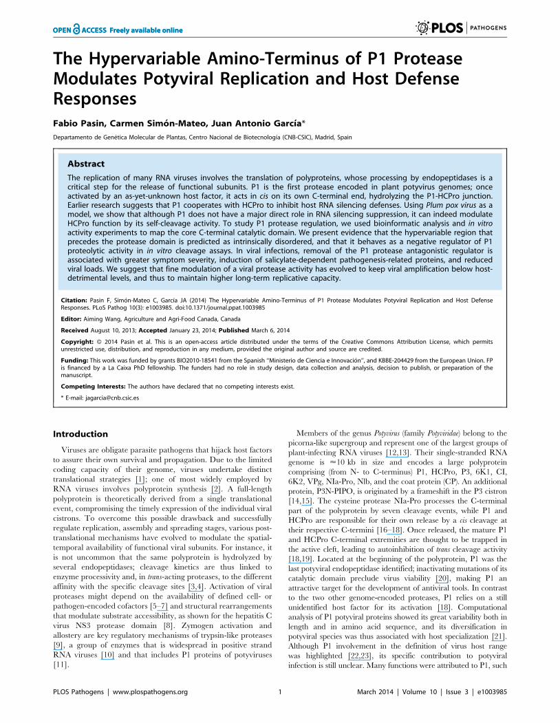
pathways. At 15 days post-agro-inoculation (dpi), DP1-infected
Col-0 and dcl2/3/4 plants were almost symptomless, whereas
PPV-infected Col-0 plants showed leaf chlorosis, which was
stronger in the dcl2/3/4 mutant line (Figure 1B). The presence of
DP1 and wild-type PPV in systemic leaves was evidenced by GFP
fluorescence detection (Figure 1B). Anti-PPV CP western blot
analysis of systemically infected leaves showed that, in both host
genotypes, viral accumulation was significantly higher in wild-
type PPV than in DP1-infected plants (Figure 1C and D). The
results confirmed that lack of P1 sequence did not compromise
virus ability to replicate or to move systemically, as also shown
for another potyvirus, Tobacco etch virus (TEV) [32]. Although
DP1 appears to replicate better in the silencing deficient plants
than in wild-type Col-0, the dcl2/3/4 line did not restore the
DP1 phenotype and viral accumulation to wild-type PPV
levels (Figure 1B and D). Based on these results, the main role
of P1 in viral amplification appears unrelated to RNA silencing
suppression.
The N-terminal region of potyviral P1 is predicted to beintrinsically disordered
To further analyze possible P1 functions, we coupled mul-
tiple sequence alignment and structural predictors for bioinfor-
matic analysis of its sequence. Intrinsic protein disorder was
estimated using DISOPRED2 [33] and MetaDisorderMD2
[34].
From the in silico analysis of P1 proteins from different potyviral
species groups (Table S1, in Text S1), we identified FxxLE as a
conserved motif in the P1 N-terminal region (Figure 2), in
conjunction with the reported IxFG and ISI motifs [21]. The
relatively well-conserved C-terminal regions of the proteins, which
correspond to the protease domain (defined below) and include the
VELI motif, are predicted mainly as structured. The residues from
N-terminal sequence patches with least conservation are generally
predicted to be intrinsically unstructured (Figure 2), suggesting that
final protein conformation is the main evolutionary target, rather
Figure 1. Arabidopsis mutant plants with defective RNAsilencing pathways fail to rescue PPV DP1 amplificationdefects. (A) Representation of wild-type PPV and DP1, a PPV clonewith deletion of the entire P1 sequence. The reporter sGFP(S65T) [103]gene is present between NIb and CP coding sequences of both viralclones. Boxes with diagonal lines indicate P3N-PIPO protein. (B)Symptoms and GFP fluorescence of Arabidopsis Col-0 and dcl2/3/4plants agro-inoculated with wild-type PPV or with DP1. Pictures weretaken in an epifluorescence microscope at 15 dpi. Scale bar, 1 cm. (C)DP1 and wild-type PPV viral accumulation in A. thaliana Col-0 systemicleaves was assessed by anti-PPV CP (CP) western blot assay (15 dpi).Each lane represents a pool of samples used for signal quantification. H,healthy Col-0 plant sample. Relative CP signal intensities were plottedusing average PPV value equal to 100. (D) DP1 and wild-type PPV viralaccumulation in A. thaliana dcl2/3/4 systemic leaves was assessed byanti-PPV CP (CP) western blot assay of upper non-inoculated leaves(15 dpi). Each lane represents a pool of samples used for signalquantification. H, healthy dcl2/3/4 plant sample. Relative CP signalintensities were plotted using average PPV value equal to 100.Histograms show mean 6SD (n = 6 samples/condition, from twoindependent Agrobacterium cultures); *** p,0.001, Student’s t-test.Ponceau red-stained blots are shown as loading control.doi:10.1371/journal.ppat.1003985.g001
Author Summary
RNA viruses are ideal systems for the study of populationdynamics, relationships among pathogen traits such asfitness and virulence, and of host immune responses topathogen attacks. Based on experimental evolution stud-ies, early models equated parasite virulence with fitness.Some reports showed that viral virulence and fitness canbe unlinked. Here we present evidence that the highlydisordered N-terminal region of a potyviral P1 proteinnegatively regulates its self-cleavage activity. Removal ofthis regulator domain greatly affects viral infection, whichis characterized by accelerated early replication andenhanced symptom severity. These properties are none-theless associated with low viral accumulation and highinduction of antiviral resistance markers. Finally, wepropose that host-dependent regulation of P1 processingefficiency modulates viral virulence and alleviates the hostantiviral responses.
P1 Protease Modulates Host Defense Responses
PLOS Pathogens | www.plospathogens.org 2 March 2014 | Volume 10 | Issue 3 | e1003985

than maintenance of the primary sequence, as proposed [35]. The
disorder predictions were further supported by P1 sequence
analysis of Scallion mosaic virus and the monocotyledon-infecting
Sugarcane mosaic virus group (Figure S1), which comprise the smallest
known P1 sequences and are suggested to be at the base of the
potyviral evolutionary tree [13].
Figure 2. Alignment of P1 amino acid sequences from PPV and six reference potyviruses. GenBank accession numbers are reported inTable S1, in Text S1; amino acid background is assigned according to the ClustalX color scheme [120]. Residues aligning to PPV P1 minimal proteasedomain (aa 165-308, based on our analysis) are shown in black letters and bright-colored background. Alignment quality, based on BLOSUM62 scores,is shown as a bar graph (below). FxxLE and the previously reported conserved motifs IxFG, ISI, VELI are boxed. The protease conserved RG dipeptide ismarked with black bar and the catalytic triad His, Asp and Ser is marked with stars. PPV MD2 and PPV DP2 lines show protein disorder prediction ofPPV P1 sequence according to MetadisorderMD2 and DISOPRED2 analyses, respectively, where ‘‘+’’ are disordered and ‘‘2’’ ordered residues.Additionally, DISOPRED2 prediction confidence for each P1 sequence is plotted: PPV, black line; PVY, orange; PVA, turquoise; PVV, magenta; TuMV,blue; VVY, purple; YMV, green. The first 46 residues of TuMV P1 are hidden, as they do not align to the other accessions.doi:10.1371/journal.ppat.1003985.g002
P1 Protease Modulates Host Defense Responses
PLOS Pathogens | www.plospathogens.org 3 March 2014 | Volume 10 | Issue 3 | e1003985
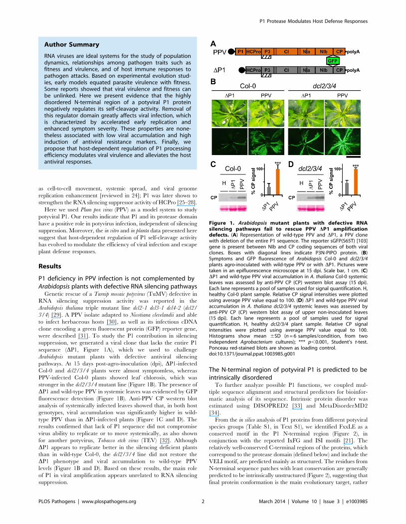
Involvement of P1 conserved motifs in protease self-cleavage
To study the relevance of P1 motifs in protease activity, we
mutated selected PPV P1 conserved amino acids to alanine
(Figure 3A). The viral cDNA constructs were transcribed and in
vitro-translated using the wheat germ extract (WGE) system.
Replacement of catalytic S259 (S) and P2 and P1 cleavage site
residues HY-307,308 (HY) with alanine impaired P1 protease self-
cleavage, precluding release of the mature 35.3 kDa P1 protein
and the 11.1 kDa HCPro fragment (HC-97) (Figure 3B). No
proteolytic processing was detected in the construct bearing the
VE-189,190-AA (VE) substitution in the VELI motif, indicating
that it indeed belongs to the minimal protease domain, consistent
with the disorder predictions. We found no appreciable
differences between the wild-type P1 (WT) construct and those
with FG-6,7-AA (FG) substitution in the IxFG motif or W63A
E67A (WE) substitution in the FxxLE motif (Figure 3B), in
accordance with the finding that the N-terminal region is
dispensable for potyvirus P1 processing [18]. We tested whether
the Thosea asigna virus 2A (T2A) ‘‘self-cleaving’’ peptide [36] could
overcome P1 protease defects and restore P1-HCPro separation.
We confirmed that T2A insertion between P1 S259A and HCPro
(ST2A) restored precursor processing, which, considering the
barely detectable level of the predicted 48.9 kDa uncleaved
product, was more efficient than with the wild-type P1 construct.
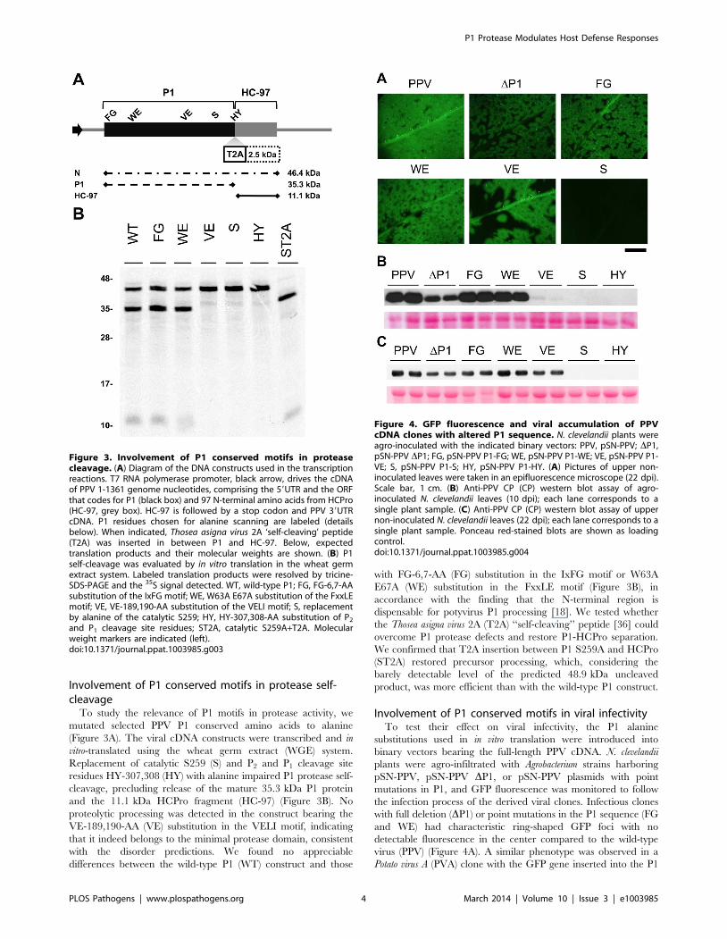
Involvement of P1 conserved motifs in viral infectivityTo test their effect on viral infectivity, the P1 alanine
substitutions used in in vitro translation were introduced into
binary vectors bearing the full-length PPV cDNA. N. clevelandii
plants were agro-infiltrated with Agrobacterium strains harboring
pSN-PPV, pSN-PPV DP1, or pSN-PPV plasmids with point
mutations in P1, and GFP fluorescence was monitored to follow
the infection process of the derived viral clones. Infectious clones
with full deletion (DP1) or point mutations in the P1 sequence (FG
and WE) had characteristic ring-shaped GFP foci with no
detectable fluorescence in the center compared to the wild-type
virus (PPV) (Figure 4A). A similar phenotype was observed in a
Potato virus A (PVA) clone with the GFP gene inserted into the P1
Figure 3. Involvement of P1 conserved motifs in proteasecleavage. (A) Diagram of the DNA constructs used in the transcriptionreactions. T7 RNA polymerase promoter, black arrow, drives the cDNAof PPV 1-1361 genome nucleotides, comprising the 59UTR and the ORFthat codes for P1 (black box) and 97 N-terminal amino acids from HCPro(HC-97, grey box). HC-97 is followed by a stop codon and PPV 39UTRcDNA. P1 residues chosen for alanine scanning are labeled (detailsbelow). When indicated, Thosea asigna virus 2A ‘self-cleaving’ peptide(T2A) was inserted in between P1 and HC-97. Below, expectedtranslation products and their molecular weights are shown. (B) P1self-cleavage was evaluated by in vitro translation in the wheat germextract system. Labeled translation products were resolved by tricine-SDS-PAGE and the 35S signal detected. WT, wild-type P1; FG, FG-6,7-AAsubstitution of the IxFG motif; WE, W63A E67A substitution of the FxxLEmotif; VE, VE-189,190-AA substitution of the VELI motif; S, replacementby alanine of the catalytic S259; HY, HY-307,308-AA substitution of P2
and P1 cleavage site residues; ST2A, catalytic S259A+T2A. Molecularweight markers are indicated (left).doi:10.1371/journal.ppat.1003985.g003
Figure 4. GFP fluorescence and viral accumulation of PPVcDNA clones with altered P1 sequence. N. clevelandii plants wereagro-inoculated with the indicated binary vectors: PPV, pSN-PPV; DP1,pSN-PPV DP1; FG, pSN-PPV P1-FG; WE, pSN-PPV P1-WE; VE, pSN-PPV P1-VE; S, pSN-PPV P1-S; HY, pSN-PPV P1-HY. (A) Pictures of upper non-inoculated leaves were taken in an epifluorescence microscope (22 dpi).Scale bar, 1 cm. (B) Anti-PPV CP (CP) western blot assay of agro-inoculated N. clevelandii leaves (10 dpi); each lane corresponds to asingle plant sample. (C) Anti-PPV CP (CP) western blot assay of uppernon-inoculated N. clevelandii leaves (22 dpi); each lane corresponds to asingle plant sample. Ponceau red-stained blots are shown as loadingcontrol.doi:10.1371/journal.ppat.1003985.g004
P1 Protease Modulates Host Defense Responses
PLOS Pathogens | www.plospathogens.org 4 March 2014 | Volume 10 | Issue 3 | e1003985

region [37], suggesting that in this recombinant potyvirus, P1
function was also affected. Plants challenged with clones harboring
the S or the HY mutations, which had no proteolytic activity in the
in vitro translation assays, did not show GFP fluorescence, infection
symptoms or CP accumulation (Figure 4A–C and not shown).
Although P1 VE-189,190-AA replacement led to no detectable
cleavage in WGE experiments (Figure 3), a faint CP accumulation
signal (compared to wild-type PPV) was visible in leaves agro-
infiltrated with the VE clone harboring the same mutations, and
the CP signal was greatly enhanced in systemically infected leaves
(Figure 4B and C). These leaves showed the characteristic ring-
shaped GFP foci typical of the other infectious P1 mutants
(Figure 4A). The results led us to hypothesize that partial reversion
might occur, restoring P1 protease activity. Western blot
performed using anti-PPV HCPro antibody confirmed correct
P1-HCPro processing in the upper non-inoculated leaves of plants
agro-inoculated with the VE mutant clone (Figure S2A). Samples
of systemically infected leaves from these plants were further
subjected to RT-PCR amplification of a PPV genome fragment
spanning the mutations introduced. Though the E190A mutation
was maintained, we detected reversion of V189A to the wild-type
residue at 22 dpi (Figure S2B). It is likely that, in contrast to clones
S and HY, clone VE maintained minimal P1 self-processing
activity sufficient to initiate viral replication and to select viral
mutant progeny with improved cleavage efficiency. This is
consistent with a report of a TEV clone in which disruption of
the (at that time undescribed) VELI motif appeared to preclude P1
self-processing in vitro. A virus harboring the same mutation was
infectious, however, and able to move systemically, with a marked
delay compared to the parental virus [20].
At the same time point at which P1 VE-189,190-AA reversion
was detected (22 dpi), the rest of the mutations introduced were
stably maintained in both FG and WE infectious clones (verified
by RT-PCR and sequencing, not shown), confirming that
mutations of N-terminal motifs are less detrimental than protease
defects.
P1 protease defects impair the silencing suppressoractivity of HCPro
Viability of the TEV clones altered by mutations in the P1
protease can be restored by insertion of a surrogate cleavage site
recognized by the TEV NIa protease [20], or in transgenic plants
expressing the P1-HCPro cistron [32]. To test whether these
rescue strategies complement defects in HCPro function rather
than in P1, we performed a transient RNA silencing assay in N.
benthamiana. Leaves were co-infiltrated with an Agrobacterium strain
bearing p35S:GFP as a silencing reporter and Agrobacterium strains
containing the PPV silencing suppressor HCPro preceded by the
wild-type P1 (pWT), by P1 with an alanine replacement of the
catalytic S259 (pS) or by P1 S259A plus T2A (pST2A). GFP
fluorescence was visible in all agroinfiltrated patches at 3 days
post-agro-infiltration (dpa) (not shown). At 6 dpa, there were no
differences between the empty control and the S construct. In
contrast, bright fluorescence as a result of silencing suppression
was maintained when HCPro was effectively released by P1
protease activity or by the ribosome skipping mechanism of T2A
(Figure 5A).
PPV viral clone ST2A, into which we inserted the T2A peptide
sequence between P1 S259A and HCPro, was able to initiate viral
replication (6 dpi; Figure 5B) and move systemically despite the P1
protease-inactivating mutation (Figure S2C; maintenance of P1
S259A substitution was verified by RT-PCR and sequencing, not
shown). The presence of T2A was nevertheless insufficient to fully
complement viral defects, since the ST2A GFP fluorescence
phenotype and CP accumulation levels differed considerably from
wild-type PPV (Figure 5B, Figure S2C and D).
To further validate these results, A. thaliana Col-0 and the RNA
silencing-defective dcl2/3/4 line were agro-inoculated with the
PPV S259A mutant (S), using wild-type PPV as control. As
predicted, wild-type PPV infected 16 of 16 challenged plants of
both the mutant line and its wild-type background, at 23 dpi.
Although we could not detect replication of the S mutant in Col-0
by GFP fluorescence or western blot analysis, GFP fluorescence
was observed in 11 of 16 dcl2/3/4 plants (Figure 5C). In spite of
the cleavage-disturbing mutation in the S cDNA clone, P1-HCPro
proteolytic separation was rescued in dcl2/3/4 plants inoculated
with PPV S259A, as verified by western blot assay of HCPro
(Figure 5D). RT-PCR analysis of samples collected at 23 dpi
confirmed that the original serine and the protease activity were
restored in the viral progeny, since the wild-type serine 259 codon
AGC, which was mutated to alanine GCC in the PPV S259A
cDNA clone, was further mutated to serine UCC (Figure 5E). The
fact that viral reversion mutations are promptly selected in PPV
S259A-infected plants suggests that P1, in addition to the release of
an active silencing suppressor, has further function(s).
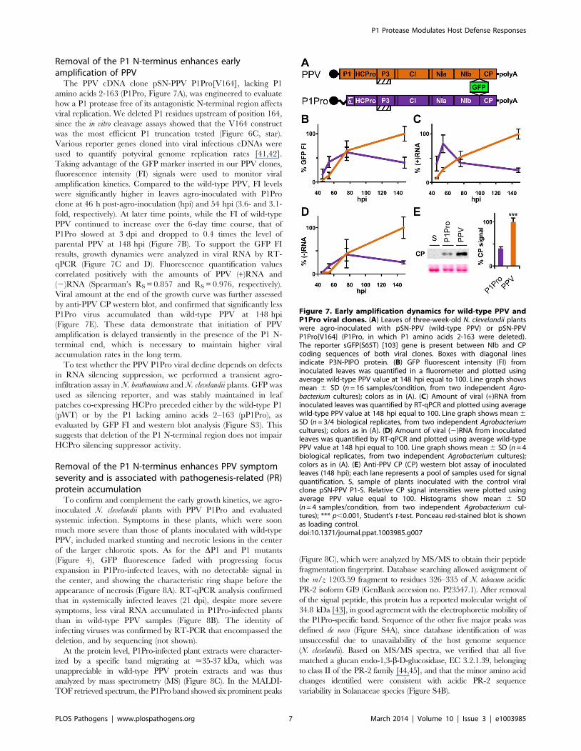
The first 164 N-terminal residues are dispensable for PPVP1 self-processing and act as a negative regulator in invitro cleavage assay
According to MEROPS classification [38], potyviral P1 serine
protease belongs to subclan PA(S), whose representative member,
trypsin, is synthesized as the inactive precursor trypsinogen, with
a disordered loop partially obstructing the substrate-binding cleft
[39]. Supported by the intrinsic disorder prediction of the P1 N-
terminal region and since a non-viral factor is needed for P1
protease activation [18], we tested whether P1 also fits the trypsin
model. Previous studies on TEV [40], sequence alignment,
secondary structure predictions, intrinsic disorder confidence, as
well as the finding that in PPV, the VE-189,190-AA substitution
disturbed P1 self-processing, were considered in choosing PPV P1
residues T162 and S170 as truncation points for a preliminary
trial (Figure 6A). N-terminal deletion constructs were made by
removing the P1 sequence upstream of the codon for each amino
acid selected, except for the initial methionine. To test P1
protease activity, we used in vitro translation in WGE and rabbit
reticulocyte lysate (RRL) systems. The full-length P1 (WT)
construct released the mature 35.3 kDa P1 protein in WGE but
not in the RRL system (Figure 6B, single asterisk), as anticipated
[18]. Nonetheless, the T162 deletion construct successfully self-
cleaved in both WGE and RRL systems, as shown by release of
the 16.7 kDa P1 processed fragment (Figure 6B, double asterisks).
The S170 construct lacked activity in both in vitro translation
systems, and only its uncleaved 27.0 kDa precursor was
detectable (Figure 6B), suggesting that the P1 residue delimiting
the N-terminal minimal protease domain is located between
positions 162 and 169.
To fine-map this boundary, additional single amino acid
deletions were engineered, transcribed, and tested by in vitro
translation. As a further control, the catalytic S259A mutation was
included in the T162 construct to rule out misleading non-specific
protein degradation. Efficient cleavage activity was maintained in
both WGE and RRL after deletion of P1 N-terminal amino acids
2-164 (Figure 6C, R165 construct), confirmed by release of the
<16 kDa P1 mature fragment. Further truncations of the protease
domain led to the drastic disappearance of protease activity in
RRL, and gradually decreasing efficiency in WGE. The control
protease catalytic mutant T162-S showed only a band corre-
sponding to the unprocessed 27.8 kDa precursor. The results
P1 Protease Modulates Host Defense Responses
PLOS Pathogens | www.plospathogens.org 5 March 2014 | Volume 10 | Issue 3 | e1003985
indicate that the protease catalytic domain is correctly folded even
in RRL, and that the first 164 N-terminal residues are not only
dispensable for this activity, but show an antagonistic effect on P1
self-processing in RRL.
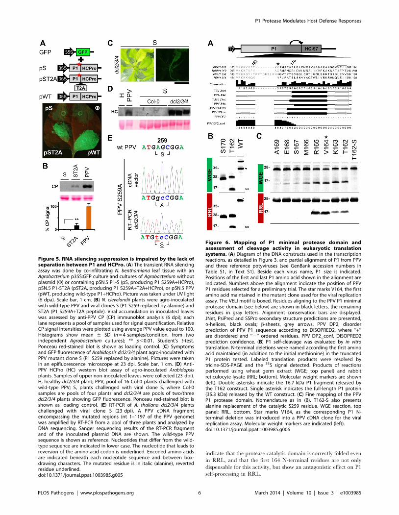
Figure 5. RNA silencing suppression is impaired by the lack ofseparation between P1 and HCPro. (A) The transient RNA silencingassay was done by co-infiltrating N. benthamiana leaf tissue with anAgrobacterium p35S:GFP culture and cultures of Agrobacterium withoutplasmid (F) or containing pSN.5 P1-S (pS, producing P1 S259A+HCPro),pSN.5 P1-ST2A (pST2A, producing P1 S259A+T2A+HCPro), or pSN.5 PPV(pWT, producing wild-type P1+HCPro). Picture was taken under UV light(6 dpa). Scale bar, 1 cm. (B) N. clevelandii plants were agro-inoculatedwith wild-type PPV and viral clones S (P1 S259 replaced by alanine) andST2A (P1 S259A+T2A peptide). Viral accumulation in inoculated leaveswas assessed by anti-PPV CP (CP) immunoblot analysis (6 dpi); eachlane represents a pool of samples used for signal quantification. RelativeCP signal intensities were plotted using average PPV value equal to 100.Histograms show mean 6 SD (n = 4 samples/condition, from twoindependent Agrobacterium cultures); ** p,0.01, Student’s t-test.Ponceau red-stained blot is shown as loading control. (C) Symptomsand GFP fluorescence of Arabidopsis dcl2/3/4 plant agro-inoculated withPPV mutant clone S (P1 S259 replaced by alanine). Pictures were takenin an epifluorescence microscope at 23 dpi. Scale bar, 1 cm. (D) Anti-PPV HCPro (HC) western blot assay of agro-inoculated Arabidopsisplants. Samples of upper non-inoculated leaves were collected (23 dpi).H, healthy dcl2/3/4 plant; PPV, pool of 16 Col-0 plants challenged withwild-type PPV; S, plants challenged with viral clone S, where Col-0samples are pools of four plants and dcl2/3/4 are pools of two/threedcl2/3/4 plants showing GFP fluorescence. Ponceau red-stained blot isshown as loading control. (E) RT-PCR of A. thaliana dcl2/3/4 plantschallenged with viral clone S (23 dpi). A PPV cDNA fragmentencompassing the mutated regions (nt 1–1197 of the PPV genome)was amplified by RT-PCR from a pool of three plants and analyzed byDNA sequencing. Sanger sequencing results of the RT-PCR fragmentand of the inoculated plasmid DNA are shown. The wild-type PPVsequence is shown as reference. Nucleotides that differ from the wild-type sequence are indicated in lower case. The nucleotide that leads toreversion of the amino acid codon is underlined. Encoded amino acidsare indicated beneath each nucleotide sequence and between box-drawing characters. The mutated residue is in italic (alanine), revertedresidue underlined.doi:10.1371/journal.ppat.1003985.g005
Figure 6. Mapping of P1 minimal protease domain andassessment of cleavage activity in eukaryotic translationsystems. (A) Diagram of the DNA constructs used in the transcriptionreactions, as detailed in Figure 3, and partial alignment of P1 from PPVand three reference potyviruses (see GenBank accession numbers inTable S1, in Text S1). Beside each virus name, P1 size is indicated.Positions of the first and last P1 amino acid shown in the alignment areindicated. Numbers above the alignment indicate the position of PPVP1 residues selected for a preliminary trial. The star marks V164, the firstamino acid maintained in the mutant clone used for the viral replicationassay. The VELI motif is boxed. Residues aligning to the PPV P1 minimalprotease domain (see below) are shown in black letters, the remainingresidues in gray letters. Alignment conservation bars are displayed.JNet, PsiPred and SSPro secondary structure predictions are presented,a-helices, black ovals; b-sheets, grey arrows. PPV DP2, disorderprediction of PPV P1 sequence according to DISOPRED2, where ‘‘+’’are disordered and ‘‘2’’ ordered residues. PPV DP2_conf, DISOPRED2prediction confidence. (B) P1 self-cleavage was evaluated by in vitrotranslation. N-terminal deletions were named according the first aminoacid maintained (in addition to the initial methionine) in the truncatedP1 protein tested. Labeled translation products were resolved bytricine-SDS-PAGE and the 35S signal detected. Products of reactionsperformed using wheat germ extract (WGE; top panel) and rabbitreticulocyte lysate (RRL; bottom). Molecular weight markers are shown(left). Double asterisks indicate the 16.7 kDa P1 fragment released bythe T162 construct. Single asterisk indicates the full-length P1 protein(35.3 kDa) released by the WT construct. (C) Fine mapping of the PPVP1 protease domain. Nomenclature as in (B). T162-S also presentsalanine replacement of the catalytic S259 residue. WGE reaction, toppanel; RRL, bottom. Star marks V164, as the corresponding P1 N-terminal deletion was introduced into a PPV cDNA clone for the viralreplication assay. Molecular weight markers are indicated (left).doi:10.1371/journal.ppat.1003985.g006
P1 Protease Modulates Host Defense Responses
PLOS Pathogens | www.plospathogens.org 6 March 2014 | Volume 10 | Issue 3 | e1003985
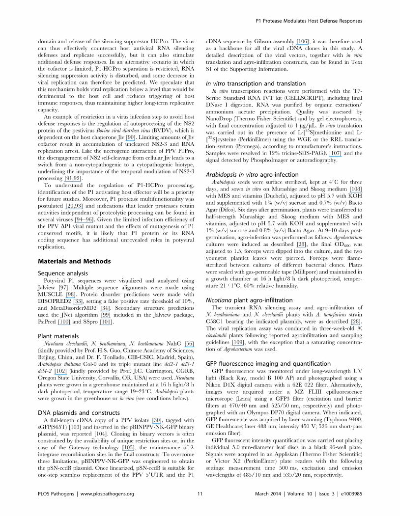
Removal of the P1 N-terminus enhances earlyamplification of PPV
The PPV cDNA clone pSN-PPV P1Pro[V164], lacking P1
amino acids 2-163 (P1Pro, Figure 7A), was engineered to evaluate
how a P1 protease free of its antagonistic N-terminal region affects
viral replication. We deleted P1 residues upstream of position 164,
since the in vitro cleavage assays showed that the V164 construct
was the most efficient P1 truncation tested (Figure 6C, star).
Various reporter genes cloned into viral infectious cDNAs were
used to quantify potyviral genome replication rates [41,42].
Taking advantage of the GFP marker inserted in our PPV clones,
fluorescence intensity (FI) signals were used to monitor viral
amplification kinetics. Compared to the wild-type PPV, FI levels
were significantly higher in leaves agro-inoculated with P1Pro
clone at 46 h post-agro-inoculation (hpi) and 54 hpi (3.6- and 3.1-
fold, respectively). At later time points, while the FI of wild-type
PPV continued to increase over the 6-day time course, that of
P1Pro slowed at 3 dpi and dropped to 0.4 times the level of
parental PPV at 148 hpi (Figure 7B). To support the GFP FI
results, growth dynamics were analyzed in viral RNA by RT-
qPCR (Figure 7C and D). Fluorescence quantification values
correlated positively with the amounts of PPV (+)RNA and
(2)RNA (Spearman’s RS = 0.857 and RS = 0.976, respectively).
Viral amount at the end of the growth curve was further assessed
by anti-PPV CP western blot, and confirmed that significantly less
P1Pro virus accumulated than wild-type PPV at 148 hpi
(Figure 7E). These data demonstrate that initiation of PPV
amplification is delayed transiently in the presence of the P1 N-
terminal end, which is necessary to maintain higher viral
accumulation rates in the long term.
To test whether the PPV P1Pro viral decline depends on defects
in RNA silencing suppression, we performed a transient agro-
infiltration assay in N. benthamiana and N. clevelandii plants. GFP was
used as silencing reporter, and was stably maintained in leaf
patches co-expressing HCPro preceded either by the wild-type P1
(pWT) or by the P1 lacking amino acids 2–163 (pP1Pro), as
evaluated by GFP FI and western blot analysis (Figure S3). This
suggests that deletion of the P1 N-terminal region does not impair
HCPro silencing suppressor activity.
Removal of the P1 N-terminus enhances PPV symptomseverity and is associated with pathogenesis-related (PR)protein accumulation
To confirm and complement the early growth kinetics, we agro-
inoculated N. clevelandii plants with PPV P1Pro and evaluated
systemic infection. Symptoms in these plants, which were soon
much more severe than those of plants inoculated with wild-type
PPV, included marked stunting and necrotic lesions in the center
of the larger chlorotic spots. As for the DP1 and P1 mutants
(Figure 4), GFP fluorescence faded with progressing focus
expansion in P1Pro-infected leaves, with no detectable signal in
the center, and showing the characteristic ring shape before the
appearance of necrosis (Figure 8A). RT-qPCR analysis confirmed
that in systemically infected leaves (21 dpi), despite more severe
symptoms, less viral RNA accumulated in P1Pro-infected plants
than in wild-type PPV samples (Figure 8B). The identity of
infecting viruses was confirmed by RT-PCR that encompassed the
deletion, and by sequencing (not shown).
At the protein level, P1Pro-infected plant extracts were character-
ized by a specific band migrating at <35-37 kDa, which was
unappreciable in wild-type PPV protein extracts and was thus
analyzed by mass spectrometry (MS) (Figure 8C). In the MALDI-
TOF retrieved spectrum, the P1Pro band showed six prominent peaks
(Figure 8C), which were analyzed by MS/MS to obtain their peptide
fragmentation fingerprint. Database searching allowed assignment of
the m/z 1203.59 fragment to residues 326–335 of N. tabacum acidic
PR-2 isoform GI9 (GenBank accession no. P23547.1). After removal
of the signal peptide, this protein has a reported molecular weight of
34.8 kDa [43], in good agreement with the electrophoretic mobility of
the P1Pro-specific band. Sequence of the other five major peaks was
defined de novo (Figure S4A), since database identification of was
unsuccessful due to unavailability of the host genome sequence
(N. clevelandii). Based on MS/MS spectra, we verified that all five
matched a glucan endo-1,3-b-D-glucosidase, EC 3.2.1.39, belonging
to class II of the PR-2 family [44,45], and that the minor amino acid
changes identified were consistent with acidic PR-2 sequence
variability in Solanaceae species (Figure S4B).
Figure 7. Early amplification dynamics for wild-type PPV andP1Pro viral clones. (A) Leaves of three-week-old N. clevelandii plantswere agro-inoculated with pSN-PPV (wild-type PPV) or pSN-PPVP1Pro[V164] (P1Pro, in which P1 amino acids 2-163 were deleted).The reporter sGFP(S65T) [103] gene is present between NIb and CPcoding sequences of both viral clones. Boxes with diagonal linesindicate P3N-PIPO protein. (B) GFP fluorescent intensity (FI) frominoculated leaves was quantified in a fluorometer and plotted usingaverage wild-type PPV value at 148 hpi equal to 100. Line graph showsmean 6 SD (n = 16 samples/condition, from two independent Agro-bacterium cultures); colors as in (A). (C) Amount of viral (+)RNA frominoculated leaves was quantified by RT-qPCR and plotted using averagewild-type PPV value at 148 hpi equal to 100. Line graph shows mean 6
SD (n = 3/4 biological replicates, from two independent Agrobacteriumcultures); colors as in (A). (D) Amount of viral (2)RNA from inoculatedleaves was quantified by RT-qPCR and plotted using average wild-typePPV value at 148 hpi equal to 100. Line graph shows mean 6 SD (n = 4biological replicates, from two independent Agrobacterium cultures);colors as in (A). (E) Anti-PPV CP (CP) western blot assay of inoculatedleaves (148 hpi); each lane represents a pool of samples used for signalquantification. S, sample of plants inoculated with the control viralclone pSN-PPV P1-S. Relative CP signal intensities were plotted usingaverage PPV value equal to 100. Histograms show mean 6 SD(n = 4 samples/condition, from two independent Agrobacterium cul-tures); *** p,0.001, Student’s t-test. Ponceau red-stained blot is shownas loading control.doi:10.1371/journal.ppat.1003985.g007
P1 Protease Modulates Host Defense Responses
PLOS Pathogens | www.plospathogens.org 7 March 2014 | Volume 10 | Issue 3 | e1003985

PR proteins of different families overaccumulate in tobacco
plants that show hypersensitivity to Tobacco mosaic virus (TMV) [46–
49], but also after bacterial and fungal infection and in response to
abiotic stress [50]. We first confirmed the MS results by western blot
analysis for PR-2; next, to test whether PR-2 induction is a specific
P1-associated defense mechanism or part of a broader stress
response, we assessed class II PR-3 protein expression in infected N.
clevelandii plants (Figure 8D). We found that, despite the lower viral
CP levels, PR-2 and PR-3 accumulation was significantly higher in
P1Pro- compared to wild-type PPV-infected plants (Figure 8E).
Immune response to PPV P1Pro is attenuated bydownregulation of salicylic acid signaling
In tobacco plants, exogenous salicylic acid (SA) treatment
induces class II PR-2 and class II PR-3 transcription [48,51,52],
and activates the resistance responses associated to TMV-induced
hypersensitivity [53,54]. Downregulation of SA accumulation and
systemic acquired resistance was reported in transgenic plants that
express the bacterial salicylate-hydroxylase gene nahG [55].
In N. benthamiana, while wild-type PPV-infected plants appeared
almost symptomless, PPV P1Pro-infected plants showed extended
chlorosis and necrotic lesions, similar to those observed in
N. clevelandii (Figure 9A). We therefore used transgenic
N. benthamiana NahG plants [56] to evaluate the SA contribution
in the PPV P1Pro host immune response. In accordance with
previous studies [56], downregulation of SA signaling had no
appreciable effect on the wild-type PPV phenotype. In contrast, in
P1Pro-infected NahG plants, systemic symptom severity was
attenuated (10 dpi; Figure 9A and B). This result was supported by
a sharp reduction in PR-2 protein accumulation in P1Pro-infected
NahG plants and a weak increase in viral load, estimated by anti-
PPV CP western blot analysis (12 dpi; Figure 9C and D). These
data suggest that although SA-mediated antiviral pathways have
only a minor role in wild-type PPV infection, they take part in
P1Pro immune responses.
Several stress response proteins are upregulated inP1Pro-infected NahG plants
In NahG-expressing plants (i) P1Pro-induced chlorosis was
more accentuated than in wild-type PPV-infected plants
(Figure 9A), (ii) although lower than the wild-type host, PR-2
abundance in P1Pro samples was significantly higher than in wild-
type PPV, and (iii) PR-2 reduction was insufficient to fully restore
P1Pro viral accumulation to parental PPV levels (Figure 9D).
These findings prompted us to further investigate P1Pro-related
defense responses after downregulation of SA signaling. To
identify proteins whose abundance was significantly changed in
P1Pro-infected NahG plants relative to wild-type PPV-infected
NahG plants, we performed quantitative proteomic analysis using
isobaric tag labeling (iTRAQ) and liquid chromatography (LC)-
MS/MS [57]. A draft sequence of the N. benthamiana genome was
released [58,59], and a search against its predicted protein
database enabled us to identify more than a thousand non-
redundant proteins. Of these, 23 were considered to accumulate
differentially in P1Pro versus wild-type PPV-infected plant
samples, since they were found in both P1Pro biological replicates
#A and #B, with a false discovery rate ,5% as statistical cut-off
(Figure 10A). Gene ontology term enrichment analysis showed
that, according to plant symptoms, the 23 dysregulated proteins
associated significantly with the GO term ‘‘response to stress’’ (GO
ID: 6950, p,0.0001; Figure 10B). In Figure 10C, we present a
heat map of these quantified P1Pro proteins with their average
iTRAQ ratios (expressed in LOG2) relative to wild-type PPV
biological replica #A. As predicted, P1Pro biological replicates
#A and #B are grouped in the same hierarchical cluster, which
differs from PPV biological replica #B. In P1Pro samples, several
proteins from different PR families were more abundant than in
wild-type PPV. These include a class II PR-2 (in accordance with
the western blot result; Figure 9C) and other acidic members,
which are regulated in tobacco mainly by SA [50,52], and basic
counterparts such as the basic PR-1, whose transcription is
effectively activated by ethylene [60–64]. Abscisic acid and
osmotic stress are reported to induce expression of basic PR-5
[50,65], as well as of dehydrin-like proteins [66]. The contribution
Figure 8. Symptoms and viral accumulation of PPV lacking P1N-terminal residues. N. clevelandii plants were agro-inoculated withwild-type PPV or P1Pro, pSN-PPV P1Pro[V164] in which P1 amino acids2–163 were deleted; data were collected at 21 dpi. (A) Pictures of N.clevelandii systemic leaves. Boxed leaf areas were magnified and theirGFP fluorescence, acquired by laser scanning, is shown. Scale bar,2.5 cm in whole leaf pictures, 0.4 cm in details. (B) Amount of viral(+)RNA from systemically infected leaves was quantified by RT-qPCRand plotted using average wild-type PPV value equal to 100.Histograms show mean 6 SD (n = 4 biological replicates, from twoindependent Agrobacterium cultures); *** p,0.001, Student’s t-test. (C)Total protein extracts from systemic leaf samples were resolved byglycine-SDS-PAGE and Coomassie blue stained, molecular weightmarkers are indicated (left). The <35–37 kDa P1Pro-specific band wasanalyzed by MALDI-TOF and its peptide mass fingerprint is shown.Intensity in the y axis; m/z values are indicated for the six P1Pro samplepeptides further analyzed by MS/MS. (D) Anti-PPV CP (CP), anti-PR-2(PR-2) and anti-PR-3 (PR-3) western blot assays of upper non-inoculatedleaves; each lane represents a pool of samples used for signalquantification. S, sample of plants inoculated with the control viralclone pSN-PPV P1-S. Ponceau red-stained blot as loading control ofprotein extracts. (E) Relative PR-2 and PR-3 signal intensities wereplotted using average P1Pro value equal to 100. Histograms show mean6 SD (n = 4 samples/condition, from two independent Agrobacteriumcultures); *** p,0.001, Student’s t-test.doi:10.1371/journal.ppat.1003985.g008
P1 Protease Modulates Host Defense Responses
PLOS Pathogens | www.plospathogens.org 8 March 2014 | Volume 10 | Issue 3 | e1003985

of oxidative stress in the SA-dependent response to P1Pro is
underlined by the detection of a peroxidase and a catalase; the
tobacco homologue of the latter was initially identified as a SA-
binding protein [67]. Only three of the 23 differentially
accumulated host proteins were downregulated in P1Pro; these
include two plastocyanins and a CP12-like accession, proteins
essential for photosynthesis [68,69]. These data suggest that in the
transgenic host, an important component of SA signaling is
maintained and/or that its downregulation is partially compen-
sated by alternative defense components.
Discussion
Besides the self-cleavage activity intrinsic to its C-terminal end,
other activities of potyviral P1 and its real contributions to viral
replication remained vague, so much so that P1 earned the
appellative ‘‘mysterious protein’’ in a recent review [24].
In concomitance with the discovery that HCPro inhibits
posttranscriptional gene silencing, it was suggested that P1 could
act in conjunction with and strengthen the silencing suppressor
activity of HCPro [25,26]. A characteristic ring-shaped GFP focus
phenotype was related in tobamovirus infections with deficiency in
RNA silencing suppression [70,71]. The same GFP phenotype was
distinctive of all our PPV infectious clones with P1 sequence
deletions and point mutations. In contrast, our Arabidopsis dcl2/3/4
complementation data demonstrate a major function for P1
independent of RNA silencing suppression. The ring GFP
phenotype of P1 mutants might thus be related not only to a
silencing suppression defect, but also to other altered functions.
Recently, the silencing suppressor-enhancing effect of P1 was
attributed to cis elements that improve translation efficiency, rather
than to complementary activity of the P1 protein [72].
Our results nonetheless show that P1 is unquestionably
involved in RNA silencing by limiting HCPro function.
Deficiencies in polyprotein processing at the P1-HCPro junction
abrogated PPV infectivity in wild-type plants, as also reported for
TEV [20], and were partially complemented by insertion of the
T2A ‘‘self-cleaving’’ peptide. To further define the nature of this
viral weakness, we used a transient RNA silencing assay to show
that for correct silencing suppression, P1 must be effectively
separated from HCPro, whether mediated by its own protease
domain or by the ribosome skipping mechanism of T2A. In
plants with impaired antiviral RNA silencing machineries the
PPV S259A clone, with a cleavage-inactive P1 protease,
recovered infectivity. The selection of viral revertant progeny
demonstrated that strong evolutionary pressure for proteolytic
competence of P1 was maintained, despite the host mutant
background. The lack of separation between P1 and HCPro
probably impairs not only the RNA silencing suppressor activity,
but also other important viral functions.
Figure 9. Viral accumulation and immune response induction in NahG-expressing N. benthamiana. (A) Wild-type (N.b. wt) and NahG-expressing (N.b. NahG) N. benthamiana plants were agro-inoculated with wild-type PPV and P1Pro viral clones. Pictures of systemic leaves were takenat 10 dpi. GFP fluorescence, acquired by laser scanning, is shown. Scale bar, 4 cm. (B) Details of (A) showing symptoms of wild-type and NahG-expressing plants infected with the P1Pro viral clone. (C) Anti-PPV CP (CP) and anti-PR-2 (PR-2) western blot analysis of upper non-inoculated leavesof wild-type and NahG-expressing plants (12 dpi); each lane represents a pool of samples used for signal quantification. S, sample of plantsinoculated with the control viral clone pSN-PPV P1-S. Below, blot stained with Ponceau red as loading control of protein extracts. (D) Relative PR-2and CP signal intensities were plotted using average P1Pro or wild-type PPV value equal to 100, respectively. Histograms show mean 6 SD(n = 4 samples/condition, from two independent Agrobacterium cultures). Bars with different letters are statistically significant, p,0.05, one-wayAnova and Tukey’s HSD test.doi:10.1371/journal.ppat.1003985.g009
P1 Protease Modulates Host Defense Responses
PLOS Pathogens | www.plospathogens.org 9 March 2014 | Volume 10 | Issue 3 | e1003985

Of the mature proteins encoded by the potyviral genome, P1
presents the greatest variability in length and in amino acid
sequence [73]. In silico analysis showed that the limited primary
sequence conservation of the P1 N-terminal region is associated
with residues predicted to be part of intrinsically disordered loops.
The relevance of unfolded regions in the proteolytic maturation of
viral polyproteins has been shown [6,7], and flexible loops in
peptidase precursors act in many cases as protease activation
switches [39]. Previous results and those reported here show
successful self-cleavage activity of potyviral P1 after in vitro
translation only in the WGE system, but not in RRL. In
consequence, it was suggested that a non-viral factor, present in
WGE and absent in RRL, might be involved in correct folding of
the protease domain or presentation of the cleavage site [18]. Our
evaluation of the cleavage performance of P1 N-terminal end
truncations demonstrates that the P1 minimal protease domain is
active in WGE but also in RRL, suggesting that both eukaryotic
translation systems support correct folding and activity of the P1
protease domain. Hence, the P1 N-terminal residues are not only
dispensable for HCPro release, but also act as a negative regulator
of P1 self-processing.
In the context of viral infection, removal of the P1 protease
antagonistic extension accelerated early viral replication and was
followed by enhancement of symptom severity, with the appear-
ance of plant stunting and necrotic lesions that did not
characterize wild-type PPV. The greater aggressiveness did not
parallel viral loads, however, as viral RNA and CP levels in plants
systemically infected with the deletion mutant were lower than in
wild-type PPV-infected plants. Lack of positive correlation
between symptom severity and fitness is reported for several viral
systems, including potyviral infections [74–76]. This can be
justified by the crossing of a virulence threshold, which would
result in higher induction of immune responses or excessive host
debilitation [77,78]. Accordingly, the necrotic phenotype that
characterizes PPV P1Pro was associated with overaccumulation of
class II PR-2 and PR-3 proteins. Acidic PR-2 and PR-3 family
members are indicators of SA-mediated responses to biotic stress
and are hallmarks of systemic acquired resistance [48,50,79]. In a
transgenic NahG-expressing plants, a decrease in SA signaling
attenuated PPV P1Pro symptom severity. P1Pro-induced chlorosis
was nonetheless more pronounced than in wild-type PPV-infected
plants. Complex hormone crosstalk follows plant infection [80],
and compounds other than SA operate in the defense against
pathogens [81–83]. Quantitative proteomic analysis of NahG
plants infected with the P1Pro viral clone allowed us to identify a
large set of stress response-associated proteins whose abundance
was significantly altered compared to wild-type PPV. This includes
upregulation of members of different PR families whose
transcription is reported to be activated by SA but also by
ethylene, and further modulated by abscisic acid [50], as well as
rearrangement of the antioxidant system, and downregulation of
photosynthetic components, similar to other studies [84–86].
Coordinated action of both SA-dependent and -independent
pathways probably contributes to the immune response activated
by PPV P1Pro. In turn, the N-terminal extremity of the P1
protease, absent in P1Pro but maintained in wild-type PPV, helps
to bypass induction of host defense responses. Several plant viruses
interfere with salicylate pathways [87–89], and it would be of
interest to determine whether P1 also has an active role in
suppressing basal host defenses independent of its HCPro activity-
modulating effect.
Considering that (i) defects in P1 self-cleavage preclude viral
viability, (ii) viral RNA silencing suppression is impaired by the
lack of separation between P1 and HCPro, (iii) P1 residues 1–164
are predicted to be mainly disordered, and negatively affect P1-
HCPro processing in RRL, (iv) PPV early amplification dynamics
are enhanced by removal of the P1 N-terminal end, (v) increased
defense responses are associated with deletion of the P1 N-
terminus, and (vi) removal of the P1 N-terminus reduces viral
accumulation, we propose a model by which P1 acts to fine-tune
potyviral replication by sensing specific host effectors. The
presence of these cofactors leads to activation of the P1 protease
Figure 10. Quantitative proteomic analysis of PPV P1Pro-infected NahG plants. (A) Venn diagram of proteins whoseabundance was significantly changed in P1Pro biological replicates#A and #B, relative to wild-type PPV sample #A. A false discovery rateof ,5% was applied as statistical cut-off. Numbers in the non-overlapping areas represent unique significant proteins of eachbiological replica; the number in the overlapping area, 23, shows thecommon significant proteins for further analysis. (B) Map of geneontology terms significantly enriched in 23 dysregulated proteinscommon to both P1Pro replicates. Relevant biological process nodesare colored from yellow to red, where red is the most significantlyoverrepresented category. In white, non-significant node. (C) Heat mapof 23 significantly differentially regulated proteins common to P1Proreplicates #A and #B. Fold change was calculated using PPV #A valuesas 1, and expressed in LOG2. Quantification values of PPV biologicalreplica #B are shown as control. Sample tree was built by hierarchicalclustering analysis. Beside each N. benthamiana accession ID, proteinisoelectric point is indicated. Brief protein descriptions are reported onthe right; PR proteins are grouped by families according van Loon et al.[50].doi:10.1371/journal.ppat.1003985.g010
P1 Protease Modulates Host Defense Responses
PLOS Pathogens | www.plospathogens.org 10 March 2014 | Volume 10 | Issue 3 | e1003985
domain and release of the silencing suppressor HCPro. The virus
can thus effectively counteract host antiviral RNA silencing
defenses and replicate successfully, but it can also stimulate
additional defense responses. In an alternative scenario in which
the cofactor is limited, P1-HCPro separation is restricted, RNA
silencing suppression activity is disturbed, and some decrease in
viral replication can therefore be predicted. We speculate that
this mechanism holds viral replication below a level that would be
detrimental to the host cell and reduces triggering of host
immune responses, thus maintaining higher long-term replicative
capacity.
An example of restriction in a virus infection step to avoid host
defense responses is the regulation of autoprocessing of the NS2
protein of the pestivirus Bovine viral diarrhea virus (BVDV), which is
dependent on the host chaperone Jiv [90]. Limiting amounts of Jiv
cofactor result in accumulation of uncleaved NS2-3 and RNA
replication arrest. Like the necrogenic interaction of PPV P1Pro,
the disengagement of NS2 self-cleavage from cellular Jiv leads to a
switch from a non-cytopathogenic to a cytopathogenic biotype,
underlining the importance of the temporal modulation of NS2-3
processing [91,92].
To understand the regulation of P1-HCPro processing,
identification of the P1 activating host effector will be a priority
for future studies. Moreover, P1 protease multifunctionality was
postulated [20,93] and indications that leader proteases retain
activities independent of proteolytic processing can be found in
several viruses [94–96]. Given the limited infection efficiency of
the PPV DP1 viral mutant and the effects of mutagenesis of P1
conserved motifs, it is likely that P1 protein or its RNA
coding sequence has additional unrevealed roles in potyviral
replication.
Materials and Methods
Sequence analysisPotyviral P1 sequences were visualized and analyzed using
Jalview [97]. Multiple sequence alignments were made using
MUSCLE [98]. Protein disorder predictions were made with
DISOPRED2 [33], setting a false positive rate threshold of 10%,
and MetaDisorderMD2 [34]. Secondary structure predictions
used the JNet algorithm [99] included in the Jalview package,
PsiPred [100] and SSpro [101].
Plant materialsNicotiana clevelandii, N. benthamiana, N. benthamiana NahG [56]
(kindly provided by Prof. H.S. Guo, Chinese Academy of Sciences,
Beijing, China, and Dr. F. Tenllado, CIB-CSIC, Madrid, Spain),
Arabidopsis thaliana Col-0 and its triple mutant line dcl2-1 dcl3-1
dcl4-2 [102] (kindly provided by Prof. J.C. Carrington, CGRB,
Oregon State University, Corvallis, OR, USA) were used. Nicotiana
plants were grown in a greenhouse maintained at a 16 h light/8 h
dark photoperiod, temperature range 19–23uC. Arabidopsis plants
were grown in the greenhouse or in vitro (see conditions below).
DNA plasmids and constructsA full-length cDNA copy of a PPV isolate [30], tagged with
sGFP(S65T) [103] and inserted in the pBINPPV-NK-GFP binary
plasmid, was reported [104]. Cloning in binary vectors is often
constrained by the availability of unique restriction sites or, in the
case of the Gateway technology [105], the maintenance of lintegrase recombination sites in the final constructs. To overcome
these limitations, pBINPPV-NK-GFP was engineered to obtain
the pSN-ccdB plasmid. Once linearized, pSN-ccdB is suitable for
one-step seamless replacement of the PPV 59UTR and the P1
cDNA sequence by Gibson assembly [106]; it was therefore used
as a backbone for all the viral cDNA clones in this study. A
detailed description of the viral vectors, together with in vitro
translation and agro-infiltration constructs, can be found in Text
S1 of the Supporting Information.
In vitro transcription and translationIn vitro transcription reactions were performed with the T7-
Scribe Standard RNA IVT kit (CELLSCRIPT), including final
DNase I digestion. RNA was purified by organic extraction/
ammonium acetate precipitation. Quality was assessed by
NanoDrop (Thermo Fisher Scientific) and by gel electrophoresis,
with final concentration adjusted to 1 mg/mL. In vitro translation
was carried out in the presence of L-[35S]methionine and L-
[35S]cysteine (PerkinElmer) using the WGE or the RRL transla-
tion system (Promega), according to manufacturer’s instructions.
Samples were resolved in 12% tricine-SDS-PAGE [107] and the
signal detected by PhosphoImager or autoradiography.
Arabidopsis in vitro agro-infectionArabidopsis seeds were surface sterilized, kept at 4uC for three
days, and sown in vitro on Murashige and Skoog medium [108]
with MES and vitamins (Duchefa), adjusted to pH 5.7 with KOH
and supplemented with 1% (w/v) sucrose and 0.7% (w/v) Bacto
Agar (Difco). Six days after germination, plants were transferred to
half-strength Murashige and Skoog medium with MES and
vitamins, adjusted to pH 5.7 with KOH and supplemented with
1% (w/v) sucrose and 0.8% (w/v) Bacto Agar. At 9–10 days post-
germination, agro-infection was performed as follows. Agrobacterium
cultures were induced as described [28], the final OD600 was
adjusted to 1.5, forceps were dipped into the culture, and the two
youngest plantlet leaves were pierced. Forceps were flame-
sterilized between cultures of different bacterial clones. Plates
were sealed with gas-permeable tape (Millipore) and maintained in
a growth chamber at 16 h light/8 h dark photoperiod, temper-
ature 2161uC, 60% relative humidity.
Nicotiana plant agro-infiltrationThe transient RNA silencing assay and agro-infiltration of
N. benthamiana and N. clevelandii plants with A. tumefaciens strain
C58C1 bearing the indicated plasmids, were as described [28].
The viral replication assay was conducted in three-week-old N.
clevelandii plants following reported agroinfiltration and sampling
guidelines [109], with the exception that a saturating concentra-
tion of Agrobacterium was used.
GFP fluorescence imaging and quantificationGFP fluorescence was monitored under long-wavelength UV
light (Black Ray, model B 100 AP) and photographed using a
Nikon D1X digital camera with a 62E 022 filter. Alternatively,
images were acquired under a MZ FLIII epifluorescence
microscope (Leica) using a GFP3 filter (excitation and barrier
filters at 470/40 nm and 525/50 nm, respectively) and photo-
graphed with an Olympus DP70 digital camera. When indicated,
GFP fluorescence was acquired by laser scanning (Typhoon 9400,
GE Healthcare; laser 488 nm, intensity 450 V; 526 nm short-pass
emission filter).
GFP fluorescent intensity quantification was carried out placing
individual 5.0 mm-diameter leaf discs in a black 96-well plate.
Signals were acquired in an Appliskan (Thermo Fisher Scientific)
or Victor X2 (PerkinElmer) plate readers with the following
settings: measurement time 500 ms, excitation and emission
wavelengths of 485/10 nm and 535/20 nm, respectively.
P1 Protease Modulates Host Defense Responses
PLOS Pathogens | www.plospathogens.org 11 March 2014 | Volume 10 | Issue 3 | e1003985

Western blot assaysPlant tissue was ground in a mortar in liquid nitrogen or
homogenized in a TissueLyzer bead mill (Qiagen). Total proteins
were extracted in 150 mM Tris-HCl pH 7.5, 6 M urea, 2% (w/v)
SDS, 5% (v/v) glycerol and 5% (v/v) b-mercaptoethanol; heat
denatured (96uC, 5 min) and centrifuged (14000 rpm, 10 min) to
remove cell debris. Protein samples were separated by glycine-
SDS-PAGE and electroblotted onto a nitrocellulose membrane.
Ponceau red staining was used to control protein loading
equivalence. Proteins were detected using anti-PPV CP and anti-
PPV HCPro rabbit sera, and anti-GFP monoclonal antibody
(clones 7.1 and 13.1, Roche) as primary antibodies. Antibodies
raised against tobacco class II PR-2 [44] and class II PR-3 proteins
[47] were kindly provided by Dr. T. Heitz (IBMP-CNRS,
Strasbourg, France). Horseradish peroxidase-conjugated goat
anti-rabbit IgG (Jackson) or sheep anti-mouse IgG (GE Health-
care) were used as secondary antibody. Immunostained proteins
were visualized by enhanced chemiluminescence detection with a
LiteABlot kit (Euroclone). For signal quantification, chemiolumi-
nescence was acquired in a ChemiDoc XRS imager (BioRad) and
analyzed with ImageJ software [110].
RT-PCR and RT-qPCRTotal RNA was extracted with the FavorPrep Plant Total RNA
Mini kit (Favorgen). Fragments spanning the PPV 59UTR, and the
coding sequences of P1 and the HCPro N-terminus were amplified
with the Titan One Tube RT-PCR kit (Roche) using primers
1595_F/1597_R (Table S2, in Text S1). When indicated,
fragments were purified using the FavorPrep Gel/PCR Purifica-
tion kit (Favorgen) and DNA was sequenced.
Strand-specific quantification of PPV RNA was done for at least
three biological replicates per condition using tagged cDNA
primers in the RT step [111–113], and will be detailed elsewhere
(in preparation). Briefly, equal amounts of DNAseI-treated total
RNA were used for cDNA synthesis using Superscript III
(Invitrogen) and primer Q26_R or Q29_F to transcribe cDNA
from positive and negative PPV genomes, respectively. Technical
triplicate qPCR reactions were prepared using HOT FIREPol
EvaGreen qPCR Mix Plus (Solis BioDyne) in 384-well optical
plates and run in a 7900HT Fast Real-Time PCR System (Applied
Biosystems). Primer pairs Q27_F/Q28_R and Q30_F/Q31_R
were used for positive and negative genome quantifications,
respectively. The amount of target RNA in the analyzed samples
was estimated by absolute quantification using an external DNA
standard curve [114].
MALDI MS/MS and data analysisPlant total protein extracts were separated by glycine-SDS-
PAGE and stained with Coomassie blue. Gel bands of interest
were excised manually, reduced, alkylated and in-gel digested.
The tryptic-eluted peptides were subjected to MALDI-TOF/
TOF analysis. Data were automatically acquired in an ABi
4800 MALDI TOF/TOF mass spectrometer (AB Sciex)
and searched against NCBI non-redundant protein database,
NCBInr_20121116. Mass tolerance for precursors was set to
650 ppm and for MS/MS fragment ions to 60.3 Da. The
confidence interval for protein identification was set to $95%
(p,0.05) and only peptides with an individual ion score above the
identity threshold were considered correctly identified.
Manual peptide de novo sequencing was performed according
Ma and Johnson [115]. Residues with near/isobaric masses were
bona fide assigned according alignment consensus. Confidence of
retrieved results was further tested with Peaks Studio software
(BSI) [116].
iTRAQ sample labeling, LC-MS/MS and data analysisN. benthamiana NahG plants were agro-inoculated with two
independent bacteria cultures per each viral cDNA clone (n = 2
biological replicates); upper non-inoculated leaves were collected
from 8 plants per culture (12 dpi). Total protein extracts were
prepared as described for western blot assays and further purified
by methanol/chloroform precipitation. Protein pellets were
resuspended in 6 M guanidine hydrochloride and 100 mM
HEPES, pH 7.5, and concentration was determined by RC DC
assay (BioRad). Equal amounts of protein for each condition were
trypsin-digested and labeled with iTRAQ Reagent Multi-plex kit
(AB Sciex). Tags 114 and 116 were used for P1Pro biological
replicates and 115 and 117 for wild-type PPV biological replicates.
Labeled peptide samples were combined and subjected to LC-
MS/MS analysis (three technical replicates) using a nano liquid
chromatography system (Eksigent Technologies) coupled to a
Triple TOF 5600 mass spectrometer (AB Sciex). MS and MS/MS
data were processed using Analyst TF 1.5.1 software (AB Sciex)
and searched using the Mascot Server v. 2.3.02 (Matrix Science)
against a customized database represented by N. benthamiana
genome-predicted proteins (available at Sol Genomics Network
[59]) plus their corresponding reversed entries. Peptide mass
tolerance was set to 620 ppm for precursors and 0.05 Da for
fragment masses. The confidence interval for protein identification
was set to $95% (p,0.05), and only peptides with an individual
ion score above the identity threshold were maintained in
quantification analysis. Proteins were considered differentially
expressed if they had at least two quantified peptides and they
were present in both P1Pro biological replicates with a false
discovery rate ,5%. LOG2 ratios relative to PPV biological
replica #A (iTRAQ tag 115) were visualized by MultiExperiment
Viewer [117]. Hierarchical clustering analysis with Pearson
correlation distance metric was used to build the sample tree
[118]. Sequences of reliably quantified N. benthamiana proteins
were used as query in a WU-BLAST2 search against the TAIR10
protein dataset, to generate a list of homologous A. thaliana gene
IDs. This was used as input in gene ontology term enrichment
analysis using BinGO [119].
Accession numbersThe following GenBank (http://www.ncbi.nlm.nih.gov) accessions
were used in the viral sequence analysis: YMV (YP_022752.1); PVV
(NP_734369.1); PVY (NP_734243.1); VVY (YP_001931974.1);
CaYSV (YP_003208051.1); JGMV (NP_734408.1); MDMV
(NP_734143.1); SCMV (NP_734133.1); SrMV (CAC84438.1);
PVA (NP_734359.1); TEV (NP_734207.1); ScaMV (NP_734123.1);
TuMV (BAC02772.1). Plant GenBank accessions considered: N.
tabacum class II PR-2 (P23547.1); N. tabacum class I PR-2
(AAA63541.1); S. tuberosum class II PR-2 (CAE52322.1); S. lycopersicum
class II PR-2 (NP_001234798.1). N. benthamiana accessions can be
found at Sol Genomics Network (ftp://ftp.solgenomics.net/
genomes/Nicotiana_benthamiana/annotation/).
Supporting Information
Figure S1 Alignment and protein disorder prediction ofthe six smallest known potyviral P1 sequences. GenBank
accession numbers are reported in Table S1, in Text S1; for PPV
P1, only resides 164–308 were considered. Amino acid back-
ground is assigned according the ClustalX color scheme [120].
Residues aligning to PPV P1 minimal protease domain are shown
in black letters and bright-colored background. The VELI motif is
boxed; The protease conserved RG dipeptide is marked with black
bar and the catalytic triad His, Asp and Ser is marked with stars.
P1 Protease Modulates Host Defense Responses
PLOS Pathogens | www.plospathogens.org 12 March 2014 | Volume 10 | Issue 3 | e1003985

MD2 lines show protein disorder prediction of P1 sequence
according MetadisorderMD2, where ‘‘+’’ are disordered and ‘‘2’’
ordered residues. DISOPRED2 prediction confidence is plotted:
ScaMV, green line; SrMV, purple; SCMV, orange; JGMV, blue;
MDMV, turquoise; CaYSV, magenta.
(TIF)
Figure S2 Pseudo-reversions of P1 VE-189,190-AA mu-tations and infectivity of PPV ST2A viral clone. N. clevelandii
plants were agro-inoculated with wild-type PPV and viral clone
VE (P1 VE-189,190-AA), clone S (P1 S259 replaced by alanine)
and clone ST2A (P1 S259A+T2A peptide). (A) Anti-PPV HCPro
(HC) western blot assay of upper non-inoculated leaves, collected
at 22 dpi. For each clone, both samples shown in Figure 4C were
analyzed together. Ponceau red-stained blot is shown as loading
control. (B) RT-PCR analysis of plants challenged with VE
mutant clone (22 dpi). A PPV cDNA fragment encompassing the
mutated region (nt 1–1197 of the PPV genome) was amplified by
RT-PCR from a pool of two plants and analyzed by DNA
sequencing. Sanger sequencing results of the RT-PCR fragment
and of the inoculated plasmid DNA are shown. The wild-type
PPV sequence is shown as reference. Nucleotides that differ from
the wild-type sequence are indicated in lower case. The nucleotide
that leads to reversion of the amino acid codon is underlined.
Encoded amino acids are indicated beneath each nucleotide
sequence and between box-drawing characters. Mutated residues
are in italic (alanine), reverted residue underlined. (C) GFP-
fluorescence pictures of upper non-inoculated N. clevelandii leaves
were taken in an epifluorescence microscope (22 dpi). Scale bar,
1 cm. (D) Anti-PPV CP (CP) western blot assay of upper non-
inoculated leaves (22 dpi); each lane corresponds to a single plant
sample. Ponceau red-stained blot is shown as loading control.
(TIF)
Figure S3 HCPro silencing suppression activity ismaintained in the absence of the P1 N-terminal region.Transient RNA silencing assay was done by co-infiltrating leaf
tissue with an Agrobacterium p35S:GFP culture and cultures of
Agrobacterium pSN.5 P1-S (pS, producing P1 S259A+HCPro),
pSN.5 PPV (pWT, producing wild-type P1+HCPro), or pSN.5
P1Pro (pP1Pro, producing P1 lacking amino acids 22163+HCPro). (A) Picture of N. benthamiana agro-infiltrated leaf was
taken in an epifluorescence microscope (6 dpa). (B) GFP
fluorescent intensity (FI) from N. benthamiana infiltrated leaf patches
was quantified in a fluorometer and plotted using average pWT
value at 6 dpa equal to 100. Line graph shows mean 6 SD
(n = 16 samples/condition, from two independent Agrobacterium
cultures); colors as in (A). For each time point, the mean GFP
FI value (n = 16) from untreated leaves was used as blank.
Analyzed time points (dpa) in the x axis. (C) Western blot detection
of GFP protein in N. clevelandii samples (6 dpa); each lane
represents a pool of samples from 2/3 infiltrated patches. N,
non-infiltrated leaf sample. Ponceau red staining is shown as
loading control. (D) Relative signal quantification for the
immunoblot shown in (C) using average pWT value equal to
100. Histogram shows mean 6 SD (n = 6 samples/condition, from
two independent Agrobacterium cultures); ns, not significant by
Student’s t-test.
(TIF)
Figure S4 Nicotiana clevelandii class II PR-2 peptidesequences assigned by MS/MS analysis. (A) Identified
sequence of MALDI-TOF peptides labeled in Figure 8C. Mass
measurement accuracy of the retrieved peptides is indicated in
parts per million (ppm). In de novo defined peptides, residues that
differ from tobacco class II PR-2 isoform GI9 sequence (GenBank
accession no. P23547.1) are in bold and underlined. Amino acids
with near-/isobaric masses were bona fide assigned according to
alignment consensus of the following panel. (B) Alignment of
reference PR-2 sequences with N. clevelandii peptides (shown above
the alignment). Residues that align with the peptides identified are
boxed; background according the ClustalX color scheme [120]. In
the rest of the alignment, only K/R and P are colored to
highlight possible trypsin cleavage sites. Ntab_PR-2II, tobacco
class II PR-2 isoform GI9 (GenBank P23547.1); Nben_PR-2II,
N. benthamiana class II PR-2 predicted protein (Sol Genomics
NbC25742659g0003.1); Stub_PR-2II, Solanum tuberosum class II
PR-2 (GenBank CAE52322.1); Slyc_PR-2II, S. lycopersicum class II
PR-2 (GenBank NP_001234798.1); Ntab_PR-2I, tobacco class I
PR-2 clone gglb50 (GenBank AAA63541.1) as outlier. The last 22
residues of Ntab_PR-2I are hidden, as they do not align to the
other accessions. In each alignment lane, positions of the first and
last accession amino acids are indicated. In de novo defined
peptides, residues that differ from the Ntab_PR-2II sequence are
marked with a star.
(TIF)
Text S1 This file contains: Supplemental Methods;Supplemental References; Table S1, Virus species andGenBank accession numbers for viral protein sequencesused in the study; Table S2, List of the primers used inthe study.(PDF)
Acknowledgments
We thank C. Mark for editorial assistance, M. Fernandez for help with
laser scanning, Dr. J.A. Abelenda and L. Almonacid for advices on qPCR,
Prof. H.S. Guo and Dr. F. Tenllado for providing N. benthamiana NahG
plants, Prof. J.C. Carrington for providing Arabidopsis dcl2-1 dcl3-1 dcl4-2
line seeds and Dr. T. Heitz for providing anti-PR protein sera. Special
thanks to S. Ciordia, I. Zapico and S. Juarez for assistance with mass
spectrometry analysis. FP is grateful to C. Gasparotto and K. Zajac for
their support.
Author Contributions
Conceived and designed the experiments: FP CSM JAG. Performed the
experiments: FP. Analyzed the data: FP JAG. Contributed reagents/
materials/analysis tools: FP CSM JAG. Wrote the paper: FP JAG.
References
1. Walsh D, Mathews MB, Mohr I (2013) Tinkering with translation: protein
synthesis in virus-infected cells. Cold Spring Harb Perspect Biol 5: a012351.
doi:10.1101/cshperspect.a012351.
2. Summers DF, Maizel Jr JV (1968) Evidence for large precursor proteins in
poliovirus synthesis. Proc Natl Acad Sci U S A 59: 966.
3. Dougherty WG, Dawn Parks T (1989) Molecular genetic and biochemical
evidence for the involvement of the heptapeptide cleavage sequence in
determining the reaction profile at two tobacco etch virus cleavage sites in
cell-free assays. Virology 172: 145–155. doi:10.1016/0042-6822(89)90116-
5.
4. Garcıa JA, Martın MT, Cervera MT, Riechmann JL (1992) Proteolyticprocessing of the plum pox potyvirus polyprotein by the Nla protease at a novel
cleavage site. Virology 188: 697–703. doi:10.1016/0042-6822(92)90524-S.
5. Tong L (2002) Viral proteases. Chem Rev 102: 4609–4626. doi:10.1021/
cr010184f.
6. Satheshkumar PS, Gayathri P, Prasad K, Savithri HS (2005) ‘‘Nativelyunfolded’’ VPg is essential for Sesbania mosaic virus serine protease activity.
J Biol Chem 280: 30291–30300. doi:10.1074/jbc.M504122200.
7. Mathur C, Jimsheena VK, Banerjee S, Makinen K, Gowda LR, et al. (2012)
Functional regulation of PVBV Nuclear Inclusion protein-a protease activity
P1 Protease Modulates Host Defense Responses
PLOS Pathogens | www.plospathogens.org 13 March 2014 | Volume 10 | Issue 3 | e1003985

upon interaction with Viral Protein genome-linked and phosphorylation.Virology 422: 254–264. doi:10.1016/j.virol.2011.10.009.
8. Saalau-Bethell SM, Woodhead AJ, Chessari G, Carr MG, Coyle J, et al. (2012)
Discovery of an allosteric mechanism for the regulation of HCV NS3 protein
function. Nat Chem Biol 8: 920–925. doi:10.1038/nchembio.1081.
9. Pozzi N, Vogt AD, Gohara DW, Di Cera E (2012) Conformational selection in
trypsin-like proteases. Curr Opin Struct Biol 22: 421–431. doi:10.1016/
j.sbi.2012.05.006.
10. Gorbalenya AE, Donchenko AP, Blinov VM, Koonin EV (1989) Cysteineproteases of positive strand RNA viruses and chymotrypsin-like serine
proteases: a distinct protein superfamily with a common structural fold. FEBSLett 243: 103–114. doi:10.1016/0014-5793(89)80109-7.
11. Valli A (2013) Potyvirus P1 proteinase. Handbook of proteolytic enzymes (3rd
edition). Elsevier. pp. 3130–3133.
12. Koonin EV, Wolf YI, Nagasaki K, Dolja VV (2008) The Big Bang of picorna-like virus evolution antedates the radiation of eukaryotic supergroups. Nat Rev
Microbiol 6: 925–939. doi:10.1038/nrmicro2030.
13. Gibbs A, Ohshima K (2010) Potyviruses and the digital revolution. Annu RevPhytopathol 48: 205–223. doi:10.1146/annurev-phyto-073009-114404.
14. Chung BY-W, Miller WA, Atkins JF, Firth AE (2008) An overlapping essential
gene in the Potyviridae. Proc Natl Acad Sci U S A 105: 5897–5902.doi:10.1073/pnas.0800468105.
15. Wei T, Zhang C, Hong J, Xiong R, Kasschau KD, et al. (2010) Formation of
complexes at plasmodesmata for potyvirus intercellular movement is mediatedby the viral protein P3N-PIPO. PLoS Pathog 6: e1000962. doi:10.1371/
journal.ppat.1000962.
16. Lopez-Moya JJ, Valli A, Garcıa JA (2009) Potyviridae. Encyclopedia of lifesciences. John Wiley & Sons, Ltd. pp. 1–10.
17. Carrington JC, Freed DD, Sanders TC (1989) Autocatalytic processing of the
potyvirus helper component proteinase in Escherichia coli and in vitro. J Virol 63:
4459–4463.
18. Verchot J, Herndon KL, Carrington JC (1992) Mutational analysis of the
tobacco etch potyviral 35-kDa proteinase: identification of essential residues
and requirements for autoproteolysis. Virology 190: 298–306. doi:10.1016/0042-6822(92)91216-H.
19. Guo B, Lin J, Ye K (2011) Structure of the autocatalytic cysteine protease
domain of potyvirus helper-component proteinase. J Biol Chem 286: 21937–21943. doi:10.1074/jbc.M111.230706.
20. Verchot J, Carrington JC (1995) Debilitation of plant potyvirus infectivity by P1
proteinase-inactivating mutations and restoration by second-site modifications.J Virol 69: 1582–1590.
21. Valli A, Lopez-Moya JJ, Garcıa JA (2007) Recombination and gene duplication
in the evolutionary diversification of P1 proteins in the family Potyviridae. J GenVirol 88: 1016–1028. doi:10.1099/vir.0.82402-0.
22. Salvador B, Saenz P, Yanguez E, Quiot JB, Quiot L, et al. (2008) Host-specific
effect of P1 exchange between two potyviruses. Mol Plant Pathol 9: 147–155.doi:10.1111/j.1364-3703.2007.00450.x.
23. Maliogka VI, Salvador B, Carbonell A, Saenz P, San Leon D, et al. (2012)
Virus variants with differences in the P1 protein coexist in a Plum pox virus
population and display particular host-dependent pathogenicity features. MolPlant Pathol 13: 877–886. doi:10.1111/j.1364-3703.2012.00796.x.
24. Rohozkova J, Navratil M (2011) P1 peptidase – a mysterious protein of family
Potyviridae. J Biosci 36: 189–200. doi:10.1007/s12038-011-9020-6.
25. Anandalakshmi R, Pruss GJ, Ge X, Marathe R, Mallory AC, et al. (1998) A
viral suppressor of gene silencing in plants. Proc Natl Acad Sci U S A 95:
13079–13084. doi:10.1073/pnas.95.22.13079.
26. Kasschau KD, Carrington JC (1998) A counterdefensive strategy of plantviruses: suppression of posttranscriptional gene silencing. Cell 95: 461–470.
doi:10.1016/S0092-8674(00)81614-1.
27. Rajamaki ML, Kelloniemi J, Alminaite A, Kekarainen T, Rabenstein F, et al.(2005) A novel insertion site inside the potyvirus P1 cistron allows expression of
heterologous proteins and suggests some P1 functions. Virology 342: 88–101.doi:10.1016/j.virol.2005.07.019.
28. Valli A, Martın-Hernandez AM, Lopez-Moya JJ, Garcıa JA (2006) RNA
silencing suppression by a second copy of the P1 serine protease of Cucumber vein
yellowing ipomovirus, a member of the family Potyviridae that lacks the cysteine
protease HCPro. J Virol 80: 10055–10063. doi:10.1128/JVI.00985-06.
29. Garcia-Ruiz H, Takeda A, Chapman EJ, Sullivan CM, Fahlgren N, et al.
(2010) Arabidopsis RNA-dependent RNA polymerases and Dicer-like proteins inantiviral defense and small interfering RNA biogenesis during Turnip mosaic virus
infection. Plant Cell 22: 481–496. doi:10.1105/tpc.109.073056.
30. Riechmann JL, Laın S, Garcıa JA (1990) Infectious in vitro transcripts from aplum pox potyvirus cDNA clone. Virology 177: 710–716. doi:10.1016/0042-
6822(90)90537-2.
31. Fernandez-Fernandez MR, Mourino M, Rivera J, Rodrıguez F, Plana-DuranJ, et al. (2001) Protection of rabbits against rabbit hemorrhagic disease virus by
immunization with the VP60 protein expressed in plants with a potyvirus-basedvector. Virology 280: 283–291. doi:10.1006/viro.2000.0762.
32. Verchot J, Carrington JC (1995) Evidence that the potyvirus P1 proteinase
functions in trans as an accessory factor for genome amplification. J Virol 69:3668–3674.
33. Ward JJ, McGuffin LJ, Bryson K, Buxton BF, Jones DT (2004) The
DISOPRED server for the prediction of protein disorder. Bioinformatics 20:
2138–2139. doi:10.1093/bioinformatics/bth195.
34. Kozlowski LP, Bujnicki JM (2012) MetaDisorder: a meta-server for the
prediction of intrinsic disorder in proteins. BMC Bioinformatics 13: 111.
doi:10.1186/1471-2105-13-111.
35. Abrescia NGA, Bamford DH, Grimes JM, Stuart DI (2012) Structure unifies
the viral universe. Annu Rev Biochem 81: 795–822. doi:10.1146/annurev-
biochem-060910-095130.
36. Donnelly MLL, Hughes LE, Luke G, Mendoza H, ten Dam E, et al. (2001)
The ‘‘cleavage’’ activities of foot-and-mouth disease virus 2A site-directed
mutants and naturally occurring ‘‘2A-like’’ sequences. J Gen Virol 82: 1027–
1041.
37. Kelloniemi J, Makinen K, Valkonen JPT (2008) Three heterologous proteins
simultaneously expressed from a chimeric potyvirus: infectivity, stability and
the correlation of genome and virion lengths. Virus Res 135: 282–291.
doi:10.1016/j.virusres.2008.04.006.
38. Rawlings ND, Barrett AJ, Bateman A (2012) MEROPS: the database of
proteolytic enzymes, their substrates and inhibitors. Nucleic Acids Res 40:
D343–D350. doi:10.1093/nar/gkr987.
39. Khan AR, James MNG (1998) Molecular mechanisms for the conversion of
zymogens to active proteolytic enzymes. Protein Sci 7: 815–836. doi:10.1002/
pro.5560070401.
40. Verchot J, Koonin EV, Carrington JC (1991) The 35-kDa protein from the N-
terminus of the potyviral polyprotein functions as a third virus-encoded
proteinase. Virology 185: 527–535. doi:10.1016/0042-6822(91)90522-D.
41. Dolja VV, McBride HJ, Carrington JC (1992) Tagging of plant potyvirus
replication and movement by insertion of beta-glucuronidase into the viral
polyprotein. Proc Natl Acad Sci U S A 89: 10208–10212.
42. Hafren A, Hofius D, Ronnholm G, Sonnewald U, Makinen K (2010) HSP70
and its cochaperone CPIP promote potyvirus infection in Nicotiana benthamiana
by regulating viral coat protein functions. Plant Cell 22: 523–535. doi:10.1105/
tpc.109.072413.
43. Linthorst HJ, Melchers LS, Mayer A, van Roekel JS, Cornelissen BJ, et al.
(1990) Analysis of gene families encoding acidic and basic beta-1,3-glucanases
of tobacco. Proc Natl Acad Sci U S A 87: 8756–8760. doi:10.1073/
pnas.87.22.8756.
44. Kauffmann S, Legrand M, Geoffroy P, Fritig B (1987) Biological function of
‘‘pathogenesis-related’’ proteins: four PR proteins of tobacco have 1,3-beta-
glucanase activity. EMBO J 6: 3209–3212.
45. Leubner-Metzger G, Meins Jr F (1999) Functions and regulation of plant beta-
1,3-glucanases (PR-2). Pathogenesis-related proteins in plants. CRC Press. pp.
1–31.
46. Van Loon LC, Gerritsen YAM, Ritter CE (1987) Identification, purification,
and characterization of pathogenesis-related proteins from virus-infected
Samsun NN tobacco leaves. Plant Mol Biol 9: 593–609. doi:10.1007/
BF00020536.
47. Legrand M, Kauffmann S, Geoffroy P, Fritig B (1987) Biological function of
pathogenesis-related proteins: Four tobacco pathogenesis-related proteins are
chitinases. Proc Natl Acad Sci U S A 84: 6750–6754.
48. Ward ER, Uknes SJ, Williams SC, Dincher SS, Wiederhold DL, et al. (1991)
Coordinate gene activity in response to agents that induce systemic acquired
resistance. Plant Cell 3: 1085–1094. doi:10.1105/tpc.3.10.1085.
49. Heitz T, Fritig B, Legrand M (1994) Local and systemic accumulation of
pathogenesis-related proteins in tobacco plants infected with tobacco mosaic
virus. Mol Plant Microbe Interact 7: 776–779.
50. Van Loon LC, Rep M, Pieterse CMJ (2006) Significance of inducible defense-
related proteins in infected plants. Annu Rev Phytopathol 44: 135–162.
doi:10.1146/annurev.phyto.44.070505.143425.
51. Hennig J, Dewey RE, Cutt JR, Klessig DF (1993) Pathogen, salicylic acid and
developmental dependent expression of a b-1,3-glucanase/GUS gene fusion in
transgenic tobacco plants. Plant J 4: 481–493. doi:10.1046/j.1365-
313X.1993.04030481.x.
52. Niki T, Mitsuhara I, Seo S, Ohtsubo N, Ohashi Y (1998) Antagonistic effect of
salicylic acid and jasmonic acid on the expression of pathogenesis-related (PR)
protein genes in wounded mature tobacco leaves. Plant Cell Physiol 39: 500–
507. doi:10.1093/oxfordjournals.pcp.a029397.
53. White RF (1979) Acetylsalicylic acid (aspirin) induces resistance to tobacco
mosaic virus in tobacco. Virology 99: 410–412. doi:10.1016/0042-
6822(79)90019-9.
54. Yalpani N, Silverman P, Wilson TM, Kleier DA, Raskin I (1991) Salicylic acid
is a systemic signal and an inducer of pathogenesis-related proteins in virus-
infected tobacco. Plant Cell 3: 809–818. doi:10.1105/tpc.3.8.809.
55. Gaffney T, Friedrich L, Vernooij B, Negrotto D, Nye G, et al. (1993)
Requirement of salicylic acid for the induction of systemic acquired resistance.
Science 261: 754–756. doi:10.1126/science.261.5122.754.
56. Ying X-B, Dong L, Zhu H, Duan C-G, Du Q-S, et al. (2010) RNA-dependent
RNA polymerase 1 from Nicotiana tabacum suppresses RNA silencing and
enhances viral infection in Nicotiana benthamiana. Plant Cell 22: 1358–1372.
doi:10.1105/tpc.109.072058.
57. Ross PL, Huang YN, Marchese JN, Williamson B, Parker K, et al. (2004)
Multiplexed protein quantitation in Saccharomyces cerevisiae using amine-reactive
isobaric tagging reagents. Mol Cell Proteomics 3: 1154–1169. doi:10.1074/
mcp.M400129-MCP200.
58. Bombarely A, Rosli HG, Vrebalov J, Moffett P, Mueller LA, et al. (2012) A
draft genome sequence of Nicotiana benthamiana to enhance molecular
P1 Protease Modulates Host Defense Responses
PLOS Pathogens | www.plospathogens.org 14 March 2014 | Volume 10 | Issue 3 | e1003985

plant-microbe biology research. Mol Plant Microbe Interact 25: 1523–1530.
doi:10.1094/MPMI-06-12-0148-TA.
59. Bombarely A, Menda N, Tecle IY, Buels RM, Strickler S, et al. (2011) The Sol
Genomics Network (solgenomics.net): growing tomatoes using Perl. Nucleic
Acids Res 39: D1149–D1155. doi:10.1093/nar/gkq866.
60. Memelink J, Linthorst HJ, Schilperoort RA, Hoge JHC (1990) Tobacco genes
encoding acidic and basic isoforms of pathogenesis-related proteins display
different expression patterns. Plant Mol Biol 14: 119–126. doi:10.1007/
BF00018553.
61. Brederode FT, Linthorst HJ, Bol JF (1991) Differential induction of acquired
resistance and PR gene expression in tobacco by virus infection, ethephon
treatment, UV light and wounding. Plant Mol Biol 17: 1117–1125.
doi:10.1007/BF00028729.
62. Eyal Y, Meller Y, Lev-Yadun S, Fluhr R (1993) A basic-type PR-1 promoter
directs ethylene responsiveness, vascular and abscission zone-specific expres-
sion. Plant J 4: 225–234. doi:10.1046/j.1365-313X.1993.04020225.x.
63. Xu YI, Chang P-FL, Liu D, Narasimhan ML, Raghothama KG, et al. (1994)
Plant defense genes are synergistically induced by ethylene and methyl
jasmonate. Plant Cell 6: 1077–1085. doi:10.1105/tpc.6.8.1077.
64. Xie W, Goodwin PH (2009) A PRp27 gene of Nicotiana benthamiana contributes
to resistance to Pseudomonas syringae pv. tabaci but not to Colletotrichum destructivum
or Colletotrichum orbiculare. Funct Plant Biol 36: 351–361. doi:10.1071/FP08241.
65. Singh NK, Nelson DE, Kuhn D, Hasegawa PM, Bressan RA (1989) Molecular
cloning of osmotin and regulation of its expression by ABA and adaptation to
low water potential. Plant Physiol 90: 1096–1101. doi:10.1104/pp.90.3.1096.
66. Hanin M, Brini F, Ebel C, Toda Y, Takeda S, et al. (2011) Plant dehydrins and
stress tolerance: versatile proteins for complex mechanisms. Plant Signal Behav
6: 1503–1509. doi:10.4161/psb.6.10.17088.
67. Chen Z, Silva H, Klessig DF (1993) Active oxygen species in the induction of
plant systemic acquired resistance by salicylic acid. Science 262: 1883–1886.
doi:10.1126/science.8266079.
68. Weigel M, Varotto C, Pesaresi P, Finazzi G, Rappaport F, et al. (2003)
Plastocyanin is indispensable for photosynthetic electron flow in Arabidopsis
thaliana. J Biol Chem 278: 31286–31289. doi:10.1074/jbc.M302876200.
69. Fermani S, Trivelli X, Sparla F, Thumiger A, Calvaresi M, et al. (2012)
Conformational selection and folding-upon-binding of intrinsically disordered
protein CP12 regulate photosynthetic enzymes assembly. J Biol Chem 287:
21372–21383. doi:10.1074/jbc.M112.350355.
70. Kubota K, Tsuda S, Tamai A, Meshi T (2003) Tomato mosaic virus replication
protein suppresses virus-targeted posttranscriptional gene silencing. J Virol 77:
11016–11026. doi:10.1128/JVI.77.20.11016-11026.2003.
71. Ishibashi K, Meshi T, Ishikawa M (2011) Gaining replicability in a nonhost
compromises the silencing suppression activity of Tobacco mild green mosaic virus in
a host. J Virol 85: 1893–1895. doi:10.1128/JVI.01741-10.
72. Tena Fernandez F, Gonzalez I, Doblas P, Rodrıguez C, Sahana N, et al. (2013)
The influence of cis-acting P1 protein and translational elements on the
expression of Potato virus Y helper-component proteinase (HCPro) in
heterologous systems and its suppression of silencing activity. Mol Plant Pathol
14: 530–541. doi:10.1111/mpp.12025.
73. Adams MJ, Antoniw JF, Fauquet CM (2005) Molecular criteria for genus and
species discrimination within the family Potyviridae. Arch Virol 150: 459–479.
doi:10.1007/s00705-004-0440-6.
74. Carrasco P, de la Iglesia F, Elena SF (2007) Distribution of fitness and virulence
effects caused by single-nucleotide substitutions in Tobacco etch virus. J Virol 81:
12979–12984. doi:10.1128/JVI.00524-07.
75. Rolland M, Kerlan C, Jacquot E (2009) The acquisition of molecular
determinants involved in potato virus Y necrosis capacity leads to fitness
reduction in tobacco plants. J Gen Virol 90: 244–252. doi:10.1099/
vir.0.005140-0.
76. Herrera M, Garcıa-Arriaza J, Pariente N, Escarmıs C, Domingo E (2007)
Molecular basis for a lack of correlation between viral fitness and cell killing
capacity. PLoS Pathog 3: e53. doi:10.1371/journal.ppat.0030053.
77. Coll NS, Epple P, Dangl JL (2011) Programmed cell death in the plant immune
system. Cell Death Differ 18: 1247–1256. doi:10.1038/cdd.2011.37.
78. Lancaster KZ, Pfeiffer JK (2012) Viral population dynamics and virulence
thresholds. Curr Opin Microbiol 15: 525–530. doi:10.1016/j.mib.2012.05.007.
79. Ryals JA, Neuenschwander UH, Willits MG, Molina A, Steiner HY, et al.
(1996) Systemic acquired resistance. Plant Cell 8: 1809–1819. doi:10.1105/
tpc.8.10.1809.
80. Robert-Seilaniantz A, Grant M, Jones JD (2011) Hormone crosstalk in plant
disease and defense: more than just JASMONATE-SALICYLATE antago-
nism. Annu Rev Phytopathol 49: 317–343. doi:10.1146/annurev-phyto-
073009-114447.
81. Pieterse CM, Leon-Reyes A, Van der Ent S, Van Wees SC (2009) Networking
by small-molecule hormones in plant immunity. Nat Chem Biol 5: 308–316.
doi:10.1038/nchembio.164.
82. Garcıa-Marcos A, Pacheco R, Manzano A, Aguilar E, Tenllado F (2013)
Oxylipin biosynthesis genes positively regulate programmed cell death during
compatible infections with the synergistic pair Potato virus X-Potato virus Y and
Tomato spotted wilt virus. J Virol 87: 5769–5783. doi:10.1128/JVI.03573-12.
83. Gimenez-Ibanez S, Solano R (2013) Nuclear jasmonate and salicylate signaling
and crosstalk in defense against pathogens. Front Plant Sci 4: 72. doi:10.3389/
fpls.2013.00072.
84. Dıaz-Vivancos P, Rubio M, Mesonero V, Periago PM, Barcelo AR, et al.
(2006) The apoplastic antioxidant system in Prunus: response to long-term plum
pox virus infection. J Exp Bot 57: 3813–3824. doi:10.1093/jxb/erl138.
85. Elvira MI, Galdeano MM, Gilardi P, Garcia-Luque I, Serra MT (2008)
Proteomic analysis of pathogenesis-related proteins (PRs) induced by
compatible and incompatible interactions of pepper mild mottle virus
(PMMoV) in Capsicum chinense L3 plants. J Exp Bot 59: 1253–1265.
doi:10.1093/jxb/ern032.
86. Di Carli M, Benvenuto E, Donini M (2012) Recent insights into plant-virus
interactions through proteomic analysis. J Proteome Res 11: 4765–4780.
doi:10.1021/pr300494e.
87. Ji LH, Ding SW (2001) The suppressor of transgene RNA silencing encoded by
Cucumber mosaic virus interferes with salicylic acid-mediated virus resistance. Mol
Plant Microbe Interact 14: 715. doi:10.1094/MPMI.2001.14.6.715.
88. Wang X, Goregaoker SP, Culver JN (2009) Interaction of the Tobacco mosaic
virus replicase protein with a NAC domain transcription factor is associated
with the suppression of systemic host defenses. J Virol 83: 9720–9730.
doi:10.1128/JVI.00941-09.
89. Love AJ, Geri C, Laird J, Carr C, Yun B-W, et al. (2012) Cauliflower mosaic virus
protein P6 inhibits signaling responses to salicylic acid and regulates innate
immunity. PLoS One 7: e47535. doi:10.1371/journal.pone.0047535.
90. Rinck G, Birghan C, Harada T, Meyers G, Thiel H-J, et al. (2001) A cellular J-
domain protein modulates polyprotein processing and cytopathogenicity of a
pestivirus. J Virol 75: 9470–9482. doi:10.1128/JVI.75.19.9470-9482.2001.
91. Lackner T, Muller A, Pankraz A, Becher P, Thiel H-J, et al. (2004) Temporal
modulation of an autoprotease is crucial for replication and pathogenicity of an
RNA virus. J Virol 78: 10765–10775. doi:10.1128/JVI.78.19.10765-
10775.2004.
92. Lackner T, Muller A, Konig M, Thiel H-J, Tautz N (2005) Persistence of
bovine viral diarrhea virus is determined by a cellular cofactor of a viral
autoprotease. J Virol 79: 9746–9755. doi:10.1128/JVI.79.15.9746-9755.2005.
93. Pruss G, Ge X, Shi XM, Carrington JC, Vance VB (1997) Plant viral
synergism: the potyviral genome encodes a broad-range pathogenicity
enhancer that transactivates replication of heterologous viruses. Plant Cell 9:
859–868. doi:10.1105/tpc.9.6.859.
94. Choi H-K, Tong L, Minor W, Dumas P, Boege U, et al. (1991) Structure of
Sindbis virus core protein reveals a chymotrypsin-like serine proteinase and the
organization of the virion. Nature 354: 37–43. doi:10.1038/354037a0.
95. Peng C-W, Peremyslov VV, Mushegian AR, Dawson WO, Dolja VV (2001)
Functional specialization and evolution of leader proteinases in the family
Closteroviridae. J Virol 75: 12153–12160. doi:10.1128/JVI.75.24.12153-
12160.2001.
96. Hilton L, Moganeradj K, Zhang G, Chen Y-H, Randall RE, et al. (2006) The
NPro product of bovine viral diarrhea virus inhibits DNA binding by interferon
regulatory factor 3 and targets it for proteasomal degradation. J Virol 80:
11723–11732. doi:10.1128/JVI.01145-06.
97. Waterhouse AM, Procter JB, Martin DMA, Clamp M, Barton GJ (2009)
Jalview version 2–a multiple sequence alignment editor and analysis
workbench. Bioinformatics 25: 1189–1191. doi:10.1093/bioinformatics/
btp033.
98. Edgar RC (2004) MUSCLE: multiple sequence alignment with high accuracy
and high throughput. Nucleic Acids Res 32: 1792–1797. doi:10.1093/nar/
gkh340.
99. Cuff JA, Barton GJ (2000) Application of multiple sequence alignment profiles
to improve protein secondary structure prediction. Proteins 40: 502–511.
doi:10.1002/1097-0134(20000815)40:3,502::AID-PROT170.3.0.CO;2-Q.
100. Jones DT (1999) Protein secondary structure prediction based on position-
specific scoring matrices. J Mol Biol 292: 195–202. doi:10.1006/
jmbi.1999.3091.
101. Cheng J, Randall AZ, Sweredoski MJ, Baldi P (2005) SCRATCH: a protein
structure and structural feature prediction server. Nucleic Acids Res 33: W72–
W76. doi:10.1093/nar/gki396.
102. Deleris A, Gallego-Bartolome J, Bao J, Kasschau KD, Carrington JC, et al.
(2006) Hierarchical action and inhibition of plant Dicer-like proteins in
antiviral defense. Science 313: 68–71. doi:10.1126/science.1128214.
103. Chiu W, Niwa Y, Zeng W, Hirano T, Kobayashi H, et al. (1996) Engineered
GFP as a vital reporter in plants. Curr Biol 6: 325–330. doi:10.1016/S0960-
9822(02)00483-9.
104. Lucini C (2004) Expresion de proteınas heterologas en plantas por medio del
virus de la sharka (PPV) Madrid: Universidad Politecnica de Madrid.
105. Hartley JL, Temple GF, Brasch MA (2000) DNA cloning using in vitro site-
specific recombination. Genome Res 10: 1788–1795. doi:10.1101/gr.143000.
106. Gibson DG, Young L, Chuang R-Y, Venter JC, Hutchison III CA, et al. (2009)
Enzymatic assembly of DNA molecules up to several hundred kilobases. Nat
Methods 6: 343–345. doi:10.1038/nmeth.1318.
107. Schagger H, von Jagow G (1987) Tricine-sodium dodecyl sulfate-polyacryl-
amide gel electrophoresis for the separation of proteins in the range from 1 to
100 kDa. Anal Biochem 166: 368–379. doi:10.1016/0003-2697(87)90587-2.
108. Murashige T, Skoog F (1962) A revised medium for rapid growth and bio
assays with tobacco tissue cultures. Physiol Plant 15: 473–497. doi:10.1111/
j.1399-3054.1962.tb08052.x.
109. Eskelin K, Suntio T, Hyvarinen S, Hafren A, Makinen K (2010) Renilla
luciferase-based quantitation of Potato virus A infection initiated with
P1 Protease Modulates Host Defense Responses
PLOS Pathogens | www.plospathogens.org 15 March 2014 | Volume 10 | Issue 3 | e1003985
Agrobacterium infiltration of N. benthamiana leaves. J Virol Methods 164: 101–110.
doi:10.1016/j.jviromet.2009.12.006.110. Schneider CA, Rasband WS, Eliceiri KW (2012) NIH Image to ImageJ: 25
years of image analysis. Nat Methods 9: 671–675. doi:10.1038/nmeth.2089.
111. Lanford RE, Sureau C, Jacob JR, White R, Fuerst TR (1994) Demonstration ofin vitro infection of chimpanzee hepatocytes with hepatitis C virus using strand-
specific RT/PCR. Virology 202: 606–614. doi:10.1006/viro.1994.1381.112. Komurian-Pradel F, Perret M, Deiman B, Sodoyer M, Lotteau V, et al. (2004)
Strand specific quantitative real-time PCR to study replication of hepatitis C
virus genome. J Virol Methods 116: 103–106. doi:10.1016/j.jviro-met.2003.10.004.
113. Plaskon NE, Adelman ZN, Myles KM (2009) Accurate strand-specificquantification of viral RNA. PLoS One 4: e7468. doi:10.1371/journal.-
pone.0007468.114. Pfaffl MW, Hageleit M (2001) Validities of mRNA quantification using
recombinant RNA and recombinant DNA external calibration curves in real-
time RT-PCR. Biotechnol Lett 23: 275–282. doi:10.1023/A:1005658330108.
115. Ma B, Johnson R (2012) De novo sequencing and homology searching. Mol Cell
Proteomics 11: 1–16. doi:10.1074/mcp.O111.014902.116. Zhang J, Xin L, Shan B, Chen W, Xie M, et al. (2012) PEAKS DB: de novo
sequencing assisted database search for sensitive and accurate peptide
identification. Mol Cell Proteomics 11: 1–8. doi:10.1074/mcp.M111.010587.117. Saeed A, Sharov V, White J, Li J, Liang W, et al. (2003) TM4: a free, open-
source system for microarray data management and analysis. Biotechniques 34:374–378.
118. Eisen MB, Spellman PT, Brown PO, Botstein D (1998) Cluster analysis and
display of genome-wide expression patterns. Proc Natl Acad Sci U S A 95:14863–14868.
119. Maere S, Heymans K, Kuiper M (2005) BiNGO: a Cytoscape plugin to assessoverrepresentation of gene ontology categories in biological networks.
Bioinformatics 21: 3448–3449. doi:10.1093/bioinformatics/bti551.120. Procter JB, Thompson J, Letunic I, Creevey C, Jossinet F, et al. (2010)
Visualization of multiple alignments, phylogenies and gene family evolution.
Nat Methods 7: S16–S25. doi:10.1038/nmeth.1434.
P1 Protease Modulates Host Defense Responses
PLOS Pathogens | www.plospathogens.org 16 March 2014 | Volume 10 | Issue 3 | e1003985