TCF4 Deletions in Pitt-Hopkins Syndrome
10
HUMAN MUTATION Mutation in Brief #1026, 29:E242-E251, (2008) Online MUTATION IN BRIEF © 2008 WILEY-LISS, INC. Received 26 March 2008; accepted revised manuscript 1 June 2008. TCF4 Deletions in Pitt-Hopkins Syndrome Irina Giurgea 1* , Chantal Missirian 2 , Pierre Cacciagli 2 , Sandra Whalen 1 , Tessa Fredriksen 1 , Thierry Gaillon 1 , Julia Rankin 3 , Michele Mathieu-Dramard 4 , Gilles Morin 4 , Dominique Martin-Coignard 5 , Christèle Dubourg 6 , Brigitte Chabrol , Jacqueline Arfi , Fabienne Giuliano , Jean Claude Lambert , Nicole Philip , Pierre Sarda , Laurent Villard , Michel Goossens , and Anne Moncla 7 8 9 9 2-8 10 8 1 2-8 1 INSERM U841, IMRB, Département de Génétique, Equipe 11, Créteil, F-94000, France; Université Paris 12, Faculté de Médecine, IFR10, Créteil, F-94000, France and AP-HP, Groupe Hospitalier Henri Mondor-Albert Chenevier, Service de Biochimie et Génétique, Créteil, F-94000, France. 2 Département de Génétique Médicale, Hôpital des enfants de la Timone, 264, rue Saint Pierre, 13385 Marseille Cedex 5, France. 3 Department of Clinical Genetics, Royal Devon and Exeter NHS Foundation Trust (Heavitree), Gladstone Road, Exeter, EX1 2ED, United Kingdom. 4 Département de Génétique Médicale, CHU d’Amiens - Nord, Place Victor Pauchet, 80054 Amiens Cedex 1, France. 5 Département de Génétique Médicale, CH du Mans, 194, avenue Rubillard 72037 Le Mans cedex 09, France. 6 Laboratoire de Génétique Moléculaire, CHU Pontchaillou, 2 rue Henri Le Guilloux 35033 Rennes Cedex 9, France. 7 Service de Neuropédiatrie, Hôpital des enfants de la Timone, 264, rue Saint Pierre, 13385 Marseille cedex 05, France. 8 Unité INSERM U491 « Génétique Médicale & Développement », Faculté de Médecine La Timone, 27, Bd. Jean Moulin, 13385 Marseille Cedex 5, France. 9 Service de Génétique Médicale, Hôpital L'Archet 2, 151 route de Saint Antoine de Genestrière, BP 3079, 06202 Nice Cedex3, France. 10 Unité de Génétique Médicale et Foetopathologie, CHU Montpellier, Hôpital Arnaud de Villeneuve, 371, av du Doyen G. Giraud, 34295 Montpellier Cedex 5, France. *Correspondence to: Dr. Irina Giurgea, Service de biochimie et génétique, Hôpital Henri Mondor, 51, avenue du mal de Lattre de Tassigny, 94010 CRETEIL Cedex, France, E-mail address: [email protected], Phone: +33149814843; Fax: +33149812842 Contract grant sponsor: Agence Nationale de la Recherche; Contract grant number: ANR-05-MRAR-008-01. Communicated by Ravi Savarirayan Pitt-Hopkins syndrome (PHS) is a probably underdiagnosed, syndromic mental retardation disorder, marked by hyperventilation episodes and characteristic dysmorphism (large beaked nose, wide mouth, fleshy lips, and clubbed fingertips). PHS was shown to be caused by de novo heterozygous mutations of the TCF4 gene, located in 18q21. We selected for this study 30 unrelated patients whose phenotype overlapped PHS but which had been initially addressed for Angelman, Mowat-Wilson, or Rett syndromes. In 10 patients we identified nine novel mutations (four large cryptic deletions, including one in mosaic, and five small deletions), and a recurrent one. So far, a total of 20 different TCF4 gene mutations have been DOI: 10.1002/humu.20859
-
Upload
independent -
Category
Documents
-
view
0 -
download
0
Transcript of TCF4 Deletions in Pitt-Hopkins Syndrome

HUMAN MUTATION Mutation in Brief #1026, 29:E242-E251, (2008) Online
MUTATION IN BRIEF
© 2008 WILEY-LISS, INC.
Received 26 March 2008; accepted revised manuscript 1 June 2008.
TCF4 Deletions in Pitt-Hopkins Syndrome
Irina Giurgea1*, Chantal Missirian2, Pierre Cacciagli2, Sandra Whalen1, Tessa Fredriksen1, Thierry Gaillon1, Julia Rankin3, Michele Mathieu-Dramard4, Gilles Morin4, Dominique Martin-Coignard5, Christèle Dubourg6, Brigitte Chabrol , Jacqueline Arfi , Fabienne Giuliano , Jean Claude Lambert , Nicole Philip , Pierre Sarda , Laurent Villard , Michel Goossens , and Anne Moncla
7 8
9 9 2-8 10 8
1 2-8
1INSERM U841, IMRB, Département de Génétique, Equipe 11, Créteil, F-94000, France; Université Paris 12, Faculté de Médecine, IFR10, Créteil, F-94000, France and AP-HP, Groupe Hospitalier Henri Mondor-Albert Chenevier, Service de Biochimie et Génétique, Créteil, F-94000, France. 2Département de Génétique Médicale, Hôpital des enfants de la Timone, 264, rue Saint Pierre, 13385 Marseille Cedex 5, France. 3Department of Clinical Genetics, Royal Devon and Exeter NHS Foundation Trust (Heavitree), Gladstone Road, Exeter, EX1 2ED, United Kingdom. 4Département de Génétique Médicale, CHU d’Amiens - Nord, Place Victor Pauchet, 80054 Amiens Cedex 1, France. 5Département de Génétique Médicale, CH du Mans, 194, avenue Rubillard 72037 Le Mans cedex 09, France. 6Laboratoire de Génétique Moléculaire, CHU Pontchaillou, 2 rue Henri Le Guilloux 35033 Rennes Cedex 9, France. 7Service de Neuropédiatrie, Hôpital des enfants de la Timone, 264, rue Saint Pierre, 13385 Marseille cedex 05, France. 8Unité INSERM U491 « Génétique Médicale & Développement », Faculté de Médecine La Timone, 27, Bd. Jean Moulin, 13385 Marseille Cedex 5, France. 9Service de Génétique Médicale, Hôpital L'Archet 2, 151 route de Saint Antoine de Genestrière, BP 3079, 06202 Nice Cedex3, France. 10Unité de Génétique Médicale et Foetopathologie, CHU Montpellier, Hôpital Arnaud de Villeneuve, 371, av du Doyen G. Giraud, 34295 Montpellier Cedex 5, France.
*Correspondence to: Dr. Irina Giurgea, Service de biochimie et génétique, Hôpital Henri Mondor, 51, avenue du mal de Lattre de Tassigny, 94010 CRETEIL Cedex, France, E-mail address: [email protected], Phone: +33149814843; Fax: +33149812842 Contract grant sponsor: Agence Nationale de la Recherche; Contract grant number: ANR-05-MRAR-008-01. Communicated by Ravi Savarirayan
Pitt-Hopkins syndrome (PHS) is a probably underdiagnosed, syndromic mental retardation disorder, marked by hyperventilation episodes and characteristic dysmorphism (large beaked nose, wide mouth, fleshy lips, and clubbed fingertips). PHS was shown to be caused by de novo heterozygous mutations of the TCF4 gene, located in 18q21. We selected for this study 30 unrelated patients whose phenotype overlapped PHS but which had been initially addressed for Angelman, Mowat-Wilson, or Rett syndromes. In 10 patients we identified nine novel mutations (four large cryptic deletions, including one in mosaic, and five small deletions), and a recurrent one. So far, a total of 20 different TCF4 gene mutations have been
DOI: 10.1002/humu.20859
TCF4 deletions in Pitt-Hopkins syndrome E243
reported, most of which either consist in deletion of significant portions of the TCF4 coding sequence, or generate premature stop codons. No obvious departure was observed between the patients harboring point mutations and large deletions at the 18q21 locus, further supporting TCF4 haploinsufficiency as the molecular mechanism underling PHS. In this report, we also further specify the phenotypic spectrum of PHS, enlarged to behavior, with aim to increase the rate and specificity of PHS diagnosis. © 2008 Wiley-Liss, Inc.
KEY WORDS: Pitt-Hopkins syndrome, mental retardation, TCF4, genotype-phenotype correlation, semi-quantitative multiplex fluorescent PCR
INTRODUCTION
Pitt-Hopkins syndrome (PHS; MIM# 610954) is a syndromic mental retardation disorder marked by hyperventilation episodes and characteristic dysmorphism consisting in a large beaked nose, a wide mouth, fleshy lips, cup-shaped ears with broad helices, and clubbed fingertips (Pitt and Hopkins, 1978; Peippo, et al., 2006). Other common signs reported are short stature, microcephaly, epilepsy and minor brain abnormalities such as hypoplasia of corpus callosum. Recently, PHS was shown to be caused by de novo heterozygous mutations of the TCF4 gene (MIM# 602272), located in 18q21 (Amiel, et al., 2007; Brockschmidt, et al., 2007; Zweier, et al., 2007). TCF4 (transcription factor 4), also called E2-2, ITF2, SEF2, consists of 20 exons and spans 360 kb. It encodes a basic Helix-Loop-Helix (bHLH) transcription factor which belongs to the family of E-proteins (the first class of HLH proteins), characterized by a broad expression pattern and the capacity to form homodimers and heterodimers with the other two classes of HLH proteins (Murre, et al., 1989; Jan and Jan, 1993; Zhuang, et al., 1996). The variety of interactions between different tissue-specific HLH proteins may sustain the different phenotypes observed in E-protein deficiencies (Zhuang, et al., 1996). Clinical features observed in PHS could result from haploinsufficiency of the TCF4 protein, but also from a deregulation of its partners. To date, only a few mutations (two deletions or insertions of one or more nucleotides, a splice-site mutation, a nonsense mutation, and two missense mutations in the bHLH domain) and five large deletions have been reported (Gustavsson, et al., 1999; Amiel, et al., 2007; Brockschmidt, et al., 2007; Zweier, et al., 2007; Andrieux, et al., 2008).
To further delineate the phenotypic spectrum of TCF4 gene mutations, or deletions of the 18q21 region encompassing this gene, we studied 30 unrelated patients whose phenotype overlapped PHS. These patients were selected among a cohort of patients without molecular abnormalities with the currently available genetic testing for Angelman (AS; MIM# 105830), Mowat-Wilson (MWS; MIM# 235730), or Rett (RS; MIM# 312750) syndromes. Inclusion criteria were severe mental retardation and one or more of the following: PHS facial morphological signs, microcephaly, epilepsy, and hyperventilation.
MATERIALS AND METHODS
Patients Patients’ medical records were evaluated at the Henri Mondor and La Timone hospitals. Clinical data were
collated from clinical research ascertainment and from data submitted by the referring physicians. DNA studies were performed after informed consents were obtained for all patients.
We searched for mutations and deletions in the TCF4 gene, or cryptic deletions of the 18q21 region encompassing this gene, in 30 patients with syndromic mental retardation disorders apparently overlapping PHS, but initially suspected with Angelman, Mowat-Wilson, or Rett syndrome before the recent involvement of TCF4 gene mutations in PHS.
Direct sequencing of the TCF4 gene The coding exons and flanking intronic sequences of the TCF4 gene (GenBank accession number:
ENSG00000196628) were amplified by PCR from genomic DNA, isolated by standard procedures and directly sequenced. The primer sequences are available upon request. PCR products were purified by Qiagen affinity chromatography, sequenced using the Big Dye Terminator reaction kit (Applied Biosystems) and a 16-capillary ABI Prism sequencer, and analyzed with the SeqScape software (Applied Biosystems).

E244 Giurgea et al.
DNA mutations numbering was based on cDNA sequence (NM_001083962.1) where +1 corresponds to the A of the ATG translation initiation codon.
Semi-quantitative multiplex fluorescent PCR (QMF-PCR) analysis of the TCF4 gene Large or point deletions or insertions within the TCF4 gene were detected by QMF-PCR. Fragments from all
exons of the TCF4 gene and two control genes (DSCRI on chromosome 21, BCOR on chromosome X) were amplified in two multiplex reactions. The primer sequences are available upon request. The forward primers were labeled with the fluorescent phosphoramidite 6-FAM dye. Amplifications were performed in duplicates in 25 µl reactions using the QIAGEN Multiplex PCR kit (Qiagen, France), with 75 ng of genomic DNA and a mix of primers (concentration range 1.3 to 8 µM). The reaction started with an initial denaturation of 15 min at 95 °C followed by 23 cycles at 95 °C for 30 sec, 60 °C for 30 sec, and 72 °C for 45 sec with an increment of 3 sec per cycle, and a final extension of 10 min at 72 °C. Three microlitres of the purified PCR products were processed as previously described (Niel, et al., 2004). Two control DNAs (male and female) were included in each experiment. Results were analyzed with the GenScan software (Applied Biosystems) by superimposing fluorescent profiles of patients and controls.
Comparative genomic hybridisation In order to further characterize the molecular sizes of deletions, we performed comparative genomic hybridization
(CGH) on DNA microarrays by using oligonucleotide microarrays slides (Human Genome CGH Microarray 44A, Agilent). These microarrays, consisting of approximatively 40,000 60-mer probes, provide an average resolution of 35 kb for detection of chromosome imbalances. The slides were scanned using the manufacturer’s scanner and the data analyzed with the Agilent CGH analytics software version 3.2. Genomic DNA nomenclature (g.DNA) was based on the reference sequence of chromosome 18 (NC_000018).
Cytogenetic investigations Metaphase spreads with RGH banding were prepared from phytohaemaglutinin stimulated peripheral blood
lymphocytes cultures using standard procedures. Karyotyping was performed at a resolution of 500 to 850 bands.
RESULTS
Molecular analysis of TCF4 We identified molecular abnormalities of the TCF4 gene in 10 patients, among which 9 were novel mutations. A
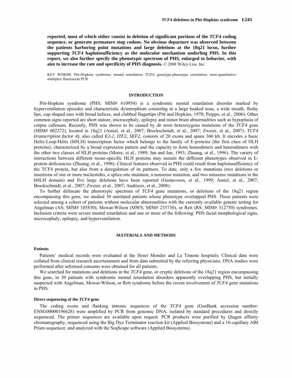
summary of patients’ phenotypes and mutations is given in the table (Table 1). TCF4 mutations (among them 5 were small deletions of 1 to 5 base pairs) were identified in 6 cases, and larges deletions of the 18q21.1-q22.1 encompassing the TCF4 gene in 4 cases. The positions of mutations and deletions relative to the exon sequences and the 18q21 region are shown in the figure (Figure 1) and reported in TCF4 database http://www.LOVD.nl/TCF4.
Large cryptic deletions In patient 1, karyotype revealed a de novo deletion of the sub-band 18q21.2-q22.1. CGH assays confirmed the
deletion, extending from the proximal breakpoint at position 51,046123 to the distal breakpoint at position 60,408993 (g.51,046123_60,408993), with a predicted size of 9.36 Mb.
In patient 2, a mosaic 18q21.1-q22.1 deletion was identified in 80% of metaphases analyzed both on lymphocytes and fibroblasts. CGH assays delineate the deletion breakpoints from the proximal position at 45,572497 to the distal position at 57,471912 (g.45,572497_57,471912) with a predicted size of 11.89 Mb.
In patient 3, karyotype revealed a de novo deletion of the sub-band 18q21.1-q22.1. No DNA sample was available for this patient to perform CGH assays.
In patient 4, a deletion of the entire gene was detected using QMF-PCR. CGH assays were undertaken for confirmation purposes and to delineate the deletion with the proximal breakpoint at position 50,248665 Mb and the distal breakpoint at position 56,857440 (g.50,248665_56,857440) with a predicted size of 6.6 Mb.
Point mutations of TCF4 gene In patient 5, QMF-PCR allowed the detection of a deletion of five base pairs, and in patients 6 to 9 of deletions of
one base pair. Direct sequencing of the relevant exons was undertaken to further confirm these small deletions. In patient 10, a missense mutation in the HLH-b specifying region was found by direct sequencing.

TCF4 deletions in Pitt-Hopkins syndrome E245
Chromosome 18
Figure 1. Location of reported mutations in the 18q21 region and within the TCF4 gene
The TCF4 gene exon information was from Ensembl (GenBank accession number: ENSG00000196628). DNA
mutations numbering was based on cDNA sequence (NM_001083962.1) where +1 corresponds to the A of the ATG translation initiation codon.
The exon-intron organization of the TCF4 gene is schematically represented in bottom of the figure: the non coding exons are represented in white and the coding exons in grey; the green bar localizes the gene region specifying the basic Helix-Loop-Helix (bHLH) domain of the protein.
The mutations reported in black below the diagram are as described in previous reports: c.1739G>A, p.Arg580Gln, reported as c.1727C>T, R576Q; c.1738C>T, p.Arg580Trp, reported as c.1726C>T, R576W; c.1738C>T, p.Arg580Trp, reported as c.1726/1738C→T, R576/580W; c.693dupT, p.Gly232TrpfsX25, reported as c.692-694insT, p.G232fsX256; c.965_969delATGCT, p.Asp322AlafsX15, reported as c.965-969delATGCT, p.D322fsX336; c.1153C>T, p.Arg385X, reported as c.1153C→T, p.R385X; c.656-1G>C, reported as IVS9-1G→C. The mutations reported in red above the diagram are those identified in this study (see also Table 1). Large deletions are indicated by solid horizontal lines (in black are those described in previous reports and in red
are those identified in this study). For the previously reported mutations, the references are mentioned on the left side. * = recurrent mutation, first
reported by Amiel et al, AJHG 2007. All TCF4 mutations are reported in TCF4 database http://www.LOVD.nl/TCF4.
c.656-1G>C(IVS9-1G>C)
c.965_969delATGCTp.Asp322AlafsX15
c.1153C>Tp.Arg385X
c.1739G>Ap.Arg580Gln
bHLH
2 3 4 5 6 7 8 10 11 12 13 14 15 16 17 19181 209
c.788delTp.Leu263X
c.1142delCp.Ser381PhefsX10
c.1018delAp.Asn340AlafsX51
c.687delCp.Ser230ProfsX4
c.1738C>T*p.Arg580Trp
50.0 50.5 51.551.0 52.0 52.5 53.0 53.5 54.0 54.5 55.0 55.5 56.0 56.5[Mb]
Gustavsson et al, AJMG 1999 ndrieux et al, EJMG 2008 A
c.1626_1630delATCAAp.Lys542AsnfsX3
TCF4
Amiel et al, AJHG 2007weier et al, AJHG 2007
kschmidt et al, HMG 2007
miel et al, AJHG 2007weier et al, AJHG 2007
del 6.6 Mb
del > 10 Mbdel 11.89 Mb
del 9.36 Mbdel > 13 Mb
del 6.6 Mb
del 1.8 Mbdel 1.2 Mb
del 0.5 MbZBroc
AZ
c.693dupT p.Gly232TrpfsX25

E246 Giurgea et al.
Clinical features of patients with TCF4 mutations
Facial morphology and extremities In our patients, the constant features were a wide mouth with a protruding, cupid-bowed upper lip, and an everted
lower lip, associated in the majority (9/10) with enophthalmia, a broad and beaked nasal bridge, a pointed nasal tip lip, mildly cup-shaped or fleshy ears, and widely spaced teeth, when teeth were already present. Flaring nostrils were present in 6 patients and a prognathism in 7 patients. We also observed at least one of the following features on our patients’ hands: long fingers (4/10), broad finger tips (4/10), overlapping fingers (4/10), fetal finger pads (6/10), single palmar creases (3/9) (Figure 2).
Figure 2. Facial morphology and extremities of patients with TCF4 molecular abnormalities Patients 1 to 4 harbor large deletions at the 18q21 locus, and patients 5 to 10 point mutations within TCF4. (P = patient)

TCF4 deletions in Pitt-Hopkins syndrome E247
Neurological signs All patients had severe developmental delay, late (after 5 years) or absent walking, and no speech. Microcephaly
(between -2 and -3SD) was observed in 9/10 patients. In contrast, epilepsy was inconstant (3/10). Most patients presented with axial hypotonia in infancy followed by delayed walking and an ataxic gate. Peripheral hypotonia was observed in two-third of patients. In contrast, the other one-third presented with lower limbs hypertonia and knee flexure. Hyperventilation episodes were observed in 8/10 patients. These episodes were occasional and sometimes triggered by excitement or anxiety. Irregular breathing at night was reported by the parents in an 18-months old patient (P4) as well as in two 18-years old patients (P5 and P10).
Behavioral features Behavioral peculiarities were present in all of our patients. These patients were unusually good and quiet babies
and slept in excess. During the first years of life, the patients had a poor emotional reaction, and played with their hands. In childhood, they were very sociable and did sometimes laugh with no apparent reason, but without bursts of laughter. They also could present with anxiety, self-aggression (bites on their hands), and occasionally hetero-aggression episodes which could be triggered by sudden changes in the daily routine. All patients had stereotypic movements of the hands (mouthing or flapping of hands) and sometimes of the head. Sleep disturbances were observed in 3 patients only. Some of the patients appeared to love water, swimming, and music.
Ocular signs In all patients who had undergone eye testing (9/9) myopia was found, and all the patients (10/10) had strabismus.
In four patients, severe myopia (> 6 dioptres) was diagnosed before two years of age. Isolated signs, such as nystagmus were observed in patients (3/4) with large deletions only.
Digestive signs Severe constipation was observed in 6 patients, but none of them had Hirschsprung disease. Two patients only
presented with severe gastrointestinal reflux in infancy.
Miscellaneous abnormalities Postnatal growth retardation was observed in 4 patients. Anomalies such as scoliosis, cryptorchidism, a small
penis, and a supernumerary nipple were observed only as single instances. None of them had other visceral malformation.
Magnetic resonance imaging (MRI) MRI showed a hypoplastic corpus callosum in two patients, and no abnormalities of the caudate nuclei,
hippocampus or the frontal lobes were observed in the 5 patients who had undergone MRI.

Giurgea et al.
Table 1. Clinical and genetic features of patients with TCF4 mutations
Patient P1 P2 P3 P4 P5 P6 P7 P8 P9 P10 Total Gender M F F M F M F F F M Age (years) 18 4 3.5 1.5 18 6 16 8 8 18 Neurological signs Severe mental retardation + + + + + + + + + + 10 Walking age (years) 8 - - - 10 5 - 5 5 9 10 Speech - - - - - - - - - - 10 Microcephaly (SD) -2 -2 -0.5 -3 -2 -2 -3 -3 -3 -2 9 Occasional hyperventilation + - + + + - + + + + 8 Seizures + - - - - - - + + 3 Behavioral characteristics Happy disposition + + + + + + + + + + 10 Stereotypic movements of hands and head + + + + + + + + + + 10
Self-aggression - - + - + - - + - + 4 Anxiety - - - - + - - + - + 3 Sleep disorders - - ? + - - - + - + 3 Ocular signs Strabismus + + + + + + + + + + 10 Myopia (right/left diopters) -2/-2 -15/-15 + + -9/-8 -1/-2 -6/-9 -14/-15 + ? 9 Nystagmus - + + + - - - - - - 3 Digestive signs Severe constipation + - + + + - + + - - 6 Hirschsprung - - - - - - - - - - 0 Gastro-esophageal reflux - - - + + - - - - - 2 Others signs Moderated post-natal growth delay - - - - + - + + + - 4
Scoliosis / Lordosis + - - - - - + - - - 1 Supernumerary nipple - - - - - - + - - - 1
0 Congenital malformation - - - - - - - - - -
Mutation 9.36 Mb deletion
11.89 Mb deletion
>10 Mb deletion
6.6 Mb deletion
c.1626_1630del p.Lys542fs
c.1018delA p.Asn340fs
c.687delC p.Ser230fs
c.1142delC p.Ser381fs
c.788delT p.Leu263X
c.1738C>T p.Arg580Trp
E248

TCF4 deletions in Pitt-Hopkins syndrome E249
Table 1 legend: The TCF4 gene exon information was from Ensembl (GenBank accession number: ENSG00000196628). DNA mutations numbering was based on cDNA sequence (NM_001083962.1) where +1 corresponds to the A of the ATG translation initiation codon. (P = patient, SD = standard deviation, F = female, M = male)
DISCUSSION
PHS is a presumably underdiagnosed syndromic mental retardation disorder, sharing some phenotypic characteristics with AS, MWS or RS. We studied 30 unrelated patients whose phenotype overlapped PHS and identified nine novel and one recurrent mutations in 10 of them. We report the first case of a mosaic cryptic deletion of 18q21 region. So far, a total of 20 different mutations of the TCF4 gene have been reported in 22 unrelated patients. Most of these mutations (18/20), either consist in deletion of significant portions of the TCF4 coding sequence, or generate premature stop codons that are presumed to result in lack of protein via nonsense-mediated RNA decay. The reported missense mutations (2/20) alter a specific conserved residue within the bHLH domain of the protein (Ma, et al., 1994), and are thereby supposed to disrupt DNA binding (Amiel, et al., 2007). Haploinsufficiency for TCF4 by decreased amount of wild-type TCF4 protein and entailed developmental insults in specific cell lineages, is the presumed molecular mechanism underling PHS (Amiel, et al., 2007; Brockschmidt, et al., 2007; Zweier, et al., 2007). Indeed, let alone subtle dysmorphic signs, the cardinal features are shared between patients with point mutations and those with large cryptic deletions, hampering genotype-phenotype correlation.
In this study, we also focused on delineating the spectrum and specificity of the clinical features observed in PHS and concluded that the cardinal manifestations were facial dysmorphism, severe developmental delay, late (> 5 years) or absent walking, no speech, and microcephaly. Of note, myopia, a sign not commonly associated with PHS in the current literature, was found in all patients who had undergone eye testing (9/9), and in most of them was severe and occurred precociously. Strabismus was a constant sign in all our patients. From the above, it can be concluded that ophthalmologic examination is required in all PHS patients in order to prevent retinal detachment and amblyopia.
In this paper we described a behavioral trait with happy disposition and stereotypic movements of hands in all our patients. Consistent with hypotonia observed in infancy, walking was delayed with ataxic gait. The majority of our patients had hyperventilation, but this sign was occasional, could occur in mid or late childhood and as minor or brief episodes only.
Dysmorphic facial features including wide mouth with a protruding, cupid-bowed upper lip, a broad nasal bridge and beaked nose, a pointed nasal tip, flaring nostrils, and thin eyebrows are cardinal signs for the diagnosis of PHS and for discriminating PHS from overlapping syndromes such as AS, MWS, or RS. In addition, several clinical features allow to further discriminate PHS from the said overlapping syndromes.
As far as the facial dysmorphism is concerned, thick, horizontal, and low set eyebrows, and the uplifted ear lobes with a central depression are the most outstanding features of MWS which depart from PHS.
In addition, PHS does not include cardiac or uro-genital (particularly hypospadias) malformations, both of which are present in half of MWS patients (Dastot-Le Moal, et al., 2007). Furthermore, Hirschsprung disease was reported as a single instance in PHS (Zweier, et al., 2007), but is found in 50% of MWS patients (Dastot-Le Moal, et al., 2007).
As far as neurological signs are concerned, PHS patients are less ataxic than AS (Clayton-Smith and Pembrey, 1992) thereby an ataxic gait was observed in most of them. Further, sleep disturbances are less common in PHS and epilepsy even scarcer, occurring later, and without typical electroencephalographic changes as compared to AS.
Only few signs are shared by PHS and RS, such as hyperventilation episodes and repeated mouthing of hands. However, PHS patients did not present loss of skills, or of purposeful hands movements. Psychomotor regression has never been observed in PHS patients. Furthermore, RS patients do not have the dysmorphism seen in PHS.
Finally, severe myopia, of early onset, seems to be an outstanding feather in PHS, and not observed in any of the three overlapping syndromes (AS, MWS, RS).
In summary, we report nine novel mutations and recapitulate the current knowledge of TCF4 molecular pathology. The large number of deletions and small deletions/insertions observed in TCF4 prompts, after standard

E250 Giurgea et al.
karyotyping, to start the molecular study by QMF-PCR analysis. No obvious departure was observed between the patients harboring point mutations and large deletions at the 18q21 locus, further supporting TCF4 haploinsufficiency as the molecular mechanism underling PHS. In this report, we also further specify the phenotypic spectrum of PHS, enlarged to behavior, with aim to increase the rate and specificity of PHS diagnosis.
ACKNOWLEDGMENTS
We are grateful to Dr. Michel Bahuau for critical reading of the manuscript. This work was funded by Agence Nationale de la Recherche (ANR-05-MRAR-008-01).
WEBRESOURCES
Accession numbers and URLs for data presented herein are as follows Online Mendelian Inheritance in Man (OMIM) http://www.ncbi.nlm.nih.gov/Omim GenBank Ensembl http://www.ensembl.org/index.html TCF4 database http://www.LOVD.nl/TCF4
REFERENCES
Amiel J, Rio M, de Pontual L, Redon R, Malan V, Boddaert N, Plouin P, Carter NP, Lyonnet S, Munnich A, Colleaux L. 2007. Mutations in TCF4, encoding a class I basic helix-loop-helix transcription factor, are responsible for Pitt-Hopkins syndrome, a severe epileptic encephalopathy associated with autonomic dysfunction. Am J Hum Genet 80:988-93.
Andrieux J, Lepretre F, Cuisset JM, Goldenberg A, Delobel B, Manouvrier-Hanu S, Holder-Espinasse M. 2008. Deletion 18q21.2q21.32 involving TCF4 in a boy diagnosed by CGH-array. Eur J Med Genet 51:172-7.
Brockschmidt A, Todt U, Ryu S, Hoischen A, Landwehr C, Birnbaum S, Frenck W, Radlwimmer B, Lichter P, Engels H, Driever W, Kubisch C, Weber RG. 2007. Severe mental retardation with breathing abnormalities (Pitt-Hopkins syndrome) is caused by haploinsufficiency of the neuronal bHLH transcription factor TCF4. Hum Mol Genet 16:1488-94.
Clayton-Smith J, Pembrey ME. 1992. Angelman syndrome. J Med Genet 29:412-5.
Dastot-Le Moal F, Wilson M, Mowat D, Collot N, Niel F, Goossens M. 2007. ZFHX1B mutations in patients with Mowat-Wilson syndrome. Hum Mutat 28:313-21.
Gustavsson P, Kimber E, Wahlstrom J, Anneren G. 1999. Monosomy 18q syndrome and atypical Rett syndrome in a girl with an interstitial deletion (18)(q21.1q22.3). Am J Med Genet 82:348-51.
Jan YN, Jan LY. 1993. HLH proteins, fly neurogenesis, and vertebrate myogenesis. Cell 75:827-30.
Ma PC, Rould MA, Weintraub H, Pabo CO. 1994. Crystal structure of MyoD bHLH domain-DNA complex: perspectives on DNA recognition and implications for transcriptional activation. Cell 77:451-9.
Murre C, McCaw PS, Vaessin H, Caudy M, Jan LY, Jan YN, Cabrera CV, Buskin JN, Hauschka SD, Lassar AB, et al. 1989. Interactions between heterologous helix-loop-helix proteins generate complexes that bind specifically to a common DNA sequence. Cell 58:537-44.
Niel F, Martin J, Dastot-Le Moal F, Costes B, Boissier B, Delattre V, Goossens M, Girodon E. 2004. Rapid detection of CFTR gene rearrangements impacts on genetic counselling in cystic fibrosis. J Med Genet 41:e118.
Peippo MM, Simola KO, Valanne LK, Larsen AT, Kahkonen M, Auranen MP, Ignatius J. 2006. Pitt-Hopkins syndrome in two patients and further definition of the phenotype. Clin Dysmorphol 15:47-54.
Pitt D, Hopkins I. 1978. A syndrome of mental retardation, wide mouth and intermittent overbreathing. Aust Paediatr J 14:182-4.
Zhuang Y, Cheng P, Weintraub H. 1996. B-lymphocyte development is regulated by the combined dosage of three basic helix-loop-helix genes, E2A, E2-2, and HEB. Mol Cell Biol 16:2898-905.

TCF4 deletions in Pitt-Hopkins syndrome E251
Zweier C, Peippo MM, Hoyer J, Sousa S, Bottani A, Clayton-Smith J, Reardon W, Saraiva J, Cabral A, Gohring I, Devriendt K, de Ravel T, Bijlsma EK, Hennekam RC, Orrico A, Cohen M, Dreweke A, Reis A, Nurnberg P, Rauch A. 2007. Haploinsufficiency of TCF4 causes syndromal mental retardation with intermittent hyperventilation (Pitt-Hopkins syndrome). Am J Hum Genet 80:994-1001.