The Mre11 Complex Mediates the S-Phase Checkpoint through an Interaction with Replication Protein A
15
MOLECULAR AND CELLULAR BIOLOGY, Sept. 2007, p. 6053–6067 Vol. 27, No. 17 0270-7306/07/$08.000 doi:10.1128/MCB.00532-07 Copyright © 2007, American Society for Microbiology. All Rights Reserved. The Mre11 Complex Mediates the S-Phase Checkpoint through an Interaction with Replication Protein A ¶ Erin Olson, 1 † Christian J. Nievera, 1 † Enbo Liu, 1 ‡ Alan Yueh-Luen Lee, 1 Longchuan Chen, 2 and Xiaohua Wu 1 * Department of Molecular and Experimental Medicine, The Scripps Research Institute, La Jolla, California 92037, 1 and Department of Pathology, VA Medical Center, Long Beach, California 90822 2 Received 27 March 2007/Returned for modification 14 May 2007/Accepted 8 June 2007 The Mre11/Rad50/Nbs1 complex (MRN) plays an essential role in the S-phase checkpoint. Cells derived from patients with Nijmegen breakage syndrome and ataxia telangiectasia-like disorder undergo radioresis- tant DNA synthesis (RDS), failing to suppress DNA replication in response to ionizing radiation (IR). How MRN affects DNA replication to control the S-phase checkpoint, however, remains unclear. We demonstrate that MRN directly interacts with replication protein A (RPA) in unperturbed cells and that the interaction is regulated by cyclin-dependent kinases. We also show that this interaction is needed for MRN to correctly localize to replication centers. Abolishing the interaction of Mre11 with RPA leads to pronounced RDS without affecting phosphorylation of Nbs1 or SMC1 following IR. Moreover, MRN is recruited to sites at or adjacent to replication origins by RPA and acts there to inhibit new origin firing upon IR. These studies suggest a direct role of MRN at origin-proximal sites to control DNA replication initiation in response to DNA damage, thereby providing an important mechanism underlying the intra-S-phase checkpoint in mammalian cells. The Mre11/Rad50/Nbs1 complex (MRN) participates in multiple pathways to maintain genome stability (1, 10, 55). In humans, hypomorphic mutations in NBS1 and MRE11 lead to Nijmegen breakage syndrome (NBS) and ataxia-telangiectasia- like disorder (ATLD), respectively (7, 41, 54, 64). Both NBS and ATLD patients show developmental defects, immunode- ficiency, and a high incidence of cancer, phenotypically similar to the ATM-deficient disorder ataxia-telangiectasia (AT) (53, 60). In addition, cells from NBS and ATLD patients are sen- sitive to radiation, exhibit genome instability, and have a de- fective S-phase checkpoint. The phenotypic resemblance of NBS, ATLD, and AT cells implies that MRN and ATM participate in similar biological pathways. This is supported by recent findings that MRN acts both upstream and downstream of ATM to mediate the dam- age response when double-strand breaks (DSBs) are generated (32). MRN migrates to DSB sites immediately after damage in an ATM-independent manner (44, 47) and is required for ATM activation, especially in response to low doses of ionizing radiation (IR) (6, 8, 25, 35, 63). MRN is also a direct substrate of ATM. Multiple ATM phosphorylation sites on Nbs1 have been identified, and these phosphorylation events play impor- tant roles in mediating the intra-S and G 2 /M checkpoints, although the mechanism by which Nbs1 phosphorylation reg- ulates these pathways is not well defined (8, 22, 36, 69, 75). Mre11 is the core of the MRN complex and interacts with both Rad50 and Nbs1 (24, 34, 62). Mre11 carries 3 to 5 exonuclease activity and single-strand endonuclease activity and acts on various types of DSB ends and hairpins (49, 61). These nuclease activities are believed to be involved in pro- cessing of DNA ends for DSB repair (18, 37, 50). In multiple organisms, it has been shown that MRN is essential for DNA repair by homologous recombination (27, 59, 70). The radioresistant DNA synthesis (RDS) phenotype of NBS and ATLD cells suggests an important role of MRN in the S-phase checkpoint (53, 54). ATM activates the S-phase check- point, and two distinct pathways, ATM-Chk2-Cdc25A-Cdk2 and ATM-MRN-SMC1, have been suggested to mediate the S-phase checkpoint downstream of ATM (16). The ATM- Chk2 pathway modulates Cdc25A stability by Chk2-mediated phosphorylation and ultimately regulates Cdc45 chromatin loading (15, 16). In NBS and ATLD cells, Chk2 activation, Cdc25A degradation, and Cdc45 chromatin loading are indis- tinguishable from those of normal cells after IR, suggesting that the ATM-Chk2-Cdc25A-Cdk2 pathway is intact and that the RDS phenotype observed in NBS and ATLD cells is due to a defect independent from the regulation of Cdc45 chromatin loading (16). SMC1 has been shown to be phosphorylated in the ATM-Nbs1 pathway in response to IR in an ATM- and Nbs1-dependent manner (28, 71). The IR-induced phosphory- lation of SMC1 is important for mediating the S-phase check- point; thus, SMC1 was proposed as a downstream effector of the ATM-Nbs1 pathway. However, the details of the mecha- nisms by which SMC1 inhibits DNA synthesis are not clear. A connection of MRN to DNA replication was illustrated by its colocalization with proliferating cell nuclear antigen (PCNA) and BrdU incorporation sites in the S phase (45). Chromatin immunoprecipitation (ChIP) also revealed that MRN binds to replication-origin-proximal sites in mammalian cells in a manner similar to that seen with E2F family members * Corresponding author. Mailing address: Department of Molecular and Experimental Medicine, The Scripps Research Institute, La Jolla, CA 92037. Phone: (858) 784-7910. Fax: (858) 784-7978. E-mail: [email protected]. † E.O. and C.J.N. contributed equally to this work. ‡ Present address: Signal Transduction Program, Burnham Institute for Medical Research, La Jolla, CA 92037. ¶ Supplemental material for this article may be found at http://mcb .asm.org/. Published ahead of print on 25 June 2007. 6053 on June 24, 2016 by guest http://mcb.asm.org/ Downloaded from
Transcript of The Mre11 Complex Mediates the S-Phase Checkpoint through an Interaction with Replication Protein A
MOLECULAR AND CELLULAR BIOLOGY, Sept. 2007, p. 6053–6067 Vol. 27, No. 170270-7306/07/$08.00�0 doi:10.1128/MCB.00532-07Copyright © 2007, American Society for Microbiology. All Rights Reserved.
The Mre11 Complex Mediates the S-Phase Checkpoint through anInteraction with Replication Protein A�¶
Erin Olson,1† Christian J. Nievera,1† Enbo Liu,1‡ Alan Yueh-Luen Lee,1Longchuan Chen,2 and Xiaohua Wu1*
Department of Molecular and Experimental Medicine, The Scripps Research Institute, La Jolla, California 92037,1 andDepartment of Pathology, VA Medical Center, Long Beach, California 908222
Received 27 March 2007/Returned for modification 14 May 2007/Accepted 8 June 2007
The Mre11/Rad50/Nbs1 complex (MRN) plays an essential role in the S-phase checkpoint. Cells derivedfrom patients with Nijmegen breakage syndrome and ataxia telangiectasia-like disorder undergo radioresis-tant DNA synthesis (RDS), failing to suppress DNA replication in response to ionizing radiation (IR). HowMRN affects DNA replication to control the S-phase checkpoint, however, remains unclear. We demonstratethat MRN directly interacts with replication protein A (RPA) in unperturbed cells and that the interaction isregulated by cyclin-dependent kinases. We also show that this interaction is needed for MRN to correctlylocalize to replication centers. Abolishing the interaction of Mre11 with RPA leads to pronounced RDS withoutaffecting phosphorylation of Nbs1 or SMC1 following IR. Moreover, MRN is recruited to sites at or adjacentto replication origins by RPA and acts there to inhibit new origin firing upon IR. These studies suggest a directrole of MRN at origin-proximal sites to control DNA replication initiation in response to DNA damage, therebyproviding an important mechanism underlying the intra-S-phase checkpoint in mammalian cells.
The Mre11/Rad50/Nbs1 complex (MRN) participates inmultiple pathways to maintain genome stability (1, 10, 55). Inhumans, hypomorphic mutations in NBS1 and MRE11 lead toNijmegen breakage syndrome (NBS) and ataxia-telangiectasia-like disorder (ATLD), respectively (7, 41, 54, 64). Both NBSand ATLD patients show developmental defects, immunode-ficiency, and a high incidence of cancer, phenotypically similarto the ATM-deficient disorder ataxia-telangiectasia (AT) (53,60). In addition, cells from NBS and ATLD patients are sen-sitive to radiation, exhibit genome instability, and have a de-fective S-phase checkpoint.
The phenotypic resemblance of NBS, ATLD, and AT cellsimplies that MRN and ATM participate in similar biologicalpathways. This is supported by recent findings that MRN actsboth upstream and downstream of ATM to mediate the dam-age response when double-strand breaks (DSBs) are generated(32). MRN migrates to DSB sites immediately after damage inan ATM-independent manner (44, 47) and is required forATM activation, especially in response to low doses of ionizingradiation (IR) (6, 8, 25, 35, 63). MRN is also a direct substrateof ATM. Multiple ATM phosphorylation sites on Nbs1 havebeen identified, and these phosphorylation events play impor-tant roles in mediating the intra-S and G2/M checkpoints,although the mechanism by which Nbs1 phosphorylation reg-ulates these pathways is not well defined (8, 22, 36, 69, 75).
Mre11 is the core of the MRN complex and interacts withboth Rad50 and Nbs1 (24, 34, 62). Mre11 carries 3� to 5�exonuclease activity and single-strand endonuclease activityand acts on various types of DSB ends and hairpins (49, 61).These nuclease activities are believed to be involved in pro-cessing of DNA ends for DSB repair (18, 37, 50). In multipleorganisms, it has been shown that MRN is essential for DNArepair by homologous recombination (27, 59, 70).
The radioresistant DNA synthesis (RDS) phenotype of NBSand ATLD cells suggests an important role of MRN in theS-phase checkpoint (53, 54). ATM activates the S-phase check-point, and two distinct pathways, ATM-Chk2-Cdc25A-Cdk2and ATM-MRN-SMC1, have been suggested to mediate theS-phase checkpoint downstream of ATM (16). The ATM-Chk2 pathway modulates Cdc25A stability by Chk2-mediatedphosphorylation and ultimately regulates Cdc45 chromatinloading (15, 16). In NBS and ATLD cells, Chk2 activation,Cdc25A degradation, and Cdc45 chromatin loading are indis-tinguishable from those of normal cells after IR, suggestingthat the ATM-Chk2-Cdc25A-Cdk2 pathway is intact and thatthe RDS phenotype observed in NBS and ATLD cells is due toa defect independent from the regulation of Cdc45 chromatinloading (16). SMC1 has been shown to be phosphorylated inthe ATM-Nbs1 pathway in response to IR in an ATM- andNbs1-dependent manner (28, 71). The IR-induced phosphory-lation of SMC1 is important for mediating the S-phase check-point; thus, SMC1 was proposed as a downstream effector ofthe ATM-Nbs1 pathway. However, the details of the mecha-nisms by which SMC1 inhibits DNA synthesis are not clear.
A connection of MRN to DNA replication was illustratedby its colocalization with proliferating cell nuclear antigen(PCNA) and BrdU incorporation sites in the S phase (45).Chromatin immunoprecipitation (ChIP) also revealed thatMRN binds to replication-origin-proximal sites in mammaliancells in a manner similar to that seen with E2F family members
* Corresponding author. Mailing address: Department of Molecularand Experimental Medicine, The Scripps Research Institute, La Jolla,CA 92037. Phone: (858) 784-7910. Fax: (858) 784-7978. E-mail:[email protected].
† E.O. and C.J.N. contributed equally to this work.‡ Present address: Signal Transduction Program, Burnham Institute
for Medical Research, La Jolla, CA 92037.¶ Supplemental material for this article may be found at http://mcb
.asm.org/.� Published ahead of print on 25 June 2007.
6053
on June 24, 2016 by guesthttp://m
cb.asm.org/
Dow
nloaded from
(38). However, it remains unclear how MRN is recruited toreplication centers and origin-proximal sites.
An association of replication protein A (RPA) and MRNafter replication fork blockage induced by hydroxyurea hasbeen previously described (52), but the biological significanceof this interaction has not been elucidated. We independentlydetected this interaction and found that, in fact, a subset ofnuclear MRN and RPA form stable complexes through a di-rect interaction in unperturbed cells. This interaction increasesat the G1-to-S transition with a peak in S phase and is regu-lated by cyclin-dependent kinase (CDK)-mediated phosphory-lation. We also demonstrated that through this interaction,MRN is recruited to replication centers and origin-proximalsites in S phase. Importantly, the Mre11 mutants specificallydefective in RPA binding exhibit a profound RDS phenotype,although phosphorylation of Nbs1 and SMC1 remains unaf-fected. These studies suggest that recruitment of MRN toreplication-proximal sites by RPA is critical for MRN to sup-press DNA replication initiation in response to IR, therebysuggesting a new mechanism of the ATM-Nbs1 pathway tomediate the S-phase checkpoint.
MATERIALS AND METHODS
Cell culture, cell synchronization, retroviral infections, and short hairpinRNA (shRNA). HeLa, IMR90, U2OS, and 293T cells were cultured in Dulbeccomodified Eagle medium containing 10% fetal bovine serum. The Nbs1-deficient(GM07166) or Mre11-deficient (ATLD) cell lines immortalized by human telo-merase reverse transcriptase (hTERT) (43) were cultured as previously de-scribed (51). T98G cells were synchronized by culturing in Dulbecco modifiedEagle medium supplemented with 0.1% fetal bovine serum for 48 h and thenreleasing by adding 10% fetal bovine serum.
Different Nbs1 and Mre11 alleles were introduced into the GM07166, ATLD,or U2OS cell line by infection of retroviruses followed by selection of puromycin-or G418-resistant cells. Silencing of endogenous Mre11 in U2OS cells comple-mented by shRNA-resistant wild-type Mre11, Mre11-NAAIRS, or Mre11-DDalleles was accomplished by two rounds of retroviral infection using pMKOvector (39) expressing two different Mre11 shRNA target sequences, GATGAGAACTCTTGGTTTAAC and GAGTATAGATTTAGCAGAACA.
MRN complex purification. Nuclear extracts were prepared from 5g of BJABcells and were fractionated by using phosphocellulose resin P11 followed byDEAE-Sephacel resin as previously described (19). Columns were loaded with0.1 M KCl in BC buffer (20 mM Tris [pH 7.3], 0.2 mM EDTA) and eluted with0.75 M KCl (BC buffer). Eluted fractions from DEAE were pooled, dialyzed, andapplied to a Superose 6 (Pharmacia) column in BC buffer with 0.1 M KCl.
Plasmids and mutagenesis. Full-length Mre11 and Nbs1, fragments of Mre11and Nbs1, and Mre11 internal-deletion alleles were subcloned into pcDNA3�, amammalian expression vector containing the sequence encoding the myc epitope(9). Glutathione S-transferase–Mre11 (GST-Mre11) (amino acids 360 to 708)and GST-Nbs1 (amino acids 476 to 754) were constructed by cloning PCRproducts into pGEX4T-1 (Pharmacia). His-tagged RPA1/3/2, RPA1/3, andRPA3/2 were made by cloning PCR products generated by using p11d-tRDSRPA (23) (a generous gift from Marc Wold) as a template with pET28b (No-vagen). Mre11 and Nbs1 mutations were generated by site-directed mutagenesis(Stratagene).
Immunoprecipitation, in vitro binding, and immunofluorescence. Immuno-precipitation (IP) and Western blot analysis were performed as previously de-scribed (9). IP in the presence of protein phosphatase was conducted by incu-bating cell lysates prepared from each 10-cm dish with 400 U of lambdaphosphatase (NEB) for 30 min at room temperature followed by immunopre-cipitation at 4°C.
Flag-Nbs1, Flag-Mre11, and Flag-Mre11 mutants were expressed in insectcells and purified using anti-Flag M2 agarose (Sigma) followed by elution with3xFlag peptide (sigma). His-tagged RPA was purified from Escherichia coli onnickel-nitrilotriacetic acid resin (QIAGEN), and GST-Mre11 (amino acids 360to 708) was purified using glutathione Sepharose (GE). In vitro binding wasperformed with NETN buffer (20 mM Tris [pH 8.0], 1 mM EDTA, 150 mMNaCl, 0.5% NP-40).
For immunostaining of S-phase foci, IMR90 cells were fixed with a modifiedStreck Tissue fixative for 30 min at room temperature and then permeabilized for15 min at room temperature as previously described (47). After blocking with 5%goat serum in phosphate-buffered saline (PBS), cells were stained with primaryantibody at 4°C overnight followed by staining for 1 h at room temperature witheither fluorescein isothiocyanate-conjugated anti-mouse or rhodamine-conju-gated anti-rabbit secondary antibodies (Jackson ImmunoResearch Laborato-ries). For ATLD fibroblast cells, fixation and immunostaining were performed5 h after release from a double thymidine (2 mM) block. To detect damage foci,asynchronized cells were fixed by 70% methanol and 30% acetone at �20°C for15 min followed by immunostaining analysis as previously described (68).
Antibodies. The polyclonal antibodies against Nbs1 (D29), and Mre11 (D27)and the monoclonal antibody against Nbs1 (EE15) were described previously(67, 69). Antiserum to GST was raised in rabbits by use of purified GST. Theother antibodies used were purchased from Oncogene (ATM, Mre11, Nbs1,RPA1, and RPA2), Upstate (Rad50), Cell Signaling Technology (pT68-Chk2and pS343Nbs1), Novus (pS1981-ATM), Bethyl (pS966-SMC1), and Santa CruzBiotechnology (Chk2 and Ku70).
ChIP. ChIP was performed as previously described (48) with the followingmodifications. T98G and its derivative cell lines were synchronized by serumstarvation in the presence of 0.1% fetal bovine serum (FBS) for 48 h and releasedinto the cell cycle by the addition of 10% FBS into the culture medium. Atspecified time points after release, cells were cross-linked with 5 mM dimethyl-3,3�-dithiobispropionimidate-2HCl (DTBP) in PBS for 30 min on ice (21) fol-lowed by 1% formaldehyde in PBS at 37°C for 10 min. Chromatin was shearedto obtain DNA fragments approximately 1 kb in length. IP was performed usinganti-Myc antibody at 4°C overnight. IP with rabbit anti-mouse immunoglobulinG (IgG) antibody was used as a negative control. After washing, the immuno-precipitates were eluted, incubated at 65°C overnight to reverse the cross-links,and then analyzed by PCR using primers specific to B2-lamin, heat shock protein70 (HSP70), and c-myc loci (2).
Radioresistant DNA synthesis. Inhibition of DNA synthesis following IR wasperformed as previously described (46, 54). Cells were grown in the presence of20 nCi/ml (U2OS) or 50 nCi/ml (ATLD and GM07166) [14C]thymidine for 24 h.After removal of [14C]thymidine, cells were either mock treated or treated with10 Gy of IR, allowed to recover for 1 h, and then labeled with 2.5 �Ci/ml[3H]thymidine for 15 min (U2OS) (46) or 2 h (ATLD and GM07166) (54). Cellswere spotted onto Whatman glass microfiber filters and washed with trichloro-acetic acid and 100% ethanol. The filters were allowed to dry, and radioactivitywas measured using a liquid scintillation counter and a dual energy program.
Sensitivity to ionizing radiation. ATLD cells complemented with vector,Mre11, Mre11-NAAIRS, or Mre11-DD were plated in triplicate at 3.5 � 105
cells per dish. Cells were treated with 0, 2, 5, or 10 Gy of IR. Seven days later,cells were stained with trypan blue and counted.
RESULTS
A subset of nuclear MRNs and RPAs form stable complexesin unperturbed cells. When nuclear extracts were fraction-ated using columns (Fig. 1A), we found a small percentageof RPA was consistently copurified with MRN. While mostMRNs bound to the phosphocellulose P11 column, only10% to 20% of RPA species were retained on the P11column and the rest were present in the flow-through vol-ume (Fig. 1B, left panel). Further purification showed thatmost P11-bound MRN and RPA species were retained on aDEAE column (Fig. 1B, right panel). Protein complexeseluted from the DEAE column were separated according tomolecular weight by Superose 6 gel filtration (Fig. 1C, leftpanel). Most input RPA was coeluted with MRN as largeprotein complexes. Notably, RPA was present only in thefractions containing MRNs of relatively higher molecularweight and there was a substantial amount of MRN that waseluted in the absence of RPA. Coeluted RPA and MRNwere truly associated with each other, as demonstrated bycoimmunoprecipitation (Fig. 1C, right panel). These studiessuggest that a subset of MRNs and RPAs form stable com-plexes in unperturbed cells. The large size of the complexes
6054 OLSON ET AL. MOL. CELL. BIOL.
on June 24, 2016 by guesthttp://m
cb.asm.org/
Dow
nloaded from
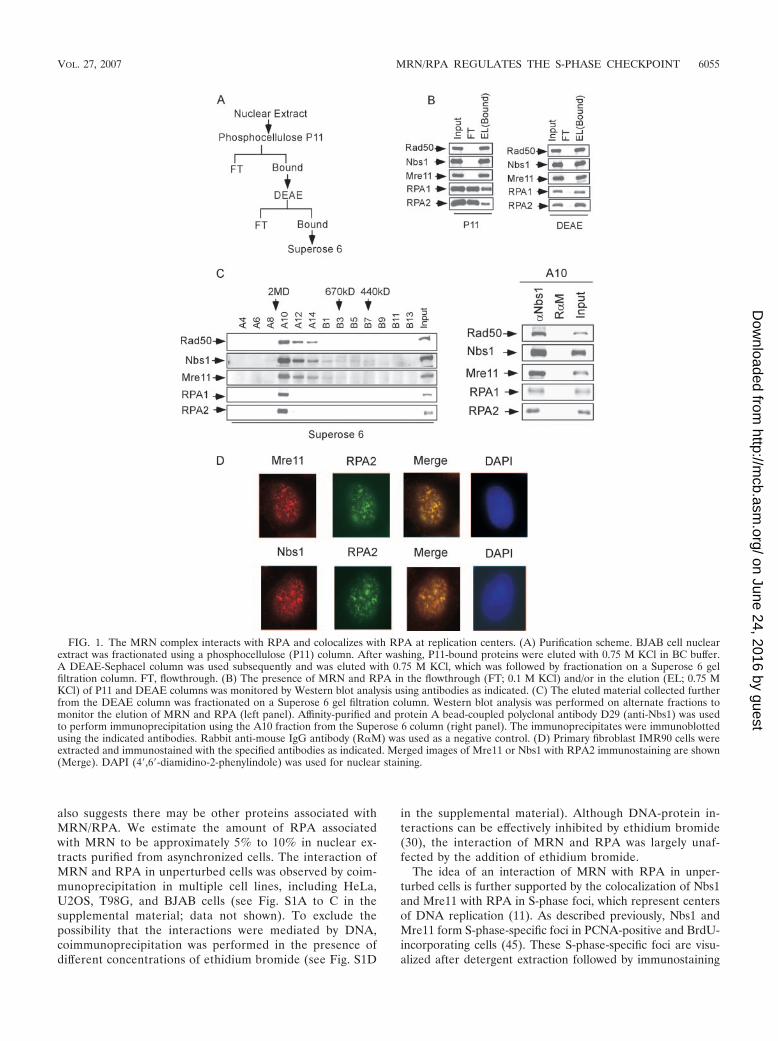
also suggests there may be other proteins associated withMRN/RPA. We estimate the amount of RPA associatedwith MRN to be approximately 5% to 10% in nuclear ex-tracts purified from asynchronized cells. The interaction ofMRN and RPA in unperturbed cells was observed by coim-munoprecipitation in multiple cell lines, including HeLa,U2OS, T98G, and BJAB cells (see Fig. S1A to C in thesupplemental material; data not shown). To exclude thepossibility that the interactions were mediated by DNA,coimmunoprecipitation was performed in the presence ofdifferent concentrations of ethidium bromide (see Fig. S1D
in the supplemental material). Although DNA-protein in-teractions can be effectively inhibited by ethidium bromide(30), the interaction of MRN and RPA was largely unaf-fected by the addition of ethidium bromide.
The idea of an interaction of MRN with RPA in unper-turbed cells is further supported by the colocalization of Nbs1and Mre11 with RPA in S-phase foci, which represent centersof DNA replication (11). As described previously, Nbs1 andMre11 form S-phase-specific foci in PCNA-positive and BrdU-incorporating cells (45). These S-phase-specific foci are visu-alized after detergent extraction followed by immunostaining
FIG. 1. The MRN complex interacts with RPA and colocalizes with RPA at replication centers. (A) Purification scheme. BJAB cell nuclearextract was fractionated using a phosphocellulose (P11) column. After washing, P11-bound proteins were eluted with 0.75 M KCl in BC buffer.A DEAE-Sephacel column was used subsequently and was eluted with 0.75 M KCl, which was followed by fractionation on a Superose 6 gelfiltration column. FT, flowthrough. (B) The presence of MRN and RPA in the flowthrough (FT; 0.1 M KCl) and/or in the elution (EL; 0.75 MKCl) of P11 and DEAE columns was monitored by Western blot analysis using antibodies as indicated. (C) The eluted material collected furtherfrom the DEAE column was fractionated on a Superose 6 gel filtration column. Western blot analysis was performed on alternate fractions tomonitor the elution of MRN and RPA (left panel). Affinity-purified and protein A bead-coupled polyclonal antibody D29 (anti-Nbs1) was usedto perform immunoprecipitation using the A10 fraction from the Superose 6 column (right panel). The immunoprecipitates were immunoblottedusing the indicated antibodies. Rabbit anti-mouse IgG antibody (R�M) was used as a negative control. (D) Primary fibroblast IMR90 cells wereextracted and immunostained with the specified antibodies as indicated. Merged images of Mre11 or Nbs1 with RPA2 immunostaining are shown(Merge). DAPI (4�,6�-diamidino-2-phenylindole) was used for nuclear staining.
VOL. 27, 2007 MRN/RPA REGULATES THE S-PHASE CHECKPOINT 6055
on June 24, 2016 by guesthttp://m
cb.asm.org/
Dow
nloaded from
using specific antibodies. Consistent with previous findings(45), Mre11 and Nbs1 formed S-phase foci in all S-phase cellsduring an unperturbed cell cycle (data not shown). TheseMre11 and Nbs1 S-phase foci were colocalized with those ofRPA1 and RPA2 in multiple cell lines, including IMR90,WI38, and U2OS cells (Fig. 1D and data not shown).
The observation that MRN and RPA were colocalized in
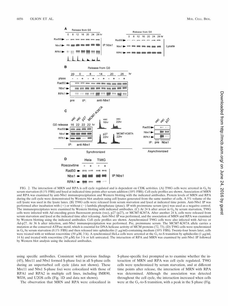
S-phase-specific foci prompted us to examine whether the in-teraction of MRN and RPA was cell cycle regulated. T98Gcells were synchronized by serum starvation, and at differenttime points after release, the interaction of MRN with RPAwas determined. Although the association was detectedthroughout the cell cycle, the interaction increased when cellswere at the G1-to-S transition, with a peak in the S phase (Fig.
FIG. 2. The interaction of MRN and RPA is cell cycle regulated and is dependent on CDK activities. (A) T98G cells were arrested in G0 byserum starvation (0.1% FBS) and lysed at indicated time points after serum addition (10% FBS). Cell cycle profiles are shown. Association of MRNand RPA was examined by anti-Nbs1 immunoprecipitation and Western blotting with the indicated antibodies. Protein levels of MRN and RPAduring the cell cycle were demonstrated by Western blot analysis using cell lysates generated from the same number of cells. A 5% volume of thecell lysate was used in the lysate lanes. (B) T98G cells were released from serum starvation and lysed at indicated time points. Anti-Nbs1 IP wasperformed after incubation with (�) or without (�) lambda phosphatase (ptase). IP with preimmune serum (pre) was used as a negative control.The immunoprecipitates were examined by Western blotting with indicated antibodies. (C) At 24 h after arrest in G0 by serum starvation, T98Gcells were infected with Ad encoding green fluorescent protein (vec), p27 (p27), or MCM7-K387A. After another 24 h, cells were released fromserum starvation and lysed at the indicated time after releasing. Anti-Nbs1 IP was performed, and the association of MRN and RPA was examinedby Western blotting using the indicated antibodies. Cell cycle profiles are shown. Asynchronized T98G cells were also infected with Ad-vec orAd-p27. At 36 h after infection, anti-Nbs1 immunoprecipitation was performed. Pre, preimmune serum. The MCM7-K387A allele carries amutation at the conserved ATPase motif, which is essential for DNA helicase activity of MCM proteins (72, 73). (D) T98G cells were synchronizedin G0 by serum starvation (0.1% FBS) and then released into aphidicolin (1 �g/ml)-containing medium (10% FBS). Twenty-four hours later, cellswere treated with or without roscovitine (50 �M, 3 h). A synchronized HeLa cells were arrested at the G1-to-S transition by aphidicolin (1 �g/ml,14 h) and treated with roscovitine (50 �M) for 3 h or left untreated. The interaction of RPA and MRN was examined by anti-Nbs1 IP followedby Western blot analysis using the indicated antibodies.
6056 OLSON ET AL. MOL. CELL. BIOL.
on June 24, 2016 by guesthttp://m
cb.asm.org/
Dow
nloaded from
2A). Similar results were obtained when HeLa cells were syn-chronized by release from nocodazole-mediated mitotic arrest(data not shown). Increased association of MRN/RPA rightbefore the onset of S phase and the colocalization of MRN andRPA S-phase foci suggest a potential role of MRN/RPA com-plexes in regulating DNA replication and/or sensing and re-pairing DSBs at replication forks.
CDK activity is important for regulating the interaction ofMRN and RPA. Since the interaction of MRN and RPA isregulated during the cell cycle, we investigated whether theinteraction is phosphorylation dependent. Coimmunoprecipi-tation of MRN and RPA was performed using cell lysatesprepared from different stages of the cell cycle with or withoutphosphatase treatment (Fig. 2B). The increased association ofMRN and RPA at the G1-to-S transition and in S phase wasabolished after phosphatase treatment, suggesting that cell-cycle-regulated phosphorylation is required for stimulating theinteraction and maintaining the increased MRN/RPA interac-tion. Since the interaction started to increase at the G1-to-Stransition, we examined whether CDKs that are required forreplication initiation are involved in regulating the MRN/RPAinteraction. T98G cells were infected with adenovirus (Ad)encoding p27 or green fluorescent protein while arrested in G0
by serum starvation, and 22 h after release from G0, coimmu-noprecipitation was performed. Inhibition of CDK activity byp27 prevented cells from entering the S phase and also inhib-ited the interaction of MRN and RPA (Fig. 2C, left panel).The loss of the interaction of MRN and RPA was not due toa failure of replicating DNA, since overexpression of anMCM7 helicase mutant (MCM-K387A), which suppressedDNA replication (72, 73; A. Lee and X. Wu, unpublisheddata), did not significantly impair the interaction of MRN andRPA. Aphidicolin and thymidine treatment, which arrestedcells at the G1-to-S transition, also did not prevent the associ-ation (data not shown). These data support the idea that inhi-bition of the interaction by p27 is due to an inhibition of CDKactivities. These data also suggest that active DNA replicationis not necessarily required for stimulating the interaction ofMRN and RPA; thus, the increased association of MRN andRPA in the S phase is likely not triggered by spontaneous DNAdamage generated during DNA replication.
Overexpression of p27 in asynchronized cells also inhibitedthe association of MRN and RPA (Fig. 2C, right panel), sug-gesting that CDK activity is required to mediate the interactionof MRN and RPA not only for cells exiting from G0 but alsofor normal cycling cells. To more directly examine whetherCDKs are involved in regulating the MRN/RPA interaction,T98G cells were released from G0 and arrested at the G1/Sboundary by aphidicolin when the interaction reached rela-tively high levels. Inhibition of CDK activity by the presence ofroscovitine significantly reduced the interaction of MRN withRPA (Fig. 2D). Similar results were obtained when HeLa cellswere used. These data suggest that CDK activity is not onlyimportant for stimulating the cell-cycle-dependent associationof MRN and RPA but is also required for maintaining theincreased association.
A specific site at the C terminus of Mre11 directly interactswith RPA1. Obtaining MRN mutants specifically defective inRPA binding would help to clarify the biological role(s) of theinteraction of MRN and RPA. To do this, a series of Myc-
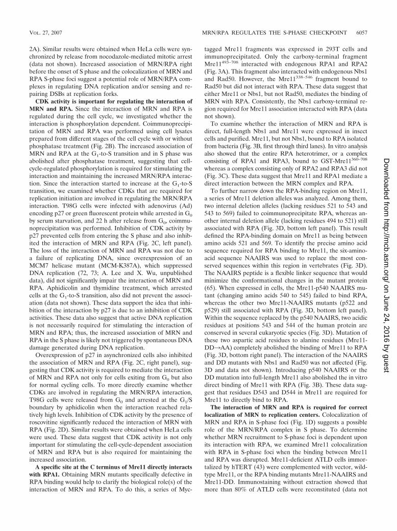
tagged Mre11 fragments was expressed in 293T cells andimmunoprecipitated. Only the carboxy-terminal fragmentMre11493–708 interacted with endogenous RPA1 and RPA2(Fig. 3A). This fragment also interacted with endogenous Nbs1and Rad50. However, the Mre11338–546 fragment bound toRad50 but did not interact with RPA. These data suggest thateither Mre11 or Nbs1, but not Rad50, mediates the binding ofMRN with RPA. Consistently, the Nbs1 carboxy-terminal re-gion required for Mre11 association interacted with RPA (datanot shown).
To examine whether the interaction of MRN and RPA isdirect, full-length Nbs1 and Mre11 were expressed in insectcells and purified. Mre11, but not Nbs1, bound to RPA isolatedfrom bacteria (Fig. 3B, first through third lanes). In vitro analysisalso showed that the entire RPA heterotrimer, or a complexconsisting of RPA1 and RPA3, bound to GST-Mre11360–708
whereas a complex consisting only of RPA2 and RPA3 did not(Fig. 3C). These data suggest that Mre11 and RPA1 mediate adirect interaction between the MRN complex and RPA.
To further narrow down the RPA-binding region on Mre11,a series of Mre11 deletion alleles was analyzed. Among them,two internal deletion alleles (lacking residues 521 to 543 and543 to 569) failed to coimmunoprecipitate RPA, whereas an-other internal deletion allele (lacking residues 494 to 521) stillassociated with RPA (Fig. 3D, bottom left panel). This resultdefined the RPA-binding domain on Mre11 as being betweenamino acids 521 and 569. To identify the precise amino acidsequence required for RPA binding to Mre11, the six-amino-acid sequence NAAIRS was used to replace the most con-served sequences within this region in vertebrates (Fig. 3D).The NAAIRS peptide is a flexible linker sequence that wouldminimize the conformational changes in the mutant protein(65). When expressed in cells, the Mre11-p540 NAAIRS mu-tant (changing amino acids 540 to 545) failed to bind RPA,whereas the other two Mre11-NAAIRS mutants (p522 andp529) still associated with RPA (Fig. 3D, bottom left panel).Within the sequence replaced by the p540 NAAIRS, two acidicresidues at positions 543 and 544 of the human protein areconserved in several eukaryotic species (Fig. 3D). Mutation ofthese two aspartic acid residues to alanine residues (Mre11-DD3AA) completely abolished the binding of Mre11 to RPA(Fig. 3D, bottom right panel). The interaction of the NAAIRSand DD mutants with Nbs1 and Rad50 was not affected (Fig.3D and data not shown). Introducing p540 NAAIRS or theDD mutation into full-length Mre11 also abolished the in vitrodirect binding of Mre11 with RPA (Fig. 3B). These data sug-gest that residues D543 and D544 in Mre11 are required forMre11 to directly bind to RPA.
The interaction of MRN and RPA is required for correctlocalization of MRN to replication centers. Colocalization ofMRN and RPA in S-phase foci (Fig. 1D) suggests a possiblerole of the MRN/RPA complex in S phase. To determinewhether MRN recruitment to S-phase foci is dependent uponits interaction with RPA, we examined Mre11 colocalizationwith RPA in S-phase foci when the binding between Mre11and RPA was disrupted. Mre11-deficient ATLD cells immor-talized by hTERT (43) were complemented with vector, wild-type Mre11, or the RPA binding mutants Mre11-NAAIRS andMre11-DD. Immunostaining without extraction showed thatmore than 80% of ATLD cells were reconstituted (data not
VOL. 27, 2007 MRN/RPA REGULATES THE S-PHASE CHECKPOINT 6057
on June 24, 2016 by guesthttp://m
cb.asm.org/
Dow
nloaded from
shown) and that the Mre11 wild type as well as the DD andNAAIRS mutants were all correctly located in the nucleus (seeFig. S2A in the supplemental material). Mre11, Mre11-DD,and Mre11-NAAIRS, as well as Rad50 and Nbs1, were also
restored to similar protein levels in the reconstituted ATLDcells (Fig. 4A, top row). To enrich the S-phase population, cellswere synchronized by a double thymidine block. Five hoursafter release from thymidine block, approximately 20% of the
FIG. 3. The direct interaction between MRN and RPA requires residues D543 and D544 at the C terminus of Mre11. (A) Fragments of Mre11corresponding to the indicated positions of the human Mre11 protein were transiently transfected into 293T cells and immunoprecipitated usingan anti-Myc antibody (9E10). The immunoprecipitates were immunoblotted as indicated. The schematic representation of the fragments indicateswhether each retains (�) or loses (�) its ability to interact with RPA, Nbs1, and Rad50. (B) Anti-Flag immunoprecipitation was performed afterincubating RPA purified from E. coli with Flag-Nbs1, -Mre11, -Mre11-�521–543, -Mre11-p540NAAIRS, or -Mre11-DD that was expressed andpurified from baculovirus-producing insect cells. Immunoprecipitates were immunoblotted with anti-RPA1, -RPA2, and -Flag antibodies. Input,total input of RPA; No, RPA alone with no Mre11 or Nbs1 protein added. (C) GST-Mre11360–708 or GST was expressed in E. coli and purified withglutathione Sepharose beads. GST-Mre11360–708 or GST on beads was incubated with imidazole-eluted His-tagged RPA complexes purified from E.coli. After the mixture was washed, RPA1 and RPA2 retained on the GST-Mre11360–708 beads were detected by Western blotting. Input, 50% ofthe total input of various RPA complexes. (D) The amino acid sequence from positions 521 to 560 of the human Mre11 protein was aligned with Mre11from other indicated species. Black boxes indicate conserved residues, and gray boxes indicate similar residues. The positions of the three NAAIRSmutations that were introduced into Mre11 are indicated above the sequence at the positions at which each mutation occurred. The two aspartic acidresidues represented in white bold characters are essential for the interaction between the human Mre11 protein and RPA. Three Myc-taggedMre11-NAAIRS mutants (p522 [changing amino acids 522 to 527], p529 [changing amino acids 529 to 534], and p540 [changing amino acids 540 to 545]),wild-type (WT) Mre11, and empty vector were transiently transfected into 293T cells and immunoprecipitated using an anti-Myc antibody (9E10)followed by immunoblotting as indicated (left panel). Similar experiments were performed using the Myc-tagged Mre11-DD to -AA mutant (aspartic acidresidues 543 and 544 mutated to alanine residues), p540NAAIRS, and wild-type Mre11 (right panel). C. elegans, Caenorhabditis elegans.
6058 OLSON ET AL. MOL. CELL. BIOL.
on June 24, 2016 by guesthttp://m
cb.asm.org/
Dow
nloaded from
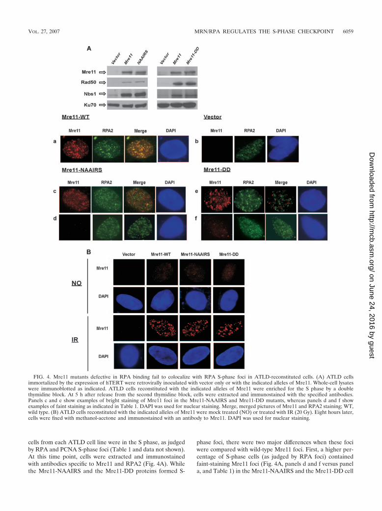
cells from each ATLD cell line were in the S phase, as judgedby RPA and PCNA S-phase foci (Table 1 and data not shown).At this time point, cells were extracted and immunostainedwith antibodies specific to Mre11 and RPA2 (Fig. 4A). Whilethe Mre11-NAAIRS and the Mre11-DD proteins formed S-
phase foci, there were two major differences when these fociwere compared with wild-type Mre11 foci. First, a higher per-centage of S-phase cells (as judged by RPA foci) containedfaint-staining Mre11 foci (Fig. 4A, panels d and f versus panela, and Table 1) in the Mre11-NAAIRS and the Mre11-DD cell
FIG. 4. Mre11 mutants defective in RPA binding fail to colocalize with RPA S-phase foci in ATLD-reconstituted cells. (A) ATLD cellsimmortalized by the expression of hTERT were retrovirally inoculated with vector only or with the indicated alleles of Mre11. Whole-cell lysateswere immunoblotted as indicated. ATLD cells reconstituted with the indicated alleles of Mre11 were enriched for the S phase by a doublethymidine block. At 5 h after release from the second thymidine block, cells were extracted and immunostained with the specified antibodies.Panels c and e show examples of bright staining of Mre11 foci in the Mre11-NAAIRS and Mre11-DD mutants, whereas panels d and f showexamples of faint staining as indicated in Table 1. DAPI was used for nuclear staining. Merge, merged pictures of Mre11 and RPA2 staining; WT,wild type. (B) ATLD cells reconstituted with the indicated alleles of Mre11 were mock treated (NO) or treated with IR (20 Gy). Eight hours later,cells were fixed with methanol-acetone and immunostained with an antibody to Mre11. DAPI was used for nuclear staining.
VOL. 27, 2007 MRN/RPA REGULATES THE S-PHASE CHECKPOINT 6059
on June 24, 2016 by guesthttp://m
cb.asm.org/
Dow
nloaded from
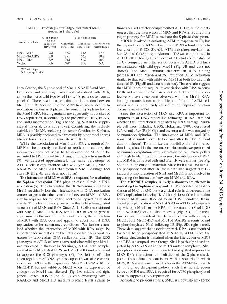
lines. Second, the S-phase foci of Mre11-NAAIRS and Mre11-DD, both faint and bright, were not colocalized with RPA,unlike the foci of wild-type Mre11 (Fig. 4A, panels c to f versuspanel a). These results suggest that the interaction betweenMre11 and RPA is required for MRN to correctly localize toreplication centers in S phase. The remaining S-phase foci ofthe Mre11 RPA-binding mutants are apparently not at sites ofDNA replication, as defined by the presence of RPA, PCNA,and BrdU incorporation (Fig. 4A; see Fig. S2B in the supple-mental material; data not shown). Considering the multipleactivities of MRN, including its repair function in S phase,MRN is possibly anchored to chromatin by other mechanismswhen it loses its ability to associate with RPA.
While the association of Mre11 with RPA is required forMRN to be properly localized to replication centers, theinteraction does not seem to be needed for MRN to berecruited to IR-induced foci. Using a nonextraction method(7), we detected approximately the same percentage ofATLD cells complemented with wild-type Mre11, Mre11-NAAIRS, or Mre11-DD that formed Mre11 damage fociafter IR (Fig. 4B and data not shown).
The interaction of MRN with RPA is required for mediatingthe S-phase checkpoint. RPA plays an essential role in DNAreplication (5). The observation that RPA-binding mutants ofMre11 specifically lose their interaction with DNA replicationcenters suggests that the association between MRN and RPAmay be required for replication control or replication-relatedevents. This idea is also supported by the cell-cycle-regulatedassociation of MRN and RPA. Since ATLD cells reconstitutedwith Mre11, Mre11-NAAIRS, Mre11-DD, or vector grew atapproximately the same rate (data not shown), the interactionof MRN with RPA does not appear to affect normal DNAreplication under nonstressed conditions. We further exam-ined whether the interaction of MRN with RPA might beimportant for mediation of the intra-S-phase checkpoint re-sponse by suppressing DNA replication after IR. The RDSphenotype of ATLD cells was corrected when wild-type Mre11was expressed in these cells. Strikingly, ATLD cells comple-mented with Mre11-NAAIRS and Mre11-DD mutants failedto suppress the RDS phenotype (Fig. 5A, left panel). Thedown-regulation of DNA synthesis upon IR was also compro-mised in U2OS cells expressing Myc-Mre11-NAAIRS orMre11-DD but not wild-type Mre11 when the expression ofendogenous Mre11 was silenced (Fig. 5A, middle and rightpanels). Since RDS in the ATLD cells expressing Mre11-NAAIRS and Mre11-DD mutants reached levels similar to
those seen with vector-complemented ATLD cells, these datasuggest that the interaction of MRN and RPA is required in amajor pathway for MRN to mediate the S-phase checkpoint.
MRN is involved in activating ATM in response to IR, butthe dependence of ATM activation on MRN is limited only tolow doses of IR (25, 35, 63). ATM autophosphorylation atSer1981 and Chk2 phosphorylation at T68 was compromised inATLD cells following IR at a dose of 2 Gy but not at a dose of10 Gy compared with the results seen with ATLD cell linesreconstituted with wild-type Mre11 (Fig. 5B and data notshown). The Mre11 mutants defective in RPA binding(Mre11-DD and Mre-NAAIRS) exhibited ATM activationsimilar to that seen with wild-type Mre11 at both low and highdoses of IR (Fig. 5B and data not shown). These results suggestthat MRN does not require its association with RPA to senseDSBs and activate the S-phase checkpoint. Therefore, the de-fective S-phase checkpoint observed with the Mre11 RPA-binding mutants is not attributable to a failure of ATM acti-vation and is more likely caused by an impaired functiondownstream of ATM.
Since the interaction of MRN and RPA is important forsuppression of DNA replication following IR, we examinedwhether this interaction is regulated by DNA damage. Multi-ple cell lines, including U2OS, HeLa, and T98G, were lysedbefore and after IR (10 Gy), and the interaction was assayed bycoimmunoprecipitation. The interaction of MRN and RPAremained at similar levels before and after IR (Fig. 5C anddata not shown). To minimize the possibility that the interac-tion is regulated in the presence of chromatin, we performedcoimmunoprecipitation after extraction of cell lysate pelletswith high levels of salt and detergent; the interaction of RPAand MRN in untreated cells and after IR were similar (see Fig.S3 in the supplemental material). Since both Nbs1 and Mre11are phosphorylated after IR, these data also suggest that IR-induced phosphorylation of Nbs1 and Mre11 is not involved inregulating the interaction between MRN and RPA.
The MRN/RPA complex is likely a downstream effector inmediating the S-phase checkpoint. ATM-mediated phosphor-ylation of Nbs1 at S343 plays a critical role in down-regulatingDNA replication following IR. Although loss of the interactionbetween MRN and RPA led to an RDS phenotype, IR-in-duced phosphorylation of Nbs1 at S343 in ATLD cells express-ing wild-type Mre11 or the RPA-binding mutants (Mre11-DDand -NAAIRS) was at similar levels (Fig. 5D, left panel).Furthermore, in similarity to the results seen with wild-typeMre11, both Mre11-DD and Mre11-NAAIRS mutants boundto phosphorylated Nbs1 following IR (Fig. 5D, right panel).These data suggest that association with RPA is not requiredfor Nbs1 to be phosphorylated at S343 by ATM. Since theS-phase checkpoint is impaired when the interaction of MRNand RPA is disrupted, even though Nbs1 is perfectly phosphor-ylated by ATM at S343 in the MRN mutant complexes, Nbs1phosphorylation must occur prior to the step that requires theMRN-RPA interaction for mediation of the S-phase check-point. These data are consistent with a scenario in whichMRN/RPA is a downstream effector in the ATM-Nbs1 branchof the S-phase checkpoint pathway such that the interactionbetween MRN and RPA is required for ATM-phosphorylatedNbs1 to suppress DNA replication.
According to previous studies, SMC1 is a downstream effector
TABLE 1. Percentages of wild-type and mutant Mre11proteins in S-phase foci
Protein or vehicle
% of S-phasecells (as
judged byRPA foci)
% of S-phase cells:
With brightMre11 foci
With faintMre11 foci
That were notreconstituted
Mre11-WTa 19.2 69.9 12.5 17.6Mre11-NAAIRS 17.8 26.5 62.7 10.8Mre11-DD 18.9 30.1 51.9 18.0Vector 19.6 NAb NA NA
a WT, wild type.b NA, not applicable.
6060 OLSON ET AL. MOL. CELL. BIOL.
on June 24, 2016 by guesthttp://m
cb.asm.org/
Dow
nloaded from
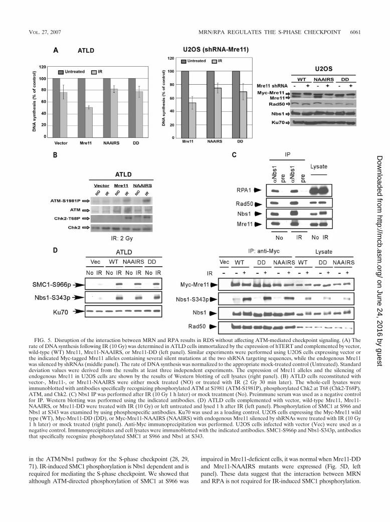
in the ATM/Nbs1 pathway for the S-phase checkpoint (28, 29,71). IR-induced SMC1 phosphorylation is Nbs1 dependent and isrequired for mediating the S-phase checkpoint. We showed thatalthough ATM-directed phosphorylation of SMC1 at S966 was
impaired in Mre11-deficient cells, it was normal when Mre11-DDand Mre11-NAAIRS mutants were expressed (Fig. 5D, leftpanel). These data suggest that the interaction between MRNand RPA is not required for IR-induced SMC1 phosphorylation.
FIG. 5. Disruption of the interaction between MRN and RPA results in RDS without affecting ATM-mediated checkpoint signaling. (A) Therate of DNA synthesis following IR (10 Gy) was determined in ATLD cells immortalized by the expression of hTERT and complemented by vector,wild-type (WT) Mre11, Mre11-NAAIRS, or Mre11-DD (left panel). Similar experiments were performed using U2OS cells expressing vector orthe indicated Myc-tagged Mre11 alleles containing several silent mutations at the two shRNA targeting sequences, while the endogenous Mre11was silenced by shRNAs (middle panel). The rate of DNA synthesis was normalized to the appropriate mock-treated control (Untreated). Standarddeviation values were derived from the results at least three independent experiments. The expression of Mre11 alleles and the silencing ofendogenous Mre11 in U2OS cells are shown by the results of Western blotting of cell lysates (right panel). (B) ATLD cells reconstituted withvector-, Mre11-, or Mre11-NAAIRS were either mock treated (NO) or treated with IR (2 Gy 30 min later). The whole-cell lysates wereimmunoblotted with antibodies specifically recognizing phosphorylated ATM at S1981 (ATM-S1981P), phosphorylated Chk2 at T68 (Chk2-T68P),ATM, and Chk2. (C) Nbs1 IP was performed after IR (10 Gy 1 h later) or mock treatment (No). Preimmune serum was used as a negative controlfor IP. Western blotting was performed using the indicated antibodies. (D) ATLD cells complemented with vector, wild-type Mre11, Mre11-NAAIRS, or Mre11-DD were treated with IR (10 Gy) or left untreated and lysed 1 h after IR (left panel). Phosphorylation of SMC1 at S966 andNbs1 at S343 was examined by using phosphospecific antibodies. Ku70 was used as a loading control. U2OS cells expressing the Myc-Mre11 wildtype (WT), Myc-Mre11-DD (DD), or Myc-Mre11-NAAIRS (NAAIRS) with endogenous Mre11 silenced by shRNAs were treated with IR (10 Gy1 h later) or mock treated (right panel). Anti-Myc immunoprecipitation was performed. U2OS cells infected with vector (Vec) were used as anegative control. Immunoprecipitates and cell lysates were immunoblotted with the indicated antibodies. SMC1-S966p and Nbs1-S343p, antibodiesthat specifically recognize phosphorylated SMC1 at S966 and Nbs1 at S343.
VOL. 27, 2007 MRN/RPA REGULATES THE S-PHASE CHECKPOINT 6061
on June 24, 2016 by guesthttp://m
cb.asm.org/
Dow
nloaded from
The MRN/RPA complex is situated downstream of ATM-depen-dent phosphorylation of SMC1, or it acts in a separate pathway tomediate the S-phase checkpoint.
MRN is tethered to replication-proximal sites via its inter-action with RPA and acts to suppress origin firing upon IR tomediate the S-phase checkpoint. Our studies have shown thatthe interaction of MRN with RPA is required for the inhibitionof DNA synthesis following IR. Previous studies have shownthat IR-induced down-regulation of replication is mainlycaused by blocking new origin firing whereas fork movement isminimally affected (31, 33, 42). However, the nature of themechanisms that mediate the inhibition of origin firing duringthe intra-S-phase checkpoint in mammalian cells is still notclear. We investigated whether the MRN complex and theinteraction of MRN with RPA would contribute to the pre-vention of origin firing after IR. We used a DNA fiber-labelingassay to quantitatively assess replication origin firing beforeand after IR. ATLD cells complemented with wild-type Mre11,Mre11-NAAIRS, Mre11-DD, or vector were pulse labeledwith CldU, a modified nucleotide precursor, and subsequentlytreated with IR or left untreated followed by pulse labelingwith IdU, another nucleotide precursor (Fig. 6A). After im-munostaining of chromatin fibers with antibodies recognizingCldU and IdU, DNA that is being actively synthesized duringthe labeling time is visualized as labeled tracks, which could beinterpreted as (i) newly fired origins, (ii) ongoing forks, or (iii)terminating forks. We counted origins fired during IdU label-ing in comparison to the total number of forks. As shown inFig. 6A, the percentage of newly fired origins after IR in thewild-type Mre11-reconstituted ATLD cell line was greatly re-duced, suggesting that new origin firing was inhibited followingIR. However, the IR-mediated inhibition of new origin firingwas severely compromised in the vector and the Mre11-NAAIRSi-expressing and Mre11-DD-expressing ATLD celllines. These data suggest that Mre11, as well as the interactionof Mre11 with RPA, is required for preventing new originfiring following IR.
A connection of MRN with replication is elucidated by itsassociation with sites near replication origins in the S phase(38). To further explore the mechanism of how MRN preventsnew origin firing by way of its interaction with RPA, we deter-mined whether Mre11 binding to RPA is essential for MRN toassociate with known replication origins. One possible scenariois that MRN is recruited to origins by RPA and subsequentlyacts to suppress new origin firing when the intra-S-phasecheckpoint is activated. The Myc-tagged Mre11 wild type andthe Mre11 mutants (NAAIRRS and DD), as well as Myc-tagged RPA2, were stably expressed at levels similar to thoseseen with endogenous proteins in T98G cells after retroviralinfection (see Fig. S4 in the supplemental material). ChIP wasperformed 8 h (G1 phase) or 20 h (S phase) after release ofT98G cells from serum starvation. Primers specific to sites ofDNA replication initiation, such as those in the B2-lamin gene(2, 4), the HSP70 gene (2, 56), and the c-myc gene (38, 58),were used to detect bound DNA. Wild-type Myc-Mre11 boundto the origin-proximal sites specifically in S-phase cells in amanner similar to that seen with Myc-RPA2 (Fig. 6B and datanot shown). However, binding of the Mre11 mutants(NAAIRS and DD) defective in RPA binding at origin-prox-imal sites was dramatically reduced (Fig. 6C). These results
suggest that Mre11 largely depends on RPA to associate withsites near DNA replication origins. Since MRN acts to inhibitDNA replication only after IR, we examined whether Mre11origin binding was altered after IR. ChIP analysis showed thatthe results with respect to association of Mre11 with origin-proximal sites at HSP70, c-myc, and B2-lamin were similarbefore and after IR (Fig. 6D), as is consistent with the obser-vation that the interaction of MRN and RPA was not alteredby IR (Fig. 5C). These data suggest that IR-induced damagesignaling does not modulate RPA-mediated recruitment ofMRN to replication-proximal sites. The inhibition of replica-tion initiation likely occurs after ATM-phosphorylated MRN isrecruited to the origin-proximal sites by RPA (see the model inFig. 7B and the Discussion).
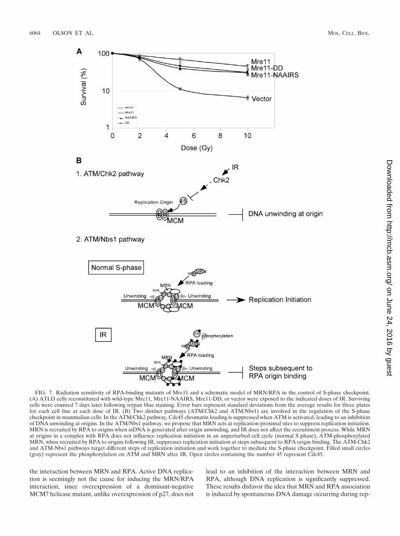
The RPA-binding mutants of Mre11 exhibit minor radiationsensitivity. Both MRN and RPA are required for DNA repair(40, 66); thus, we investigated whether their interaction mightalso be needed for damage repair in addition to its role ininhibiting DNA replication after IR. We examined the abilityof the reconstituted ATLD cells to survive following IR (Fig.7A). The cells expressing Mre11-NAAIRS or Mre11-DD wereslightly more sensitive to IR than Mre11-expressing cells butmuch less sensitive than vector-complemented cells. This dem-onstrates that cell survival following IR is dependent on Mre11but only minimally dependent on the interaction betweenMre11 and RPA. Our results suggest that Mre11, but not theinteraction of MRN with RPA, plays a critical role in the DNArepair processes that allow a cell to survive after IR treatment.This result is also consistent with the observation that MRNforms damage foci in a manner independent of its interactionwith RPA (Fig. 4B). The slight increase of radiation sensitivityin the RPA-binding mutants of Mre11 is likely caused by theloss of the intra-S-phase checkpoint.
DISCUSSION
MRN plays a critical role in the S-phase checkpoint, but thenature of the underlying mechanism is not clear. Our studydemonstrated that MRN is recruited to replication-proximalsites via a direct interaction with RPA and acts there to inhibitDNA replication initiation in response to IR. Since loss of theinteraction between Mre11 and RPA leads to a profound RDSphenotype at levels similar to those caused by Mre11 or Nbs1deficiency, our studies suggest that the recruitment of MRN byRPA to replication-proximal sites is a major mechanism in theATM-Nbs1 pathway to regulate the S-phase checkpoint.
The interaction of MRN and RPA is cell cycle regulated. Aprevious study showed that MRN forms S-phase-specific focicolocalized with sites of BrdU incorporation (45). Our studiesfurther demonstrated that MRN also colocalizes with RPA inthe S phase, and importantly, that the interaction of RPA withMre11 is critical for the recruitment of MRN to the replicationcenters, revealing an important mechanism for MRN for lo-calization to S-phase-specific foci.
The interaction of MRN with RPA appears to be regulatedby CDKs. p27 overexpression or roscovitine treatment leads toa decrease in the association between MRN and RPA. Phos-phatase treatment abolishes cell-cycle-dependent MRN-RPAassociation, suggesting that CDK activities are not only re-quired for stimulating but are also important for maintaining
6062 OLSON ET AL. MOL. CELL. BIOL.
on June 24, 2016 by guesthttp://m
cb.asm.org/
Dow
nloaded from
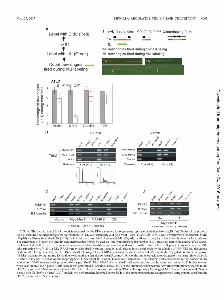
FIG. 6. The recruitment of Mre11 to origin-proximal sites by RPA is required for suppressing replication initiation following IR. (A) Outline of the protocolused to examine new origin firing after IR treatment. ATLD cells expressing wild-type Mre11, Mre11-NAAIRS, Mre11-DD, or vector were labeled with CIdU(10 �M) for 20 min, treated with IR (10 Gy) or left untreated, and labeled again with IdU (15 �M) for 40 min. Examples of labeled replication tracks are shown.The percentage of fired origins after IR treatment was determined for each cell line by normalizing the number of IdU tracks (green) to the number of all labeledtracks counted (100 in each experiment). The average and standard deviation values were derived from the results of three independent experiments. (B) T98Gcells expressing Myc-Mre11 or Myc-RPA2 were synchronized by serum starvation and released into the cell cycle by the addition of 10% FBS into the culturemedium. At 8 h (G1 enriched) and 20 h (S enriched) following release, ChIP analysis was performed using anti-Myc antibody conjugated on protein A agarose(IP:Myc) and a rabbit anti-mouse IgG antibody was used as a negative control (IP:control). PCR of the immunoprecipitates was performed using primers specificto HSP70 and c-myc to detect coimmunoprecipitated DNA. Input, 0.1% of the total isolated chromatin. The cell cycle profile was monitored by flow cytometryanalysis. (C) T98G cells expressing vector, Myc-tagged Mre11, Mre11-NAAIRS, or Mre11-DD were synchronized by serum starvation. At 20 h after release,when cells entered the S phase, ChIP analysis was performed as described above. PCR of the immunoprecipitates was performed with primers specific to theHSP70, c-myc, and B2-lamin origins. (D) At 20 h after release from serum starvation, T98G cells expressing Myc-tagged Mre11 were mock treated (No) ortreated with IR (10 Gy 1 h later). ChIP analysis was performed as described above. PCR of the immunoprecipitates was performed using primers specific to theHSP70, c-myc, and B2-lamin origins.
VOL. 27, 2007 MRN/RPA REGULATES THE S-PHASE CHECKPOINT 6063
on June 24, 2016 by guesthttp://m
cb.asm.org/
Dow
nloaded from
the interaction between MRN and RPA. Active DNA replica-tion is seemingly not the cause for inducing the MRN/RPAinteraction, since overexpression of a dominant-negativeMCM7 helicase mutant, unlike overexpression of p27, does not
lead to an inhibition of the interaction between MRN andRPA, although DNA replication is significantly suppressed.These results disfavor the idea that MRN and RPA associationis induced by spontaneous DNA damage occurring during rep-
FIG. 7. Radiation sensitivity of RPA-binding mutants of Mre11 and a schematic model of MRN/RPA in the control of S-phase checkpoint.(A) ATLD cells reconstituted with wild-type Mre11, Mre11-NAAIRS, Mre11-DD, or vector were exposed to the indicated doses of IR. Survivingcells were counted 7 days later following trypan blue staining. Error bars represent standard deviations from the average results for three platesfor each cell line at each dose of IR. (B) Two distinct pathways (ATM/Chk2 and ATM/Nbs1) are involved in the regulation of the S-phasecheckpoint in mammalian cells. In the ATM/Chk2 pathway, Cdc45 chromatin loading is suppressed when ATM is activated, leading to an inhibitionof DNA unwinding at origins. In the ATM/Nbs1 pathway, we propose that MRN acts at replication-proximal sites to suppress replication initiation.MRN is recruited by RPA to origins when ssDNA is generated after origin unwinding, and IR does not affect the recruitment process. While MRNat origins in a complex with RPA does not influence replication initiation in an unperturbed cell cycle (normal S phase), ATM-phosphorylatedMRN, when recruited by RPA to origins following IR, suppresses replication initiation at steps subsequent to RPA origin binding. The ATM-Chk2and ATM-Nbs1 pathways target different steps of replication initiation and work together to mediate the S-phase checkpoint. Filled small circles(gray) represent the phosphorylation on ATM and MRN after IR. Open circles containing the number 45 represent Cdc45.
6064 OLSON ET AL. MOL. CELL. BIOL.
on June 24, 2016 by guesthttp://m
cb.asm.org/
Dow
nloaded from
lication in the S phase. Increased association of MRN andRPA at the G1-to-S transition and in the S phase is most likelydue to a rise in CDK activities. Although MRN and RPAassociation is regulated during the cell cycle in a phosphoryla-tion-dependent manner, the association remained at similarlevels before and after IR. These data suggest that DSBs gen-erated after IR- and ATM-mediated phosphorylation events,including Nbs1 phosphorylation, do not play a major role inregulating the interaction between MRN and RPA.
The interaction of RPA and MRN is direct and mediated bya specific site in Mre11. Since a direct interaction of MRN andRPA can be achieved by using purified Mre11 and RPA ex-pressed in insect cells and bacteria, respectively, it seems thatCDK-mediated phosphorylation events stimulate the interac-tion but are not absolutely required for initiation of the inter-action. Interestingly, two DD residues at the RPA-binding siteare critical for Mre11 to bind RPA, which is analogous to therequirement of DD/EE residues in conserved motifs in Nbs1,ATRIP, and Ku80 to mediate the interaction of Nbs1/ATM,ATRIP/ATR, and Ku80/DNA-PK (see Fig. S5 in the supple-mental material) (14). This raises the possibility that a generalmechanism may be involved in mediating protein-protein in-teractions for DNA damage responses. The nature of the exactmechanism by which CDKs regulate the interaction betweenMRN and RPA is not clear. The increased association of MRNand RPA could be caused by direct phosphorylation of MRN,RPA, or both by CDKs. Previous studies have shown that RPAis phosphorylated by CDKs at multiple sites during the cellcycle (12, 17, 20). In vivo mass spectrometry analysis alsoidentified phosphorylation of Nbs1 at CDK conserved sites inunperturbed cells (reference 69 and data not shown). CDKactivity is important for activation of homologous recombina-tion in the S phase when DSBs are generated (3, 13, 26). Ourfindings indicating that CDK activity is also required for stim-ulation of the interaction between MRN and RPA to mediateS-phase checkpoint control suggest an interesting mechanismto couple S-phase checkpoint control with DNA repair.
The interaction of MRN and RPA is essential for mediatingthe S-phase checkpoint. The ATM/Chk2 and ATM/Nbs1 path-ways are two parallel pathways required to mediate the intra-S-phase checkpoint (16). After ATM activation, the ATM/Chk2 pathway inhibits Cdc45 chromatin loading (15), thuspreventing MCM-mediated DNA unwinding at origins (Fig.7B). What is the mechanism by which the ATM/Nbs1 complexinhibits replication after DNA damage? Our studies suggestthat when ATM-phosphorylated MRN is recruited to origin-proximal sites by RPA, it acts there to prevent replicationinitiation.
At the onset of replication initiation, DNA at origins isunwound by MCM proteins, forming single-stranded DNA(ssDNA) that is subsequently bound by RPA (57) and probablyat the same time MRN is recruited by RPA to replicationorigins. This idea is supported by the ChIP analysis resultsshowing that MRN binds to replication origin-proximal sites ina manner similar to that seen with RPA, while Mre11 mutantsdefective in RPA binding fail to do so.
The ATLD cell line expressing Mre11 mutants defective inRPA binding do not show obvious growth defects, suggestingthat the presence of MRN at replication origin-proximal sitesin a complex with RPA does not affect DNA replication per se
in a normal cell cycle. However, loss of this interaction causesa severe defect in down-regulation of DNA replication follow-ing IR. Since MRN/RPA complexes associate with origin-prox-imal sites similarly before and after IR, damage-induced inhi-bition of replication initiation likely occurs after RPA originloading. One possible scenario is that ATM phosphorylatesMRN after IR and that it is only when phosphorylated MRNis recruited by RPA to origins that replication initiation isprevented (Fig. 7B).
The phosphorylation of Nbs1 at S343 is critical for the S-phase checkpoint (36, 75). Our studies showed that in theMre11 mutant cell lines defective in RPA binding, IR-inducedNbs1 phosphorylation at S343 was not affected. Mre11-DDand Mre11-NAAIRS form complexes with phosphorylatedNbs1 (S343) in a manner similar to that seen with wild-typeMre11, but those mutant cell lines exhibit a profound RDSphenotype. These data support the idea that MRN/RPA com-plexes are a downstream effector for mediation of the S-phasecheckpoint after Nbs1 S343 is phosphorylated by ATM; this isconsistent with the observation that the association of MRNwith RPA is not required for ATM activation. In the ATM/Nbs1 branch of the S-phase checkpoint pathway, SMC1 isdownstream of Nbs1 (28, 71). SMC1 phosphorylation is Nbs1dependent and is important for mediating the S-phase check-point. Although IR-induced SMC1 phosphorylation dependson MRN, disruption of the interaction of MRN and RPA doesnot influence this ATM-mediated SMC1 phosphorylation. Thisagain suggests that the MRN/RPA complex is a downstreameffector situated downstream of ATM-directed SMC1 phos-phorylation events for mediation of replication inhibition fol-lowing IR.
It is still unclear what step in DNA replication followingRPA loading is disrupted by ATM-mediated phosphorylationof MRN at origin-proximal sites. Given the critical role of RPAin replication initiation, MRN may directly inhibit RPA repli-cation activity to inhibit replication initiation in response to IR.For instance, RPA is required for recruiting DNA polymerase�/primase and is also needed for replication factor C to loadPCNA onto primed origins to initiate DNA replication (74). Aconformational change of MRN/RPA may be induced byATM-directed phosphorylation of MRN following IR, whichin turn would lead to an inhibition of RPA function in repli-cation initiation. Alternatively, phosphorylation of MRN mayinterfere with the association of MRN/RPA with other repli-cation factors at origins, causing the suppression of replicationinitiation. Nevertheless, our results suggest a direct communi-cation of MRN with the cellular replication machinery at rep-lication-proximal sites to mediate the intra-S-phase check-point. While the ATM/Chk2 pathway inhibits Cdc45 originbinding before DNA unwinding, the ATM/Nbs1 pathway likelyprevents replication initiation after phosphorylated MRN isrecruited by RPA to replication origins when RPA binds tossDNA generated through MCM-mediated DNA unwinding(Fig. 7B). Since inactivation of either of the two pathways(ATM/Chk2 and ATM/Nbs1) leads to a partial RDS pheno-type, both of these mechanisms are required for sufficientinactivation of replication initiation following IR.
The MRN/RPA complex and DSB repair. DSB repair via thehomologous recombination pathway requires both MRN andRPA complexes (10, 66), which raises a question as to whether
VOL. 27, 2007 MRN/RPA REGULATES THE S-PHASE CHECKPOINT 6065
on June 24, 2016 by guesthttp://m
cb.asm.org/
Dow
nloaded from
the interaction of MRN/RPA is also involved in mediating therepair process. The radiation survival assay demonstrated thatMre11 deficiency causes severe radiation sensitivity whereasthe RPA-binding mutants of Mre11 exhibited only a minordefect. This suggests that the MRN/RPA complex is not in-volved in the major pathways mediated by MRN for repair ofDSBs after IR, although a minor role cannot be excluded. Inthis aspect, DSB recognition by MRN seemingly does notrequire its association with RPA, since ATM activation thatrequires MRN at DSBs is not dependent on the interaction ofMRN with RPA. The Mre11 mutants defective in RPA bindingalso migrate to DSBs to form damage foci similar to those seenwith wild-type Mre11. Partial radiation sensitivity of Mre11mutants defective in RPA binding is likely caused by a severedefect in the S-phase checkpoint, leading to inefficient repairof DSBs generated by IR in the S phase.
MRN forms multiple protein complexes to mediate variouscellular functions. Forming MRN/RPA complexes is essentialfor the S-phase checkpoint. Upon ATM activation, MRN isphosphorylated, conceivably in different protein complexes.Phosphorylated MRN at DSB sites may stimulate the repairprocess, while phosphorylated MRN/RPA that is recruited toreplication-proximal sites acts to prevent origin firing. ThroughS-phase checkpoint activation, ATM-mediated phosphoryla-tion of MRN may provide an important mechanism to linkDNA repair with replication and thus ensure the delay of DNAreplication until the completion of DNA repair.
ACKNOWLEDGMENTS
We thank Marc Wold and William Hahn for generously providingreagents used in this study. We also thank Michael Hock for assistancewith the chromatin immunoprecipitation assay. We thank StevenReed, Clare McGowan, and Paul Russell for critical reading of themanuscript.
This work was supported by NIH grant CA102361, an Ellison Med-ical Foundation New Scholar award (AG-NS-0251-04) to X.W., andNIH training grant 5 T32 DK007022 to E.O.
REFERENCES
1. Assenmacher, N., and K. P. Hopfner. 2004. MRE11/RAD50/NBS1: complexactivities. Chromosoma 113:157–166.
2. Avni, D., H. Yang, F. Martelli, F. Hofmann, W. M. ElShamy, S. Ganesan, R.Scully, and D. M. Livingston. 2003. Active localization of the retinoblastomaprotein in chromatin and its response to S phase DNA damage. Mol. Cell12:735–746.
3. Aylon, Y., B. Liefshitz, and M. Kupiec. 2004. The CDK regulates repair ofdouble-strand breaks by homologous recombination during the cell cycle.EMBO J. 23:4868–4875.
4. Biamonti, G., G. Perini, F. Weighardt, S. Riva, M. Giacca, P. Norio, L.Zentilin, S. Diviacco, D. Dimitrova, and A. Falaschi. 1992. A human DNAreplication origin: localization and transcriptional characterization. Chromo-soma 102:S24–S31.
5. Binz, S. K., A. M. Sheehan, and M. S. Wold. 2004. Replication protein Aphosphorylation and the cellular response to DNA damage. DNA Repair(Amsterdam) 3:1015–1024.
6. Buscemi, G., C. Savio, L. Zannini, F. Micciche, D. Masnada, M. Nakanishi,H. Tauchi, K. Komatsu, S. Mizutani, K. Khanna, P. Chen, P. Concannon, L.Chessa, and D. Delia. 2001. Chk2 activation dependence on Nbs1 after DNAdamage. Mol. Cell. Biol. 21:5214–5222.
7. Carney, J. P., R. S. Maser, H. Olivares, E. M. Davis, M. Le Beau, J. R. YatesIII, L. Hays, W. F. Morgan, and J. H. Petrini. 1998. The hMre11/hRad50protein complex and Nijmegen breakage syndrome: linkage of double-strandbreak repair to the cellular DNA damage response. Cell 93:477–486.
8. Carson, C. T., R. A. Schwartz, T. H. Stracker, C. E. Lilley, D. V. Lee, andM. D. Weitzman. 2003. The Mre11 complex is required for ATM activationand the G2/M checkpoint. EMBO J. 22:6610–6620.
9. Chen, J., D. P. Silver, D. Walpita, S. B. Cantor, A. F. Gazdar, G. Tomlinson,F. J. Couch, B. L. Weber, T. Ashley, D. M. Livingston, and R. Scully. 1998.Stable interaction between the products of the BRCA1 and BRCA2 tumorsuppressor genes in mitotic and meiotic cells. Mol. Cell 2:317–328.
10. D’Amours, D., and S. P. Jackson. 2002. The Mre11 complex: at the cross-roads of DNA repair and checkpoint signalling. Nat. Rev. Mol. Cell Biol.3:317–327.
11. Dimitrova, D. S., and D. M. Gilbert. 2000. Stability and nuclear distributionof mammalian replication protein A heterotrimeric complex. Exp. Cell Res.254:321–327.
12. Dutta, A., and B. Stillman. 1992. cdc2 family kinases phosphorylate a humancell DNA replication factor, RPA, and activate DNA replication. EMBO J.11:2189–2199.
13. Esashi, F., N. Christ, J. Gannon, Y. Liu, T. Hunt, M. Jasin, and S. C. West.2005. CDK-dependent phosphorylation of BRCA2 as a regulatory mecha-nism for recombinational repair. Nature 434:598–604.
14. Falck, J., J. Coates, and S. P. Jackson. 2005. Conserved modes of recruit-ment of ATM, ATR and DNA-PKcs to sites of DNA damage. Nature434:605–611.
15. Falck, J., N. Mailand, R. G. Syljuasen, J. Bartek, and J. Lukas. 2001. TheATM-Chk2-Cdc25A checkpoint pathway guards against radioresistant DNAsynthesis. Nature 410:842–847.
16. Falck, J., J. H. Petrini, B. R. Williams, J. Lukas, and J. Bartek. 2002. TheDNA damage-dependent intra-S phase checkpoint is regulated by parallelpathways. Nat. Genet. 30:290–294.
17. Fang, F., and J. W. Newport. 1993. Distinct roles of cdk2 and cdc2 in RP-Aphosphorylation during the cell cycle. J. Cell Sci. 106:983–994.
18. Farah, J. A., E. Hartsuiker, K. Mizuno, K. Ohta, and G. R. Smith. 2002.A 160-bp palindrome is a Rad50.Rad32-dependent mitotic recombinationhotspot in Schizosaccharomyces pombe. Genetics 161:461–468.
19. Flores, O., H. Lu, and D. Reinberg. 1992. Factors involved in specific tran-scription by mammalian RNA polymerase II. Identification and character-ization of factor IIH. J. Biol. Chem. 267:2786–2793.
20. Fotedar, R., and J. M. Roberts. 1992. Cell cycle regulated phosphorylation ofRPA-32 occurs within the replication initiation complex. EMBO J. 11:2177–2187.
21. Fujita, N., and P. A. Wade. 2004. Use of bifunctional cross-linking reagentsin mapping genomic distribution of chromatin remodeling complexes. Meth-ods 33:81–85.
22. Gatei, M., D. Young, K. M. Cerosaletti, A. Desai-Mehta, K. Spring, S.Kozlov, M. F. Lavin, R. A. Gatti, P. Concannon, and K. Khanna. 2000.ATM-dependent phosphorylation of nibrin in response to radiation expo-sure. Nat. Genet. 25:115–119.
23. Henricksen, L. A., C. B. Umbricht, and M. S. Wold. 1994. Recombinantreplication protein A: expression, complex formation, and functional char-acterization. J. Biol. Chem. 269:11121–11132.
24. Hopfner, K. P., A. Karcher, L. Craig, T. T. Woo, J. P. Carney, and J. A.Tainer. 2001. Structural biochemistry and interaction architecture of theDNA double-strand break repair Mre11 nuclease and Rad50-ATPase. Cell105:473–485.
25. Horejsi, Z., J. Falck, C. J. Bakkenist, M. B. Kastan, J. Lukas, and J. Bartek.2004. Distinct functional domains of Nbs1 modulate the timing and magni-tude of ATM activation after low doses of ionizing radiation. Oncogene23:3122–3127.
26. Ira, G., A. Pellicioli, A. Balijja, X. Wang, S. Fiorani, W. Carotenuto, G.Liberi, D. Bressan, L. Wan, N. M. Hollingsworth, J. E. Haber, and M.Foiani. 2004. DNA end resection, homologous recombination and DNAdamage checkpoint activation require CDK1. Nature 431:1011–1017.
27. Khanna, K. K., and S. P. Jackson. 2001. DNA double-strand breaks: signal-ing, repair and the cancer connection. Nat. Genet. 27:247–254.
28. Kim, S. T., B. Xu, and M. B. Kastan. 2002. Involvement of the cohesinprotein, Smc1, in Atm-dependent and independent responses to DNA dam-age. Genes Dev. 16:560–570.
29. Kitagawa, R., C. J. Bakkenist, P. J. McKinnon, and M. B. Kastan. 2004.Phosphorylation of SMC1 is a critical downstream event in the ATM-NBS1-BRCA1 pathway. Genes Dev. 18:1423–1438.
30. Lai, J. S., and W. Herr. 1992. Ethidium bromide provides a simple tool foridentifying genuine DNA-independent protein associations. Proc. Natl.Acad. Sci. USA 89:6958–6962.
31. Larner, J. M., H. Lee, R. D. Little, P. A. Dijkwel, C. L. Schildkraut, and J. L.Hamlin. 1999. Radiation down-regulates replication origin activity through-out the S phase in mammalian cells. Nucleic Acids Res. 27:803–809.
32. Lavin, M. F. 2004. The Mre11 complex and ATM: a two-way functionalinteraction in recognising and signaling DNA double strand breaks. DNARepair (Amsterdam) 3:1515–1520.
33. Lee, H., J. M. Larner, and J. L. Hamlin. 1997. A p53-independent damage-sensing mechanism that functions as a checkpoint at the G1/S transition inChinese hamster ovary cells. Proc. Natl. Acad. Sci. USA 94:526–531.
34. Lee, J. H., R. Ghirlando, V. Bhaskara, M. R. Hoffmeyer, J. Gu, and T. T.Paull. 2003. Regulation of Mre11/Rad50 by Nbs1: effects on nucleotide-dependent DNA binding and association with ataxia-telangiectasia-like dis-order mutant complexes. J. Biol. Chem. 278:45171–45181.
35. Lee, J. H., and T. T. Paull. 2004. Direct activation of the ATM protein kinaseby the Mre11/Rad50/Nbs1 complex. Science 304:93–96.
36. Lim, D. S., S. T. Kim, B. Xu, R. S. Maser, J. Lin, J. H. Petrini, and M. B.
6066 OLSON ET AL. MOL. CELL. BIOL.
on June 24, 2016 by guesthttp://m
cb.asm.org/
Dow
nloaded from
Kastan. 2000. ATM phosphorylates p95/nbs1 in an S-phase checkpoint path-way. Nature 404:613–617.
37. Lobachev, K. S., D. A. Gordenin, and M. A. Resnick. 2002. The Mre11complex is required for repair of hairpin-capped double-strand breaks andprevention of chromosome rearrangements. Cell 108:183–193.
38. Maser, R. S., O. K. Mirzoeva, J. Wells, H. Olivares, B. R. Williams, R. A.Zinkel, P. J. Farnham, and J. H. Petrini. 2001. Mre11 complex and DNAreplication: linkage to E2F and sites of DNA synthesis. Mol. Cell. Biol.21:6006–6016.
39. Masutomi, K., E. Y. Yu, S. Khurts, I. Ben Porath, J. L. Currier, G. B. Metz,M. W. Brooks, S. Kaneko, S. Murakami, J. A. DeCaprio, R. A. Weinberg,S. A. Stewart, and W. C. Hahn. 2003. Telomerase maintains telomere struc-ture in normal human cells. Cell 114:241–253.
40. Matsuura, S., J. Kobayashi, H. Tauchi, and K. Komatsu. 2004. Nijmegenbreakage syndrome and DNA double strand break repair by NBS1 complex.Adv. Biophys. 38:65–80.
41. Matsuura, S., H. Tauchi, A. Nakamura, N. Kondo, S. Sakamoto, S. Endo, D.Smeets, B. Solder, B. H. Belohradsky, K. Der, V., M. Oshimura, M. Isomura,Y. Nakamura, and K. Komatsu. 1998. Positional cloning of the gene forNijmegen breakage syndrome. Nat. Genet. 19:179–181.
42. Merrick, C. J., D. Jackson, and J. F. Diffley. 2004. Visualization of alteredreplication dynamics after DNA damage in human cells. J. Biol. Chem.279:20067–20075.
43. Meyerson, M., C. M. Counter, E. N. Eaton, L. W. Ellisen, P. Steiner, S. D.Caddle, L. Ziaugra, R. L. Beijersbergen, M. J. Davidoff, Q. Liu, S. Bacchetti,D. A. Haber, and R. A. Weinberg. 1997. hEST2, the putative human telome-rase catalytic subunit gene, is up-regulated in tumor cells and during immor-talization. Cell 90:785–795.
44. Mirzoeva, O. K., and J. H. Petrini. 2001. DNA damage-dependent nucleardynamics of the Mre11 complex. Mol. Cell. Biol. 21:281–288.
45. Mirzoeva, O. K., and J. H. Petrini. 2003. DNA replication-dependent nu-clear dynamics of the Mre11 complex. Mol. Cancer Res. 1:207–218.
46. Morgan, S. E., C. Lovly, T. K. Pandita, Y. Shiloh, and M. B. Kastan. 1997.Fragments of ATM which have dominant-negative or complementing activ-ity. Mol. Cell. Biol. 17:2020–2029.
47. Nelms, B. E., R. S. Maser, J. F. MacKay, M. G. Lagally, and J. H. Petrini.1998. In situ visualization of DNA double-strand break repair in humanfibroblasts. Science 280:590–592.
48. Orlando, V., H. Strutt, and R. Paro. 1997. Analysis of chromatin structure byin vivo formaldehyde cross-linking. Methods 11:205–214.
49. Paull, T. T., and M. Gellert. 1998. The 3� to 5� exonuclease activity of Mre11 facilitates repair of DNA double-strand breaks. Mol. Cell 1:969–979.
50. Paull, T. T., and M. Gellert. 1999. Nbs1 potentiates ATP-driven DNAunwinding and endonuclease cleavage by the Mre11/Rad50 complex. GenesDev. 13:1276–1288.
51. Ranganathan, V., W. F. Heine, D. N. Ciccone, K. L. Rudolph, X. Wu, S.Chang, H. Hai, I. M. Ahearn, D. M. Livingston, I. Resnick, F. Rosen, E.Seemanova, P. Jarolim, R. A. DePinho, and D. T. Weaver. 2001. Rescue ofa telomere length defect of Nijmegen breakage syndrome cells requires NBSand telomerase catalytic subunit. Curr. Biol. 11:962–966.
52. Robison, J. G., J. Elliott, K. Dixon, and G. G. Oakley. 2004. Replicationprotein A and the Mre11.Rad50.Nbs1 complex co-localize and interact atsites of stalled replication forks. J. Biol. Chem. 279:34802–34810.
53. Shiloh, Y. 1997. Ataxia-telangiectasia and the Nijmegen breakage syndrome:related disorders but genes apart. Annu. Rev. Genet. 31:635–662.
54. Stewart, G. S., R. S. Maser, T. Stankovic, D. A. Bressan, M. I. Kaplan, N. G.Jaspers, A. Raams, P. J. Byrd, J. H. Petrini, and A. M. Taylor. 1999. TheDNA double-strand break repair gene hMRE11 is mutated in individualswith an ataxia-telangiectasia-like disorder. Cell 99:577–587.
55. Stracker, T. H., J. W. Theunissen, M. Morales, and J. H. Petrini. 2004. TheMre11 complex and the metabolism of chromosome breaks: the importanceof communicating and holding things together. DNA Repair (Amsterdam)3:845–854.
56. Taira, T., S. M. Iguchi-Ariga, and H. Ariga. 1994. A novel DNA replicationorigin identified in the human heat shock protein 70 gene promoter. Mol.Cell. Biol. 14:6386–6397.
57. Takeda, D. Y., and A. Dutta. 2005. DNA replication and progression throughS phase. Oncogene 24:2827–2843.
58. Tao, L., Z. Dong, M. Leffak, M. Zannis-Hadjopoulos, and G. Price. 2000.Major DNA replication initiation sites in the c-myc locus in human cells.J. Cell. Biochem. 78:442–457.
59. Tauchi, H., J. Kobayashi, K. Morishima, D. C. van Gent, T. Shiraishi, N. S.Verkaik, D. vanHeems, E. Ito, A. Nakamura, E. Sonoda, M. Takata, S.Takeda, S. Matsuura, and K. Komatsu. 2002. Nbs1 is essential for DNArepair by homologous recombination in higher vertebrate cells. Nature 420:93–98.
60. Taylor, A. M., A. Groom, and P. J. Byrd. 2004. Ataxia-telangiectasia-likedisorder (ATLD)—its clinical presentation and molecular basis. DNA Re-pair (Amsterdam) 3:1219–1225.
61. Trujillo, K. M., S. S. Yuan, E. Y. Lee, and P. Sung. 1998. Nuclease activitiesin a complex of human recombination and DNA repair factors Rad50,Mre11, and p95. J. Biol. Chem. 273:21447–21450.
62. Usui, T., T. Ohta, H. Oshiumi, J. Tomizawa, H. Ogawa, and T. Ogawa. 1998.Complex formation and functional versatility of Mre11 of budding yeast inrecombination. Cell 95:705–716.
63. Uziel, T., Y. Lerenthal, L. Moyal, Y. Andegeko, L. Mittelman, and Y. Shiloh.2003. Requirement of the MRN complex for ATM activation by DNAdamage. EMBO J. 22:5612–5621.
64. Varon, R., C. Vissinga, M. Platzer, K. M. Cerosaletti, K. H. Chrzanowska, K.Saar, G. Beckmann, E. Seemanova, P. R. Cooper, N. J. Nowak, M. Stumm,C. M. Weemaes, R. A. Gatti, R. K. Wilson, M. Digweed, A. Rosenthal, K.Sperling, P. Concannon, and A. Reis. 1998. Nibrin, a novel DNA double-strand break repair protein, is mutated in Nijmegen breakage syndrome. Cell93:467–476.
65. Wilson, I. A., D. H. Haft, E. D. Getzoff, J. A. Tainer, R. A. Lerner, and S.Brenner. 1985. Identical short peptide sequences in unrelated proteins canhave different conformations: a testing ground for theories of immune rec-ognition. Proc. Natl. Acad. Sci. USA 82:5255–5259.
66. Wold, M. S. 1997. Replication protein A: a heterotrimeric, single-strandedDNA-binding protein required for eukaryotic DNA metabolism. Annu. Rev.Biochem. 66:61–92.
67. Wu, X., D. Avni, T. Chiba, F. Yan, Q. Zhao, Y. Lin, H. Heng, and D.Livingston. 2004. SV40 T antigen interacts with Nbs1 to disrupt DNA rep-lication control. Genes Dev. 18:1305–1316.
68. Wu, X., J. H. Petrini, W. F. Heine, D. T. Weaver, D. M. Livingston, andJ. Chen. 2000. Independence of R/M/N focus formation and the presence ofintact BRCA1. Science 289:11.
69. Wu, X., V. Ranganathan, D. S. Weisman, W. F. Heine, D. N. Ciccone, T. B.O’Neill, K. E. Crick, K. A. Pierce, W. S. Lane, G. Rathbun, D. M. Livingston,and D. T. Weaver. 2000. ATM phosphorylation of Nijmegen breakage syn-drome protein is required in a DNA damage response. Nature 405:477–482.
70. Yang, Y. G., A. Saidi, P. O. Frappart, W. Min, C. Barrucand, V. Dumon-Jones, J. Michelon, Z. Herceg, and Z. Q. Wang. 2006. Conditional deletionof Nbs1 in murine cells reveals its role in branching repair pathways of DNAdouble-strand breaks. EMBO J. 25:5527–5538.
71. Yazdi, P. T., Y. Wang, S. Zhao, N. Patel, E. Y. Lee, and J. Qin. 2002. SMC1is a downstream effector in the ATM/NBS1 branch of the human S-phasecheckpoint. Genes Dev. 16:571–582.
72. You, Z., Y. Ishimi, H. Masai, and F. Hanaoka. 2002. Roles of Mcm7 andMcm4 subunits in the DNA helicase activity of the mouse Mcm4/6/7 com-plex. J. Biol. Chem. 277:42471–42479.
73. You, Z., Y. Komamura, and Y. Ishimi. 1999. Biochemical analysis of theintrinsic Mcm4-Mcm6-mcm7 DNA helicase activity. Mol. Cell. Biol. 19:8003–8015.
74. Yuzhakov, A., Z. Kelman, J. Hurwitz, and M. O’Donnell. 1999. Multiplecompetition reactions for RPA order the assembly of the DNA polymerasedelta holoenzyme. EMBO J. 18:6189–6199.
75. Zhao, S., Y. C. Weng, S. S. Yuan, Y. T. Lin, H. C. Hsu, S. C. Lin, E. Gerbino,M. H. Song, M. Z. Zdzienicka, R. A. Gatti, J. W. Shay, Y. Ziv, Y. Shiloh, andE. Y. Lee. 2000. Functional link between ataxia-telangiectasia and Nijmegenbreakage syndrome gene products. Nature 405:473–477.
VOL. 27, 2007 MRN/RPA REGULATES THE S-PHASE CHECKPOINT 6067
on June 24, 2016 by guesthttp://m
cb.asm.org/
Dow
nloaded from