The adnAB locus, encoding a putative helicase-nuclease activity, is essential in Streptomyces
Upload
khangminh22Category
view
2download
0
Zurich Open Repository andArchiveUniversity of ZurichMain LibraryStrickhofstrasse 39CH-8057 Zurichwww.zora.uzh.ch
Year: 2015
Biochemical Analysis and In Vivo Role of Dna2 Nuclease-Helicase
Levikova, Maryna
Abstract: Genome instability is a characteristic of almost all human cancers and is a prerequisite foracquisition of further hallmarks of cancer. While in hereditary cancers it arises due to mutations inDNA repair genes, in sporadic cancers it appears that oncogene-induced replication stress is the maincause for genomic instability. Hence, faithful DNA replication and repair are crucial to preserve genomeintegrity, thus contributing to cancer prevention, and to properly transmit genetic information acrossgenerations. Dna2 is an essential enzyme that is conserved from yeast to humans and is involved inthe maintenance of genome stability at multiple levels. It plays a role in unperturbed DNA replicationas well as under conditions of replication stress. In addition, Dna2 functions together with Sgs1 (inyeast; Bloom or Werner in humans) in the repair of genotoxic double-strand DNA breaks, specificallyin DNA end resection, which is the commitment step to mostly error-free homologous recombinationpathway. Furthermore, Dna2 was described to be part of telomeric and mitochondrial DNA maintenancesystems, and to mediate checkpoint activation in yeast. During my PhD I was investigating the functionsof Dna2 in Saccaromyces cerevisiae and was mainly working with purified yeast proteins. First, weexpressed and purified yeast Dna2 and were able to show that it possesses not only a nuclease, but alsoa vigorous helicase activity. Then we set out to analyze the regulation of the two activities within theDna2 protein. Using in vitro and in vivo approaches we show that yeast Dna2 is regulated by a post-translational modification termed sumoylation. On the biochemical level, sumoylation of the N-terminusof Dna2 selectively attenuated its nuclease activity, thus changing the balance between the helicase andthe nuclease within the protein. In vivo, we show that sumoylation of Dna2 is increased in the lateS/G2 phases of the cell cycle and appears to be involved in regulation of Dna2 upon treatment withalkylating agents. Next, we addressed the essential function of Dna2 in lagging strand DNA replication,where it acts together with Fen1 (Flap endonuclease I) in the processing of long DNA flap structuresarising during the maturation of Okazaki fragments. The nucleolytic cleavage of these flaps is requiredfor removal of the potentially mutagenic RNA/DNA primer initially used for the synthesis of the Okazakifragment and allows ligation of the neighboring fragments. While short flaps are processed by Fen1, longflaps that are bound by replication protein A (RPA) need sequential cleavage by both Dna2 and Fen1enzymes. Using in vitro reconstitution assays, we show that Dna2 is capable of processing the long flapsto products that can be subsequently ligated by DNA ligase I and that Dna2 is highly efficient as a solenuclease in Okazaki fragment maturation in concert with replication, without the requirement of a secondnucleolytic activity of Fen1. We suggest that Fen1 processes most of the flaps in S phase, where it ismainly expressed, and Dna2 is responsible for the cleavage of DNA flaps at later replication time points orpossibly also during post-replicative repair processes. Furthermore, we examined the role of Dna2 motoractivity in the context of DNA end resection, which initiates homologous recombination. Employingbiochemical approaches we show that on long stretches of ssDNA the motor activity of Dna2 acts as assDNA translocase, especially in presence of RPA, and highly stimulates efficient DNA degradation, aneffect that we also see when it acts together with Sgs1. We propose that in resection the motor activityof Dna2 functions as a ssDNA translocase, rather than a helicase, and is thus allowing Dna2 to keep upwith Sgs1 and promoting efficient DNA degradation. Moreover, in collaborative projects we were able toshow that Dna2 is also involved in the processing of replication forks that reversed upon replication stressand provide further evidence that human DNA2 cooperates with BLM and WRN to promote long-rangeresection. Additionally, another collaboration yielded proof that the helicase activity of Dna2 is required

for the response to replication stress and for the completion of replication. Lastly, single-molecule analysisof RPA association to forked DNA substrates done by our collaborators sheds light on its mechanisticrole during DNA replication.
Posted at the Zurich Open Repository and Archive, University of ZurichZORA URL: https://doi.org/10.5167/uzh-120850DissertationPublished Version
Originally published at:Levikova, Maryna. Biochemical Analysis and In Vivo Role of Dna2 Nuclease-Helicase. 2015, Universityof Zurich, Faculty of Science.
2

Biochemical Analysis and
In Vivo Role of Dna2 Nuclease-‐Helicase
DISSERTATION
ZUR
ERLANGUNG DER NATURWISSENSCHAFTLICHEN DOKTORWÜRDE
(Dr. sc. nat.)
VORGELEGT DER
MATHEMATISCH-‐NATURWISSENSCHAFTLICHEN FAKULTÄT
DER
UNIVERSITÄT ZÜRICH
VON
MARYNA LEVIKOVA
AUS
DEUTSCHLAND
PROMOTIONSKOMITEE:
PROF. DR. PETR CEJKA (VORSITZ UND LEITUNG DER DISSERTATION)
PROF. DR. MASSIMO LOPES
PROF. DR. RALF SEIDEL
ZÜRICH, 2015


~ TO MY PARENTS ~


TABLE OF CONTENTS
3
TABLE OF CONTENTS
ZUSAMMENFASSUNG ........................................................................................................ 5
SUMMARY ......................................................................................................................... 8
1. INTRODUCTION ............................................................................................................ 10 1.1 Genome instability as a hallmark of cancer .................................................................. 10 1.2 Dna2 .............................................................................................................................. 12
1.2.1 Domain structure and activities ............................................................................ 12 1.2.2 Dna2 and replication: Okazaki fragment maturation ............................................ 14 1.2.3 Dna2 and replication fork reversal ........................................................................ 16 1.2.4 Role of Dna2 in double-‐strand break repair .......................................................... 18 1.2.5 Dna2 as checkpoint activator ................................................................................ 22 1.2.6 Additional tasks for Dna2: telomere and mitochondrial DNA maintenance ......... 24 1.2.7 Dna2 as a potential therapeutic target .................................................................. 25
2. RESULTS ....................................................................................................................... 28 2.1 Summary of results ....................................................................................................... 28 2.2 Primary results .............................................................................................................. 30
2.2.1 Nuclease activity of Saccharomyces cerevisiae Dna2 inhibits its potent helicase
activity ............................................................................................................................ 30 2.2.2 Saccharamyces cerevisiae Dna2 is regulated by sumoylation ............................... 45 2.2.3 The S. cerevisiae Dna2 can function as a sole nuclease in the processing of
Okazaki fragments in DNA replication ............................................................................ 69 2.2.4 The helicase activity of S. cerevisiae Dna2 acts as a ssDNA translocase and
promotes ssDNA degradation ...................................................................................... 110 2.3 Results from collaborations ........................................................................................ 131
2.3.1 DNA2 drives processing and restart of reversed replication forks in human cells
...................................................................................................................................... 131 2.3.2 Dna2 nuclease-‐helicase and Holliday junction resolvase Yen1 provide a two-‐tiered
response to replication stress ...................................................................................... 155 2.3.3 DNA2 cooperates with WRN and BLM RecQ helicases to mediate long-‐range DNA
end resection in human cells ........................................................................................ 158 2.3.4 Force regulated dynamics of RPA on a DNA fork ................................................ 172
3. DISCUSSION ............................................................................................................... 183 3.1 Biochemical activities of Dna2 and its regulation ....................................................... 183 3.2 Role of Dna2 in replication and replication stress response ....................................... 184 3.3 Dna2 and DNA end resection ...................................................................................... 185
4. PERSPECTIVES ............................................................................................................ 187
5. REFERENCES ............................................................................................................... 188
6. ACKNOWLEDGEMENTS .............................................................................................. 197
7. CURRICULUM VITAE ................................................................................................... 198


5
ZUSAMMENFASSUNG
ZUSAMMENFASSUNG
Genomische Instabilität ist ein Merkmal von fast allen Krebsarten und ist die
Voraussetzung für das Erlangen weiterer Krebskennzeichen. In erblichen bedingten
Krebstypen entsteht genomische Instabilität aufgrund von Mutationen in Genen, die
an der DNS Reparatur beteiligt sind. Dies unterscheidet sich von den sporadischen
Krebsarten, bei welchen der Onkogen-‐induzierte Replikationsstress als Hauptursache
für genomische Intsabilität gehandelt wird. Daher sind fehlerfreie DNS Replikation
und Reparatur ausschlaggebend für die Erhaltung der Integrität des Genoms und
somit auch für die Krebsvorbeugung, sowie für die korrekte Übermittlung
genetischer Informationen an nachkommende Generationen.
Dna2 ist ein essentielles Enzym, das in der Evolution zwischen Hefe und Mensch
konserviert ist. Es ist auf mehreren Ebenen in die Aufrechterhaltung der
genomischen Stabilität involviert. Dna2 erfüllt mehrere Funktionen in der DNS
Replikation und während erhöhtem Replikationsstress. Zusätzlich ist Dna2
zusammen mit Sgs1 (in Hefe; Bloom oder Werner Proteine im Menschen) in die
Reparatur der genotoxischen DNS Doppelstrang-‐Brüche eingebunden, genauer
gesagt in die Resektion der DNS Enden, welche den verbindlichen Schritt für die
Reparatur durch mehrheitlich fehlerfreien Weg der homologen Rekombination
darstellt. Außerdem wurde Dna2 sowohl als Teil der telomerischen und
mitochondrialen DNS Aufrechterhaltungssysteme beschrieben, als auch ist es
bekannt, dass Dna2 für die Aktivierung des Checkpoints für DNS Schäden in Hefe
verantwortlich ist.
Während meiner Doktorarbeit habe ich die Funktionen von Dna2 in der Hefe
Saccharomyces cerevisiae untersucht und dabei hauptsächlich mit aufgereinigten
Proteinen gearbeitet. Zunächst haben wir das Dna2 Protein in Hefe exprimiert und
aufgereinigt. Wir konnten zeigen, dass das Enzym zusätzlich zur Nuklease-‐Aktivität
auch eine starke Helikase-‐Aktivität besitzt. Dann begannen wir die Regulation der
beiden Aktivitäten innerhalb des Dna2 Proteins zu analysieren. Wir zeigen mithilfe
von in vitro und in vivo Methoden, dass Hefe Dna2 durch eine post-‐translationale
Modifikaton, die als Sumoylierung bezeichnet wid, reguliert wird. Sumoylierung des

6
ZUSAMMENFASSUNG
N-‐Terminus des Dna2 Proteins verminderte selektiv die Nuklease-‐Aktivität auf der
biochemischen Ebene und veränderte damit die Balance zwischen Nuklease und
Helikase innerhalb von Dna2. Zudem fanden wir in vivo eine verstärkte Sumoylierung
von Dna2 in der späten S-‐Phase/frühen G2-‐Phase des Zellzyklus. Weiterhin ist
Modifikation von Dna2 durch SUMO an der Regulation des Proteins infolge der
Behandlung mit alkylierenden Substanzen beteiligt.
Als Nächstes haben wir uns mit der essentiellen Funktion von Dna2 während der
Replikation des DNS Folgestrangs beschäftigt. Dabei verarbeitet Dna2 zusammen mit
der Flap Endonuklease I (Fen1) lange Einzelstrang-‐DNS Strukturen ('flaps'), die
während der Synthese der DNS-‐Einzelfragmente (Okazaki-‐Fragmente) entstehen. Die
nukleolytische Abspaltung dieser 'flaps' ist notwendig für das Entfernen der
möglicherweise genverändernden RNS/DNS Primer, die für die ursprüngliche
Synthese der Okazaki-‐Fragmente benötigt wurden, und erlaubt die Ligation
benachbarter Fragmente. Während kurze DNS 'flaps' von Fen1 abgespalten werden,
benötigen lange DNS 'flap' Strukturen, die von dem Replikationsprotein A (RPA)
gebunden werden, eine sequenzielle Verarbeitung durch sowohl Dna2, als auch Fen1
Enzyme. Mittels in vitro Rekonstitutions-‐Experimente konnten wir zeigen, dass Dna2
allein zu der Verarbeitung der langen DNS 'flaps' fähig ist und die Produkte
anschließend durch DNS Ligase I ligiert werden können. Zudem war Dna2 sehr
effizient als alleinige Nuklease während der Verarbeitung der Okazaki-‐Fragmente in
Zusammenarbeit mit der DNS Replikation, ohne dass die nukleolytische Aktivität von
Fen1 benötigt wurde. Aufgrund dieser Erkenntnisse glauben wir, dass Fen1 die
meisten DNS 'flaps' in der S-‐Phase des Zellzyklus, in der Fen1 hauptsächlich
exprimiert wird, verarbeiten kann, während Dna2 für die Abspaltung der 'flaps' zu
späteren Zeitpunkten der Replikation, oder auch möglicherweise während der post-‐
replikativen DNS Reparatur, verantwortlich ist.
Desweiteren haben wir die Rolle der Motoraktivität von Dna2 im Zusammenhang mit
der Resektion der DNS Enden, die die homologe Rekombination einleitet,
untersucht. Unter Gebrauch biochemischer Methoden konnten wir aufdecken, dass
die Motoraktivität von Dna2 auf langen Einzelstrang-‐DNS Passagen vor Allem in
Anwesenheit von RPA als Einzelstrang-‐DNS Translokase agiert und effizienten DNS

7
ZUSAMMENFASSUNG
Abbau stark stimuliert; ein Effekt, den wir auch beobachteten als Dna2 zusammen
mit Sgs1 gehandelt hat. Daher schlagen wir vor, dass die Motoraktivität von Dna2
während der DNS Resektion nicht als DNS Helikase, sondern als Einzelstrang-‐DNS
Translokase funktioniert, und somit erlaubt sie Dna2 mit Sgs1 mitzuhalten und
schnellen DNS Abbau zu fördern.
Vielmehr zeigen wir in einem kollaborativen Projekt, dass Dna2 auch in das
Verarbeiten der DNS Replikationsgabeln, die sich nach dem Replikationsstress
umgedreht haben, invololviert ist. Außerdem liefern wir weiteren Beweis für das
Mitwirken des menschlichen DNA2 Proteins bei der Langstrecken-‐Resektion
zusammen mit Bloom und Werner Proteinen. Zusätzlich konnten wir während einer
anderen Kollaboration aufzeigen, dass die Helikase-‐Aktivität von Dna2 für die
zelluläre Antwort auf den Replikationsstress und für die Vervollständigung der
Replikation benötigt wird. Zuletzt haben unsere Kollaborateure eine Einzelmolekül-‐
Analyse von der Assoziation des RPA Proteins zu Gabel-‐DNS Substraten
durchgeführt, die über die mechanistische Rolle von RPA während der DNS
Replikation Aufschluss gegeben hat.

8
SUMMARY
SUMMARY
Genome instability is a characteristic of almost all human cancers and is a
prerequisite for acquisition of further hallmarks of cancer. While in hereditary
cancers it arises due to mutations in DNA repair genes, in sporadic cancers it appears
that oncogene-‐induced replication stress is the main cause for genomic instability.
Hence, faithful DNA replication and repair are crucial to preserve genome integrity,
thus contributing to cancer prevention, and to properly transmit genetic information
across generations.
Dna2 is an essential enzyme that is conserved from yeast to humans and is involved
in the maintenance of genome stability at multiple levels. It plays a role in
unperturbed DNA replication as well as under conditions of replication stress. In
addition, Dna2 functions together with Sgs1 (in yeast; Bloom or Werner in humans)
in the repair of genotoxic double-‐strand DNA breaks, specifically in DNA end
resection, which is the commitment step to mostly error-‐free homologous
recombination pathway. Furthermore, Dna2 was described to be part of telomeric
and mitochondrial DNA maintenance systems, and to mediate checkpoint activation
in yeast.
During my PhD I was investigating the functions of Dna2 in Saccaromyces cerevisiae
and was mainly working with purified yeast proteins. First, we expressed and
purified yeast Dna2 and were able to show that it possesses not only a nuclease, but
also a vigorous helicase activity. Then we set out to analyze the regulation of the two
activities within the Dna2 protein. Using in vitro and in vivo approaches we show
that yeast Dna2 is regulated by a post-‐translational modification termed
sumoylation. On the biochemical level, sumoylation of the N-‐terminus of Dna2
selectively attenuated its nuclease activity, thus changing the balance between the
helicase and the nuclease within the protein. In vivo, we show that sumoylation of
Dna2 is increased in the late S/G2 phases of the cell cycle and appears to be involved
in regulation of Dna2 upon treatment with alkylating agents.

9
SUMMARY
Next, we addressed the essential function of Dna2 in lagging strand DNA replication,
where it acts together with Fen1 (Flap endonuclease I) in the processing of long DNA
flap structures arising during the maturation of Okazaki fragments. The nucleolytic
cleavage of these flaps is required for removal of the potentially mutagenic
RNA/DNA primer initially used for the synthesis of the Okazaki fragment and allows
ligation of the neighboring fragments. While short flaps are processed by Fen1, long
flaps that are bound by replication protein A (RPA) need sequential cleavage by both
Dna2 and Fen1 enzymes. Using in vitro reconstitution assays, we show that Dna2 is
capable of processing the long flaps to products that can be subsequently ligated by
DNA ligase I and that Dna2 is highly efficient as a sole nuclease in Okazaki fragment
maturation in concert with replication, without the requirement of a second
nucleolytic activity of Fen1. We suggest that Fen1 processes most of the flaps in S
phase, where it is mainly expressed, and Dna2 is responsible for the cleavage of DNA
flaps at later replication time points or possibly also during post-‐replicative repair
processes.
Furthermore, we examined the role of Dna2 motor activity in the context of DNA
end resection, which initiates homologous recombination. Employing biochemical
approaches we show that on long stretches of ssDNA the motor activity of Dna2 acts
as a ssDNA translocase, especially in presence of RPA, and highly stimulates efficient
DNA degradation, an effect that we also see when it acts together with Sgs1. We
propose that in resection the motor activity of Dna2 functions as a ssDNA
translocase, rather than a helicase, and is thus allowing Dna2 to keep up with Sgs1
and promoting efficient DNA degradation.
Moreover, in collaborative projects we were able to show that Dna2 is also involved
in the processing of replication forks that reversed upon replication stress and
provide further evidence that human DNA2 cooperates with BLM and WRN to
promote long-‐range resection. Additionally, another collaboration yielded proof that
the helicase activity of Dna2 is required for the response to replication stress and for
the completion of replication. Lastly, single-‐molecule analysis of RPA association to
forked DNA substrates done by our collaborators sheds light on its mechanistic role
during DNA replication.

10
INTRODUCTION
1. INTRODUCTION
1.1 Genome instability as a hallmark of cancer
Cancer is a very complex disease and is according to the WHO the second leading
cause of death in Europe. In 2000, Douglas Hanahan and Robert Weinberg suggested
six main capabilities that are acquired during multistep tumor development:
sustaining proliferative signaling, evading growth suppressors, activating invasion
and metastasis, enabling replicative immortality, inducing angiogenesis, and resisting
cell death (Figure 1, left part, and (1)). Eleven years later the same authors added
deregulating cellular energetics and avoiding immune destruction to the emerging
hallmarks, as well as two enabling characteristics: tumor-‐promoting inflammation,
and genome instability and mutation (Figure 1, right part, and (2)).
Figure 1. The hallmarks of cancer. Left: original six hallmark capabilities proposed by D. Hanahan and
R. A. Weinberg in 2000. Right: four additional hallmarks were suggested by the same authors in 2011,
among them genome instability as an enabling characteristic driving tumor progression. Modified
from (2).
Genome instability is the main reason for the acquisition of multiple hallmarks
described above. Cancers carry a variety of mutations and genetic alterations that
drive tumor progression, e.g. mutations inactivating tumor suppressors (2). The
genome maintenance systems of our cells are usually very effective in repairing DNA
defects that arise from endogenous and exogenous sources, like inaccurate DNA
replication and reactive oxygen species (endogenous), or ionizing radiation, UV light
and genotoxic chemicals used in chemotherapy (exogenous) (3,4). Each cell in the
human body is subjected to 104-‐106 DNA lesions per day which are removed by the

11
INTRODUCTION
highly conserved DNA repair systems, specialized for the particular DNA damage
(Figure 2 and (5,6)).
X-rays
Oxygen rad ica ls
A lkylating agents
Spontaneous reactions
Rep lication
errors
UV light
Polycyc lic aromatic
hydrocarbons
Damaging agent
X-rays
Anti-tumour agents
(cis-Pt, MM C )
GTCA
Inhibition of:
• Transcription
• Replication
• Chromosome segregation
Mutations
Chromosomeaberrations
Urac il
Abasic site
8-Oxoguanine
S ingle-strand break
A–G M ismatch
T–C M ismatch
Insertion
De letion
(6–4)PP
Bulky adduct
C PD
Interstrand cross-link
Doub le-strand break
Base-exc isionrepa ir (BER)
M ismatch repa irNuc leotide-exc isionrepa ir (NER)
Recomb inationa l
repa ir (HR, EJ)
Consequences
(Transient)ce ll-cyc learrest
Apoptosis
(ce ll death)
C ancer
Age ing
Inbornd isease
a b
UG
G
GG
T
T
T C
=
G1G2
M
S
Repa ir process
Figure 2. The variety of DNA damage, repair mechanisms and consequences. Various DNA damaging
agents lead to different DNA lesions that are removed by the corresponding DNA repair mechanism.
Consequences of DNA damage is cell-‐cycle arrest, changes in DNA metabolism and mutations (long-‐
term). Abbreviations: cis-‐Pt, cisplatin; MMC, mitomycin C; (6-‐4)PP, 6-‐4 photoproduct; CPD,
cyclobutane pyrimidine dimer; BER, base-‐excision repair; NER, nucleotide-‐excision repair; HR,
homologous recombination; EJ, end joining. Modified from (5).
Small chemical alterations of DNA like oxidative lesions are removed via incision of
the damaged base by base excision repair (BER), while helix-‐distorting lesions that
interfere with transcription and replication are targeted by nucleotide excision repair
(NER) that removes a short oligonucleotide sequence containing the injury (7,8).
Mismatch repair (MMR) pathway is responsible for correction of insertions,
deletions and mismatched base pairs incorporated during DNA replication (9). DNA
double-‐strand breaks (DSBs) belong to the most toxic DNA lesions and are repaired
either by homologous recombination (HR) in S and G2 phases of the cell cycle, or by
non-‐homologous end joining (NHEJ) predominantly in G1 (10). In addition, lesions
that block DNA replication fork progression can also be bypassed by translesion
synthesis (TLS) polymerases without being repaired (11). Fanconi anemia (FA)
pathway together with proteins from HR, NER and TLS pathways is responsible for
the removal of interstrand crosslinks (ICLs), bridging two opposite DNA strands (12).

12
INTRODUCTION
Despite the existence of such a plethora of sophisticated DNA repair systems,
genome instability is present in all stages of cancer (13). In hereditary cancers the
presence of the genomic instability was clearly linked to the mutations in DNA repair
genes, also called caretaker genes, that further drive tumorigenesis (mutator
hypothesis, (14)). However, for sporadic cancers the molecular mechanism
responsible for genome instability is not yet clear but there are two main models
existing that could explain it. One model is supporting the mutator hypothesis,
although the mutation frequency in caretaker genes in sporadic tumors is rather low
and seems to increase only in late cancer development or even during therapy (13).
The second one suggests oncogene-‐induced DNA replication stress as the main
reason for genome instability (15). It is supported by high-‐throughput sequencing
studies showing that in sporadic cancers only very few genes are mutated with high
frequency, coding either for growth signaling proteins (oncogenes or anti-‐
oncogenes) or for DNA checkpoint proteins like the tumor suppressor protein p53.
Those mutations might be the initiating event in cancer development, further
leading to DNA damage and replication stress, genome instability, followed by p53
inactivation and the establishment of the other hallmarks (13). Therefore, DNA
repair processes as well as DNA replication have to be precisely regulated to prevent
cancer development.
1.2 Dna2
1.2.1 Domain structure and activities
Dna2 is an essential protein that was found to be required for DNA replication in vivo
(16-‐18). The S. cerevisiae Dna2 has a size of 172 kDa and contains an unstructured N-‐
terminal regulatory domain, a RecB family nuclease domain and a superfamily I
helicase domain (Figure 3). Four cysteine residues (519, 768, 771, 777) within the
nuclease domain coordinate an iron-‐sulfur cluster that most likely plays a structural
role and thus affects the stability of the protein (19,20). Dna2 is evolutionarily

13
INTRODUCTION
conserved from yeast to human, although the conservation is usually limited to the
nuclease and helicase domains, while the N-‐terminus remains variable (21-‐23).
Figure 3. Domain structure of S. cerevisiae and human Dna2 proteins. Both proteins contain a
nuclease and a helicase domain, yeast protein additionally possesses a regulatory N-‐terminal domain.
[4Fe-‐4S], iron-‐sulfur cluster. Modified from (24).
S. cerevisiae Dna2 possesses both nuclease and helicase activities that require a free
ssDNA end for loading (16,25-‐28). The nuclease activity has a 5' to 3' polarity in
presence of replication protein A (RPA) and is essential (27-‐29). In contrast, the ATP-‐
dependent helicase activity, that has the same polarity as the nuclease, was
described to be weak and helicase-‐dead mutants were shown to be viable but
exhibited growth defects and sensitivity to methyl methanesulfonate (MMS) and
ionizing radiation (IR) (16,17,30,31). The human protein was found to have a
nuclease activity as well, while the presence of a helicase activity has been
questioned (32-‐34).
The N-‐terminal domain (NTD) of yeast Dna2 was shown to be dispensable for the
nuclease and helicase activities of the protein, but it was required for normal growth
as deletion of the first 405 amino acids rendered yeast cells temperature-‐sensitive
(35). This can be explained by three functions of the NTD that have been found so
far. First, the NTD was shown to mediate Dna2 binding to DNA flaps containing
secondary structures and Fen1 (flap endonuclease 1, also Rad27) overexpression
rescued the temperature-‐sensitivity of dna2delta405N mutants (35,36). Second, the
NTD of Dna2 was found to mediate checkpoint activation through Trp128 and
Tyr130 residues (37). Interestingly, two overlapping bipartite NLS (nuclear
localization signals) exist within the first 48 amino acids of yeast Dna2 protein (38),
and there is a third NLS within the C-‐terminus, which alone is sufficient for nuclear
transport as dna2delta405N cells are viable (35). Third, NTD was shown to be
involved in RPA binding by Dna2 (39).

14
INTRODUCTION
Human DNA2 does not possess an unstructured N-‐terminal domain, although the
exact translation start is not clear and longer variants (e.g. 1146 amino acids)
containing an N-‐terminal tail might exist (32,40). The human protein does not have
any NLS but is still important for nuclear functions despite also being localized to the
mitochondria (40,41).
1.2.2 Dna2 and replication: Okazaki fragment maturation
DNA replication is an essential event in living cells and the main mechanism to
maintain genome integrity. It occurs in the S phase of the cell cycle before each cell
division. The fidelity of replication is safeguarded by many regulatory mechanisms
ensuring that all the chromosomes are replicated on time and in an error-‐free
manner before subsequent distribution to the daughter cells (42). Checkpoints
within the cell cycle control the cell cycle progression and intervene in case of DNA
damage or perturbations of DNA synthesis (43). Replication is initiated from specific
sites called origins (ARS, autonomously replicating sequences in budding yeast), by
the pre-‐replicative complex composed of ORC (origin-‐recognition complex), MCM2-‐7
(minichromosome maintenance proteins) helicase complex, Cdc6 and Cdt1. The
activation of replication requires CDK (cyclin-‐dependent kinase) and DDK (Dbf4-‐
dependent kinase) activity as well as further proteins; among them are Cdc45,
Mcm10 and the essential DNA replication complex GINS (44). Upon unwinding of the
dsDNA at the origin by the MCM helicase both strands are primed by the polymerase
(pol) α-‐primase (Stillman 2008). Importantly, unwinding of the DNA results in
topological stress due to supercoiling that is then resolved either by transiently
breaking one DNA strand and passing the other strand through the break (type I
topoisomerases), or by breaking both complementary strands and passing a dsDNA
segment (type II topoisomerases) (45). While the leading strand DNA is synthesized
continuously by pol ε, the lagging strand DNA is replicated discontinuously in short
fragments called Okazaki fragments due to 5' to 3' polarity of eukaryotic DNA
polymerases. The lagging strand DNA replication starts with the action of the error-‐
prone pol α that synthesizes a ~30 nucleotides-‐long RNA-‐DNA primer (Figure 4).
Proliferating cell nuclear antigen (PCNA) is then recruited by replication factor C

15
INTRODUCTION
(RFC) and mediates the polymerase switch to pol δ, which further extends the
synthesized DNA fragment until it reaches the 5' end of the downstream Okazaki
fragment (~200 nucleotides; (46,47)). The RNA-‐DNA primer is then displaced leading
to the formation of 5' flap structures that have to be cleaved before ligation by DNA
ligase I can occur. This process is very important for genomic integrity as it ensures
the removal of RNA (if not yet removed by RNase HI, (48-‐50)) and DNA synthesized
by the error-‐prone pol α (51,52). Short 5' flaps displaced by pol δ are removed by the
nucleolytic activity of flap endonuclease 1 (Fen1). However, if flaps become long
enough to bind RPA, Fen1 is not able anymore to process them (53,54). Current
model of long flap processing assumes a sequential action of Dna2 and Fen1 (Figure
4). First, RPA-‐covered flaps become substrate for Dna2 that shortens them up to 5-‐8
nucleotides. Only after this initial processing Fen1 can cleave the remaining flap
structure that does not support stable RPA binding anymore (25,53-‐55).
RPA$
Pol$α$
5’$5’$3’$
RNA/DNA$
primer$
5’$5’$3’$
5’$
3’$ 5’$5’$3’$
5’$
3’$
Dna2$
5’$5’$3’$
3’$
DNA$ligase$I$
5’$5’$3’$
3’$
Pol$δ$
5’$5’$3’$
PCNA$
30$nt$
5’$5’$3’$
5’$
3’$150>200$nt$
5’$5’$3’$
5’$
3’$
Fen1$5>7$nt$
Figure 4. Okazaki fragment processing by sequential action of Dna2 and Fen1. The initial RNA/DNA
primer is synthesized by the error-‐prone polymerase α (pol α) followed by a PCNA-‐mediated
polymerase switch to polymerase δ (pol δ), which performs the synthesis of 150-‐200 nucleotides. Pol
δ displaces the 5' end of the downstream primer leading to the formation of a flap structure that can
bind RPA. The flap DNA remaining after Dna2-‐catalyzed cleavage varies between 5-‐8 nucleotides in
length, so that stable association of RPA is not supported and Fen1 can process the flap structure to a
ligatable substrate for DNA ligase I.

16
INTRODUCTION
However, dna2Δ mutation is lethal, while rad27Δ cells are viable (16,56,57), which
contradicts the notion of Dna2 acting upstream of Fen1.
Further genetic studies showed that mutations promoting the generation of long
flaps such as increased strand displacement by exonuclease-‐deficient pol δ are very
sensitive to Dna2 dysfunction. In contrast, limited displacement synthesis resulting in
shorter flaps, e.g. by deletion of either Pif1 helicase or POL32 subunit of pol δ, or by
the overexpression of Fen1, can suppress lethality of dna2Δ (58-‐61).
Despite being considered as the most important and essential physiological function
of Dna2, Dna2-‐mediated Okazaki fragment processing was never shown in vivo.
Moreover, there is no in vitro data so far about the role of human DNA2 in lagging
strand replication. In vivo, human DNA2 was found to have a role in replication, but
likely independently of Okazaki fragment processing (62).
1.2.3 Dna2 and replication fork reversal
DNA replication fork reversal is now recognized as a global and evolutionary
conserved response to replication stress caused by a variety of means, from
exogenously induced DNA lesions to endogenous replication perturbations arising
from DNA regions that are difficult to replicate (63-‐65).
First proposed in 1976 (66), fork reversal is defined as a remodeling of a replication
fork into a four-‐way junction by unwinding the nascent DNA strands followed by
annealing of parental strands as well as the newly synthesized daughter strands that
then form the regressed arm (Figure 5). A number of proteins are involved in the
replication fork remodeling process. The fork reversal was shown to be dependent
on the recombinase Rad51 as well as on the F-‐box DNA helicase protein 1 (FBH1)
(65,67). The restart of the forks is then driven by the DNA helicase RECQ1, which is in
its turn inhibited by PARP1-‐mediated ADP ribosylation (68). However, forks reversed
in response to nucleotide depletion or oncogene-‐induced replication stress can also
be processed nucleolytically by MUS81 and SLX4 nucleases possibly resulting in fork
breakage and genome instability (69,70). Interestingly, cells suffering from
oncogene-‐induced replication stress showed MUS81-‐dependent DSBs upon WRN-‐
depletion (71).
17
INTRODUCTION
Figure 5: The process of replication fork reversal. First, newly synthesized strands are unwound (top
panel), followed by reannealing of parental (middle panel) and nascent strands (bottom panel).
Modified from (64).
A recent study in fission yeast suggests that also Dna2 is involved in the fork
remodeling process, namely by stabilizing stalled forks upon induction of the intra-‐S
phase checkpoint by hydroxyurea (HU) or MMS (72). Checkpoint-‐mediated
phosphorylation of Dna2 was necessary to promote its association with stalled DNA
replication forks (72). Surprisingly, in human cells, depletion of DNA2 resulted in cell
cycle arrest in S/G2 phases accompanied by internuclear chromatin bridges and this
phenotype was not rescued by FEN1 overexpression (41,62). This finding suggests
that the Okazaki-‐fragment processing independent role of human DNA2 in
unperturbed replication might be during the fork remodeling that is also occurring
under unperturbed conditions (63,65,73).
While replication fork reversal is regarded to be beneficial upon DNA damage, as it
protects the fork and gives more time for repair of the lesions, in some cases it can
also promote disease development by e.g. leading to expansion of DNA repeats in
neurodegenerative syndroms (63,74); or by contributing to genomic instability in
cancers in case the reversed forks are subjected to unscheduled cleavage (70). Thus,
studying the mechanisms of replication fork remodeling is crucial for understanding
its physiological and pathological roles, and possibly for finding applications for
cancer therapy.

18
INTRODUCTION
1.2.4 Role of Dna2 in double-‐strand break repair
DSB repair is composed of four different pathways: homologous recombination (HR),
single-‐strand annealing (SSA), non-‐homologous end-‐joining (NHEJ) and alternative
NHEJ (alt-‐NHEJ or also microhomology-‐mediated NHEJ, MMEJ) (Figure 6). NHEJ (also
called classical or canonical NHEJ) occurs during all phases of the cell cycle and is
initiated by binding of Ku70-‐Ku80 to broken DNA ends, followed by DNA-‐PKCs
induced end processing by Artemis and finally ligation by DNA ligase IV (75). NHEJ is
error-‐prone, so e.g. B cells make use of it for generating antibody diversity during
V(D)J recombination (76).
Figure 6: Pathways of double-‐strand break repair. Abbreviations: HR, homologous recombination;
SSA, single-‐strand annealing; NHEJ, non-‐homologous end joining; alt-‐NHEJ, alternative-‐NHEJ;
microhomology-‐mediated NHEJ (MMEJ). Modified from (77).
The other three DSB repair pathways require resection of the broken ends prior
repair. Alt-‐NHEJ needs only minimal end processing to uncover microhomologies (5-‐
25 bp) between the two strands allowing annealing (78). SSA is characterized by
annealing of longer tandemly repeated DNA sequences flanking broken DNA ends
and is mediated by Rad52 that reanneals RPA-‐covered ssDNA. Both pathways lead to
deletions and are therefore error-‐prone (77-‐79).

19
INTRODUCTION
HR is mainly active in S and G2 phases of the cell cycle as it needs the sister
chromatid as a template and is the mostly error-‐free pathway for the repair or
tolerance of DNA damage (80,81). The DNA lesions repaired by HR can be DSBs
induced by exogenous and endogenous sources (e.g. ionizing radiation or reactive
oxygen species), as well as programmed DSBs during meiosis (82). In addition, HR is
involved in repair of ICLs and DNA gaps, as well as in recovery of stalled or broken
replication forks (79). The latter is associated with post-‐replication repair (PRR) also
termed DNA damage tolerance (DDT), processes that allow cells to survive
replication-‐blocking lesions. Two main pathways have evolved in DDT, both
governed by Rad6 family proteins and distinct modifications of PCNA: one is error-‐
prone and involves TLS polymerases, while the second pathway is error-‐free and
assumes transient template switch to the undamaged strand presumably by fork
regression (already discussed above) and/or post-‐replicative gap repair by HR
(64,79,83,84).
Mechanistically, HR is a very complex process and can result in both crossovers and
non-‐crossovers (Figure 7). Resection leads to the formation of 3' ssDNA tails that are
covered by RPA. Various recombination mediators (e.g. Rad51 paralogs, Rad52 in
yeast or BRCA2 in humans) allow Rad51 filament formation on the ssDNA; the
filament then performs homology search through DNA-‐strand invasion, thus
generating a displacement loop (D-‐loop) structure. The D-‐loop formation is the
branching point for the three HR sub-‐pathways: break-‐induced replication (BIR),
synthesis-‐dependent strand annealing (SDSA) and double Holliday junction (dHJ)
pathway. BIR occurs in the absence of a second DNA end and leads to a half-‐
crossover often associated with the loss-‐of-‐heterozygocity. SDSA produces
exclusively non-‐crossovers and requires the presence of the second DNA end that is
then annealed to the newly synthesized strand after D-‐loop disruption. The dHJ
pathway is responsible for the generation of crossovers in meiotic recombination,
while in vegetative cells mainly non-‐crossovers are produced (79).

20
INTRODUCTION
Figure 7. Mechanisms of homologous recombination. Abbreviations: BIR, break-‐induced replication;
dHJ, double Holliday junction; LOH, loss of heterozygosity; SDSA, synthesis-‐dependent strand
annealing. Modified from (79).
Dissolution and resolution are the two mechanisms of dHJ processing. In the
dissolution pathway Sgs-‐Top3-‐Rmi1 (STR) complex in yeast (BLM-‐TopoIIIα-‐RMI1-‐
RMI2, BTR, in humans) drives the convergent branch migration of dHJ resulting in a
hemicatenane structure; Top3 action is required during branch migration and for the
dissociation of the hemicatenane (85-‐87).
The resolution of a dHJ junction can be performed by three different structure-‐
specific nucleases: Mus81-‐Mms4, Slx1-‐Slx4 and Yen1 (in humans: MUS81-‐EME1,
SLX1-‐SLX4 and GEN1). While Mus81-‐Mms4 and Slx1-‐Slx4 cleave the dHJ
asymmetrically, Yen1 was found to introduce symmetrical nicks across the junction.
The resolution pathway yields crossover as well as non-‐crossover products (87).
21
INTRODUCTION
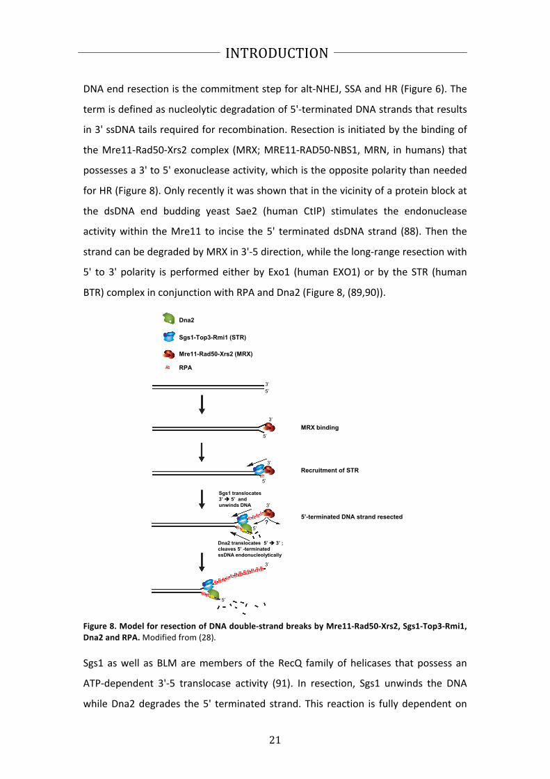
DNA end resection is the commitment step for alt-‐NHEJ, SSA and HR (Figure 6). The
term is defined as nucleolytic degradation of 5'-‐terminated DNA strands that results
in 3' ssDNA tails required for recombination. Resection is initiated by the binding of
the Mre11-‐Rad50-‐Xrs2 complex (MRX; MRE11-‐RAD50-‐NBS1, MRN, in humans) that
possesses a 3' to 5' exonuclease activity, which is the opposite polarity than needed
for HR (Figure 8). Only recently it was shown that in the vicinity of a protein block at
the dsDNA end budding yeast Sae2 (human CtIP) stimulates the endonuclease
activity within the Mre11 to incise the 5' terminated dsDNA strand (88). Then the
strand can be degraded by MRX in 3'-‐5 direction, while the long-‐range resection with
5' to 3' polarity is performed either by Exo1 (human EXO1) or by the STR (human
BTR) complex in conjunction with RPA and Dna2 (Figure 8, (89,90)).
Dna2
Sgs1-Top3-Rmi1 (STR)
RPA
3’
5’
5’
3’
5’
3’
5’
3’
Mre11-Rad50-Xrs2 (MRX)
3’
5’
MRX binding
Recruitment of STR
5’-terminated DNA strand resected
Sgs1 translocates
3’ 5’ and
unwinds DNA
Dna2 translocates 5’ 3’ ;
cleaves 5’ -terminated
ssDNA endonucleolytically
?
Figure 8. Model for resection of DNA double-‐strand breaks by Mre11-‐Rad50-‐Xrs2, Sgs1-‐Top3-‐Rmi1,
Dna2 and RPA. Modified from (28).
Sgs1 as well as BLM are members of the RecQ family of helicases that possess an
ATP-‐dependent 3'-‐5 translocase activity (91). In resection, Sgs1 unwinds the DNA
while Dna2 degrades the 5' terminated strand. This reaction is fully dependent on

22
INTRODUCTION
RPA and requires Top3-‐Rmi1 in vivo (28,92,93). The nuclease activity of Dna2 is
essential for DNA end resection, while the helicase was shown to be dispensable
(28,93). The long-‐range resection can generate 2-‐4 kb of ssDNA with a speed of 4 kb
per hour in budding yeast, ensuring efficiency and fidelity of HR (94). Additionally,
hDNA2 was shown to function with FANCD2 (Fanconi anemia complementation
group D2) in ICL repair. Depletion of DNA2 led to increase sensitivity to ICL agents
and reduced HR frequency (95).
1.2.5 Dna2 as checkpoint activator
The cellular response to DNA damage and replication stress, which is part of the cell
cycle checkpoint response, is triggered by two protein kinases of the
phosphatidylinositol 3-‐kinase-‐related protein kinase (PIKK) family: Mec1 (homolog of
human ATR) that is activated by the presence of RPA-‐coated ssDNA, and Tel1
(human ATM) that recognizes DSBs. Activation of the checkpoint results in the
stabilization of stalled replication forks, increased repair and dNTP synthesis,
inhibition of late origin firing and changes in gene expression allowing the cell to
overcome DNA damage; or in case of severe damage it initiates apoptosis in higher
eukaryotes (96). Tel1 (ATM) is recruited to DSBs via its interaction with MRX (MRN)
and phosphorylates histone H2AX as well as Rad53 (human Chk2) in some cases,
leading to the activation of further downstream mediator and effector proteins, and
cell cycle arrest. UV light and oxidative damage that are repaired by BER and NER,
respectively, activate Mec1 due to ssDNA gaps appearing in course of the repair (96).
Moreover, DSBs that become subject of resection are serving as Mec1 activator
upon exposure of the 3' terminated ssDNA tails (97). Mec1 is associated with Ddc2
(human ATRIP) and acts only as a heterodimer (98). Although Mec1-‐Ddc2 is recruited
to ssDNA via the interaction with RPA70 subunit, it still needs further interactions
with sensor proteins to stimulate its kinase activity (99,100). Three of these Mec1-‐
activator proteins were found in S. cerevisiae (Figure 9). Their activity is dependent
on the cell cycle and show partial redundancy for checkpoint activation (24). The first
activator is the 9-‐1-‐1 checkpoint clamp (yeast Ddc1-‐Rad17-‐Mec3; human RAD9-‐

23
INTRODUCTION
RAD1-‐HUS1) that is the sole activator of Mec1 in G1 in S. cerevisiae. The stimulatory
function is carried out by the Ddc1 protein (101).
Figure 9. Activation of Mec1
ATR during the cell cycle. While in G1 9-‐1-‐1 protein (RAD9-‐RAD1-‐HUS1 in
humans; Ddc1-‐Rad17-‐Mec3 in S. cerevisiae) is the sole activator of Mec1, in G2 Dpb11 can carry out
this function as well. However, in S phase Dna2 can serve as a Mec1 activator in a third redundant
pathway, upon replication stalling induced by hydroxyurea. Modified from (24).
In G2 phase, however, the 9-‐1-‐1 protein is additionally responsible for the
recruitment of the second activator, Dpb11 (human TOPBP1), and both proteins are
required for full G2 checkpoint signaling (102). In metazoans Dpb11 (TOPBP1) is the
sole activator of Mec1 (ATR), although 9-‐1-‐1 is still required for Dpb11 recruitment
(103). The S-‐phase checkpoint is quite complex and demonstrates a high level of
redundancy. In budding yeast, in addition to the activation by Ddc1 and Dpb11,
Mec1 can also be activated by Dna2 in S-‐phase, requiring its two aromatic residues
within the N-‐terminal domain − Trp128 and Tyr130 (37). To abrogate Mec1
activation in S-‐phase all three of these proteins have to be inactivated. However, to
completely abolish the S-‐phase checkpoint also Tel1 has to be inactivated and these
checkpoint-‐deficient cells are very sick, grow slowly and fail to complete DNA
replication even in the absence of DNA damaging agents (37). These findings
underline the importance of the checkpoint function even in unperturbed DNA
replication.

24
INTRODUCTION
1.2.6 Additional tasks for Dna2: telomere and mitochondrial DNA
maintenance
Chromosomal ends or telomeres resemble in many ways a DSB. However, it is
pivotal for the cells that the DSB repair pathways do not initiate on a telomere, as
this would result in fusion or loss of the telomeres, which actually can be seen in
most cancers and largely contributes to the genome instability (104). Telomeres
consist of long non-‐protein coding repeats synthesized by the telomerase, a
telomere-‐specific reverse transcriptase. At the end of the telomere there is a 3'
ssDNA overhang (50-‐300 nucleotides) that pairs with the repeats in the dsDNA, thus
generating a protective t-‐loop in human cells (105). This structure together with the
mammalian telomere-‐specific shelterin complex protects the telomere end from
DNA repair enzymes and DNA damage signaling (104). In budding yeast, the 3' tail
that is also called G-‐tail at the end of the telomere is much shorter (12-‐15
nucleotides) and serves as the binding platform for the CST capping complex (Cdc3-‐
Stn1-‐Ten1). Furthermore, dsDNA repeats are bound by Rif1/2 and Rap1 proteins that
together with CST protect telomeres from repair and checkpoint activation (106).
Surprisingly, many DNA repair proteins are found at the telomeres: e.g. Ku70-‐Ku80
that contributes to capping, or Sgs1 and Sae2 that are responsible for C-‐strand
resection after replication to generate the 3' G-‐tails (107,108). Also Dna2 was shown
to localize to telomeres during G1 and late S/G2 phases (109). In mammals, DNA2
was found at telomeres as well and deletion of DNA2 in mouse cells led to telomere
replication defects, fragile telomeres and sister telomere associations. Furthermore,
heterozygous DNA2 knockout mice developed aneuplody-‐associated cancers with
dysfunctional telomeres (110). Telomeric repeats are G-‐rich and were shown to fold
in G-‐quadruplex (G4) structures that might block replication (111,112). Both human
and yeast Dna2 were described to unwind/cleave the G4 structures which might be
one of the functions of Dna2 at telomeres and would explain the phenotype of DNA2
knockout mice (110,113).
The finding that dna2Δ cells containing a pif1-‐m2 variant (necessary for their
viability), which still localized to mitochondria and showed full function, failed to

25
INTRODUCTION
grow on glycerol media suggested a mitochondrial function for Dna2 as well (58).
Also human DNA2 was reported to be mainly found in mitochondria where it
colocalized with the helicase Twinkle (40,41). Moreover, human DNA2 interacted
and stimulated pol γ and is thought to participate together with FEN1 in long-‐patch
base excision repair of oxidative lesions in mtDNA (40). Finally, patients suffering
from progressive myopathy were found to carry DNA2 mutations inferring that this
protein is not only important for nuclear but also for mitochondrial DNA
maintenance (114).
1.2.7 Dna2 as a potential therapeutic target
Cancer research of more than three decades allowed developing a steadily growing
number of cancer therapies that target one of the hallmarks of cancer (Figure 10).
Figure 10. Therapeutic targeting of the hallmarks of cancer. Schematics showing compounds
interfering with the single acquired capabilities required for tumor growth and progression. Modified
from (2).
Also, the targeting of genome instability became an attractive therapy. So far, only
the inhibitors of PARP1 enzyme made it to the clinics as a so-‐called personalized

26
INTRODUCTION
medicine for treatment of BRCA1/BRCA2 deficient tumors (115). Their efficacy is
based on the concept of synthetic lethality: PARP1 inhibitors selectively kill cells that
are HR deficient due to BRCA1/BRCA2 mutation (116). It is obvious that more
therapies are required that target the hallmark of genome instability.
There is a growing amount of evidence that DNA2 is involved in cancer development
and progression, although the exact contribution is far from being clear. DNA2 was
found to be overexpressed in a variety of human cancers and the expression
correlated with the disease outcome, suggesting a tumor-‐promoting role for DNA2
(117). Conversely, a recent study uncovered activity-‐impairing mutations of DNA2 in
estrogen-‐dependent cancers, although depletion of DNA2 inhibited xenograft
growth in mice (118). The authors suggested that impairment of DNA2 activity might
trigger cancer development, while an elevated DNA2 activity is possibly important
for cancer progression and allows the tumor to cope with replication stress, thus
providing a growth advantage to the cancer cells (118). This hypothesis is further
supported by the phenotype of DNA2 knockout mice that develop aneuplody-‐
associated cancers (110). Taken together, these findings suggest that cancer patients
might benefit from therapies targeting DNA2: tumors with high DNA2 levels might
thus loose their proliferative capacity, while cancers with mutated DNA2 form could
be treated with additional inhibitors of DNA repair/replication proteins to induce
synthetic lethality (118).
Despite the tremendous progress in targeted cancer therapies, chemo-‐ and
radiotherapy are still very often used as treatments for many types of tumors. Both
therapies are based on the induction of a large amount of DNA damage in cancer
cells, thus leading to cell death (119). Caffeine was shown to sensitize cancer cells to
radio-‐ and chemotherapy (120,121). On the molecular level, caffeine was suggested
to inhibit p53, ATM and ATR proteins (122). However, caffeine-‐induced
radiosensitivity was additionally proposed to result from HR-‐inhibition (123). A very
recent study by the group of James Haber provided a possible explanation how
caffeine affects HR: they show that caffeine treatment leads to proteosomal
degradation of both Sae2 and Dna2 in budding yeast, thus compromising DNA end

27
INTRODUCTION
resection pathway (124). This underlines the potential of DNA2 not only in a
targeted cancer therapy, but also in classical chemo-‐ and/or radiotherapy.

28
RESULTS
2. RESULTS
2.1 Summary of results
My PhD project started with the expression and purification of S. cerevisiae Dna2 protein
followed by biochemical characterization of its helicase activity. Dna2 is an iron-‐sulfur
cluster protein and is thus very sensitive to oxidation and long dialysis procedures.
Therefore, we shortened and optimized the protein purification protocol, and obtained a
highly active Dna2 protein preparation. We were able to show that our Dna2
preparation exhibits a vigorous but cryptic helicase activity, in contrast to previous
reports from other groups, and that the helicase activity of Dna2 is masked by its
nuclease (Levikova et al., PNAS 2013; see 2.2.1).
Next, we wanted to investigate how both helicase and nuclease activities are regulated
within the Dna2 protein. We found out that Dna2 is modified by sumoylation in vitro and
in vivo. Sumoylation of Dna2 attenuated the nuclease activity of Dna2 without affecting
its helicase. Moreover, sumoylation levels of Dna2 varied during the cell cycle being
highest in late S/G2 phases. Dna2 protein levels differed as well depending on the cell
cycle stage and also decreased upon treatments with MMS and bleomycin, representing
an additional level of regulation of Dna2 (Levikova et al., manuscript in preparation, see
2.2.2).
Dna2 was shown to function together with Fen1 in Okazaki fragment processing during
lagging strand DNA replication. Having a preparation of S. cerevisiae Dna2 protein that
displayed a higher and also somewhat different activity, we set out to revisit its exact
role in Okazaki fragment maturation. Previous studies postulated a model stating that
Dna2 shortens long DNA flaps arising during lagging strand DNA synthesis up to 5-‐8 nt,
thus requiring a second nuclease activity of Fen1, before adjacent Okazaki fragments can
be ligated. We demonstrated that Dna2 is able to cleave DNA flaps at or near their base,
and is very efficient in Okazaki fragment maturation without Fen1, which is supported by
genetic data. We suggest a Fen1-‐independent role for Dna2 in lagging strand DNA
synthesis, probably in late replication for processing of DNA flaps that escaped Fen1
cleavage (Levikova and Cejka, NAR 2015, in press, see 2.2.3)
29
RESULTS
Further, we analyzed the potent helicase activity within the wild type Dna2 protein in
more detail with regard to its role in DNA end resection. We demonstrated that the
motor activity of Dna2 can act as a ssDNA translocase, rather than a helicase, to
stimulate fast and efficient ssDNA degradation particularly in the presence of RPA. We
propose that in DNA end resection, where Dna2 acts with Sgs1 to degrade 5'-‐terminated
DNA strands, the translocase activity of Dna2 helps it to keep up with the lead helicase
Sgs1 and to rapidly cleave the unwound 5'-‐terminated DNA strand (Levikova and Cejka,
manuscript in preparation, see 2.2.4).
In collaboration with the laboratory of Prof. Alessandro Vindigni (St Louis University
School of Medicine, MO, USA) we set out to define the role of Dna2 in the processing
and restart of reversed DNA replication forks. We showed by employing biochemical and
cell biological methods that human DNA2 cooperates with WRN (and yeast Dna2 with
Sgs1) to process replication forks upon replication stress-‐induced reversal, thus driving
their restart (Thangavel et al., JCB 2015, see 2.3.1).
In another collaborative project with the laboratory of Prof. Ulrich Rass (FMI Basel,
Switzerland) we elucidated the role of Dna2 helicase in completion of DNA replication
and its interplay with the Holliday-‐junction resolvase Yen1 in budding yeast. We
provided biochemical data with the helicase-‐dead Dna2 variant (Ölmezer et al., under
revision in Nat. Com., see 2.3.2)
In a further collaboration with the laboratory of PD Dr. Pavel Janscak (University of
Zurich, Switzerland) we expressed and purified human DNA2 protein that is similarly
difficult to purify as its yeast homolog. We showed that human DNA2 cooperates with
WRN and BLM helicases in long-‐range DNA end resection (Sturzenegger et al., JBC 2014,
see 2.3.3).
RPA was shown to specifically interact with Dna2 and stimulate its activity. The detailed
properties of yeast and human RPA were analyzed in single molecule experiments
performed in the laboratory of Prof. Ralf Seidel (University of Leipzig, Germany). We
provided the purified proteins for this study (Kemmerich et al., manuscript in
preparation, see 2.2.4).

30
RESULTS
2.2 Primary results
2.2.1 Nuclease activity of Saccharomyces cerevisiae Dna2 inhibits its potent
helicase activity
Maryna Levikova, Daniel Klaue, Ralf Seidel and Petr Cejka.
Article published in PNAS, 2013.
I designed the research together with R.S. and P.C. and performed most of the
experiments with the help of P.C. The single molecule experiments in Figure 3 and
Supplementary Figure S6 were carried out by D.K. All authors analyzed the data and I
wrote the manuscript together with R.S and P.C.

31
RESULTS
Nuclease activity of Saccharomyces cerevisiae Dna2inhibits its potent DNA helicase activityMaryna Levikovaa, Daniel Klaueb, Ralf Seidelb,c, and Petr Cejkaa,1
aInstitute of Molecular Cancer Research, University of Zurich, 8057 Zurich, Switzerland; bBiotechnology Center, Technische Universität Dresden, 01307Dresden, Germany; and cInstitute for Molecular Cell Biology, University of Münster, 48149 Münster, Germany
Edited* by Stephen C. Kowalczykowski, University of California, Davis, CA, and approved April 23, 2013 (received for review January 8, 2013)
Dna2 is a nuclease-helicase involved in several key pathways of
eukaryotic DNA metabolism. The potent nuclease activity of Saccha-
romyces cerevisiae Dna2 was reported to be required for all its in
vivo functions tested to date. In contrast, its helicase activity was
shown to be weak, and its inactivation affected only a subset of
Dna2 functions. We describe here a complex interplay of the two
enzymatic activities. We show that the nuclease of Dna2 inhibits its
helicase by cleaving 5′ flaps that are required by the helicase do-
main for loading onto its substrate. Mutational inactivation of Dna2
nuclease unleashes unexpectedly vigorous DNA unwinding activity,
comparable with that of the most potent eukaryotic helicases. Thus,
the ssDNA-specific nuclease activity of Dna2 limits and controls the
enzyme’s capacity to unwind dsDNA. We postulate that regulation
of this interplay could modulate the biochemical properties of Dna2
and thus license it to carry out its distinct cellular functions.
DNA nuclease | replication protein-A | Sgs1
The Dna2 enzyme functions at the crossroads of key DNAmetabolic processes. It was initially identified in screens for
Saccharomyces cerevisiae mutants deficient in DNA replication (1,2), and its importance was underscored by the finding that dna2Δmutants are not viable (3). When the DNA2 gene was cloned, itwas shown to be conserved in evolution from yeast to humansand found to contain conserved nuclease and helicase motifs (4).Further work identified a number of genetic and physical inter-actions of Dna2 with factors required for the synthesis and matu-ration of the lagging strand during DNA replication, includingRad27 [homolog of human Flap endonuclease 1, FEN1 (5)].Overexpression of Rad27 rescued growth defects of some dna2point mutants, and, conversely, overexpression of Dna2 suppressedrad27Δ defects (5). Subsequent work established that Dna2 func-tions together with Rad27 in the removal of single-stranded (ss)flaps generated at the 5′ termini of Okazaki fragments by poly-merase δ-catalyzed strand displacement. Although Rad27 seems tohave a more general role in flap processing, Dna2 is required tocleave only a subset of longer flaps bound by Replication Protein A(RPA), which stimulates its nuclease activity while inhibitingcleavage by Rad27. In this process, Dna2 and Rad27 were proposedto function in a sequential manner, with Dna2 loading first onto theflap termini and shortening them with its 5′–3′ nuclease. Rad27 wasthen proposed to further cleave the shortened flaps at the ss/dsDNA junctions, creating thus ligatable substrates for Cdc9 (DNAligase 1), which completes Okazaki fragment maturation (6–8).The role of Dna2 in Okazaki fragment processing is now gen-erally accepted although it still remains somewhat puzzling whydna2Δ cells are inviable whereas rad27Δ mutants are not.More recently, it has been shown that Dna2 has an independent
and conserved function in dsDNA break repair (9). Specifically,Dna2 belongs to one of the pathways that resect 5′ ends of dsDNAbreaks to initiate homologous recombination. This process leadsto the formation of long 3′ overhangs, which become coated by thestrand exchange protein Rad51, and which also prime DNA syn-thesis during the downstream steps in homologous recombination.Genetic and later biochemical work established that Dna2 func-tions together with the vigorous Sgs1 helicase (Bloom in human
cells) downstream of the Mre11-Rad50-Xrs2 (MRX) complex (9–14). MRX first recognizes dsDNA breaks and is likely involved intheir initial processing, and subsequently helps recruit Sgs1 andDna2. Sgs1 helicase then unwinds the DNA from the break andthe ssDNA is coated by RPA, which stimulates the 5′–3′ nucleaseactivity of Dna2. The specific degradation of the 5′ end of theunwound DNA ensures the correct polarity of resection, which isrequired for homologous recombination (12, 13). However, theroles of Dna2 are not limited to Okazaki fragment and dsDNAbreak processing. Dna2 was also shown to function in telomeremaintenance (15), aging (16), long-patch base excision repair (17),and prevention of reversal of stalled replication forks (18). Theexpression of human DNA2 was increased in human cancers andnegatively correlated with disease outcome, indicating that DNA2function is relevant for human health (19). However, the role ofDna2/DNA2 in these latter processes remains poorly defined.The potent nuclease activity of Dna2 seems to be critical for all
of its functions, including replication and recombination. Pointmutants lacking the nuclease activity are inviable as a dna2Δstrain (20). The nuclease activity of Dna2 is ssDNA-specific, andshows both 5′–3′ and 3′–5′ polarities. Because RPA stimulates the5′–3′ nuclease and inhibits the 3′–5′ activity, it is likely that onlythe 5′–3′ directionality is important in vivo (12, 13). It has beenshown that the Dna2 nuclease can load only on a free ssDNA tail,and that it subsequently cleaves DNA endonucleolytically intoshort oligonucleotides (21, 22).Much less is known about the function of the helicase activity
of Dna2. To date, very weak 5′–3′ unwinding capacity has beendemonstrated for the yeast enzyme (4, 7, 23). Interestingly,similarly to the Dna2 nuclease, also the helicase domain requiresa free DNA end (22). In contrast, no unwinding activity could bedetected in the Xenopus laevis Dna2. Whether human DNA2possesses helicase activity remains controversial (24–27). Yeastpoint mutants lacking helicase activity show impaired growth but
Significance
The integrity of DNA must be preserved to pass genetic in-
formation onto the next generation and to prevent genomic
instability. One of the key enzymes involved in DNA metabo-
lism is the nuclease-helicase Dna2, which is required for both
DNA replication and the repair of broken DNA. Our work
revealed that Dna2 from Saccharomyces cerevisiae possesses
a potent but cryptic helicase capacity, which is controlled by
the nuclease activity of the same polypeptide. Regulating the
interplay between both enzymatic activities might explain how
Dna2 can take on its distinct cellular roles.
Author contributions: M.L., R.S., and P.C. designed research; M.L., D.K., and P.C. per-
formed research; M.L., D.K., R.S., and P.C. analyzed data; and M.L., R.S., and P.C. wrote
the paper.
The authors declare no conflict of interest.
*This Direct Submission article had a prearranged editor.
1To whom correspondence should be addressed. E-mail: [email protected].
This article contains supporting information online at www.pnas.org/lookup/suppl/doi:10.
1073/pnas.1300390110/-/DCSupplemental.
E1992–E2001 | PNAS | Published online May 13, 2013 www.pnas.org/cgi/doi/10.1073/pnas.1300390110

32
RESULTS
are viable under most conditions (23). Overexpression of Rad27partially rescues dna2 helicase-deficient mutants, suggesting thatthe helicase activity might play a supportive but nonessential role inDNA replication (28). The helicase-deficient mutants show a dra-matic sensitivity to the DNA alkylating drug methylmethanesulfo-nate (MMS), pointing to an as-yet uncharacterized role of Dna2helicase in the repair of DNA damage (29). In contrast, Dna2helicase activity had no detectable effect on DNA end resection (9).Due to the limited unwinding capacity of Dna2 observed in vitro,the motor was proposed to function as an ssDNA translocase to aidpositioning of the nuclease domain on ssDNA and to removesecondary structures from ssDNA, rather than as a DNA helicaseto unwind dsDNA (23).In this work, we expressed S. cerevisiaeDna2 and its variants and
optimized the purification of these polypeptides. We now showthat Dna2 is a potent but cryptic DNA helicase. It functionallyinteracts with RPA, which enables it to unwind tens of kilobases ofdsDNA. Surprisingly, the nuclease of wild-type Dna2 interfereswith this remarkable helicase capacity by cleaving ssDNA tails thatthe helicase requires for loading onto DNA to initiate unwinding.The interplay between the two main biochemical activities of Dna2might fine-tune its behavior to suit its distinct cellular roles.
Results
Expression and Purification of Wild-Type Dna2 and Nuclease- and
Helicase-Dead Variants. S. cerevisiae Dna2 is a large protein(172 kDa) consisting of 1,522 amino acids (Fig. 1A). We no-ticed previously that recombinant Dna2 was rather unstable,rapidly losing activity during extended dialysis procedures used inearlier preparation protocols (12). Furthermore, it was sensitiveto the omission of reducing agents, possibly due to the presenceof an oxidation-prone iron–sulfur cluster (30, 31). Modification ofthe purification procedure allowed us to obtain Dna2 with a highspecific nuclease activity (12). In this work, we further optimizedand shortened the purification process (Materials and Methods)and obtained nearly homogenous wild-type Dna2 and nuclease-dead Dna2 E675A, as well as helicase-dead Dna2 K1080E var-iants (Fig. 1 A and B and Fig. S1 A and B).
Dna2 Possesses a Vigorous DNA Helicase Activity. The ssDNA-specificnuclease activity of Dna2 was proposed to obscure the detection ofits limited unwinding property (22). Therefore, the nuclease-deadDna2 variants were used to characterize its helicase activity. One ofthese mutants is Dna2 E675A, which had been designed based onthe homology betweenEscherichia coliRecB and S. cerevisiaeDna2nuclease sites (20). Indeed, mutation of the conserved glutamate atposition 675 to alanine largely inactivated the nuclease activity ofDna2, and Dna2 E675A has been shown to exhibit a weak helicaseactivity (22, 23). Here, having used the optimized preparationprocedure, we set out to examine the helicase activity of this variant.Dna2 E675A could readily unwind a synthetic Y-structure oligo-nucleotide-based substrate containing 31 base pairs of dsDNA (Fig.1C). To our great surprise, the helicase was active at subnanomolarconcentrations (Fig. 1C). Previously, ∼20 nM enzyme was neededto unwind a similar length of dsDNA (22, 23), indicating that ourpreparation possesses >20-fold higher specific DNA unwindingactivity. In agreement with previous data (23), supplementing thereaction with S. cerevisiae replication protein A (RPA) did notfurther stimulate its unwinding capacity whereas E. coli SingleStrand DNA Binding protein (SSB) strongly inhibited DNA un-winding (Fig. 1 C and D). We show that a 5′-tailed DNA is a pre-ferred structure for Dna2 E675A unwinding, with ∼0.05 nM Dna2E675A required to unwind 50% of the DNA substrate, which wasused at 1 nMconcentration (Fig. 1E andFig. S2). The unwinding ofthe 5′-tailed DNA substrate was therefore clearly catalytic, withone enzyme molecule being capable of unwinding at least ∼10DNA substrate molecules during the time course of the reaction.The vigorous activity stands in contrast with previous studies
where ∼15- to 100-fold excess enzyme over DNA substrate wasrequired to detect DNA unwinding (22, 23). We further show that∼0.3 nM Dna2 E675A was required to unwind 50% of the Y-struc-ture DNA substrate; thus, the presence of an additional 3′ ssDNAarm (Y-structure vs. 5′ overhang) inhibitedDNAunwinding∼sixfold(Fig. 1E and Fig. S2). As anticipated for a 5′–3′ DNA helicase, theunwinding of 3′-tailed or fully dsDNA substrates was inefficient(Fig. 1E and Fig. S2). The difference between the specific DNA
Fig. 1. The nuclease-inactive Dna2 E675A variant possesses a vigorous DNA
helicase activity. (A) A schematic representation of the recombinant Dna2 pro-
tein used in this study. The polypeptide contains an N-terminal FLAG and HA
tags, and a C-terminal 6xHis tag. Positions of mutations inactivating the nuclease
activity (E675A) or helicase activity (K1080E) are indicated. (B) Purified Dna2
wild-type (wt), E675A, and K1080E variant proteins (550 ng each) used in this
study were stained with Coomassie blue. (C) Representative polyacrylamide gels
(10%) showing the DNA helicase activity of Dna2 E675A on a 32P-labeled Y-
structure DNA substrate (1 nM). The length of both ssDNA arms was 19 nt. As
indicated, the reactions were supplemented with either S. cerevisiae RPA or
E. coli SSB (both 22 nM). *, position of the 32P label. (D) Quantitation of the
helicase assays such as shown in C. Error bars, SE, n = 3. (E) Quantitation of the
helicase assays such as shown in Fig. S2. Error bars, SE, n = 3. (F) Quantitation of
electrophoretic mobility shift assays showing the binding of Dna2 E675A to
various 32P-labeled DNA substrates (1 nM). Error bars, SE, n = 3.
Levikova et al. PNAS | Published online May 13, 2013 | E1993
BIOCHEMISTRY
PNASPLU
S

33
RESULTS
unwinding activity presented here and in earlier reports (22, 23) islikely due to the optimized enzyme preparation procedure usedin this work (Materials and Methods and Discussion).Next, we used electrophoretic mobility shift assays to assess the
binding of Dna2 E675A to DNA. We saw that Dna2 E675A bindsrather indiscriminately (Kd ∼5 nM) to structures that containssDNA. A 5′ ssDNA overhang was bound equally well as a 3′
ssDNA overhang, despite the fact that the former structure is anexcellent substrate for the Dna2 E675A helicase whereas thelatter is not (Fig. 1F and Fig. S3A). Similar results were obtainedin the presence of competitor DNA in the electrophoretic mo-bility shift assays (Fig. S3B). These results show that Dna2E675A is a vigorous DNA helicase with high affinity for DNA.
RPA Stimulates Dna2 E675A to Unwind Long Stretches of DNA. Dna2nuclease-deficient variants were previously shown to be only ca-pable of unwinding short, oligonucleotide-based DNA structures.The fraction of unwound substrate decreased dramatically with thelength of the duplex DNA; the unwinding of a 91-bp duplex was∼15-fold less efficient than the unwinding of a 30-bp duplex (23).Is has been proposed that Dna2 is a weak and nonprocessive DNAhelicase (23). We have decided to analyze unwinding of long-length DNA with our preparation of Dna2 E675A. Surprisingly,the enzyme could readily unwind plasmid-based 2.7-kbp dsDNAcontaining a 3-nt-long 5′ ssDNA tail (Fig. 2A). Half of the DNAsubstrate was unwound by only 200 pM Dna2 E675A, a concen-tration similar to that required for the unwinding of oligonucleo-tide-based DNA substrates (Fig. 1 C and E). However, unlike inthe case of the oligonucleotide-based substrates, the unwinding ofthe 2.7-kbp dsDNA was absolutely dependent on RPA, as noDNA unwinding was observed when RPA was omitted (Fig. 2A,lane 12). E. coli SSB could not replace RPA (Fig. 2A, lane 13),indicating that species-specific interaction between yeast Dna2 andyeast RPA plays an essential role in promoting long-length DNAunwinding by the Dna2 helicase.Sgs1 is the most vigorous DNA helicase characterized to date in
eukaryotes (32). Given that it functions in DNA end resection to-gether with Dna2 (9, 12, 13), we set out to compare the unwindingcapacities of Sgs1 andDna2E675A. Sgs1 helicase can initiateDNAunwinding of blunt-ended, 3′− or 5′-tailed DNA with similar effi-ciency (32). To compare the relative activities of both enzymes, weselected 5′-tailed DNA, which is required for unwinding by Dna2E675A. Remarkably, we show that Sgs1 was only slightly (∼30%)better in unwinding of the 2.7-kbp-long dsDNA than Dna2 E675A(Fig. 2 A and B), and similar results were obtained in kineticexperiments (Fig. 2C and Fig. S4). We thus conclude that Dna2E675A is almost as vigorous a DNA helicase as Sgs1.To further characterize the unwinding capacity of Dna2 E675A,
we tested its ability to unwind long stretches of dsDNA. We havedigested bacteriophage λ DNA with HindIII and 32P-labeled therestriction fragments. This procedure created linear DNA mole-cules of up to 23 kbp in length with 3-nt-long 5′ overhangs. Dna2E675A could efficiently unwind DNA fragments of all lengths andproduced a pattern of ssDNA molecules that comigrated with theheat-denatured substrate (Fig. 2 D and E). The reaction was re-markably efficient; 1.2 nM Dna2 E675A unwound ∼50% of theDNA substrate, which was present at a concentration corre-sponding to 2.4 nMDNA ends. Furthermore, the 23-kbp fragmentwas unwound only ∼twofold less efficiently than the 2.0-kbp frag-ment, showing that the length of duplex DNA only marginallyaffects the unwinding efficiency in the presence of S. cerevisiaeRPA. Unlike E. coli SSB, human RPA could substitute cognate S.cerevisiae RPA, and, as expected, the reaction was completelydependent on ATP (Fig. S5). We conclude that S. cerevisiaeDna2E675A is a very vigorous DNA helicase, capable of unwinding tensof kilobases of dsDNA in length in a reaction dependent on RPAand ATP.
Single Molecule Experiments Reveal High DNA Unwinding Processivity
of Dna2 E675A. To gain insight into the processivity and the rate ofDNA unwinding by Dna2 E675A, we carried out single-moleculemagnetic tweezers experiments (Fig. 3 A and B). As substrate weused a 6.6-kbp-long dsDNA fragment that contained 0.6-kbpdsDNA tails with multiple digoxigenin or biotin modifications ateither end to allow its binding to the surface of an anti-digox-igenin–coated fluidic cell and to streptavidin-coated magneticbeads. The construct contained a 40-nt 5′-terminated ssDNA flapto allow the loading of Dna2. A pair of magnets above the fluidiccell was used to stretch single bead-tetheredDNA constructs, andvideo microscopy was used to read out the DNA end-to-enddistance in real time (33).When adding Dna2 E675A in the presence of RPA and ATP,
rapid DNA unwinding was observed that resulted in an apparentincrease in the DNA length (Fig. 3 B and C). This increase is dueto the larger extension of RPA-coated ssDNA in comparisonwith dsDNA (34, 35). No significant DNA lengthening (>100 bp)
Fig. 2. Dna2 E675A can unwind long lengths of dsDNA in the presence of
RPA. (A) Representative agarose gels (0.8%) showing the helicase activity of
Dna2 E675A or Sgs1 on a 32P-labeled 2.7-kbp-long dsDNA substrate with
a 3-nt 5′ ssDNA overhang (1 nM). The reactions were supplemented with S.
cerevisiae RPA (0.4 μM) except lane 12 (RPA omitted) and lane 13 (RPA
replaced with E. coli SSB). Heat, heat-denatured DNA substrate; *, position
of the 32P label. (B) Quantitation of the helicase assays as in A. Error bars, SE,
n = 3. (C) Kinetics of DNA unwinding of 5′-tailed 2.7-kbp-long dsDNA (1 nM)
by either Dna2 E675A (1 nM) or Sgs1 (1 nM) in the presence of RPA (0.4 μM).
Error bars, SE, n = 3. (D) λ phage DNA was digested with HindIII to produce
dsDNA fragments ranging from 125 bp to 23 kbp in length, and 32P-labeled
with Klenow fragment of DNA polymerase I. The resulting DNA fragments
had 5′ ssDNA tails of 3 nt in length at their ends. Various concentrations of
Dna2 E675A were incubated with the restricted DNA (2.4 nM of DNA ends)
in the presence of RPA (1.08 μM). Heat, heat-denatured substrate. The panel
shows a representative agarose gel (1%). (E) Quantitation of the helicase
assays such as shown in D. Error bars, SE, n = 3.
E1994 | www.pnas.org/cgi/doi/10.1073/pnas.1300390110 Levikova et al.
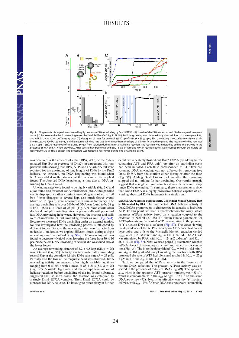
34
RESULTS
was observed in the absence of either RPA, ATP, or the 5′-ter-minated flap (but in presence of Dna2), in agreement with ourprevious data showing that RPA, ATP, and a 5′ ssDNA tail wererequired for the unwinding of long lengths of DNA by the Dna2helicase. As expected, no DNA lengthening was found whenRPA was added in the absence of the helicase at the appliedforces. The observed DNA lengthening is thus due to DNA un-winding by Dna2 E675A.Unwinding rates were found to be highly variable (Fig. 3 C and
D) as found also for other DNA translocases (36). Although someevents displayed a rather constant unwinding rate of up to 120bp·s−1 over distances of several kbp, also much slower events(down to 15 bp·s−1) were observed with similar frequency. Theaverage unwinding rate over 500 bp of DNA was found to be 38 ±4 bp·s−1 (SE) at a force of 25 pN (Fig. 3D). Slow events oftendisplayed multiple unwinding rate changes or stalls, with periods offast DNA unwinding in between. However, rate changes and stallswere characteristic of fast unwinding events as well (Fig. S6A).Because we measured DNA unwinding under an external tension,we also investigated how the unwinding process is influenced bydifferent forces. Because the unwinding rates were variable frommolecule to molecule, we applied different forces during a singleunwinding run of a molecule (Fig. S6B). The unwinding rate wasfound to decrease ∼fivefold when lowering the force from 30 to 10pN. Nonetheless DNA unwinding of several kbp was found also atthe lower forces.An average unwinding distance of 4.2 ± 0.5 kbp (SE, n = 25)
was obtained (Fig. 3C). Activity was terminated after unwindingseveral kbp or the complete 6.1-kbp DNA substrate (F= 25 pN).Partially also the loss of the magnetic bead was observed. DNAunwinding activity commenced after highly variable lag timesranging from 0 to 800 s with a mean of 85 ± 31 s (SE, n = 25)(Fig. 3C). Variable lag times and the abrupt termination ofhelicase reactions before unwinding of the full-length substrate,suggested that, in most cases, the reaction was catalyzed bya single Dna2 E675A complex. Thus, Dna2 E675A could bea processive DNA helicase. To investigate processivity in further
detail, we repeatedly flushed out Dna2 E675A (by adding buffercontaining ATP and RPA only) just after an unwinding eventhad been initiated. Each flush corresponded to ∼1.7 flow cellvolumes. DNA unwinding was not affected by removing freeDna2 E675A from the solution either during or after the flush(Fig. 3E). Adding Dna2 E675A back in after the unwindingstopped did not initiate further unwinding. Our results stronglysuggest that a single enzyme complex drives the observed long-range DNA unwinding. In summary, these measurements showthat Dna2 E675A is a highly processive helicase capable of un-winding kbp-sized DNA fragments in a single run.
Dna2 E675A Possesses Vigorous DNA-Dependent Atpase Activity That
Is Stimulated by RPA. The unexpected DNA helicase activity ofDna2 E675A prompted us to characterize its capacity to hydrolyzeATP. To this point, we used a spectrophotometric assay, whichmeasures ATPase activity based on a reaction coupled to theoxidation of NADH (37, 38). To obtain kinetic parameters forATP hydrolysis, we first varied ATP concentration in the presenceof Y-structure DNA as a cofactor (Fig. S7). The curve showingthe dependence of the ATPase activity on ATP concentration washyperbolic, and a fit to the Michaelis–Menten equation yieldedVmax = 21 ± 2 μM·min−1 and Km = 130 ± 24 μM. The ATPasewas stimulated by RPA, with Vmax = 24 ± 2 μM·min−1 and Km =56 ± 10 μM (Fig. S7). Next, we used poly(dT) as cofactor, which isssDNA devoid of secondary structure, and varied its concentra-tion (Fig. 4A). The fit to the data yielded Vmax = 9.6 ± 1 μM·min−1
and Km = 164 ± 44 nM. Supplementing the reactions with RPApromoted the rate of ATP hydrolysis and resulted in Vmax = 22 ±
2 μM·min−1 and Km = 181 ± 33 nM.Next, we compared the ATPase activity in the presence of
various DNA cofactors. The greatest ATPase activity was ob-served in the presence of 5′-tailed DNA (Fig. 4B). The apparentkcat, which is the apparent ATP turnover number, was ∼85 s−1,which is comparable with the kcat of Sgs1 ∼82 s−1 on the sameDNA structure (32). Nearly as effective was the Y-structuredsDNA, with kcat ∼79 s
−1. Other DNA substrates were substantially
Fig. 3. Single molecule experiments reveal highly processive DNA unwinding by Dna2 E675A. (A) Sketch of the DNA construct and (B) the magnetic tweezers
assay. (C) Representative DNA unwinding events by Dna2 E675A (F = 25 ± 2 pN, SD). DNA lengthening was observed only after addition of the enzyme, RPA,
and ATP in the reaction buffer (gray box). (D) Histogram of rates for unwinding 500 bp of DNA (F = 25 ± 2 pN, SD). Unwinding trajectories (n = 14) were split
into successive 500-bp segments, and the mean unwinding rate was determined from the slope of a linear fit to each segment. The mean unwinding rate was
38 ± 4 bp·s−1 (SE). (E) Removal of free Dna2 E675A from solution during a DNA unwinding reaction. The reaction was initiated by adding the enzyme in the
presence of RPA and ATP (left gray box). After several hundred unwound bps, ∼50 μl of ATP and RPA in reaction buffer were flushed through the fluidic cell
(cell volume 30 μl) (blue boxes). The procedure was repeated four times during one unwinding event.
Levikova et al. PNAS | Published online May 13, 2013 | E1995
BIOCHEMISTRY
PNASPLU
S

35
RESULTS
less effective; ssDNA stimulated about 2.6-fold less than 5′-tailedDNA, and 3′-tailedDNAand dsDNA stimulated 3.5- or 6.1-fold lessefficiently, respectively (Fig. 4B). The capacity of theDNAstructuresto promote ATP hydrolysis by Dna2 E675A corresponded to theunwinding preference. The 5′-tailed and Y-structure DNA sub-strates were the preferred substrates for DNA unwinding by Dna2E675A (Fig. 1E), and they were also the most efficient in stimulatingits ATPase activity (Fig. 4B). Almost no ATPase activity was ob-served in the absence ofDNA, showing that the ATPase activity ofDna2 E675A is DNA-dependent (Fig. 4B). As expected, theATPase-deficient Dna2 K1080E variant showed no ATPase ac-tivity (Fig. 4B). In summary, these parameters establish thatDna2 E675A is a strong ATPase that has a high affinity for bothATP and DNA.
Nuclease Activity Within Dna2 Limits Its Unwinding Capacity. Havingestablished that Dna2 E675A variant lacking the nuclease activityis a vigorous DNA helicase, we next wanted to study the interplayof the nuclease with the helicase within the wild-type protein. Thenuclease of Dna2 is ssDNA-specific but may act on dsDNA uponspontaneous denaturation/melting of dsDNA ends (39). We showin Fig. 5A that, whereas 1 nM Dna2 E675A was sufficient tocompletely unwind a 2.7-kbp dsDNA substrate (lane 2), the sameconcentration of wild-type Dna2 had no detectable activity (lane3). When we used 10- to 50-fold higher wild-type Dna2 concen-tration (10 and 50 nM), only limited DNA degradation was ob-served, as indicated by the release of the radioactive label (Fig. 5A,lanes 4 and 5), or mobility shift of unlabeled DNA stained withethidium bromide (Fig. 5B). DNA degradation by wild-type Dna2was largely restricted to the vicinity of the DNA ends (comparepanels A and B in Fig. 5). Thus, wild-type Dna2 apparently lackedthe vigorous helicase activity observed in the nuclease-deadE675Avariant. This observation suggests either that the helicase/ATPasedomain of the wild-type protein is compromised, or that the nu-clease of Dna2 inhibits its own helicase activity. To differentiatebetween these two possibilities, we compared the initial rates ofATP hydrolysis between thewild-type and the nuclease-deadDna2E675A proteins. We show that both polypeptides display similarlevels of ATPase activity (Fig. 5C), indicating that the ATPase/helicase domain in the wild-type protein is active and thereforelikely properly folded. This result suggested that the nuclease ofwild-type Dna2 limits the engagement of its helicase. We next setout to study this phenomenon in more detail.Because the nuclease activity of Dna2 preferentially degrades
5′ ssDNA overhangs, and these overhangs are required for DNAunwinding by theDna2 E675A variant (Fig. 1E), we asked whetherthe nuclease of wild-type Dna2 might cleave the 5′ ssDNA tails
before they can be accessed by the helicase. This activity couldexplain the apparent lack of DNA unwinding activity of wild-typeDna2. To this point, we set out to compare enzyme concentrationsrequired for the unwinding and/or the nucleolytic degradation ofoligonucleotide-based DNA substrates. Specifically, we comparedthe nuclease activity of Dna2 wild type and Dna2 K1080E with thehelicase activity of Dna2 E675A. All these enzyme variants wereprepared using an identical procedure, and we noted that in-dependent preparations of the same enzyme variant led to con-sistent levels of specific activity (Materials and Methods). Weobserved that, first, the nuclease activities of both wild-type andhelicase-dead K1080E enzymes were indistinguishable based onsubstrate utilization (Fig. 5D). The lack of the helicase activitytherefore did not affect the initial 5′ end cleavage byDna2. Second,the nuclease activity was ∼sixfold more frequent than the helicaseactivity based on substrate utilization: although 50 pM wild-typeand helicase-dead K1080E Dna2 cleaved ∼50% of the 5′ tailswithin 30 min, as much as 300 pM of nuclease-dead Dna2 E675Awas required to unwind 50%of theDNAsubstrate during the sametime period (Fig. 5D). These results show that the wild-type protein
Fig. 4. Dna2 E675A shows DNA-dependent ATPase activity that is stimu-
lated by RPA. (A) Rate of ATP hydrolysis and its dependence on the DNA
concentration. The reactions contained Dna2 E675A (4 nM), ATP (1 mM), RPA
where indicated (150 nM), and the indicated concentrations of poly(dT). (B)
Apparent ATP turnover number and its dependence on various DNA struc-
tures (1 μM nucleotides). The reactions contained Dna2 E675A (3 nM) and
ATP (1 mM). Error bars, SE, n = 2.
Fig. 5. The nuclease activity of Dna2 interferes with its helicase activity. (A)
A representative experiment comparing the activities of wild-type Dna2 and
nuclease-dead E675A variant on 5′-tailed 2.7-kbp dsDNA substrate 32P-la-
beled at the 3′ end (1 nM). Reactions were incubated in the presence of
S. cerevisiae RPA (0.4 μM). *, position of the 32P label. Heat, heat-denatured
substrate. (B) A representative experiment comparing the activities of wild-
type Dna2 and nuclease-dead E675A variant on 5′-tailed 2.7-kbp dsDNA
substrate (3.8 nM). Reactions were incubated for 30 min in the presence of
S. cerevisiae RPA (1.68 μM). A 1% agarose gel was stained with ethidium
bromide. Heat, heat-denatured substrate. (C) Comparison of apparent ATP
turnover numbers of Dna2 wild-type and Dna2 E675A variant. The reactions
contained the indicated DNA structures (1 μM nucleotides), Dna2 wild type
or E675A (3 nM) and ATP (1 mM). Error bars, SE, n = 2. (D) Comparison of
nuclease and/or helicase activities of Dna2 wild-type, K1080E, and E675A
variants, respectively, based on substrate utilization. All assays contained
RPA (22.5 nM). Error bars, SE, n = 2. (E) Dependence of Dna2 activities on the
length of the 5′ ssDNA overhang. Dna2 concentrations required for 50% of
maximal DNA cleavage (by Dna2 wild type or Dna2 K1080E) or 50% of
maximal DNA unwinding (by Dna2 E675A) were plotted against the length
of the 5′ ssDNA overhang. The data were obtained by analysis of the results
such as from Fig. S8.
E1996 | www.pnas.org/cgi/doi/10.1073/pnas.1300390110 Levikova et al.
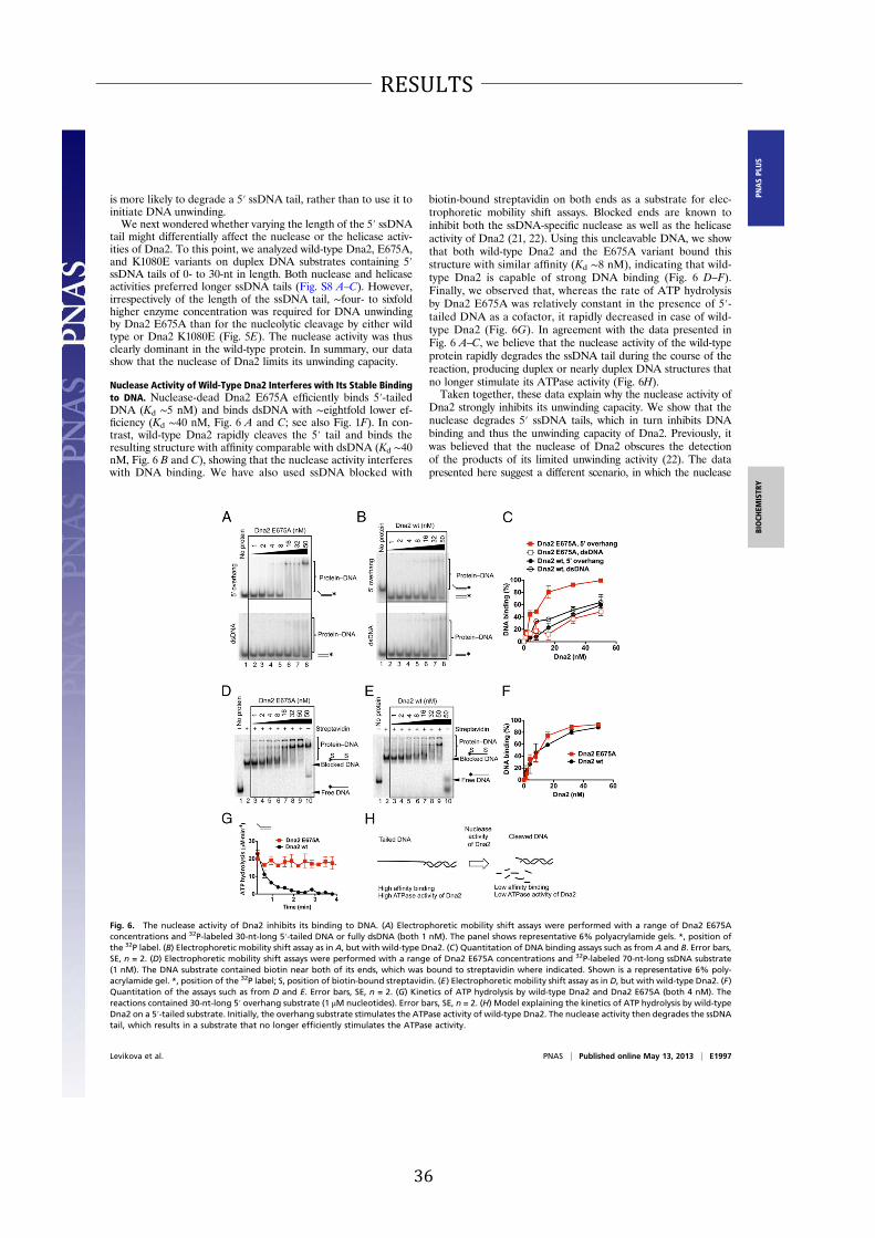
36
RESULTS
is more likely to degrade a 5′ ssDNA tail, rather than to use it toinitiate DNA unwinding.We next wondered whether varying the length of the 5′ ssDNA
tail might differentially affect the nuclease or the helicase activ-ities of Dna2. To this point, we analyzed wild-type Dna2, E675A,and K1080E variants on duplex DNA substrates containing 5′
ssDNA tails of 0- to 30-nt in length. Both nuclease and helicaseactivities preferred longer ssDNA tails (Fig. S8 A–C). However,irrespectively of the length of the ssDNA tail, ∼four- to sixfoldhigher enzyme concentration was required for DNA unwindingby Dna2 E675A than for the nucleolytic cleavage by either wildtype or Dna2 K1080E (Fig. 5E). The nuclease activity was thusclearly dominant in the wild-type protein. In summary, our datashow that the nuclease of Dna2 limits its unwinding capacity.
Nuclease Activity of Wild-Type Dna2 Interferes with Its Stable Binding
to DNA. Nuclease-dead Dna2 E675A efficiently binds 5′-tailedDNA (Kd ∼5 nM) and binds dsDNA with ∼eightfold lower ef-ficiency (Kd ∼40 nM, Fig. 6 A and C; see also Fig. 1F). In con-trast, wild-type Dna2 rapidly cleaves the 5′ tail and binds theresulting structure with affinity comparable with dsDNA (Kd ∼40nM, Fig. 6 B and C), showing that the nuclease activity interfereswith DNA binding. We have also used ssDNA blocked with
biotin-bound streptavidin on both ends as a substrate for elec-trophoretic mobility shift assays. Blocked ends are known toinhibit both the ssDNA-specific nuclease as well as the helicaseactivity of Dna2 (21, 22). Using this uncleavable DNA, we showthat both wild-type Dna2 and the E675A variant bound thisstructure with similar affinity (Kd ∼8 nM), indicating that wild-type Dna2 is capable of strong DNA binding (Fig. 6 D–F).Finally, we observed that, whereas the rate of ATP hydrolysisby Dna2 E675A was relatively constant in the presence of 5′-tailed DNA as a cofactor, it rapidly decreased in case of wild-type Dna2 (Fig. 6G). In agreement with the data presented inFig. 6 A–C, we believe that the nuclease activity of the wild-typeprotein rapidly degrades the ssDNA tail during the course of thereaction, producing duplex or nearly duplex DNA structures thatno longer stimulate its ATPase activity (Fig. 6H).Taken together, these data explain why the nuclease activity of
Dna2 strongly inhibits its unwinding capacity. We show that thenuclease degrades 5′ ssDNA tails, which in turn inhibits DNAbinding and thus the unwinding capacity of Dna2. Previously, itwas believed that the nuclease of Dna2 obscures the detectionof the products of its limited unwinding activity (22). The datapresented here suggest a different scenario, in which the nuclease
Fig. 6. The nuclease activity of Dna2 inhibits its binding to DNA. (A) Electrophoretic mobility shift assays were performed with a range of Dna2 E675A
concentrations and 32P-labeled 30-nt-long 5′-tailed DNA or fully dsDNA (both 1 nM). The panel shows representative 6% polyacrylamide gels. *, position of
the 32P label. (B) Electrophoretic mobility shift assay as in A, but with wild-type Dna2. (C) Quantitation of DNA binding assays such as from A and B. Error bars,
SE, n = 2. (D) Electrophoretic mobility shift assays were performed with a range of Dna2 E675A concentrations and 32P-labeled 70-nt-long ssDNA substrate
(1 nM). The DNA substrate contained biotin near both of its ends, which was bound to streptavidin where indicated. Shown is a representative 6% poly-
acrylamide gel. *, position of the 32P label; S, position of biotin-bound streptavidin. (E) Electrophoretic mobility shift assay as in D, but with wild-type Dna2. (F)
Quantitation of the assays such as from D and E. Error bars, SE, n = 2. (G) Kinetics of ATP hydrolysis by wild-type Dna2 and Dna2 E675A (both 4 nM). The
reactions contained 30-nt-long 5′ overhang substrate (1 μM nucleotides). Error bars, SE, n = 2. (H) Model explaining the kinetics of ATP hydrolysis by wild-type
Dna2 on a 5′-tailed substrate. Initially, the overhang substrate stimulates the ATPase activity of wild-type Dna2. The nuclease activity then degrades the ssDNA
tail, which results in a substrate that no longer efficiently stimulates the ATPase activity.
Levikova et al. PNAS | Published online May 13, 2013 | E1997
BIOCHEMISTRY
PNASPLU
S

37
RESULTS
activity prevents the helicase activity from engaging its respectivesubstrate.
Helicase Activity of Wild-Type Dna2 Promotes Its Capacity to Degrade
dsDNA. Up to this point, we demonstrated that the nuclease ofwild-type Dna2 inhibits its unwinding capacity. By cleavingssDNA tails that serve as entry points for the helicase, the nu-clease impedes DNA binding and thus the initiation of dsDNAunwinding. In subsequent experiments, we set out to find con-ditions to detect the unwinding capacity of the wild-type enzyme.Using 5′-tailed dsDNA substrate 32P-labeled at the ssDNA tail,
the activity of wild-type Dna2 was indistinguishable from that ofthe helicase-dead K1080E variant. The helicase activity did notstimulate cleavage of the 5′-terminated ssDNA tail, and we coulddetect no DNA unwinding intermediates (Fig. S9 A and B). Sim-ilarly to experiments presented in Fig. 5 D and E, the helicaseactivity thus had no apparent effect on the behavior of Dna2. Wereasoned that Dna2 binds and translocates on the tailed oligonu-cleotide, which gets then preferentially cleaved, and this cleavagemay limit the detection of DNA unwinding intermediates. We nextlabeled the bottom oligonucleotide that does not contain theoverhang (see diagram at the top of Fig. 7A), and analyzed theactivity of wild-type, helicase-dead K1080E, and nuclease-deadDna2 E675A variants. The nuclease of both wild type and K1080Edegraded the 5′-terminated DNA overhang, resulting in speciesthat comigrated with the tail-less dsDNA marker (Fig. 7 A and B).Importantly, we could also clearly detect ssDNA in reactions withwild-type Dna2 (Fig. 7A, lanes 3–6). In contrast, ssDNA was nearlyundetectable in reactions with the helicase-dead K1080E variant;the minimal amount of ssDNA we observed was likely the resultof the nuclease acting on the top strand upon spontaneousthermal melting of the DNA substrate in the presence of RPA(Fig. 7B). The ssDNA was apparent at wild-type Dna2 concen-trations as low as 16–32 pM (Fig. 7A), which was also the min-imal protein concentration required to detect DNA unwindingby the nuclease-dead Dna2 E675A variant (Fig. 7C). Theseresults, together with the ATPase assays (Fig. 5C), indicate thatwild-type Dna2 likely possesses the same level of helicase activityas the nuclease dead Dna2 E675A variant.The experiments in Fig. 7 A and B also showed that the
helicase activity of Dna2 promotes degradation of dsDNA. Thedegradation of DNA by the helicase-deficient Dna2 K1080Evariant was mostly limited to the 5′-terminated ssDNA over-hang, as the species comigrating with tail-less DNA was thedominant product across a wide range of enzyme concen-trations (Fig. 7B). In contrast, wild-type Dna2 could more ef-ficiently enter and degrade dsDNA as well, as witnessed by theappearance of small-molecular-weight DNA species, denotedas degraded DNA (Fig. 7A).
To further investigate the role of the Dna2 helicase in deg-radation of dsDNA, we carried out additional experiments witha 5′-tailed substrate that was 3′-labeled at the dsDNA end (seediagram at the top of Fig. 8A). As above, the helicase activity ofwild-type Dna2 promoted degradation of dsDNA (Fig. 8 A andB). We show that dsDNA degradation requires the presence ofthe 5′-terminated ssDNA tail, as tail-less dsDNA was not cleavedat similar enzyme concentrations (Fig. S10A). Furthermore, thedsDNA degradation was dependent on RPA, as no significant 3′
end cleavage product was observed in the absence of RPA, withboth wild-type Dna2 or the helicase-dead K1080E variant (Fig.S10B). RPA therefore stimulates the degradation of dsDNA byboth wild-type and helicase-dead Dna2 K1080E, probably bymelting the dsDNA ends. The dsDNA degradative capacity is thenfurther enhanced by the helicase activity of Dna2 (Fig. 8 A and B).Finally, we investigated the degradation of plasmid-sized tailed
DNA on agarose gels stained with ethidium bromide. Limiteddegradation of DNA near its ends resulted in change of mobility(Fig. 8C). In the absence of ATP, a condition favored by the Dna2nuclease (4), both wild-type and helicase-dead Dna2 K1080Evariant degraded DNA to a similar extent. Upon supplementingthe reaction with ATP, which lowered the free magnesium con-centration required by the nuclease and activated the helicase,DNA degradation by the wild-type enzyme was promoted whereasDNA degradation by the K1080E variant was inhibited. Thus, inthe presence of ATP, the helicase of Dna2 clearly promotes deg-radation of dsDNA. The extent of DNA degradation is, however,limited in comparison with the extent of DNA unwound by theequivalent concentration of the nuclease-dead Dna2 E675A mu-tant (Fig. 5 A and B).In summary, our data reveal an unusual interplay of the nu-
clease and helicase activities of Dna2. The nuclease activitystrongly reduces the ability of Dna2 to unwind DNA. However,the inhibition of the DNA unwinding capacity is not total, and weshow that the helicase activity may promote nucleolytic degra-dation of dsDNA. The ssDNA-specific nuclease activity thuscontrols the unwinding capacity and the ability of the enzyme toprocess and degrade dsDNA.We propose that modulation of theinterplay between the nuclease and helicase activities in vivomight represent a regulatory mechanism for Dna2 to fine-tune itsbehavior to carry out specific cellular functions. Conditions thatpromote the nuclease activity would result in Dna2 degradationlimited to ssDNA tails whereas inhibition of the nuclease ac-tivity would allow the enzyme to process also dsDNA (Fig. 9 andDiscussion).
Discussion
The Dna2 nuclease-helicase functions in multiple fundamentalcellular processes ranging from lagging-strand DNA synthesis,
Fig. 7. Wild-type Dna2 possesses DNA helicase activity. (A) Processing of 5′-tailed DNA by wild-type Dna2. The bottom oligonucleotide was 32P-labeled at the 5′ end.
The reactions contained RPA (22.5 nM). Heat, heat-denatured DNA substrate. *, position of the 32P label. The panel shows a representative 10% polyacrylamide gel.
(B) Experiment as in panel A, but with helicase-dead Dna2 K1080E variant. (C) Experiment as in panel A, but with nuclease-dead Dna2 E675A variant.
E1998 | www.pnas.org/cgi/doi/10.1073/pnas.1300390110 Levikova et al.

38
RESULTS
replication fork stability, and dsDNA break repair, to the repairof damaged DNA (6, 18, 29, 39). Recombinant Dna2 possessesa potent nuclease activity that appears to be essential for allDna2 functions, but the 5′–3′ DNA helicase activity of the en-zyme was described as weak. The observed limited unwindingcapacity led to the speculation that the motor function of Dna2might act as an ssDNA translocase that removes secondarystructures from ssDNA rather than to unwind dsDNA (23). Wenoticed that recombinant Dna2 was very unstable and sensitiveto the omission of reducing agents during purification. BecauseDna2 was recently shown to contain an iron–sulfur cluster (30, 31),we speculated that it might be prone to inactivation by oxidation.Indeed, our current purification protocol yielded a highlyactive enzyme with strong nuclease and helicase activities.
Unexpectedly, biochemical characterization of the wild-type,nuclease-dead, and helicase-dead enzyme variants generated inthe course of this study revealed that Dna2 possesses a vigorousbut cryptic helicase activity. Mutational inactivation of the Dna2nuclease unleashes this helicase activity. Nuclease-dead Dna2E675A variant is capable of catalytic unwinding of oligonucleo-tide-based DNA substrates at subnanomolar enzyme concen-trations (Fig. 1). Furthermore, it shows functional interactionwith RPA, which allows it to unwind tens of kilobases of dsDNAin length at low nanomolar enzyme concentrations (Fig. 2). Thehelicase and ATPase activities of Dna2 E675A compare favor-ably with the RecQ family Sgs1 helicase (Figs. 2 and 4), which ispossibly the most vigorous eukaryotic helicase characterized todate (32). Single molecule experiments revealed that DNA un-winding by Dna2 E675A is highly processive (Fig. 3).The Dna2 nuclease inhibits this vigorous unwinding capacity;
although the ATPase activity of wild-type Dna2 is comparable withthat of the nuclease-dead Dna2 E675A variant, the former proteinis inefficient in dsDNA unwinding (Fig. 5). The Dna2 helicaserequires a free 5′ terminus to load onto to initiate DNA un-winding. We show that the nuclease activity of wild-type Dna2 ismore likely to degrade such structures rather than use them forDNA unwinding (Figs. 5 and 6). Dna2 thus represents a remark-able example of an enzyme in which one biochemical property(nuclease) negatively regulates the other (helicase) by competingfor the same substrate (5′-tailed DNA).The nuclease-mediated inhibition of the unwinding capacity
was not total, and we were able to observe a fraction of un-wound DNA in reactions with wild-type Dna2 as well (Fig. 7).As a result, the helicase activity of wild-type Dna2 promoted thedegradation of dsDNA to a limited extent (Figs. 7 and 8). Wetherefore speculate that modulation of the nuclease activitymight regulate the global activity of Dna2. In this scenario,moderate inhibition of the nuclease activity might promotedsDNA unwinding and, consequently, also degradation of dsDNA.Conversely, stimulation of the nuclease would inhibit the helicase,which would restrict the nucleolytic activity of the enzyme tossDNA (Fig. 9). The regulatory mechanism proposed here mighttherefore restrict the unwinding capacity only to situations when itis required. It is of interest that overexpression of nuclease-deadDna2 proteins had a dominant negative effect in dna2-1 mutantswith impaired both helicase and nuclease functions of Dna2 undercertain growth conditions (20). Similarly, overexpression ofnuclease-dead human DNA2 was more detrimental than over-expression of both nuclease and helicase-dead construct (40). Thisresult, together with our data showing that the nuclease activitylimits the helicase function, implies that hyperactive and un-regulated Dna2 helicase is toxic, and highlights the importanceof restricting the helicase activity of Dna2 in vivo.How could the activity of Dna2 be regulated in vivo? Ob-
vious candidates for potential regulators are interactors and/or posttranslational modifications. During DNA replication,Dna2 forms a complex with Rad27 whereas, during dsDNAbreak repair, it interacts with Sgs1 (5, 12). It is thus possible thatthese two proteins affect the balance between the helicase andnuclease activities. Furthermore, the activity of human DNA2 wasfound to be stimulated by acetylation (41). The effect of acetyla-tion on the relative activities of the helicase and nuclease functionsis unknown. Similarly, Dna2 is also phosphorylated in vivo, whichregulates its recruitment to DNA and controls its function onstalled replication forks (18, 42). It will be of interest to investigatehow these modifications affect the biochemical behavior of Dna2.The Dna2 polypeptide contains a large (∼405-amino acid)N-terminal domain, which mediates the interaction with RPA,but which was also found to interact with a region of Dna2located between the helicase and nuclease domains (43). Basedon this intramolecular interaction, it has been proposed thatthe N-terminal domain might have a regulatory role, in that its
Fig. 8. The helicase activity of wild-type Dna2 moderately promotes its
capacity to degrade dsDNA. (A) A representative polyacrylamide gel (10%)
showing the activity of wild-type Dna2 and K1080E variant on 3′-labeled 5′-
ssDNA tailed substrate (1 nM). *, position of the 32P label. All reactions
contained RPA (22.5 nM). (B) Quantitation of data such as from panel A.
Error bars, SE, n = 2. (C). Degradation of 2.7-kbp dsDNA with 4-nt-long 5′
ssDNA overhangs by wild-type Dna2 and helicase dead Dna2 K1080E variant
in the presence of various ATP concentrations, as indicated. The reactions
contained 3.8 nM DNA and were supplemented with RPA (1.68 μM). The
reaction products were separated on 1% agarose gel and stained with
ethidium bromide. Panel shows a representative experiment.
Fig. 9. Model of the interplay of helicase and nuclease activities of Dna2. In
the presence of vigorous nuclease activity, the helicase capacity is inhibited
and the DNA degradation is limited to ssDNA (Left). Moderate inhibition of
the nuclease activity might allow the enzyme to degrade dsDNA (Center).
Complete inactivation of nuclease activity, such as in nuclease-dead Dna2
E675A variant, turns the enzyme into a vigorous DNA helicase (Right). See
text for details.
Levikova et al. PNAS | Published online May 13, 2013 | E1999
BIOCHEMISTRY
PNASPLU
S

39
RESULTS
position might affect the biochemical activities of Dna2 (43).Finally, it is also possible that the interplay between thenuclease and helicase activities of Dna2 might be controlled viathe redox states of its iron–sulfur cluster (30, 31).Dna2 physically and functionally interacts with the Sgs1 helicase
in DNA end resection to initiate homologous recombination (12).The Sgs1–Dna2 heterodimer is functionally similar to the re-section machinery of E. coli, the RecBCD factor. Both complexespossess two helicases and a single nuclease activity, and bothmustload on free DNA ends, yet degrade DNA endonucleolytically(12). Similarly, the Sgs1–Dna2 complex could be a bipolar heli-case, with each subunit translocating on opposite strands but inthe same overall direction (12). The unwinding capacity of theheterodimer cannot be inhibited by 5′ cleavage, as Sgs1 is likelythe lead helicase that translocates with a 3′ to 5′ polarity. A vig-orous motor activity of Dna2 would be important here, as onlyrapidly moving Dna2 could “keep up” with the Sgs1 helicase. Wealso point out that the motor activity of Dna2 was reported dis-pensable for DNA end resection in an earlier study (9). In com-plementation experiments, the expression of wild-type and Dna2variants was driven from a plasmid in the latter work, and higherthan physiological Dna2 levels may have masked the involvementof the Dna2 helicase. Nevertheless, the role of the Dna2 helicasein DNA end resection remains unclear and deserves furtherattention.In RecBCD, both RecB and RecD motors precede the RecB
nuclease domain to feed ssDNA into the RecB nuclease site ina highly coordinated manner (44). The RecC subunit, however,contains a defunct nuclease domain, which is interestingly situ-ated ahead of the RecD motor (44–46). Our biochemical datapresented here might imply that the Dna2 nuclease is similarlyplaced ahead of the Dna2 helicase although detailed structuralanalysis will be required to test this hypothesis. In Bacillussubtilis, the processing of dsDNA ends is catalyzed by theAddAB complex, which in contrast contains one helicase motorand two nuclease domains (47). The AddB subunit is howeverclearly related to Dna2, and both proteins contain an iron–sulfurcluster (30, 31). Data presented here together with previouswork (12, 47, 48) demonstrate that the interplay of helicase andnuclease activities regulates dsDNA end processing in eukar-yotes as well.
Materials and MethodsDNA Substrates. The oligonucleotides used for the preparation of the Y-
structure, dsDNA, ssDNA, 19-nt 5′-tailed and 19-nt 3′-tailed ssDNA substrates
have been described previously (32). The oligonucleotide used to prepare
the 30-nt 5′-tailed ssDNA substrate was PC 92, GGTACTCAAGTGACGTCA-
TAGACGATTACATTGCTAGGACATGCTGTCTAGAGACTATCGC. The oligonu-
cletides were 32P-labeled either at the 5′terminus with [γ-32P]ATP and T4
polynucletide kinase (New England Biolabs), or at the 3′ end with [α-32P]
cordycepin-5′-triphosphate and terminal transferase (New England Biolabs)
according to the manufacturer’s instructions. Unincorporated nucleotides
were removed using MicroSpin G25 columns (GE Healthcare). pUC19 dsDNA
and bacteriophage λ dsDNA were digested with HindIII and purified by
phenol-chloroform extraction and ethanol precipitation. The linearized
dsDNA was then 3′-labeled with [α-32P]dATP and Klenow fragment of DNA
polymerase I (New England Biolabs). Unincorporated nucleotides were re-
moved using MicroSpin G25 columns (GE Healthcare). Streptavidin-blocked
oligonucleotide was prepared by incubating streptavidin (15 nM) with the
DNA substrate in reaction buffer for 15 min before addition of other
recombinant proteins and initiation of the reaction.
Recombinant Proteins. Sgs1 and RPA proteins were expressed and purified as
described (32, 49). SSB was a gift from S. Kowalczykowski (University of
California, Davis) and human RPA from Stephanie Bregenhorn (University of
Zurich). Wild-type Dna2 as well as Dna2 E675A and K1080E variants were
expressed from a modified pGAL:DNA2 (20) vector that contained amino
(N)-terminal Flag and HA tags and a C-terminal His6 tag, in the S. cerevisiae
strain WDH668 (50). Yeast cells (4L) were grown to approximately OD ∼0.6 in
a standard synthetic medium, lacking uracil, and supplemented with glycerol
(3% vol/vol) and lactic acid (2% vol/vol) as carbon sources. Expression of
Dna2 was induced with galactose, the cells were lysed, and Dna2 was bound
to Ni2+-NTA agarose (Qiagen) as described previously (12). Dna2 was eluted
with 250 mM imidazole. The fractions containing proteins were pooled,
diluted 1:8 with de-gassed dilution buffer (25 mM Tris·HCl (pH 7.5), 150 mM
NaCl, 10% (vol/vol) glycerol, 0.5 mM β-mercaptoethanol, 10 μg·ml−1 leu-
peptin, 1 mM phenylmethylsulphonyl fluoride and protease-inhibitor mix-
ture (Sigma, P8340, diluted 1:1,000)) and incubated batch-wise with M2 anti-
FLAG affinity resin (0.5 mL; Sigma) for 45 min. The resin was then washed
with degassed wash buffer (25 mM Tris·HCl (pH 7.5), 150 mM NaCl, 10% (vol/vol)
glycerol, 1 mM β-mercaptoethanol), and Dna2 was eluted with wash buffer
supplemented with 4xFLAG Peptide (200 μg·ml−1; Sigma). Fractions containing
protein were pooled, and small aliquots were frozen in liquid nitrogen and
stored at −80 °C. The final protein concentration was estimated by densi-
tometry by comparison with dilution series of broad range protein marker
(BioRad) on 10% (wt/vol) polyacrylamide gel stained with Coomassie blue. The
protein yields were ∼150–250 μg and concentration ∼100–400 nM for Dna2
wild-type, K1080E, and E675A variants. The specific activities of independent
enzyme preparations varied by about ±15% with respect to a mean value.
Helicase and Nuclease Assays. The experiments were performed in a 15-μl
volume in 25 mM Tris-acetate (pH 7.5), 2 mMmagnesium acetate, 1 mM ATP,
1 mM DTT, 0.1 mg/mL BSA (New England Biolabs), 1 mM phosphoenolpyr-
uvate, 16 U/mL pyruvate kinase, DNA substrate, and recombinant proteins,
as indicated. Unless indicated otherwise, the reactions were incubated for
30 min at 30 °C and carried out and analyzed as described previously (32).
We noted that recombinant Dna2 was unusually sensitive to protein di-
lution, and supplementing the reactions with BSA was often required to
observe enzymatic activity.
Magnetic Tweezers Assay. The DNA construct was prepared by digesting
plasmid pNLrep with BamHI, BsrGI, and Nt.BbvCI, filling part of the the gap
created by the nicking enzymewith an DNA oligomer (oliomer sequence: 5′-
TTTTTTTTTTTTTTTTTTTTTTTTTTTTTTTTTTTTTTCAGCTAGCCTCAGCCTACAAT-
CACC) and ligating the biotin (BsrGI digested) and digoxigenin-modified
(BamHI digested) tails as described (53, 52). Magnetic tweezers experiments
were carried out as described before (33). Helicase experiments were done
at room temperature using the same buffer as the bulk helicase assays (but
lacking the ATP recycling system) using 1 nM Dna2 E675A and 22 nM RPA.
RPA itself at this concentration was found to slowly unwind dsDNA (4 bp·s−1)
by polymerizing on ssDNA at forces of 35 pN. Experiments were therefore
always performed at lower forces where this behavior was not observed. To
convert DNA length changes during unwinding at a given force into a cor-
responding number of unwound bp, force-extension measurements on the
bare DNA construct and on the unwound RPA-coated construct were used.
ATPase and Electrophoretic Mobility Shift Assays. These experiments were
performed as described previously (32, 37, 38).
ACKNOWLEDGMENTS. We thank Stephen Kowalczykowski (University ofCalifornia, Davis) for help during the initial phase of this project and for therecombinant SSB protein. We thank Judith Campbell (California Institute ofTechnology) for the Dna2 expression construct and Stephanie Bregenhorn(University of Zurich) for human RPA. We acknowledge Pavel Janscak, EldaCannavo, Lepakshi, Josef Jiricny (all from University of Zurich), Cosimo Pinto(ETH Zurich), and Mark Dillingham (University of Bristol) for discussions andhelpful comments on the manuscript. This work was supported by SwissNational Science Foundation Professorship PP00P3 133636.
1. Kuo C, Nuang H, Campbell JL (1983) Isolation of yeast DNA replication mutants in
permeabilized cells. Proc Natl Acad Sci USA 80(21):6465–6469.
2. Budd ME, Campbell JL (1995) A yeast gene required for DNA replication encodes
a protein with homology to DNA helicases. Proc Natl Acad Sci USA 92(17):7642–7646.
3. Budd ME, Choe WC, Campbell JL (1995) DNA2 encodes a DNA helicase essential for
replication of eukaryotic chromosomes. J Biol Chem 270(45):26766–26769.
4. Bae SH, et al. (1998) Dna2 of Saccharomyces cerevisiae possesses a single-stranded
DNA-specific endonuclease activity that is able to act on double-stranded DNA in the
presence of ATP. J Biol Chem 273(41):26880–26890.
5. Budd ME, Campbell JL (1997) A yeast replicative helicase, Dna2 helicase, interacts
with yeast FEN-1 nuclease in carrying out its essential function. Mol Cell Biol 17(4):
2136–2142.
6. Bae SH, Bae KH, Kim JA, Seo YS (2001) RPA governs endonuclease switching
during processing of Okazaki fragments in eukaryotes. Nature 412(6845):
456–461.
7. Bae SH, Seo YS (2000) Characterization of the enzymatic properties of the yeast dna2
Helicase/endonuclease suggests a new model for Okazaki fragment processing. J Biol
Chem 275(48):38022–38031.
E2000 | www.pnas.org/cgi/doi/10.1073/pnas.1300390110 Levikova et al.
40
RESULTS
8. Kao HI, Veeraraghavan J, Polaczek P, Campbell JL, Bambara RA (2004) On the roles of
Saccharomyces cerevisiae Dna2p and Flap endonuclease 1 in Okazaki fragment
processing. J Biol Chem 279(15):15014–15024.
9. Zhu Z, Chung WH, Shim EY, Lee SE, Ira G (2008) Sgs1 helicase and two nucleases Dna2
and Exo1 resect DNA double-strand break ends. Cell 134(6):981–994.
10. Mimitou EP, Symington LS (2008) Sae2, Exo1 and Sgs1 collaborate in DNA double-
strand break processing. Nature 455(7214):770–774.
11. Gravel S, Chapman JR, Magill C, Jackson SP (2008) DNA helicases Sgs1 and BLM
promote DNA double-strand break resection. Genes Dev 22(20):2767–2772.
12. Cejka P, et al. (2010) DNA end resection by Dna2-Sgs1-RPA and its stimulation by
Top3-Rmi1 and Mre11-Rad50-Xrs2. Nature 467(7311):112–116.
13. Niu H, et al. (2010) Mechanism of the ATP-dependent DNA end-resection machinery
from Saccharomyces cerevisiae. Nature 467(7311):108–111.
14. Nimonkar AV, et al. (2011) BLM-DNA2-RPA-MRN and EXO1-BLM-RPA-MRN constitute
two DNA end resection machineries for human DNA break repair. Genes Dev 25(4):
350–362.
15. Choe W, Budd M, Imamura O, Hoopes L, Campbell JL (2002) Dynamic localization of
an Okazaki fragment processing protein suggests a novel role in telomere replication.
Mol Cell Biol 22(12):4202–4217.
16. Lesur I, Campbell JL (2004) The transcriptome of prematurely aging yeast cells is
similar to that of telomerase-deficient cells. Mol Biol Cell 15(3):1297–1312.
17. Zheng L, et al. (2008) Human DNA2 is a mitochondrial nuclease/helicase for efficient
processing of DNA replication and repair intermediates. Mol Cell 32(3):325–336.
18. Hu J, et al. (2012) The intra-S phase checkpoint targets Dna2 to prevent stalled
replication forks from reversing. Cell 149(6):1221–1232.
19. Peng G, et al. (2012) Human nuclease/helicase DNA2 alleviates replication stress by
promoting DNA end resection. Cancer Res 72(11):2802–2813.
20. Budd ME, Choe Wc, Campbell JL (2000) The nuclease activity of the yeast DNA2
protein, which is related to the RecB-like nucleases, is essential in vivo. J Biol Chem
275(22):16518–16529.
21. Kao HI, Campbell JL, Bambara RA (2004) Dna2p helicase/nuclease is a tracking
protein, like FEN1, for flap cleavage during Okazaki fragment maturation. J Biol
Chem 279(49):50840–50849.
22. Balakrishnan L, Polaczek P, Pokharel S, Campbell JL, Bambara RA (2010) Dna2 exhibits
a unique strand end-dependent helicase function. J Biol Chem 285(50):38861–38868.
23. Bae SH, et al. (2002) Coupling of DNA helicase and endonuclease activities of yeast
Dna2 facilitates Okazaki fragment processing. J Biol Chem 277(29):26632–26641.
24. Kim JH, et al. (2006) Isolation of human Dna2 endonuclease and characterization of
its enzymatic properties. Nucleic Acids Res 34(6):1854–1864.
25. Masuda-Sasa T, Polaczek P, Campbell JL (2006) Single strand annealing and ATP-
independent strand exchange activities of yeast and human DNA2: Possible role in
Okazaki fragment maturation. J Biol Chem 281(50):38555–38564.
26. Liu Q, Choe W, Campbell JL (2000) Identification of the Xenopus laevis homolog of
Saccharomyces cerevisiae DNA2 and its role in DNA replication. J Biol Chem 275(3):
1615–1624.
27. Masuda-Sasa T, Imamura O, Campbell JL (2006) Biochemical analysis of human Dna2.
Nucleic Acids Res 34(6):1865–1875.
28. Budd ME, Campbell JL (2000) The pattern of sensitivity of yeast dna2 mutants to DNA
damaging agents suggests a role in DSB and postreplication repair pathways. Mutat
Res 459(3):173–186.
29. Formosa T, Nittis T (1999) Dna2 mutants reveal interactions with Dna polymerase
alpha and Ctf4, a Pol alpha accessory factor, and show that full Dna2 helicase activity
is not essential for growth. Genetics 151(4):1459–1470.
30. Yeeles JT, Cammack R, Dillingham MS (2009) An iron-sulfur cluster is essential for the
binding of broken DNA by AddAB-type helicase-nucleases. J Biol Chem 284(12):
7746–7755.
31. Pokharel S, Campbell JL (2012) Cross talk between the nuclease and helicase activities
of Dna2: Role of an essential iron-sulfur cluster domain. Nucleic Acids Res 40(16):
7821–7830.
32. Cejka P, Kowalczykowski SC (2010) The full-length Saccharomyces cerevisiae Sgs1
protein is a vigorous DNA helicase that preferentially unwinds holliday junctions.
J Biol Chem 285(11):8290–8301.
33. Klaue D, Seidel R (2009) Torsional stiffness of single superparamagnetic microspheres
in an external magnetic field. Phys Rev Lett 102(2):028302.
34. De Vlaminck I, et al. (2010) Torsional regulation of hRPA-induced unwinding of
double-stranded DNA. Nucleic Acids Res 38(12):4133–4142.
35. Dessinges MN, Lionnet T, Xi XG, Bensimon D, Croquette V (2004) Single-molecule
assay reveals strand switching and enhanced processivity of UvrD. Proc Natl Acad Sci
USA 101(17):6439–6444.
36. Fuller DN, Raymer DM, Kottadiel VI, Rao VB, Smith DE (2007) Single phage T4 DNA
packaging motors exhibit large force generation, high velocity, and dynamic
variability. Proc Natl Acad Sci USA 104(43):16868–16873.
37. Kreuzer KN, Jongeneel CV (1983) Escherichia coli phage T4 topoisomerase. Methods
Enzymol 100:144–160.
38. Kowalczykowski SC, Krupp RA (1987) Effects of Escherichia coli SSB protein on the
single-stranded DNA-dependent ATPase activity of Escherichia coli RecA protein:
Evidence that SSB protein facilitates the binding of RecA protein to regions of
secondary structure within single-stranded DNA. J Mol Biol 193(1):97–113.
39. Kang YH, Lee CH, Seo YS (2010) Dna2 on the road to Okazaki fragment processing
and genome stability in eukaryotes. Crit Rev Biochem Mol Biol 45(2):71–96.
40. Duxin JP, et al. (2012) Okazaki fragment processing-independent role for human
Dna2 enzyme during DNA replication. J Biol Chem 287(26):21980–21991.
41. Balakrishnan L, Stewart J, Polaczek P, Campbell JL, Bambara RA (2010) Acetylation of
Dna2 endonuclease/helicase and flap endonuclease 1 by p300 promotes DNA stability
by creating long flap intermediates. J Biol Chem 285(7):4398–4404.
42. Chen X, et al. (2011) Cell cycle regulation of DNA double-strand break end resection
by Cdk1-dependent Dna2 phosphorylation. Nat Struct Mol Biol 18(9):1015–1019.
43. Bae SH, et al. (2001) Tripartite structure of Saccharomyces cerevisiae Dna2 helicase/
endonuclease. Nucleic Acids Res 29(14):3069–3079.
44. Dillingham MS, Kowalczykowski SC (2008) RecBCD enzyme and the repair of double-
stranded DNA breaks. Microbiol Mol Biol Rev 72(4):642–671.
45. Rigden DJ (2005) An inactivated nuclease-like domain in RecC with novel function:
Implications for evolution. BMC Struct Biol 5:9.
46. Singleton MR, Dillingham MS, Gaudier M, Kowalczykowski SC, Wigley DB (2004)
Crystal structure of RecBCD enzyme reveals a machine for processing DNA breaks.
Nature 432(7014):187–193.
47. Yeeles JT, Gwynn EJ, Webb MR, Dillingham MS (2011) The AddAB helicase-nuclease
catalyses rapid and processive DNA unwinding using a single Superfamily 1A motor
domain. Nucleic Acids Res 39(6):2271–2285.
48. Saikrishnan K, et al. (2012) Insights into Chi recognition from the structure of an
AddAB-type helicase-nuclease complex. EMBO J 31(6):1568–1578.
49. Kantake N, Sugiyama T, Kolodner RD, Kowalczykowski SC (2003) The recombination-
deficient mutant RPA (rfa1-t11) is displaced slowly from single-stranded DNA by
Rad51 protein. J Biol Chem 278(26):23410–23417.
50. Solinger JA, Lutz G, Sugiyama T, Kowalczykowski SC, Heyer WD (2001) Rad54 protein
stimulates heteroduplex DNA formation in the synaptic phase of DNA strand
exchange via specific interactions with the presynaptic Rad51 nucleoprotein filament.
J Mol Biol 307(5):1207–1221.
51. Luzzietti N, Knappe S, Richter I, Seidel R (2012) Nicking enzyme-based internal
labeling of DNA at multiple loci. Nat Protoc 7(4):643–653.
52. Luzzietti N, et al. (2011) Efficient preparation of internally modified single-molecule
constructs using nicking enzymes. Nucleic Acids Res 39(3):e15.
Levikova et al. PNAS | Published online May 13, 2013 | E2001
BIOCHEMISTRY
PNASPLU
S

41
RESULTS
Supporting Information
Levikova et al. 10.1073/pnas.1300390110
Fig. S1. (A) A schematic representation of the nuclease-helicase Dna2 purification process. See Materials and Methods for details. (B) Purification of wild-type
Dna2 and its variants. Panels show representative 10% SDS/PAGE gels stained with Coomassie Brilliant Blue.
Fig. S2. Representative polyacrylamide gels (10%) showing DNA helicase activity of Dna2 E675A on various 32P-labeled DNA substrates (1 nM), as indicated.
The length of the ssDNA tails was 19 nt. Reactions were supplemented with Saccharomyces cerevisiae replication protein A [RPA, 22 nM, except lane 9 (RPA
omitted) and lane 10 (RPA replaced with Esherichia coli single-strand DNA binding protein). The second image (Y-structure) is identical to the top image from
Fig. 1C.
Levikova et al. www.pnas.org/cgi/content/short/1300390110 1 of 4

42
RESULTS
Fig. S3. (A) Electrophoretic mobility shift assays were performed with a range of Dna2 E675A concentrations with various DNA substrates (1 nM), as indicated.
Shown are representative 6% polyacrylamide gels. *, position of the 32P label. (B) Quantitation of electrophoretic mobility shift assays carried out in the
presence of a dsDNA competitor (50 ng pUC19 dsDNA). The reactions were performed with a range of Dna2 E675A concentrations with various DNA substrates
(1 nM), as indicated, and analyzed on 6% polyacrylamide gels. *, position of the 32P label. Data represent the average of at least two independent experiments.
Error bars, SE.
Fig. S4. A representative experiment showing the kinetics of unwinding of 5′-tailed 2.7-kbp-long dsDNA (1 nM) by either Dna2 E675A (1 nM) or Sgs1 (1 nM) in
the presence of RPA (0.4 μM).
Levikova et al. www.pnas.org/cgi/content/short/1300390110 2 of 4

43
RESULTS
Fig. S5. Unwinding of long lengths of dsDNA by Dna2 E675A. λ phage DNA was digested with HindIII to produce dsDNA fragments ranging from 125 bp to 23
kbp in length, and 32P-labeled with Klenow fragment of DNA polymerase I. The resulting DNA fragments had 5′ ssDNA tails of 3 nt in length at their ends. The
15 nM Dna2 E675A was incubated with the restricted DNA (2.4 nM of DNA ends) in the absence or presence of yeast RPA (1.08 μM) or human RPA (hRNA) (1.08
μM), where indicated. ATP was omitted from the reaction in lane 5. Heat, heat-denatured substrate. Sizes of dsDNA substrates are indicated on the left. The
panel shows a representative agarose gel (1%).
Fig. S6. Single molecule analysis of DNA unwinding by Dna2 E675A. (A) Magnified views of stalling events in the black and light green trajectories from Fig.
3C. (B) DNA unwinding by Dna2 E675A at various forces, as indicated by the legend. Two different DNA molecules (left and right traces at the given color) are
shown. For each molecule, different forces were applied during a single unwinding run to ensure that the same helicase molecule was studied. Mean un-
winding rates were 6 ± 2, 11 ± 4, and 30 ± 9 bp·s−1 (SE) at 10, 20, and 30 pN of applied force, respectively.
Fig. S7. Rate of ATP hydrolysis and its dependence on ATP concentration. The reactions contained Dna2 E675A (3 nM), Y-structure DNA substrate (1 μM
nucleotides), RPA where indicated (22.5 nM), and varying concentrations of ATP. The kinetic parameters for ATP hydrolysis were obtained by fitting the data
into a Michaelis–Menten equation.
Levikova et al. www.pnas.org/cgi/content/short/1300390110 3 of 4

44
RESULTS
Fig. S8. Comparison of nuclease and/or helicase activities of Dna2 wild-type, K1080E, and E675A variants. (A) Quantitation of DNA cleavage by wild-type
Dna2. 32P-labeled DNA substrates with 5′ ssDNA tails of different lengths, as indicated, were used in reactions supplemented with RPA (22.5 nM) and sub-
sequently analyzed on 10% polyacrylamide gels. The data represent the average of at least two independent experiments. Error bars, SE. (B) Quantitation of
DNA cleavage by Dna2 K1080E variant. Reactions were carried out and analyzed as in A. The data represent the average of at least two independent ex-
periments. Error bars, SE. See panel A for legend. (C) Quantitation of DNA unwinding by Dna2 E675A. Reactions were carried out and analyzed as in A. The
data represent the average of at least two independent experiments. Error bars, SE. See panel A for legend.
Fig. S9. Processing of 5′-tailed DNA by wild-type and helicase-dead Dna2 K1080E variant. (A) DNA degradation by wild-type Dna2. The 5′-labeled 5′ ssDNA
tailed DNA substrate (1 nM) was used in a kinetic assay with 240 pM Dna2 in the presence of RPA (22.5 nM). Heat, heat-denatured DNA substrate. *, position of
the 32P label. The panel shows a representative 10% polyacrylamide gel. (B) Same assay as in A, but with Dna2 K1080E variant.
Fig. S10. Degradation of dsDNA by wild-type Dna2 requires 5′ overhang and RPA. (A) A representative polyacrylamide gel (10%) showing the activity of wild-
type Dna2 on tail-less dsDNA substrate (1 nM). *, position of the 32P label. All reactions contained RPA (9.6 nM). (B) A representative polyacrylamide gel (10%)
showing the activity of wild-type and K1080E Dna2 on tailed dsDNA substrate (1 nM) in the absence of RPA. *, position of the 32P label.
Levikova et al. www.pnas.org/cgi/content/short/1300390110 4 of 4

45
RESULTS
2.2.2 Saccharamyces cerevisiae Dna2 is regulated by sumoylation
Maryna Levikova, Veronika Altmannova, Lumir Krejci and Petr Cejka.
Manuscript.
I designed the research together with L.K. and P.C. and performed the experiments with
the help of V.A. I analyzed the data together with L.K. and P.C. and wrote the
manuscript.
46
RESULTS
!!
!
!
Saccharomyces+cerevisiae!Dna2!is!regulated!by!sumoylation!
!
Maryna!Levikovaa,!Veronika!Altmannova
b,!Lumir!Krejci
b,c,d!and!Petr!Cejka
a!
!
a!Institute!of!Molecular!Cancer!Research,!University!of!Zurich,!Winterthurerstrasse!
190,!8057!Zurich,!Switzerland!
b!Department!of!Biology,!Masaryk!University,!Brno!62500,!Czech!Republic!
c!National!Center!for!Biomolecular!Research,!Masaryk!University,!Brno!62500,!Czech!
Republic!
d!International!Clinical!Research!Center,!St.!Anne’s!University!Hospital,!Brno!62500,!
Czech!Republic!
!
!
!
!
Key!words:!DNA!helicase!/!DNA!nuclease!/!Dna2!/!Sumoylation!/!Cell!cycle!!
!
!
!
Address!correspondence!to:!Petr!Cejka,!Institute!of!Molecular!Cancer!Research,!UniV
versity!of!Zurich,!Winterthurerstrasse!190,!Zurich,!8057.!Phone:!+41V44V635V4786;!EV
mail:[email protected]!
47
RESULTS
!
!
!
Abstract!
Dna2! is! an! essential! nucleaseVhelicase! and! a! key! player! in! several! DNA!metabolic!
pathways!including!DNA!replication!and!repair.!To!execute!all!its!functions!Dna2!has!
to!be!precisely!regulated!at!multiple!levels.!Here!we!show!that!sumoylation!is!one!of!
the!mechanisms!controlling!S.#cerevisiae#Dna2!activity!and!stability.!We!report!that!
Dna2!is!sumoylated!in#vitro!and!in#vivo,!and!sumoylation!as!well!as!protein!levels!of!
Dna2!are!cell!cycle!dependent.!We!map!the!sites!of!sumoylation!to!the!NVterminal!
regulatory! domain.! Further,!we! demonstrate! that! sumoylation! of! Dna2! impairs! its!
nuclease!but!not!its!helicase!activity.!Taken!together,!these!findings!strongly!suggest!
that!Dna2!sumoylation!regulates!its!activity!in#vivo.!!
!
!
! !

48
RESULTS
Introduction!
!
Posttranslational! modifications! (PTMs)! modulate! protein! properties! and! regulate!
their! functions.! Covalent! attachment! of! small! ubiquitinVlike! modifier! (SUMO)! proV
teins!to!target!substrates!is!an!established!PTM!with!an!emerging!role!in!DNA!damV
age! response! (DDR)! (1V5).! SUMO! is! a! 76! amino! acid! containing! protein! that! is! atV
tached!to!the!lysine!of!the!target!substrate!via!its!CVterminal!glycine!residue!by!enV
zymatic!reactions!carried!out!by!E1,!E2!and!E3!ligases,!similarly!to!the!ubiquitin!sysV
tem.!While!vertebrate!cells!posses!three!SUMO!proteins!(SUMO1,!and!almost!identiV
cal!SUMO2!and!SUMO3),!budding!yeast!contain!only!a!single!SUMO!protein!named!
Smt3!(2,6).!!
Sumoylation!can!have!a!variety!of!effects!on!the!target!protein.!As!it!targets!the!lyV
sines!of!its!substrate!it!may!compete!with!other!PTMs!like!acetylation!and!ubiquitiV
nation!(7,8).!For!IκBα!and!Rad52!it!was!shown!that!sumoylation!prevents!proteosoV
mal! degradation! of! these! proteins! by! blocking! ubiquitination! (7,9).! Next,! sumoyV
lation!can!inhibit!proteinVprotein!interactions,!as!it!was!shown!for!yeast!proliferating!
cell!nuclear!antigen!(PCNA)!and!PCNAVinteracting!proteinVbox!(PIPVbox)! factor!Eco1!
(10).! Finally,! sumoylation! can! also! promote! proteinVprotein! interactions! (3).! The!
stimulatory!effect!is!based!on!the!property!of!the!SUMO!proteins!to!bind!to!SUMOV
interacting!motifs,!SIMs!(11).!Remarkably,!the!SUMOVSIM!interaction!can!be!not!only!
intermolecular!(called!SUMO!glue),!but!also!intramolecular,!as!most!of!the!proteins!
targeted!by!sumoylation!also!bear!SIMs!(3,4,12).!A!wellVstudied!example!for!intramoV
lecular!SUMOVSIM!interaction!is!the!thymine!DNA!glycosylase!(TDG)!that!functions!in!
the!base!excision!pathway.!Sumoylation!of!TDG!results! in!a!conformational!change!
of!the!enzyme!involving!its!NVterminal!domain!(13).!!
Many! proteins! acting! in! DNA!metabolism! are! regulated! by! sumoylation,! although!
typically!only!a!minor! fraction!of!a!protein! is!modified! (4).!Proteins! involved! in! the!
homologous!recombination!(HR)!pathway!and!DNA!damage!checkpoint!were!shown!
to!be!subjects!of!a!collective!sumoylation!wave!triggered!by!DNA!end!resection!upon!
DNA!damage! (12,14).!Also!human! flap!endonuclease!1! (FEN1),!a!protein! that! is! inV
volved! in! lagging!strand!replication!as!well!as! in!several!DNA!repair!pathways,!was!
49
RESULTS
shown! to!be! regulated!by! sumoylation.! Specifically,! sumoylation!of! FEN1! led! to! its!
degradation! via! the! ubiquitinVproteasome! system! after! the! completion! of! the! SV
phase!(15).!!
Dna2!is!an!essential!nucleaseVhelicase!involved!in!several!key!processes!of!DNA!meV
tabolism,! including!DNA!replication,!homologous!recombination!and!checkpoint!acV
tivation!(16V21).!The!nuclease!activity!was!shown!to!be!vigorous!and!essential!for!the!
replication!function!of!Dna2!(22V24).!Recently,!Dna2!was!additionally!found!to!be!a!
strong!helicase,!although!the!helicase!activity!is!masked!by!its!nuclease!(25).!!!!
Here!we!report!that!S.#cerevisiae!Dna2!is!sumoylated!in#vitro!and!in#vivo.!SumoyV
lation!attenuates!the!nuclease!activity!of!Dna2,!while!the!helicase!activity!is!not!afV
fected.!In#vivo,!sumoylation!appears!to!be!protective!against!MMSV!and!bleomycinV
induced!decrease!in!Dna2!protein!levels.!Furthermore,!sumoylation!as!well!as!proV
tein!levels!of!Dna2!are!regulated!in!a!cellVcycle!dependent!manner,!suggesting!that!
Dna2!presence!and!activity!is!tightly!regulated!during!DNA!replication!and!cell!diviV
sion.!!
!
Results!
!
S.+cerevisiae!Dna2!is!sumoylated!in+vitro!and!in+vivo!
To!test!whether!Dna2!as!many!other!proteins!involved!in!DNA!metabolism!is!sumoyV
lated!(4),!we!first!performed!an!in#vitro#sumoylation!assay!with!recombinant!proteins!
from!S.#cerevisiae!(Fig.!1A).!Indeed,!when!Smt3!was!included!in!the!sumoylation!reV
action,!wild!type!Dna2!as!well!as!Dna2!variants!migrated!higher!in!the!silverVstained!
gel! than!the!mockVtreated!proteins! (Fig.!1A).!This! led!us! to!a!conclusion!that!yeast!
Dna2!can!be!sumoylated! in#vitro.#Next,!we!set!out!to!test!whether!the!sumoylation!
of!S.#cerevisiae#Dna2!is!also!occurring!in#vivo#(Fig.!1B,!C).!To!detect!modified!Dna2!we!
overexpressed! HisVtagged! Smt3! and! performed! NiVNTA! affinity! pulldowns! of! HisV
Smt3Vprotein!conjugates!under!denaturing!conditions,!as!described!previously!(26).!
We!were!able!to!detect!slower!migrating!Dna2VmycVform!in!the!pulldown!samples,!
while! this! band! was! missing! when! the! pulldown! was! performed! with! cells! transV
formed!with!the!empty!parental!vector!control!(Figure!1B,!compare!lanes!7!and!11),!

50
RESULTS
or!when!Dna2!was!not!endogenously!mycVtagged!(Fig.!1B,!compare!lanes!7!and!9).!
We!suggest!that!this!Dna2!variant!represents!a!sumoylated!form!of!Dna2.! InterestV
ingly,!upon!treatment!of!cells!with!MMS,!an!alkylating!agent,!the!sumoylation!level!
of!Dna2!decreased!by!≈60%!(Fig.!1B,!compare! lanes!7!and!8,!Fig.!1C).!We!also!obV
served!an!overall!decrease!of!Dna2!protein!in!the!cell!extracts!upon!treatment!with!
MMS!(Fig.!1B,!compare!lanes!1!and!2,!or!5!and!6).!Same!effect!was!visible!when!cells!
were! treated!with!bleomycin! (radiomimetic!drug)! and!etoposide! (topoisomerase! II!
inhibitor)!(Supplementary!Fig.!1,!compare!lanes!1,!3!and!4).!However,!topoisomerase!
inhibition!did!not!lead!to!decrease!in!Dna2!sumoylation!(Supplementary!Fig.!1,!lane!
9),!while!MMS!and!bleomycin!treatments!seemed!to!have!two!effects:!the!amount!
of!Dna2VmycVSmt3!species!was!somewhat!reduced,!while!at!the!same!time!presumV
ably! unmodified! Dna2Vmyc! (lower! band)! disappeared! completely! (Supplementary!
Fig.!1,!lanes!7!and!8).!Proteasomal!degradation!of!Dna2!upon!genotoxic!treatments!
was!just!recently!reported!for!yeast!cells!treated!with!caffeine,!independently!of!its!
checkpoint!inhibition!effect!(27).!Our!data!infer!that!budding!yeast!Dna2!is!modified!
by!sumoylation!and!the!sumoylated! form!appears! to!be!partially! resistant! to!MMS!
and!bleomycinVinduced!decrease!in!Dna2!protein!levels.!!
!
CellAcycle!dependent!regulation!of!Dna2!sumoylation!!
Cell!cycle! is!a!highly!regulated!process! involving!a! large!amount!of!various!proteins!
that! are! fineVtuned! by! PTMs.! Sumoylation! of! human! FEN1,! a! nuclease! involved! in!
Okazaki!fragment!processing,!was!shown!to!peak!in!late!S!and!precedes!its!ubiquitinV
mediated!proteasomal!degradation!(15).!In!yeast,!Dna2!was!also!implied!to!function!
together!with!Fen1!in!the!processing!of!flaps!arising!during!lagging!strand!DNA!synV
thesis!(16,28).!We!set!out!to!test!whether!Dna2,!similarly!to!Fen1,!is!regulated!during!
cell!cycle!progression.!For!this!purpose!we!synchronized!the!S.#cerevisiae!cells!in!the!
G1!phase!by!addition!of!αVfactor!and!monitored!Dna2!protein!as!well!as!sumoylation!
levels! after! the! release! in! S! phase! (Fig.! 2A,! B).! Surprisingly,! the! amounts! of! Dna2!
were!low!in!G1!and!S!phases,!while!they!substantially!increased!in!late!S!and!G2/M!
phases!(Fig.!2B,!left!part).!This!variation!of!Dna2!protein!levels!contrasts!to!that!deV
scribed!for!FEN1,!as!FEN1! levels!were!highest! in!G1!and!S!and! largely!decreased! in!
G2/M!(15).!When!checking!Dna2!sumoylation,!we!observed!the!highest!levels!in!late!

51
RESULTS
S/G2!phase!(Fig.!2B,!lane!9).!Unlike!it!was!shown!for!FEN1!(15),!Dna2!protein!levels!
did! not! decrease! after! its!modification! by! SUMO! (Fig.! 2B,! lane! 4,!G2/M),! inferring!
that!sumoylation!of!Dna2!does!not!likely!promote!its!degradation.!Additionally,!cells!
treated!with!MMS!displayed! an! accumulation! in! SVphase! (Fig.! 2C),! suggesting! that!
the!MMSVinduced!decrease!in!sumoylation!in!Figure!1C!might!be!attributable!to!cell!
cycle!arrest.!Taken!together,!these!findings!demonstrate!that!Dna2!is!regulated!durV
ing!the!cell!cycle!not!only!at!protein!level,!but!also!likely!by!postVtranslational!modifiV
cations!such!as!sumoylation.!!
!
In+vitro!sumoylation!of!Dna2!impairs!its!nuclease!activity!!
Next,!we!decided!to!evaluate!the!impact!of!sumoylation!on!the!biochemical!activities!
of! S.# cerevisiae#Dna2.! Specifically,!we!wanted! to! test!whether! sumoylation! affects!
the!balance!between! the!nuclease!and!helicase!activities!within! the!Dna2!enzyme,!
thus!changing! the!conditions!compared! to!unmodified!protein!where! the!nuclease!
masks!the!helicase!(25).!In!Figure!3!we!analyzed!the!nuclease!and!helicase!activities!
of! in# vitro! sumoylated!Dna2! in! comparison! to!mockVtreated!proteins!on!a!5'Vtailed!
substrate.!Sumoylated!wild!type!Dna2!was!≈3Vfold!less!efficient!in!substrate!cleavage!
than! its! nonVsumoylated! counterpart! (Fig.! 3A,! B).! The! length! distribution! of! DNA!
fragments!produced!by!modified! compared! to!unmodified!Dna2!was!not! changed,!
when!we!tested!for!ssDNA!degradation!(Supplementary!Fig.!2A,!B),!suggesting!that!
sumoylated!Dna2!was! still! able! to! efficiently! translocate! on! ssDNA.! This! is! further!
supported!by!the!fact!that!ATPase!activity,!which!powers!the!ssDNA!translocase,!of!
sumoylated!Dna2!wt! did! not! differ! from! that! of! the!mockVtreated!protein! and! exV
cludes!the!possibility!that!the!in#vitro!sumoylation!reaction!compromises!the!whole!
protein!(Supplementary!Fig.!2C).!Moreover,!helicaseVdead!Dna2!K1080E!variant!disV
played! the! same! decrease! in! DNA! cleavage! activity! upon! sumoylation! as! the!wild!
type!protein!(Fig.!3C,!D).!However,!nucleaseVdead!Dna2!E675A!did!not!show!any!deV
fect! in!DNA!unwinding!upon!sumoylation!(Fig.!3E,!F).!This! is! in!agreement!with!the!
results! indicating! that! ssDNA! translocase! and! ATPase! activities! of! wild! type! Dna2!
were!not!affected!by!sumoylation.!Also,!in!an!assay!using!a!5'!tailed!substrate!where!
the!bottom!oligonucleotide!is!radioactively!labeled!at!the!5'!end,!which!can!be!used!
to!see!unwinding!by!Dna2!wt!as!described!in!(25),!no!difference!in!accumulation!of!

52
RESULTS
tailed!DNA!(means!unwound!substrate!detected!by!the!presence!of!bottom!oligonuV
cleotide)!was!observed!between!sumoylated!and!unmodified!Dna2!wt!(SupplemenV
tary!Fig.!2D).!Collectively,!this!data!show!that!sumoylation!of!Dna2!selectively!attenV
uates!its!nuclease!activity!without!impairing!the!helicase,!thus!changing!the!balance!
of!the!two!key!biochemical!activities!within!Dna2.!!!
!
Sumoylation!of!Dna2!6KA!is!abolished!in+vitro!
Eventually,!the!question!remained!which!lysines!within!the!Dna2!protein!are!modiV
fied!by!sumoylation.!To!address!this!issue!we!performed!mass!spectrometry!analysis!
of! in#vitro! sumoylated!Dna2! that!determined!six!potential! lysine! residues! (Fig.!4A).!
All!of!these!lysines!are!part!of!the!unstructured!NVterminal!domain!of!Dna2!that!was!
shown!to!mediate!the!checkpoint!activation,!to!promote!binding!to!secondary!strucV
ture!DNA,!as!well!as!to!be!involved!in!the!interaction!with!RPA!(19,29V31).!First,!we!
purified!Dna2!variants!lacking!the!NVterminal!405!amino!acids!and!tested!them!in!an!
in# vitro! sumoylation!assay! (Figure!4B,!C).! The! sumoylation!of!Dna2!Δ405N!variants!
was!largely!abrogated!compared!to!the!fullVlength!protein!(Fig.!4C,!compare!lanes!3,!
5! and!8! to! lane! 11).!Next,!we! substituted! all! the! six! lysines! to! alanines!within! the!
Dna2!enzyme! (Dna2!6KA),!purified! the!protein!and! tested!again!whether! it! can!be!
sumoylated! in# vitro! (Fig.! 4D,! E).! As! anticipated,! the! sumoylation! of! Dna2! 6KA!was!
highly! inefficient! in! comparison! to! the!wild! type!protein! (Fig.! 4E,! compare! lanes! 4!
and!6).!Thus,!the!six!lysine!residues!identified!by!mass!spectrometry!are!most!likely!
to!be!modified!by!SUMO!protein!in#vitro.!!
!
Discussion!
!
Dna2!is!an!essential!protein!that!is!carrying!out!a!multitude!of!functions!in!DNA!metV
abolic!pathways!(21).!It!is!obvious!that!Dna2!has!to!be!carefully!regulated!to!be!able!
to! exert! all! its! tasks.! Cdk1Vdependent!phosphorylation!of!Dna2!at! Thr4,! Ser17! and!
Ser237! residues! was! already! shown! to! stimulate! its! recruitment! to! doubleVstrand!
breaks,! to! stimulate! resection!and!subsequent!checkpoint!activation! (30).!Here!we!
demonstrate! that! Dna2! is! also! regulated! by! sumoylation.! We! show! that! Dna2! is!

53
RESULTS
sumoylated!in#vitro!and!in#vivo!(Fig.!1).!Sumoylation!of!Dna2!appears!to!be!protective!
from!MMS!and!bleomycinVinduced!decrease! in!Dna2!protein! levels!(Supplementary!
Fig.!1),!suggesting!an!involvement!in!regulation!of!homologous!recombination.!A!reV
cent!study!inferred!that!Dna2!is!subjected!to!proteasomal!degradation!upon!caffeine!
treatment,!independently!of!checkpoint!inhibition!(27).!Our!data!would!now!suggest!
that! sumoylated! Dna2! is! degradationVresistant.! Next,! we! reveal! that! protein! and!
sumoylation!levels!of!Dna2!also!vary!during!the!cell!cycle!exactly!in!the!way!opposite!
to!what!was!described!for!human!FEN1!(Fig.!2!and!(15)).!Having!in!mind!that!Dna2!
and!Fen1!were!shown!to!process! long!DNA! flaps!during!Okazaki! fragment!maturaV
tion!(16,28)!and!Dna2!is!essential!for!replication!(17),!it!is!tempting!to!speculate!that!
Dna2!is!more!required!in!late!S!phase!to!process!DNA!flaps!that!escaped!from!Fen1!
cleavage.! Alternatively,! Dna2! could! be! also! recruited! to! DNA! flaps! arising! during!
postVreplicative!repair.!This!is!further!supported!by!data!that!suggested!a!role!for!S.#
cerevisiae!Dna2!in!late!S!phase,!as!dna2Δ!cells!arrested!in!G2/M!(32).!Furthermore,!
also!human!cells!depleted!from!DNA2!arrest!in!late!S/G2,!while!fen1Δ!yeast!cells!acV
cumulate!already!in!S,!again!supporting!the!notion!of!Fen1!and!Dna2!acting!in!differV
ent! time! frames! (33,34).! Additionally,! our! recent!work! demonstrated! that!Dna2! is!
able!to!process!long!DNA!flaps!without!the!need!of!Fen1,!thus!explaining!high!Dna2!
protein!levels!in!late!S/G2,!when!Fen1!is!most!likely!already!sequestered!by!the!proV
teasome! ((48)VLevikova! and! Cejka,! NAR! 2015,! in! press;! (15)).! It! is! possible,! that!
sumoylation!affects!Fen1!and!Dna2!in!two!distinct!manners:!for!Fen1,!it!mediates!its!
degradation! in! late!S! (15),!while!modification!of!Dna2! leads!to!protein!stabilization!
possibly! by! shielding! it! from! ubiquitinVmediated! proteasomal! degradation,! so! that!
Dna2!can! finalize! the!processing!of!DNA! flaps!and!complete!DNA! replication.! IntriV
guingly,!human!EXO1!was! just!recently!shown!to!be!targeted!by!SUMO!and!subseV
quently! degraded! by! ubiquitinVdependent! proteasome,! similarly! to! FEN1! (15,35).!
Given!the!fact!that!Exo1!functions!in!a!DNA!resection!pathway!that!is!redundant!to!
that!involving!Sgs1VTop3VRmi1!and!Dna2!(36),!differential!effects!of!sumoylation!on!
Exo1!and!Dna2!might!influence!the!DNA!resection!pathway!choice.!
Moreover,!we!showed!that!sumoylation!impairs!the!nuclease!activity!of!Dna2!withV
out!affecting!its!helicase!(Fig.!3),!suggesting!that!sumoylation!of!Dna2!might!occur!in!

54
RESULTS
cases!when!its!helicase!activity!is!required,!possibly!after!MMS!treatment.!In!accord,!
it!was!shown!that!helicaseVdead#dna2!mutants!are!sensitive!to!MMS!(24).!FurtherV
more,!we!were!able! to! identify! six! lysine! residues!of!Dna2! that! all! locate! to! its!NV
terminal! domain.! Substitution! of! those! lysines! to! alanines! abolished! Dna2! sumoyV
lation! in#vitro!(Fig.!4).!By!using!the! in#silico#SIM!prediction!tool!(37,38)!we!obtained!
four!possible!SIMs!within!Dna2:!at!the!amino!acid!positions!470V474,!638V642,!671V
675,!1183V1187.!Interestingly,!the!first!three!SIMs!cluster!in!the!nuclease!domain!of!
Dna2.!This!led!us!to!the!hypothesis!that!sumoylated!NVterminus!of!Dna2!might!bind!
to! the!SIMs!within! the!nuclease!domain! intramolecularly,! similarly! to!nonVcovalent!
contacts!within!sumoylated!TDG!shown!previously! (13,39,40),! thus!attenuating! the!
nuclease!activity!of!Dna2!as!observed!in!our!assays.!!
Taken!together,!our!results!suggest!that!Dna2!is!regulated!by!sumoylation!in!multiV
ple!ways:!sumoylated!Dna2!appears!to!be!resistant!to!proteasomal!degradation! inV
duced! by! genotoxic! treatments! and! exhibits! impaired! nuclease! activity;!moreover,!
sumoylation!levels!of!Dna2!vary!throughout!the!cell!cycle!being!highest!in!late!S/G2!
when!Dna2!is!most!likely!executing!its!essential!replication!function.!
!
Materials!and!Methods!
!
Recombinant!proteins!
Wild!type!Dna2!as!well!Dna2!E675A!and!Dna2!K1080E!variants!were!expressed!from!
a!modified!pGAL:DNA2!vector!(23).!Dna2!Δ405N!proteins!(wt,!E675A!and!K1080E)!as!
well!as!Dna2!6KA!were!expressed!from!a!pYes2!vector.!All!Dna2!proteins!were!puriV
fied! as! described! previously! (25).! RPA! protein! was! expressed! and! purified! as! deV
scribed!(41).!
Expression!and!purification!of!S.#cerevisiae!SUMO!machinery!proteins,!including!GSTV
Aos1/Uba1,! HisVUbc9,! HisVFlagVSmt3,! HisVSiz1! (1V465)! and! HisVSiz2! was! described!
previously! (42,43).!HisVFlagVSmt3VKR! (all! lysines! substituted! by! arginines)!was! puriV
fied!the!same!way!as!wild!type!Smt3.!
!

55
RESULTS
In+vitro!sumoylation!assays!and!mass!spectrometry!analysis!
In# vitro! sumoylation! assays!were! performed! as! described! previously! (44).! Proteins!
were! then! separated! by! SDSVPAGE! and! stained! by! silver! staining.! The!mass! specV
trometry! analysis! of! sumoylated! Dna2!was! carried! out! by! Dorothea! Anrather! and!
Gustav!Ammerer!in!Max.!F.!Perutz!Laboratories,!Vienna,!Austria.!!
DNA!substrates!
The!oligonucleotides!X12V3!and!X12V4SC!were!used!for!the!preparation!of!the!19!ntV
5'Vtailed!ssDNA!substrate,!and!292!and!X12V4SC!for!30!ntV5'Vtailed!ssDNA!substrate,!
as!described!previously!(25,45).!The!oligonucleotides!used!for!nuclease!and!helicase!
assays!were!32PVlabeled!at!the!5'!terminus!with![gammaV
32P]!ATP!and!T4!polynucleoV
tide!kinase!(New!England!Biolabs)!according!to!manufacturer! instructions.!UnincorV
porated! nucleotides! were! removed! using!MicroSpin! G25! columns! (GE! Healthcare)!
before!annealing!of!the!substrates.!The!randomly!labeled!2200!ntVlong!substrate!was!
prepared!by!amplification!of!the!yeast!DNA!Ligase!I!gene!by!PCR!from!yeast!genomic!
DNA! using! the! following! primers:! forward:! 5'! ACGCATTAGCTAGCGGATCCCTGV
GAAGTTCTGTTCCAGGGGCCCATGCGCAGATTACTGACCGGTTG! 3';! reverse:! 5'! ACV
GCATTACTCGAGATTTTGCATGTGGGATTGGT! 3'.! In! addition! to! the! standard! dNTP!
concentration!in!the!PCR!reaction!(200!nM!each),![alphaV32P]!dATP!(60nM)!was!addV
ed.!The!PCR!reaction!was!purified!using!Chroma!Spin!TEV400!columns!(Clontech).!!
Nuclease,!helicase!and!ATPase!assays!
The!experiments!were!performed!in!a!15Vμl!volume!in!25!mM!TrisVacetate!(pH!7.5),!2!
mM!magnesium!acetate,!1!mM!ATP,!1!mM!dithiothreitol,!0.1!mg/ml!BSA!(New!EngV
land!Biolabs),!1!mM!phosphoenolpyruvate,!16!U/ml!pyruvate!kinase,!1!nM!DNA!subV
strate,!16.8!nM!RPA!for!5'Vtailed!oligonucleotideVbased!substrates,!350!nM!RPA!for!
2200!ntVlong!substrate,!and!Dna2!variants,!as!indicated.!The!reactions!with!oligonuV
cleotideVbased!substrates!were! incubated! for!30!min!at!30!°C,!and!carried!out!and!
analyzed! as! described! previously! (45).! Reactions! with! the! 2200! ntVlong! substrate!
were!incubated!at!30°C!and!aliquots!were!taken!at!indicated!time!points.!Reactions!
were!stopped!by!adding!equal!volume!of!the!formamide!dye!(95%!(v/v)!formamide,!

56
RESULTS
20!mM!EDTA,!0.01%!bromophenol!blue),!samples!were!heated!at!95°C!for!4!min!and!
separated! on! 20!%! denaturing! polyacrylamide! gels! (ratio! acrylamide:bisacrylamide!
19:1,!Biorad).!After!fixing!in!a!solution!containing!40%!methanol,!10%!acetic!acid!and!
5%!glycerol!for!30!min,!the!gels!were!dried!on!DE81!chromatography!paper!(WhatV
man),!and!exposed!to!storage!phosphor!screens!(GE!Healthcare).!The!screens!were!
scanned! by! Typhoon! phosphor! imager! (GE! Healthcare).! ATPase! assays! were! perV
formed!as!described!previously!(45,46).!
Yeast!strains!and!plasmids!
All! experiments! were! performed! using! S.# cerevisiae# FF18733! strain! (MATa! his7V2!
leu2V3,112! lys1V1! trp1V289! ura3V52,! F.! Fabre).! Dna2! gene!was! CVterminally! tagged!
with!9VMYC!tag!using!pYM18!plasmid,!as!described!previously!(47).!Plasmids!used!for!
HisVSmt3!pulldown!experiments!were!YEp181VCUP1VHisVSMT3!and!the!parental!vecV
tor!YEplac181.!
Detection!of!Smt3ADna2!conjugates!
Yeast!strains!were!grown!exponentially!until!OD600=1!in!minimal!medium,!lysed!unV
der! denaturing! conditions,! proteins!were!precipitated! and!NiVNTA!affinity! chromaV
tography!was!performed!as!described!previously!(26).!Sumoylation!of!Dna2!was!anaV
lyzed! by! western! blotting! using! the! following! antibodies:! antiVcVMycVtag! mAb!
(A00704,! GenScript),! antiVSmt3! (ab14405,! Abcam),! antiVHisVtag! mAb! (A00186,!
GenScript).!Genotoxic!treatments!were!conducted!always!for!1.5!h!before!harvesting!
using!the!following!drug!amounts!(final!concentration):!MMS!0.03%!(SigmaVAldrich),!
Bleomycin!5!μg/mL!(Merck!Millipore),!Etoposide!295!μg/mL!(SigmaVAldrich).!
Cell!synchronization!and!flow!cytometry!
Cells! were! grown! until! OD600=0.3V0.4! and! synchronized! by! addition! of! αVfactor! (4!
μg/mL,!Primmbiotech)! in!YPD!medium!for!2!h.!Cells!were!released! into!SVphase!by!
treatment!with!Pronase!(20!mg/mL,!SigmaVAldrich).!Samples!were!taken!for!flow!cyV
tometry! and!NiVNTA! pulldowns! at! indicated! time! points.! For! flow! cytometry,! cells!
were!fixed!in!70%!ethanol!and!250!mM!TrisVHCl!pH!7.5!at!4°C!for!at!least!overnight,!
treated!with!RNase!A!(1!mg/mL,!Roche)!at!37°C!overnight,!washed,!and!resuspended!

57
RESULTS
in!50!mM!TrisVHCl!pH!7.5,!200!mM!NaCl!and!80!mM!MgCl2!containing!propidium!ioV
dide!(50!μg/mL,!SigmaVAldrich).!!After!brief!sonication!in!50!mM!TrisVHCl!pH!7.5!the!
DNA! content!was!measured! using! CyAn! ADP! 9! flow! cytometer! (Beckman! Coulter)!
operated!with! Summit! software! and! the! data! was! analyzed!with! FlowJo! software!
(TreeStar).!!!
!
Acknowledgements!
!
This!work!has!been! supported!by!Swiss!National! Science!Foundation!Professorship!
PP00P3!133636! for!P.C.,! and! the!Czech!Science!Foundation! (GACR!13V!26629S!and!
207/12/2323)!and! the!European!Regional!Development!Fund! (Project!FNUSAVICRC)!
(No.!CZ.1.05/1.1.00/02.0123)!for!L.K.!We!thank!Dorothea!Anrather!and!Gustav!AmV
merer!(Max.!F.!Perutz!Laboratories,!Vienna,!Austria)!for!mass!spectrometry!analysis.!
We!would!like!to!thank!Judith!Campbell!(Caltech,!USA)!for!the!Dna2!expression!conV
struct,! Helle! Ulrich! (Mainz,! Germany)! for! YEp181VCUP1VHisVSMT3! and! YEplac181!
plasmids,!and!Primo!Schär!(Basel,!Switzerland)!for!pYM18!plasmid.!!
Author#contribution:!P.C.,!M.L.!and!L.K.!conceived!the!general! idea!of!the!study,!all!
authors!planned!experiments,!M.L.!and!V.A.!carried!out! the!experiments,!and!M.L.!
wrote!the!manuscript.!
!
Conflict!of!interest!
The!authors!declare!no!conflict!of!interest.!
!
! !

58
RESULTS
References:!
1.! Flotho,! A.! and! Melchior,! F.! (2013)! Sumoylation:! a! regulatory! protein!
modification!in!health!and!disease.!Annu#Rev#Biochem,!82,!357V385.!
2.! Jackson,!S.P.!and!Durocher,!D.! (2013)!Regulation!of!DNA!damage!responses!
by!ubiquitin!and!SUMO.!Mol#Cell,!49,!795V807.!
3.! Jentsch,! S.! and!Psakhye,! I.! (2013)!Control!of!nuclear!activities!by! substrateV
selective!and!proteinVgroup!SUMOylation.!Annu#Rev#Genet,!47,!167V186.!
4.! Sarangi,! P.! and!Zhao,!X.! (2015)! SUMOVmediated! regulation!of!DNA!damage!
repair!and!responses.!Trends#Biochem#Sci,!40,!233V242.!
5.! Ulrich,! H.D.! (2014)! TwoVway! communications! between! ubiquitinVlike!
modifiers!and!DNA.!Nat#Struct#Mol#Biol,!21,!317V324.!
6.! Bergink,! S.! and! Jentsch,! S.! (2009)! Principles! of! ubiquitin! and! SUMO!
modifications!in!DNA!repair.!Nature,!458,!461V467.!
7.! Sacher,!M.,! Pfander,! B.,! Hoege,! C.! and! Jentsch,! S.! (2006)! Control! of! Rad52!
recombination!activity!by!doubleVstrand!breakVinduced!SUMO!modification.!
Nat#Cell#Biol,!8,!1284V1290.!
8.! Shalizi,!A.,!Gaudilliere,!B.,!Yuan,!Z.,!Stegmuller,!J.,!Shirogane,!T.,!Ge,!Q.,!Tan,!
Y.,!Schulman,!B.,!Harper,!J.W.!and!Bonni,!A.!(2006)!A!calciumVregulated!MEF2!
sumoylation!switch!controls!postsynaptic!differentiation.!Science,!311,!1012V
1017.!
9.! Desterro,!J.M.,!Rodriguez,!M.S.!and!Hay,!R.T.!(1998)!SUMOV1!modification!of!
IkappaBalpha!inhibits!NFVkappaB!activation.!Mol#Cell,!2,!233V239.!
10.! Moldovan,! G.L.,! Pfander,! B.! and! Jentsch,! S.! (2006)! PCNA! controls!
establishment!of!sister!chromatid!cohesion!during!S!phase.!Mol#Cell,!23,!723V
732.!
11.! Kerscher,! O.! (2007)! SUMO! junctionVwhat's! your! function?! New! insights!
through!SUMOVinteracting!motifs.!EMBO#Rep,!8,!550V555.!
12.! Psakhye,! I.!and!Jentsch,!S.! (2012)!Protein!group!modification!and!synergy! in!
the!SUMO!pathway!as!exemplified!in!DNA!repair.!Cell,!151,!807V820.!
13.! Steinacher,! R.! and! Schar,! P.! (2005)! Functionality! of! human! thymine! DNA!
glycosylase!requires!SUMOVregulated!changes! in!protein!conformation.!Curr#
Biol,!15,!616V623.!
14.! Cremona,! C.A.,! Sarangi,! P.! and! Zhao,! X.! (2012)! Sumoylation! and! the! DNA!
damage!response.!Biomolecules,!2,!376V388.!
15.! Guo,!Z.,!Kanjanapangka,! J.,! Liu,!N.,! Liu,! S.,! Liu,!C.,!Wu,!Z.,!Wang,!Y.,! Loh,!T.,!
Kowolik,!C.,!Jamsen,!J.#et#al.!(2012)!Sequential!posttranslational!modifications!
program! FEN1! degradation! during! cellVcycle! progression.!Mol# Cell,!47,! 444V
456.!
16.! Bae,!S.H.,!Bae,!K.H.,!Kim,!J.A.!and!Seo,!Y.S.!(2001)!RPA!governs!endonuclease!
switching!during!processing!of!Okazaki!fragments!in!eukaryotes.!Nature,!412,!
456V461.!
17.! Budd,! M.E.,! Choe,! W.C.! and! Campbell,! J.L.! (1995)! DNA2! encodes! a! DNA!
helicase! essential! for! replication! of! eukaryotic! chromosomes.! J# Biol# Chem,!
270,!26766V26769.!
18.! Cejka,!P.,!Cannavo,!E.,!Polaczek,!P.,!MasudaVSasa,!T.,!Pokharel,!S.,!Campbell,!
J.L.! and! Kowalczykowski,! S.C.! (2010)! DNA! end! resection! by! Dna2VSgs1VRPA!

59
RESULTS
and! its! stimulation!by!Top3VRmi1!and!Mre11VRad50VXrs2.!Nature,!467,!112V
116.!
19.! Kumar,!S.!and!Burgers,!P.M.!(2013)!Lagging!strand!maturation!factor!Dna2!is!
a!component!of! the!replication!checkpoint! initiation!machinery.!Genes#Dev,!
27,!313V321.!
20.! Thangavel,! S.,! Berti,! M.,! Levikova,! M.,! Pinto,! C.,! Gomathinayagam,! S.,!
Vujanovic,! M.,! Zellweger,! R.,! Moore,! H.,! Lee,! E.H.,! Hendrickson,! E.A.# et# al.!
(2015)! DNA2! drives! processing! and! restart! of! reversed! replication! forks! in!
human!cells.!J#Cell#Biol,!208,!545V562.!
21.! Wanrooij,!P.H.!and!Burgers,!P.M.!(2015)!Yet!another!job!for!Dna2:!Checkpoint!
activation.!DNA#Repair#(Amst).!
22.! Bae,!S.H.,!Choi,!E.,!Lee,!K.H.,!Park,!J.S.,!Lee,!S.H.!and!Seo,!Y.S.!(1998)!Dna2!of!
Saccharomyces! cerevisiae! possesses! a! singleVstranded! DNAVspecific!
endonuclease! activity! that! is! able! to! act! on! doubleVstranded! DNA! in! the!
presence!of!ATP.!J#Biol#Chem,!273,!26880V26890.!
23.! Budd,!M.E.,!Choe,!W.!and!Campbell,! J.L.! (2000)!The!nuclease!activity!of! the!
yeast!DNA2!protein,!which!is!related!to!the!RecBVlike!nucleases,!is!essential!in!
vivo.!J#Biol#Chem,!275,!16518V16529.!
24.! Formosa,!T.!and!Nittis,!T.! (1999)!Dna2!mutants!reveal! interactions!with!Dna!
polymerase!alpha!and!Ctf4,!a!Pol!alpha!accessory! factor,!and!show!that! full!
Dna2!helicase!activity!is!not!essential!for!growth.!Genetics,!151,!1459V1470.!
25.! Levikova,!M.,! Klaue,! D.,! Seidel,! R.! and! Cejka,! P.! (2013)! Nuclease! activity! of!
Saccharomyces!cerevisiae!Dna2!inhibits!its!potent!DNA!helicase!activity.!Proc#
Natl#Acad#Sci#U#S#A,!110,!E1992V2001.!
26.! Davies,! A.A.! and! Ulrich,! H.D.! (2012)! Detection! of! PCNA! modifications! in!
Saccharomyces!cerevisiae.!Methods#Mol#Biol,!920,!543V567.!
27.! Tsabar,!M.,!Eapen,!V.V.,!Mason,!J.M.,!Memisoglu,!G.,!Waterman,!D.P.,!Long,!
M.J.,! Bishop,! D.K.! and! Haber,! J.E.! (2015)! Caffeine! impairs! resection! during!
DNA!break!repair!by!reducing!the!levels!of!nucleases!Sae2!and!Dna2.!Nucleic#
Acids#Res.!
28.! Ayyagari,! R.,!Gomes,! X.V.,!Gordenin,!D.A.! and!Burgers,! P.M.! (2003)!Okazaki!
fragment!maturation!in!yeast.!I.!Distribution!of!functions!between!FEN1!AND!
DNA2.!J#Biol#Chem,!278,!1618V1625.!
29.! Bae,!K.H.,!Kim,!H.S.,!Bae,!S.H.,!Kang,!H.Y.,!Brill,!S.!and!Seo,!Y.S.!(2003)!Bimodal!
interaction! between! replicationVprotein! A! and! Dna2! is! critical! for! Dna2!
function!both!in!vivo!and!in!vitro.!Nucleic#Acids#Res,!31,!3006V3015.!
30.! Chen,!X.,!Niu,!H.,!Chung,!W.H.,!Zhu,!Z.,!Papusha,!A.,!Shim,!E.Y.,!Lee,!S.E.,!Sung,!
P.! and! Ira,! G.! (2011)! Cell! cycle! regulation! of! DNA! doubleVstrand! break! end!
resection!by!Cdk1Vdependent!Dna2!phosphorylation.!Nat#Struct#Mol#Biol,!18,!
1015V1019.!
31.! Lee,! C.H.,! Lee,!M.,! Kang,!H.J.,! Kim,!D.H.,! Kang,! Y.H.,! Bae,! S.H.! and! Seo,! Y.S.!
(2013)!The!NVterminal!45VkDa!domain!of!Dna2!endonuclease/helicase!targets!
the!enzyme!to!secondary!structure!DNA.!J#Biol#Chem,!288,!9468V9481.!
32.! Fiorentino,!D.F.!and!Crabtree,!G.R.!(1997)!Characterization!of!Saccharomyces!
cerevisiae!dna2!mutants!suggests!a!role!for!the!helicase!late!in!S!phase.!Mol#
Biol#Cell,!8,!2519V2537.!

60
RESULTS
33.! Duxin,! J.P.,! Dao,! B.,! Martinsson,! P.,! Rajala,! N.,! Guittat,! L.,! Campbell,! J.L.,!
Spelbrink,! J.N.! and! Stewart,! S.A.! (2009)! Human! Dna2! is! a! nuclear! and!
mitochondrial!DNA!maintenance!protein.!Mol#Cell#Biol,!29,!4274V4282.!
34.! Vallen,!E.A.!and!Cross,!F.R.!(1995)!Mutations!in!RAD27!define!a!potential!link!
between!G1!cyclins!and!DNA!replication.!Mol#Cell#Biol,!15,!4291V4302.!
35.! Bologna,! S.,! Altmannova,! V.,! Valtorta,! E.,! Koenig,! C.,! Liberali,! P.,! Gentili,! C.,!
Anrather,!D.,!Ammerer,!G.,!Pelkmans,!L.,!Krejci,!L.#et#al.! (2015)!Sumoylation!
regulates!EXO1!stability!and!processing!of!DNA!damage.!Cell#Cycle,!0.!
36.! Symington,! L.S.! (2014)! End! resection! at! doubleVstrand! breaks:! mechanism!
and!regulation.!Cold#Spring#Harb#Perspect#Biol,!6.!
37.! Ren,!J.,!Gao,!X.J.,!Jin,!C.J.,!Zhu,!M.,!Wang,!X.W.,!Shaw,!A.,!Wen,!L.P.,!Yao,!X.B.!
and!Xue,!Y.!(2009)!Systematic!study!of!protein!sumoylation:!Development!of!
a!siteVspecific!predictor!of!SUMOsp!2.0.!Proteomics,!9,!3409V3412.!
38.! Zhao,!Q.,!Xie,!Y.,!Zheng,!Y.,!Jiang,!S.,!Liu,!W.,!Mu,!W.,!Liu,!Z.,!Zhao,!Y.,!Xue,!Y.!
and!Ren,!J.! (2014)!GPSVSUMO:!a!tool!for!the!prediction!of!sumoylation!sites!
and!SUMOVinteraction!motifs.!Nucleic#Acids#Res,!42,!W325V330.!
39.! Baba,!D.,!Maita,!N.,!Jee,!J.G.,!Uchimura,!Y.,!Saitoh,!H.,!Sugasawa,!K.,!Hanaoka,!
F.,! Tochio,! H.,! Hiroaki,! H.! and! Shirakawa,! M.! (2005)! Crystal! structure! of!
thymine!DNA!glycosylase!conjugated!to!SUMOV1.!Nature,!435,!979V982.!
40.! Mohan,! R.D.,! Rao,! A.,! Gagliardi,! J.! and! Tini,! M.! (2007)! SUMOV1Vdependent!
allosteric! regulation! of! thymine! DNA! glycosylase! alters! subnuclear!
localization!and!CBP/p300!recruitment.!Mol#Cell#Biol,!27,!229V243.!
41.! Kantake,! N.,! Sugiyama,! T.,! Kolodner,! R.D.! and! Kowalczykowski,! S.C.! (2003)!
The! recombinationVdeficient!mutant!RPA! (rfa1Vt11)! is!displaced! slowly! from!
singleVstranded!DNA!by!Rad51!protein.!J#Biol#Chem,!278,!23410V23417.!
42.! Altmannova,!V.,! EckertVBoulet,!N.,!Arneric,!M.,!Kolesar,!P.,!Chaloupkova,!R.,!
Damborsky,! J.,! Sung,! P.,! Zhao,! X.,! Lisby,! M.! and! Krejci,! L.! (2010)! Rad52!
SUMOylation!affects!the!efficiency!of!the!DNA!repair.!Nucleic#Acids#Res,!38,!
4708V4721.!
43.! Takahashi,!Y.,!Toh,!E.A.!and!Kikuchi,!Y.! (2003)!Comparative!analysis!of!yeast!
PIASVtype!SUMO!ligases!in!vivo!and!in!vitro.!J#Biochem,!133,!415V422.!
44.! Sarangi,!P.,!Bartosova,!Z.,!Altmannova,!V.,!Holland,!C.,!Chavdarova,!M.,!Lee,!
S.E.,! Krejci,! L.! and! Zhao,! X.! (2014)! Sumoylation! of! the! Rad1! nuclease!
promotes!DNA!repair!and!regulates!its!DNA!association.!Nucleic#Acids#Res,!42,!
6393V6404.!
45.! Cejka,! P.! and! Kowalczykowski,! S.C.! (2010)! The! fullVlength! Saccharomyces!
cerevisiae!Sgs1!protein!is!a!vigorous!DNA!helicase!that!preferentially!unwinds!
holliday!junctions.!J#Biol#Chem,!285,!8290V8301.!
46.! Kowalczykowski,! S.C.! and! Krupp,! R.A.! (1987)! Effects! of! Escherichia! coli! SSB!
protein!on!the!singleVstranded!DNAVdependent!ATPase!activity!of!Escherichia!
coli! RecA!protein.! Evidence! that! SSB!protein! facilitates! the! binding! of! RecA!
protein!to!regions!of!secondary!structure!within!singleVstranded!DNA.!J#Mol#
Biol,!193,!97V113.!
47.! Janke,! C.,!Magiera,!M.M.,! Rathfelder,!N.,! Taxis,! C.,! Reber,! S.,!Maekawa,!H.,!
MorenoVBorchart,! A.,! Doenges,! G.,! Schwob,! E.,! Schiebel,! E.# et# al.! (2004)! A!
versatile! toolbox! for! PCRVbased! tagging! of! yeast! genes:! new! fluorescent!

61
RESULTS
proteins,!more!markers!and!promoter!substitution!cassettes.!Yeast,!21,!947V
962.!!
48! Levikova,!M.,! and!Cejka,! P.! (2015)! The!S.# cerevisiae!Dna2! can! function! as! a!
sole!nuclease!in!the!processing!of!Okazaki!fragments!in!DNA!replication.!NuO
cleic#Acids#Res,#in#press.!!
!
Figure!Legends!
Figure!1:!Dna2! is!sumoylated! in+vitro!and! in+vivo.!(A)!SilverVstained!gel!showing! in#
vitro!sumoylated!(+)!and!mockVtreated!(−)!Dna2!proteins.!Sumoylated!Dna2!migrates!
higher! in! the!gel!and! is! indicated!on! the! right.!Dna2!K1080E! V!helicaseVdead,!Dna2!
E675A!V!nucleaseVdead!variant.!(B)!NiVNTA!pulldown!of!HisVSmt3!protein!conjugates!
from! S.# cerevisiae! cells! containing! epitopically! mycVtagged! Dna2! and! were! transV
formed!with!HisVSmt3!plasmid!or!an!empty!vector.!Input!and!pulldown!samples!were!
analyzed!by!western!blotting!using!antiVmyc,!antiVSmt3!and!antiVHis!antibodies.!Cells!
were! treated! with! 0.03%! MMS,! where! indicated.! Representative! western! blot! is!
shown.!(C)!Quantitation!of!Dna2!sumoylation! levels!such! in!(B).!*Relative! intensity:!
intensity!of!Dna2VSmt3!band!normalized!to!Dna2Vmyc! input;!untreated!sample!was!
set!as!100%.!Averages!shown,!n=3;!s.e.m.!
Figure!2:!Protein!levels!as!well!as!sumoylation!of!Dna2!are!regulated!during!the!cell!
cycle.! (A)!S.# cerevisiae! cells! containing! epitopically!mycVtagged!Dna2! and!HisVSmt3!
plasmid!were!synchronized!in!G1!by!the!αVfactor!and!released!into!S!phase.!Cell!cycle!
progression!was!monitored!by!flow!cytometry!(1N!and!2N!DNA!content! indicated).!
(B)! Sumoylation! of! Dna2! was!monitored! by! NiVNTA! pulldown! of! HisVSmt3! protein!
conjugates!in!course!of!the!cell!cycle.!Input!and!pulldown!samples!were!analyzed!by!
western! blotting! using! antiVmyc,! and! antiVHis! antibodies.! Representative! western!
blot! from! three! independent! experiments! is! shown.! Dna2! levels! are!markedly! deV
creased!in!G1!and!S,!while!sumoylation!of!Dna2!is!highest!in!late!S/G2!and!decreases!
in! G2/M.! (C).! Cell! cycle! profile! of! S.# cerevisiae# cells! containing! epitopically! mycV
tagged!Dna2! that!were! treated!with! 0.03%!MMS,! analyzed!by! flow! cytometry! (1N!
and!2N!DNA!content!indicated).!MMS!treatment!leads!to!cell!cycle!arrest!in!SVphase.!!!!

62
RESULTS
Figure!3:!Sumoylation!of!Dna2!attenuates!its!nuclease!but!not!its!helicase!activity.!
(A)!Representative!native!PAA!gel!showing!the!degradation!of!5’!tailed!substrate!by!
sumoylated!and!mockVtreated!Dna2!wt.!The!top!oligonucleotide!was!32P! labeled!at!
the!5’!end.!The!reactions!contained!RPA!(22!nM)!and!various!Dna2!concentrations,!
as!indicated.!*,!position!of!32P!!label.!(B)!Quantitation!of!the!experiments!such!as!in!
(A).!Averages! shown,!n=2;!error!bars! s.e.m.! (C)! Experiment!as! in! (A),!but!with!helV
icaseVdead!Dna2!K1080E.!(D)!Quantitation!of!the!experiments!such!as!in!(C).!AveragV
es!shown,!n=2;!error!bars!s.e.m.!(E)!Representative!native!PAA!gel!showing!the!unV
winding!of!5’!tailed!substrate!by!sumoylated!and!mockVtreated!nucleaseVdead!Dna2!
E675A.! The! top! oligonucleotide! was!32P! labeled! at! the! 5’! end.! The! reactions! conV
tained!RPA!(22!nM)!and!various!Dna2!concentrations,!as!indicated.!*,!position!of!32P!!
label.!Heat,!heatVdenatured!substrate.!(F)!Quantitation!of!the!experiments!such!as!in!
(E).!Averages!shown,!n=2;!error!bars!s.e.m.!
Figure!4:!Dna2!6KA!mutant!is!weakly!sumoylated! in+vitro.!(A)!Schematic!represenV
tation!of! the!domain! structure!of! the!Dna2!protein.! Lysine! residues! that!were!deV
termined!by!mass!spectrometry!of! in#vitro! sumoylated!protein!are!depicted! in!red.!
(B)!Coomassie!stained!gel!showing!purified!Dna2!wt,!E675A!and!K1080E!proteins,!all!
lacking! the! NVterminal! 405! amino! acids,! from! S.# cerevisiae.! (C)! SilverVstained! gel!
showing# in# vitro# sumoylated! (+)! and!mockVtreated! (−)! Δ405N! and! fullVlength!Dna2!
proteins.! Sumoylated! Dna2! migrates! higher! in! the! gel.! Dna2! Δ405N! proteins! are!
sumoylated!much!less!than!the!fullVlength!Dna2.!(D)!Coomassie!stained!gel!showing!
purified!Dna2!6KA! (K21A,!K33A,!K60A,!K93A,!K103A,!K247A)! from!S.# cerevisiae.! (E)!
SilverVstained!gel!showing!in#vitro!sumoylated!(+)!and!mockVtreated!(−)!wt!Dna2!and!
6KA!proteins.! Sumoylated!Dna2!migrates! higher! in! the! gel! and! is! indicated!on! the!
right.!Dna2!6KA!is!sumoylated!only!weakly.!!
!!
!!
!

63
RESULTS
Figure 1
A B
1 2 3 4 5 6 7 8
+ − + − + − Smt3
SUMOylated Dna2
Dna2
250
150
100
75
+
kDa
Ma
rke
r
C
Untr
eate
d
0.03
% M
MS
0
20
40
60
80
100
Rela
tive in
ten
sity (%
) *
Input Ni-NTA pulldown
− − + + MMS + − − − + + + −
1 2 3 4 5 6 7 8 9 10 11 12
His-Smt3
Dna2-myc
− − − − + + + + + + + +
+ + + + − − + + + + − −
anti-myc
anti-Smt3
anti-His
Dna2-myc
His-Smt3
His-Smt3
Dna2-myc-Smt3
Ponceau
Ponceau
His-Smt3- conjugates
Figure 1: Dna2 is sumoylated in vitro and in vivo.
(A) Silver-stained gel showing in vitro sumoylated (+) and mock-treated (−) Dna2 proteins.
Sumoylated Dna2 migrates higher in the gel and is indicated on the right. Dna2 K1080E - helicase-
dead, Dna2 E675A - nuclease-dead variant. (B) Ni-NTA pulldown of His-Smt3 protein conjugates from
S. cerevisiae cells containing epitopically myc-tagged Dna2 and were transformed with His-Smt3
plasmid or an empty vector. Input and pulldown samples were analyzed by western blotting using
anti-myc, anti-Smt3 and anti-His antibodies. Cells were treated with 0.03% MMS, where indicated.
Representative Western blot is shown. (C) Quantitation of Dna2 sumoylation levels such in (B).
*Relative intensity: intensity of Dna2-Smt3 band normalized to Dna2-myc input; untreated sample was
set as 100%. Averages shown, n=3; s.e.m.

64
RESULTS
Figure 2
A
C
2 1
Tim
e a
fte
r re
lea
se
fro
m G
1 a
rre
st (m
in)
G1
15 Early S
45 Late S/G2
70 G2/M
100 G1
100 Async
B
anti-myc
G1
Input Ni-NTA pulldown
1 2 3 4 5 6 7 8 9 10 11 12
Ea
rly S
La
te S
/G2
G2
/M
G1
Asyn
c
Dna2-myc
Dna2-myc-Smt3
Ponceau
G1
Ea
rly S
La
te S
/G2
G2
/M
G1
Asyn
c
His-Smt3- conjugates
anti-His
His-Smt3
+MMS
−MMS
1 2
Figure 2: Protein levels as well as sumoylation of Dna2 are regulated during the cell cycle.
(A) S. cerevisiae cells containing epitopically myc-tagged Dna2 and His-Smt3 plasmid were
synchronized in G1 by the α-factor and released into S phase. Cell cycle progression was monitored
by flow cytometry (1N and 2N DNA content indicated). (B) Sumoylation of Dna2 was monitored by Ni-
NTA pulldown of His-Smt3 protein conjugates in course of the cell cycle. Input and pulldown samples
were analyzed by western blotting using anti-myc, and anti-His antibodies. Representative western
blot from three independent experiments is shown. Dna2 levels are markedly decreased in G1 and S,
while sumoylation of Dna2 is highest in late S/G2 and decreases in G2/M. (C). Cell cycle profile of S.
cerevisiae cells containing epitopically myc-tagged Dna2 that were treated with 0.03% MMS,
analyzed by flow cytometry (1N and 2N DNA content indicated). MMS treatment leads to cell cycle
arrest in S-phase.

65
RESULTS
5’ 3’
3’ 5’
31 bp
!
1 2 3 4 5 6 7 8 9 10
+Smt3
No
pro
tein
Dna2 wt (pM)
17
35
70
14
0
+S
mt3
, –
Dn
a2
Dna2 wt
(pM)
17
35
70
14
0
−Smt3
Substrate
Cleaved DNA
5’ 3’
3’ 5’
31 bp
!
1 2 3 4 5 6 7 8 9 10
+Smt3
No
pro
tein
Dna2 K1080E (pM)
17
35
70
14
0
+S
mt3
, –
Dn
a2
Dna2 K1080E (pM)
17
35
70
14
0
−Smt3
Substrate
Cleaved DNA
5’ 3’
3’ 5’
31 bp
!
1 2 3 4 5 6 7 8 9 10
+Smt3
No
pro
tein
Dna2 E675A (pM)
17
35
70
14
0
He
at
Dna2 E675A (pM)
17
35
70
14
0
−Smt3
Substrate Unwound
DNA
C D
E F
0 50 100 1500
20
40
60
80 Dna2 E675A, +Smt3Dna2 E675A, -Smt3
Dna2 E675A (pM)
Un
wo
un
d D
NA
(%
)
0 20 40 60 800
20
40
60
80
100 Dna2 K1080E, +Smt3Dna2 K1080E, -Smt3
Dna2 K1080E (pM)
Cle
aved
DN
A (%
)0 20 40 60 80
0
20
40
60
80
100 Dna2 wt, +Smt3Dna2 wt, -Smt3
Dna2 (pM)
Cle
aved
DN
A (%
)
Figure 3
A B
Figure 3: Sumoylation of Dna2 attenuates its nuclease but not its helicase activity
(A) Representative native PAA gel showing the degradation of 5’ tailed substrate by sumoylated and
mock-treated Dna2 wt. The top oligonucleotide was 32P labeled at the 5’ end. The reactions contained
RPA (22 nM) and various Dna2 concentrations, as indicated. *, position of 32P label. (B) Quantitation
of the experiments such as in (A). Averages shown, n=2; error bars s.e.m. (C) Experiment as in (A),
but with helicase-dead Dna2 K1080E. (D) Quantitation of the experiments such as in (C). Averages
shown, n=2; error bars s.e.m. (E) Representative native PAA gel showing the unwinding of 5’ tailed
substrate by sumoylated and mock-treated nuclease-dead Dna2 E675A. The top oligonucleotide was 32P labeled at the 5’ end. The reactions contained RPA (22 nM) and various Dna2 concentrations, as
indicated. *, position of 32P label. Heat, heat-denatured substrate. (F) Quantitation of the experiments
such as in (E). Averages shown, n=2; error bars s.e.m.

66
RESULTS
kDa 205
116
45
Ma
rke
r
1 2
Dna2
97
Dn
a2
6K
A
66
kDa
1 2 3 4 5 6
Smt3 + − + −
SUMOylated Dna2
205
97
66
Dna2
+ Ma
rke
r
Figure 4
400 800 1200 1522
helicase nuclease unstructured
S. cerevisiae Dna2 protein
K21 K33
K60 K93 K103
K247
A
B C
D
Ma
rke
r
kDa
1 2 3 4
205
116 97
66
45
Dna2
Dn
a2
wt Δ
40
5N
D
na
2 E
67
5A
Δ4
05
N
Dn
a2
K1
08
0E
Δ4
05
N
E
Dna2 Δ405N
1 2 3 4 5 6 7 8 9 10 11
205
116
97
66
Smt3
Dna2
+ −
Dna2 full-length
kDa
+ − + − + − +
− + + + + + + + +
Figure 4: Dna2 6KA mutant is weakly sumoylated in vitro.
(A) Schematic representation of the domain structure of the Dna2 protein. Lysine residues that were
determined by mass spectrometry of in vitro sumoylated protein are depicted in red. (B) Coomassie
stained gel showing purified Dna2 wt, E675A and K1080E proteins, all lacking the N-terminal 405
amino acids, from S. cerevisiae. (C) Silver-stained gel showing in vitro sumoylated (+) and mock-
treated (−) Δ405N and full-length Dna2 proteins. Sumoylated Dna2 migrates higher in the gel. Dna2
Δ405N proteins are SUMOylated much less than the full-length Dna2. (D) Coomassie stained gel
showing purified Dna2 6KA (K21A, K33A, K60A, K93A, K103A, K247A) from S. cerevisiae. (E) Silver-
stained gel showing in vitro sumoylated (+) and mock-treated (−) wt Dna2 and 6KA proteins.
Sumoylated Dna2 migrates higher in the gel and is indicated on the right. Dna2 6KA is sumoylated
only weakly.

67
RESULTS
Supplementary Figure 1
anti-myc
Un
tre
ate
d
Input Ni-NTA pulldown
+ + + −
1 2 3 4 5 6 7 8 9 10
+ His-SMT3
MM
S
Ble
om
ycin
Eto
po
sid
e
Un
tre
ate
d
Un
tre
ate
d
+ + + − + M
MS
Eto
po
sid
e
Ble
om
ycin
Un
tre
ate
d
anti-His
Dna2-myc
Dna2-myc-Smt3
His-Smt3
Ponceau
Ponceau
His-Smt3- conjugates
Supplementary Figure 1: Sumoylation of Dna2 is decreased after MMS and Bleomycin
treatments in vivo. Ni-NTA pulldown of His-Smt3 protein conjugates from S. cerevisiae cells
containing epitopically myc-tagged Dna2 and were transformed with His-Smt3 plasmid or an empty
vector. Input and pulldown samples were analyzed by western blotting using anti-myc and anti-His
antibodies. Cells were treated with either 0.03% MMS, or 5 µg/mL Bleomycin, or 295 µg/mL (0.5 mM)
Etoposide, where indicated. Representative western blot from three independent experiments is
shown.

68
RESULTS
Supplementary Figure 2
A B
C D
Ma
rke
r
Dna2 wt (3 nM) +Smt3
1 2 3 4 5 6 7 8 9
100
Time (min)
0 1 2 4 8 16 30 30
+S
mt3
, –
Dn
a2
ssDNA degradation
nt
15 10
1
2200 nt
! ! !
substrate
Ma
rke
r
Dna2 wt (3 nM) −Smt3
1 2 3 4 5 6 7 8 9
100
Time (min)
0 1 2 4 8 16 30 30
−S
mt3
, –
Dn
a2
ssDNA degradation
nt
15 10
1
2200 nt
! ! !
substrate
!
!
!
Dna2 wt (pM)
No
pro
tein
1 2 3 4 5 6 7 8 9 10
Dna2 wt (pM)
17
70
14
0
!
substrate
tail-less DNA
tailed DNA
degraded DNA
+Smt3 −Smt3
35
+S
mt3
–D
na
2
17
70
14
0
35
5’
3’
3’ 5’
31 bp
1 2 3 41.05
1.10
1.15
1.20Dna2 wt (4nM), -Smt3Dna2 wt (4nM), +Smt3
-Dna2, +Smt3
Time (min)
Ab
so
rban
ce a
t 340 n
m
Supplementary Figure 2: In vitro sumoylated Dna2 exhibits decreased nuclease activity, while
helicase and ATPase remain untouched. (A). Representative denaturing 20% PAA gel showing the
degradation kinetics of a uniformly labeled 2200 nt-long substrate by sumoylated Dna2 wt. (B)
Experiment as in (A), but with mock-treated Dna2 wt. (C) Kinetics of ATP hydrolysis by sumoylated
Dna2 wt compared to mock-treated control. The reactions contained 30 nt-long 5’ overhang substrate
(1 µM in nucleotides). (D) Processing of 5’ tailed DNA substrate by sumoylated Dna2 wt. The bottom
oligonucleotide was 32P labeled at the 5’ end. The reactions contained RPA (22.5 nM) and various
Dna2 concentrations, as indicated. *, position of 32P label.

69
RESULTS
2.2.3 The S. cerevisiae Dna2 can function as a sole nuclease in the processing of
Okazaki fragments in DNA replication
Maryna Levikova and Petr Cejka.
Article in press in Nucleic Acids Research, 2015.
I designed the research together with P.C. and performed the experiments. I analyzed
the data and wrote the manuscript together with P.C..

70
RESULTS
!
!
!
!
!
!
!
!
!
The!S.#cerevisiae!Dna2!can!function!as!a!sole!nuclease!in!the!processing!
of!Okazaki!fragments!in!DNA!replication!!
!
!
!
!
Maryna!Levikova!and!Petr!Cejka*!
!
!
Institute!of!Molecular!Cancer!Research,!University!of!Zurich,!Winterthurerstrasse!
190,!CH=8057!Zurich,!Switzerland!
!
!
!
!
!
!
!
*!To!whom!correspondence!should!be!addressed.!Tel:!+41!44!635!4786;!Email:!
!
!
!
!
!

71
RESULTS
!
!
!
ABSTRACT!!
!
During!DNA! replication,! synthesis! of! the! lagging! strand!occurs! in! stretches! termed!
Okazaki! fragments.!Before!adjacent! fragments!are! ligated,!any! flaps! resulting! from!
the!displacement!of!the!5'!DNA!end!of!the!Okazaki!fragment!must!be!cleaved.!Previ=
ously,!Dna2!was! implicated!to! function!upstream!of!Fen1! in! the!processing!of! long!
flaps!bound!by!the!replication!protein!A!(RPA).!Here!we!show!that!Dna2!efficiently!
cleaves! long!DNA!flaps!exactly!at!or!directly!adjacent!to!the!base.!A!fraction!of!the!
flaps!cleaved!by!Dna2!can!be!immediately!ligated.!When!coupled!with!DNA!replica=
tion,! the! flap! processing! activity! of!Dna2! leads! to! a! nearly! complete!Okazaki! frag=
ment!maturation!at!sub=nanomolar!Dna2!concentrations.!Our!results!indicate!that!a!
subsequent! nucleolytic! activity! of! Fen1! is! not! required! in! most! cases.! In! contrast!
Dna2! is! completely! incapable! to! cleave! short! flaps.!We! show! that! also! Dna2,! like!
Fen1,!interacts!with!PCNA.!We!propose!a!model!where!Dna2!alone!is!responsible!for!
cleaving!of!RPA=bound!long!flaps,!while!Fen1!or!Exo1!cleave!short!flaps.!Our!results!
argue!that!Dna2!can!function!in!a!separate,!rather!than!in!a!Fen1=dependent!path=
way.!

72
RESULTS
!
INTRODUCTION!
!
All!cells!must!replicate!their!DNA!before!each!cell!division.!While!leading!strand!DNA!
synthesis!occurs!continuously!in!a!5'!to!3'!direction,!the!lagging!strand!is!synthesized!
in!short!stretches!termed!Okazaki!fragments!due!to!the!5'!to!3'!polarity!of!DNA!pol=
ymerases.!First,! the!DNA!polymerase!α=primase!complex! (pol!α)!synthesizes!a!~30=
nucleotide!RNA=DNA!primer.!The!replication! factor!C! (RFC)! then!binds! the! junction!
between!the!RNA=DNA!primer!and!the!parental!DNA!strand!and!initiates!the!loading!
of!proliferating!cell!nuclear!antigen!(PCNA).!Recruitment!of!PCNA!mediates!a!switch!
from!pol!α!to!pol!δ,!which!extends!the!newly!synthesized!DNA!strand!to!up!to!~200!
nucleotides!(nt)!(1,2).!RNAse!H1!is!primarily!responsible!for!the!removal!of!RNA!from!
DNA!(3=6).!PCNA!binds!pol!δ!(7,8)!and!enhances!its!processivity!(9),!and!also!serves!
as! a! binding! platform! for! further! replicative! factors! including! flap! endonuclease! 1!
(Fen1!or!Rad27)!and!DNA!ligase!I!(Lig1!or!Cdc9)!(8,10=14).!After!pol!δ!reaches!the!5'!
end!of!the!downstream!Okazaki!fragment!it!may!continue!DNA!synthesis!leading!to!
the!displacement!of!the!RNA=DNA!primer.!This!creates!5'=terminated!flaps!of!various!
lengths!that!must!be!cleaved!before! ligation!by!Lig1!can!occur.!This!process! is!also!
important!for!the!maintenance!of!genome!stability!as!it!contributes!to!the!removal!
of! RNA! as! well! as! DNA! from! the! initial! primer! synthesized! by! error=prone! pol! α!
(15,16).!
Dna2! is!an!essential!protein! that!was! found!to!be!necessary! for!DNA!replication! in#
vivo#(17=19).!While!it! is!not!necessary!for!bulk!DNA!synthesis!(20),!newly!replicated!
DNA!in!dna2)1!cells!contained!low!molecular!weight!fragments,!showing!that!Dna2!is!
required!for!sealing!nicks! in!newly!replicated!DNA,!reminiscent!of!cells! lacking!Lig1!
(18,21).!Dna2!has!both!DNA!nuclease!and!helicase!activities!(22=25).!While!a!loss!of!
its! nuclease! activity! is! lethal,! helicase=deficient! mutants! are! viable! under! some!
growth! conditions! (19,24).! This! suggested! that! specifically! the! nuclease! activity! of!
Dna2!is!essential!for!DNA!replication.!The!Pif1!helicase!was!shown!to!stimulate!the!
displacement!activity!of!pol!δ,!leading!thus!to!long!flap!formation!and!providing!re=
quirement!for!Dna2!(26=28).!In!accord,!pif1Δ!mutation!rescues!the!lethality!of!dna2Δ#
cells!(29).!Additional!deletion!of!pol32#(pol!δ!subunit!responsible!for!DNA!strand!dis=
placement! activity)! further! suppresses! the! growth! defects! of! pif1Δ# dna2Δ# cells!

73
RESULTS
!
(29,30).!At!the!same!time,!the!lethality!of!dna2Δ#cells!can!be!also!rescued!by!a!muta=
tion!of!rad9,! leading!to! inactivation!of!DNA!damage!checkpoint! (31).!Nevertheless,!
these! data! collectively! suggest! that! Dna2! in# vivo! is! required! for! the! processing! of!
long!flaps!in!S.#cerevisiae!DNA!replication.!
Short! flaps! are! primarily! processed! by! the! flap! endonuclease! 1! (Fen1).! Fen1! is! a!
component!of!the!Okazaki!fragment!maturation!complex!composed!of!pol!δ,!PCNA!
and!Lig1! (7,8,10=14,32).! It!has!been!demonstrated!that!Fen1!becomes! incapable!of!
cleaving! flaps! that!are! long!enough! to!bind!RPA.!Thus,!RPA!mediates! the!nuclease!
switch!between!Fen1!and!Dna2!(33).!While!inhibiting!DNA!cleavage!by!Fen1!(26,33),!
RPA! promotes! the! nuclease! of! Dna2! (22,33,34).! However,! recombinant! Dna2!was!
shown!to!only!shorten!long!flaps!to!~5=8!nt,!even!when!used!at!very!high!concentra=
tions.!DNA!cleavage!at! these!positions!did!not!support! ligation!and!therefore!a!se=
cond! nuclease! activity!was! needed! (22,33,35).! As!Dna2!was! found! to! be! part! of! a!
complex!with!Fen1! in#vivo#(36),!it!was!proposed!that!Fen1!must!act!downstream!of!
Dna2!(32,33,35).!Indeed,!a!combination!of!recombinant!Dna2!and!Fen1!allowed!Oka=
zaki!fragment!processing!in#vitro,!leading!to!a!two=step!Okazaki!fragment!processing!
model!(33).!In!contrast!to!dna2Δ#cells,!rad27Δ#mutants!are!viable,!albeit!grow!slowly!
and! display! elevated! recombination! and! mutation! rates! (37,38).! The! viability! of!
rad27Δ! cells! seems! to!contradict! the!model!where!Fen1!acts!downstream!of!Dna2!
(32,33,35).! To! this!point,! Fen1!activity!was!proposed! to!be! redundant!with! that!of!
the!exonuclease!1!(Exo1)!(33).!Indeed,!overexpression!of!Exo1!could!suppress!some!
of! the!phenotypic!defects!of! rad27Δ! cells! and!exo1Δ! rad27Δ!mutant! is! lethal! (39).!
However,!there!is!no!evidence!to!date!suggesting!that!Dna2!physically!interacts!with!
Exo1,!nor!that!Exo1!acts!downstream!of!Dna2.!On!the!contrary,!Dna2!and!Exo1!nu=
cleases! function! in! strictly! separate! pathways! during!DNA! end! resection! (40).! Fur=
thermore,!the!direct!interaction!between!Dna2!and!Fen1!could!not!be!confirmed!in!a!
later!study!(41).!Also,!overexpression!of!Dna2!suppressed!the!growth!defects!associ=
ated!with!deletion!of!rad27,!while!overexpression!of!Fen1!suppressed!the!lethality!of!
dna2Δ#cells! (36).!Also! this! supports! the!notion! that!Dna2!and!Fen1!can! function! in!
separate!pathways.!Despite!that,!available!in#vitro!data!with!recombinant!Dna2!were!
in!contrast!with!such!an!explanation.!

74
RESULTS
!
Here!we!show!that!Dna2!cleaves!DNA!flaps!near!their!base,!and!is!thus!able!to!sup=
port!complete!Okazaki!fragment!maturation!without!the!requirement!of!Fen1!during!
DNA!replication! in#vitro.!This! finding!provides!a!possible!explanation!of! the!diverse!
phenotypes!of!dna2Δ!and!rad27Δ#cells.!It!strongly!suggests!that!Dna2!can!function!as!
the! sole!nuclease! in! the!processing!of!at! least!a! fraction!of! long!DNA! flaps! in!DNA!
replication.!!
!
MATERIALS!AND!METHODS!
!
DNA!substrates!
The!oligonucleotides!used!to!prepare!the!flapped!substrates!were!as!follows:!"bot=
tom"!X12=4NC!and!"top"!Flap!19!X12=4C!were!annealed!with!a!variety!of!"top!flap"!
oligonucleotides!to!prepare!flapped!substrates!of!30!(oligonucleotide!292),!8!(293),!
and!4!nt!in!length!(294):!X12=4NC,!5'=GCGATAGTCTCTAGACAGCATGTCCTAGCAAGCC!
AGAATTCGGCAGGCTA=3';! Flap! 19! X12=4C,! 5'=TAGCCTGCCGAATTCTGGC=3';! 292,! 5'=
GGTACTCAAGTGACGTCATAGACGATTACATTGCTAGGACATGCTGTCTAGAGACTATCGC=
3';!293,!5'=GGATTACATTGCTAGGACATGCTGTCTAGAGACTATCGC=3';!294,!5'=GACAT!
TGCTAGGACATGCTGTCTAGAGACTATCGC=3'.!For!8!nt!double!flap!substrate,!oligonu=
cleotides!X12=4NC,!Flap!20!X12=4C!and!293!were!annealed.!!The!sequence!of!Flap!20!
X12=4C!is:!5'=TAGCCTGCCGAATTCTGGCA=3'.!!
The!oligonucleotides!were!32P=labeled!at! the!3'! end!with! [alpha=
32P]! cordycepin=5'=
triphosphate! and! terminal#deoxynucleotidyl# transferase! (New! England! Biolabs)! ac=
cording! to! manufacturer! instructions.! Unincorporated! nucleotides! were! removed!
using!MicroSpin!G25!columns!(GE!Healthcare).!!
The!circular!3.197!kb=long!ssDNA!used!in!the!replication!assays!was!pGEM=3Zf(=)!that!
was! prepared! as! described! previously! (42).! This! DNA! was! annealed! with! either!
pR_T12flap! 5'=TTTTTTTTTTTTACCATCTGGCCCCAGTGCTGCAATG=3'! (primer!with! a! 12!
nt=flap),!pR_T30flap!5'=TTTTTTTTTTTTTTTTTTTTTTTTTTTTTTACCATCTGGCCCCAGTGCT!
GCAATG=3'!!(primer!with!a!30!nt=flap),!or!pR_20!5'=GCGATCTGTCTATTTCGTTC=3'!(ful=
ly!complementary!primer!without!a!flap).!!
!
!

75
RESULTS
!
Recombinant!proteins!
RPA! protein!was! prepared! as! described! (43).!Wild! type!Dna2! as!well! as! nuclease=
dead!E675A,!helicase=dead!K1080E!and!the!E675A/K1080E!double!mutant!were!ex=
pressed!and!purified!as!described!previously!(25).!We!note!that!care!must!be!taken!
during!cell!lysis!to!prevent!a!loss!of!activity.!Also,!Dna2!is!particularly!sensitive!to!ox=
idation,!so!reducing!agents!must!be!included!throughout!the!procedure.!Yeast!LIG1!
(CDC9)!gene!was!amplified!from!yeast!genomic!DNA!by!PCR!using!primers!Lig1_for!
5'=!ACGCATTAGCTAGCGGATCCCTGGAAGTTCTGTTCCAGGGGCCCATGCGCAGATTACT!
GACCGGTTG=3'! and! Lig1_rev! 5'=ACGCATTACTCGAGATTTTGCATGTGGGATTGGT=3'.!
The! PCR! product! was! digested! by! NheI! and! XhoI! restriction! endonucleases! (both!
New!England!Biolabs)!and!cloned!into!corresponding!sites!in!pFB=MBP=Sgs1=his!vec=
tor! (44),!creating!pFB=MBP=Lig1=his.!Lig1!was!expressed! in! insect!Sf9!cells!and!puri=
fied!by!affinity!chromatography!as!described!previously!for!Sgs1!(44).!
Yeast!FEN1! (RAD27)!was! amplified! from!yeast! genomic!DNA!by!PCR!using!primers!
Fen1_for:!5'=!ACGCATTAGCTAGCGAATTCCTGGAAGTTCTGTTCCAGGGGCCCATGGGTAT!
TAAAGGTTTGAATGC=3'!and!Fen1_rev:!5'=!ACGCATTACTCGAGTCTTCTTCCCTTTGTGAC!
TT=3'.! The! PCR! product! was! digested! by! NheI! and! XhoI! restriction! endonucleases!
(both!New!England!Biolabs)!and!cloned!into!corresponding!sites!in!pFB=MBP=Sgs1=his!
vector!(44),!creating!pFB=MBP=Fen1=his.!MBP!tag!was!excised!from!this!vector!by!di=
gestion!with!the!restriction!endonuclease!BamHI!(New!England!Biolabs)!followed!by!
self=ligation!of!pFB=Fen1=his.!Then,!a!fragment!coding!for!PP=MBP!(PP!is!PreScission!
protease!cleavage!site)!was!amplified!by!PCR!from!the!pFB=MBP=Sgs1=his!vector!(44)!
using!primers!XhoI_PP2G_MBP_for!5'=!ACGCATTACTCGAGCTGGAAGTTCTGTTCCAGG!
GGCCCGGTGGTATGAAAATCGAAGAAGGTAA=3'!and!MBP_XhoI_rev!5'ACGCATTACTCG!
AGCCCGAGGTTGTTGTTATTGT=3'.! The!PCR!product!was!digested!by!XhoI! restriction!
endonuclease! (New! England! Biolabs)! and! cloned! into! pFB=Fen1=His! directly! after!
Fen1,!creating!pFB=Fen1=MBP=his.!Fen1!was!expressed!in!insect!Sf9#cells.!Cells!were!
lysed!and!MBP=tagged!Fen1!was!first!incubated!with!amylose!resin!as!described!pre=
viously! for! Sgs1! (44).! Eluates! from! amylose! resin!were! applied! on!Ni=NTA! agarose!
column!and!extensively!washed!with!wash!buffer!(50!mM!Tris=HCl!pH!7.5,!2!mM!β=
mercaptoethanol,! 10%! glycerol,! 0.5! mM! phenylmethylsulfonyl! fluoride,! 1! M! NaCl!
and!20!mM!imidazole),!followed!by!a!wash!with!the!same!buffer,!but!containing!0.3!

76
RESULTS
!
M!NaCl!and!60!mM!imidazole.!Protein!was!eluted!with!wash!buffer!containing!0.3!M!
NaCl!and!0.3!M! imidazole.!MBP!and!His! tags!were!cleaved!by!PreScission!protease!
and!the!eluate!was!incubated!with!glutathione!and!amylose!resins.!The!sample!was!
centrifuged! (2000!g,!5!min),!and!supernatant!containing!Fen1!was!dialyzed! into!50!
mM!Tris=HCl!pH!7.5,!5!mM!β=mercaptoethanol,!150!mM!NaCl,!10%!glycerol!and!fro=
zen!in!small!aliquots.!
Yeast!three!subunit!pol!δ!and!pol!δ!exonuclease=deficient!variant!(Pol3=D520V,!mu=
tation! introduced!by! site=directed!mutagenesis)!were!expressed! in! the!yeast! strain!
WDH668! as! described! previously! (45)! and! purified! according! to! existing! protocols!
(27).!PCNA!and!RFC!were!expressed!and!purified!from!E.#coli!by!minor!modifications!
of!previously!established!procedures!(46,47).!
!
Nuclease!assays!
Nuclease!assays!were!performed!in!a!15!μl!volume!in!25!mM!Tris=acetate!pH!7.5,!10!
mM! magnesium! acetate! (unless! indicated! otherwise),! 1! mM! ATP,! 1! mM! dithio=
threitol,!0.1!mg/ml!bovine!serum!albumin!(BSA,!New!England!Biolabs),!1!mM!phos=
phoenolpyruvate,!80!U/ml!pyruvate!kinase,!1!nM!DNA!substrate!(in!molecules)!and!
recombinant! proteins! as! indicated.! Where! indicated,! RPA! was! present! at! 27! nM,!
which! is! sufficient! to! fully! cover! the!entire!DNA! in! the! reactions!assuming!all!DNA!
was! single=stranded.! Samples! were! incubated! at! 30°C! for! 30! min,! reaction! was!
stopped! by! adding! an! equal! amount! of! formamide! dye! (95%! (v/v)! formamide,! 20!
mM!EDTA,!0.01%!bromophenol!blue),! samples!were!heated!at!95°C! for!4!min!and!
separated! on! 20!%! denaturing! polyacrylamide! gels! (ratio! acrylamide:bisacrylamide!
19:1,!Biorad).!After!fixing!in!a!solution!containing!40%!methanol,!10%!acetic!acid!and!
5%!glycerol!for!30!min,!the!gels!were!dried!on!DE81!chromatography!paper!(What=
man),!and!exposed!to!storage!phosphor!screens!(GE!Healthcare).!The!screens!were!
scanned!by!Typhoon!phosphor!imager!(GE!Healthcare).!!
!
Replication!assays!
Replication! assays! with! plasmid=based! substrates! were! performed! similarly! as! de=
scribed!previously!(32)!in!a!15!μl!volume!in!25!mM!Tris=acetate!pH!7.5,!10!mM!mag=
nesium!acetate,!125!mM!NaCl,!1!mM!ATP,!1!mM!dithiothreitol,!0.1!mg/ml!BSA!(New!

77
RESULTS
!
England! Biolabs),! 1! mM! phosphoenolpyruvate,! 80! U/ml! pyruvate! kinase,! 100! μM!
dNTPs!(each)!and!6.4!nM!(molecules,!100!ng)!ssDNA!substrate.!PCNA!(20!nM),!RFC!
(20!nM)!and!RPA!(1!μM,!concentration!saturating!100%!of!DNA)!were!added!to!the!
reaction!and!preincubated!for!1!min!at!30!°C.!Pol!δ!(5!nM),!Dna2!and/or!yeast!Fen1!
(concentrations!as!indicated!in!figures!or!figure!legends)!and!Lig1!(20!nM)!were!then!
added!and!the!reactions!were! incubated,! if!not! indicated!otherwise,!at!30°C!for!60!
min.!!The!reactions!were!stopped!by!adding!5!μl!of!2%!stop!solution!(150!mM!EDTA,!
2%!SDS,!30%!glycerol,!bromophenol!blue)!and!1!μl!proteinase!K!(20.3!mg/ml,!Roche)!
for!10!min!at!30°C!and! separated!on!1%!agarose!gels! containing!GelRed! (1:10,000!
v/v,!Biotinum).!Gels!were!analyzed!by!an!AlphaImager!gel!imaging!system.!!
!
Pulldown!assays!
To!test!for!interactions!between!Dna2!and!RPA!or!PCNA,!2!μg!of!recombinant!Dna2!
was! diluted! in! Tris=buffered! saline! (TBS,! 50! mM! Tris=HCl,! pH! 7.5,! 150! mM! NaCl),!
bound!to!anti=HA!resin!(25!μl,!Pierce)!and!washed!with!TBS=T!(TBS!containing!0.05%!
Tween!20).!Then,!recombinant!RPA!(1.33!μg)!or!PCNA!(0.34!μg)!were!added!and!the!
resin!was!incubated!at!4°C!for!1!h.!The!resin!was!again!washed!with!TBS=T!and!pro=
teins!were!eluted!with!0.1!M!glycine,!pH!2.5,! according! to!manufacturer's! instruc=
tions.!The!proteins!in!the!eluate!were!analyzed!by!SDS=PAGE!stained!with!silver.!
!
RESULTS!
!
Dna2!cleaves!DNA!flaps!near!their!base!!
Previously,!we!were!able!to!purify!recombinant!S.#cerevisiae!Dna2!with!high!levels!of!
DNA!helicase!and!nuclease!activities! (25,34).!Here!we!tested! the!behavior!of!Dna2!
and!mutant! variants! (Supplementary!Figure!S1A)!on! flapped! substrates! that!mimic!
structures!arising!upon!displacement!synthesis!during!Okazaki! fragment!processing!
(Figure!1A).!We!first!used!a!substrate!with!a!30!nt=long!5'!ssDNA!flap,!and!examined!
the!exact!cleavage!position!by!S.#cerevisiae!Dna2!in!the!presence!of!RPA.!Cleavage!at!
exactly!the!base!of!the!flap!(position!0)!would!produce!a!fragment!of!32!nucleotides!
in! length.! It! is!known!that!Dna2!must! load!onto!the!5'! ssDNA!end,!and!translocate!
along!the!ssDNA!flap!before!cleavage!occurs!(23,35).!We!show!that!wild!type!Dna2!

78
RESULTS
!
protein!efficiently!cleaved!the!flap!at!=1,!0!or!+1!positions!in!most!cases!(Figure!1B,!
cleavage!at! =1!position! leaves!behind!a! flap!of!1!nt! in! length,! cleavage!adjacent! to!
flap! base! within! dsDNA! corresponds! to! +1! position).! ! In! contrast,! nuclease=dead!
(E675A)! and! double=dead! (nuclease! and! helicase=dead,! E675A/K1080E)! Dna2! vari=
ants!did!not! show!any!activity,! demonstrating! that! the! cleavage! is! inherent! to! the!
nuclease! of!Dna2! and!not! a! product! of! a! contamination! (Figure! 1B).! These! results!
differ!from!those!published!previously!(33,35),!which!reported!that!Dna2!only!short=
ens!flaps!up!to!the!length!of!~5=8!nucleotides!in!a!vast!majority!of!cases.!The!position!
of! cleavage!was! unchanged! in!magnesium! concentrations! between! 2! and! 10!mM,!
indicating!that!DNA!melting!near!the!flap!base!cannot!explain!the!observed!position!
of! cleavage! (compare! Figure! 1B!with! Supplementary! Figure! S1C).! A! comparison! of!
DNA! flap!cleavage!by!Dna2!and!Fen1! is! shown! in!Supplementary!Figure!S1D;!Fen1!
cleaves!past!the!flap!just!within!the!dsDNA.!Furthermore,!Dna2!cleaved!the!long!flap!
sequentially,!with!the!first!cut!being!~5=10!nt!away!from!the!flap!end!(Supplemen=
tary! Figure! S1E).! This! is! in! agreement!with! previous! observations! that! Dna2!must!
load!on!a!free!flap!end!and!translocate!along!DNA!before!cleavage!occurs!(35).!RPA!
did!not!stimulate!the!overall!efficiency!of!DNA!cleavage!by!Dna2,!but!promoted!cut=
ting!at!positions!near!the!flap!base!(Figure!1C=E).!Without!RPA,!Dna2!often!cleaved!at!
=1! or! 0! positions,!while! no! cleavage! at! +1! position!was! observed! (Figure! 1C,! right!
part).!Furthermore,!without!RPA,!a!large!fraction!of!the!flap!was!only!cleaved!~20!nt!
away!from!the!flap!base!(Figure!1C,!right!part),! likely!due!to!secondary!structure!in!
the!flap!that!prevents!Dna2!translocation!along!the!ssDNA,!which!is!in!full!agreement!
with!previous!data!(48).!With!RPA,!Dna2!cleaved!majority!of!the!substrate!in!the!vi=
cinity!of!the!flap!base!(Figure!1C,!left!part,!Figure!1E).!We!also!show!that!Dna2!was!
able!to!cleave!an!8!nt=long!flap!in!the!same!manner!as!the!30!nt=long!flap,!but!could!
not!cleave!a!4!nt=long!flap!(Supplementary!Figure!S2A!and!B),!which!is!instead!as!ex=
pected!a!good! substrate! for! Fen1! (Supplementary!Figure!S2C=E).!RPA! inhibited! the!
cleavage!of!the!30!nt=long!flaps!by!Fen1,!while!it!had!no!effect!on!the!processing!of!
the! short! flaps! (Supplementary! Figure! S2C=G),! in! agreement! with! previous! data!
(33,35,49=54).! !The!helicase!activity!of!Dna2!did!not!significantly!affect!the!position!
of!cleavage!(Figure!1F!and!Supplementary!Figure!S3A!and!B).!In!summary,!these!data!

79
RESULTS
!
suggest!that!Dna2!is!able!to!cleave!flaps!of!at!least!8!nt!in!length!at!or!very!near!their!
base.!
!
The!product!of!Dna2!can!be!directly!ligated!!
The! capacity! of! Dna2! to! cut! at! or! near! the! flap! base! prompted! us! to! investigate!
whether!a!fraction!of!the!cleaved!flaps!can!be!ligated!by!the!cognate!Lig1.!Previous=
ly,! it!was!shown!not!to!be!the!case!as!Dna2!was! leaving!a!~5=8!nt! flap!behind!that!
prevented! ligation! in! a! vast!majority! of! cases! (33).! This! gave! rise! to! the! two=step!
model!where!a!second!nucleolytic!activity!was!needed!(33).!We!show!in!Figure!2A=C!
that!~10=15%!of!the!cleaved!flap!structures!could!be!directly! ligated!by!Lig1,!which!
corresponded!to!the!flaps!cleaved!exactly!at!the!base!(position!"0"!in!Figure!2B).!RPA!
moderately!increased!ligation!efficiency!(Figure!2C,!primary!data!for!reactions!with=
out!RPA!not! shown),!while! the!helicase!of!Dna2!had!no!effect! (Figure!2D;! Supple=
mentary!Figure!S3C).!Furthermore,!~15=25%!of!8!nt=long!flap!structures!cleaved!by!
either!wild!type!or!helicase=dead!Dna2!were!ligated!by!Lig1!as!well!(Supplementary!
Figure! S4A=D,!primary!data! for! reactions!without!RPA!not! shown).! In! contrast,! the!
nuclease!activity!of!Fen1!resulted!in!almost!undetectable!ligation!efficiency!on!4!nt!
or! longer! flaps! (~1%!product,! Supplementary!Figure!S5A!and!B).!This! is!most! likely!
due! to! the! fact! that!Fen1!predominantly! cleaves!DNA!not!exactly!at! the! flap!base,!
but!just!inside!dsDNA!(+1!position,!Supplementary!Figure!S1D),!and!this!cleavage!po=
sition!does!not! produce! a! substrate! for! Lig1.! In! accord,! Fen1! is! known! to!prefer! a!
substrate!containing!an!additional!1!nt!3'!flap!(55).!Taken!together,!these!data!indi=
cate!that!a!fraction!of!flap!structures!cleaved!by!Dna2!exactly!at!their!base!(~15=20%)!
can!be!directly!ligated!by!Lig1.!The!nuclease!but!not!the!helicase!of!Dna2!is!essential!
for!this!process.!!
!
Dna2!is!highly!efficient!in!flap!processing!during!replication!
In!replication,!the!processing!of!flap!structures!occurs!coupled!with!DNA!synthesis!by!
pol! δ! (1).! It! was! shown! that! pol! δ! can! accommodate! for! inaccurate! cleavage!
(9,32,56,57).!Hence,!flap!cleavage!at!the!+1!position!(in!dsDNA),!can!be!coupled!with!
1!nt!synthesis!by!pol!δ,!which!creates!a!ligatable!substrate!(9).!Likewise,!cleavage!at!=
1!position!(leaving!behind!a!1!nt!flap)!can!be!accommodated!by!the!proofreading!3'!=!

80
RESULTS
!
5'!exonuclease!activity!of!pol!δ.!By!going!1!nt!backwards,! the!exonuclease!of!pol!δ!
likewise! leads! to! a! ligatable! substrate! (57).!We! expressed! and! purified! the! three=
subunit!pol!δ,!PCNA!and!RFC!to!test!whether!Dna2!on!its!own!can!mediate!efficient!
flap!processing!in!the!context!of!ongoing!DNA!replication.!Previously,! it!was!shown!
that! Dna2!was! required! for! cleavage! of! exclusively! long! flaps! (e.g.! 30! nt)! that! are!
bound!by!RPA,!and!efficient!maturation!was!only!achieved!in!conjunction!with!Fen1!
(32,33).!We!used!a!plasmid!based!ssDNA!substrate!with!a!primer!containing!30!nt=
long!ssDNA!flap!(Figure!3A;!Figure!3B,!lane!2).!Pol!δ!in!conjunction!with!RFC,!PCNA,!
RPA! and! Lig1! efficiently! synthesized! DNA! (Figure! 3B,! lane! 3,! open! circular! DNA,!
ocDNA),!but!no!covalently! closed! supercoiled!DNA! (scDNA)!was!detected,! showing!
that!the!flap!structure!prevented!ligation.!As!shown!in!Figure!3B,!lanes!4=8,!supple=
menting! the! reactions! further!with!Dna2! resulted! in!nearly! complete!Okazaki! frag=
ment! maturation! already! at! sub=nanomolar! Dna2! concentrations! (see! also! Figure!
3C).!DNA!synthesis!was!fully!dependent!on!the!presence!of!pol!δ,!PCNA,!RFC!and!RPA!
but!did!not!require!Lig1,!while!generation!of!scDNA!required!additionally!both!Dna2!
and!Lig1!(Figure!3D).!The!same!results!were!obtained!when!we!used!a!substrate!with!
a!12!nt=long! flap! (Supplementary!Figure!S6A=D).!Nuclease=!and!helicase=dead!Dna2!
was!not!able!to!support!the!reactions!(Figure!3E,!Supplementary!Figure!S6E).!When!
using!a!substrate!without!a!flap,!the!maturation!was!fully!independent!of!Dna2!(Fig=
ure!3F),!as!expected,!but! still! required!all!other! components! (Figure!3G).!Next,!we!
tested!to!which!extent!the!Okazaki!fragment!maturation!activity!of!Dna2!is!depend=
ent!on!the!proofreading!exonuclease!of!pol!δ.!To!this!point,!we!substituted!wild!type!
pol!δ!with!3'!exo=!pol!δ!variant,!Pol3(D520V),! in! the! replication!assays! (Figure!4A).!
The!generation!of!scDNA!was!strongly! inhibited! in!the!absence!of!the!pol!δ!exonu=
clease!(Figure!4B,! lane!14!and!Figure!4C),!showing!that!the!proofreading!activity!of!
pol!δ!is!very!important!for!flap!processing!during!DNA!replication!in!conjunction!with!
Dna2,!in!accord!with!a!previous!study!(57).!
Fen1,!in!contrast,!was!unable!to!process!the!30!nt=long!flap!in!the!presence!of!RPA!
(Supplementary!Figure!S7A!and!B),!while! it!very!efficiently!supported!the!reactions!
with!a!4!nt=long!flapped!substrate!(Supplementary!Figure!S7C=E)!and!to!a!lesser!de=
gree!reactions!with!a!12!nt=long!flaps!(Supplementary!Fig.!S7F=H)!as!expected!(58).!!
Furthermore,!Fen1!was!fully!incapable!to!process!the!4!nt=long!flaps!without!the!3'!

81
RESULTS
!
exonuclease!of!pol!δ!!(Supplementary!Figure!S7J=K),!most!likely!due!to!the!increased!
strand!displacement!activity!that!was!described!for!the!Pol3=DV!mutant!(57).!Genetic!
studies!showed!that!rad27Δ#pol3)DV!cells!grow!very!slowly!and!an!additional!dele=
tion!of!rad51!is!lethal!(56,57).!However,!the!lethality!of!the!rad27Δ#pol3)DV#rad51Δ#
triple!mutant!can!be!rescued!by!overexpression!of!Dna2,!suggesting!that!Dna2!can!
process!these!flaps!in!the!absence!of!Fen1!(57).!Our!experiments!demonstrate!that!
Dna2!in!concert!with!DNA!replication!is!highly!efficient!in!flap!processing!as!a!single!
nuclease,!thus!suggesting!that!Dna2!may!function!in!Okazaki!fragment!maturation!as!
the!sole!flap!processing!enzyme.!
!!
Dna2!does!not!function!with!Fen1!in!a!concerted!manner!
Our!data!so!far!indicated!that!the!Dna2!nuclease!was!remarkably!capable!of!promot=
ing!flap!processing!during!DNA!replication.!To!test!whether!Fen1!can!further!stimu=
late!the!ligation!efficiency!in!conjunction!with!Dna2,!we!combined!the!two!nucleases!
together!with!RPA!and!Lig1!on! the!30!nt=long! flap!oligonucleotide=based!substrate!
(Figure!5A).!As!shown!in!Figure!5B!and!C,!the!presence!of!Fen1!lowered!ligation!effi=
ciency!from!~15%!to!~5%!in!a!Fen1!concentration=dependent!manner,!in!accord!with!
the!observed!cleavage!position!of!Fen1!that!precludes! ligation!(Supplementary!Fig=
ure!1D).!!
We! next! analyzed! the! activity! of! Fen1! and! Dna2! in! replication=coupled! assays! on!
plasmid!based!DNA!substrates.!As!Fen1!was!incapable!to!process!the!30!nt!flapped!
substrates! (Supplementary!Figure!S7A!and!B),!we!tested!whether! it!could!promote!
the!production!of!covalently!closed!DNA! in!concert!with!Dna2.!Using!a! suboptimal!
Dna2!concentration!(0.13!nM),!we!show!in!kinetic!assays!that!an!equimolar!concen=
tration!of!Fen1!had!no!effect!on!the!ligation!efficiency!(Figure!5D,!E!and!G).!When!a!
10=fold!higher!Fen1!concentration!was!used,!a!moderate!stimulation!of!the!reaction!
was!observed!(Figure!5F!and!G).!Therefore,!Fen1!was!able!to!complete!the!flap!pro=
cessing!downstream!of!Dna2!in!some!cases.!We!showed!above!that!Dna2!cleaves!the!
flap!sequentially;!thus,!we!believe!that!Fen1!could!process!the!flaps!that!were!previ=
ously!shortened!by!Dna2.!The!fact!that!the!stimulation!occurred!at!Fen1!concentra=
tions! that! exceeded! those! of! Dna2! argues! against! the! notion! that! both! enzymes!
function!in!a!coordinated!manner.!However,!this!assumes!that!both!protein!prepara=

82
RESULTS
!
tions!contain!an!identical!proportion!of!an!active!enzyme.!To!study!the!effect!of!Fen1!
on!flap!processing!by!Dna2!in!more!detail,!we!next!performed!experiments!with!an!
even! lower!Dna2! concentration! (32!pM),! and! titrated! Fen1! into! the! reactions.!We!
observed!that!Fen1!promoted!the!formation!of!covalently!closed!DNA!at!concentra=
tions!equal!or!higher!than!128!pM!(Supplementary!Figure!S8A=C).!However,!very!sim=
ilar!Fen1!concentrations!promoted!the!4!nt=long!flap!processing!without!Dna2!(Sup=
plementary!Figure!S7C=E).!In!case!of!a!concerted!reaction,!we!would!expect!Fen1!be=
ing!more!efficient!in!reactions!with!Dna2!rather!then!on!its!own,!which!was!not!the!
case.!We!conclude! that!Dna2! is! sufficient! for!Okazaki! fragment!processing! in!most!
cases!on!its!own!without!Fen1.!However,!we!do!not!exclude!that!Fen1!can!function!
downstream!of!Dna2!in!a!small!number!of!cases!when!either!Dna2!does!not!cleave!
near! the!base!of! the! flap,!or!when!pol!δ!displaces! the!annealed!5'!end!upon!Dna2!
cleavage! before! ligation! occurs.! In! accord,! we! observed! that! Fen1! but! not! Dna2!
could!promote!processing!of!a!substrate!without!a!flap!(Supplementary!Figure!S9A=
E).! These! results! collectively! argue! against! the! requirement! for! a! cooperation! be=
tween!Dna2!and!Fen1! in!flap!processing!and!rather!suggest!that!their!action! is!not!
concerted!in!most!cases.!
PCNA!has!a!central!function!in!lagging!strand!DNA!replication!as!it!interacts!with!RFC,!
pol!δ,!Lig1!and!Fen1!(7,8,10,11,13,14).!PCNA!also!stimulates!Fen1!activity!(54).!PCNA!
thus!not!only!promotes!DNA!synthesis!by!pol!δ!as!a!processivity!factor!(9),!but!also!
serves!as!a!docking!platform! for! factors! required! for!Okazaki! fragment!maturation!
(59).!We!show!in!Supplementary!Figure!10!that!Dna2!can!also!directly!interact!with!
PCNA! under! physiological! salt! concentrations.! In! addition,! as! demonstrated! previ=
ously!(60),!we!confirm!that!Dna2!interacts!with!RPA!(Supplementary!Figure!S10).!The!
observation!that!Dna2!binds!PCNA!is!in!accord!with!human!DNA2,!which!was!found!
to!be! in! complex!with! replication! component!And=1! in# vivo# (61).! Thus,!our!data! in!
conjunction!with!previous!work!suggest!a!model!(Figure!6)!where!Dna2!is!primarily!
responsible!for!the!processing!of!long!DNA!flaps!coated!with!RPA.!This!is!facilitated!
by! the!direct! interaction!between!Dna2!and!RPA,! and! the! capacity!of!Dna2! to!de=
grade!RPA=coated!ssDNA!more!rapidly!than!naked!DNA!(10,12,34,35,60,62).!Fen1!is!
primarily!responsible!for!short!flaps,!and!is!recruited!to!those!via!its!structure!specif=
ic!DNA=binding!capacity!(32,35,50).!The!polymerase!and!3'=5'!exonuclease!of!pol!δ!is!

83
RESULTS
!
then! required! in!most! cases! downstream! of! both! Dna2! and! Fen1! before! adjacent!
fragments!can!be!sealed!by!Lig1!(9,57).!!
!
DISCUSSION!
!
Synthesis!of!the! lagging!DNA!strand!is!discontinuous!and!occurs! in!short!fragments!
of! ~200! nt! in! length.! In! order! to! complete! DNA! replication,! the! adjacent! Okazaki!
fragments!must!be!ligated!to!achieve!integrity!of!the!nascent!DNA.!Direct!ligation!is!
often!not!possible!due!the!displacement!synthesis!of!the!lagging!DNA!strand!pol!δ,!
which! leads! to! flap!structures!of!various! lengths! (1,2).! It!has!been!established! that!
Fen1! cleaves! short! flaps! that! are!not!bound!by!RPA! (33).! The! strand!displacement!
activity!of!pol!δ,! in!concert!with!the!Pif1!helicase,!can! lead!to! longer! flaps!that!be=
come! a! substrate! for! RPA! (26).! Binding! of! ssDNA! by! RPA! inhibits! the! cleavage! by!
Fen1!(33).!It!has!been!proposed!that!the!nuclease!of!Dna2!is!specifically!involved!in!
the!processing!of!these!RPA=bound!long!flaps!(32,33).! Importantly,! it!has!been!pre=
sented!that!Dna2!only!shortens!long!flaps!to!~5=8!nucleotides.!In!previous!prepara=
tions!of!Dna2,!only!a!very!small!proportion!of!flaps!was!cleaved!at!the!base,!which!
was!attributed!to!the!dsDNA!melting!capacity!of!RPA!at!low!magnesium!concentra=
tions,!which! creates! a! substrate! that!Dna2! can! cut! (63).! Therefore,! Fen1!was! pro=
posed!to!function!downstream!of!Dna2!(33,35).!This!hypothesis!was!however!in!con=
trast!with!the!viability!of!rad27Δ!but!lethality!of!dna2Δ#mutants!(19,36=38).!Previous=
ly,!we!characterized!recombinant!Dna2!that!exhibited!vigorous!nuclease!and!helicase!
activities!(25).!Here!we!show!that!recombinant!Dna2!cleaves!efficiently!DNA!directly!
at!or!on!either!side!of!the!flap!base!(Figure!1).!The!cleavage!of!DNA!by!Dna2!is!un=
changed! in!magnesium!concentrations!up!to!10!mM!and!requires!the!nuclease!but!
not!the!helicase!activity!of!Dna2.!A!fraction!of!flaps!that!are!cleaved!precisely!at!their!
base!can!be!directly! ligated!by!Lig1!(Figure!2).!When!coupled!with!DNA!replication,!
Dna2!was!remarkably!effective!in!flap!processing!by!allowing!a!nearly!complete!Oka=
zaki!fragment!maturation!at!sub=nanomolar!concentrations!(Figure!3),!supported!by!
the! polymerase! and! 3'=5'! exonuclease! activities! of! pol! δ! (Figure! 4)! (9,57).! This!
showed!that!in!most!cases!the!activity!of!Fen1!downstream!of!Dna2!is!not!required,!
arguing!against!the!two=nuclease!model.!We!showed!that!adding!Fen1!to!Dna2!reac=
84
RESULTS
!
tions!only!led!to!an!increase!in!ligation!efficiency!when!Fen1!concentration!exceeded!
that!of!Dna2,!and!we!failed!to!obtain!evidence!for!cooperativity!between!Fen1!and!
Dna2!(Figure!5).!Our!results!thus!demonstrate!that!Fen1!does!not!promote!the!pro=
cessing!of!a!large!fraction!of!long!flaps!by!Dna2!and!suggest!that!Dna2!can!function!
in!Okazaki!fragment!processing!as!the!sole!nuclease.!However,!we!believe!that!Fen1!
can!still! function!downstream!of!Dna2! in!cases!when!Dna2!cleaves!the!flap! inaccu=
rately!or!when!5'!strand!displacement!occurs!again!upon!Dna2!cleavage!before!the!
fragments!are!ligated.!!
Dna2! in!multiple!organisms!was!proposed! to!be!part!of! the! replication!complex.! It!
has!been!shown!that!Xenopus#laevis!Dna2!is!forming!a!complex!with!Mcm10!(mini=
chromosome!maintenance!complex!component!10)!and!human!Dna2!was!found!to!
be! in! complex! with! And=1! during! G1/S! transition! (61,64).! Here! we! report! that! S.#
cerevisiae!Dna2! interacts!with!PCNA! (Supplementary! Figure! S10),! similarly! to! Fen1!
(10,12).!This,!together!with!previously!published!data,!might!suggest!that!Dna2!and!
Fen1!travel!with!the!DNA!replication!machinery!as!components!of!the!Okazaki!frag=
ment!maturation!complex.!Alternatively,!Dna2!can!be!recruited!to!gapped!DNA!near!
unprocessed!flaps!via! its! interaction!with!PCNA!post=replicatively.!This! is!supported!
by!our!observation!that!the!levels!of!Dna2!are!low!in!early!S!and!increase!significant=
ly!in!late!S/G2!phase!of!the!cell!cycle!(M.L.!and!P.C.,!unpublished).!This!contrasts!with!
the!expression!profile!of!human!FEN1!that!is!highly!expressed!in!the!G1!and!S!phases!
of!the!cell!cycle!and!gets!rapidly!degraded!in!late!S/G2!(65).!This!would!suggest!that!
Dna2! functions! rather! late! in!DNA!replication! to!cleave! flaps! that!are! refractory! to!
Fen1!and/or!Exo1.!In!accord,!yeast!dna2!mutants!are!proficient!in!bulk!DNA!synthe=
sis,!but!arrest!in!G2/M!phase!of!the!cell!cycle!(21).!Similarly,!replication!fork!progres=
sion! is!not!affected! in!human!cells!upon!DNA2!downregulation;!these!cells!also!ac=
cumulate! in! late! S/G2! (66).! In! contrast! fen1! cells! accumulate! in! S! phase! due! to! a!
block! in!DNA!replication!at!non=permissive!temperature!(67).!Collectively,!we!show!
that!Dna2!can!function!in!flap!processing!independently!of!Fen1.!These!results!are!in!
agreement!with! the! lethality! of!dna2Δ!mutation! (36),! viability! of! rad27Δ#or!exo1Δ!
cells!(37=39),!as!well!as!the!lethality!of!rad27Δ#exo1Δ#double!mutants!(39).!The!flap!
processing!activity!of!Dna2!described!here!may!also!play!a!role!in!other!processes!of!
DNA!metabolism!not!limited!to!DNA!replication.!!!

85
RESULTS
!
SUPPLEMENTARY!DATA!
!
Supplementary!Data!are!available!at!NAR!Online.!!
!
FUNDING!
!
This! work! was! supported! by! Swiss! National! Science! Foundation! Grant! [PP00P3!
133636!to!P.C.].!Funding!for!open!access!charge:!University!of!Zurich.!!
Conflict#of#interest#statement.!None!declared.!
!
ACKNOWLEDGEMENTS!
!
We! thank!Ulrich!Hübscher! (University! of! Konstanz,!Germany),! Elda!Cannavo,! Lucie!
Mlejnkova,!Cosimo!Pinto,!Lepakshi!Ranjha!and!Roopesh!Anand!(all!University!of!Zur=
ich)! for! discussions! and! comments! on! the! manuscript.! We! would! like! to! thank!
Mariela!Artola!(University!of!Zurich)!for!the!help!in!preparation!of!the!ssDNA!plasmid!
used!in!replication!assays.!We!would!like!to!acknowledge!Louise!Prakash!(University!
of!Texas!Medical!Branch)!and!Patrick!Sung!(Yale!university)!for!pol!δ!expression!vec=
tors.! We! thank! Robert! Bambara! (University! of! Rochester)! and! Manju! Hingorani!
(Wesleyan! University)! for! PCNA! and! RFC! constructs.!
86
RESULTS
!
REFERENCES!
!
1.! Hubscher,!U.!and!Seo,!Y.S.!(2001)!Replication!of!the!lagging!strand:!a!concert!
of!at!least!23!polypeptides.!Mol#Cells,!12,!149=157.!
2.! Kang,!Y.H.,!Lee,!C.H.!and!Seo,!Y.S.!(2010)!Dna2!on!the!road!to!Okazaki!
fragment!processing!and!genome!stability!in!eukaryotes.!Crit#Rev#Biochem#
Mol#Biol,!45,!71=96.!
3.! Huang,!L.,!Kim,!Y.,!Turchi,!J.J.!and!Bambara,!R.A.!(1994)!Structure=specific!
cleavage!of!the!RNA!primer!from!Okazaki!fragments!by!calf!thymus!RNase!HI.!
J#Biol#Chem,!269,!25922=25927.!
4.! Turchi,!J.J.,!Huang,!L.,!Murante,!R.S.,!Kim,!Y.!and!Bambara,!R.A.!(1994)!
Enzymatic!completion!of!mammalian!lagging=strand!DNA!replication.!Proc#
Natl#Acad#Sci#U#S#A,!91,!9803=9807.!
5.! Murante,!R.S.,!Rumbaugh,!J.A.,!Barnes,!C.J.,!Norton,!J.R.!and!Bambara,!R.A.!
(1996)!Calf!RTH=1!nuclease!can!remove!the!initiator!RNAs!of!Okazaki!
fragments!by!endonuclease!activity.!J#Biol#Chem,!271,!25888=25897.!
6.! Qiu,!J.,!Qian,!Y.,!Frank,!P.,!Wintersberger,!U.!and!Shen,!B.!(1999)!
Saccharomyces!cerevisiae!RNase!H(35)!functions!in!RNA!primer!removal!
during!lagging=strand!DNA!synthesis,!most!efficiently!in!cooperation!with!
Rad27!nuclease.!Mol#Cell#Biol,!19,!8361=8371.!
7.! Gerik,!K.J.,!Li,!X.,!Pautz,!A.!and!Burgers,!P.M.!(1998)!Characterization!of!the!
two!small!subunits!of!Saccharomyces!cerevisiae!DNA!polymerase!delta.!J#Biol#
Chem,!273,!19747=19755.!
8.! Dovrat,!D.,!Stodola,!J.L.,!Burgers,!P.M.!and!Aharoni,!A.!(2014)!Sequential!
switching!of!binding!partners!on!PCNA!during!in!vitro!Okazaki!fragment!
maturation.!Proc#Natl#Acad#Sci#U#S#A,!111,!14118=14123.!
9.! Garg,!P.,!Stith,!C.M.,!Sabouri,!N.,!Johansson,!E.!and!Burgers,!P.M.!(2004)!Idling!
by!DNA!polymerase!delta!maintains!a!ligatable!nick!during!lagging=strand!
DNA!replication.!Genes#Dev,!18,!2764=2773.!
10.! Li,!X.,!Li,!J.,!Harrington,!J.,!Lieber,!M.R.!and!Burgers,!P.M.!(1995)!Lagging!
strand!DNA!synthesis!at!the!eukaryotic!replication!fork!involves!binding!and!
stimulation!of!FEN=1!by!proliferating!cell!nuclear!antigen.!J#Biol#Chem,!270,!
22109=22112.!
11.! Montecucco,!A.,!Rossi,!R.,!Levin,!D.S.,!Gary,!R.,!Park,!M.S.,!Motycka,!T.A.,!
Ciarrocchi,!G.,!Villa,!A.,!Biamonti,!G.!and!Tomkinson,!A.E.!(1998)!DNA!ligase!I!
is!recruited!to!sites!of!DNA!replication!by!an!interaction!with!proliferating!cell!
nuclear!antigen:!identification!of!a!common!targeting!mechanism!for!the!
assembly!of!replication!factories.!Embo#J,!17,!3786=3795.!
12.! Gomes,!X.V.!and!Burgers,!P.M.!(2000)!Two!modes!of!FEN1!binding!to!PCNA!
regulated!by!DNA.!Embo#J,!19,!3811=3821.!
13.! Levin,!D.S.,!McKenna,!A.E.,!Motycka,!T.A.,!Matsumoto,!Y.!and!Tomkinson,!A.E.!
(2000)!Interaction!between!PCNA!and!DNA!ligase!I!is!critical!for!joining!of!
Okazaki!fragments!and!long=patch!base=excision!repair.!Curr#Biol,!10,!919=
922.!

87
RESULTS
!
14.! Tom,!S.,!Henricksen,!L.A.,!Park,!M.S.!and!Bambara,!R.A.!(2001)!DNA!ligase!I!
and!proliferating!cell!nuclear!antigen!form!a!functional!complex.!J#Biol#Chem,!
276,!24817=24825.!
15.! Bambara,!R.A.,!Murante,!R.S.!and!Henricksen,!L.A.!(1997)!Enzymes!and!
reactions!at!the!eukaryotic!DNA!replication!fork.!J#Biol#Chem,!272,!4647=4650.!
16.! Burgers,!P.M.!(2009)!Polymerase!dynamics!at!the!eukaryotic!DNA!replication!
fork.!J#Biol#Chem,!284,!4041=4045.!
17.! Kuo,!C.,!Nuang,!H.!and!Campbell,!J.L.!(1983)!Isolation!of!yeast!DNA!replication!
mutants!in!permeabilized!cells.!Proc#Natl#Acad#Sci#U#S#A,!80,!6465=6469.!
18.! Budd,!M.E.,!Choe,!W.C.!and!Campbell,!J.L.!(1995)!DNA2!encodes!a!DNA!
helicase!essential!for!replication!of!eukaryotic!chromosomes.!J#Biol#Chem,!
270,!26766=26769.!
19.! Formosa,!T.!and!Nittis,!T.!(1999)!Dna2!mutants!reveal!interactions!with!Dna!
polymerase!alpha!and!Ctf4,!a!Pol!alpha!accessory!factor,!and!show!that!full!
Dna2!helicase!activity!is!not!essential!for!growth.!Genetics,!151,!1459=1470.!
20.! Kang,!H.Y.,!Choi,!E.,!Bae,!S.H.,!Lee,!K.H.,!Gim,!B.S.,!Kim,!H.D.,!Park,!C.,!
MacNeill,!S.A.!and!Seo,!Y.S.!(2000)!Genetic!analyses!of!Schizosaccharomyces!
pombe!dna2(+)!reveal!that!dna2!plays!an!essential!role!in!Okazaki!fragment!
metabolism.!Genetics,!155,!1055=1067.!
21.! Fiorentino,!D.F.!and!Crabtree,!G.R.!(1997)!Characterization!of!Saccharomyces!
cerevisiae!dna2!mutants!suggests!a!role!for!the!helicase!late!in!S!phase.!Mol#
Biol#Cell,!8,!2519=2537.!
22.! Bae,!S.H.,!Choi,!E.,!Lee,!K.H.,!Park,!J.S.,!Lee,!S.H.!and!Seo,!Y.S.!(1998)!Dna2!of!
Saccharomyces!cerevisiae!possesses!a!single=stranded!DNA=specific!
endonuclease!activity!that!is!able!to!act!on!double=stranded!DNA!in!the!
presence!of!ATP.!J#Biol#Chem,!273,!26880=26890.!
23.! Bae,!S.H.!and!Seo,!Y.S.!(2000)!Characterization!of!the!enzymatic!properties!of!
the!yeast!dna2!Helicase/endonuclease!suggests!a!new!model!for!Okazaki!
fragment!processing.!J#Biol#Chem,!275,!38022=38031.!
24.! Budd,!M.E.,!Choe,!W.!and!Campbell,!J.L.!(2000)!The!nuclease!activity!of!the!
yeast!DNA2!protein,!which!is!related!to!the!RecB=like!nucleases,!is!essential!in!
vivo.!J#Biol#Chem,!275,!16518=16529.!
25.! Levikova,!M.,!Klaue,!D.,!Seidel,!R.!and!Cejka,!P.!(2013)!Nuclease!activity!of!
Saccharomyces!cerevisiae!Dna2!inhibits!its!potent!DNA!helicase!activity.!Proc#
Natl#Acad#Sci#U#S#A,!110,!E1992=2001.!
26.! Rossi,!M.L.,!Pike,!J.E.,!Wang,!W.,!Burgers,!P.M.,!Campbell,!J.L.!and!Bambara,!
R.A.!(2008)!Pif1!helicase!directs!eukaryotic!Okazaki!fragments!toward!the!
two=nuclease!cleavage!pathway!for!primer!removal.!J#Biol#Chem,!283,!27483=
27493.!
27.! Wilson,!M.A.,!Kwon,!Y.,!Xu,!Y.,!Chung,!W.H.,!Chi,!P.,!Niu,!H.,!Mayle,!R.,!Chen,!
X.,!Malkova,!A.,!Sung,!P.#et#al.!(2013)!Pif1!helicase!and!Poldelta!promote!
recombination=coupled!DNA!synthesis!via!bubble!migration.!Nature,!502,!
393=396.!
28.! Pike,!J.E.,!Burgers,!P.M.,!Campbell,!J.L.!and!Bambara,!R.A.!(2009)!Pif1!helicase!
lengthens!some!Okazaki!fragment!flaps!necessitating!Dna2!nuclease/helicase!
action!in!the!two=nuclease!processing!pathway.!J#Biol#Chem,!284,!25170=
25180.!

88
RESULTS
!
29.! Budd,!M.E.,!Reis,!C.C.,!Smith,!S.,!Myung,!K.!and!Campbell,!J.L.!(2006)!Evidence!
suggesting!that!Pif1!helicase!functions!in!DNA!replication!with!the!Dna2!
helicase/nuclease!and!DNA!polymerase!delta.!Mol#Cell#Biol,!26,!2490=2500.!
30.! Stith,!C.M.,!Sterling,!J.,!Resnick,!M.A.,!Gordenin,!D.A.!and!Burgers,!P.M.!
(2008)!Flexibility!of!eukaryotic!Okazaki!fragment!maturation!through!
regulated!strand!displacement!synthesis.!J#Biol#Chem,!283,!34129=34140.!
31.! Budd,!M.E.,!Antoshechkin,!I.A.,!Reis,!C.,!Wold,!B.J.!and!Campbell,!J.L.!(2011)!
Inviability!of!a!DNA2!deletion!mutant!is!due!to!the!DNA!damage!checkpoint.!
Cell#Cycle,!10,!1690=1698.!
32.! Ayyagari,!R.,!Gomes,!X.V.,!Gordenin,!D.A.!and!Burgers,!P.M.!(2003)!Okazaki!
fragment!maturation!in!yeast.!I.!Distribution!of!functions!between!FEN1!AND!
DNA2.!J#Biol#Chem,!278,!1618=1625.!
33.! Bae,!S.H.,!Bae,!K.H.,!Kim,!J.A.!and!Seo,!Y.S.!(2001)!RPA!governs!endonuclease!
switching!during!processing!of!Okazaki!fragments!in!eukaryotes.!Nature,!412,!
456=461.!
34.! Cejka,!P.,!Cannavo,!E.,!Polaczek,!P.,!Masuda=Sasa,!T.,!Pokharel,!S.,!Campbell,!
J.L.!and!Kowalczykowski,!S.C.!(2010)!DNA!end!resection!by!Dna2=Sgs1=RPA!
and!its!stimulation!by!Top3=Rmi1!and!Mre11=Rad50=Xrs2.!Nature,!467,!112=
116.!
35.! Kao,!H.I.,!Campbell,!J.L.!and!Bambara,!R.A.!(2004)!Dna2p!helicase/nuclease!is!
a!tracking!protein,!like!FEN1,!for!flap!cleavage!during!Okazaki!fragment!
maturation.!J#Biol#Chem,!279,!50840=50849.!
36.! Budd,!M.E.!and!Campbell,!J.L.!(1997)!A!yeast!replicative!helicase,!Dna2!
helicase,!interacts!with!yeast!FEN=1!nuclease!in!carrying!out!its!essential!
function.!Mol#Cell#Biol,!17,!2136=2142.!
37.! Gary,!R.,!Park,!M.S.,!Nolan,!J.P.,!Cornelius,!H.L.,!Kozyreva,!O.G.,!Tran,!H.T.,!
Lobachev,!K.S.,!Resnick,!M.A.!and!Gordenin,!D.A.!(1999)!A!novel!role!in!DNA!
metabolism!for!the!binding!of!Fen1/Rad27!to!PCNA!and!implications!for!
genetic!risk.!Mol#Cell#Biol,!19,!5373=5382.!
38.! Symington,!L.S.!(1998)!Homologous!recombination!is!required!for!the!viability!
of!rad27!mutants.!Nucleic#Acids#Res,!26,!5589=5595.!
39.! Tishkoff,!D.X.,!Boerger,!A.L.,!Bertrand,!P.,!Filosi,!N.,!Gaida,!G.M.,!Kane,!M.F.!
and!Kolodner,!R.D.!(1997)!Identification!and!characterization!of!
Saccharomyces!cerevisiae!EXO1,!a!gene!encoding!an!exonuclease!that!
interacts!with!MSH2.!Proc#Natl#Acad#Sci#U#S#A,!94,!7487=7492.!
40.! Cannavo,!E.,!Cejka,!P.!and!Kowalczykowski,!S.C.!(2013)!Relationship!of!DNA!
degradation!by!Saccharomyces!cerevisiae!exonuclease!1!and!its!stimulation!
by!RPA!and!Mre11=Rad50=Xrs2!to!DNA!end!resection.!Proc#Natl#Acad#Sci#U#S#
A,!110,!E1661=1668.!
41.! Bae,!S.H.!and!Seo,!Y.S.!(2000)!In!vitro!evidence!that!purified!yeast!Rad27!and!
Dna2!are!not!stably!associated!with!each!other!suggests!that!an!additional!
protein(s)!is!required!for!a!complex!formation.!J#Biochem#Mol#Biol,!33,!155=
161.!
42.! Baerenfaller,!K.,!Fischer,!F.!and!Jiricny,!J.!(2006)!Characterization!of!the!
"mismatch!repairosome"!and!its!role!in!the!processing!of!modified!
nucleosides!in!vitro.!Methods#Enzymol,!408,!285=303.!

89
RESULTS
!
43.! Kantake,!N.,!Sugiyama,!T.,!Kolodner,!R.D.!and!Kowalczykowski,!S.C.!(2003)!
The!recombination=deficient!mutant!RPA!(rfa1=t11)!is!displaced!slowly!from!
single=stranded!DNA!by!Rad51!protein.!J#Biol#Chem,!278,!23410=23417.!
44.! Cejka,!P.!and!Kowalczykowski,!S.C.!(2010)!The!full=length!Saccharomyces!
cerevisiae!Sgs1!protein!is!a!vigorous!DNA!helicase!that!preferentially!unwinds!
holliday!junctions.!J#Biol#Chem,!285,!8290=8301.!
45.! Johnson,!R.E.,!Prakash,!L.!and!Prakash,!S.!(2006)!Yeast!and!human!translesion!
DNA!synthesis!polymerases:!expression,!purification,!and!biochemical!
characterization.!Methods#Enzymol,!408,!390=407.!
46.! Biswas,!E.E.,!Chen,!P.H.!and!Biswas,!S.B.!(1995)!Overexpression!and!rapid!
purification!of!biologically!active!yeast!proliferating!cell!nuclear!antigen.!
Protein#Expr#Purif,!6,!763=770.!
47.! Finkelstein,!J.,!Antony,!E.,!Hingorani,!M.M.!and!O'Donnell,!M.!(2003)!
Overproduction!and!analysis!of!eukaryotic!multiprotein!complexes!in!
Escherichia!coli!using!a!dual=vector!strategy.!Anal#Biochem,!319,!78=87.!
48.! Bae,!S.H.,!Kim,!D.W.,!Kim,!J.,!Kim,!J.H.,!Kim,!D.H.,!Kim,!H.D.,!Kang,!H.Y.!and!
Seo,!Y.S.!(2002)!Coupling!of!DNA!helicase!and!endonuclease!activities!of!yeast!
Dna2!facilitates!Okazaki!fragment!processing.!J#Biol#Chem,!277,!26632=26641.!
49.! Harrington,!J.J.!and!Lieber,!M.R.!(1994)!The!characterization!of!a!mammalian!
DNA!structure=specific!endonuclease.!Embo#J,!13,!1235=1246.!
50.! Murante,!R.S.,!Rust,!L.!and!Bambara,!R.A.!(1995)!Calf!5'!to!3'!
exo/endonuclease!must!slide!from!a!5'!end!of!the!substrate!to!perform!
structure=specific!cleavage.!J#Biol#Chem,!270,!30377=30383.!
51.! Hosfield,!D.J.,!Frank,!G.,!Weng,!Y.,!Tainer,!J.A.!and!Shen,!B.!(1998)!Newly!
discovered!archaebacterial!flap!endonucleases!show!a!structure=specific!
mechanism!for!DNA!substrate!binding!and!catalysis!resembling!human!flap!
endonuclease=1.!J#Biol#Chem,!273,!27154=27161.!
52.! Bornarth,!C.J.,!Ranalli,!T.A.,!Henricksen,!L.A.,!Wahl,!A.F.!and!Bambara,!R.A.!
(1999)!Effect!of!flap!modifications!on!human!FEN1!cleavage.!Biochemistry,!
38,!13347=13354.!
53.! Kimura,!S.,!Ueda,!T.,!Hatanaka,!M.,!Takenouchi,!M.,!Hashimoto,!J.!and!
Sakaguchi,!K.!(2000)!Plant!homologue!of!flap!endonuclease=1:!molecular!
cloning,!characterization,!and!evidence!of!expression!in!meristematic!tissues.!
Plant#Mol#Biol,!42,!415=427.!
54.! Tom,!S.,!Henricksen,!L.A.!and!Bambara,!R.A.!(2000)!Mechanism!whereby!
proliferating!cell!nuclear!antigen!stimulates!flap!endonuclease!1.!J#Biol#Chem,!
275,!10498=10505.!
55.! Kao,!H.I.,!Henricksen,!L.A.,!Liu,!Y.!and!Bambara,!R.A.!(2002)!Cleavage!
specificity!of!Saccharomyces!cerevisiae!flap!endonuclease!1!suggests!a!
double=flap!structure!as!the!cellular!substrate.!J#Biol#Chem,!277,!14379=
14389.!
56.! Jin,!Y.H.,!Obert,!R.,!Burgers,!P.M.,!Kunkel,!T.A.,!Resnick,!M.A.!and!Gordenin,!
D.A.!(2001)!The!3'==>5'!exonuclease!of!DNA!polymerase!delta!can!substitute!
for!the!5'!flap!endonuclease!Rad27/Fen1!in!processing!Okazaki!fragments!and!
preventing!genome!instability.!Proc#Natl#Acad#Sci#U#S#A,!98,!5122=5127.!
57.! Jin,!Y.H.,!Ayyagari,!R.,!Resnick,!M.A.,!Gordenin,!D.A.!and!Burgers,!P.M.!(2003)!
Okazaki!fragment!maturation!in!yeast.!II.!Cooperation!between!the!

90
RESULTS
!
polymerase!and!3'=5'=exonuclease!activities!of!Pol!delta!in!the!creation!of!a!
ligatable!nick.!J#Biol#Chem,!278,!1626=1633.!
58.! Kao,!H.I.,!Veeraraghavan,!J.,!Polaczek,!P.,!Campbell,!J.L.!and!Bambara,!R.A.!
(2004)!On!the!roles!of!Saccharomyces!cerevisiae!Dna2p!and!Flap!
endonuclease!1!in!Okazaki!fragment!processing.!J#Biol#Chem,!279,!15014=
15024.!
59.! Moldovan,!G.L.,!Pfander,!B.!and!Jentsch,!S.!(2007)!PCNA,!the!maestro!of!the!
replication!fork.!Cell,!129,!665=679.!
60.! Bae,!K.H.,!Kim,!H.S.,!Bae,!S.H.,!Kang,!H.Y.,!Brill,!S.!and!Seo,!Y.S.!(2003)!Bimodal!
interaction!between!replication=protein!A!and!Dna2!is!critical!for!Dna2!
function!both!in!vivo!and!in!vitro.!Nucleic#Acids#Res,!31,!3006=3015.!
61.! Duxin,!J.P.,!Moore,!H.R.,!Sidorova,!J.,!Karanja,!K.,!Honaker,!Y.,!Dao,!B.,!
Piwnica=Worms,!H.,!Campbell,!J.L.,!Monnat,!R.J.,!Jr.!and!Stewart,!S.A.!(2012)!
Okazaki!fragment!processing=independent!role!for!human!Dna2!enzyme!
during!DNA!replication.!J#Biol#Chem,!287,!21980=21991.!
62.! Bae,!S.H.,!Kim,!J.A.,!Choi,!E.,!Lee,!K.H.,!Kang,!H.Y.,!Kim,!H.D.,!Kim,!J.H.,!Bae,!
K.H.,!Cho,!Y.,!Park,!C.#et#al.!(2001)!Tripartite!structure!of!Saccharomyces!
cerevisiae!Dna2!helicase/endonuclease.!Nucleic#Acids#Res,!29,!3069=3079.!
63.! Stewart,!J.A.,!Miller,!A.S.,!Campbell,!J.L.!and!Bambara,!R.A.!(2008)!Dynamic!
removal!of!replication!protein!A!by!Dna2!facilitates!primer!cleavage!during!
Okazaki!fragment!processing!in!Saccharomyces!cerevisiae.!J#Biol#Chem,!283,!
31356=31365.!
64.! Wawrousek,!K.E.,!Fortini,!B.K.,!Polaczek,!P.,!Chen,!L.,!Liu,!Q.,!Dunphy,!W.G.!
and!Campbell,!J.L.!(2010)!Xenopus!DNA2!is!a!helicase/nuclease!that!is!found!
in!complexes!with!replication!proteins!And=1/Ctf4!and!Mcm10!and!DSB!
response!proteins!Nbs1!and!ATM.!Cell#Cycle,!9,!1156=1166.!
65.! Guo,!Z.,!Kanjanapangka,!J.,!Liu,!N.,!Liu,!S.,!Liu,!C.,!Wu,!Z.,!Wang,!Y.,!Loh,!T.,!
Kowolik,!C.,!Jamsen,!J.#et#al.!(2012)!Sequential!posttranslational!modifications!
program!FEN1!degradation!during!cell=cycle!progression.!Mol#Cell,!47,!444=
456.!
66.! Duxin,!J.P.,!Dao,!B.,!Martinsson,!P.,!Rajala,!N.,!Guittat,!L.,!Campbell,!J.L.,!
Spelbrink,!J.N.!and!Stewart,!S.A.!(2009)!Human!Dna2!is!a!nuclear!and!
mitochondrial!DNA!maintenance!protein.!Mol#Cell#Biol,!29,!4274=4282.!
67.! Vallen,!E.A.!and!Cross,!F.R.!(1995)!Mutations!in!RAD27!define!a!potential!link!
between!G1!cyclins!and!DNA!replication.!Mol#Cell#Biol,!15,!4291=4302.!
!
!
!
!
!

91
RESULTS
!
FIGURE!LEGENDS!
!
Figure!1.!Dna2!cleaves!DNA!near!a!base!of!a!flap.!(A)!Nuclease!assay.!(B)!Wild!type!
(wt),!nuclease=dead!(E675A)!or!double=dead!(E675A/K1080E,!EA/KE)!Dna2!variants!
(all!2!nM)!were!incubated!with!a!DNA!substrate!containing!a!30!nt=long!flap,!(*,!radi=
oactive!label),!in!the!presence!of!RPA!in!a!buffer!containing!2!mM!magnesium!ace=
tate.!The!reaction!products!were!separated!on!20%!polyacrylamide!denaturing!urea!
gel.!Cleavage!at!the!base!of!the!flap!produces!a!fragment!of!32!nt!in!length!(position!
0).!(C)!Increasing!concentrations!of!Dna2!were!incubated!with!a!substrate!containing!
a!30!nt=long!flap!as!in!(B),!but!in!10!mM!magnesium!acetate!buffer,!with!or!without!
RPA,!as!indicated.!§,!substrate!cleaved!by!Dna2!in!the!absence!of!RPA.!(D)!Quantifi=
cation!of!experiments!such!as!in!(C).!Averages!shown,!n=2;!error!bars,!s.e.m.!(E)!
Quantitation!of!products!cleaved!within!5!nt!of!flap!base!from!(C).!Averages!shown,!
n=2;!error!bars,!s.e.m.!(F)!Experiment!as!in!(C),!but!with!helicase=dead!Dna2!K1080E!
variant!in!the!presence!of!RPA.!!!
!
Figure!2.!The!product!of!Dna2!can!be!directly!ligated.!(A)!Assay.!(B)!Increasing!con=
centrations!of!wild!type!Dna2!were!incubated!with!a!substrate!containing!a!30!nt=
long!flap!with!or!without!Lig1,!in!the!presence!of!RPA.!The!reaction!products!were!
separated!on!a!20%!polyacrylamide!denaturing!urea!gel.!Cleavage!at!the!base!of!the!
flap!produces!a!fragment!of!32!nt!in!length.!Ligation!of!the!cleaved!intermediate!re=
sults!in!a!final!product!of!51!nt!in!length.!(C)!Quantitation!of!data!such!as!in!(B),!with!
wild!type!or!mutants!of!Dna2!(nuclease=dead,!E675A;!helicase!and!nuclease=dead,!
EA/KE).!The!32P!label!was!placed!either!at!the!3'!terminus!of!the!flapped!oligonucleo=
tide!or!at!the!5'!terminus!of!the!upstream!primer.!Averages!shown,!n=2;!error!bars,!
s.e.m.!(D)!Quantitation!of!experiments!such!as!in!(B),!but!with!helicase=dead!Dna2!
K1080E.!The!32P!label!was!placed!either!at!the!3'!terminus!of!the!flapped!oligonucle=
otide!or!at!the!5'!terminus!of!the!upstream!primer.!Averages!shown,!n=2;!error!bars,!
s.e.m.!
!

92
RESULTS
!
Figure!3.!Dna2!is!highly!efficient!in!flap!processing!during!replication.!(A)!Replication!
assay.!See!'Materials!and!Methods'!for!details.!(B)!Dna2!is!required!for!the!comple=
tion!of!replication!of!a!substrate!containing!a!5'!ssDNA!flap!of!30!nt!in!length.!Reac=
tions!contained!pol!δ,!PCNA,!RFC,!RPA,!Lig1!and!Dna2!as!indicated.!Positions!of!sub=
strate!ssDNA,!open!circular!intermediate!(ocDNA)!and!closed!circular!supercoiled!
final!product!(scDNA)!are!indicated.!Final!product!appears!in!a!Dna2!concentration!
dependent!manner.!(C)!Quantitation!of!data!such!as!in!(B).!Averages!shown,!n=3;!
error!bars,!s.e.m.!(D)!Pol!δ,!RFC,!PCNA!and!RPA!are!required!for!DNA!synthesis,!Dna2!
and!Lig1!for!the!formation!of!scDNA!product.!A!“complete”!reaction!contained!all!
components!as!described!in!(B)!without!Dna2.!Proteins!were!omitted!from!the!reac=
tions!as!indicated.!(E)!Nuclease!activity!of!Dna2!is!required!for!flap!processing.!Nu=
clease=!and!helicase=dead!Dna2!E675A/K1080E!variant!was!used!where!indicated.!(F)!
Same!assay!as!in!(E),!but!using!a!substrate!without!a!flap.!Dna2!was!not!required!for!
the!processing!of!the!flap=less!substrate.!A!“complete”!reaction!contained!all!com=
ponents!as!described!in!(B)!without!Dna2.!Marker,!a!sample!containing!DNA!species!
corresponding!to!scDNA,!linear!DNA!and!ocDNA.!(G)!Replication!assay!with!a!flap=
less!substrate.!Protein!components!were!omitted!from!the!reactions!where!indicat=
ed.!A!“complete”!reaction!contained!all!components!as!described!in!(B)!without!
Dna2.!
!
Figure!4.!Effect!of!pol!δ!exonuclease!activity!on!Okazaki!fragment!processing!by!
Dna2!and!Fen1.!(A)!Replication!assay!with!a!30!nt=long!flapped!primer.!(B)!Reactions!
contained!RFC,!PCNA,!RPA,!Lig1!and!Dna2,!where!indicated.!Pol!δ!wild!type!or!the!
exonuclease=deficient!mutant!(pol!δ!exo−)!were!titrated!into!the!reactions.!Positions!
of!substrate!ssDNA,!open!circular!intermediate!(ocDNA)!and!closed!circular!super=
coiled!final!product!(scDNA)!are!indicated.!In!the!presence!of!pol!δ!exo−,!Dna2!stimu=
lates!the!completion!of!replication!only!to!a!minor!extent.!(C)!Quantitation!of!data!
such!as!in!(B).!Averages!shown,!n=2;!error!bars,!s.e.m.!!
!
Figure!5.!Effect!of!Fen1!on!the!flap!processing!by!Dna2.!(A)!Assay.!(B)!Increasing!con=
centrations!of!Fen1!were!used!to!supplement!reactions!containing!RPA,!and!Dna2!or!
Lig1!as!indicated.!The!presence!of!Fen1!led!to!a!decrease!of!the!final!ligated!reaction!

93
RESULTS
!
product.!(C)!Quantitation!of!data!such!as!in!(B).!Averages!shown,!n=2;!error!bars,!
s.e.m.!(D)!Replication!assay.!(E)!Kinetic!replication!reactions!contained!pol!δ,!PCNA,!
RFC,!RPA,!Lig1!and!either!0.13!nM!Dna2!(left!part),!or!0.13!nM!Dna2!and!0.13!nM!
Fen1!(right!part).!Positions!of!substrate!ssDNA,!open!circular!intermediate!(ocDNA)!
and!closed!circular!supercoiled!final!product!(scDNA)!are!indicated!on!the!right.!
Samples!were!terminated!at!various!time!points,!as!indicated.!(F)!Same!assay!as!in!
(E)!but!with!0.13!nM!Dna2!and!1.3!nM!Fen1.!(G)!Quantitation!of!experiments!such!as!
in!(E)!and!(F)!Averages!shown,!n=3;!error!bars,!s.e.m.!!!
!
Figure!6.!Model!of!Okazaki!fragment!processing!in!eukaryotes.!In!cases!when!flaps!
formed!upon!the!strand!displacement!activity!of!pol!δ!are!long!enough!to!bind!RPA,!
Dna2!alone!is!primarily!responsible!for!their!processing!in!most!cases!(left).!Short!
flaps!are!primarily!processed!by!Fen1,!Exo1!or!possibly!other!nucleases!(middle!and!
right).!See!text!for!details.!
!
!
!
!

94
RESULTS
Figure 1
A C F
D
B
!
5’
5’ 30 nt 3’
5’ 3’ Flapped substrate
Dna2 !
5’ 32 bp 3’ 5’ 3’
Cleaved flap D
na
2 E
67
5A
32
nt m
ark
er
Dn
a2
EA
/KE
1 2 3 4 5 6 7 8 9 10
Dn
a2
wt
Ma
rke
r
15
20
25
30
35 40 45 50
80 100
60
Su
bstr
ate
33
nt m
ark
er
34
nt m
ark
er
35
nt m
ark
er
Su
bstr
ate
nt
Su
bstr
ate
0 −1
+1
Cle
ave
d fla
p
E
32
nt m
ark
er
32
nt m
ark
er
1 2 3 4 5 6 7 8 9 10 11 12 13 14 15
Dna2 wt (pM)
Ma
rke
r
0 8
32
12
8
51
2
20
00
RPA + + +
15
20
25
30
35 40
50
80
+ + +
Dna2 wt (pM)
0
8
32
12
8
51
2
20
00
–
60
nt
§
0 −1
+1
Cle
ave
d fla
p
1 2 3 4 5 6 7 8
Dna2 K1080E (pM)
Ma
rke
r
32
nt m
ark
er
0 8
32
12
8
51
2
20
00
RPA + + +
15
20
25
30
35 40 45 50
80
+ + +
60
nt
0 −1
+1
Cle
ave
d fla
p
32 bp
Su
bstr
ate
– – – – –
Su
bstr
ate
0 200 400 5120
20
40
60
80
100
+RPA-RPA
Dna2 wt (pM)
Su
bstr
ate
utilizatio
n (%
)
0 200 400 5120
20
40
60
80
100
+RPA-RPA
Dna2 wt (pM)P
rod
ucts
near
base
(% o
f to
tal su
bstr
ate
)

95
RESULTS
0 100 200 500 1000 1500 20000
5
10
15
20
+RPA-RPA
Dna2 K1080E (pM)
Lig
ate
d D
NA
(%
)
0 100 200 500 1000 1500 20000
5
10
15 Dna2 wt, +RPA Dna2 wt, -RPA
Dna2 E675A, +RPADna2 EA/KE, +RPA
Dna2 (pM)
Lig
ate
d D
NA
(%
)
A
D
B
C
32
nt m
ark
er
32
nt m
ark
er
51
nt m
ark
er
51
nt m
ark
er
1 2 3 4 5 6 7 8 9 10 11 12 13 14 15 16 17 18 19
Dna2 wt (pM)
Ma
rke
r
Ma
rke
r
0 8
32
12
8
51
2
20
00
Lig1 +
15
20
25
30
40
50
80 100 nt
Dna2 wt (pM)
0
8
32
12
8
51
2
20
00
− − − − − −
10
60
+ + + + + +
− D
na2
nt
Substrate
Product
Cle
ave
d fla
p
0
−1
+1
Figure 2
!
5’
5’ 30 nt
3’
5’ 3’ Flapped substrate
Dna2
!
5’ 32 bp 3’ 5’ 3’
Cleaved flap
Lig1
!
5’ 51 bp 3’ 5’ 3’
Ligated product
32 bp

96
RESULTS
A
B C
D
5’
3197 nt ssDNA
E
G
1kb
ma
rke
r
ssDNA
Ma
rke
r
No
pro
tein
Co
mp
lete
, −
Dn
a2
1 2 3 4 5
ocDNA
scDNA
1
2
3
10 kb N
o p
rote
in
−P
CN
A/R
FC
10
1kb
ma
rke
r
ssDNA
Co
mp
lete
−L
ig1
−P
ol δ
−R
PA
1 2 3 4 5 6 7
ocDNA scDNA
1
2 3
kb
5’
3197 nt ssDNA
F
5’ 30 nt
3197 nt ssDNA
5’ 30 nt
3197 bp open
circular
RPA, PCNA, RFC, Pol δ Dna2, Lig1 3197 bp
closed circular
−P
ol δ
−P
CN
A/R
FC
1kb
ma
rke
r
ssDNA
No
pro
tein
Co
mp
lete
−R
PA
Co
mp
lete
−
Lig
1
−P
ol δ
−P
CN
A/R
FC
−R
PA
1 2 3 4 5 6 7 8 9 10 11 12
ocDNA scDNA
1
2 3
10
−L
ig1
+Dna2 (8 nM) −Dna2
kb
1kb
ma
rke
r
No
pro
tein
Dna2 wt (nM)
1 2 3 4 5 6 7 8
1
2
3
10
0.0
32
0.1
3
0.5
2 8
0
kb
ocDNA scDNA
ssDNA
10
−D
na
2
1kb
ma
rke
r
No
pro
tein
1 2 3 4 5
1
2 3
kb
ssDNA
ocDNA scDNA
+D
na
2 E
A/K
E (
8 n
M)
+D
na
2 w
t (8
nM
)
10
Figure 3
Co
mp
lete
, +
Dn
a2
Substrate: Substrate:
0.0 0.5 1.0 1.5 2.00
20
40
60
80
100
Dna2 wt (nM)
Clo
sed
cir
cu
lar
DN
A (%
)

97
RESULTS
Figure 4
5’ 30 nt
3197 nt ssDNA
5’ 30 nt
3197 bp open
circular
RPA, PCNA, RFC Dna2, Lig1 3197 bp
closed circular
0 2 4 60
20
40
60
80
Dna2 (0.5 nM) + Pol Delta wtDna2 (0.5 nM) + Pol Delta exo-
Pol Delta (nM)
Clo
sed
cir
cu
lar
DN
A (%
)
A
B C
10 kb 1
kb
ma
rke
r
ssDNA
No
pro
tein
Dna2 (0.5 nM)
1 2 3 4 5 6 7 8 9 11 12 13 14 15
ocDNA
scDNA 1
2
3 − + + + + + −
Pol δ exo− (nM)
0.1
3
0.5
5
+ + + + + +
52
0.0
32
Pol δ wt (nM)
0.1
3
0.5
552
0.0
32
0

98
RESULTS
0 4 8 12 160
5
10
15
Dna2 (1 nM)
Fen1 (nM)
Lig
ate
d D
NA
(%
)
A
B
D
5’
5’ 30 nt
3197 nt ssDNA
5’ 30 nt
3197 bp open
circular
RPA, PCNA, RFC, Pol δ
Fen1, Dna2, Lig1 3197 bp
closed circular
E
F
Figure 5
!
5’
30 nt
3’ 5’ 3’
Flapped substrate
Dna2, Fen1
!
5’ 32 bp 3’ 5’ 3’
Cleaved flap
Lig1
!
5’ 51 bp 3’ 5’ 3’
Ligated product
32 bp
C
Lig1
51
nt m
ark
er
+ + Dna2
+ + +
+
8
2
4
16
Fen1 (nM)
16
−
0
+ + +
Substrate
Product
Cle
ave
d fla
p
1 2 3 4 5 6 7 8 9
25
30
40
50 60
80
nt +
+
No
pro
tein
Ma
rke
r
100
1kb
ma
rke
r
ssDNA
No
pro
tein
Dna2 (0.13 nM)
1 2 3 4 5 6 7 8 9 10 11 12 13 14 15 16
ocDNA
scDNA
1
2
3
10
60
kb
Time (min)
30
16
8
4
2
0
Dna2 (0.13 nM) + Fen1 (0.13 nM)
60
Time (min)
30
16
8
4
2
0
ssDNA
ocDNA
scDNA
Dna2 (0.13 nM), Fen1 (1.3 nM)
60
Time (min)
30
16
8
4
2
0
1 2 3 4 5 6 7 8 9 10
1kb
ma
rke
r
No
pro
tein
1
2
3
10 kb
Fe
n1
(1
.3n
M)
60
0 20 40 600
20
40
60
80
Dna2 (0.13nM)
Dna2 (0.13nM) + Fen1 (0.13nM)
Dna2 (0.13nM) + Fen1 (1.3nM)
Time (min)
Clo
sed
cir
cu
lar
DN
A (%
)
G

99
RESULTS
Figure 6
5’#
5’#
5’#3’#
3’#
5’#
5’#3’#
3’#
5’#5’#3’#
3’#
DNA#ligase#I#
RPA#PCNA#
Pol#δ#
Dna2#
5’# 5’#
5’#3’#
3’#
5’#
5’#
5’#3’#
3’# 5’#
5’#
5’#3’#
3’# 5’#
5’#
5’#3’#
3’#
5’#
5’#
5’#3’#
3’# 5’#
5’#
5’#3’#
3’#
Exo1#Fen1#

100
RESULTS
A B
Dn
a2
wt
33
nt m
ark
er
1 2 3 4 5 6 7
Ma
rke
r
32
nt m
ark
er
15
20
25
30
35 40 45 50
80
100
60
Su
bstr
ate
34
nt m
ark
er
35
nt m
ark
er
nt
Flap
0 −1
+1
Cle
ave
d fla
p
Supplementary Figure 1
C
Dn
a2
K1
08
0E
Dn
a2
E6
75
A
Dn
a2
EA
//K
E
Dn
a2
wt
RP
A
Ma
rke
r
kDa
1 2 3 4 5 6
205
116 97 66 45
29 20 15 7
Dna2
Rfa1
Rfa2
Rfa3
!
5’
5’ 30 nt 3’
5’ 3’ Flapped substrate
Dna2/ Fen1
!
5’ 32 bp 3’ 5’ 3’
Cleaved flap
32 bp
D
E
Dn
a2
wt +
RP
A
32
nt m
ark
er
1 2 3 4 5
Ma
rke
r
15
20
25
30
35 40 45 50
80
100
60
Su
bstr
ate
nt
Su
bstr
ate
0 −1
+1
Cle
ave
d fla
p
Fen1 −
RP
A
!
5’
5’ 30 nt
5’ 3’ Flapped substrate
No
pro
tein
1 2 3 4 5 6 7 8
1
10
0.5
nt
+RPA
Ma
rke
r
Dna2 wt (nM)
0.1
3
2
0.1
3
2
0.5
Dna2 wt (nM)
−RPA
+ATP
100
15
No
pro
tein
1 2 3 4 5 6 7 8
1
10
0.5
nt
+RPA
Ma
rke
r
Dna2 wt (nM)
0.1
3
2
0.1
3
2
0.5
Dna2 wt (nM)
−RPA
−ATP
100
15
Supplementary Figure 1. Flap cleavage by Dna2 and Fen1. (A) Coomassie stained polyacrylamide
gel showing purified wild type Dna2 (wt), Dna2 E675A (nuclease-dead variant), Dna2 K1080E
(helicase-dead variant), Dna2 E675A/K1080E (EA/KE, helicase- and nuclease-dead variant) and
RPA (Rfa1, Rfa2, Rfa3 subunits) used in this study. (B) Nuclease assay. (C) Experiment as in Figure
1B, but in a buffer containing 10 mM magnesium acetate. (D) Experiment as in (C) showing a
comparison of Dna2 and Fen1 nuclease activities. Lane 2, Dna2 (2 nM), with RPA. Lane 3, Fen1 (8
nM), no RPA. (E) Experiment as in (C), but with a 30 nt flapped substrate where the 32P label was
placed at the 5’ terminus of the downstream primer. Various concentrations of wt Dna2 in the
absence or presence of RPA and ATP were used, as indicated.

101
RESULTS
A B
Supplementary Figure 2
1 2 3 4 5 6 7 8 9 10 11 12 13 14 15 16
Dna2 wt (pM)
Ma
rke
r
Ma
rke
r
32
nt m
ark
er
32
nt m
ark
er
0 8
32
12
8
51
2
20
00
RPA + + +
15
20
25
30
35 40 45 50
80
+ + +
Dna2 wt (pM)
0
8
32
12
8
51
2
20
00
− − − − − −
10
60
Su
bstr
ate
C
lea
ve
d fla
p
nt
1 2 3 4 5 6 7 8 9 10 11 12 13 14 15 16
Dna2 wt (pM)
Ma
rke
r
Ma
rke
r
32
nt m
ark
er
32
nt m
ark
er
0 8
32
12
8
51
2
20
00
RPA + + +
15
20
25
30
35 40 45 50
80
+ + +
Dna2 wt (pM)
0
8
32
12
8
51
2
20
00
− − − − − −
10
60
Su
bstr
ate
nt
!
5’ 3’ 5’ 3’ Flapped
substrate
Dna2 !
5’ 32 bp 3’ 5’ 3’
Cleaved flap
32 bp
!
5’ 3’
5’ 3’ Flapped substrate
Dna2 !
5’ 32 bp 3’ 5’ 3’
Cleaved flap
32 bp
C F D
Fen1 (nM)
Ma
rke
r
Ma
rke
r
32
nt m
ark
er
32
nt m
ark
er
0.5
8
2
RPA +
15
20
25
30
35 40
50 60 80
+ +
Fen1 (nM)
− − −
10
No
pro
tein
− − − + + +
No
pro
tein
1 2 3 4 5 6 7 8 9 10 11 12 13 14 15 16 17 18
− −
0.5
8
2
0.5
2
0.5
8
2 8
nt
Su
bstr
ate
C
lea
ve
d fla
p
Ma
rke
r
32
nt m
ark
er
RPA
15
20
25
30
35 40
50 60 80
Fen1 (nM)
− − −
10
+ + +
No
pro
tein
1 2 3 4 5 6 7 8 9
− 0
.5
2
0.5
8
2 8
nt
Su
bstr
ate
C
lea
ve
d fla
p
!
5’ 3’ 5’ 3’ Flapped
substrate
32 bp !
5’ 3’
5’ 3’ Flapped substrate
32 bp
!
5’
5’ 30 nt
3’
5’ 3’ Flapped substrate
32 bp 4 nt flap
0 2 4 6 80
20
40
60
80
100
+RPA-RPA
Fen1 (nM)
Su
bstr
ate
utilizatio
n (%
)
8 nt flap
0 2 4 6 80
20
40
60
80
100
+RPA-RPA
Fen1 (nM)
Su
bstr
ate
utilizatio
n (%
)
Supplementary Figure 2. Cleavage of various flapped substrates by Dna2 and Fen1. (A) Experiment
as in Figure 1C, but with a DNA substrate containing an 8 nt-long flap. Dna2 is able to cleave flaps of 8
nt in length. (B) Experiment as in Figure 1C, but with a DNA substrate containing a 4 nt-long flap. Dna2
is not able to cleave short flaps of 4 nt in length. (C) Various concentrations of Fen1 protein were
incubated with a substrate containing either a 4 nt- or 8 nt-long flap, with or without RPA, as indicated.
Cleavage at the base of the flap would produce a DNA fragment of 32 nt in length. Fen1 preferentially
cleaves DNA beyond the flap base within the dsDNA region. (D) Quantitation of 4 nt-long flapped
substrate degradation by Fen1 from experiments such as in (C). Averages shown, n=2; error bars,
s.e.m. (E) Quantitation of 8 nt-long flapped substrate degradation by Fen1 from experiments such as in
(C). Averages shown, n=2; error bars, s.e.m. (F) Experiment as in (C), but with a 30 nt-long flapped
substrate. (G) Quantitation of experiments such as in (F). Averages shown, n=2; error bars, s.e.m.
E
G
30 nt flap
0 2 4 6 80
20
40
60
80
100
+RPA
-RPA
Fen1 (nM)
Su
bstr
ate
utilizatio
n (%
)

102
RESULTS
Supplementary Figure 3
A B
1 2 3 4 5 6 7 8 9
Ma
rke
r
32
nt m
ark
er
RPA
15
20
25
30
35 40 45 50
80
Dna2 K1080E (pM)
0
8
32
12
8
51
2
20
00
− − − − − −
10
60 Su
bstr
ate
C
lea
ve
d fla
p
nt
1 2 3 4 5 6 7 8 9 10 11 12 13 14 15 16
Dna2 K1080E (pM)
Ma
rke
r
Ma
rke
r
32
nt m
ark
er
32
nt m
ark
er
0 8
32
12
8
51
2
20
00
RPA + + +
15
20
25
30
35 40 45 50
80
+ + +
Dna2 K1080E (pM)
0
8
32
12
8
51
2
20
00
− − − − − −
10
60
Su
bstr
ate
C
lea
ve
d fla
p
nt
*
!
5’ 3’
5’ 3’ Flapped substrate
Dna2 !
5’ 32 bp 3’ 5’ 3’
Cleaved flap
32 bp
!
5’
5’ 30 nt 3’
5’ 3’ Flapped substrate
Dna2 !
5’ 32 bp 3’ 5’ 3’
Cleaved flap
32 bp
1 2 3 4 5 6 7 8 9 10 11 12 13 14 15 16 17 18 19
Dna2 K1080E (pM)
Ma
rke
r
Ma
rke
r
32
nt m
ark
er
32
nt m
ark
er
0 8
32
12
8
51
2
20
00
Lig1 +
15
20
25
30
40
50
80 100
Dna2 K1080E (pM)
0
8
32
12
8
51
2
20
00
− − − − − −
10
60
+ + + + + +
51
nt m
ark
er
51
nt m
ark
er
−D
na
2
35
Substrate
Product
nt
Cle
ave
d fla
p
0
−1
+1
!
5’
5’ 30 nt
3’
5’ 3’ Flapped substrate
Dna2
!
5’ 32 bp 3’ 5’ 3’
Cleaved flap
Lig1
!
5’ 51 bp 3’ 5’ 3’
Ligated product
32 bp
C
Supplementary Figure 3. Cleavage and ligation of DNA flaps by helicase-dead Dna2. (A)
Experiment as in Figure 1C, but with a helicase-dead Dna2 K1080E variant, without RPA. *,
Substrate cleaved by Dna2 in the absence of RPA. The helicase activity of Dna2 does not affect
DNA flap cleavage. (B) Experiment as in Figure 1C, but with a 8 nt-long DNA flap, and helicase-
dead Dna2 K1080E variant. (C) Experiment as in Figure 2B, but with helicase-dead Dna2
K1080E.

103
RESULTS
Supplementary figure 4
A
B
C
D
1 2 3 4 5 6 7 8 9 10 11 12 13 14 15 16 17 18 19
Dna2 wt (pM)
Ma
rke
r
Ma
rke
r
32
nt m
ark
er
32
nt m
ark
er
0 8
32
12
8
51
2
20
00
Lig1 +
15
20
25
30
40
50
80 100 nt
Dna2 wt (pM)
0
8
32
12
8
51
2
20
00
− − − − − −
10
60
+ + + + + +
51
nt m
ark
er
51
nt m
ark
er
−D
na
2
35
Substrate Product
Cle
ave
d fla
p
nt
1 2 3 4 5 6 7 8 9 10 11 12 13 14 15 16 17 18 19
Dna2 K1080E (pM)
Ma
rke
r
Ma
rke
r
32
nt m
ark
er
32
nt m
ark
er
0 8
32
12
8
51
2
20
00
Lig1 +
15
20
25
30
40
50
80 100 nt
Dna2 K1080E (pM)
0
8
32
12
8
51
2
20
00
− − − − − −
10
60
+ + + + + +
51
nt m
ark
er
51
nt m
ark
er
−D
na
2
35
nt
Product
Cle
ave
d fla
p
Substrate
Dna2 !
5’ 32 bp 3’ 5’ 3’
Cleaved flap
Lig1 !
5’ 51 bp 3’ 5’ 3’
Ligated product
!
5’ 3’
5’ 3’ Flapped substrate
32 bp Dna2 !
5’ 32 bp 3’ 5’ 3’
Cleaved flap
Lig1 !
5’ 51 bp 3’ 5’ 3’
Ligated product
!
5’ 3’
5’ 3’ Flapped substrate
32 bp
0 100 200 500 1000 1500 20000
5
10
15
20
25
+RPA-RPA
Dna2 wt (pM)
Lig
ate
d D
NA
(%
)
0 100 200 500 1000 1500 20000
10
20
30
+RPA-RPA
Dna2 K1080E (pM)
Lig
ate
d D
NA
(%
)
Supplementary Figure 4. Processing of 8 nt-long flaps by Dna2 and Lig1. (A) Experiment as in
Figure 2B, but with a DNA substrate containing an 8 nt-long flap. (B) Quantitation of experiments
such as in (A). Averages shown, n=2; error bars, s.e.m. (C) Experiment as in Figure 2B, but with
a DNA substrate containing an 8 nt-long flap, and helicase-dead Dna2 K1080E. (D) Quantitation
of experiments such as in (C). Averages shown, n=2; error bars, s.e.m.

104
RESULTS
Supplementary figure 5
B A
Fen1 (nM)
Ma
rke
r
32
nt m
ark
er
51
nt m
ark
er
0.5
8
2
Lig1 +
15
20
25
30
35 40
50 60 80
100 nt
+ +
Fen1 (nM)
− − −
10
0
− − − + + +
0
1 2 3 4 5 6 7 8 9 10 11 12 13 14 15 16 17 18 19
− −
0.5
8
2
0.5
2
0.5
8
2 8
51
nt m
ark
er
32
nt m
ark
er
Fen1 (nM)
nt
Substrate
Product
Cle
ave
d fla
p
Fen1 (nM)
Ma
rke
r
51
nt m
ark
er
Lig1
15
20
25
30
35 40
50
60
80
− − + + +
0
1 2 3 4 5 6 7 8 9 10 11 12
0.5
2
8
100 nt
10
− − − +
32
32
nt m
ark
er
Fen1 (nM)
2
8
32
0.5
nt
Substrate
Product
Cle
ave
d fla
p
!
5’ 3’ 5’ 3’ Flapped
substrate
32 bp !
5’ 3’
5’ 3’ Flapped substrate
32 bp !
5’
5’ 30 nt
3’
5’ 3’ Flapped substrate
32 bp
Supplementary Figure 5. Cleavage of flapped substrates by Fen1 does not efficiently produce a
substrate for Lig1. (A) Increasing concentrations of Fen1 were incubated with a substrate containing
either a 4 nt- or 8 nt-long flap with or without Lig1 in the absence of RPA. Cleavage at the base of the
flap would produce a fragment of 32 nt in length. Ligation of the cleaved intermediate would result in a
final product of 51 nt in length. (B) Experiment as in (A), but with a 30 nt-long flapped substrate.
105
RESULTS
1kb
ma
rke
r
ssDNA
No
pro
tein
Dna2 wt (nM)
1 2 3 4 5 6 7 8
ocDNA
scDNA
1
2
3
10
0.0
32
0.1
3
0.5
2 8
0
kb
−P
ol δ
−P
CN
A/R
FC
1kb
ma
rke
r
ssDNA
No
pro
tein
Co
mp
lete
−R
PA
Co
mp
lete
−
Lig
1
−P
ol δ
−P
CN
A/R
FC
−R
PA
1 2 3 4 5 6 7 8 9 10 11 12
ocDNA
scDNA
1
2
3
10
−L
ig1
+Dna2 (8 nM) −Dna2
kb
–D
na
2
1kb
ma
rke
r
No
pro
tein
1 2 3 4 5
1
2
3
10 kb
ssDNA
ocDNA
scDNA
+D
na
2 E
A/K
E (
8 n
M)
+D
na
2 w
t (8
nM
)
5’ 12 nt
3197 nt ssDNA
5’ 12 nt
3197 bp open
circular
RPA, PCNA, RFC, Pol δ Dna2, Lig1
3197 bp closed circular
Supplementary figure 6
A
B C
D E
0.0 0.5 1.0 1.5 2.00
20
40
60
80
100
Dna2 wt (nM)
Clo
sed
cir
cu
lar
DN
A (%
)
Supplementary Figure 6. Processing of 12 nt-long flaps by Dna2 during DNA replication. (A) Replication
assay. (B) Experiment as in Figure 3B, but with a DNA substrate containing a 12 nt-long flap. (C)
Quantitation of data such as in (B). Averages shown, n=2; error bars, s.e.m. (D) Pol δ, RFC, PCNA and
RPA are required for DNA synthesis, Dna2 and Lig1 for the formation of scDNA product. A “complete”
reaction contained all components as described in (B) without Dna2. Proteins were omitted from the
reactions as indicated. (E) Nuclease activity of Dna2 is required for flap processing during replication.
Nuclease- and helicase-dead Dna2 E675A/K1080E variant was used where indicated.

106
RESULTS
Supplementary figure 7
5’ 12 nt
3197 nt ssDNA
5’ 12 nt
3197 bp open
circular
RPA, PCNA, RFC, Pol δ Fen1, Lig1
3197 bp closed circular
+ RPA − 1kb
ma
rke
r
ssDNA
No
pro
tein
Fen1 (nM)
1 2 3 4 5 6 7
ocDNA
scDNA
1
2
3
10
1 4
16
16
0
kb + + +
G
F
H
5’ 30 nt
3197 nt ssDNA
5’ 30 nt
3197 bp open
circular
RPA, PCNA, RFC, Pol δ,
Fen1 and/or Dna2, Lig1 3197 bp
closed circular
ocDNA scDNA
ssDNA
0.5
8
32
2
Fen1 (nM)
1kb
ma
rke
r
No
pro
tein
1 2 3 4 5 6
1
2
10
kb
3
A B
C D E 5’ 4 nt
3197 nt ssDNA
5’ 4 nt
3197 bp open
circular
RPA, PCNA, RFC, Pol δ Fen1, Lig1
3197 bp closed circular
1kb
ma
rke
r
No
pro
tein
Fen1 (pM)
1 2 3 4 5 6 7 8 9 10
1
2
3
10
8
16
32
64
12
8
0
kb
ocDNA
scDNA
ssDNA
25
0
50
0
0 200 400 6000
20
40
60
80
Fen1 (pM)
Clo
sed
cir
cu
lar
DN
A (%
)
0 4 8 12 160
20
40
60
80
100
Fen1 (nM)
Clo
sed
cir
cu
lar
DN
A (%
)
Supplementary Figure 7. Analysis of a flap endonuclease activity of Fen1 during DNA replication.
(A) Replication assay with a 30 nt-long flapped primer. (B) Reactions contained pol δ, PCNA, RFC, RPA,
Lig1 and Fen1 as indicated. Positions of substrate ssDNA, open circular intermediate (ocDNA) and closed
circular supercoiled final product (scDNA) are indicated. Fen1 is unable to promote completion of replication.
(C) Replication assay with a 4 nt-long flapped primer. (D) Same experiment as in (B), but with a 4 nt flapped
primer. Final product appears in a Fen1 concentration-dependent manner. (E) Quantitation of experiments
such as in (D). Averages shown, n=2; error bars, s.e.m. (F) Replication assay with a 12 nt-long flapped
primer. (G) Same experiment as in (B), but with a 12 nt flapped primer. Final product appears in a Fen1
concentration-dependent manner. (H) Quantitation of experiments such as in (G). Averages shown, n=2;
error bars, s.e.m. (I) Replication assay with a 4 nt-long flapped primer. (J) Experiment as in Figure 4B, but
with a 4 nt flapped primer substrate and Fen1 instead of Dna2. No scDNA product is detectable with pol δ
exo− and Fen1. (K) Quantitation of data such as in (J). Averages shown, n=2; error bars, s.e.m.
5’ 4 nt
3197 nt ssDNA
5’ 4 nt
3197 bp open
circular
RPA, PCNA, RFC Fen1, Lig1 3197 bp
closed circular Pol δ wt/
Pol δ exo−
0 2 4 60
20
40
60
80
Fen1 (0.25 nM) + Pol Delta wtFen1 (0.25 nM) + Pol Delta exo
Pol Delta (nM)
Clo
sed
cir
cu
lar
DN
A (%
)
I
J
K
10 kb 1
kb
ma
rke
r
ssDNA
No
pro
tein
Fen1 (0.25 nM)
1 2 3 4 5 6 7 8 9 11 12 13 14 15
ocDNA
scDNA
1
2
3
− + + + + + −
Pol δ exo− (nM)
0.1
3
0.5
5
+ + + + + +
52
0.0
32
Pol δ wt (nM)
0.1
3
0.5
552
0.0
32
0

107
RESULTS
Supplementary figure 8
5’ 30 nt
3197 nt ssDNA
5’ 30 nt
3197 bp open
circular
RPA, PCNA, RFC, Pol δ,
Fen1 and/or Dna2, Lig1 3197 bp
closed circular
A
B C
10 kb 1
kb
ma
rke
r
ssDNA
No
pro
tein
Dna2 (32 pM)
1 2 3 4 5 6 7 8 9 11 12 13
ocDNA
scDNA
1
2
3
0
8 32
64
−
12
8
0
+ + + + + −
Fen1 (pM)
16
25
0
50
0
20
00
20
00
+ + + +
Dna2 (32 pM)
0 100 200 300 400 5000
10
20
30
40
Fen1 (pM)
Clo
sed
cir
cu
lar
DN
A (%
)
Supplementary Figure 8. Fen1 moderately promotes flap processing activity of Dna2 in some cases.
(A) Replication assay. (B) Replication reactions contained pol δ, PCNA, RFC, RPA, Lig1, Dna2 and
Fen1 at indicated concentrations. Positions of substrate ssDNA, open circular intermediate (ocDNA)
and expected closed circular supercoiled final product (scDNA) are indicated. Addition of at least 128
nM Fen1 moderately stimulated the appearance of the scDNA product. (C) Quantitation of
experiments such as in (B). Averages shown, n=2; error bars, s.e.m.

108
RESULTS
Supplementary figure 9
1kb
ma
rke
r
ssDNA
No
pro
tein
Fen1 (0.5 nM)
1 2 3 4 5 6 7 8 9 10 11 12 13 14 15 16
ocDNA
scDNA
1
2
3
10
60
kb
Time (min)
30
16
8
4
2
0
Fen1 (5 nM)
60
Time (min)
30
16
8
4
2
0
5’
3197 nt ssDNA
3197 bp open
circular
RPA, PCNA, RFC, Pol δ
Fen1 and/or Dna2, Lig1 3197 bp
closed circular
ssDNA
ocDNA
scDNA
Dna2 (0.5 nM)
60
Time (min)
30
16
8
4
2
0
1 2 3 4 5 6 7 8 9
1kb
ma
rke
r
No
pro
tein
1
2
3
10 kb
ssDNA
ocDNA
scDNA
No Fen1/Dna2
60
Time (min)
30
16
8
4
2
0
1 2 3 4 5 6 7 8 9
1kb
ma
rke
r
No
pro
tein
1
2
3
10 kb
A
B C
D E
0 20 40 600
20
40
60
80
100
Fen1 (0.5 nM)
Fen1 (5 nM)
No Dna2/Fen1
Dna2 (0.5 nM)
Time (min)
Clo
sed
cir
cu
lar
DN
A(%
)
Supplementary Figure 9. Fen1 moderately promotes replication of a substrate containing a primer
without a flap. (A) Replication assay. (B) Kinetic replication reactions contained pol δ, PCNA, RFC,
RPA and Lig1. Positions of substrate ssDNA, open circular intermediate (ocDNA) and closed circular
supercoiled final product (scDNA) are indicated on the right. Samples were terminated at various time
points, as indicated. (C) Experiment as in (B), but also containing Dna2 (0.5 nM). Addition of Dna2
had not effect on the kinetics of appearance of scDNA product. (D) Experiment as in (B), but also
containing Fen1 (0.5 or 5 nM). Addition of 5 nM Fen1 moderately accelerated the appearance of
scDNA product. (E) Quantitation of experiments such in (B), (C) and (D). Averages shown, n=2; error
bars, s.e.m.

109
RESULTS
205
kDa
116
1 2 3 4 5 6 7 8
Dn
a2
-HA
, R
PA
RP
A
anti-HA pulldown
Dn
a2
-HA
, P
CN
A
PC
NA
re
co
mb
ina
nt R
PA
re
co
mb
ina
nt P
CN
A
Dn
a2
-HA
Ma
rke
r
97 66
45
29
Dna2
Rfa1
Rfa2 PCNA
anti-HA pulldown
20
*
Supplementary figure 10
Supplementary Figure 10. Dna2 directly interacts with PCNA. Purified recombinant HA-tagged Dna2
was bound to anti-HA resin, and incubated (lanes 3 and 6) or not (lane 2) with recombinant RPA or
PCNA, respectively. RPA and PCNA do not bind to anti-HA resin alone (lanes 4 and 7). Controls,
recombinant RPA (80 ng, lane 5) and PCNA (20 ng, lane 8). *, degradation product of Dna2. Rfa1, Rfa2
and Rfa3 are the three subunits of RPA. Shown is a silver-stained polyacrylamide gel representative of
three independent experiments.

110
RESULTS
2.2.4 The helicase activity of S. cerevisiae Dna2 acts as a ssDNA translocase and
promotes ssDNA degradation
Maryna Levikova and Petr Cejka.
Manuscript.
I designed the research together with P.C. and performed the experiments with the help
of P.C. I analyzed the data together with P.C. and wrote the manuscript.
111
RESULTS
!
!
The$helicase$activity$of!S.!cerevisiae$Dna2$acts$as$ssDNA$translocase$
and$promotes$ssDNA$degradation$
! !
Maryna$Levikova$and$Petr$Cejka!
!
Institute! of! Molecular! Cancer! Research,! University! of! Zurich,! Winterthurerstrasse!
190,!8057!Zurich,!Switzerland!
!
!
!
Key!words:!DNA!helicase!/!DNA!nuclease!/!Dna2!/!ssDNA!translocase!/!DNA!resection!!
!
!
!
!
!
!
Address!correspondence!to:!Petr!Cejka,!Institute!of!Molecular!Cancer!Research,!UniN
versity!of!Zurich,!Winterthurerstrasse!190,!Zurich,!8057.!Phone:!+41N44N635N4786;!EN
mail:[email protected]!
!
112
RESULTS
$
Abstract$
$
Dna2! is! an! essential! nucleaseNhelicase! involved! in!multiple! processes! of! DNA!meN
tabolism.!During!the!initiation!step!of!homologous!recombination,!the!DNA!end!reN
section,! Dna2!was! shown! to! function! together!with! Sgs1! to! degrade! 5'Nteminated!
DNA!ends.!So!far,!the!helicase!activity!of!Dna2!was!reported!to!be!dispensable!for!all!
of!its!functions.!We!show!here!that!efficient!ssDNA!degradation!by!the!Dna2!enzyme!
is! stimulated! by! its!motor! activity! that! acts! as! a! ssDNA! translocase,! rather! than! a!
DNA! helicase.! Furthermore,! RPA! promotes! Dna2Nmediated! ssDNA! degradation! at!
multiple!levels.!Also,!in!reconstituted!DNA!resection!assays!with!Sgs1,!Dna2!helicase!
promotes!DNA!degradation.!Thus,!we!suggest!that!the!motor!activity!of!Dna2!funcN
tions!as!a! ssDNA! translocase! to!enhance! the!degradative!capability!of!Dna2!by!acN
tively!translocating!on!ssDNA!resulting!from!dsDNA!unwinding!by!Sgs1.!!
!
!
!
! !

113
RESULTS
Introduction$
!
DNA! helicases! function! in! all! processes! of! DNA! metabolism! that! require! strandN
separation!of!duplex!DNA.!The!majority!of!these!enzymes!unwind!nucleic!acids!with!
either!3'!to!5'!or!5'!to!3'!polarity!by!coupling!ATP!hydrolysis!to!translocation!on!ssDN
NA!(1).!However,!it!is!not!yet!clear!whether!dsDNA!unwinding!by!helicases!is!only!a!
"byproduct"!of!their!translocation!on!ssDNA,!or!whether!it!is!an!additional!process.!
Furthermore,!structurally!similar!enzymes!were!described,!termed!DNA!translocases,!
that! employ! their! ATPase! activity! only! for! the!motion! along! DNA!without! the! unN
winding!reaction!(2).!Hence,!the!difference!between!DNA!helicases!and!ssDNA!transN
locases!is!not!completely!clarified.!!
Dna2!is!an!essential!enzyme!that!possesses!a!RecB!family!nuclease!domain!and!a!helN
icase!activity!provided!by!superfamily!II!helicase!domain!(3N5).!The!helicase!of!Dna2!
has!a!5'!to!3'!polarity!and!is!stimulated!by!replication!protein!A!(RPA),!while!the!nuN
clease!can!act! in!both!directions,!although! in!presence!of!RPA!DNA!degradation!by!
Dna2!is!inhibited!in!the!3'!to!5'!direction!(6,7),!so!Dna2!likely!degrades!DNA!5'!to!3'!
under!physiological!conditions.!However,!although!the!helicase!activity!of!Dna2!was!
recently!shown!to!be!very!strong!and!processive,!it!is!still!masked!by!the!nuclease!of!
the!wild! type!protein! (7).! The!nuclease!activity!of!Dna2! is! essential,!while! the!helN
icase!mutants!(dna2%2)!are!viable,!but!exhibit!growth!defects!and!sensitivity!to!alkylN
ating!agents!and!ionizing!radiation!(8,9).!!
S.( cerevisiae(Sgs1!contains!a! superfamily! II!helicase!domain!with!a!3'! to!5'!polarity!
and! is! a! RecQ! helicase! (10N12).! It! forms! a! complex! with! Top3! and! Rmi1! proteins!
(13,14).!Sgs1!was!shown!to!be!involved!in!homologous!recombination!(HR)!pathway!
at!different!steps:!during!initial!DNA!end!resection!and!later,!in!the!double!Holliday!
junction! dissolution! pathway! (6,15N18).! The! DNA! end! resection! is! carried! out! by!
Sgs1NTop3NRmi1!complex!in!conjunction!with!Dna2!and!RPA:!Sgs1!unwinds!the!DNA!
and!Dna2!degrades!the!5'Nterminated!strand,!thus!producing!RPANcovered!3'!singleN
stranded!overhangs!that!are!required!for!downstream!steps!of!HR!(6,18,19).!HowevN
er,!only!the!nuclease!activity!of!Dna2!was!shown!to!be!required!for!this!function! in(
vivo,!although!the!study!was!carried!out!by!ectopically!overexpressing!Dna2!helicaseN

114
RESULTS
dead!variant,!thus!possibly!masking!potential!effects!(18).!Interestingly,!sgs1Δ(dna2%
2!double!mutants!were!more!sensitive!to!ionizing!radiation!than!the!single!mutants,!
indicating!that!Sgs1!activity!is!required!in!absence!of!Dna2!helicase!(8).!Moreover,!a!
recent!study!uncovered!that!Dna2!and!Sgs1!cooperate!in!another!DNA!repair!pathN
way,!namely!during!restart!of!reversed!DNA!replication!forks!by!most!likely!degradN
ing!the!reversed!arm.!Again,!the!helicase!of!Dna2!was!shown!to!be!dispensable!for!
this!activity!(20).!These!findings!raised!the!question!why!Dna2!evolved!such!a!potent!
helicase!activity,!which!is!comparable!to!that!of!Sgs1!(7),!without!any!implications!in(
vivo.!One!possible!hypothesis!originates! from!studies!of! the!nucleaseNhelicase!RecN
BCD!complex! in!bacteria.!RecB!has!a!3'!to!5'!helicase!and!a!nuclease!activity,!while!
the!faster!RecD!translocates!on!the!opposite!strand!with!5'!to!3'!polarity,!thus!coorN
dinating! two!helicase! and!one!nuclease! activity!within! a! functional! unit,! similar! to!
Sgs1NDna2.!Upon!encountering!the!Chi!(crossover!hotspot!instigator)!sequence!RecB!
becomes!the!lead!motor!and!degrades!preferentially!only!the!5'Nterminated!strand.!
The!helicase! activities!within! RecBCD!are! highly! coordinated:! the!RecBCD! complex!
can!clearly!carry!out!functions!that!exceed!the!sum!of!its!parts!(21).!So,!we!decided!
to!test!whether!there!is!any!similar!coordination!between!Sgs1!and!Dna2!activities!as!
well.!!
Here!we! report! that! the!motor! activity! within! the!wild! type! Dna2! protein! greatly!
stimulates!ssDNA!degradation!by!acting!as!a!ssDNA!translocase.!The!nucleolytic!degN
radation!capacity!of!Dna2!is!strongly!dependent!on!the!presence!of!RPA!and!ATP.!In!
in(vitro(reconstituted!resection!assays,!wild!type!Dna2!is!more!efficient!than!the!helN
icaseNdead!variant,!suggesting!that!the!helicase!activity!of!Dna2!promotes!DNA!end!
resection.!!
!
Results$
$
Helicase$activity$of$wild$type$Dna2$promotes$efficient$ssDNA$degradation$
Previously,!we!have!shown!that!our!Dna2!preparations!exhibit!strong!nuclease!and!
helicase! activities! (7).! In! Figure! 1A,! we! confirm! that! wild! type! and! helicaseNdead!
Dna2!efficiently!degrade!a!YNstructure!substrate,!while!nucleaseNdead!Dna2!unwinds!

115
RESULTS
it.!In!agreement!with!previous!results!(7),!no!helicase!activity!of!wt!Dna2!is!detectaN
ble!when!using!this!substrate!with!5'!labeled!top!oligonucleotide!and!we!did!not!obN
serve!any!difference! in!DNA!cleavage!when!comparing!wt!Dna2!and!helicaseNdead!
variant! (Fig.! 1A,! left! and!middle!panel).!We!hypothesized! that! the!helicase!activity!
might!become!more!crucial!when!using! longer!ssDNA!substrates!mimicking! the!reN
section!situation!in(vivo,(when!multiple!thousands!of!nucleotides!are!resected.!Next,!
we! explored! the! degradation! of! ssDNA! fragments! of! various! lengths! (between! 0.1!
and!23!kb)!by!wt!Dna2!compared!to!helicaseNdead!Dna2!K1080E!variant!in!a!kinetic!
setup! (Fig.! 1B).! Surprisingly,! we! observed! that! wild! type! Dna2! was! multiple! folds!
faster! in! degradation! of! all! ssDNA! fragments! than! the! helicaseNdead!Dna2! K1080E!
(Fig.!2B!left!compared!to!right!part).!After!4!minutes!almost!all!fragments!were!deN
graded!by!wt!Dna2,!while!Dna2!K1080E!needed!16!minutes!(Fig.!1B,!compare!lanes!5!
and!16).! Taken! together,! these! results! let! us! conclude! that! the!helicase! activity!of!
Dna2!strongly!promotes!degradation!of!long!stretches!of!ssDNA.!!
!
Dna2$can$act$as$an$endonuclease$on$circular$plasmid$DNA$only$in$absence$of$RPA$
Next,!we!wondered!whether!the!fast!ssDNA!degradation!by!wild!type!Dna2!might!be!
achieved! through! its! endonucleolytic! activity,! as! e.g.! human! DNA2! was! shown! to!
cleave! telomeric!G4! quadruplex! structures! endonucleolytically! (22).!We! set! out! to!
test! this!by!using!circular!plasmid!ssDNA!and!dsDNA!substrates! (Figure!2).! In!presN
ence!of!RPA!no!endonuclease!activity!was!observed!on!circular!ssDNA!substrate!with!
wild! type! Dna2! and! its! variants! in! both,! magnesiumN! and! manganeseNcontaining!
buffer!(Figure!2AB).!However,!if!RPA!was!omitted!from!the!reactions,!wild!type!and!
helicaseNDna2!were! able! to! cleave! ssDNA!endonucleolytically,!while! nucleaseNdead!
Dna2!E675A!was!not,! showing! that! the!activity!was! specific! for!Dna2! (Figure!2AB).!
Finally,! Dna2!was! not! able! to! cleave! circular! dsDNA! (Figure! 2C).! Altogether,! these!
experiments!show!that!Dna2!cannot!cleave!DNA!endonucleolytically! in!presence!of!
RPA,!which! the! physiological! condition! in!most! cases.! Thus,! the! observed! efficient!
ssDNA!degradation!by!Dna2!was!not! stimulated!by! its!endonucleolytic!activity!and!
the!enzyme!had!to!initiate!the!cleavage!from!the!free!DNA!end,!as!described!previN
ously!(23).!!
!

116
RESULTS
ssDNA$translocase$activity$of$Dna2$stimulates$degradation$of$long$DNA$
Next,!we!investigated!what! is!the!length!of!ssDNA!products!generated!by!Dna2.!To!
be!able!to!monitor!the!length!distribution!of!DNA!fragments!processed!by!Dna2!we!
generated!a!randomly!32P!labeled!2.2!kbpNlong!substrate!by!PCR!(see!Materials!and!
Methods! for!details).!This! substrate!was!heated! to!yield!2200!ntNlong!ssDNA,! incuN
bated!with!wt!Dna2!and!K1080E!variant!in!a!kinetic!experiment.!Products!were!sepaN
rated!on!an!agarose!gel! (Fig.!3A).!Wild! type!Dna2!was!again!more!efficient! in!DNA!
degradation! than! the! helicaseNdead! protein! (Fig.! 3A,! compare! 4!min! time! points).!
Moreover,!we!observed!an!additional!band!appearing!above!the!degradation!prodN
uct!"cloud"!produced!by!Dna2!wt,!but!not!by!Dna2!K1080E!(Fig.!3A,!lanes!3N6),!indiN
cating! that! the!product! lengths!might!be!different.!To!better! resolve! these!species!
the!same!reactions!were!separated!on!denaturing!20%!polyacrylamide!urea!gels!(Fig.!
3B).!The!cleavage!pattern!produced!by!Dna2!wt!contained!fragments!ranging!from!5!
to!100!nt!in!length,!appearing!as!a!smear!on!the!gel!(Fig.!3B,!left!panel).!In!contrast,!
Dna2!K1080E!was! less!efficient! in!DNA!degradation!and!yielded!fragments!only!beN
tween!5!and!12!nt!in!length!(Fig.!3B!and!D).!Image!analysis!of!fragment!distribution!
revealed!that!Dna2!wt!first!produces!an!80!ntNlong!DNA!product!that!is!subsequently!
degraded!to!5N12!ntNlong!pieces!(Fig.!3C!left!panel!and!Fig.!3E),!while!ssDNA!degradaN
tion!by!Dna2!K1080E!only!results!in!fragments!of!5N12!nt!in!length!(Fig.!3!right!panel!
and!Fig.!3E).!These!observations!suggested!that!the!ATPNdependent!Dna2!motor!acN
tivity!acts!as!a!ssDNA!translocase!to!promote!efficient!ssDNA!degradation.!This!hyN
pothesis!is!further!supported!by!the!fact!that!in!absence!of!ATP!the!cleavage!pattern!
produced!by!wt!Dna2!is!indistinguishable!from!that!of!Dna2!K1080E!(Fig.!3F).!!!!
!
!RPA$strongly$promotes$fast$ssDNA$degradation$by$Dna2$
RPA!was!shown!to!specifically!interact!with!Dna2!and!to!stimulate!both!its!nuclease!
and! helicase! activities,! especially!when! the! enzyme! acted! on! long!DNA! substrates!
(6,7,24).!To!test!the!contribution!of!RPA!to!ssDNA!degradation!by!wild!type!Dna2!we!
carried!out!experiments!using! the! randomly! labeled!PCRNbased!ssDNA!substrate! in!
the!presence!and!absence!of!RPA!(Fig.!4ANC).!Without!RPA,!the!degradation!of!DNA!
was!slower.!Furthermore,!both!wild!type!and!helicaseNdead!Dna2!were!comparable!

117
RESULTS
in!their!capacity!to!degrade!ssDNA!(Fig.!3A!and!B).!Remarkably,!in!reactions!without!
RPA!(but!with!ATP)!wt!Dna2!degraded!DNA!in!a!way!reminiscent!of!the!Dna2!K1080E!
variant!(Fig.!4A!and!C).!This!means!that!RPA!stimulates!the!ssDNA!translocase!activiN
ty! of! Dna2,! which! is! in! agreement! with! previously! published! observations! of! RPA!
promoting!Dna2!helicase!and!ATPase!activities!(7).!!
Next,!as!we!knew!that!Dna2!has!to!start!DNA!degradation!from!the!free!end,!we!inN
vestigated!how!the!size!of!the!initially!cleaved!ssDNA!product!is!influenced!by!both!
RPA!and! the!helicase!activity!of!Dna2.!For! this!purpose!we!used!a!93!ntNlong!DNA!
substrate!labeled!at!the!5'!end!and!monitored!Dna2Nmediated!cleavage!by!separatN
ing!the!products!on!denaturing!urea!gels!(Fig.!4DNF).!Cleavage!by!wild!type!Dna2!in!
presence!of!RPA!led!to!the!appearance!of!two!species:!the!already!known!fragment!
between!5!and!12!nt!in!length!and!a!smaller!amount!of!longer!products!showing!as!a!
smear,! especially! at! earlier! time! points! (Fig.! 4D,! left! gel;! see! lanes! 2N5! for! longer!
products).!However,!without!RPA!the!DNA!degradation!was!very!inefficient!resulting!
only!in!a!small!amount!of!products!between!8!and!100!nt!at!later!time!points!(Fig.!4D!
and!F,!right!gels),!suggesting!that!RPA!additionally!promotes!efficient!recruitment!of!
Dna2! to! the! substrate,! in! agreement! with! previously! published! data! (24).! Finally,!
when!the!helicase!activity!of!Dna2!was!compromised!either!by!absence!of!ATP!(Fig.!
4E)!or!in!a!helicaseNdead!Dna2!variant!(Fig.!4F,!left!part),!only!5N12!ntNlong!fragments!
showed!on!the!gel,!but!not!the!longer!products!that!were!visible!with!the!wild!type!
protein,! supporting! the! notion! that! Dna2!motor! function! acts! as! a! translocase! to!
promote!degradation!of!ssDNA.!These!data!led!us!to!the!model!showed!in!Figure!3G.!
For!fast!ssDNA!degradation,!initiated!by!cuts!that!are!approximately!80!nt!apart!from!
each! other,! Dna2! requires! its! ATPNdependent! helicase/translocase! activity! and! the!
presence! of! RPA.! If! the!motor! activity! is! compromised! by!mutation! or! absence! of!
ATP,!or!RPA!is!omitted,!the!DNA!cleavage!is!slow!and!the!cuts!are!close!to!each!othN
er.!RPA!might!play!here!two!distinct!roles:!it!stimulates!Dna2!recruitment!to!the!subN
strate,!but! it!also!promotes!efficient!translocation!of!Dna2!on!ssDNA!and!substrate!
cleavage.!!
!
!
!

118
RESULTS
Resection$by$Sgs1$and$Dna2$is$stimulated$by$Dna2$helicase$$
Dna2! was! shown! to! function! together! with! Sgs1! in! the! resection! of! DNA! breaks!
(6,18).!To!test!whether!the!helicase!activity!also!exhibits!its!stimulatory!effect!when!
Dna2!is!coupled!to!Sgs1!we!performed!an!in#vitro#resection!assay!with!a!2.7!kbpNlong!
substrate!(Fig.!5A!and!B).!Wild!type!Dna2!was!≈2Nfold!more!efficient! in!DNA!resecN
tion!with!Sgs1!than!the!helicaseNdead!Dna2!K1080E!(Fig.!5A!and!B).!We!then!wanted!
to!monitor!the!length!of!produced!DNA!products!by!using!the!randomly!labeled!PCRN
based!substrate!as!a!bluntNended!dsDNA!in!a!kinetic!resection!experiment!(Fig.!5C).!
Moreover,!in!this!assay!we!also!included!other!components!of!the!resection!machinN
ery:!Top3NRmi1!and!Mre11NRad50NXrs2!(6,10,14,18).!Although!no!difference!in!resecN
tion!speed!between!reactions!containing!Dna2!wt!and!K1080E!variant!were!detected!
(Fig.!4C),!we!observed!the!appearance!of!larger!DNA!fragments!with!Dna2!wild!type!
protein! inherent! to! its! translocase!activity! (Fig.!5C! left!gel),!despite!being! less!proN
nounced! than!previously! seen! in! Figure! 2B.! Those! results! can!be! explained!by! the!
fact!that!the!substrate!is!bluntNended!and!MRX!cannot!initiate!the!resection!properly!
without!the!stimulation!of! its!endonuclease!activity!by!Sae2!and!a!protein!block!at!
the!DNA!end,!as!it!was!shown!previously!(25).!Nevertheless,!collectively!these!obserN
vations!imply!that!the!helicase!activity!of!Dna2!also!contributes!to!more!efficient!reN
section!together!with!Sgs1.!
$
Discussion$
$
Dna2!is! involved!in!several!key!processes!of!DNA!metabolism,!including!DNA!repair!
by!homologous!recombination!and!DNA!replication!(6,20,26,27).!So!far,!primarily!the!
nuclease! activity! of!Dna2!was! implicated! to! be! important! to! carry! out! these! funcN
tions!((18,20)!and!(30)NLevikova!and!Cejka,!NAR!2015,!in!press).!However,!in!our!reN
cent!study!we!were!able!to!show!that!Dna2!is!a!very!strong!helicase!and!its!activity!is!
comparable!to!that!of!Sgs1!(7).!In!Figure!1!we!demonstrate!that!the!helicase!activity!
of! wild! type! Dna2! greatly! promotes! ssDNA! degradation.!We! show! then! that! DNA!
degradation!was!not!due!to!the!endonucleolase!activity!of!Dna2,!confirming!that!the!
enzyme!needs!a!free!end!to!engage!(Figure!2).!Furthermore,!we!show!that!efficient!
119
RESULTS
nucleolytic!activity!of!Dna2!is!dependent!on!its!ATPNmediated!ssDNA!translocase!acN
tivity!and!the!presence!of!RPA!(Fig.!3!and!4).!RPA!appears!to!promote!ssDNA!degraN
dation! in! two! various!ways:! by! enhanced!Dna2! recruitment! and! by! stimulating! its!
translocase!activity!(Fig.!4).!Our!data!indicate!that!in!absence!of!RPA!Dna2!is! ineffiN
cient!in!DNA!degradation!and!translocation,!as!well!as!that!it! is!capable!of!endonuN
cleolytic! cleavage.! A! recent! study! reported! a! genomeNwide! DNA! replication! fork!
breakage!upon!RPA!exhaustion! in!ATRNdeficient! cells! (28).! Some!of! the!effects!deN
scribed!in!this!study!might!be!thus!attributable!to!altered!Dna2!activity!in!absence!of!
RPA.!Our!in(vitro!resection!assays!indicate!that!Dna2!helicase!activity!promotes!DNA!
end!degradation! in! conjunction!with! Sgs1! (Fig.! 5).! Considering! that!Dna2!and!Sgs1!
helicase!activities!are!comparably!strong!(7),!we!think!that!in!DNA!resection!pathway!
Sgs1!might!be!the!lead!helicase,!similar!to!RecB!in!bacteria,!unwinding!dsDNA,!while!
the!motor!activity!of!Dna2!acts!as!an!efficient!ssDNA!translocase,!rather!than!a!helN
icase,!thus!helping!to!keep!up!with!Sgs1.!In!contrast!to!RecBCD,!the!nuclease!activity!
is!associated!with!Dna2,!which!is!the!5'!to!3'!translocating!enzyme.!The!notion!that!
Dna2!helicase!activity!is!contributing!to!efficient!DNA!end!resection!is!in!agreement!
with!genetic!experiments!with!dna2%2!(helicaseNdead)!budding!yeast!strains!showing!
increased!sensitivity!to!alkylating!agents!and!ionizing!radiation,!treatments!that!also!
induce!DNA!doubleNstrand!breaks! (8,9).! Interestingly,!dna2%2! cells! show!a!slight! inN
crease!in!telomere!length!(9),!which!is!indicative!of!more!roles!for!Dna2!helicase!acN
tivity!in!DNA!metabolism!that!need!to!be!further!investigated.!!
$
Materials$and$Methods$
Recombinant$proteins$
Wild!type!Dna2!as!well!Dna2!E675A!and!Dna2!K1080E!variants!were!expressed!from!
a!modified!pGAL:DNA2!vector!(4)!and!were!purified!as!described!previously!(7).!Sgs1,!
Top3NRmi1!and!Mre11NTop3NRmi1!were!expressed!and!purified!as!described!previN
ously!(6,16,25).!RPA!protein!was!expressed!and!purified!as!described!(29).!
$

120
RESULTS
DNA$substrates!
The! oligonucleotides! X12N3! and! X12N4NC!were! used! for! the! preparation! of! the! YN
structure! DNA! substrate! (7,16),! and! the! oligonucleotide! X12N3! TOPL! (20)! N! for! the!
preparation! of! 93! ntNlong! ssDNA.! The! oligonucleotides! were!32PNlabeled! at! the! 5'!
terminus!with![gammaN32P]!ATP!and!T4!polynucleotide!kinase!(New!England!Biolabs)!
according!to!manufacturer! instructions.!Unincorporated!nucleotides!were!removed!
using!MicroSpin!G25!columns!(GE!Healthcare)!before!annealing!of!the!substrates.!!
pUC19!dsDNA!was!linearized!with!HindIII!and!purified!by!phenolNchloroform!extracN
tion!and!ethanol!precipitation.!Bacteriophage!λ!dsDNA!digested!by!HindIII!was!purN
chased! from!New!England!Biolabs.! The! linearized! dsDNA!was! then! 3'Nlabeled!with!
[alphaN32P]!dATP!and!Klenow!fragment!of!DNA!polymerase!I!(New!England!Biolabs).!
Unincorporated! nucleotides! were! removed! using! MicroSpin! G25! columns! (GE!
Healthcare).!Circular!singleNstranded!and!doubleNstranded!6.4!kbNlong!plasmid!DNA!
was!derived!from!bacteriophage!M13!and!purchase!from!New!England!Biolabs.!
The!randomly! labeled!2200!ntNlong!substrate!was!prepared!by!amplification!of! the!
yeast!DNA!Ligase!I!gene!by!PCR!from!yeast!genomic!DNA!using!the!following!primers:!
forward:! 5'! ACGCATTAGCTAGCGGATCCCTGGAAGTTCTGTTCCAGGGGCCCATGCGN
CAGATTACTGACCGGTTG! 3';! reverse:! 5'! ACGCATTACTCGAGATTTTGCATGTGGN
GATTGGT! 3'.! In! addition! to! the! standard! dNTP! concentration! in! the! PCR! reaction!
(200!nM!each),! [alphaN32P]!dATP!(60nM)!was!added.!The!PCR!reaction!was!purified!
using!Chroma!Spin!TEN400!columns!(Clontech).!!
Nuclease$and$helicase$assays$
The!experiments!were!performed!in!a!15Nμl!volume!in!25!mM!TrisNacetate!(pH!7.5),!2!
mM!magnesium!acetate!(if!not!indicated!otherwise),!1!mM!ATP,!1!mM!dithiothreitol,!
0.1! mg/ml! BSA! (New! England! Biolabs),! 1! mM! phosphoenolpyruvate,! 16! U/ml! pyN
ruvate!kinase!and!1!nM!DNA!substrate,!unless!indicated!otherwise.!RPA!was!includN
ed! into! the! reactions,!where! indicated,! at! a! concentration! saturating! all! ssDNA! to!
300%! for! oligonucleotideNbased! substrates! and! to! 150%! for! longer! substrates,! asN
suming!a!20!ntNlong!binding!site!for!RPA.!Recombinant!proteins!were!added!at!indiN
cated!concentrations.!The!reactions!were!incubated!at!30!°C!for!30!min,!if!not!indiN
cated!otherwise.!Reactions!were!either!stopped!by!adding!5!μl!2%!stop!solution!and!

121
RESULTS
separated!on!an!agarose!gels,!as!described!previously!(16),!or!they!were!stopped!by!
adding!15!μl!formamide!dye!(95%!(v/v)!formamide,!20!mM!EDTA,!0.01%!bromopheN
nol!blue),! heated!at! 95°C! for!4!min!and! separated!on!20!%!denaturing!polyacrylaN
mide!gels!(ratio!acrylamide:bisacrylamide!19:1,!Biorad).!After!fixing!in!a!solution!conN
taining! 40%!methanol,! 10%! acetic! acid! and! 5%! glycerol! for! 30!min,! the! gels!were!
dried!on!DE81!chromatography!paper!(Whatman),!and!exposed!to!storage!phosphor!
screens! (GE! Healthcare).! The! screens!were! scanned! by! Typhoon! phosphor! imager!
(GE!Healthcare).!!
$
Acknowledgements$
$
This!work!has!been! supported!by!Swiss!National! Science!Foundation!Professorship!
PP00P3!133636!for!P.C.!We!would! like!to!thank!Judith!Campbell! (Caltech,!USA)! for!
the!Dna2!expression!construct.!!
Author(contribution:!P.C.!and!M.L.!conceived!the!general!idea!of!the!study,!M.L.!and!
P.C.!carried!out!the!experiments!and!M.L.!wrote!the!manuscript.!
!
Conflict$of$interest$
The!authors!declare!no!conflict!of!interest.!
!
References:$
$
1.! Wu,! L.! and! Hickson,! I.D.! (2006)! DNA! helicases! required! for! homologous!
recombination!and!repair!of!damaged!replication!forks.!Annu(Rev(Genet,!40,!
279N306.!
2.! Huang,! C.,! Sloan,! E.A.! and! Boerkoel,! C.F.! (2003)! Chromatin! remodeling! and!
human!disease.!Curr(Opin(Genet(Dev,!13,!246N252.!
3.! Bae,!S.H.,!Choi,!E.,!Lee,!K.H.,!Park,!J.S.,!Lee,!S.H.!and!Seo,!Y.S.!(1998)!Dna2!of!
Saccharomyces! cerevisiae! possesses! a! singleNstranded! DNANspecific!
endonuclease! activity! that! is! able! to! act! on! doubleNstranded! DNA! in! the!
presence!of!ATP.!J(Biol(Chem,!273,!26880N26890.!
4.! Budd,!M.E.,!Choe,!W.!and!Campbell,! J.L.! (2000)!The!nuclease!activity!of! the!
yeast!DNA2!protein,!which!is!related!to!the!RecBNlike!nucleases,!is!essential!in!
vivo.!J(Biol(Chem,!275,!16518N16529.!

122
RESULTS
5.! Budd,! M.E.,! Choe,! W.C.! and! Campbell,! J.L.! (1995)! DNA2! encodes! a! DNA!
helicase! essential! for! replication! of! eukaryotic! chromosomes.! J( Biol( Chem,!
270,!26766N26769.!
6.! Cejka,!P.,!Cannavo,!E.,!Polaczek,!P.,!MasudaNSasa,!T.,!Pokharel,!S.,!Campbell,!
J.L.! and! Kowalczykowski,! S.C.! (2010)! DNA! end! resection! by! Dna2NSgs1NRPA!
and! its! stimulation!by!Top3NRmi1!and!Mre11NRad50NXrs2.!Nature,!467,!112N
116.!
7.! Levikova,!M.,! Klaue,! D.,! Seidel,! R.! and! Cejka,! P.! (2013)! Nuclease! activity! of!
Saccharomyces!cerevisiae!Dna2!inhibits!its!potent!DNA!helicase!activity.!Proc(
Natl(Acad(Sci(U(S(A,!110,!E1992N2001.!
8.! Budd,!M.E.!and!Campbell,!J.L.!(2000)!The!pattern!of!sensitivity!of!yeast!dna2!
mutants!to!DNA!damaging!agents!suggests!a!role!in!DSB!and!postreplication!
repair!pathways.!Mutat(Res,!459,!173N186.!
9.! Formosa,!T.!and!Nittis,!T.! (1999)!Dna2!mutants!reveal! interactions!with!Dna!
polymerase!alpha!and!Ctf4,!a!Pol!alpha!accessory! factor,!and!show!that! full!
Dna2!helicase!activity!is!not!essential!for!growth.!Genetics,!151,!1459N1470.!
10.! Bennett,! R.J.,! Sharp,! J.A.! and! Wang,! J.C.! (1998)! Purification! and!
characterization! of! the! Sgs1! DNA! helicase! activity! of! Saccharomyces!
cerevisiae.!J(Biol(Chem,!273,!9644N9650.!
11.! Gangloff,!S.,!McDonald,!J.P.,!Bendixen,!C.,!Arthur,!L.!and!Rothstein,!R.!(1994)!
The! yeast! type! I! topoisomerase! Top3! interacts! with! Sgs1,! a! DNA! helicase!
homolog:!a!potential!eukaryotic!reverse!gyrase.!Mol(Cell(Biol,!14,!8391N8398.!
12.! Watt,!P.M.,!Louis,!E.J.,!Borts,!R.H.!and!Hickson,!I.D.!(1995)!Sgs1:!a!eukaryotic!
homolog! of! E.! coli! RecQ! that! interacts!with! topoisomerase! II! in! vivo! and! is!
required!for!faithful!chromosome!segregation.!Cell,!81,!253N260.!
13.! Bennett,! R.J.,! NoirotNGros,!M.F.! and!Wang,! J.C.! (2000)! Interaction! between!
yeast! sgs1! helicase! and! DNA! topoisomerase! III.! J( Biol( Chem,! 275,! 26898N
26905.!
14.! Mullen,! J.R.,!Nallaseth,!F.S.,!Lan,!Y.Q.,!Slagle,!C.E.!and!Brill,!S.J.! (2005)!Yeast!
Rmi1/Nce4!controls!genome!stability!as!a!subunit!of!the!Sgs1NTop3!complex.!
Mol(Cell(Biol,!25,!4476N4487.!
15.! Bizard,! A.H.! and! Hickson,! I.D.! (2014)! The! dissolution! of! double! Holliday!
junctions.!Cold(Spring(Harb(Perspect(Biol,!6,!a016477.!
16.! Cejka,! P.! and! Kowalczykowski,! S.C.! (2010)! The! fullNlength! Saccharomyces!
cerevisiae!Sgs1!protein!is!a!vigorous!DNA!helicase!that!preferentially!unwinds!
holliday!junctions.!J(Biol(Chem,!285,!8290N8301.!
17.! Cejka,! P.,! Plank,! J.L.,! Dombrowski,! C.C.! and! Kowalczykowski,! S.C.! (2012)!
Decatenation!of!DNA!by!the!S.!cerevisiae!Sgs1NTop3NRmi1!and!RPA!complex:!
a!mechanism!for!disentangling!chromosomes.!Mol(Cell,!47,!886N896.!
18.! Zhu,!Z.,!Chung,!W.H.,!Shim,!E.Y.,!Lee,!S.E.!and!Ira,!G.!(2008)!Sgs1!helicase!and!
two! nucleases! Dna2! and! Exo1! resect! DNA! doubleNstrand! break! ends.! Cell,!
134,!981N994.!
19.! Niu,!H.,!Chung,!W.H.,!Zhu,!Z.,!Kwon,!Y.,!Zhao,!W.,!Chi,!P.,!Prakash,!R.,!Seong,!
C.,! Liu,! D.,! Lu,! L.( et( al.! (2010)!Mechanism!of! the! ATPNdependent!DNA! endN
resection!machinery!from!Saccharomyces!cerevisiae.!Nature,!467,!108N111.!
20.! Thangavel,! S.,! Berti,! M.,! Levikova,! M.,! Pinto,! C.,! Gomathinayagam,! S.,!
Vujanovic,! M.,! Zellweger,! R.,! Moore,! H.,! Lee,! E.H.,! Hendrickson,! E.A.( et( al.!

123
RESULTS
(2015)! DNA2! drives! processing! and! restart! of! reversed! replication! forks! in!
human!cells.!J(Cell(Biol,!208,!545N562.!
21.! Dillingham,!M.S.! and! Kowalczykowski,! S.C.! (2008)! RecBCD! enzyme! and! the!
repair!of!doubleNstranded!DNA!breaks.!Microbiol(Mol(Biol(Rev,!72,!642N671,!
Table!of!Contents.!
22.! Lin,!W.,!Sampathi,!S.,!Dai,!H.,!Liu,!C.,!Zhou,!M.,!Hu,!J.,!Huang,!Q.,!Campbell,!J.,!
ShinNYa,! K.,! Zheng,! L.( et( al.! (2013)! Mammalian! DNA2! helicase/nuclease!
cleaves!GNquadruplex!DNA!and!is!required!for!telomere!integrity.!Embo(J,!32,!
1425N1439.!
23.! Kao,!H.I.,!Campbell,!J.L.!and!Bambara,!R.A.!(2004)!Dna2p!helicase/nuclease!is!
a! tracking! protein,! like! FEN1,! for! flap! cleavage! during! Okazaki! fragment!
maturation.!J(Biol(Chem,!279,!50840N50849.!
24.! Bae,!K.H.,!Kim,!H.S.,!Bae,!S.H.,!Kang,!H.Y.,!Brill,!S.!and!Seo,!Y.S.!(2003)!Bimodal!
interaction! between! replicationNprotein! A! and! Dna2! is! critical! for! Dna2!
function!both!in!vivo!and!in!vitro.!Nucleic(Acids(Res,!31,!3006N3015.!
25.! Cannavo,!E.!and!Cejka,!P.!(2014)!Sae2!promotes!dsDNA!endonuclease!activity!
within!Mre11NRad50NXrs2!to!resect!DNA!breaks.!Nature,!514,!122N125.!
26.! Bae,!S.H.,!Bae,!K.H.,!Kim,!J.A.!and!Seo,!Y.S.!(2001)!RPA!governs!endonuclease!
switching!during!processing!of!Okazaki!fragments!in!eukaryotes.!Nature,!412,!
456N461.!
27.! Wanrooij,!P.H.!and!Burgers,!P.M.!(2015)!Yet!another!job!for!Dna2:!Checkpoint!
activation.!DNA(Repair((Amst).!
28.! Toledo,! L.I.,! Altmeyer,!M.,! Rask,!M.B.,! Lukas,! C.,! Larsen,!D.H.,! Povlsen,! L.K.,!
BekkerNJensen,! S.,!Mailand,!N.,!Bartek,! J.! and! Lukas,! J.! (2013)!ATR!prohibits!
replication! catastrophe! by! preventing! global! exhaustion! of! RPA.! Cell,! 155,!
1088N1103.!
29.! Kantake,! N.,! Sugiyama,! T.,! Kolodner,! R.D.! and! Kowalczykowski,! S.C.! (2003)!
The! recombinationNdeficient!mutant!RPA! (rfa1Nt11)! is!displaced! slowly! from!
singleNstranded!DNA!by!Rad51!protein.!J(Biol(Chem,!278,!23410N23417.!
30.! Levikova,!M.,! and!Cejka,! P.! (2015)! The!S.( cerevisiae!Dna2! can! function! as! a!
! sole!nuclease! in!the!processing!of!Okazaki!fragments! in!DNA!replication.!Nu
( cleic(Acids(Res,(in(press.!$
!
$Figure$Legends$
Figure$1:$Helicase$activity$of$Dna2$promotes$ssDNA$degradation.$(A)!Processing!of!
YNstructure!DNA! substrate!by!wild! type!Dna2,!helicaseNdead!K1080E!and!nucleaseN
dead!Dna2!E675A!variants.! The! top!oligonucleotide!was!32PNlabelled!at! the!5’! end.!
The!reactions!contained!RPA!(22.5!nM),!and!various!Dna2!concentrations,!as!indicatN
ed,!position!of!the!32P!label.!Heat,!heatNdenatured!substrate.!The!panel!shows!repreN
sentative!10%!polyacrylamide!gels.!(B)!Representative!agarose!gel!(1%)!showing!degN
radation!kinetics!of!radioactively! labeled!ssDNA!fragments!by!Dna2!wt!and!K1080E!

124
RESULTS
(7.5!nM).!λ!phage!DNA!was!digested!with!HindIII!to!produce!dsDNA!fragments!rangN
ing!from!125!bp!to!23!kbp!in!length,!and!32PNlabeled!with!Klenow!fragment!of!DNA!
polymerase! I.! The! resulting!DNA! fragments! had! 5'! ssDNA! tails! of! 3! nt! in! length! at!
their!ends.!Dna2!wt!or!K1080E!were! incubated!for! indicated!time!periods!with!the!
heatNdenatured!restricted!DNA!(ssDNA!fragments)!in!the!presence!of!RPA!(1.08!μM).!
Heat,!heatNdenatured!substrate.!Sizes!of!dsDNA!substrates!are!indicated!on!the!left.!
Figure$2:$$Dna2$has$an$endonuclease$activity$on$circular$ssDNA$only$in$absence$of$
RPA.$(A)!A!representative!1%!agarose!gel,!showing!a!comparison!of!the!activities!of!
wild!type!Dna2,!helicaseNdead!K1080E!and!nucleaseNdead!E675A!variants!on!circular!
6.4!kbNlong! ssDNA!substrate! (1.6!nM).!Reactions!were! incubated! for!30!min! in! the!
presence!or!absence!of!RPA!(770!nM),!as!indicated.!Reaction!buffer!contained!2!mM!
manganese!acetate.!DNA!was!visualized!by!postNstaining!with!GelRed.!(B)!Same!exN
periment!as!in!(A),!but!in!2!mM!magnesium!acetate.!(C)!Same!experiment!as!in!(A),!
but!with!circular!6.4!kbpNlong!dsDNA! in!presence!or!absence!of!RPA! (1.54!μM),!as!
indicated.!Reaction!buffer!contained!2!mM!magnesium!acetate.!!
Figure$3:$Efficient$ssDNA$degradation$by$Dna2$ is$promoted$by$ its$ATPTdependent$
ssDNA$ translocase$ activity.$ (A)! Representative! agarose! gel! (1%)! showing!degradaN
tion!kinetics!of!randomly!radioactively!labeled!PCR!product!by!Dna2!wt!and!K1080E!
(3! nM).! PCR! reaction!was! conducted! in! presence! of! [alphaN32P]! dATP,! the! 2.2! kbp!
product!was!PCR!purified,!heatNdenatured!and!used!for!the!kinetic!experiment.!Dna2!
wt! or! K1080E!were! incubated! for! indicated! time! periods!with! the! heatNdenatured!
DNA!in!the!presence!of!RPA!(315!nM).!Heat,!heatNdenatured!substrate.!(B)!Same!as!
(A),!the!reactions!were!separated!on!20%!polyacrylamide!denaturing!urea!gel.!MarkN
er:! Low! molecular! weight! marker! (Affymetrix),! 10N100! nt,!32PNlabeled! at! the!
5'terminus!with![gammaN32P]!ATP!and!T4!polynucleotide!kinase.!(C)!Histograms!of!an!
experiment!such!as!in!Figures!3A,!left!panel!only,!showing!the!optical!density!analyN
sis!of!the!gel!lanes!1N9.!Red!arrows!indicate!the!80!nt!peak.!(D),!(E),!Quantitation!of!
experiments!such!as!in!Figure!3A.!Averages!shown,!n=3;!error!bars,!s.e.m.!In!(E)!only!
the!quantitation!of!product!accumulation!with!1!nM!Dna2!is!shown.!!(F)!Degradation!
kinetics! of! randomly! radioactively! labeled!PCR!product! by!Dna2!wt! and!K1080E! (3!
nM)!in!absence!of!ATP.!Experiment!as!shown!in!(B).!$
125
RESULTS
Figure$4:$RPA$stimulates$ ssDNA$degradation$by$Dna2.$ (A)!Experiment!as! in!Figure!
2B!showing!the!degradation!of!PCR!substrate!by!Dna2!wt!or!K1080E!in!presence!(left!
panel)!or!absence!of!RPA!(right!panel!as!indicated).!ssDNA!substrate!was!incubated!
with! increasing!amounts!of!Dna2! (1,!3,!10!nM)! for!10!min.! (B),! (C)!Quantitation!of!
experiments!such!as!in!(A).!The!data!with!RPA!are!the!same!as!in!Figure!1DE.!!AverN
ages!shown,!n=3;!error!bars,!s.e.m.!In!(C)!only!the!quantitation!of!product!accumulaN
tion!with!1!nM!Dna2!is!shown.!(D)!Degradation!kinetics!of!a!93!ntNlong!oligonucleoN
tide,!32P!labeled!at!the!5’!terminus.!Dna2!wt!was!added!in!presence!(left)!or!absence!
(right)!of!RPA.!The!size!of! first!cleavage!product!by!Dna2! in!presence!of!RPA! is!beN
tween!7N11!nt.!(E)!Same!assay!as!in!(D),!but!without!ATP.!(F)!Same!experiment!as!in!
(D),!but!with!helicaseNdead!Dna2!K1080E.!(G)!Model!of!ssDNA!degradation!by!Dna2!
wild!type!protein.!In!presence!of!the!helicase!activity!of!Dna2,!ATP!and!RPA!the!degN
radation! is! fast! and! long! DNA! fragments! are! generated! first.! In! absence! of! either!
RPA,!or!ATP,!or!the!helicase!activity!of!Dna2!the!ssDNA!degradation!is!slow!and!short!
fragments!are!produced.!!
Figure$5:$Helicase$activity$of$Dna2$wt$promotes$resection$of$dsDNA$together$with$
Sgs1.$ (A)!A!representative!1%!agarose!gel!showing!a!comparison!of!the!activities!of!
wild! type!Dna2!and!helicaseNdead!K1080E! variant! on!5'! tailed!2.7! kbp!dsDNA! subN
strate!(7.5!nM)!in!conjunction!with!1nM!Sgs1.!Reactions!were!incubated!for!30!min!
in!the!presence!of!RPA!(1.5!μM).!Reaction!buffer!contained!2!mM!ATP.!DNA!was!visN
ualized!by!staining!with!ethidium!bromide.!(B)!Quantitation!of!(A).!Averages!shown,!
n=2;!error!bars,!s.e.m.!(C)!Representative!20%!polyacrylamide!denaturing!urea!gels!
showing! the! resection! kinetics! of! uniformly! radioactively! labeled! PCR! product! by!
Dna2!wt!and!K1080E!(0.2!nM),!Sgs1!(0.3!nM),!Top3NRmi1!(10!nM)!and!Mre11NRad50N
Xrs2!(40!nM).!PCR!reaction!was!conducted! in!presence!of![alphaN32P]!dATP,!the!2.2!
kb!product!was!PCR!purified!and!used!for!the!kinetic!experiment.!Marker:!Low!moN
lecular! weight! marker! (Affymetrix),! 10N100! nt,!32PNlabeled! at! the! 5'terminus! with!
[gammaN32P]!ATP!and!T4!polynucleotide!kinase.!$
$$
!!

126
RESULTS
0.1 – 23 kbp dsDNA
! !
!
!
Heat ssDNA
Dna2 !
!
!
! wt or
K1080E
A
B
Figure 1: Helicase activity of Dna2 promotes ssDNA degradation
(A) Processing of Y-structure DNA substrate by wild type Dna2, helicase-dead K1080E and nuclease-dead
Dna2 E675A variants. The top oligonucleotide was 32P-labelled at the 5’ end. The reactions contained RPA
(22.5 nM), and various Dna2 concentrations, as indicated !, position of the 32P label. Heat, heat-denatured
substrate. The panel shows representative 10% polyacrylamide gels. (B) Representative agarose gel (1%)
showing degradation kinetics of radioactively labeled ssDNA fragments by Dna2 wt and K1080E (7.5 nM). λ
phage DNA was digested with HindIII to produce dsDNA fragments ranging from 125 bp to 23 kbp in length,
and 32P-labeled with Klenow fragment of DNA polymerase I. The resulting DNA fragments had 5' ssDNA tails
of 3 nt in length at their ends. Dna2 wt or K1080E were incubated for indicated time periods with the heat-
denatured restricted DNA (ssDNA fragments) in the presence of RPA (1.08 µM). Heat, heat-denatured
substrate. Sizes of dsDNA substrates are indicated on the left.
No
pro
tein
Dna2 wt (nM)
!
!
1 2 3 4 5 6 7 8 9 10
0.0
08
0.0
15
0.0
3
0.0
6
0.1
2
0.2
4
0.4
8
0.9
6
1.9
2
!
No
pro
tein
Dna2 K1080E (nM)
1 2 3 4 5 6 7 8 9 10 0
.00
8
0.0
15
0.0
3
0.0
6
0.1
2
0.2
4
0.4
8
0.9
6
1.9
2
substrate DNA
unwound DNA
cleaved DNA
No
pro
tein
Dna2 E675A (nM)
1 2 3 4 5 6 7 8 9
0.2
0.4
1.0
2.1
4.6
10
.2
20
He
at
Su
bstr
ate
No
pro
tein
Dna2 wt (7.5 nM) Dna2 K1080E (7.5 nM)
0 1 2 4 8 16 30 0 1 2 4 8 16 30
Time (min)
He
at
30
23 130
9 416 6 557 4 361
2 322 2 027
1 2 3 4 5 6 7 8 9 10 11 12 13 14 15 16 17
bp Time (min)
Figure 1
127
RESULTS
2 mM MnAc
6.4 kb ssDNA
A
B
C
Figure 2 0
.5
No
pro
tein
Dna2 wt (nM)
No
pro
tein
Dna2 K1080E(nM)
1 2 3 4 5 6 7 8 9 10 11 12 13 14 15 16 17 18 19 20 21
+RPA -RPA +RPA -RPA
No
pro
tein
Dna2 E675A (nM)
Dna2 E675A (nM)
+RPA
2.5
12
.5
0.5
Dna2 wt
(nM)
2.5
12
.5
0.5
2.5
12
.5
0.5
2.5
12
.5
Dna2 K1080E(nM)
-RPA
0.5
2.5
12
.5
0.5
2.5
12
.5
2 mM MgAc
6.4 kb ssDNA
0.5
No
pro
tein
Dna2 wt (nM)
No
pro
tein
Dna2 K1080E(nM)
1 2 3 4 5 6 7 8 9 10 11 12 13 14 15 16 17 18 19 20 21
+RPA -RPA +RPA -RPA
No
pro
tein
Dna2 E675A (nM)
Dna2 E675A (nM)
+RPA
2.5
12
.5
0.5
Dna2 wt (nM)
2.5
12
.5
0.5
2.5
12
.5
0.5
2.5
12
.5
Dna2 K1080E(nM)
-RPA
0.5
2.5
12
.5
0.5
2.5
12
.5
2 mM MgAc
6.4 kbp dsDNA
0.5
No
pro
tein
Dna2 wt (nM)
No
pro
tein
Dna2 K1080E(nM)
1 2 3 4 5 6 7 8 9 10 11 12 13 14 15 16 17 18 19 20 21
+RPA -RPA +RPA -RPA
No
pro
tein
Dna2 E675A (nM)
Dna2 E675A (nM)
+RPA
2.5
12
.5
0.5
Dna2 wt (nM)
2.5
12
.5
0.5
2.5
12
.5
0.5
2.5
12
.5
Dna2 K1080E(nM)
-RPA
0.5
2.5
12
.5
0.5
2.5
12
.5
Figure 2: Dna2 has an endonuclease activity on circular ssDNA only in absence of RPA.
(A) A representative 1% agarose gel, showing a comparison of the activities of wild type Dna2, helicase-dead
K1080E and nuclease-dead E675A variants on circular 6.4 kb-long ssDNA substrate (1.6 nM). Reactions
were incubated for 30 min in the presence or absence of RPA (770 nM), as indicated. Reaction buffer
contained 2 mM manganese acetate. DNA was visualized by post-staining with GelRed. (B) Same
experiment as in (A), but in 2 mM magnesium acetate. (C) Same experiment as in (A), but with circular 6.4
kbp-long dsDNA in presence or absence of RPA (1.54 µM), as indicated. Reaction buffer contained 2 mM
magnesium acetate.

128
RESULTS
Dna2 wt Dna2 K1080E
Marker
No protein
1 nM
3 nM
10 nM
1nt 1nt 10nt 100nt
10nt 100nt
A
C
E
B
Figure 3: Efficient ssDNA degradation by Dna2 is promoted by its ATP-dependent ssDNA translocase
activity
(A) Representative agarose gel (1%) showing degradation kinetics of randomly radioactively labeled PCR
product by Dna2 wt and K1080E (3 nM). PCR reaction was conducted in presence of [alpha-32P] dATP, the
2.2 kbp product was PCR purified, heat-denatured and used for the kinetic experiment. Dna2 wt or K1080E
were incubated for indicated time periods with the heat-denatured DNA in the presence of RPA (315 nM).
Heat, heat-denatured substrate. (B) Same as (A), the reactions were separated on 20% polyacrylamide
denaturing urea gel. Marker: low molecular weight marker (Affymetrix), 10-100 nt, 32P-labeled at the
5'terminus with [gamma-32P] ATP and T4 polynucleotide kinase. (C) Histograms of an experiment such as in
Figures 3A, left panel only, showing the optical density analysis of the gel lanes 1-9. Red arrows indicate the
80 nt peak. (D), (E), Quantitation of experiments such as in Figure 3A. Averages shown, n=3; error bars,
s.e.m. In (E) only the quantitation of product accumulation with 1 nM Dna2 is shown. (F) Degradation kinetics
of randomly radioactively labeled PCR product by Dna2 wt and K1080E (3 nM) in absence of ATP. Experiment
as shown in (B).
1 2 3 4 5 6 7 8 1 2 3 4 5 6 7 8
Ma
rke
r
Ma
rke
r
Dna2 wt (3 nM) Dna2 K1080E (3 nM)
0 1 2 4 8 16 30
Time (min) Time (min)
0 1 2 4 8 16 30
! ! 2200 nt
ssDNA degradation
nt
100
15 10
1
nt
100
10
1
15
F
D
ssDNA degradation
Su
bstr
ate
Dna2 wt (3 nM)
0 1 2 4 8 16 30
No
pro
tein
Dna2 K1080E (3 nM)
0 1 2 4 8 16 30 30 He
at
1 2 3 4 5 6 7 8 9 10 11 12 13 14 15 16 17
! !
! ! 2.2 kbp
! !
Time (min) Time (min)
Dna2
wt +
RPA
Dna2
K10
80E +
RPA
0
10
20
30
Pro
du
ct >20n
t (%
)
Figure 3
1 2 3 4 5 6 7 8 1 2 3 4 5 6 7 8
Ma
rke
r
Ma
rke
r
Dna2 wt (3 nM) - ATP Dna2 K1080E (3 nM) - ATP
0 1 2 4 8 16 30
Time (min)
Time (min)
0 1 2 4 8 16 30
ssDNA degradation
! ! 2200 nt
15
nt
100
10
1
0 2 4 6 8 100
20
40
60
80
100
Dna2 wt, +RPA
Dna2 K1080E, +RPA
Dna2 (nM)
Deg
rad
ed
DN
A (%
)

129
RESULTS
15
nt
1 2 3 4 5 6 7 8 9
Ma
rke
r
Dna2 wt (300 pM) – RPA
0 0.5 1 2 4 8 16 30
Time (min)
ssDNA degradation
100
10
1
Dna2 wt + RPA + ATP
FAST
SLOW
Dna2 wt - RPA Dna2 wt - ATP Dna2 K1080E
Figure 4: RPA stimulates ssDNA degradation by Dna2
(A) Experiment as in Figure 2B showing the degradation of PCR substrate by Dna2 wt or K1080E in
presence (left panel) or absence of RPA (right panel as indicated). ssDNA substrate was incubated with
increasing amounts of Dna2 (1, 3, 10 nM) for 10 min. (B), (C) Quantitation of experiments such as in (A).
The data with RPA are the same as in Figure 1DE. Averages shown, n=3; error bars, s.e.m. In (C) only the
quantitation of product accumulation with 1 nM Dna2 is shown. (D) Degradation kinetics of a 93 nt-long
oligonucleotide, 32P labeled at the 5’ terminus. Dna2 wt was added in presence (left) or absence (right) of
RPA. The size of first cleavage product by Dna2 in presence of RPA is between 7-11 nt. (E) Same assay
as in (D), but without ATP. (F) Same experiment as in (D), but with helicase-dead Dna2 K1080E. (G) Model
of ssDNA degradation by Dna2 wild type protein. In presence of the helicase activity of Dna2, ATP and
RPA the degradation is fast and long DNA fragments are generated first. In absence of either RPA, or ATP,
or the helicase activity of Dna2 the ssDNA degradation is slow and short fragments are produced.
A
D
B
nt
1 2 3 4 5 6 7 8 9
Ma
rke
r
Dna2 wt (300 pM) + RPA
0 0.5 1 2 4 8 16 30
Time (min)
! 93 nt 5’ 3’
ssDNA degradation
100
15 10
1
nt
1 2 3 4 5 6 7 8 9
Ma
rke
r
Dna2 K1080E (300 pM) + RPA
0 0.5 1 2 4 8 16 30
Time (min)
! 93 nt 5’ 3’
ssDNA degradation
100
15 10
1
15
nt
1 2 3 4 5 6 7 8 9
Ma
rke
r
Dna2 K1080E (300 pM) – RPA
0 0.5 1 2 4 8 16 30
Time (min)
ssDNA degradation
100
10
1
E
15
nt
1 2 3 4 5 6 7 8 9
Ma
rke
r
Dna2 wt (300 pM) – ATP
0 0.5 1 2 4 8 16 30
Time (min)
ssDNA degradation
100
10
1
! 93 nt 5’ 3’
F G
Dna2
wt +
RPA
Dna2
wt -
RPA
Dna2
K10
80E +
RPA
Dna2
K10
80E -
RPA
0
10
20
30
Pro
du
ct >20n
t (%
)
C
Figure 4
No
pro
tein
1 2 3 4 5 6 7 8 9
-RPA
No
pro
tein
Ma
rke
r
1 3
10
3
10
1
Dna2 wt (nM)
K1080E (nM)
-RPA
No
pro
tein
1 2 3 4 5 6 7 8 9
+RPA
No
pro
tein
1 3 10
3
10
1
Dna2 wt (nM)
K1080E (nM)
+RPA
ssDNA degradation
Ma
rke
r
! ! 2200 nt
15
nt
100
10
1
0 2 4 6 8 100
20
40
60
80
100
Dna2 wt, +RPA
Dna2 K1080E, +RPA
Dna2 K1080E, -RPADna2 wt - RPA
Dna2 (nM)
Deg
rad
ed
DN
A (%
)

130
RESULTS
Figure 5
0 2 4 6 80
20
40
60
80
100
120
Dna2 wtDna2 K1080E (HD)
Dna2 (nM)
Inta
ct d
sD
NA
(with
1n
M S
gs1)
A
–
B
C
Figure 5: Helicase activity of Dna2 wt promotes resection of dsDNA together with Sgs1.
(A) A representative 1% agarose gel showing a comparison of the activities of wild type Dna2 and helicase-
dead K1080E variant on 5' tailed 2.7 kbp dsDNA substrate (7.5 nM) in conjunction with 1nM Sgs1. Reactions
were incubated for 30 min in the presence of RPA (1.5 µM). Reaction buffer contained 2 mM ATP. DNA was
visualized by staining with ethidium bromide. (B) Quantitation of (A). Averages shown, n=2; error bars, s.e.m.
(C) Representative 20% polyacrylamide denaturing urea gels showing the resection kinetics of randomly
radioactively labeled PCR product by Dna2 wt and K1080E (0.2 nM), Sgs1 (0.3 nM), Top3-Rmi1 (10 nM) and
Mre11-Rad50-Xrs2 (40 nM). PCR reaction was conducted in presence of [alpha-32P] dATP, the 2.2 kbp
product was PCR purified and used for the kinetic experiment. Marker: low molecular weight marker
(Affymetrix), 10-100 nt, 32P-labeled at the 5'terminus with [gamma-32P] ATP and T4 polynucleotide kinase.
1 2 3 4 5 6 7 8 9 10
Ma
rke
r Dna2 wt (0.2 nM)
0 1 2 4 8 16 30 60 60
Time (min)
1 2 3 4 5 6 7 8 9 10
Ma
rke
r Dna2 K1080E (0.2 nM)
Time (min)
DNA resection
0 1 2 4 8 16 30 60 60
+STR, MRX +STR, MRX
! !
! ! 2.2 kbp
nt
100
10
1
15
Dna2 wt (nM) Dna2 K1080E (nM)
Dn
a2
E6
75
A
Sgs1 (1 nM)
dsDNA
ssDNA
resected DNA
pUC19/HindIII, unlabeled (7.5 nM)
2.7 kbp dsDNA, 3-nt 5’ ssDNA tail
0.1
1 2 3 4 5 6 7 8 9 10 11 12 13 14 15 16 17
+ + + + + + + + + + + + + + + +
annealed DNA
0.2
0.4
0.8
1.6
3.2
6.4
0.1
0.2
0.4
0.8
1.6
3.2
6.4

131
RESULTS
2.3 Results from collaborations
2.3.1 DNA2 drives processing and restart of reversed replication forks in human
cells
Saravanabhavan Thangavel, Matteo Berti, Maryna Levikova, Cosimo Pinto, Shivasankari
Gomathinayagham, Marko Vujanovic, Ralph Zellweger, Hayley Moore, Eu Han Lee, Eric
A. Hendrickson, Petr Cejka, Sheila Stewart, Massimo Lopes and Alessandro Vindigni.
Article published in Journal of Cell Biology, 2015.
I contributed to this publication by performing biochemical assays shown in Figure 8 A-‐D,
F-‐G, J, K; Supplementary Figure S4 A-‐G, J, K; Supplementary Figure S5.
132
RESULTS
JCB: Article
The Rockefeller University Press $30.00J. Cell Biol. Vol. 208 No. 5 545–562www.jcb.org/cgi/doi/10.1083/jcb.201406100 JCB 545
*S. Thangavel and M. Berti contributed equally to this paper.
Correspondence to Alessandro Vindigni: [email protected]
Abbreviations used in this paper: CPT, camptothecin; DSB, double-strand DNA break; EXO1, human exonuclease I; HDR, Homology directed repair; HR, ho-mologous recombination; HU, hydroxyurea; MMC, mitomycin C; MRN, MRE11-RAD50-NBS1; TOP1, DNA topoisomerase I.
Introduction
The accurate replication of our genome is an essential require-
ment for the high-fidelity transmission of genetic information
to daughter cells. DNA replication forks are constantly chal-
lenged and arrested by DNA lesions, induced by endogenous
and exogenous agents, and by a diverse range of intrinsic repli-
cation fork obstacles, such as transcribing RNA polymerases,
unusual DNA structures or tightly bound protein–DNA com-
plexes (Carr and Lambert, 2013). An emerging model of how
stalled or damaged forks are processed is that replication forks
can reverse to aid repair of the damage (Atkinson and McGlynn,
2009; Ray Chaudhuri et al., 2012; Berti et al., 2013). This model
implies significant remodeling of replication fork structures into
four-way junctions and the molecular determinants required for
reversed fork processing and restart are just beginning to be
elucidated. The first evidence that supports the physiological re-
levance of this DNA transaction during replication stress in
human cells arose from studies with DNA topoisomerase I
(TOP1) inhibitors (Ray Chaudhuri et al., 2012). Additional
studies established that the human RECQ1 helicase promotes
the restart of replication forks that have reversed upon TOP1
inhibition by virtue of its ATPase and branch migration activi-
ties (Berti et al., 2013). These observations were recently ex-
tended to show that the RECQ1 mechanism of reversed fork
restart is a more general response to a wide variety of replica-
tion challenges (Zellweger et al., 2015). Nonetheless, new lines
of evidence point to alternative mechanisms and factors that
might mediate either formation or processing of reversed repli-
cation forks (Bétous et al., 2012; Gari et al., 2008). These puta-
tive mechanisms likely include nucleases that are capable of
processing stalled replication intermediates upon genotoxic
stress (Cotta-Ramusino et al., 2005; Schlacher et al., 2011;
Hu et al., 2012; Ying et al., 2012).
Here, we investigate the contribution of the human DNA2
nuclease/helicase in reversed fork processing. DNA2 is a highly
conserved nuclease/helicase initially identified in Saccharomyces
cerevisiae screening for mutants deficient in DNA replication
(Kuo et al., 1983; Budd and Campbell, 1995). Yeast Dna2 plays
Accurate processing of stalled or damaged DNA replication forks is paramount to genomic integ-rity and recent work points to replication fork
reversal and restart as a central mechanism to ensuring high-fidelity DNA replication. Here, we identify a novel DNA2- and WRN-dependent mechanism of reversed rep-lication fork processing and restart after prolonged geno-toxic stress. The human DNA2 nuclease and WRN ATPase activities functionally interact to degrade reversed replica-tion forks with a 5 -to-3 polarity and promote replication
restart, thus preventing aberrant processing of unresolved replication intermediates. Unexpectedly, EXO1, MRE11, and CtIP are not involved in the same mechanism of reversed fork processing, whereas human RECQ1 limits DNA2 activity by preventing extensive nascent strand degradation. RAD51 depletion antagonizes this mecha-nism, presumably by preventing reversed fork formation. These studies define a new mechanism for maintaining genome integrity tightly controlled by specific nucleolytic activities and central homologous recombination factors.
DNA2 drives processing and restart of reversed replication forks in human cells
Saravanabhavan Thangavel,1* Matteo Berti,1* Maryna Levikova,2 Cosimo Pinto,2 Shivasankari Gomathinayagam,1 Marko Vujanovic,2 Ralph Zellweger,2 Hayley Moore,3 Eu Han Lee,4 Eric A. Hendrickson,4 Petr Cejka,2 Sheila Stewart,3 Massimo Lopes,2 and Alessandro Vindigni1
1Department of Biochemistry and Molecular Biology, Saint Louis University School of Medicine, St. Louis, MO 631042Institute of Molecular Cancer Research, University of Zurich, CH-8057 Zurich, Switzerland3Department of Cell Biology and Physiology, Washington University School of Medicine, St. Louis, MO 631104Department of Biochemistry, Molecular Biology, and Biophysics, University of Minnesota, Minneapolis, MN 55455
© 2015 Thangavel et al. This article is distributed under the terms of an Attribution–Noncommercial–Share Alike–No Mirror Sites license for the first six months after the pub-lication date (see http://www.rupress.org/terms). After six months it is available under a Creative Commons License (Attribution–Noncommercial–Share Alike 3.0 Unported license, as described at http://creativecommons.org/licenses/by-nc-sa/3.0/).
TH
EJ
OU
RN
AL
OF
CE
LL
BIO
LO
GY

133
RESULTS
JCB • VOLUME 208 • NUMBER 5 • 2015 546
single-molecule DNA fiber replication assays. We pulse-
labeled human osteosarcoma (U-2 OS) cells with the thymidine
analogue CldU for 20 min, followed by a 60-min exposure to a
selected genotoxic agent during the CldU labeling period, and
by labeling with the second thymidine analogue, IdU, for an ad-
ditional 40 min after removal of the genotoxic drug. We found
that DNA2 plays an important role in restarting replication
forks after treatment with the ribonucleotide reductase inhibitor
hydroxyurea (HU), the topoisomerase I inhibitor camptothecin
(CPT), and the interstrand cross-linking agent mitomycin C
(MMC) (Fig. 1 A). In addition, DNA2 depletion increased the
percentage of origin firing, but not of fork termination events
(Fig. S1 A). Genetic knockdown–rescue experiments confirmed
that complementation in DNA2-depleted U-2 OS cells with
siRNA-resistant WT DNA2 abrogated the effect of DNA2 de-
pletion on replication fork restart upon HU treatment. More-
over, expression of the nuclease-deficient DNA2 mutant D294A
in DNA2-depleted cells revealed that the nuclease activity
of DNA2 was essential for its role in replication fork restart
(Fig. 1 B and Fig. S1 B).
We next measured whether DNA2 uses its nuclease activ-
ity to process stalled replication intermediates by monitoring
the integrity of the newly synthesized DNA after HU treatment.
To this purpose, we changed the DNA labeling scheme. We first
pulsed U-2 OS cells with IdU for 45 min, and then varied the
exposure time to HU from 0 to 8 h. The mean length of the IdU
tracts progressively decreased during HU treatment from 18.2 µm
(0 h) to 12.0 µm (8 h; Fig. 1 C). However, shRNA-mediated
DNA2 depletion largely prevented IdU tract shortening, con-
firming that DNA2 is responsible for the observed nascent
strand degradation (Fig. 1 D). Double-labeling experiments
confirmed that the observed nascent tract shortening is indeed
caused by the DNA2-dependent processing of ongoing replica-
tion forks and that this degradation is important to mediate ef-
ficient replication fork restart upon prolonged HU treatment
(Fig. 1 E). Clonogenic analysis of U-2 OS cells treated with the
same HU concentration used for the DNA fiber experiments
showed a significantly reduced cell survival upon DNA2 deple-
tion, indicating that the DNA2-dependent processing of stalled
replication intermediates is critical for recovery from replica-
tion fork blockage (Fig. 2 A). The results obtained with the
shRNA DNA2-depleted U-2 OS cells were validated using a
new conditional knockout human colorectal carcinoma cell line
(HCT116) where addition of tamoxifen to the culture medium
led to DNA2-null cells. Analysis of the mean tract lengths con-
firmed that DNA2 knockout in HCT116 cells abrogates the
prominent degradation observed upon HU treatment (Fig. 2 B).
Collectively, these results indicate that human DNA2 degrades
nascent strands at stalled replication forks to facilitate fork re-
start and promote viability after genotoxic stress induction.
RECQ1 regulates the fork processing
activity of DNA2
On the basis of the recent discovery that RECQ1 is required to
restart replication forks that have reversed upon genotoxic stress
induction (Berti et al., 2013), we investigated whether RECQ1
regulates the fork processing activity of DNA2. Nascent IdU
an essential role in Okazaki fragment maturation during lagging
strand DNA replication (Budd and Campbell, 1997; Bae et al.,
2001; Ayyagari et al., 2003). However, increasing evidence sug-
gests that DNA2 has important—albeit yet undefined—roles in
DNA replication stress response and DNA repair, which go be-
yond its postulated role in Okazaki fragment processing (Duxin
et al., 2012; Karanja et al., 2012; Peng et al., 2012). The notion
that DNA2 is important for DNA replication is strengthened by
the observation that DNA2 forms a complex with various repli-
cation core components, including the replisome protein And-1
(Wawrousek et al., 2010; Duxin et al., 2012). Moreover, human
DNA2 seems to play a partially redundant role with human exo-
nuclease I (EXO1) in replication-coupled repair (Karanja et al.,
2012), whereas a recent study in S. pombe suggested that the
nuclease activity of DNA2 is required to prevent stalled forks
from reversing upon HU treatment (Hu et al., 2012).
DNA2 also has an independent function in dsDNA break re-
pair. Two distinct pathways act redundantly to mediate processive
DSB resection downstream from the MRE11-RAD50-NBS1
(MRN) and CtIP factors in eukaryotic cells: one requires DNA2
and the other EXO1 (Gravel et al., 2008; Mimitou and Symington,
2008; Zhu et al., 2008; Nicolette et al., 2010). Specifically, DNA2
and EXO1 resect the 5 ends of double-strand DNA breaks (DSBs)
to generate 3 single-stranded overhangs, which are essential to
initiate homologous recombination. In yeast, DNA2-dependent
dsDNA-end resection reaction requires the Sgs1 helicase to un-
wind the DNA from the break (Zhu et al., 2008; Cejka et al., 2010;
Niu et al., 2010). This mechanism appears to be largely conserved
in mammalian cells where DNA2 cooperates with the human BLM
helicase to resect dsDNA ends in vitro (Nimonkar et al., 2011).
However, mammalian cells possess five human RecQ homologues
(RECQ1, RECQ4, RECQ5, BLM, and WRN) and WRN can also
assist DNA2-dependent end resection, suggesting that BLM might
not be the sole RecQ homologue required for this process (Liao
et al., 2008; Sturzenegger et al., 2014). The ability of DNA2 and
EXO1 to process dsDNA ends might also be relevant in the con-
text of DNA replication to prevent the accumulation of replica-
tion-associated DSBs by promoting homologous recombination
(HR) repair (Peng et al., 2012). Alternatively, these nucleases
might be involved in the recovery of replication fork blockage
by processing specific stalled replication fork structures.
This work uncovers a new DNA2- and WRN-dependent
mechanism that mammalian cells use to process replication
forks that have reversed as a result of replication inhibition. Im-
portantly, it also shows that this mechanism is tightly regulated
by human RECQ1 and the HR factor RAD51. Our observations
shed light on a novel pathway for the suppression of chromo-
somal instability in mammalian cells and provide important
new insight into the mechanisms of replication stress response
associated with chemotherapeutic drug damage.
Results
DNA2 is required for stalled fork
processing and restart
To begin elucidating the role of human DNA2 during replication
stress, we monitored replication perturbation by genome-wide
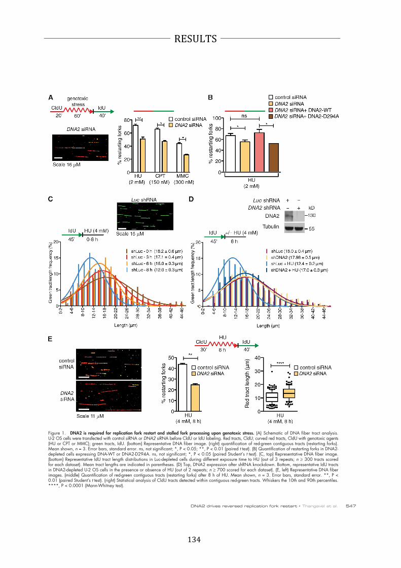
134
RESULTS
547DNA2 drives reversed replication fork restart • Thangavel et al.
Figure 1. DNA2 is required for replication fork restart and stalled fork processing upon genotoxic stress. (A) Schematic of DNA fiber tract analysis. U-2 OS cells were transfected with control siRNA or DNA2 siRNA before CldU or IdU labeling. Red tracts, CldU; curved red tracts, CldU with genotoxic agents (HU or CPT or MMC); green tracts, IdU. (bottom) Representative DNA fiber image. (right) quantification of red-green contiguous tracts (restarting forks). Mean shown, n = 3. Error bars, standard error. ns, not significant; *, P < 0.05; **, P < 0.01 (paired t test). (B) Quantification of restarting forks in DNA2-depleted cells expressing DNA-WT or DNA2-D294A. ns, not significant; *, P < 0.05 (paired Student’s t test). (C, top) Representative DNA fiber image. (bottom) Representative IdU tract length distributions in Luc-depleted cells during different exposure time to HU (out of 3 repeats; n ≥ 300 tracts scored for each dataset). Mean tract lengths are indicated in parentheses. (D) Top, DNA2 expression after shRNA knockdown. Bottom, representative IdU tracts in DNA2-depleted U-2 OS cells in the presence or absence of HU (out of 2 repeats; n ≥ 700 scored for each dataset). (E, left) Representative DNA fiber images. (middle) Quantification of red-green contiguous tracts (restarting forks) after 8 h of HU. Mean shown, n = 3. Error bars, standard error. **, P < 0.01 (paired Student’s t test). (right) Statistical analysis of CldU tracts detected within contiguous red-green tracts. Whiskers the 10th and 90th percentiles. ****, P < 0.0001 (Mann-Whitney test).

135
RESULTS
JCB • VOLUME 208 • NUMBER 5 • 2015 548
Genetic knockdown–rescue experiments confirmed that
complementation in RECQ1-depleted U-2 OS cells with shRNA-
resistant WT RECQ1 abrogates the effect of RECQ1 depletion
on replication fork processing upon HU treatment (Fig. 3 F).
Interestingly, expression of the ATPase-deficient RECQ1 mutant
K119R in RECQ1-depleted cells also abrogated the effect of
RECQ1 depletion indicating that the ATPase activity of RECQ1
was not required for its role in protecting stalled forks from
DNA2-dependent degradation (Fig. 3 F). These results point to
an additional role of RECQ1 in protecting replication forks
from extensive DNA2-dependent degradation, which is inde-
pendent of RECQ1 ATPase activity.
tracts were substantially shorter in RECQ1-depleted cells com-
pared with control when replication forks were stalled with HU
(after 8 h of HU treatment, the mean tract lengths were 7.9 and
12.0 µm, respectively; P < 0.0001; Fig. 3, A and B). In agree-
ment with results from luciferase-depleted cells, DNA2 was
also responsible for the nascent strand degradation phenotype
observed in RECQ1-deficient U-2 OS cells (Fig. 3 C). Analo-
gous results were obtained using the conditional DNA2 knock-
out HCT116 cell line (Fig. 2 C). In addition, we confirmed that
the DNA2-dependent nascent strand degradation observed in
the absence of RECQ1 is not limited to a specific replication in-
hibitor by replacing HU with CPT or MMC (Fig. 3, D and E).
Figure 2. DNA2 processes stalled replication forks. (A, top) DNA2 expression after siRNA knockdown. (bottom) Colony-forming assays in control and DNA2-depleted U-2 OS cells treated with 4 mM HU for the indicated time. (B) Representative IdU tracts in DNA2 conditional knockout HCT116 cells (out of two repeats). Tamoxifen was added to generate conditional knockout cells (see Materials and methods). (C, left) Expression of DNA2 and RECQ1 in tamoxifen-treated HCT116 cells. Right, representative IdU tracts in DNA2 conditional knockout HCT116 cells depleted for Luc or RECQ1 (out of three repeats). n ≥ 300 tracts scored for each dataset shown in B and C.

136
RESULTS
549DNA2 drives reversed replication fork restart • Thangavel et al.
Figure 3. RECQ1 regulates the DNA2-dependent degradation of stalled forks. (A) Representative IdU tracts in RECQ1-depleted U-2 OS cells during different exposure time to HU (out of 2 repeats; n ≥ 350 tracts scored for each dataset). (B) Bar graph represents the mean values of each time point from Figs. 1 C and 2 A. (top) RECQ1 expression after shRNA knockdown. (C, D, and E) Representative IdU tracts in RECQ1-, DNA2-, or RECQ1/DNA2-codepleted U-2 OS cells in the presence of HU (C), CPT (D), and MMC (E; out of 2 repeats; n ≥ 300 tracts scored for each dataset).(top) RECQ1 and DNA2 expression after shRNA or siRNA knockdown. (F) Representative IdU tracts in RECQ1-depleted U-2 OS cells complemented with shRNA-resistant WT RECQ1 (WT) or ATPase-deficient (K119R) RECQ1 (out of 2 repeats; n ≥ 325 tracts scored for each dataset). (top) Expression of Flag-tagged RECQ1-WT and RECQ1-K119R in RECQ1-depleted cells.

137
RESULTS
JCB • VOLUME 208 • NUMBER 5 • 2015 550
part of the WRN:DNA2 complex. Collectively, these results
suggest that DNA2 cooperates with WRN to promote nascent
strand processing and fork restart after HU treatment.
The nuclease activity of DNA2
and the ATPase activity of WRN are
essential to process stalled replication forks
DNA2 is characterized by an N-terminal nuclease domain and
by a C-terminal helicase domain, but the function of its helicase
activity is still debated (Masuda-Sasa et al., 2006). To assess the
roles of these two activities in stalled fork processing, we per-
formed genetic knockdown-rescue experiments where we de-
pleted DNA2 and then attempted to rescue fork processing by
expressing a Flag-tagged siRNA resistant WT DNA2 control,
nuclease-deficient DNA2-D294A, or ATPase-deficient DNA2-
K671E. All the experiments were performed in RECQ1-depleted
cells, where the effect of DNA2 is more apparent. DNA fiber
analysis showed that complementation with nuclease-deficient
DNA2 prevents fork processing, whereas complementation
with WT or ATPase-deficient DNA2 leads to the same fork pro-
cessing phenotype observed in DNA2-proficient cells (Fig. 5 D
and Fig. S2 C). Therefore, the nuclease, but not the ATPase ac-
tivity of DNA2, is necessary for fork processing.
Next, we used a Werner Syndrome (WS) fibroblast cell
line (AG11395) expressing missense mutant forms of WRN,
which inactivate either the exonuclease (WRN-E84A) or the
ATPase (K577M) activity of WRN (Pirzio et al., 2008). The
ATPase, but not the nuclease activity of WRN, was important
for fork processing (Fig. 5 E and Fig. S2 D). These findings
were validated by genetic knockdown-rescue experiments where
we complemented WRN-depleted U-2 OS cells either with
an shRNA resistant WT WRN control or the ATPase-deficient
WRN-K577M mutant and found that complementation with
the ATPase-deficient mutant prevented fork processing (Fig. S2,
E and F). Collectively, these results show that human DNA2
needs the support of the ATPase activity of WRN to promote
degradation of the nascent DNA strands.
DNA2 processes reversed replication forks
To gain insight into the actual replication structures processed
by DNA2, we inspected the fine architecture of the replication
intermediates using a combination of in vivo psoralen cross-
linking and EM (Neelsen et al., 2014). Our analysis showed
a substantial fraction of reversed replication forks ( 24% of
molecules analyzed) in control U-2 OS cells treated with 4 mM
HU. RECQ1-depletion, and to an even greater extent DNA2-
depletion, resulted in a higher frequency of fork reversal events
( 30 and 40%, respectively) compared with HU-treated cells.
Co-depletion of RECQ1 and DNA2 further increased the fre-
quency of reversed forks ( 50%), suggesting that RECQ1 and
DNA2 are involved into two distinct mechanisms of reversed
fork processing. Interestingly, RECQ1 and/or DNA2 depletion
also led to a significant amount of fork reversal events in unper-
turbed U-2 OS cells (Fig. 6, A and B). WRN-depletion pheno-
copied DNA2-depletion in terms of reversed fork accumulation,
both the presence and in the absence of HU. Moreover, DNA2/
DNA2 function in stalled fork processing
is distinct from EXO1, Mre11, and CtIP
Next, we tested whether other nucleases share a function similar
to DNA2 in stalled fork processing. To address this point, we
depleted Mre11, EXO1, and CtIP in U-2 OS cells with siRNA-
mediated technologies. We found that none of these nucleases
share the same phenotype of DNA2 in RECQ1-proficient cells
(Fig. 4 A). Furthermore, depletion of these nucleases had only a
marginal effect on the rescue of the prominent nascent strand
degradation phenotype observed in the absence of RECQ1, indi-
cating that DNA2 has a unique function in reversed fork pro-
cessing that is not shared by these human nucleases (Fig. 4, B–D).
MUS81 is another structure-specific nuclease that plays a critical
role in replication fork rescue by converting stalled replication
forks into DNA DSBs that can be processed by Homology Di-
rected Repair (HDR) (Hanada et al., 2007; Franchitto et al.,
2008). This raised the possibility that the DNA2-dependent
degradation originated from the processing of MUS81-dependent
DSBs. However, MUS81 depletion did not prevent nascent
strand degradation, indicating that DNA2 is not processing
stalled replication intermediates that are cleaved by MUS81
(Fig. 4 E).
DNA2 and WRN act together to process
stalled replication forks
DNA2-dependent dsDNA-end resection needs the support of
a RecQ helicase to unwind the DNA from the break (Cejka
et al., 2010; Niu et al., 2010; Nimonkar et al., 2011). To deter-
mine the identity of the helicase that acts in conjunction with
DNA2 in stalled fork processing, we measured the extent of na-
scent strand degradation in BLM-, WRN-, and RECQ4-depleted
U-2 OS cells. Our DNA fiber analysis showed that WRN deple-
tion mimicked the effect of DNA2-depletion, completely abro-
gating the prominent nascent strand degradation phenotype
observed in RECQ1-depleted U-2 OS cells (Fig. 5 A). The same
results were confirmed using WRN and DNA2 codepleted cells,
suggesting that DNA2 and WRN are epistatic in nucleolytic
processing of stalled forks (Fig. S1 C). The partial nascent
strand degradation observed in RECQ1-proficient U-2 OS cells
was also abrogated by WRN depletion (Fig. S1 D). Conversely,
BLM depletion had only a marginal effect on the nascent strand
degradation phenotype observed in RECQ1-depleted cells,
whereas RECQ4 depletion had no effect (Fig. S2, A and B). Thus,
the WRN helicase plays a prominent role in assisting DNA2-
dependent degradation of stalled replication forks.
We next compared the percentage of restarting replication
forks in DNA2-depleted, WRN-depleted, and DNA2/WRN-
codepleted cells. WRN depletion leads to a decrease in restart-
ing forks (69 to 50%; P = 0.0068). These results are almost
identical to those obtained with the DNA2-depleted or DNA2/
WRN-codepleted cells, implying that WRN and DNA2 are
epistatic also in the restart process (Fig. 5 B). The notion that
DNA2 and WRN functionally interact to process stalled repli-
cation intermediates is further supported by our observation that
the two proteins form a complex both in the presence and ab-
sence of replication stress (Fig. 5 C). Of note, RECQ1 is not
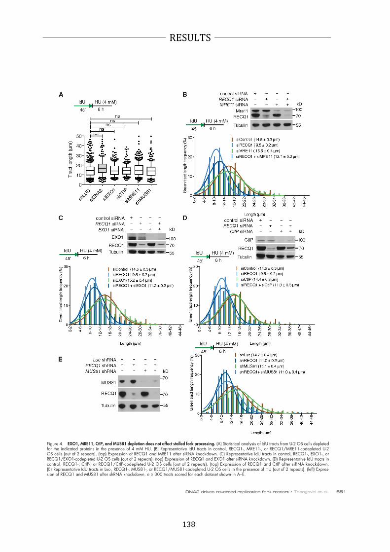
138
RESULTS
551DNA2 drives reversed replication fork restart • Thangavel et al.
Figure 4. EXO1, MRE11, CtIP, and MUS81 depletion does not affect stalled fork processing. (A) Statistical analysis of IdU tracts from U-2 OS cells depleted for the indicated proteins in the presence of 4 mM HU. (B) Representative IdU tracts in control, RECQ1-, MRE11-, or RECQ1/MRE11-codepleted U-2 OS cells (out of 2 repeats). (top) Expression of RECQ1 and MRE11 after siRNA knockdown. (C) Representative IdU tracts in control, RECQ1-, EXO1-, or RECQ1/EXO1-codepleted U-2 OS cells (out of 2 repeats). (top) Expression of RECQ1 and EXO1 after siRNA knockdown. (D) Representative IdU tracts in control, RECQ1-, CtIP-, or RECQ1/CtIP-codepleted U-2 OS cells (out of 2 repeats). (top) Expression of RECQ1 and CtIP after siRNA knockdown. (E) Representative IdU tracts in Luc-, RECQ1-, MUS81-, or RECQ1/MUS81-codepleted U-2 OS cells in the presence of HU (out of 2 repeats). (left) Expres-sion of RECQ1 and MUS81 after shRNA knockdown. n ≥ 300 tracts scored for each dataset shown in A–E.

139
RESULTS
JCB • VOLUME 208 • NUMBER 5 • 2015 552
Figure 5. DNA2 and WRN are epistatic in stalled fork processing and replication restart. (A) Representative IdU tracts in RECQ1-, WRN-, or RECQ1/WRN-codepleted U-2 OS cells (out of 2 repeats; n ≥ 300 tracts scored for each dataset). (top) RECQ1 and WRN expression after shRNA knockdown. (B) Quantification of restarting forks in DNA2-, WRN-, or DNA2/WRN-codepleted cells. Mean shown, n = 3. Error bars, standard error. *, P < 0.05; **, P < 0.01 (paired t test). (top) Expression of WRN and DNA2 after shRNA knockdown. (C) Co-IP experiments in HEK293T cells transfected with empty vec-tors, Flag-DNA2, or Strep-HA-WRN. Cells were treated with 4 mM HU (3 h) where indicated. Whole-cell extracts were analyzed before (input) and after IP. (D) Statistical analysis of IdU tracts from RECQ1/DNA2-codepleted U-2 OS cells complemented with WT, ATPase-deficient (K671E), or nuclease-deficient (D294A) DNA2, when indicated. (E) Statistical analysis of IdU tracts from RECQ1-depleted WS cells complemented with WT, ATPase-deficient (K577M), or nuclease-deficient (E84A) WRN. Whiskers in D and E indicate the 10th and 90th percentiles. ns, not significant; ****, P < 0.0001 (Mann-Whitney test). n ≥ 300 tracts scored for each dataset shown in D and E.

140
RESULTS
553DNA2 drives reversed replication fork restart • Thangavel et al.
Figure 6. DNA2 resects reversed replication forks. (A) Electron micrograph of a partially single-stranded (left) and entirely double-stranded (right) reversed fork observed on genomic DNA upon HU-treatment. The black arrow points to the ssDNA region on the re-versed arm. Inset, magnified four-way junction at the reversed replication fork. D, Daughter strand; P, Parental strand; R, Reversed arm. (B) Frequency of fork reversal and ssDNA composition of the reversed arms in RECQ1- or DNA2-depleted U-2 OS cells treated with HU (left) or in unperturbed conditions (right). The percentage values are indicated on the top of the bar. “# RI” indicates the number of analyzed replication intermediates. Data in B are reproduced with very similar results in at least one independent experiment.
WRN-codepletion did not cause a further increase in reversed
fork frequency, thus supporting our conclusion that DNA2 and
WRN work together in reversed fork processing (Fig. S3 A).
Next, we evaluated the single-strand composition of the
regressed arms. To measure ssDNA, we carefully inspected
the frequency and length of ssDNA regions on the regressed
arms by detecting local difference in filament thickness.
DNA2 depletion led to a higher frequency of reversed forks
with a dsDNA arm—and a corresponding decrease of partially
or entirely single-stranded reversed forks—in both RECQ1-
proficient and deficient cells (Fig. 6). Thus, DNA2-mediated
resection is directed to completely or partially digest one
strand of the reversed arm leading to reversed forks that are
either entirely single stranded or have a protruding ssDNA
tail. However, prolonged stalling by HU was associated with
accumulation of postreplicative ssDNA gaps on replicated
duplexes, which was maximal in RECQ1-depleted cells and
suppressed by DNA2 depletion (Fig. S3, B and C). Conse-
quently, ssDNA gaps may reflect additional activity of the
same nucleolytic apparatus along the postreplicated duplexes
or restart of partially resected reversed forks.
As an alternative readout for DNA2-dependent resection,
we examined the phosphorylation status of RPA and the check-
point kinase Chk1 (Zeman and Cimprich, 2014). DNA2 deple-
tion caused a reduction in RPA and Chk1 phosphorylation in both
RECQ1-proficient and RECQ1-deficient U-2 OS cells, suggest-
ing that the DNA2-dependent resection of nascent strands might
also contribute to checkpoint activation (Fig. S3 D).
RAD51 promotes DNA2-dependent
degradation of reversed replication forks
The central recombinase factor RAD51 is directly implicated in
reversed fork formation upon genotoxic stress (Zellweger et al.,
2015). Thus, we investigated whether RAD51 depletion may
affect the reversed fork processing activity of DNA2. We found
that RAD51 knockdown largely prevents DNA2 nucleolytic
processing both in RECQ1 proficient and RECQ1-deficient
cells (Fig. 7 A). Genetic knockdown–rescue experiments
confirmed that expression of exogenous RAD51 in RAD51-
depleted U-2 OS cells restored the fork processing phenotype
(Fig. 7 B). These results indicate that DNA2-dependent nucleolytic
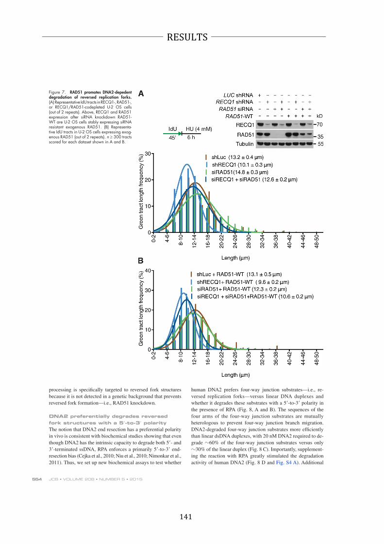
141
RESULTS
JCB • VOLUME 208 • NUMBER 5 • 2015 554
Figure 7. RAD51 promotes DNA2-depedent degradation of reversed replication forks. (A) Representative IdU tracts in RECQ1-, RAD51-, or RECQ1/RAD51-codepleted U-2 OS cells (out of 2 repeats). Above, RECQ1 and RAD51 expression after siRNA knockdown RAD51-WT are U-2 OS cells stably expressing siRNA resistant exogenous RAD51. (B) Representa-tive IdU tracts in U-2 OS cells expressing exog-enous RAD51 (out of 2 repeats). n ≥ 300 tracts scored for each dataset shown in A and B.
processing is specifically targeted to reversed fork structures
because it is not detected in a genetic background that prevents
reversed fork formation—i.e., RAD51 knockdown.
DNA2 preferentially degrades reversed
fork structures with a 5 -to-3 polarity
The notion that DNA2 end resection has a preferential polarity
in vivo is consistent with biochemical studies showing that even
though DNA2 has the intrinsic capacity to degrade both 5 - and
3 -terminated ssDNA, RPA enforces a primarily 5 -to-3 end-
resection bias (Cejka et al., 2010; Niu et al., 2010; Nimonkar et al.,
2011). Thus, we set up new biochemical assays to test whether
human DNA2 prefers four-way junction substrates—i.e., re-
versed replication forks—versus linear DNA duplexes and
whether it degrades these substrates with a 5 -to-3 polarity in
the presence of RPA (Fig. 8, A and B). The sequences of the
four arms of the four-way junction substrates are mutually
heterologous to prevent four-way junction branch migration.
DNA2-degraded four-way junction substrates more efficiently
than linear dsDNA duplexes, with 20 nM DNA2 required to de-
grade 60% of the four-way junction substrates versus only
30% of the linear duplex (Fig. 8 C). Importantly, supplement-
ing the reaction with RPA greatly stimulated the degradation
activity of human DNA2 (Fig. 8 D and Fig. S4 A). Additional

142
RESULTS
555DNA2 drives reversed replication fork restart • Thangavel et al.
Figure 8. Human DNA2 preferentially degrades branched DNA in a 5 -3 direction in reactions stimulated by WRN. (A) Degradation of a four-way junction by human DNA2 (hDNA2) in the presence of hRPA (native 6% polyacryl-amide gel) (B) Experiment as in A, but with dsDNA. (C) Quantitation of data from A and B. Averages shown ± SEM; n = 2. (D) DNA degradation is stimulated by hRPA. The data points from +hRPA condition are the same as in C. Averages shown ± SEM; n = 2. (E) Quan-titation of degradation of a 3 or 5 ssDNA-tailed three-way junction by hDNA2. The reactions were performed in 3 mM magnesium acetate and 22.3 nM hRPA. Averages shown ± SEM; n = 2. (F) Kinetics of degradation of a four-way junction by hDNA2 (9 nM) in the presence of hRPA (denaturing 20% polyacrylamide gel). The substrate was labeled at the 5 end (*). D294A, nuclease-dead variant of hDNA2. (G) Experiment as in F, but using a four-way junction labeled at the 3 end. (H) Quantitation of DNA cleavage near (less than 15 nt) a 5 or 3 DNA end from experiments of F and G. Averages shown ± SEM; n = 2. (I) WRN and hDNA2 degrade four-way junction DNA in a synergistic manner. Reactions with indicated hDNA2 and/or WRN concentrations and 65 nM hRPA were ana-lyzed on a 6% native polyacrylamide gel. Heat, partially heated DNA substrate indicating the positions of DNA unwinding intermediates. (J) Quantitation of four-way junc-tion and dsDNA degradation by human EXO1 (hEXO1). Averages shown ± SEM; n = 2.
experiments using either 5 -end or 3 -end 32P-labeled four-way
junctions confirmed that human DNA2 had a strong 5 -to-3
bias in end resection in the presence of RPA (Fig. 8, E–H; and
Fig. S4, B and C). Catalytically dead DNA2 D294A had no
capacity to degrade DNA, showing that the nuclease activity
is inherent to WT DNA2 (Fig. 8 F). The same results were
recapitulated using purified yeast DNA2 (Fig. S5, A–F). Inter-
estingly, addition of the ATPase-deficient RECQ1 mutant
(RECQ1-K119R) to the reaction mix significantly inhibited
the four-way junction degradation activity of human DNA2
(Fig. S4, D and E). These results suggest that the binding of
RECQ1 to stalled replication forks limits the fork processing

143
RESULTS
JCB • VOLUME 208 • NUMBER 5 • 2015 556
DNA2 function during DNA replication is vital for main-
tenance of genome stability (this study; Duxin et al., 2012;
Karanja et al., 2012). These findings indicate that the controlled
DNA2-dependent degradation of reversed replication forks is a
physiologically relevant mechanism to provide resistance to
prolonged genotoxic treatments. This mechanism is distinct
from the pathological MRE11-dependent degradation of stalled
replication intermediates detected in the absence of crucial
Fanconi Anemia (FA)/HR factors (Schlacher et al., 2011, 2012;
Hashimoto et al., 2012; Ying et al., 2012).
We find that depletion of the central recombinase factor
RAD51 prevents nascent strand degradation. This finding, coupled
with the recent observation that RAD51 is directly implicated in
reversed fork formation (Zellweger et al., 2015), reinforce our con-
clusion that the DNA2-dependent pathway starts from the reversed
arm of stalled replication forks and acts downstream of the
RAD51-mediated replication fork reversal. Given that RAD51 is
required for reversed fork formation (Zellweger et al., 2015), we
speculate that the MRE11-dependent pathway is only uncovered in
the absence of fork reversal—i.e., via a perturbation in RAD51
function—and likely attacks unprotected and nonreversed forks
upon prolonged stalling. A crucial challenge for future studies will
be to investigate why we do not observe a contribution of the
MRE11 pathway in nascent strand degradation upon RAD51
depletion. It is tempting to speculate that RAD51 depletion might
interfere with MRE11-dependent fork processing, in addition to
preventing fork reversal. Conversely, perturbation of RAD51
function—e.g., via BRCA2 depletion (Schlacher et al., 2011)—
might be sufficient to prevent fork reversal—hence DNA2-
dependent degradation—but still allow residual RAD51 loading
to promote MRE11-dependent degradation.
Our DNA fiber analysis suggests that DNA2 degrades
stalled replication intermediates beyond the maximum length
of the reversed arms measured by EM (up to several kilobases).
A possible interpretation of these results is that after the initial
DNA2/WRN-mediated regressed arm degradation is complete,
other nucleolytic activities or DNA2 itself may codegrade both
sides of the replication fork, thus leading to extensive degra-
dation events detectable by DNA fibers. In this scenario, our
EM images likely represent snapshots of the “slow steps” of
this reaction—i.e., the DNA2/WRN-mediated degradation of
the regressed arms—resulting in the drastic increase in reversed
fork frequency observed in the absence of DNA2. Once the
regressed arm has been resolved, the nucleolytic degradation
might quickly proceed to degrade nascent strands behind the
junction—as suggested by the DNA2-dependent increase in
ssDNA gaps behind the observed forks—finally leading to re-
annealing of the parental strands and backtracking of the fork
(Fig. S3 E). A new reversal event may occur when this extensive
degradation leads to asymmetric ssDNA accumulation at the fork
(Zellweger et al., 2015), resetting the backtracked fork to the slow
step of the process. However, fork backtracking is only one possi-
ble model to explain the extensive degradation detected by DNA
fibers and further work would be required to uncover additional
nucleolytic activities that might be involved in this process.
Biochemical studies suggested that Schizosaccharomyces
pombe Dna2 cleaves the leading and lagging reversed strands of
activity of DNA2, as inferred by our cellular studies. However,
we cannot rule out the possibility that the inhibitory effect ob-
served in the biochemical assays is simply associated with com-
petition for substrate recognition between the two proteins. In
agreement with our in vivo data, we show that WRN promoted
the degradative capacity of DNA2 on nicked, gapped, or four-
way junction substrates (Fig. 8 I and Fig. S4, F and G); similar
behavior was observed when yeast Dna2 was coupled with the
Sgs1 helicase (Fig. S5, G and H). DNA was degraded by WRN
and DNA2 in a remarkably synergistic manner: 5 nM concen-
tration of either WRN or DNA2 alone led only to a minor DNA
unwinding/degradation (Fig. 8 I, lanes 2 and 8). When com-
bined, both enzymes completely degraded the four-way junc-
tion DNA (Fig. 8 I, lane 5). In contrast, no such synergy was
observed when human DNA2 was combined with the noncog-
nate yeast meiotic Mer3 helicase (Fig. S4 H), suggesting that
the species-specific interaction between DNA2 and WRN re-
sults in a vigorous DNA degradation. Similarly, WT RECQ1
did not promote DNA degradation by DNA2 (Fig. S4 I).
On the basis of our results that DNA2 does not share the
same function of EXO1 in reversed fork processing, we decided to
compare the end-resection activities of human DNA2 and human
EXO1 using the four-way junction substrates. EXO1—unlike
DNA2—degraded both four-way junction substrates and linear
duplexes with equal efficiency (Fig. 8 J and Fig. S4, J and K). The
use of yeast variants of Dna2 and Exo1 yielded analogous results
(Fig. S5, I–K). Collectively, these studies further implicate DNA2,
and its nuclease activity, in reversed replication fork degradation—
that is specifically stimulated by WRN—and point to an important
difference in substrate preference between DNA2 and EXO1.
Moreover, the polarity of reversed fork degradation by DNA2
measured in the presence of RPA displays the same bias antici-
pated from the EM analysis of the replication intermediates.
Discussion
The present work uncovers a new mechanism for reversed fork
processing and restart that requires the coordinated activities of
the human DNA2 nuclease and WRN helicase (Fig. 9). The
DNA2-dependent end resection leads to partially single-stranded
reversed forks and is required for efficient replication fork re-
start under conditions of persistent replication blockage. WRN
interacts with DNA2 and its ATPase activity is needed for
DNA2-dependent degradation, presumably to transiently open
the dsDNA arm of the reversed replication forks.
To date, we have identified two mechanisms of reversed
replication fork resolution, one dependent on RECQ1 ATPase
and branch migration activity (Berti et al., 2013) and the other on
DNA2 nuclease and WRN ATPase activity. Moreover, the DNA2/
WRN mechanism is tightly regulated by an ATPase-independent
function of RECQ1 that might limit DNA2 activity by binding to
reversed forks. Of note, our EM experiments show that reversed
replication forks accumulate in RECQ1- and DNA2-depleted
cells also in unperturbed conditions suggesting that fork reversal
is remarkably frequent when DNA replication faces intrinsic rep-
lication fork obstacles, and that RECQ1 and DNA2 have a con-
served role in restarting reversed forks in unperturbed S-phase.

144
RESULTS
557DNA2 drives reversed replication fork restart • Thangavel et al.
depletion is consistent with observations that the deregulation
of checkpoint activity leads to a large increase in the number of
newly initiated origins (Couch et al., 2013). However, the extent
of ATR activation does not necessarily reflect the amount of
ssDNA detected at replication forks, whether at the junction, at
ssDNA gaps, or at regressed arms (Zellweger et al., 2015). In
light of these findings, we rather suggest that DNA2-dependent
ATR activation may reflect DNA2 recruitment to the stalled
forks per se, or subtle changes of fork architecture that are asso-
ciated with its recruitment but possibly escape our EM analysis.
This interpretation is supported by the recent discovery that
yeast Dna2 has a direct role in Mec1 activation (the ortholog of
human ATR), independent from its nuclease or helicase activity
(Kumar and Burgers, 2013). Of note, the increased origin firing
frequency observed upon DNA2 depletion is not associated to a
parallel increase in the frequency of termination events (Fig. S1 A)
possibly because the defects in replication fork restart associated
a model replication fork with similar efficiency in the absence of
replication protein A (Hu et al., 2012). However, it is likely that
only the 5 -to-3 directionality is important in vivo, because RPA
is known to stimulate the 5 -to-3 and inhibit the 3 -to-5 nuclease
activity of yeast DNA2 (Cejka et al., 2010; Niu et al., 2010). In
agreement with this conclusion, our biochemical data show that
DNA2-dependent end resection proceeds with a 5 to 3 polarity
in the presence of RPA. Moreover, our EM experiments clearly
show that DNA2 depletion affects the frequency of reversed forks
that are either entirely or partially single-stranded supporting the
notion that DNA2-dependent degradation of reversed forks oc-
curs with a preferential polarity in vivo.
The resection activity of human DNA2 was postulated to
activate the ATR/Chk1 checkpoint under conditions of replica-
tion stress (Karanja et al., 2012). Indeed, we find that DNA2 de-
pletion prevents ATR checkpoint activation after HU treatment.
Moreover, the increased origin firing observed upon DNA2
Figure 9. Schematic model for the combined roles of DNA2 and WRN in reversed fork pro-cessing. DNA2 and WRN functionally interact to process reversed forks. DNA2 degrades reversed forks with a 5 -to-3 polarity. WRN ATPase activity assists DNA2 degradation pos-sibly by promoting the opening of the reversed arm of the fork. RECQ1 limits DNA2 activity by an ATPase-independent function. Branch migration factors specifically recognize the partially resected reversed forks to promote fork restart. Alternatively, the newly formed 3 overhang of the reversed fork invades the duplex ahead of the fork, resulting in Holliday junction structures that can be resolved by spe-cific resolvases or dissolvases to promote fork restart. Gray box, RECQ1 can independently restart reversed forks by virtue of its ATPase and branch migration activity.

145
RESULTS
JCB • VOLUME 208 • NUMBER 5 • 2015 558
specific genetic backgrounds. For example, MRE11 degrades
stalled replication intermediates only in a BRCA2-deficent back-
ground, as already discussed (Schlacher et al., 2011). Moreover,
the cleavage of unresolved replicative intermediates by the
structure-specific MUS81 endonuclease is a late response to
replicative stress, which becomes activated only when other
attempts to overcome stalled replication have been exhausted
(Hanada et al., 2007; Franchitto et al., 2008). Thus, MUS81
might still resolve reversed replication forks as a back-up sys-
tem to unlink sister chromatids and facilitate mitotic segregation
in the absence of DNA2 or WRN.
Collectively, these studies highlight a new important
mechanism for the recovery from replication blockage. This
mechanism relies on the DNA2-dependent processing of re-
versed forks—leading to ssDNA stretches on the regressed
arms—which appear to promote efficient fork restart. A possi-
ble explanation for the need of partially single-stranded DNA
structures to promote fork restart is that they represent a key in-
termediate to activate an HDR-like mechanism of reversed fork
restart, as recently proposed in S. pombe (Carr and Lambert,
2013). In particular, the newly formed 3 overhang of the re-
versed fork might invade the duplex ahead of the fork resulting
in Holliday junction structures that can be resolved by specific
resolvases or dissolved by the combined action of the BLM
helicase (Sgs1 in yeast) and the type I topoisomerase TOP3
(Fig. 9). Alternatively, resumption of DNA replication might be
obtained by reverse branch migration, where the partially re-
sected reversed fork structures might be specifically recognized
by a motor protein—e.g., SMARCAL1 (Béous et al., 2013) or a
human RecQ helicase—to promote the branch migration-assisted
reestablishment of a functional replication fork.
Materials and methods
Cell lines, culture conditions, and reagentsU-2 OS, HEK 293, and Werner Syndrome fibroblast (AG11395) cells were grown in DMEM supplemented with 10% FBS at 37°C in 5% CO2. HCT116 cells were grown in McCoy’s 5A medium supplemented with 10% FBS. CldU, IdU, BrdU, hydroxyurea, mitomycin C, camptothecin, tamoxifen, pu-romycin, and hygromycin were obtained from Sigma-Aldrich.
DNA2 conditional knockout HCT116 cellsTo examine the response of cells to the complete absence of DNA2, we used a DNA2 conditional knockout cell line where exon 2 of the DNA2 gene is deleted (Karanja et al., 2014). The colorectal carcinoma HCT116 cell line carries 3 copies of DNA2 due to a duplication on chromosome 10. Two chromosomal copies were disrupted using rAAV-mediated gene targeting technology and exon 2 of the third allele was replaced with a conditional exon where the exon was flanked by loxP sites (DNA2flox/ / ). To create a conditional cell line these cells were stably transduced with a tamoxifen (4-OHT)-inducible Cre recombinase. Thus, the cell line is viable and can be propagated. The addition of tamoxifen to the culture media leads to excision of the endogenous DNA2 and the generation of a true DNA2-null cell. Com-plete loss of DNA2 occurs after 72 h of tamoxifen treatment. However, the DNA fiber experiments were performed after 40 h of tamoxifen treatment to have enough S-phase cells for DNA labeling.
AntibodiesAnti-DNA2 rabbit polyclonal (ab96488; 1:1,000), anti-MUS81 mouse monoclonal (ab14387; 1:1,000), and anti-CldU/BrdU rat monoclonal (ab6326; 1:6) antibodies (all from Abcam); anti-CtIP rabbit polyclonal (A300-488A; 1:1,000), anti-EXO1 rabbit polyclonal (A302-639A; 1:1,000), anti-pRPA32 (S4/S8) rabbit polyclonal (A300-245A; 1:1,000), and anti-pRPA32 (S33) rabbit polyclonal (300-246A; 1:2,000; all from
with DNA2 depletion limit the number of termination events
even under conditions of increased origin firing.
WRN plays an important—albeit mechanistically ill-
defined—role in the recovery from replication blockage, and
mutations in the WRN gene are linked to the cancer predisposi-
tion disorder Werner Syndrome (Sidorova et al., 2008; Murfuni
et al., 2012). Our studies infer that the high genomic instability
of WRN-deficient cells may result from aberrant processing of
reversed replication intermediates. In particular, given the con-
solidated role of WRN at difficult-to-replicate regions—e.g.,
telomeres and fragile sites (Crabbe et al., 2004; Murfuni et al.,
2012)—we speculate that WRN, in conjunction with DNA2, is
required to process reversed forks arising spontaneously at
these genomic loci. Biochemical studies pointed to a putative
role of WRN in fork reversal and/or restart by showing that
WRN efficiently promotes both the formation and restoration
of oligonucleotide-based reversed fork substrates (Machwe
et al., 2011). We show that WRN ATPase activity is needed for
the DNA2-dependent degradation of reversed replication forks.
Our interpretation for the role of WRN ATPase activity is that
it facilitates DNA2-dependent degradation of the reversed forks
by transiently opening the dsDNA arm of the reversed fork.
This mechanism is reminiscent to the DNA2-dependent mecha-
nism of DSB resection where the yeast Sgs1 helicase is required
to transiently open the DNA duplex to generate a 5 ssDNA tail
that is in turn degraded by DNA2 (Zhu et al., 2008; Cejka et al.,
2010; Niu et al., 2010). We suggest that WRN is the functional
homologue of Sgs1 in mammalian cells, at least in the context
of DNA2-dependent reversed replication fork processing. How-
ever, BLM was also shown to interact and cooperate with DNA2
to resect dsDNA ends in vitro opening the possibility that other
human RecQ helicases might substitute for WRN, depending
on the nature of the DNA lesion being processed or the particu-
lar cellular context (Nimonkar et al., 2011; Sturzenegger et al.,
2014). This mechanism seems to be well-conserved throughout
evolution because it is highly reminiscent of the stalled fork
processing pathway described in E. coli where the RecJ nucle-
ase cooperates with bacterial RecQ to process blocked replication
intermediates (Courcelle et al., 2003). In addition, the prokary-
otic RecBCD helicase-nuclease plays an important role in re-
secting replication forks after reversal (Seigneur et al., 1998)
and DNA2 is of the same family of nucleases as RecB. Whether
the DNA2/WRN-mediated resection activity can degrade addi-
tional stalled replication intermediates other than reversed forks
is worth future investigation.
EXO1, MRE11, and CtIP play central roles in DNA repair
and are also implicated in the recovery from replication fork
blockage (Cotta-Ramusino et al., 2005; Schlacher et al., 2011;
Yeo et al., 2014). None of these nucleases, however, partici-
pates in the DNA2-dependent processing of reversed replica-
tion forks pointing to a specific role of DNA2 that, unlike
its function in DSB resection, is not shared by other nucleases.
A possible interpretation of these results is that the reversed
forks are characterized by a particular structure of the terminal
end that does not require the trimming activity of other nucle-
ases to promote DNA2-dependent resection. However, some of
these nucleases might still be able to access stalled forks under

146
RESULTS
559DNA2 drives reversed replication fork restart • Thangavel et al.
were kind gifts from Dr. Pietro Pichierri (Insituto Superiore di Sanità, Rome, Italy). All transfections were done with Lipofectamine 2000 (Life technolo-gies Catalog no: 11668027). An shRNA targeting luciferase (5 -ACGCT-GAGTACTTCGAAATGT-3 ) was used for control shRNA experiments. The silencer select negative control (Life technologies, Catalog no. 4390843) or an siRNA targeting luciferase (5 -CGUACGCGGAAUACUUCGA-3 ) were used for control siRNA experiments, as indicated. Lentiviral mediated shRNA depletions were achieved using the following sequences cloned into the pLKO.1 lentiviral shRNA expression vector: BLM (5 -CGAAGGAAGTTGTAT-GCACTA-3 ), WRN (5 -GCTGGCAATTACCAGAACAAT-3 ), and MUS81 (5 -CACGCGCTTCGTATTTCAGAA-3 ). The procedure for lentiviral genera-tion and transduction has been described (Berti et al., 2013). Transduced U-2 OS cells were selected with 6 µg/ml puromycin. siRNA-mediated depletions were achieved using the following siRNAs from Invitrogen: DNA2 (5 -AUA-GCCAGUAGUAUUCGAU-3 ), CtIP (5 -CGAAUCUUAGAUGCACAAA-3 ), EXO1 (Invitrogen-HSS113557), and RAD51 (Invitrogen-1299001). In brief, siRNAs were transfected using Lipofectamine RNAiMAX (Life Technologies) following the manufacturer’s protocol. MRE11 (5 -GAAAGGCUCUAUC-GAAUGU-3 ) and RECQ4 (SMART pool) siRNAs were from Dharmacon and were transfected as previously described (Thangavel et al., 2010).
Microfluidic-assisted DNA fiber stretchingFor DNA replication fork restart analysis, asynchronous cells were pulse-labeled with 50 µM CldU for 20–30 min. 2 mM HU, 300 nM MMC, or 150 nM CPT was added to the CldU containing media and incubated for the indicated times. Cells were washed three times with medium and released with 50 µM IdU for 40 min. For nascent strand degradation analysis, asyn-chronous cells were pulse-labeled with 50 µM IdU for 45 min, washed three times with medium, incubated with 4 mM HU, 100 nM CPT, 200 nM MMC, or medium for times indicated. The pulse-labeled cells were trypsin collected and lysed in agarose plugs to prevent any mechanical breakage of replication tracts. Microfluidic platform for stretching the high-molecular weight DNA, coverslips, immunostaining and image acquisition of replica-tion tracts were performed as described (Sidorova et al., 2009; Berti et al., 2013). In brief, polydimethylsiloxane (PDMS) stamps with microchannels were Oxygen plasma treated and reversibly sealed to the silanized cover-slips. High-molecular weight DNA was loaded and stretched by capillary force into the microchannels. PDMS stamps were peeled-off and coverslips were left drying overnight. For immunostaining, DNA-stretched coverslips were denatured (2.5N HCL for 45 min), neutralized (0.1 M sodium borate and 3 washes with PBS), blocked (5% BSA and 0.5% Tween 20 in PBS for 30 min), incubated with primary antibodies (Anti-IdU/BrdU or both anti-IdU/BrdU and anti-CldU/BrdU for 30 min), washed (1% BSA and 0.1% Tween 20 in PBS, 3 times 5 min each) and incubated with secondary anti-bodies (anti–mouse Alexa Fluor 488-conjugated, or both anti–mouse Alexa Fluor 488–conjugated and anti–rat Alexa Fluor 594–conjugated for 1 h). Washed slides were mounted in prolong gold anti-fade reagent (Life Tech-nologies) and images were sequentially acquired (for double-label) with LAS AF software using TCS SP5 confocal microscope (Leica). A 63×/1.4 oil immersion objective was used. Images were captured at room tempera-ture. n ≥ 300 fiber tracts scored for each dataset. The DNA tract lengths were measured using ImageJ and the pixel length values were converted into micrometers using the scale bars created by the microscope. Statistical analysis was done using GraphPad Prism.
Clonogenic survival assayColony-forming assays were performed as previously described (Franken et al., 2006). In brief, 1,000 cells were plated per well and treated on the next day with 4 mM HU for 3, 6, and 8 h or 100 nM CPT for 6 h. Colonies were fixed, stained, and quantified 10 d after release from genotoxic stress. The plating efficiency and survival fraction were calculated as previously described (Franken et al., 2006). In brief, colonies were counted using an inverted stereomicroscope and the plating efficiency was calculated using the following formula: Plating Efficiency (PE) = (no. of colonies formed/no. of cells seeded) × 100%. From the plating efficiency, the surviving fraction (SF) was calculated as: SF = (no. of colonies formed after treatment/no. of cells seeded) × PE. The experiments were performed in triplicate and the statistical analysis was performed using GraphPad Prism.
Western blottingCells were washed with PBS and lysed either in standard RIPA buffer (PBS, 1% NP-40, 0.5% sodium deoxycholate, 0.1% SDS, 10 µg/ml aprotinin, 10 µg/ml PMSF, 1 mM Na3VO4, and 1 mM NaF) or MCL buffer (50 mM Tris, pH 8.0, 5 mM EDTA, 0.5% NP-40, 100 mM NaCl, 2 mM DTT, and freshly added protease and phosphatase inhibitors from Roche (1 tablet/10 ml of buffer). Cell lysates were resolved by SDS-PAGE and transferred to
Bethyl); anti-WRN rabbit polyclonal (NB100-471; 1:1,000); and anti-MRE11 rabbit polyclonal (NB100-142; 1:2,000; Novus); anti-RAD51 (H-92) rabbit polyclonal (sc-8349; 1:1,000) and anti-RECQ1 rabbit polyclonal (sc-25547; 1:2,000) from Santa Cruz; anti-rat Alexa (594-A11007; 1:1,000); and anti–mouse Alexa Flour (488-A11001; 1:1,000; Invitro-gen); anti-rabbit (31460; 1:10,000; Thermo Fisher Scientific); anti-tubulin mouse monoclonal (T5168; 1:5,000; Sigma-Aldrich); anti-IdU/BrdU mouse monoclonal (347580; 1:6) from BD; anti-Chk1 mouse monoclonal (sc-8408; 1:1,000; Santa Cruz Biotechnology, Inc.); anti-p-Chk1 (S345) rabbit monoclonal (2348; 1:1,000; Cell Signaling Technology); anti-RPA32 mouse monoclonal (NA19L; 1:1,000) from EMD Millipore; anti-RECQ1 rabbit polyclonal, raised against residues 634–649 of human RECQ1, is custom made (Mendoza-Maldonado et al., 2011); anti-BLM rabbit poly-clonal, raised against residues 1–449 of human BLM (Wu and Hickson, 2003), was a gift from I. Hickson (University of Copenhagen, Copenha-gen, Denmark); and anti-RECQ4 rabbit polyclonal, raised against residues 60–111 of human RECQ4 (Yin et al., 2004), was a gift from W. Wang (National Institute on Aging, Baltimore, MD).
Recombinant proteinsYeast Dna2 was expressed in yeast WDH668 strain from pGAL:DNA2 vector (Budd et al., 2000) and purified as previously described (Levikova et al., 2013). In brief, the cells were lysed and Dna2 was purified by affinity chromatography on Ni-NTA agarose (QIAGEN) and anti-Flag M2 affinity gel (Sigma-Aldrich). Yeast RPA was expressed in yeast BJ5464 strain containing three plasmids coding for Rfa1, Rfa2, and Rfa3 and purified as previously described (Kantake et al., 2003). In brief, the cells were lysed and yeast RPA was purified by affinity on ssDNA cellulose column (USB corporation) and by ion exchange chromatography using HiTrap Q column (GE Healthcare). Human DNA2 was expressed in Sf9 cells from a pFastBac:hDNA2 vector (polyhedrin promoter) provided by J. Campbell (Masuda-Sasa et al., 2006). The soluble extracts were obtained by salt extraction as previously described for Sgs1 (Cejka and Kowalc-zykowski, 2010). The subsequent purification of hDNA2 was performed as previously described for yeast Dna2 (Levikova et al., 2013) by affinity chromatography using Ni-NTA agarose (QIAGEN) and Anti-Flag M2 affinity gel (Sigma-Aldrich). Human RPA was expressed from p11d-tRPA vector (Henricksen et al., 1994) in BL21 E. coli cells and purified as described (Henricksen et al., 1994). In brief, hRPA was first bound to HiTrap Blue column (GE healthcare) and then to HiTrap Q column. The sequence coding for yeast Mer3 helicase was amplified from yeast genomic DNA (SK1 strain) using primers Mer3FO (5 -GCGCGCGGGCCCATGAAAA-CAAAGTTTGATCGCCTCGGTACAGGAAAAAGAAGTAGACCCTCTC-CAAATAATATTGACTTTAACGACCAG-3 ) and Mer3RE (5 -CGCGCGCTC-GAGTTCAAACTCTATATCGGAAC-3 ). The PCR product was digested with ApaI and XhoI restriction endonucleases (both from New England Biolabs) and cloned into corresponding sites in pFB-MBP-Sgs1-his after the polyhedrin promoter, creating pFB-MBP-Mer3-his vector. Mer3 was then expressed in Sf9 cells and purified using affinity chromatography as previously described for Sgs1 (Cejka and Kowalczykowski, 2010). In brief, MBP-tagged Mer3 was first bound to amylose resin (New England Biolabs), eluted and digested with PreScission protease to cleave the MBP tag. Mer3 was further purified by affinity on Ni-NTA agarose (QIAGEN) exploiting the 10x His-tag at its C-terminus. Sequence information is available on request.
Genetic knock-down-rescue experimentsRECQ1, DNA2, and RAD51 genetic knockdown-rescue experiments were performed using the procedure described (Berti et al., 2013; Yata et al., 2012). In brief, RECQ1 is depleted using the pLKO.1-puro-shRECQ1 (5 -GAG-CTTATGTTACCAGTTA-3 ) construct and rescue experiments are performed using the shRNA resistant pIRES-RECQ1-WT or K119R (ATPase dead) con-structs as described (Berti et al., 2013). DNA2 is depleted using an siRNA targeting the 3 UTR of DNA2 (5 -CAGUAUCUCCUCUAGCUAG-3 ). At least one isoform of DNA2 is not targeted by this sequence. DNA2 rescue experiments are performed using the pBabe-hygro-3xFLAG-DNA2 WT, D294A (Nuclease dead), or K671E (helicase dead) constructs. RAD51 is depleted using siRNAs targeting the 3 UTR (5 -GACUGCCAGGAU-AAAGCUU-3 and 5 -GUGCUGCAGCCUAAUGAGA-3 ) in U-2 OS stable cell lines expressing WT RAD51 as described (Yata et al., 2012). WRN depletions were achieved using pRS-puro-shWRN (5 -AGGCAGGTGTAG-GAATTGAAGGAGATCAG-3 ; sequence ID: TI333414) and exogenous expression is done with the shRNA resistant Flag-pCMVTag2B-WRN WT or K577M (helicase dead) constructs. Constructs for WRN depletion and overexpression of WT WRN and ATPase-deficient WRN (WRN-K577M)

147
RESULTS
JCB • VOLUME 208 • NUMBER 5 • 2015 560
pyruvate kinase, 1 nM DNA substrate (molecules), and recombinant pro-teins, as indicated. The reactions were assembled on ice and incubated for 30 min at 30°C for yeast proteins and at 37°C for human proteins. Un-less indicated otherwise, RPA was present in the reactions at saturating concentrations corresponding to a threefold excess over DNA, assuming all DNA was single-stranded and a DNA-binding site size of 25 nt for hRPA and of 20 nt for yRPA. The reactions were terminated by adding 5 µl Stop buffer (150 mM EDTA, 2% SDS, 30% glycerol, and 0.01% bromo-phenol blue), incubated for 30 min at room temperature and separated on polyacrylamide gels in TBE buffer under native conditions. Alternatively, for denaturing conditions, the reaction were terminated by adding 15 µl Formamide stop buffer (95% (vol/vol) formamide, 20 mM EDTA, 0.01% bromophenol blue), denatured by heating at 95°C for 5 min and sepa-rated on 20% denaturing polyacrylamide gels in TBE buffer. Gels were fixed, dried, exposed to a storage phosphor screen, and analyzed on Typhoon phosphor imager (GE Healthcare).
Online supplemental materialFig. S1 shows quantification of stalled forks, new origins, and termination events in DNA2-depleted cells upon genotoxic stress induction, as well as the statistical analysis of IdU tracts from RECQ1-, DNA2-, WRN-, RECQ1/DNA2-, RECQ1/WRN-, WRN/DNA2-, and RECQ1/WRN/DNA2-depleted U-2 OS cells. Fig. S2 shows the IdU tract length distribution in BLM- and RECQ4-depleted cells, respectively, as well as statistical analysis of IdU tracts from RECQ1/WRN-codepleted cells complemented with WT WRN or with ATPase-deficient WRN. Fig. S3 shows additional EM analysis, as well as the Western blot analysis of ATR-checkpoint activation in RECQ1- and/or DNA2-depleted U-2 OS cells. Fig. S4 shows additional biochemi-cal analysis of substrate specificity of human DNA2 and human EXO1. TFig. S5 shows biochemical assays of substrate specificity of yeast Dna2 and yeast Exo1. Online supplemental material is available at http://www .jcb.org/cgi/content/full/jcb.201406100/DC1.
We are grateful to Pietro Pichierri (Istituto Superiore di Sanità, Rome) for provid-ing the WS cells and WRN constructs, Damian Dalcher for his help with the EM analysis, Stephanie Felscher (University of Zurich) for kindly providing human EXO1 protein, Marc Wold (University of Iowa) for human RPA expres-sion construct, Lepakshi Ranjha (University of Zurich) for Mer3 protein, Fumiko Esashi (University of Oxford) for the Rad51 siRNAs and the U-2 OS cells stably expressing exogenous RAD51, and Judith Campbell (California Institute of Technology) for human and yeast DNA2/Dna2 expression constructs. We thank the Research Microscopy Core Facility of Saint Louis University for techni-cal support.
This work was supported by National Institutes of Health grant R01GM108648 to A. Vindigni, by startup funding from the Doisy Department of Biochemistry and Molecular Biology and from the Saint Louis University Cancer Center to A. Vindigni, by grants from the President’s Research Fund of Saint Louis University and by the GLIOMA-Interreg (Slovenian-Italian Coopera-tion 2007-2013) project to A. Vindigni, by the Swiss National Science Foun-dation grants 31003A_146924 to M. Levikova and PP00P3 133636 to P. Cejka, by National Institutes of Health grant GM0088351 and CA15446 to E.A. Hendrickson, and by a research contract from Horizon Discovery, Ltd to E.A. Hendrickson. M. Berti was supported by an EMBO short-term fellowship to perform EM experiments in M. Levikova laboratory.
The authors declare no competing financial interests.
Submitted: 24 June 2014Accepted: 14 January 2015
References
Atkinson, J., and P. McGlynn. 2009. Replication fork reversal and the main-tenance of genome stability. Nucleic Acids Res. 37:3475–3492. http://dx.doi.org/10.1093/nar/gkp244
Ayyagari, R., X.V. Gomes, D.A. Gordenin, and P.M. Burgers. 2003. Okazaki fragment maturation in yeast. I. Distribution of functions between FEN1 AND DNA2. J. Biol. Chem. 278:1618–1625. http://dx.doi.org/10.1074/jbc.M209801200
Bae, S.H., K.H. Bae, J.A. Kim, and Y.S. Seo. 2001. RPA governs endonuclease switching during processing of Okazaki fragments in eukaryotes. Nature. 412:456–461. http://dx.doi.org/10.1038/35086609
Berti, M., A. Ray Chaudhuri, S. Thangavel, S. Gomathinayagam, S. Kenig, M. Vujanovic, F. Odreman, T. Glatter, S. Graziano, R. Mendoza-Maldonado, et al. 2013. Human RECQ1 promotes restart of replication forks reversed by DNA topoisomerase I inhibition. Nat. Struct. Mol. Biol. 20:347–354. http://dx.doi.org/10.1038/nsmb.2501
PVDF membrane (GE Healthcare). Incubation with antibodies was per-formed overnight at 4°C. Proteins were visualized using ECL (Thermo Fisher Scientific) according to the manufacturer’s instructions.
Co-immunoprecipitation experimentsHEK293T cells were transfected with empty vectors, FLAG-DNA2, and Strep-HA-WRN by calcium phosphate. 48 h after transfection, cells were treated with 4 mM HU for 3 h, lysed in benzonase lysis buffer (50 mM Tris-HCl, pH 7.5, 75 mM KCl, 2 mM MgCl2, 20 mM NaF, 10 mM -glycerophosphate, 0.2 mM Na3VO4, and 0.2% Triton X-100) supplemented with protease in-hibitors (EDTA-free tablet; Sigma-Aldrich) by passing 10 times through a 26-G syringe needle and incubated 1 h at 4°C with 2 U/µl Benzonase (Sigma-Aldrich) to digest genomic DNA. KCl and EDTA concentrations were adjusted to 120 and 3 mM, respectively, and lysates were centrifuged at 14,000 rpm for 30 min. Immunoprecipitations of clarified lysates were per-formed with FLAG M2 or HA affinity agarose resin (Sigma-Aldrich) overnight at 4°C. Beads were washed 5 times with wash buffer (50 mM Tris-HCl, pH 7.5, 150 mM KCl, 3 mM EDTA, 2 mM MgCl2, 20 mM NaF, 10 mM -glycerophosphate, 0.2 mM Na3VO4, and 0.2% Triton X-100) and bound
proteins were eluted by boiling in SDS-PAGE sample buffer.
EM analysis of genomic DNA in mammalian cellsEM analysis of replication intermediates has been described in detail (Ray Chaudhuri et al., 2012; Neelsen et al., 2014), including a description of the important parameters to consider specifically for the identification and the scoring of reversed forks (Neelsen et al., 2014). In brief, 5–10 × 106 U-2 OS cells were harvested and genomic DNA was cross-linked by two rounds of incubation in 10 µg/ml 4,5 ,8-trimethylpsoralen (Sigma-Aldrich) and 3 min of irradiation with 366 nm UV light on a precooled metal block. Cells were lysed and genomic DNA was isolated from the nuclei by proteinase K (Roche) digestion and phenol-chloroform extraction. DNA was purified by isopropanol precipitation, digested with PvuII HF in the proper buffer for 3–5 h at 37°C, and replication intermediates were enriched on a benzoylated naphthoylated DEAE–cellulose (Sigma-Aldrich) column. EM samples were prepared by spreading the DNA on carbon-coated grids in the presence of benzyl-dimethyl-alkylammonium chloride and visualized by platinum rotary shadowing. Images were acquired on a transmission electron microscope (JOEL 1200 EX) with side-mounted camera (AMTXR41 supported by AMT software v601) and analyzed with ImageJ (National Institutes of Health).
Preparation of oligonucleotide-based DNA substratesDNA oligonucleotides were purchased from Microsynth and 32P-labeled either at the 5 terminus with [ -32P] ATP and T4 polynucleotide kinase (New England Biolabs), or at the 3 end with [ -32P] cordycepin-5 -triphosphate and terminal transferase (New England Biolabs) according to manufacturer’s instructions. Unincorporated nucleotides were removed using MicroSpin G25 columns (GE Healthcare). The substrates were prepared by heating the respective oligonucleotides at 95°C and gradually cooling to room temperature. The following oligonucleotides were used for the preparation of the four- way junction (X12-3 TOP L, HJ 1, HJ 2, and HJ 3), three-way junction with 3 tail (X12-3 TOP L, HJ 1, HJ 2Sb, and HJ 3), three-way junction with 5 ssDNA tail (X12-3 TOP L, HJ 1S, HJ 2, and HJ 3), nicked four-way junction (X12-3 TOP L, HJ 1, HJ 2Sa, HJ 2Sb, and HJ 3), replication fork (X12-3 TOP L, HJ 1S, HJ 2Sb, and HJ 3), and dsDNA (X12-3 TOP L and Bottom LC), respec-tively. The sequences of the oligonucleotides were: X12-3 TOP L (93 nt), 5 -GACGTCATAGACGATTACATTGCTAGGACATGCTGTCTAGAGACTATC-GCGACTTACGTTCCATCGCTAGGTTATTTTTTTTTTTTTTTTTTT-3 X12-3 HJ 1 (93 nt), 5 -AAAAAAAAAAAAAAAAAAATAACCTAGCGATGGAACGTA-AGTCGCGATGGGCTTAACTAGGATGCTACTGGCCCCGAATCAACCGT-ACTTGGG-3 X12-3 HJ 1S (48 nt), 5 -AAAAAAAAAAAAAAAAAAAT-AACCTAGCGATGGAACGTAAGTCGCGAT-3 X12-3 HJ 2 (93 nt), 5 -CCCAAGTACGGTTGATTCGGGGCCAGTAGCATCCTAGTT AAGCCCA-TTACGATTCGTTACCCATTCACTGTCAGAAGGCACCAGATAGATCTC-3 X12-3 HJ 2Sa (45 nt), 5 -CCCAAGTACGGTTGATTCGGGGCCAGTAGCA-TCCTAGTTAAGCCC-3 X12-3 HJ 2Sb (48 nt), 5 -ATTACGATTCGTTACCC-ATTCACTGTCAGAAGGCACCAGATAGATCTC-3 X12-3 HJ 3 (93 nt), 5 -GAGATCTATCTGGTGCCTTCTGACAGTGAATGGGTAACGAATCGT-AATAGTCTCTAGACAGCATGTCCTAGCAATGTAATCGTCTATGACGTC-3 X12-3 BOTTOM LC, 5 -AAAAAAAAAAAAAAAAAAATAACCTAGCGAT-GGAACGTAAGTCGCGATAGTCTCTAGACAGCATGTCCTAGCAATGTA-ATCGTCTATGACGTC-3 .
Nuclease assaysThe experiments were performed in a 15-µl volume in 25 mM Tris-acetate (pH 7.5), 2 mM magnesium acetate, 1 mM ATP, 1 mM dithiothreitol, 0.1 mg/ml BSA (New England Biolabs), 1 mM phosphoenolpyruvate, 80 U/ml

148
RESULTS
561DNA2 drives reversed replication fork restart • Thangavel et al.
Kantake, N., T. Sugiyama, R.D. Kolodner, and S.C. Kowalczykowski. 2003. The recombination-deficient mutant RPA (rfa1-t11) is displaced slowly from single-stranded DNA by Rad51 protein. J. Biol. Chem. 278:23410–23417. http://dx.doi.org/10.1074/jbc.M302995200
Karanja, K.K., S.W. Cox, J.P. Duxin, S.A. Stewart, and J.L. Campbell. 2012. DNA2 and EXO1 in replication-coupled, homology-directed repair and in the interplay between HDR and the FA/BRCA network. Cell Cycle. 11:3983–3996. http://dx.doi.org/10.4161/cc.22215
Karanja, K.K., E.H. Lee, E.A. Hendrickson, and J.L. Campbell. 2014. Preventing over-resection by DNA2 helicase/nuclease suppresses repair defects in Fanconi anemia cells. Cell Cycle. 13:1540–1550. http://dx.doi .org/10.4161/cc.28476
Kumar, S., and P.M. Burgers. 2013. Lagging strand maturation factor Dna2 is a component of the replication checkpoint initiation machinery. Genes Dev. 27:313–321. http://dx.doi.org/10.1101/gad.204750.112
Kuo, C., H. Nuang, and J.L. Campbell. 1983. Isolation of yeast DNA replication mutants in permeabilized cells. Proc. Natl. Acad. Sci. USA. 80:6465–6469. http://dx.doi.org/10.1073/pnas.80.21.6465
Levikova, M., D. Klaue, R. Seidel, and P. Cejka. 2013. Nuclease activity of Saccharomyces cerevisiae Dna2 inhibits its potent DNA helicase activ-ity. Proc. Natl. Acad. Sci. USA. 110:E1992–E2001. http://dx.doi.org/ 10.1073/pnas.1300390110
Liao, S., T. Toczylowski, and H. Yan. 2008. Identification of the Xenopus DNA2 protein as a major nuclease for the 5 ->3 strand-specific process-ing of DNA ends. Nucleic Acids Res. 36:6091–6100. http://dx.doi.org/ 10.1093/nar/gkn616
Machwe, A., R. Karale, X. Xu, Y. Liu, and D.K. Orren. 2011. The Werner and Bloom syndrome proteins help resolve replication blockage by con-verting (regressed) holliday junctions to functional replication forks. Biochemistry. 50:6774–6788. http://dx.doi.org/10.1021/bi2001054
Masuda-Sasa, T., O. Imamura, and J.L. Campbell. 2006. Biochemical analy-sis of human Dna2. Nucleic Acids Res. 34:1865–1875. http://dx.doi.org/ 10.1093/nar/gkl070
Mendoza-Maldonado, R., V. Faoro, S. Bajpai, M. Berti, F. Odreman, M. Vindigni, T. Ius, A. Ghasemian, S. Bonin, M. Skrap, et al. 2011. The human RECQ1 helicase is highly expressed in glioblastoma and plays an important role in tumor cell proliferation. Mol. Cancer. 10:83. http://dx.doi.org/10.1186/1476-4598-10-83
Mimitou, E.P., and L.S. Symington. 2008. Sae2, Exo1 and Sgs1 collaborate in DNA double-strand break processing. Nature. 455:770–774. http://dx.doi.org/10.1038/nature07312
Murfuni, I., A. De Santis, M. Federico, M. Bignami, P. Pichierri, and A. Franchitto. 2012. Perturbed replication induced genome wide or at common fragile sites is differently managed in the absence of WRN. Carcinogenesis. 33:1655–1663. http://dx.doi.org/10.1093/carcin/bgs206
Neelsen, K.J., A.R. Chaudhuri, C. Follonier, R. Herrador, and M. Lopes. 2014. Visualization and interpretation of eukaryotic DNA replication interme-diates in vivo by electron microscopy. Methods Mol. Biol. 1094:177–208. http://dx.doi.org/10.1007/978-1-62703-706-8_15
Nicolette, M.L., K. Lee, Z. Guo, M. Rani, J.M. Chow, S.E. Lee, and T.T. Paull. 2010. Mre11-Rad50-Xrs2 and Sae2 promote 5 strand resection of DNA double-strand breaks. Nat. Struct. Mol. Biol. 17:1478–1485. http://dx.doi .org/10.1038/nsmb.1957
Nimonkar, A.V., J. Genschel, E. Kinoshita, P. Polaczek, J.L. Campbell, C. Wyman, P. Modrich, and S.C. Kowalczykowski. 2011. BLM-DNA2-RPA-MRN and EXO1-BLM-RPA-MRN constitute two DNA end resec-tion machineries for human DNA break repair. Genes Dev. 25:350–362. http://dx.doi.org/10.1101/gad.2003811
Niu, H., W.H. Chung, Z. Zhu, Y. Kwon, W. Zhao, P. Chi, R. Prakash, C. Seong, D. Liu, L. Lu, et al. 2010. Mechanism of the ATP-dependent DNA end-resection machinery from Saccharomyces cerevisiae. Nature. 467:108–111. http://dx.doi.org/10.1038/nature09318
Peng, G., H. Dai, W. Zhang, H.J. Hsieh, M.R. Pan, Y.Y. Park, R.Y. Tsai, I. Bedrosian, J.S. Lee, G. Ira, and S.Y. Lin. 2012. Human nuclease/heli-case DNA2 alleviates replication stress by promoting DNA end resec-tion. Cancer Res. 72:2802–2813. http://dx.doi.org/10.1158/0008-5472 .CAN-11-3152
Pirzio, L.M., P. Pichierri, M. Bignami, and A. Franchitto. 2008. Werner syn-drome helicase activity is essential in maintaining fragile site stability. J. Cell Biol. 180:305–314. http://dx.doi.org/10.1083/jcb.200705126
Ray Chaudhuri, A., Y. Hashimoto, R. Herrador, K.J. Neelsen, D. Fachinetti, R. Bermejo, A. Cocito, V. Costanzo, and M. Lopes. 2012. Topoisomerase I poisoning results in PARP-mediated replication fork reversal. Nat. Struct. Mol. Biol. 19:417–423. http://dx.doi.org/10.1038/nsmb.2258
Schlacher, K., N. Christ, N. Siaud, A. Egashira, H. Wu, and M. Jasin. 2011. Double-strand break repair-independent role for BRCA2 in blocking stalled replication fork degradation by MRE11. Cell. 145:529–542. http://dx.doi .org/10.1016/j.cell.2011.03.041
Bétous, R., A.C. Mason, R.P. Rambo, C.E. Bansbach, A. Badu-Nkansah, B.M. Sirbu, B.F. Eichman, and D. Cortez. 2012. SMARCAL1 catalyzes fork regression and Holliday junction migration to maintain genome stabil-ity during DNA replication. Genes Dev. 26:151–162. http://dx.doi.org/10 .1101/gad.178459.111
Bétous, R., F.B. Couch, A.C. Mason, B.F. Eichman, M. Manosas, and D. Cortez. 2013. Substrate-selective repair and restart of replication forks by DNA translocases. Cell Reports. 3:1958–1969. http://dx.doi.org/ 10.1016/j.celrep.2013.05.002
Budd, M.E., and J.L. Campbell. 1995. A yeast gene required for DNA replication encodes a protein with homology to DNA helicases. Proc. Natl. Acad. Sci. USA. 92:7642–7646. http://dx.doi.org/10.1073/pnas.92.17.7642
Budd, M.E., and J.L. Campbell. 1997. A yeast replicative helicase, Dna2 he-licase, interacts with yeast FEN-1 nuclease in carrying out its essential function. Mol. Cell. Biol. 17:2136–2142.
Budd, M.E., W. Choe, and J.L. Campbell. 2000. The nuclease activity of the yeast DNA2 protein, which is related to the RecB-like nucleases, is essential in vivo. J. Biol. Chem. 275:16518–16529. http://dx.doi.org/10.1074/jbc.M909511199
Carr, A.M., and S. Lambert. 2013. Replication stress-induced genome instability: the dark side of replication maintenance by homologous recombination. J. Mol. Biol. 425:4733–4744. http://dx.doi.org/10.1016/j.jmb.2013.04.023
Cejka, P., and S.C. Kowalczykowski. 2010. The full-length Saccharomyces cerevisiae Sgs1 protein is a vigorous DNA helicase that preferentially unwinds holliday junctions. J. Biol. Chem. 285:8290–8301. http://dx.doi .org/10.1074/jbc.M109.083196
Cejka, P., E. Cannavo, P. Polaczek, T. Masuda-Sasa, S. Pokharel, J.L. Campbell, and S.C. Kowalczykowski. 2010. DNA end resection by Dna2-Sgs1-RPA and its stimulation by Top3-Rmi1 and Mre11-Rad50-Xrs2. Nature. 467:112–116. http://dx.doi.org/10.1038/nature09355
Cotta-Ramusino, C., D. Fachinetti, C. Lucca, Y. Doksani, M. Lopes, J. Sogo, and M. Foiani. 2005. Exo1 processes stalled replication forks and counteracts fork reversal in checkpoint-defective cells. Mol. Cell. 17:153–159. http://dx.doi.org/10.1016/j.molcel.2004.11.032
Couch, F.B., C.E. Bansbach, R. Driscoll, J.W. Luzwick, G.G. Glick, R. Bétous, C.M. Carroll, S.Y. Jung, J. Qin, K.A. Cimprich, and D. Cortez. 2013. ATR phosphorylates SMARCAL1 to prevent replication fork collapse. Genes Dev. 27:1610–1623. http://dx.doi.org/10.1101/gad.214080.113
Courcelle, J., J.R. Donaldson, K.H. Chow, and C.T. Courcelle. 2003. DNA dam-age-induced replication fork regression and processing in Escherichia coli. Science. 299:1064–1067. http://dx.doi.org/10.1126/science.1081328
Crabbe, L., R.E. Verdun, C.I. Haggblom, and J. Karlseder. 2004. Defective telo-mere lagging strand synthesis in cells lacking WRN helicase activity. Science. 306:1951–1953. http://dx.doi.org/10.1126/science.1103619
Duxin, J.P., H.R. Moore, J. Sidorova, K. Karanja, Y. Honaker, B. Dao, H. Piwnica-Worms, J.L. Campbell, R.J. Monnat Jr., and S.A. Stewart. 2012. Okazaki fragment processing-independent role for human Dna2 enzyme during DNA replication. J. Biol. Chem. 287:21980–21991. http://dx.doi.org/ 10.1074/jbc.M112.359018
Franchitto, A., L.M. Pirzio, E. Prosperi, O. Sapora, M. Bignami, and P. Pichierri. 2008. Replication fork stalling in WRN-deficient cells is overcome by prompt activation of a MUS81-dependent pathway. J. Cell Biol. 183: 241–252. http://dx.doi.org/10.1083/jcb.200803173
Franken, N.A., H.M. Rodermond, J. Stap, J. Haveman, and C. van Bree. 2006. Clonogenic assay of cells in vitro. Nat. Protoc. 1:2315–2319. http://dx.doi.org/10.1038/nprot.2006.339
Gari, K., C. Décaillet, M. Delannoy, L. Wu, and A. Constantinou. 2008. Remodeling of DNA replication structures by the branch point translocase FANCM. Proc. Natl. Acad. Sci. USA. 105:16107–16112. http://dx.doi.org/10.1073/ pnas.0804777105
Gravel, S., J.R. Chapman, C. Magill, and S.P. Jackson. 2008. DNA helicases Sgs1 and BLM promote DNA double-strand break resection. Genes Dev. 22:2767–2772. http://dx.doi.org/10.1101/gad.503108
Hanada, K., M. Budzowska, S.L. Davies, E. van Drunen, H. Onizawa, H.B. Beverloo, A. Maas, J. Essers, I.D. Hickson, and R. Kanaar. 2007. The structure-specific endonuclease Mus81 contributes to replication restart by generating double-strand DNA breaks. Nat. Struct. Mol. Biol. 14:1096–1104. http://dx.doi.org/10.1038/nsmb1313
Hashimoto, Y., F. Puddu, and V. Costanzo. 2012. RAD51- and MRE11-dependent reassembly of uncoupled CMG helicase complex at collapsed replication forks. Nat. Struct. Mol. Biol. 19:17–24. http://dx.doi.org/10.1038/nsmb.2177
Henricksen, L.A., C.B. Umbricht, and M.S. Wold. 1994. Recombinant replica-tion protein A: expression, complex formation, and functional character-ization. J. Biol. Chem. 269:11121–11132.
Hu, J., L. Sun, F. Shen, Y. Chen, Y. Hua, Y. Liu, M. Zhang, Y. Hu, Q. Wang, W. Xu, et al. 2012. The intra-S phase checkpoint targets Dna2 to prevent stalled replication forks from reversing. Cell. 149:1221–1232. http://dx.doi.org/10.1016/j.cell.2012.04.030

149
RESULTS
JCB • VOLUME 208 • NUMBER 5 • 2015 562
Schlacher, K., H. Wu, and M. Jasin. 2012. A distinct replication fork protection pathway connects Fanconi anemia tumor suppressors to RAD51-BRCA1/2. Cancer Cell. 22:106–116. http://dx.doi.org/10.1016/j.ccr.2012.05.015
Seigneur, M., V. Bidnenko, S.D. Ehrlich, and B. Michel. 1998. RuvAB acts at arrested replication forks. Cell. 95:419–430. http://dx.doi.org/10.1016/ S0092-8674(00)81772-9
Sidorova, J.M., N. Li, A. Folch, and R.J. Monnat Jr. 2008. The RecQ helicase WRN is required for normal replication fork progression after DNA dam-age or replication fork arrest. Cell Cycle. 7:796–807. http://dx.doi.org/ 10.4161/cc.7.6.5566
Sidorova, J.M., N. Li, D.C. Schwartz, A. Folch, and R.J. Monnat Jr. 2009. Microfluidic-assisted analysis of replicating DNA molecules. Nat. Protoc. 4:849–861. http://dx.doi.org/10.1038/nprot.2009.54
Sturzenegger, A., K. Burdova, R. Kanagaraj, M. Levikova, C. Pinto, P. Cejka, and P. Janscak. 2014. DNA2 cooperates with the WRN and BLM RecQ helicases to mediate long-range DNA end resection in human cells. J. Biol. Chem. 289:27314–27326. http://dx.doi.org/10.1074/jbc.M114.578823
Thangavel, S., R. Mendoza-Maldonado, E. Tissino, J.M. Sidorova, J. Yin, W. Wang, R.J. Monnat Jr., A. Falaschi, and A. Vindigni. 2010. Human RECQ1 and RECQ4 helicases play distinct roles in DNA replication initiation. Mol. Cell. Biol. 30:1382–1396. http://dx.doi.org/10.1128/MCB.01290-09
Wawrousek, K.E., B.K. Fortini, P. Polaczek, L. Chen, Q. Liu, W.G. Dunphy, and J.L. Campbell. 2010. Xenopus DNA2 is a helicase/nuclease that is found in complexes with replication proteins And-1/Ctf4 and Mcm10 and DSB response proteins Nbs1 and ATM. Cell Cycle. 9:1156–1166. http://dx.doi.org/10.4161/cc.9.6.11049
Wu, L., and I.D. Hickson. 2003. The Bloom’s syndrome helicase suppresses crossing over during homologous recombination. Nature. 426:870–874. http://dx.doi.org/10.1038/nature02253
Yata, K., J. Lloyd, S. Maslen, J.Y. Bleuyard, M. Skehel, S.J. Smerdon, and F. Esashi. 2012. Plk1 and CK2 act in concert to regulate Rad51 during DNA double strand break repair. Mol. Cell. 45:371–383. http://dx.doi.org/10 .1016/j.molcel.2011.12.028
Yeo, J.E., E.H. Lee, E.A. Hendrickson, and A. Sobeck. 2014. CtIP mediates rep-lication fork recovery in a FANCD2-regulated manner. Hum. Mol. Genet. 23:3695–3705. http://dx.doi.org/10.1093/hmg/ddu078
Yin, J., Y.T. Kwon, A. Varshavsky, and W. Wang. 2004. RECQL4, mutated in the Rothmund-Thomson and RAPADILINO syndromes, interacts with ubiquitin ligases UBR1 and UBR2 of the N-end rule pathway. Hum. Mol. Genet. 13:2421–2430. http://dx.doi.org/10.1093/hmg/ddh269
Ying, S., F.C. Hamdy, and T. Helleday. 2012. Mre11-dependent degradation of stalled DNA replication forks is prevented by BRCA2 and PARP1. Cancer Res. 72:2814–2821. http://dx.doi.org/10.1158/0008-5472.CAN-11-3417
Zellweger, R., D. Dalcher, K. Mutreja, R. Herrador, M. Berti, A. Vindigni, and M. Lopes. 2015. Rad51-mediated replication fork reversal is a global response to genotoxic treatments in human cells. J. Cell Biol. 208:563–579.
Zeman, M.K., and K.A. Cimprich. 2014. Causes and consequences of replication stress. Nat. Cell Biol. 16:2–9. http://dx.doi.org/10.1038/ncb2897
Zhu, Z., W.H. Chung, E.Y. Shim, S.E. Lee, and G. Ira. 2008. Sgs1 helicase and two nucleases Dna2 and Exo1 resect DNA double-strand break ends. Cell. 134:981–994. http://dx.doi.org/10.1016/j.cell.2008.08.037

150
RESULTS
DNA2 drives reversed replication fork restart • Thangavel et al.
TH
E J
OU
RN
AL
O
F C
EL
L B
IO
LO
GY
S1
JCB SUPPLEMENTAL MATERIAL
Thangalev et al., http://www.jcb.org/cgi/content/full/jcb.201406100/DC1
[ID]FIGS 1 [/ID]
Figure S1. DNA2 and WRN function in stalled fork processing. (A, left) Schematic of DNA fi ber tract analysis. (right) Quantifi cation of red tracts (stalled forks), green tracts (new origins), and contiguous red–green–red tracts (termination events). Proper quantifi cation of stalled forks is complicated by the fact that termination events might also lead to red tracts if termination occurs before the addition of the second label. Mean shown, n = 3. Error bars, standard error. ns, not signifi cant; *, P < 0.05; **, P < 0.01; ***, P < 0.001 (paired Student’s t test). (B, left) Expression of Flag-tagged WT (DNA2-WT) or nuclease-dead (DNA2-D294A) DNA2 in DNA2-depleted U-2 OS cells. (right) Quantifi cation of stalled forks in DNA2-depleted cells expressing DNA-WT or DNA2-D294A. Mean shown, n = 3. Error bars, standard error. ns, not signifi cant; *, P < 0.05 (paired t test). (C, top) Expression of RECQ1, WRN, DNA2, and tubulin in U-2 OS cells transfected with the indicated shRNA or siRNA. (middle) Statistical analysis of IdU tracts from U-2 OS cells depleted for the indicated proteins in the presence of 4 mM HU. (bottom) Statistical analysis of IdU tracts from U-2 OS cells depleted for the indicated proteins in the absence of drug treatment. Whiskers indicate the 10th and 90th percentiles. ns, not signifi cant (Mann-Whitney test). n ≥ 300 scored for each dataset. (D, top) Expression of WRN after WRN knockdown and representative fi ber tract images in Luc- and WRN-depleted U-2 OS cells. Bar, 15 µm. Representative IdU tracts in WRN-depleted U-2 OS cells in the presence or absence of HU (out of 2 repeats; n ≥ 700 scored for each dataset).

151
RESULTS
JCB
Figure S2. BLM or RECQ4 depletion does not have a signifi cant effect on stalled fork processing. (A) Representative IdU tracts in Luc-, RECQ1-, BLM-, or RECQ1/BLM-codepleted U-2 OS cells in the presence of HU (out of 2 repeats; n ≥ 300 scored for each dataset). (top) Expression of RECQ1, BLM and tubu-lin in U-2 OS cells transfected with indicated shRNA. (B) Representative IdU tracts in control, RECQ1-, RECQ4-, or RECQ1/RECQ4-codepleted U-2 OS cells in the presence of HU (out of 2 repeats; n ≥ 350 scored for each dataset). (top) Expression of RECQ1, RECQ4 and tubulin in U-2 OS cells transfected with indicated shRNA. (C) Expression of RECQ1, DNA2-WT, DNA2-K671E, and DNA2-D294A in U-2 OS cells transfected with the indicated shRNA or siRNA. (D) Expression of RECQ1, WRN-WT, WRN-K577M, and WRN-E84A in WS cells. (E) Statistical analysis of IdU tracts from RECQ1-, WRN-, or RECQ1/WRN-codepleted U-2 OS cells. The RECQ1/WRN-codepleted cells were complemented with WT or ATPase-defi cient (K577M) WRN, where indicated. Whiskers indicate the 10th and 90th percentiles. ns, not signifi cant; ****, P < 0.0001 (Mann-Whitney test). (F) Expression of RECQ1, WRN-WT, and WRN-K577M in U-2 OS cells transfected with the indicated shRNA.

152
RESULTS
DNA2 drives reversed replication fork restart • Thangavel et al. S3
Figure S3. DNA2 promotes ssDNA gap accumulation on replicated duplexes and the ATR-mediated checkpoint activation. (A) Frequency of fork reversal and ssDNA composition of the reversed arms in WRN- and/or DNA2-depleted U-2 OS cells in the presence and absence of HU. The percentage values are indicated on the top of the bar. “# RI” indicates the number of analyzed replication intermediates. (B) Representative electron micrographs of replication forks displaying ssDNA gaps on the replicated duplexes or at the replication fork junction observed on genomic DNA in shRECQ1 U-2 OS cells upon HU-treatment. The black arrows point to ssDNA gaps. D, Daughter strand; P, Parental strand. (C) Statistical distribution of ssDNA gaps on newly replicated du-plexes in RECQ1- and/or DNA2-depleted U-2 OS cells treated with HU (top) or in unperturbed conditions (bottom). “# RI” is the number of analyzed replication intermediates. (D) Western blot analysis of ATR-checkpoint activation (pChk1 and pRPA) in RECQ1- and/or DNA2-depleted U-2 OS cells with or without HU treatment. Total Chk1 and RPA level are displayed and used as loading control. (E) Schematic of the different structures detected by EM and DNA fi bers. EM is a static method, which enriches for snapshots of the “slow steps” of a reaction (i.e., partially resected reversed forks). After fork restart, the nucleolytic degradation quickly proceeds to degrade nascent strands behind the junction. Reannealing of the parental strands leads to “backtracking” of the fork. A new reversal event arises as a consequence of asymmetric degradation, and thus ssDNA accumulation in proximity to the fork. Backtracking is easily detected by DNA fi ber, but not by EM because a reversed fork formed after degradation and backtracking is indistinguishable from the original re-versed fork present before initial degradation.

153
RESULTS
JCBS4
Figure S4. Human DNA2 but not EXO1 preferentially degrades branched DNA. (A) Degradation of a four-way junction by hDNA2 without human RPA (hRPA). Reaction products were separated on a native polyacrylamide gel (6%); *, position of the 32 P label. (B) Degradation of a three-way junction with a 5 ! ssDNA tail by hDNA2 in the presence of hRPA (22.3 nM). Reaction products were separated on a native polyacrylamide gel (6%); *, position of the 32 P label. (C) Same experiment as in B, but with a junction containing a 3 ! ssDNA tail. (D) RECQ1 K119R (ATPase-dead) inhibits four-way junction degrada-tion by hDNA2. Increasing concentrations of RECQ1 (K119R) were preincubated with the substrate, and then hDNA2 (20 nM) was added to the reaction mixture. All reactions contained hRPA (65 nM). Reaction products were separated on a native polyacrylamide gel (6%); *, position of the 32 P label. (E) Quantitation of data from D. Averages shown ± SEM, n = 2. (F) Synergistic action of hDNA2 and WRN on a nicked plasmid based DNA substrate. The reactions contained 614 nM hRPA and were incubated at 37°C for 60 min. Products were separated on a 1% agarose gel and stained with GelRed. WRN helicase promotes degradation of nicked DNA by hDNA2. (G) Same experiment as in F, but with a gapped DNA substrate. (H) Degradation of a four-way junction by hDNA2 and S. cerevisiae Mer3. hDNA2 only degrades ssDNA unwound by Mer3, no synergy in DNA degradation was observed. All reactions contained hRPA, and were analyzed on a native polyacrylamide gel (6%). (I) Degradation of four-way junction by hDNA2 is not stimulated by WT hRECQ1. All reactions contained hRPA (65 nM). Reaction products were separated on a native polyacrylamide gel (6%). (J) Degradation of a four-way junction by hEXO1. Reaction products were separated on a native polyacrylamide gel (6%); *, position of the 32 P label. (K) Same experiment as in J, but with dsDNA.

154
RESULTS
DNA2 drives reversed replication fork restart • Thangavel et al. S5
Figure S5. Yeast Dna2 but not Exo1 preferentially degrades branched DNA. (A) Degradation of a four-way junction by yDna2 in presence (left) or absence (right) of yeast RPA (yRPA). Reactions were separated on a native polyacrylamide gel (6%), *, position of the 32 P label. (B) Experiment as in A, but with dsDNA and yRPA. (C) Yeast Dna2 preferentially degrades four-way junctions in the presence of yRPA. Quantitation of data from A and B. Averages shown ± SEM; n = 2. (D) yRPA promotes DNA degradation by yDna2. Quantitation of data from A. The data points representing the degradation of a four-way junction in the presence of yRPA are identical to those from C. Mean shown ± SEM, n = 2. (E) Yeast Dna2 preferentially degrades branched structures over dsDNA. Quantitation of degradation of various DNA substrates as indicated (cartoons on the right) by yDna2 WT in presence of yRPA. Averages shown ± SEM; n = 2. (F) Denaturing 20% polyacryl-amide gel showing that nuclease-dead yDna2 E675A variant does not degrade the four-way junction substrate. (G) Synergistic action of yDna2 and Sgs1 helicase on a nicked dsDNA plasmid based substrates. The reactions contained 770 nM yRPA and were incubated at 30°C for 60 min before being separated on a 1% agarose gel containing GelRed. (H) Experiment as in G, but with gapped DNA substrate. (I) Degradation of a four-way junction by yExo1 in the presence (right) or absence (left) of yRPA. (J) Same experiment as in I, but with dsDNA. (K) Quantitation of data from I and J. Averages shown ± SEM, n = 2.

155
RESULTS
2.3.2 Dna2 nuclease-‐helicase and Holliday junction resolvase Yen1 provide a
two-‐tiered response to replication stress
Gizem Ölmezer, Maryna Levikova, Dominique Klein, Gabriele Fontana, Petr Cejka and
Ulrich Rass.
Article under revision in Nature Communications.
I contributed to this manuscript by performing biochemical assays with helicase-‐dead
Dna2 R1253Q variant.

156
RESULTS
Dna2 nuclease-‐helicase and Holliday junction resolvase Yen1 provide a
two-‐tiered response to replication stress
Gizem Ölmezer1, Maryna Levikova2,*, Dominique Klein1,*, Gabriele Fontana1,
Petr Cejka2 & Ulrich Rass1,§
1 Friedrich Miescher Institute for Biomedical Research, Maulbeerstrasse 66, CH-‐4058
Basel, Switzerland.
2 Institute of Molecular Cancer Research, Winterthurerstrasse 190, CH-‐8057 Zürich,
Switzerland.
* Both authors contributed equally to this work.
§ To whom correspondence should be addressed: [email protected].
Key words: chromosome stability, replication fork reversal, replication fork regression,
DNA repair, Holliday junction resolution, structure-‐specific nucleases

157
RESULTS
Abstract
Transmission of a stable genome from mother cell to daughter cell requires each
chromosome to be fully replicated. To avoid underreplication, cells have evolved
mechanisms to protect, restart, and repair perturbed replication forks. Using budding
yeast as a model, we find that the helicase activity of the conserved nuclease-‐helicase
Dna2 is required for the completion of DNA replication. Dna2 helicase-‐defective cells
accumulate post-‐replicative inter-‐sister chromatid DNA links. Yen1, but not the other
Holliday junction (HJ) resolvases Slx1-‐Slx4 and Mus81-‐Mms4, targets these DNA
structures to safeguard chromosome segregation along a pathway distinct from
canonical HJ resolution. Because Yen1 activation requires entry into mitosis, Dna2
helicase-‐defective cells are susceptible to terminal DNA damage checkpoint arrest at the
G2/M transition. These findings explain the exquisite sensitivity of Dna2 helicase-‐
defective cells to replication stress and suggest molecular mechanisms of genome
instability and cell death that may underlie human pathologies associated with DNA2
such as cancer and Seckel syndrome.
158
RESULTS
2.3.3 DNA2 cooperates with WRN and BLM RecQ helicases to mediate long-‐
range DNA end resection in human cells
Andreas Sturzenegger, Kamila Burdova, Radhakrishnan Kanagaraj, Maryna Levikova,
Cosimo Pinto, Petr Cejka and Pavel Janscak.
Article published in Journal of Biological Chemistry, 2014.
I contributed to this publication by performing protein expression and purification of
human DNA2 protein together with C.P. and P.C.

159
RESULTS
DNA2 Cooperates with the WRN and BLM RecQ Helicases toMediate Long-range DNA End Resection in Human Cells*
Received for publication, May 3, 2014, and in revised form, August 12, 2014 Published, JBC Papers in Press, August 13, 2014, DOI 10.1074/jbc.M114.578823
Andreas Sturzenegger‡1, Kamila Burdova§1, Radhakrishnan Kanagaraj‡2, Maryna Levikova‡, Cosimo Pinto‡,Petr Cejka‡, and Pavel Janscak‡§3
From the ‡Institute of Molecular Cancer Research, University of Zurich, 8057 Zurich, Switzerland and the §Institute of MolecularGenetics, Academy of Sciences of the Czech Republic, 14300 Prague, Czech Republic
Background: DNA end resection is a critical step in the homology-directed repair of DNA double strand breaks (DSBs).
Results: Human WRN helicase stimulates the DNA2-catalyzed resection of DNA ends and acts in concert with DNA2 to
promote DSB repair by single strand annealing.
Conclusion: DNA2 cooperates with WRN or BLM to mediate the resection of DSBs in mammalian cells.
Significance:Defects in DNA end resectionmight, in part, account for the genomic instability phenotype ofWerner syndrome.
The 5!-3! resection of DNA ends is a prerequisite for the
repair of DNAdouble strand breaks by homologous recombina-
tion, microhomology-mediated end joining, and single strand
annealing. Recent studies in yeast have shown that, following
initial DNA end processing by the Mre11-Rad50-Xrs2 complex
and Sae2, the extension of resection tracts is mediated either by
exonuclease 1 or by combined activities of theRecQ familyDNA
helicase Sgs1 and the helicase/endonuclease Dna2. Although
human DNA2 has been shown to cooperate with the BLM heli-
case to catalyze the resectionofDNAends, it remains amatter of
debate whether another human RecQ helicase, WRN, can sub-
stitute for BLM in DNA2-catalyzed resection. Here we present
evidence that WRN and BLM act epistatically with DNA2 to
promote the long-range resectionof double strandbreak ends in
human cells. Our biochemical experiments show thatWRN and
DNA2 interact physically and coordinate their enzymatic activ-
ities to mediate 5!-3! DNA end resection in a reaction depen-
dent on RPA. In addition, we present in vitro and in vivo data
suggesting that BLMpromotesDNA end resection as part of the
BLM-TOPOIII!-RMI1-RMI2 complex.Our studyprovidesnew
mechanistic insights into the process of DNA end resection in
mammalian cells.
DNA double strand breaks (DSBs)4 are a very dangerousformofDNAdamage because they can cause cell death or chro-
mosomal rearrangements, a hallmark of cancer (1). DSBs can
occur accidentally during normal cellular metabolism or upon
exposure of cells to exogenous agents such as ionizing radiation
and radiomimetic drugs (2). There are also programmed DSBs
that drive recombination events essential for physiological pro-
cesses, such as meiosis and lymphocyte development (3, 4). In
eukaryotic cells, DSBs are repaired by one of two major path-
ways: non-homologous end joining (NHEJ) and homologous
recombination (HR). NHEJ involves religation of the broken
DNA ends and is frequently associated with a short deletion or
insertion of DNA at the break site (5). In contrast, HR restores
the DNA integrity accurately because it uses sister chromatids
or homologous chromosomes as a template for repair (6, 7). HR
is initiated by resection of the broken DNA ends to generate 3!
single-stranded (ss) DNA tails that are utilized by the RAD51
recombinase for a homology search on the donor DNA mole-
cule (6, 7). Genetic and biochemical studies in budding yeast
have shown that broken DNA ends are resected in a two-step
process (8–10). DNA end resection in yeast is initiated by the
Mre11-Rad50-Xrs2 complex in conjunction with Sae2 (8, 9,
11). These proteinsmay initiate resection of the 5! strand of the
brokenDNA to remove a stretch of about 100–200 nucleotides
from the DNA end (8, 9, 11). The Mre11-Rad50-Xrs2 complex
also recruits the components of the long-range resection path-
ways Exo1 or Dna2-Sgs1 (8–10, 12, 13). Exo1 is a dsDNA-de-
pendent 5!-3! exonuclease that preferentially degrades DNA
substrates with a 3! ssDNA tail in a reaction stimulated by the
ssDNA-binding protein RPA (13). Dna2 is a ssDNA-specific
nuclease and aDNAhelicase that functions in conjunctionwith
the RecQ family DNA helicase Sgs1 and RPA to catalyze long-
range DNA end resection (10, 14). In this reaction, RPA stimu-
lates DNA unwinding by Sgs1 and promotes degradation of the
5!-terminated strand by Dna2 while protecting the growing 3!
ssDNA tail (10). DNAend resection is also the initial step in two
other DSB repair pathways, single strand annealing (SSA) and
microhomology-mediated end joining (8, 15).
The molecular machinery of DNA end resection appears to
be largely conserved between yeast andman (15–19). However,
it remains a matter of debate which DNA helicase mediates
DNA2-catalyzed resection in mammalian cells. Mammals pos-
* This work was supported by Swiss National Science Foundation Grants31003A-129747 and 31003A_146206, by Czech Science Foundation GrantGAP305/10/0281, and by the Stiftung zur Krebsbekämpfung. This workwas also supported by Swiss National Science Foundation Grant PP00P3133636 (to P.C.) and by Forschungskredit of the University of Zurich GrantFK-13-098 (to A. S.).
1 Both authors contributed equally to this work.2 Present address: London Research Institute, Cancer Research UK, Clare Hall
Laboratories, South Mimms, Herts, EN6 3LD, UK.3 To whom correspondence should be addressed: Institute of Molecular Can-
cer Research, University of Zurich, Winterthurerstr. 190, 8057 Zurich, Swit-zerland. Tel.: 41-44-6353470; Fax: 41-44-6353484; E-mail: [email protected].
4 The abbreviations used are: DSB, double strand break; NHEJ, non-homolo-gous end joining; HR, homologous recombination; ssDNA, single-strandedDNA; SSA, single strand annealing; BTRR, BLM-TOPOIII!-RMI1-RMI2; nt,nucleotide(s); CPT, camptothecin.
THE JOURNAL OF BIOLOGICAL CHEMISTRY VOL. 289, NO. 39, pp. 27314 –27326, September 26, 2014© 2014 by The American Society for Biochemistry and Molecular Biology, Inc. Published in the U.S.A.
27314 JOURNAL OF BIOLOGICAL CHEMISTRY VOLUME 289 • NUMBER 39 • SEPTEMBER 26, 2014

160
RESULTS
sess five RecQ homologues: RECQ1, BLM, WRN, RECQ4, andRECQ5 (20). Biochemical studies have shown that humanDNA2 can act in conjunction with the BLM helicase and RPAto mediate 5!-3! resection of DNA ends in vitro (17). In agree-ment with these findings, it has been observed that cellsdepleted of both BLM and EXO1 show a reduction in the for-mation of RPA foci in response to DSBs and are defective inDSB repair by HR (16, 19). However, studies usingXenopus eggextracts and purified proteins have shown that Dna2 mediatesDNA end resection together with WRN rather than BLM (21–23). This discrepancy prompted us to investigate the role ofWRN in DNA end resection in human cells. Here we demon-strate that WRN helicase is capable of acting in concert withDNA2 and RPA to resect 5!-recessed DNA ends in vitro with acatalytic efficiency even higher than that of BLM. Moreover,our results show that human cells may employ either BLM orWRN to assist DNA2 in long-rangeDNAend resection. Finally,we present data suggesting that BLMacts inDNAend resectionas part of the BLM-TOPOIII!-RMI1-RMI2 (BTRR) complex.
EXPERIMENTAL PROCEDURES
Antibodies and siRNA—Primary antibodies used for immu-noblotting were as follows: mouse monoclonal anti-WRN (BDBiosciences, catalog no. 611169), rabbit polyclonal anti-DNA2(Abcam, catalog no. ab96488), rabbit polyclonal anti-BLM(Abcam, catalog no. ab476), rabbit polyclonal anti-TFIIH(Santa Cruz Biotechnology, catalog no. sc293), mouse mono-clonal anti-FLAG (Sigma, catalog no. F1804), and rabbit poly-clonal anti-RMI1 (Proteintech, catalog no. 14630-1-AP).Anti-FLAG M2 magnetic beads (Sigma) were used for immu-noprecipitation. Primary antibodies used for immunofluores-cence staining were as follows: mouse monoclonal anti-RPA2(Abcam, catalog no. ab2175) and rabbit monoclonal anti-"-H2AX (Cell Signaling Technology, catalog no. 9718S). Rabbitpolyclonal anti-WRN antibody used for immunoprecipitationhas been described previously (24).
All siRNA oligoduplexes used in this study were purchasedfrom Microsynth. The sequences of the sense strands of theseduplexes were as follows: siLuc, 5!-CGUACGCGGAAUAC-UUCGAdTdT-3!; siWRN, 5!-UAGAGGGAAACUUGGCAA-AdTdT-3!; siBLM, 5!-CCGAAUCUCAAUGUACAUAGAd-TdT-3!; siDNA2, 5!-UACCGCUUAAAUCUAAGUCAAdTdT-3!; siEXO1, 5!-CAGCCAUUCUUACUACGCUAAdTdT-3!;siMRE11, 5!-GAGCAUAACUCCAUAAGUAdTdT-3! (25);siCtIP, 5!-UCCACAACAUAAUCCUAAUdTdT-3! (26); andsiRMI1, 5!-AGCCUUCACGAAUGUUGAUdTdT-3! (27).Plasmid Constructions—The human DNA2 (hDNA2) ORF
was amplified by PCRwithout the initiation and stop codons togenerate a fragment including ggatcc-hDNA2-ctcgag. Afterdigestion with BamHI and XhoI, the hDNA2 fragment wascloned into pFLAG-CMV2 (Sigma) digested with BglII/SalI(pFLAG-CMV2-hDNA2). The humanWRN(hWRN)ORFwasinserted into pcDNA3.1/Hygro(") (Invitrogen) via the NheIand DraI sites (pcDNA3.1-hWRN). The siRNA-resistant formof this construct was generated by changing four nucleotides inthe siWRN-targeting region (T270C, A273G, G276C, andA279G) using the QuikChange site-directed mutagenesis kit(Stratagene).
Protein Purifications—Wild-type andmutant forms ofWRN,
BLM, EXO1, and RPAwere produced and purified as described
previously (28–31). The TOPOIII!-RMI1-RMI2 (TRR) com-
plex was a gift from Drs. Kata Sarlos and Ian Hickson (Univer-
sity of Copenhagen, Denmark). DNA2was produced as a fusion
with aHis6 tag (N terminus) and a FLAG tag (C terminus) in Sf9
cells using theBac-to-Bac baculovirus expression system (Invit-
rogen). The transfer vector for bacmid preparation was a gift
from Dr. Judith L. Campbell (32). The transfer vectors for
nuclease-deficient (D227A) and helicase-deficient (K654R)
mutants of DNA2 were generated using the QuikChange site-
directed mutagenesis kit (Stratagene). Sf9 cells expressing
DNA2 fusion proteins were harvested 52 h after infection (typ-
ically a 800-ml culture) and washed with PBS. All subsequent
steps were carried out at 4 °C. Pelleted cells were resuspended
in lysis buffer (25 mM Tris-HCl (pH 7.5), 2 mM #-mercaptoeth-
anol, 1# complete EDTA-free protease inhibitor (Roche), 1mM
phenylmethylsulfonyl fluoride, 30 $g/ml leupeptin, and 15 mM
imidazole) and incubated for 20min under continuous stirring.
Subsequently, glycerol and 5 MNaCl were added slowly to final
concentrations of 15% (v/v) and 300 mM, respectively, while
mixing the sample. The cell suspension was then incubated for
an additional 30 min under continuous stirring. The cell lysate
was centrifuged at 55,000 # g for 30 min to obtain soluble
extract, which was then incubated with 5 ml of nickel-nitrilo-
triacetic acid-agarose beads (Qiagen) for 1 h batchwise. The
resin was washed extensively with lysis buffer containing 10%
(v/v) glycerol and 1 M NaCl. The protein was eluted with lysis
buffer supplementedwith 10% (v/v) glycerol, 100mMNaCl, and
250 mM imidazole. Fractions containing detectable amounts of
protein, as measured by Bradford assay, were pooled, diluted
1:1 with TBS buffer (50 mM Tris-HCl (pH 7.5) and 150 mM
NaCl) and incubated batchwise with 1 ml of anti-FLAG M2
affinity resin (Sigma) for 30min. The resin was then transferred
to a gravity flow column and washed with TBS-PI buffer (TBS
buffer containing 1 mM #-mercaptoethanol and 5 $g/ml leu-
peptin). Elution of the protein was achieved by adding TBS-PI
buffer supplemented with 200 $g/ml 3# FLAG peptide
(Sigma). Fractions containing DNA2 were pooled, diluted with
0.5 volumes of water and 1 volume of AQ buffer (25 mM Tris-
HCl (pH 7.5), 100 mM NaCl, 10% (v/v) glycerol, and 5 mM
#-mercaptoethanol) and loaded onto a 1-ml HiTrap Q column
(GE Healthcare) pre-equilibrated with AQ buffer. The column
was washedwith AQbuffer andDNA2was eluted byAQbuffer
supplemented with 600 mM NaCl. Fractions containing DNA2
were identified by SDS-PAGE, pooled, and stored at "80 °C.
The activity of purified recombinant DNA2 proteins was tested
using a Y structure oligonucleotide duplexwith single-stranded
arms (10). In agreementwith previous reports, wild-typeDNA2
was found to be capable of degrading both ssDNA arms of this
structure (data not shown) (10, 17). In the presence of RPA, the
cleavage of the 3! ssDNA arm by DNA2 was inhibited, and
DNA2 degraded preferentially the 5! ssDNA arm (data not
shown) (10, 17). TheDNA2-D227Amutant did not contain any
nuclease activity, which indicated that the nuclease activity of
our wild-type DNA2 preparation was inherent to DNA2 (data
not shown).
The Role of WRN and BLM in DNA End Resection
SEPTEMBER 26, 2014 • VOLUME 289 • NUMBER 39 JOURNAL OF BIOLOGICAL CHEMISTRY 27315

161
RESULTS
Nuclease andHelicase Assays—To test the activity of purifiedDNA2, we used a 31-bp forked duplex with 19-nt ssDNA arms,as described previously (10). The helicase activity of WRN andBLM was tested using a 29-bp forked duplex generated byannealing of the following oligonucleotides: f-9 (5!-ACTAT-CATTC AGTCATGTAA CCTAGTCAAT CTGCGAGCTCGAATTCACTG GAGTGACCT-3!) and f-10 (5!-GAGGT-CACTC CAGTGAATTC GAGCTCGCAG TCAATGTCGACATACCTAGT ACTTTACTCC-3!). Both DNA substrateswere radiolabeled at the end of the 5! ssDNA arm.
Nuclease and helicase assays were performed in buffer con-taining 25mMTris acetate (pH 7.5), 2mMmagnesium acetate, 1mM dithiothreitol, 0.1 mg/ml BSA, 10.7 mM phosphocreatine,and 0.02mg/ml creatine phosphokinase. Reactions (15!l) con-tained 1nM 32P-labeled forkedDNAsubstrate and the indicatedconcentrations of DNA2 orWRN/BLM.Where indicated, RPAwas present at a concentration of 6 nM. Reactions were assem-bled on ice and started by addition of ATP to a concentration of1 mM. Reactions were incubated for 30 min at 37 °C. Termina-tion of the reactions was achieved by adding 1/3 volume of stopsolution (150 mM EDTA, 2% (w/v) SDS, 30% (v/v) glycerol, and0.1% (w/v) bromphenol blue) and 1/15 volume of Proteinase K(10 mg/ml), followed by incubation at 37 °C for 15 min. Thereaction products were separated by electrophoresis in a 10%Tris borate-EDTA polyacrylamide gel. Gels were dried onWhatmanMM3paper and analyzed by phosphorimaging usinga Typhoon 9400 scanner (GE Healthcare). Images were quan-tified using ImageQuantTL software.Construction of DNA Substrates for Resection Assays—The
DNA substrates used in resection assays were derived from theplasmid pUC19 (2686 bp). The self-complementary oligonu-cleotide, 5!-AGCT GCTGAGG GCTGAGG GCTGAGGGCTGAGG AGGCCT CCTCAGC CCTCAGC CCTCAGCCCTCAGC-3!, was annealed to form a duplex that was clonedinto the HindIII site of pUC19. This destroyed the HindIII siteand inserted a single recognition sequence for StuI (AGGCCT)flanked on each side by four recognition sequences for the nick-ase Nt.BbvCI (CC*TCAGC; the cleavage position is indicatedby the asterisk) that are oriented as an inverted repeat withrespect to the StuI site. The resulting pOH-S plasmid allowedus to prepare a linear DNA substrate with 3! overhangs of 26nucleotides (nt) in length. A blunt-ended substrate was gener-ated by digestion of pOH-S with StuI (New England Biolabs),followed by DNA purification using a Macherey Nagel Nucle-oSpin! gel and PCR cleanup kit. The substrate with 26-nt 3!
overhangs was generated as follows. After digestion of pOH-Swith StuI and its heat inactivation, Nt.BbvCI (New EnglandBiolabs) was added, and the reaction was incubated further for2 h at 37 °C. Subsequently, the reaction mixture was diluted sixtimes with water and incubated at 85 °C for 15 min. DNA puri-ficationwas performed as described above. DNAconcentrationwas determined using a NanoDrop ND-1000 spectrophotome-ter (Witec AG).DNA End Resection Assays—DNA end resection reactions
were carried out in a buffer containing 25 mM Tris acetate (pH7.5), 2 mM magnesium acetate, 1 mM dithiothreitol, 0.1 mg/mlBSA, 10.7 mM phosphocreatine, 0.02 mg/ml creatine phospho-kinase, and 1 mM ATP. Reactions contained 2 nM DNA sub-
strate (molecules), 8 nMDNA2, 350 nMRPA (100%DNA strandcoverage, assuming all DNA was single-stranded), and variousconcentrations of WRN or BLM as indicated. EXO1 was pres-ent at a concentration of 20 nM. The reactions were assembledon ice and initiated by the addition of ATP. Reaction mixtures(15 !l) were incubated at 37 °C for 60min in the case of proteintitration experiments. In time course experiments, 15-!l reac-tion aliquots were withdrawn at defined time points as indi-cated. Reactions were terminated as described for the helicaseassays. The samples were subjected to electrophoresis in a 1%agarose gel run in 1" TAE buffer. Gels were post-stained withSYBR Gold (Invitrogen) and analyzed using MultiImage LightCabinet (Alpha Innotech). To monitor resection by hybridiza-tion of radiolabeled oligonucleotide probes, terminated reac-tions (21!l) were divided equally into two tubes. 5! end-labeledoligonucleotide probes were then added to a final concentra-tion of 5 nM. This mixture was heated in an oven to 75 °C for 5min and then slowly cooled down to room temperature over2.5 h. Reaction products were separated by electrophoresis in a1% agarose gel. Gels were dried on DE81 anion exchange paper(Whatman) and subjected to phosphorimaging analysis using aTyphoon 9400 scanner (GE Healthcare). Images were quanti-fied using ImageQuantTL software. The relative concentrationof the resection products generated in WRN-DNA2 or BLM-DNA2 reactions was calculated as a percentage of the productgenerated in a reaction containing 20 nM EXO1 at the 2-mintime point, which led to 100% resection within the regionprobed with radiolabeled oligonucleotides. Usually, the EXO1reaction was loaded on each gel in triplicates. The followingoligonucleotides were used for the preparation of the hybrid-ization probes: oligo#224, 5!-GGCCGTCGTTTTACAA-CGTCGT-3! (it anneals to the 3!-terminated strand; annea-ling position, 112–133 nt upstream of the StuI cleavage site; thecomplementary sequence is underlined); oligo#227, 5!-GGCA-TAGTTAAGCCAGCCCCGA-3! (it anneals to the 3!-termin-ated strand; annealing position, 353–374 nt upstream of theStuI cleavage site); and oligo#237, 5!-GGTCGGGGCTGGCT-TAACTATG-3! (it anneals to the 5!-terminated strand; anne-aling position, 122–133 nt upstream of the StuI cleavage site).Oligonucleotides were 5! end-labeled using ["-32P]ATP andT4polynucleotide kinase (New England Biolabs). The two non-complementary dG residues at the 5! end of the oligonucleot-ides were added to ensure equal labeling efficiency.Cell Culture and Transfection—U2OS and HEK293 cells
were grown in DMEM (Sigma) supplemented with 10% fetalcalf serum (Invitrogen) and streptomycin/penicillin (100 units/ml). Plasmid DNA was transfected using standard linear poly-ethyleneiminemethod. Lipofectamine RNAiMAX (Invitrogen)was used for siRNA transfection. To generate HEK293 clonesstably expressing FLAG-DNA2, cells were cotransfected withpFLAG-CMV2-hDNA2 and pBABE-puro (Addgene) and sub-jected to puromycin 1 (!g/ml) selection. Puromycin-resistantclones were tested for expression of FLAG-DNA2 by Westernblotting.Immunoprecipitation—HEK293 cells were transfected with
the pcDNA3.1-hWRN and/or pFLAG-CMV2-hDNA2 vectors.Cells were harvested to lysis buffer (50 mM Tris-HCl (pH 8.0),120 mM NaCl, 20 mM NaF, 15 mM sodium pyrophosphate, and
The Role of WRN and BLM in DNA End Resection
27316 JOURNAL OF BIOLOGICAL CHEMISTRY VOLUME 289 • NUMBER 39 • SEPTEMBER 26, 2014

162
RESULTS
0.5% (v/v) Nonidet P-40) supplemented before use with prote-ase (Complete EDTA-free, Roche) and phosphatase (PhosS-TOP, Roche) inhibitors, 2 mMMgCl2 and benzonase (50 units/ml). Cells were sonicated briefly, and lysates were clarified bycentrifugation at 16,000 ! g for 30 min. Cell extracts (1 mg ofprotein) were subjected to immunoprecipitation using anti-FLAG M2 magnetic beads (10 !l) or Protein A/G PlusUltraLink Resin (10 !l, Thermo Scientific) coated with rabbitpolyclonal anti-WRN antibody (10 !g), which was carried outovernight at 4 °C. Immunoprecipitates were washed four timeswith lysis buffer. Bound proteins were eluted by Laemmli sam-ple buffer and analyzed by SDS-PAGE and Western blotting.To test the interaction between purifiedWRN and DNA2 pro-teins, 500 ng of each protein was mixed in 200 !l of NET-N100buffer (10 mM Tris-HCl (pH 8.0), 1 mM EDTA, 100 mM NaCl,and 0.5% (v/v) Nonidet P-40) and incubated at 4 °C for 4 h. As acontrol, DNA2was incubated in the absence ofWRN. The pro-tein mixtures were subsequently subjected to immunoprecipi-tation using anti-WRN antibody (4 !g), which was carried outat 4 °C for 2 h. Immunoprecipitates were washed four timeswith NET-N100 buffer. Bound proteins were eluted by Laem-mli sample buffer and analyzed by SDS-PAGE and Westernblotting.GST Pulldown Assay—GST-tagged fragments of WRN were
produced in the Escherichia coli BL21-CodonPlus(DE3)-RILstrain (Stratagene) and bound to GSH Sepharose 4B (GEHealthcare) as described previously (24). As a control, beadswere coated with GST protein only. The beads were incubatedwith 500 ng of purified His6-DNA2-FLAG protein in 400 !l ofNET-N100 buffer at 4 °C for 2 h. After extensive washing withNET-N100 buffer, proteins bound to the beads were analyzedbyWestern blotting. Blots were first stained in Ponceau S solu-tion (0.1% (w/v) Ponceau S and 5% (v/v) acetic acid) to visualizeWRN fragments and subsequently probed with anti-FLAGantibody.Reverse Transcription and Quantitative Real-time PCR—
Total RNA was isolated from cells using the RNeasy mini kit(Qiagen). 200 ng of RNA was used for cDNA synthesis using ahigh-capacity cDNA reverse transcription kit (Applied Biosys-tems). The target gene expression level was determined byquantitative real-time PCR that was performed on a ABI Prism7300 (Applied Biosystems) using SYBR Select Master Mix(Applied Biosystems). The following primer pairs were usedto determine EXO1 mRNA levels: 5"-ACCTCTAAGGAACAAGGTTC-3" (forward) and 5"-AGGAGGAAGCTTTTC-AGAATC-3" (reverse). The housekeeping gene RPLPO, used asa control, was amplified with the following primers: 5"-CCAG-TCTGGA GAAACTGCTG-3" (forward) and 5"-CAGCAG-CTGGCACCTTATTGG-3" (reverse). The Pfaffl equation wasused for normalization and calculation of relative EXO1 ex-pression levels in comparison with the control gene (33).SA-GFP Reporter Assay—SA-GFP reporter assays were per-
formed as described previously (34, 35). HEK293/SA-GFP cellswere seeded in poly-L-lysine-coated 6-well plates at a density of0.5 million cells/well. U2OS/SA-GFP cells were seeded in6-well plates at a density of 0.25 million cells/well. The nextday, cells were transfected with appropriate siRNA (40 nM)using Lipofectamine RNAiMAX (Invitrogen). After 24 h,
siRNA-transfected cells were transferred into a 12-well plate,with 200,000 cells/well for HEK293/SA-GFP and 100,000 cells/well for U2OS/SA-GFP. 44 h after siRNA transfection, cellswere transfected with 0.6 !g of the I-SceI expression vectorpCBASce (36) using linear polyethyleneimine and, 6 h later,with appropriate siRNA (20 nM) using the standard calciumphosphate method. 52 h after I-SceI transfection, cells wereharvested and subjected to flow cytometry analysis using LSRII(BD Biosciences) and FlowJo software to determine the per-centage of GFP-positive cells. The mean values obtained withcontrol siRNA (siLuc) samples were 0.9% for HEK293/SA-GFPcells and 2.0% for U2OS/SA-GFP cells. To test the effect ofectopic expression of WRN on SSA repair efficiency of WRN-depleted HEK293/SA-GFP cells, the mutant form of thepcDNA3.1-hWRN construct harboring silent mutations in thesiWRN-targeting region (0.6 !g) was cotransfected withpCBASce (0.6 !g). The plasmid pcDNA3.1 was used as a con-trol vector in these experiments. Cells were subjected to flowcytometry analysis at 52 h after plasmid transfection.Immunofluorescence Assays—U2OS cells transfected with
the indicated siRNAs were cultured on glass coverslips. 48 hafter siRNA transfection, cells were treated with 1 !M camp-tothecin (CPT) for 1 h. After pre-extraction for 5 min on ice in25 mM HEPES (pH 7.4) buffer containing 0.5% (v/v) TritonX-100, 50 mM NaCl, 1 mM EDTA, 3 mM MgCl2 and 0.3 M
sucrose, cells were fixed with 4% (v/v) formaldehyde for 15minat room temperature. Subsequently, cells were permeabilizedby soaking in 0.2% (v/v) Triton X-100 for 5 min at room tem-perature.After blocking inPBS containing 10mg/mlBSA for 30min at room temperature, fixed cells were incubated for 2 h atroom temperature with the indicated primary antibodies. Theslides were washed with PBS and incubated for 1 h at roomtemperature with secondary antibodies diluted in blockingsolution (Alexa Fluor 568-conjugated goat anti-rabbit IgG andAlexa Fluor 488-conjugated goat anti-mouse IgG (Invitrogen)).After washing with PBS, coverslips were mounted usingVectashield containing DAPI (Vector Laboratories). Auto-mated image acquisition was performed using an OlympusIX70 microscope equipped with the ScanˆR imaging platform.A!40/1.3 numerical aperture objective was used. 10 z stacks ata spacing of 0.3 !m were taken, and 100 images were acquiredfor each sample. Analysis was performed using ScanˆR analysissoftware. Nuclei were identified on the basis of theDAPI signal,and RPA foci were identified on the basis of edge-based subob-ject counts. At least 1000 cellswere analyzed for each condition.
RESULTS
DNA2 CanMediate DNA End Resection in Conjunction with
WRNHelicase—To test whether the humanWRN helicase canmediate resection of broken DNA ends in concert with DNA2,we purified these proteins to homogeneity and analyzed theiractivities in vitro (Fig. 1A). WRN and BLM unwind DNA in the3"-5" direction and require a 3" ssDNA tail for loading onto theDNA substrate (37, 38). Therefore, we generated a derivative ofthe pUC19 plasmid inwhich a StuI site was flanked on each sideby four recognition sites for the nicking endonuclease Nt.BbvCI. Cleavage of this pUC19 derivative with StuI andNt.BbvCIresulted in a 2.7-kb-long linear DNA molecule ending with 3"
The Role of WRN and BLM in DNA End Resection
SEPTEMBER 26, 2014 • VOLUME 289 • NUMBER 39 JOURNAL OF BIOLOGICAL CHEMISTRY 27317

163
RESULTS
overhangs of 26 nt in length, whereas its cleavage by StuI alonegave rise to a linear DNAmolecule with blunt ends. Processingof the DNA substrates was monitored by agarose gel electro-phoresis followed by SYBR gold staining (Fig. 1B). In addition,32P-labeled synthetic oligonucleotides complementary to the3!-terminated strand were used as hybridization probes todetect ssDNA generated by resection at specific positions (Fig.1B) (10). We found that WRN, together with DNA2 and RPA,could catalyze efficient 5! end resection on the 3!-tailed sub-strate but not the blunt-ended substrate (Fig. 1C). As expected,no reaction products were detected with an oligonucleotideprobe complementary to the 5!-terminated strand, indicatingthat the observed DNA resection activity is limited to the 5!
strand (Fig. 1D). Of note, the end product of the resection reac-tion on the 3!-tailedDNA substrate appeared as a discrete bandonSYBRgold-stained gel thatwas clearly shiftedwith respect tothe unprocessed dsDNA substrate, indicating extensive resec-tion (Fig. 1C, top panel, compare lanes 2 and 7). In contrast, nogradual shift in the electrophoretic mobility of the resection
product was apparent after annealing of the radioactive probes.This ismost likely due to the fact thatDNA2nuclease generatesshort oligonucleotides that can reanneal to the resected DNAalong with the radioactive probe, leading to a DNA moleculewith an electrophoretic mobility similar to that of the DNAsubstrate. Together, these results clearly demonstrate thatDNA2, in conjunction withWRN and RPA, can catalyze exten-sive 5! end resection, providing that the DNA substrate con-tains a 3! ssDNA overhang.
To further characterize the DNA end resection reactionmediated by WRN-DNA2, reactions with the 3!-tailed sub-strate were carried out at various WRN concentrations,whereas DNA2 was kept at a concentration of 8 nM. Weobserved that the amount of resection product increased grad-ually with WRN concentration, reaching a plateau at about 10nM (Fig. 1E, lanes 2–7, and Fig. 1F). Quantitative analysis of gelimages revealed that about 35% of the DNA substrate wasresected to the position of 133 nt from the 3! end and that about15% of the DNA substrate was resected to the position of 374 nt
FIGURE 1. DNA end resection by DNA2 and WRN. A, SDS-PAGE analysis of purified WRN (0.7 !g) and DNA2 (0.4 !g) proteins. Gel was stained with CoomassieBrilliant Blue R-250. The molecular weights of protein standards are indicated on the left. B, schematic of the DNA end resection assay. The resection productswere either left untreated or hybridized with synthetic 32P-labeled oligonucleotides complementary to the 3!-terminated strand. DNA species were separatedby electrophoresis in a 1% agarose gel and visualized by SYBR gold staining and phosphorimaging, respectively. Probes complementary to the regionsspanning nt positions 112–133 and 353–374 (relative to the 3! end) were used in this study. Only a part of the DNA substrate is shown. C, time course ofresection of 3!-tailed (26 nt) and blunt-ended DNA substrates by DNA2 and WRN. Reactions were carried out at 37 °C and contained 2 nM DNA, 350 nM RPA, 10nM WRN, and 8 nM DNA2. Reaction products at the indicated time points were analyzed as outlined in B. The 112- to 133-nt probe was used in this analysis. Lane1, heat-denatured substrate; lane 14, 3!-tailed substrate incubated with 20 nM EXO1 and 350 nM RPA for 2 min (R1); lane 15, blunt-ended substrate incubatedwith 20 nM EXO1 and 350 nM RPA for 2 min (R2). D, directionality of DNA end resection by WRN-DNA2. Reactions were carried out at 37 °C for 60 min andcontained 2 nM 3!-tailed DNA substrate, 350 nM RPA, 8 nM DNA2, and 10 nM WRN. Resection products were annealed with radiolabeled oligonucleotide probescomplementary to either 3!-terminated (position 353–374 nt relative to the 3! end) or 5!-terminated (position 353–374 nt relative to 3! end) strand andanalyzed as in C. E, 5! end resection of 3!-tailed DNA substrate by WRN-DNA2 is dependent on WRN concentration and the presence of ATP and RPA. Reactionswere carried out at 37 °C for 60 min and contained, as indicated, 2 nM DNA, 350 nM RPA, 1 mM ATP, 8 nM DNA2, and different WRN concentrations. Resectionproducts were detected using the 112–133-nt probe. F, dependence of WRN-DNA2-catalyzed resection of 3!-tailed substrate on WRN concentration. Resectionat the positions of 112–133 nt and 353–374 nt from the 3! end of the DNA substrate was monitored. Reactions were carried out as in E. Relative concentrationof the resection product generated by WRN-DNA2 at each WRN concentration was calculated as a percentage of the product generated by 20 nM EXO1 after2 min. Data are mean " S.D. (n # 3).
The Role of WRN and BLM in DNA End Resection
27318 JOURNAL OF BIOLOGICAL CHEMISTRY VOLUME 289 • NUMBER 39 • SEPTEMBER 26, 2014

164
RESULTS
within 1 h of incubation (Fig. 1F). Interestingly, a small amountof resected product (1–2%) could also be detected in theabsence of WRN, suggesting that DNA2 itself could slowlyresect dsDNA ends, likely following RPA-mediated stabiliza-tion of ssDNA ends generated by thermal fraying (Fig. 1E, lane2). In the absence of DNA2, WRN was only capable of DNAunwinding, as evident from the appearance of a fast-migratingband (Fig. 1E, lane 8). The resection process catalyzed byWRNand DNA2 was found to be dependent on the presence of ATPand RPA, as expected for a helicase-driven reaction (Fig. 1E,compare lanes 9–12).
WRN acts not only as a 3!-5! DNA helicase, but it also pos-sesses a dsDNA-dependent 3!-5! exonuclease activity residingin a separate domain located in the N-terminal portion of theprotein (39, 40). DNA2 functions as a 5!-3! helicase and assDNA-specific endonuclease (32, 41). To define the functionsof the enzymatic activities of WRN and DNA2 in DNA endresection, we carried out a set of resection reactions with the3!-tailed pUC19 substrate where either WRN or DNA2 weresubstituted with catalytically inactive mutants. We found thatthe helicase-deficient mutant of WRN (K567M) failed to stim-ulate DNA resection by DNA2, whereas the nuclease-deficientmutant of WRN (E84A) behaved similarly as the wild-typeWRN in this reaction (Fig. 2, lanes 2–4). Substitution of DNA2with its nuclease-deficient mutant (D277A) completely abol-ished resection and stimulated unwinding of the plasmid sub-strate (Fig. 2, lane 6). In contrast, the helicase-deficient mutantof DNA2 (K654E) could resect the DNA substrate to the samedegree as the wild-type protein (Fig. 2, compare lanes 3 and 7).These results indicate that DNA end resection mediated byDNA2, WRN, and RPA is dependent on the helicase activity ofWRN and the endonuclease activity of DNA2.DNA2 and WRN Interact Physically—Yeast Dna2 has been
shown to interact physically with Sgs1 (10). Likewise, BLMforms a complex with human DNA2 (17). Therefore, we inves-tigated whether human DNA2 interacts physically with WRN.
To this end, HEK293 cells were transfected with plasmidsexpressing WRN and FLAG-tagged DNA2, respectively, andcomplex formation between these proteins was tested byimmunoprecipitation using beads conjugated with anti-FLAGM2antibody.We found thatWRNcoimmunoprecipitatedwithFLAG-DNA2, indicating that these proteins form a complex invivo (Fig. 3A, lane 3). This interactionwas specific because anti-FLAG beads did not immunoprecipitate WRN from an extractlacking FLAG-DNA2 (Fig. 3A, lane 1). To further investigatecomplex formation between WRN and DNA2, we generated astable HEK293 cell line expressing FLAG-DNA2. By immuno-precipitation using anti-FLAG M2 beads or anti-WRN anti-body, we found that FLAG-DNA2 formed a complex withendogenousWRN in these cells (Fig. 3, B and C). Western blotanalysis indicated that the level of FLAG-DNA2 in these cellswas only slightly higher than that of endogenous DNA2, sug-gesting thatWRN andDNA2 form a complex under physiolog-ical conditions (Fig. 3B, top panel). Interaction between FLAG-DNA2 and endogenous BLM was also detected as expected(Fig. 3B) (17). The cellular concentration of these protein com-plexes was not altered when cells were subjected to treatmentwithCPT,which causes breakage ofDNA replication forks (Fig.3B, lanes 2–5) (42). This suggests that the interaction of DNA2with WRN and BLM in the cell is not dependent on DNAdamage.To test whether WRN and DNA2 interact directly, purified
proteins were mixed and incubated at 4 °C for 4 h. Complexformation between WRN and DNA2 was tested by immuno-precipitation using anti-WRN antibody. We found that DNA2coimmunoprecipitated with WRN. DNA2 was not present inthe immunoprecipitated material whether WRN was omitted,confirming a direct protein-protein interaction (Fig. 3D). Tomap the interaction site ofDNA2onWRN,we tested binding ofpurified His6-DNA2-FLAG protein to variousWRN fragmentscovering the entireWRNpolypeptide (Fig. 3E). TheWRN frag-ments were produced in E. coli as fusions with a GST tag andisolated on GSH-Sepharose beads. Using a GST pulldownassay, we found that DNA2 bound specifically to a WRN frag-ment including the core helicase domain (helicase/Zn2"-bind-ing domains) and the winged helix domain (Fig. 3F, comparelanes 1 and 4) a binding site of a number of other proteinsshown to interact withWRN (43, 44). DNA2 was also bound toa fragment containing only the helicase core or to the C-termi-nal portion of WRN starting at the beginning of the wingedhelix domain (Fig. 3F, compare lanes 1, 3, 4, and 5). In contrast,DNA2 did not bind theN-terminal portion ofWRN containingthe exonuclease domain (Fig. 3F, lane 2). Collectively, theseresults suggest that there are at least two DNA2-interactionsites on WRN: one located in the central helicase domain andthe other in the C-terminal region of WRN.WRN-DNA2 Resects DNA Ends More Efficiently Than
BLM-DNA2—Next, we set out to compareWRNandBLMwithrespect to their abilities to resect DNA ends in concert withDNA2 and RPA in vitro. Using a Y structure oligonucleotideduplex (29 bp)with single-stranded arms (30 nt each), we foundthat our preparations ofWRNandBLMexhibited similar levelsof specific helicase activity (Fig. 4A). For resection reactions, weused the 3!-tailed DNA substrate that was readily processed by
FIGURE 2. 5! end resection of 3!-tailed DNA substrate by WRN-DNA2depends on the helicase activity of WRN and the nuclease activity ofDNA2. Reactions were carried out at 37 °C for 60 min and contained 2 nM
DNA, 350 nM RPA, 1 mM ATP, 8 nM DNA2, and 10 nM WRN. Resection productswere detected using the 112–133 nt probe. WRNHD, helicase-deficient mutantof WRN (K567M); WRNND, nuclease-deficient mutant of WRN (E84A); DNA2HD,helicase-deficient mutant of DNA2 (K654R); DNA2ND, nuclease-deficientmutant of DNA2 (D277A).
The Role of WRN and BLM in DNA End Resection
SEPTEMBER 26, 2014 • VOLUME 289 • NUMBER 39 JOURNAL OF BIOLOGICAL CHEMISTRY 27319

165
RESULTS
WRN-DNA2 in the presence of RPA (Fig. 1C). The extent ofDNA resection at various reaction time points was monitoredby annealing of radiolabeled oligonucleotide probes. Theseexperiments clearly showed that WRN-DNA2 resected theDNA substrate at a much higher rate compared with BLM-DNA2 (Fig. 4,B andC). Notably,WRN-DNA2-catalyzed resec-tion to the position of 374 nt away from the 3! end was fasterthan BLM-DNA2-catalyzed resection to the position of 133 nt(Fig. 4, B and C). We also compared the activities of WRN-DNA2 and BLM-DNA2 on blunt-ended DNA substrate in thepresence of RPA.We found that this DNA substrate was largelyrefractory not only to processing by WRN-DNA2 but also toprocessing by BLM-DNA2 (Fig. 4D, compare lanes 4 and 5 tolanes 10 and 11). Taken together, we show that WRN-DNA2resects DNA ends more efficiently than BLM-DNA2 in vitro.Dissection of Pathways Involved in DNA End Resection in
HumanCells—To assesswhetherWRN is involved inDNAendresection in vivo, we investigated the effect of its depletion onthe efficiency of SSA-mediated repair of endonuclease-inducedDSBs in cells that were either proficient or deficient for EXO1
and DNA2, respectively. For this epistasis analysis, we initiallyused the human embryonic kidney cell line HEK293 stablytransfectedwith the SA-GFP reporter cassette consisting of twotruncated GFP gene alleles (5!GFP and Sce3!GFP) that form adirect sequence repeat (280 bp) separated by a region of about2.4 kb (Fig. 5A) (34, 45). SSA-mediated recombination betweenthese homologous sequences triggered by a DSB generated inthe distal GFP allele by the I-SceI endonuclease results in theformation of a functional GFP gene (Fig. 5A). This requiresextensive DNA end resection to expose the complementaryssDNA regions for annealing. The proteins of interest weredepleted from HEK293/SA-GFP cells by RNA interference.Cells were subsequently transfected with an I-SceI expressionvector to create a DSB in the reporter cassette, and the percent-age of GFP positive cells arising upon SSA-mediated repair wasdetermined by flow cytometry 2 days after plasmid transfec-tion. We found that cells depleted of either EXO1, WRN, orDNA2 exhibited a marked reduction in the frequency of SSArepair events (55, 65, and 75%, respectively) compared withmock-depleted cells (Fig. 5,B andC). In contrast, knockdownof
FIGURE 3. Physical interaction between DNA2 and WRN in vitro and in vivo. A, coimmunoprecipitation of WRN with DNA2 from human cells. HEK293 cellswere transfected with vectors expressing FLAG-DNA2 and WRN as indicated. Cell extracts were immunoprecipitated (IP) with anti-FLAG antibody as describedunder “Experimental Procedures.” Blots were probed with the indicated antibodies. 5% of input material was loaded. B, effect of DNA damage on the formationof DNA2-WRN and DNA2-BLM complexes in human cells. HEK293 cells stably transfected with the FLAG-DNA2 construct (HEK293-D) were treated with 1 !M
CPT. At the indicated time points, complex formation between FLAG-DNA2 and endogenous WRN and BLM, respectively, was tested by immunoprecipitationusing anti-FLAG antibody. C, coimmunoprecipitation of DNA2 with WRN from human cells. Extracts from HEK293-D cells were subjected to immunoprecipi-tation with anti-WRN antibody or control IgG. The immunoprecipitates were tested for the presence of FLAG-DNA2 and WRN by Western blotting. As a control,a WRN immunoprecipitate from HEK293 cells was also analyzed (lane 3). D, coimmunoprecipitation of DNA2 with WRN from a mixture of purified proteins.DNA2 (500 ng) was incubated with or without WRN (500 ng) at 4 °C for 4 h. The mixtures were subjected to immunoprecipitation with anti-WRN antibody. E,domain organization of WRN. Exo, exonuclease domain; Zn, zinc-binding domain; WH, winged-helix domain; HRDC, helicase and RNaseD C-terminal domain.Black lines indicate WRN fragments used for mapping the DNA2-interaction site on WRN. F, GST pulldown assay. Glutathione beads coated with the indicatedGST-tagged fragments of WRN were incubated with purified His6-DNA2-FLAG protein at 4 °C for 2 h, and bound proteins were analyzed by Western blotting asdescribed under “Experimental Procedures.” 1% of input was loaded in B and C, whereas 10% of input was loaded in D and F.
The Role of WRN and BLM in DNA End Resection
27320 JOURNAL OF BIOLOGICAL CHEMISTRY VOLUME 289 • NUMBER 39 • SEPTEMBER 26, 2014

166
RESULTS
BLM was found to be associated with a significant increase inSSA repair efficiency (140%) comparedwith control cells (Fig. 5,BandC).Ofnote, theSSArepairdefectofWRN-depletedcells couldbe rescued by ectopic expression of the siRNA-resistant form ofWRN, excluding an off-target effect of theWRN siRNA used inthis study (Fig. 5D). Combined depletion of EXO1 andWRNorEXO1 and DNA2 further decreased the repair efficiency com-pared with the respective single depletions, whereas codeple-tion of DNA2 and WRN did not (Fig. 5, B and C). In addition,combined depletion of EXO1 and BLM had nearly the sameeffect on the SSA repair efficiency as EXO1 depletion (Fig. 5, Band C). Therefore, these findings suggest that HEK293 cellshave at least two pathways for long-range resection of DSBends: one mediated by EXO1 and the other dependent uponDNA2 and WRN.
To substantiate these findings, we performed a similar set ofexperiments using U2OS/SA-GFP cells (35). This analysis indi-cated that combined depletion of EXO1 and DNA2 almostcompletely abolished (reduced by 91%) SSA-mediated DSBrepair in U2OS/SA-GFP cells, as did depletion of MRE11 (by89%) or CtIP (by 82%), suggesting that long-range DNA endresection in U2OS cells is largely dependent on EXO1 andDNA2 (Fig. 5, E–G). However, in contrast to the resultsobtained with HEK293/SA-GFP cells, we observed a significantreduction in SSA repair efficiency not only after depletion ofWRN (by 57%) but also after depletion of BLM (by 59%) (Fig. 5,E and F). Codepletion of BLM andWRN further decreased therepair efficiency to a level comparable with that in DNA2-de-pleted (by 73%) cells (Fig. 5, E and F). Moreover, combineddepletion of DNA2 with either BLM or WRN had nearly the
FIGURE 4. Comparison of DNA end resection activities of WRN-DNA2 and BLM-DNA2. A, comparison of helicase activities of WRN and BLM. Reactionscontained 1 nM
32P-labeled forked DNA duplex (inset) and different concentrations of WRN or BLM. Reactions were incubated at 37 °C for 30 min, and reactionproducts were quantified as described under “Experimental Procedures.” Data are mean ! S.D. (n " 3). B, time course of resection of 3#-tailed DNA substratecatalyzed by WRN-DNA2 and BLM-DNA2, respectively. Reactions contained 2 nM DNA, 350 nM RPA, 8 nM DNA2, and 10 nM WRN/BLM. Reaction aliquotswithdrawn at the indicated time points were subjected to electrophoresis on a 1% agarose gel after hybridization of radiolabeled probes complementary to3#-terminated strand at the indicated positions. Radiolabeled DNA species were visualized by phosphorimaging. C, quantification of the reactions in B. Relativeconcentration of resection products generated at each time point was calculated as a percentage of the product generated by 20 nM EXO1 after 2 min. Data aremean ! S.D. (n " 3). D, processing of 3#-tailed (26 nt) and blunt-ended DNA substrates in reactions with indicated composition. Reactions were carried out at37 °C for 60 min and contained 2 nM DNA, 350 nM RPA, and, where indicated, 8 nM DNA2, 20 nM WRN, and 20 nM BLM. Reaction products were analyzed as in Fig.1C. Lane 1, heat-denatured substrate; lane 14, 3#-tailed substrate incubated with 20 nM EXO1 for 2 min (R1); lane 15, blunt-ended substrate incubated with 20nM EXO1 for 2 min (R2).
The Role of WRN and BLM in DNA End Resection
SEPTEMBER 26, 2014 • VOLUME 289 • NUMBER 39 JOURNAL OF BIOLOGICAL CHEMISTRY 27321

167
RESULTS
same inhibitory effect on SSA repair as DNA2 depletion (Fig. 5,E and F). On the contrary, codepletion of EXO1 with eitherWRN or BLM caused a much higher reduction in repair effi-ciency than depletion of DNA2 alone, and triple depletion ofEXO1, BLM, and WRN brought repair efficiency down to thelevel measured in cells depleted of EXO1 and DNA2 (Fig. 5, Eand F). Collectively, these data suggest that, in U2OS cells, bothWRNandBLMassist DNA2 tomediate long-range resection ofbroken DNA ends.
To bolster our conclusion that DNA2, WRN, and BLM havean epistatic relationship in DSB end resection, we extended ouranalysis to measurement of RPA focus formation in U2OS cellstreated with CPT. As expected, 1 h after addition of CPT, RPAformed numerous foci in !-H2AX-positive cells, which weredependent on the presence of CtIP (Fig. 6). Depletion of DNA2resulted in a marked reduction in the number of RPA foci per
cell compared with mock-depleted cells (Fig. 6). Cells depletedof BLM or WRN displayed a mild decrease in RPA focus fre-quency comparedwithmock-depleted cells (Fig. 6). In contrast,combined depletion of BLM and WRN caused approximatelythe same reduction in RPA focus frequency as depletion ofDNA2 alone. Moreover, cells depleted of DNA2 and BLM orDNA2 andWRN displayed an RPA foci frequency comparablewith that of DNA2-depleted cells (Fig. 6). These data furthersupport the conclusion that DNA2,WRN, and BLM operate inthe same DNA end resection pathway.Role of the BLM-TOPOIII"-RMI1-RMI2 Complex in DNA
End Resection—In human cells, BLM exists in a complex withTOPOIII", RMI1, and RMI2, which is known to catalyze dou-ble Holliday junction dissolution during HR (46–49). Studiesin yeast have shown that Top3" and Rmi1 are also required forDNA-end resection in vivo and stimulateDNAend resection by
FIGURE 5. WRN and BLM interact epistatically with DNA2 to promote DSB repair by SSA in human cells. A, schematic of the SA-GFP reporter cassette.SSA-mediated repair of a DSB at the I-SceI-cutting site results in the formation of a functional GFP allele. B, efficiency of SSA-mediated repair of I-SceI-inducedDSB in HEK293/SA-GFP cells treated with the indicated siRNAs. Cells were transfected with the appropriate siRNAs (40 nM) 2 days prior to transfection of theI-SceI-expressing plasmid. The percentage of GFP-positive cells in each sample was measured by flow cytometry 2 days after I-SceI plasmid transfection andtaken as a measure of DSB repair efficiency. The plotted values represent the relative repair efficiency calculated as a percentage of repair efficiency measuredin cells transfected with control siRNA (siLuc, 100%). Data are mean ! S.D. (n # 3). C, Western blot analysis of extracts from HEK293/SA-GFP cells transfectedwith indicated siRNAs under the same conditions as for SA-GFP reporter assays. Blots were probed with the indicated antibodies. D, rescue of the SSA-repairdefect of WRN-depleted HEK293/SA-GFP cells by expression of the siRNA-resistant variant of WRN. An SA-GFP reporter assay was performed as in B. The WRNplasmid (WRN) or empty vector (EV) were cotransfected with the I-SceI plasmid. E, efficiency of SSA-mediated repair of I-SceI-induced DSB in U2OS/SA-GFP cellstreated with the indicated siRNAs. Experiments were performed as in B. F, Western blot analysis of extracts from U2OS/SA-GFP cells transfected with theindicated siRNAs. Blots were probed with the indicated antibodies. G, quantitative real-time PCR showing that EXO1 mRNA levels are down-regulated byspecific siRNA. Data are mean ! S.D. (n " 3).
The Role of WRN and BLM in DNA End Resection
27322 JOURNAL OF BIOLOGICAL CHEMISTRY VOLUME 289 • NUMBER 39 • SEPTEMBER 26, 2014

168
RESULTS
Sgs1-Dna2 in vitro by promoting the helicase activity of Sgs1 (9,10, 14). Our study revealed that BLM-DNA2 resects DNA endsless efficiently than WRN-DNA2 in vitro, whereas in vivo, atleast in U2OS cells, BLM and WRN appeared to contributeequally to promote DNA end resection (Fig. 5E). Therefore, weinvestigated whether BLM requires TOPOIII!, RMI1, andRMI2 (TRR) to efficiently support DNA end resection byDNA2. To this end, we first investigated the effect of a purifiedTRR complex on DNA end resection by BLM-DNA2 in vitro
(Fig. 7A). We found that TRR enhanced resection of the3!-tailed pUC19 substrate by BLM-DNA2 (Fig. 7,B andC, lanes3–6). On the contrary, the TRR complex had no effect on DNAend resection by WRN-DNA2 (data not shown). Moreover, itcould not enhance DNA end resection by DNA2 in the absenceof BLM (Fig. 7, B and C, compare lanes 2 and 7).Next we tested the effect of depletion of RMI1 on the effi-
ciency of SSA-mediated repair of I-SceI-induced DSBs inU2OS/SA-GFP cells. We found that RMI1 depletion reducedthe repair efficiency to the level displayed by BLM- or DNA2-depleted cells (Fig. 7, D and E). Importantly, codepletion ofRMI1 with BLM or DNA2 did not further reduce the repairefficiency compared with single depletions of these proteins,suggesting that RMI1, BLM, andDNA2act in the samepathway(Fig. 7, D and E). Collectively, these results suggest that, inhuman cells, BLM promotes long-range DNA end resection aspart of the BTRR complex.
DISCUSSION
Here we present evidence suggesting that humanDNA2 actsin conjunctionwith eitherWRNor BLM tomediate long-rangeresection of broken DNA ends in vivo. Moreover, we show thatWRN helicase can cooperate with DNA2 and RPA to catalyzeresection of DNA ends in vitro, generating long 3!-terminatedssDNA tails. Our study also reveals that bothWRN-DNA2 andBLM-DNA2 require a 3! ssDNA overhang to efficiently initiate
DNA end resection in vitro, which is in agreement with the“two-step” resectionmodel in which the initial 5! end trimmingis carried out by the MRE11-RAD50-NBS1/Xrs2 complex inconjunction with CtIP/Sae2 (8, 9, 11, 19). In addition, we pres-ent evidence that BLM and DNA2 interact epistatically withRMI1 to mediate DNA end resection in vivo. Moreover, weshow that the TRR complex stimulates DNA end resection byBLM-DNA2 in vitro. These data suggest that, in cells, BLMmediates DNA end resection as part of the BTRR complex.
Our discovery of the involvement ofWRN inDNAend resec-tion is consistent with the findings that WRN interacts physi-cally with the MRN complex and accumulates at sites of DSBsin human cells (50, 51). Moreover, it has been demonstratedthat WRN depletion leads to a marked reduction in the fre-quency of RPA and BrdU/ssDNA foci formed in response toionizing radiation, indicative of a resection defect (52). A simi-lar phenotype has been observed in DNA2-depleted cells (18).Although the previous studies did not address the relationshipbetween WRN and DNA2, they demonstrated that theseenzymes act synergistically with EXO1 to promote DNA endresection in human cells (18, 52). A role for WRN as a criticalDNA end resection factor is also consistent with the cellularphenotype of Werner syndrome, a severe premature aging dis-order caused by inherited mutations in the WRN gene (53).Cells derived from Werner syndrome patients are character-ized by non-homologous chromosome exchanges, termed var-iegated translocation mosaicism, and large chromosomal dele-tions that may result from aberrant DSB repair by NHEJ as aconsequence of a defect inDNA end resection (54–56). Indeed,it is becoming clear that NHEJ accounts formost chromosomaltranslocations in humans (57). Moreover, a role for DNA endresection as the critical determinant of DSB repair pathwaychoice is well established (58). Accumulating evidence suggeststhat defects in homology-directed repair pathways, which aredependent onDNAend resection, result in overuse ofNHEJ forrepair, leading to accumulation of chromosomal rearrange-ments (57). However, it should be noted that WRN is alsoknown to promote DSB repair by the classical Ku-dependentNHEJ (C-NHEJ) pathway to suppress microhomology-medi-ated end joining (59, 60). This alternative end joining pathway iscapable of producing chromosomal translocations, particularlywhen Ku-dependent NHEJ is deficient (57). Moreover, WRNhas been shown to be involved in the resolution HR intermedi-ates (61, 62). Therefore, it seems that the genomic instability inWerner syndrome is a consequence of multiple defects in DNArepair pathways.
Our finding that the TRR complex stimulates DNA endresection by BLM-DNA2 in vitro is consistent with previousreports showing that the association of BLM with TOPOIII!and RMI1 enhances its DNA unwinding activity, which drivesthe BLM-DNA2-catalyzed resection reaction (17, 63). Simi-larly, RMI1 and RMI2 have been shown to enhance the effi-ciency of the BLM-TOPOIII!-mediated double Holliday junc-tion dissolution reaction (48, 49, 64). It has also been shown thatRMI1 forms a complex with RPA and that this interaction isessential for the stimulatory effect of RPA on double Hollidayjunction dissolution by the BTRR complex (65). Therefore, it ispossible that a physical interaction between RMI1 and RPA
FIGURE 6. DNA2, WRN, and BLM act in the same pathway of DSB endresection. A, frequency of camptothecin-induced RPA foci in nuclei of U2OScells depleted of the indicated proteins. Cells were transfected with appropri-ate siRNAs and, 48 h later, treated with 1 "M camptothecin for 1 h. Cells werethen detergent-extracted and fixed with formaldehyde. RPA and #-H2AX (amarker of DNA damage) were visualized by indirect immunofluorescence.DAPI was used to stain nuclei. The average number of RPA foci per #-H2AX-positive cell was determined for each sample using an Olympus ScanˆRscreening station. The data points are mean " S.D. (n # 3). B, Western blotanalysis of extracts from U2OS cells transfected with indicated siRNAs. Blotswere probed with the antibodies indicated on the right.
The Role of WRN and BLM in DNA End Resection
SEPTEMBER 26, 2014 • VOLUME 289 • NUMBER 39 JOURNAL OF BIOLOGICAL CHEMISTRY 27323

169
RESULTS
loaded on the 3!-terminated DNA strand during DNA2-cata-lyzed resection might enhance the DNA unwinding processiv-ity of the BTRR complex and, hence, increase the efficiency ofthe resection reaction. However, it should be noted that thestimulatory effect of the TRR complex on DNA end resectionby BLM-DNA2 in vitro was rather modest under our experi-mental conditions. On the contrary, RMI1 depletion in U2OS/SA-GFP cells reduced the efficiency of SSA-mediated DSBrepair to levels displayed byBLM-orDNA2-deficient cells, sug-gesting that BLM requires RMI1 to promote DNA end resec-tion in vivo. Of note, it has been shown that silencing of RMI1 orRMI2 expression by RNA interference destabilizes both BLMandTOPOIII! (47, 49). Therefore, it is evident that, in additionto being important for the functional attributes of the BTRRcomplex, RMI1 and RMI2 are indispensable for the structuralintegrity of its components in vivo.
Although BLM depletion compromised SSA-mediated DSBrepair in U2OS/SA-GFP cells, it had an opposite effect on SSAin HEK293/SA-GFP cells. Similarly, the efficiency of SSA-me-diatedDSB repair inHEK293/SA-GFP cells was elevated signif-icantly upon depletion of RMI1 (data not shown). These find-ings suggest that, inHEK293 cells, the BTRR complexmight act
as an SSA suppressor, most likely through unwinding of theannealed intermediate formed following DNA end resection.Strikingly, we found that BLM concentration in HEK293 cellswas much higher than in U2OS cells (data not shown). There-fore, it appears that the BTRR complex exerts an inhibitoryeffect on SSAwhen its concentration in the cell exceeds certainthreshold.
Acknowledgments—We thank Kata Sarlos and Ian D. Hickson for the
purified TRR complex, Judith L. Campbell for the transfer vector for
preparation of the bacmid expressing His6-hDNA2-FLAG, JeremyM.
Stark for the HEK293/SA-GFP and U2OS/SA-GFP cell lines, Stefano
Ferrari and Stephanie Bregenhorn for help with protein purification,
and Christiane Koenig for technical assistance.
REFERENCES
1. Jackson, S. P., and Bartek, J. (2009) The DNA-damage response in human
biology and disease. Nature 461, 1071–1078
2. Khanna, K. K., and Jackson, S. P. (2001) DNA double-strand breaks: sig-
naling, repair and the cancer connection. Nat. Genet. 27, 247–254
3. Longhese, M. P., Bonetti, D., Guerini, I., Manfrini, N., and Clerici, M.
(2009) DNA double-strand breaks in meiosis: checking their formation,
FIGURE 7. Involvement of TOPOIII!, RMI1, and RMI2 in DNA end resection. A, SDS-PAGE analysis of purified TRR complex (1.5 "g) and BLM (0.5 "g). The gelwas stained with Coomassie Brilliant Blue R-250. The molecular weights of protein standards are indicated on the left. B, stimulation of BLM-DNA2-catalyzedDNA end resection by the TRR complex. Reactions contained 2 nM 3!-tailed pUC19 substrate, 8 nM DNA2, 10 nM BLM, 350 nM RPA, and varying concentrationsof TRR. BLM and TRR were preincubated for 5 min on ice prior to addition to the reaction. Reaction products were analyzed as in Fig. 1C using the 112–133-ntprobe. C, quantification of the product of reactions in B. Data are mean " S.D. (n # 3). The data are normalized to the amount of product in the reactioncontaining only BLM and DNA2 (100%). D, RMI1 acts epistatically with BLM and DNA2 to promote DSB repair by SSA in human cells. Efficiency of SSA-mediatedrepair of I-SceI-induced DSB in U2OS/SA-GFP cells transfected with indicated siRNAs was measured as in Fig. 5E. E, Western blot analysis of extracts fromU2OS/SA-GFP cells transfected with indicated siRNAs under the same conditions as for SA-GFP reporter assays. Blots were probed with the indicatedantibodies.
The Role of WRN and BLM in DNA End Resection
27324 JOURNAL OF BIOLOGICAL CHEMISTRY VOLUME 289 • NUMBER 39 • SEPTEMBER 26, 2014

170
RESULTS
processing and repair. DNA Repair 8, 1127–1138
4. Soulas-Sprauel, P., Rivera-Munoz, P., Malivert, L., Le Guyader, G.,
Abramowski, V., Revy, P., and de Villartay, J. P. (2007) V(D)J and immu-
noglobulin class switch recombinations: a paradigm to study the regula-
tion of DNA end-joining. Oncogene 26, 7780–7791
5. Lieber, M. R. (2010) The mechanism of double-strand DNA break repair
by the nonhomologous DNA end-joining pathway. Annu. Rev. Biochem.
79, 181–211
6. San Filippo, J., Sung, P., and Klein, H. (2008) Mechanism of eukaryotic
homologous recombination. Annu. Rev. Biochem. 77, 229–257
7. Heyer, W. D., Ehmsen, K. T., and Liu, J. (2010) Regulation of homologous
recombination in eukaryotes. Annu. Rev. Genet. 44, 113–139
8. Mimitou, E. P., and Symington, L. S. (2008) Sae2, Exo1 and Sgs1 collabo-
rate in DNA double-strand break processing. Nature 455, 770–774
9. Zhu, Z., Chung, W. H., Shim, E. Y., Lee, S. E., and Ira, G. (2008) Sgs1
helicase and two nucleases Dna2 and Exo1 resect DNA double-strand
break ends. Cell 134, 981–994
10. Cejka, P., Cannavo, E., Polaczek, P., Masuda-Sasa, T., Pokharel, S., Camp-
bell, J. L., and Kowalczykowski, S. C. (2010) DNA end resection by Dna2-
Sgs1-RPA and its stimulation by Top3-Rmi1 andMre11-Rad50-Xrs2.Na-
ture 467, 112–116
11. Nicolette,M. L., Lee, K., Guo, Z., Rani,M., Chow, J.M., Lee, S. E., and Paull,
T. T. (2010) Mre11-Rad50-Xrs2 and Sae2 promote 5! strand resection of
DNA double-strand breaks. Nat. Struct. Mol. Biol. 17, 1478–1485
12. Shim, E. Y., Chung, W. H., Nicolette, M. L., Zhang, Y., Davis, M., Zhu, Z.,
Paull, T. T., Ira, G., and Lee, S. E. (2010) Saccharomyces cerevisiaeMre11/
Rad50/Xrs2 and Ku proteins regulate association of Exo1 and Dna2 with
DNA breaks. EMBO J. 29, 3370–3380
13. Cannavo, E., Cejka, P., and Kowalczykowski, S. C. (2013) Relationship of
DNAdegradation by Saccharomyces cerevisiae exonuclease 1 and its stim-
ulation by RPA andMre11-Rad50-Xrs2 to DNA end resection. Proc. Natl.
Acad. Sci. U.S.A. 110, E1661–E1668
14. Niu, H., Chung, W. H., Zhu, Z., Kwon, Y., Zhao, W., Chi, P., Prakash, R.,
Seong, C., Liu, D., Lu, L., Ira, G., and Sung, P. (2010) Mechanism of the
ATP-dependent DNA end-resection machinery from Saccharomyces
cerevisiae. Nature 467, 108–111
15. Truong, L. N., Li, Y., Shi, L. Z., Hwang, P. Y., He, J.,Wang, H., Razavian, N.,
Berns, M. W., and Wu, X. (2013) Microhomology-mediated end joining
and homologous recombination share the initial end resection step to
repair DNA double-strand breaks in mammalian cells. Proc. Natl. Acad.
Sci. U.S.A. 110, 7720–7725
16. Gravel, S., Chapman, J. R., Magill, C., and Jackson, S. P. (2008) DNA heli-
cases Sgs1 and BLM promote DNA double-strand break resection.Genes
Dev. 22, 2767–2772
17. Nimonkar, A. V., Genschel, J., Kinoshita, E., Polaczek, P., Campbell, J. L.,
Wyman, C., Modrich, P., and Kowalczykowski, S. C. (2011) BLM-DNA2-
RPA-MRN and EXO1-BLM-RPA-MRN constitute two DNA end resec-
tion machineries for human DNA break repair. Genes Dev. 25, 350–362
18. Karanja, K. K., Cox, S. W., Duxin, J. P., Stewart, S. A., and Campbell, J. L.
(2012) DNA2 and EXO1 in replication-coupled, homology-directed re-
pair and in the interplay between HDR and the FA/BRCA network. Cell
Cycle 11, 3983–3996
19. Shibata, A., Moiani, D., Arvai, A. S., Perry, J., Harding, S. M., Genois,
M.M.,Maity, R., van Rossum-Fikkert, S., Kertokalio, A., Romoli, F., Ismail,
A., Ismalaj, E., Petricci, E., Neale, M. J., Bristow, R. G., Masson, J. Y.,
Wyman,C., Jeggo, P. A., andTainer, J. A. (2014)DNAdouble-strand break
repair pathway choice is directed by distinct MRE11 nuclease activities.
Mol. Cell 53, 7–18
20. Bernstein, K. A., Gangloff, S., and Rothstein, R. (2010) The RecQ DNA
helicases in DNA repair. Annu. Rev. Genet. 44, 393–417
21. Yan, H., McCane, J., Toczylowski, T., and Chen, C. (2005) Analysis of the
Xenopus Werner syndrome protein in DNA double-strand break repair.
J. Cell Biol. 171, 217–227
22. Liao, S., Toczylowski, T., and Yan, H. (2008) Identification of the Xenopus
DNA2 protein as a major nuclease for the 5!33! strand-specific process-
ing of DNA ends. Nucleic Acids Res. 36, 6091–6100
23. Liao, S., Guay, C., Toczylowski, T., andYan,H. (2012)Analysis ofMRE11’s
function in the 5!33! processing of DNA double-strand breaks. Nucleic
Acids Res. 40, 4496–4506
24. Saydam, N., Kanagaraj, R., Dietschy, T., Garcia, P. L., Peña-Diaz, J., Shev-
elev, I., Stagljar, I., and Janscak, P. (2007) Physical and functional interac-
tions betweenWerner syndrome helicase and mismatch-repair initiation
factors. Nucleic Acids Res. 35, 5706–5716
25. Adams, K. E.,Medhurst, A. L., Dart, D. A., and Lakin, N. D. (2006) Recruit-
ment of ATR to sites of ionising radiation-induced DNA damage requires
ATM and components of the MRN protein complex. Oncogene 25,
3894–3904
26. Sartori, A. A., Lukas, C., Coates, J., Mistrik, M., Fu, S., Bartek, J., Baer, R.,
Lukas, J., and Jackson, S. P. (2007) HumanCtIP promotes DNA end resec-
tion. Nature 450, 509–514
27. Yang, J., O’Donnell, L., Durocher, D., and Brown, G. W. (2012) RMI1
promotes DNA replication fork progression and recovery from replica-
tion fork stress.Mol. Cell Biol. 32, 3054–3064
28. Orren, D. K., Brosh, R. M., Jr., Nehlin, J. O., Machwe, A., Gray, M. D., and
Bohr, V. A. (1999) Enzymatic and DNA binding properties of purified
WRN protein: high affinity binding to single-stranded DNA but not to
DNA damage induced by 4NQO. Nucleic Acids Res. 27, 3557–3566
29. Kanagaraj, R., Saydam, N., Garcia, P. L., Zheng, L., and Janscak, P. (2006)
Human RECQ5! helicase promotes strand exchange on synthetic DNA
structures resembling a stalled replication fork. Nucleic Acids Res. 34,
5217–5231
30. El-Shemerly, M., Janscak, P., Hess, D., Jiricny, J., and Ferrari, S. (2005)
Degradation of human exonuclease 1b upon DNA synthesis inhibition.
Cancer Res. 65, 3604–3609
31. Henricksen, L. A., Umbricht, C. B., and Wold, M. S. (1994) Recombinant
replication protein A: expression, complex formation, and functional
characterization. J. Biol. Chem. 269, 11121–11132
32. Masuda-Sasa, T., Imamura, O., and Campbell, J. L. (2006) Biochemical
analysis of human Dna2. Nucleic Acids Res. 34, 1865–1875
33. Pfaffl, M.W. (2001) A newmathematical model for relative quantification
in real-time RT-PCR. Nucleic Acids Res. 29, e45
34. Bennardo, N., Cheng, A., Huang, N., and Stark, J. M. (2008) Alternative-
NHEJ is a mechanistically distinct pathway of mammalian chromosome
break repair. PLoS Genet. 4, e1000110
35. Gunn, A., and Stark, J. M. (2012) I-SceI-based assays to examine distinct
repair outcomes of mammalian chromosomal double strand breaks.
Methods Mol. Biol. 920, 379–391
36. Richardson, C., Moynahan, M. E., and Jasin, M. (1998) Double-strand
break repair by interchromosomal recombination: suppression of chro-
mosomal translocations. Genes Dev. 12, 3831–3842
37. Karow, J. K., Chakraverty, R. K., and Hickson, I. D. (1997) The Bloom’s
syndrome gene product is a 3!-5! DNA helicase. J. Biol. Chem. 272,
30611–30614
38. Brosh, R. M., Jr., Waheed, J., and Sommers, J. A. (2002) Biochemical char-
acterization of the DNA substrate specificity of Werner syndrome heli-
case. J. Biol. Chem. 277, 23236–23245
39. Shen, J. C., Gray,M. D., Oshima, J., Kamath-Loeb, A. S., Fry,M., and Loeb,
L. A. (1998)Werner syndrome protein: I: DNA helicase and DNA exonu-
clease reside on the same polypeptide. J. Biol. Chem. 273, 34139–34144
40. Kamath-Loeb, A. S., Shen, J. C., Loeb, L. A., and Fry, M. (1998) Werner
syndrome protein: II: characterization of the integral 3!3 5! DNA exo-
nuclease. J. Biol. Chem. 273, 34145–34150
41. Kim, J. H., Kim, H. D., Ryu, G. H., Kim, D. H., Hurwitz, J., and Seo, Y. S.
(2006) Isolation of humanDNA2 endonuclease and characterization of its
enzymatic properties. Nucleic Acids Res. 34, 1854–1864
42. Avemann, K., Knippers, R., Koller, T., and Sogo, J. M. (1988) Camptoth-
ecin, a specific inhibitor of type I DNA topoisomerase, induces DNA
breakage at replication forks.Mol. Cell Biol. 8, 3026–3034
43. Lee, J.W., Harrigan, J., Opresko, P. L., and Bohr, V. A. (2005) Pathways and
functions of theWerner syndromeprotein.Mech. AgeingDev. 126, 79–86
44. Kanagaraj, R., Parasuraman, P., Mihaljevic, B., van Loon, B., Burdova, K.,
König, C., Furrer, A., Bohr, V. A., Hübscher, U., and Janscak, P. (2012)
Involvement ofWerner syndrome protein inMUTYH-mediated repair of
oxidative DNA damage. Nucleic Acids Res. 40, 8449–8459
45. Stark, J. M., Pierce, A. J., Oh, J., Pastink, A., and Jasin, M. (2004) Genetic
steps of mammalian homologous repair with distinct mutagenic conse-
The Role of WRN and BLM in DNA End Resection
SEPTEMBER 26, 2014 • VOLUME 289 • NUMBER 39 JOURNAL OF BIOLOGICAL CHEMISTRY 27325

171
RESULTS
quences.Mol. Cell Biol. 24, 9305–9316
46. Wu, L., and Hickson, I. D. (2003) The Bloom’s syndrome helicase sup-
presses crossing over during homologous recombination. Nature 426,
870–874
47. Yin, J., Sobeck, A., Xu, C., Meetei, A. R., Hoatlin, M., Li, L., andWang, W.
(2005) BLAP75, an essential component of Bloom’s syndrome protein
complexes that maintain genome integrity. EMBO J. 24, 1465–1476
48. Wu, L., Bachrati, C. Z., Ou, J., Xu, C., Yin, J., Chang, M., Wang, W., Li, L.,
Brown, G. W., and Hickson, I. D. (2006) BLAP75/RMI1 promotes the
BLM-dependent dissolution of homologous recombination intermedi-
ates. Proc. Natl. Acad. Sci. U.S.A. 103, 4068–4073
49. Xu, D., Guo, R., Sobeck, A., Bachrati, C. Z., Yang, J., Enomoto, T., Brown,
G. W., Hoatlin, M. E., Hickson, I. D., and Wang, W. (2008) RMI, a new
OB-fold complex essential for Bloom syndrome protein to maintain ge-
nome stability. Genes Dev. 22, 2843–2855
50. Cheng, W. H., von Kobbe, C., Opresko, P. L., Arthur, L. M., Komatsu, K.,
Seidman, M. M., Carney, J. P., and Bohr, V. A. (2004) Linkage between
Werner syndrome protein and theMre11 complex viaNbs1. J. Biol. Chem.
279, 21169–21176
51. Lan, L., Nakajima, S., Komatsu, K., Nussenzweig, A., Shimamoto, A., Os-
hima, J., and Yasui, A. (2005) Accumulation of Werner protein at DNA
double-strand breaks in human cells. J. Cell Sci. 118, 4153–4162
52. Tomimatsu, N., Mukherjee, B., Deland, K., Kurimasa, A., Bolderson, E.,
Khanna, K. K., and Burma, S. (2012) Exo1 plays a major role in DNA end
resection in humans and influences double-strand break repair and dam-
age signaling decisions. DNA Repair 11, 441–448
53. Yu, C. E., Oshima, J., Fu, Y. H., Wijsman, E. M., Hisama, F., Alisch, R.,
Matthews, S., Nakura, J., Miki, T., Ouais, S., Martin, G. M., Mulligan, J.,
and Schellenberg, G. D. (1996) Positional cloning of the Werner’s syn-
drome gene. Science 272, 258–262
54. Salk, D., Au, K., Hoehn, H., and Martin, G. M. (1981) Cytogenetics of
Werner’s syndrome cultured skin fibroblasts: variegated translocation
mosaicism. Cytogenet. Cell Genet. 30, 92–107
55. Fukuchi, K., Martin, G. M., and Monnat, R. J., Jr. (1989) Mutator pheno-
type of Werner syndrome is characterized by extensive deletions. Proc.
Natl. Acad. Sci. U.S.A. 86, 5893–5897
56. Melcher, R., von Golitschek, R., Steinlein, C., Schindler, D., Neitzel, H.,
Kainer, K., Schmid, M., and Hoehn, H. (2000) Spectral karyotyping of
Werner syndrome fibroblast cultures. Cytogenet. Cell Genet. 91, 180–185
57. Bunting, S. F., andNussenzweig, A. (2013) End-joining, translocations and
cancer. Nat. Rev. Cancer 13, 443–454
58. Symington, L. S., andGautier, J. (2011) Double-strand break end resection
and repair pathway choice. Annu. Rev. Genet. 45, 247–271
59. Chen, L., Huang, S., Lee, L., Davalos, A., Schiestl, R. H., Campisi, J., and
Oshima, J. (2003) WRN, the protein deficient in Werner syndrome, plays
a critical structural role in optimizing DNA repair. Aging Cell 2, 191–199
60. Perry, J. J., Yannone, S.M., Holden, L. G., Hitomi, C., Asaithamby, A., Han,
S., Cooper, P. K., Chen, D. J., and Tainer, J. A. (2006) WRN exonuclease
structure and molecular mechanism imply an editing role in DNA end
processing. Nat. Struct. Mol. Biol. 13, 414–422
61. Saintigny, Y., Makienko, K., Swanson, C., Emond, M. J., andMonnat, R. J.,
Jr. (2002) Homologous recombination resolution defect in Werner syn-
drome.Mol. Cell Biol. 22, 6971–6978
62. Swanson, C., Saintigny, Y., Emond, M. J., andMonnat, R. J., Jr. (2004) The
Werner syndromeprotein has separable recombination and survival func-
tions. DNA Repair 3, 475–482
63. Bussen, W., Raynard, S., Busygina, V., Singh, A. K., and Sung, P. (2007)
Holliday junction processing activity of the BLM-Topo III!-BLAP75
complex. J. Biol. Chem. 282, 31484–31492
64. Raynard, S., Bussen, W., and Sung, P. (2006) A double Holliday junction
dissolvasome comprising BLM, topoisomerase III!, and BLAP75. J. Biol.
Chem. 281, 13861–13864
65. Xue, X., Raynard, S., Busygina, V., Singh, A. K., and Sung, P. (2013) Role of
replication protein A in double Holliday junction dissolution mediated by
the BLM-Topo III!-RMI1-RMI2 protein complex. J. Biol. Chem. 288,
14221–14227
The Role of WRN and BLM in DNA End Resection
27326 JOURNAL OF BIOLOGICAL CHEMISTRY VOLUME 289 • NUMBER 39 • SEPTEMBER 26, 2014

172
RESULTS
2.3.4 Force regulated dynamics of RPA on a DNA fork
Felix E. Kemmerich, Peter Daldrop, Maryna Levikova, Cosimo Pinto, Petr Cejka and Ralf
Seidel.
Manuscript.
I contributed to this manuscript by performing expression and purification of yeast and
human RPA proteins together with C.P. and P.C.
173
RESULTS
Published online XX month year Nucleic Acids Research, 2014, Vol. XX, No. XX 1–10
doi:10.1093/nar/gkn000
Force regulated dynamics of RPA on a DNA forkFelix E. Kemmerich †,1,2, Peter Daldrop †,2,Maryna Levikova 3, Cosimo Pinto 3, Petr Cejka 3 and Ralf
Seidel 1,2∗
†Equal contribution
1Molecular Biophysics group, Institute of Experimental Physics I, Universitat Leipzig, Linnestr. 5, 04103 Leipzig, Germany,1Institute for Molecular Cell Biology, University of Munster, Schlossplatz 5, 48149 Munster, Germany,3Institute of Molecular Cancer Research, University of Zurich, Winterthurerstr. 190, CH-8057 Zurich, Switzerland.
Received January 1, 2014; Revised February 1, 2014; Accepted March 1, 2014
ABSTRACT
Replication protein A (RPA) is a single-stranded DNAbinding protein, involved in most aspects of eukaryotic DNAmetabolism. The strong binding of RPA to single-strandedDNA is well established. However, at boundaries betweensingle- and double-stranded DNA (ssDNA and dsDNA)the binding and release of RPA must be carefully tunedsuch that the DNA can be made accessible to the DNAprocessing machinery without interfering with its function.Here we study the dynamics of RPA for such a situationusing a replication fork mimic that is subjected to force bymagnetic tweezers. We show that both yeast and humanRPA can open the forked DNA in a step-wise mannerwhen sufficient tension is applied. In contrast, when theforce is reduced, RPA becomes rapidly displaced by therehybridization of the progressively closing DNA fork. Asimple theoretical model can explain the observed dynamicsof DNA opening and closing, which is driven by passivebinding of RPA to the ssDNA fork arms rather than activeDNA duplex destabilization. Rate limiting step for bothprocesses is only the binding or the release of a few bp-longRPA microdomain (toehold). This provides an extremelyrapid exchange dynamics of RPA at the fork. Fork rezippingrates reach up to hundreds of base pairs per second, beingorders of magnitudes faster than RPA dissociation fromssDNA alone. Additionally, we show that RPA undergoesdiffusive motion on ssDNA, such that it can be pushed overlong distances by a rezipping fork. Generally the behaviorof both RPA types is generally very similar. However, thedissociation of human RPA from ssDNA is greatly reducedat low Mg2+ concentrations, such that it can melt DNA inabsence of force. In contrast the behavior of yeast RPA israther invariant against changes of the Mg2+ level.
∗To whom correspondence should be addressed. Tel: +49 341 97 32501; Fax: +49 341 97 32599; Email: [email protected]
INTRODUCTION
Replication protein A (RPA) is a highly ubiquitous (1),heterotrimeric (2), protein essential in virtually all aspects ofeukaryotic DNA processing involving single-stranded DNA(ssDNA) intermediates (3). Due to the strong binding of RPAto ssDNA (2, 4, 5, 6) RPA was originally thought to solelyprevent the formation of secondary structures and conferprotection from nucleolytic degradation. However, strongevidence for direct interactions with specific protein partnershas been reported (7, 8, 9, 10), and a new paradigm emerged.In there RPA additionally acts as a scaffold for the recruitmentof other DNA processing enzymes on ssDNA intermediates,in order to channel the processing along specific pathways(11, 12). Of particular interest are hereby boundaries betweenssDNA and double-stranded DNA (dsDNA), where manyDNA processing factors are acting. There, in particular inthe cellular context, the binding and release of RPA must behighly dynamic, and organized in such a way that the DNAcan be rapidly made accessible to the subsequent processingmachinery.
The importance of ssDNA-dsDNA boundaries is also high-lighted by the fact, that despite the low affinity towards dsDNA(13), RPA binds appreciably to damaged dsDNA (14, 15),is able to disrupt partially double-stranded DNA structuressuch as triplexes (16), tetraplexes (17, 18), and suppressesformation of secondary structures such as hairpins (11).Under certain circumstances, the ATP-independent melting ofdsDNA by RPA has also been shown, (19, 20, 21) where it wasproposed that the observed duplex destabilization proceeds bytrapping fluctuations of the helix (21).
Several recent studies have advanced our understanding ofthe molecular mechanisms that may control the coordinationof RPA by employing single-molecule analysis techniques:(i) Using single-molecule DNA supercoiling experiments inmagnetic tweezers it was shown that RPA can bind to transien-tly forming bubbles in the DNA duplex in a torque-dependentmanner (22). (ii) Single-molecule imaging of fluorescent RPAhas shown that RPA bound ssDNA may undergo more rapidexchange in presence of free RPA in solution (23). (iii) Using
c� 2015 The Author(s)
This is an Open Access article distributed under the terms of the Creative Commons Attribution Non-Commercial License (http://creativecommons.org/licenses/
by-nc/2.0/uk/) which permits unrestricted non-commercial use, distribution, and reproduction in any medium, provided the original work is properly cited.

174
RESULTS
2 Nucleic Acids Research, 2014, Vol. XX, No. XX
a combination of single-molecule fluorescence techniques itwas found that under certain conditions RPA may diffuse/slidealong ssDNA (24), transpiring the intriguing possibility thatthis way it provides access of other enzymes to the DNA.Recently Chen and Wold (12) pointed out that, central toall of these single-molecule studies is the emerging viewthat RPA binding is highly dynamic and that microscopicrearrangements of the RPA DNA binding domains (DBDs)are underlying the observed dynamics. However, it was alsoemphasized that more work is required to fully understand therich dynamics RPA in compex with various DNA structures.
Here, we investigate in detail the dynamics of RPA atthe boundary of ssDNA and dsDNA such as arising ata replication fork. For this we utilize magnetic tweezersthat allow precise manipulation and length determinationof immobilized DNA substrates via an attached magneticmicrosphere (25). At the single-molecule level they supportthe study of fast dynamic processes while preserving intrinsicmolecular variation with spatial resolution on the scale of onebase-pair (25).
By these means, we have characterized the force-dependentbinding dynamics of RPA from human and budding yeast(Saccharomyces cerevisiae) on a DNA fork down to singleprotein association events. The interplay between RPA, forkand force tightly regulates the opening and closing of the fork.Our results indicate that RPA uses a ’toehold’-like mechanismto trap small transient openings of the DNA helix with amicrodomain that then get expanded by binding of the fullprotein. Similarly RPA displacement by the rezipping forkfirst occurs through an initial rate-limiting displacement ofa toehold. This gives rise to a very rapid helix rezippingupon which RPA dissociates much faster then on ssDNA.Thus, while RPA protects ssDNA rather firmly and statically,it is extremely dynamic at DNA processing sites. This isadditionally supported by the observation that a DNA fork canslide/push RPA upon rezipping.
MATERIALS AND METHODS
DNA and proteins
The hairpin substrate containing a 488 bp long hairpin wasprepared as described (26). The 5’ end the of the hairpincarried a single biotin modification, while the 3’ end waslinked through a 40 nt ssDNA spacer to a ∼600 bp ds DNAspacer folled by a ∼600 bp digoxigenin-modified attachmenthandle.
The nicked dsDNA substrate was prepared as described(27). Central part is a 6.1 kbp unmodified dsDNA with a nickbeing located at 700 bp from its proximal DNA end. ∼600 bpattachment handles carrying multiple digoxigenins and biotinswere attached to the 6.1 kbp fragment at its nick-proximal anddistal ends, respectively.
yRPA (28) and hRPA were recombinantly expressed andpurified as described.
Magnetic tweezers experiments
For the single molecule experiments a home-built magnetictweezers setup was applied (25, 29). Magnetic tweezers expe-riments were conducted at room temperature using flow cellsassembled from two coverslips that were separated by a layer
of Parafilm into which a sample chamber was cut out. Thebottom coverslip was coated with polystyrene. 3 µm carboxyl-modified latex beads (Invitrogen) that served as referencewere attached to the bottom slide of the mounted flow cellby incubation in 1 M NaCl for 1 h. Subsequently, anti-digoxigenin (Roche) was allowed to unspecifically bind to thecoated surface of the flow cell, by incubation with 50 µg/mLfor 1 h at room temperature. Subsequently, the flow cell waspassivated by over-night incubation with 10 mg/mL bovineserum albumin (BSA) (NEB). DNA constructs were boundto streptavidin-coated M280 magnetic beads (Invitrogen) andthen flushed into the flow cell. After allowing them to bindfor approximately 5 min, excess beads were washed out withPBS rendering the sample chamber ready for experiments. Thepositions of reference and DNA-attached beads were trackedin all three dimensions at 300 Hz using videomicroscopyand real-time GPU-accelerated image analysis (25). Typicallymultiple beads were evaluated in parallel. Forces were calibra-ted using a recent methodology that supports the usage of shortmolecules and high forces (30). Experiments were conductedin 50 mM Tris acetate pH 7.5 supplemented with magnesiumacetate in concentrations as described in the results. Data wereanalyzed in Labview (National Instruments), Origin 9.1 (Ori-ginLab) and Matlab (MathWorks). Length changes measuredin nm for opening of the hairpin or melting of the nicked DNAconstruct were converted into the number of opened base pairsas detailed in Supplementary Methods.
RESULTS
RPA association and dissociation at a DNA fork
We measured the association and dissociation of RPA on a488 bp long DNA hairpin substrate using magnetic tweezers.One end of the hairpin is immobilized via a dsDNA spacer atthe bottom surface of a fluidic-cell. The other end is tetheredto a 2.8µm magnetic bead (Figure 1a). A set of permanentmagnets is mounted on a movable stage above the fluidic-cell, such that the magnetic force acting on the bead can becontrolled by lowering or raising the magnets (see Materials& Methods for details).
The DNA hairpin substrate can be opened mechanically byapplying sufficient force on the bead. At forces above a criticalforce, further called unzipping curve, a series of sudden, well-defined transitions in DNA extension occurs, amounting toabout 475 nm from the fully-closed to the fully-open state overthe course of about 1 s (see Supplementary Figure S1).
In contrast, when adding 20 nM yRPA a gradual openingof the hairpin at forces well below the unzipping force wasobserved. Also the opening process extended over a muchlonger time-scale and continued until the hairpin was extendedto the fully opened state (Figure 1b). Upon force reduction , agradual reversion to the closed state took place. We interpretthese gradual transitions as the result of RPA binding to thefork of the hairpin. The sequential association of more RPAopened the hairpin and generated RPA-covered ssDNA. Uponlowering the force, RPA is displaced by the rezipping DNA(Figure 1a). The slopes of both association and dissociationare approximately constant throughout the complete hairpinopening or closing process, irrespective of the amount ofdouble stranded substrate remaining, suggesting that binding

175
RESULTS
Nucleic Acids Research, 2014, Vol. XX, No. XX 3
Figure 1. Force-controlled association and dissociation of RPA on a DNAfork substrate. (a) Schematic of the experiment: A 488 base-pair long DNAhairpin substrate (red), is immobilized onto a glass-surface and tethered to amagnetic bead. When sufficient force is applied to the bead, RPA (yellow)can bind to the ssDNA/dsDNA interface at the fork. As a result, the hairpinis opened to accommodate the entire RPA heterotrimer. Further associationproceeds by the contiguous binding of RPA to the fork until the hairpin is fullyopened. Upon lowering the magnetic force the DNA helix refolds reversiblyand RPA dissociates from the ssDNA. (b) Example time-trace of force-controlled RPA association and dissociation in presence of 3 mM Mg2+. Ata force of 13.2 pN, sequential binding of RPA opens the hairpin with a rate of4.7 bp/s, until it is fully opened and completely covered with RPA. Loweringthe force to 4.5 pN causes dissociation of RPA evident in rapid refolding ofthe hairpin with a rate of 107 bp/s.
occurs only at the fork in a contiguous manner with respect thepreviously bound RPA. We emphasize that the process is fullyreversible and the same molecule can be opened and closedmultiple times without systematic alteration of the resultingcurves.
Force-dependence of RPA binding at the fork
Next, we investigated the influence of the applied force onthe association and dissociation rates of RPA on the DNAhairpin substrate. These rates showed a strong dependence onthe applied force. Above 11 pN we observed a gradual openingof the hairpin and the rate of opening increased with strongerforce (Figure 2a). When the force acting on such an RPAcovered, open hairpin, was reduced below 11 pN, we observeda gradual closing of the hairpin and the rate of closing was thefaster, the lower the applied force was (Figure 2b).
Plotting the rates of hairpin opening and closing against theapplied force, it became apparent, that both the association anddissociation rates varied exponentially with force as shownin Figure 2c. We devised a model in which the net rate ofRPA binding to the DNA is the rate difference between force-dependent RPA association and RPA dissociation at the fork.Each rate is is expressed by an exponential Arrhenius termsas obtained from transition-state theory in which the appliedforce F effects the height of the transition barrier:
vnet=kon ·exp
✓
Fc∆zonkBT
◆
−koff ·exp
✓
−Fc∆zoff
kBT
◆
(1)
Hereby the pre-exponential factors kon and koff describe
the expected rates for association and dissociation at zeroforce. The second pair of fit parameters (∆zon and ∆zoff )
corresponds to the distance of the initial state (before asso-ciation/dissoctiation of a new RPA) from the transition statealong the relevant reaction coordinate, in this case the numberof base-pairs along the DNA hairpin. For association (∆zon)it is thus the number of base pairs that need to spontaneouslyopen to allow a sufficiently long part of an RPA complex tobind to allow full complex binding. For dissociation ∆zoffit is the number of base pairs that need to rezip and displacepart of the RPA complex to allow full complex dissociation.The factor c converts the number of base-pairs into a DNAextension change, i.e. a length. c is itself a function of theforce and was determined as described in the SupplementaryInformation. The model well describes the force-dependentrates, as evident in a fit to the data (Figure 2c). This providedat 20 nM yRPA and 3 mM Mg2+ a hairpin closure rate atzero force of k
off =308.9 bp s�1. Furthermore, ∆zon=3.5 bpand ∆z
off =2.5 bp was obtained, i.e. that spontaneous helixopening or partial RPA displacement amounting to only fewbase pairs is required to overcome the transitions state for RPAassociation or dissociation, respectively. This is reminiscentto the toehold mechanism in DNA nanotechnology, where anassociation of a small protein microdomain (toehold) is therate limiting step for full protein binding and further helixopening. Vice versa disengagement of a terminal microdomainis rate limiting for helix rezipping. The fit also provides thehairpin opening rate at zero force of kon=9.8 × 10
�4 bp s�1
(see Table 1 for full set of fit parameters). This implies that inthe absence of force, dissociation dominates and equilibriumfavors the unbound state. The equilibrium force at which thetwo processes balance out resulting in a net rate of zero is11.2 pN.
For the distance of the initial reactant state from thetransition state we find values of ∆zon=3.5 bp for theassociation reaction and ∆z
off =2.5 bp for dissociation.
RPA binding on a nicked DNA duplex substrate
Next, we probed whether the observed RPA binding behavioris unique to the DNA hairpin geometry, or if it couldbe observed on a nicked DNA duplex as well. The DNAsubstrate utilized in this case consists of a 6.1 kbp stretch ofdsDNA with a nicked strand (see Materials and Methods), asillustrated in Figure 3a. This DNA construct undergoes a rapiddisruption of the base-pairing in the dsDNA when the appliedforce exceeds 65 pN, as indicated by a marked increase inthe DNA length. This corresponds to the DNA overstretchingtransition (31, 32).
In the presence of 20 nM of yRPA, again a gradualincrease in DNA length for forces well below the overstre-tching transition can be observed, which proceeds for severalthousand base-pairs, see Figure 3b. This is in agreementwith the association of RPA at the junction of ssDNA anddsDNA of the substrate, generating RPA-coated ssDNA asillustrated in Figure 3a. As observed previously for the DNAhairpin geometry, the process is again fully reversible. Uponreduction of the applied force the DNA extension graduallydecreases until the duplex is fully restored, corresponding tothe sequential refolding of the helix when RPA dissociates.
Examining the association and dissociation rates as functionof the applied force, again an exponential dependence isrevealed. Applying our previous model yields an excellent

176
RESULTS
4 Nucleic Acids Research, 2014, Vol. XX, No. XX
Figure 2. Force dependence of the RPA association/dissociation kinetics at the fork in presence of 3 mM Mg2+. (a) Example time-traces of RPA associationon the DNA hairpin at different forces. The DNA hairpin sequentially opened due to successive association events of RPA. With increasing forces the overallassociation rate becomes faster, ranging from 2.8 bp s−1 at 12.4 pN (blue) to 36.6 bp s−1 at 15.7 pN (red). (b) RPA dissociation time-traces for varying force.Following a complete coverage of RPA on the DNA hairpin substrate, the force was lowered to the indicated values causing the hairpin to close and RPA todissociate. The rate of dissociation is much faster than association and ranges from 107 bp s−1 at 2.8 pN (blue) to 16.2 bp s−1 at 6.3 pN (red). (c) Associationand dissociation rates as function of force obtained by tracking multiple DNA substrates with magnetic beads in parallel (open circles). A fit to the data withEquation 1 (red line) zero-force rates of 9.8×10⁻⁴ bp s−1 for association and 308.9 bp s−1 for dissociation.
fit to the data (Figure 3c). The zero force association anddissociation rate parameters from the fit are comparable withthe hairpin geometry, with kon=5.9×10
−3 bp/s and koff =
564.0 bp/s. Because of the altered geometry of this DNAsubstrate, an elevated force of 43 pN is required to bringthese competing processes to equilibrium. The distances to thetransition state, ∆zon=2.9 bp and ∆z
off =1.6 bp, are also ingood agreement to the values obtained for the DNA hairpingeometry (see Table 1).
Magnesium dependence of RPA binding
The ionic strength in general and magnesium in particular areof vital importance for DNA-protein interactions due to botha general screening of charges and also the specific mediationof important contacts by Mg2+(33, 34). Our bulk experiments,confirm earlier work reporting that hRPA is capable of meltingdsDNA even in the absence of force under conditions of lowmagnesium concentration (19, 20, 21), see SupplementaryFigure S2. Therefore, we probed RPA-mediated opening /
closing of DNA hairpins as a function of the magnesiumconcentration also at the single-molecule level.
To this end, the behavior of hRPA binding to the DNAhairpin under comparable conditions as for yRPA was inve-stigated. Generally speaking, hRPA behaves comparably toyRPA. Again the force dependent opening and closing ofthe DNA hairpin is observed, which can be attributed to theassociation and dissociation of hRPA on the DNA at thefork (data not shown). Also in terms of the absolute force-dependent rates the two paralogs are comparable as shown inFigure 4a. Our model (Equation 1) also fits the hRPA datawell, yielding fit parameters that closely resemble those ofyRPA as summarized in Table 1.
Varying the concentration of Mg2+ in the range of1 to 10 mM shows no considerable effect for yRPA interms of the force-dependent kinetics, as shown in Figure 4a.Within the margins of experimental uncertainty and stochasticerror, the fit parameters remain practically unchanged overthe examined concentration range as tabulated in Table 1.For hRPA however, there is a pronounced effect on the
yRPA hRPA
1 mM Mg2+ 3 mM Mg2+ 10 mM Mg2+ 3 mM Mg2+ 5 mM Mg2+ 10 mM Mg2+
kon [bp/s] 3.6×10⁻⁷
9.8×10⁻⁵
3.3×10⁻³
9.2×10⁻⁵
1.2×10⁻²
1.4×10⁻²
koff [bp/s] 310 300 400 40 120 200
dzon [bp] 5.3 3.5 2.5 3.2 2.1 2.5
dzoff [bp] 2.3 2.0 2.0 0.9 1.0 0.8
Table 1. Fit parameters for yRPA and hRPA association and dissociation kinetics. kon and koff are the apparent rates of association and dissociation at zero
force, dzon and dzoff are the transition state distances for binding or dissociation of an RPA heterotrimer in base-pairs. The parameters were obtained by fitting
the force-dependent association and dissociation rates of yRPA and hRPA on the 488 base-pair DNA hairpin, to a model comprised of two Arrhenius terms (see
Equation 1).

177
RESULTS
Nucleic Acids Research, 2014, Vol. XX, No. XX 5
Figure 3. Association and dissociation can be measured also on a DNA duplex substrate. (a) RPA also associates reversibly onto a 6.1 kbp long DNA duplexwith a 40 nt long 5’-end flap subject to force. (b) Example time-trace of RPA association and dissociation. The duplex is seen to open continuously over severalthousand base-pairs, caused by association of RPA. In this geometry, association proceeds at a rate of 63.8 bp/s, when a magnetic force of 48.7 pN is exerted.Complete opening is avoided, to prevent detachment of the magnetic bead, by lowering the force. At 26.7 pN force, the helix refolds rapidly with a rate of 70 bp/sas RPA dissociates. (c) Association and dissociation rates are plotted against the applied force (open circles). Again both the association and dissociation ratesvary exponentially with the applied force. A double exponential (see main text) fits the data well (red line), and reveals zero-force rates of 5.9×10⁻³ bp/s forassociation and 564.0 bp/s for dissociation.
dissociation rates, which drop from 200 bp/s in 10 mM Mg2+
down to 120 bp/s at 5 mM Mg2+ and 40 bp/s at 3 mM Mg2+.Below 3 mM Mg2+ dissociation is no longer measurable. Thisis demonstrated by the exemplary time traces recorded at 2 pNforce shown in Figure 4b. The DNA hairpin remains openunder conditions where yRPA dissociates with the same rateas at higher levels of Mg2+.
In fact, even in the absence of force the DNA hairpin iscompletely opened after introducing hRPA in buffer contai-ning <3 mM Mg2+. This is in agreement to the melting of the
duplex DNA under similar conditions evident from our bulkexperiments (Supplementary Figure S2). Furthermore, extra-polation of the dissociation rates from the single-moleculeassay shows that the rate is expected to reach zero below2.5 mM Mg2+. This is consistent with the onset of meltingactivity observed in bulk.
Finally, the measurement of the RPA binding kinetics wererepeated in the absence of salt, and find that the entire curve isshifted towards lower forces while its shape is preserved, andthe shift is consistent with the shift of 2 pN that we see for the
Figure 4. Comparison of human and yeast RPA association/dissociation on the DNA hairpin substrate, and the effect of varying the magnesium concentration.(a) The association and dissociation behavior of yeast RPA (left panel) and human RPA (right panel) is generally quite similar. Individual association/dissociationrates measured over a range of forces are indicated by markers, and are on the same order of magnitude for human and yeast RPA. For both paralogs the ratesexhibit exponential force dependence, which is well described by a double exponential model (solid lines, see main text). Force-dependent association anddissociation rates of yRPA are not affected significantly by changing the concentration of Mg2+ in the range of 1 to 10 mM, our model fits the data well andparameters obtained are comparable (see Table 1). For hRPA our model describes the force-dependence equally well, however, lowering the concentration ofMg2+ results in significantly reduced dissociation rates in the range of 3 to 10 mM. (b) Example time-traces of dissociation are shown for both yRPA and hRPA atforces close to 2 pN for different magnesium concentrations. For yRPA, changing Mg2+ concentration from 1 to 10 mM has no marked effect as indicated by thehairpin opening distance decreasing with comparable slope in all cases. In contrast, the dissociation rate of hRPA becomes reduced at lower Mg2+ concentrationsuntil no dissociation is observable at all below 3 mM, as shown by the hairpin remaining fully open (right panel, blue curve).

178
RESULTS
6 Nucleic Acids Research, 2014, Vol. XX, No. XX
Figure 5. Concentration dependent association of yRPA on the DNA hairpinsubstrate. (a) Example time-traces of association at a force of 12.7 pN areshown. The rate of association is increased from 0.4 bp/s at 5 nM RPA (redcurve), to 16.2 bp/s at 50 nM (blue curve). (b) Observed association rates(open-circles) vary linearly with the concentration of RPA as indicated bythe linear fit (red line). Points shown correspond to mean values of triplicatemeasurements with error bars representing one standard deviation.
difference in unzipping force in salt-free buffer and containing1 mM Mg2+ (see Supplementary Information).
Concentration effect on RPA association kinetics
We also examined the influence of the concentration of yRPAon the rate of association on the DNA hairpin substrate. Timetraces recorded at concentrations ranging from 2 to 50 nM areshown in Figure 5a for a given force. It can be seen that the rateof association is increased from 0.6 bp/s at 5 nM to 16 bp/s at50 nM, and as expected, the association rate follows a lineardependence on the RPA concentration (Figure 5b).
Binding-site size determination
We carefully examined our data to investigate whether wecould identify consistently sized steps in the opening ofthe DNA hairpin resulting from RPA binding. In conditionsfavoring very slow association (moderate force and low RPAconcentration) steps become well resolved and discernible asshown in Figure 6a.
Constructing dwell-state histograms reveals that the DNAhairpin opening occurs via discrete well-populated stateswhich are separated by sharp transitions (steps). Examiningthe separation of adjacent dwell states provides some estimateof the step-size. The value of ∼20 nt is consistent the binding-site size of RPA reported in literature (24, 35).
We interpret these steps as the consequence of singlebinding events, and thus analyzed our data using an in-house developed step-finding algorithm (see SupplementaryMethods). Using this analysis, an average step-size of 24 foryRPA and 26 for hRPA is most prominent, see Figure 6b. Stepsof this size are persistent throughout the length of the hairpinopening and this step-size is independent of the applied force.In conditions where dissociation occurs at appreciable rates,backward steps of similar size were observed.
Figure 6. Binding-site size of RPA. (a) Exemplary time trace for yRPAassociation on the hairpin substrate showing clearly resolved steps. (a) Step-size distribution for yRPA (from multiple traces collected under the sameconditions), calculated using a step-finding algorithm. The step-size of 25 bpoccurs most frequently in the data), co (c) The step-size distribution for hRPAis similar, with the most common size being 28 bp.
Sliding of hRPA along ssDNA
Finally, it was investigated whether a single hRPA heterotri-mer could be pushed along the ssDNA by the hairpin refoldingbehind it. To achieve this, the hairpin was repetitively openedand closed by alternating between forces above and below thecharacteristic unzipping force (22.5 and 15.5 pN, respectively)in about 0.5 s intervals. In the absence of hRPA the hairpinreproducibly closes in a single fast transition. At very lowconcentrations of hRPA (150 pM), the hairpin closing isin some instances perturbed, such that a continuous slowerrefolding takes place (Figure 7a). This slow closing takes placeonly about once in 10 force cycles, and at a force for which ourprevious experiments indicate that association is favored overdissociation. Furthermore, the closing observed in this casetakes place in a smooth fashion, whereas dissociation data athigher hRPA concentration (down to 5 nM) takes place in amore discontinuous almost step-wise manner, as one hRPAheterotrimer is sheared off after the other.
Therefore, we believe that the events observed here corre-spond to the binding of a single hRPA to the ssDNA while thehairpin is open, and a pushing of the refolding hairpin againstthe friction of the single hRPA heterotrimer sliding alongthe ssDNA (see Figure 7b). The fact that this sliding can beinterrupted by again increasing the force to open the hairpin,and then resumes upon re-lowering the force (see Figure 7a,second highlighted portion) substantiates this explanation.
Systematically varying the closing force (defined as thedifference between the applied force and the characteristicunzipping force of 17.8 pN under these conditions), revealsthat the average velocity depends linearly on the force.Figure 7c shows the mean velocities as function of the closingforce. A linear fit describes the observed trend well (R2=0.86),with the intersection of the velocity axis at -18.23 nt/s for zeroforce. Furthermore, the slope of this fit can be used to calculatea friction coefficient, as the velocity is expected to vary with

179
RESULTS
Nucleic Acids Research, 2014, Vol. XX, No. XX 7
Figure 7. hRPA can slide along ssDNA. (a) Repetitively opening and closingthe hairpin by alternating the applied force between 22.5 and 15.5 pN (asindicated in red) in conditions of very low (150 pM) hRPA concentrationreveals sliding of hRPA. For the majority of these cycles (gray background)the closing of the hairpin is unperturbed, however approximately once in tencycles, a continuous slower closing is observed (pale yellow background).(b) Cartoon illustrating the observed behavior. After opening the hairpin,a single hRPA heterotrimer may bind to the exposed ssDNA. When theforce is reduced, the closing hairpin pushes the hRPA along the ssDNA.(c) Estimation of the friction coefficient. The mean sliding velocity (blackmarkers, error bars indicate standard errors) is shown to vary linearly withthe closing force (defined as the difference between the applied force andcharacteristic unzipping force). The slope of the linear fit to the data (shownin red) provides an estimate for the friction coefficient, for which a value ofζ=0.005 pN·nt−1·s.
the force as given by F =ζ ·v, for which we find a value ofζ=0.005 ± 0.0009 pN·nt�1·s.
DISCUSSION
We have shown that both human and yeast RPA destabilizeforked dsDNA. The fact that both yeast and human RPAexhibit comparable behavior underlines the relevance of ourfindings. In both the DNA hairpin and duplex geometries agradual opening is observed, at forces far below the chara-cteristic forces for unzipping or the overstretching transition,respectively. We attribute this gradual opening to the bindingof RPA to short stretches of single-stranded DNA exposed byintermittent openings of the DNA helix. As a result of thestrong binding of RPA to ssDNA, these transient openings aretrapped and the hairpin is opened further to accommodate theentire unit of RPA. The sequential opening of the dsDNA thenproceeds by successive binding events of RPA to the interfaceof ssDNA and dsDNA.
This interpretation of our data is consistent with previouswork investigating the association and dissociation of yRPAon super-coiled dsDNA (22), where RPA was found to bindto transient bubbles that occur on the DNA, destabilized bythe applied torque. The forked geometry is of advantagebecause it is more well-defined and much better resolved. Onsuper-coiled substrates bubbles that are trapped by RPA mayoccur in multiple sites, as dsDNA overstretching experiments,using fluorescently-modified RPA as probe for single-strandedregions, show that melting initiates throughout the DNA
molecule by localized base-pair breaking, leading to meltingbubbles (36, 37). In our forked substrates the unzippingmost likely initiates only from the fork, albeit binding canoccur to either strand. Especially for the hairpin substrate, inwhich the opening of a single base-pair causes an extensionof two nucleotides single-stranded DNA, our fork geometryprovides a much improved signal as compared to super-coiledsubstrates. Consistently with these super-coiling experiments,however, our measurement of the RPA dynamics on forkedDNA also suggests a passive mechanism for the observedhelix opening, as had already been proposed previously (21).
The strong force dependence of the kinetics of RPAassociation supports this passive view, as the breathing ofthe DNA helix required for initial RPA binding becomesenhanced by stronger forces in both its length scale andduration. Our model for this force-dependence consists of anArrhenius exponential. Using the association rate extrapolatedto zero force kon from this model, together with the linearconcentration dependence (that one may expect for simplefirst-order binding kinetics), and the determined footprint ofyRPA, one can estimate the absolute rate (for 2 mM Mg2+)
as k on=9.8⇥10−5 bp/s20 nM · 25 bp
=2×10�7
nM�1
s�1. The rate of
association on a DNA fork is thus 7 orders of magnitudeslower than the association rate on pure single-stranded DNAof 2 nM�1
·s�1 (also at 2 mM Mg2+)(38), consistent withview that binding to the fork is rate-limited by the fluctuationsof the DNA helix. Similarly, an absolute dissociation rate can
be estimated: koff =
300 bp/s25 bp
=60 s�1, which is 4 orders of
magnitude faster than the rate of 0.006 s�1 (measured in5mM Mg2+) for dissociation from ssDNA (39). Again this isconsistent with the interpretation that the rapid rehybridizationof the helix is the driving force for dissociation on forkedDNA. This provides further support for the fact that a passivehelix destabilization mechanism underlies the opening andclosing of forked DNA mediated by RPA, and the dynamicsare governed by simple mechanics.
By comparison of equilibrium forces (at which the rates ofassociation and dissociation balance out to yield a net rateof zero) with the force required to mechanically unzip theDNA (the unzipping force or the overstretching force for thetwo respective geometries), the energetic contribution of RPAtowards destabilization of the DNA helix can be calculated.For yRPA and the DNA hairpin configuration, using the valuesof 17.2 pN for mechanical unzipping in buffer containing2 mM magnesium and the equilibrium force of 11.2 pN, thiscorresponds to an energetic contribution of 35 %. Intriguingly,the relative fraction is the same also for yRPA on the nickedduplex DNA, for which we find 34 % using 65 pN for the DNAoverstretching transition and an equilibrium force of 43 pN.This implies that RPA supplies a contribution of one-thirdof the total energy required to fully destabilize dsDNA in afork. Using these equilibrium force values together with thesize of the RPA binding site determined by our analysis, it isfurthermore possible to estimate the binding energy of RPA:
∆Gbind=FRPAeq ·zRPA=138 pN·nm, which corresponds to
the binding energy of a single RPA heterotrimer to the stretchof DNA corresponding to its binding site size and is equal to
a standard free-energy change of ∆G bind=83 kJ mol�1. Thisenergy, however, cannot be converted into a binding constant

180
RESULTS
8 Nucleic Acids Research, 2014, Vol. XX, No. XX
in a straightforward manner. RPA can bind on both strands ofthe DNA duplex and additionally neighboring RPAs might bebound with some gaps in between the individual heterotrimers.However, our simple model (Equation 1) for the force-dependent net rate, which sufficiently describes the observedkinetics as the superposition of two Arrhenius exponentialsfor the association and dissociation rate, respectively, allowsan estimation of an apparent dissociation constant. Comparingthe parameters kon and k
off , Kd≈kon/koff ≈20 nM. Itbecomes clear, that in the absence of force dissociationstrongly dominates and the RPA unbound state is favored.
This observation holds over a wide range of ionic strengthconditions, with the exception of hRPA dissociation (discus-sed below), such that no considerable changes to the kineticsare instigated in the range of 1 to 10 mM Mg2+. We examinedhow the unzipping force of our DNA hairpin construct variesunder the same conditions in the absence of RPA, and foundthat these vary only slightly between 1 and 2.5 mM, and donot vary at all in excess of 2.5 mM. The predominant effectof increasing the ionic strength is the stabilization of the DNAhelix by screening the negative charges in the phosphodiesterbackbone, however the largest part of this effect takes placealready below 1 mM Mg2+. Our data recorded in the absenceof salt indicates that yRPA - DNA interactions are unchangedand the observed shift in the equilibrium point is causedby the reduced helix stability under these conditions. Thefact that apart from DNA stabilization, the ionic strength hasinsignificant effect on our measured kinetics, is in agreementwith our passive model of the destabilization of the helixmediated by RPA.
The magnesium dependence of the dissociation rate weobserved is specific to hRPA, yRPA did not exhibit thisbehavior. The ionic strength in general and magnesium inparticular are of vital importance for DNA-protein interacti-ons. Beyond the effect of charge screening on the DNA, themutual approach of negatively charged RPA and the DNA isalso affected. Together these factors may account for a minuteincrease we see in the apparent association rates. However,the much more significant influence on the hRPA dissociationrates seems to involve a highly specific effect of magnesiumon the binding of RPA to the DNA. It is conceivable thatyRPA may have conserved a salt-independence evolutionarily,in order to allow for variable magnesium concentrations inthe cell. Yeast cells are expected to have greater variabilityof their intracellular ion composition due to the diversityof environments in which they grow. Indeed intracellularmagnesium concentrations strongly depend on the magnesiumconcentration outside of the cell (40). Alternatively thepossibility exists that the hRPA binding equilibrium could beregulated by cell-cycle dependent regulation of intra-nuclearmagnesium levels. Moderate magnesium concentrations areessential for the activity of a whole host of DNA repairproteins (41) and the distribution of intracellular magnesiumis both variable, (40, 42, 43), and tightly regulated throughoutthe cell-cycle progression (41).
Our model describing the force-dependence of kinetics isbased on transition-state theory. In this view the parameters∆zon and ∆z
off correspond to the distance between the initialreactant state and the transition state along the relevant rea-ction coordinate, in this case the number of base-pairs alongthe DNA hairpin. An analogous formalism was previously
used, where the corresponding parameter descibes the step-size for a helicase (44). The values we find for RPA are on theorder of only a few base-pairs throughout the experimentalconditions we examined (Table 1). Compared to the binding-site size of RPA this is quite small, indicating that it issufficient for only a small portion of RPA to engage contactwith the DNA, in order for the rest of the RPA heterotrimer tothen wedge in, and open the DNA hairpin by length necessaryto accommodate the entire RPA heterotrimer. Similarly, in thecontext of dissociation, where the corresponding parameteris consistently slightly lower than for the reverse reaction,this means that it is sufficient for the helix to refold byonly one-two base-pairs in order to destabilize the bindingof RPA sufficiently and shear it off. Such a toe-hold modelhas been proposed previously, in order to explain the observedbinding properties of RPA (12, 22, 23). Furthermore, recentstudies suggest that these binding properties can be understoodin terms of the structural information available for RPAbinding to the DNA. RPA has multiple DNA binding domains(DBDs) flexibly-linked together that interact with the DNAin a defined orientation. Interestingly the toe-hold size weobserve for the association compares well with the lengthof DNA that interacts with a single DBD as determinedcrystallographically (45, 46).
Despite the strong binding reported for each of the DBDsubdomains (3), diffusion of hRPA on ssDNA as reported in(24). We investigated this possibility in terms of single hRPAheterotrimers sliding along the ssDNA, pushed by the closinghairpin. Since the capability to diffuse is a prerequisite tothe sliding behavior, that we indeed observe, we can confirmthat hRPA is capable of diffusion on ssDNA. Furthermore,from our data of the mean sliding velocities as function ofthe closing force, a friction coefficient of ζ=0.005 pN·nt�1·scan be calculated. This can in turn be used to estimatethe diffusion coefficient via the Einstein relation: D=
kBTζ
.
From this data a one-dimensional diffusion coefficient ofD=960 ± 350nt2/s on ssDNA can be estimated, whichis surprisingly well comparable to the published value ofD=2800 ± 200 nt2/s (24), particularly given the high saltconcentration of 0.5 M NaCl used in that study. The possibilityof RPA to diffuse along allows for the possibility of boundhRPA to rearrange itself on the ssDNA, in order to free-upaccess to other processing factors.
To recapitulate, we find that RPA dynamics on forkedDNA are governed by a passive mechanism in which a RPAbinds to short stretches of ssDNA that are intermittentlyexposed from the dsDNA helix, using a small toe-hold.As the binding energy is only sufficient to overcome one-third of the base-pairing energy of the DNA, and given thepredominance of the dissociation rate in the absence of force,we conjecture that it is unlikely that RPA disrupts dsDNA onits own in a cellular context. It seems more likely that RPAstrongly binds and protects ssDNA generated in other DNAprocessing steps, such as DNA repair, stalled-replication, andhomologous recombination, and for the case of a DNA fork,DNA processing helicases or other enzymes bound to the fork,will prevent RPA from dissociation due to the refolding of thehelix. In such a context it could then provide a scaffold forthe recruitment of further DNA processing machinery, and thecapability to diffuse suggests a mechanism in which access to

181
RESULTS
Nucleic Acids Research, 2014, Vol. XX, No. XX 9
the DNA can be freed-up. Then, the possibility exists that thedegree of RPA binding could be controlled by small changes inforce or torque exerted by downstream enzymes. Remarkablyhowever, the refolding of the helix is sufficient to rapidly expelRPA by shearing it off the ssDNA with speeds of severalhundred base-pairs per second corresponding to a removalof tens of RPA heterotrimers per second. Thus RPA may notbe sufficient to maintain a stretch of DNA as single-strandedby impeding the helix rehybridization, eg. once a helicaseuncouples its activity, as has recently been postulated forSSBs in general (47), as the refolding helix could very rapidlydrive the removal of RPA. Furthermore, this also implies thatno further processing enzymes are absolutely necessary toremove RPA, as could be expected and would be a plausiblerole for helicases that preferentially rewind the helix (26) ashas also previously been speculated (48). Instead the helixrefolding can be sufficient to return the dsDNA to its nativeRPA-unbound state, once processing has been completed andthe processing machinery has dissociated from the DNA.
CONCLUSION
We have shown that RPA destabilizes the double helixprotruding from a DNA fork by binding to intermittentlyexposed stretches of ssDNA using a microscopic toe-hold.These findings hold for both yRPA as well as hRPA. Thisdestabilization can be understood as passive trapping offluctuations on the helix, and with the aid of additional force,it is sufficient to disrupt DNA double helix. Our resultsindicate that RPA binding to forked DNA is highly dynamic,as it is governed by competition with the hybridization ofthe DNA helix. As the binding energy is only sufficient toovercome one-third of the base-pairing energy of the DNA,we conjecture that it is unlikely that RPA disrupts dsDNA onits own in a cellular context. For yRPA, we found the processto be independent of the magnesium concentration. For hRPAin contrast, the dissociation of RPA from the single strandis highly dependent on magnesium, such that under certaincircumstances, equilibrium may favor DNA melting in theabsence of force. Furthermore, we confirm a previous reportthat revealed that hRPA is capable of diffusion on ssDNA,and this capability may allow RPA to free-up access to thessDNA for other processing factors. In summary, our single-molecule measurements characterize the binding kinetics ofRPA on forked DNA and provide insight into the underlyingmolecular mechanisms. The mechanistic details we draw fromour results fit in well with the emerging view, RPA binding ishighly dynamic, and governed by microscopic domains, thatare compete for ssDNA with the hybridization of the helix.Regarding its function in vivo, we believe that RPA does notmelt dsDNA on its own, but rather coats ssDNA intermediatesto protect them, while providing a scaffold for the recruitmentof further effector proteins. These may then dislodge RPA byrearrangement of RPAs microscopic domains DNA bindingand furthermore RPA may slide along the ssDNA to permitaccess. Ultimately, for the release of RPA from a fork afterDNA processing, we believe that rehybridization of the doublehelix after removal of the processing machinery, is sufficientas the driving force.
ACKNOWLEDGMENTS
We thank Dominik Kauert, Jasmina Dikic, Christophe Rou-illon and Marius Rutkauskas for discussion and advice.Furthermore, we thank Ina Kowsky and Gerda Scheidtgen-Kleyboldt for general laboratory support.
Conflict of interest statement. None declared.
REFERENCES
1. Seroussi,E. and Lavi,S. (1993) Replication protein A is the major single-stranded DNA binding protein detected in mammalian cell extracts by gelretardation assays and UV cross-linking of long and short single-strandedDNA molecules. J. Biol. Chem., 268, 7147–7154.
2. Wold,M.S. and Kelly,T. (1988) Purification and characterization ofreplication protein A, a cellular protein required for in vitro replicationof simian virus 40 DNA. Proc. Natl. Acad. Sci. U. S. A., 85, 2523–2527.
3. Wold,M.S. (1997) REPLICATION PROTEIN A:A Heterotrimeric,Single-Stranded DNA-Binding Protein Required for Eukaryotic DNAMetabolism. Annu. Rev. Biochem., 66, 61–92.
4. Wobbe,C.R., Weissbach,L., Borowiec,J.A., Dean,F.B., Murakami,Y., Bul-lock,P., and Hurwitz,J. (1987) Replication of simian virus 40 origin-containing DNA in vitro with purified proteins. Proc. Natl. Acad. Sci.
U. S. A., 84, 1834–1838.5. Kim,C., Paulus,B.F., and Wold,M.S. (1994) Interactions of human
replication protein A with oligonucleotides. Biochemistry, 33, 14197–14206.
6. Brill,S.J. and Stillman,B. (1989) Yeast replication factor-A functions inthe unwinding of the SV40 origin of DNA replication. Nature, 342, 92–95.
7. Cejka,P., Cannavo,E., Polaczek,P., Masuda-Sasa,T., Pokharel,S., Camp-bell,J.L., and Kowalczykowski,S.C. (2010) DNA end resection by Dna2-Sgs1-RPA and its stimulation by Top3-Rmi1 and Mre11-Rad50-Xrs2.Nature, 467, 112–116.
8. Namiki,Y. and Zou,L. (2006) ATRIP associates with replication proteinA-coated ssDNA through multiple interactions. Proc. Natl. Acad. Sci. U.
S. A., 103, 580–585.9. Oakley,G.G., Tillison,K., Opiyo,S.A., Glanzer,J.G., Horn,J.M., and
Patrick,S.M. (2009) Physical Interaction between Replication ProteinA (RPA) and MRN: Involvement of RPA2 Phosphorylation and theN-Terminus of RPA1. Biochemistry, 48, 7473–7481.
10. Feldkamp,M.D., Mason,A.C., Eichman,B.F., and Chazin,W.J. (2014)Structural Analysis of Replication Protein A Recruitment of the DNADamage Response Protein SMARCAL1. Biochemistry, 53, 3052–3061.
11. Chen,H., Lisby,M., and Symington,L. (2013) RPA Coordinates DNA EndResection and Prevents Formation of DNA Hairpins. Mol. Cell, 50, 589–600.
12. Chen,R. and Wold,M.S. (2014) Replication protein A: Single-strandedDNA’s first responder. Bioessays, n/a–n/a.
13. Kim,C., Snyder,R.O., and Wold,M.S. (1992) Binding properties ofreplication protein A from human and yeast cells. Mol. Cell. Biol., 12,3050–3059.
14. Patrick,S.M. and Turchi,J.J. (1999) Replication Protein A (RPA) Bindingto Duplex Cisplatin-damaged DNA Is Mediated through the Generationof Single-stranded DNA. J. Biol. Chem., 274, 14972–14978.
15. Lao,Y., Gomes,X.V., Ren,Y., Taylor,J.S., and Wold,M.S. (2000) Replica-tion Protein A Interactions with DNA. III. Molecular Basis of Recognitionof Damaged DNA. Biochemistry, 39, 850–859.
16. Wu,Y., Rawtani,N., Thazhathveetil,A.K., Kenny,M.K., Seidman,M.M.,and Brosh,R.M. (2008) Human Replication Protein A Melts a DNATriple Helix Structure in a Potent and Specific Manner. Biochemistry, 47,5068–5077.
17. Salas,T.R., Petruseva,I., Lavrik,O., Bourdoncle,A., Mergny,J.L., Favre,A.,and Saintom,C. (2006) Human replication protein A unfolds telomericG-quadruplexes. Nucleic Acids Res., 34, 4857–4865.
18. Prakash,A., Natarajan,A., Marky,L.A., Ouellette,M.M., and Borg-stahl,G.E.O. (2011) Identification of the DNA-Binding Domains ofHuman Replication Protein A That Recognize G-Quadruplex DNA. J.
Nucleic Acids, 2011, 14.19. Georgaki,A., Strack,B., Podust,V., and Hbscher,U. (1992) DNA unwin-
ding activity of replication protein A. FEBS Lett., 308, 240–244.

182
RESULTS
10 Nucleic Acids Research, 2014, Vol. XX, No. XX
20. Treuner,K., Ramsperger,U., and Knippers,R. (1996) Replication ProteinA Induces the Unwinding of Long Double-stranded DNA Regions. J.
Mol. Biol., 259, 104–112.21. Lao,Y., Lee,C.G., and Wold,M.S. (1999) Replication Protein A Inte-
ractions with DNA. 2. Characterization of Double-Stranded DNA-Binding/Helix-Destabilization Activities and the Role of the Zinc-FingerDomain in DNA Interactions. Biochemistry, 38, 3974–3984.
22. De Vlaminck,I., Vidic,I., van Loenhout,M.T.J., Kanaar,R., Leb-bink,J.H.G., and Dekker,C. (2010) Torsional regulation of hRPA-inducedunwinding of double-stranded DNA. Nucleic Acids Res., 38, 4133–4142.
23. Gibb,B., Ye,L.F., Gergoudis,S.C., Kwon,Y., Niu,H., Sung,P., and Gre-ene,E.C. (2014) Concentration-Dependent Exchange of Replication Pro-tein A on Single-Stranded DNA Revealed by Single-Molecule Imaging.PLoS ONE, 9, e87922–.
24. Nguyen,B., Sokoloski,J., Galletto,R., Elson,E.L., Wold,M.S., and Loh-man,T.M. (2014) Diffusion of Human Replication Protein A alongSingle-Stranded DNA. J. Mol. Biol., 426, 3246–3261.
25. Huhle,A., Klaue,D., Brutzer,H., Daldrop,P., Joo,S., Otto,O., Keyser,U.,and Seidel,R. (2014) Camera-based real-time 3D particle tracking at kHzrates and ngstrm accuracy. Nat. Commun.
26. Klaue,D., Kobbe,D., Kemmerich,F., Kozikowska,A., Puchta,H., and Sei-del,R. (2013) Fork sensing and strand switching control antagonisticactivities of RecQ helicases. Nat. Commun., 4, 2024.
27. Levikova,M., Klaue,D., Seidel,R., and Cejka,P. (2013) Nuclease activityof Saccharomyces cerevisiae Dna2 inhibits its potent DNA helicaseactivity. Proc. Natl. Acad. Sci. U. S. A., 110, E1992–E2001.
28. Kantake,N., Sugiyama,T., Kolodner,R.D., and Kowalczykowski,S.C.(2003) The recombination-deficient mutant RPA (rfa1-t11) is displacedslowly from single-stranded DNA by Rad51 protein. J. Biol. Chem., 278,23410–23417.
29. Klaue,D. and Seidel,R. (2009) Torsional stiffness of single superparama-gnetic microspheres in an external magnetic field. Phys. Rev. Lett., 102,028302.
30. Daldrop,P., Brutzer,H., Huhle,A., Kauert,D.J., and Seidel,R. (2015)Extending the range for force calibration in magnetic tweezers. Biophys.
J., 108, 2550–2561.31. Smith,S.B., Cui,Y., and Bustamante,C. (1996) Overstretching B-DNA:
The Elastic Response of Individual Double-Stranded and Single-StrandedDNA Molecules. Science, 271, 795–799.
32. Cluzel,P., Lebrun,A., Heller,C., Lavery,R., Viovy,J.L., Chatenay,D., andCaron,F. (1996) DNA: An Extensible Molecule. Science, 271, 792–794.
33. Anderson,C.F. and Record,Jr,M. (1995) Salt-nucleic acid interactions.Annu. Rev. Phys. Chem., 46, 657–700.
34. Gueroult,M., Boittin,O., Mauffret,O., Etchebest,C., and Hartmann,B.(2012) Mg2+ in the major groove modulates B-DNA structure anddynamics. PLoS One, 7, e41704.
35. Kumaran,S., Kozlov,A.G., and Lohman,T.M. (2006) Saccharomycescerevisiae Replication Protein A Binds to Single-Stranded DNA inMultiple Salt-Dependent Modes. Biochemistry, 45, 11958–11973.
36. van Mameren,J., Gross,P., Farge,G., Hooijman,P., Modesti,M., Fal-kenberg,M., Wuite,G.J.L., and Peterman,E.J.G. (2009) Unraveling thestructure of DNA during overstretching by using multicolor, single-molecule fluorescence imaging. Proc. Natl. Acad. Sci. U. S. A., 106,18231–18236.
37. King,G.A., Gross,P., Bockelmann,U., Modesti,M., Wuite,G.J.L., andPeterman,E.J.G. (2013) Revealing the competition between peeledssDNA, melting bubbles, and S-DNA during DNA overstretching usingfluorescence microscopy. Proc. Natl. Acad. Sci. U. S. A., 110, 3859–3864.
38. Patrick,S.M. and Turchi,J.J. (2001) Stopped-flow Kinetic Analysis ofReplication Protein A-binding DNA: DAMAGE RECOGNITION ANDAFFINITY FOR SINGLE-STRANDED DNA REVEAL DIFFEREN-TIAL CONTRIBUTIONS OF k on ANDk off RATE CONSTANTS. J.
Biol. Chem., 276, 22630–22637.39. Deng,S.K., Gibb,B., de Almeida,M.J., Greene,E.C., and Symington,L.S.
(2014) RPA antagonizes microhomology-mediated repair of DNAdouble-strand breaks. Nat. Struct. Mol. Biol., 21, 405–412.
40. Kroeger,H. and Trosch,W. (1974) Influence of the explantation milieu onintranuclear [Na], [K] and [Mg] of Chironomus thummi salivary glandcells. J. Cell. Physiol., 83, 19–25.
41. Hartwig,A. (2001) Role of magnesium in genomic stability. Mutat. Res.,–.
42. Cohen SM FAU Burt,C.T. and CT,B. (1977) 31P nuclear magneticrelaxation studies of phosphocreatine in intact muscle: determination ofintracellular free magnesium. Proc. Natl. Acad. Sci. U. S. A., 74, –.
43. Adhikari,S., Karmahapatra,S., Karve,T., Bandyopadhyay,S., Woodrick,J.,Manthena,P., Glasgow,E., Byers,S., Saha,T., and Uren,A. (2012) Cha-racterization of magnesium requirement of human 5’-tyrosyl DNAphosphodiesterase mediated reaction. BMC Research Notes, 5, 134–.
44. Lionnet,T., Spiering,M.M., Benkovic,S.J., Bensimon,D., and Croqu-ette,V. (2007) Real-time observation of bacteriophage T4 gp41 helicasereveals an unwinding mechanism. Proc. Natl. Acad. Sci. U. S. A., 104,19790–19795.
45. Bochkarev,A., Pfuetzner,R.A., Edwards,A.M., and Frappier,L. (1997)Structure of the single-stranded-DNA-binding domain of replicationprotein A bound to DNA. Nature, 385, 176–181.
46. Cai,L., Roginskaya,M., Qu,Y., Yang,Z., Xu,Y., and Zou,Y. (2007) Stru-ctural Characterization of Human RPA Sequential Binding to Single-Stranded DNA Using ssDNA as a Molecular Ruler. Biochemistry, 46,8226–8233.
47. Yeeles,J., vanAelst,K., Dillingham,M., and Moreno-Herrero,F. (2011)Recombination Hotspots and Single-Stranded DNA Binding ProteinsCouple DNA Translocation to DNA Unwinding by the AddAB Helicase-Nuclease. Mol. Cell, 42, 806–816.
48. Yusufzai,T. and Kadonaga,J.T. (2008) HARP Is an ATP-Driven AnnealingHelicase. Science, 322, 748–750.

183
DISCUSSION
3. DISCUSSION
3.1 Biochemical activities of Dna2 and its regulation
S. cerevisiae Dna2 protein has a tripartite structure as it contains a C-‐terminal
superfamily I helicase domain, a central RecB family nuclease domain and an
unstructured N-‐terminal tail (35). The nuclease activity has a 5' to 3' polarity in the
presence of RPA and is essential for cell survival (25,27,28). The ATP-‐dependent
helicase activity of Dna2 has been considered as weak, although helicase-‐dead
mutants exhibited increased sensitivity to alkylating agents and ionizing radiation
(17,30,31). The N-‐terminal domain has been proposed to mediate DNA binding to
secondary structure DNA, to be involved in the interaction with RPA as well as to be
a Mec1 activator (36,37,39).
We were able to show that Dna2 has a vigorous, but cryptic helicase activity that is
comparable to that of Sgs1 ((125); see 2.2.1). However, in the wild type Dna2 the
nuclease activity masks the helicase, so the question arised whether there is an
additional regulatory mechanism attenuating Dna2 nuclease activity. Indeed, we
demonstrate that Dna2 is sumoylated in vitro and in vivo (Levikova et al., manuscript
in preparation, see 2.2.2). In vitro, we show that six lysines within the N-‐terminal
domain are modified by SUMO and that the sumoylation selectively attenuates the
nuclease activity of Dna2, without affecting its helicase or ATPase. We hypothesize
that the sumoylated N-‐terminal tail of Dna2 might interact with the SUMO-‐
interaction motifs (SIMs) within the nuclease domain of Dna2, thus partially
inhibiting its activity. In vivo, we found Dna2 to be sumoylated primarily in the late
S/G2 phases of the cell cycle. Furthermore, sumoylation appeared to protect Dna2
from reduction of protein levels induced by the alkylating agent MMS and
radiomimetic drug Bleomycin. It appears that sumoylation is not only modulating the
balance between the nuclease and helicase within Dna2, but also has a protective
function. The attenuation of the nuclease induced by sumoylation brings possibly the
helicase into play, which seems to be important in late S/G2 phases of the cell cycle

184
DISCUSSION
and upon genotoxic stress. Interestingly, we detected sumoylated Dna2 upon
treatment with alkylating and radiomimetic drugs; both treatments to which
helicase-‐dead mutants displayed increased sensitivity (17,31). Taken together, these
results would infer that in late replication and upon genotoxic stress Dna2 nuclease
is attenuated by sumoylation so the potent helicase can come into play and possibly
contribute to more efficient "screening" of DNA for specific structures/lesions like
flaps, DNA breaks and G4 quadruplexes. Moreover, sumoylation hubs were
described to occur at sites of DNA damage as well as at telomeres (126), suggesting
that Dna2 might be part of those events.
3.2 Role of Dna2 in replication and replication stress response
Dna2 was described to be essential for replication (16). It has an established role in
Okazaki fragment processing where it acts together with Fen1 to remove flaps
arising form strand displacement activity of pol δ that are refractory to subsequent
ligation (53,54). However, this sequential model was based on the biochemical
observation that Dna2 can cleave the DNA flap structures only up to 5-‐8 nucleotides
away from the base, thus providing the requirement for a second nuclease activity
(54). Moreover, dna2Δ mutation is lethal, while rad27Δ cells are viable (16,56,57),
which stands in contrast to the notion of Dna2 acting upstream of Fen1.
We show that our Dna2 protein preparation is able to cleave the flaps at or near
their base and the products can be ligated by DNA ligase I (Levikova and Cejka, NAR
2015, in press; see 2.2.3). In conjunction with replication Dna2 was highly efficient in
Okazaki fragment processing as a sole nuclease. Addition of Fen1 had only a very
moderate additive effect on the overall processing efficiency, suggesting that Dna2 is
able to process long RPA-‐bound DNA flaps alone, which fits with genetic data. These
observations raise the question about the redundancy of the Okazaki fragment
processing pathways. We propose that Fen1 and Dna2 act in different time frames
during the cell cycle. Several lines of evidence suggest that Fen1 is acting primarily in
the S phase of the cell cycle as human FEN1 is degraded in late S/G2 and fen1 yeast
cells arrest already in S phase (127,128). In contrast, dna2 mutants are proficient in

185
DISCUSSION
DNA bulk synthesis, but arrest in G2/M (129). Also human cell depleted from DNA2
arrest in late S/G2 (41). So, we think that Fen1 is still responsible for the processing
of the majority of flaps in S phase, while Dna2 takes care of those that became long
enough to bind RPA, which is refractory to Fen1 cleavage. It is also possible that
Dna2 is additionally involved in the cleavage of flaps that arised during post-‐
replicative repair in late S/G2 phases. Furthermore, Dna2 might function in telomere
replication as well, as it was shown to localize to telomeres in late S/G2 and to
cleave/unwind telomeric G4 structures (109,110,113).
Additionally, we show that Dna2 is also involved in replication under perturbed
conditions. In a collaborative work we demonstrate that Dna2 is able to process
reversed forks, thus driving their restart ((130); see 2.3.1). Furthermore, we
demonstrate that the helicase activity of Dna2 is required for completion of
replication and helicase-‐deficient cells depend on Yen1 upon replication stress
(Ölmezer et al., under revision in Nat. Com., see 2.3.2). Hence, these findings
support the idea of Dna2 involvement in error-‐free DNA damage tolerance pathway
at multiple levels.
Taken together, our results posit Dna2 as a safeguard of replication and its
checkpoint function (37) stresses even more the importance of Dna2 for genome
stability.
3.3 Dna2 and DNA end resection
Dna2 has a well-‐established role in long-‐range DNA end resection where it acts
together with Sgs1 (28,92,93). So far, the contribution of Dna2 helicase activity to
this process was negligible (93).
Using biochemical assays we show now that the motor activity of Dna2 acts as a
ssDNA translocase especially in presence of RPA and greatly promotes degradation
of long stretches of ssDNA (Levikova and Cejka, manuscript in preparation, see
2.2.4). RPA stimulates the recruitment of Dna2 and its translocase as well as its
nuclease activities. We think that in resection Sgs1 is the lead helicase, translocating
with 3' to 5' polarity, while Dna2 translocase activity allows it to keep up with Sgs1

186
DISCUSSION
on the 5'-‐terminated strand. These findings provide a further proof for analogy
between Dna2-‐Sgs1 and the RecBCD complex. Nevertheless, additional research is
needed to show the contribution of Dna2 helicase activity to DNA end resection in
vivo.
Another collaborative work provided further evidence for human DNA2 acting in
conjunction with BLM or WRN in long-‐range DNA resection ((131); see 2.3.3). This
work underlines the evolutionary conservation of DNA2 enzymatic functions. It
would be interesting to know whether human DNA2 helicase plays a similar role
here as in the yeast protein.
We and others demonstrated the importance of RPA in DNA end resection and its
stimulatory activity on Dna2 (28,54,92,125). However, the molecular basis of RPA
polymerization on ssDNA and depolymerization was still outstanding. The single-‐
molecule analysis of RPA behavior was conducted by the laboratory of Prof. Ralf
Seidel (Kemmerich et al., manuscript in preparation, see 2.2.4). Their results infer
that RPA is not capable of disrupting dsDNA on its own, but rather it binds and
protects ssDNA. This is in agreement with the role of RPA in resection where it
promotes Dna2-‐mediated degradation of the 5'-‐terminated strand, while protecting
the 3'-‐terminated tail (28,92). Furthermore, the study confirms the previously
reported diffusion of human RPA along ssDNA (132), thus leading to "melting" of
dsDNA and possibly allowing access to ssDNA to other proteins.

187
PERSPECTIVES
4. PERSPECTIVES
This work underlines the multifunctionality of the essential Dna2 enzyme and its
importance for maintaining genome integrity. It provides possible explanations for
the complex role of Dna2 in cancer development where its involvement is
ambiguous. Dna2 is a caretaker gene, thus loss of its activity might be one of the
initial events in cancer development and the driving force of genome instability,
while during cancer progression overexpression of Dna2 provides cancer cells with
additional fitness and helps to survive higher levels of replication stress induced by
increased proliferation. Thus, targeting Dna2 in cancer therapy is a considerable
option, especially in combination with other treatments. However, thorough analysis
of tumor genome and gene expression profile as well as proper staging is pivotal
before such treatment. In collaboration with the laboratory of Prof. Lumir Krejci
(Masaryk University, Brno, Czech Republic) we are currently conducting a high-‐
throughput screening of a small molecule library in order to obtain a selective
inhibitor of both yeast and human Dna2. In case of success, first biochemical and cell
biological validation would be required, with a possible long-‐term perspective of
testing it in mouse models.
Cancer is a very complex disease and we are now only starting to see the big picture.
The idea of finding THE cancer-‐inducing mutation and targeting the mutated protein
turned out to be utopic, as cancers display a large variety of mutational landscapes
that even differ within one tumor entity (133). Further, the concept of oncogene
addiction and targeting the oncogene within the corresponding pathway appears to
be difficult (134), as cancers develop multiple escape mechanisms and drug
resistance. It appears that personalized combinational therapies, targeting multiple
hallmarks, are the future of cancer therapy. To develop these therapies we need to
collaborate on an interdisciplinary level and above all the teamwork between
clinicians and basic scientists is of an essential importance.

188
REFERENCES
5. REFERENCES
1. Hanahan, D. and Weinberg, R.A. (2000) The hallmarks of cancer. Cell, 100, 57-‐
70.
2. Hanahan, D. and Weinberg, R.A. (2011) Hallmarks of cancer: the next
generation. Cell, 144, 646-‐674.
3. De Bont, R. and van Larebeke, N. (2004) Endogenous DNA damage in
humans: a review of quantitative data. Mutagenesis, 19, 169-‐185.
4. Schar, P. (2001) Spontaneous DNA damage, genome instability, and cancer-‐-‐
when DNA replication escapes control. Cell, 104, 329-‐332.
5. Hoeijmakers, J.H. (2001) Genome maintenance mechanisms for preventing
cancer. Nature, 411, 366-‐374.
6. Hoeijmakers, J.H. (2009) DNA damage, aging, and cancer. N Engl J Med, 361,
1475-‐1485.
7. Robertson, A.B., Klungland, A., Rognes, T. and Leiros, I. (2009) DNA repair in
mammalian cells: Base excision repair: the long and short of it. Cell Mol Life
Sci, 66, 981-‐993.
8. Scharer, O.D. (2013) Nucleotide excision repair in eukaryotes. Cold Spring
Harb Perspect Biol, 5, a012609.
9. Jiricny, J. (2006) The multifaceted mismatch-‐repair system. Nat Rev Mol Cell
Biol, 7, 335-‐346.
10. Chapman, J.R., Taylor, M.R. and Boulton, S.J. (2012) Playing the end game:
DNA double-‐strand break repair pathway choice. Mol Cell, 47, 497-‐510.
11. Prakash, S., Johnson, R.E. and Prakash, L. (2005) Eukaryotic translesion
synthesis DNA polymerases: specificity of structure and function. Annu Rev
Biochem, 74, 317-‐353.
12. Kim, H. and D'Andrea, A.D. (2012) Regulation of DNA cross-‐link repair by the
Fanconi anemia/BRCA pathway. Genes Dev, 26, 1393-‐1408.
13. Negrini, S., Gorgoulis, V.G. and Halazonetis, T.D. (2010) Genomic instability-‐-‐
an evolving hallmark of cancer. Nat Rev Mol Cell Biol, 11, 220-‐228.
14. Loeb, L.A. (1991) Mutator phenotype may be required for multistage
carcinogenesis. Cancer Res, 51, 3075-‐3079.
15. Halazonetis, T.D., Gorgoulis, V.G. and Bartek, J. (2008) An oncogene-‐induced
DNA damage model for cancer development. Science, 319, 1352-‐1355.
16. Budd, M.E., Choe, W.C. and Campbell, J.L. (1995) DNA2 encodes a DNA
helicase essential for replication of eukaryotic chromosomes. J Biol Chem,
270, 26766-‐26769.
17. Formosa, T. and Nittis, T. (1999) Dna2 mutants reveal interactions with Dna
polymerase alpha and Ctf4, a Pol alpha accessory factor, and show that full
Dna2 helicase activity is not essential for growth. Genetics, 151, 1459-‐1470.
18. Kuo, C.L. and Campbell, J.L. (1983) Cloning of Saccharomyces cerevisiae DNA
replication genes: isolation of the CDC8 gene and two genes that compensate
for the cdc8-‐1 mutation. Mol Cell Biol, 3, 1730-‐1737.
189
REFERENCES
19. Pokharel, S. and Campbell, J.L. (2012) Cross talk between the nuclease and
helicase activities of Dna2: role of an essential iron-‐sulfur cluster domain.
Nucleic Acids Res, 40, 7821-‐7830.
20. Yeeles, J.T., Cammack, R. and Dillingham, M.S. (2009) An iron-‐sulfur cluster is
essential for the binding of broken DNA by AddAB-‐type helicase-‐nucleases. J
Biol Chem, 284, 7746-‐7755.
21. Eki, T., Okumura, K., Shiratori, A., Abe, M., Nogami, M., Taguchi, H., Shibata,
T., Murakami, Y. and Hanaoka, F. (1996) Assignment of the closest human
homologue (DNA2L:KIAA0083) of the yeast Dna2 helicase gene to
chromosome band 10q21.3-‐q22.1. Genomics, 37, 408-‐410.
22. Gould, K.L., Burns, C.G., Feoktistova, A., Hu, C.P., Pasion, S.G. and Forsburg,
S.L. (1998) Fission yeast cdc24(+) encodes a novel replication factor required
for chromosome integrity. Genetics, 149, 1221-‐1233.
23. Liu, Q., Choe, W. and Campbell, J.L. (2000) Identification of the Xenopus
laevis homolog of Saccharomyces cerevisiae DNA2 and its role in DNA
replication. J Biol Chem, 275, 1615-‐1624.
24. Wanrooij, P.H. and Burgers, P.M. (2015) Yet another job for Dna2: Checkpoint
activation. DNA Repair (Amst).
25. Bae, S.H., Choi, E., Lee, K.H., Park, J.S., Lee, S.H. and Seo, Y.S. (1998) Dna2 of
Saccharomyces cerevisiae possesses a single-‐stranded DNA-‐specific
endonuclease activity that is able to act on double-‐stranded DNA in the
presence of ATP. J Biol Chem, 273, 26880-‐26890.
26. Bae, S.H. and Seo, Y.S. (2000) Characterization of the enzymatic properties of
the yeast dna2 Helicase/endonuclease suggests a new model for Okazaki
fragment processing. J Biol Chem, 275, 38022-‐38031.
27. Budd, M.E., Choe, W. and Campbell, J.L. (2000) The nuclease activity of the
yeast DNA2 protein, which is related to the RecB-‐like nucleases, is essential in
vivo. J Biol Chem, 275, 16518-‐16529.
28. Cejka, P., Cannavo, E., Polaczek, P., Masuda-‐Sasa, T., Pokharel, S., Campbell,
J.L. and Kowalczykowski, S.C. (2010) DNA end resection by Dna2-‐Sgs1-‐RPA
and its stimulation by Top3-‐Rmi1 and Mre11-‐Rad50-‐Xrs2. Nature, 467, 112-‐
116.
29. Lee, K.H., Kim, D.W., Bae, S.H., Kim, J.A., Ryu, G.H., Kwon, Y.N., Kim, K.A., Koo,
H.S. and Seo, Y.S. (2000) The endonuclease activity of the yeast Dna2 enzyme
is essential in vivo. Nucleic Acids Res, 28, 2873-‐2881.
30. Balakrishnan, L., Polaczek, P., Pokharel, S., Campbell, J.L. and Bambara, R.A.
(2010) Dna2 exhibits a unique strand end-‐dependent helicase function. J Biol
Chem, 285, 38861-‐38868.
31. Budd, M.E. and Campbell, J.L. (2000) The pattern of sensitivity of yeast dna2
mutants to DNA damaging agents suggests a role in DSB and postreplication
repair pathways. Mutat Res, 459, 173-‐186.
32. Kim, J.H., Kim, H.D., Ryu, G.H., Kim, D.H., Hurwitz, J. and Seo, Y.S. (2006)
Isolation of human Dna2 endonuclease and characterization of its enzymatic
properties. Nucleic Acids Res, 34, 1854-‐1864.
33. Masuda-‐Sasa, T., Imamura, O. and Campbell, J.L. (2006) Biochemical analysis
of human Dna2. Nucleic Acids Res, 34, 1865-‐1875.
190
REFERENCES
34. Masuda-‐Sasa, T., Polaczek, P. and Campbell, J.L. (2006) Single strand
annealing and ATP-‐independent strand exchange activities of yeast and
human DNA2: possible role in Okazaki fragment maturation. J Biol Chem, 281,
38555-‐38564.
35. Bae, S.H., Kim, J.A., Choi, E., Lee, K.H., Kang, H.Y., Kim, H.D., Kim, J.H., Bae,
K.H., Cho, Y., Park, C. et al. (2001) Tripartite structure of Saccharomyces
cerevisiae Dna2 helicase/endonuclease. Nucleic Acids Res, 29, 3069-‐3079.
36. Lee, C.H., Lee, M., Kang, H.J., Kim, D.H., Kang, Y.H., Bae, S.H. and Seo, Y.S.
(2013) The N-‐terminal 45-‐kDa domain of Dna2 endonuclease/helicase targets
the enzyme to secondary structure DNA. J Biol Chem, 288, 9468-‐9481.
37. Kumar, S. and Burgers, P.M. (2013) Lagging strand maturation factor Dna2 is
a component of the replication checkpoint initiation machinery. Genes Dev,
27, 313-‐321.
38. Kosugi, S., Hasebe, M., Matsumura, N., Takashima, H., Miyamoto-‐Sato, E.,
Tomita, M. and Yanagawa, H. (2009) Six classes of nuclear localization signals
specific to different binding grooves of importin alpha. J Biol Chem, 284, 478-‐
485.
39. Bae, K.H., Kim, H.S., Bae, S.H., Kang, H.Y., Brill, S. and Seo, Y.S. (2003) Bimodal
interaction between replication-‐protein A and Dna2 is critical for Dna2
function both in vivo and in vitro. Nucleic Acids Res, 31, 3006-‐3015.
40. Zheng, L., Zhou, M., Guo, Z., Lu, H., Qian, L., Dai, H., Qiu, J., Yakubovskaya, E.,
Bogenhagen, D.F., Demple, B. et al. (2008) Human DNA2 is a mitochondrial
nuclease/helicase for efficient processing of DNA replication and repair
intermediates. Mol Cell, 32, 325-‐336.
41. Duxin, J.P., Dao, B., Martinsson, P., Rajala, N., Guittat, L., Campbell, J.L.,
Spelbrink, J.N. and Stewart, S.A. (2009) Human Dna2 is a nuclear and
mitochondrial DNA maintenance protein. Mol Cell Biol, 29, 4274-‐4282.
42. DePamphilis, M.L., Blow, J.J., Ghosh, S., Saha, T., Noguchi, K. and Vassilev, A.
(2006) Regulating the licensing of DNA replication origins in metazoa. Curr
Opin Cell Biol, 18, 231-‐239.
43. Kelly, T.J. and Brown, G.W. (2000) Regulation of chromosome replication.
Annu Rev Biochem, 69, 829-‐880.
44. Tanaka, S. and Araki, H. (2013) Helicase activation and establishment of
replication forks at chromosomal origins of replication. Cold Spring Harb
Perspect Biol, 5, a010371.
45. Wang, J.C. (2002) Cellular roles of DNA topoisomerases: a molecular
perspective. Nat Rev Mol Cell Biol, 3, 430-‐440.
46. Hubscher, U. and Seo, Y.S. (2001) Replication of the lagging strand: a concert
of at least 23 polypeptides. Mol Cells, 12, 149-‐157.
47. Kang, Y.H., Lee, C.H. and Seo, Y.S. (2010) Dna2 on the road to Okazaki
fragment processing and genome stability in eukaryotes. Crit Rev Biochem
Mol Biol, 45, 71-‐96.
48. Huang, L., Kim, Y., Turchi, J.J. and Bambara, R.A. (1994) Structure-‐specific
cleavage of the RNA primer from Okazaki fragments by calf thymus RNase HI.
J Biol Chem, 269, 25922-‐25927.
191
REFERENCES
49. Murante, R.S., Rumbaugh, J.A., Barnes, C.J., Norton, J.R. and Bambara, R.A.
(1996) Calf RTH-‐1 nuclease can remove the initiator RNAs of Okazaki
fragments by endonuclease activity. J Biol Chem, 271, 25888-‐25897.
50. Turchi, J.J., Huang, L., Murante, R.S., Kim, Y. and Bambara, R.A. (1994)
Enzymatic completion of mammalian lagging-‐strand DNA replication. Proc
Natl Acad Sci U S A, 91, 9803-‐9807.
51. Burgers, P.M. (1998) Eukaryotic DNA polymerases in DNA replication and
DNA repair. Chromosoma, 107, 218-‐227.
52. Burgers, P.M. (2009) Polymerase dynamics at the eukaryotic DNA replication
fork. J Biol Chem, 284, 4041-‐4045.
53. Ayyagari, R., Gomes, X.V., Gordenin, D.A. and Burgers, P.M. (2003) Okazaki
fragment maturation in yeast. I. Distribution of functions between FEN1 AND
DNA2. J Biol Chem, 278, 1618-‐1625.
54. Bae, S.H., Bae, K.H., Kim, J.A. and Seo, Y.S. (2001) RPA governs endonuclease
switching during processing of Okazaki fragments in eukaryotes. Nature, 412,
456-‐461.
55. Kao, H.I., Campbell, J.L. and Bambara, R.A. (2004) Dna2p helicase/nuclease is
a tracking protein, like FEN1, for flap cleavage during Okazaki fragment
maturation. J Biol Chem, 279, 50840-‐50849.
56. Gary, R., Park, M.S., Nolan, J.P., Cornelius, H.L., Kozyreva, O.G., Tran, H.T.,
Lobachev, K.S., Resnick, M.A. and Gordenin, D.A. (1999) A novel role in DNA
metabolism for the binding of Fen1/Rad27 to PCNA and implications for
genetic risk. Mol Cell Biol, 19, 5373-‐5382.
57. Symington, L.S. (1998) Homologous recombination is required for the viability
of rad27 mutants. Nucleic Acids Res, 26, 5589-‐5595.
58. Budd, M.E., Reis, C.C., Smith, S., Myung, K. and Campbell, J.L. (2006) Evidence
suggesting that Pif1 helicase functions in DNA replication with the Dna2
helicase/nuclease and DNA polymerase delta. Mol Cell Biol, 26, 2490-‐2500.
59. Budd, M.E., Tong, A.H., Polaczek, P., Peng, X., Boone, C. and Campbell, J.L.
(2005) A network of multi-‐tasking proteins at the DNA replication fork
preserves genome stability. PLoS Genet, 1, e61.
60. Pike, J.E., Burgers, P.M., Campbell, J.L. and Bambara, R.A. (2009) Pif1 helicase
lengthens some Okazaki fragment flaps necessitating Dna2 nuclease/helicase
action in the two-‐nuclease processing pathway. J Biol Chem, 284, 25170-‐
25180.
61. Stith, C.M., Sterling, J., Resnick, M.A., Gordenin, D.A. and Burgers, P.M.
(2008) Flexibility of eukaryotic Okazaki fragment maturation through
regulated strand displacement synthesis. J Biol Chem, 283, 34129-‐34140.
62. Duxin, J.P., Moore, H.R., Sidorova, J., Karanja, K., Honaker, Y., Dao, B.,
Piwnica-‐Worms, H., Campbell, J.L., Monnat, R.J., Jr. and Stewart, S.A. (2012)
Okazaki fragment processing-‐independent role for human Dna2 enzyme
during DNA replication. J Biol Chem, 287, 21980-‐21991.
63. Follonier, C., Oehler, J., Herrador, R. and Lopes, M. (2013) Friedreich's ataxia-‐
associated GAA repeats induce replication-‐fork reversal and unusual
molecular junctions. Nat Struct Mol Biol, 20, 486-‐494.

192
REFERENCES
64. Neelsen, K.J. and Lopes, M. (2015) Replication fork reversal in eukaryotes:
from dead end to dynamic response. Nat Rev Mol Cell Biol, 16, 207-‐220.
65. Zellweger, R., Dalcher, D., Mutreja, K., Berti, M., Schmid, J.A., Herrador, R.,
Vindigni, A. and Lopes, M. (2015) Rad51-‐mediated replication fork reversal is
a global response to genotoxic treatments in human cells. J Cell Biol, 208,
563-‐579.
66. Higgins, N.P., Kato, K. and Strauss, B. (1976) A model for replication repair in
mammalian cells. J Mol Biol, 101, 417-‐425.
67. Fugger, K., Mistrik, M., Neelsen, K.J., Yao, Q., Zellweger, R., Kousholt, A.N.,
Haahr, P., Chu, W.K., Bartek, J., Lopes, M. et al. (2015) FBH1 Catalyzes
Regression of Stalled Replication Forks. Cell Rep.
68. Berti, M., Ray Chaudhuri, A., Thangavel, S., Gomathinayagam, S., Kenig, S.,
Vujanovic, M., Odreman, F., Glatter, T., Graziano, S., Mendoza-‐Maldonado, R.
et al. (2013) Human RECQ1 promotes restart of replication forks reversed by
DNA topoisomerase I inhibition. Nat Struct Mol Biol, 20, 347-‐354.
69. Hanada, K., Budzowska, M., Davies, S.L., van Drunen, E., Onizawa, H.,
Beverloo, H.B., Maas, A., Essers, J., Hickson, I.D. and Kanaar, R. (2007) The
structure-‐specific endonuclease Mus81 contributes to replication restart by
generating double-‐strand DNA breaks. Nat Struct Mol Biol, 14, 1096-‐1104.
70. Neelsen, K.J., Zanini, I.M., Herrador, R. and Lopes, M. (2013) Oncogenes
induce genotoxic stress by mitotic processing of unusual replication
intermediates. J Cell Biol, 200, 699-‐708.
71. Murfuni, I., De Santis, A., Federico, M., Bignami, M., Pichierri, P. and
Franchitto, A. (2012) Perturbed replication induced genome wide or at
common fragile sites is differently managed in the absence of WRN.
Carcinogenesis, 33, 1655-‐1663.
72. Hu, J., Sun, L., Shen, F., Chen, Y., Hua, Y., Liu, Y., Zhang, M., Hu, Y., Wang, Q.,
Xu, W. et al. (2012) The intra-‐S phase checkpoint targets Dna2 to prevent
stalled replication forks from reversing. Cell, 149, 1221-‐1232.
73. Ray Chaudhuri, A., Ahuja, A.K., Herrador, R. and Lopes, M. (2015) Poly(ADP-‐
ribosyl) glycohydrolase prevents the accumulation of unusual replication
structures during unperturbed S phase. Mol Cell Biol, 35, 856-‐865.
74. McMurray, C.T. (2010) Mechanisms of trinucleotide repeat instability during
human development. Nat Rev Genet, 11, 786-‐799.
75. Mladenov, E. and Iliakis, G. (2011) Induction and repair of DNA double strand
breaks: the increasing spectrum of non-‐homologous end joining pathways.
Mutat Res, 711, 61-‐72.
76. Soulas-‐Sprauel, P., Le Guyader, G., Rivera-‐Munoz, P., Abramowski, V., Olivier-‐
Martin, C., Goujet-‐Zalc, C., Charneau, P. and de Villartay, J.P. (2007) Role for
DNA repair factor XRCC4 in immunoglobulin class switch recombination. J Exp
Med, 204, 1717-‐1727.
77. Jasin, M. and Rothstein, R. (2013) Repair of strand breaks by homologous
recombination. Cold Spring Harb Perspect Biol, 5, a012740.
78. Bennardo, N., Cheng, A., Huang, N. and Stark, J.M. (2008) Alternative-‐NHEJ is
a mechanistically distinct pathway of mammalian chromosome break repair.
PLoS Genet, 4, e1000110.

193
REFERENCES
79. Heyer, W.D., Ehmsen, K.T. and Liu, J. (2010) Regulation of homologous
recombination in eukaryotes. Annu Rev Genet, 44, 113-‐139.
80. Malkova, A. and Haber, J.E. (2012) Mutations arising during repair of
chromosome breaks. Annu Rev Genet, 46, 455-‐473.
81. Mehta, A. and Haber, J.E. (2014) Sources of DNA double-‐strand breaks and
models of recombinational DNA repair. Cold Spring Harb Perspect Biol, 6,
a016428.
82. Keeney, S. and Neale, M.J. (2006) Initiation of meiotic recombination by
formation of DNA double-‐strand breaks: mechanism and regulation. Biochem
Soc Trans, 34, 523-‐525.
83. Andersen, P.L., Xu, F. and Xiao, W. (2008) Eukaryotic DNA damage tolerance
and translesion synthesis through covalent modifications of PCNA. Cell Res,
18, 162-‐173.
84. Hoege, C., Pfander, B., Moldovan, G.L., Pyrowolakis, G. and Jentsch, S. (2002)
RAD6-‐dependent DNA repair is linked to modification of PCNA by ubiquitin
and SUMO. Nature, 419, 135-‐141.
85. Bizard, A.H. and Hickson, I.D. (2014) The dissolution of double Holliday
junctions. Cold Spring Harb Perspect Biol, 6, a016477.
86. Cejka, P., Plank, J.L., Dombrowski, C.C. and Kowalczykowski, S.C. (2012)
Decatenation of DNA by the S. cerevisiae Sgs1-‐Top3-‐Rmi1 and RPA complex:
a mechanism for disentangling chromosomes. Mol Cell, 47, 886-‐896.
87. Sarbajna, S. and West, S.C. (2014) Holliday junction processing enzymes as
guardians of genome stability. Trends Biochem Sci, 39, 409-‐419.
88. Cannavo, E. and Cejka, P. (2014) Sae2 promotes dsDNA endonuclease activity
within Mre11-‐Rad50-‐Xrs2 to resect DNA breaks. Nature, 514, 122-‐125.
89. Daley, J.M., Niu, H., Miller, A.S. and Sung, P. (2015) Biochemical mechanism
of DSB end resection and its regulation. DNA Repair (Amst).
90. Symington, L.S. (2014) End resection at double-‐strand breaks: mechanism
and regulation. Cold Spring Harb Perspect Biol, 6.
91. Bernstein, K.A., Gangloff, S. and Rothstein, R. (2010) The RecQ DNA helicases
in DNA repair. Annu Rev Genet, 44, 393-‐417.
92. Niu, H., Chung, W.H., Zhu, Z., Kwon, Y., Zhao, W., Chi, P., Prakash, R., Seong,
C., Liu, D., Lu, L. et al. (2010) Mechanism of the ATP-‐dependent DNA end-‐
resection machinery from Saccharomyces cerevisiae. Nature, 467, 108-‐111.
93. Zhu, Z., Chung, W.H., Shim, E.Y., Lee, S.E. and Ira, G. (2008) Sgs1 helicase and
two nucleases Dna2 and Exo1 resect DNA double-‐strand break ends. Cell,
134, 981-‐994.
94. Symington, L.S. and Gautier, J. (2011) Double-‐strand break end resection and
repair pathway choice. Annu Rev Genet, 45, 247-‐271.
95. Karanja, K.K., Cox, S.W., Duxin, J.P., Stewart, S.A. and Campbell, J.L. (2012)
DNA2 and EXO1 in replication-‐coupled, homology-‐directed repair and in the
interplay between HDR and the FA/BRCA network. Cell Cycle, 11, 3983-‐3996.
96. Harrison, J.C. and Haber, J.E. (2006) Surviving the breakup: the DNA damage
checkpoint. Annu Rev Genet, 40, 209-‐235.
97. Shiotani, B. and Zou, L. (2009) ATR signaling at a glance. J Cell Sci, 122, 301-‐
304.

194
REFERENCES
98. Paciotti, V., Clerici, M., Lucchini, G. and Longhese, M.P. (2000) The checkpoint
protein Ddc2, functionally related to S. pombe Rad26, interacts with Mec1
and is regulated by Mec1-‐dependent phosphorylation in budding yeast.
Genes Dev, 14, 2046-‐2059.
99. Ball, H.L., Ehrhardt, M.R., Mordes, D.A., Glick, G.G., Chazin, W.J. and Cortez,
D. (2007) Function of a conserved checkpoint recruitment domain in ATRIP
proteins. Mol Cell Biol, 27, 3367-‐3377.
100. Zou, L. and Elledge, S.J. (2003) Sensing DNA damage through ATRIP
recognition of RPA-‐ssDNA complexes. Science, 300, 1542-‐1548.
101. Majka, J., Niedziela-‐Majka, A. and Burgers, P.M. (2006) The checkpoint clamp
activates Mec1 kinase during initiation of the DNA damage checkpoint. Mol
Cell, 24, 891-‐901.
102. Navadgi-‐Patil, V.M. and Burgers, P.M. (2009) A tale of two tails: activation of
DNA damage checkpoint kinase Mec1/ATR by the 9-‐1-‐1 clamp and by
Dpb11/TopBP1. DNA Repair (Amst), 8, 996-‐1003.
103. Delacroix, S., Wagner, J.M., Kobayashi, M., Yamamoto, K. and Karnitz, L.M.
(2007) The Rad9-‐Hus1-‐Rad1 (9-‐1-‐1) clamp activates checkpoint signaling via
TopBP1. Genes Dev, 21, 1472-‐1477.
104. De Lange, T. (2005) Telomere-‐related genome instability in cancer. Cold
Spring Harb Symp Quant Biol, 70, 197-‐204.
105. Palm, W. and de Lange, T. (2008) How shelterin protects mammalian
telomeres. Annu Rev Genet, 42, 301-‐334.
106. Wellinger, R.J. and Zakian, V.A. (2012) Everything you ever wanted to know
about Saccharomyces cerevisiae telomeres: beginning to end. Genetics, 191,
1073-‐1105.
107. Bonetti, D., Martina, M., Clerici, M., Lucchini, G. and Longhese, M.P. (2009)
Multiple pathways regulate 3' overhang generation at S. cerevisiae
telomeres. Mol Cell, 35, 70-‐81.
108. Gravel, S., Larrivee, M., Labrecque, P. and Wellinger, R.J. (1998) Yeast Ku as a
regulator of chromosomal DNA end structure. Science, 280, 741-‐744.
109. Choe, W., Budd, M., Imamura, O., Hoopes, L. and Campbell, J.L. (2002)
Dynamic localization of an Okazaki fragment processing protein suggests a
novel role in telomere replication. Mol Cell Biol, 22, 4202-‐4217.
110. Lin, W., Sampathi, S., Dai, H., Liu, C., Zhou, M., Hu, J., Huang, Q., Campbell, J.,
Shin-‐Ya, K., Zheng, L. et al. (2013) Mammalian DNA2 helicase/nuclease
cleaves G-‐quadruplex DNA and is required for telomere integrity. Embo J, 32,
1425-‐1439.
111. Paeschke, K., Capra, J.A. and Zakian, V.A. (2011) DNA replication through G-‐
quadruplex motifs is promoted by the Saccharomyces cerevisiae Pif1 DNA
helicase. Cell, 145, 678-‐691.
112. Paeschke, K., Simonsson, T., Postberg, J., Rhodes, D. and Lipps, H.J. (2005)
Telomere end-‐binding proteins control the formation of G-‐quadruplex DNA
structures in vivo. Nat Struct Mol Biol, 12, 847-‐854.
195
REFERENCES
113. Masuda-‐Sasa, T., Polaczek, P., Peng, X.P., Chen, L. and Campbell, J.L. (2008)
Processing of G4 DNA by Dna2 helicase/nuclease and replication protein A
(RPA) provides insights into the mechanism of Dna2/RPA substrate
recognition. J Biol Chem, 283, 24359-‐24373.
114. Ronchi, D., Di Fonzo, A., Lin, W., Bordoni, A., Liu, C., Fassone, E., Pagliarani, S.,
Rizzuti, M., Zheng, L., Filosto, M. et al. (2013) Mutations in DNA2 link
progressive myopathy to mitochondrial DNA instability. Am J Hum Genet, 92,
293-‐300.
115. Tangutoori, S., Baldwin, P. and Sridhar, S. (2015) PARP inhibitors: A new era
of targeted therapy. Maturitas, 81, 5-‐9.
116. Rouleau, M., Patel, A., Hendzel, M.J., Kaufmann, S.H. and Poirier, G.G. (2010)
PARP inhibition: PARP1 and beyond. Nat Rev Cancer, 10, 293-‐301.
117. Peng, G., Dai, H., Zhang, W., Hsieh, H.J., Pan, M.R., Park, Y.Y., Tsai, R.Y.,
Bedrosian, I., Lee, J.S., Ira, G. et al. (2012) Human nuclease/helicase DNA2
alleviates replication stress by promoting DNA end resection. Cancer Res, 72,
2802-‐2813.
118. Strauss, C., Kornowski, M., Benvenisty, A., Shahar, A., Masury, H., Ben-‐Porath,
I., Ravid, T., Arbel-‐Eden, A. and Goldberg, M. (2014) The DNA2
nuclease/helicase is an estrogen-‐dependent gene mutated in breast and
ovarian cancers. Oncotarget, 5, 9396-‐9409.
119. Torgovnick, A. and Schumacher, B. (2015) DNA repair mechanisms in cancer
development and therapy. Front Genet, 6, 157.
120. Kawahara, M., Takahashi, Y., Takazawa, K., Tsuchiya, H., Tomita, K.,
Yokogawa, K. and Miyamoto, K. (2008) Caffeine dose-‐dependently
potentiates the antitumor effect of cisplatin on osteosarcomas. Anticancer
Res, 28, 1681-‐1685.
121. Sinn, B., Tallen, G., Schroeder, G., Grassl, B., Schulze, J., Budach, V. and
Tinhofer, I. (2010) Caffeine confers radiosensitization of PTEN-‐deficient
malignant glioma cells by enhancing ionizing radiation-‐induced G1 arrest and
negatively regulating Akt phosphorylation. Mol Cancer Ther, 9, 480-‐488.
122. Powell, S.N., DeFrank, J.S., Connell, P., Eogan, M., Preffer, F., Dombkowski, D.,
Tang, W. and Friend, S. (1995) Differential sensitivity of p53(-‐) and p53(+)
cells to caffeine-‐induced radiosensitization and override of G2 delay. Cancer
Res, 55, 1643-‐1648.
123. Wang, H., Wang, X., Iliakis, G. and Wang, Y. (2003) Caffeine could not
efficiently sensitize homologous recombination repair-‐deficient cells to
ionizing radiation-‐induced killing. Radiat Res, 159, 420-‐425.
124. Tsabar, M., Eapen, V.V., Mason, J.M., Memisoglu, G., Waterman, D.P., Long,
M.J., Bishop, D.K. and Haber, J.E. (2015) Caffeine impairs resection during
DNA break repair by reducing the levels of nucleases Sae2 and Dna2. Nucleic
Acids Res.
125. Levikova, M., Klaue, D., Seidel, R. and Cejka, P. (2013) Nuclease activity of
Saccharomyces cerevisiae Dna2 inhibits its potent DNA helicase activity. Proc
Natl Acad Sci U S A, 110, E1992-‐2001.
126. Jentsch, S. and Psakhye, I. (2013) Control of nuclear activities by substrate-‐
selective and protein-‐group SUMOylation. Annu Rev Genet, 47, 167-‐186.
196
REFERENCES
127. Guo, Z., Kanjanapangka, J., Liu, N., Liu, S., Liu, C., Wu, Z., Wang, Y., Loh, T.,
Kowolik, C., Jamsen, J. et al. (2012) Sequential posttranslational modifications
program FEN1 degradation during cell-‐cycle progression. Mol Cell, 47, 444-‐
456.
128. Vallen, E.A. and Cross, F.R. (1995) Mutations in RAD27 define a potential link
between G1 cyclins and DNA replication. Mol Cell Biol, 15, 4291-‐4302.
129. Fiorentino, D.F. and Crabtree, G.R. (1997) Characterization of Saccharomyces
cerevisiae dna2 mutants suggests a role for the helicase late in S phase. Mol
Biol Cell, 8, 2519-‐2537.
130. Thangavel, S., Berti, M., Levikova, M., Pinto, C., Gomathinayagam, S.,
Vujanovic, M., Zellweger, R., Moore, H., Lee, E.H., Hendrickson, E.A. et al.
(2015) DNA2 drives processing and restart of reversed replication forks in
human cells. J Cell Biol, 208, 545-‐562.
131. Sturzenegger, A., Burdova, K., Kanagaraj, R., Levikova, M., Pinto, C., Cejka, P.
and Janscak, P. (2014) DNA2 cooperates with the WRN and BLM RecQ
helicases to mediate long-‐range DNA end resection in human cells. J Biol
Chem, 289, 27314-‐27326.
132. Nguyen, B., Sokoloski, J., Galletto, R., Elson, E.L., Wold, M.S. and Lohman,
T.M. (2014) Diffusion of human replication protein A along single-‐stranded
DNA. J Mol Biol, 426, 3246-‐3261.
133. Burrell, R.A., McGranahan, N., Bartek, J. and Swanton, C. (2013) The causes
and consequences of genetic heterogeneity in cancer evolution. Nature, 501,
338-‐345.
134. Torti, D. and Trusolino, L. (2011) Oncogene addiction as a foundational
rationale for targeted anti-‐cancer therapy: promises and perils. EMBO Mol
Med, 3, 623-‐636.
197
ACKNOWLEDGEMENTS
6. ACKNOWLEDGEMENTS
First of all I would like to thank Prof. Petr Cejka for great supervision, always helpful
advice and very productive ideas.
A special thanks goes to all Cejka lab members for making working in the lab easy
and enjoyable. I would like to thank Elda and Lepakshi for valuable discussions. Many
thanks to Cosimo for his help and support, and also for uncomplicated and effective
collaboration on multiple projects.
I would like to thank my PhD Committee members Prof. Massimo Lopes, Prof. Ralf
Seidel and Dr. Pavel Janscak for their scientific advice and fruitful discussions during
my PhD.
Huge thanks to IMCR for very interactive working atmosphere, and of course to Joe
Jiricny for sustaining such a great institute. Special thanks to Marko for always being
there for me.
I thank Cancer Biology PhD program where I found many friends.
Last but not least I would like to thank my family and friends, especially my parents
and my grandmother who always supported me.

198
CURRICULUM VITAE
7. CURRICULUM VITAE
Surname LEVIKOVA
Name Maryna
Date of birth 12.12.1986
Place of birth Charkiw, Ukraine
Nationality German
Education
01/2012 -‐ present PhD studies, Cancer Biology PhD program, Group of Prof. Dr. Petr
Cejka, Institute of Molecular Cancer Research, University of Zurich,
Switzerland. Title of PhD thesis: Biochemical analysis and in vivo role
of Dna2 nuclease-‐helicase.
10/2009 -‐ 11/2011 Master of Science in Biotechnology (with distinction), Aachen
University of Technology, Germany. Master thesis in the laboratory
of Prof. Dr. Bernhard Lüscher, Institute of Biochemistry and
Molecular Biology, University Hospital of Aachen University of
Technology. Title: Mechanism of endotoxin tolerance in mast cells.
09/2006 -‐ 09/2009 Bachelor of Science in Biotechnology, Aachen University of
Technology, Germany. Bachelor thesis at Fraunhofer Center for
Molecular Biotechnology, Newark, Delaware, USA. Title: Antibody
production in clonal cultures.
08/2001 -‐ 06/2006 Certificate of secondary education (Abitur), Hugo-‐Junkers
Gymnasium, Mönchengladbach, Germany.
Professional experience
02/2010 -‐ 10/2010 Student research assistant at Fraunhofer Institute of Molecular
Biology and Applied Ecology, Department of Plant Biotechnology,
Aachen, Germany.
199
CURRICULUM VITAE
Teaching experience
03/2013 -‐ 03/2014 Supervision of a master student, laboratory of Prof. Petr Cejka.
04/2012, 04/2015 Teaching assistant, practical block course "Genome instability and
Molecular Cancer Research", University of Zurich.
11/2013 -‐ 12/2013 Teaching assistant, basic practical block course in biology for
bachelor students, University of Zurich.
05/2011 -‐ 06/2011 Teaching assistant, practical block course in biochemistry and
molecular biology for medical students, Aachen University of
Technology.
Awards and Scholarships
• Poster prize at the 6th Cancer Network Zurich retreat, 04/2015. Poster: Role of Dna2
in eukaryotic DNA replication.
• Poster prize at the Fusion Conference "DNA replication as a source of DNA damage",
Morocco, 09/2014. Poster: Fen1-‐independent role for Dna2 in flap processing.
• "Springorum Denkmünze" for passing the master examination in biotechnology with
distinction.
• Schoeneborn award for excellent performance during Bachelor of Science studies in
biotechnology.
• Scholarship for bachelor and master studies by Studienstiftung des Deutschen
Volkes e. V. (03/2007 -‐ 12/2011).
Scientific Conferences and Workshops
• StratCan Interactive Summer School in Cancer 2015 "Targeting DNA Repair in
Cancer", Stockholm, Sweden, 22-‐25 June 2015. Talk: Function of Dna2 in eukaryotic
DNA replication.
• Abcam conference "Maintenance of Genome Stability", St. Kitts, St Kitts and Nevis,
3-‐6 March 2014. Poster: Nuclease activity of Saccharomyces cerevisiae Dna2 inhibits
its potent helicase activity.
• EMBO workshop "Structure-‐Specific Nucleases in Replication", Côte d'Azur, France,
16-‐20 September 2012.

200
CURRICULUM VITAE
Publications
Kemmerich FE, Daldrop P, Levikova M, Pinto C, Cejka P, Seidel R. Force regulated dynamics
of RPA on a DNA fork. Manuscript in preparation.
Ölmezer G, Levikova M, Klein D, Fontana G, Cejka P, Rass U. Dna2 nuclease-‐helicase and
Holliday junction resolvase Yen1 provide a two-‐tiered response to replication stress. Under
revision in Nat Com.
Levikova M, Cejka P. The S. cerevisiae Dna2 can function as a sole nuclease in the processing
of Okazaki fragments in DNA replication. Nucleic Acids Res 2015. In press.
Thangavel S, Berti M, Levikova M, Pinto C, Gomathinayagham S, Vujanovic M, Zellweger R,
Moore H, Lee EH, Hendrickson EA, Cejka P, Stewart S, Lopes M, Vindigni A. DNA2 drives
processing and restart of reversed replication forks in human cells. J Cell Biol. 2015 Mar
2;208(5):545-‐62.
Sturzenegger A, Burdova K, Kanagaraj R, Levikova M, Pinto C, Cejka P, Janscak P. DNA2
cooperates with the WRN and BLM RecQ helicases to mediate long-‐range DNA end resection
in human cells. J Biol Chem. 2014 Sep 26;289(39):27314-‐26.
Levikova M, Klaue D, Seidel R, Cejka P. Nuclease activity of Saccharomyces cerevisiae Dna2
inhibits its potent helicase activity. Proc Natl Acad Sci U S A. 2013 May 28;110(22):E1992-‐
2001.
Copyright © 2022 FDOKUMEN