Mortality risk associated with haloperidol use compared with ...
Upload
independentCategory
view
0download
0
r Human Brain Mapping 00:00–00 (2012) r
Acute Effects of Single-Dose Aripiprazole andHaloperidol on Resting Cerebral Blood Flow
(rCBF) in the Human Brain
Rowena Handley,1* Fernando O. Zelaya,2 A.A.T. Simone Reinders,1,3,4
Tiago Reis Marques,1 Mitul A. Mehta,2 Ruth O’Gorman,2,5
David C. Alsop,6 Heather Taylor,1 Atholl Johnston,7 Steve Williams,2
Philip McGuire,1 Carmine M. Pariante,8 Shitij Kapur,1 and Paola Dazzan1,9
1Department of Psychosis Studies, King’s College London, Institute of Psychiatry, United Kingdom2Department of Neuroimaging, King’s College London, Institute of Psychiatry, United Kingdom3Department of Neuroscience, University Medical Center Groningen, University of Groningen,
Groningen, The Netherlands4BCN Neuroimaging Center, University of Groningen, Groningen, The Netherlands
5University Children’s Hospital, Zurich, Switzerland6Beth Israel Deaconess Medical Center and Harvard Medical School, Boston, MA, USA
7Barts and the London School of Medicine and Dentistry, Queen Mary University of London, UnitedKingdom
8Department of Psychological Medicine, Perinatal Psychiatry & Stress, King’s College London,Institute of Psychiatry, United Kingdom
9NIHR Biomedical Research Centre for Mental Health at the South London and Maudsley NHSFoundation Trust, London, United Kingdom
r r
Abstract: Antipsychotic drugs act on the dopaminergic system (first-generation antipsychotics, FGA),but some also directly affect serotonergic function (second-generation antipsychotics, SGA) in thebrain. Short and long-term effects of these drugs on brain physiology remain poorly understood. More-over, it remains unclear whether any physiological effect in the brain may be different for FGAs andSGAs. Immediate (þ3.30 h) and different effects of single-dose FGA (haloperidol, 3 mg) and a SGA(aripiprazole, 10 mg) on resting cerebral blood flow (rCBF) were explored in the same 20 healthy vol-unteers using a pulsed continuous arterial spin labeling (pCASL) sequence (1.5T) in a placebo-controlled, repeated measures design. Both antipsychotics increased striatal rCBF but the effect wasgreater after haloperidol. Both decreased frontal rCBF, and opposite effects of the drugs were observed
Contract grant sponsors: American Psychiatric Institute forResearch and Education (APIRE) and the NIHR BiomedicalResearch Centre for Mental Health, South London; MaudsleyNHS Foundation Trust and Institute of Psychiatry, King’s College,London; GE Healthcare and Merck & Co. (to D.C.A.).
D.C.A. is a vendor of MRI scanners and techniques. He is an in-ventor on patents related to the blood flow MRI techniques in thisstudy, for which he may receive royalties. He also served as aconsultant to Merck & Co. within the past 2 years. M.A.M. is ascientific advisor for Cambridge Cognition and has receivedgrants from Eli Lilly for other projects. S.K. has served as consul-tant and/or speaker for AstraZeneca, Bioline, BMS-Otsuka, EliLilly, Janssen (J&J), Lundbeck, NeuroSearch, Pfizer, Roche, Servier
and Solvay Wyeth. SK has received grants for other projects fromEli Lilly (to M.A.M.), AstraZeneca and GSK.*Correspondence to: Rowena Handley, Department of PsychosisStudies, King’s College London, Institute of Psychiatry, PO40, 16De Crespigny Park, London, SE5 8AF, UK. E-mail: [email protected]
Received for publication 21 January 2011; Revised 22 June 2011;Accepted 25 July 2011
DOI: 10.1002/hbm.21436Published online in Wiley Online Library (wileyonlinelibrary.com).
VC 2012 Wiley Periodicals, Inc.
in the temporal cortex (haloperidol decreased, aripiprazole increased rCBF) and in the posteriorcingulate (haloperidol increased, aripiprazole decreased rCBF). Further increases were evident in theinsula, hippocampus, and anterior cingulate after both antipsychotics, in the motor cortex followinghaloperidol and in the occipital lobe the claustrum and the cerebellum after aripiprazole. Furtherdecreases were observed in the parietal and occipital cortices after aripiprazole. This study suggeststhat early and different rCBF changes are evident following a single-dose of FGA and SGA. The effectsoccur in healthy volunteers, thus may be independent from any underlying pathology, and in thesame regions identified as structurally and functionally altered in schizophrenia, suggesting a possiblerelationship between antipsychotic-induced rCBF changes and brain alterations in schizophrenia. HumBrain Mapp 00:000–000, 2012. VC 2012 Wiley Periodicals, Inc.
Keywords: arterial spin labeling; resting blood flow; antipsychotics; haloperidol; aripiprazole
r r
INTRODUCTION
Antipsychotic drugs are the pharmacological treatmentof choice for schizophrenia. First-generation antipsychotics(FGA; such as haloperidol) have a high selectivity for do-pamine receptors, with an antagonistic action at dopamineD2 receptor subtypes and a comparatively lower affinityfor other receptors, such as serotonin 5-HT2, in the brain[Meltzer et al., 1989]. Second-generation antipsychotics(SGA) have a lower propensity to cause extra pyramidalside effects than FGAs [EPS; Meltzer, 2004]. SGAs arecharacterized by significant pharmacological heterogeneity.Pharmacologically, they have a broader neurochemicalaction, and a combined affinity for the dopamine D2 andserotonin 5-HT2 receptor subtypes [Horacek et al., 2006].Aripiprazole is a SGA, but unlike other drugs in thisgroup, is a partial D2 receptor agonist and, similarly tohaloperidol, has a lower affinity for the serotonin 5-HT2Athan for the dopamine D2 receptor subtype [Mamo et al.,2007]. As with other SGAs, aripiprazole is also a partialagonist at 5-HT1A receptors [Jordan et al., 2002].
Though our understanding of the pharmacodynamiceffects of different antipsychotics has advanced, the precisenature of different antipsychotics short and long-termeffects on brain physiology remains unclear. Growing evi-dence from our and other groups suggests that structuralbrain changes observed in schizophrenia are partly associ-ated with antipsychotic treatment, with FGAs being associ-ated with increased basal ganglia and decreased frontaland temporal cortical volumes, and SGAs less so [Dazzanet al., 2005; Navari et al., 2009]. The mechanisms underpin-ning such changes are unknown, and it remains unclearwhether they occur independently from the underlyingpathophysiology of psychosis. Positron emission tomogra-phy (PET) studies in schizophrenia indicate that antipsy-chotic drugs also alter resting state activity andmetabolism of these brain areas (basal ganglia, temporaland frontal cortices) [Miller et al., 2001; Lahti et al., 2003,2009]. This suggests that resting cerebral blood flow(rCBF) alterations could possibly mediate some structuralbrain changes observed in schizophrenia. In support of
this, a recent study reported decreased striatal volume and
subsequent functional-structural decoupling 1 to 2 hours
following a single haloperidol administration in healthy
volunteers [Tost et al., 2010].Exploring antipsychotic-specific rCBF changes in healthy
individuals can provide essential insights into the mecha-nisms underpinning antipsychotic action, while eliminatingthe confounding effects of illness status. Few studies haveused this approach with inconsistent results, in terms of thedirection and timing of changes observed. While some havefound both FGA and SGA to be associated with striatalmetabolic changes 12 hours [Bartlett et al., 1994; Kim et al.,2007], but not 2 hours postadministration [Bartlett et al.,1996; Lane et al., 2004], others have reported an early effect(2 hours) on metabolism with SGA (risperidone) in frontalregions [Lane et al., 2004]. More recently, increased rCBF inthe putamen, consistent with the location of D2 receptors,and decreased rCBF in the insula and anterior temporallobes, have been associated with the D2 receptor antagonistmetoclopramide, as early as 60 min after dose administra-tion [Fernandez-Seara et al., 2010]. Only one study hasexplored the early effects of a single dose of FGA and SGA,in patients, using a between-subjects design [Lahti et al.,2005]. Results showed that both drug groups were associ-ated with increased striatal (greater after FGA than SGA)and decreased frontal rCBF. Also, while SGA increasedtemporal lobe rCBF, FGA decreased rCBF in this region.
Therefore, existing evidence suggests that antipsychoticsare associated with rCBF changes and these may be drugclass specific. Unfortunately, until now the immediate anddifferent effects of antipsychotics have been explored pre-dominantly using cross-sectional designs, comparing theeffects of different drugs across different individuals, inclinical and healthy populations. A repeated measuresdesign controls for potential cohort differences, reducingthe likelihood that differences between drugs are due tointersubject variation. Implementation of this approach hasbeen limited by the need to use a radio-labeled tracer toexplore drug effects on rCBF in PET imaging. Recently, anovel, contrast-free approach to determine rCBF in a non-invasive manner has become available: arterial spin
r Handley et al. r
r 2 r
labeling imaging (ASL) [Detre et al. 1992; Williams et al.,1992]. ASL has been recently successfully used to investi-gate the effects of psychoactive compounds on resting stateblood flow [Fernandez-Seara et al., 2011; Kofke et al., 2007;Perthen et al., 2008]. In this study, we used a recent variantof ASL known as pulsed-continuous arterial spin labeling(pCASL) [Dai et al., 2008] in a repeated measures design to:(a) evaluate the effects of antipsychotic administration onrCBF; and (b) determine if a single dose of the FGA halo-peridol and the SGA aripiprazole, have immediate (within4 hours) and different effects on regional and global rCBF.
Based on findings from existing studies, and the knownpharmacological action of the compounds used in ourstudy, we hypothesized that (1) striatal rCBF wouldincrease after both haloperidol and aripiprazole, butwould be greater following haloperidol than aripiprazole,on account of the D2 antagonist (haloperidol) versus D2partial agonist (aripiprazole) action of these drugs; (2) atcortical level, frontal rCBF would decrease after both halo-peridol and aripiprazole, while temporal rCBF wouldincrease only after aripiprazole.
MATERIALS AND METHODS
Sample
Twenty healthy right-handed English speaking Cauca-sian males, aged 18 to 33 (mean 23 years SD 4.5), partici-pated. Mean intelligence, measured with the NationalAdult Reading Test [NART: Nelson, 1982], was 118.3 (SD6.1) and body mass index was within the normal range(mean 23.6, SD 3.7). Participants were nonsmoking, univer-sity students with no recent or current drug or medicationuse and had no exposure to psychotropic medication, orhistory of personal or familial psychiatric diagnosis. Thestudy was approved by the Human Research Ethics Com-mittee of the Institute of Psychiatry, London, and con-ducted in compliance with the Declaration of Helsinki.Written informed consent was obtained from all partici-pants after the nature of the experimental procedures wasexplained to them.
Design
We used a fully counterbalanced, randomized within-subject, double blinded cross-over design ensuring neitherparticipant or researcher were aware of the interventionadministered on each visit. Participants received a singledose of haloperidol (3 mg), aripiprazole (10 mg), and pla-cebo, in a randomized order on three visits. Compoundswere administered in identical capsules. Antipsychoticdose and time of administration (3 h and 30 min) beforescanning were intended to achieve a striatal D2 receptoroccupancy level comparable across the two compoundsand that was within the ‘‘window of therapeutic range’’(65–80%) during scan acquisition [Fitzgerald et al., 2000;
Grunder et al., 2008; Kapur et al., 2000; Kubo et al., 2005;Yokoi et al., 2002]. Fourteen days minimum separatedeach visit to allow for drug washout. No alcohol or medi-cations were used for 24 h, or caffeine for 6 h, beforescanning.
Blood pressure and clinical side effects, measured usingthe Barnes Akathisia scale [Barnes, 1989], the SimpsonAngus scale [Simpson and Angus, 1970], and the Abnor-mal Involuntary Movement Scale (AIMS: National Instituteof Mental Health), were evaluated 30 min before and post-scan acquisition. Blood samples, to measure plasma drugmetabolite levels, were acquired 15 min before each scanand the estimated level of D2 receptor occupancy for thedifferent antipsychotics was predicted using these plasmalevels and Emax models. The Emax models (see below),based on putamen and caudate D2 receptor occupancies,using ED50 and Emax for haloperidol [Fitzgerald et al.,2000] and aripiprazole [Grunder et al., 2008] were com-puted with the following formula:
occupancy ¼ Emax � conc½ �ð ÞED50 þ conc½ �ð Þ
where ED50 (plasma concentration to provide 50% receptoroccupancy) was 0.40 ng/ml for haloperidol and 9.5 ng/mlfor aripiprazole, and Emax (max attainable receptor occu-pancy) was 100% for haloperidol and 92% for aripiprazole.
To explore the relationship between plasma level (aproxy for D2 receptor occupancy) and quantitative CBF,the mean plasma drug levels were correlated with themean CBF values of brain areas where antipsychotic spe-cific CBF changes were identified by the voxel-wiseanalysis.
Image Acquisition
Participants were scanned with eyes open and imageswere acquired in a General Electric Signa HDX 1.5 T scan-ner at the Centre for Neuroimaging Sciences, Institute ofPsychiatry. rCBF measurements were made using apulsed-continuous arterial spin labeling technique(pCASL, Dai et al., 2008]. Arterial blood was labeled usinga train of Hanning radio frequency (RF) pulses each 500 lsin duration, with a repetition rate of 1500 ls resulting in a1000 ls space between pulses. A train of gradient pulsesof similar duration and repetition rate (each followed by arefocusing lobe) were employed to achieve flow-driven ad-iabatic inversion. The maximum gradient amplitude underthe Hanning pulses and the average gradient intensityover the RF train duration, were 9 mT/m and 1 mT/m,respectively. These values were chosen to ensure that theadiabatic condition for inversion and the exclusion of thefirst aliased labeling plane away from the excitation band-width of the Hanning pulse, were both met [Dai et al.,2008]. In the control phase, the sign of alternate Hanningpulses was reversed, and the amplitudes of the gradient
r Antipsychotic Effects on Resting Blood Flow r
r 3 r

pulses were adjusted so that the net RF and gradientamplitudes over the 1.5 s irradiation were both zero. Thus,the magnetization transfer effect is compensated whilstachieving no inversion of arterial spins.
After a postlabeling delay of 1.5 s, the image wasacquired with a three-dimensional Fast Spin Echo (FSE)spiral multishot readout. This delay has been found to beappropriate in investigations using healthy participants ofa similar age range as the ones included in our study[Howard et al., 2011]. To minimize blurring, the spiral ac-quisition was very short (4 ms), and the required resolu-tion was achieved with eight interleaves (TE 32 ms/TR ¼5,500 ms; ETL ¼ 64). Images were acquired at a 48 � 64 �48 matrix on a 18 � 24 � 18 cm field of view. Images werereconstructed to a 2562 matrix, resulting in a nominal spa-tial resolution of 1 � 1 mm in plane. Sixty slices of 3 mmthickness were obtained. Three pairs of tagged-untaggedimages were collected. Selective saturation of the imageslab was applied at 4.3 s before acquisition; selective inver-sion was applied 3 s before acquisition with further nonse-lective inversions at 1.5 s, 764 ms, 334 ms and 84 msbefore imaging. This repeated inversion achieved success-ful suppression of the background static tissue signal, thusmaximizing the sensitivity to blood perfusion.
Following the three arterial spin labeling (ASL) control-label pair averages, images were acquired with the sameimaging sequence but with inversion recovery preparationinstead of ASL. One sequence with saturation of 4.3,s andthen an inversion at 1,650 ms before imaging was used tocreate a fluid suppressed image. A second sequence withsaturation at 4.3 s and then inversion at both 2,408 ms and511 ms was also acquired to create a fluid and white mat-ter suppressed image. For both these sequences, the re-ceiver gain was automatically lowered by 21 dB relative tothe ASL sequence to avoid receiver saturation. Theseimages were used to help quantify blood flow from theASL as described in the following paragraph.
For quantification of flow, the sensitivity of the imagewas calibrated to water at each voxel [Alsop and Detre,1996; Buxton et al., 1998; Williams et al., 1992]. When sur-face coils are employed, the spatially nonuniform sensitiv-ity complicates this calibration. Often the underlyingtissue signal is used as an indicator of water sensitivity,but a water density in each voxel, or partition coefficient,must then be assumed. We observed that the signal inten-sity in the inversion-prepared fluid-suppressed image wasrelatively constant for different tissues. This is likelybecause more complete recovery occurs for shorter T1 tis-sues, which tend to have lower water density. Using aneighborhood maximum algorithm to avoid regions withpartial volume of suppressed fluid, a low-resolution sensi-tivity map was created. This map was calibrated for watersensitivity by assuming the tissue was white matter with awater concentration of 0.735 gm/ml [Herscovitch andRaichle, 1985] and a T1 of 900 ms, and using the equationsfor inversion recovery signal attenuation. By assuminggray matter with a water concentration of 0.88 gm/ml and
a T1 of 1,150 there was only a 5% calibration difference.This calibration produced a sensitivity map, C, equal tothe fully relaxed MRI signal intensity produced by 1 g ofwater per ml of brain.
With this coregistered sensitivity map C, we calculatedcerebral blood flow (CBF) using the equation
CBF ¼ qb Sc � Slð Þ2aC xaT1a exp � w
T1a
� �1� exp � tl
T1a
� �� �
where qb is 1.05 g/ml [the density of brain tissue; Hersco-vitch and Raichle, 1985], [alpha] is the labeling efficiency[assumed to be 95% for labeling times 75% for backgroundsuppression; Garcia et al., 2005], w is 1.5 s [the postlabelingdelay; Alsop and Detre, 1996], tl is 500 ms (the labelingduration), T1a is 1.4 ms [the T1 of arterial blood whichwas slightly lower than the value of Lu et al., 2004], xa
0.85 g/ml (the density of water in blood; Herscovitch andRaichle, 1985], Sl and Sc are the signal intensities in the la-beled and control images, respectively.
This equation assumes that the labeled blood remains inthe arterioles and capillaries and does not reach the tissue.The CBF quantification process does not alter the qualita-tive appearance of the images obtained by subtracting thelabel from the control image.
The whole ASL pulse sequence, including the acquisi-tion of calibration images, was performed in 6:08 min.Two additional structural, high spatial resolution imageswere acquired for coregistration and normalization,including a T2 weighted fast spin echo and fluid-attenu-ated inversion-recovery fast spin echo (FLAIR FSE) scan toexclude the presence of any brain pathology.
Image Preprocessing
rCBF images were processed using Statistical ParametricMapping (SPM, version 5, available at: www.fil.ion.ucl.a-c.uk/spm). For each participant, one T2 scan was used inthe preprocessing steps of the CASL images acquiredunder each treatment condition, which ensured that thenormalization parameters applied to each scan were iden-tical within individual. To improve the spatial normaliza-tion of the rCBF maps to the Montreal NeurologicalInstitute, we employed a three-step procedure: (i) extra-cerebral signal from the T2 scan was removed using the‘‘Brain Extraction Tool’’ (BET) of the Functional SoftwareLibrary [FSL: Smith, 2002]. The T2 scan was used here asthe threshold based approach of the BET algorithm per-forms better with standard anatomical images. The skullstripped T2 volume and its corresponding binary maskwere then coregistered to the rCBF map; (ii) the coregis-tered, binary, brain-only mask, was multiplied by therCBF map to remove extra-cerebral signal from this scan.The skull stripped T2 and rCBF maps were then coregis-tered back to the space of the original T2 scan (returned totheir original frame of reference); (iii) the original T2 scan
r Handley et al. r
r 4 r
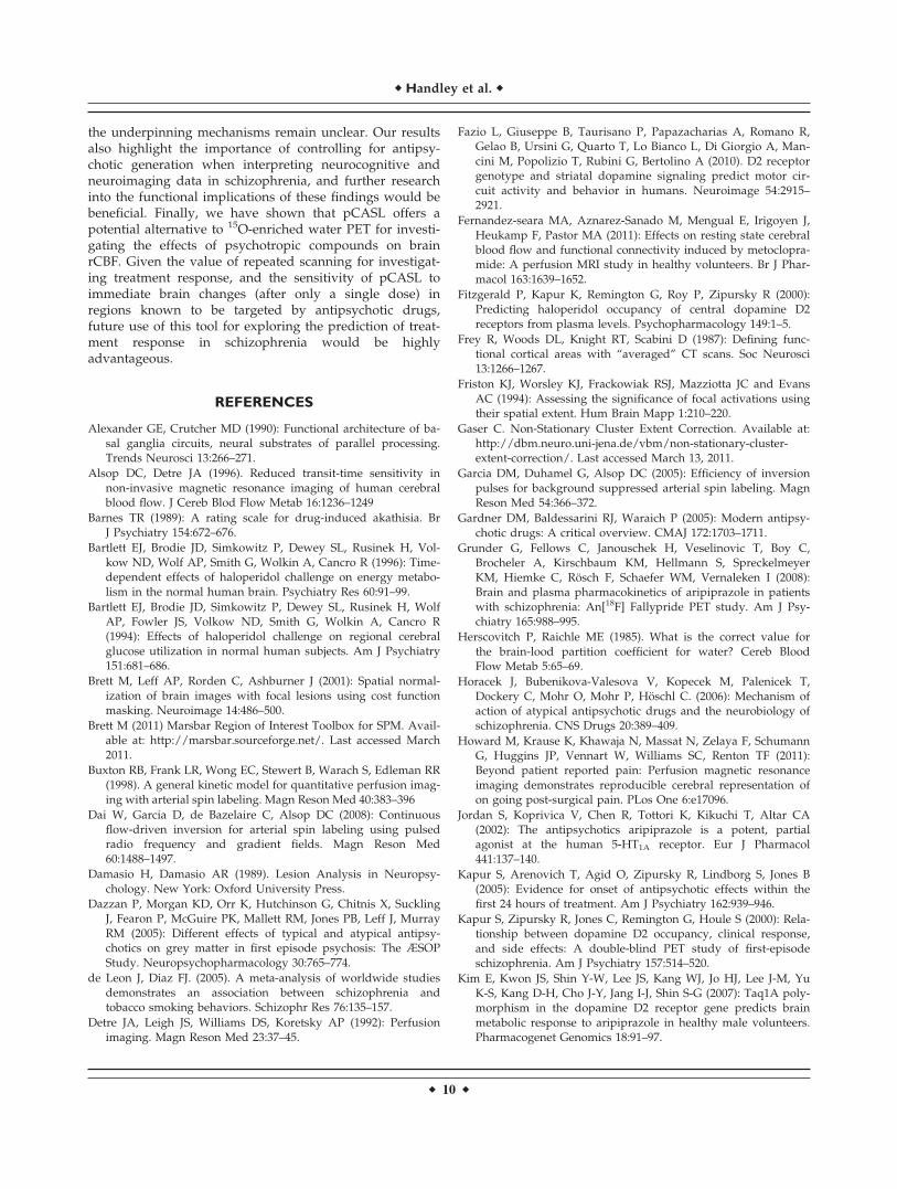
was normalized to the MNI based T2 template providedin SPM, and the transformation matrix was applied to therCBF map and the T2 scan. This procedure enhanced thequality of the normalization. The data were then smoothedusing a 10 mm Gaussian Smoothing kernel (see Fig. 1 forpreprocessed rCBF maps).
Statistical Analysis
For analysis of all other descriptive data, the StatisticalPackage for the Social Sciences (SPSS), version 15 wasused. Haloperidol and placebo comparisons were basedon 20 subjects. Comparisons with aripiprazole were basedon 18 participants as two developed nausea and vomiting.rCBF maps of all subjects, acquired on each scan visit,were entered into a flexible factorial model in SPM andanalyzed using a random effects, general linear regressionmodel. This model was first run without covarying forglobal CBF, then repeated including global CBF as a cova-riate. Even if global CBF was not significantly different atthe group level, this approach explored whether small
interindividual physiological differences limited the identi-fication of antipsychotic-induced regional CBF changes.The effects of antipsychotic on global CBF change wasexplored using a repeated measures ANOVA in SPSS, hav-ing extracted the mean global CBF values using theMarsBar toolbox [Brett, 2010; available at: http://marsbar.-sourceforge.net/ last updated 2010] in SPM5. Within eachrandom effects model in SPM, we performed three pairedcontrasts (t-tests) to identify increased and decreasedrCBF between the antipsychotic conditions: haloperidolversus placebo, haloperidol versus aripiprazole, aripipra-zole versus placebo. For each comparison, we first con-ducted an initial voxel-wise whole-brain search, using athreshold of P < 0.001 uncorrected for multiple compari-sons. To account for any nonisotropic smoothness of thedata, nonstationary cluster extent correction was applied[Gaser, 2007], and statistically significant clusters wereaccepted at P < 0.05 (corrected for multiple comparisons[Friston et al., 1994]). Additionally, small volume correc-tions were applied bilaterally to each contrast using inde-pendent, anatomically-defined, a priori regions, createdwith the anatomical automatic labeling [AAL: Tzourio-Mazoyer et al., 2002] toolbox extension in SPM5. Region ofinterest (ROI)-level statistics were accepted at P < 0.05 cor-rected for family-wise error (FWE). Based on the PET stud-ies by Lahti et al. [2003, 2005], nine regions of interestwere examined: the inferior frontal, middle frontal, ante-rior cingulate, superior temporal, middle temporal, hippo-campus, thalamus, putamen, and caudate. To achieveoptimal accuracy, we chose not to use a linear transforma-tion and instead labeled MNI coordinates using a combi-nation of FSL view MNI structural atlas [Mazziotta et al.,2001], the Talairach and Tournoux atlas [Talairach andTournoux, 1988], and the Brodmann template [Brett et al.,2001; Damasio and Damasio, 1989; Frey et al., 1987; Ror-den and Brett, 2001].
RESULTS
Global CBF Changes
No significant differences in global CBF were observedacross the three intervention groups (F(2,34) ¼ 0.122 n.s.;Fig. 2). The global signal change after each antipsychotic,relative to placebo was less than 1%.
Voxel-Wise and Region of Interest Analysis
Covarying for Global CBF
Haloperidol versus placebo
Whole brain analysis revealed that haloperidol inducedgreater perfusion than placebo in three clusters (Table I);The first cluster centered in the right putamen (see Fig.3), extended into the left putamen, bilaterally into thecaudate and parahippocampal gyrus and also into the leftinsula and right hippocampus; The second cluster
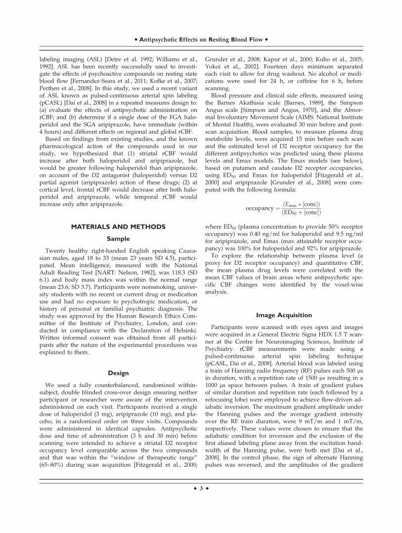
Figure 1.
A sample of 25 (from a data set of 60) horizontal slices, from a
CBF map from one of the study participants showing image
quality in native space obtained with pCASL using a three-
dimensional-fast spin echo readout. The maps in this figure
have been multiplied by a binary brain mask obtained from the
individual T2-weighted anatomical scan of each subject. These
images are acquired in 6:08 min and have a nominal resolution
of 1 � 1 � 3 mm. Note the absence of the image distortion
and signal dropout artefacts commonly observed with gradient
recalled readout techniques.
r Antipsychotic Effects on Resting Blood Flow r
r 5 r

centered in the left precentral gyrus (primary motor cor-tex), extended into the right precentral gyrus (primarymotor cortex), left premotor cortex (supplementary motorarea), right postcentral gyrus, and right ventral posteriorcingulate; The third cluster centered on the left premotorcortex (supplementary motor cortex), extended into the
left precentral gyrus (primary motor cortex) and left post-central gyrus (primary somatosensory cortex). ROI analy-sis revealed that haloperidol also induced greaterperfusion than placebo in the left hippocampus, and inthe left ventral anterior cingulate.
Whole brain analysis revealed that haloperidol reducedperfusion compared with placebo in a cluster centered onthe left inferior temporal gyrus (Fig. 3; Table I). ROI analy-sis showed reduced perfusion after haloperidol than pla-cebo in the left middle frontal gyrus.
Aripiprazole versus placebo
Whole brain analysis revealed that aripiprazole induced
greater perfusion than placebo in four clusters (Table I);
The first cluster centered in the left claustrum (see Fig. 4),
extending into the left putamen, insula, caudate, superior
temporal gyrus, and hippocampus; The second cluster cen-
tered in the right putamen, extending into the right claus-
trum and insula. The third and fourth clusters were
centered in the right ventral anterior cingulate (see Fig. 4)
and right cerebellum respectively, with the latter cluster
extending into the inferior occipital lobe. ROI analysisrevealed that aripiprazole induced greater perfusion thanplacebo in the left superior temporal gyrus.
Whole brain analysis revealed that aripiprazole reducedperfusion, relative to placebo, in five clusters. The firstcluster was centered in the right ventral posterior
TABLE I. Significant clusters of rCBF change after each antipsychotic
Contrast/brain area HEM
MNI coordinates
T scores P
Mean CBF (�SD)% CBFchangeX Y Z
No.voxels PLA HAL ARI
Haloperidol > placebo
Putamen R 30 4 0 8.72 7,295 0.000 366(45) 401(51) Na 9.6
Precentral gyrus (primary motor) L 0 �22 66 5.36 1,185 0.000 519(81) 549(90) Na 5.3
Premotor cortex (supplementary motor) L �20 �26 70 4.21 295 0.036 519(82) 554(106) Na 6.6
Haloperidol < placebo
Inferior temporal gyrus L �58 �56 �14 6.19 1,153 0.000 487(72) 445(50) Na 8.6
Haloperidol > aripiprazole
Putamen R 12 10 �10 5.46 743 0.002 Na 404(53) 375(56) 8.0
Aripiprazole > placebo
Claustrum L �32 �14 4 6.20 2,668 0.000 435(51) Na 468(64) 7.6
Putamen R 30 6 0 5.64 754 0.002 421(48) Na 452(56) 7.3
Anterior cingulate gyrus R 2 12 32 5.19 533 0.006 560(73) Na 600(91) 7.2
Cerebellum R 44 �80 �28 5.11 423 0.010 330(60) Na 379(69) 14.9
Ariprazole < placebo
Posterior cingulate gyrus R 2 �26 30 5.74 904 0.001 694(102) Na 652(94) 6.0
Superior frontal gyrus (PFC) R 16 54 24 5.16 333 0.025 402(67) Na 367(73) 8.6
Superior parietal lobe L �36 �76 48 5.00 334 0.025 603(114) Na 557(99) 7.7
Inferior occipital gyrus R 12 �94 �4 4.83 350 0.022 542(120) Na 499(71) 7.9
Orbitofrontal cortex (PFC) R 10 64 �12 4.17 285 0.039 370(53) Na 346(55) 6.4
P (corrected P value): P < 0.05.Results are presented from the GLM covarying for global CBF effects. % CBF changes were calculated using the mean change, relativeto placebo.Hem: hemisphere; MNI: Montreal Neurological Institute anatomical coordinates; SD: standard deviation; PLA: placebo; HAL: haloperi-dol; ARI: aripiprazole; PFC: prefrontal cortex.
Figure 2.
Mean and standard error bars showing global CBF values for
each intervention. Values represent millilitres of blood per 100 g
tissue per minute. PLA, placebo; HAL, haloperidol; ARIP, aripi-
prazole. No significant differences across treatment conditions
were detected.
r Handley et al. r
r 6 r
cingulate and extended into the left precuneus and pari-eto-occipital sulcus. The second cluster was centered in theright superior frontal gyrus. The third was centered in theleft superior parietal lobe, extending into the inferior pari-etal lobe. The fourth and fifth clusters were centered in theright inferior occipital lobe, extending into the left inferioroccipital lobe and in the right orbitofrontal cortex respec-tively (Fig. 4, Table I). The ROI analysis identified lowerperfusion after aripiprazole than placebo in the left middlefrontal gyrus (dorsolateral prefrontal cortex).
Haloperidol versus aripiprazole
Whole brain analysis revealed that haloperidol inducedgreater perfusion than aripiprazole in a cluster centered inthe right ventral putamen, and extending into the leftputamen, left caudate and left amygdala (Fig. 5, Table I).There were no brain regions in which greater perfusionwas evident after aripiprazole compared with haloperidolin the whole brain analysis. However, ROI analysisrevealed a greater perfusion in the left superior temporalgyrus following aripiprazole relative to haloperidol.
Voxel-Wise Analysis Without Covarying for
Global CBF
Whole brain analysis revealed that haloperidol signifi-cantly increased perfusion relative to placebo in two clus-
ters. The first was centered on the right putamen, andextended into the right caudate, and the second was cen-tered on the left putamen and extended into the left cau-date. No other areas of significant CBF change wereevident when the interventions were compared with oneanother or placebo.
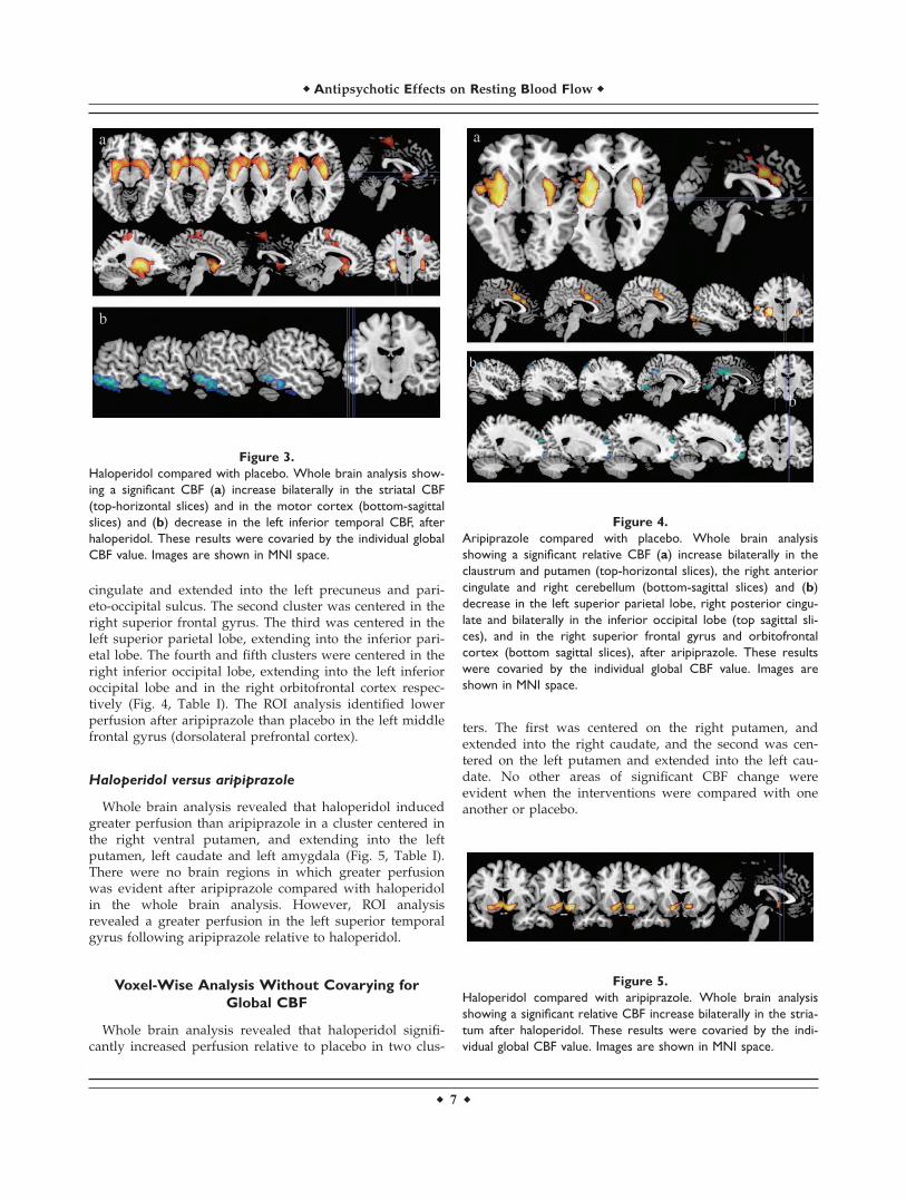
Figure 5.
Haloperidol compared with aripiprazole. Whole brain analysis
showing a significant relative CBF increase bilaterally in the stria-
tum after haloperidol. These results were covaried by the indi-
vidual global CBF value. Images are shown in MNI space.
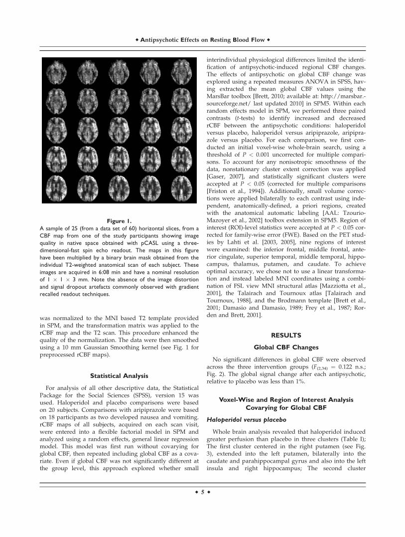
Figure 3.
Haloperidol compared with placebo. Whole brain analysis show-
ing a significant CBF (a) increase bilaterally in the striatal CBF
(top-horizontal slices) and in the motor cortex (bottom-sagittal
slices) and (b) decrease in the left inferior temporal CBF, after
haloperidol. These results were covaried by the individual global
CBF value. Images are shown in MNI space.
Figure 4.
Aripiprazole compared with placebo. Whole brain analysis
showing a significant relative CBF (a) increase bilaterally in the
claustrum and putamen (top-horizontal slices), the right anterior
cingulate and right cerebellum (bottom-sagittal slices) and (b)
decrease in the left superior parietal lobe, right posterior cingu-
late and bilaterally in the inferior occipital lobe (top sagittal sli-
ces), and in the right superior frontal gyrus and orbitofrontal
cortex (bottom sagittal slices), after aripiprazole. These results
were covaried by the individual global CBF value. Images are
shown in MNI space.
r Antipsychotic Effects on Resting Blood Flow r
r 7 r
Plasma Antipsychotic Levels and Predicted
Dopamine D2 Receptor Occupancies
At 3 h 20 min postintervention, mean plasma metabolitelevels of haloperidol and aripiprazole were 1 lg/l (�SD: 0.6)and 42 lg/l (�SD: 11), respectively. The Emax models pre-dicted that mean striatal D2 receptor occupancy was 68%(range 56–86%) and 74% (range 65–78%) after haloperidoland aripiprazole respectively, indicating that the level of re-ceptor occupancy was comparable across the two drugs.
The Relationship Between Plasma Antipsychotic
Levels and CBF Changes
There was a significant negative correlation (r ¼ �0.485,P < 0.05) between haloperidol plasma level and CBF afterhaloperidol in the premotor cortex (the third cluster inwhich haloperidol elicited a significant elevation in perfu-sion relative to placebo), indicating that a higher plasmadrug level was associated with lower regional CBF. Meanplasma antipsychotic level was not correlated with anyother regional mean CBF values identified as being alteredby antipsychotic administration in the voxel-wise analysis.
Blood Pressure and Clinical Side Effects
Haloperidol and aripiprazole induced more extra-py-ramidal side effects than placebo (v2 ¼ 7.37, P < 0.05), im-mediately before scanning, but these were not evidentafter the scanning session (v2 ¼ 2.0, n.s.). There were nosignificant differences in tardive dyskinesia (v2 ¼ 2.0, n.s),akathisia (all participants had a negative score), systolic(F(2,32) ¼ 2.27, n.s.) or diastolic (F(2,32) ¼ 0.22, n.s.) bloodpressure.
DISCUSSION
Using a novel, contrast-free approach, we directly com-pared the effects of a FGA (haloperidol) and a SGA (aripi-prazole) on rCBF in the same healthy individuals, andfound early and antipsychotic specific changes in striatal,frontal, and temporal regions.
Increased striatal rCBF was evident after both antipsy-chotics, only 3 h and 30 min after administration. This isthe earliest change reported for haloperidol and for anySGA and is consistent with studies reporting later effectsat 12 h after administration of haloperidol or aripiprazole[Bartlett et al., 1994; Kim et al., 2007]. Interestingly, such achange was not evident in early investigations of haloperi-dol and SGAs in healthy volunteers, which report nochange in striatal glucose metabolism 2 h after administra-tion (haloperidol or risperidone), using fluorodeoxyglucosePET [FDG-PET; Bartlett et al., 1996; Lane et al., 2004]. Theearly striatal rCBF increase following haloperidol however,is consistent with findings from the first (early) investiga-tion of a different D2 antagonist on resting blood flowusing PET [Mehta et al., 2003], and a recent ASL study
exploring metoclopramide (also a D2 antagonist) inhealthy volunteers [Fernandez-Seara et al., 2010]. Interest-ingly, the three positive studies all used a water-labelingmethod (15O-enriched water PET and ASL), suggestingthat the measurement of resting state CBF via this nonin-vasive technique may provide a sensitive probe of earlypsychoactive effects.
Despite its unique high affinity and partial agonism atD2 receptors [Mamo et al., 2007], aripiprazole inducedstriatal changes similar to those reported in patients fol-lowing other SGAs [Lahti et al., 2005, 2009; Liddle et al.,2000]. This suggests that the functional effects of dopami-nergic modulation associated with SGAs in this brainregion are comparable, regardless of the heterogeneous re-ceptor affinity profiles and intrinsic activity of this groupof drugs.
Alongside this striatal change, we have found a rCBFincrease in cortical motor areas following haloperidoladministration. This is the first report of early rCBF corti-cal motor changes in healthy individuals, a previousreport having observed the increase after 1 week of halo-peridol treatment in patients [Lahti et al., 2009]. This find-ing is consistent with data on the susceptibility of thecortical-striatal circuitry to functional remodeling inresponse to dopaminergic modulation [Tost et al., 2010].Furthermore, the absence of such effects in these brainregions after aripiprazole indicates D2 receptor antago-nism and not partial agonism may result in this brain cir-cuitry becoming susceptible to alteration.
We observed a decrease in rCBF in frontal areas afterboth antipsychotics, although this was more widespread(evident in the middle, orbitofrontal, and superior frontalgyrus) after aripiprazole. These findings support previousreports of metabolism reductions in this brain region fol-lowing haloperidol in healthy individuals and in patients[Bartlett et al., 1994; Lahti et al., 2005]. Aripiprazole effectsare also in line with the only other PET study conductedin healthy volunteers [Kim et al., 2007], and are notablycomparable to those associated with other SGAs [Nganet al., 2002]. Both antipsychotics also altered rCBF in thecingulate, with haloperidol being associated with a rCBFincrease in the anterior, and a rCBF decrease in the poste-rior, cingulate. Aripiprazole unexpectedly increased ante-rior cingulate rCBF, an effect not observed in the onlyother aripiprazole study exploring metabolism 12 hours af-ter administration [Kim et al., 2007]. However, anotherSGA (olanzapine) has also been reported to increase ante-rior cingulate rCBF 5 h after administration in patients[Lahti et al., 2005]. Olanzapine and aripiprazole share sim-ilar affinity for serotonin 5HT-2A receptors [Gardner et al.,2005], suggesting that antagonism at this receptor subtypecould underpin the immediate rCBF changes observed inthis brain region.
In the temporal lobe, we observed different effects forhaloperidol and aripiprazole. Increased rCBF after aripi-prazole in the superior temporal gyrus is the earliestchange to be reported in this brain region, and is
r Handley et al. r
r 8 r

consistent with effects observed at 12 h [Kim et al., 2007].Olanzapine has also been associated with increased tempo-ral rCBF in patients [Lahti et al., 2005, 2009], providingfurther evidence for the possible role of serotonin blockadein these rCBF alterations. Haloperidol was associated withdecreased temporal cortex rCBF, whereas previous healthyinvestigations with PET reported no change [Bartlett et al.,1994, 1996]. Our findings are based on a larger sample sizeand are in agreement with patient studies reporting thatsingle, short-term, and chronic haloperidol administrationare all associated with decreased temporal rCBF [Lahtiet al., 2005, 2009].
Our findings of an early effect of both antipsychotics onbrain perfusion are consistent with PET occupancy studies,showing that antipsychotics impact at the receptor levelwithin hours of administration [Nordstrom et al., 1992;Tauscher et al., 2002]. This suggests a possible relationshipbetween the ability to block dopamine receptors and thelevel of resting blood perfusion. The validity of this possi-ble relationship is at least partly supported by independ-ent data which shows that treatment response isassociated with both the level of striatal rCBF [Lahti et al.,2009] and of dopamine D2 receptor occupancy [Kapuret al., 2000]. Interestingly, our data also reveal that higherhaloperidol plasma levels, a proxy for occupancy, are cor-related with lower rCBF in the premotor cortex followinghaloperidol. Still, a direct link between occupancy aloneand perfusion change seems unlikely, since although aripi-prazole and haloperidol bind with similar high affinity toD2 receptors, they are nevertheless associated with differ-ent striatal rCBF changes in our sample.
Although the precise mechanisms underpinning rCBFchanges in response to administration of psychoactivesubstances are unclear, the underlying assumption of ourwork is that enhanced neuronal and postsynaptic activityincreases energy demand [Sadaghiani et al., 2010], result-ing in areas of increased perfusion following antipsy-chotic administration. In brain areas where dopamine D2receptors are densely distributed (such as the striatum),antagonism may give rise to increased presynaptic dopa-mine synthesis and release, due to loss of inhibitory feed-back mechanisms. In brain regions innervated by thesedensely D2 populated areas, such as the frontal and tem-poral cortices, downstream inhibitory and excitatoryprocesses may also be involved [Fazio et al., 2010; Mattayet al., 2002]. Using ASL, however, we cannot exclude thatsome of the effects observed have a vascular rather thanentirely neuronal origin. This might explain some of thediscrepancies between our results and those carried outusing FDG-PET.
Finally, our Emax models indicated that drug plasmalevels predicted similar striatal D2 receptor occupancy forboth compounds and the comparable increase in extra-py-ramidal symptoms (EPS) we observed after both drugs isconsistent with this [Kapur et al., 2000]. It is also interest-ing that CBF increased in the striatum after both antipsy-chotics, supporting the role of this brain region in motor
behavior [Alexander and Crutcher, 1990] and in the motorside effects associated with antipsychotics. Importantly,our predicted occupancies were within the therapeuticrange [Kapur et al., 2000], indicating our findings are rele-vant to the doses used in patients. Furthermore, the rCBFalterations cannot simply be explained by differences indrug occupancy. This is particularly relevant when inter-preting the opposite effects of these drugs on rCBF in thetemporal cortex.
It is noteworthy that, when global CBF was not includedas a covariate, there were fewer rCBF changes observed.However, global CBF itself was not significantly differentafter each intervention, suggesting that without its inclu-sion, small interindividual physiological differences mayhave limited the identification of regional differences atgroup-level. Therefore, inclusion of global CBF as a covari-ate helped resolve regional differences by removing smallinterindividual differences.
One limitation of the study may be the use of a singlescan after each intervention in this within-subject design,which did not allow us to explore whether drug effects onrCBF persisted beyond the day of scanning. However,data suggesting that cognitive effects do not persist 24 hafter antipsychotic administration [Legangneux et al.,2000], suggests our 14 day washout period was sufficient.Also, intellectual capacity and smoking are different inpatients compared with controls [De Leon and Diaz, 2005;Lewis and Levitt, 2002], potentially limiting the generaliza-tion of our findings to schizophrenia. For example, thecomparability of single dose and chronic treatment effectsremains unknown. Nevertheless, rCBF changes, particu-larly in temporal and corticostriatal circuitry, observed inthis study are consistent with those observed in single andchronic dosing studies in patients [Lahti et al., 2005, 2009].Thus, researching the effects of these compounds on rCBFin healthy individuals offers valuable insights into theireffects in patient populations, indicating that CBF altera-tions evident after repeated dosing are in fact observedwithin hours of a single drug administration, and are notdue to an interaction with the pathogenesis of thedisorder.
CONCLUSIONS
This study indicates that there are immediate and differ-ent rCBF changes after a single dose of antipsychotics inthe striatum and cortical-striatal loop, frontal, and tempo-ral cortices. The sensitivity of these brain regions to imme-diate antipsychotic effects in our study might suggest thatrCBF changes could underpin or reflect the ‘‘early-onset’’of therapeutic effect proposed by Kapur et al. [2005].Moreover, changes were observed in regions in which ourand other groups have previously observed structuralalterations associated with FGA or SGA effects in schizo-phrenia, suggesting that rCBF alterations may interactwith, or partly contribute to, structural changes, although
r Antipsychotic Effects on Resting Blood Flow r
r 9 r
the underpinning mechanisms remain unclear. Our resultsalso highlight the importance of controlling for antipsy-chotic generation when interpreting neurocognitive andneuroimaging data in schizophrenia, and further researchinto the functional implications of these findings would bebeneficial. Finally, we have shown that pCASL offers apotential alternative to 15O-enriched water PET for investi-gating the effects of psychotropic compounds on brainrCBF. Given the value of repeated scanning for investigat-ing treatment response, and the sensitivity of pCASL toimmediate brain changes (after only a single dose) inregions known to be targeted by antipsychotic drugs,future use of this tool for exploring the prediction of treat-ment response in schizophrenia would be highlyadvantageous.
REFERENCES
Alexander GE, Crutcher MD (1990): Functional architecture of ba-sal ganglia circuits, neural substrates of parallel processing.Trends Neurosci 13:266–271.
Alsop DC, Detre JA (1996). Reduced transit-time sensitivity innon-invasive magnetic resonance imaging of human cerebralblood flow. J Cereb Blod Flow Metab 16:1236–1249
Barnes TR (1989): A rating scale for drug-induced akathisia. BrJ Psychiatry 154:672–676.
Bartlett EJ, Brodie JD, Simkowitz P, Dewey SL, Rusinek H, Vol-kow ND, Wolf AP, Smith G, Wolkin A, Cancro R (1996): Time-dependent effects of haloperidol challenge on energy metabo-lism in the normal human brain. Psychiatry Res 60:91–99.
Bartlett EJ, Brodie JD, Simkowitz P, Dewey SL, Rusinek H, WolfAP, Fowler JS, Volkow ND, Smith G, Wolkin A, Cancro R(1994): Effects of haloperidol challenge on regional cerebralglucose utilization in normal human subjects. Am J Psychiatry151:681–686.
Brett M, Leff AP, Rorden C, Ashburner J (2001): Spatial normal-ization of brain images with focal lesions using cost functionmasking. Neuroimage 14:486–500.
Brett M (2011) Marsbar Region of Interest Toolbox for SPM. Avail-able at: http://marsbar.sourceforge.net/. Last accessed March2011.
Buxton RB, Frank LR, Wong EC, Stewert B, Warach S, Edleman RR(1998). A general kinetic model for quantitative perfusion imag-ing with arterial spin labeling. Magn Reson Med 40:383–396
Dai W, Garcia D, de Bazelaire C, Alsop DC (2008): Continuousflow-driven inversion for arterial spin labeling using pulsedradio frequency and gradient fields. Magn Reson Med60:1488–1497.
Damasio H, Damasio AR (1989). Lesion Analysis in Neuropsy-chology. New York: Oxford University Press.
Dazzan P, Morgan KD, Orr K, Hutchinson G, Chitnis X, SucklingJ, Fearon P, McGuire PK, Mallett RM, Jones PB, Leff J, MurrayRM (2005): Different effects of typical and atypical antipsy-chotics on grey matter in first episode psychosis: The ÆSOPStudy. Neuropsychopharmacology 30:765–774.
de Leon J, Diaz FJ. (2005). A meta-analysis of worldwide studiesdemonstrates an association between schizophrenia andtobacco smoking behaviors. Schizophr Res 76:135–157.
Detre JA, Leigh JS, Williams DS, Koretsky AP (1992): Perfusionimaging. Magn Reson Med 23:37–45.
Fazio L, Giuseppe B, Taurisano P, Papazacharias A, Romano R,Gelao B, Ursini G, Quarto T, Lo Bianco L, Di Giorgio A, Man-cini M, Popolizio T, Rubini G, Bertolino A (2010). D2 receptorgenotype and striatal dopamine signaling predict motor cir-cuit activity and behavior in humans. Neuroimage 54:2915–2921.
Fernandez-seara MA, Aznarez-Sanado M, Mengual E, Irigoyen J,Heukamp F, Pastor MA (2011): Effects on resting state cerebralblood flow and functional connectivity induced by metoclopra-mide: A perfusion MRI study in healthy volunteers. Br J Phar-macol 163:1639–1652.
Fitzgerald P, Kapur K, Remington G, Roy P, Zipursky R (2000):Predicting haloperidol occupancy of central dopamine D2receptors from plasma levels. Psychopharmacology 149:1–5.
Frey R, Woods DL, Knight RT, Scabini D (1987): Defining func-tional cortical areas with ‘‘averaged’’ CT scans. Soc Neurosci13:1266–1267.
Friston KJ, Worsley KJ, Frackowiak RSJ, Mazziotta JC and EvansAC (1994): Assessing the significance of focal activations usingtheir spatial extent. Hum Brain Mapp 1:210–220.
Gaser C. Non-Stationary Cluster Extent Correction. Available at:http://dbm.neuro.uni-jena.de/vbm/non-stationary-cluster-extent-correction/. Last accessed March 13, 2011.
Garcia DM, Duhamel G, Alsop DC (2005): Efficiency of inversionpulses for background suppressed arterial spin labeling. MagnReson Med 54:366–372.
Gardner DM, Baldessarini RJ, Waraich P (2005): Modern antipsy-chotic drugs: A critical overview. CMAJ 172:1703–1711.
Grunder G, Fellows C, Janouschek H, Veselinovic T, Boy C,Brocheler A, Kirschbaum KM, Hellmann S, SpreckelmeyerKM, Hiemke C, Rosch F, Schaefer WM, Vernaleken I (2008):Brain and plasma pharmacokinetics of aripiprazole in patientswith schizophrenia: An[18F] Fallypride PET study. Am J Psy-chiatry 165:988–995.
Herscovitch P, Raichle ME (1985). What is the correct value forthe brain-lood partition coefficient for water? Cereb BloodFlow Metab 5:65–69.
Horacek J, Bubenikova-Valesova V, Kopecek M, Palenicek T,Dockery C, Mohr O, Mohr P, Hoschl C. (2006): Mechanism ofaction of atypical antipsychotic drugs and the neurobiology ofschizophrenia. CNS Drugs 20:389–409.
Howard M, Krause K, Khawaja N, Massat N, Zelaya F, SchumannG, Huggins JP, Vennart W, Williams SC, Renton TF (2011):Beyond patient reported pain: Perfusion magnetic resonanceimaging demonstrates reproducible cerebral representation ofon going post-surgical pain. PLos One 6:e17096.
Jordan S, Koprivica V, Chen R, Tottori K, Kikuchi T, Altar CA(2002): The antipsychotics aripiprazole is a potent, partialagonist at the human 5-HT1A receptor. Eur J Pharmacol441:137–140.
Kapur S, Arenovich T, Agid O, Zipursky R, Lindborg S, Jones B(2005): Evidence for onset of antipsychotic effects within thefirst 24 hours of treatment. Am J Psychiatry 162:939–946.
Kapur S, Zipursky R, Jones C, Remington G, Houle S (2000): Rela-tionship between dopamine D2 occupancy, clinical response,and side effects: A double-blind PET study of first-episodeschizophrenia. Am J Psychiatry 157:514–520.
Kim E, Kwon JS, Shin Y-W, Lee JS, Kang WJ, Jo HJ, Lee J-M, YuK-S, Kang D-H, Cho J-Y, Jang I-J, Shin S-G (2007): Taq1A poly-morphism in the dopamine D2 receptor gene predicts brainmetabolic response to aripiprazole in healthy male volunteers.Pharmacogenet Genomics 18:91–97.
r Handley et al. r
r 10 r

Kofke WA, Blissitt PA, Rao H, Wang J, Addya K, Detre J (2007):Remifentanil-induced cerebral blood flow effects in normalhumans: Dose and ApoE genotype. Neurosurg AnaesthesiolNeurosci 105:167–175.
Kubo M, Koue T, Inaba A, Takeda H, Maune H, Fukuda T (2005):Influence of itraconazole co-administration and CYP2D6 geno-type on the pharmacokinetics of the new antipsychotic aripi-prazole. Drug Met Pharmacokinet 20:55–64.
Lahti AC, Holcomb HH, Weiler MA, Medoff DR, Tamminga CA(2003): Time course of rCBF changes with haloperidol and olanza-pine over a six week treatment period. Biol Psychiatry 53:601–608.
Lahti AC, Weiler MA, Holcomb HH, Tamminga CA, Cropsey KL(2009): Modulation of limbic circuitry predicts treatment responseto antipsychotic medication: A functional imaging study ion schiz-ophrenia. Neuropsychopharmacology 34:2675–2690.
Lahti AC, Weiler MA, Medoff DR, Tamminga CA, Holcomb HH(2005): Functional effects of single dose first- and second-gen-eration antipsychotic administration in subjects with schizo-phrenia. Psychiatry Res 139:19–30.
Lane CJ, Ngan ETC, Yatham LN, Ruth TJ, Liddle PF (2004): Im-mediate effects of risperidone on cerebral activity in healthysubjects: A comparison with subjects with first-episode schizo-phrenia. J Psychiatry Neurosci 29:30–37.
Legangneux E, McEwen J, Wesnes KA, Bergougnan L, Miget N,Canal M, L’Heritier CL, Pinquier JL, Rosenzweig P (2000):The acute effects of amisulpride (50 mg and 200 mg) andhaloperidol (2 mg) on cognitive function in healthy elderlyvolunteers. J Psychopharmacol 14:164–171.
Lewis DA, Levitt P (2002): Schizophrenia as a disorder of neuro-development. Annu Rev Neurosci 25:409–432.
Liddele PF, Lane CJ, Ngan ETC (2000): Immediate effects of ris-peridone on cortico-striato-thalamic loops and the hippocam-pus. Br J Psychiatry 177:402–407.
Lu H, Clingman C, Golay X, Van Zijl PCM (2004). Determining thelongitudinal relaxation time (T1) of blood at 3.0 tesla. 52:679–682.
Mamo D, Graff A, Mizrahi R, Shammi CM, Romeyer F, Kapur S(2007): Differential effects of aripiprazole on D(2), 5-HT(2), and5-HT(1A) receptor occupancy in patients with schizophrenia: atriple tracer PET study. Am J Psychiatry 164:1411–1417.
Mattay VS, Tessitore A, Callicott JH, Bertolino A, Goldberg TE,Chase TN, Hyde TM, Weinberger DR (2002): Dopaminergicmodulation of cortical funciton in patients with Parkinson’sdisease. Ann Neurol 51:156–164.
Mazziotta J, Toga A, Evans A, Fox P, Lancaster J, Zilles K, WoodsR, Paus T, Simpson G, Pike B, Holmes C, Collins L, ThompsonP, MacDonald D, Iacoboni M, Schormann T, Amunts K, Palo-mero-Gallagher N, Geyer S, Parsons L, Narr K, Kabani N, LeGoualher G, Boomsma D, Cannon T, Kawashima R, Mazoyer(2001): A probabilistic atlas and reference system for thehuman brain: International Consortium for Brain Mapping(ICBM). Phil Trans Royal Soc B Biol Sci 356:1293–1322.
Mehta MA, McGowan SW, Lawrence AD, Aitken MRF, Montgom-ery AJ, Grasby PM (2003): Systemic sulpiride modulates stria-tal blood flow: Relationship to spatial working memory andplanning. Neuroimage 20:1982–1994.
Meltzer HY, Matsubara S, Lee J-C (1989): Classification of typical andatypical antipsychotic drugs on the basis of dopamine D-1, D-2and seotonin2 pKi values. J Pharmacol Exp Ther 251:238–246.
Meltzer HY (2004): What’s atypical about atypical antipsychoticdrugs? Curr Opin Pharmacol 4:53–57.
Miller DD, Andreasen NC, O’Leary DS, Watkins GL, Boles PontoLL, Hichwa RD. (2001): Comparison of the effects of risperi-done and haloperidol on regional cerebral blood flow in schiz-ophrenia. Biol Psychiatry 49:704–715.
National Institute of Mental Health (1976): Abnormal involuntarymovement scale (AIMS). In: Guy W, editor. ECDEU: Assess-ment Manual for Psychopharmacology. Rockville: NationalInstitute of Mental Health, DHEW Publication (ADM). pp 534–537.
Navari S., Dazzan P (2009): Do antipsychotic drugs affect brainstructure? A systematic and critical review of MRI findings.Psychol Med 39:1763–1777.
Nelson HE (1982). National Adult Reading Test (NART): TestManual. Windsor: NFER-Nelson.
Ngan ET, Lane CJ, Ruth TJ, Liddle PF (2002): Immediate anddelayed effects of risperidone on cerebral metabolism in neuro-leptic naive schizophrenic patients: Correlations with symptomchange 2. J Neurol Neurosurg Psychiatry 72:106–110.
Nordstrom AL, Farde L, Halldin C (1992): Time course of D2-dopamine receptor occupancy examined by PET after singleoral doses of haloperidol. Psychopharmacology (Berl)106:433–438.
Perthen JE, Lansing AE, Liau J, Liu TT, Buxton RB (2008): Caf-feine-induced uncoupling of cerebral blood flow and oxygenmetabolism: A calibrated BOLD fMRI study. Neuroimage40:237–247.
Rorden C, Brett M (2001): Stereotaxic display of brain lesions.Behav Neurol 21:191–200.
Simpson GN, Angus JWS (1970): A rating scale for extrapyramidalside effects. Acta Psychiatr Scand Suppl 212:11–19.
Smith S (2002): Fast robust automated brain extraction. Hum BrainMapp 17:143–155.
Talairach J, Tournoux P (1988). Co-planar Stereotaxic Atlas of theHuman Brain: 3-Dimensional Proportional System—AnApproach to Cerebral Imaging. New York: Thieme MedicalPublishers.
Tauscher J, Jones C, Remington G, Zipursky RB, Kapur S (2002):Significant dissociation of brain and plasma kinetics with anti-psychotics. Mol Psychiatry 7:317–321.
Tost H, Braus DF, Hakimi S, Ruf M, Vollmert C, Hohn F, Mayer-Lindenberg A (2010): Acute D2 receptor blockade inducesrapid, reversible remodelling of human cortical-striatal circuits.Nat Neurosci 13:920–922.
Tzourio-Mazoyer N, Landeau B, Papathanassiou D, Crivello F,Etard O, Delcroix N (2002): Automated anatomical labeling ofactivations in SPM using a macroscopic anatomical par-cellation of the MNI MRI single-subject brain. Neuroimage15:273–289.
Williams DS, Detre JA, Leigh JS, Koretsky AP (1992): Magneticresonance imaging of perfusion using spin inversion of arterialwater. Proc Natl Acad Sci USA 89:212–216.
Yokoi F, Grunder G, Biziere K, Staphane M, Dogan AS, DannalsRF (2002): Dopamine D2 and D3 receptor occupancy in normalhumans treated with the antipsychotic drug aripiprazole (OPC14597): A study using positron emission tomography and[11C] raclopride. Neuropsychopharmacology 27:248–259.
r Antipsychotic Effects on Resting Blood Flow r
r 11 r
Copyright © 2022 FDOKUMEN