TprC/D (Tp0117/131), a trimeric, pore-forming rare outer membrane protein of Treponema pallidum, has...
13
TprC/D (Tp0117/131), a Trimeric, Pore-Forming Rare Outer Membrane Protein of Treponema pallidum, Has a Bipartite Domain Structure Arvind Anand, a Amit Luthra, a Star Dunham-Ems, a Melissa J. Caimano, a,e Carson Karanian, e Morgan LeDoyt, a Adriana R. Cruz, d Juan C. Salazar, c,e and Justin D. Radolf a,b,c,e Departments of Medicine, a Genetics and Developmental Biology, b and Immunology, c University of Connecticut Health Center, Farmington, Connecticut, USA; Centro Internacional de Entrenamiento e Investigaciones Médicas (CIDEIM), Cali, Colombia d ; and Department of Pediatrics, Connecticut Children’s Medical Center, Division of Pediatric Infectious Diseases, Hartford, Connecticut, USA e Identification of Treponema pallidum rare outer membrane proteins (OMPs) has been a longstanding objective of syphilis re- searchers. We recently developed a consensus computational framework that employs a battery of cellular localization and topo- logical prediction tools to generate ranked clusters of candidate rare OMPs (D. L. Cox et al., Infect. Immun. 78:5178 –5194, 2010). TP0117/TP0131 (TprC/D), a member of the T. pallidum repeat (Tpr) family, was a highly ranked candidate. Circular dichroism, heat modifiability by SDS-PAGE, Triton X-114 phase partitioning, and liposome incorporation confirmed that full-length, re- combinant TprC (TprC Fl ) forms a -barrel capable of integrating into lipid bilayers. Moreover, TprC Fl increased efflux of terbi- um-dipicolinic acid complex from large unilamellar vesicles and migrated as a trimer by blue-native PAGE. We found that in T. pallidum, TprC is heat modifiable, trimeric, expressed in low abundance, and, based on proteinase K accessibility and op- sonophagocytosis assays, surface exposed. From these collective data, we conclude that TprC is a bona fide rare OMP as well as a functional ortholog of Escherichia coli OmpF. We also discovered that TprC has a bipartite architecture consisting of a soluble N-terminal portion (TprC N ), presumably periplasmic and bound directly or indirectly to peptidoglycan, and a C-terminal -barrel (TprC C ). Syphilitic rabbits generate antibodies exclusively against TprC C , while secondary syphilis patients fail to mount a detectable antibody response against either domain. The syphilis spirochete appears to have resolved a fundamental dilemma arising from its extracellular lifestyle, namely, how to enhance OM permeability without increasing its vulnerability to the antibody-mediated defenses of its natural human host. S yphilis is a multistage, sexually transmitted illness renowned for its protean clinical manifestations and protracted natural history, both of which reflect the extraordinary invasiveness and immunoevasiveness of its etiologic agent, Treponema pallidum subsp. pallidum (34, 49). Although T. pallidum is often analogized to Gram-negative bacteria because it has both an inner and an outer membrane (IM and OM, respectively), the structure, com- position, and physical properties of its cell envelope differ consid- erably from those of Gram-negative bacteria. For example, the OM of T. pallidum is devoid of lipopolysaccharide, contains phos- phatidylcholine and phosphatidylserine as its major phospholip- ids, and is relatively permeable to lipophilic molecules (12, 22, 53). In Escherichia coli, lipoproteins are associated predominantly with the inner leaflet of the OM (60), while, with one exception, TP0453 (25), all lipoproteins localized to date in T. pallidum have been found to be tethered to the IM (7, 11, 32). In T. pallidum, the peptidoglycan (PG) resides approximately midway within the periplasmic space (30, 32), whereas in Gram-negative bacteria the murein layer is bound to the underside of the OM via covalent and noncovalent interactions with OM proteins (OMPs) and OM-associated lipoproteins (60). Most notably, the density of OMPs in T. pallidum is markedly lower than that in E. coli, ap- proximately 1% based on freeze fracture electron microscopy (52, 64); the paucity of OM-spanning proteins and lack of surface- exposed lipoproteins are believed to be the ultrastructural basis for the spirochete’s stealth pathogenicity (7, 48). Rare OMPs presumably fulfill virulence-related and physio- logical functions in T. pallidum comparable to those of their much better characterized, nonorthologous Gram-negative counter- parts (7, 16, 43, 60). Moreover, as with Gram-negative bacteria (58, 61), surface-exposed OMPs of T. pallidum represent potential targets for protective host defenses (15, 29, 50). Indeed, both rabbit and human syphilitic sera contain antibodies that label in- tact organisms by immunofluorescence (11) and promote op- sonophagocytosis of spirochetes by monocytes/macrophages (13, 35, 36). These results suggest that at least some OMPs generate antibody responses that contribute to reduction of spirochete bur- dens and containment of disease. Over the years, a variety of fac- tors have hindered efforts by investigators to identify T. pallidum OM-spanning proteins. Chief among these are their low abun- dance, their lack of sequence relatedness to known OMPs of Gram-negative organisms, and the fragility of the T. pallidum OM, all compounded by the spirochete’s recalcitrance to in vitro cultivation and genetic manipulation (7, 22, 29, 31, 48). Almost all known OM-spanning proteins adopt a -barrel conformation (24, 56, 67). Three theoretical considerations de- rived from the vast literature on the topology and export of inte- Received 20 January 2012 Accepted 23 February 2012 Published ahead of print 2 March 2012 Address correspondence to Justin D. Radolf, [email protected]. Supplemental material for this article may be found at http://jb.asm.org/. Copyright © 2012, American Society for Microbiology. All Rights Reserved. doi:10.1128/JB.00101-12 0021-9193/12/$12.00 Journal of Bacteriology p. 2321–2333 jb.asm.org 2321
-
Upload
independent -
Category
Documents
-
view
0 -
download
0
Transcript of TprC/D (Tp0117/131), a trimeric, pore-forming rare outer membrane protein of Treponema pallidum, has...

TprC/D (Tp0117/131), a Trimeric, Pore-Forming Rare OuterMembrane Protein of Treponema pallidum, Has a Bipartite DomainStructure
Arvind Anand,a Amit Luthra,a Star Dunham-Ems,a Melissa J. Caimano,a,e Carson Karanian,e Morgan LeDoyt,a Adriana R. Cruz,d
Juan C. Salazar,c,e and Justin D. Radolfa,b,c,e
Departments of Medicine,a Genetics and Developmental Biology,b and Immunology,c University of Connecticut Health Center, Farmington, Connecticut, USA; CentroInternacional de Entrenamiento e Investigaciones Médicas (CIDEIM), Cali, Colombiad; and Department of Pediatrics, Connecticut Children’s Medical Center, Division ofPediatric Infectious Diseases, Hartford, Connecticut, USAe
Identification of Treponema pallidum rare outer membrane proteins (OMPs) has been a longstanding objective of syphilis re-searchers. We recently developed a consensus computational framework that employs a battery of cellular localization and topo-logical prediction tools to generate ranked clusters of candidate rare OMPs (D. L. Cox et al., Infect. Immun. 78:5178 –5194, 2010).TP0117/TP0131 (TprC/D), a member of the T. pallidum repeat (Tpr) family, was a highly ranked candidate. Circular dichroism,heat modifiability by SDS-PAGE, Triton X-114 phase partitioning, and liposome incorporation confirmed that full-length, re-combinant TprC (TprCFl) forms a �-barrel capable of integrating into lipid bilayers. Moreover, TprCFl increased efflux of terbi-um-dipicolinic acid complex from large unilamellar vesicles and migrated as a trimer by blue-native PAGE. We found that in T.pallidum, TprC is heat modifiable, trimeric, expressed in low abundance, and, based on proteinase K accessibility and op-sonophagocytosis assays, surface exposed. From these collective data, we conclude that TprC is a bona fide rare OMP as well as afunctional ortholog of Escherichia coli OmpF. We also discovered that TprC has a bipartite architecture consisting of a solubleN-terminal portion (TprCN), presumably periplasmic and bound directly or indirectly to peptidoglycan, and a C-terminal�-barrel (TprCC). Syphilitic rabbits generate antibodies exclusively against TprCC, while secondary syphilis patients fail tomount a detectable antibody response against either domain. The syphilis spirochete appears to have resolved a fundamentaldilemma arising from its extracellular lifestyle, namely, how to enhance OM permeability without increasing its vulnerability tothe antibody-mediated defenses of its natural human host.
Syphilis is a multistage, sexually transmitted illness renownedfor its protean clinical manifestations and protracted natural
history, both of which reflect the extraordinary invasiveness andimmunoevasiveness of its etiologic agent, Treponema pallidumsubsp. pallidum (34, 49). Although T. pallidum is often analogizedto Gram-negative bacteria because it has both an inner and anouter membrane (IM and OM, respectively), the structure, com-position, and physical properties of its cell envelope differ consid-erably from those of Gram-negative bacteria. For example, theOM of T. pallidum is devoid of lipopolysaccharide, contains phos-phatidylcholine and phosphatidylserine as its major phospholip-ids, and is relatively permeable to lipophilic molecules (12, 22, 53).In Escherichia coli, lipoproteins are associated predominantly withthe inner leaflet of the OM (60), while, with one exception,TP0453 (25), all lipoproteins localized to date in T. pallidum havebeen found to be tethered to the IM (7, 11, 32). In T. pallidum, thepeptidoglycan (PG) resides approximately midway within theperiplasmic space (30, 32), whereas in Gram-negative bacteriathe murein layer is bound to the underside of the OM via covalentand noncovalent interactions with OM proteins (OMPs) andOM-associated lipoproteins (60). Most notably, the density ofOMPs in T. pallidum is markedly lower than that in E. coli, ap-proximately 1% based on freeze fracture electron microscopy (52,64); the paucity of OM-spanning proteins and lack of surface-exposed lipoproteins are believed to be the ultrastructural basis forthe spirochete’s stealth pathogenicity (7, 48).
Rare OMPs presumably fulfill virulence-related and physio-logical functions in T. pallidum comparable to those of their much
better characterized, nonorthologous Gram-negative counter-parts (7, 16, 43, 60). Moreover, as with Gram-negative bacteria(58, 61), surface-exposed OMPs of T. pallidum represent potentialtargets for protective host defenses (15, 29, 50). Indeed, bothrabbit and human syphilitic sera contain antibodies that label in-tact organisms by immunofluorescence (11) and promote op-sonophagocytosis of spirochetes by monocytes/macrophages (13,35, 36). These results suggest that at least some OMPs generateantibody responses that contribute to reduction of spirochete bur-dens and containment of disease. Over the years, a variety of fac-tors have hindered efforts by investigators to identify T. pallidumOM-spanning proteins. Chief among these are their low abun-dance, their lack of sequence relatedness to known OMPs ofGram-negative organisms, and the fragility of the T. pallidumOM, all compounded by the spirochete’s recalcitrance to in vitrocultivation and genetic manipulation (7, 22, 29, 31, 48).
Almost all known OM-spanning proteins adopt a �-barrelconformation (24, 56, 67). Three theoretical considerations de-rived from the vast literature on the topology and export of inte-
Received 20 January 2012 Accepted 23 February 2012
Published ahead of print 2 March 2012
Address correspondence to Justin D. Radolf, [email protected].
Supplemental material for this article may be found at http://jb.asm.org/.
Copyright © 2012, American Society for Microbiology. All Rights Reserved.
doi:10.1128/JB.00101-12
0021-9193/12/$12.00 Journal of Bacteriology p. 2321–2333 jb.asm.org 2321

gral membrane proteins in bacteria support the assumption thatT. pallidum rare OMPs likewise form �-barrels (11, 15, 48): (i)�-helical transmembrane domains act as stop-transfer sequencesduring translocation of polypeptides across the IM by the Sec ex-port machinery (47); (ii) the presence of multiple short amphi-pathic segments circumvents the constraints to Sec-dependent ex-port of integral membrane proteins across the IM (24, 60, 63); and(iii) even with the spirochete’s remarkably low rate of multiplica-tion (38) and comparatively permeable OM lipid bilayer (7, 12),fixed porin-like channels are needed to mediate nutrient uptakefrom the host milieu (43). Our recent demonstration that thespirochete contains an ortholog for BamA, TP0326, the centralcomponent of the molecular machine that chaperones newly ex-ported precursors into the OM (24, 60, 63), constitutes prima facieevidence for the existence of unidentified �-barrels (19). Becausethe T. pallidum genome does not encode orthologs for knownOMPs of proteobacteria other than TP0326/BamA (22), insilico identification of �-barrels rests upon structural predic-tions. Whereas the �-helical transmembrane domains of IMproteins are readily identifiable, such is not the case for themembrane-spanning elements (i.e., amphipathic �-strands) ofOMPs (41, 56, 63).
We recently devised a consensus computational frameworkthat employs a battery of cellular localization and topological pre-diction tools to generate ranked clusters of candidate rare OMPs,collectively referred to as the putative T. pallidum OMPeome (11).Among the most highly ranked candidates were the identical pro-teins TP0117/TP0131 (TprC/D, here referred to as TprC), mem-bers of the T. pallidum repeat (Tpr) family long suspected of har-boring rare OMPs (7, 8, 22, 31, 33). Here we demonstrate thatTprC does indeed meet criteria for designation as a bona fide rareOMP (48), namely, �-barrel structure, amphiphilicity, low abun-dance, and surface exposure. Moreover, similar to E. coli OmpF,an archetypal �-barrel (43, 60, 65), TprC is trimeric and formschannels in artificial membranes. We also found that TprC has abipartite architecture consisting of a soluble N-terminal portion(TprCN), presumably periplasmic and bound directly or indi-rectly to peptidoglycan, and a C-terminal �-barrel (TprCC) solelyresponsible for membrane association and pore formation. Inter-estingly, T. pallidum-infected rabbits generate antibodies exclu-sively against TprCC, predicted to contain the surface-exposeddeterminants, while humans with secondary syphilis fail to mounta detectable antibody response against either domain. The syphilisspirochete appears to have resolved a fundamental dilemma aris-ing from its extracellular lifestyle, namely, how to enhance OM
permeability without increasing its vulnerability to the antibody-mediated defenses of its natural host.
MATERIALS AND METHODSPropagation and harvesting of Treponema pallidum. Animal protocolsdescribed in this work strictly follow the recommendations of the Guidefor Care and Use of Laboratory Animals of the National Institutes ofHealth and were approved by the University of Connecticut Health Cen-ter Animal Care Committee under the auspices of Animal Welfare Assur-ance A347-01. The Nichols strain of T. pallidum subsp. pallidum waspropagated by intratesticular inoculation of adult male New ZealandWhite rabbits with 1 � 108 treponemes per testis and harvested approxi-mately 10 days later (25). To extract the treponemes, the testes were sur-gically removed, minced, and subsequently incubated on a rotator for 45to 60 min in a 50-ml conical tube at room temperature (RT) in 5 ml ofCMRL medium (Invitrogen, Carlsbad, CA) supplemented with 100 �l ofprotease inhibitor cocktail (PIC) (Sigma-Aldrich, St. Louis, MO). For theOMP surface accessibility and motility assays (see below), PIC was notadded to the extraction medium. To remove testicular debris, the trepo-nemal suspension was transferred to a sterile tube and centrifuged for 10min at 500 � g. Treponemes were enumerated by dark-field microscopyusing a Petroff-Hausser counting chamber (Hausser Scientific Company,Horsham, PA).
Bioinformatics analysis. Multiple sequence alignments were per-formed in MacVector (Cary, NC; v.11.1.0) using sequences in the NCBIdatabase and our in-house-derived sequence for Cali-77 TprC (see be-low). The conserved domains within TprC were identified using the NCBIconserved domain database (CDD) server (http://www.ncbi.nlm.nih.gov/Structure/cdd/wrpsb.cgi).
Cloning of tprC constructs. DNAs encoding full-length TprC(TprCFl) and a portion of the TprC/D protein predicted by sequencealignment of the TprC/D/F/I family to be TprC/D specific (TprCSp; seeFig. S1A in the supplemental material) were PCR amplified from T. pal-lidum DNA using the primers listed in Table 1. The resulting ampliconswere cloned into the BamHI and HindIII restriction sites of expressionvector pET23b in frame with the C-terminal polyhistidine tag (Novagen,San Diego, CA). DNAs encoding the N-terminal and C-terminal portionsof TprC (TprCN and TprCC, respectively) were amplified from thepET23b plasmid harboring tprCFl using primers listed in Table 1 andcloned into the EcoRI and HindIII restriction sites of pET23b in framewith the C-terminal polyhistidine tag (Novagen, San Diego, CA). All con-structs were confirmed by nucleotide sequencing.
Expression, purification, and folding of recombinant proteins.326P1– 4�, which contains polypeptide transport-associated (POTRA)domains 1 to 4 plus 14 residues from POTRA5 of TP0326, was expressedand purified as previously described (11). Expression, purification, andfolding of the TP0326 �-barrel, 326�b (19); E. coli OmpG (19); and OmpF(37) were described previously. TprCFl, TprCN, and TprCC were ex-pressed in the BL21(DE3) Rosetta-gami strain (Agilent Technologies,
TABLE 1 Primers used in this study
Primer designation Sequence (5=-3=)a T. pallidum Nichols genome coordinates
TprCFL (5=) TTTTGGATCCCATGGGCGTACTCACTCCG 134982-134967TprCFL (3=) GCCCAAGCTTCCATGTCACTTTCATTCCGCA 136673-136697TprCSp (5=) ACCGGATCCCATGAGAGCTATCCTCAAAGCA 135789-135771TprCSp (3=) GCGCTAAGCTTGACAACTCTTGGATCGGA 136044-136062TprCNT (5=) GATAGAATTCTATGGGGGTGTGGGCACAGCTGCAG 135167-135147TprCNT (3=) GATAAAGCTTACTCTGGTGTTGGTTACCGGCGTCGA 135744-134770TprCCT (5=) GATAGAATTCCATGCTCAACATAGACGCGCTCCTGC 136114-136092TprCCT (3=) GATCAAGCTTCCATGTCACTTTCATTCCGCAGA 136671-136694TprC (5=) (Cali-77 amplification) CAAGAGAAGGCGAGTGTGTAGGTGCTCATG 134830-134801TprC (3=) (Cali-77 amplification) GAAGAGGCAGCCCTCATCCGAGACAAA 136769-136797a Underlined sequences indicate restriction sites.
Anand et al.
2322 jb.asm.org Journal of Bacteriology

Inc., Santa Clara, CA). For batch purification, 1 liter of LB was inoculatedwith 50 ml of overnight culture grown at 37°C; IPTG (isopropyl-�-D-thiogalactopyranoside; final concentration, 0.1 mM) was added when theculture reached an optical density (600 nm) between 0.2 and 0.3. Cellswere grown for an additional 3 h and then harvested by centrifugation at6,000 � g for 15 min at 4°C. The pellets were resuspended with 20 ml of 50mM Tris (pH 7.5), 100 �g of lysozyme, and 100 �l of protease inhibitorcocktail (PIC) and stored at �20°C. After thawing, the bacterial suspen-sion was lysed by sonication for three 30-s pulses interspersed with 30 s ofrest on ice. The pellet was recovered by centrifugation at 20,000 � g for 30min at 4°C and then incubated in solubilization buffer (100 mMNaH2PO4 [pH 8.0], 10 mM Tris, 8 M urea) for 30 min at 4°C; the remain-ing insoluble material was removed by centrifugation at 20,000 � g for 30min at 4°C. The supernatant was added to nickel-nitrilotriacetic acid(Ni-NTA) agarose matrix (Qiagen) that had been equilibrated in solubi-lization buffer and incubated with shaking at RT for 30 min. The matrixwas washed with wash buffer (100 mM NaH2PO4 [pH 6.3], 10 mM Tris, 8M urea) and subsequently eluted with elution buffer (100 mM NaH2PO4
[pH 4.5], 10 mM Tris, 8 M urea). SDS-PAGE and immunoblot analysisusing a mouse monoclonal antibody directed against polyhistidine tag(Sigma-Aldrich) were employed to identify the protein during purifica-tion and assess its purity. Purified TprCFl and TprCC were incubated infolding buffer (1% n-dodecyl �-D-maltoside [DDM], 100 mM NaCl, 50mM Tris [pH 7.5]) for 24 h at 4°C to ensure complete folding of theprotein. The samples were centrifuged at 20,000 � g for 30 min at 4°C toremove misfolded aggregates. Purified TprCN was diluted 10-fold andfurther dialyzed to perform protein renaturation in different concentra-tions (6, 4, and 2 mM) of urea. Finally, TprCN was dialyzed against 50 mMTris (pH 7.5) and 50 mM NaCl buffer.
TprCSp was expressed in the BL21(DE3) Rosetta-gami strain (AgilentTechnologies, Inc., Santa Clara, CA). For purification, the harvested cellpellet was resuspended with 20 ml of 50 mM Tris (pH 7.5), 300 mM NaCl,10 mM imidazole 10% glycerol, 100 �g of lysozyme, and 100 �l of PIC andstored at �20°C. After thawing, the bacterial suspension was lysed bysonication for three 30-s pulses interspersed with 30 s of rest on ice. Thesupernatants were then cleared of cellular debris by centrifugation at18,000 � g for 20 min at 4°C and applied to a superflow Ni-NTA (Qiagen,Valencia, CA) immobilized metal-affinity chromatography (IMAC) col-umn, which had been equilibrated with 50 mM Tris (pH 7.5), 300 mMNaCl, 10% glycerol (buffer A). The protein was eluted with buffer A sup-plemented with 250 mM imidazole. Fractions containing the protein wereconcentrated using an Amicon-Ultra concentrator (Millipore, Billerica,MA) with a nominal molecular mass cutoff of 10 kDa and dialyzed intophosphate-buffered saline (PBS). Protein concentrations were deter-mined by measuring A280 in 20 mM sodium phosphate (pH 6.5) and 6 Mguanidine hydrochloride (20). The ProtParam tool provided by the Ex-PASy proteomics server (23) was used to calculate the molar extinctioncoefficients (M�1 cm�1) of proteins.
Tryptophan fluorescence. Spectra were obtained using a HitachiF-2500 fluorescence spectrophotometer with samples placed in a 5-mm-path-length quartz cell at 25°C. The excitation wavelength was 295 nm,and the bandwidth of the excitation monochromator was 2.5 nm. Thefolding and denaturing buffers for TprCFl were 50 mM NaCl, 10 mM Tris(pH 7.5), 0.5% DDM (DDM buffer) and 100 mM NaH2PO4, 8 M urea(urea buffer), respectively. Tryptophan emission spectra were recordedbetween 300 and 400 nm. Background spectra without TprCFl were sub-tracted to obtain the final emission curves.
CD spectroscopy. Circular dichroism (CD) analyses were per-formed using a Jasco J-715 spectropolarimeter (Jasco, Easton, MD).Far-UV CD spectra were acquired at 20°C in a 1-mm-path-lengthcuvette, with a 1-nm bandwidth, an 8-s response time, and a scan rateof 20 nm/min. Spectra of each sample, representing the average of ninescans, were baseline corrected by subtracting the spectral attributes ofthe buffer. The DICHROWEB server was utilized to assess the second-ary structure contents of the proteins from their spectra (66).
Heat modifiability. Recombinant proteins solubilized in SDS samplebuffer (SB) were subjected to SDS-PAGE with or without boiling (10 min)followed by staining with GelCode Blue. To generate native lysates fromfreshly harvested T. pallidum, samples were incubated overnight at 4°Cwith 50 mM Tris (pH 7.0), 0.5% DDM, and 5% PIC (native lysis buffer)for solubilization. Insoluble material was removed from the sample lysedwith native lysis buffer by centrifugation at 20,000 � g for 20 min at 4°C.To examine the heat modifiability of endogenous TprC, lysates solubi-lized in SB were split in half; one aliquot was boiled for 10 min. Subse-quently, the lysates were resolved by SDS-PAGE and the gel including thestack was transferred to nitrocellulose membranes (0.45-�m pore size; GEHealthcare) at 25 V for 25 min using a semidry apparatus (Bio-Rad).Membranes were blocked for 1 h with PBS, 5% nonfat dry milk, 5% fetalbovine serum, and 0.1% Tween 20 and probed overnight at 4°C withprimary antibodies directed against the TprC at a dilution of 1:5,000. Afterwashing with PBS and 0.05% Tween 20 (PBST), the membranes wereincubated for 1 h at 4°C with a horseradish peroxidase (HRP)-conjugatedgoat anti-rat antibody (Southern Biotech, Birmingham, AL) at a dilutionsof 1:30,000. Following washes with PBST, the immunoblots were devel-oped using the SuperSignal West Pico chemiluminescent substrate(Thermo Fisher Scientific).
Triton X-114 phase partitioning. Phase partitioning of TprC in T.pallidum was performed using (i) standard (5, 11) and (ii) modified pro-tocols. (i) Approximately 4 � 109 freshly harvested treponemes were pel-leted by centrifugation at 10,000 � g for 20 min at 4°C; resuspended in 250�l of PBS, 2% Triton X-114 (TX-114), and 0.005% PIC; and then dilutedwith 990 �l of PBS. After incubation for 1 h at 4°C, the solubilized T.pallidum organisms were dialyzed overnight in PBS at 4°C. Insoluble ma-terial was recovered by centrifugation at 20,000 � g for 20 min at 4°C,washed 4 times with PBS, and stored at �80°C. The supernatants werephase separated, and the resulting detergent and aqueous fractions werereextracted five additional times. Samples then were precipitated with 10volumes of acetone overnight at �80°C. Each fraction (aqueous, deter-gent, and insoluble) was resuspended with 50 �l of 1� Laemmli samplebuffer (Bio-Rad, Hercules, CA) and subsequently boiled for SDS-PAGEand immunoblotting analysis using TprC antiserum. (ii) In the modifiedprotocol, the pelleted T. pallidum organisms were incubated in 100 �l of2% DDM for 1 h at 4°C prior to phase partitioning according to thestandard protocol described above. For recombinant proteins, 10 �g ofeach sample was phase partitioned using the standard protocol.
BN-PAGE analysis of endogenous and recombinant TprCFl. Freshlyharvested treponemes were solubilized overnight at 4°C with 50 mM Tris(pH 7.0), 0.5 to 4.0% DDM, and 5% PIC. Prior to electrophoresis, lysateswere cleared of detergent-insoluble material by centrifugation at 20,000 �g for 20 min at 4°C. Native lysates consisting of 5 � 108 to 1 � 109
spirochetes were resolved in a 4 to 12% Bis-Tris acrylamide gel (Bio-Rad)at 4°C using the blue-native polyacrylamide gel electrophoresis (BN-PAGE) method (68). The cathode buffer (50 mM Tricine [pH 7.0] and 15mM Bis-Tris) contained 0.02% Coomassie brilliant blue G-250 (CBB-G250) for the first 1/3 of the run, after which the gel was run with freshcathode buffer without CBB-G250. For the duration of the run, the anodebuffer consisted of 50 mM Bis-Tris (pH 7.0). Resolved lysates were trans-ferred to a nitrocellulose membrane in 50 mM Tricine (pH 7.0), followedby immunoblotting using TprCSp antiserum. Samples containing TprCFl
were diluted to reduce the concentration of DDM to 0.5% and incubatedon ice for 30 min prior to BN-PAGE. The resolved samples were trans-ferred as described above to nitrocellulose followed by immunoblottingusing TprCFl antiserum.
Electron microscopy. The TX-114-insoluble material, washed exten-sively with PBS, was prepared for whole-mount electron microscopy bythe single-droplet method (51) on Parlodion (Ted Pella, Inc., Tustin, CA)and carbon-coated 400-mesh copper grids (Ted Pella). Specimens werenegatively stained with 1% uranyl acetate (Sigma) and examined at 60 kVof accelerating voltage on a Hitachi H-765 electron microscope.
TprC, a Rare T. pallidum OMP
May 2012 Volume 194 Number 9 jb.asm.org 2323

Preparation of liposomes. All phospholipids were purchased fromAvanti Polar Lipids (Alabaster, AL). Large unilamellar vesicles (LUVs)were generated using an Avanti Mini-Extruder. A mixture of 1-palmitoyl-2-oleoyl-sn-glycero-3-phosphocholine, 1-palmitoyl-2-oleoyl-sn-glycero-3-[phospho-L-serine] (sodium salt), 1-palmitoyl-2-oleoyl-sn-glycero-3-[phospho-(racemic)-glycerol] (sodium salt), and 1,2-dioleoyl-sn-glycero-3-phosphoethanolamine (69.3:17:13:0.7 mol%, respectively) inchloroform was dried at RT under argon and kept in vacuum for at least 4h. This ratio simulates the phospholipid composition of the T. pallidumouter membrane (53). To hydrate the lipid mixture, 0.5 ml of 50 mMHEPES (pH 7.5), 100 mM NaCl (liposome buffer) was added to the driedphospholipids, and the sample was incubated for 30 min at 37°C. Thelipids were resuspended by vortexing and then passed 21 times at 23°Cthrough the extruder equipped with a 100-nm-pore-size polycarbonatefilter. The filter dictates the final size of the vesicles (mean diameter, �110nm) and reduces the chances of contamination with larger particles orforeign material. The resulting liposomes were stored at 4°C for up to 2weeks. Liposomes containing terbium-dipicolinic acid complex[Tb(DPA)3
3�] were prepared as described above except that liposomebuffer, including 3 mM terbium(III) chloride (TbCl3) and 9 mM 2,6-pyridinedicarboxylic acid (Sigma) neutralized to pH 7, was added to thelipid film. The suspended phospholipid mixtures were frozen in liquid N2
and thawed at 37°C a total of 6 times to reduce the number of multilamel-lar liposomes and to enhance the trapped volumes of the vesicles. Loadedliposomes were separated from nonencapsulated Tb(DPA)3
3� by gel fil-tration using Superdex TM 75 (GE Healthcare) in 50 mM HEPES (pH7.5), 100 mM NaCl.
Liposome flotation assay. TprCFl, 326P1-4�, and 326�b (300 nM finalconcentration) were incubated with 100 �g of LUVs for 1 h at 22°C in 50mM Tris (pH 7.5), 100 mM NaCl buffer. Incorporation was terminated byadding sucrose to a final concentration of 50%, and the mixtures weretransferred to an ultracentrifuge tube. Discontinuous sucrose gradientswere made by gently adding layers of 40 and 6% sucrose to the ultracen-trifuge tubes. After centrifugation at 90,000 � g for 1 h at 4°C, the lipo-some (top layer)- and non-liposome (middle and bottom layer)-contain-ing fractions were carefully removed.
Pore formation assay. Pore formation assays were performed usingLUVs loaded with the fluorophore Tb(DPA)3
3� as previously described(28, 37). For endpoint measurements, LUVs loaded with Tb(DPA)3
3�
were diluted to a final lipid concentration of 100 �M using 50 mM Tris(pH 7.5), 100 mM NaCl supplemented with 5 mM EDTA. The net initialemission intensity (F0) was determined after equilibration of the sample at25°C for 5 min. Aliquots of 326P1-4�, TprCN, TprCFl, TprCC, and 326�b
proteins were added to the liposome suspension at 100 nM final concen-trations, and samples were incubated for 30 min at 37°C. After reequili-bration to 25°C, the final net emission intensity (Ff) of the sample wasdetermined (i.e., after blank subtraction and dilution correction) and thefraction of Tb(DPA)3
3� quenched was estimated using Ff/F0.Immunologic reagents. Rat polyclonal antisera directed against
326P1-4� (11), TroA (TP0163) (1), TP0453 (25), and Tp47 (TP0574) (26)were described previously. Rat antisera directed against TprCFl andTprCSp were generated in 6-week-old Sprague-Dawley rats by intraperi-toneal injection with 30 �g of purified protein in a 1:1 mixture of phos-phate-buffered saline (PBS) (pH 7.4) and complete Freund’s adjuvant; 1month and 6 weeks later, the animals received 15-�g booster doses in a 1:1mixture of PBS and incomplete Freund’s adjuvant. Rabbit antiserum di-rected against TprCFl was generated by immunizing adult male New Zea-land White rabbits subcutaneously with 100 �g of protein in a 1:1 mixtureof PBS and TiterMax Gold; 1 month and 6 weeks later, the animals wereboosted with 50 �g of protein in TiterMax Gold. The mouse monoclonalantibody (hybridoma clone HIS-1) specific for polyhistidine tags was pur-chased from Sigma-Aldrich. Immune rabbit serum (IRS) was describedpreviously (11, 26); normal rabbit serum (NRS) was obtained fromhealthy, uninfected animals. Normal human serum (NHS) was obtainedfrom a healthy volunteer without a history of syphilis and confirmed to be
nonreactive by rapid plasma reagent testing. Sera from HIV-seronegativepersons with secondary syphilis were obtained from individuals enrolledat a study site located in Cali, Colombia (14). Serum specimens wereobtained following informed consent under protocols approved by thehuman subject boards at the Connecticut Children’s Medical Center, theUniversity of Connecticut Health Center (UCHC), and the Centro Inter-nacional de Entrenamiento e Investigaciones Médicas (CIDEIM).
Quantitative immunoblot analysis. The number of copies of TprCper T. pallidum cell was determined by quantitative immunoblotting aspreviously described (19). Lysates from 5 � 108 treponemes and gradedamounts of recombinant TprCFl were electrophoresed on 10% SDS-poly-acrylamide gels and immunoblotted with TprCSp antibodies. A standardcurve, generated by densitometric analysis of the scanned immunoblotsusing ImageJ (NIH, v. 1.44c), was used to calculate the total amount ofendogenous TprC in the resolved treponemal lysates. The amount ofTprC per T. pallidum cell was determined by dividing the correspondingdensitometrically derived value in the lysates by numbers of T. pallidumorganisms. Copy numbers were determined by the molecular mass(MMTprC � 62,881 Da without signal sequence).
PCR amplification of tprC from a skin biopsy specimen obtainedfrom a patient with secondary syphilis. Following informed consent, a4-mm punch biopsy specimen was obtained from affected skin of a pa-tient with untreated secondary syphilis (designated Cali-77) seen at ourCali, Colombia, study site (14). DNA was extracted using the QIAampDNA minikit (Qiagen Inc., Valencia, CA) according to procedures rec-ommended by the manufacturer, eluted from the Qiagen columns in 100�l of elution buffer at 70°C, and stored at �80°C. The concentration ofDNA was determined spectrophotometrically by absorbance at 260/280nm. The tprC gene (tp0117) plus flanking DNA was amplified using theprimers listed in Table 1, cloned into the TOPO cloning vector (Invitro-gen), and sequenced.
Accessibility of endogenous TprC to surface proteolysis. The acces-sibility of TprC to proteolysis in intact treponemes was assessed as recentlydescribed for TP0326 (19). All steps were performed at RT unless statedotherwise. To determine the lowest concentration of proteinase K (PK)(Invitrogen) required to achieve surface proteolysis of TprC, freshly har-vested organisms (5 � 108/ml in CMRL without PIC) were subjected todigestion with graded concentrations (0.1 to 10 �g) of PK for 1 h. Forproteolysis of periplasmic controls, spirochetes were harvested by centrif-ugation at 10,000 � g for 20 min, resuspended with 200 �l of PK lysisbuffer (50 mM Tris [pH 7.0], 0.5% Triton X-100, 0.1% �-mercaptoetha-nol, and 50 �g of lysozyme [Sigma-Aldrich]), incubated for 1 h, and thentreated with 10 �g/ml of PK for 1 h. The activity of the protease wasstopped by the addition of phenylmethylsulfonyl fluoride (PMSF) to 1mg/ml. Intact treponemes were pelleted by centrifugation at 20,000 � gfor 20 min and subsequently resuspended in SB. Immunoblotting wasperformed as described above. To assess motility, aliquots (10 �l) of eachsample immediately following PK treatment (1 h of incubation) weretransferred onto a glass microscope slide, gently overlaid with a coverglass, and then viewed by dark-field microscopy on an Olympus BX41microscope (Center Valley, PA) using a 100� (1.4-numerical-aperture[NA]) oil immersion objective. The motility of the organisms was ob-served visually and recorded using a Retiga Exi charge-coupled device(CCD) camera (QImaging, Surrey, BC, Canada) and StreamPix (NorPix,Montreal, QC, Canada) software. ImageJ was used to adjust the brightnessand contrast of the images. Images were converted into movies usingQuickTime Pro (Apple Inc., Cupertino, CA; v.7.0).
Opsonophagocytosis assays. Opsonophagocytosis assays were per-formed as described previously (8, 26, 35, 59). Rabbit peritoneal macro-phages were elicited by intraperitoneal injection of 10 ml of 15% sterileProteose Peptone no. 3 (Sigma-Aldrich). Cells were harvested 3 to 5 dayslater by peritoneal lavage with PBS containing 10 U of heparin (Sigma-Aldrich) per ml, centrifuged at 900 � g for 10 min, and resuspended inDulbecco modified Eagle medium (DMEM) supplemented with 10%fetal bovine serum (FBS) (Mediatech), 100 U of penicillin/ml, and 100 �g
Anand et al.
2324 jb.asm.org Journal of Bacteriology

of streptomycin/ml. The macrophages then were counted with a hemacy-tometer and plated in poly-D-lysine-treated culture slides (BioCoat; BDBiosciences) at a density of 5 � 105 cells/ml. After incubation for 2 h at37°C, nonadherent cells were removed by washing the monolayers twicewith DMEM. The cells were maintained overnight at 37°C in a reduced-oxygen atmosphere (3% O2 and 5% CO2). The following day, the adher-ent macrophages were washed twice with gassed DMEM. The cells thenwere incubated for 4 h at 37°C in a reduced-oxygen atmosphere (3% O2
and 5% CO2) with T. pallidum (10 organisms per macrophage) in thepresence of CMRL medium containing 10% FBS plus 10% heat-inacti-vated NRS, IRS, rabbit anti-TprC, or rabbit anti-Tp47. After incubation,supernatants were removed to count the remaining T. pallidum organ-isms. Internalization of treponemes by macrophages was determined byindirect immunofluorescence assay (IFA). Slides were fixed with 4% (vol/vol) paraformaldehyde for 10 min at room temperature (RT), blockedwith CMRL with 10% FCS for 30 min at RT, and incubated with ratanti-FlaA antiserum (1:75) for 1 h at RT. The slides then were washed withPBS-Tween and incubated with goat anti-rat–Alexa Fluor 488 (Invitro-gen) for 30 min at RT. The slides were given one final wash and mountedin Vectashield antifade reagent (Invitrogen) containing 4=,6-diamidino-2-phenylindole (DAPI). Fluorescent images were acquired on an epifluo-rescence Olympus BX-41 microscope using a 40� (1.4-NA) oil immer-sion objective equipped with a Retiga Exi CCD camera (Q Imaging,Tucson, AZ) and the following Omega filter sets: DAPI, fluorescein iso-thiocyanate (FITC), and rhodamine. The percentages of rabbit macro-phages containing degraded fluorescent spirochetes were systematicallyquantified for each of the conditions studied.
Nucleotide sequence accession number. The tprC gene plus flankingDNA was sequenced and given the accession number JQ418492.
RESULTSRecombinant TprC forms an amphiphilic �-barrel. Experi-ments were conducted with recombinant protein to test the pre-diction that TprC possesses physical properties consistent with anamphiphilic �-barrel. Upon folding, the fluorescence emissionmaximum of tryptophan-containing integral membrane proteinsblueshifts as the tryptophan residues move from an aqueous to ahydrophobic environment (27). We began, therefore, by usingthis method to monitor folding of full-length recombinantTprCFl, which contains 12 tryptophan residues. Figure 1A showsthat unfolded TprCFl has an emission maximum at 342 nm com-pared to the maximum of 334 nm for the folded protein in adetergent buffer; the folded protein also displayed increased emis-sion intensity. We next employed circular dichroism (CD) spec-troscopy to assess its �-sheet content (Fig. 1B). In contrast to theunfolded protein, folded TprCFl displayed broad minima center-ing on 218 nm, indicating a predominance of �-structure, as didthe E. coli OmpG control. �-Barrel-forming proteins characteris-tically retain a high degree of �-sheet content when solubilized inSDS at RT and, consequently, run with lower apparent molecularmasses by SDS-PAGE than when denatured by boiling; this prop-erty is termed heat modifiability (2, 9). Along with OmpG and the�-barrel domain of TP0326 (326�b), folded TprCFl displayed heatmodifiability, whereas boiling did not affect the SDS-PAGE mo-bility of 326P1-4�, which contains the periplasmic polypeptidetransport-associated (POTRA) domains 1 to 4 plus 14 residuesfrom POTRA5 (Fig. 1C) (19).
FIG 1 TprC forms a �-barrel. (A) Tryptophan fluorescence emission spectra of unfolded and folded TprCFl in urea and DDM buffer, respectively. (B) CD spectraof unfolded and folded TprCFl (5 �M) in DDM buffer and OmpG (5 �M) in 50 mM NaCl, 10 mM Tris (pH 7.5), 0.2% n-octyl-�-D-glucopyranoside. (C) Heatmodifiability of TprCFl, E. coli OmpG, 326�b, and 326P1-4�. Proteins were stained with GelCode Blue following SDS-PAGE with (�) or without (�) boiling infinal sample buffer (SB). Molecular mass standards (kDa) for SDS-PAGE and immunoblot analyses are on the left of each panel. (D) T. pallidum samples (2 �108 organisms) were dissolved in SB with (�) or without (�) boiling and immunoblotted using rat anti-TprCSp antiserum.
TprC, a Rare T. pallidum OMP
May 2012 Volume 194 Number 9 jb.asm.org 2325

We next used TX-114 phase partitioning to determine whetherTprCFl possesses the amphiphilic character expected of an OM-spanning �-barrel. As shown in Fig. 2A, TprCFl partitioned exclu-sively into the detergent-enriched phase, as did 326�b, but not326P1-4�. To extend these findings, we examined the ability ofTprCFl to incorporate into liposomes that simulate the phospho-lipid composition of the T. pallidum OM (53). Following separa-tion on discontinuous sucrose gradients, TprC and 326�b wererecovered from the liposome-containing top fractions, whereas326P1-4� was found only in the middle and bottom fractions con-taining unincorporated material (Fig. 2B).
TprC is trimeric and forms channels in large unilamellar ves-icles (LUVs). OMPs of Gram-negative bacteria often exist as mul-timers (40). We used BN-PAGE to examine the oligomeric state ofTprCFl. As shown in Fig. 2C, TprCFl migrated exclusively as atrimer under nondenaturing conditions. The observations that (i)E. coli OmpF, an archetypal porin, is trimeric (43, 60, 65) and (ii)the major sheath protein (Msp) of Treponema denticola, the pre-sumptive evolutionary ortholog of the Tpr family (8, 22), formslarge pores in black lipid membranes (21) prompted us to ask ifTprCFl can form channels in artificial membranes. Figure 2Dshows that this was indeed the case. TprCFl promoted greater ef-flux of the small (10-Å, 650-Da) fluorophore Tb(DPA)3
3� fromLUVs than did OmpF. Pore formation by BamA orthologs hasbeen studied extensively and found to reside exclusively in the
�-barrel portion of the molecule (4, 55, 62). To validate the resultsfor TprCFl, we also assessed pore formation by 326�b and 326P1-4�.As anticipated, 326�b promoted efflux of fluorophore, though to alesser extent than either OmpF or TprCFl, while 326P1-4� was de-void of activity (Fig. 2D).
TprC expressed by T. pallidum is heat modifiable, trimeric,and amphiphilic but tethered to the peptidoglycan sacculus. T.pallidum Nichols encodes two Tprs that have an extremely highdegree of sequence relatedness to TprC: the truncated TprF andthe full-length TprI (MMs of 39 and 66 kDa, respectively, withsignal sequences; see alignment in Fig. S1A in the supplementalmaterial). In order to assess the properties of TprC in T. pallidum,it was first necessary to generate an antiserum specific for theprotein. We accomplished this by cloning a fragment of the tprCgene, designated tprCSp, encoding residues 290 to 381, a stretch ofthe polypeptide distal to the C terminus of TprF and highly diver-gent in sequence from TprI (see Fig. S1A). In contrast to the an-tiserum raised against TprCFl, which reacts with several polypep-tides in T. pallidum lysates by standard immunoblotting,including one with the predicted molecular mass of mature TprC(64 kDa), the antiserum produced by animals immunized withTprCSp recognized only the 64-kDa protein (see Fig. S1B), whichwe now designate as endogenous TprC. Additional immunoblotexperiments revealed that the antiserum against TprCSp was ex-quisitely sensitive, capable of detecting less than 1 ng of TprCFl
(data not shown). Endogenous TprC displayed heat modifiability(Fig. 1D) and migrated as a trimer in DDM-solubilized lysatesexamined by BN-PAGE (Fig. 2C). TX-114 phase partitioning,however, yielded a surprising result. When treponemes were sub-jected to our standard phase-partitioning protocol, virtually all ofthe TprC was recovered with the TX-114-insoluble material (Fig.3A, top panel). As previously reported (51), by transmission elec-tron microscopy, this material consists of peptidoglycan-contain-ing cell ghosts (sacculi) with attached flagellar filaments (Fig. 3B).Immunoblot analysis of the sacculi confirmed that this insolublematerial contains TprC and, based on immunoreactivity with rab-bit syphilitic serum, retained lipoproteins (Fig. 3C). In contrast,when phase partitioning was performed on DDM-solubilized ly-sates (see Materials and Methods), TprC partitioned exclusively inthe detergent-enriched phase (Fig. 3A, bottom panel). The sim-plest interpretation of these collective results is that TprC ex-pressed by T. pallidum is a trimeric OM �-barrel but also is linkeddirectly or indirectly to the PG by noncovalent bonds which can bedisrupted by DDM but not by TX-114.
TprC is expressed in T. pallidum at low abundance and issurface exposed. To further our characterization of TprC as apotential rare OM protein, we next examined its expression in T.pallidum. In our recent study of TP0326/BamA (19), we deter-mined the mean copy number of tprC transcript to be 212 � 35per 10,000 copies of flaA. Although more than 3-fold greater thanlevels of tp0326 transcript (61 � 16), this value is markedly lowerthan the more than 2,500 flaA-normalized copies of tp0547 mes-sage, which encodes the abundant lipoprotein carboxypeptidaseTp47 (17, 19). Expression of TprC on a per-cell basis was heredetermined by quantitative immunoblot analysis (Fig. 4A). Themean copy number (163 � 42), calculated from three indepen-dent experiments, agrees extremely well with both the quantitativereverse transcription-PCR (qRT-PCR) result and our previousdetermination that treponemes express approximately 15,000copies of FlaA protein per cell (45).
FIG 2 TprC is amphiphilic and trimeric and displays pore-forming activity.(A) Ten micrograms each of TprCFl, 326P1-4�, and folded 326�b was phasepartitioned in TX-114 and stained with GelCode Blue following SDS-PAGE.Lanes: aqueous (Aq) and detergent-enriched (Det) phases. (B) Liposomeswere reconstituted with 10 �g each of TprCFl, 326P1-4�, or 326�b followed bysucrose density gradient ultracentrifugation. Fractions were subjected to im-munoblotting with antisera directed against TprCFl, 326P1-4�, or 326�b follow-ing SDS-PAGE. Lanes: top fractions (TF) contain liposome-incorporated ma-terial whereas middle and bottom fractions (MF and BF, respectively) containunincorporated material. (C) BN-PAGE and immunoblot analysis of TprCFl
and T. pallidum lysate solubilized in 0.5% DDM with 50 mM Tris (pH 7.0).Molecular mass standards (kDa) are shown on the left. (D) Quenching ofLUVs encapsulating Tb(DPA)3
3� after incubation with 100 nM TprCFl, E. coliOmpF, 326�b, or 326P1-4� in 50 mM Tris (pH 7.5), 100 mM NaCl supple-mented with 5 mM EDTA. Each bar represents the mean � standard error ofthe mean for three independent experiments.
Anand et al.
2326 jb.asm.org Journal of Bacteriology
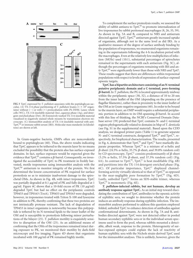
In Gram-negative bacteria, OMPs often are noncovalentlybound to peptidoglycan (60). Thus, the above results indicatingthat TprC appears to be tethered to the murein layer by no meansexcluded the possibility that the protein also has surface-exposeddomains. In fact, surface exposure would be expected given theevidence that TprCFl contains a �-barrel. Consequently, we inves-tigated the accessibility of TprC to PK treatment in freshly har-vested, motile treponemes using immunoblot analysis with theTprCSp antiserum to monitor integrity of the protein. We firstdetermined the lowest concentration of PK required for surfaceproteolysis so as to minimize inadvertent damage to the spiro-chetal OMs. As shown in Fig. 4B, with intact treponemes, TprCwas partially degraded at 0.1 �g/ml of PK and fully degraded at 1�g/ml. Figure 4C shows that a 10-fold excess of PK (10 �g/ml)degraded TprC but had no effect on the periplasmic controlsTP0453 and TP0163 (TroA). TP0453 and TP0163 were degraded,however, in treponemes treated with Triton X-100 and lysozymein addition to PK, thereby confirming that these two proteins arenot intrinsically protease resistant. The lack of degradation ofTP0453 in intact organisms is noteworthy because this polypep-tide is tethered by its N-terminal lipids to the inner leaflet of theOM and is susceptible to proteolysis following minor perturba-tions of the bilayer (25). T. pallidum motility is exquisitely sensi-tive to disruption of the OM (25). As an additional means ofconfirming that the OMs of the treponemes remained intact dur-ing exposure to PK, we monitored their motility by dark-fieldmicroscopy and live imaging. Figure 4D shows that organismsincubated with 100 �g/ml of PK remained highly motile.
To complement the surface proteolysis results, we assessed theability of rabbit antisera to TprCFl to promote internalization oflive treponemes by rabbit peritoneal macrophages (8, 26, 35, 59).As shown in Fig. 5A and B, compared to NRS and antiserumdirected against Tp47, TprCFl antiserum greatly increased uptakeof organisms, although not to the same level as did IRS. As aqualitative measure of the degree of surface antibody binding bythe population of treponemes, we enumerated organisms remain-ing in the supernatants following the 4-h incubation period withthe macrophages. Even at the relatively low multiplicities of infec-tion (MOIs) used (10:1), substantial percentages of spirochetesremained in the supernatants with each antiserum (Fig. 5C), al-though the percentages of organisms remaining with IRS and an-ti-TprCFl were significantly lower than those with NRS and Tp47.These results suggest that there are differences within treponemalpopulations with respect to levels of expression of surface-exposedopsonic targets.
TprC has a bipartite architecture consisting of an N-terminalputative periplasmic domain and a C-terminal, pore-forming�-barrel. In T. pallidum, the PG is located approximately midwaywithin the periplasmic space (30, 32), a distance of 10 to 20 nmfrom the inner leaflet of the OM (depending on the presence offlagellar filaments), rather than in proximity to the inner leaflet ofthe OM as in Gram-negative organisms (60). In order to be boundto the murein layer, an OMP would require a substantial periplas-mic portion in addition to its OM-spanning �-barrel. In accordwith this line of thinking, the NCBI’s Conserved Domain Data-base server (39) predicted that TprC contains N- and C-terminalregions phylogenetically related to domains in Msp of T. denticola(see Fig. S1 in the supplemental material). Based upon the CDDanalysis, we designed primer pairs (Table 1) to generate separateN- and C-terminal constructs, designated TprCN and TprCC, re-spectively, for biophysical characterization. The results, presentedin Fig. 6, demonstrate that TprCN and TprCC have markedly dis-parate properties. Whereas TprCN is a mixture of �-helix and�-sheet (24.5% �-helix, 35.7% �-sheet, and 39.8% random coil)by CD spectroscopy, TprCC consists predominantly of �-sheet(5.2% �-helix, 57.5% �-sheet, and 37.3% random coil) (Fig.6A). In contrast to TprCN, TprCC is heat modifiable (Fig. 6B)and partitions into the TX-114 detergent-enriched phase (Fig.6C). Of particular importance, TprCC displayed channel-forming activity virtually identical to that of TprCFl as opposedto the near-negligible pore formation by TprCN (Fig. 6D).Lastly, unboiled TprCC forms an SDS-stable trimer, whereasTprCN is monomeric (Fig. 6E).
T. pallidum-infected rabbits, but not humans, develop anantibody response against TprC. As an initial step toward eluci-dating the contribution of TprC antibodies to the opsonic activityof syphilitic sera, we sought to determine whether the proteininduces an antibody response during syphilitic infection. The im-munoblot analyses performed to address this question employedfolded, unboiled TprC to enhance detection of antibodies againstconformational determinants. As shown in Fig. 7A and B, anti-bodies directed against TprC were not detected either in pooledhuman secondary syphilitic sera or in the individual serum spec-imens used to form the pool, whereas rabbits mounted a detect-able, albeit weak, response to the protein. Heterogeneity of sur-face-exposed epitopes could explain the lack of reactivity ofhuman syphilitic sera with the Nichols strain-derived TprC usedfor the immunoblot analyses. This is unlikely, however, given the
FIG 3 TprC expressed by T. pallidum associates with the peptidoglycan sac-culus. (A) TX-114 phase partitioning of T. pallidum lysates (1 � 109 organ-isms) without (�) or with (�) preincubation with 2% DDM. Lanes: wholecells (WC), TX-114-insoluble material (Ins), aqueous phase (Aq), and deter-gent-enriched phase (Det). (B) Extensively washed TX-114-insoluble materialvisualized in negatively stained whole mounts by transmission electron mi-croscopy. (C) Immunoblot analysis of TX-114-insoluble material with anti-TprCSp or immune rabbit serum (IRS). Molecular mass standards (in kilodal-tons) are shown at left.
TprC, a Rare T. pallidum OMP
May 2012 Volume 194 Number 9 jb.asm.org 2327

extremely high degree of sequence conservation of TprC se-quences, including the �-barrels (see Fig. S2 in the supplementalmaterial). Note that the Cali-77 sequence in the alignment wasderived from a tprC gene that was PCR amplified from the skin ofa patient seen at our Cali, Colombia, enrollment site (14); serumfrom this patient also was used for immunoblotting (Fig. 7B).Interestingly, antibody reactivities to 326P1– 4� and folded, un-boiled 326�b differed strikingly from those for TprC. As recentlyreported (19), and also shown in Fig. 7A and B, humans oftenresponded well to the POTRA domain but not to the �-barrel,while infected rabbits recognized both POTRA and �-barrel do-mains. Lastly, we wished to confirm that the opsonic rabbit seraused for the experiment shown in Fig. 5 contained antibodiesdirected against the �-barrel. This was found to be the case forboth rabbit anti-TprCFl serum and IRS, although their reactivitiesdiffered considerably; the former reacted with both TprCN andTprCC while the latter recognized only TprCC (Fig. 7C).
DISCUSSION
Certain general features of early human syphilis, such as its waxingand waning course and the tendency for disease progression tooccur in the face of a robust immune response (34, 50), have longperplexed clinicians and syphilis researchers. In recent years, in-
vestigators have begun to devise ex vivo and translational strategiesto relate these enigmatic disease manifestations to the syphilis spi-rochete’s unusual cell envelope architecture (14, 42), most notablyits paucity of surface antigenic targets (7, 10, 11). Critical to thiseffort is the need to molecularly characterize the bacterium’s rareOMPs, entities which were originally observed by freeze fractureelectron microscopy more than 20 years ago (52, 64) but only justbeginning to be definitively identified more than a decade after thepublication of the bacterium’s genomic sequence (7, 22, 29).Based on theoretical considerations, we postulated that OMPs inT. pallidum would, like their nonorthologous Gram-negativecounterparts, adopt a �-barrel conformation (11, 48). Our limitedsuccess in pursuing our quest for T. pallidum OMPs at the proteinlevel prompted us to adopt an in silico-based approach in whichwe used a battery of computational tools to mine the T. pallidumgenome for proteins predicted to form �-barrels; TprC, the focusof the present study, emerged from this analysis as one of ourstrongest candidates (11).
Strategies for characterizing OMP candidates must, by neces-sity, rely heavily upon the use of recombinant proteins but with aneye toward confirming key in vitro findings with the endogenousprotein and establishing surface exposure in live treponemes.Along these lines, we used TprCFl to establish that this recombi-
FIG 4 TprC is expressed at low abundance and is accessible to proteinase K in motile treponemes. (A) Quantitative immunoblot analysis of TprC expressed inT. pallidum. T. pallidum lysates in amounts indicated were immunoblotted with anti-TprCSp antiserum followed by densitometric analysis; a standard curvegenerated from densitometric values obtained for graded amounts of TprCFl was used to determine the copy number of TprC per T. pallidum cell. Molecular massstandards (kDa) are indicated on the left. (B) Immunoblot analysis of TprC, detected by the anti-TprCSp antiserum, in motile treponemes (1.0 � 108 T. pallidumorganisms/lane) treated for 1 h with graded concentrations of proteinase K (PK). (C) PK accessibility of TprC and two periplasmic controls in intact anddetergent-lysozyme-treated organisms incubated with (�) or without (�) 10 �g/ml of PK. Each lane represents 1.0 � 108 T. pallidum organisms immunoblottedwith antisera to TprCSp, TP0453, or TroA. (D) Live imaging of intact treponemes with and without incubation with 100 �g/ml of PK. Asterisks and arrowheadsindicate flexing and wave form propagation, respectively. Time scale is 0 to 0.12 s.
Anand et al.
2328 jb.asm.org Journal of Bacteriology

nant protein is rich in �-sheet content, is heat modifiable, formstrimers, and is capable of inserting into lipid bilayers whose phos-pholipid composition mimics that of the T. pallidum OM (53).While it is not possible to replicate all of these results with endog-enous protein, we did find that the latter is heat modifiable, am-phiphilic, and trimeric, results consistent with formation of�-barrel multimers in the spirochetal OM. Furthermore, we dem-onstrated surface exposure by two very different methods, pro-tease accessibility and opsonophagocytosis, employing live organ-isms under conditions in which the integrity of the OM wasmaintained. The two approaches yielded an interesting counter-point; one showed that all of the TprC in the population of trepo-nemes is accessible to PK digestion, while the other revealed thatnot all treponemes in a population express sufficient TprC foropsonization (a result mirrored with IRS, which presumably con-tains antibodies against opsonic targets in addition to TprC). Wealso confirmed that sera with opsonic activity contained antibod-ies directed against the portion of TprC expected to harbor sur-face-exposed determinants. These results, coupled with our ob-servation that tprC/TprC is expressed at exceedingly low copynumbers, leave little doubt as to its authenticity as a rare OMP.
In comparison with the extensively characterized porin E. coliOmpF, the recombinant protein also readily promoted efflux ofthe fluorophore Tb(DPA)3
3� from LUVs. Based on the overallsimilarity in biophysical characteristics of the recombinant andendogenous proteins, we propose that TprC does indeed functionto enhance the permeability of the T. pallidum OM and, as such, isthe first bona fide OM-spanning protein in the syphilis spirocheteshown to do so. Importantly, a channel large enough to permitrapid efflux of Tb(DPA)3
3� (MM, 650 Da) should be large enoughto allow passive influx of glucose, amino acids, and numerous
other nutrients which T. pallidum must parasitize from the bodyfluids of its obligate human host. Additional support for this ideacomes from Msp of T. denticola, reported to form extremely largeungated channels in black lipid membranes (21). Efforts to under-stand the mechanisms of spirochetal persistence have generallyfocused on the bacterium’s ability to evade host defenses (7, 31,49). In order to establish chronic infection, T. pallidum also mustacquire essential nutrients from every microenvironment inwhich it takes up residence. Our findings provide a small physio-logical piece of the persistence puzzle which emerged from thediscovery of the bacterium’s unique, protein-scarce OM (48). Wehypothesize that the copy number of TprC and the size of itschannel have been calibrated during the reductive evolutionaryprocess which gave rise to pathogenic treponemes (57) to meet themetabolic demands of this very slowly replicating organism (38)while maintaining the lowest possible immunologic profile.
With most Gram-negative OMPs, the entire polypeptide formsa cylinder consisting of an even number of �-strands framed bylarge external loops and small periplasmic turns (56, 67). The22-stranded �-barrel predicted for TprC by TMBpro (54) con-forms to this generalization (11). The data generated here usingthe full-length recombinant TprCFl gave no reason to question theassumption that the entire polypeptide forms the barrel. Two em-pirical findings, both obtained from T. pallidum, however,prompted the experiments which now lead to a substantially re-vised working model for TprC (Fig. 8), in addition to underscor-ing the uncertainties of in silico �-barrel structural predictions.One was the association of endogenous TprC with the TX-114-insoluble material, which consists of cell ghosts with remnant,unextracted proteins (51). The other came from cryo-electrontomographic analysis of T. pallidum, which localized the spiro-
FIG 5 TprC antibodies have opsonic activity. (A) Percentages of rabbit peritoneal macrophages containing internalized treponemes. Results shown are means �standard errors of the means for three independent experiments. (B) Representative micrographs of macrophages incubated with motile treponemes and theindicated sera. Each image is a composite of bright-field images, nuclei stained with DAPI, and FITC-labeled T. pallidum. (C) Percentages of treponemesrecovered following incubation of rabbit peritoneal macrophages for 4 h with the indicated sera. Results shown are means � standard errors of the means forthree independent experiments. P values of �0.05 (Student’s t test) were considered significant.
TprC, a Rare T. pallidum OMP
May 2012 Volume 194 Number 9 jb.asm.org 2329

chete’s thin PG layer to approximately midway within theperiplasmic space (30, 32). The two findings together suggestedthat TprC contains a periplasmic domain that tethers the OM-inserted barrel to the PG sacculus. We confirmed this deductionby demonstrating that the �-barrel and channel-forming activityreside entirely within the C-terminal 202 residues; this portion ofTprC also contains the predicted sequence for recognition ofTprC precursors by the POTRA domain of BamA (19), a prereq-uisite for OM localization (3, 24, 55, 63) (see Fig. S1 in the supple-mental material). We note that our data do not directly addressthe location of the stretch of polypeptide containing TprCSp,which we place in the periplasm (Fig. 8). Because we did not con-sistently detect a PK degradation product reactive with our TprCSp
antiserum (data not shown), the possibility that this portion of theprotein is surface exposed and, therefore, part of the �-barrel alsoneeds to be considered. Arguing strongly against this topologicalassignment is our demonstration that TprCC alone, which doesnot include the contiguous TprCSp, is sufficient to circularize as astable (i.e., heat-modifiable), trimeric �-barrel with the sameproperties as that of the full-length polypeptide. Given the weightof evidence that the outer membranes of the treponemes re-mained intact throughout incubation with PK, we attribute theinability to detect a PK degradation product reactive with ourTprCSp antiserum to the induction of a cell envelope stress re-sponse that results in degradation of this fragment. The watersolubility of TprCSp (data not shown) further supports its assign-ment to the periplasmic compartment.
While we have not yet directly proven that TprCC and TprCN
reside in the OM and periplasm, respectively, these are the onlyconceivable compartmental assignments. We do note, however,that TprCN lacks a recognizable OmpA-like peptidoglycan-bind-ing motif (18, 44), an observation that we do not find surprisinggiven the reverse bipartite topologies of TprC and OmpA (61) andthe phylogenetic distance separating spirochetes from didermswhich do possess OmpA orthologs (46). Furthermore, it has notescaped our attention that the topologic model proposed for TprCcan be extended readily to TprI, which differs from TprC mainlywithin the periplasmic domain, and TprF, which is predicted to beentirely periplasmic (Fig. 8B). Inasmuch as the domain predic-tions for TprC were based on T. denticola Msp, it is tempting tospeculate that this molecular architecture applies to more dis-tantly related Tpr family members as well as the parental ortholog(8, 11, 22). Our finding a number of years ago that Msp is bothsurface exposed and periplasmic based on immunofluorescenceanalysis (6) is consistent with the concept of a broadly conservedbipartite domain structure for this phylogenetically related groupof OMPs.
Our concept of stealth pathogenicity, dating from the pre-genomic era, was based on the premise that rare OMPs would bepoorly immunogenic because they are nonlipidated and expressedat extremely low copy numbers (48). The availability of properlyfolded bona fide OMPs has enabled us to assess this generaliza-tion, which has turned out to be less straightforward and far moreintriguing than the “one size fits all” hypothesis originally envi-
FIG 6 TprCC contains the �-barrel of TprC and forms a trimer. (A to C) CD spectroscopy, heat modifiability, and TX-114 phase partitioning of TprCN andTprCC. Aq and Det, aqueous and detergent-enriched phases, respectively. (D) Quenching of Tb(DPA)3
3� encapsulated within LUVs following incubation withTprCFl, TprCN, TprCC, and E. coli OmpF. (E) SDS-PAGE of unboiled TprCN and TprCC (asterisk indicates TprCC trimer). Numbers at left in panels B and E aremolecular masses in kilodaltons.
Anand et al.
2330 jb.asm.org Journal of Bacteriology

sioned. As reported elsewhere (19) and reproduced here, TP0326often induces an antibody response in humans but the antibodiesappear to be directed exclusively toward the periplasmic POTRAdomains and away from the potentially lethal surface-exposed de-terminants. In contrast, neither domain of TprC appears to beimmunogenic in humans. The higher copy number of TprCwould suggest that expression levels alone cannot explain this di-chotomy, although one must bear in mind the critical proviso thatcopy numbers were assessed in spirochetes harvested from onetissue in a nonnatural host at a single time point during infection.Nothing is known yet about the expression levels of these proteins
during the period of acute disease when the immune system actu-ally encounters them. In the case of TP0326, one must furtherpostulate a dissociation in the response to the periplasmic andOM-spanning domains, also surprising because �-barrels con-taining large extracellular loops, as predicted for TP0326 (19), areexpected to be antigenic (58).
Collectively, these results support our longstanding conten-tion, buttressed by recent clinical studies (14), that poor antibodybinding is a prime virulence determinant of the pathogen thatfinds clinical expression during the spirochetemia of secondarysyphilis. They also represent something of a paradox since human
FIG 7 T. pallidum-infected rabbits, but not humans, mount an antibody response against TprC. (A) Reactivities of normal rabbit serum (NRS), immune rabbitserum (IRS; representative of 3 different animals), normal human serum (NHS), and pooled human secondary syphilitic sera (HSS) against folded, unboiledTprCFl; 326P1-4�; and folded, unboiled 326�b. (B) Immunoblot reactivities of individual patient sera. (C) Immunoblot reactivities of the anti-TprCFl antiserumand IRS used for the opsonophagocytosis assays in Fig. 5 against TprCN and folded, unboiled TprCC. Each immunoblot assay was performed using 100 ng ofrecombinant protein. Numbers at left are molecular masses in kilodaltons.
FIG 8 Topological models of TprC, TprI, and TprF. (A) Comparison of domain architectures. (B) TprC/D and TprI are proposed to consist of trimers withidentical �-barrels and N-terminal periplasmic domains that directly or indirectly tether the barrels to the peptidoglycan (PG) sacculus. TprF is entirelyperiplasmic. TprCSp is depicted as periplasmic based on its solubility in aqueous buffer. TprI shares with TprF a putative periplasmic fragment not present inTprC.
TprC, a Rare T. pallidum OMP
May 2012 Volume 194 Number 9 jb.asm.org 2331

syphilitic sera do contain antibodies capable of surface labelingand promoting opsonophagocytosis of subpopulations of trepo-nemes (11, 13, 42). Either these antibodies are directed againstyet-to-be-identified OMPs or the immunoblots are not giving afully accurate readout of the responses to the proteins, possibilitiesthat we intend to aggressively explore in the near future. Theenigma of the immune response to rare OMPs ostensibly deepenswhen one considers that rabbits infected with T. pallidum via theintratesticular route make antibodies against the �-barrels of bothTP0326 and TprC. We believe that this observation reflects theunique evolutionary relationship between the syphilis spirocheteand its natural host. The syphilis field may finally have advanced tothe point where the antibody response in rabbits can be dissectedand exploited as an immunologic road map to the long-soughtsyphilis vaccine for humans.
ACKNOWLEDGMENTS
This work was supported by NIH grants AI-26756 (J.D.R.), K23 AI62439(J.C.S.), 5R03TW008023-3 (J.C.S. and A.R.C.), and 5R03TW008023(J.C.S.) and by the Connecticut Children’s Medical Center (CCMC) Ar-rison and Burr Curtis Research Funds (J.C.S.).
REFERENCES1. Akins DR, et al. 1997. Tromp1, a putative rare outer membrane protein,
is anchored by an uncleaved signal sequence to the Treponema pallidumcytoplasmic membrane. J. Bacteriol. 179:5076 –5086.
2. Beher MG, Schnaitman CA, Pugsley AP. 1980. Major heat-modifiableouter membrane protein in gram-negative bacteria: comparison with theompA protein of Escherichia coli. J. Bacteriol. 143:906 –913.
3. Bennion D, Charlson ES, Coon E, Misra R. 2010. Dissection of �-barrelouter membrane protein assembly pathways through characterizingBamA POTRA 1 mutants of Escherichia coli. Mol. Microbiol. 77:1153–1171.
4. Bredemeier R, et al. 2007. Functional and phylogenetic properties of thepore-forming beta-barrel transporters of the Omp85 family. J. Biol.Chem. 282:1882–1890.
5. Brusca JS, Radolf JD. 1994. Isolation of integral membrane proteins byphase partitioning with Triton X-114. Methods Enzymol. 228:182–193.
6. Caimano MJ, Bourell KW, Bannister TD, Cox DL, Radolf JD. 1999. TheTreponema denticola major sheath protein is predominantly periplasmicand has only limited surface exposure. Infect. Immun. 67:4072– 4083.
7. Cameron CE. 2006. The T. pallidum outer membrane and outer mem-brane proteins, p 237–266. In Radolf JD, Lukehart SA (ed), PathogenicTreponema: molecular and cellular biology. Caister Academic Press, Nor-wich, United Kingdom.
8. Centurion-Lara A, et al. 1999. Treponema pallidum major sheath proteinhomologue TprK is a target of opsonic antibody and the protective im-mune response. J. Exp. Med. 189:647– 656.
9. Conlan S, Bayley H. 2003. Folding of a monomeric porin, OmpG, indetergent solution. Biochemistry 42:9453–9465.
10. Cox DL, Akins DR, Porcella SF, Norgard MV, Radolf JD. 1995. Trepo-nema pallidum in gel microdroplets: a novel strategy for investigation oftreponemal molecular architecture. Mol. Microbiol. 15:1151–1164.
11. Cox DL, et al. 2010. Surface immunolabeling and consensus computa-tional framework to identify candidate rare outer membrane proteins ofTreponema pallidum. Infect. Immun. 78:5178 –5194.
12. Cox DL, Radolf JD. 2001. Insertion of fluorescent fatty acid probes intothe outer membranes of the pathogenic spirochaetes Treponema pallidumand Borrelia burgdorferi. Microbiology 147:1161–1169.
13. Cruz AR, et al. 2008. Phagocytosis of Borrelia burgdorferi, the Lymedisease spirochete, potentiates innate immune activation and inducesapoptosis in human monocytes. Infect. Immun. 76:56 –70.
14. Cruz AR, et al. 2010. Secondary syphilis in Cali, Colombia: new conceptsin disease pathogenesis. PLoS Negl. Trop. Dis. 4:e690.
15. Cullen PA, Cameron CE. 2006. Progress towards an effective syphilisvaccine: the past, present and future. Expert Rev. Vaccines 5:67– 80.
16. Cullen PA, Haake DA, Adler B. 2004. Outer membrane proteins ofpathogenic spirochetes. FEMS Microbiol. Rev. 28:291–318.
17. Deka RK, Machius M, Norgard MV, Tomchick DR. 2002. Crystalstructure of the 47-kilodalton lipoprotein of Treponema pallidum reveals anovel penicillin-binding protein. J. Biol. Chem. 277:41857– 41864.
18. De Mot R, Vanderleyden J. 1994. The C-terminal sequence conservationbetween OmpA-related outer membrane proteins and MotB suggests acommon function in both gram-positive and gram-negative bacteria, pos-sibly in the interaction of these domains with peptidoglycan. Mol. Micro-biol. 12:333–334.
19. Desrosiers DC, et al. 2011. TP0326, a Treponema pallidum �-barrel as-sembly machinery A (BamA) orthologue and rare outer membrane pro-tein. Mol. Microbiol. 80:1496 –1515.
20. Edelhoch H. 1967. Spectroscopic determination of tryptophan and ty-rosine in proteins. Biochemistry 6:1948 –1954.
21. Egli C, Leung WK, Muller KH, Hancock RE, McBride BC. 1993.Pore-forming properties of the major 53-kilodalton surface antigen fromthe outer sheath of Treponema denticola. Infect. Immun. 61:1694 –1699.
22. Fraser CM, et al. 1998. Complete genome sequence of Treponema palli-dum, the syphilis spirochete. Science 281:375–388.
23. Gasteiger E, et al. 2003. ExPASy: the proteomics server for in-depthprotein knowledge and analysis. Nucleic Acids Res. 31:3784 –3788.
24. Hagan CL, Silhavy TJ, Kahne D. 2011. �-barrel membrane proteinassembly by the Bam complex. Annu. Rev. Biochem. 80:189 –210.
25. Hazlett KR, et al. 2005. TP0453, a concealed outer membrane protein ofTreponema pallidum, enhances membrane permeability. J. Bacteriol. 187:6499 – 6508.
26. Hazlett KR, et al. 2001. The TprK protein of Treponema pallidum isperiplasmic and is not a target of opsonic antibody or protective immu-nity. J. Exp. Med. 193:1015–1026.
27. Heuck AP, Johnson AE. 2002. Pore-forming protein structure analysis inmembranes using multiple independent fluorescence techniques. CellBiochem. Biophys. 36:89 –101.
28. Heuck AP, Tweten RK, Johnson AE. 2003. Assembly and topography ofthe prepore complex in cholesterol-dependent cytolysins. J. Biol. Chem.278:31218 –31225.
29. Ho EL, Lukehart SA. 2011. Syphilis: using modern approaches to under-stand an old disease. J. Clin. Invest. 121:4584 – 4592.
30. Izard J, et al. 2009. Cryo-electron tomography elucidates the moleculararchitecture of Treponema pallidum, the syphilis spirochete. J. Bacteriol.191:7566 –7580.
31. Lafond RE, Lukehart SA. 2006. Biological basis for syphilis. Clin. Micro-biol. Rev. 19:29 – 49.
32. Liu J, et al. 2010. Cellular architecture of Treponema pallidum: novelflagellum, periplasmic cone, and cell envelope as revealed by cryo electrontomography. J. Mol. Biol. 403:546 –561.
33. Lukehart SA. 2008. Scientific monogamy: thirty years dancing with thesame bug: 2007 Thomas Parran Award Lecture. Sex. Transm. Dis. 35:2–7.
34. Lukehart SA. 2008. Syphilis, p 1038 –1046. In Fauci AS, et al (ed), Harri-son’s principles of internal medicine, vol 17. McGraw-Hill, New York, NY.
35. Lukehart SA, Miller JN. 1978. Demonstration of the in vitro phagocytosisof Treponema pallidum by rabbit peritoneal macrophages. J. Immunol.121:2014 –2024.
36. Lukehart SA, Shaffer JM, Baker-Zander SA. 1992. A subpopulation ofTreponema pallidum is resistant to phagocytosis: possible mechanism ofpersistence. J. Infect. Dis. 166:1449 –1453.
37. Luthra A, et al. 2011. The transition from closed to open conformation ofTreponema pallidum outer membrane-associated lipoprotein TP0453 in-volves membrane sensing and integration by two amphipathic helices. J.Biol. Chem. 286:41656 – 41668.
38. Magnuson HJ, Eagle H, Fleischman R. 1948. The minimal infectiousinoculum of Spirochaeta pallida (Nichols strain), and a consideration of itsrate of multiplication in vivo. Am. J. Syph. Gonorrhea Vener. Dis. 32:1–19.
39. Marchler-Bauer A, et al. 2011. CDD: a Conserved Domain Database forthe functional annotation of proteins. Nucleic Acids Res. 39:D225–D229.
40. Meng G, Fronzes R, Chandran V, Remaut H, Waksman G. 2009.Protein oligomerization in the bacterial outer membrane (review). Mol.Membr. Biol. 26:136 –145.
41. Mizianty MJ, Kurgan L. 2011. Improved identification of outer mem-brane beta barrel proteins using primary sequence, predicted secondarystructure, and evolutionary information. Proteins 79:294 –303.
42. Moore MW, et al. 2007. Phagocytosis of Borrelia burgdorferi and Trepo-nema pallidum potentiates innate immune activation and induces gammainterferon production. Infect. Immun. 75:2046 –2062.
Anand et al.
2332 jb.asm.org Journal of Bacteriology

43. Nikaido H. 2003. Molecular basis of bacterial outer membrane permea-bility revisited. Microbiol. Mol. Biol. Rev. 67:593– 656.
44. Park JS, et al. 2012. Mechanism of anchoring of OmpA protein to the cellwall peptidoglycan of the gram-negative bacterial outer membrane.FASEB J. 26:219 –228.
45. Parsonage D, et al. 2010. Broad specificity AhpC-like peroxiredoxin andits thioredoxin reductant in the sparse antioxidant defense system ofTreponema pallidum. Proc. Natl. Acad. Sci. U. S. A. 107:6240 – 6245.
46. Paster BJ, Dewhirst FE. 2000. Phylogenetic foundation of spirochetes. J.Mol. Microbiol. Biotechnol. 2:341–344.
47. Pugsley AP. 1993. The complete general secretory pathway in gram-negative bacteria. Microbiol. Rev. 57:50 –108.
48. Radolf JD. 1995. Treponema pallidum and the quest for outer membraneproteins. Mol. Microbiol. 16:1067–1073.
49. Radolf JD, Hazlett KRO, Lukehart SA. 2006. Pathogenesis of syphilis,p 197–236. In Radolf JD, Lukehart SA (ed), Pathogenic treponema:cellular and molecular biology. Caister Academic Press, Norwich,United Kingdom.
50. Radolf JD, Lukehart SA. 2006. Immunology of syphilis, p 285–322. InRadolf JD, Lukehart SA (ed), Pathogenic Treponema: cellular and molec-ular biology. Caister Academic Press, Norwich, United Kingdom.
51. Radolf JD, Moomaw C, Slaughter CA, Norgard MV. 1989. Penicillin-binding proteins and peptidoglycan of Treponema pallidum subsp. palli-dum. Infect. Immun. 57:1248 –1254.
52. Radolf JD, Norgard MV, Schulz WW. 1989. Outer membrane ultrastruc-ture explains the limited antigenicity of virulent Treponema pallidum.Proc. Natl. Acad. Sci. U. S. A. 86:2051–2055.
53. Radolf JD, et al. 1995. Characterization of outer membranes isolatedfrom Treponema pallidum, the syphilis spirochete. Infect. Immun. 63:4244 – 4252.
54. Randall A, Cheng J, Sweredoski M, Baldi P. 2008. TMBpro: secondarystructure, �-contact and tertiary structure prediction of transmembrane�-barrel proteins. Bioinformatics 24:513–520.
55. Robert V, et al. 2006. Assembly factor Omp85 recognizes its outer mem-
brane protein substrates by a species-specific C-terminal motif. PLoS Biol.4:e377.
56. Schulz GE. 2002. The structure of bacterial outer membrane proteins.Biochim. Biophys. Acta 1565:308 –317.
57. Seshadri R, et al. 2004. Comparison of the genome of the oral pathogenTreponema denticola with other spirochete genomes. Proc. Natl. Acad. Sci.U. S. A. 101:5646 –5651.
58. Sette A, Rappuoli R. 2010. Reverse vaccinology: developing vaccines inthe era of genomics. Immunity 33:530 –541.
59. Shevchenko DV, et al. 1999. Membrane topology and cellular location ofthe Treponema pallidum glycerophosphodiester phosphodiesterase(GlpQ) ortholog. Infect. Immun. 67:2266 –2276.
60. Silhavy TJ, Kahne D, Walker S. 2010. The bacterial cell envelope. ColdSpring Harb. Perspect. Biol. 2:a000414.
61. Smith SG, Mahon V, Lambert MA, Fagan RP. 2007. A molecular Swissarmy knife: OmpA structure, function and expression. FEMS Microbiol.Lett. 273:1–11.
62. Stegmeier JF, Andersen C. 2006. Characterization of pores formed byYaeT (Omp85) from Escherichia coli. J. Biochem. 140:275–283.
63. Tommassen J. 2010. Assembly of outer-membrane proteins in bacteriaand mitochondria. Microbiology 156:2587–2596.
64. Walker EM, Zampighi GA, Blanco DR, Miller JN, Lovett MA. 1989.Demonstration of rare protein in the outer membrane of Treponema pal-lidum subsp. pallidum by freeze-fracture analysis. J. Bacteriol. 171:5005–5011.
65. Watanabe Y, Inoko Y. 2009. Reassembly of an integral oligomeric mem-brane protein OmpF porin in n-octyl beta-D:-glucopyranoside-lipidsmixtures. Protein J. 28:66 –73.
66. Whitmore L, Wallace BA. 2004. DICHROWEB, an online server forprotein secondary structure analyses from circular dichroism spectro-scopic data. Nucleic Acids Res. 32:W668 –W673.
67. Wimley WC. 2003. The versatile �-barrel membrane protein. Curr. Opin.Struct. Biol. 13:404 – 411.
68. Wittig I, Braun HP, Schagger H. 2006. Blue native PAGE. Nat. Protoc.1:418 – 428.
TprC, a Rare T. pallidum OMP
May 2012 Volume 194 Number 9 jb.asm.org 2333