Purification, characterization, and immunogenicity of a soluble trimeric envelope protein containing...
16
JOURNAL OF VIROLOGY, Oct. 2003, p. 11244–11259 Vol. 77, No. 20 0022-538X/03/$08.000 DOI: 10.1128/JVI.77.20.11244–11259.2003 Copyright © 2003, American Society for Microbiology. All Rights Reserved. Purification, Characterization, and Immunogenicity of a Soluble Trimeric Envelope Protein Containing a Partial Deletion of the V2 Loop Derived from SF162, an R5-Tropic Human Immunodeficiency Virus Type 1 Isolate Indresh K. Srivastava, 1 * Leonidas Stamatatos, 2 Elaine Kan, 1 Michael Vajdy, 1 Ying Lian, 1 Susan Hilt, 1 Loic Martin, 3 Claudio Vita, 3 Ping Zhu, 4 Kenneth H. Roux, 4 Lucia Vojtech, 2 David C. Montefiori, 5 John Donnelly, 1 Jeffrey B. Ulmer, 1 and Susan W. Barnett 1 * Vaccines Research, Chiron Corporation, Emeryville, California 94608 1 ; Department of Pathobiology, University of Washington and Seattle Biomedical Research Institute, Seattle, Washington 98103 2 ; Department of Protein Engineering and Research, CEA Saclay, Gif-sur-Yvette, France 3 ; Department of Biological Science and Structural Biology Program, Florida State University, Tallahassee, Florida 32306 4 ; and Department of Surgery, Duke University Medical Center, Durham, North Carolina 27710 5 Received 21 March 2003/Accepted 14 July 2003 The envelope (Env) glycoprotein of human immunodeficiency virus type 1 (HIV-1) is the major target of neutralizing antibody responses and is likely to be a critical component of an effective vaccine against AIDS. Although monomeric HIV envelope subunit vaccines (gp120) have induced high-titer antibody responses and neutralizing antibodies against laboratory-adapted HIV-1 strains, they have failed to induce neutralizing antibodies against diverse heterologous primary HIV isolates. Most probably, the reason for this failure is that the antigenic structure(s) of these previously used immunogens does not mimic that of the functional HIV envelope, which is a trimer, and thus these immunogens do not elicit high titers of relevant functional antibodies. We recently reported that an Env glycoprotein immunogen (o-gp140SF162V2) containing a partial deletion in the second variable loop (V2) derived from the R5-tropic HIV-1 isolate SF162, when used in a DNA priming-protein boosting vaccine regimen in rhesus macaques, induced neutralizing antibodies against heterologous subtype B primary isolates as well as protection to the vaccinated animals upon challenge with pathogenic SHIV SF162P4 virus. Here we describe the purification of this protein to homogeneity, its characterization as trimer, and its ability to induce primary isolate-neutralizing responses in rhesus macaques. Optimal mutations in the primary and secondary protease cleavage sites of the env gene were identified that resulted in the stable secretion of a trimeric Env glycoprotein in mammalian cell cultures. We determined the molecular mass and hydrodynamic radius (R h ) using a triple detector analysis (TDA) system. The molecular mass of the oligomer was found to be 324 kDa, close to the expected M w of a HIV envelope trimer protein (330 kDa), and the hydrodynamic radius was 7.27 nm. Negative staining electron microscopy of o-gp140SF162V2 showed that it is a trimer with considerable structural flexibility and supported the data obtained by TDA. The structural integrity of the purified trimeric protein was also confirmed by determinations of its ability to bind the HIV receptor, CD4, and its ability to bind a panel of well-characterized neutralizing monoclonal antibodies. No deleterious effect of V2 loop deletion was observed on the structure and conformation of the protein, and several critical neutralization epitopes were preserved and well exposed on the purified o-gp140SF162V2 protein. In an intranasal priming and intramuscular boosting regimen, this protein induced high titers of functional antibodies, which neutralized the vaccine strain, i.e., SF162. These results highlight a potential role for the trimeric o-gp140SF162V2 Env immunogen in a successful HIV vaccine. AIDS continues to be a major health problem throughout the world, with approximately 40 million cases and 4 million deaths having been recorded so far. In certain parts of the world, nearly 30% of the total population is infected with human immunodeficiency virus (HIV). An estimated 40,000 to 80,000 primary infections occur each year in the United States alone. The situation is deteriorating as a result of rapid emer- gence of drug resistance against most of the effective antivirals. Therefore, there is an urgent need for an effective anti-HIV vaccine that may be used alone for protection or in conjunction with effective antivirals for treatment. HIV Env glycoprotein is the major target of neutralizing antibody responses and thus will be an important component of a vaccine. Historically, it has been difficult to induce neu- tralizing antibody responses against diverse primary HIV type 1 (HIV-1) strains by utilizing monomeric HIV Env (i.e., gp120) glycoprotein as a vaccine. Therefore, it seems that there are differences between qualities of antibodies induced during the natural course of infection and those induced through vacci- nation. This is not due to the absence of conserved neutraliza- tion epitopes on monomeric gp120 proteins (81) but is most likely due to their level of exposure and state of presentation on monomeric Env, compared to the level and state on native trimeric Env, present on the surface of virion (37, 51, 59, 69, 77, * Corresponding author. Mailing address: Vaccines Research, Chi- ron Corporation, 4560 Horton St., Emeryville, CA 94608. Phone for Indresh K. Srivastava: (510) 923-5485. Phone for Susan W. Barnett: (510) 923-7565. Fax: (510) 923 2586. E-mail for Indresh K. Srivastava: [email protected]. E-mail for Susan W. Barnett: [email protected]. 11244 on January 23, 2016 by guest http://jvi.asm.org/ Downloaded from
-
Upload
independent -
Category
Documents
-
view
4 -
download
0
Transcript of Purification, characterization, and immunogenicity of a soluble trimeric envelope protein containing...
JOURNAL OF VIROLOGY, Oct. 2003, p. 11244–11259 Vol. 77, No. 200022-538X/03/$08.00�0 DOI: 10.1128/JVI.77.20.11244–11259.2003Copyright © 2003, American Society for Microbiology. All Rights Reserved.
Purification, Characterization, and Immunogenicity of a SolubleTrimeric Envelope Protein Containing a Partial Deletion of the V2Loop Derived from SF162, an R5-Tropic Human Immunodeficiency
Virus Type 1 IsolateIndresh K. Srivastava,1* Leonidas Stamatatos,2 Elaine Kan,1 Michael Vajdy,1 Ying Lian,1 Susan Hilt,1
Loic Martin,3 Claudio Vita,3 Ping Zhu,4 Kenneth H. Roux,4 Lucia Vojtech,2 David C. Montefiori,5John Donnelly,1 Jeffrey B. Ulmer,1 and Susan W. Barnett1*
Vaccines Research, Chiron Corporation, Emeryville, California 946081; Department of Pathobiology, University of Washington andSeattle Biomedical Research Institute, Seattle, Washington 981032; Department of Protein Engineering and Research, CEA Saclay,Gif-sur-Yvette, France3; Department of Biological Science and Structural Biology Program, Florida State University, Tallahassee,
Florida 323064; and Department of Surgery, Duke University Medical Center, Durham, North Carolina 277105
Received 21 March 2003/Accepted 14 July 2003
The envelope (Env) glycoprotein of human immunodeficiency virus type 1 (HIV-1) is the major target ofneutralizing antibody responses and is likely to be a critical component of an effective vaccine against AIDS.Although monomeric HIV envelope subunit vaccines (gp120) have induced high-titer antibody responses andneutralizing antibodies against laboratory-adapted HIV-1 strains, they have failed to induce neutralizingantibodies against diverse heterologous primary HIV isolates. Most probably, the reason for this failure is thatthe antigenic structure(s) of these previously used immunogens does not mimic that of the functional HIVenvelope, which is a trimer, and thus these immunogens do not elicit high titers of relevant functionalantibodies. We recently reported that an Env glycoprotein immunogen (o-gp140SF162�V2) containing apartial deletion in the second variable loop (V2) derived from the R5-tropic HIV-1 isolate SF162, when usedin a DNA priming-protein boosting vaccine regimen in rhesus macaques, induced neutralizing antibodiesagainst heterologous subtype B primary isolates as well as protection to the vaccinated animals upon challengewith pathogenic SHIVSF162P4 virus. Here we describe the purification of this protein to homogeneity, itscharacterization as trimer, and its ability to induce primary isolate-neutralizing responses in rhesus macaques.Optimal mutations in the primary and secondary protease cleavage sites of the env gene were identified thatresulted in the stable secretion of a trimeric Env glycoprotein in mammalian cell cultures. We determined themolecular mass and hydrodynamic radius (Rh) using a triple detector analysis (TDA) system. The molecularmass of the oligomer was found to be 324 kDa, close to the expected Mw of a HIV envelope trimer protein (330kDa), and the hydrodynamic radius was 7.27 nm. Negative staining electron microscopy of o-gp140SF162�V2showed that it is a trimer with considerable structural flexibility and supported the data obtained by TDA. Thestructural integrity of the purified trimeric protein was also confirmed by determinations of its ability to bindthe HIV receptor, CD4, and its ability to bind a panel of well-characterized neutralizing monoclonal antibodies.No deleterious effect of V2 loop deletion was observed on the structure and conformation of the protein, andseveral critical neutralization epitopes were preserved and well exposed on the purified o-gp140SF162�V2protein. In an intranasal priming and intramuscular boosting regimen, this protein induced high titers offunctional antibodies, which neutralized the vaccine strain, i.e., SF162. These results highlight a potential rolefor the trimeric o-gp140SF162�V2 Env immunogen in a successful HIV vaccine.
AIDS continues to be a major health problem throughoutthe world, with approximately 40 million cases and 4 milliondeaths having been recorded so far. In certain parts of theworld, nearly 30% of the total population is infected withhuman immunodeficiency virus (HIV). An estimated 40,000 to80,000 primary infections occur each year in the United Statesalone. The situation is deteriorating as a result of rapid emer-gence of drug resistance against most of the effective antivirals.Therefore, there is an urgent need for an effective anti-HIV
vaccine that may be used alone for protection or in conjunctionwith effective antivirals for treatment.
HIV Env glycoprotein is the major target of neutralizingantibody responses and thus will be an important componentof a vaccine. Historically, it has been difficult to induce neu-tralizing antibody responses against diverse primary HIV type1 (HIV-1) strains by utilizing monomeric HIV Env (i.e., gp120)glycoprotein as a vaccine. Therefore, it seems that there aredifferences between qualities of antibodies induced during thenatural course of infection and those induced through vacci-nation. This is not due to the absence of conserved neutraliza-tion epitopes on monomeric gp120 proteins (81) but is mostlikely due to their level of exposure and state of presentationon monomeric Env, compared to the level and state on nativetrimeric Env, present on the surface of virion (37, 51, 59, 69, 77,
* Corresponding author. Mailing address: Vaccines Research, Chi-ron Corporation, 4560 Horton St., Emeryville, CA 94608. Phone forIndresh K. Srivastava: (510) 923-5485. Phone for Susan W. Barnett:(510) 923-7565. Fax: (510) 923 2586. E-mail for Indresh K. Srivastava:[email protected]. E-mail for Susan W. Barnett:[email protected].
11244
on January 23, 2016 by guesthttp://jvi.asm
.org/D
ownloaded from
87). Structural studies indicated that HIV Env is present as atrimer on the surface of the virion, whereas monomers havebeen used for immunization (32, 84, 88). These conformationaldifferences may affect the exposure of critical neutralizingepitopes on recombinant gp120 compared to those on nativeEnv. In general, compared to nonneutralizing antibodies,broadly neutralizing antibodies have stronger affinities for thenative envelope (trimer) (7, 8, 26, 52, 53, 60, 63, 69, 70),suggesting that the neutralizing epitopes recognized by thesebroadly reactive monoclonal antibodies (MAbs) are better rep-resented on oligomeric Env. Therefore, efforts have beenmade to purify the HIV Env protein in its functional, trimericconformation and evaluate its ability to induce primary isolate-neutralizing antibodies in animal models. Earlier studies havesuggested that HIV Env oligomers, consisting of gp120 and theectodomain of gp41, might be superior to monomeric gp120for inducing strong humoral responses toward conformationalepitopes (6, 18, 62, 83). In addition, antibodies induced byoligomeric Env cross-reacted with HIV envelopes of othersubtypes and neutralized both T cell-adapted and some pri-mary isolates of HIV-1 (22, 83, 92). More recent evidence hasfurther demonstrated that oligomers may be superior at induc-ing primary isolate-neutralizing antibodies compared to mono-mers (22, 92). Therefore, trimeric HIV Env has the potential tobe a better immunogen for inducing broadly cross-reactiveneutralizing antibodies.
The high degree of glycosylation (approximately 50% of themolecular mass) of HIV Env is critical not only for the intra-cellular transport of the envelope protein, but also for provid-ing correct functional conformation for its binding to CD4.Glycosylation also can mask T- and B-cell epitopes and therebymay decrease Env immunogenicity. Therefore, it may be thatthe extensive glycosylation is used by the virus not only forinvading the new cells, but also as an immune shield to maskcritical neutralizing epitopes. Recent evidence from simianimmunodeficiency virus (SIV) and simian-human immunode-ficiency virus (SHIV) rhesus infection models suggests that theemergence of viruses that are resistant to antibody-mediatedneutralization is associated with modification of the glycosyla-tion profiles and that resistance is at least partially due to thealteration in the glycosylation sites within the viral envelope(13, 41, 55–57, 66). Therefore, one strategy to increase theimmunogenicity of Env would be to remove glycosylation siteswithout affecting the structure and conformation of the Envprotein to expose more neutralizing epitopes. It was demon-strated that elimination of specific N-linked glycosylation sitesfrom variable loops of the HIV and SIV models renders thesemutant viruses more susceptible to neutralization by human oranimal sera as well as by specific MAbs (1, 9, 33, 42, 44, 61, 72,76). An important observation for HIV vaccine development isthat sera collected from macaques infected with partially deg-lycosylated SIVmac239 viruses from which the V1 loop hasbeen deleted have a higher neutralization potential against theparental fully glycosylated SIVmac239 viruses than the seracollected from macaques infected with the parental virus itself(61). Furthermore, a mutant of the SF162 envelope proteinlacking 30 out of 40 amino acids of the V2 loop (termed �V2)appeared to be more effective in eliciting cross-reactive neu-tralizing antibodies than the parental SF162 envelope (2). Onthe background of the SF162 envelope, partial deletion of the
V2 loop appears to increase the exposure and immunogenicityof certain neutralization epitopes. Since the oligomeric soluble�V2 envelope elicits cross-reactive neutralizing antibodies andsuch antibodies can reduce viral replication in macaques chal-lenged with the pathogenic R5 SHIVSF162P4 isolate, we believethat this immunogen could be included in an anti-HIV vaccine(2, 14, 15).
Here we describe the purification and biochemical, biophys-ical, structural, and immunochemical characterization of solu-ble oligomeric gp140 protein containing a partial deletion inV2 loop derived from the well-characterized HIV-1 SF162envelope (termed o-gp140SF162�V2). Our electron micro-scopic (EM) studies indicate that the purified protein is in astable trimeric conformation, and immunochemical studies in-dicate that critical neutralizing epitopes are exposed and wellpreserved. Moreover, in an intranasal priming and intramus-cular boosting regimen, o-gp140SF162�V2 protein inducedhigh titers of antibodies in rhesus macaques, which neutralizedthe homologous primary isolate, i.e., SF162.
MATERIALS AND METHODS
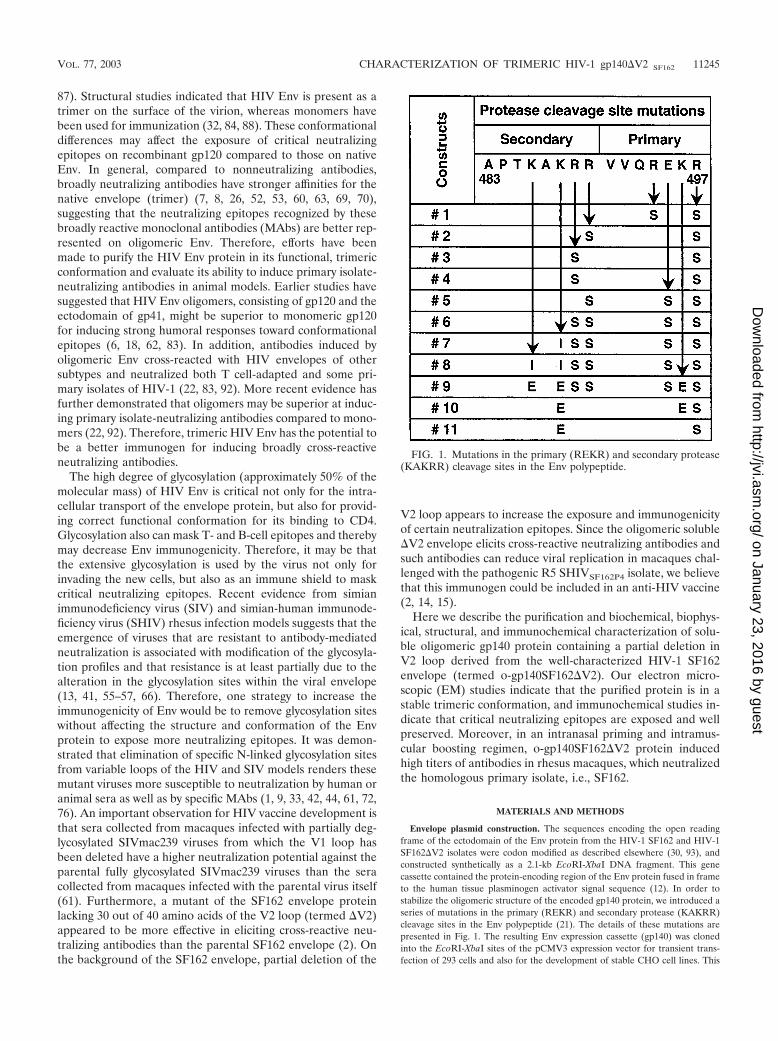
Envelope plasmid construction. The sequences encoding the open readingframe of the ectodomain of the Env protein from the HIV-1 SF162 and HIV-1SF162�V2 isolates were codon modified as described elsewhere (30, 93), andconstructed synthetically as a 2.1-kb EcoRI-XbaI DNA fragment. This genecassette contained the protein-encoding region of the Env protein fused in frameto the human tissue plasminogen activator signal sequence (12). In order tostabilize the oligomeric structure of the encoded gp140 protein, we introduced aseries of mutations in the primary (REKR) and secondary protease (KAKRR)cleavage sites in the Env polypeptide (21). The details of these mutations arepresented in Fig. 1. The resulting Env expression cassette (gp140) was clonedinto the EcoRI-XbaI sites of the pCMV3 expression vector for transient trans-fection of 293 cells and also for the development of stable CHO cell lines. This
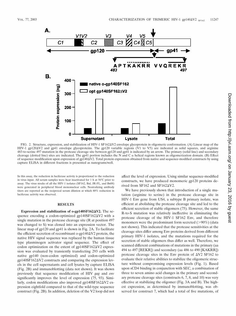
FIG. 1. Mutations in the primary (REKR) and secondary protease(KAKRR) cleavage sites in the Env polypeptide.
VOL. 77, 2003 CHARACTERIZATION OF TRIMERIC HIV-1 gp140�V2 SF162 11245
on January 23, 2016 by guesthttp://jvi.asm
.org/D
ownloaded from
vector contains the cytomegalovirus enhancer/promoter elements, an ampicillinresistance gene, and sequences encoding a fusion protein composed of dihydro-folate reductase and an attenuated neomycin resistance protein.
Transient transfection of 293 cells. In each well of a six-well plate, 1 � 106 293cells were plated. At approximately 70% confluence, the cells were washed withDPBS (Mediatech, Inc., Herdon, Va.), resuspended in 3 ml of OPTI-MEM(Gibco, Grand Island, N.Y.), and incubated with DNA/LT1 complex (5 �l ofLT1/�g of DNA) for 5 h. At the end of the incubation period, the cells wereresuspended in regular MEM (Mediatech) containing 10% fetal bovine serumand incubated for 48 h. At the end of the incubation period, medium wascollected and 1 mM EDTA and 1 mM EGTA were added before the perfor-mance of small-scale protein enrichment for expression and structural analysis.
Development of stable CHO cell lines secreting ��gp140SF162�V2. StableCHO cell lines secreting gp140SF162�V2 were derived by using DG-44 cells witha double deletion mutation in the dihydrofolate reductase gene, thus making thecell line dependent on the addition of hypoxanthine, glycine, and thymidine tothe growth medium, following the experimental protocol described elsewhere indetail (75).
Production of o-gp140 SF162�V2. The three highest expressinggp140SF162�V2-CHO cell clones were used to seed a 3-liter bioreactor (one foreach clone) for adaptation to medium containing a very low level of serum(0.5%) and to a cell density and perfusion rate that facilitated the maximumexpression of gp140SF162�V2 in its oligomeric conformation. Bioreactors weremonitored daily for cell density, pH, CO2, and O2 concentration, etc. Thestructure, conformation, and expression levels of secreted gp140SF162�V2 weremonitored weekly. From the three bioreactor-adapted clones, the best producerclone was used to seed a 12.5-liter bioreactor for production run. At the end ofthe run, collected medium was concentrated 20-fold through a 100-kDa-pore-size membrane filter and stored at �80°C in presence of 1 mM EDTA and 1 mMEGTA.
Envelope antigen capture ELISA. Envelope antigen capture enzyme-linkedimmunosorbent assay (ELISA) was performed by following standard protocoldescribed elsewhere in detail (75). gp140SF162�V2 concentration was calculatedfrom a standard curve derived using serial dilutions of a known concentration ofrecombinant gp120SF162.
Purification of o-gp140SF162�V2. The concentrated CHO cell supernatantwas thawed in presence of complete protease inhibitor cocktail (BoehringerMannheim) at 4°C, and after its conductivity was adjusted, it was loaded onto aGalanthus Nivalis-agarose column (GNA) (Vector Laboratories, Burlingame,Calif.) equilibrated with 20 mM Tris–100 mM NaCl (pH 7.4). Bound gp140 waseluted with 500 mM methyl mannose pyranoside (Sigma Chemical Co., St. Louis,Mo.) in equilibration buffer. The eluate after the GNA column was loaded ontoa DEAE (Pharmacia) column equilibrated with buffer (20 mM Tris, 100 mMNaCl [pH 8.0]). Under these conditions, o-gp140�V2 did not bind to the column,but contaminating proteins were retained on the column. The DEAEflowthrough was adjusted to 10 mM PO4 concentration, pH was adjusted to 6.8,and the flowthrough was loaded onto a ceramic hydroxyapatite (CHAP) (Bio-Rad Laboratories, Hercules, Calif.) column equilibrated with buffer (10 mMNa2HPO4, 100 mM NaCl [pH 6.8]). Under these conditions, gp140�V2 did notbind to CHAP column and was recovered in the flowthrough. All the columnfractions were analyzed on polyacrylamide gels (native and reduced and dena-tured) and also in a CD4 receptor-binding assay (see below). The fractionsshowing strong CD4-binding activity were pooled and fractionated on a 16- by90-mm Superdex–200 column (Pharmacia) equilibrated with 10 mM NaCitratecontaining 300 mM NaCl. Fractions containing o-gp140�V2 were pooled, con-centrated using a Stir cell (Millipore, Inc, Bedford, Mass.), and stored frozen at�80°C. During the purification process, fractions were analyzed by polyacryl-amide gel electrophoresis (PAGE) both under reducing and denaturing andunder native conditions following standard methods. Gels were stained withCoomassie brilliant blue or processed for immunoblotting.
Biophysical characterization of o-gp140SF162�V2. The molecular mass (Mw),hydrodynamic radius (Rh), and intrinsic viscosity of o-gp140SF162�V2 weredetermined using a triple detector array (TDA) system (TDA 300, ViscotekCorp. Houston, Tex.) in conjunction with a precalibrated gel filtration high-performance liquid chromatography (HPLC) column (Bio Sil SEC-250; Bio-RadLaboratories) using the Alliance 2690 HPLC system (Waters Corporation, Mil-ford, Mass.). Approximately 100 �g of the purified protein (o-gp140SF162�V2and gp120SF162�V2) was analyzed in 20 mM NaH2PO4–2 mM Na2HPO4 bufferat pH 7.4 at the flow rate of 1 ml/min. The signals from light scattering, aviscometer, and refractive index detectors were collected and analyzed using theEZ Pro software package (Viscotek Corp. Houston, Tex.).
Immunoblot detection of o-gp140SF162�V2. Approximately 0.5 to 1 �g ofo-gp140�V2 was separated on sodium dodecyl sulfate (SDS)-PAGE. After elec-
trophoresis, the gel was treated with 50 mM Tris–HCl (pH 8.0) containing 20%glycerol for 30 min at room temperature. Proteins were transferred on to a0.22-�M nitrocellulose membrane using a semidry transfer system (Bio-RadLaboratories). Immunodetection of o-gp140�V2 was performed exactly as de-scribed previously using anti-gp120 C4 domain-specific MAb 20-2-C8.5F3 (74;Steimer et al., unpublished observations) with minor modifications.
CD4 binding assay. To determine the capability of purified envelope proteinto bind CD4, we used an HPLC-based assay with fluoresceinated CD4, asdescribed elsewhere in detail (75).
Glycosylation profile analysis. Enzymatic deglycosylation of the purified HIV-Env proteins was performed using reagents from Bio-Rad Research Laborato-ries (catalog no. 170-6500). To detect N-linked oligosaccharides, 25 to 100 �g ofpurified protein was treated with 2 �l of peptidyl-N glycosidase F (PNGF) (2.5U/ml) for 1 h at 37°C. To study O-linked oligosaccharides, 25 �g of the purifiedprotein was treated with 2 �l of NANase II (10 U/ml) and 2 �l of O-glycosidase(1 U/ml) at 37°C for 1 h. In addition, we have also performed Endo-H digestionof the purified protein following manufacturer’s protocol (Boehringer Mann-heim). Enzyme-treated samples were diluted in sample buffer containing 0.1%SDS and �-mercaptoethanol and analyzed by SDS-PAGE.
Immunochemical characterization of o-gp140SF162�V2. The binding of well-characterized HIV-Env-specific MAbs to o-gp140SF162�V2 and gp120SF162proteins was performed by a capture ELISA. Purified proteins were adsorbed onELISA wells by an overnight incubation at room temperature (100 ng/well). Theadsorbed proteins were incubated with a serial dilution of various MAbs (0.01 to10 �g/ml in SuperBlock–0.05% Tween-20) for 4 h 37°C. For each MAb concen-tration, duplicate wells were used. The wells were washed with Tris-bufferedsaline and incubated with goat anti-human immunoglobulin G (IgG) coupled toalkaline phosphatase (Zymed) (1:30,000 dilution in SuperBlock–0.05% Tween-20) for an overnight incubation at 4°C. Substrate for alkaline phosphatase(DAKO Corp.) was added (50 �l/well) for 1 h at room temperature. Finally, 50�l of amplifier (DAKO Corp.) was added per well, and absorbance was recordedat 490 nm using a Vmax microplate reader (Molecular Devices). A plot of theabsorbance at 490 nm versus the MAb concentration was generated.
Surface plasmon resonance assay. Surface plasmon resonance assays wereperformed using a BIACORE 3000 optical biosensor system (Biacore AB, Upp-sala, Sweden) with simultaneous monitoring of relevant flow cells. To performthe kinetic study of the binding of o-gp140SF162�V2 to sCD4, sCD4 was im-mobilized using carbodiimide coupling onto a low-charge B1 sensor chip toattain a response of 2,000 response units. Using PBS buffer (pH 7.4) with 0.05%Tween 20, association was assessed by passing gp120SF162 or gp140SF162�V2over the chip surface at a flow rate of 30 �l/min. The concentration ranges forgp120SF162 and gp140SF162�V2 analytes were 59 to 300 nM (59, 89, 133, 200,and 300 nM) and 4.4 to 33 nM (4.4, 6.3, 9.6, 14, 22, and 33 nM), respectively. Asa negative control, we used a control surface without sCD4. For kinetic analysis,Biosensor data were prepared by subtracting binding responses obtained from ablank reference surface (without sCD4) from the specific responses. The asso-ciation and dissociation phase data were fitted simultaneously to a single-sitebinding model by using the nonlinear data analysis program Biaevaluation 3.2.
Immunoelectron microscopy. EM analysis of the oligomeric and monomericgp140SF162�V2 was performed by negative staining as previously described (64,65). The oligomeric (see Fig. 5A, peak A) and monomeric (see Fig. 5A, peak B)proteins obtained after the final preparative sizing column were diluted in boratebuffered saline, affixed to carbon membranes, stained with 1% uranyl formate,and mounted on copper grids for analysis. EMs were recorded at a nominalmagnification of �100,000 at 100 kV on a JEOL JEM 1200EX electron micro-scope. Measurements were made using the image analysis software Image-ProPlus. Fifty or more particles from fractions corresponding to the putative mono-mer/dimer and trimer of gp140SF162�V2 were measured and analyzed statisti-cally.
Stability studies. Purified o-gp140 SF162�V2 protein collected after the finalgel filtration column was used for performing the stability studies. Approximately2 to 5 �g of the purified protein was incubated at different temperatures (25 to100°C) for 1 h. Following this treatment the protein was analyzed by CD4binding, size-exclusion chromatography (SEC)-HPLC, SDS-PAGE, and immu-noprobing.
Antibody ELISA and virus neutralization assays. Antibody ELISA was per-formed on serum samples collected 2 weeks every immunization, essentially asdescribed previously (75).
Neutralization of homologous (SF162) and heterologous (Bal, JR-FL, andBx08) viruses was measured in 5.25.EGFP.Luc.M7 (M7-luc) cells obtained fromNathaniel Landau. The format of this assay was identical to that of the MT-2assay described elsewhere (50) except that virus infection was quantified byluciferase reporter gene expression using a commercial luciferase kit (Promega).
11246 SRIVASTAVA ET AL. J. VIROL.
on January 23, 2016 by guesthttp://jvi.asm
.org/D
ownloaded from
In this assay, the reduction in luciferase activity is proportional to the reductionin virus input. All serum samples were heat inactivated for 1 h at 56°C prior toassay. The virus stocks of all the HIV-1 isolates (SF162, Bal, JR-FL, and Bx08)were generated in peripheral blood mononuclear cells. Neutralizing antibodytiters are reported as the reciprocal serum dilution at which 80% reduction inluciferase activity was observed.
RESULTS
Expression and stabilization of o-gp140SF162�V2. The se-quence encoding a codon-optimized gp140SF162�V2 with asingle mutation in the protease cleavage site (R at position 497was changed to S) was cloned into an expression vector. Thelinear map of gp120 and gp41 is shown in Fig. 2A. To facilitatethe efficient secretion of recombinant o-gp140�V2 protein, thenative HIV signal sequence was replaced by the human tissuetype plasminogen activator signal sequence. The effect ofcodon optimization on the extent of gp140SF162�V2 expres-sion was evaluated by transiently transfecting 293 cells withnative gp140 (non-codon optimized) and codon-optimizedgp140SF162�V2 constructs and comparing the expression lev-els in the cell supernatants and cell lysates by capture ELISA(Fig. 2B) and immunoblotting (data not shown). It was shownpreviously that sequence modification of HIV gag and envsignificantly improves the level of expression (75, 93). Simi-larly, codon modifications also improved gp140SF162�V2 ex-pression eightfold compared to that of the wild-type sequenceconstruct (Fig. 2B). In addition, deletion of the V2 loop did not
affect the level of expression. Using similar sequence-modifiedconstructs, we have produced monomeric gp120 proteins de-rived from SF162 and SF162�V2.
We have previously shown that introduction of a single mu-tation (arginine to serine) in the protease cleavage site inHIV-1 Env gene from US4, a subtype B primary isolate, wasefficient at abolishing the protease cleavage site and led to theefficient secretion of stable oligomers (75). However, the sameR-to-S mutation was relatively ineffective in eliminating theprotease cleavage of the HIV-1 SF162 Env, and thereforemonomers were the predominant form secreted (�90%) (datanot shown). This indicated that the protease sensitivities at thecleavage sites differ among Env proteins derived from differentprimary HIV-1 isolates, and the mutations required for thesecretion of stable oligomers thus differ as well. Therefore, wescanned different combinations of mutations in the primary (aa494 to 497 [REKR]) and secondary (aa 486 to 490 [KAKRR])protease cleavage sites in the Env protein of �V2 SF162 toevaluate their relative abilities to stabilize the oligomeric struc-ture without compromising expression levels (Fig. 1). Basedupon sCD4 binding in conjunction with SEC, a combination ofthree to seven amino acid changes in the primary and second-ary protease cleavage sites (constructs 6, 7, 8, and 10) was veryeffective at stabilizing the oligomer (Fig. 3A and B). The high-est expression, as determined by immunoblotting, was ob-served for construct 7, which had a total of five mutations, of
FIG. 2. Structure, expression, and stabilization of HIV-1 SF162�V2 envelope glycoprotein in oligomeric conformation. (A) Linear map of theHIV-1 gp120�V2 and gp41 envelope glycoproteins. The gp120 variable regions (V1 to V5) are indicated as solid squares, and arginine483-to-serine 497 mutation in the protease cleavage site between gp120 and gp41 is indicated by an arrow. The primary (solid line) and secondarycleavage (dotted line) sites are indicated. The gp41 portion includes the N and C � helical regions known as oligomerization domain. (B) Effectof sequence modification upon expression of gp140�V2. Total protein expression obtained from native and sequence-modified constructs by usingcapture ELISA in different fractions is presented as nanograms/well.
VOL. 77, 2003 CHARACTERIZATION OF TRIMERIC HIV-1 gp140�V2 SF162 11247
on January 23, 2016 by guesthttp://jvi.asm
.org/D
ownloaded from
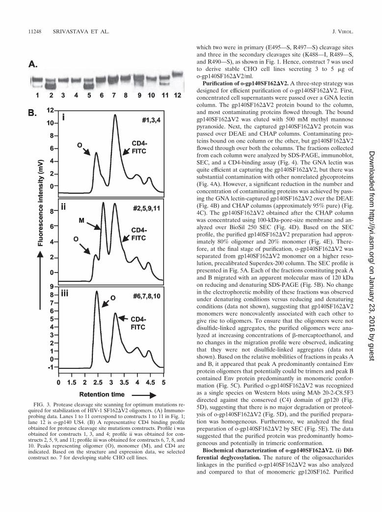
which two were in primary (E495—S, R497—S) cleavage sitesand three in the secondary cleavages site (K488—I, R489—S,and R490—S), as shown in Fig. 1. Hence, construct 7 was usedto derive stable CHO cell lines secreting 3 to 5 �g ofo-gp140SF162�V2/ml.
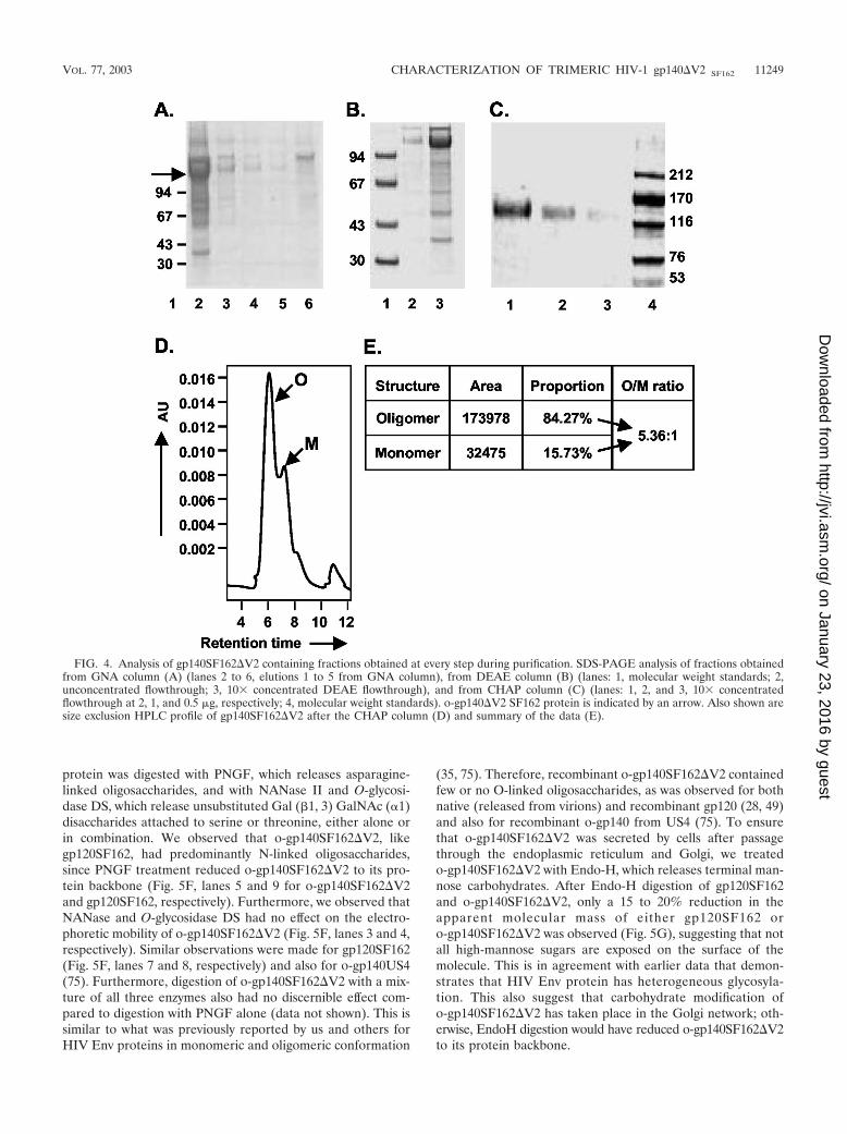
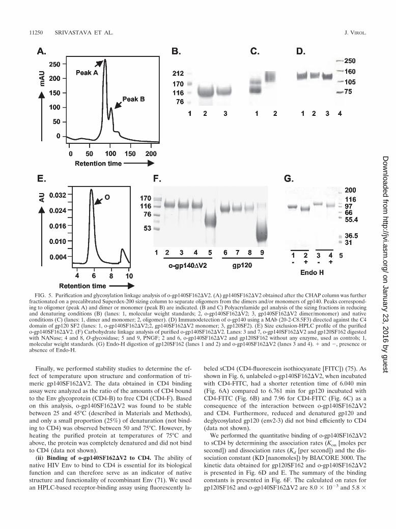
Purification of o-gp140SF162�V2. A three-step strategy wasdesigned for efficient purification of o-gp140SF162�V2. First,concentrated cell supernatants were passed over a GNA lectincolumn. The gp140SF162�V2 protein bound to the column,and most contaminating proteins flowed through. The boundgp140SF162�V2 was eluted with 500 mM methyl mannosepyranoside. Next, the captured gp140SF162�V2 protein waspassed over DEAE and CHAP columns. Contaminating pro-teins bound on one column or the other, but gp140SF162�V2flowed through over both the columns. The fractions collectedfrom each column were analyzed by SDS-PAGE, immunoblot,SEC, and a CD4-binding assay (Fig. 4). The GNA lectin wasquite efficient at capturing the gp140SF162�V2, but there wassubstantial contamination with other nonrelated glycoproteins(Fig. 4A). However, a significant reduction in the number andconcentration of contaminating proteins was achieved by pass-ing the GNA lectin-captured gp140SF162�V2 over the DEAE(Fig. 4B) and CHAP columns (approximately 95% pure) (Fig.4C). The gp140SF162�V2 obtained after the CHAP columnwas concentrated using 100-kDa-pore-size membrane and an-alyzed over BioSil 250 SEC (Fig. 4D). Based on the SECprofile, the purified gp140SF162�V2 preparation had approx-imately 80% oligomer and 20% monomer (Fig. 4E). There-fore, at the final stage of purification, o-gp140SF162�V2 wasseparated from gp140SF162�V2 monomer on a higher reso-lution, precalibrated Superdex-200 column. The SEC profile ispresented in Fig. 5A. Each of the fractions constituting peak Aand B migrated with an apparent molecular mass of 120 kDaon reducing and denaturing SDS-PAGE (Fig. 5B). No changein the electrophoretic mobility of these fractions was observedunder denaturing conditions versus reducing and denaturingconditions (data not shown), suggesting that gp140SF162�V2monomers were noncovalently associated with each other togive rise to oligomers. To ensure that the oligomers were notdisulfide-linked aggregates, the purified oligomers were ana-lyzed at increasing concentrations of �-mercaptoethanol, andno changes in the migration profile were observed, indicatingthat they were not disulfide-linked aggregates (data notshown). Based on the relative mobilities of fractions in peaks Aand B, it appeared that peak A predominantly contained Envprotein oligomers that potentially could be trimers and peak Bcontained Env protein predominantly in monomeric confor-mation (Fig. 5C). Purified o-gp140SF162�V2 was recognizedas a single species on Western blots using MAb 20-2-C8.5F3directed against the conserved (C4) domain of gp120 (Fig.5D), suggesting that there is no major degradation or proteol-ysis of o-gp140SF162�V2 (Fig. 5D), and the purified prepara-tion was homogeneous. Furthermore, we analyzed the finalpreparation of o-gp140SF162�V2 by SEC (Fig. 5E). The datasuggested that the purified protein was predominantly homo-geneous and potentially in trimeric conformation.
Biochemical characterization of o-gp140SF162�V2. (i) Dif-ferential deglycosylation. The nature of the oligosaccharideslinkages in the purified o-gp140SF162�V2 was also analyzedand compared to that of monomeric gp120SF162. Purified
FIG. 3. Protease cleavage site scanning for optimum mutations re-quired for stabilization of HIV-1 SF162�V2 oligomers. (A) Immuno-probing data. Lanes 1 to 11 correspond to constructs 1 to 11 in Fig. 1;lane 12 is o-gp140 US4. (B) A representative CD4 binding profileobtained for protease cleavage site mutations constructs. Profile i wasobtained for constructs 1, 3, and 4; profile ii was obtained for con-structs 2, 5, 9, and 11; profile iii was obtained for constructs 6, 7, 8, and10. Peaks representing oligomer (O), monomer (M), and CD4 areindicated. Based on the structure and expression data, we selectedconstruct no. 7 for developing stable CHO cell lines.
11248 SRIVASTAVA ET AL. J. VIROL.
on January 23, 2016 by guesthttp://jvi.asm
.org/D
ownloaded from
protein was digested with PNGF, which releases asparagine-linked oligosaccharides, and with NANase II and O-glycosi-dase DS, which release unsubstituted Gal (�1, 3) GalNAc (�1)disaccharides attached to serine or threonine, either alone orin combination. We observed that o-gp140SF162�V2, likegp120SF162, had predominantly N-linked oligosaccharides,since PNGF treatment reduced o-gp140SF162�V2 to its pro-tein backbone (Fig. 5F, lanes 5 and 9 for o-gp140SF162�V2and gp120SF162, respectively). Furthermore, we observed thatNANase and O-glycosidase DS had no effect on the electro-phoretic mobility of o-gp140SF162�V2 (Fig. 5F, lanes 3 and 4,respectively). Similar observations were made for gp120SF162(Fig. 5F, lanes 7 and 8, respectively) and also for o-gp140US4(75). Furthermore, digestion of o-gp140SF162�V2 with a mix-ture of all three enzymes also had no discernible effect com-pared to digestion with PNGF alone (data not shown). This issimilar to what was previously reported by us and others forHIV Env proteins in monomeric and oligomeric conformation
(35, 75). Therefore, recombinant o-gp140SF162�V2 containedfew or no O-linked oligosaccharides, as was observed for bothnative (released from virions) and recombinant gp120 (28, 49)and also for recombinant o-gp140 from US4 (75). To ensurethat o-gp140SF162�V2 was secreted by cells after passagethrough the endoplasmic reticulum and Golgi, we treatedo-gp140SF162�V2 with Endo-H, which releases terminal man-nose carbohydrates. After Endo-H digestion of gp120SF162and o-gp140SF162�V2, only a 15 to 20% reduction in theapparent molecular mass of either gp120SF162 oro-gp140SF162�V2 was observed (Fig. 5G), suggesting that notall high-mannose sugars are exposed on the surface of themolecule. This is in agreement with earlier data that demon-strates that HIV Env protein has heterogeneous glycosyla-tion. This also suggest that carbohydrate modification ofo-gp140SF162�V2 has taken place in the Golgi network; oth-erwise, EndoH digestion would have reduced o-gp140SF162�V2to its protein backbone.
FIG. 4. Analysis of gp140SF162�V2 containing fractions obtained at every step during purification. SDS-PAGE analysis of fractions obtainedfrom GNA column (A) (lanes 2 to 6, elutions 1 to 5 from GNA column), from DEAE column (B) (lanes: 1, molecular weight standards; 2,unconcentrated flowthrough; 3, 10� concentrated DEAE flowthrough), and from CHAP column (C) (lanes: 1, 2, and 3, 10� concentratedflowthrough at 2, 1, and 0.5 �g, respectively; 4, molecular weight standards). o-gp140�V2 SF162 protein is indicated by an arrow. Also shown aresize exclusion HPLC profile of gp140SF162�V2 after the CHAP column (D) and summary of the data (E).
VOL. 77, 2003 CHARACTERIZATION OF TRIMERIC HIV-1 gp140�V2 SF162 11249
on January 23, 2016 by guesthttp://jvi.asm
.org/D
ownloaded from
Finally, we performed stability studies to determine the ef-fect of temperature upon structure and conformation of tri-meric gp140SF162�V2. The data obtained in CD4 bindingassay were analyzed as the ratio of the amounts of CD4 boundto the Env glycoprotein (CD4-B) to free CD4 (CD4-F). Basedon this analysis, o-gp140SF162�V2 was found to be stablebetween 25 and 45°C (described in Materials and Methods),and only a small proportion (25%) of denaturation (not bind-ing to CD4) was observed between 50 and 75°C. However, byheating the purified protein at temperatures of 75°C andabove, the protein was completely denatured and did not bindto CD4 (data not shown).
(ii) Binding of o-gp140SF162�V2 to CD4. The ability ofnative HIV Env to bind to CD4 is essential for its biologicalfunction and can therefore serve as an indicator of nativestructure and functionality of recombinant Env (71). We usedan HPLC-based receptor-binding assay using fluorescently la-
beled sCD4 (CD4-fluorescein isothiocyanate [FITC]) (75). Asshown in Fig. 6, unlabeled o-gp140SF162�V2, when incubatedwith CD4-FITC, had a shorter retention time of 6.040 min(Fig. 6A) compared to 6.761 min for gp120 incubated withCD4-FITC (Fig. 6B) and 7.96 for CD4-FITC (Fig. 6C) as aconsequence of the interaction between o-gp140SF162�V2and CD4. Furthermore, reduced and denatured gp120 anddeglycosylated gp120 (env2-3) did not bind efficiently to CD4(data not shown).
We performed the quantitative binding of o-gp140SF162�V2to sCD4 by determining the association rates (Kon [moles persecond]) and dissociation rates (Kd [per second]) and the dis-sociation constant (KD [nanomoles]) by BIACORE 3000. Thekinetic data obtained for gp120SF162 and o-gp140SF162�V2is presented in Fig. 6D and E. The summary of the bindingconstants is presented in Fig. 6F. The calculated on rates forgp120SF162 and o-gp140SF162�V2 are 8.0 � 10�3 and 5.8 �
FIG. 5. Purification and glycosylation linkage analysis of o-gp140SF162�V2. (A) gp140SF162�V2 obtained after the CHAP column was furtherfractionated on a precalibrated Superdex-200 sizing column to separate oligomers from the dimers and/or monomers of gp140. Peaks correspond-ing to oligomer (peak A) and dimer or monomer (peak B) are indicated. (B and C) Polyacrylamide gel analysis of the sizing fractions in reducingand denaturing conditions (B) (lanes: 1, molecular weight standards; 2, o-gp140SF162�V2; 3, gp140SF162�V2 dimer/monomer) and nativeconditions (C) (lanes: 1, dimer and monomer; 2, oligomer). (D) Immunodetection of o-gp140 using a MAb (20-2-C8.5F3) directed against the C4domain of gp120 SF2 (lanes: 1, o-gp140SF162�V2;2, gp140SF162�V2 monomer; 3, gp120SF2). (E) Size exclusion-HPLC profile of the purifiedo-gp140SF162�V2. (F) Carbohydrate linkage analysis of purified o-gp140SF162�V2. Lanes: 3 and 7, o-gp140SF162�V2 and gp120SF162 digestedwith NANase; 4 and 8, O-glycosidase; 5 and 9, PNGF; 2 and 6, o-gp140SF162�V2 and gp120SF162 without any enzyme, used as controls; 1,molecular weight standards. (G) Endo-H digestion of gp120SF162 (lanes 1 and 2) and o-gp140SF162�V2 (lanes 3 and 4). � and �, presence orabsence of Endo-H.
11250 SRIVASTAVA ET AL. J. VIROL.
on January 23, 2016 by guesthttp://jvi.asm
.org/D
ownloaded from
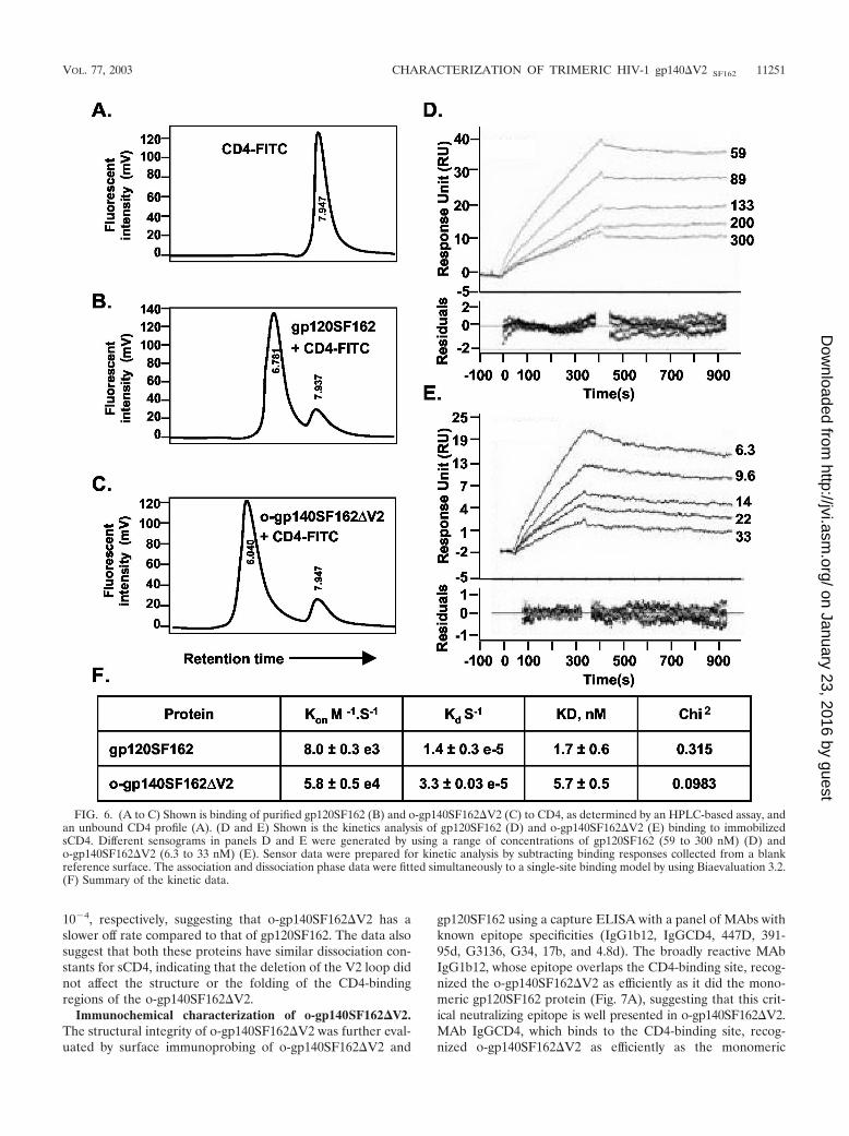
10�4, respectively, suggesting that o-gp140SF162�V2 has aslower off rate compared to that of gp120SF162. The data alsosuggest that both these proteins have similar dissociation con-stants for sCD4, indicating that the deletion of the V2 loop didnot affect the structure or the folding of the CD4-bindingregions of the o-gp140SF162�V2.
Immunochemical characterization of o-gp140SF162�V2.The structural integrity of o-gp140SF162�V2 was further eval-uated by surface immunoprobing of o-gp140SF162�V2 and
gp120SF162 using a capture ELISA with a panel of MAbs withknown epitope specificities (IgG1b12, IgGCD4, 447D, 391-95d, G3136, G34, 17b, and 4.8d). The broadly reactive MAbIgG1b12, whose epitope overlaps the CD4-binding site, recog-nized the o-gp140SF162�V2 as efficiently as it did the mono-meric gp120SF162 protein (Fig. 7A), suggesting that this crit-ical neutralizing epitope is well presented in o-gp140SF162�V2.MAb IgGCD4, which binds to the CD4-binding site, recog-nized o-gp140SF162�V2 as efficiently as the monomeric
FIG. 6. (A to C) Shown is binding of purified gp120SF162 (B) and o-gp140SF162�V2 (C) to CD4, as determined by an HPLC-based assay, andan unbound CD4 profile (A). (D and E) Shown is the kinetics analysis of gp120SF162 (D) and o-gp140SF162�V2 (E) binding to immobilizedsCD4. Different sensograms in panels D and E were generated by using a range of concentrations of gp120SF162 (59 to 300 nM) (D) ando-gp140SF162�V2 (6.3 to 33 nM) (E). Sensor data were prepared for kinetic analysis by subtracting binding responses collected from a blankreference surface. The association and dissociation phase data were fitted simultaneously to a single-site binding model by using Biaevaluation 3.2.(F) Summary of the kinetic data.
VOL. 77, 2003 CHARACTERIZATION OF TRIMERIC HIV-1 gp140�V2 SF162 11251
on January 23, 2016 by guesthttp://jvi.asm
.org/D
ownloaded from
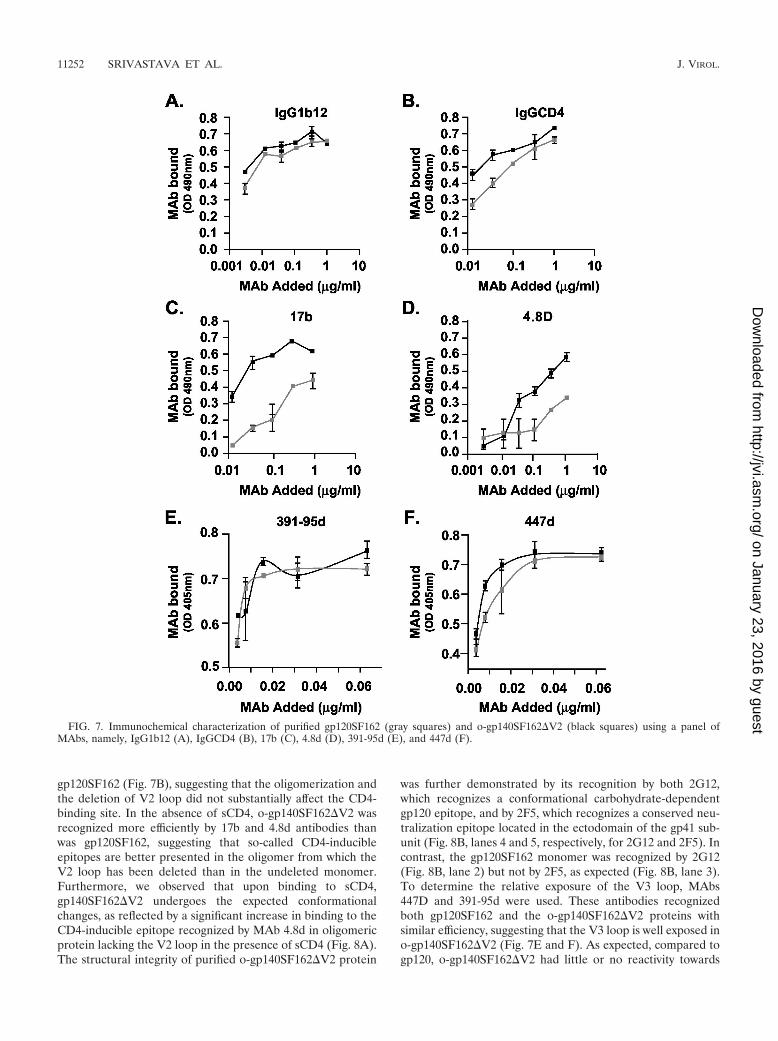
gp120SF162 (Fig. 7B), suggesting that the oligomerization andthe deletion of V2 loop did not substantially affect the CD4-binding site. In the absence of sCD4, o-gp140SF162�V2 wasrecognized more efficiently by 17b and 4.8d antibodies thanwas gp120SF162, suggesting that so-called CD4-inducibleepitopes are better presented in the oligomer from which theV2 loop has been deleted than in the undeleted monomer.Furthermore, we observed that upon binding to sCD4,gp140SF162�V2 undergoes the expected conformationalchanges, as reflected by a significant increase in binding to theCD4-inducible epitope recognized by MAb 4.8d in oligomericprotein lacking the V2 loop in the presence of sCD4 (Fig. 8A).The structural integrity of purified o-gp140SF162�V2 protein
was further demonstrated by its recognition by both 2G12,which recognizes a conformational carbohydrate-dependentgp120 epitope, and by 2F5, which recognizes a conserved neu-tralization epitope located in the ectodomain of the gp41 sub-unit (Fig. 8B, lanes 4 and 5, respectively, for 2G12 and 2F5). Incontrast, the gp120SF162 monomer was recognized by 2G12(Fig. 8B, lane 2) but not by 2F5, as expected (Fig. 8B, lane 3).To determine the relative exposure of the V3 loop, MAbs447D and 391-95d were used. These antibodies recognizedboth gp120SF162 and the o-gp140SF162�V2 proteins withsimilar efficiency, suggesting that the V3 loop is well exposed ino-gp140SF162�V2 (Fig. 7E and F). As expected, compared togp120, o-gp140SF162�V2 had little or no reactivity towards
FIG. 7. Immunochemical characterization of purified gp120SF162 (gray squares) and o-gp140SF162�V2 (black squares) using a panel ofMAbs, namely, IgG1b12 (A), IgGCD4 (B), 17b (C), 4.8d (D), 391-95d (E), and 447d (F).
11252 SRIVASTAVA ET AL. J. VIROL.
on January 23, 2016 by guesthttp://jvi.asm
.org/D
ownloaded from
the V2 loop-specific antibodies (G3136 and G34) (data notshown). In addition, sera from HIV-1-infected individuals rec-ognized both o-gp140SF162�V2 and gp120SF162, suggestingthat the deletion of the V2 loop did not affect the exposure ofthe immunodominant epitopes (data not shown). Based onthese observations, most of the critical neutralizing epitopeswere intact and accessible in recombinant o-gp140SF162�V2.
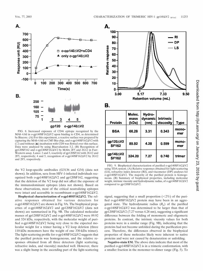
Biophysical characterization of o-gp140SF162�V2. The rel-ative responses obtained for various detectors foro-gp140SF162�V2 are shown in Fig. 9A. The biophysical prop-erties of o-gp140SF162�V2 and gp120SF162�V2 (data notshown) are summarized in Fig. 9B. The calculated molecularmasses of gp120SF162�V2 and o-gp140SF162�V2 were 89.92and 324 kDa, respectively, with the molecular weight of puri-fied o-gp140SF162�V2 being very close to the expected mo-lecular weight for a trimer having a V2 loop deletion (three110-kDa monomers have the weight of one 330-kDa trimer).The light-scattering profile for o-gp140SF162�V2 suggests thatthe purified protein was homogeneous, and the relative re-sponses obtained from all three detectors (light scattering,refractive index, and viscosity) matched well. However, therewas a slight hump in the ascending part of the light-scattering
signal, suggesting that a small proportion (2%) of the puri-fied o-gp140SF162�V2 protein may have been in an aggre-gated state. The hydrodynamic radius (Rh) of the purifiedo-gp140SF162�V2 was determined to be larger than that ofgp120SF162�V2 (7.27 versus 4.28 nm), suggesting a significantdifference between the folding of monomeric and oligomericproteins. In contrast, the intrinsic viscosity values for bothproteins were in a similar range (Fig. 9B), indicating that theproteins had not become unfolded during the purification pro-cess. Therefore, the differences observed in the biophysicalproperties of these molecules likely were inherent to nativeproteins and were not caused by denaturation or unfolding.
Negative-stain EM. The above data indicate that most of thepurified o-gp140SF162�V2 is in a trimeric conformation, witha smaller fraction in the monomer-to-dimer range (Fig. 5). To
FIG. 8. Increased exposure of CD4i epitope recognized by theMAb 4.8d in o-gp140SF162�V2 upon binding to CD4, as determinedby Biacore. (A) For this experiment, a reactive surface was prepared bycapturing the MAb 4.8d on CM5 Bia-chip, and o-gp140SF162�V2 with(�) and without (Œ) incubation with CD4 was flowed over this surface.Data were analyzed by using Biaevaluation 3.2. (B) Recognition ofgp120SF162 and o-gp140SF162�V2 by MAbs 2F5 and 2G12 in Farr-Western assay. Lanes: 2 and 3, reactivity of gp120SF162 with 2G12 and2F5, respectively; 4 and 5, recognition of o-gp140SF162�V2 by 2G12and 2F5, respectively.
FIG. 9. Biophysical characterization of purified o-gp140SF162�V2using TDA system. (A) Relative responses obtained for light scattering(LS), refractive index detector (RI), and viscometer (DP) analyses foro-gp140SF162�V2. The majority of the purified protein is homoge-neous. (B) Summary of biophysical properties, including molecularweight, intrinsic viscosity and hydrodynamic radius, of o-gp140SF162�V2compared to gp120SF162�V2.
VOL. 77, 2003 CHARACTERIZATION OF TRIMERIC HIV-1 gp140�V2 SF162 11253
on January 23, 2016 by guesthttp://jvi.asm
.org/D
ownloaded from
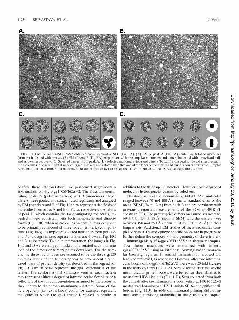
confirm these interpretations, we performed negative-stainEM analysis on the o-gp140SF162�V2. The fractions consti-tuting peaks A (putative trimers) and B (monomers and/ordimers) were pooled and concentrated separately and analyzedby EM (panels A and B of Fig. 10 show representative fields ofmolecules from peaks A and B of Fig. 5, respectively). Analysisof peak B, which contains the faster-migrating molecules, re-vealed images consistent with both monomeric and dimericforms (Fig. 10B), whereas molecules present in Peak A appearto be primarily composed of three-lobed, (trimeric) configura-tions (Fig. 10A). Examples of selected molecules from peaks Aand B and diagrammatic representations are shown in Fig. 10Cand D, respectively. To aid in interpretation, the images in Fig.10C and D were enlarged, masked, and rotated such that onelobe of the dimers or trimers points downward. For the trim-ers, the three radial lobes are assumed to be the three gp120moieties. Many of the trimers appear to have a centrally lo-cated mass of protein density (as described in the legend forFig. 10C) which could represent the gp41 ectodomain of thetrimer. The conformational variations seen in each fractionmay represent either a degree of intramolecular flexibility or areflection of the random orientation assumed by molecules asthey adhere to the carbon membrane substrate. Some of theheterogeneity (i.e., extra lobes) could, for example, representmolecules in which the gp41 trimer is viewed in profile in
addition to the three gp120 moieties. However, some degree ofmolecular heterogeneity cannot be ruled out.
The dimensions of the monomeric gp140SF162�V2moleculesranged between 60 and 100 A (mean standard error of themean [SEM], 74 13 A) from peak B and are consistent withpreviously reported measurements of the SOS gp140JR-FLconstruct (73). The presumptive dimers measured, on average,69 9 by 154 18 A (mean SEM) ,and the trimers werebetween 150 and 250 A (mean SEM, 191 23 A) in theirlongest axis. Additional EM studies of these molecules com-plexed with sCD4 and epitope-specific MAbs are in progress tofurther define the composition and geometry of these trimers.
Immunogenicity of o-gp140SF162�V2 in rhesus macaques.Two rhesus macaques were immunized with trimericgp140SF162�V2 using an intranasal priming and intramuscu-lar boosting regimen. Intranasal immunization induced lowlevels of systemic IgG responses. However, after two intramus-cular boosts with o-gp140SF162�V2, there was a 20-fold increasein the antibody titers (Fig. 11A). Sera collected after the secondintramuscular protein boosts were tested for their abilities toneutralize HIV-1 isolates (Fig. 11B). Sera collected from boththe animals after the intramuscular boost with o-gp140SF162�V2neutralized homologous HIV-1 isolate SF162 at significant di-lutions (Fig. 11B). In addition, intranasal priming did not in-duce any neutralizing antibodies in these rhesus macaques.
FIG. 10. EMs of o-gp140SF162�V2 obtained from preparative SEC (Fig. 5A). (A) EM of peak A (Fig. 5A) containing trilobed molecules(trimers) indicated with arrows. (B) EM of peak B (Fig. 5A) preparation with presumptive monomers and dimers indicated with arrowhead-ballsand arrows, respectively. (C) Selected trimers from peak A. (D) Selected monomers (top) and dimers (bottom) from peak B. To aid interpretation,the molecules in panels C and D were enlarged, masked, and rotated such that one of the lobes of the dimers and trimers points downward. Graphicrepresentations of a trimer and monomer and dimer (not drawn to scale) are shown in panels C and D, respectively. Bars, 20 nm.
11254 SRIVASTAVA ET AL. J. VIROL.
on January 23, 2016 by guesthttp://jvi.asm
.org/D
ownloaded from
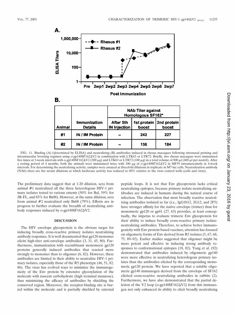
The preliminary data suggest that at 1:20 dilution, sera fromanimal #1 neutralized all the three heterologous HIV-1 pri-mary isolates tested to various extents (50% for Bal, 59% forJR-FL, and 83% for Bx08). However, at the same dilution, serafrom animal #2 neutralized only Bx08 (79%). Efforts are inprogress to further evaluate the breadth of neutralizing anti-body responses induced by o-gp140SF162�V2.
DISCUSSION
The HIV envelope glycoprotein is the obvious target forinducing broadly cross-reactive primary isolates neutralizingantibody responses. It is well documented that gp120 monomerelicits high-titer anti-envelope antibodies (3, 31, 45, 80). Fur-thermore, immunization with recombinant monomeric gp120proteins generally induced antibodies that reacted morestrongly to monomer than to oligomer (6, 82). However, theseantibodies are limited in their ability to neutralize HIV-1 pri-mary isolates, especially those of the R5 phenotype (46, 51, 82,86). The virus has evolved ways to minimize the immunoge-nicity of the Env protein by extensive glycosylation of themolecule with nascent carbohydrate (high terminal mannose),thus minimizing the efficacy of antibodies by shielding theconserved region. Moreover, the receptor-binding site is bur-ied within the molecule and is partially shielded by external
peptide loops. It is not that Env glycoprotein lacks criticalneutralizing epitopes, because primary isolate-neutralizing an-tibodies are induced in humans during the natural course ofinfection. The observation that most broadly reactive neutral-izing antibodies isolated so far (i.e., IgG1b12, 2G12, and 2F5)have stronger affinity for the native envelope (trimer) than formonomeric gp120 or gp41 (27, 63) provides, at least concep-tually, the impetus to evaluate trimeric Env glycoprotein fortheir ability to induce broadly cross-reactive primary isolate-neutralizing antibodies. Therefore, to achieve better immuno-genicity with Env protein-based vaccines, attention has focusedon oligomeric forms of Env derived from R5 isolates (5, 67, 68,75, 89–92). Earlier studies suggested that oligomer might bemore potent and effective in inducing strong antibody re-sponses to conformational epitopes (18, 82). Yang et al. (92)demonstrated that antibodies induced by oligomeric gp140were more effective in neutralizing heterologous primary iso-lates than the antibodies elicited by the corresponding mono-meric gp120 protein. We have reported that a soluble oligo-meric gp140 immunogen derived from the envelope of SF162elicited cross-reactive neutralizing antibodies in rabbits (2).Furthermore, we have also demonstrated that the partial de-letion of the V2 loop (o-gp140SF162�V2) from this immuno-gen not only enhanced its ability to elicit broadly neutralizing
FIG. 11. Binding (A) (determined by ELISA) and neutralizing (B) antibodies induced in rhesus macaques following intranasal priming andintramuscular boosting regimen using o-gp140SF162�V2 in combination with LTK63 or LTR72. Briefly, two rhesus macaques were immunizedfive times at 3-week intervals with o-gp140SF162�V2 (300 �g) and LTK63 or LTR72 (100 �g) in a total volume of 800 �l (400 �l per nostril). Aftera resting period of 4 months, both the animals were immunized twice with 100 �g of o-gp140SF162�V2 in MF59 intramuscularly at 4-weekintervals. For determining the neutralizing activity, samples were assayed at threefold dilutions in triplicate in M7-luc cells. Neutralization antibody(NAb) titers are the serum dilutions at which luciferase activity was reduced to 80% relative to the virus control wells (cells and virus).
VOL. 77, 2003 CHARACTERIZATION OF TRIMERIC HIV-1 gp140�V2 SF162 11255
on January 23, 2016 by guesthttp://jvi.asm
.org/D
ownloaded from
antibodies, but also partially protected vaccinated rhesus ma-caques upon pathogenic challenge with SHIVSF162P4 (2).
To elicit the functional antibody responses, oligomeric HIVenvelope immunogens need to be stabilized so that they retaintheir oligomeric nature upon delivery. Several approaches arecurrently under evaluation, namely: (i) elimination of the pro-tease cleavage site between gp120 and gp41 (6, 18, 20, 22), (ii)introduction of intramolecular disulfide bond formation (SOS)between gp120 and gp41 (5, 23, 67, 73), (iii) introduction, inaddition to the SOS modifications, of an isoleucine-to-prolinesubstitution at position 559 in the N-terminal heptad repeatregion of gp41 (SOSIP) (68), (iv) extension of the gp41 coiledcoil region by the addition of GCN4 (a transcription factor thatnormally forms stable homotrimers) sequences, either alone orin conjunction with added cysteines between gp120 and gp41(23, 89, 90), and (v) addition of a trimerization domain fromthe C terminus of bacteriophage T4 fibritin (91). We evaluatedthe efficacy of both protease site mutation as well as the SOSapproach to create stable trimeric HIV envelope proteins de-rived from different clades (B and C). Although, a single mu-tation in the primary protease cleavage site (R to S) is sufficientfor the formation of stable oligomers from US4, an R5 usingHIV-1 primary isolate (75), this modification alone is ineffec-tive in stabilizing the oligomers with and without V2 loopderived from SF162 (data not shown). Similar observationswere made by Earl et al. (18–21) and Yang et al. (90) fordifferent isolates. In our hands, the SOS approach that hasbeen used by Binley et al. (5) and Sanders et al. (67) forcreating stable oligomers from both primary (JF-RL) andTCLA isolates has failed to generate stable oligomers with orwithout theV2 loop from SF162 (I. Srivastava, J. Ulmer, and S.Barnett, unpublished observations). However, these were nottotally unexpected results, because follow-up studies fromSchulke et al. have also demonstrated the inefficiency of thisapproach in creating stable oligomers from JR-FL (73). Inessence, these observations suggest that strategies for the gen-eration of stable soluble HIV envelope oligomers from a givenisolate or clade will need to be determined experimentally.Sequence variation, glycosylation, size of variable loops, ex-pression levels, and the type of protein expression system mayall play a critical role in stabilizing the HIV Env glycoprotein inoligomeric conformation (89, 90).
There is no consensus regarding the structure and the massof recombinant HIV Env glycoproteins. Differences in ob-served mass of the trimeric molecule may be because thesemolecules were derived from different isolates and character-ized by different techniques in different laboratories. Althoughit is expected that the trimeric Env glycoprotein derived fromdifferent isolates (such as o-gp140 US4 and SF162) will differ intheir molecular masses due to sequence differences, theyshould be at least in a similar mass range. Most of the solublerecombinant Env oligomeric proteins purified to date havebeen shown to be either dimers, tetramers (6, 19, 20), ormonomers (73), with the possible exception of those describedby Grundner et al. (29), Yang et al. (89, 91), and Chakrabartiet al. (11). Most of the studies used either SEC or sucrosegradient centrifugation to determine the mass of the oligo-meric molecule. This may not be the most appropriate meansto determine the molecular weight of purified proteins due toa lack of direct relationship between molecular size and mo-
lecular weight of proteins, even under strongly denaturing con-ditions (47). In addition, proteins with extensive posttransla-tional modifications, such as glycosylation (85), as is the casewith HIV envelope proteins, may further complicate this typeof analysis. The most appropriate and precise methods fordetermining the molecular mass are osmometry, analyticalcentrifugation, and classical light scattering. Osmometry andanalytical ultracentrifugation are labor intensive and time con-suming. In addition, the high shear force induced during theultracentrifugation may cause the dissociation of noncovalentlyassociated HIV envelope subunits. In contrast, classical lightscattering can be performed quickly and relatively easily andunder very mild conditions; therefore, it is a reasonable choicefor determining the molecular mass of the proteins (17, 36, 43).We previously used this approach to successfully characterizeo-gp140-US4 and demonstrated that the purified protein waspotentially in trimeric conformation (75). The calculated mo-lecular mass for o-gp140 US4 was found to be 474 kDa, whichis in good agreement with the molecular mass of 486 kDa ofthe intact Env glycoprotein isolated from the surface of virionas demonstrated recently by Center et al. using transmission-scanning electron microscope images (10). To ascertain theoligomeric status of o-gp140SF162�V2 produced in the CHOcell lines, we performed a detailed biophysical characterizationof the purified protein using a TDA system coupled to a sizeexclusion column to determine the Mw, Rh and intrinsic vis-cosity. The Mw of the purified HIV envelope oligomer wasestimated to be 324 kDa, very close to the expected Mw of atrimer (330 kDa). The differences in the calculated molecularmass of o-gp140 US4 and o-gp140SF162�V2 may be partiallyexplained by the deletion of V2 loop, which also includes thedeletion of one N-linked glycosylation site at position 186 (42,43). Furthermore, the Env glycoprotein (trimers and mono-mers) derived from SF162 is slightly smaller than a similarprotein derived from US4, as observed by SDS-PAGE analysis,suggesting that sequence differences may indeed play a role indetermining the molecular mass of the protein. The reportedmolecular mass for transiently expressed Env protein oli-gomers derived from JR-FL, YU2, and HxBc2 is smaller thanexpected (400 to 410 kDa) (5, 89), and the molecular mass ofthe oligomer derived from JR-FL using a SOSIP approach isconsiderably larger than expected (29, 68). However, furthercharacterization will be needed to confirm the trimeric natureof these purified proteins.
In the present study, we performed an in-depth biophysicalcharacterization of the purified o-gp140SF162�V2 protein byusing triple detectors and negative-stain EM of the purifiedEnv glycoproteins to determine the structure and conforma-tion of the purified protein. Combined, these methods stronglysupport the conclusion that the purified o-gp140SF162�V2 ispredominantly in a trimeric conformation. As shown earlier,we found that the monomeric proteins are loosely folded glob-ular structures with higher Rh values (5.07 nm for gp120 US4[75] and 4.33 nm for gp120SF162�V2). In contrast, Env gly-coprotein trimers are tightly folded structures, as indicated bytheir smaller-than-expected Rh values (8.4 nm for o-gp140 US4[75] and 7.6 nm for o-gp140SF162�V2) for oligomers. Thesedata are consistent with earlier observations that there aresignificant differences in the structure and folding of monomerand trimer (54, 87).
11256 SRIVASTAVA ET AL. J. VIROL.
on January 23, 2016 by guesthttp://jvi.asm
.org/D
ownloaded from
It has been proposed that cleavable trimers may be betterthan uncleaved trimers for inducing the primary isolate-neu-tralizing antibody responses, mainly because native Env is be-lieved to undergo structural rearrangement on the viral surfaceand thus be more likely to express native epitopes followingcleavage. However, recombinant cleavable Env proteins arestructurally labile and need to be stabilized in the trimericconformation in a manner that does not compromise the ex-posure of critical neutralizing epitopes. While there are noclear immunogenicity nor challenge data to suggest thatcleaved oligomers are more potent for inducing strong neu-tralizing or protective antibodies, it may suffice to use cleavableEnv immunogens during the DNA-priming phase (C. Buckneret al., submitted for publication).
The binding of HIV envelope protein to CD4, an obligatorystep in viral replication (16, 34), is dependent upon both con-formation (38, 48) and glycosylation (4, 40, 58). Evidence so farsuggests that N-linked glycosylation is important for the for-mation of correct disulfide bonds and for providing functionalconformation to the Env protein (24, 25, 39, 40) to foster CD4binding, even though the carbohydrate side chains are notdirectly involved in CD4 binding (40). The V2 loop deletion,which also involves the deletion of a glycosylation site at po-sition 186 (42, 43), did not affect the structure and conforma-tion of HIV Env because purified o-gp140SF162�V2 bound tosCD4 with an affinity and efficiency similar to that of gp120monomer, as determined qualitatively (HPLC assay) andquantitatively (Biacore). This finding suggests that the purifiedprotein is in its correct native conformation. This was notentirely unexpected as it was previously demonstrated that theSF162 envelope from which V2 has been deleted maintains afunctional structure and allows the virus to efficiently replicatein both peripheral blood mononuclear cells and macrophagesin a CD4/CCR5-dependent manner (76), and transiently ex-pressed protein was efficiently recognized by CD4-binding siteMAbs (78). We have also demonstrated that the purifiedo-gp140SF162�V2, like native and recombinant monomericand oligomeric Env proteins, contains predominantly N-linkedglycosylation (35, 75).
We have determined the structural integrity of the purifiedgp140�V2 trimers with the use of several MAbs of knownepitope specificity. Important epitopes exposed on purifiedtrimers include those near or on the CD4-binding site (recog-nized by MAbs IgG1b12 and CD4-IgG2), the CD4-induciblesite that overlaps the coreceptor binding site (recognized byMAbs 17b and 4.8d), gp41 (2F5), a carbohydrate-dependentepitope on gp120 (recognized by 2G12), and V3 loop epitopes(recognized by MAbs 447D and 391-95D). The monoclonalbinding results obtained for o-gp140SF162�V2 are similar tothose observed with corresponding native Env present on in-fectious SF162�V2 virions (79). It seems that irrespective ofthe cleavage status, critical neutralizing epitopes are properlyexposed on recombinantly produced Env oligomers. In fact,the oligomers from which the V2 loop was deleted were rec-ognized more efficiently by MAbs 17b and 4.8d in the absenceof sCD4 compared to the monomeric form (Fig. 7C and D). Inaddition, upon binding to sCD4, there was significant up reg-ulation of CD4-inducible epitope recognized by 4.8d.
In an intranasal priming and intramuscular boosting immu-nogenicity study in rhesus macaques, we have clearly demon-
strated that o-gp140SF162�V2 induced strong Env-specific an-tibodies, and these antibodies were able to neutralize ahomologous HIV-1 clade B isolate, i.e., SF162. The prelimi-nary data suggest that the immunization regimen used in thisstudy has induced cross-reactive neutralizing antibody re-sponses against Bx08. In-depth analysis of the breadth of neu-tralizing antibody responses is ongoing and will be presentedseparately in a follow-up manuscript. Furthermore, intranasalpriming with o-gp140SF162�V2 also induced mucosal re-sponses (M. Vajdy et al., unpublished observations).
These data suggest that the deletion of V2 loop from theSF162 envelope increases the exposure of CD4-inducibleepitope on o-gp140SF162�V2 compared to monomer. Thepreservation of critical neutralizing epitopes on the recombi-nant trimers and the ability of this immunogen to induce neu-tralizing antibody activity against heterologous viruses furtheremphasize the potential of trimeric gp140SF162�V2 for vac-cine application.
ACKNOWLEDGMENTS
We thank Margaret Liu for her support and encouragement duringher tenure as Vice President for Vaccines and Gene Therapy Researchat Chiron. We also thank Dennis Burton, Susan Zolla-Pazner, MichaelGorny, James Robinson, and Herman Katinger for providing mono-clonal antibodies for the structural characterization of the purifiedproteins. We also thank Michael Houghton and Gib Otten for criticallyreading the manuscript and Nelle Cronen and Randy Deck for excel-lent editorial and administrative support. We thank Mark Wininger,Karen Matsuoka, Erika Calderon, and Robert Chow for their help incarrying out large-scale cell culture experiments.
This work was supported by financial support provided by NIHCRDA (NO-AI-95367, TO#3) and by D&DT contract NO-1-AI-05396 and R21 AI50430-01 to M.V. Research in the laboratories ofLeonidas Stamatatos and Kenneth Roux was supported by investiga-tor-initiated NIH research grants RO1 AI47708 and R21 A144291,respectively.
REFERENCES
1. Back, N. K., L. Smit, J. J. De Jong, W. Keulen, M. Schutten, J. Goudsmit,and M. Tersmette. 1994. An N-glycan within the human immunodeficiencyvirus type 1 gp120 V3 loop affects virus neutralization. Virology 199:431–438.
2. Barnett, S. W., S. Lu, I. Srivastava, S. Cherpelis, A. Gettie, J. Blanchard, S.Wang, I. Mboudjeka, L. Leung, Y. Lian, A. Fong, C. Buckner, A. Ly, S. Hilt,J. Ulmer, C. T. Wild, J. R. Mascola, and L. Stamatatos. 2001. The ability ofan oligomeric human immunodeficiency virus type 1 (HIV-1) envelope an-tigen to elicit neutralizing antibodies against primary HIV-1 isolates is im-proved following partial deletion of the second hypervariable region. J. Vi-rol. 75:5526–5540.
3. Barnett, S. W., S. Rajasekar, H. Legg, B. Doe, D. H. Fuller, J. R. Haynes,C. M. Walker, and K. S. Steimer. 1997. Vaccination with HIV-1 gp120 DNAinduces immune responses that are boosted by a recombinant gp120 proteinsubunit. Vaccine 15:869–873.
4. Barr, P. J., K. S. Steimer, E. A. Sabin, D. Parkes, C. George-Nascimento,J. C. Stephans, M. A. Powers, A. Gyenes, G. A. Van Nest, E. T. Miller, et al.1987. Antigenicity and immunogenicity of domains of the human immuno-deficiency virus (HIV) envelope polypeptide expressed in the yeast Saccha-romyces cerevisiae. Vaccine 5:90–101.
5. Binley, J. M., R. W. Sanders, B. Clas, N. Schuelke, A. Master, Y. Guo, F.Kajumo, D. J. Anselma, P. J. Maddon, W. C. Olson, and J. P. Moore. 2000.A recombinant human immunodeficiency virus type 1 envelope glycoproteincomplex stabilized by an intermolecular disulfide bond between the gp120and gp41 subunits is an antigenic mimic of the trimeric virion-associatedstructure. J. Virol. 74:627–643.
6. Broder, C. C., P. L. Earl, D. Long, S. T. Abedon, B. Moss, and R. W. Doms.1994. Antigenic implications of human immunodeficiency virus type 1 enve-lope quaternary structure: oligomer-specific and -sensitive monoclonal anti-bodies. Proc. Natl. Acad. Sci. USA 91:11699–11703.
7. Burton, D. R., and D. C. Montefiori. 1997. The antibody response in HIV-1infection. AIDS 11(Suppl. A):S87–S98.
8. Burton, D. R., and P. W. Parren. 2000. Vaccines and the induction offunctional antibodies: time to look beyond the molecules of natural infec-tion? Nat. Med. 6:123–125.
VOL. 77, 2003 CHARACTERIZATION OF TRIMERIC HIV-1 gp140�V2 SF162 11257
on January 23, 2016 by guesthttp://jvi.asm
.org/D
ownloaded from

9. Cao, J., N. Sullivan, E. Desjardin, C. Parolin, J. Robinson, R. Wyatt, and J.Sodroski. 1997. Replication and neutralization of human immunodeficiencyvirus type 1 lacking the V1 and V2 variable loops of the gp120 envelopeglycoprotein. J. Virol. 71:9808–9812.
10. Center, R. J., R. D. Leapman, J. Lebowitz, L. O. Arthur, P. L. Earl, and B.Moss. 2002. Oligomeric structure of the human immunodeficiency virus type1 envelope protein on the virion surface. J. Virol. 76:7863–7867.
11. Chakrabarti, B. K., W. P. Kong, B. Y. Wu, Z. Y. Yang, J. Friborg, X. Ling,S. R. King, D. C. Montefiori, and G. J. Nabel. 2002. Modifications of thehuman immunodeficiency virus envelope glycoprotein enhance immunoge-nicity for genetic immunization. J. Virol. 76:5357–5368.
12. Chapman, B. S., R. M. Thayer, K. A. Vincent, and N. L. Haigwood. 1991.Effect of intron A from human cytomegalovirus (Towne) immediate-earlygene on heterologous expression in mammalian cells. Nucleic Acids Res.19:3979–3986.
13. Cheng-Mayer, C., A. Brown, J. Harouse, P. A. Luciw, and A. J. Mayer. 1999.Selection for neutralization resistance of the simian/human immunodefi-ciency virus SHIVSF33A variant in vivo by virtue of sequence changes in theextracellular envelope glycoprotein that modify N-linked glycosylation. J. Vi-rol. 73:5294–5300.
14. Cherpelis, S., X. Jin, A. Gettie, D. D. Ho, S. W. Barnett, I. Shrivastava, andL. Stamatatos. 2001. DNA-immunization with a V2 deleted HIV-1 envelopeelicits protective antibodies in macaques. Immunol. Lett. 79:47–55.
15. Cherpelis, S., I. Srivastava, A. Gettie, X. Jin, D. D. Ho, S. W. Barnett, andL. Stamatatos. 2001. DNA vaccination with the human immunodeficiencyvirus type 1 SF162�V2 envelope elicits immune responses that offer partialprotection from simian/human immunodeficiency virus infection to CD8�
T-cell-depleted rhesus macaques. J. Virol. 75:1547–1550.16. Dalgleish, A. G., P. C. Beverley, P. R. Clapham, D. H. Crawford, M. F.
Greaves, and R. A. Weiss. 1984. The CD4 (T4) antigen is an essentialcomponent of the receptor for the AIDS retrovirus. Nature 312:763–767.
17. Dollinger, G., B. Cunico, M. Kunitani, D. Johnson, and R. Jones. 1992.Practical on-line determination of biopolymer molecular weights by high-performance liquid chromatography with classical light-scattering detection.J. Chromatogr. 592:215–228.
18. Earl, P. L., C. C. Broder, D. Long, S. A. Lee, J. Peterson, S. Chakrabarti,R. W. Doms, and B. Moss. 1994. Native oligomeric human immunodeficiencyvirus type 1 envelope glycoprotein elicits diverse monoclonal antibody reac-tivities. J. Virol. 68:3015–3026.
19. Earl, P. L., R. W. Doms, and B. Moss. 1990. Oligomeric structure of thehuman immunodeficiency virus type 1 envelope glycoprotein. Proc. Natl.Acad. Sci. USA 87:648–652.
20. Earl, P. L., S. Koenig, and B. Moss. 1991. Biological and immunologicalproperties of human immunodeficiency virus type 1 envelope glycoprotein:analysis of proteins with truncations and deletions expressed by recombinantvaccinia viruses. J. Virol. 65:31–41.
21. Earl, P. L., and B. Moss. 1993. Mutational analysis of the assembly domainof the HIV-1 envelope glycoprotein. AIDS Res. Hum. Retrovir. 9:589–594.
22. Earl, P. L., W. Sugiura, D. C. Montefiori, C. C. Broder, S. A. Lee, C. Wild,J. Lifson, and B. Moss. 2001. Immunogenicity and protective efficacy ofoligomeric human immunodeficiency virus type 1 gp140. J. Virol. 75:645–653.
23. Farzan, M., H. Choe, E. Desjardins, Y. Sun, J. Kuhn, J. Cao, D. Archam-bault, P. Kolchinsky, M. Koch, R. Wyatt, and J. Sodroski. 1998. Stabilizationof human immunodeficiency virus type 1 envelope glycoprotein trimers bydisulfide bonds introduced into the gp41 glycoprotein ectodomain. J. Virol.72:7620–7625.
24. Fennie, C., and L. A. Lasky. 1989. Model for intracellular folding of thehuman immunodeficiency virus type 1 gp120. J. Virol. 63:639–646.
25. Fenouillet, E., B. Clerget-Raslain, J. C. Gluckman, D. Guetard, L. Montag-nier, and E. Bahraoui. 1989. Role of N-linked glycans in the interactionbetween the envelope glycoprotein of human immunodeficiency virus and itsCD4 cellular receptor. Structural enzymatic analysis. J. Exp Med. 169:807–822.
26. Fouts, T. R., J. M. Binley, A. Trkola, J. E. Robinson, and J. P. Moore. 1997.Interaction of polyclonal and monoclonal anti-glycoprotein 120 antibodieswith oligomeric glycoprotein 120-glycoprotein 41 complexes of a primaryHIV type 1 isolate relationship to neutralization. AIDS Res. Hum. Retrovir.14:591–597.
27. Fouts, T. R., J. M. Binley, A. Trkola, J. E. Robinson, and J. P. Moore. 1997.Neutralization of the human immunodeficiency virus type 1 primary isolateJR-FL by human monoclonal antibodies correlates with antibody binding tothe oligomeric form of the envelope glycoprotein complex. J. Virol. 71:2779–2785.
28. Geyer, H., C. Holschbach, G. Hunsmann, and J. Schneider. 1988. Carbohy-drates of human immunodeficiency virus. Structures of oligosaccharideslinked to the envelope glycoprotein 120. J. Biol. Chem. 263:11760–11767.
29. Grundner, C., T. Mirzabekov, J. Sodroski, and R. Wyatt. 2002. Solid-phaseproteoliposomes containing human immunodeficiency virus envelope glyco-proteins. J. Virol. 76:3511–3521.
30. Haas, J., E. C. Park, and B. Seed. 1996. Codon usage limitation in theexpression of HIV-1 envelope glycoprotein. Curr. Biol. 6:315–324.
31. Haigwood, N. L., P. L. Nara, E. Brooks, G. A. Van Nest, G. Ott, K. W.Higgins, N. Dunlop, C. J. Scandella, J. W. Eichberg, and K. S. Steimer. 1992.Native but not denatured recombinant human immunodeficiency virus type1 gp120 generates broad-spectrum neutralizing antibodies in baboons. J. Vi-rol. 66:172–182.
32. Hill, C. P., D. Worthylake, D. P. Bancroft, A. M. Christensen, and W. I.Sundquist. 1996. Crystal structures of the trimeric human immunodeficiencyvirus type 1 matrix protein: implications for membrane association andassembly. Proc. Natl. Acad. Sci. USA 93:3099–3104.
33. Johnson, W. E., J. Morgan, J. Reitter, B. A. Puffer, S. Czajak, R. W. Doms,and R. C. Desrosiers. 2002. A replication-competent, neutralization-sensitivevariant of simian immunodeficiency virus lacking 100 amino acids of enve-lope. J. Virol. 76:2075–2086.
34. Klatzmann, D., E. Champagne, S. Chamaret, J. Gruest, D. Guetard, T.Hercend, J. C. Gluckman, and L. Montagnier. 1984. T-lymphocyte T4 mol-ecule behaves as the receptor for human retrovirus LAV. Nature 312:767–768.
35. Kozarsky, K., M. Penman, L. Basiripour, W. Haseltine, J. Sodroski, and M.Krieger. 1989. Glycosylation and processing of the human immunodeficiencyvirus type 1 envelope protein. J. Acquir. Immune Defic. Syndr. 2:163–169.
36. Krull, I. S., H. H. Stuting, and S. C. Krzysko. 1988. Conformational studiesof bovine alkaline phosphatase in hydrophobic interaction and size-exclusionchromatography with linear diode array and low-angle laser light scatteringdetection. J. Chromatogr. 442:29–52.
37. Kwong, P. D., R. Wyatt, J. Robinson, R. W. Sweet, J. Sodroski, and W. A.Hendrickson. 1998. Structure of an HIV gp120 envelope glycoprotein incomplex with the CD4 receptor and a neutralizing human antibody. Nature393:648–659.
38. Lasky, L. A., G. Nakamura, D. H. Smith, C. Fennie, C. Shimasaki, E. Patzer,P. Berman, T. Gregory, and D. J. Capon. 1987. Delineation of a region of thehuman immunodeficiency virus type 1 gp120 glycoprotein critical for inter-action with the CD4 receptor. Cell 50:975–985.
39. Leonard, C. K., M. W. Spellman, L. Riddle, R. J. Harris, J. N. Thomas, andT. J. Gregory. 1990. Assignment of intrachain disulfide bonds and charac-terization of potential glycosylation sites of the type 1 recombinant humanimmunodeficiency virus envelope glycoprotein (gp120) expressed in Chinesehamster ovary cells. J. Biol. Chem. 265:10373–10382.
40. Li, Y., L. Luo, N. Rasool, and C. Y. Kang. 1993. Glycosylation is necessary forthe correct folding of human immunodeficiency virus gp120 in CD4 binding.J. Virol. 67:584–588.
41. Lue, J., M. Hsu, D. Yang, P. Marx, Z. Chen, and C. Cheng-Mayer. 2002.Addition of a single gp120 glycan confers increased binding to dendriticcell-specific ICAM-3-grabbing nonintegrin and neutralization escape to hu-man immunodeficiency virus type 1. J. Virol. 76:10299–10306.
42. Ly, A., and L. Stamatatos. 2000. V2 loop glycosylation of the human immu-nodeficiency virus type 1 SF162 envelope facilitates interaction of this pro-tein with CD4 and CCR5 receptors and protects the virus from neutraliza-tion by anti-V3 loop and anti-CD4 binding site antibodies. J. Virol. 74:6769–6976.
43. Maezawa, S., Y. Hayashi, T. Nakae, J. Ishii, K. Kameyama, and T. Takagi.1983. Determination of molecular weight of membrane proteins by the useof low-angle laser light scattering combined with high-performance gel chro-matography in the presence of a non-ionic surfactant. Biochim. Biophys.Acta 747:291–297.
44. Malenbaum, S. E., D. Yang, L. Cavacini, M. Posner, J. Robinson, and C.Cheng-Mayer. 2000. The N-terminal V3 loop glycan modulates the interac-tion of clade A and B human immunodeficiency virus type 1 envelopes withCD4 and chemokine receptors. J. Virol. 74:11008–11016.
45. Mascola, J. R., M. G. Lewis, G. Stiegler, D. Harris, T. C. VanCott, D. Hayes,M. K. Louder, C. R. Brown, C. V. Sapan, S. S. Frankel, Y. Lu, M. L. Robb,H. Katinger, and D. L. Birx. 1999. Protection of macaques against patho-genic simian/human immunodeficiency virus 89.6PD by passive transfer ofneutralizing antibodies. J. Virol. 73:4009–4018.
46. Mascola, J. R., S. W. Snyder, O. S. Weislow, S. M. Belay, R. B. Belshe, D. H.Schwartz, M. L. Clements, R. Dolin, B. S. Graham, G. J. Gorse, M. C.Keefer, M. J. McElrath, M. C. Walker, K. F. Wagner, J. G. McNeil, F. E.McCutchan, D. S. Burke, et al. 1996. Immunization with envelope subunitvaccine products elicits neutralizing antibodies against laboratory-adaptedbut not primary isolates of human immunodeficiency virus type 1. J. Infect.Dis. 173:340–348.
47. McDonnell, M. E., and A. M. Jamieson. 1976. Rapid characterization ofprotein molecular weights and hydrodynamic structures by quasielastic laser-light scattering. Biopolymers 15:1283–1299.
48. McDougal, J. S., J. K. Nicholson, G. D. Cross, S. P. Cort, M. S. Kennedy, andA. C. Mawle. 1986. Binding of the human retrovirus HTLV-III/LAV/ARV/HIV to the CD4 (T4) molecule: conformation dependence, epitope map-ping, antibody inhibition, and potential for idiotypic mimicry. J. Immunol.137:2937–2944.
49. Mizuochi, T., M. W. Spellman, M. Larkin, J. Solomon, L. J. Basa, and T.Feizi. 1988. Structural characterization by chromatographic profiling of theoligosaccharides of human immunodeficiency virus (HIV) recombinant en-
11258 SRIVASTAVA ET AL. J. VIROL.
on January 23, 2016 by guesthttp://jvi.asm
.org/D
ownloaded from
velope glycoprotein gp120 produced in Chinese hamster ovary cells. Biomed.Chromatogr. 2:260–270.
50. Montefiori, D. C., W. E. Robinson, Jr., S. S. Schuffman, and W. M. Mitchell.1988. Evaluation of antiviral drugs and neutralizing antibodies to humanimmunodeficiency virus by a rapid and sensitive microtiter infection assay.J. Clin. Microbiol. 26:231–235.
51. Moore, J. P., Y. Cao, L. Qing, Q. J. Sattentau, J. Pyati, R. Koduri, J.Robinson, C. F. Barbas, D. R. Burton, and D. D. Ho. 1995. Primary isolatesof human immunodeficiency virus type 1 are relatively resistant to neutral-ization by monoclonal antibodies to gp120, and their neutralization is notpredicted by studies with monomeric gp120. J. Virol. 69:101–109.
52. Moore, J. P., and D. D. Ho. 1995. HIV-1 neutralization: the consequences ofviral adaptation to growth on transformed T cells. AIDS 9(Suppl. A):S117–S136.
53. Moore, J. P., P. W. Parren, and D. R. Burton. 2001. Genetic subtypes,humoral immunity, and human immunodeficiency virus type 1 vaccine de-velopment. J. Virol. 75:5721–5729.
54. Moore, J. P., Q. J. Sattentau, R. Wyatt, and J. Sodroski. 1994. Probing thestructure of the human immunodeficiency virus surface glycoprotein gp120with a panel of monoclonal antibodies. J. Virol. 68:469–484.
55. Narayan, S. V., S. Mukherjee, F. Jia, Z. Li, C. Wang, L. Foresman, C.McCormick-Davis, E. B. Stephens, S. V. Joag, and O. Narayan. 1999. Char-acterization of a neutralization-escape variant of SHIVKU-1, a virus thatcauses acquired immune deficiency syndrome in pig-tailed macaques. Virol-ogy 256:54–63.
56. Overbaugh, J., and L. M. Rudensey. 1992. Alterations in potential sites forglycosylation predominate during evolution of the simian immunodeficiencyvirus envelope gene in macaques. J. Virol. 66:5937–5948.
57. Overbaugh, J., L. M. Rudensey, M. D. Papenhausen, R. E. Benveniste, andW. R. Morton. 1991. Variation in simian immunodeficiency virus env isconfined to V1 and V4 during progression to simian AIDS. J. Virol. 65:7025–7031.
58. Papandreou, M. J., T. Idziorek, R. Miquelis, and E. Fenouillet. 1996. Gly-cosylation and stability of mature HIV envelope glycoprotein conformationunder various conditions. FEBS Lett. 379:171–176.
59. Parren, P. W., J. P. Moore, D. R. Burton, and Q. J. Sattentau. 1999. Theneutralizing antibody response to HIV-1: viral evasion and escape fromhumoral immunity. AIDS 13(Suppl. A):S137–S162.
60. Poignard, P., E. O. Saphire, P. W. Parren, and D. R. Burton. 2001. gp120:biologic aspects of structural features. Annu. Rev. Immunol. 19:253–274.
61. Reitter, J. N., R. E. Means, and R. C. Desrosiers. 1998. A role for carbohy-drates in immune evasion in AIDS. Nat. Med. 4:679–684.
62. Richardson, T. M., B. L. Stryjewski, C. C. Broder, J. A. Hoxie, J. R. Mascola,P. L. Earl, and R. W. Doms. 1996. Humoral response to oligomeric humanimmunodeficiency virus type 1 envelope protein. J. Virol. 70:753–762.
63. Roben, P., J. P. Moore, M. Thali, J. Sodroski, C. F. Barbas, and D. R.Burton. 1994. Recognition properties of a panel of human recombinant Fabfragments to the CD4 binding site of gp120 that show differing abilities toneutralize human immunodeficiency virus type 1. J. Virol. 68:4821–4828.
64. Roux, K. H. 1989. Immunoelectron microscopy of idiotype–anti-idiotypecomplexes. Methods Enzymol. 178:130–144.
65. Roux, K. H. 1996. Negative-stain immunoelectron-microscopic analysis ofsmall macromolecules of immunologic significance. Methods 10:247–256.
66. Rudensey, L. M., J. T. Kimata, E. M. Long, B. Chackerian, and J. Over-baugh. 1998. Changes in the extracellular envelope glycoprotein of variantsthat evolve during the course of simian immunodeficiency virus SIVMneinfection affect neutralizing antibody recognition, syncytium formation, andmacrophage tropism but not replication, cytopathicity, or CCR-5 coreceptorrecognition. J. Virol. 72:209–217.
67. Sanders, R. W., L. Schiffner, A. Master, F. Kajumo, Y. Guo, T. Dragic, J. P.Moore, and J. M. Binley. 2000. Variable-loop-deleted variants of the humanimmunodeficiency virus type 1 envelope glycoprotein can be stabilized by anintermolecular disulfide bond between the gp120 and gp41 subunits. J. Virol.74:5091–5100.
68. Sanders, R. W., M. Vesanen, N. Schuelke, A. Master, L. Schiffner, R. Kaly-anaraman, M. Paluch, B. Berkhout, P. J. Maddon, W. C. Olson, M. Lu, andJ. P. Moore. 2002. Stabilization of the soluble, cleaved, trimeric form of theenvelope glycoprotein complex of human immunodeficiency virus type 1.J. Virol. 76:8875–8889.
69. Sattentau, Q. J., and J. P. Moore. 1995. Human immunodeficiency virus type1 neutralization is determined by epitope exposure on the gp120 oligomer. J.Exp. Med. 182:185–196.
70. Sattentau, Q. J., S. Zolla-Pazner, and P. Poignard. 1995. Epitope exposureon functional, oligomeric HIV-1 gp41 molecules. Virology 206:713–717.
71. Scandella, C. J., J. Kilpatrick, W. Lidster, C. Parker, J. P. Moore, G. K.Moore, K. A. Mann, P. Brown, S. Coates, B. Chapman, et al. 1993. Nonaf-finity purification of recombinant gp120 for use in AIDS vaccine develop-ment. AIDS Res. Hum. Retrovir. 9:1233–1244.
72. Schonning, K., B. Jansson, S. Olofsson, J. O. Nielsen, and J. S. Hansen.
1996. Resistance to V3-directed neutralization caused by an N-linked oligo-saccharide depends on the quaternary structure of the HIV-1 envelopeoligomer. Virology 218:134–140.
73. Schulke, N., M. S. Vesanen, R. W. Sanders, P. Zhu, M. Lu, D. J. Anselma,A. R. Villa, P. W. Parren, J. M. Binley, K. H. Roux, P. J. Maddon, J. P.Moore, and W. C. Olson. 2002. Oligomeric and conformational properties ofa proteolytically mature, disulfide-stabilized human immunodeficiency virustype 1 gp140 envelope glycoprotein. J. Virol. 76:7760–7776.
74. Srivastava, I. K., M. Schmidt, U. Certa, H. Dobeli, and L. H. Perrin. 1990.Specificity and inhibitory activity of antibodies to Plasmodium falciparumaldolase. J. Immunol. 144:1497–1503.
75. Srivastava, I. K., L. Stamatatos, H. Legg, E. Kan, A. Fong, S. R. Coates, L.Leung, M. Wininger, J. J. Donnelly, J. B. Ulmer, and S. W. Barnett. 2002.Purification and characterization of oligomeric envelope glycoprotein from aprimary r5 subtype B human immunodeficiency virus. J. Virol. 76:2835–2847.
76. Stamatatos, L., and C. Cheng-Mayer. 1998. An envelope modification thatrenders a primary, neutralization-resistant clade B human immunodeficiencyvirus type 1 isolate highly susceptible to neutralization by sera from otherclades. J. Virol. 72:7840–7845.
77. Stamatatos, L., and C. Cheng-Mayer. 1995. Structural modulations of theenvelope gp120 glycoprotein of human immunodeficiency virus type 1 uponoligomerization and differential V3 loop epitope exposure of isolates dis-playing distinct tropism upon virion-soluble receptor binding. J. Virol. 69:6191–6198.
78. Stamatatos, L., M. Lim, and C. Cheng-Mayer. 2000. Generation and struc-tural analysis of soluble oligomeric gp140 envelope proteins derived fromneutralization-resistant and neutralization-susceptible primary HIV type 1isolates.. AIDS Res. Hum. Retrovir. 16:981–994.
79. Stamatatos, L., M. Wiskerchen, and C. Cheng-Mayer. 1998. Effect of majordeletions in the V1 and V2 loops of a macrophage-tropic HIV type 1 isolateon viral envelope structure, cell entry, and replication. AIDS Res. Hum.Retrovir. 14:1129–1139.
80. Steimer, K. S., and N. L. Haigwood. 1991. Importance of conformation onthe neutralizing antibody response to HIV-1 gp120. Biotechnol. Ther. 2:63–89.
81. Steimer, K. S., C. J. Scandella, P. V. Skiles, and N. L. Haigwood. 1991.Neutralization of divergent HIV-1 isolates by conformation-dependent hu-man antibodies to Gp120. Science 254:105–108.
82. VanCott, T. C., F. R. Bethke, D. S. Burke, R. R. Redfield, and D. L. Birx.1995. Lack of induction of antibodies specific for conserved, discontinuousepitopes of HIV-1 envelope glycoprotein by candidate AIDS vaccines. J. Im-munol. 155:4100–4110.
83. VanCott, T. C., J. R. Mascola, R. W. Kaminski, V. Kalyanaraman, P. L.Hallberg, P. R. Burnett, J. T. Ulrich, D. J. Rechtman, and D. L. Birx. 1997.Antibodies with specificity to native gp120 and neutralization activity againstprimary human immunodeficiency virus type 1 isolates elicited by immuni-zation with oligomeric gp160. J. Virol. 71:4319–4330.
84. Weissenhorn, W., A. Dessen, S. C. Harrison, J. J. Skehel, and D. C. Wiley.1997. Atomic structure of the ectodomain from HIV-1 gp41. Nature 387:426–430.
85. Wen, J., T. Arakawa, and J. S. Philo. 1996. Size-exclusion chromatographywith on-line light-scattering, absorbance, and refractive index detectors forstudying proteins and their interactions. Anal. Biochem. 240:155–166.
86. Wrin, T., and J. H. Nunberg. 1994. HIV-1MN recombinant gp120 vaccineserum, which fails to neutralize primary isolates of HIV-1, does not antag-onize neutralization by antibodies from infected individuals. AIDS 8:1622–1623.
87. Wyatt, R., P. D. Kwong, E. Desjardins, R. W. Sweet, J. Robinson, W. A.Hendrickson, and J. G. Sodroski. 1998. The antigenic structure of the HIVgp120 envelope glycoprotein. Nature 393:705–711.
88. Wyatt, R., and J. Sodroski. 1998. The HIV-1 envelope glycoproteins: fuso-gens, antigens, and immunogens. Science 280:1884–1888.
89. Yang, X., M. Farzan, R. Wyatt, and J. Sodroski. 2000. Characterization ofstable, soluble trimers containing complete ectodomains of human immu-nodeficiency virus type 1 envelope glycoproteins. J. Virol. 74:5716–5725.
90. Yang, X., L. Florin, M. Farzan, P. Kolchinsky, P. D. Kwong, J. Sodroski, andR. Wyatt. 2000. Modifications that stabilize human immunodeficiency virusenvelope glycoprotein trimers in solution. J. Virol. 74:4746–4754.
91. Yang, X., J. Lee, E. M. Mahony, P. D. Kwong, R. Wyatt, and J. Sodroski.2002. Highly stable trimers formed by human immunodeficiency virus type 1envelope glycoproteins fused with the trimeric motif of T4 bacteriophagefibritin. J. Virol. 76:4634–4642.
92. Yang, X., R. Wyatt, and J. Sodroski. 2001. Improved elicitation of neutral-izing antibodies against primary human immunodeficiency viruses by solublestabilized envelope glycoprotein trimers. J. Virol. 75:1165–1171.
93. zur Megede, J., M. C. Chen, B. Doe, M. Schaefer, C. E. Greer, M. Selby, G. R.Otten, and S. W. Barnett. 2000. Increased expression and immunogenicity ofsequence-modified human immunodeficiency virus type 1 gag gene. J. Virol.74:2628–2635.
VOL. 77, 2003 CHARACTERIZATION OF TRIMERIC HIV-1 gp140�V2 SF162 11259
on January 23, 2016 by guesthttp://jvi.asm
.org/D
ownloaded from