Intravascular administration of tumor tropic neural progenitor cells permits targeted delivery of...
6
Intravascular administration of tumor tropic neural progenitor cells permits targeted delivery of interferon-bb and restricts tumor growth in a murine model of disseminated neuroblastoma Paxton V. Dickson a,b , John B. Hamner a,b , Rebecca A. Burger c , Elizabeth Garcia f , Annastasia A. Ouma e , Seung U. Kim d , Catherine Y.C. Ng a , John T. Gray e , Karen S. Aboody f , Mary K. Danks c , Andrew M. Davidoff a,b, * a Department of Surgery, St. Jude Children’s Research Hospital, Memphis, TN 38105, USA b Department of Surgery, The University of Tennessee–Memphis Health Science Center, Memphis, TN 38163, USA c Department of Molecular Pharmacology, St. Jude Children’s Research Hospital, Memphis, TN 38105, USA d Division of Neurology, UBC Hospital, University of British Columbia, Vancouver, Canada V6T 2B5 e Division of Experimental Hematology, St. Jude Children’s Research Hospital, Memphis, TN 38105, USA f Divisions of Hematology and Hematopoietic Cell Transplantation and Neurosciences and Beckman Research Institute, City of Hope National Medical Center, Duarte, CA 91010, USA Abstract Background: Interferon-b (IFN-b) has potent antitumor activity; however, systemic toxicity has limited its clinical use. We investigated the potential of targeted delivery using tumor-tropic neural progenitor cells (NPCs) transduced to express human IFN-b (hIFN-b). Methods: Disseminated neuroblastoma was established in SCID mice by tail vein injection of tumor cells. Fourteen days after tumor cell inoculation, systemic disease was confirmed with bioluminescence imaging (BLI). Mice were then treated by intravenous injection of human F3.C1 NPCs that had been transduced with a replication deficient adenovirus to overexpress hIFN-b (F3-IFN-b). Two injections were given: the first at 14 days and the second at 28 days following tumor cell injection. Control mice received NPCs transduced with empty vector adenovirus at the same time points. Progression of disease was monitored using BLI. At sacrifice, organ weights and histology further evaluated tumor burden. Results: After initiation of therapy, BLI demonstrated a significant decrease in the rate of disease progression in mice receiving F3-IFN-b. At necropsy, control mice had bulky tumor replacing the liver and kidneys, as well as extensive retroperitoneal and mediastinal adenopathy. Impressively, these sites within mice receiving F3-IFN-b therapy appeared grossly normal with the exception of small nodules within the kidneys of some of the F3-IFN-b–treated mice. The accumulation of F3.C1 cells within sites of tumor growth was confirmed by fluorescence imaging. Importantly, systemic levels of hIFN-b in the treated mice remained below detectable levels. 0022-3468/$ – see front matter D 2007 Elsevier Inc. All rights reserved. doi:10.1016/j.jpedsurg.2006.09.050 Presented at the 37th Annual Meeting of the American Pediatric Surgical Association, May 20–24, 2006, Hilton Head, SC. * Corresponding author. Department of Surgery, St. Jude Children’s Research Hospital, Memphis, TN 38105, USA. Tel.: +1 901 495 4060; fax: +1 901 495 2176. E-mail address: [email protected] (A.M. Davidoff). Index words: Interferon; Neural progenitor cells; Neuroblastoma Journal of Pediatric Surgery (2007) 42, 48–53 www.elsevier.com/locate/jpedsurg
-
Upload
independent -
Category
Documents
-
view
0 -
download
0
Transcript of Intravascular administration of tumor tropic neural progenitor cells permits targeted delivery of...

www.elsevier.com/locate/jpedsurg
Intravascular administration of tumor tropic neuralprogenitor cells permits targeted delivery of interferon-bband restricts tumor growth in a murine model ofdisseminated neuroblastoma
Paxton V. Dicksona,b, John B. Hamnera,b, Rebecca A. Burgerc, Elizabeth Garciaf,Annastasia A. Oumae, Seung U. Kimd, Catherine Y.C. Nga, John T. Graye,Karen S. Aboodyf, Mary K. Danksc, Andrew M. Davidoffa,b,*
aDepartment of Surgery, St. Jude Children’s Research Hospital, Memphis, TN 38105, USAbDepartment of Surgery, The University of Tennessee–Memphis Health Science Center, Memphis, TN 38163, USAcDepartment of Molecular Pharmacology, St. Jude Children’s Research Hospital, Memphis, TN 38105, USAdDivision of Neurology, UBC Hospital, University of British Columbia, Vancouver, Canada V6T 2B5eDivision of Experimental Hematology, St. Jude Children’s Research Hospital, Memphis, TN 38105, USAfDivisions of Hematology and Hematopoietic Cell Transplantation and Neurosciences and Beckman Research Institute,
City of Hope National Medical Center, Duarte, CA 91010, USA
0022-3468/$ – see front matter D 2007
doi:10.1016/j.jpedsurg.2006.09.050
Presented at the 37th Annual Meetin
* Corresponding author. Departmen
495 2176.
E-mail address: andrew.davidoff@st
Index words:Interferon;
Neural progenitor cells;
Neuroblastoma
AbstractBackground: Interferon-b (IFN-b) has potent antitumor activity; however, systemic toxicity has limited
its clinical use. We investigated the potential of targeted delivery using tumor-tropic neural progenitor
cells (NPCs) transduced to express human IFN-b (hIFN-b).Methods: Disseminated neuroblastoma was established in SCID mice by tail vein injection of tumor
cells. Fourteen days after tumor cell inoculation, systemic disease was confirmed with bioluminescence
imaging (BLI). Mice were then treated by intravenous injection of human F3.C1 NPCs that had been
transduced with a replication deficient adenovirus to overexpress hIFN-b (F3-IFN-b). Two injections
were given: the first at 14 days and the second at 28 days following tumor cell injection. Control mice
received NPCs transduced with empty vector adenovirus at the same time points. Progression of disease
was monitored using BLI. At sacrifice, organ weights and histology further evaluated tumor burden.
Results: After initiation of therapy, BLI demonstrated a significant decrease in the rate of disease
progression in mice receiving F3-IFN-b. At necropsy, control mice had bulky tumor replacing the liver
and kidneys, as well as extensive retroperitoneal and mediastinal adenopathy. Impressively, these sites
within mice receiving F3-IFN-b therapy appeared grossly normal with the exception of small nodules
within the kidneys of some of the F3-IFN-b–treated mice. The accumulation of F3.C1 cells within sites
of tumor growth was confirmed by fluorescence imaging. Importantly, systemic levels of hIFN-b in the
treated mice remained below detectable levels.
Journal of Pediatric Surgery (2007) 42, 48–53
Elsevier Inc. All rights reserved.
g of the American Pediatric Surgical Association, May 20–24, 2006, Hilton Head, SC.
t of Surgery, St. Jude Children’s Research Hospital, Memphis, TN 38105, USA. Tel.: +1 901 495 4060; fax: +1 901
jude.org (A.M. Davidoff).
Intravascular administration of tumor tropic NPCs 49
Conclusions: These data indicate that in this model of disseminated neuroblastoma, the tumor-tropic
property of F3.C1 NPCs was exploited to target delivery of IFN-b to disseminated tissue foci, resulting
in significant tumor growth delay. The described novel approach for effective IFN-b therapy may
circumvent limitations associated with the systemic toxicity of IFN-b.D 2007 Elsevier Inc. All rights reserved.
as antiviral proteins, type I telencephalon of 15 weeks of gestation. Permission to use
Although originally describedinterferons (a/b) also display pleiotropic antitumor effects
[1-5]. However, despite exciting preclinical results, these
agents have achieved only modest success in clinical trials.
Primary limitations include an extremely short half-life upon
administration of recombinant protein as well as significant
systemic toxicity at doses needed to achieve an antitumor
effect [6]. Alternative methods of interferon (IFN) adminis-
tration permitting sustained protein delivery at levels sufficient
to yield antitumor efficacywhile minimizing toxicity are likely
needed to help overcome these limiting factors.
Neural progenitor cells (NPCs) migrate toward sites of
pathology, including tumors, after introduction at local sites of
disease or after intravascular injection [7-9]. Therefore, the
ability to manipulate these cell lines to overexpress a
therapeutic protein of interest makes them potentially valuable
agents in anticancer gene therapy strategies. Early investiga-
tions using NPCs in preclinical models have primarily
examined tumors of and within the central nervous system
(CNS). Recently, work by Brown et al [10] demonstrated the
ability of intravenously administered NPCs to localize to and
infiltrate tumors of both neural and nonneural origin grown
subcutaneously in the flank of nude mice, suggesting the
possibility of targeting tumors outside of the CNS. Our interest
is in determining the potential of using NPCs to target
metastatic solid tumors, particularly neuroblastoma.
Despite development of increasingly aggressive multi-
modality therapies, the prognosis for children with high-risk
neuroblastoma remains poor. As a result, preclinical and
clinical trials evaluating immunotherapy, radionuclide ther-
apy, differentiating agents, and angiogenesis inhibitors are
underway. Prior work in our laboratory has demonstrated
that human IFN-b (hIFN-b) has significant antitumor
efficacy against neuroblastoma xenografts in both localized
and disseminated models of disease [11]. In previous
experiments, we used a liver-targeted adenoassociated viral
vector encoding the cDNA for hIFN-b to mediate contin-
uous, low-dose, systemic protein delivery. In the current
investigation, we have assessed the potential of the neural
progenitor cell line F3.C1 manipulated to overexpress hIFN-
b (F3-IFN-b) to localize to and produce antitumor effects
selectively at disseminated sites of neuroblastoma.
1. Methods
1.1. Cell lines
The F3.C1 neural progenitor cell line was derived from
the primary dissociated cell culture of a human fetal
autopsied human materials was granted (to SUK) by the
Clinical Screening Committee of the University of British
Columbia. Primary cultures were initially grown in UBC1
serum-free medium containing insulin, transferrin, seleni-
um, hydrocortisone, T3, and bFGF, as reported previously
[12]. The F3.C1 clonal cell line was immortalized by
retroviral transduction with v-myc, also as described
previously [12]. The cell line is maintained in Dulbecco’s
minimum essential medium supplemented with 10% fetal
bovine serum in a humidified atmosphere with 10% CO2.
The human neuroblastoma cell line NB-1691 was
engineered to constitutively express firefly luciferase, as
previously described [13]. These NB1691luc cells were
maintained in RPMI-1640 (Mediatech Cellgro, Herndon,
Va) supplemented with 10% heat-inactivated fetal
bovine serum (Mediatech Cellgro), 1% l-glutamine (Medi-
atech Cellgro), 100 U/mL penicillin (GIBCO BRL, Grand
Island, NY), and 100 lg/mL streptomycin (GIBCO BRL).
1.1.1. Adenoviral construction and transduction ofF3.C1 cells
To construct the adenoviral-hIFN-b vector (VAd-IFN-b),the human IFN-b gene was cloned from AAV-CAGG-
hIFN-b using EcoR I and Dpn I and ligated into the multiple
cloning site of pENTR1A (Invitrogen, Carlsbad, CA).
Subsequently, the gene was shuttled into the adenovirus
vector plasmid Ad-Gateway using Gateway recombination
methods (Invitrogen). The resulting E1-E3 deleted vector
expresses hIFN-b via a cytomegalovirus promoter/human
b-globin intron and polyadenylation sequence cassette [14].
Twenty-four hours before injection, F3.C1 cells in log
growth are transduced with VAd-IFN-b at a multiplicity of
infection of 20.
1.1.2. Animal modelAll murine experiments were performed in accordance
with a protocol approved by the Institutional Animal Care
and Use Committee of St Jude Children’s Research
Hospital. Disseminated neuroblastoma was established by
intravenous (IV) injection of 2 � 106 NB-1691luc into 4- to
6-week-old white C.B-17 SCID mice (Charles’ River
Laboratories, Wilmington, Mass). Fourteen days later,
animals were imaged using bioluminescence imaging
(BLI) and separated into 2 groups (n = 9 per group) with
equal signal intensity and disease distribution. Group 1
(treatment group) received tail vein injection of 2 � 106
F3-IFN-b at 14 and 28 days after tumor cell inoculation,
and group 2 (control group), 2 � 106 F3-cytotoxic
T-lymphocyte (CTL) cells on the same schedule. To

P.V. Dickson et al.50
determine circulating levels of hIFN-b in mice treated with
F3-IFN-b vs F3-CTL, mice were bled at 1, 4, 7, and 14
days after injection of these cells. Plasma was collected,
and a commercially available enzyme-linked immunosor-
bent assay (ELISA) (Biosource International, Camarillo,
Calif) was used to measure hIFN-b levels according to the
manufacturers protocol. The reported level of sensitivity for
hIFN-b is 0.1 ng/mL.
In cohorts of mice used to determine F3.C1 localization
to sites of tumor growth, the F3.C1 cells were labeled with
the lipophilic tracer CellTracker CM-DiI (C-7000; Invitro-
gen Molecular Probes, Eugene, Ore) immediately before
injection. Cells were incubated with CM-DiI working
solution, 1 lg/mL in 1� phosphate-buffered saline (PBS),
at 378C for 5 minutes and then 48C for 15 minutes. Cells
were then washed twice in PBS and resuspended for
injection. Animals were given 2 � 106 DiI-labeled F3.C1
cells, sacrificed 3 days later and organs harvested for
histology. DAPI and hematoxylin and eosin–stained sec-
tions were viewed and digitally photographed using an
Olympus U-SPT microscope (Olympus, Center Valley, PA)
equipped with both fluorescence and brightfield illumina-
tion with an attached CCD camera. Images were saved as
JPEG files for further processing in Adobe PhotoShop
(Adobe Systems Inc, San Jose, Calif).
For real-time in vivo assessment of tumor burden,
bioluminescence imaging was performed using an IVIS
Imaging System 100 Series (Xenogen Corporation, Alame-
da, Calif). Mice received an intraperitoneal injection of
d-luciferin (Xenogen) at 15 mg/mL in sterile PBS and were
imaged 5 minutes later. All animals were imaged in a supine
position at a range of 25 cm for 3 minutes. If an image was
saturated, the image time was reduced by 30-second
Fig. 1 Three days after administration to mice with disseminated ne
for the presence of CM-DiI–labeled F3.C1 cells. These cells (red) were d
(C), whereas lack of cells was detected in non–tumor-bearing intestine (D
shown (E-H).
intervals until saturation was eliminated. Acquired images
were analyzed using Living Image Software version 2.50
(Xenogen). Bioluminescence measurements were recorded
as photons per second.
1.1.3. Statistical analysisThe Sigmaplot program (SPSS Inc, Chicago, Ill) was used
to analyze and graphically present the data. Comparisons
between bioluminescence signal intensity as well as animal
and organ weights were made using the Student t test.
A P value of b .05 was considered statistically significant.
2. Results
2.1. Neural progenitor cell tropism for sites ofdisseminated tumor growth
To confirm localization of the F3.C1 cells to sites of
tumor growth, mice with disseminated neuroblastoma were
administered DiI-labeled F3.C1 cells by tail vein injection.
Three days later, animals were killed and organs harvested.
Fluorescence microscopy demonstrated the presence of
F3.C1 cells at sites of tumor growth including the liver,
kidneys, and bone marrow (Fig. 1A-C). These data indicate
that the F3.C1 cell line exhibits tropism to disseminated
neuroblastoma tumor sites within 3 days after IV admin-
istration. Importantly, when non–tumor-bearing organs such
as intestine (Fig. 1D) were assessed, NPCs were not
detected. Because the liver and kidney seemed like
potential sites of accumulation of NPCs following IV
administration, we also injected DiI-labeled F3.C1 cells
into naive (non–tumor-bearing) mice. Subsequent evalua-
tion of tissues revealed only a few NPCs within the liver
uroblastoma, tumor-bearing organs were harvested and evaluated
etectable in tumor bearing liver (A), bone marrow (B), and kidneys
). Corresponding hematoxylin and eosin–stained sections are also

Fig. 2 Bioluminescence imaging to assess tumor burden
demonstrated delay in tumor growth in F3-IFN-b–treated mice
over controls. *P = .028; **P = .001.
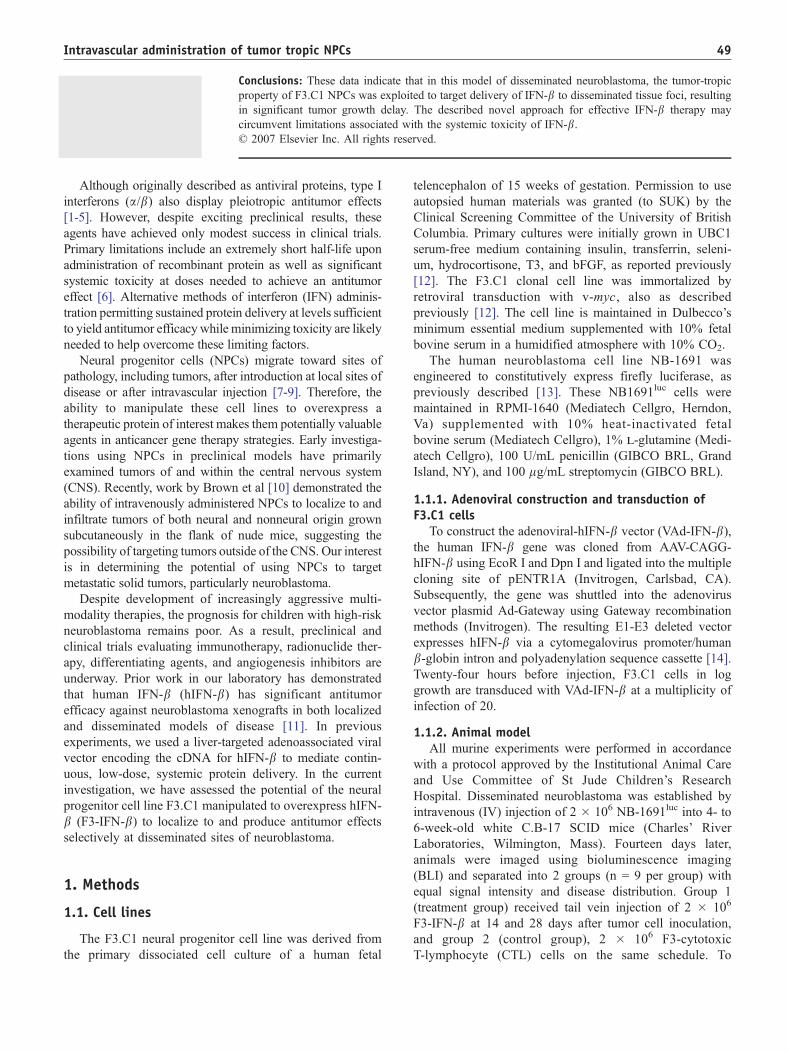
Fig. 4 At necropsy, livers of F3-CTL mice were replaced with
bulky disease (A), whereas those of F3-IFN-b–treated mice
appeared normal (B). Hematoxylin and eosin staining of these
organs further demonstrated almost complete replacement of
normal liver parenchyma in control mice (C), whereas only small
nests of tumor cells were detected in F3-IFN-b–treated animals
(D). The differences in liver weights are shown graphically (E).
**P b .001 vs both naive and control mice.
Intravascular administration of tumor tropic NPCs 51
sections compared with the number seen in tumor-bearing
mice and no F3.C1 cells within the kidneys sections of
naive mice.
2.2. Effect of IFN-bb–expressing NPCs ondisseminated disease
We next evaluated the therapeutic effects of F3-IFN-bcells in the model of disseminated neuroblastoma. Fourteen
days after NB-1691 tumor cell inoculation, animals were
divided into 2 groups with equal tumor burden, based on
BLI (8.74 F 1.57 � 105 vs 8.92 F 1.95 � 105 photons per
second, P = .94). Mice were then administered 2 � 106 F3-
IFN-b or F3-CTL cells by tail vein injection. Biolumines-
cence imaging 2 weeks after the initial administration of
NPCs revealed a significant restriction of tumor progression
in F3-IFN-b–treated mice (2.61 F 0.58 � 107photons per
second) vs controls (5.14 F 0.87 � 107photons per second,
P = .028) (Fig. 2). Animals were then administered a second
injection of F3-IFN-b or F3-CTL. Twelve days after this
Fig. 3 On day 40 after tumor cell injection, control mice were
ill-appearing and cachectic, whereas F3-IFN-b–treated mice
remained healthy and maintained normal body weights. *P = .015.
second treatment, 2 mice within the F3-CTL group
succumbed to disease, whereas while the remaining mice
within this group appeared ill and cachectic (animal weight
23.01 F 1.24 g). Impressively, all mice within the F3-IFN-bgroup appeared healthy at this time (animal weight 26.40 F0.51 g, P = .015 vs controls) (Fig. 3). Bioluminescence
imaging further demonstrated a significant delay in tumor
progression in F3-IFN-b–treated mice (1.69 F 0.69 � 109
photons per second) vs F3-CTL (3.13 F 0.50 � 1010pho-
tons per second, P b .001) (Fig. 2).
Due to the severity of disease progression in F3-CTL
mice, animals in both groups were killed for further
evaluation and comparison of disease burden on day 40
after tumor cell injection. Upon necropsy, mice within the
control group were found to have bulky tumor replacing the
liver and kidneys (Figs. 4A and 5A) (liver weight 3.77 F

Fig. 5 At necropsy, kidneys of F3-CTL mice also were replaced
with bulky disease (A), whereas those of F3-IFN-b–treated mice
appeared normal, with the exception of tumor nodules seen in
some mice (B). Hematoxylin and eosin staining of these organs
further demonstrated almost complete replacement of normal
kidney parenchyma in control mice (C), whereas less tumor
burden was detected in F3-IFN-b–treated animals (D). The
differences in kidney weights are shown graphically (E).
**P b .001 vs naive and control mice; #P = .02 vs naive mice.
P.V. Dickson et al.52
0.42 g; kidney weight 1.24 F 0.23 g, P b .001 vs both NPC-
IFN treated and naive mice) (Figs. 4E and 5E) as well as
extensive retroperitoneal and mediastinal adenopathy. In
contrast, these sites within mice receiving F3-IFN-b therapy
appeared grossly normal with the exception of small nodules
within the kidneys in some of these animals (Figs. 4B
and 5B) (liver weight 1.61 F 0.09 vs 1.55 F 0.04 g in naive
mice, P = .32; kidney weight 0.633F 0.03 vs 0.480F 0.03 g
in naive mice, P = .02) (Figs. 4E and 5E). Histologic analysis
of the livers and kidneys within these mice further
highlighted the tumor growth restriction within mice treated
with F3-IFN-b vs controls (Figs. 4C and D and 5C and D). In
mice bled at multiple time points after F3-IFN-b and F3-
CTL injection, plasma levels of hIFN-b were undetectable,
as assessed by ELISA. These data demonstrate that after
intravascular administration, F3-IFN-b cells localize to sites
of disseminated disease and express levels of hIFN-bsufficient to achieve a significant antitumor effect.
3. Discussion
The type I interferons are known to have multiple
antitumor effects including direct inhibition of tumor cell
proliferation through both cell cycle arrest and induction of
apoptosis [1,5] as well as indirect antitumor activity
through immunodmodulation [2,3] and inhibition of angio-
genesis [5,11]. Unfortunately, an extremely short half-life
and significant toxicity have hindered their use in the
clinical setting. An alternative dosing schedule directed at
achieving sustained low-level systemic or tumor-targeted
protein delivery is likely to help circumvent these
limitations. Gene therapy–based approaches for the deliv-
ery of IFNs have shown some promise in this regard. These
strategies have included both systemic [15] and intra-
tumoral [16] administration of adenoviral vectors, as well
as cationic liposomes [17] carrying the IFN-b gene.
Furthermore, we have recently shown that a liver-targeted
adenoassociated viral vector carrying the cDNA for IFN-bmediates continuous, low-dose, systemic protein delivery
and has significant antitumor activity against a variety of
tumor histologies [5,11]. In the current investigation, we
have demonstrated that intravascular administration of
tumor-tropic neural progenitor cells permits targeted
delivery of IFN-b and results in significant antitumor
efficacy without elevating systemic protein levels in a
murine model of disseminated neuroblastoma.
NPCs display remarkable tropism and migratory capacity
to sites of malignant growth. Importantly, these cells can be
modified to stably express a therapeutic transgene. Several
recent investigations have examined their use in cell-based
gene therapy strategies aimed at brain tumors [7,9,18].
Although initial approaches used local or direct intratumoral
administration of NPCs, further work has demonstrated
these cells retain their ability to bhome inQ on sites of tumor
growth after systemic administration [9,10]. Furthermore, it
has been shown that after IV injection, NPCs engineered to
express a lacZ reporter and the prodrug activating enzyme
cytosine deaminase migrated to established subcutaneous
SH-SY5Y human neuroblastoma xenografts [10]. In the
present study, the F3.C1 NPC line localized to sites of
disseminated tumor foci in a model of disseminated
neuroblastoma. When injected IV, NB-1691 neuroblastoma
cells consistently produce disease in the liver, kidneys, bone
marrow, and retroperitoneal lymph nodes. At a point when
these mice have miliary or microscopic disease at multiple
sites, 2 � 106 DiI-labeled F3.C1 cells were administered by
tail vein injection and animals killed 3 days later.
Subsequent histologic analysis demonstrated accumulation
of the F3.C1 cells in or near sites of tumor growth,
demonstrating the ability of these cells to migrate toward

Intravascular administration of tumor tropic NPCs 53
tumors outside of the CNS. Moreover, when injected into
naive mice, a few cells were detected in the liver and none
in the kidney. It is believed that in the absence pathology,
NPCs undergo apoptosis and are very short-lived. Although
only a 3-day time point after F3.C1 injection was evaluated
in the current investigation, NPCs have been found to
localize to sites of tumor growth as early as 30 minutes and
detected as far out as 7 days after injection [9,10]. Further
experiments are being designed to appropriately assess the
arrival and duration of survival of NPCs at sites of
disseminated neuroblastoma.
In this investigation, F3.C1 cells were engineered to
overexpress hIFN-b by adenoviral transduction. Application
of vector to these cells has been found to yield significant
transgene expression within 24 hours with hIFN-b levels
remaining stable in vitro for approximately 2 weeks (data
not shown). To assess the antitumor efficacy of F3-IFN-bcells in vivo, 2 � 106 cells were administered IV to SCID
mice with disseminated NB-1691 tumors. Significant tumor
growth restriction was demonstrated by serial BLI as well as
gross and histologic evaluation at necropsy. Importantly,
when mice were bled at 1, 4, 7, and 14 days after F3-IFN-binjection, plasma levels of hIFN-b were not detectable by
ELISA. These data are consistent with prior experiments
evaluating the antitumor effects of hIFN-b against NB-1691
xenografts [11] and highlight the use of a tumor tropic, cell-
based approach to achieve tumor targeted protein delivery.
The ability to effect sustained F3.C1 expression of hIFN-bat disseminated sites of microscopic disease represents a
novel therapeutic approach in a model designed to mimic a
minimal residual disease state.
In conclusion, the neural progenitor cell line F3.C1
displays tropism for sites of disseminated neuroblastoma
in a murine model and permits targeted delivery of
IFN-b,resulting in significant tumor growth restriction.
Future studies examining the mechanisms of NPC tumor
tropism, the timing and duration of localization, and
efficacy of F3-IFN-b against additional neuroblastoma cell
lines are ongoing.
References
[1] Stark GR, Kerr IM, Williams BR, et al. How cells respond to
interferons. Annu Rev Biochem 1998;67:227-64.
[2] Belardelli F, Ferrantini M, Proietti E, et al. Interferon-alpha in tumor
immunity and immunotherapy. Cytokine Growth Factor Rev 2002;13:
119-34.
[3] Dvorak HF, Gresser I. Microvascular injury in pathogenesis of
interferon-induced necrosis of subcutaneous tumors in mice. J Natl
Cancer Inst 1989;81:497-502.
[4] Slaton JW, Perrotte P, Inoue K, et al. Interferon-alpha–mediated down-
regulation of angiogenesis-related genes and therapy of bladder cancer
are dependent on optimization of biological dose and schedule. Clin
Cancer Res 1999;5:2726-34.
[5] Streck CJ, Dickson PV, Ng CY, et al. Antitumor efficacy of AAV-
mediated systemic delivery of interferon-beta. Cancer Gene Ther
2006;13:99-106.
[6] Einhorn S, Grander D. Why do so many cancer patients fail to respond
to interferon therapy? J Interferon Cytokine Res 1996;16:275-81.
[7] Benedetti S, Pirola B, Pollo B, et al. Gene therapy of experimental
brain tumors using neural progenitor cells. Nat Med 2000;6:447 -50.
[8] Aboody KS, Najbauer J, Schmidt NO, et al. Targeting of melanoma
brain metastases using engineered neural stem/progenitor cells.
Neurooncol 2006;8:119 -26.
[9] Aboody KS, Brown A, Rainov NG, et al. Neural stem cells
display extensive tropism for pathology in adult brain: evi-
dence from intracranial gliomas. Proc Natl Acad Sci U S A 2000;
97:12846-51.
[10] Brown AB, Yang W, Schmidt NO, et al. Intravascular delivery of
neural stem cell lines to target intracranial and extracranial tumors of
neural and non-neural origin. Hum Gene Ther 2003;14:1777-85.
[11] Streck CJ, Dickson PV, Ng CY, et al. Adeno-associated virus vector-
mediated systemic delivery of IFN-beta combined with low-dose
cyclophosphamide affects tumor regression in murine neuroblastoma
models. Clin Cancer Res 2005;11:6020-9.
[12] Kim SU, Nakagawa E, Hatori K, et al. Production of immortalized
human neural crest stem cells. Methods Mol Biol 2002;198:55 -65.
[13] Dickson PV, Ng CY, Zhou J, et al. In vivo bioluminescence imaging
for early detection and monitoring of disease progression in murine
model of neuroblastoma. J Am Coll Surg 1995;201:S53 -4.
[14] Ory DS, Neugeboren BA, Mulligan RC. A stable human-derived
packaging cell line for production of high titer retrovirus/vesicular
stomatitis virus G pseudotypes. Proc Natl Acad Sci U S A 1996;93:
11400-6.
[15] Choi EA, Lei H, Maron DJ, et al. Combined 5-fluorouracil/systemic
interferon-beta gene therapy results in long-term survival in mice with
established colorectal liver metastases. Clin Cancer Res 2004;10:
1535-44.
[16] Zhang F, Lu W, Dong Z. Tumor-infiltrating macrophages are involved
in suppressing growth and metastasis of human prostate cancer cells
by INF-beta gene therapy in nude mice. Clin Cancer Res 2002;8:
2942-51.
[17] Yoshida J, Mizuno M, Wakabayashi T. Interferon-beta gene therapy
for cancer: basic research to clinical application. Cancer Sci 2004;95:
858 -65.
[18] Yip S, Aboody KS, Burns M, et al. Neural stem cell biology may be
well suited for improving brain tumor therapies. Cancer J 2003;9:
189 -204.