Structural, Bioinformatic, and In Vivo Analyses of Two Treponema pallidum Lipoproteins Reveal a...
33
Structural, bioinformatic, and in vivo analyses of two Treponema pallidum lipoproteins reveal a unique TRAP transporter Ranjit K. Deka a,* , Chad A. Brautigam b,* , Martin Goldberg a , Peter Schuck c , Diana R. Tomchick b , and Michael V. Norgard a,§ a Department of Microbiology, The University of Texas, Southwestern Medical Center at Dallas, 5323 Harry Hines Blvd., Dallas, TX 75390, USA b Department of Biochemistry, The University of Texas, Southwestern Medical Center at Dallas, 5323 Harry Hines Blvd., Dallas, TX 75390, USA c Dynamics of Molecular Assembly Section, Laboratory of Cellular Imaging and Macromolecular Biophysics, NIBIB, National Institutes of Health, Bethesda, MD, USA Abstract Treponema pallidum, the bacterial agent of syphilis, is predicted to encode one tripartite ATP- independent periplasmic transporter (TRAP-T). TRAP-Ts typically employ a periplasmic substrate-binding protein (SBP) to deliver the cognate ligand to the transmembrane symporter. Herein, we demonstrate that the genes encoding the putative TRAP-T components from T. pallidum, tp0957 (the SBP) and tp0958 (the symporter) are in an operon with an uncharacterized third gene, tp0956. We determined the crystal structure of recombinant Tp0956; the protein is trimeric and perforated by a pore. Part of Tp0956 forms an assembly similar to those of “tetratricopeptide repeat” (TPR) motifs. The crystal structure of recombinant Tp0957 was also determined; like the SBPs of other TRAP-Ts, there are two lobes separated by a cleft. In these other SBPs, the cleft binds a negatively charged ligand. However, the cleft of Tp0957 has a strikingly hydrophobic chemical composition, indicating that its ligand may be substantially different and likely hydrophobic. Analytical ultracentrifugation of the recombinant versions of Tp0956 and Tp0957 established that these proteins associate avidly. This unprecedented interaction was confirmed for the native molecules using in vivo cross-linking experiments. Finally, bioinformatic analyses suggested that this transporter exemplifies a new subfamily of TPR-protein associated TRAP transporters (TPATs) that require the action of a TPR-containing accessory protein for the periplasmic transport of a potentially hydrophobic ligand(s). Keywords TRAP transporter; syphilis; Treponema pallidum; TPR motif; protein interactions © 2012 Elsevier Ltd. All rights reserved. § To whom correspondence should be addressed: Phone: 214-633-0015, Fax: 214-648-5905, [email protected]. * These authors contributed equally to this work. Accession Numbers Coordinates and structure factors for the TatT and TatP T structures have been deposited in the Protein Data Bank; the accession numbers are 3U64 and 3U65, respectively. Publisher's Disclaimer: This is a PDF file of an unedited manuscript that has been accepted for publication. As a service to our customers we are providing this early version of the manuscript. The manuscript will undergo copyediting, typesetting, and review of the resulting proof before it is published in its final citable form. Please note that during the production process errors may be discovered which could affect the content, and all legal disclaimers that apply to the journal pertain. NIH Public Access Author Manuscript J Mol Biol. Author manuscript; available in PMC 2013 March 9. Published in final edited form as: J Mol Biol. 2012 March 9; 416(5): 678–696. doi:10.1016/j.jmb.2012.01.015. NIH-PA Author Manuscript NIH-PA Author Manuscript NIH-PA Author Manuscript
-
Upload
utsouthwestern -
Category
Documents
-
view
0 -
download
0
Transcript of Structural, Bioinformatic, and In Vivo Analyses of Two Treponema pallidum Lipoproteins Reveal a...

Structural, bioinformatic, and in vivo analyses of two Treponemapallidum lipoproteins reveal a unique TRAP transporter
Ranjit K. Dekaa,*, Chad A. Brautigamb,*, Martin Goldberga, Peter Schuckc, Diana R.Tomchickb, and Michael V. Norgarda,§
aDepartment of Microbiology, The University of Texas, Southwestern Medical Center at Dallas,5323 Harry Hines Blvd., Dallas, TX 75390, USAbDepartment of Biochemistry, The University of Texas, Southwestern Medical Center at Dallas,5323 Harry Hines Blvd., Dallas, TX 75390, USAcDynamics of Molecular Assembly Section, Laboratory of Cellular Imaging and MacromolecularBiophysics, NIBIB, National Institutes of Health, Bethesda, MD, USA
AbstractTreponema pallidum, the bacterial agent of syphilis, is predicted to encode one tripartite ATP-independent periplasmic transporter (TRAP-T). TRAP-Ts typically employ a periplasmicsubstrate-binding protein (SBP) to deliver the cognate ligand to the transmembrane symporter.Herein, we demonstrate that the genes encoding the putative TRAP-T components from T.pallidum, tp0957 (the SBP) and tp0958 (the symporter) are in an operon with an uncharacterizedthird gene, tp0956. We determined the crystal structure of recombinant Tp0956; the protein istrimeric and perforated by a pore. Part of Tp0956 forms an assembly similar to those of“tetratricopeptide repeat” (TPR) motifs. The crystal structure of recombinant Tp0957 was alsodetermined; like the SBPs of other TRAP-Ts, there are two lobes separated by a cleft. In theseother SBPs, the cleft binds a negatively charged ligand. However, the cleft of Tp0957 has astrikingly hydrophobic chemical composition, indicating that its ligand may be substantiallydifferent and likely hydrophobic. Analytical ultracentrifugation of the recombinant versions ofTp0956 and Tp0957 established that these proteins associate avidly. This unprecedentedinteraction was confirmed for the native molecules using in vivo cross-linking experiments.Finally, bioinformatic analyses suggested that this transporter exemplifies a new subfamily ofTPR-protein associated TRAP transporters (TPATs) that require the action of a TPR-containingaccessory protein for the periplasmic transport of a potentially hydrophobic ligand(s).
KeywordsTRAP transporter; syphilis; Treponema pallidum; TPR motif; protein interactions
© 2012 Elsevier Ltd. All rights reserved.§To whom correspondence should be addressed: Phone: 214-633-0015, Fax: 214-648-5905, [email protected].*These authors contributed equally to this work.Accession NumbersCoordinates and structure factors for the TatT and TatPT structures have been deposited in the Protein Data Bank; the accessionnumbers are 3U64 and 3U65, respectively.Publisher's Disclaimer: This is a PDF file of an unedited manuscript that has been accepted for publication. As a service to ourcustomers we are providing this early version of the manuscript. The manuscript will undergo copyediting, typesetting, and review ofthe resulting proof before it is published in its final citable form. Please note that during the production process errors may bediscovered which could affect the content, and all legal disclaimers that apply to the journal pertain.
NIH Public AccessAuthor ManuscriptJ Mol Biol. Author manuscript; available in PMC 2013 March 9.
Published in final edited form as:J Mol Biol. 2012 March 9; 416(5): 678–696. doi:10.1016/j.jmb.2012.01.015.
NIH
-PA Author Manuscript
NIH
-PA Author Manuscript
NIH
-PA Author Manuscript

IntroductionTreponema pallidum, the etiologic agent of syphilis, is an obligate human pathogen thatmetabolically relies heavily on its human host 1. The spirochete encodes only 1,067 genes(cf. the genome of Escherichia coli K-12, which has over 4,000 genes). T. pallidum lacksgenes of the biosynthetic pathways for fatty acids, nucleotides, most of the amino acids, andmany other vital metabolites. Natural selection has thus honed T. pallidum to be an organismwith an extremely parsimonious genome, likely at the cost of its ability to be free-living.From a more practical perspective, the need for T. pallidum to acquire so many of its vitalnutrients from its human host likely has accounted for the historic inability to continuouslycultivate the organism in vitro 2. Indeed, much of our lack of understanding of this enigmatichuman pathogen derives directly from this major investigative obstacle.
Bacteria use many means to import needed metabolites from their extracellular milieu intotheir cytoplasms. The primary ABC transporters and secondary symporters are examples 3;4.ABC transporters utilize a substrate-binding protein (SBP) in the periplasm to secure theligand and deliver it to a transmembrane permease, which uses the energy from ATPhydrolysis to transport the ligand against a concentration gradient into the cytoplasm.Canonical symporters do not require the services of an SBP; rather, a periplasmic ligandbinds directly to the transmembrane symporter. An abundant ion (usually either H+ or Na+)also binds, and the favorable energetics of co-transporting the ion are used to allow deliveryof the ligand to the cytoplasm, again typically against a concentration gradient.
Recently, a different family of solute transporters has been described— the tripartite ATP-independent periplasmic transporters (TRAP-Ts) 5. The TRAP-Ts incorporate features ofboth the primary and secondary transporters. The dctPQM operon of Rhodobactercapsulatus is the prototypical TRAP-T; it is responsible for the import of C4-dicarboxylates 6. Like the ABC transporters, the DctPQM system and all other TRAP-Tsemploy an SBP (called “P” herein) to capture the ligand 7 and deliver it to thetransmembrane parts of the transporter (called “Q” and “M” herein) 6. ATP hydrolysis doesnot energize these transporters. Rather, like secondary transporters, the energy stored in anelectrochemical gradient across the plasma membrane facilitates ligand transport 8. Themolecular details of how the transport is achieved are not yet fully elucidated, though studieson the SiaPQM TRAP-T indicate the requirement for the P component, an electric gradient,and the co-transport of at least two Na+ ions per ligand transported 8. The ligands for TRAP-Ts are typically acidic small molecules 9. A related, but distinct, family of transporters thatimport tricarboxylic acids has also been described 10.
The genome of T. pallidum is predicted to encode a single TRAP-T 1; 5, apparentlyorganized as an operon. The genes are tp0957 (the P component) and tp0958 (putative fusedQ and M components). A third gene within this putative operon is tp0956, which belongs tothe Cluster of Orthologous Groups (COG) 566011, a family of putative integral membraneproteins. This protein is also flagged as a member of the RskA superfamily11; inMycobacterium tuberculosis, this protein binds to sigma factor K, repressing its activity12.Alternatively, because it is similar in size to the P protein, it had been speculated thatTp0956 might serve as a second SBP for this TRAP-T 5. Having a second SBP would likelyhave important functional consequences, given the very limited genome of T. pallidum.
In this study, we first set out to characterize this putative second SBP, Tp0956. However, T.pallidum is not cultivatable in vitro, thereby precluding genetic manipulation of theorganism. Thus, we took advantage of a structure-to-function approach for this protein thathas been successful for other T. pallidum proteins of unknown functions 13;14;15;16. First,reverse transcription and amplification of RNA from rabbit-tissue-derived T. pallidum
Deka et al. Page 2
J Mol Biol. Author manuscript; available in PMC 2013 March 9.
NIH
-PA Author Manuscript
NIH
-PA Author Manuscript
NIH
-PA Author Manuscript

confirmed that tp0956, tp0957, and tp0958 constitute a single transcriptional unit. We thendetermined the crystal structure of Tp0956. Notably, we found that the protein has nostructural homology to known P proteins, making it unlikely that it serves as an additionalSBP for this putative TRAP-T. The protein is a mostly α-helical irregular cylinder with apore running through its center. It features substructures similar to tetratricopeptide repeat(TPR) motifs, which are hallmarks of proteins that are involved in protein-proteininteractions17;18. Because of the unusual nature of this TRAP operon, we also pursued andobtained a crystal structure for Tp0957, the putative P component. The overall fold ofTp0957 is similar to P components from other TRAP-Ts, but there are notable differences,particularly in the ligand-binding clefts of the proteins; that of Tp0957 is more hydrophobicthan other structurally characterized P proteins. Although there is some evidence of a small-molecule ligand bound to the protein, Tp0957 adopts a conformation more similar to anunliganded TRAP-T SBP. The existence of TPR motifs in Tp0956 led us to seek its bindingpartner(s). Using analytical ultracentrifugation (AUC), we found that trimeric Tp0956interacts with Tp0957. This observation was confirmed in vivo with formaldehyde cross-linking. Finally, we showed that proteins homologous to Tp0956 are found in other TRAP-Toperons. Thus, the T. pallidum TRAP-T may serve as a paradigm for a hitherto unknownclass of TRAP-Ts (which we call TPATs; see below) that apparently require a TPR-containing accessory protein.
ResultsAnalyses of tp0956, tp0957, and tp0958
Genomic analysis of T. pallidum suggested that tp0956, tp0957 and tp0958 are oriented inthe same direction and thus may be cotranscribed1 (Fig. 1). Furthermore, bacterial genesbelonging to a transporter often are cotranscribed, and bioinformatic analyses predictedtp0957 and tp0958 as components of a TRAP-T. Indeed, the TRAP-T operon (SiaPQM)from Haemophilus influenzae is organized similarly19 (Fig. 1). Therefore, we performedRT-PCR to examine the transcriptional linkage of these genes. As shown in Fig. 1, the three-gene cluster from T. pallidum is transcriptionally linked. One potential caveat is that the RT-PCR product observed between the tp0954 and tp0956 pair (Fig. 1; Table S1) may be due tothe presence of non-coding mRNA that is transcribed in the opposite direction of thisoperon.
In accord with its putative function as a permease, Tp0958 (a likely fusion of the M and Qcomponents) is quite hydrophobic (average hydropathy = 1.084) and is predicted to be anall-α-helical transmembrane protein. Signal sequences at their N-termini identify bothTp0956 and Tp0957 as putative lipoproteins20 (see Table S2). As such, their N-terminalsignal sequences would be removed, leaving a tri-acylated cysteine residue at theirrespective N-termini that would anchor the proteins to the periplasmic side of either theinner or outer membranes of the bacterium.
A conserved-domain database (CDD) search11 using the sequence of Tp0956 places it inCOG5660, a family of integral membrane proteins. This fact raises the question: is theTp0956 an integral membrane protein or a lipoprotein? We purified recombinant forms ofboth Tp0956 and Tp0957 almost identically (see Materials & Methods). In these proteins,the N-terminal cysteines had been removed. This expedient was designed to prevent theacylation reaction mentioned above, and should render the proteins water soluble. Indeed,they could be purified with only a low concentration (2 mM; well below the critical micelleconcentration) of the non-ionic detergent 2-octyl-glucopyranoside (β-OG). We often includethis additive in preparations of recombinantly solubilized treponemal lipoproteins13;14;15;16,as it appears to improve their stability in solution. Additionally, b-OG can have a positiveeffect on protein crystallization21. The water solubility and probable lipidation of both
Deka et al. Page 3
J Mol Biol. Author manuscript; available in PMC 2013 March 9.
NIH
-PA Author Manuscript
NIH
-PA Author Manuscript
NIH
-PA Author Manuscript
proteins suggest that they exist in the periplasm, not in the cell membrane, as implied forTp0956. The CDD search also placed Tp0956 in the RskA superfamily; as noted above,RskA is an anti-sigma factor in M. tuberculosis. It seems unlikely that Tp0956 could play arole in transcription, given its probable periplasmic localization.
The crystal structure of Tp0956In view of the possibility that Tp0956 may be a second SBP for a TRAP-T, we set out todetermine the three-dimensional structure of Tp0956 (Fig. 2; Table 1). The crystal structureof Tp0956 was refined using X-ray diffraction data to 2.3 Å resolution. The model (Figs. 2A& B) has 13 α-helices, one short 310 helix, and no β-strands. The structure is very differentfrom the bilobed structures (see below) of known P components of TRAP-Ts, making itunlikely that Tp0956 fulfills this proposed role. Residues 31-301 (in the numbering of themature, processed form of the protein) are visible in electron-density maps; the first thirtyresidues of this protein construct are presumed present but were disordered in the crystals.The α-helices, designated “A” through “M”, have an antiparallel arrangement that forms anirregular ring (Figs. 2A-C). The ring surrounds a pore, which perforates the entire protein(Fig. 2C). Hereafter, we refer to the “N,” “C,” and “lateral” faces of Tp0956, which aredefined in Fig. 2B. The “N” and “C” faces are so called because they respectively house theobserved N-terminus and C-terminus of Tp0956.
Characteristics of the poreTp0956’s pore runs from the N to the C faces of the protein, a total of ~43 Å (Fig. 3A). Ithas an inner portion (~20 Å in length; average diameter ~5.8 Å; minimum diameter = 4.8 Å;see Fig. S1) and two vestibules (the “N” and “C” vestibules, depending on their respectiveproximity to the N and C faces). Some soluble protein enzymes have pores that are related totheir respective functions; the archeal 20 S proteasome is a multimeric assembly that hasnarrow (~13 Å) annuli22, and the DNA topoisomerase of E. coli has a large (~28 Å) hole 23.However, there is no evidence of an enzymatic activity for Tp0956. Although Tp0956 is asoluble protein (see above), the dimensions of its channel invite comparisons to membrane-spanning proteins. For example, the minimum pore diameters of aquaporin (AQP1),glyceroporins (GlpF), and an ion channel (KcsA) range from 2.6 to 3.7 Å 24;25;26, and thoseof bacterial porin outer-membrane proteins can be as small as 5 Å 27. The pore of Tp0956accommodates water molecules and a sulfate ion (from the precipitation buffer). There is ahydrophobic antechamber (HA) in the N vestibule (Fig 3C); it measures approximately 10 ×5 Å. The identities of the side chains lining the HA are shown in Table S3. Electrostaticcalculations show (Fig. 2D) that the C vestibule is mostly negatively charged, the Nvestibule is mostly positive, and the pore is mixed. No particular class of amino aciddominates the lining of the pore.
Comparisons to other structuresDALI 28 was used to find two structural homologs of Tp0956. They are DrR162B fromDeinococcus radiodurans (PDB ID: 3GW4, no associated publication; Fig. 4A) andTTC0263 (PDB ID: 2PL2) from Thermus thermophilus strain HB2729. Their structuralhomologies (both r.m.s.d.’s are 1.8 Å) apply to roughly half (148 and 149, respectively) ofthe Cα atoms in Tp0956. Neither homologous protein forms a pore, nor do they appear to beinvolved in a TRAP-T operon.
These structural comparisons reveal that Tp0956 contains four helical hairpins that arestructurally similar to “tetratricopeptide repeat” (TPR) motifs. Both DrR162B and TTC0263contain these motifs, which occur as tandem, 34-amino-acid repeats in the proteins thatharbor them. The TPR-like portions of Tp0956 encompass the α-helical pairs αC/αD, αG/αH, αI/αJ, and αK/αL (Fig. 4B); thus, these motifs form part of the pore and also part of the
Deka et al. Page 4
J Mol Biol. Author manuscript; available in PMC 2013 March 9.
NIH
-PA Author Manuscript
NIH
-PA Author Manuscript
NIH
-PA Author Manuscript

lateral surface of the protein. To our knowledge, Tp0956 is the only known protein havingTPR-like motifs that form part of a pore.
TPR motifs exist in many proteins17. Their folds comprise an antiparallel α-helical hairpincontaining two α-helices and a short turn that connects them. These motifs are most oftenfound as tandem repeats of three or more in proteins, where they stack to form a superhelicalarrangement that features a concave and a convex surface. The concave surface of the TPR-like superhelix in Tp0956 forms part of the protein’s pore (Fig. 4C). The superhelical stackis often “capped” by a C-terminal α-helix with an unrelated sequence; helix αM serves thisfunction in Tp0956. The tandem arrays of TPR motifs mostly function as protein-interactiondomains 17 in glucocorticoid receptors 30 and in the anaphase-promoting complex31. Todate, the transporter association of Tp0956 (and others identified below) is unique amongTPR-motif-bearing proteins.
The TPR motifs in Tp0956 are cryptic. That is, they failed to be detected by a hidden-Markhov algorithm designed to reveal their presence32, and they do not adhere to thecanonical 34-residue length. Nevertheless, the structural homology between the TPRs ofDrR162B and Tp0956 is pronounced (Fig. 4A). Sequence analysis of Tp0956 in its TPR-likeregions reveals that the protein does exhibit TPR-like sequence signatures (Fig. S2), but theinterhelical region is longer than usual in all cases except one. These cryptic TPRs ofTp0956 are termed “cTPR motifs” (below).
The quaternary structure of Tp0956Although there is only one copy of Tp0956 in the asymmetric unit, applyingcrystallographic symmetry to the protein reveals the presence of multiple potentialoligomeric forms. There are two identical dodecamers in the unit cell, and these comprise 8identical trimers (Fig. 5). Physicochemical characterization of the interfaces in the crystalstructure using PISA33 demonstrated that the most stable oligomer present in the crystals isthe crystallographic trimer shown in Fig. 5. To assess the oligomeric state of Tp0956 insolution, we subjected it to sedimentation velocity (SV) analytical ultracentrifugation. Asseen in Fig. 6A, there are two dominant forms of the protein under our solution conditions.The virtually concentration-independent sedimentation coefficients and the diffusionalspreads of the clearly separate boundaries denote the presence of slowly interconvertingspecies with apparent molar masses of 98,000 and 380,000 g/mol, within error consistentwith trimeric and dodecameric forms of Tp0956 (theoretical values of 101,028 and 404,112g/mol, respectively). There is evidence of monomeric Tp0956 (Fig. 6A), but only at a lowconcentration (≤ 1.6 μM). The dodecameric form only becomes prevalent at highconcentrations (>20 μM). In the presence of a low concentration (2 mM; well below thecritical micelle concentration) of β-OG, the trimeric form is predominant, and the tendencyto form dodecamers is substantially reduced (Fig. S3). It therefore appears that the detergentdisrupts the relatively weak interactions that drive dodecamer formation, but it cannotdisrupt the formation of the trimer at this concentration. We therefore conclude that thetrimeric form of Tp0956 is the most relevant in solution. The cTPR motifs are arrayed on theoutside of this assembly (Fig. 5). Importantly, all of the membrane-anchoring N-termini (i.e.the “N” faces) are located on the same face of this trimer, i.e. facing away from the viewerin Fig. 5. This datum confirms the topological feasibility of this assembly. In the dodecamer(not shown), the N faces all point to the center of the “ball” of Tp09056s, a very unlikelyarrangement for a membrane-tethered oligomer.
The crystal structure of Tp0957The existence of a cTPR-motif-containing protein in the operon of this putative treponemalTRAP-T suggested that this transporter may act differently than other TRAP-Ts. Further, no
Deka et al. Page 5
J Mol Biol. Author manuscript; available in PMC 2013 March 9.
NIH
-PA Author Manuscript
NIH
-PA Author Manuscript
NIH
-PA Author Manuscript

crystallographically characterized P components had a sequence more than 25% identical tothe putative treponemal P, Tp0957. To explore the impact these differences have on thestructure of the P component, we determined the crystal structure of Tp0957. X-raydiffraction data to 1.4 Å were used for the determination and model refinement (Table 1).The protein is bilobed (Figs. 7A & 7B); here, we call the two lobes the “Lobe I” (residues9-136 and 226-263) and “Lobe II” (residues 137-225 and 264-320). The protein chaincrosses from one lobe to the other at three points, the most prominent being a 40-residue α-helix (α11) that forms a backbone-like feature in the protein. Lobe I has a central, five-stranded β-sheet that is surrounded by helices. Four of the strands in the β-sheet are parallel.Like Lobe I, Lobe II also has a central β-sheet in which only one strand is antiparallel;however, this sheet has six strands. This second β-sheet is also surrounded by helices. Theprotein has no outstanding surface electrostatic features (Fig. 7C). Between the lobes is anopening (or “cleft”) in the protein (Figs. 7C & 8A).
There are two copies of Tp0957 in the asymmetric unit of these crystals. These moleculeshave very similar structures; the r.m.s.d. of all comparable Cα atoms is 0.5 Å. The contentsof this asymmetric unit raise the question of the oligomeric status of Tp0957. Mostcharacterized P proteins are monomeric, but two show evidence of oligomerization 34;35. Weused SV experiments to establish Tp0957’s oligomeric status in solution, and we found thatTp0957 is monomeric under our solution conditions (Fig. 6B). Further, a physicochemicalanalysis 33 of the interactions of the monomers found none that would lead to the formationof oligomers.
The fold of Tp0957 is typical of a P component of a TRAP-T. This group of SBPs belongsto superfamily 7 of the extracellular transport proteins 36. Thus far, the structures of Pcomponents suggest that the proteins use a “Venus-fly trap” motion to capture the ligand ina cleft between the two lobes9. This mechanism has been convincingly shown for SiaP,which is part of a TRAP-T for sialic acid 19. That protein has been crystallized in both an“open” (unliganded) form and a “closed” (liganded) form 37;38. The open-to-closedtransition may be characterized 37 as a ~30° rigid-body rotation of Lobe I closer to Lobe IIabout three hinge points (Fig. 8B). The structure of Tp0957 is much closer to the “open”form of SiaP (Fig. 8C); according to DaliLite 39, compared to Tp0957, 296 comparable Cαpositions of “open” SiaP have an r.m.s.d. of 2.8 Å, while 297 Cαs of “closed” SiaP have anr.m.s.d. of 3.6 Å.
The unique cleft of Tp0957As mentioned above, Tp0957 has a cleft between the two lobes. This opening leads to threedifferent cavities (Figs. 7C & 8A). We call them Cavity A (19 Å deep by 5 Å wide), CavityB (11 × 7 Å), and Cavity C (10 × 4 Å). The residues whose side chains line these cavitiesare shown in Table S3. They are predominantly hydrophobic. While Cavities B & C haveanalogs in other P structures, Cavity A does not.
In comparison to other P proteins, the ligand-binding pocket of Tp0957 is morehydrophobic. Indeed, almost all of the residues making polar contacts to ligands in other Pproteins are hydrophobic at the equivalent positions in Tp0957. Notably, an arginine knownto contact a carboxylate moiety in the ligands of structurally characterized P proteins9;40;41
(cf. R147 of SiaP) is not present in Tp0957 (it is an alanine), and no compensatorypositively charged residue is observed near to this position. The lack of polar side chains inthis area implies that ligand recognition by Tp0957 is based on shape complementarityalone; matching patterns of opposite charge or hydrogen bond donors/acceptors on theligand and protein are not likely to be binding determinants. This finding could imply a lackof specificity for ligands.
Deka et al. Page 6
J Mol Biol. Author manuscript; available in PMC 2013 March 9.
NIH
-PA Author Manuscript
NIH
-PA Author Manuscript
NIH
-PA Author Manuscript

Further, the shapes of the clefts of “open” SiaP and Tp0957 differ significantly. In its openform, the cleft of SiaP is deep and spans the protein (Fig. 8B, left), whereas the cleft ofTp0957 resembles a much smaller pocket leading to the three aforementioned cavities (Figs.7C and 8A). In comparing the two clefts, that of Tp0957 appears to have been partiallycovered with protein residues from a loop between β6 and α7 (residues 161-164), formingCavity B. For Tp0957 to undergo the rigid-body rotation observed in SiaP, this loop wouldhave to be significantly rearranged. These data suggest that the cognate ligand of Tp0957differs in its chemical character from the small organic acid ligands of most P proteins andthat the ligand-capture mechanism of Tp0957 may deviate from those of other P and SBPproteins.
There is no clear evidence for a bound ligand in the electron-density maps for Tp0957.There is a small patch of density proximal to the indole ring of W24 (Fig.8D), as if theunidentified chemical moiety were stacked on the ring. This density does not closelycorrespond to the structures of any buffer, salt, precipitant, or cryoprotectant present. It mayrepresent a small molecule that copurified with the protein or a contaminant in theprecipitation or cryoprotection solutions. Despite this ambiguity, the position of the densityfeature roughly corresponds to that adopted by the ligands in other P-protein structures.Besides the side chain of W24, the feature is proximal to the side chains of residues I17,P19, A136, L163, and I228, making it likely that the moiety bound here is hydrophobic.
The interaction of Tp0956 and Tp0957Given that cTPR motifs exist inTp0956, we set out to identify its binding partner. Our firstattempt was with Tp0957, as the transcription of the two proteins is linked (Fig. 1). SVexperiments were thus used to query a possible interaction between Tp0956 and Tp0957 insolution. We studied a mixture of the proteins in which the Tp0956 trimer (0.7 μM) wassaturated with a 24-fold molar excess of Tp0957. As is evident in Fig. 6C, all of the Tp0956was in a complex that sedimented faster than Tp0956 alone. The 9.3-S complex has anestimated molar mass of 217,000 g/mol, consistent with a single trimer of Tp0956interacting with three monomers of Tp0957, forming a 1:3 complex (theoretical molar mass= 214,560 g/mol). A mixture of Tp0956 and Tp0957 also formed a co-eluting complex insize-exclusion chromatography experiments (not shown). As a negative control todemonstrate specificity, we also sedimented Tp0956 with the treponemal lipoproteinTp0655, the SBP of an ABC-type polyamine transporter 16, and there was no evidence of aninteraction between the two (Fig. S4). Further, TDE 1020, the T. denticola homolog ofTp0957, does not interact well with Tp0956, demonstrating species specificity in thisinteraction (Fig. S4).
We then used SV to examine in more detail the strength of the interaction between Tp0956and Tp0957, exploiting the exquisite sensitivity of the time-average sedimentation velocityof the reaction boundary to the complex size and the fractional dissociation of itscomponents. We analyzed data from mixtures of Tp0956 and Tp0957 at a range ofconcentrations with a model directly fitted to the SV data (Figs. 5D & S6) given the reactionkinetics, sedimentation coefficients, and three macroscopic equilibrium associationconstants associated with an A + B + B + B ↔ AB + B + B ↔ ABB + B ↔ ABBB reactionscheme (see Materials & Methods; here, A ≡ Tp0956 trimer and B ≡ Tp0957). Because allof the binding sites on trimeric Tp0956 are identical, the model includes the Kd of the firstbinding event (Kd(1)) and cooperativity factors (α-factors) for adding the second (α2) andthird (α3) molecules of B (an α > 1 suggests positive cooperativity and vice versa). Excellentglobal fits to the raw data were achieved (Fig. 6D & S6), demonstrating that the first bindingevent is strong (Kd(1) = KA(1)-1 = 60 [50, 80] nM; the numbers in square brackets indicatethe 68.3% error interval), and that a slight positive cooperativity may be associated with thesecond event (α2 = 1.5 [1, 3]). However, there is a small, but statistically significant (on a
Deka et al. Page 7
J Mol Biol. Author manuscript; available in PMC 2013 March 9.
NIH
-PA Author Manuscript
NIH
-PA Author Manuscript
NIH
-PA Author Manuscript
99.3% confidence interval) negative cooperativity (α3 = 0.34 [0.23, 0.51]) for binding thethird Tp0957 to the Tp0956 trimer.
In vivo evidence for interaction between native Tp0956 and Tp0957The strong, specific interaction between the two recombinant proteins in vitro prompted usto demonstrate an association between native Tp0956 and Tp0957 in vivo. We thereforeused in vivo formaldehyde cross-linking of T. pallidum to preserve putative Tp0956-Tp0957complexes. Formaldehyde is a short cross-linker that covalently links primary amines within~2 Å of each other (reflecting protein-protein interactions as they occur in vivo). The link isreversible and used to preserve in vivo protein-protein complexes. In this study, wechemically cross-linked treponemes with 1% formaldehyde. Lysed cells were resolved induplicate on two identical SDS polyacrylamide gels, electroblotted, and then individuallyprobed with either an anti-Tp0956 or an anti-Tp0957 antibody (Fig. 9). As a negativecontrol, immunodetection was also performed using pre-immune serum collected from thesame rats later immunized with antigens (data not shown). In the presence of cross-linker,both blots featured a strong band at a position consistent with a 250 kDa protein or complex(Lanes 2). The molecules in this band were recognized by both the Tp0956 and Tp0957antibodies, providing evidence for the occurrence of an in vivo complex. The blots were thenstripped and re-probed with the negative control antibody raised against Tp47 (the mostabundant periplasmic lipoprotein in T. pallidum). Nonspecific cross-linked intermediates ofnative Tp47 were not observed with either native Tp0956 or Tp0957 (data not shown). Thebroader profiles of cross-linked species may be caused by inefficient dissociation of thecomplex in the presence of SDS. As shown in Fig. 9, the large complexes formed uponformaldehyde-treatment were no longer detected after boiling; only Tp0956 and Tp0957monomers were observed (Lanes 3). For a number of reasons, including the unequalelectrotransfer of small versus large complex proteins, the signals on the blot do not give anaccurate representation of the amount of target proteins (especially in the case of cross-linked complexes of ~250 kDa). Nevertheless, this result strongly supports the conclusionthat the large entities (Fig. 9, Lanes 2) are reversible complexes of Tp0956 and Tp0957.Whereas the majority of Tp0956 protein is found at the apparent molecular mass of ~250kDa, Tp0956-specific bands corresponding to molecular masses of 35, 75 and 120 kDaarose, indicating partial dissociation of native complexes (Tp0956-Tp0957) into Tp0956trimer, dimer and monomer in the presence of SDS upon incubation at 65°C. The smearobserved in the cross-linked sample developed using anti-Tp0957 antibody also warrantscomment. Some of the broader bands might correspond to Tp0957 cross-linked to as yetunknown physiological partner(s) or possibly to its operonic partner, Tp0958 (however, wedo not possess Tp0958 antibodies to substantiate this second possibility). A controlexperiment was conducted in which treponemes were incubated under cross-linkingconditions without the addition of formaldehyde. In this control, only uncomplexed Tp0956and Tp0957 were observed (Lanes 1). Based on these results, we contend that there is a bonafide interaction between native Tp0956 and Tp0957 in vivo.
Tp0956 and Tp0957 homologs in other microorganismsA position-specific iterated BLAST search 42 of genomic databases for proteins similar toTp0956 returned only 38 proteins that may reliably be considered homologous (see Table 2).For comparison, there are 756 P-protein homologs currently collected in a database ofknown TRAP system proteins 43. Homologous proteins are clearly present in the genusTreponema, but are also found in other spirochetes, as well as in β-, γ-, and δ-proteobacteria.No eukaryotic homologs were found. The treponemes are the only human pathogens thatwere found to harbor Tp0956 homologs. One Tp0956-homolog-containing organism,Bacteriovorax marinus, parasitizes other bacteria 44;45. The remaining proteins were from
Deka et al. Page 8
J Mol Biol. Author manuscript; available in PMC 2013 March 9.
NIH
-PA Author Manuscript
NIH
-PA Author Manuscript
NIH
-PA Author Manuscript

diverse free-living oligotrophic bacteria. Remarkably, roughly one third of the 35 speciesharboring Tp0956-like proteins appear to metabolize saturated or aromatic hydrocarbons.
We examined the genome maps for all organisms whose genome contains a Tp0956homolog. Strikingly, 92% of the Tp0956 homologs appear to be components of TRAP-Ts(Table 2). Of those that do not, the Tp0956-like gene is directly adjacent to a putative Pprotein of a TRAP system 67% of the time; i.e., the Q and M components are missing,perhaps encoded elsewhere in the genome. These correlations strongly suggest that theTp0956 homologs are carrying out an essential function for their respective TRAP-T. Thatfunction probably involves binding to the P component. Indeed, a preliminary study showsthat the Tp0956- and Tp0957-like proteins (Tde1020 and Tde1021, respectively) from T.denticola interact (Fig. S5). Given the structural and functional characterizations delineatedabove, the Tp0956 homologs likely perform an auxiliary function in the transport of thetarget ligand. We name these cTPR-containing proteins the “T components”.
Do the TRAP-Ts with T components have other features that distinguish them fromcanonical TRAP systems? To begin to answer this question, we compared the sequences of218 P proteins, including all of those that neighbor T components. The resultingphylogenetic tree (Figs. 10 & S7) demonstrates that the Ps associated with T-containingTRAP-Ts (PTs) form a distinct clade; i.e. they are evolutionarily more related to each otherthan to other Ps (Fig. 10). Further, as noted above, most P proteins have an arginine residueat the position equivalent to A159 of Tp0957 9. The positively charged guanidinium moietyof this residue is critical for the binding of the carboxylate moiety of the small carboxylicacids that are normally bound by P proteins. This arginine is found in only 3 of the 36 PTsthat we have characterized to date. The two most likely explanations for this phenomenonare (1) the PTs bind to chemically different compounds or (2) they bind to chemicallysimilar compounds in a manner that differs from most P proteins. Regardless, we concludethat the transport systems harboring a T component are a distinct subfamily of TRAP-Ts thatwe call the TPATs (TPR-protein-associated TRAP transporters). We suggest that the operontake on the prefix “tat”, standing for “TPR-protein-associated transporter”. Thus thetreponemal operon would be called tatTPQ/M.
DiscussionThis investigation has uncovered a new subfamily of TRAP-Ts, the TPATs (Fig. 10). Theexistence of a strong, specific interaction in vitro (Fig. 6C, see above) and in vivo (Fig. 9)between the T and PT components suggests that the mechanism of these transportersapparently is assisted by this association. In ABC and TRAP transporters, obviously protein-protein interactions between the SBP and the transmembrane component(s) are functionallyvital 3;8;46;47. In Halomonas elongata, the cytoplasmic, tetrameric, ATP-binding universalstress protein TeaD of the teaABCD modulates the activity of an ectoine TRAP transporter(TeaA/TeaB/TeaC), presumably by affecting translation of its components or the activity ofthe permease48. However, the association of an SBP with another periplasmic protein isunprecedented.
What purpose is served by the binding of the T and PT components of the TPATs? The fullanswer to this question is not known at this time. It is possible that T may have a role indelivering a ligand to PT or in receiving a ligand from PT. Interestingly, if the ligand doesinteract with T, this association may represent an additional layer of specificity for theTPAT; i.e., ligands must have chemical characteristics that are compatible with associationwith both the T and PT components. Additionally, if T interacts with the ligand, it impliesthat the protein may be acting as a chaperone to escort the ligand through a periplasmicenvironment that is otherwise hostile to its stability or solubility. Recently, a similar protein-
Deka et al. Page 9
J Mol Biol. Author manuscript; available in PMC 2013 March 9.
NIH
-PA Author Manuscript
NIH
-PA Author Manuscript
NIH
-PA Author Manuscript
mediated mechanism for the transport of hydrophobic compounds was proposed for anABC-type transporter 49.
What is the ligand for this TPAT system in T. pallidum? Three lines of evidence suggest thatthe ligand is hydrophobic: (1) the existence of the hydrophobic antechamber (HA) of the Tcomponent (Fig. 3C, Table S3) (2) the hydrophobic nature of the PT component’s cleft andassociated cavities (Fig. 8A; Table S3), and (3) the preponderance of T-containingorganisms that utilize hydrophobic compounds (Table 2). While these oligotrophs canmetabolize essentially insoluble petroleum hydrocarbons, treponemes probably acquired orretained the TPATs during their evolution to import required hydrophobic ligands that theyencounter in humans.
This study represents only the beginning of the characterization of the TPATs. It will benecessary to discern the ligands of these transporters; such research currently remainsinfeasible for T. pallidum, owing to the organism’s need to be cultivated in rabbit testicles.The nature of the ligand may explain the need for the T component and may also providenew seminal clues concerning what “nutrients” are needed ultimately to achieve the in vitroculture of T. pallidum. Also, knowledge of whether the ligand interacts with and passesthrough the T component’s pore is imperative for a full understanding of the system. Finally,additional biophysical, biochemical, and biological studies of the interactions between the T,PT, Q, and M (or Q/M) components are warranted to better comprehend the mechanism(s) ofthis new subfamily of bacterial transporters. Further examinations of this system might bebetter accomplished in a different bacterium, such as T. denticola, which can replicate inculture. Additionally, information regarding ligand variability in the TPAT P componentsshould be available from a more comprehensive study of the TPAT proteins from diversebacterial species (Table 2).
Materials and MethodsProtein expression and purification
To produce a non-lipidated, recombinant derivative of Tp0957 in Escherichia coli, the DNAfragment encoding amino acid residues 7-328 (cloned without the post-translationallymodified N-terminal Cys plus five other hydrophobic residues) of Tp0957 was PCRamplified using ends-specific primers. Note that the recombinant versions of both Tp0956and Tp0957 are numbered in this report such that the processed, modified cysteine residue isResidue 1. The PCR product was subcloned into the pGEM-T Easy vector (Promega) andtransformed into E. coli XL1-Blue cells (Stratagene). The plasmids isolated from coloniesthat tested positive by restriction digestion were verified by DNA sequencing. The insertedfragment was excised by digestion with NotI and BamHI and then ligated into thecorresponding restriction sites of the expression vector pIVEX2.4d vector (5 Prime). Theresultant construct encoded a fusion protein with a His6-tag at its N-terminus. Ligationmixtures were transformed into E. coli XL1-Blue cells, and plasmids isolated from colonieswere verified by DNA sequencing. A verified plasmid was then cotransformed withpGroESL (TAKARA) into E. coli BL21 AI (Invitrogen) cells for soluble protein expression.
E. coli BL21 AI cells were grown at 37° C in LB medium containing 0.1% (w/v) glucose,100 μg/mL of ampicillin and 30 μg/mL of chloramphenicol until the cell density reached anA600 of 0.5. The culture was then induced for 3 h with 0.2% (w/v) L-arabinose. Cellsderived from one liter of culture were harvested by centrifugation and lysed at roomtemperature with gentle rocking for 30 min using 50 mL of B-PER II (Thermo Scientific).The resulting suspension was centrifuged at 25,000 x g for 15 min to remove cell debris.Tp0957 was isolated from the supernatant by affinity chromatography using Ni-NTAAgarose (Qiagen). The protein was then subjected to size-exclusion chromatography (SEC)
Deka et al. Page 10
J Mol Biol. Author manuscript; available in PMC 2013 March 9.
NIH
-PA Author Manuscript
NIH
-PA Author Manuscript
NIH
-PA Author Manuscript
using a HiLoad 16/60 Superdex 200 prep grade column (GE Healthcare) equilibrated withBuffer A (20 mM Hepes, 0.1 M NaCl, pH 7.5, 2 mM n-octyl β-D-glucoside (β-OG)). Peakfractions were analyzed by SDS-PAGE. Fractions containing purified rTp0957 were pooledand stored at 4° C in buffer A. After two weeks, mass spectrometry and N-terminalsequencing determined that the tag of rTp0957 was specifically cleaved, leaving tworesidual amino acid residues (Gly-Arg) at the N-terminus followed by residues 7-328 ofTp0957. The mixture was further purified by SEC using buffer A. This purified, cleavedTp0957 was used for co-crystallization with Tp0956.
For the production of a non-lipidated, recombinant derivative of Tp0956 in E. coli, a DNAfragment encoding residues 2-302 (cloned without the post-translationally modified N-terminal Cys) was amplified by PCR from treponemal genomic DNA using primer pairsencoding the predicted 5′-and 3′-termini. The PCR product was subcloned into BsaI/XbaIdigested pE-SUMOpro3-based bacterial expression vector (LifeSensors) in frame with an N-terminal His-SUMO tag. The construct was confirmed by DNA sequencing. Therecombinant protein was overexpressed in E. coli BL21(DE3) cells. The selenomethionine-substituted protein was overexpressed in the E. coli methionine auxotroph B834(DE3).Protein expression was induced using an overnight express autoinduction system 2(Novagen). Cells harvested from one litre of induced culture were lysed at room temperaturefor 30 min using 50 ml of B-PER II (Thermo Scientific) with gentle rocking. Lysed cellswere centrifuged at 25,000 x g for 15 min to remove cell debris and soluble recombinantTp0956 was immobilized on a 4 mL Ni2+ affinity column (Qiagen). The immobilizedprotein was then washed with 10 column volumes of 20 mM Tris-HCl, 20 mM NaCl, 20mM imidazole, pH 8.5 (buffer B), followed by 10 column volumes of 20 mM Tris-HCl, 1 MNaCl, pH 8.5 and 5 column volumes of buffer B. The protein was eluted with 3 columnvolumes of 20 mM Tris-HCl, 200 mM NaCl, 200 mM imidazole, pH 8.5. The eluted proteinwas then subjected size-exclusion chromatography (SEC) using a HiLoad 16/60 Superdex200 prep grade column (GE Healthcare) equilibrated with Buffer A (20 mM Hepes, 0.1 MNaCl, pH 7.5, 2 mM β -OG). The SEC was performed at 4°C using a FPLC system (GEHealthcare). The His-SUMO tag was removed by the SUMO-specific protease 2(LifeSensors), and the digestion mixture was further purified by SEC as above. Theselenomethionine variant of Tp0956 was purified in the presence of 2 mM DTT.
Protein concentrations were determined spectrophotometrically from their extinctioncoefficients calculated using the ProtParam utility of ExPASy (http://us.expasy.org).
RNA isolation and RT-PCRTreponemal RNA extraction and RT-PCR methods were described previously 13;14. Themulti-gene operon was examined by RT-PCR using RNA isolated from T. pallidum that hadbeen extracted from rabbit tissue. Intergenic regions were amplified to verify that the genesare cotranscribed in one polycistronic mRNA. cDNA and cDNA negative control were usedas templates. The latter ensured that no chromosomal DNA was carried out over to cDNApreparation. The PCR amplification was carried out with GoTaq DNA polymerase(Promega) with a standard protocol and the primers listed in Table S1.
Analytical UltracentrifugationAll AUC experiments were performed in an Optima XL-I centrifuge (Beckman-Coulter,Fullerton, CA). The samples, which had been incubated overnight at 4° C, were placed incentrifugation cells equipped with dual-sectored Epon centerpieces and sapphire windowsinserted in an An50-Ti rotor. The samples were centrifuged at speeds between 45,000 and50,000 rpm at 20° C after they had equilibrated for several hours. Both absorbance andinterference optics were used to acquire the concentration profiles.
Deka et al. Page 11
J Mol Biol. Author manuscript; available in PMC 2013 March 9.
NIH
-PA Author Manuscript
NIH
-PA Author Manuscript
NIH
-PA Author Manuscript

The c(s) analyses were carried out using SEDFIT and SEDPHAT 50. All sedimentationcoefficients were converted to s20,w-values. These distributions typically were regularizedusing the maximum-entropy approach with a confidence level of 0.7. Alternatively, toevaluate the binding equilibria, Tp0956:Tp0957 mixture data were globally analyzed 51;52
by direct modeling with a set of Lamm equations describing the coupled sedimentation/diffusion/reaction process for the three-site scheme with macroscopic equilibrium constants:
where A≡ Tp0956 trimer and B≡Tp0957 monomer. Because the binding sites for B on Aare identical, statistical factors govern that KA(2)/KA(1) = 1/3, and KA(3)/KA(1) = 1/9 in theabsence of cooperativity. Thus, the equilibrium was treated as a system of equations with
In the nonlinear regression, in addition to KA(1), the factors α2 and α3 were allowed to refineto account for cooperativity. The kinetic component of all of the reactions was fixed suchthat the koff for reaction 1 was 10−3 s−1, and no kinetic cooperativity was taken into account.We used HYDROPRO 53 to estimate the sedimentation coefficients of the complexes usingthe appropriately edited complex crystal structures 54. Because the program overestimatedthis value for Tp0956, we compensated by lowering the estimated complex sedimentationcoefficients by about 3%. These values were also fixed during the analyses. Thus, the onlyrefined quantities were the positions of the menisci, the exact concentrations of solubleTp0956 and Tp0957 present in the centrifugation cells, and the association constantsparameters log(KA(1)), log(1/3·α2), and log(1/9·α3).
In vivo formaldehyde cross-linkingProtein cross-linking was performed according to the method of Skare et al. 55.Approximately 1×10⁸ freshly harvested T. pallidum (prepared as described above for RNAextraction) in phosphate-buffered saline (PBS) was incubated with 1% (v/v) methanol-freeformaldehyde (Thermo Scientific) for 2 h at 25°C with gentle rocking on a nutating mixer.To stop the cross-linking reaction, 1.25 M glycine was added to a final concentration of 125mM (5 min at 25°C). A control reaction was performed similarly, but without addingformaldehyde. Both cross-linked and uncross-linked treponemes were harvested bycentrifugation at 16,000 x g and rinsed twice with ice-cold PBS to remove excess unreactedcross-linkers, and frozen at −70°C until needed for analysis.
Deka et al. Page 12
J Mol Biol. Author manuscript; available in PMC 2013 March 9.
NIH
-PA Author Manuscript
NIH
-PA Author Manuscript
NIH
-PA Author Manuscript

Antibodies, SDS PAGE, electroblotting and immunodetectionAntisera recognizing Tp0956 and Tp0957 were obtained from rats by injecting purifiedrecombinant antigens. Antibodies from the antisera were then affinity purified using anantibody purification kit (Thermo Scientific). The specificity of the purified antibodies wasdocumented against recombinant test and control antigens (not shown).
For SDS PAGE analysis, ~1×10⁸ treponemes were lysed in 50 μl of 2X SDS Laemmlisample buffer containing 500 mM β-mercaptoethanol. Cross-linked samples were eitherincubated at 65°C for 5 min (to maintain the cross-links) or boiled for 20 min (to break thechemical cross-links) prior to SDS PAGE analysis on a 4-15% (w/v) polyacrylamide TGXprecast gel (BioRad). The control sample was boiled for 20 min. Identical numbers of cells(~5×10⁷ per lane or 25 μl per lane) were loaded within each experiment. After gelelectrophoresis, the proteins were electrotransferred onto a nitrocellulose membrane andprobed with antigen-specific antibodies using a standard Western blot protocol. The primaryantibodies were detected using anti-goat IgG-HRP antibodies (Jackson ImmunoResearch)and visualized by an enhanced chemiluminescent reaction system (Thermo Scientific) andexposure to a Fuji luminescent image analyzer LAS-3000.
Crystallization and X-ray diffraction data collectionAll crystals were obtained using the hanging-drop vapor diffusion method. Crystals ofselenomethionine-substituted Tp0956 were grown at 20° C from solutions containing 4 μLprotein (40 mg/mL in buffer A containing 2 mM dithiothreitol) and 4 μL of reservoirsolution (30% (v/v) PEG-400, 0.1 M Bicine, pH 9.0, 0.2 M MgCl2) that were equilibratedover 0.5 mL of reservoir solution. Cryoprotection was performed by transferring the crystalsfirst to a stabilization solution of 30% PEG-400, 0.1 M Bicine, pH 9.0, 0.2 M MgCl2, 0.1 MNaCl, then serially transferring them to similar solutions of increasing [PEG-400]; the final[PEG-400] was 40%. The crystals were flash-cooled in liquid nitrogen. Native crystals ofTp0956 were obtained and treated similarly, except as noted below. A 4-μL drop of theprotein solution (13 mg/mL in buffer A) was mixed with 4 μL of a different reservoirsolution (1.26 M ammonium sulfate, 0.1 M CHES, pH 9.5, 0.2 M NaCl). The stabilizationsolution was 1.36 M ammonium sulfate, 5% (v/v) ethylene glycol, 0.1 M CHES, pH 9.0, and0.3 M NaCl, and the final cryostabilization solution was 1.36 M ammonium sulfate, 15% (v/v) ethylene glycol, 0.1 M CHES, pH 9.0, and 0.3 M NaCl. All X-ray diffraction data in thisreport (Table 1) were obtained at 100 K at beamline 19-ID at the Structural Biology Centerat the Advanced Photon Source in Argonne National Laboratories. Tp0956 crystalsexhibited the symmetry of space group I23 and contained one molecule of Tp0956 perasymmetric unit. Data were indexed, integrated and scaled using the HKL-3000 programpackage 56. Data from selenomethionine-substituted crystals were indexed in the lowersymmetry space group C2.
Tp0957 was crystallized at 20°C. Drops comprised 4 μL protein solution (12 mg/mL inBuffer A) and 4 μL of reservoir solution (0.2 M KSCN, 20% (w/v) polyethylene glycol3350). Parallelepiped-shaped crystals appeared within four days and varied widely in size.The crystal used for data collection was approximately 0.1 × 0.1 × 0.25 mm. Crystals werestabilized by transferring them to a solution consisting of 0.1 M Hepes pH 7.5, 0.2 MKSCN, 2 mM β-OG, 20% PEG 3350, and 5% (v/v) ethylene glycol. Stabilized crystals wereserially transferred into similar solutions that contained increasing concentrations ofethylene glycol; the final [ethylene glycol] was 25%. After about 5 min. in this finalsolution, the crystals were flash-cooled in liquid nitrogen.
Deka et al. Page 13
J Mol Biol. Author manuscript; available in PMC 2013 March 9.
NIH
-PA Author Manuscript
NIH
-PA Author Manuscript
NIH
-PA Author Manuscript

The crystals, which had the symmetry of space group P21, were diffracted as noted abovefor Tp0956. The X-ray diffraction data were indexed, integrated, and scaled using the sameprogram and procedures as used for Tp0956.
Phase determination, structure refinement & analysisPhases for Tp0956 were obtained from a single-wavelength anomalous diffractionexperiment using selenomethionine-substituted protein (Table 1). SHELXD 57 was used tolocate the selenium atoms. Phases were refined with MLPHARE 58 and were furtherimproved by density modification with DM 59. An initial model containing 82% of allTp0956 residues was generated by alternating cycles of Resolve 60 and ARP/wARP(Langer, 2006). Additional residues were manually modeled in O 61. Refinement (Table 1)was performed with the data collected on a native crystal using PHENIX 62.
The Tp0957 structure was determined using molecular replacement. The search model wasthat of Tp0957 that had been obtained from a co-crystal structure of the Tp0956:Tp0957complex (C.A.B., R.K.D., and M.V.N, unpublished results). Two molecules of Tp0957 werelocated in the asymmetric unit of the Tp0957 crystals using Phaser 63. The model wassubjected to Cartesian simulated-annealing, positional, and individual B-factor refinement inPHENIX. At this point, missing parts of the molecules could be built using Coot 64. Themodel refinement was finalized using PHENIX; riding hydrogens were added to the modelin the final stages of refinement. Electrostatic calculations were performed with APBS 65,using a protein dielectric constant of 1, and monovalent ion concentrations of 0.15 M. Allstructure figures were made using the PyMOL version 1.2 (Schrödinger, LLC). For bothproteins, MolProbity was used to verify the structures66.
BioinformaticsThe amino-acid sequence of Tp0956 was subjected to PSI-BLAST 42;67. Five iterationswere performed. We discarded “hits” with E-values above 1e-32, because such proteinswere deemed to be non-orthologous (other TPR-containing proteins). The sequences of 218P proteins were selected for comparison. The selection criteria were:
1. All P proteins with Tp0956-like genetic neighbors were included (n = 36).
2. All P proteins with known crystal structures were included (n = 8).
3. The remaining proteins (n = 175) were selected from the TRAP database 43. Oneprotein was included from each organism present. In cases where more than one Pprotein was present in an organism, one was chosen at random.
PROMALS-3D 68 was used to align the sequences; the structures of the proteins fromcriterion 2 above were used to provide structural constraints for the algorithm (the structureof Tp0957 was not included in this group). The unedited alignment was converted toPHYLIP format, and the phylogenetic tree was calculated using PhyML 69; the LG model ofamino-acid substitution 70 was employed. The tree was viewed and output usingArchaeopteryx (C.M. Zmasek, www.phylosoft.org).
Supplementary MaterialRefer to Web version on PubMed Central for supplementary material.
AcknowledgmentsWe thank Dr. Zhiming Ouyang for technical assistance with the RT-PCR analyses, the scientists in the UTSouthwestern Protein Chemistry Core for protein-sequence and mass analyses, and Dr. Lisa Kinch for helpfulcomments on the bioinformatics. This research was supported by an NIH grant (AI056305) and a Welch
Deka et al. Page 14
J Mol Biol. Author manuscript; available in PMC 2013 March 9.
NIH
-PA Author Manuscript
NIH
-PA Author Manuscript
NIH
-PA Author Manuscript
Foundation grant (I-0940) to M.V.N. This work was also supported in part by the Intramural Research Program ofthe National Institute of Biomedical Imaging and Bioengineering (P.S.). Results shown in this report are derivedfrom work performed at Argonne National Laboratory, Structural Biology Center at the Advanced Photon Source.Argonne is operated by UChicago Argonne, LLC, for the U.S. Department of Energy, Office of Biological andEnvironmental Research under contract DE-AC02-06CH11357.
Abbreviations
AUC analytical ultracentrifugation
β-OG n-octyl β-D-glucoside
COG cluster of orthologous group
CDD conserved domain database
HA hydrophobic antechamber
SBP substrate-binding protein
SEC size-exclusion chromatography
SV sedimentation velocity
TRAP-T tripartite ATP-independent periplasmic transporter
TPR tetratricopeptide repeat
TPAT TPR-protein associated TRAP transporter
References1. Fraser CM, Norris SJ, Weinstock GM, White O, Sutton GG, Dodson R, Gwinn M, Hickey EK,
Clayton R, Ketchum KA, Sodergren E, Hardham JM, McLeod MP, Salzberg S, Peterson J, KalakH, Richardson D, Howell JK, Chidambaram M, Utterback T, McDonald L, Artiach P, Bowman C,Cotton MD, Fujii C, Garland S, Hatch B, Horst K, Roberts K, Sandusky M, Weidman J, Smith HO,Venter JC. Complete genome sequence of Treponema pallidum, the syphilis spirochete. Science.1998; 281:375–388. [PubMed: 9665876]
2. Norris SJ. Polypeptides of Treponema pallidum: progress towared understanding their structural,functional, and immunologic roles. Microbiological Reviews. 1993; 57:750–779. [PubMed:8246847]
3. Davidson AL, Maloney PC. ABC transporters: how small machines do a big job. Trends inMicrobiology. 2007; 15:448–455. [PubMed: 17920277]
4. Abramson J, Wright EM. Structure and function of Na+-symporters with inverted repeats. CurrentOpinion in Structural Biology. 2009; 19:425–432. [PubMed: 19631523]
5. Kelly DJ, Thomas GH. The tripartite ATP-independent periplasmic (TRAP) transporters of bacteriaand archaea. FEMS Microbiology Reviews. 2001; 25:405–424. [PubMed: 11524131]
6. Forward JA, Behrendt MC, Wyborn NR, Cross R, Kelly DJ. TRAP transporters: a new family ofperiplasmic solute transport systems encoded by the dctPQM genes of Rhodobacter capsulatus andby homologs in diverse gram-negative bacteria. Journal of Bacteriology. 1997; 179:5482–5493.[PubMed: 9287004]
7. Shaw JG, Hamblin MJ, Kelly DJ. Purification, characterization and nucleotide sequence of theperiplasmic C4-dicarboxylate-binding protein (DctP) from Rhodobacter capsulatus. MolecularMicrobiology. 1991; 5:3055–3062. [PubMed: 1809844]
8. Mulligan C, Geertsma ER, Severi E, Kelly DJ, Poolman B, Thomas GH. The substrate-bindingprotein imposes directionality on an electrochemical sodium gradient-driven TRAP transporter.Proceedings of the National Academy of Sciences, USA. 2009; 106:1778–1783.
9. Fischer M, Zhang QY, Hubbard RE, Thomas GH. Caught in a TRAP: substrate-binding proteins insecondary transport. Trends in Microbiology. 2010; 18:471–478. [PubMed: 20656493]
Deka et al. Page 15
J Mol Biol. Author manuscript; available in PMC 2013 March 9.
NIH
-PA Author Manuscript
NIH
-PA Author Manuscript
NIH
-PA Author Manuscript

10. Winnen B, Hvorup RN, Saier MH Jr. The tripartite tricarboxylate transporter (TTT) family.Research in Microbiology. 2003; 154:457–465. [PubMed: 14499931]
11. Marchler-Bauer A, Lu S, Anderson JB, Chitsaz F, Derbyshire MK, DeWeese-Scott C, Fong JH,Geer LY, Geer RC, Gonzales NR, Gwadz M, Hurwitz DI, Jackson JD, Ke Z, Lanczycki CJ, Lu F,Marchler GH, Mullokandov M, Omelchenko MV, Robertson CL, Song JS, Thanki N, YamashitaRA, Zhang D, Zhang N, Zheng C, Bryant SH. CDD: a Conserved Domain Database for thefunctional annotation of proteins. Nucleic Acids Research. 2011; 39:D225–D229. [PubMed:21109532]
12. Saïd-Salim B, Mostowy S, Kristof AS, Behr MA. Mutations in Mycobacterium tuberculosisRv0444c, the gene encoding anti-SigK, explain high level expression of MPB70 and MPB83 inMycobacterium bovis. Molecular Microbiology. 2006; 62:1251–1263. [PubMed: 17064366]
13. Deka RK, Brautigam CA, Tomson FL, Lumpkins SB, Tomchick DR, Machius M, Norgard MV.Crystal Structure of the Tp34 (TP0971) lipoprotein of Treponema pallidum: implications of itsmetal-bound state and affinity for human lactoferrin. Journal of Biological Chemistry. 2007;282:5944–5958. [PubMed: 17192261]
14. Deka RK, Brautigam CA, Yang XF, Blevins JS, Machius M, Tomchick DR, Norgard MV. ThePnrA (Tp0319; TmpC) lipoprotein represents a new family of bacterial purine nucleoside receptorencoded within an ATP-binding cassette (ABC)-like operon in Treponema pallidum. Journal ofBiological Chemistry. 2006; 281:8072–8081. [PubMed: 16418175]
15. Deka RK, Machius M, Norgard MV, Tomchick DR. Crystal structure of the 47-kDa lipoprotein ofTreponema pallidum reveals a novel penicillin-binding protein. Journal of Biological Chemistry.2002; 277:41857–41864. [PubMed: 12196546]
16. Machius M, Brautigam CA, Tomchick DR, Ward P, Otwinowski Z, Blevins JS, Deka RK, NorgardMV. Structural and biochemical basis for polyamine binding to the Tp0655 lipoprotein ofTreponema pallidum: putative role for Tp0655 (TpPotD) as a polyamine receptor. Journal ofMolecular Biology. 2007; 373:681–694. [PubMed: 17868688]
17. D’Andrea LD, Regan L. TPR proteins: the versatile helix. Trends in Biochemical Sciences. 2003;28:655–662. [PubMed: 14659697]
18. Sampathkumar P, Roach C, Michels PAM, Hol WGJ. Structural Insights into the Recognition ofPeroxisomal Targeting Signal 1 by Trypanosoma brucei Peroxin 5. Journal of Molecular Biology.2008; 381:867–880. [PubMed: 18598704]
19. Severi E, Randle G, Kivlin P, Whitfield K, Young R, Moxon R, Kelly DJ, Hood D, Thomas GH.Sialic acid transport in Haemophilus influenzae is essential for lipopolysaccharide sialylation andserum resistance and is dependent on a novel tripartite ATP-independent periplasmic transporter.Molecular Microbiology. 2005; 58
20. Setubal JC, Reis M, Matsunaga J, Haake DA. Lipoprotein computational prediction in spirochaetalgenomes. Microbiology. 2006; 152:113–121. [PubMed: 16385121]
21. McPherson A, Koszelak S, Axelrod H, Day J, Williams R, Robinson L, McGrath M, Cascio D. Anexperiment regarding crystallization of soluble proteins in the presence of beta-octyl glucoside.Journal of Biological Chemistry. 1986; 261:1969–1975. [PubMed: 3944122]
22. Löwe J, Stock D, Jap B, Zwickl P, Baumeister W, Huber R. Crystal structure of the 20Sproteasome from the archaeon T. acidophilum at 3.4 A resolution. Science. 1995; 268:533–539.[PubMed: 7725097]
23. Lima CD, Wang JC, Mondragón A. Three-dimensional structure of the 67K N-terminal fragmentof E. coli DNA topoisomerase I. Nature. 1994; 367:138–146. [PubMed: 8114910]
24. Tajkhorshid E, Nollert P, Jensen MO, Miercke LJW, O’Connell J, Stroud RM, Schulten K. Controlof the selectivity of the aquaporin water channel family by global orientational tuning. Science.2002; 296:525–530. [PubMed: 11964478]
25. Sui H, Han B-G, Lee JK, Walian P, Jap BK. Structural basis of water-specific transport through theAQP1 water channel. Nature. 2001; 414:872–878. [PubMed: 11780053]
26. Fu D, Libson A, Miercke LJW, Weitzman C, Nollert P, Krucinski J, Stroud RM. Structure of aglycerol-conducting channel and the basis for its selectivity. Science. 2000; 290:481–486.[PubMed: 11039922]
Deka et al. Page 16
J Mol Biol. Author manuscript; available in PMC 2013 March 9.
NIH
-PA Author Manuscript
NIH
-PA Author Manuscript
NIH
-PA Author Manuscript

27. Zeth K, Diederichs K, Welte W, Engelhardt H. Crystal structure of Omp 32, the anion-selectiveporin form Comamonas acidovorans, in complex with a periplasmic peptide at 2.1 A resolution.Structure. 2000; 8:981–992. [PubMed: 10986465]
28. Holm L, Rosenstrōm P. Dali server: conservation mapping in 3D. Nucleic Acids Research. 2010;38:W545–W549. [PubMed: 20457744]
29. Lim H, Kim K, Han D, Oh J, Kim Y. Crystal structure of TTC0263, a thermophilic TPR proteinfrom Thermus thermophilus HB27. Molecules and Cells. 2007; 24:27–36. [PubMed: 17846496]
30. Smith DF. Tetratricopeptide repeat cochaperones in steroid receptor complexes. Cell Stress &Chaperones. 2004; 9:109–121. [PubMed: 15497498]
31. Vodermaier HC, Gieffers C, Maurer-Stroh S, Eisenhaber F, Peters J-M. TPR Subunits of theAnaphase-Promoting Complex Mediate Binding to the Activator Protein CDH1. Current Biology.2003; 13:1459–1468. [PubMed: 12956947]
32. Biegert A, Mayer C, Remmert M, Soding J, Lupas AN. The MPI Bioinformatics Toolkit forprotein sequence analysis. Nucleic Acids Research. 2006; 34:W335–W339. [PubMed: 16845021]
33. Krissinel E, Henrick K. Inference of Macromolecular Assemblies from Crystalline State. Journalof Molecular Biology. 2007; 372:774–797. [PubMed: 17681537]
34. Cuneo MJ, Changela A, Miklos AE, Beese LS, Krueger JK, Hellinga HW. Structural analysis of aperiplasmic binding protein in the tripartite ATP-independent transporter family reveals atetrameric assembly that may have a role in ligand transport. Journal of Biological Chemistry.2008; 283:32812–32820. [PubMed: 18723845]
35. Gonin S, Arnoux P, Pierru B, Lavergne J, Alonso B, Sabaty M, Pignol D. Crystal structures of anextracytoplasmic solute receptor from a TRAP transporter in its open and closed forms reveal ahelix-swapped dimer requiring a cation for α-keto acid binding. BMC Structural Biology. 2007;7:11. [PubMed: 17362499]
36. Tam R, Saier MH Jr. Structural, functional, and evolutionary relationships among extracellularsolute-binding receptors of bacteria. Microbiological Reviews. 1993; 57:320–346. [PubMed:8336670]
37. Müller A, Severi E, Mulligan C, Watts AG, Kelly DJ, Wilson KS, Wilkinson AJ, Thomas GH.Conservation of structure and mechanism in primary and secondary transporters exemplified bySiaP, a sialic acid binding virulence factor from Haemophilus influenzae. Journal of BiologicalChemistry. 2006; 281:22212–22222. [PubMed: 16702222]
38. Johnston JW, Coussens NP, Allen S, Houtman JCD, Turner KH, Zaleski A, Ramaswamy S,Gibson BW, Apicella MA. Characterization of the N-acetyl-5-neuraminic acid-binding site of theextracytoplasmic solute receptor (SiaP) of nontypeable Haemophilus influenzae strain 2019.Journal of Biological Chemistry. 2008; 283:855–865. [PubMed: 17947229]
39. Holm L, Park J. DaliLite workbench for protein structure comparison. Bioinformatics. 2000;16:566–567. [PubMed: 10980157]
40. Akiyama N, Takeda K, Miki K. Crystal Structure of a Periplasmic Substrate-Binding Protein inComplex with Calcium Lactate. Journal of Molecular Biology. 2009; 392:559–565. [PubMed:19631222]
41. Kuhlmann SI, van Scheltinga ACT, Bienert R, Kunte H-J, Ziegler C. 1.55 A structure of theectoine binding protein TeaA of the osmoregulated TRAP-transporter TeaABC from Halomonaselongata. Biochemistry. 2008; 47:9457–9485.
42. Altschul SF, Gish W, Miller W, Myers EW, Lipman DJ. Basic local alignment search tool. Journalof Molecular Biology. 1990; 215:403. [PubMed: 2231712]
43. Mulligan C, Kelly DJ, Thomas GH. Tripartite ATP-independent periplasmic transporters:application of a relational database for genome-wide analysis of transporter gene frequency andorganization. Journal of Molecular Microbiology and Biotechnology. 2007; 12:218–226.[PubMed: 17587870]
44. Stolp H, Starr MP. Bacteriolysis. Annual Review of Microbiology. 1965; 19:79–104.45. Baer ML, Ravel J, Piñeiro SA, Guether-Borg D, Williams HN. Reclassification of salt-water
Bdellovibrio sp. as Bacteriovorax marinus sp. nov. and Bacteriovorax litoralis sp. nov.International. Journal of Systematic and Evolutionary Microbiology. 2004; 54:1011–1016.
Deka et al. Page 17
J Mol Biol. Author manuscript; available in PMC 2013 March 9.
NIH
-PA Author Manuscript
NIH
-PA Author Manuscript
NIH
-PA Author Manuscript
46. Oldham ML, Khare D, Quiocho FA, Davidson AL, Chen J. Crystal structure of a catalyticintermediate of the maltose transporter. Nature. 2007; 450:515–520. [PubMed: 18033289]
47. Oldham ML, Chen J. Crystal structure of the maltose transporter in a pretranslocation intermediatestate. Science. 2011; 332:1202–1205. [PubMed: 21566157]
48. Schweikhard ES, Kuhlmann SI, Kunte H.-J. r. Grammann K, Ziegler CM. Structure and Functionof the Universal Stress Protein TeaD and Its Role in Regulating the Ectoine Transporter TeaABCofHalomonas elongataDSM 2581T. Biochemistry. 2010; 49:2194–2204. [PubMed: 20113006]
49. Malinverni JC, Silhavy TJ. An ABC transport system that maintains lipid asymmetry in the Gram-negative outer membrane. Proceedings of the National Academy of Sciences. 2009; 106:8009–8014.
50. Schuck P. Size distribution analysis of macromolecules by sedimentation velocityultracentrifugation and Lamm equation modeling. Biophysical Journal. 2000; 78:1606–1619.[PubMed: 10692345]
51. Brautigam CA. Using Lamm-equation modeling of sedimentation velocity data to determine thekinetic and thermodynamic properties of macromolecular interactions. Methods. 2011; 54:4–15.[PubMed: 21187153]
52. Dam J, Velikovsky CA, Mariuzza RA, Urbanke C, Schuck P. Sedimentation velocity analysis ofheterogeneous protein-protein interactions: Lamm equation modeling and sedimentationcoefficient distributions c(s). Biophysical Journal. 2005; 89:619–634. [PubMed: 15863475]
53. de la Torre JG, Huertas ML, Carrasco B. Calculation of hydrodynamic properties of globularproteins from their atomic-level structures. Biophysical Journal. 2000; 78:719–730. [PubMed:10653785]
54. Brautigam CA, Deka RK, Schuck P, Tomchick D, Norgard MV. Structural and thermodynamiccharacterization of the interaction between two periplasmic Treponema pallidum lipoproteins thatare components of a unique ATP-independent periplasmic transporter. Accompanying paper. 2011
55. Skare JT, Ahmer BMM, Seachord CL, Darveau RP, Postle K. Energy transduction betweenmembranes: TonB, a cytoplasmic membrane protein, can be chemically cross-linked in vivo to theouter membrane receptor FepA. Journal of Biological Chemistry. 1993; 268:16302–16308.[PubMed: 8344918]
56. Minor W, Cymborowski M, Otwinowski Z, Chruszcz M. HKL-3000: the integration of datareduction and structure solution - from diffraction images to an initial model in minutes. ActaCrystallographica Section D Biological Crystallography. 2006; 62:859–866.
57. Schneider TR, Sheldrick GM. Substructure solution with SHELXD. Acta CrystallographicaSection D Biological Crystallography. 2002; D58:1772–1779.
58. Otwinowski Z. Maximum likelihood refinement of heavy atom parameters. Proceedings of theCCP4 Daresbury Study Weekend. 1991:80–86.
59. Cowtan K, Main P. Miscellaneous algorithms for density modification. Acta CrystallographicaSection D Biological Crystallography. 1998; D54:487–493.
60. Terwilliger TC. Automated main-chain model building by template matching and iterativefragment extension. Acta Crystallographica. Section D: Biological Crystallography. 2003; 59:38–44.
61. Jones TA, Zou J-Y, Cowan SW. Improved methods for building protein models in electron densitymaps and the location of errors in these models. Acta Crystallographica. Section A, CrystalPhysics, Diffraction, Theoretical and General Crystallography. 1991; 47:110–119.
62. Adams PD, Afonine PV, Bunkóczi G, Chen VB, Davis IW, Echols N, Headd JJ, Hung W, KapralGJ, Grosse-Kunstleve RW, McCoy AJ, Moriarty NW, Oeffner R, Read RJ, Richardson DC,Richardson JS, Terwilliger TC, Zwart PH. PHENIX: a comprehensive Python-based system formacromolecular structure determination. Acta Crystallographica. 2010; D66:213–221.
63. Read RJ. Pushing the boundaries of molecular replacement with maximum likelihood. ActaCrystallographica. Section D: Biological Crystallography. 2001; 57:1373–1382.
64. Emsley P, Cowtan K. Coot: Model-building tools for molecular graphics. Acta Crystallographica.Section D: Biological Crystallography. 2004; 60:2126–2132.
Deka et al. Page 18
J Mol Biol. Author manuscript; available in PMC 2013 March 9.
NIH
-PA Author Manuscript
NIH
-PA Author Manuscript
NIH
-PA Author Manuscript
65. Baker NA, Sept D, Joseph S, Holst MJ, McCammon JA. Electrostatics of nanosystems: applicationto microtubules and the ribosome. Proceedings of the National Academy of Sciences of the UnitedStates of America. 2001; 98:10037–10041. [PubMed: 11517324]
66. Davis IW, Leaver-Fay A, Chen VB, Block JN, Kapral GJ, Wang X, Murray LW, Arendall WB,Snoeyink J, Richardson JS, Richardson DC. MolProbity: all-atom contacts and structure validationfor proteins and nucleic acids. Nucleic Acids Research. 2007; 35:W375–W383. [PubMed:17452350]
67. Altschul SF, Madden TL, Schäffer AA, Zhang J, Zhang Z, Miller W, Lipman DJ. Gapped BLASTand PSI-BLAST: a new generation of protein database search programs. Nucleic Acids Research.1997; 25:3389–3402. [PubMed: 9254694]
68. Pei J, Kim B-H, Grishin NV. PROMALS3D: a tool for mulotiple sequence and structurealignment. Nucleic Acids Research. 2008; 36:2295–2300. [PubMed: 18287115]
69. Guindon S, Dufayard JF, Lefort V, Anisimova M, Hordijk W, Gascuel O. New Algorithms andMethods to Estimate Maximum-Likelihood Phylogenies: Assessing the Performance of PhyML3.0. Systematic Biology. 2010; 59:307–321. [PubMed: 20525638]
70. Le SQ, Gascuel O. An Improved General Amino Acid Replacement Matrix. Molecular Biologyand Evolution. 2008; 25:1307–1320. [PubMed: 18367465]
71. Petřek M, Otyepka M, Banás P, Kosinová P, Koča J, Damborský J. CAVER: a new tool to exploreroutes from protein clefts, pockets and cavities. BMC Bioinformatics. 2006; 7:316. [PubMed:16792811]
Deka et al. Page 19
J Mol Biol. Author manuscript; available in PMC 2013 March 9.
NIH
-PA Author Manuscript
NIH
-PA Author Manuscript
NIH
-PA Author Manuscript
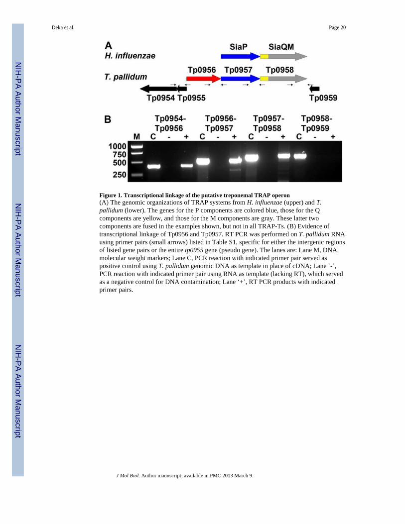
Figure 1. Transcriptional linkage of the putative treponemal TRAP operon(A) The genomic organizations of TRAP systems from H. influenzae (upper) and T.pallidum (lower). The genes for the P components are colored blue, those for the Qcomponents are yellow, and those for the M components are gray. These latter twocomponents are fused in the examples shown, but not in all TRAP-Ts. (B) Evidence oftranscriptional linkage of Tp0956 and Tp0957. RT PCR was performed on T. pallidum RNAusing primer pairs (small arrows) listed in Table S1, specific for either the intergenic regionsof listed gene pairs or the entire tp0955 gene (pseudo gene). The lanes are: Lane M, DNAmolecular weight markers; Lane C, PCR reaction with indicated primer pair served aspositive control using T. pallidum genomic DNA as template in place of cDNA; Lane ‘-’,PCR reaction with indicated primer pair using RNA as template (lacking RT), which servedas a negative control for DNA contamination; Lane ‘+’, RT PCR products with indicatedprimer pairs.
Deka et al. Page 20
J Mol Biol. Author manuscript; available in PMC 2013 March 9.
NIH
-PA Author Manuscript
NIH
-PA Author Manuscript
NIH
-PA Author Manuscript

Figure 2. The crystal structure of Tp0956(A) A topological diagram of Tp0956. The cylinders are α-helices, and are labeled with theirrespective alphabetical designations. The protein forms a ring with a pore running through it,but in this diagram, the ring as been opened and “flattened”, with the helices most proximalto the pore shown behind the others and color-coded pink. All other helices are colored red.This color-coding holds for parts B and C as well. (B) A “side view” of the protein, with theC, lateral, and N faces denoted. The model has been rotated approximately 90° around thehorizontal axis compared to that shown in (A). (C) A surface representation of the protein,viewed from the C face. (D) A cross-section through the protein, which has been coloredaccording to its electrostatic potential. The scale for the coloration is also shown. An area ofnegative potential on the lateral surface of the protein is circled.
Deka et al. Page 21
J Mol Biol. Author manuscript; available in PMC 2013 March 9.
NIH
-PA Author Manuscript
NIH
-PA Author Manuscript
NIH
-PA Author Manuscript

Figure 3. Pore characteristics of Tp0956(A) A ribbons representation of the protein with a surface representation of the pore. The N,middle, and C portions of the pore are colored blue, green, and pink, respectively. (B) Thehydrophobic antechamber (HA). A different view of the pore than that in (A) is shown, andthe HA is depicted as a yellow surface. Secondary structure is shown semi-transparently forclarity, and the α-helices that contribute residues to the HA are labeled. The HA surface wascalculated with CAVER 2.0 71.
Deka et al. Page 22
J Mol Biol. Author manuscript; available in PMC 2013 March 9.
NIH
-PA Author Manuscript
NIH
-PA Author Manuscript
NIH
-PA Author Manuscript
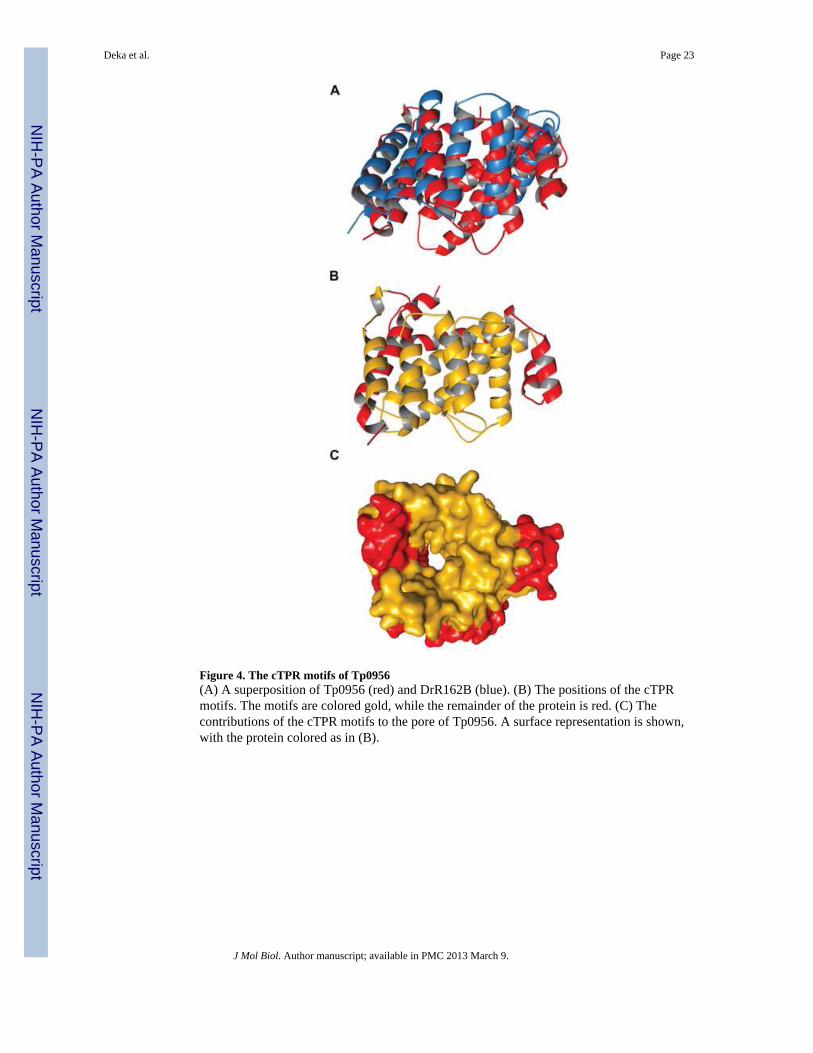
Figure 4. The cTPR motifs of Tp0956(A) A superposition of Tp0956 (red) and DrR162B (blue). (B) The positions of the cTPRmotifs. The motifs are colored gold, while the remainder of the protein is red. (C) Thecontributions of the cTPR motifs to the pore of Tp0956. A surface representation is shown,with the protein colored as in (B).
Deka et al. Page 23
J Mol Biol. Author manuscript; available in PMC 2013 March 9.
NIH
-PA Author Manuscript
NIH
-PA Author Manuscript
NIH
-PA Author Manuscript
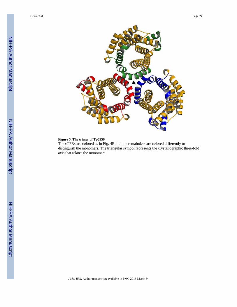
Figure 5. The trimer of Tp0956The cTPRs are colored as in Fig. 4B, but the remainders are colored differently todistinguish the monomers. The triangular symbol represents the crystallographic three-foldaxis that relates the monomers.
Deka et al. Page 24
J Mol Biol. Author manuscript; available in PMC 2013 March 9.
NIH
-PA Author Manuscript
NIH
-PA Author Manuscript
NIH
-PA Author Manuscript
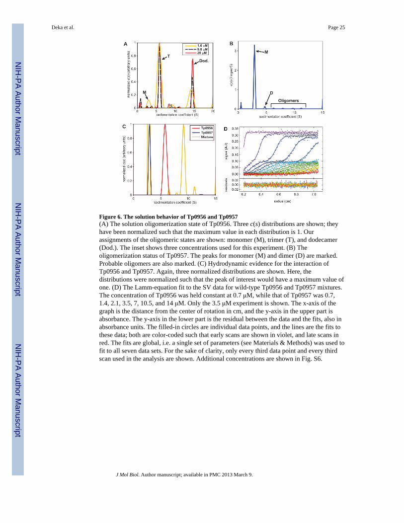
Figure 6. The solution behavior of Tp0956 and Tp0957(A) The solution oligomerization state of Tp0956. Three c(s) distributions are shown; theyhave been normalized such that the maximum value in each distribution is 1. Ourassignments of the oligomeric states are shown: monomer (M), trimer (T), and dodecamer(Dod.). The inset shows three concentrations used for this experiment. (B) Theoligomerization status of Tp0957. The peaks for monomer (M) and dimer (D) are marked.Probable oligomers are also marked. (C) Hydrodynamic evidence for the interaction ofTp0956 and Tp0957. Again, three normalized distributions are shown. Here, thedistributions were normalized such that the peak of interest would have a maximum value ofone. (D) The Lamm-equation fit to the SV data for wild-type Tp0956 and Tp0957 mixtures.The concentration of Tp0956 was held constant at 0.7 μM, while that of Tp0957 was 0.7,1.4, 2.1, 3.5, 7, 10.5, and 14 μM. Only the 3.5 μM experiment is shown. The x-axis of thegraph is the distance from the center of rotation in cm, and the y-axis in the upper part isabsorbance. The y-axis in the lower part is the residual between the data and the fits, also inabsorbance units. The filled-in circles are individual data points, and the lines are the fits tothese data; both are color-coded such that early scans are shown in violet, and late scans inred. The fits are global, i.e. a single set of parameters (see Materials & Methods) was used tofit to all seven data sets. For the sake of clarity, only every third data point and every thirdscan used in the analysis are shown. Additional concentrations are shown in Fig. S6.
Deka et al. Page 25
J Mol Biol. Author manuscript; available in PMC 2013 March 9.
NIH
-PA Author Manuscript
NIH
-PA Author Manuscript
NIH
-PA Author Manuscript
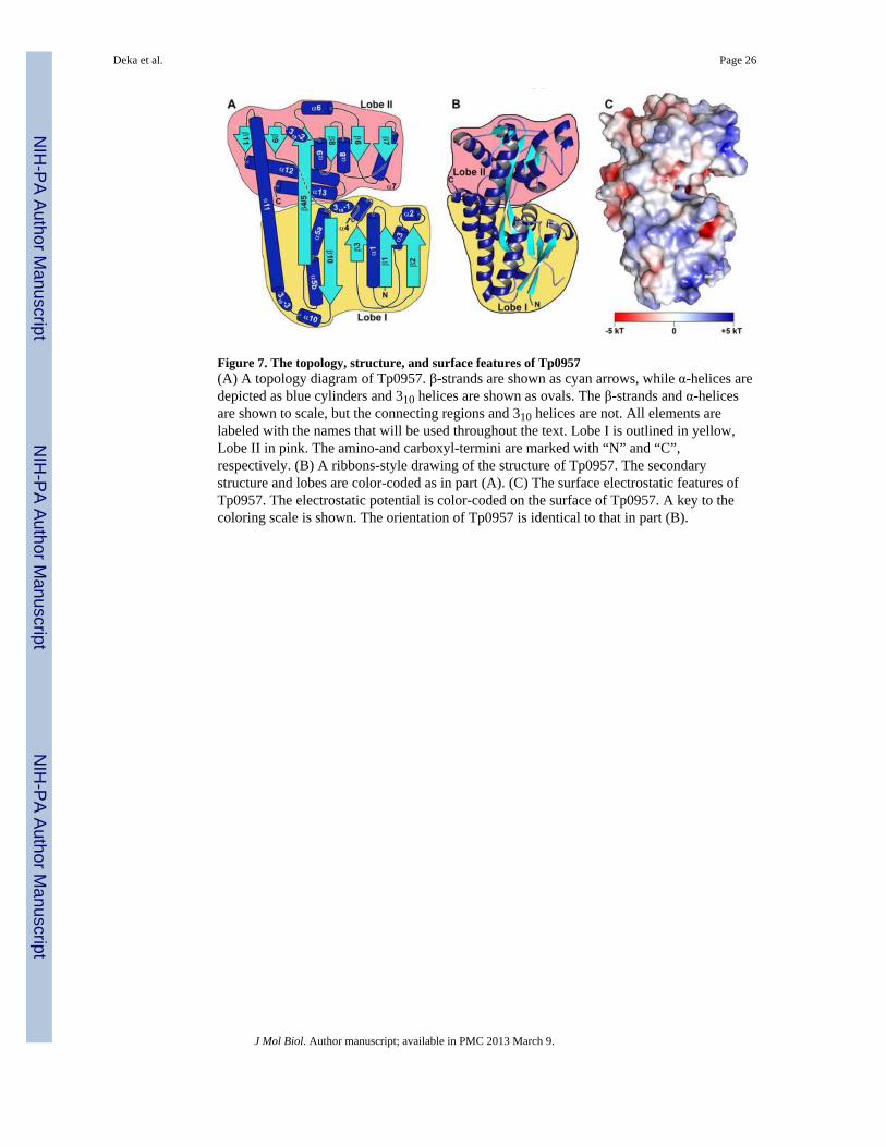
Figure 7. The topology, structure, and surface features of Tp0957(A) A topology diagram of Tp0957. β-strands are shown as cyan arrows, while α-helices aredepicted as blue cylinders and 310 helices are shown as ovals. The β-strands and α-helicesare shown to scale, but the connecting regions and 310 helices are not. All elements arelabeled with the names that will be used throughout the text. Lobe I is outlined in yellow,Lobe II in pink. The amino-and carboxyl-termini are marked with “N” and “C”,respectively. (B) A ribbons-style drawing of the structure of Tp0957. The secondarystructure and lobes are color-coded as in part (A). (C) The surface electrostatic features ofTp0957. The electrostatic potential is color-coded on the surface of Tp0957. A key to thecoloring scale is shown. The orientation of Tp0957 is identical to that in part (B).
Deka et al. Page 26
J Mol Biol. Author manuscript; available in PMC 2013 March 9.
NIH
-PA Author Manuscript
NIH
-PA Author Manuscript
NIH
-PA Author Manuscript
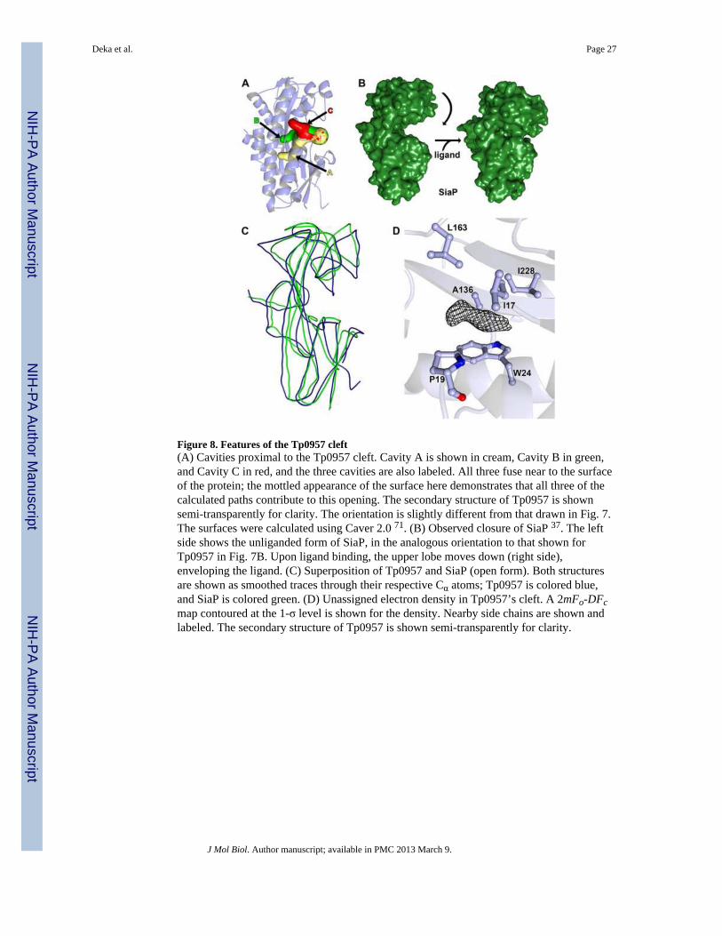
Figure 8. Features of the Tp0957 cleft(A) Cavities proximal to the Tp0957 cleft. Cavity A is shown in cream, Cavity B in green,and Cavity C in red, and the three cavities are also labeled. All three fuse near to the surfaceof the protein; the mottled appearance of the surface here demonstrates that all three of thecalculated paths contribute to this opening. The secondary structure of Tp0957 is shownsemi-transparently for clarity. The orientation is slightly different from that drawn in Fig. 7.The surfaces were calculated using Caver 2.0 71. (B) Observed closure of SiaP 37. The leftside shows the unliganded form of SiaP, in the analogous orientation to that shown forTp0957 in Fig. 7B. Upon ligand binding, the upper lobe moves down (right side),enveloping the ligand. (C) Superposition of Tp0957 and SiaP (open form). Both structuresare shown as smoothed traces through their respective Cα atoms; Tp0957 is colored blue,and SiaP is colored green. (D) Unassigned electron density in Tp0957’s cleft. A 2mFo-DFcmap contoured at the 1-σ level is shown for the density. Nearby side chains are shown andlabeled. The secondary structure of Tp0957 is shown semi-transparently for clarity.
Deka et al. Page 27
J Mol Biol. Author manuscript; available in PMC 2013 March 9.
NIH
-PA Author Manuscript
NIH
-PA Author Manuscript
NIH
-PA Author Manuscript
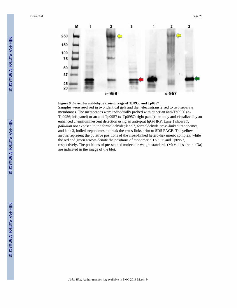
Figure 9. In vivo formaldehyde cross-linkage of Tp0956 and Tp0957Samples were resolved in two identical gels and then electrotransferred to two separatemembranes. The membranes were individually probed with either an anti-Tp0956 (α-Tp0956; left panel) or an anti-Tp0957 (α-Tp0957; right panel) antibody and visualized by anenhanced chemiluminescent detection using an anti-goat IgG-HRP. Lane 1 shows T.pallidum not exposed to the formaldehyde; lane 2, formaldehyde cross-linked treponemes,and lane 3, boiled treponemes to break the cross-links prior to SDS PAGE. The yellowarrows represent the putative positions of the cross-linked hetero-hexameric complex, whilethe red and green arrows denote the positions of monomeric Tp0956 and Tp0957,respectively. The positions of pre-stained molecular-weight standards (M; values are in kDa)are indicated in the image of the blot.
Deka et al. Page 28
J Mol Biol. Author manuscript; available in PMC 2013 March 9.
NIH
-PA Author Manuscript
NIH
-PA Author Manuscript
NIH
-PA Author Manuscript
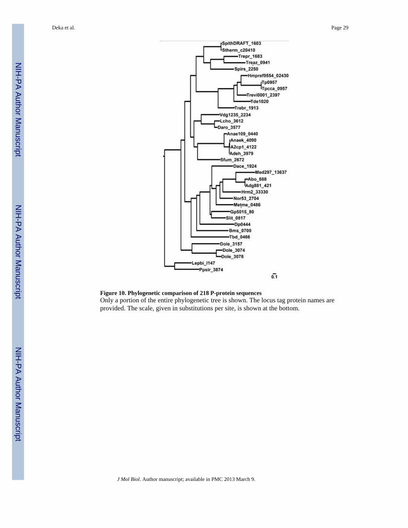
Figure 10. Phylogenetic comparison of 218 P-protein sequencesOnly a portion of the entire phylogenetic tree is shown. The locus tag protein names areprovided. The scale, given in substitutions per site, is shown at the bottom.
Deka et al. Page 29
J Mol Biol. Author manuscript; available in PMC 2013 March 9.
NIH
-PA Author Manuscript
NIH
-PA Author Manuscript
NIH
-PA Author Manuscript

NIH
-PA Author Manuscript
NIH
-PA Author Manuscript
NIH
-PA Author Manuscript
Deka et al. Page 30
Table 1Data collection, phasing and refinement statistics
Crystal Tp0956, Seapeak Tp0956, native Tp0957, native
Space group C2 I23 P21
Energy (eV) 12,687.7 12,682.7 12,675.0
Resolution range (Å) 34.1 - 2.30 (2.34- 2.30)
40.2 - 2.30(2.34 - 2.30)
37.8-1.40(1.42-1.40)
Unique reflections 86,064 (3,818)b 153,999 (769) 125,807(6,229)
Multiplicity 3.0 (2.3) 19.2 (14.3) 4.1 (3.6)
Data completeness (%) 91.8 (81.9) 99.9 (99.7) 99.8 (99.7)
Rmerge (%)b 6.3 (46.4) 7.7 (89.9) 3.7 (56.6)
I/σ(I) 25.1 (2.10) 42.7 (2.89) 33.7 (2.03)
Wilson B-value (Å2) 44.6 52.4 18.0
Anomalous scatterersselenium, 24 outof 24 possiblesites
None None
Figure of merit (90.5 - 2.30 Å) 0.13 N/A N/A
Figure of merit post-DM (90.5 -2.3 Å) 0.69 N/A N/A
Crystal Tp0956, native Tp0957, native
Resolution range (Å) 29.9 - 2.30(2.48 - 2.30)
37.8-1.40(1.41-1.40)
No. of reflections Rwork/Rfree15,250/758(2,868/146)
125,752/6,322(3,800/198)
Data completeness (%) 99.5 (100.0) 99.7 (97.0)
Atoms (non-Hprotein/solvent/ions) 2,153/61/20 5,634/431/0
Rwork (%) 19.7 (29.9) 17.8 (30.0)
Rfree (%) 24.2 (41.1) 20.1 (36.2)
R.m.s.d. bond length (Å) 0.008 0.009
R.m.s.d. bond angle (°) 1.0 1.2
Mean B-value (Å2)(protein/solvent/ions) 49.2/48.9/108.0 21.9/29.1/38.5
Ramachandran plot (%)
(favored/additional/disallowed)d98.9/1.1/0.0 99.0/1.0/0.0
Maximum likelihood coordinateerror 0.31 0.36
Missing residues A: 1-30A: 7-8, 65-66,326-328B: 7-8, 325-328
aBijvoet-pairs were kept separate for data processing
bData for the outermost shell are given in parentheses.
J Mol Biol. Author manuscript; available in PMC 2013 March 9.

NIH
-PA Author Manuscript
NIH
-PA Author Manuscript
NIH
-PA Author Manuscript
Deka et al. Page 31
cRmerge = 100 ΣhΣi|Ih, i—⟨Ih⟩|/ΣhΣiIh, i, where the outer sum (h) is over the unique reflections and the inner sum (i) is over the set of
independent observations of each unique reflection.
dAs defined by the validation suite MolProbity71.
J Mol Biol. Author manuscript; available in PMC 2013 March 9.

NIH
-PA Author Manuscript
NIH
-PA Author Manuscript
NIH
-PA Author Manuscript
Deka et al. Page 32
Tabl
e 2
Puta
tive
TPA
T T
-com
pone
nts a
nd th
eir
geno
mic
nei
ghbo
rs
Org
anis
m N
ame
Cla
ssL
ocus
Tag
of
Tp0
956-
like
Prot
ein
(T)
E-va
luea
Loc
us T
ag o
f Nea
rby
Dct
P-lik
e Pr
otei
n
Pres
ence
of D
ctM
and/
orD
ctQ
Trep
onem
a pa
llidu
mSp
iroch
aete
stp
0956
3e-9
9tp
0957
Yes
Trep
onem
a pa
ralu
iscu
nicu
liSp
iroch
aete
stp
cca_
0956
3e-9
9tp
cca_
0957
Yes
Trep
onem
a de
ntic
ola
Spiro
chae
tes
tde1
021
7e-9
2td
e102
0Y
es
Trep
onem
a ph
aged
enis
Spiro
chae
tes
hmpr
ef95
54_2
429
1e-8
7hm
pref
9554
_243
0Y
es
Trep
onem
a vi
ncen
tiiSp
iroch
aete
str
evi0
001_
2396
1e-8
7tr
evi0
001_
2397
Yes
Trep
onem
a pr
imiti
a ZA
S-2
Spiro
chae
tes
trep
r_16
829e
-85
trep
r_16
83Y
es
Verr
ucom
icro
biae
bac
teri
um D
G12
35V
erru
com
icro
biae
vdg1
235_
4783
2e-8
4vd
g123
5_22
34N
o
Trep
onem
a az
oton
utri
cum
ZA
S-9
Spiro
chae
tes
trea
z_09
403e
-83
trea
z_09
41Y
es
Trep
onem
a br
enna
bore
nse
DSM
121
68Sp
iroch
aete
str
ebr_
1914
2e-8
0tr
ebr_
1913
Yes
Alca
nivo
rax
bork
umen
sis S
K2
γ-pr
oteo
bact
eria
abo_
0687
2e-7
5ab
o_06
88Y
es
Synt
roph
obac
ter f
umar
oxid
ans M
POB
δ-pr
oteo
bact
eria
sfum
_267
33e
-75
sfum
_267
2Y
es
Spir
ocha
eta
ther
mop
hila
DSM
657
8Sp
iroch
aete
ssp
ithD
RAFT
_160
23e
-74
spith
DRA
FT_1
603
Yes
Anae
rom
yxob
acte
r deh
alog
enan
s 2C
P-C
δ-pr
oteo
bact
eria
adeh
_398
03e
-74
adeh
_397
9Y
es
Alca
nivo
rax
sp. D
G88
1γ-
prot
eoba
cter
iaad
g881
_311
34e
-72
adg8
81_4
21Y
es
Anae
rom
yxob
acte
r sp.
Fw
109-
5δ-
prot
eoba
cter
iaan
ae10
9_04
399e
-70
anae
109_
0440
Yes
Anae
rom
yxob
acte
r sp.
Kδ-
prot
eoba
cter
iaan
aeK
_409
14e
-69
anae
K_4
090
Yes
Anae
rom
yxob
acte
r deh
alog
enan
s 2C
P-1
δ-pr
oteo
bact
eria
a2cp
1_41
236e
-69
a2cp
1_41
22Y
es
Des
ulfo
tale
a ps
ychr
ophi
la L
Sv54
δ-pr
oteo
bact
eria
dp04
433e
-68
dp04
44Y
es
Spir
ocha
eta
smar
agdi
nae
DSM
112
93Sp
iroch
aete
ssp
irs_
2251
1e-6
7sp
irs_
2250
Yes
Side
roxy
dans
lith
otro
phic
us E
S-1
β-pr
oteo
bact
eria
slit_
0818
5e-6
6sl
it_08
17Y
es
Dec
hlor
omon
as a
rom
atic
a R
CB
β-pr
oteo
bact
eria
daro
_357
87e
-65
daro
_357
7Y
es
Des
ulfu
rom
onas
ace
toxi
dans
DSM
684
δ-pr
oteo
bact
eria
dace
_192
5da
ce_2
628
9e-6
34e
-33
dace
_192
4N
/AY
es No
Des
ulfo
bact
eriu
m a
utot
roph
icum
HR
M2
γ-pr
oteo
bact
eria
hrm
2_33
310
2e-6
2hr
m2_
3330
0Y
es
gam
ma
prot
eoba
cter
ium
HTC
C50
15δ-
prot
eoba
cter
iagp
5015
_96
2e-6
1gp
5015
_80
Yes
J Mol Biol. Author manuscript; available in PMC 2013 March 9.

NIH
-PA Author Manuscript
NIH
-PA Author Manuscript
NIH
-PA Author Manuscript
Deka et al. Page 33
Org
anis
m N
ame
Cla
ssL
ocus
Tag
of
Tp0
956-
like
Prot
ein
(T)
E-va
luea
Loc
us T
ag o
f Nea
rby
Dct
P-lik
e Pr
otei
n
Pres
ence
of D
ctM
and/
orD
ctQ
Des
ulfa
tibac
illum
alk
eniv
oran
s AK
-01
δ-pr
oteo
bact
eria
dalk
_268
8da
lk_2
187
4e-5
92e
-52
N/A
N/A
No
Yes
Rein
ekea
sp. M
ED29
7γ-
prot
eoba
cter
iam
ed29
7_13
632
3e-5
7m
ed29
7_13
637
Yes
Des
ulfo
cocc
us o
leov
oran
s Hxd
3δ-
prot
eoba
cter
iado
le_3
154
dole
_307
72e
-56
8e-5
4do
le_3
157
dole
_307
8, d
ole_
3074
Yes
Yes
gam
ma
prot
eoba
cter
ium
NO
R5-
3γ-
prot
eoba
cter
iano
r53_
2500
2e-5
6no
r53_
2704
Yes
Met
hylo
mon
as m
etha
nica
MC
09γ-
prot
eoba
cter
iam
etm
e_04
872e
-55
met
me_
0486
Yes
Thio
baci
llus d
enitr
ifica
ns A
TCC
252
59β-
prot
eoba
cter
iatb
d_04
652e
-55
tbd_
0466
Yes
Lept
othr
ix c
holo
dnii
SP-6
β-pr
oteo
bact
eria
lcho
_361
33e
-53
lcho
_361
2Y
es
Spir
ocha
eta
ther
mop
hila
DSM
619
2Sp
iroch
aete
sst
herm
_c20
400
6e-5
0st
herm
_c20
410
Yes
Bact
erio
vora
x m
arin
us S
Jδ-
prot
eoba
cter
iabm
s_06
992e
-44
bms_
0700
Yes
Ples
iocy
stis
pac
ifica
SIR
-1δ-
prot
eoba
cter
iapp
sir1
_387
511e
-38
ppsi
r1_3
8741
Yes
Lept
ospi
ra b
iflex
a se
rova
rPa
toc
stra
in ‘P
atoc
1 (P
aris
)’Sp
iroch
aete
sle
pbi_
I147
74e
-32
lepb
i_I1
476
Yes
a As d
efin
ed b
y PS
I-B
LAST
42; 6
7
J Mol Biol. Author manuscript; available in PMC 2013 March 9.