Nicotine induces cell proliferation, invasion and epithelial-mesenchymal transition in a variety of...
21
Nicotine induces cell proliferation, invasion and epithelial- mesenchymal transition in a variety of human cancer cell lines Piyali Dasgupta 1 , Wasia Rizwani, Smitha Pillai, Rebecca Kinkade, Michelle Kovacs, Shipra Rastogi 2 , Sarmistha Banerjee, Melanie Carless 3 , Esther Kim, Domenico Coppola, Eric Haura, and Srikumar Chellappan * H. Lee Moffitt Cancer Center and Research Institute, 12902 Magnolia Drive, Tampa, FL 33612. Abstract Cigarette smoking is strongly correlated with the onset of non-small cell lung cancer (NSCLC). Nicotine, an active component of cigarettes, has been found to induce proliferation of lung cancer cell lines. In addition, nicotine can induce angiogenesis and confer resistance to apoptosis. All these events are mediated through the nicotinic acetylcholine receptors (nAChRs) on lung cancer cells. In the present study we demonstrate that nicotine can promote anchorage-independent growth in NSCLCs. In addition, nicotine also induced morphological changes characteristic of a migratory, invasive phenotype in NSCLCs on collagen gel. These morphological changes were similar to those induced by the pro-migratory growth factor VEGF. The pro-invasive effects of nicotine were mediated by α7-nAChRs on NSCLCs. RT-PCR analysis showed that the α7-nAChRs were also expressed on human breast cancer and pancreatic cancer cell lines. Nicotine was found to promote proliferation and invasion in human breast cancer. The pro-invasive effects of nicotine were mediated via a nAChR, Src and calcium dependent signaling pathway in breast cancer cells. In a similar fashion nicotine could also induce proliferation and invasion of Aspc1 pancreatic cancer cells. Most importantly, nicotine could induce changes in gene expression consistent with epithelial to mesenchymal transition, characterized by reduction of epithelial markers like E-cadherin expression, ZO-1 staining and concomitant increase in levels of mesenchymal proteins like vimentin and fibronectin in human breast and lung cancer cells. Therefore, it is probable that the ability of nicotine to induce invasion and EMT may contribute to the progression of breast and lung cancers. Keywords lung cancer; nicotine; epithelial-mesenchymal transition; tumor progression; metastasis Introduction Cigarette smoke is the strongest documented risk factor for the development of lung cancer, accounting for about 157,000 deaths every year in the US 1. A significant proportion of non- small cell lung cancer cases are detected only in the advanced stage after the onset of metastasis, leading to a remarkably low 5-year survival rate (about 15%) in patients 1 . Tobacco derived carcinogens like 4-(methylnitrosamino)-1-(3-pyridyl)-1-butanone (NNK) and N’- nitrosonornicotine (NNN) are known to form DNA adducts, mutating vital growth regulatory *Corresponding author: Phone: (813) 745-6892, Fax: (813) 745-6748, [email protected]. 1 Current address: Dept. of Pharmacology, Physiology and Toxicology, Marshall University, BBSC, 1 John Marshall Drive, Huntington, WV 25755. 2 Dept. of Anatomy and Cell Biology, University of Western Ontario, London, Ontario, CANADA N6A 5C1. 3 Southwest Foundation for Biomedical Research, P.O. Box 760549, San Antonio, TX, 78245-0549, USA. NIH Public Access Author Manuscript Int J Cancer. Author manuscript; available in PMC 2010 February 22. Published in final edited form as: Int J Cancer. 2009 January 1; 124(1): 36–45. doi:10.1002/ijc.23894. NIH-PA Author Manuscript NIH-PA Author Manuscript NIH-PA Author Manuscript
-
Upload
independent -
Category
Documents
-
view
3 -
download
0
Transcript of Nicotine induces cell proliferation, invasion and epithelial-mesenchymal transition in a variety of...
Nicotine induces cell proliferation, invasion and epithelial-mesenchymal transition in a variety of human cancer cell lines
Piyali Dasgupta1, Wasia Rizwani, Smitha Pillai, Rebecca Kinkade, Michelle Kovacs, ShipraRastogi2, Sarmistha Banerjee, Melanie Carless3, Esther Kim, Domenico Coppola, EricHaura, and Srikumar Chellappan*H. Lee Moffitt Cancer Center and Research Institute, 12902 Magnolia Drive, Tampa, FL 33612.
AbstractCigarette smoking is strongly correlated with the onset of non-small cell lung cancer (NSCLC).Nicotine, an active component of cigarettes, has been found to induce proliferation of lung cancercell lines. In addition, nicotine can induce angiogenesis and confer resistance to apoptosis. All theseevents are mediated through the nicotinic acetylcholine receptors (nAChRs) on lung cancer cells. Inthe present study we demonstrate that nicotine can promote anchorage-independent growth inNSCLCs. In addition, nicotine also induced morphological changes characteristic of a migratory,invasive phenotype in NSCLCs on collagen gel. These morphological changes were similar to thoseinduced by the pro-migratory growth factor VEGF. The pro-invasive effects of nicotine weremediated by α7-nAChRs on NSCLCs. RT-PCR analysis showed that the α7-nAChRs were alsoexpressed on human breast cancer and pancreatic cancer cell lines. Nicotine was found to promoteproliferation and invasion in human breast cancer. The pro-invasive effects of nicotine were mediatedvia a nAChR, Src and calcium dependent signaling pathway in breast cancer cells. In a similar fashionnicotine could also induce proliferation and invasion of Aspc1 pancreatic cancer cells. Mostimportantly, nicotine could induce changes in gene expression consistent with epithelial tomesenchymal transition, characterized by reduction of epithelial markers like E-cadherin expression,ZO-1 staining and concomitant increase in levels of mesenchymal proteins like vimentin andfibronectin in human breast and lung cancer cells. Therefore, it is probable that the ability of nicotineto induce invasion and EMT may contribute to the progression of breast and lung cancers.
Keywordslung cancer; nicotine; epithelial-mesenchymal transition; tumor progression; metastasis
IntroductionCigarette smoke is the strongest documented risk factor for the development of lung cancer,accounting for about 157,000 deaths every year in the US 1. A significant proportion of non-small cell lung cancer cases are detected only in the advanced stage after the onset of metastasis,leading to a remarkably low 5-year survival rate (about 15%) in patients 1. Tobacco derivedcarcinogens like 4-(methylnitrosamino)-1-(3-pyridyl)-1-butanone (NNK) and N’-nitrosonornicotine (NNN) are known to form DNA adducts, mutating vital growth regulatory
*Corresponding author: Phone: (813) 745-6892, Fax: (813) 745-6748, [email protected] address: Dept. of Pharmacology, Physiology and Toxicology, Marshall University, BBSC, 1 John Marshall Drive, Huntington,WV 25755.2Dept. of Anatomy and Cell Biology, University of Western Ontario, London, Ontario, CANADA N6A 5C1.3Southwest Foundation for Biomedical Research, P.O. Box 760549, San Antonio, TX, 78245-0549, USA.
NIH Public AccessAuthor ManuscriptInt J Cancer. Author manuscript; available in PMC 2010 February 22.
Published in final edited form as:Int J Cancer. 2009 January 1; 124(1): 36–45. doi:10.1002/ijc.23894.
NIH
-PA Author Manuscript
NIH
-PA Author Manuscript
NIH
-PA Author Manuscript
genes like p53 and Ras, initiating oncogenesis2–4. In addition, several lines of evidenceindicate that cigarette smoking correlates with increased metastasis of lung, pancreatic, breastand bladder cancers 5–7. Although cigarette smoke is a complex mixture of over 4000compounds, nicotine has been shown the major addictive component of cigarettes 8–10.Nicotine, while not carcinogenic by itself, has been shown to induce proliferation andangiogenesis in several experimental models 11–14; these effects occurred at concentrationsnormally found in the blood stream of smokers (10−8 M to 10−7M) 12. These levels can varygreatly in smokers, and urine cotinine levels have been reported to range from 1500ng/ml to8000ng/ml 15–17.
The pathophysiological effects of nicotine are mediated by nicotinic acetylcholine receptors(nAChRs), which are expressed on neurons and neuromuscular junctions 8, 9, 18, 19; inaddition, they have been found to be expressed in a variety of non-neuronal cells includingendothelial cells and several histological types of lung tissue 20. Several convergent studiesshow that nicotine induces angiogenesis by upregulation of COX-2, prostacyclin, VEGFR-2,MMPs, uPA, e-NOS activity. Additionally, nicotine can exert its pro-angiogenic activity byincreasing the cellular levels of VEGF, bFGF and PDGF. The administration of nicotineincreased the severity of choroidal neovascularization and reversed VEGF-inducedsuppression of MMP-2 activity in mice 7, 21, 22. While there are insights into the molecularmechanisms by which nicotine induces angiogenenesis, however, the molecular mechanismsunderlying its role in tumor invasion and metastasis is not yet clear.
Clinical studies have shown an association between smoking and the development of pancreaticas well as breast cancers; further, patients who were smokers showed worse survival profilerelative to non-smokers 1, 23, 24. In the present study we show that nicotine can induce invasionand migration of NSCLC cells in a α7-receptor dependent and Src-dependent manner. Whereasthe pro-invasive effects of nicotine were mediated by α7-nAChRs in lung cancer cells, α7-nAChRs as well as DhβE-sensitive nAChRs mediated invasion of breast cancer cells. Nicotinewas also found to inhibit anoikis (loss of cell-substratum adhesion) in lung airway epithelialcells. Further, we demonstrate that nicotine induces changes in gene expression consistent withepithelial-mesenchymal transition (EMT)25–27; a signature of more advanced and lessdifferentiated cancer. Long-term treatment of lung cancer and breast cancer cells with 1µMnicotine was found to diminish levels of epithelial markers namely β-catenin and E-cadherin,and upregulate mesenchymal proteins like fibronectin and vimentin, indicative of disruptionof cell-cell contacts and increased motility. Our results suggest that nicotine plays a key rolein the regulation of the complex cellular cascades that modulate cell adhesion, invasion andmigration, leading to metastasis.
Materials and MethodsCell culture
A549, MDA-MB-468 and MCF-7 cells were cultured in F12K or DMEM (Mediatech Cellgro,Virginia) containing 10% FBS (Hyclone). Aspc1 cells were cultured in RPMI supplementedwith 10% FBS. Small airway epithelial cells (SAECs) were purchased from Clonetics andcultured in SAGM. The studies using anti-cancer drugs or signal transduction inhibitors weredone on cells that were rendered quiescent by serum starvation for 36 hours, following whichcells were treated with indicated concentrations of the drugs for 30 minutes. Thereafter, cellswere stimulated with 1µM nicotine (Sigma Chemical Company) in the presence or absence ofthe inhibitors for 18 hours. The concentrations of inhibitors used for the various experimentswere 10µM PP2, 1µM atropine, 10µM methylallylaconitine, 1µM α-lobeline, 10µMnifedipine, 1µM DhβE, 10µM α-bungarotoxin and 20µM hexamethonium bromide.
Dasgupta et al. Page 2
Int J Cancer. Author manuscript; available in PMC 2010 February 22.
NIH
-PA Author Manuscript
NIH
-PA Author Manuscript
NIH
-PA Author Manuscript
Anoikis AssaySmall airway epithelial cells were grown to 70–80% confluency in SAGM containing growthfactors. Cells were harvested by trypsinization and 105 cells were plated on 35mm culturedishes which were either uncoated or coated with polyhema (10mg/ml in ethanol).Subsequently one set of the uncoated or polyhema coated plates were stimulated with Nicotine(1µM) for 18 hours 28. Cells were collected from uncoated plates by trypsinization while inpolyhema coated plates non-adherent floating cells were collected using a pipet. After washingwith PBS, cells were suspended in 200µl PBS and Cytospin was used to collect cells on theslide by spinning at 500rpm for 15min. TUNEL assay was performed on the fixed cells usingPromega’s Dead End Colorimetric TUNEL system. TUNEL positive cells were counted in 4different fields and % positive cells were plotted.
Soft Agar Growth AssayAnchorage independent growth was assayed by the soft agar growth assay as describedelsewhere 29. The first step involves plating a bottom layer of 0.6% agar in serum-free mediain 12-well plates. The plates were incubated at room temperature for 30 minutes to solidify theagar. Cells were harvested by trypsinization and 2000 cells were mixed with 0.3% agarose(made in serum-free media) in the presence of the indicated nicotine concentration and layeredcarefully on the top of existing 0.6% agarose. Each treatment was done in triplicate. The plateswere covered with 1mL of medium supplemented with 10% FBS, in the presence or absenceof the indicated concentrations of nicotine and incubated at 37°C in a 5% CO2 incubator for 3weeks. Nicotine was added every 2 days to the test wells. The covering medium was replacedevery week. At the end of 3 weeks, cell colonies were stained with 10mg/ml MTT and colonieswith >0.1 mm in diameter were counted under a microscopic field at 40× magnification. Meancolony count was based on numbers from triplicate wells for each treatment condition and wasanalyzed using two-sided Student's t test.
Collagen gel culture3D collagen-1 gels were prepared on ice using equal volumes of 3mg/ml collagen solution and2× HEPES-buffered salt solution [50.4mM HEPES, pH 7.4, 162.6mM NaCl, 10.6mM KCl,88.2mM NaHCO3, 1.6 mM Na2HPO4, and 11mM D(+)-glucose] yielding a concentration of1.3 mg/ml following addition of culture medium 30, 31. The collagen gel solution (0.2 ml) wasadded to each well of the 96-well plate and allowed to set at 37°C for 30 minutes. A549 cellswere collected in a single cell suspension and added on the preformed collagen gel. The plateswere incubated at 37°C for 3 days, at which time complex structures had formed. The 3-Dstructures were visualized by phase contrast microscopy.
Invasion AssayThe invasive ability of Aspc-1, MCF-7, A549 and MDA-MB-468 cells was assayed accordingto the method reported before 32. Briefly, the upper surface of the filters were precoated withcollagen (100µg/filter). Matrigel was applied to the upper surface of the filters (50µg/filter)and dried in a hood. These filters were placed in Boyden chambers. Cells were grown to 70%confluency in respective media and were rendered quiescent by serum starvation, then treatedwith 1µM nicotine in the presence or absence of indicated inhibitors for 18 hours. The inhibitorswere added to the cells 30 minutes before the addition of nicotine. For some experiments cellstreated with 10% serum were used as the positive control. Following treatment, cells weretrypsinized and 7000 cells were plated in the upper chamber of the filter in media containing0.1% bovine serum albumin (Sigma), indicated inhibitors and nicotine. Media containing 20%fetal bovine serum was placed in the lower well as a chemoattractant and the chambers wereincubated at 37°C. After 18 hours, non-migrating cells on the upper surface of the filters wereremoved by wiping with cotton swabs. The filters were processed first by fixing in methanol
Dasgupta et al. Page 3
Int J Cancer. Author manuscript; available in PMC 2010 February 22.
NIH
-PA Author Manuscript
NIH
-PA Author Manuscript
NIH
-PA Author Manuscript
followed by staining with hematoxylin. The cells migrating on the other side of the filters werequantitated by counting three different fields under 40× magnification. Data presented is amean of three independent experiments.
Proliferation AssaysBromodeoxyuridine (BrdU) labeling kits were obtained from Roche Biochemicals. Cells wereplated in poly-D-lysine coated chamber slides at a density of 10,000 cells per well and renderedquiescent by serum starvation for 36 hours. Cells were then re-stimulated with 1µM nicotineor 10% FBS for 18 hours. S-phase cells were visualized by microscopy and quantitated bycounting 3 fields of 100 cells in quadruplicate. Data is presented as the percentage of BrdUpositive cells out of the 100 cells counted.
Lysate preparation and Western blottingLysates from cells treated with different agents were prepared by NP-40 lysis as describedearlier 11, 33 and 100µg protein was run on polyacrylamide-SDS gel. The proteins weretransferred to a nitrocellulose membrane and immunoblotted with antibodies corresponding tovarious EMT markers. Polyclonal β-catenin and fibronectin, monoclonal E-cadherin andVimentin antibodies were obtained from Santa Cruz Biotechnology. Monoclonal antibody toActin was purchased from Sigma Chemical Co. and polyclonal N-cadherin antibody fromAbcam. Western blots are representative of three independent experiments.
Wound Healing Assay10,000 Aspc-1 cells were plated and grown asynchronously to 90% confluency in a 6-wellplate (Falcon Becton Dickinson). These cells were starved in 0.1% fetal bovine serum for 24hours and then washed with 1× Dulbecco’s phosphate-buffered saline (MediaTech). The cellswere scratched with a sterile 200µl pipet tip in three separate places in each well and mediumcontaining 1µM nicotine or 10% fetal bovine serum was added in addition to the two wellsthat remained serum starved. After 24 hours, the wounds were observed and images were takenin 20× magnification. Similar experiments were performed with A549, MCF-7 and MDA-MB-468 cells, except that cells were not serum starved prior to inflicting the scratch. The cellswere treated with 0.01µM, 0.1µM or 1µM of nicotine in serum free media; media containing10% serum was used as a positive control. The data is representative of three independentexperiments.
Immunofluorescence and confocal microscopyA549, MCF-7 and MDA-MB-468 cells were plated onto poly-D-lysine-coated eight well glasschamber slides (7,000 cells per well) for immunostaining. Cells were rendered quiescent,followed by treatment with 1µM Nicotine for the indicated time points. The cells were fixedwith 10% buffered-formalin and double immunofluorescence was performed as per theprotocol published previously 34. Primary antibodies used were monoclonal E-cadherin, ZO-1(Abcam) and polyclonal Crm-1 (Santa Cruz) at 1:100 dilution. Secondary antibodies were goatanti-rabbit Alexa Fluor-546 and anti-mouse Alexa Fluor-488 (Molecular Probes) respectively.Control experiments demonstrated no cross-reactivity between anti-mouse secondary and anti-rabbit primary antibodies and vice-versa; nor were there any detectable staining by secondaryantibodies alone (data not shown). DAPI (Vector labs) was used to stain the nuclei. Cells werevisualized with a DM16000 inverted Leica TCS SP5 tandem scanning confocal microscopewith a 63×/1.40NA oil immersion objective. 405 diode, 488 Argon and 546 HeNe laser lineswere applied to excite the cells using AOBS line switching to minimize crosstalk betweenfluorochromes. Images were produced with three cooled photomultiplier detectors andanalyzed with the LAS AF software version 1.6.0 build 1016 (Leica Microsystems, Germany).
Dasgupta et al. Page 4
Int J Cancer. Author manuscript; available in PMC 2010 February 22.
NIH
-PA Author Manuscript
NIH
-PA Author Manuscript
NIH
-PA Author Manuscript
ResultsNicotine induces proliferation, migration and invasion of lung cancer cells
The proliferative effects of nicotine were examined in the bronchioalveolar lung carcinomacell line A549. Nicotine induced a dose-dependent increase in S-phase entry of quiescent A549cells, with the maximal proliferation being observed at 1µM nicotine (Figure 1A).12. Abilityof cells to grow independent of adhesion is a feature of cancer cells and enhanced capabilityof adherence-independent growth is a feature of advanced tumors. Since nicotine was able toincrease the proliferation of A549 cells grown on plastic, efforts were made to assess whetherit promoted adherence-independent growth as well. As shown in Figure 1B, treatment of cellswith nicotine promoted their growth in soft agar; nicotine treatment increased the number aswell as the size of the colonies. Given these results, Boyden Chamber assays were carried outto assess whether nicotine affects the invasive as well as migratory properties of A549 cells.It was found that nicotine could enhance the migration and invasion of A549 cells in a dose-dependent manner; the maximum effect was observed at 1µM and the effect was reduced at10µM (Figure 1C).
Several lines of evidence show that nicotine not only promotes tumor growth andneovascularization, but plays a role in tumorigenesis as well. Normal adherent cells undergoapoptosis shortly after loss of adhesion to substratum, a phenomenon known as anoikis 35. In-vitro-transformed cells and cancer-derived cells are able to survive and grow in the absence ofanchorage to the extracellular matrix (ECM) and their neighboring cells. This represents oneof the most important hallmarks of oncogenic transformation of normal cells 36, 37. Sincenicotine could enhance adherence-independent growth of cells in soft agar, experiments werecarried out to determine whether nicotine could promote anchorage-independent survival ofnormal lung epithelial cells. Primary small airway epithelial cells (SAECs) were grown onpolyhema coated plates to reduce adherence; these cells showed a significant increase inapoptosis compared to cells grown on regular plates. The treatment of the small airwayepithelial cells with 1µM nicotine induced resistance to anoikis when grown on polyhemacoated plates, showing reduced apoptosis (Figure 1D). This suggests that nicotine could notonly promote the proliferation of cells, but can also provide a survival advantage. Based onthese findings, attempts were made to assess whether nicotine altered the morphology of A549cells indicative of a more mesenchymal and migratory potential. Quiescent A549 cells weregrown as 3-D cultures on collagen and treated with 1µM nicotine or 100ng/ml (equivalent to2.38nM) VEGF. Figure 1E shows that the cells treated with nicotine acquired elongatedmigratory morphology relative to control cells. The angiogenic factor VEGF was taken as thepositive control for the assay and similar morphological changes were observed in both thecases. These results clearly show that nicotine can alter the proliferative, migratory and survivalproperties of cells.
Nicotine induces invasion of lung and breast cancer cells via nAChRsAbility to invade through a basement membrane and adjacent tissues is critical for metastasisof cancer cells. Since nicotine could promote the invasive properties of cells, we wanted todetermine the signaling pathways underlying the pro-invasive activity of nicotine. Towardsthis purpose, Boyden Chamber assays were done in the presence or absence of various receptorantagonists or signaling inhibitors. Nicotine-induced tumor cell invasion was completelyablated by the generalized nAChR antagonist hexamethonium, but unaffected by atropine, amuscarinic receptor antagonist. In particular, α7-nAChR antagonists α-bungarotoxin and MAAsignificantly abrogated the nicotine-stimulated invasion of A549 cells, whereas α-lobeline andDhβE did not have any effect (Figure 2A). Thus, the pro-invasive effects of nicotine in A549cells are primarily mediated by α7-nAChRs. Further, the stimulatory effects of nicotine onA549 invasion were suppressed by 1µM Iressa (gefitinib, EGFR inhibitor), 1µM PP2 (Src
Dasgupta et al. Page 5
Int J Cancer. Author manuscript; available in PMC 2010 February 22.
NIH
-PA Author Manuscript
NIH
-PA Author Manuscript
NIH
-PA Author Manuscript
inhibitor) and 1µM nifedipine (calcium channel inhibitor). These results show that the pro-invasive effects of nicotine on A549 cells involve α7-nAChR subunits, Src, EGFR activity aswell as calcium channel activity.
Since tobacco use has been implicated in the pathogenesis of several non-lung cancers likebreast cancer and pancreatic cancer 1, we examined the effect of nicotine on two human breastcancer cell lines. RT-PCR analysis shows the presence of α7 subunit of nAChRs in a varietyof breast and pancreatic cancer cell lines (Figure 2B). Given this result, we examined whethernicotine could cause S-phase entry in MCF-7 and MDA-MB-468 cells. BrdU assaysdemonstrate that the treatment of MCF-7 and MDA-MB-468 cells with varying doses ofnicotine induced a dose-dependent cell proliferation, the maximal proliferative effect beingobserved at 1µM nicotine in both cell lines (Figure 2C). Furthermore, 1µM nicotine couldinduce invasion of MCF-7 and MDA-MB-468 cells in Boyden chamber assays (Figure 2D)and promote colony formation on soft agar (Figure 2E). Invasion assays using various inhibitorsshow that the invasion of breast cancer cells in response to nicotine was mediated partially byα7-nAChR and DhβE-sensitive nAChRs (Figure 2F); this is probably due to the reducedamounts of α7 expression in these cells compared to A549 cells (Figure 2B). It can be imaginedthat DhβE-sensitive α3β4 subunits, which transmit survival signals 28,33, might be contributingto the invasion of the breast cancer cells.
Given that pancreatic cancer also shows correlation with smoking, experiments were carriedout to examine whether nicotine had similar effects on pancreatic cancer cells. It was foundthat nicotine induces proliferation of Aspc1 pancreatic cancer cells, similar to lung cancer andbreast cancer cell lines (Figure 3A); this induction was reduced by the Src kinase inhibitor PP2.This is similar to our earlier results on A549 cells 11, suggesting that the same pathways mediatenicotine-mediated proliferation of pancreatic cancer cells as well. Nicotine was also found topromote invasion of pancreatic cancer cells as measured by wound healing assays (Figure 3B).Boyden chamber assays showed that the pro-invasive activity of nicotine was abrogated by theSrc inhibitor PP2, suggesting an additional role of Src in both these processes (Figure 3C).These results show that multiple tumor types can respond to physiological concentrations ofnicotine by undergoing proliferation and that nicotine can enhance their invasive properties.
Nicotine promotes wound healing in vitro in a dose-dependent mannerGiven the results of the Boyden chamber assays and the observation that nicotine can promotemigration and wound-healing in pancreatic cancer cells, attempts were made to see whethersimilar effects are observed in NSCLC and breast cancer cells. Towards this purpose, A549,MCF-7 and MDA-MB-468 cells were grown to 90% confluency and the plates were scratchedwith a pipet tip. Cells were treated with 0.01µM, 0.1µM or 1µM nicotine for 24 hours; treatmentwith 10% serum was used as a positive control. It was found that nicotine could induce themigration of cells to the scratched area in a dose-dependent manner (Figure 4). While therewas only marginal invasion at 0.01µM nicotine, there were significantly more cells migratingto the wound when exposed to 0.1µM and 1µM nicotine. This result is in agreement with thedata obtained in Boyden chamber assays (Figure 2F).
Nicotine modulates the levels of multiple proteins involved in epithelial-to mesenchymaltransition
Epithelial to mesenchymal transition (EMT) is known to be one of the vital steps for theacquisition of malignant phenotype 25. This transition allows the cell to acquire migratoryproperties and allows metastasis to a new location. Epithelial-mesenchymal transition involvesthe repression of epithelial-specific adhesion molecules like E-cadherin and β-catenin with aconcomitant expression of proteins like fibronectin and vimentin 30. Since nicotine was foundto alter the invasive and migratory properties of cells, we wanted to examine whether long-
Dasgupta et al. Page 6
Int J Cancer. Author manuscript; available in PMC 2010 February 22.
NIH
-PA Author Manuscript
NIH
-PA Author Manuscript
NIH
-PA Author Manuscript

term treatment of cancer cells with nicotine could induce changes characteristic of the EMTprocess. Figure 5A shows that treatment of A549 cells with nicotine causes an increase in thelevels of mesenchymal proteins like fibronectin and vimentin with a concomitantdownregulation of epithelial markers E-cadherin and β-catenin. Interestingly, nicotine inducedEMT in breast cancer cell lines like MCF-7 and MDA-MB-468; this is significant sincesmoking has been correlated with increased metastasis of breast cancers. Experiments weredone to assess whether nicotine induced EMT in a dose-dependent fashion. Towards thispurpose, A549, MCF-7 or MDA-MB-468 cells were rendered quiescent by growing in 0.1%serum for 24 hours and stimulated with 0.01µM, 0.1µM or 1µM nicotine for 72 hours. It wasfound that the maximal effect of nicotine was observed at 1µM concentration while 0.1µMnicotine also induced EMT like changes (Figure 5B). The changes were less marginal at thelowest concentration tested. All the three cell lines exposed to nicotine showed a reduction inE-cadherin and β-catenin levels while levels of fibronectin, vimentin and N-cadherin wereelevated. This result shows that exposure to nicotine can induce EMT in a variety of humancancer cells.
These results were further confirmed by immunofluorescence experiments in A549, MCF-7and MDA-MB-468 cells (Figure 6). E-cadherin was localized on the outer cell membrane inall three cell lines (Figure 6, top panels); DAPI was used to stain the nuclei of the cells. It wasobserved that treatment with nicotine for 48 or 72 hours led to a significant reduction in thelevels of E-cadherin, in all three cell lines (Figure 6, middle and lower panels). These resultsconfirm that the epithelial adhesion molecule E-cadherin is indeed downregulated by nicotine,a molecular signature consistent with EMT.
Since induction of EMT facilitates detachment of contacts with adjacent cells, attempts weremade to assess whether nicotine affected the levels and localization of the tight junction protein,ZO-1. Towards this purpose, an immunofluorescence experiment was conducted on A549,MCF-7 and MDA-MB468 cells. Quiescent cells displayed membranous staining of ZO-1characteristic of epithelial tight junctions (Figure 7, top panel); Crm-1 was used as a markerfor protein levels. However, when the cells were treated with 1µM nicotine for 48 or 72 hours,diffuse ZO-1 staining was observed, characteristic of a migratory mesenchymal phenotype(Figure 7, middle and bottom panel) 26, 27. Further, the overall intensity of the ZO-1 stainingwas reduced. Taken together, our data indicates that exposure of lung cancer and breastcarcinoma cells to nicotine induce EMT like changes, which facilitate disruption of cell-cellcontacts and subsequently metastasis.
DiscussionThe present study shows that nicotine induces a dose-dependent increase in proliferation oflung cancer cells, breast cancer cells and pancreatic cancer cells via α7-nAChRs-mediatedsignal transduction pathways. These results are in agreement with our earlier studies as wellas recent studies from other labs that show the importance of the α7-receptor subunit innicotine-induced cell proliferation. Indeed, a recent study has shown that proliferation oftransplanted A549 cells in nude mice can be inhibited by α-cobratoxin, an α7 receptorantagonist10, 38. Nicotinic acetylcholine receptors particularly α7-nAChRs have been detectedin primary endothelial cells as well as human NSCLC cell lines A549, NCI-H23 and H1299;similarly, other labs have shown that these receptor subunits are expressed on human lungcancers 11, 20, 33, 39–42. Thus it appears that the presence of these receptor subunits and theirability to promote cell proliferation might contribute to the growth of tumors already initiatedby tobacco-specific carcinogens. The importance of α7-nAChRs in mediating thepathophysiological effects of nicotine is further reinforced by the fact that it is overexpressedon human NSCLC tumors relative to adjacent normal tissue. At the same time, recent studiesfrom the Minna lab had shown that the expression of α6 and β4 subunits is different on tumors
Dasgupta et al. Page 7
Int J Cancer. Author manuscript; available in PMC 2010 February 22.
NIH
-PA Author Manuscript
NIH
-PA Author Manuscript
NIH
-PA Author Manuscript
from smokers and non-smokers 43; this raises the possibility that other subunits might also becontributing to the growth of lung cancers in vivo. In addition, it has been reported thatvariations in chromosomal loci 15q24 and 15q25, which harbor genes for nicotinicacetylcholine receptors, correlate with nicotine dependence, lung cancer and peripheral arterialdisease 44–46. These and other studies lend support to the idea that exposure to nicotine andenhanced activation of nicotinic acetylcholine receptors contribute to lung cancer.
Apart from non-small cell lung cancer, cigarette smoking has been implicated in thepathogenesis of breast, gastric, colon and cervical cancers 1. Our results show that nicotine caninduce proliferation in a variety of cancer cell lines apart from NSCLC; furthermore, thisinduction appears to be through α7-nAChR subunits, which are expressed on these cell lines.Our findings seem to suggest that nAChRs may be an autocrine mitogenic signaling pathwayfacilitating the growth of several types of cancers in addition to lung cancer. Several lines ofevidence show that β-adrenergic receptors also mediate the proliferative, pro-angiogenic andanti-apoptotic effects of nicotine, mainly in non-lung cancer cells. The mitogenic andproangiogenic effects of nicotine have been found to be mediated by β-adrenergic receptors incolon and gastric cancer cells 47, 48. Studies by Shin et al. (2007) indicate that nicotinepromotes growth of gastric cancers via PKC, ERK1/2 phosphorylation, and COX-2 activationin a β-adrenergic receptor-dependent fashion 49. Further, data by Wong et al., (2007)demonstrate that nicotine-induced proliferation of HT-29 colon cancer cells are mediated byboth α7-nAChRs and β-adrenergic receptors 47. Their findings show that nicotine binds toα7-nAChRs nicotine on the membranes of HT-29 colon cancer cells and thereby facilitatesdownstream production of adrenaline and β-adrenergic activation; thus β-adrenergic receptorscontribute to the proliferation indirectly. These data reveal that the both α7-nAChRs and β-adrenergic receptors contribute to the mitogenic effects of nicotine in colon cancer cells. Theproliferative effects of the tobacco carcinogen NNK [4-(methylnitrosamino)-1-(3-pyridyl)-1-butanone] have been found to be mediated by β-adrenergic receptors on humanadenocarcinomas of the lungs, pancreas, and breast 40, 50. Thus it is possible that β-adrenergicreceptors are also contributing to the observed changes, either directly or indirectly.
Multiple lines of evidence indicate that cigarette smoking not only facilitates proliferation ofcancer cells but might also promote their metastatic spread as well 51–53. Invasion andmetastasis are complex, multi-step processes that involve alteration of cell adhesion toextracellular matrix proteins as well as disruption of cell-cell junctions. Nicotine has beenshown to promote phosphorylation of calpains, upregulation of COX2, VEGF and VEGFR2;these molecules are known to affect the metastasic process 54–56. Our findings shed light onadditional pathways that contribute to the pro-invasive effects of nicotine. These include α7-nAChR-mediated activation of Src, calcium channels and upregulation of EGFR. Thesefindings raise the possibility that inhibition of α7 nAChR activity by non-toxic agents orinhibition of its downstream mediators might open novel avenues for the therapy of cancerspromoted by smoking 57.
Our data further shows that sustained exposure to nicotine promotes anchorage-independentgrowth by downregulation of anoikis. Ability to survive independent of a substratum is a featureof cancer cells that is indispensable for metastasis 58. Anoikis prevents normal cells fromdetaching from their substratum and migrating to different locations; the ability of nicotine toenhance the survival of cancer cells independent of a substratum might be contributingsignificantly to the ability of these cells to detach and find alternate sites of attachment. Indeed,nicotine could enhance adherence-independent proliferation of tumor cells lines, showing thatthe proliferative and survival advantages it provides allows the cells to grow robustlyindependent of a substratum.
Dasgupta et al. Page 8
Int J Cancer. Author manuscript; available in PMC 2010 February 22.
NIH
-PA Author Manuscript
NIH
-PA Author Manuscript
NIH
-PA Author Manuscript
Clinical and epidemiological studies have suggested that smokers tend to have more progressedand metastatic cancer than non-smokers 23. Further, smokers had enhanced metastasis of breastcancers to the lung 51, 53. Our findings suggest that nicotine induces changes consistent withEMT is highly relevant in this context. Indeed, earlier studies had suggested that long-termexposure to nicotine could alter the phenotype of epithelial and endothelial cells 12, 28. Weprovide the molecular changes that facilitate these morphological changes. Results presentedhere show that chronic treatment with nicotine resulted in the downregulation of ECM proteinsE-cadherin and β-catenin, with concomitant increase of fibronectin and vimentin. Severalstudies show that the decrease in E-cadherin and β-catenin with a concurrent increase infibronectin and vimentin levels is one of the hallmarks of EMT in lung cancer cells. Further,clinical studies also report that smoking decreases levels of E-cadherin in lung tumors andmight be contributing to resistance to chemotherapeutic agents 33. It is probable that apart fromits pro-invasive activity, nicotine plays a role in promoting EMT via downregulation of ECMproteins.
The results presented here suggest that while nicotine is not a carcinogen by itself , it has thepotential to promote the growth and progression of tumors. The ability of nicotine to promoteproliferation, angiogenesis, adherence-independent growth, and EMT while inhibiting anoikismight be contributing significantly to the growth and metastasis of tumors that are sensitive tonicotinic acetylcholine receptors. Further, these results also show that exposure to nicotine canaffect tumors of tissues other than that of the lung. Development of agents that can disrupt thenAChR signaling pathway might prove beneficial in the treatment of such cancers.
AcknowledgmentsWe thank the Analytical Microscopy and Histopathology Core Facilities at Moffitt Cancer Center for their assistanceand George Simon for helpful discussions. These studies were supported by the Bankhead-Coley Grant #06BB-04-9587 to SC. RK is a recipient of a pre-doctoral fellowship from the American Heart Association.
References1. ACS. Cancer facts and figures. American Cancer Society. 20022. Hecht SS. Cigarette smoking and lung cancer: chemical mechanisms and approaches to prevention.
Lancet Oncol 2002;3(8):461–469. Available from http://www.ncbi.nlm.nih.gov/entrez/query.fcgi?cmd=Retrieve&db=PubMed&dopt=Citation&list_uids=12147432. [PubMed: 12147432]
3. Hecht SS, Abbaspour A, Hoffman D. A study of tobacco carcinogenesis. XLII. Bioassay in A/J miceof some structural analogues of tobacco-specific nitrosamines. Cancer Lett 1988;42(1–2):141–145.Available from http://www.ncbi.nlm.nih.gov/entrez/query.fcgi?cmd=Retrieve&db=PubMed&dopt=Citation&list_uids=3180033. [PubMed: 3180033]
4. Sekido Y, Fong KM, Minna JD. Molecular genetics of lung cancer. Annu Rev Med 2003;54:73–87.[PubMed: 12471176]
5. Arredondo J, Chernyavsky AI, Grando SA. Nicotinic receptors mediate tumorigenic action of tobacco-derived nitrosamines on immortalized oral epithelial cells. Cancer Biol Ther 2006;5(5):511–517.Available from http://www.ncbi.nlm.nih.gov/entrez/query.fcgi?cmd=Retrieve&db=PubMed&dopt=Citation&list_uids=16582591. [PubMed: 16582591]
6. Bose C, Zhang H, Udupa KB, Chowdhury P. Activation of p-ERK1/2 by nicotine in pancreatic tumorcell line AR42J: effects on proliferation and secretion. Am J Physiol Gastrointest Liver Physiol2005;289(5):G926–G934. Available from http://www.ncbi.nlm.nih.gov/entrez/query.fcgi?cmd=Retrieve&db=PubMed&dopt=Citation&list_uids=16051920. [PubMed: 16051920]
7. Shin VY, Wu WK, Chu KM, Wong HP, Lam EK, Tai EK, Koo MW, Cho CH. Nicotine inducescyclooxygenase-2 and vascular endothelial growth factor receptor-2 in association with tumor-associated invasion and angiogenesis in gastric cancer. Mol Cancer Res 2005;3(11):607–615.Available from http://www.ncbi.nlm.nih.gov/entrez/query.fcgi?cmd=Retrieve&db=PubMed&dopt=Citation&list_uids=16317086. [PubMed: 16317086]
Dasgupta et al. Page 9
Int J Cancer. Author manuscript; available in PMC 2010 February 22.
NIH
-PA Author Manuscript
NIH
-PA Author Manuscript
NIH
-PA Author Manuscript
http://www.ncbi.nlm.nih.gov/entrez/query.fcgi?cmd=Retrieve&db=PubMed&dopt=Citation&list_uids=3180033
8. Lindstrom J. Neuronal nicotinic acetylcholine receptors. Ion Channels 1996;4:377–450. Availablefrom http://www.ncbi.nlm.nih.gov/entrez/query.fcgi?cmd=Retrieve&db=PubMed&dopt=Citation&list_uids=8744214. [PubMed: 8744214]
9. Lindstrom J. Nicotinic acetylcholine receptors in health and disease. Mol Neurobiol 1997;15(2):193–222. Available from http://www.ncbi.nlm.nih.gov/entrez/query.fcgi?cmd=Retrieve&db=PubMed&dopt=Citation&list_uids=9396010. [PubMed: 9396010]
10. Trombino S, Bisio A, Catassi A, Cesario A, Falugi C, Russo P. Role of the non-neuronal humancholinergic system in lung cancer and mesothelioma: possibility of new therapeutic strategies. CurrMed Chem Anticancer Agents 2004;4(6):535–542. Available from http://www.ncbi.nlm.nih.gov/entrez/query.fcgi?cmd=Retrieve&db=PubMed&dopt=Citation&list_uids=15579018. [PubMed:15579018]
11. Dasgupta P, Rastogi S, Pillai S, Ordonez-Ercan D, Morris M, Haura E, Chellappan S. Nicotine inducescell proliferation by beta-arrestin-mediated activation of Src and Rb-Raf-1 pathways. J Clin Invest2006;116(8):2208–2217. Available from http://www.ncbi.nlm.nih.gov/entrez/query.fcgi?cmd=Retrieve&db=PubMed&dopt=Citation&list_uids=16862215. [PubMed: 16862215]
12. Heeschen C, Jang JJ, Weis M, Pathak A, Kaji S, Hu RS, Tsao PS, Johnson FL, Cooke JP. Nicotinestimulates angiogenesis and promotes tumor growth and atherosclerosis. Nature Medicine 2001;7(7):833–839.
13. Heeschen C, Weis M, Aicher A, Dimmler S, Cooke JP. A novel angiogenic pathway mediated bynon-neuronal nicotinic acetylcholine receptors. J. Clin. Invest 2002;110:527–536. [PubMed:12189247]
14. Gazdar AF. Environmental tobacco smoke, carcinogenesis, and angiogenesis: a double whammy?Cancer Cell 2003;4(3):159–160. Available from http://www.ncbi.nlm.nih.gov/entrez/query.fcgi?cmd=Retrieve&db=PubMed&dopt=Citation&list_uids=14522247. [PubMed: 14522247]
15. Mathai M, Skinner A, Lawton K, Weindling AM. Maternal smoking, urinary cotinine levels andbirth-weight. Aust N Z J Obstet Gynaecol 1990;30(1):33–36. Available from http://www.ncbi.nlm.nih.gov/entrez/query.fcgi?cmd=Retrieve&db=PubMed&dopt=Citation&list_uids=2346450. [PubMed: 2346450]
16. Matsukura S, Taminato T, Kitano N, Seino Y, Hamada H, Uchihashi M, Nakajima H, Hirata Y.Effects of environmental tobacco smoke on urinary cotinine excretion in nonsmokers. Evidence forpassive smoking. N Engl J Med 1984;311(13):828–832. Available from http://www.ncbi.nlm.nih.gov/entrez/query.fcgi?cmd=Retrieve&db=PubMed&dopt=Citation&list_uids=6472384. [PubMed: 6472384]
17. Thompson SG, Stone R, Nanchahal K, Wald NJ. Relation of urinary cotinine concentrations tocigarette smoking and to exposure to other people's smoke. Thorax 1990;45(5):356–361. Availablefrom http://www.ncbi.nlm.nih.gov/entrez/query.fcgi?cmd=Retrieve&db=PubMed&dopt=Citation&list_uids=2382242. [PubMed: 2382242]
18. Itier V, Bertrand D. Neuronal nicotinic receptors: from protein structure to function. FEBS Lett2001;504(3):118–125. Available from http://www.ncbi.nlm.nih.gov/entrez/query.fcgi?cmd=Retrieve&db=PubMed&dopt=Citation&list_uids=11532443. [PubMed: 11532443]
19. Gotti C, Clementi F. Neuronal nicotinic receptors: from structure to pathology. Prog Neurobiol2004;74(6):363–396. Available from http://www.ncbi.nlm.nih.gov/entrez/query.fcgi?cmd=Retrieve&db=PubMed&dopt=Citation&list_uids=15649582. [PubMed: 15649582]
20. Schuller HM, Plummer HK 3rd, Jull BA. Receptor-mediated effects of nicotine and its nitrosatedderivative NNK on pulmonary neuroendocrine cells. Anat Rec 2003;270A(1):51–58. Available fromhttp://www.ncbi.nlm.nih.gov/entrez/query.fcgi?cmd=Retrieve&db=PubMed&dopt=Citation&list_uids=12494489.
21. Cook JP, Bitterman H. Nicotine and angiogenesis: a new paradigm for tobacco-related diseases. AnnMed 2004;36:33–40. [PubMed: 15000345]
22. Shin VY, Wu WK, Ye YN, So WH, Koo MW, Liu ES, Luo JC, Cho CH. Nicotine promotes gastrictumor growth and neovascularization by activating extracellular signal-regulated kinase andcyclooxygenase-2. Carcinogenesis 2004;25(12):2487–2495. Available from http://www.ncbi.nlm.nih.gov/entrez/query.fcgi?cmd=Retrieve&db=PubMed&dopt=Citation&list_uids=15319299. [PubMed: 15319299]
Dasgupta et al. Page 10
Int J Cancer. Author manuscript; available in PMC 2010 February 22.
NIH
-PA Author Manuscript
NIH
-PA Author Manuscript
NIH
-PA Author Manuscript
http://www.ncbi.nlm.nih.gov/entrez/query.fcgi?cmd=Retrieve&db=PubMed&dopt=Citation&list_uids=8744214
http://www.ncbi.nlm.nih.gov/entrez/query.fcgi?cmd=Retrieve&db=PubMed&dopt=Citation&list_uids=8744214
http://www.ncbi.nlm.nih.gov/entrez/query.fcgi?cmd=Retrieve&db=PubMed&dopt=Citation&list_uids=9396010
http://www.ncbi.nlm.nih.gov/entrez/query.fcgi?cmd=Retrieve&db=PubMed&dopt=Citation&list_uids=9396010
http://www.ncbi.nlm.nih.gov/entrez/query.fcgi?cmd=Retrieve&db=PubMed&dopt=Citation&list_uids=2346450
http://www.ncbi.nlm.nih.gov/entrez/query.fcgi?cmd=Retrieve&db=PubMed&dopt=Citation&list_uids=2346450
http://www.ncbi.nlm.nih.gov/entrez/query.fcgi?cmd=Retrieve&db=PubMed&dopt=Citation&list_uids=2346450
http://www.ncbi.nlm.nih.gov/entrez/query.fcgi?cmd=Retrieve&db=PubMed&dopt=Citation&list_uids=6472384
http://www.ncbi.nlm.nih.gov/entrez/query.fcgi?cmd=Retrieve&db=PubMed&dopt=Citation&list_uids=6472384
http://www.ncbi.nlm.nih.gov/entrez/query.fcgi?cmd=Retrieve&db=PubMed&dopt=Citation&list_uids=6472384
http://www.ncbi.nlm.nih.gov/entrez/query.fcgi?cmd=Retrieve&db=PubMed&dopt=Citation&list_uids=2382242
23. Richardson GE, Tucker MA, Venzon DJ, Linnoila RI, Phelps R, Phares JC, Edison M, Ihde DC,Johnson BE. Smoking cessation after successful treatment of small-cell lung cancer is associatedwith fewer smoking-related second primary cancers. Ann Intern Med 1993;119(5):383–390.Available from http://www.ncbi.nlm.nih.gov/entrez/query.fcgi?cmd=Retrieve&db=PubMed&dopt=Citation&list_uids=8393311. [PubMed: 8393311]
24. Vander Ark W, DiNardo LJ, Oliver DS. Factors affecting smoking cessation in patients with headand neck cancer. Laryngoscope 1997;107(7):888–892. Available from http://www.ncbi.nlm.nih.gov/entrez/query.fcgi?cmd=Retrieve&db=PubMed&dopt=Citation&list_uids=9217125. [PubMed:9217125]
25. Huber MA, Kraut N, Beug H. Molecular requirements for epithelial-mesenchymal transition duringtumor progression. Curr Opin Cell Biol 2005;17(5):548–558. Available from http://www.ncbi.nlm.nih.gov/entrez/query.fcgi?cmd=Retrieve&db=PubMed&dopt=Citation&list_uids=16098727. [PubMed: 16098727]
26. Yang J, Mani SA, Donaher JL, Ramaswamy S, Itzykson RA, Come C, Savagner P, Gitelman I,Richardson A, Weinberg RA. Twist, a master regulator of morphogenesis, plays an essential role intumor metastasis. Cell 2004;117(7):927–939. Available from http://www.ncbi.nlm.nih.gov/entrez/query.fcgi?cmd=Retrieve&db=PubMed&dopt=Citation&list_uids=15210113. [PubMed:15210113]
27. Yang J, Mani SA, Weinberg RA. Exploring a new twist on tumor metastasis. Cancer Res 2006;66(9):4549–4552. Available from http://www.ncbi.nlm.nih.gov/entrez/query.fcgi?cmd=Retrieve&db=PubMed&dopt=Citation&list_uids=16651402. [PubMed: 16651402]
28. West KA, Brognard J, Clark AS, Linnoila IR, Yang X, Swain SM, Harris C, Belinsky S, Dennis PA.Rapid Akt activation by nicotine and a tobacco carcinogen modulates the phenotype of normal humanairway epithelial cells. J Clin Invest 2003;111(1):81–90. Available from http://www.ncbi.nlm.nih.gov/entrez/query.fcgi?cmd=Retrieve&db=PubMed&dopt=Citation&list_uids=12511591. [PubMed: 12511591]
29. Blaskovich MA, Lin Q, Delarue FL, Sun J, Park HS, Coppola D, Hamilton AD, Sebti SM. Design ofGFB-111, a platelet-derived growth factor binding molecule with antiangiogenic and anticanceractivity against human tumors in mice. Nat Biotechnol 2000;18(10):1065–1070. Available fromhttp://www.ncbi.nlm.nih.gov/entrez/query.fcgi?cmd=Retrieve&db=PubMed&dopt=Citation&list_uids=11017044. [PubMed: 11017044]
30. Janda E, Lehmann K, Killisch I, Jechlinger M, Herzig M, Downward J, Beug H, Grunert S. Ras andTGF[beta] cooperatively regulate epithelial cell plasticity and metastasis: dissection of Ras signalingpathways. J Cell Biol 2002;156(2):299–313. Available from http://www.ncbi.nlm.nih.gov/entrez/query.fcgi?cmd=Retrieve&db=PubMed&dopt=Citation&list_uids=11790801. [PubMed:11790801]
31. Janda E, Litos G, Grunert S, Downward J, Beug H. Oncogenic Ras/Her-2 mediate hyperproliferationof polarized epithelial cells in 3D cultures and rapid tumor growth via the PI3K pathway. Oncogene2002;21(33):5148–5159. Available from http://www.ncbi.nlm.nih.gov/entrez/query.fcgi?cmd=Retrieve&db=PubMed&dopt=Citation&list_uids=12140765. [PubMed: 12140765]
32. Vukanovic J, Passaniti A, Hirata T, Traystman RJ, Hartley-Asp B, Isaacs JT. Antiangiogenic effectsof the quinoline-3-carboxamide linomide. Cancer Res 1993;53(8):1833–1837. Available from http://www.ncbi.nlm.nih.gov/entrez/query.fcgi?cmd=Retrieve&db=PubMed&dopt=Citation&list_uids=7682157. [PubMed: 7682157]
33. Dasgupta P, Kinkade R, Joshi B, Decook C, Haura E, Chellappan S. Nicotine inhibits apoptosisinduced by chemotherapeutic drugs by up-regulating XIAP and survivin. Proc Natl Acad Sci U S A2006;103(16):6332–6337. Available from http://www.ncbi.nlm.nih.gov/entrez/query.fcgi?cmd=Retrieve&db=PubMed&dopt=Citation&list_uids=16601104. [PubMed: 16601104]
34. Rastogi S, Joshi B, Dasgupta P, Morris M, Wright K, Chellappan S. Prohibitin facilitates cellularsenescence by recruiting specific corepressors to inhibit E2F target genes. Mol Cell Biol 2006;26(11):4161–4171. Available from http://www.ncbi.nlm.nih.gov/entrez/query.fcgi?cmd=Retrieve&db=PubMed&dopt=Citation&list_uids=16705168. [PubMed: 16705168]
35. Gilmore AP. Anoikis. Cell Death Differ 2005;12:1473–1477. Available from http://www.ncbi.nlm.nih.gov/entrez/query.fcgi?cmd=Retrieve&db=PubMed&dopt=Citation&list_uids=16247493. [PubMed: 16247493]
Dasgupta et al. Page 11
Int J Cancer. Author manuscript; available in PMC 2010 February 22.
NIH
-PA Author Manuscript
NIH
-PA Author Manuscript
NIH
-PA Author Manuscript
http://www.ncbi.nlm.nih.gov/entrez/query.fcgi?cmd=Retrieve&db=PubMed&dopt=Citation&list_uids=8393311
http://www.ncbi.nlm.nih.gov/entrez/query.fcgi?cmd=Retrieve&db=PubMed&dopt=Citation&list_uids=8393311
http://www.ncbi.nlm.nih.gov/entrez/query.fcgi?cmd=Retrieve&db=PubMed&dopt=Citation&list_uids=9217125
http://www.ncbi.nlm.nih.gov/entrez/query.fcgi?cmd=Retrieve&db=PubMed&dopt=Citation&list_uids=9217125
http://www.ncbi.nlm.nih.gov/entrez/query.fcgi?cmd=Retrieve&db=PubMed&dopt=Citation&list_uids=7682157
http://www.ncbi.nlm.nih.gov/entrez/query.fcgi?cmd=Retrieve&db=PubMed&dopt=Citation&list_uids=7682157
36. Frisch SM. Anoikis. Methods Enzymol 2000;322:472–479. Available from http://www.ncbi.nlm.nih.gov/entrez/query.fcgi?cmd=Retrieve&db=PubMed&dopt=Citation&list_uids=10914040. [PubMed: 10914040]
37. Frisch SM, Screaton RA. Anoikis mechanisms. Curr Opin Cell Biol 2001;13(5):555–562. Availablefrom http://www.ncbi.nlm.nih.gov/entrez/query.fcgi?cmd=Retrieve&db=PubMed&dopt=Citation&list_uids=11544023. [PubMed: 11544023]
38. Trombino S, Cesario A, Margaritora S, Granone P, Motta G, Falugi C, Russo P. Alpha7-nicotinicacetylcholine receptors affect growth regulation of human mesothelioma cells: role of mitogen-activated protein kinase pathway. Cancer Res 2004;64(1):135–145. Available from http://www.ncbi.nlm.nih.gov/entrez/query.fcgi?cmd=Retrieve&db=PubMed&dopt=Citation&list_uids=14729617. [PubMed: 14729617]
39. Schuller HM, Orloff M. Tobacco-specific carcinogenic nitrosamines. Ligands for nicotinicacetylcholine receptors in human lung cancer cells. Biochem Pharmacol 1998;55(9):1377–1384.Available from http://www.ncbi.nlm.nih.gov/entrez/query.fcgi?cmd=Retrieve&db=PubMed&dopt=Citation&list_uids=10076528. [PubMed: 10076528]
40. Schuller HM, Tithof PK, Williams M, Plummer H 3rd. The tobacco-specific carcinogen 4-(methylnitrosamino)-1-(3-pyridyl)-1-butanone is a beta-adrenergic agonist and stimulates DNAsynthesis in lung adenocarcinoma via beta-adrenergic receptor-mediated release of arachidonic acid.Cancer Res 1999;59(18):4510–4515. Available from http://www.ncbi.nlm.nih.gov/entrez/query.fcgi?cmd=Retrieve&db=PubMed&dopt=Citation&list_uids=10493497. [PubMed:10493497]
41. Minna JD. The molecular biology of lung cancer pathogenesis. Chest 1993;103(4 Suppl):449S–456S.Available from http://www.ncbi.nlm.nih.gov/entrez/query.fcgi?cmd=Retrieve&db=PubMed&dopt=Citation&list_uids=8462339. [PubMed: 8462339]
42. Minna JD. Nicotine exposure and bronchial epithelial cell nicotinic acetylcholine receptor expressionin the pathogenesis of lung cancer. J Clin Invest 2003;111(1):31–33. Available from http://www.ncbi.nlm.nih.gov/entrez/query.fcgi?cmd=Retrieve&db=PubMed&dopt=Citation&list_uids=12511585. [PubMed: 12511585]
43. Lam DC, Girard L, Ramirez R, Chau WS, Suen WS, Sheridan S, Tin VP, Chung LP, Wong MP, ShayJW, Gazdar AF, Lam WK, et al. Expression of nicotinic acetylcholine receptor subunit genes in non-small-cell lung cancer reveals differences between smokers and nonsmokers. Cancer Res 2007;67(10):4638–4647. Available from http://www.ncbi.nlm.nih.gov/entrez/query.fcgi?cmd=Retrieve&db=PubMed&dopt=Citation&list_uids=17510389. [PubMed: 17510389]
44. Thorgeirsson TE, Geller F, Sulem P, Rafnar T, Wiste A, Magnusson KP, Manolescu A, ThorleifssonG, Stefansson H, Ingason A, Stacey SN, Bergthorsson JT, et al. A variant associated with nicotinedependence, lung cancer and peripheral arterial disease. Nature 2008;452(7187):638–642. Availablefrom http://www.ncbi.nlm.nih.gov/entrez/query.fcgi?cmd=Retrieve&db=PubMed&dopt=Citation&list_uids=18385739. [PubMed: 18385739]
45. Hung RJ, McKay JD, Gaborieau V, Boffetta P, Hashibe M, Zaridze D, Mukeria A, Szeszenia-Dabrowska N, Lissowska J, Rudnai P, Fabianova E, Mates D, et al. A susceptibility locus for lungcancer maps to nicotinic acetylcholine receptor subunit genes on 15q25. Nature 2008;452(7187):633–637. Available from http://www.ncbi.nlm.nih.gov/entrez/query.fcgi?cmd=Retrieve&db=PubMed&dopt=Citation&list_uids=18385738. [PubMed: 18385738]
46. Amos CI, Wu X, Broderick P, Gorlov IP, Gu J, Eisen T, Dong Q, Zhang Q, Gu X, Vijayakrishnan J,Sullivan K, Matakidou A, et al. Genome-wide association scan of tag SNPs identifies a susceptibilitylocus for lung cancer at 15q25.1. Nat Genet 2008;40(5):616–622. Available from http://www.ncbi.nlm.nih.gov/entrez/query.fcgi?cmd=Retrieve&db=PubMed&dopt=Citation&list_uids=18385676. [PubMed: 18385676]
47. Wong HP, Yu L, Lam EK, Tai EK, Wu WK, Cho CH. Nicotine promotes cell proliferation via alpha7-nicotinic acetylcholine receptor and catecholamine-synthesizing enzymes-mediated pathway inhuman colon adenocarcinoma HT-29 cells. Toxicol Appl Pharmacol 2007;221(3):261–267.Available from http://www.ncbi.nlm.nih.gov/entrez/query.fcgi?cmd=Retrieve&db=PubMed&dopt=Citation&list_uids=17498763. [PubMed: 17498763]
48. Wong HP, Yu L, Lam EK, Tai EK, Wu WK, Cho CH. Nicotine promotes colon tumor growth andangiogenesis through beta-adrenergic activation. Toxicol Sci 2007;97(2):279–287. Available from
Dasgupta et al. Page 12
Int J Cancer. Author manuscript; available in PMC 2010 February 22.
NIH
-PA Author Manuscript
NIH
-PA Author Manuscript
NIH
-PA Author Manuscript
http://www.ncbi.nlm.nih.gov/entrez/query.fcgi?cmd=Retrieve&db=PubMed&dopt=Citation&list_uids=8462339
http://www.ncbi.nlm.nih.gov/entrez/query.fcgi?cmd=Retrieve&db=PubMed&dopt=Citation&list_uids=17369603. [PubMed: 17369603]
49. Shin VY, Wu WK, Chu KM, Koo MW, Wong HP, Lam EK, Tai EK, Cho CH. Functional role ofbeta-adrenergic receptors in the mitogenic action of nicotine on gastric cancer cells. Toxicol Sci2007;96(1):21–29. Available from http://www.ncbi.nlm.nih.gov/entrez/query.fcgi?cmd=Retrieve&db=PubMed&dopt=Citation&list_uids=17003101. [PubMed: 17003101]
50. Schuller HM. Neurotransmitter receptor-mediated signaling pathways as modulators ofcarcinogenesis. Prog Exp Tumor Res 2007;39:45–63. Available from http://www.ncbi.nlm.nih.gov/entrez/query.fcgi?cmd=Retrieve&db=PubMed&dopt=Citation&list_uids=17314500. [PubMed:17314500]
51. Murin S, Pinkerton KE, Hubbard NE, Erickson K. The effect of cigarette smoke exposure onpulmonary metastatic disease in a murine model of metastatic breast cancer. Chest 2004;125(4):1467–1471. Available from http://www.ncbi.nlm.nih.gov/entrez/query.fcgi?cmd=Retrieve&db=PubMed&dopt=Citation&list_uids=15078760. [PubMed: 15078760]
52. Daniell HW. Increased lymph node metastases at mastectomy for breast cancer associated with hostobesity, cigarette smoking, age, and large tumor size. Cancer 1988;62(2):429–435. Available fromhttp://www.ncbi.nlm.nih.gov/entrez/query.fcgi?cmd=Retrieve&db=PubMed&dopt=Citation&list_uids=3383142. [PubMed: 3383142]
53. Murin S, Inciardi J. Cigarette smoking and the risk of pulmonary metastasis from breast cancer. Chest2001;119(6):1635–1640. Available from http://www.ncbi.nlm.nih.gov/entrez/query.fcgi?cmd=Retrieve&db=PubMed&dopt=Citation&list_uids=11399684. [PubMed: 11399684]
54. Xu L, Deng X. Protein kinase Ciota promotes nicotine-induced migration and invasion of cancer cellsvia phosphorylation of micro- and m-calpains. J Biol Chem 2006;281(7):4457–4466. Available fromhttp://www.ncbi.nlm.nih.gov/entrez/query.fcgi?cmd=Retrieve&db=PubMed&dopt=Citation&list_uids=16361262. [PubMed: 16361262]
55. Cooke JP. Angiogenesis and the role of the endothelial nicotinic acetylcholine receptor. Life Sci2007;80(24–25):2347–2351. Available from http://www.ncbi.nlm.nih.gov/entrez/query.fcgi?cmd=Retrieve&db=PubMed&dopt=Citation&list_uids=17383685. [PubMed: 17383685]
56. Kanda Y, Watanabe Y. Nicotine-induced vascular endothelial growth factor release via the EGFR-ERK pathway in rat vascular smooth muscle cells. Life Sci 2007;80(15):1409–1414. Available fromhttp://www.ncbi.nlm.nih.gov/entrez/query.fcgi?cmd=Retrieve&db=PubMed&dopt=Citation&list_uids=17286987. [PubMed: 17286987]
57. Grozio A, Catassi A, Cavalieri Z, Paleari L, Cesario A, Russo P. Nicotine, lung and cancer. AnticancerAgents Med Chem 2007;7(4):461–466. Available from http://www.ncbi.nlm.nih.gov/entrez/query.fcgi?cmd=Retrieve&db=PubMed&dopt=Citation&list_uids=17630920. [PubMed:17630920]
58. Hanahan D, Weinberg RA. The hallmarks of cancer. Cell 2000;100(1):57–70. Available from http://www.ncbi.nlm.nih.gov/entrez/query.fcgi?cmd=Retrieve&db=PubMed&dopt=Citation&list_uids=10647931. [PubMed: 10647931]
Dasgupta et al. Page 13
Int J Cancer. Author manuscript; available in PMC 2010 February 22.
NIH
-PA Author Manuscript
NIH
-PA Author Manuscript
NIH
-PA Author Manuscript
http://www.ncbi.nlm.nih.gov/entrez/query.fcgi?cmd=Retrieve&db=PubMed&dopt=Citation&list_uids=3383142
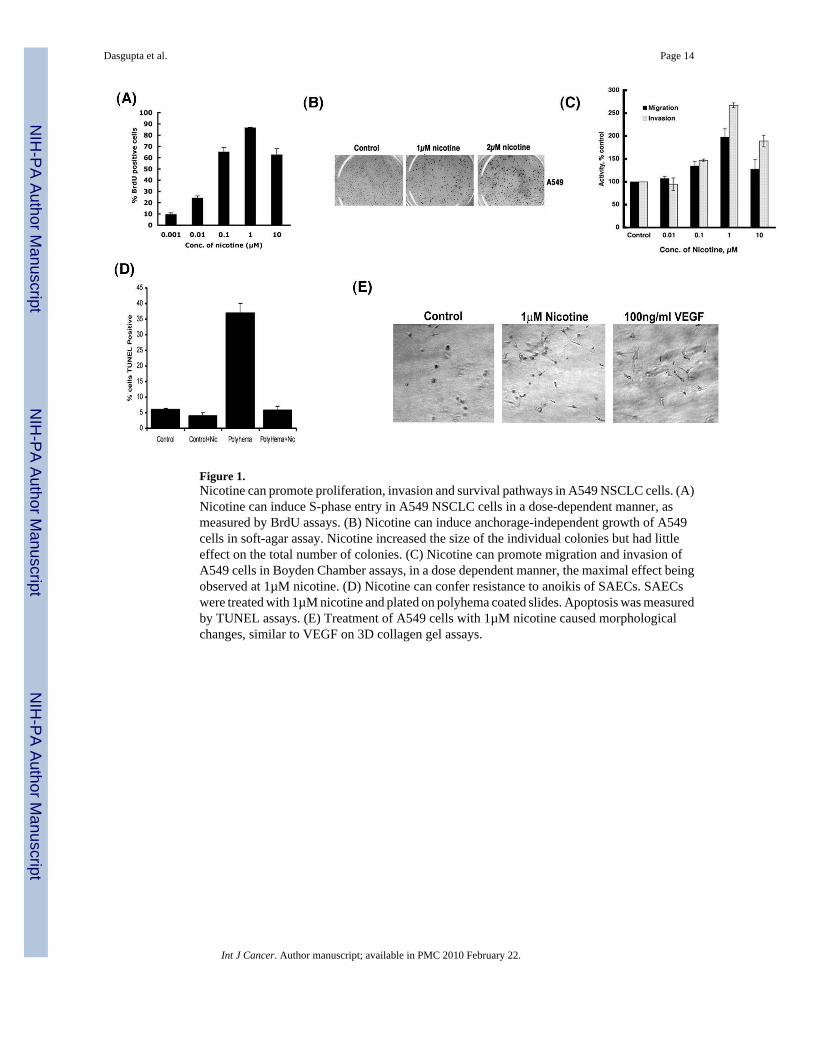
Figure 1.Nicotine can promote proliferation, invasion and survival pathways in A549 NSCLC cells. (A)Nicotine can induce S-phase entry in A549 NSCLC cells in a dose-dependent manner, asmeasured by BrdU assays. (B) Nicotine can induce anchorage-independent growth of A549cells in soft-agar assay. Nicotine increased the size of the individual colonies but had littleeffect on the total number of colonies. (C) Nicotine can promote migration and invasion ofA549 cells in Boyden Chamber assays, in a dose dependent manner, the maximal effect beingobserved at 1µM nicotine. (D) Nicotine can confer resistance to anoikis of SAECs. SAECswere treated with 1µM nicotine and plated on polyhema coated slides. Apoptosis was measuredby TUNEL assays. (E) Treatment of A549 cells with 1µM nicotine caused morphologicalchanges, similar to VEGF on 3D collagen gel assays.
Dasgupta et al. Page 14
Int J Cancer. Author manuscript; available in PMC 2010 February 22.
NIH
-PA Author Manuscript
NIH
-PA Author Manuscript
NIH
-PA Author Manuscript
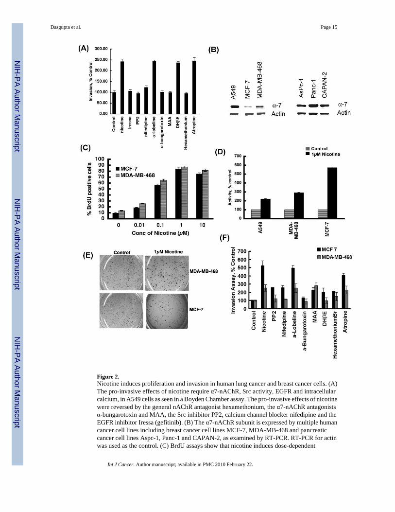
Figure 2.Nicotine induces proliferation and invasion in human lung cancer and breast cancer cells. (A)The pro-invasive effects of nicotine require α7-nAChR, Src activity, EGFR and intracellularcalcium, in A549 cells as seen in a Boyden Chamber assay. The pro-invasive effects of nicotinewere reversed by the general nAChR antagonist hexamethonium, the α7-nAChR antagonistsα-bungarotoxin and MAA, the Src inhibitor PP2, calcium channel blocker nifedipine and theEGFR inhibitor Iressa (gefitinib). (B) The α7-nAChR subunit is expressed by multiple humancancer cell lines including breast cancer cell lines MCF-7, MDA-MB-468 and pancreaticcancer cell lines Aspc-1, Panc-1 and CAPAN-2, as examined by RT-PCR. RT-PCR for actinwas used as the control. (C) BrdU assays show that nicotine induces dose-dependent
Dasgupta et al. Page 15
Int J Cancer. Author manuscript; available in PMC 2010 February 22.
NIH
-PA Author Manuscript
NIH
-PA Author Manuscript
NIH
-PA Author Manuscript

proliferation of human breast cancer cell lines MCF-7 and MDA-MB-468, the maximal effectbeing observed at 1µM. (D) Nicotine promotes invasion of MCF-7 and MDA-MB-468 breastcancer cells at a concentration of 1µM, as measured by Boyden chamber assays. A549 NSCLCcells were used as the positive control for the assay. (E) Nicotine stimulates the anchorage-independent growth of MCF-7 and MDA-MB-468 human breast cancer cells in soft agarassays. MCF-7 and MDA-MB-468 cells treated with 1µM nicotine formed larger colonies insoft agar relative to untreated controls. (F) Nicotine-mediated invasion of MCF-7 and MDA-MB-468 human breast cancer cells is dependent on Src activity and intracellular calciumpathways. The pro-invasive effects on nicotine in these breast cancer cells were mediated byboth α7-nAChR and DhβE-sensitive nAChRs.
Dasgupta et al. Page 16
Int J Cancer. Author manuscript; available in PMC 2010 February 22.
NIH
-PA Author Manuscript
NIH
-PA Author Manuscript
NIH
-PA Author Manuscript
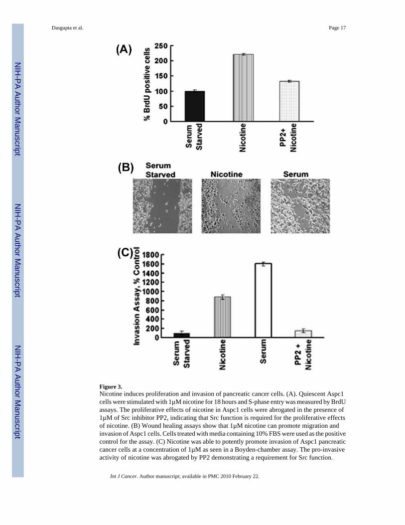
Figure 3.Nicotine induces proliferation and invasion of pancreatic cancer cells. (A). Quiescent Aspc1cells were stimulated with 1µM nicotine for 18 hours and S-phase entry was measured by BrdUassays. The proliferative effects of nicotine in Aspc1 cells were abrogated in the presence of1µM of Src inhibitor PP2, indicating that Src function is required for the proliferative effectsof nicotine. (B) Wound healing assays show that 1µM nicotine can promote migration andinvasion of Aspc1 cells. Cells treated with media containing 10% FBS were used as the positivecontrol for the assay. (C) Nicotine was able to potently promote invasion of Aspc1 pancreaticcancer cells at a concentration of 1µM as seen in a Boyden-chamber assay. The pro-invasiveactivity of nicotine was abrogated by PP2 demonstrating a requirement for Src function.
Dasgupta et al. Page 17
Int J Cancer. Author manuscript; available in PMC 2010 February 22.
NIH
-PA Author Manuscript
NIH
-PA Author Manuscript
NIH
-PA Author Manuscript
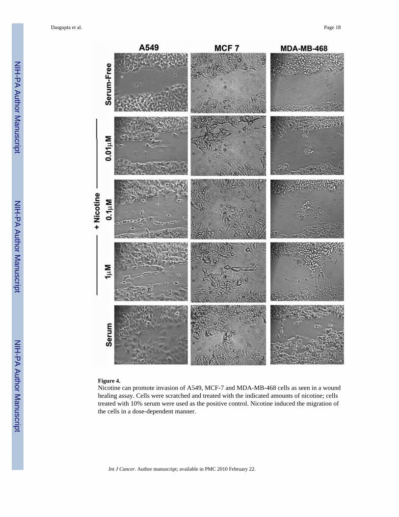
Figure 4.Nicotine can promote invasion of A549, MCF-7 and MDA-MB-468 cells as seen in a woundhealing assay. Cells were scratched and treated with the indicated amounts of nicotine; cellstreated with 10% serum were used as the positive control. Nicotine induced the migration ofthe cells in a dose-dependent manner.
Dasgupta et al. Page 18
Int J Cancer. Author manuscript; available in PMC 2010 February 22.
NIH
-PA Author Manuscript
NIH
-PA Author Manuscript
NIH
-PA Author Manuscript
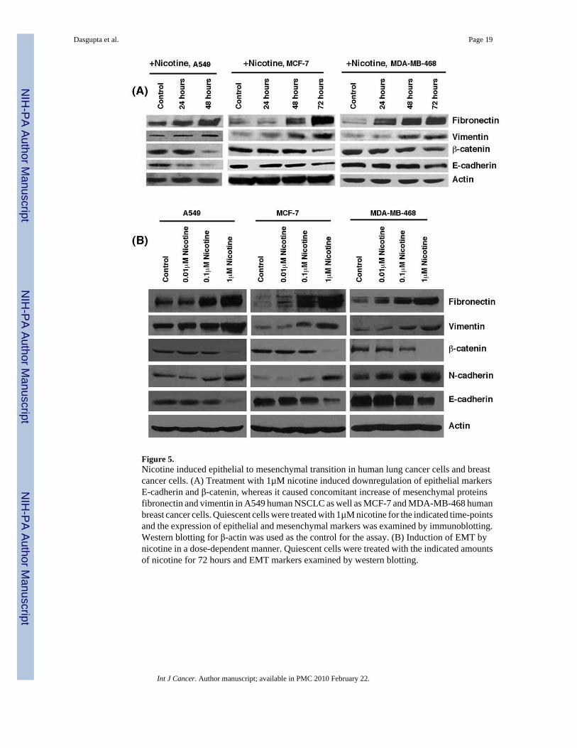
Figure 5.Nicotine induced epithelial to mesenchymal transition in human lung cancer cells and breastcancer cells. (A) Treatment with 1µM nicotine induced downregulation of epithelial markersE-cadherin and β-catenin, whereas it caused concomitant increase of mesenchymal proteinsfibronectin and vimentin in A549 human NSCLC as well as MCF-7 and MDA-MB-468 humanbreast cancer cells. Quiescent cells were treated with 1µM nicotine for the indicated time-pointsand the expression of epithelial and mesenchymal markers was examined by immunoblotting.Western blotting for β-actin was used as the control for the assay. (B) Induction of EMT bynicotine in a dose-dependent manner. Quiescent cells were treated with the indicated amountsof nicotine for 72 hours and EMT markers examined by western blotting.
Dasgupta et al. Page 19
Int J Cancer. Author manuscript; available in PMC 2010 February 22.
NIH
-PA Author Manuscript
NIH
-PA Author Manuscript
NIH
-PA Author Manuscript
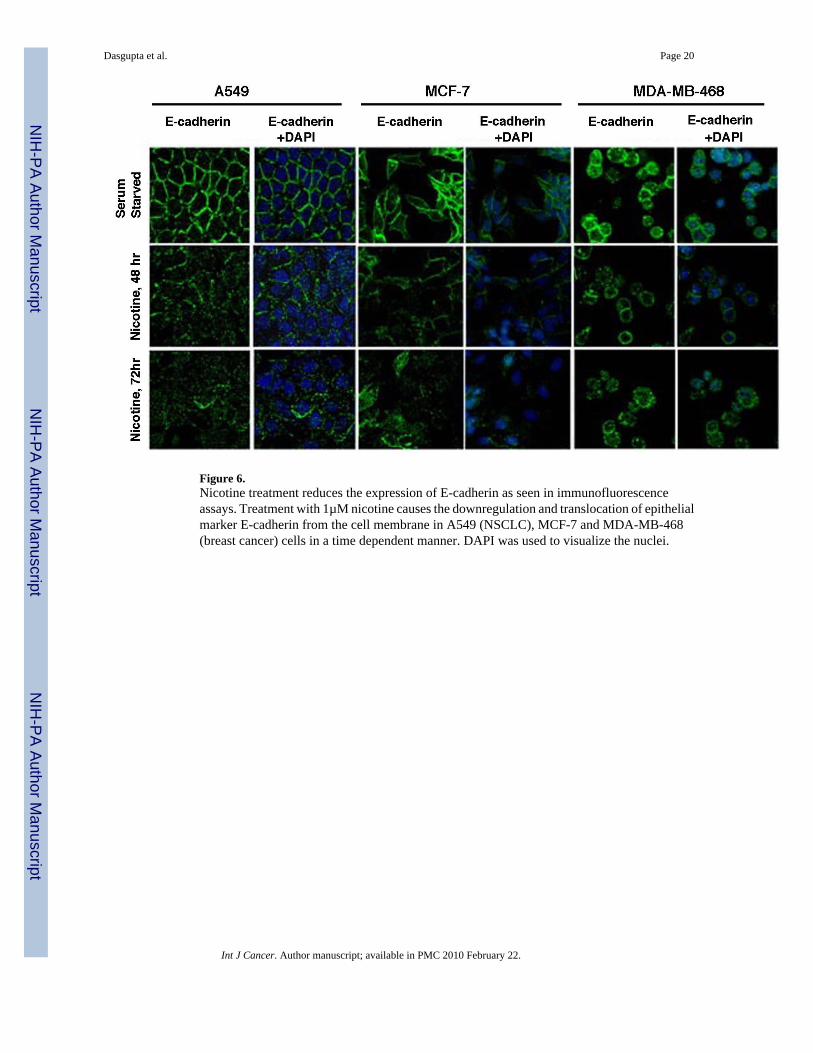
Figure 6.Nicotine treatment reduces the expression of E-cadherin as seen in immunofluorescenceassays. Treatment with 1µM nicotine causes the downregulation and translocation of epithelialmarker E-cadherin from the cell membrane in A549 (NSCLC), MCF-7 and MDA-MB-468(breast cancer) cells in a time dependent manner. DAPI was used to visualize the nuclei.
Dasgupta et al. Page 20
Int J Cancer. Author manuscript; available in PMC 2010 February 22.
NIH
-PA Author Manuscript
NIH
-PA Author Manuscript
NIH
-PA Author Manuscript
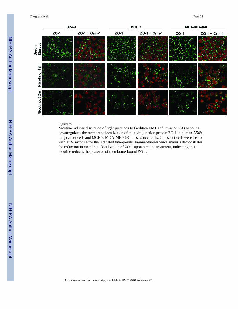
Figure 7.Nicotine induces disruption of tight junctions to facilitate EMT and invasion. (A) Nicotinedownregulates the membrane localization of the tight junction protein ZO-1 in human A549lung cancer cells and MCF-7, MDA-MB-468 breast cancer cells. Quiescent cells were treatedwith 1µM nicotine for the indicated time-points. Immunofluorescence analysis demonstratesthe reduction in membrane localization of ZO-1 upon nicotine treatment, indicating thatnicotine reduces the presence of membrane-bound ZO-1.
Dasgupta et al. Page 21
Int J Cancer. Author manuscript; available in PMC 2010 February 22.
NIH
-PA Author Manuscript
NIH
-PA Author Manuscript
NIH
-PA Author Manuscript