Structural determinants for the inhibitory ligands of orotidine-5′-monophosphate decarboxylase
25
Structural Determinants for Inhibitory Ligands of Orotidine-5′- Monophosphate Decarboxylase Maria Elena Meza-Avina 1,6 , Lianhu Wei 1 , Yan Liu 4 , Ewa Poduch 1 , Angelica M. Bello 1 , Ram K. Mishra 1 , Emil F. Pai 1,3,4,* , and Lakshmi P. Kotra 1,2,5,6,* 1 Center for Molecular Design and Preformulations and Division of Cell & Molecular Biology, Toronto General Research Institute/University Health Network, Toronto, ON M5G 1L7, Canada 2 Departments of Pharmaceutical Sciences and Chemistry, University of Toronto 3 Division of Cancer Genomics and Proteomics, Ontario Cancer Institute/University Health Network, Toronto, ON M5G 1L7, Canada 4 Departments of Biochemistry, Medical Biophysics, and Molecular Genetics, University of Toronto, Toronto, ON M5S 1A8, Canada 5 McLaughlin Center for Molecular Medicine, University of Toronto, Toronto, Ontario, Canada 6 Department of Chemistry & Biochemistry, The University of North Carolina at Greensboro, Greensboro, NC 27412, U.S.A. Abstract In recent years, orotidine-5′-monophosphate decarboxylase (ODCase) has gained renewed attention as a drug target. As a part of continuing efforts to design novel inhibitors of ODCase, we undertook a comprehensive study of potent, structurally diverse ligands of ODCase and analyzed their structural interactions in the active site of ODCase. These ligands comprise of pyrazole or pyrimidine nucleotides including the mononucleotide derivatives of pyrazofurin, barbiturate ribonucleoside, and 5-cyanouridine, as well as, in a computational approach, 1,4-dihydropyridine- based non-nucleoside inhibitors such as nifedipine and nimodipine. All these ligands bind in the active site of ODCase exhibiting distinct interactions paving the way to design novel inhibitors against this interesting enzyme. We propose an empirical model for the ligand structure for rational modifications in new drug design and potentially new lead structures. Introduction Orotidine-5′-monophosphate decarboxylase (ODCase) catalyzes the decarboxylation of OMP (1) to UMP (2) in the de novo pathway for the transformation of the amino acid, aspartic acid to UMP. ODCase has attracted much attention from biochemists because of its status as one of the most proficient enzymes in Nature accelerating the rate of decarboxylation by over 17 orders of magnitude to produce the key pyrimidine nucleotide, UMP. 1,2,3 Pyrimidine nucleotides are important building blocks for the synthesis of RNA and DNA, molecules essential for cell replication and survival. Due to its important role in the de novo nucleic acid biosynthesis, ODCase is present in most species including bacteria, parasites and humans but not in viruses. Viruses depend on their host cells for the supply of nucleotides. In humans, pyrimidine nucleotides are synthesized via two routes: the de novo and salvage pathways. 4 Whenever higher concentrations of pyrimidines are needed in the * To whom correspondence should be addressed: Mailing address: #5-356, TMDT/MaRS Center, 101 College Street, Toronto, Ontario, Canada, M5G 1L7. Tel. (416) 581-7601, [email protected]. NIH Public Access Author Manuscript Bioorg Med Chem. Author manuscript; available in PMC 2011 June 27. Published in final edited form as: Bioorg Med Chem. 2010 June 1; 18(11): 4032–4041. doi:10.1016/j.bmc.2010.04.017. NIH-PA Author Manuscript NIH-PA Author Manuscript NIH-PA Author Manuscript
Transcript of Structural determinants for the inhibitory ligands of orotidine-5′-monophosphate decarboxylase

Structural Determinants for Inhibitory Ligands of Orotidine-5′-Monophosphate Decarboxylase
Maria Elena Meza-Avina1,6, Lianhu Wei1, Yan Liu4, Ewa Poduch1, Angelica M. Bello1, RamK. Mishra1, Emil F. Pai1,3,4,*, and Lakshmi P. Kotra1,2,5,6,*
1 Center for Molecular Design and Preformulations and Division of Cell & Molecular Biology,Toronto General Research Institute/University Health Network, Toronto, ON M5G 1L7, Canada2 Departments of Pharmaceutical Sciences and Chemistry, University of Toronto3 Division of Cancer Genomics and Proteomics, Ontario Cancer Institute/University HealthNetwork, Toronto, ON M5G 1L7, Canada4 Departments of Biochemistry, Medical Biophysics, and Molecular Genetics, University ofToronto, Toronto, ON M5S 1A8, Canada5 McLaughlin Center for Molecular Medicine, University of Toronto, Toronto, Ontario, Canada6 Department of Chemistry & Biochemistry, The University of North Carolina at Greensboro,Greensboro, NC 27412, U.S.A.
AbstractIn recent years, orotidine-5′-monophosphate decarboxylase (ODCase) has gained renewedattention as a drug target. As a part of continuing efforts to design novel inhibitors of ODCase, weundertook a comprehensive study of potent, structurally diverse ligands of ODCase and analyzedtheir structural interactions in the active site of ODCase. These ligands comprise of pyrazole orpyrimidine nucleotides including the mononucleotide derivatives of pyrazofurin, barbiturateribonucleoside, and 5-cyanouridine, as well as, in a computational approach, 1,4-dihydropyridine-based non-nucleoside inhibitors such as nifedipine and nimodipine. All these ligands bind in theactive site of ODCase exhibiting distinct interactions paving the way to design novel inhibitorsagainst this interesting enzyme. We propose an empirical model for the ligand structure forrational modifications in new drug design and potentially new lead structures.
IntroductionOrotidine-5′-monophosphate decarboxylase (ODCase) catalyzes the decarboxylation ofOMP (1) to UMP (2) in the de novo pathway for the transformation of the amino acid,aspartic acid to UMP. ODCase has attracted much attention from biochemists because of itsstatus as one of the most proficient enzymes in Nature accelerating the rate ofdecarboxylation by over 17 orders of magnitude to produce the key pyrimidine nucleotide,UMP.1,2,3 Pyrimidine nucleotides are important building blocks for the synthesis of RNAand DNA, molecules essential for cell replication and survival. Due to its important role inthe de novo nucleic acid biosynthesis, ODCase is present in most species including bacteria,parasites and humans but not in viruses. Viruses depend on their host cells for the supply ofnucleotides. In humans, pyrimidine nucleotides are synthesized via two routes: the de novoand salvage pathways.4 Whenever higher concentrations of pyrimidines are needed in the
*To whom correspondence should be addressed: Mailing address: #5-356, TMDT/MaRS Center, 101 College Street, Toronto, Ontario,Canada, M5G 1L7. Tel. (416) 581-7601, [email protected].
NIH Public AccessAuthor ManuscriptBioorg Med Chem. Author manuscript; available in PMC 2011 June 27.
Published in final edited form as:Bioorg Med Chem. 2010 June 1; 18(11): 4032–4041. doi:10.1016/j.bmc.2010.04.017.
NIH
-PA Author Manuscript
NIH
-PA Author Manuscript
NIH
-PA Author Manuscript

cell, including for the normal cellular processes, during uncontrolled growth of the cell suchas in cancer, or fast replicating viral infections etc, de novo pyrimidine synthesis isupregulated, and the activity of ODCase is simultaneously operating at a higher than normallevel.5,6
In certain higher-level organisms, such as mouse or human, ODCase is part of thebifunctional enzyme, UMP synthase.7 While in pathogenic organisms, such as bacteria,fungi and parasites, ODCase is a monofunctional enzyme, although in Plasmodia it forms aheterotetramer with orotate phosphoribosyltransferase.8,9,10 In all species, ODCase seems tobe active as a dimer and the catalytic site is comprised of active residues from the secondmonomer. Plasmodia such as P. falciparum and P. vivax are dependent on their own denovo synthesis of pyrimidine nucleotides due to their lack of the salvage pathway.11 Thus,inhibition of plasmodial ODCase was proposed as a strategy for compounds directed againstmalaria, and a limited number of orotate analogs were investigated as potential drugs againstthe malaria parasite.12,13,14 ODCase has also been identified as a potential target for drugsdirected against RNA viruses like pox and flaviviruses.15,16,17,18 ODCase inhibitors havealso been effective against West Nile virus, a recent thread to humans and birds in the USand Canada.19 In the recent years, an increased interest in ODCase as a drug target is alsodue to the advances in determining the three-dimensional structures of this enzyme fromvarious species. Since 2000, when the first X-ray structures of ODCase were resolved, thereare now almost 100 coordinate sets of ODCase from at least 11 different species depositedin the Protein Databank (www.rcsb.org). These crystal structures were determined for theapo-form of the protein but mostly in complex with a variety of ligands such as UMP (2), 6-aza-UMP (3), BMP (4), XMP (5), CMP (6), as well as with a variety of mutant forms ofODCase. Despite such intense efforts in recent years, the catalytic mechanism of ODCase isstill not completely understood and the use of structure-based tools in the rational design ofsubstrate analogs of ODCase as inhibitors is still rudimentary at best.
Recently, investigations on the mechanism of decarboxylation by ODCase have gainedmomentum and there is compelling evidence that a C6 carbanion-based transition-state isformed during the decarboxylation.20,21 This transition-state intermediate appears during theearly stage of the reaction, and electrostatic stress may play a role in the process ofdecarboxylation, although other mechanisms using computational and kinetic isotopemethods suggest alternatives.20,21,22,23,24 We have recently revealed that under suitableconditions ODCase can facilitate interesting reactions other than decarboxylation, such asthe transformation of 6-cyano-UMP (9) into BMP (4).25,26 Based on the catalyticpromiscuity exhibited by ODCase, Wittmann et al. proposed the possibility of a covalentmechanism as a unifying means of addressing various biochemical reactions undertaken byODCase.27 Our group has disclosed a comprehensive time-resolved crystallography andmutant analyses on the interactions and catalysis of 9 with ODCase.28 The structuralevidence in these studies compels us to believe that the slow catalysis for the transformationof 9 into 4 represents non-covalent catalysis, involving strong electrostatic forces breakingthe resonance established in the 6-cyano-pyrimidine nucleic base.28 ODCase also exhibitsplasticity in accepting various nucleotide ligands including compounds such as XMP (5) andin fact these compounds are among the potent inhibitors of this enzyme.29 It is alsointeresting to note that ODCases from various species exhibit different binding affinitiestowards the same inhibitors.29 The new generation of inhibitors such as the novel C6-substituted uridine derivatives targeting ODCase specifically are exhibiting interesting andpromising therapeutic activities.30,31,32
Nucleosides are well established as a major source of drugs for the treatment of cancer andviral infections.33,34 A classic example of an unconventional nucleoside of medicinalinterest, and a potent inhibitor of ODCase is pyrazofurin (also known as pyrazomycin, 7,
Meza-Avina et al. Page 2
Bioorg Med Chem. Author manuscript; available in PMC 2011 June 27.
NIH
-PA Author Manuscript
NIH
-PA Author Manuscript
NIH
-PA Author Manuscript

Figure 1). This compound was originally isolated from the broth filtrates of Streptomycescandidus.35,36 It has been the subject of numerous investigations over the past three decadesdue to its C-nucleoside status and its breadth of clinically relevant biological activities.37
Pyrazofurin exhibits potent anticancer activity, especially against leukemia cell lines.38,39,40
In clinical trials, pyrazofurin was administered to cancer patients at various doses to evaluateits potential as an anticancer agent. However, its development was abandoned later due totoxicity.41 Pyrazofurin has also been evaluated as an antiviral agent targeting respiratorysyncytial virus (RSV), influenza, vaccinia, West Nile virus and other RNA viruses.19,42,43,44
Additionally, this compound has been shown to be effective against parasitic infectionsspecifically Plasmodium falciparum and also was implicated in immunosuppressivetherapy.45,46 With such a long history, pyrazofurin as well as other pyrazole-basednucleosides are continuing to generate interest in medicinal chemistry.47 The 5′-monophosphate derivative of pyrazofurin (8) is a potent inhibitor of S. cerevisiae ODCasewith inhibition constants in the low nanomolar range.38
Due to this exciting and rich biological activity profile at the molecular and clinical levels,our group is interested in further characterizing the interactions of pyrazofurin 5′-monophosphate (8) and other substrate analogs such as BMP (4), and 5-cyano-UMP (10)along with non-nucleoside inhibitors with ODCase to define pharmacophore features forsubstrate analogs. An empirical structural dissection and a pharmacophore model arepresented based on the atomic level interactions for the potential design of novel classes ofcompounds targeting ODCase.
Experimental SectionSynthesis
General—Anhydrous chemical reactions were performed under a nitrogen atmosphere. Allsolvents and reagents were obtained from commercial sources. NMR spectra were recordedon a Varian spectrometer (300 or 400 MHz for 1H, and 121.46 MHz for 31P). Chemicalshifts are reported in ppm using H2O as the reference for the 1H NMR spectra, andphosphoric acid as an external standard for the 31P spectra. Mass spectra were obtained on aQ-Star mass spectrometer using ESI or EI techniques. The monophosphate derivatives weretransformed into the corresponding ammonium salts by treatment with 0.5 M NH4OHsolution at 0 °C followed by lyophilization. Purity of the compounds 8 and 10 was checkedon a Waters Delta 660 HPLC system attached to a PDA detector, using a Waters ODS2 5micron reverse phase (4.6×100 mm) column, was > 95%. The eluting system was either100% H2O or 95% H2O:5% MeOH at a flow rate of 0.5mL/min. All HPLC solvents werefiltered through Waters membranes (47 mm GHP 0.45 μm, Pall Corporation) and degassedwith helium. Injection samples were filtered using Waters Acrodisc® Syringe Filters 4 mmPTFE 0.2 μm.
4-Hydroxy-5-(5-O-monophosphoryl-β-D-ribofuranos-1-yl)-1H-pyrazole-3-carboxamide (8)Pyrazofurin (7) (11 mg, 0.042 mmol) was dissolved in triethyl phosphate (0.5 mL) and thesolution was stirred at 45 °C for 10 min under a nitrogen atmosphere. The reaction mixturewas cooled to 0 °C and POCl3 (23.7 μL, 0.25 mmol) was added and stirring continued at thesame temperature for an additional 5 h. The reaction was then quenched with cold water (1.5mL), washed with diethyl ether (5 × 2 mL) and neutralized with 0.5 M NH4OH. The volumeof the aqueous layer was reduced under vacuum, and the crude was purified by HPLC toobtain the target compound 8. UV λmax (pH = 8) 263 nm (ε = 2,477); 1H NMR (D2O) δ 4.82(d, J=5.9 Hz, 1H, H-1′), 4.26 (br m, 1H, H-2′), 4.13 (br m, 1H, H-3′), 4.04 (br m, 1H, H-4′)3.93 (br m, 2H, H-5′); 31P NMR (D2O) δ 0.40 ppm; HRMS (ESI −ve) calculated forC9H13N3O9P (M−): 338.0394, found 338.0407.
Meza-Avina et al. Page 3
Bioorg Med Chem. Author manuscript; available in PMC 2011 June 27.
NIH
-PA Author Manuscript
NIH
-PA Author Manuscript
NIH
-PA Author Manuscript

1-(5-O-t-Butyldimethylsilyl-2,3-O-isopropyl-β-D-ribofuranosyl)-5-cyanouracil (12)Compound 1125 (2.6 g, 5.44 mmol) was dissolved in 20 mL of anhydrous DMF and treatedwith NaCN (0.43 g, 8.77 mmol) at 100 °C for 3 h. After this time, the reaction mixture wasdiluted with water (20 mL) and the pH brought to 6; the solution was extracted (3x) with 30mL of ethyl acetate, the organic layer was washed with brine, dried with sodium sulfate,evaporated. Purification by column chromatography (hexane:ethyl acetate, 9:1) gave 12 as awhite powder (2.21 g, 96%). 1H NMR (CDCl3) δ 0.13 (s, 6H), 0.91 (s, 9H), 1.37 (s, 3H),1.60 (s, 3H), 3.82 (dd, 1H, H-5′), 3.98 (dd, 1H, H-5′), 4.53 (m, 1H, H-4′), 4.69 (dd, 1H,H-2′), 4.73 (dd, 1H, H-3′), 5.89 (d, 1H, H-1′), 8.32 (s, 1H, H-6), 8.96 (broad, 1H, NH).
1-β-D-Ribofuranosyl-5-cyanouracil (13)Compound 12 (2.2 g, 5.20 mmol), was treated with 20 mL of 50 % trifluoroacetic acid at 0°C, and the reaction mixture was stirred at rt for 3 h. After this time, solvent was evaporatedand the residue was purified by column chromatography (dichloromethane: methanol, 8:2)to obtain 13 as a light solid (1.3 g, 93%). UV λmax = 278 nm; 1H NMR (CD3OD) δ 3.78 (dd,1H, H-5′), 3.95 (dd, 1H, H-5′), 4.05 (m,1H, H-4′), 4.19 (m, 2H, H-2′, H-3′), 5.83 (d, 1H,H-1′), 9.02 (s, 1H, H-6); 13C NMR (CD3OD) 162.15, 151.25, 150.898, 114.76, 92.17, 90.15,86.25, 76.47, 70.14, 61.15; HRMS (ESI) calculated for C10H12N3O6 (M+H) 270.0720,found 270.0725.
1-(5-Monophosphoryl-β-D-ribofuranos-1-yl)-5-cyanouracil (10)To a solution of water (0.050 mL, 2.78 mmol), CH3CN (4.3 mL), pyridine (0.39 mL, 4.8mmol) and POCl3 (0.612 g, 0.037 mL, 4 mmol) at 0 °C, were added 0.269 g (0.702 mmol)of compound 13. The reaction mixture was stirred for 5 h (3 hr at 0 °C and then at roomtemperature). The reaction was monitored by TLC (n-butanol, water and acetic acid, 2:2:1).After the starting material was consumed, the reaction was quenched with 2 mL of coldwater and stirred for an additional 2 h at rt. The solvent was evaporated and then the productwas purified by passing through a basic Dowex column (eluted with water followed by 5%formic acid). The concentrated product was then dissolved in 3 mL of cold water andneutralized with NH4OH. The solvent was evaporated and the product was lyophilized togive 0.287g (yield 75%) of 10 as a white solid. UV λmax = 278 nm (ε = 1,949); 1H NMR(CD3OD) δ 4.11 (dd, 1H, H-5′), 4.22 to 4.27 (m, 3H, H-3′, H-4′, H-5′), 4.33 (ddoverlap,1H,H-2′), 5.86 (d, 1H, H-1′), 8.63 (s, 1H, H-6). 31P (D2O) 0.12 (s), HRMS (ESI) calculated forC10H11N3O9P (M−) 348.0227, found 348.0238.
ODCase productionODCases from Methanobacterium thermoautotrophicum, Plasmodium falciparum andHomo sapiens were cloned, expressed and purified as described previously.25,29 Productionand purification of the Helicobacter pylori and Staphylococcus aureus enzymes will bedescribed elsewhere.48 Enzyme concentrations were determined using a BioRAD proteinassay kit with bovine serum albumin as standard.
EnzymologyAssays were performed on a VP-ITC microcalorimeter (MicroCal). For isothermal titrationcalorimetry (ITC) experiments, the initial delay (60 sec), stirring speed (300 rpm), referencepower (15 μcal/sec), and the frequency of data point collection (or filter period, 1 sec) wereidentical for all experiments. The Hs- and Mt ODCase stock solutions were prepared using50 mM Tris (pH 7.5), 20 mM DTT, and 40 mM NaCl, and incubated overnight at rt. Forinjection into ITC, the substrate was dissolved in 50 mM Tris to a final concentration of 5mM. All inhibitor solutions were prepared in the same 50 mM Tris buffer. The assay bufferfor the enzyme or enzyme/inhibitor samples consisted of 50 mM Tris (pH 7.5) and 1 mM
Meza-Avina et al. Page 4
Bioorg Med Chem. Author manuscript; available in PMC 2011 June 27.
NIH
-PA Author Manuscript
NIH
-PA Author Manuscript
NIH
-PA Author Manuscript
DTT, and was degassed prior to use. The activity of Mt ODCase (final concentration 20 nM)was determined at 55 °C. Each reaction was initiated by an 11.4 μL injection of substratesolution (5 mM). The final substrate concentration in the 1.3 mL calorimetric cell was 40μM. The activity of Hs ODCase (final concentration 60 nM) was determined in the presenceof 20 μM substrate at 37 °C, and the reaction was initiated by a 5.7 μL injection of substratesolution (5 mM).
Both Mt and Hs ODCase were tested against 5-fluoro-6-amido-UMP (11) in a reversibleinhibition assay. The final concentration of Mt ODCase was 20 nM while the concentrationof 11 was 0.5, 1, 2.5, and 5 mM. Hs ODCase (60 nM) was exposed to 0.1, 0.25, 0.5, and 1mM of 11. The volume and the final concentration of the substrate were the same asdescribed for the controls. The concentrations of compound 8 were 5, 10, 20, and 40 μMagainst Mt ODCase. The inhibition of 60 nM Hp ODCase was determined using 0.25, 0.5, 1,and 1.5 μM of compound 8.49
5-Cyano-UMP (9) was tested against Pf, Hp and Sa ODCases.49 The final concentration ofthe Pf ODCase and the substrate in the reaction assay was 60 nM and 12 μM, respectively.The concentration of the inhibitor was 0.5, 1, 2, 2.5, and 5 mM. The reversible inhibition ofHp ODCase (60 nM) was tested in the presence of 0.25, 1, 1.5, and 2.5 mM of 10. Thereversible inhibition assay with 30 nM Sa ODCase was performed using the followingconcentrations of 10: 0.1, 0.25, 0.5, 0.75, and 2 mM. The concentration of the substrate usedin the assays with Hp and Sa ODCases was 20 μM.
Inhibition kinetics of compound 8 against Hs ODCase were determined using UVspectroscopy. The absorption maxima of the ligands permitted a reliable UV-based kineticsassay. Hs ODCase stock solution (60 μM) was prepared in 50 mM Tris (pH 7.5), 20 mMDTT, and 40 mM NaCl, and incubated overnight at room temperature. Assay samples wereprepared by diluting 3 μL of the stock enzyme solution in 50 mM Tris containing 1 mMDTT to a final volume of 990 μL. Each reaction was initiated by the addition of 10 μL of theligand (either substrate, or substrate mixed with the inhibitor). The final concentrations ofHs ODCase and OMP were 180 nM and 50 μM, respectively. The concentrations ofcompound 8 were 1, 1.5, 2, 2.5, 3, and 4 μM. After the addition of ligand, the sample wasmixed quickly, and the absorption at 285 nM was recorded on a Shimadzu UV-2401PCspectrophotometer as a function of time.
The KM values for Hs and Mt ODCases (from ITC measurements) were 1.7 ± 0.1 and 3.7 ±0.2 μM, respectively. And the KM for Hs ODCase was 3.4 ± 0.5 μM as determined by UVspectroscopy.
Data analysis—The procedure used for the determination of competitive inhibitionconstants was described earlier.50 The data were fitted in Grafit 7.0 to Equation 1 tocalculate the Ki for each inhibitor.
Equation 1
Inhibition of Hs ODCase by 8—Compound 8 is a slow tight binding inhibitor of HsODCase. The rate of consumption of the substrate was converted into the rate of productformation using equation 2:
Meza-Avina et al. Page 5
Bioorg Med Chem. Author manuscript; available in PMC 2011 June 27.
NIH
-PA Author Manuscript
NIH
-PA Author Manuscript
NIH
-PA Author Manuscript

Equation 2
where vi represents the initial rate and vs is the steady-state rate. The parameter kobscorresponds to the rate constant for the conversion from initial to steady-state rate. Theinitial phase of slow and reversible inhibition is represented by Equation 3.
Equation 3
The relationship between [I] and kobs is linear, and the data fit to Equation 4 yields theassociation and dissociation constants (k3 and k4):
Equation 4
The apparent inhibition constant is calculated from the ratio of k4/k3. The true inhibitordissociation constant Ki is determined by equation 5:
Equation 5
Crystallization and Crystallographic AnalysisAll crystals were grown at rt using the hanging-drop method and mixing 2 μL of proteinsolution of 10 mg/mL enzyme in either 25 mM Tris.HCl, pH 8.0, 40 mM NaCl, 5 %glycerol for complexes of Hs ODCase with compounds 4 and 10 or in 50 mM TRIS.HCl,pH 8.5, 100 mM NaCl and 5 mM β-mercaptoethanol for compound 8, with 2 μL of therespective reservoir solution. The best co-crystals of Hs ODCase with both BMP (4) and 5-cyano-UMP (10) were grown in 100 mM Tris.HCl, pH 8.4, 2.12 M ammonium sulfate whilethe complex with compound 8 had the best results in 100 mM TRIS.HCl, pH 8.4, 1.4 Mammonium sulfate. The crystals with compounds 4 and 8 adopted space group C2221 withunit cell dimensions of a = 77.6 Å, b = 116.9 Å, c = 62.0 Å and a = 79.4 Å, b = 116.5 Å, c =62.1 Å, respectively. Crystals of the complex with compound 4 also grew in space groupC2221 but with unit cell axes a = 77.6 Å, b = 116.9 Å, c = 62.0 Å, whereas the complex withcompound 10 grew in space group P21 with unit cell dimensions of a = 69.5 Å, b = 61.6 Å, c= 70.9 Å, β = 111.8°.
For data collection, the crystals were either transferred to reservoir solution containing 20 %glycerol for cryoprotection or dipped into paratone oil before they were flash-frozen in astream of boiling nitrogen. Diffraction data for the crystals of Hs ODCase co-crystallizedwith 4 were collected at 100 K and λ = 0.9002 Å at beam line 14BM-C, BioCARS,Advanced Photon Source, Argonne National Laboratories; data for the pyrazofurin-MP (8)and 5-CN-UMP (10) complexes were collected at 100 K and λ = 0.97949 Å and λ = 0.97934Å, respectively, at beam line 08ID-1 of the Canadian Macromolecular CrystallographyFacility, Canadian Light Source. Data were reduced and scaled using the program packageHKL2000.51 Data collection statistics are given in Table 1.
Meza-Avina et al. Page 6
Bioorg Med Chem. Author manuscript; available in PMC 2011 June 27.
NIH
-PA Author Manuscript
NIH
-PA Author Manuscript
NIH
-PA Author Manuscript

The structures of all complexes were determined using molecular replacement techniqueswith the help of the program package MOLREP52 subsequent refinements were done withRefmac-5.2,53 and model building with COOT.54 Refinement statistics are also listed inTable 1. Atomic coordinates and structure factors have been deposited in the Protein DataBank (PDB IDs: 3BGG, **** and 3BK0 for the complexes with ligands 4, 8 and 10,respectively).
Computer modelingThe three-dimensional structure of the Hs ODCase complex with 6-NH2-UMP (PDB code:3DBP) was used as the protein framework for the docking study. The ligand and thecrystallographic waters were removed from the active site, and hydrogens were added to theentire protein, preparing the active site for docking. The non-nucleoside ligands, nifedipineand nimodipine were docked using Surflex-Dock, a module in the SYBYL molecularmodeling package, for various docking poses and their relative ranking. This processinvolves first, the active site was coated with three types of probes, depending on theproperties of the enzyme residues exposed into the active site: CH4 for steric, N-H for HBdonor, C=O for HB acceptor. Then the ligand fragments were generated and posed intobinding site to generate the best fitting pose for the ligand. The Hammerhead, a completelyautomatic, fast docking procedure was used to dock these two compounds to binding site.The top ranking conformation of the ligand in the binding pocket was used to analyze theinteractions.
For ligand conformational analysis, eleven co-crystal structures of ODCase with elevenligands were collected, and the inhibitors were extracted from the corresponding active sites.These structures include include UMP (1DBT), 6-aza-UMP (1DVJ), 5-I-UMP (2QCH),BMP (1X1Z), 6-NH2-UMP (2Q8Z), 5-Br-UMP (2QCG), 5-CN-UMP (3BKO), 5-F-UMP(2QCF), 6-acetyl-UMP (3L0K), 6-I-UMP (covalent complex, 3BGJ) and OMP (1KM6).
Results and DiscussionIn this study, the overarching goal is to bring together structurally diverse but potent ligandsof ODCase and investigate their interactions with the active site of ODCase. As mentionedabove, first we focused on three structurally diverse nucleoside ligands of ODCase. BMP(4), one of the ligands included in this study, carries a hydroxyl moiety at the C6 positionand is the most potent inhibitor of ODCase with a Ki of 8.8×10⁻¹² M for the yeastenzyme.55 Both the C6 hydroxyl moiety in 4 and the C4 substitution in 8 are similar in theirlocation and their ability to present the anionic moiety for transition-state mimicry.Pyrazofurin-5′-monophosphate (8) is a C-nucleoside carrying a pyrazole moiety instead ofthe pyrimidine nucleic base, and does not possess the typical glycosidic bond similar to thenatural N-nucleoside. In fact pyrazofurin (7) was one of the very few C-nucleosidesevaluated clinically as an anticancer and antiviral agent.12 The correspondingmononucleotide 8 binds tightly at the active site of ODCase, thus this compound was ofgreat interest to us and we investigated its interactions with the protein matrix in greaterdetail. Structurally, the presence of cyano substituent at the C5 position of compound 10 wasnot only intended to provide clues on the interactions of the 5-CN moiety with ODCase, butalso test whether any unusual chemistry could be observed as had been the case with 6-CN-UMP.25,26
Compound 4 was synthesized according to the reported procedure.56 The mononucleotidederivatives of 8, and 10 were synthesized according to Scheme 1. Compound 10 wassynthesized from the fully protected 5-bromouracil derivative 11 by treatment with sodiumcyanide to obtain compound 12 followed by deprotection with aqueous TFA to give 13(Scheme 1A). 5-Cyanouridine (13) was phosphorylated by treatment with POCl3 to yield 10.
Meza-Avina et al. Page 7
Bioorg Med Chem. Author manuscript; available in PMC 2011 June 27.
NIH
-PA Author Manuscript
NIH
-PA Author Manuscript
NIH
-PA Author Manuscript
Pyrazofurin (7) was phosphorylated using triethyl phosphate and POCl3 to obtain compound8 (Scheme 1B). For enzymatic studies, Hs, Mt, Sa, Pf and Hp ODCase, i.e. enzymes fromspecies spanning higher organisms, Archaea and bacteria were employed. Compounds 4, 8and 10 were co-crystallized with Hs ODCase and the corresponding three-dimensionalstructures were determined to detail the atomic interactions in the binding site of HsODCase (1.93, 1.2 and 1.6 Å resolution, respectively, Figure 2).
The active site architectures for the ODCase complexes with 4, 8 and 10 are almostidentical, and there are no major differences in the conformations of the residues thatcomprise the active site. An analysis of the hydrogen-bonding network, however, allows oneto identify the similarities and differences for the different ligands (Figure 3). Thephosphoribosyl moiety in all three inhibitors exhibited very similar interactions with theenzyme. This portion of the nucleotide engages in a network of hydrogen bondinginteractions, primarily through the 2′ and 3′ hydroxyl moieties as well as the phosphorylmoiety at the 5′-position. This is a typical feature found in the other inhibitor complexes ofODCase and this interaction is responsible for a large contribution towards the bindingenergy.57,58
In the active site of ODCase, BMP (4) positions its highly acidic hydroxyl moiety at C6close to the side chains of Lys125 and Asp123, which are both part of a strong network ofionic bonding interactions (Figure 2E, 2F, and 3C). Close by, the presence of a tightly boundwater molecule (2.75 Å distance from 6-OH moiety) correlates well with BMP binding,probably mitigating the potency of BMP to interact with ODCase (Figure 3C). 5-Cyano-UMP on the other hand does not carry any C6 substitution but a neutral cyano group at theC5 position that fits well into a small hydrophobic pocket in the active site. The remainingparts of the respective nucleic base of 4 and 10 interact with the ODCase active site in verysimilar fashion, mostly through conserved hydrogen bonds. For example, the O2, N3 and O4atoms in 4 and 10 exhibit almost identical interactions with the side chains of Gln241,Ser183 and a crystallographic water molecule (Figures 3B and 3C). At physiological pH, thehydroxyl moiety is deprotonated and presents compound 4 as a transition-state analog, afeature described earlier.3,20,21
The C-Nucleoside analog pyrazofurin (7) and its monophosphate derivative 8 arepharmacologically well-characterized compounds. Little is known, however, of 8’sinteractions with its target enzyme ODCase. Pyrazofurin does not have the typical glycosidicbond present in N-nucleosides (Figure 4). The pyrazole moiety is attached via the C5 carbonto the C1′ position of the ribosyl moiety, and in addition, carries two key substitutions on thepyrazole moiety: an amido moiety at C3 and a hydroxyl moiety at C4 position (Figure 4).This hydroxyl moiety (pKa 6.7) would be deprotonated to a large extent at physiological pH(7.4), similar to what is found with BMP (4). It is anticipated that the hydroxyl moiety couldmimic the transition-state species, again similar to BMP, when bound to the active site ofODCase. Thus we co-crystallized pyrazofurin-5′-monophosphate (8) with Hs ODCase anddetermined the three-dimensional structure of the complex at 1.2 Å resolution (Table 1,8Figures 2A, 2B and 3A). The aza-moiety in the pyrazole ring of interacts via hydrogenbonds with the side chains of Gln241 and Ser183 (Figure 3A). These hydrogen bondsaccount for the interactions of O2 and N3 in a typical pyrimidine nucleic base (Figure 3A vs3C). The carbonyl moiety in the C3 amido group of the pyrazole ring interacts with thebackbone nitrogen of Ser183 and a water molecule, equivalent to the interactions of the O4moiety in the natural uracil ring. This pattern of strong interactions locks the orientation ofthe pyrazole moiety in the ODCase binding site presenting other functional groups to theconserved catalytic residues (Figure 2A).
Meza-Avina et al. Page 8
Bioorg Med Chem. Author manuscript; available in PMC 2011 June 27.
NIH
-PA Author Manuscript
NIH
-PA Author Manuscript
NIH
-PA Author Manuscript

The second set of interactions of the pyrazole moiety includes the salt bridge between theionized hydroxyl group linked to C4 and the positively charged side chain of Lys125 as wellas two hydrogen bonds with bound water molecules (d=2.6–2.9 Å). Although theseinteractions of the pyrazole moiety in 8 are not identical to those seen with the pyrimidinenucleic base in 4, equivalent electrostatic and steric interactions nevertheless result in tightbinding of 8 to ODCase (Figure 3A vs 3C). Our results assert that the heterocycle on theribose can be varied without compromising binding affinity; in this particular case,pyrimidine and pyrazole provided the same “backbone” environment to the ligand structure.Purine heterocycles can also be substituted, as recently reported for xanthosine-5′-monophosphate (5, Figure 1), another highly potent inhibitor of ODCase.29 Even moleculeslike nifedipine and nimodipine (14 and 15, respectively, Figure 4), clinically used calciumchannel blockers, inhibit ODCase competitively, with inhibition constants (Ki) of 105 and18 μM, respectively.59 These observations contribute to the mounting evidence thatODCase, although evolved to carry out the decarboxylation of OMP with high efficiency, isinherently promiscuous in binding to other nucleosides, both purines and pyrimidines, aswell as to non-nucleoside inhibitors.
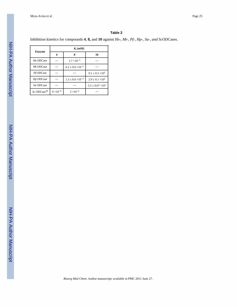
Compounds 8 and 10 were also evaluated for their potential to inhibit ODCases from P.falciparum, H. pylori, M. thermoautotrophicum, H. sapiens and S. aureus (Figure 6 andTable 2). The 5-cyanouridine derivative 10 inhibited Pf, Hp and Sa ODCases with veryweak inhibition constants (Ki) of 0.91±0.03, 0.29±0.01 and 0.55±0.007 M, respectively,similar to its inhibition of Hs and Mt ODCases (Ki = 0.14±0.06 and 0.70±0.02 M,respectively). These poor inhibitory properties also reflect the weak molecular interactionsbetween the 5-cyanouridine derivative 10 and ODCase (Figure 3B). On the other hand,compound 8 inhibited Hs- and Mt ODCases following two distinct mechanisms. While it is acompetitive inhibitor for Mt ODCase with a Ki of 6.2±0.6 μM, it interacts with Hs ODCasewith a kinetics profile akin to that of a slow, tight binding inhibitor (Table 2). For HsODCase, the inhibition constant Ki was estimated at 17 nM, about three orders of magnitudebetter than the one for Mt ODCase, but similar to that found for yeast ODCase (Table 2).38
Enzyme inhibition patterns and inhibition constants coupled to the structural interactions of4 and 8 with ODCase imply that these compounds are close mimics of the presumedtransition-state of the decarboxylation reaction catalyzed by ODCase.1 It is highly probablethat deviations from these molecular structures when designing new inhibitors wouldcompromise the binding affinities towards ODCase. This should, however, not be viewed asa negative feature because tighter binding to the enzyme does not necessarily imply higherpotency at the cellular level or a better in vivo efficacy. These latter features are virtues dueto physicochemical and biopharmaceutical properties associated with individual inhibitors.This is clearly evident in this set of compounds; despite its 3 orders of magnitude weakeraffinity when compared to 4, compound 8 nevertheless exhibited good therapeutic potential,which led to the evaluation of compound 7 in phase I clinical trials.60,61
Dissection of the ODCase ligand structureUsing the information from the above complexes, and several other three-dimensionalstructures of ODCases with nucleotide inhibitors, an empirical dissection of the ligandstructure is presented in Figure 5. There are three critical regions in the binding site, towhich these inhibitors appeal to, especially the nucleoside derivatives. Region A(highlighted in red) may be important in properly orienting the nucleic base moiety. The sizeand electronic characteristics of the substitutions in region B (in green, Figure 5) willinfluence potency and the overall inhibitory characteristics of the ligand. For example, iodo-and azido-substitutions replacing the carboxyl moiety in OMP (1) result in formation of acovalent bond to a lysine residue, and a nitrile moiety at the C6 position of UMP (2) iscatalytically converted into BMP (4), a very potent inhibitor of ODCase.25,29,30 As seen in
Meza-Avina et al. Page 9
Bioorg Med Chem. Author manuscript; available in PMC 2011 June 27.
NIH
-PA Author Manuscript
NIH
-PA Author Manuscript
NIH
-PA Author Manuscript

the co-crystal structure of compound 10 and Hs ODCase, the 5-cyano group on the ligandclosely fills a hydrophobic pocket, as was also seen with other 5-substitutions such as in 5-fluoro-UMP.27,62 Interestingly, the presence of a 5-fluoro moiety when combined with a 6-amido group (data not shown here) did not favor the inhibition of ODCase but produced oneof the least potent compounds thus far.25,30,31 It is also interesting to note that ODCaseexhibits structural plasticity in its interactions with the C5 and C6 positions of region B ofthe pyrimidine moiety.27,31 One thus could incorporate a variety of small to medium sizesubstitutions in region B of the ligand.
The third region, region C (in blue, Figure 5), especially the 5′-monophosphate moiety, isresponsible for maintaining the tight binding of the inhibitor to the active site and defines theminimal characteristics essential for effective binding to this active site and directing the restof the ligand into the binding site of ODCase. The importance of the monophosphate moietyfor ligand binding has been eloquently discussed by others and its contributions towards thebinding energy estimated to be as high as 11 kcal/mol.1 Such large contributions from aremote and non-reacting phosphoryl group also play an important role for catalysis in otherenzymes, e.g. triose phosphate isomerase, and such an effect is not unique to ODCase.63
Thus anionic charges located in region C on the liagand, either due to a phosphate moiety orother anionic groups as part of unrelated structural features, possibly discovered throughlead hopping, should convey high affinity to potential inhibitors. This will provide anopportunity for novel inhibitor design, with the potential of steering the core structure awayfrom “nucleoside”-like compounds.
Interestingly, as briefly mentioned above, two widely prescribed calcium channel blockers,nifedipine and nimodipine are also moderately potent inhibitors of ODCase with Ki valuesof 105 and 18 μM, respectively.59 It is quite intriguing to discover that non-nucleosidecompounds can compete for the same binding site on ODCase as the natural nucleotidesubstrate(s). Our experiments to obtain crystals of ODCase complexes of the channelblockers did not succeed, thus we conducted comprehensive computational dockingexperiments to better understand how these non-nucleosides might bind in the active site ofODCase (Figure 7). Both compounds fit well into the active site of ODCase, andinterestingly the nitro moiety on the phenyl group interacts with the cationic Arg262 (thisresidue also anchors the monophosphate group in nucleotide ligands which is a majorcontributor to the ligands’ binding energy). The two cyclic moieties in both nifedipine (14)and nimodipine (15) span the interior of the active site, an area with which nucleosidesubstrates typically do not interact. The side chains, methoxycarbonyl andmethoxyethoxycarbonyl moieties on the dihydropyridine of 14 and 15, respectively, bind atthe hydrophobic site where C-5 substitutions of nucleoside ligands interact. The carbonylmoiety on these side chains interacts with Gln241 and Gly229. These docked modelsreinforce the idea that one can achieve ODCase inhibition via structural features other thanthose of nucleosides.
In order to ascertain the conformational preferences and the binding mode of nucleotideanalogs, we undertook an analysis of co-crystal structures of eleven inhibitors bound in theactive site of ODCase (Figure 8). Briefly, the bound ligands were extracted from the activesite of ODCase and were overlapped at three atoms: N1, C1′ and C4′ in order to comparetheir bound conformations. Predominately these molecules assumed 2′-endo/3′-exoconformation and indicated little influence onto the proposed pharmacophore model (Figure5). We note here that there are exceptions to this generality, and we revealed through acomprehensive analysis that nucleotides such as CMP and XMP assume diverseconformations.29 Currently there is little experimental and/or theoretical evidence to drawconclusions on the unbound conformations of the nucleotides and their binding preferencesto ODCase, which are also the limitations of the current study.
Meza-Avina et al. Page 10
Bioorg Med Chem. Author manuscript; available in PMC 2011 June 27.
NIH
-PA Author Manuscript
NIH
-PA Author Manuscript
NIH
-PA Author Manuscript

The generic pharmacophore proposed in Figure 5 although was based on nucleotide ligands,above described two non-nucleosides do indeed follow the regions B and C for theirinteractions with ODCase. Therefore, the dissection of the structural determinants for thebinding of ligands to ODCase presented here will aid in the design of novel nucleoside aswell as non-nucleoside inhibitors. In summary, we present a comprehensive analysis ofstructurally diverse ligands against ODCase generating an empirical set of principles forligand interactions. These principles are applicable for the design of new generation ofinhibitors targeting ODCase.
AcknowledgmentsThe authors thank Mr. Terence To for help with the expression and purification of ODCase enzymes and ProfessorDavid Clarke for generously allowing the use of the ITC instrument. This work was supported by grants from theCanadian Institutes of Health Research (EFP & LPK) and by the University of North Carolina at Greensboro(LPK). EFP acknowledges support through the Canada Research Chairs program. We thank the staff at BioCARS,sector 14 beamlines at the Advanced Photon Source, Argonne National Laboratories, for their generous timecommitments and support. Use of the Advanced Photon Source was supported by the Basic Energy Sciences,Office of Science, United States Department of Energy, under Contract W-31-109-Eng-38. Use of the BioCARSsector 14 was supported by the National Center for Research Resources, National Institutes of Health, under GrantRR07707. We also gratefully acknowledge the help we received from the staff of the Canadian MacromolecularCrystallography Facility, at the Canadian Light Source, which is supported by NSERC, NRC, CIHR, and theUniversity of Saskatchewan.
Abbreviations
ODCase Orotidine-5′-monophosphate decarboxylase
OMP Orotidine-5′-monophosphate
UMP Uridine-5′-monohosphate
BMP barbiturate ribonucleoside-5′-monophosphate
CMP Cytidine-5′-monophosphate
XMP Xanthosine-5′-monophosphate
Mt Methanobacterium thermoautotrophicum
Hs Homo sapiens
Sc Saccharomyces cerevisiae
Pf Plasmodium falciparum
Hp Helicobacter pylori
Sa Staphylococcus aureus
References1. Miller BG, Wolfenden R. Catalytic proficiency: The unusual case of OMP decarboxylase. Ann Rev
Biochem. 2002; 71:847–885. [PubMed: 12045113]2. Miller BG. Insight into the catalytic mechanism of orotidine 5′-phosphate decarboxylase from
crystallography and mutagenesis. Topics Curr Chem. 2004; 238:43–62.3. Ning W, Pai EF. Crystallographic studies of native and mutant orotidine 5′-phosphate
decarboxylases. Topics Curr Chem. 2004; 238:23–42.4. Jones ME. Pyrimidine nucleotide biosynthesis in animals: Genes, enzymes, and regulation of UMP
biosynthesis. Annu Rev Biochem. 1980; 49:253–279. [PubMed: 6105839]5. Reyes P, Guganig ME. J Biol Chem. 1975; 250:5097–5108. [PubMed: 1171096]
Meza-Avina et al. Page 11
Bioorg Med Chem. Author manuscript; available in PMC 2011 June 27.
NIH
-PA Author Manuscript
NIH
-PA Author Manuscript
NIH
-PA Author Manuscript

6. McClard RW, Black MJ, Livingstone LR, Jones ME. Biochemistry. 1980; 19:4699–4706. [PubMed:6893554]
7. Reichard P. The enzymatic synthesis of pyrimidines. Adv Enzymol Mol Biol. 1959; 21:263–294.8. Donovan WP, Kushnerm SR. Purification and characterization of orotidine-5′-monophosphate
decarboxylase from Escherichia coli K-12. J Bacteriol. 1983; 156:620–624. [PubMed: 6355062]9. Pragobpol S, Gero AM, Lee CS, O’Sullivan WJ. Orotate phosphoribosyltransferase and orotidylate
decarboxylase from Crithidia luciliae: Subcellular location of the enzymes and a study of substratechanneling. Arch Biochem Biophys. 1984; 230:285–293. [PubMed: 6712237]
10. Krungkrai SR, Prapunwattana P, Horii T, Krungkrai J. Orotate phosphoribosyltransferase andorotidine 5′-monophosphate decarboxylase exist as multienzyme complex in human malariaparasite Plasmodium falciparum. Biochem Biophys Res Comm. 2004; 318:1012–1018. [PubMed:15147974]
11. Gero AM, O’Sullivan WJ. Purines and pyrimidines in malarial parasites. Blood Cells. 1990;16:467–484. [PubMed: 2257323]
12. Christopherson RI, Lyons SD, Wilson PK. Inhibitors of de Novo nucleotide biosynthesis as drugs.Acc Chem Res. 2002; 35:961–971. [PubMed: 12437321]
13. Seymour KK, Lyons SD, Phillips L, Rieckmann KH, Christopherson RI. Cytotoxic effects ofinhibitors of de novo pyrimidine biosynthesis upon Plasmodium falciparum. Biochemistry. 1994;33:5268–5274. [PubMed: 7909690]
14. Krungkrai J, Krungkrai SR, Phakanont K. Antimalarial activity of orotate analogs that inhibitdihydroorotase and dihydroorotate dehydrogenase. Biochem Pharmacol. 1992; 43:1295–1301.[PubMed: 1348618]
15. Smiley JA, Saleh L. Active site probes for yeast OMP decarboxylase: Inhibition constants of UMPand thio-substituted UMP analogues and greatly reduced activity toward CMP-6-carboxylate.Bioorg Chem. 1999; 27:297–306.
16. Gabrielsen B, Kirsi JJ, Kwong CD, et al. In-vitro and in-vivo antiviral (RNA) evaluation oforotidine 5′-monophosphate decarboxylase inhibitors and analogs including 6-azauridine-5′-(ethylmethoxyalaninyl)phosphate (a 5′-monophosphate prodrug). Antiviral Chem Chemother. 1994;5:209–220.
17. Nord LD, Willis RC, Smee DF, Riley TA, Revankar GR, Robins RK. Inhibition of orotidylatedecarboxylase by 4(5H)-oxo-1-beta-D-ribofuranosylpyrazolo[3,4-d] pyrimidine-3-thiocarboxamide (APR-TC) in B lymphoblasts. Activation by adenosine kinase. BiochemPharmacol. 1988; 37:4697–4705. [PubMed: 2849455]
18. Smee DF, McKernan PA, Nord LD, Willis RC, Petrie CR, Riley TM, Revankar GR, Robins RK,Smith RA. Novel pyrazolo[3,4-d]pyrimidine nucleoside analog with broad-spectrum antiviralactivity. Antimicrob Agents Chemother. 1987; 31:1535–1541. [PubMed: 3435102]
19. Morrey JD, Smee DF, Sidwell RW, Tseng C. Identification of active antiviral compounds against aNew York isolate of West Nile virus. Antiviral Res. 2002; 55:107–116. [PubMed: 12076755]
20. Toth K, Amyes TL, Wood BM, Chan K, Gerlt JA, Richard JP. Product deuterium isotope effect fororotidine 5′-monophosphate decarboxylase: Evidence for the existence of a short-lived carbanionintermediate. J Am Chem Soc. 2007; 129:12946–12947. [PubMed: 17918849]
21. Van Vleet JL, Reinhardt LA, Miller BG, Sievers A, Cleland WW. Carbon isotope effect study onorotidine 5′-monophosphate decarboxylase: Support for an anionic intermediate. Biochemistry.2008; 47:798–803. [PubMed: 18081312]
22. Wu N, Mo Y, Gao J, Pai EF. Electrostatic stress in catalysis: structure and mechanism of theenzyme orotidine monophosphate decarboxylase. Proc Natl Acad Sci USA. 2000; 97:2017–2022.[PubMed: 10681441]
23. Lee JK, Tantillo DJ. Computational studies on the mechanism of orotidine monophosphatedecarboxylase. Adv Phys Org Chem. 2003; 38:183–218.
24. Wepukhulu WO, Smiley VL, Vemulapalli B, Smiley JA, Phillips LM, Lee JK. A substantialoxygen isotope effect at O2 in the OMP decarboxylase reaction: Mechanistic implications. OrgBiomol Chem. 2008; 6:4533–4541. [PubMed: 19039361]
25. Fujihashi M, Bello AM, Poduch E, Wei L, Annedi SC, Pai EF, Kotra LP. An unprecedented twistfor ODCase catalytic activity. J Am Chem Soc. 2005; 127:15048–15050. [PubMed: 16248642]
Meza-Avina et al. Page 12
Bioorg Med Chem. Author manuscript; available in PMC 2011 June 27.
NIH
-PA Author Manuscript
NIH
-PA Author Manuscript
NIH
-PA Author Manuscript

26. Fujihashi M, Bello AM, Kotra LP, Pai EF. Structural characterization of the molecular eventsduring a slow substrate-product transition in Orotinde-5′-monophosphate decarboxylase. J MolBiol. 2009; 387:1199–1210. [PubMed: 19236876]
27. Wittmann JG, Heinrich D, Gasow K, Frey A, Diederchisen U, Rudolph MG. Structures of thehuman orotidine-5′-monophosphate decarboxylase support a covalent mechanism and provide aframework for drug design. Structure. 2008; 16:82–92. [PubMed: 18184586]
28. Fujihashi M, Bello AM, Kotra LP, Pai EF. Structural characterization of the molecular eventsduring a slow substrate-product transition in Orotinde-5′-monophosphate decarboxylase. J MolBiol. 2009; 387:1199–1210. [PubMed: 19236876]
29. Poduch E, Wei L, Pai EF, Kotra LP. Structural diversity and plasticity with nucleotides targetingorotidine monophosphate decarboxylase. J Med Chem. 2008; 51(3):432–438. [PubMed:18181562]
30. Bello AM, Poduch E, Fujihashi M, Amani M, Li Y, Crandall I, Hui R, Lee PI, Kain KC, Pai EF,Kotra LP. A potent, covalent inhibitor of orotidine 5′-monophosphate decarboxylase withantimalarial activity. J Med Chem. 2007; 50(5):915–921. [PubMed: 17290979]
31. Bello AM, Poduch E, Liu Y, Wei L, Crandall I, Wang X, Dyanand C, Kain KC, Pai EF, Kotra LP.Structure-Activity Relationships of C6-Uridine Derivatives Targeting Plasmodia OrotidineMonophosphate decarboxylase. J Med Chem. 2008; 51:439–448. [PubMed: 18189347]
32. Meza-Avina ME, Wei L, Buhendwa MG, Poduch E, Bello AM, Pai EF, Kotra LP. Inhibition ofOrotidine 5′-Monophosphate Decarboxylase and Its Therapeutic Potential. Mini-Rev Med Chem.2008; 8:239–247. [PubMed: 18336344]
33. De Clercq E. A guided tour through the antiviral drug field. Future Virol. 2006; 1:19–35.34. Van Rompay AR, Johansson M, Karlsson A. Substrate specificity and phosphorylation of antiviral
and anticancer nucleoside analogues by human deoxyribonucleoside kinases and ribonucleosidekinases. Pharmacol Therapeut. 2003; 100:119–139.
35. Williams, RH.; Hoehn, MM. US Patent#. 3,674,774. 1972.36. Williams, RH.; Gerzon, K.; Hoehn, M.; Gorman, M.; DeLong, DC. Abstract# MICR 38, 158th
American Chemical Society National Meeting; New York, N.Y.. 1969.37. Christopherson RI, Lyons SD, Wilson PK. Inhibitors of de Novo nucleotide biosynthesis as drugs.
Acc Chem Res. 2002; 35:961–971. [PubMed: 12437321]38. Dix DE, Lehman CP, Jakubowski A, Moyer JD, Handschumacher RE. Pyrazofurin metabolism,
enzyme inhibition, and resistance in L5178Y cells. Cancer Res. 1979; 39:4485–4490. [PubMed:498080]
39. Cadman E, Eiferman F, Heimer R, Davis L. Pyrazofurin enhancement of 5-azacytidine antitumoractivity in L5178Y and human leukemia cells. Cancer Res. 1978:4610–4617. [PubMed: 82479]
40. Matsumoto SS, Fujitaki JM, Dee Nord L, Willis RC, Lee VM, Sharma BM, Sanghvi YS, Kini GD,Revankar GR, Robins RK, Jolley WB, Smith RA. Inhibition of pyrimidine metabolism in myeloidleukemia cells by triazole and pyrazole nucleosides. Biochem Pharmacol. 1990; 39:455–462.[PubMed: 1968339]
41. Gutowski GE, Sweeney MJ, DeLong DC, Hamill RL, Gerzon K. Biochemistry and biologicaleffects of the pyrazofurins (pyrazomycins): Initial clinical trial. Ann New York Acad Sci. 1975;255:544–550. [PubMed: 1059372]
42. Andrei G, De Clercq E. Molecular approaches for the treatment of hemorrhagic fever virusinfections. Antiviral Res. 1993; 22:45–75. [PubMed: 8250543]
43. Wyde PR, Gilbert BE, Ambrose MW. Comparison of the anti-respiratory syncytial virus activityand toxicity of papaverine hydrochloride and pyrazofurin in vitro and in vivo. Antiviral Res. 1989;11:15–26. [PubMed: 2653219]
44. De Clercq E. Vaccinia virus inhibitors as a paradigm for the chemotherapy of pox virus infections.Clin Microbiol Rev. 2001:382–397. [PubMed: 11292644]
45. Seymour KK, Lyons SD, Phillips L, Rieckmann KH, Christopherson RI. Cytotoxic effects ofinhibitors of de novo pyrimidine biosynthesis upon Plasmodium falciparum. Biochemistry. 1994;33:5268–5274. [PubMed: 7909690]
Meza-Avina et al. Page 13
Bioorg Med Chem. Author manuscript; available in PMC 2011 June 27.
NIH
-PA Author Manuscript
NIH
-PA Author Manuscript
NIH
-PA Author Manuscript
46. Scott HV, Gero AM, O’Sullivan WJ. In vitro inhibition of Plasmodium falciparum by pyrazofurin,an inhibitor of pyrimidine biosynthesis de novo. Mol Biochem Parasitol. 1986; 18:3–15. [PubMed:3515174]
47. Elgemeie GH, Zaghary WA, Amin KM, Nasr TM. New trends in synthesis of pyrazole nucleosidesas new antimetabolites. Nucleosides Nucleotides Nucleic Acids. 2005; 24:1227–1247. [PubMed:16270665]
48. Golemi-Kotra D, Pai EF, Kotra LP. To be published.49. Poduch E, Bello AM, Tang S, Fujihashi M, Pai EF, Kotra LP. Design of inhibitors of orotidine
monophosphate decarboxylase using bioisosteric replacement and determination of inhibitionkinetics. J Med Chem. 2006; 49:4937–4945. [PubMed: 16884305]
50. Otwinowski Z, Minor W. Methods Enzymol. 1997; 276:307–326.51. Otwinowski Z, Minor W. Methods Enzymol. 1997; 276:307–326.52. Vagin A, Teplyakov A. MOLREP: an automated program for molecular replacement. J Appl
Cryst. 1997; 30:1022–1025.53. Murshudov GN, Vagin AA, Dodson EJ. Refinement of macromolecular structures by the
maximum-likehood method. Acta Crystallogr. 1997; D53:240–255.54. Emsley P, Cowtan K. Model building tools for molecular graphics. Acta Crystallogr. 2004;
D60:2126–2132.55. Levine HL, Brody RS, Westheimer FH. Inhibition of orotidine-5′-phosphate decarboxylase by 1-
(5′-phospho-b-D-ribofuranosyl)barbituric acid, 6-azauridine 5′-phosphate, and uridine 5′-phosphate. Biochemistry. 1980; 19:4993–4999. [PubMed: 7006681]
56. Otter BA, Falco EA, Fox JJ. Nucleosides. LVIII. Transformations of pyrimidine nucleosides inalkaline media. 3. Conversion of 5-halouridines into imidazoline and barbituric acid nucleosides. JOrg Chem. 1969; 34:1390–1396. [PubMed: 5786172]
57. Miller BG, Snider MJ, Short SA, Wolfenden R. Contribution of enzyme-phosphoribosyl contactsto catalysis by orotidine 5′-phosphate decarboxylase. Biochemistry. 2000; 39:8113–8118.[PubMed: 10889016]
58. Amyes TL, Richard JP, Tait JJ. Activation of orotidine 5′-monophosphate decarboxylase byphosphate dianion: the whole substrate is the sum of two parts. J Am Chem Soc. 2005;127:15708–15709. [PubMed: 16277505]
59. Najarian T, Traut TW. Nifedipine and nimodipine competitively inhibit uridine kinase andorotidine-phosphate decarboxylase: theoretical relevance to poor outcome in stroke. NeurorehabilNeural Repair. 2000; 14:237–241. [PubMed: 11272481]
60. Cadman EC, Dix DE, Handschumacher RE. Clinical, biological and biochemical effect ofpyrazofurin. Cancer Res. 1978; 38:682–698. [PubMed: 272228]
59. Christopherson RI, Lyons SD, Wilson PK. Inhibitors of de Novo nucleotide biosynthesis as drugs.Acc Chem Res. 2002; 35:961–971. [PubMed: 12437321]
62. Bello AM, Konforte D, Poduch E, Furlonger C, Wei L, Liu Y, Lewis M, Pai EF, Paige CJ, KotraLP. Structure-activity relationships of orotidine-5′-monophosphate decarboxylase inhibitors asanticancer agents. J Med Chem. 2009; 52:1648–1658. [PubMed: 19260677]
63. Morrow JR, Amyes TL, Richard JP. Phosphate binding energy and catalysis by small and largemolecules. Acc Chem Res. 2008; 41:539–548. [PubMed: 18293941]
Meza-Avina et al. Page 14
Bioorg Med Chem. Author manuscript; available in PMC 2011 June 27.
NIH
-PA Author Manuscript
NIH
-PA Author Manuscript
NIH
-PA Author Manuscript

Figure 1.OMP (1), UMP (2), 6-aza-UMP (3), BMP (4), XMP (5), CMP (6), pyrazofurin (7),pyrazofurin-5′-monophosphate (8), 6-cyano-UMP and (9) 5-cyano-UMP (10).
Meza-Avina et al. Page 15
Bioorg Med Chem. Author manuscript; available in PMC 2011 June 27.
NIH
-PA Author Manuscript
NIH
-PA Author Manuscript
NIH
-PA Author Manuscript

Figure 2.The active sites of ODCase as seen in the co-crystal structures of the complexes of 8 (panelsA and B), 10 (panels C and D), and 4 (panels E and F) with Hs ODCase. Panels B, D and Fdepict the electron densities (2Fo-Fc) corresponding to the inhibitor molecules, displayed at1σ level. Enzyme backbones are rendered according to their secondary structures, and theligands are shown in capped-stick representation.
Meza-Avina et al. Page 16
Bioorg Med Chem. Author manuscript; available in PMC 2011 June 27.
NIH
-PA Author Manuscript
NIH
-PA Author Manuscript
NIH
-PA Author Manuscript

Figure 3.Schematic of the interactions of 8, 10 and 4 with the active site of Hs ODCase (panels A, Band C, respectively).
Meza-Avina et al. Page 17
Bioorg Med Chem. Author manuscript; available in PMC 2011 June 27.
NIH
-PA Author Manuscript
NIH
-PA Author Manuscript
NIH
-PA Author Manuscript

Figure 4.Structures of nucleoside and non-nucleoside inhibitors of ODCase.
Meza-Avina et al. Page 18
Bioorg Med Chem. Author manuscript; available in PMC 2011 June 27.
NIH
-PA Author Manuscript
NIH
-PA Author Manuscript
NIH
-PA Author Manuscript
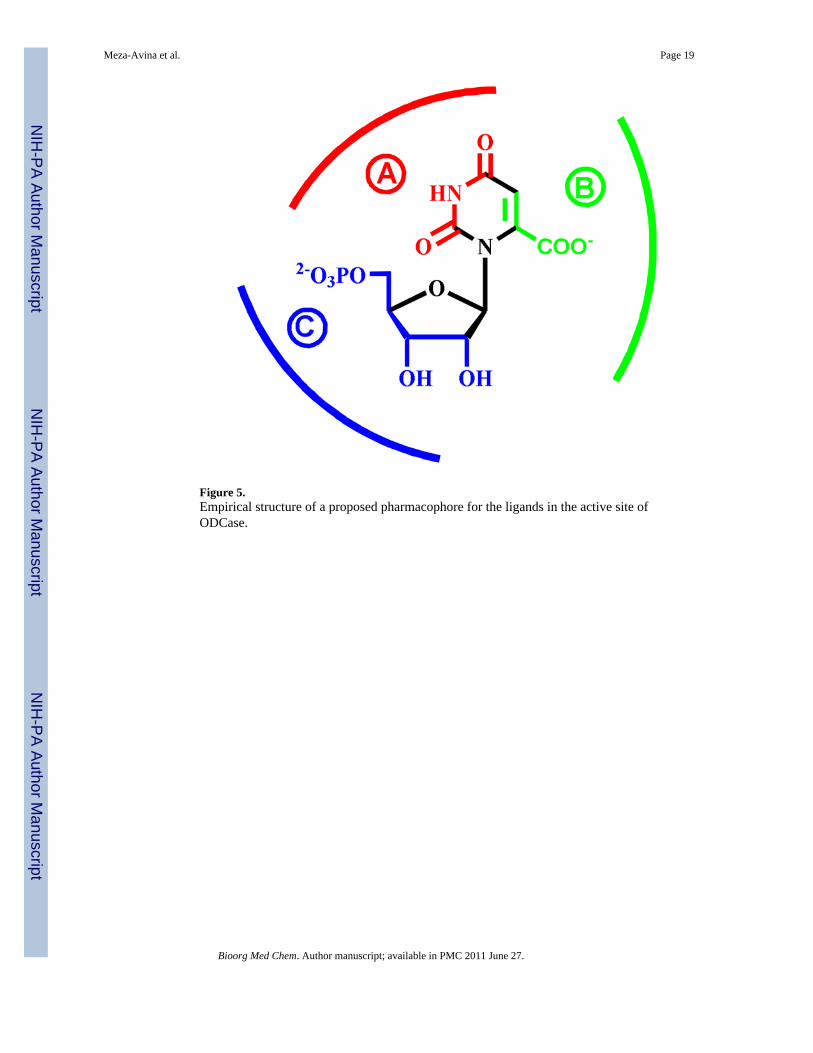
Figure 5.Empirical structure of a proposed pharmacophore for the ligands in the active site ofODCase.
Meza-Avina et al. Page 19
Bioorg Med Chem. Author manuscript; available in PMC 2011 June 27.
NIH
-PA Author Manuscript
NIH
-PA Author Manuscript
NIH
-PA Author Manuscript

Figure 6.Lineweaver-Burk reciprocal plots to determine the reversible inhibition constant Ki 8 withMt ODCase (panel A). Panel B depicts the plot of kobs as a function of [8] with Hs ODCase,and the inset in this panel shows the progress curves of the ODCase reaction in the presenceof various concentrations of 8.
Meza-Avina et al. Page 20
Bioorg Med Chem. Author manuscript; available in PMC 2011 June 27.
NIH
-PA Author Manuscript
NIH
-PA Author Manuscript
NIH
-PA Author Manuscript

Figure 7.Stereo view of the active site of Hs ODCase with docked nifedipine (Panel A) andnimodipine (Panel B). The ligand is shown in a ball-and-stick representation and theresidues of the enzyme are displayed by a capped stick model; atoms are color-codedaccording to atom type. The backbone of the enzyme is rendered according to secondarystructure, and a portion of the active site is rendered using a Connolly surface with lipophiliccharacter. A potential hydrogen bonding network is shown by dashed lines in orange.
Meza-Avina et al. Page 21
Bioorg Med Chem. Author manuscript; available in PMC 2011 June 27.
NIH
-PA Author Manuscript
NIH
-PA Author Manuscript
NIH
-PA Author Manuscript

Figure 8.Overlap of eleven ligands bound in the active site of ODCase, obtained from thecorresponding co-crystal structures. All inhibitors are shown in capped-stick representation:blue: UMP, cyan: 6-aza-UMP, green: 5-I-UMP, greenblue: BMP, magenta: 6-NH2-UMP,orange: 5-Br-UMP, purple: 5-CN-UMP, red: OMP, red-orange: 5-F-UMP, violet: 6-acetyl-UMP, yellow: 6-I-UMP.
Meza-Avina et al. Page 22
Bioorg Med Chem. Author manuscript; available in PMC 2011 June 27.
NIH
-PA Author Manuscript
NIH
-PA Author Manuscript
NIH
-PA Author Manuscript

Scheme 1.Synthesis of compound 8 and 10.
Meza-Avina et al. Page 23
Bioorg Med Chem. Author manuscript; available in PMC 2011 June 27.
NIH
-PA Author Manuscript
NIH
-PA Author Manuscript
NIH
-PA Author Manuscript

NIH
-PA Author Manuscript
NIH
-PA Author Manuscript
NIH
-PA Author Manuscript
Meza-Avina et al. Page 24
Table 1
Data collection and refinement statistics for the x-ray crystal structures of the ODCase complexes of theinhibitors, 4, 8 and 10. Numbers in brackets are for the highest resolution shells. Rsym = S|I − ‹I›/σI, where I isthe observed intensity and ‹I› is the average intensity from multiple observations of symmetry-relatedreflections.
Hs ODCase + 4 Hs ODCase + 8 Hs ODCase + 10
Diffraction data
Resolution (Å) 1.93 (1.96–1.93) 1.20 (1.30–1.20) 1.60 (1.66–1.60)
Measured reflections (n) 149,221 525,889 193,939
Unique reflections (n) 21,306 78,679 58,529
Completeness (%) 95.3 (74.6) 87.0 (60.0) 78.5 (21.7)
Rsym (%) 13.0 (50.0) 2.8 (30.6) 5.8 (32.3)
Space group C2221 C2221 P21
Unit cell dimensions (Å) a 77.6 79.4 69.5
b 116.9 116.5 61.6
c 62.0 62.1 70.9
Unit cell angle (°) α, β, χ 90, 90, 90 90, 90, 90 90, 118.9, 90
Molecules in asymmetric unit (n) 1 1 2
Refinement statistics
Resolution (Å) 50.0–1.93 (1.98–1.93) 30.4–1.20 (1.23–1.20) 50.0–1.60 (1.63–1.60)
Protein atoms (n) 2,025 2,543 4,167
Water molecules (n) 132 348 256
Reflections used for Rfree (n) 1,048 (54) 3,965 (166) 2,938 (49)
Rwork (%) 18.1 (25.0) 17.2 (27.3) 17.9 (27.5)
Rfree (%) 21.0 (25.3) 18.8 (25.1) 21.6 (33.7)
Root mean square deviation bond length (Å) 0.010 0.010 0.010
Root mean square deviation bond angle (°) 1.6 1.5 1.5
Average B-factor (Å2) 27.1 15.9 22.4
Bioorg Med Chem. Author manuscript; available in PMC 2011 June 27.
NIH
-PA Author Manuscript
NIH
-PA Author Manuscript
NIH
-PA Author Manuscript
Meza-Avina et al. Page 25
Table 2
Inhibition kinetics for compounds 4, 8, and 10 against Hs-, Mt-, Pf-, Hp-, Sa-, and ScODCases.
EnzymeKi (mM)
4 8 10
Hs ODCase --- 1.7 ×10⁻⁵ ---
Mt ODCase --- 6.2 ± 0.6 ×10⁻³ ---
Pf ODCase --- --- 9.1 ± 0.3 ×10²
Hp ODCase --- 1.3 ± 0.6 ×10⁻⁴ 2.9 ± 0.1 ×10²
Sa ODCase --- --- 5.5 ± 0.07 ×10²
Sc ODCase38 9 ×10⁻⁹ 5 ×10⁻⁶ ---
Bioorg Med Chem. Author manuscript; available in PMC 2011 June 27.