
Complete Genome Sequence and Comparative Genomics of Shigella flexneri Serotype 2a Strain 2457T
Upload
khangminh22Category
view
4download
0
ARTICLE
Immune stealth-driven O2 serotype prevalence andpotential for therapeutic antibodies againstmultidrug resistant Klebsiella pneumoniaeMeghan E. Pennini1, Anna De Marco2, Mark Pelletier1, Jessica Bonnell1, Romana Cvitkovic1, Martina Beltramello2,
Elisabetta Cameroni2, Siro Bianchi2, Fabrizia Zatta2, Wei Zhao1, Xiaodong Xiao1, Maria M. Camara1,
Antonio DiGiandomenico1, Elena Semenova1, Antonio Lanzavecchia3, Paul Warrener1, JoAnn Suzich1,
Qun Wang1, Davide Corti 2 & C.Kendall Stover 1
Emerging multidrug-resistant bacteria are a challenge for modern medicine, but how these
pathogens are so successful is not fully understood. Robust antibacterial vaccines have
prevented and reduced resistance suggesting a pivotal role for immunity in deterring anti-
biotic resistance. Here, we show the increased prevalence of Klebsiella pneumoniae lipopo-
lysaccharide O2 serotype strains in all major drug resistance groups correlating with a paucity
of anti-O2 antibodies in human B cell repertoires. We identify human monoclonal antibodies
to O-antigens that are highly protective in mouse models of infection, even against heavily
encapsulated strains. These antibodies, including a rare anti-O2 specific antibody, syner-
gistically protect against drug-resistant strains in adjunctive therapy with meropenem, a
standard-of-care antibiotic, confirming the importance of immune assistance in antibiotic
therapy. These findings support an antibody-based immunotherapeutic strategy even for
highly resistant K. pneumoniae infections, and underscore the effect humoral immunity has on
evolving drug resistance.
DOI: 10.1038/s41467-017-02223-7 OPEN
1MedImmune, 1 Medimmune Way, Gaithersburg, MD 20878, USA. 2Humabs BioMed SA, a subsidiary of Vir Biotechnology, Inc., Via Mirasole 1, 6500Bellinzona, Switzerland. 3 Institute for Research in Biomedicine, Università della Svizzera Italliana, Via Vincenzo Vela 6, 6500 Bellinzona, Switzerland.Correspondence and requests for materials should be addressed to Q.W. (email: [email protected]) or to D.C. (email: [email protected]) orto C.K.S. (email: [email protected])
NATURE COMMUNICATIONS |8: 1991 |DOI: 10.1038/s41467-017-02223-7 |www.nature.com/naturecommunications 1
1234
5678
90

Among the most problematic multidrug-resistant (MDR)bacterial pathogens are the Gram-negative carbapenem-resistant enteric bacteria (CRE), including Klebsiella
pneumoniae1, 2. K. pneumoniae colonizes the lower gastro-intestinal tract, from which it can disseminate, particularly whenthe commensal microbiota is disrupted by antibiotic treatment orwhen a patient is immunocompromised3. More than a third of K.pneumoniae clinical isolates express extended spectrum beta-lactamases (ESBL) and are resistant to third generation cepha-losporins, aminoglycosides, tetracycline, and other antibiotics4.CRE isolates are additionally resistant to carbapenems and arestrongly correlated with poor patient outcome5, 6, with mortalityrates as high as 60%.
While the majority of effort on understanding the spread ofantibiotic resistance has focused on the acquisition of mutationsor resistance genes that directly affect drug susceptibility, themost successful resistant strains clearly have additional adapta-tions. For example, the successful spread of the CRE sequencetype 258 (ST258) clonal group is the subject of intense studybecause the diverse resistance genes this group carries are notunique to this lineage7, 8. In addition to bacterial factors, there isgrowing evidence demonstrating a link between host immunityand reduced antibiotic resistance. Since the onset of routinevaccinations against Haemophilus influenzae, Streptococcuspneumoniae, and Neisseria meningitidis, resistance within theseorganisms has been reduced or nearly eradicated9. Effectiveimmunity can decrease the severity and/or duration of an infec-tion thus lessening the need for antibiotic intervention andreducing the selective pressure imparted by antibiotic exposure.Host humoral immunity can also work in concert with antibiotictherapy to synergistically clear drug-resistant pathogens thatcannot be cleared with antibiotic alone10, 11.
Lipopolysaccharide (LPS) is a critical component of the Gram-negative bacterial outer membrane. The earliest antibody-basedstrategies against Gram-negative pathogens targeted the con-served Lipid A component of the LPS inner core with the aim ofneutralizing lipid A proinflammatory properties, but these effortsfailed for a variety of reasons12–15. Beyond the conserved innercore, there are at least seven distinct Klebsiella LPS serotypesdefined by unique O-antigen structures16. K. pneumoniae O1 andO2 LPS O-antigens share the D-galactan I disaccharide unit, butO1 also includes the D-galactan II disaccharide and is thereforemore complex in structure and larger in size17, 18. In previoustarget agnostic efforts to identify antibodies against K. pneumo-niae19, we found that LPS serotype-specific antibodies were themost potent in promoting bacterial killing in vitro.
In this study, we survey a large collection of clinical isolatesto determine the relative prevalence of LPS serotypes, particu-larly in multidrug-resistant isolates. We find that LPS O2antigen serotype has increased two to threefold in prevalence inboth K. pneumoniae ESBL and CRE multidrug-resistant straingroups relative to the susceptible group. This finding is sur-prising as strains expressing the O2-antigen, includingST258 strains, are more sensitive to human serum killing thanthe related O1 antigen expressing strains. We demonstrateST258 strains are almost exclusively the O2 serotype, but theoverall increased O2 prevalence is not limited to ST258. Thesedata suggest a selective advantage for the LPS O2-antigen typein the context of antibiotic pressure. We find that O2 strainprevalence may be explained by its reduced immunogenicitythat imparts a stealth advantage against antibody-drivenmechanisms of clearance. In addition, we identify serotype-specific anti-O1 and anti-O2 human monoclonal antibodies(mAbs) that exhibit potent opsonophagocytic killing of K.pneumoniae strains and provide protection in mouse models ofinfection. These data indicate that protective non-capsular
mAbs targeting less diverse anti-O antigen types expressed byMDR strains can be identified. Importantly, these serotype-specific O1 and O2 antibodies synergistically augment anti-biotic therapy even against drug-resistant strains, further sup-porting an important role for antibody-mediated immunity inthe context of antibiotic therapy. Together, these findingssupport that O-antigen immunogenic stealth provides a selec-tive advantage to drive multidrug resistance and that protectiveantibodies can synergize with antibiotic therapy, thus under-scoring the importance of humoral immunity to drug-resistantbacteria.
NT7%O7
5%
O511%
O43%
O128%
15014080
60
% V
iabi
lity
40
20
0
O1 str
ains
O2 (2
42)
ESBL (7
3)
CRE (144
)
O2 str
ains
O217%O3
28%
NT8%
NT22%
O52%
ESBL (252)
O411%
O235%
O138%
O112%
O35%
O39%
O51%O44% O2
50%
CRE (266)
Susceptible (191)
O2 strains MLST survey
ST258100
75
ST
(%
)
50
25
0
a b
c
Fig. 1 Klebsiella pneumoniae serotype O2 prevalence in MDR strains. a LPSserotype-specific antibodies were used to determine the serotype of 709 K.pneumoniae clinical isolates from 38 different countries by western blot. Piecharts represent the distribution of each serotype as a percentage of thetotal in each antibiotic resistant group. Susceptible, ESBL and CRE strainsare categorized based on their MIC profile as described in Methods. Moreinformation about the source of the isolates is listed in SupplementaryTable 1. b Serum sensitivity assays were performed in 45% pooled humansera using a panel of 12 O1 isolates, 12 O2 isolates and two separateisogenic pairs genetically converting an O2 strain to O1 (Kp961842,magenta diamonds) or an O1 strain to O2 (Kp1131115, blue diamonds).Percent viability is the ratio between CFUs present in buffer control mediaand CFUs present in human sera after 2 h growth. c A subset of serotypeO2 isolates was further analyzed by Sanger or next generation sequencingto determine ST types. The CRE ST258 sequence type is represented by theshaded orange portion of each bar to highlight the percent of ST258isolates observed in each category
ARTICLE NATURE COMMUNICATIONS | DOI: 10.1038/s41467-017-02223-7
2 NATURE COMMUNICATIONS |8: 1991 |DOI: 10.1038/s41467-017-02223-7 |www.nature.com/naturecommunications
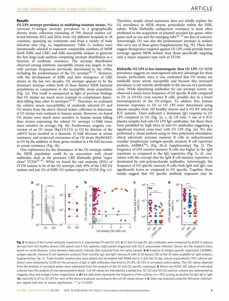
ResultsO2 LPS serotype prevalence in multidrug-resistant strains. Wesurveyed O-antigen serotype prevalence in a geographicallydiverse strain collection consisting of 709 clinical isolates col-lected between 2012 and 2014, from 162 different hospitals in 38countries, spanning six continents and from a variety of bodyinfection sites (Fig. 1a, Supplementary Table 1). Isolates wereintentionally selected to represent comparable numbers of MDR(both ESBL and CRE) and fully susceptible isolates to generatethe first large-scale analysis comparing serotype distribution as afunction of antibiotic resistance. The serotype distributionobserved among antibiotic susceptible strains was largely in linewith serotype frequencies previously determined in the 1990s,including the predominance of the O1 serotype20, 21. However,with the development of ESBL and later emergence of CREstrains in the last two decades, O2 serotype appears to be thedominant serotype within both of these distinct MDR bacterialpopulations in comparison to the susceptible strain population(Fig. 1a). This result is unexpected in light of previous findingsthat O1 strains are much more resistant to complement depen-dent killing than other O serotypes22–25. Therefore, we evaluatedthe relative serum susceptibility of randomly selected O1 andO2 strains from the above clinical isolate collection to determineif O2 strains were resistant to human serum. However, we foundO2 strains were much more sensitive to human serum killingthan strains expressing the related O1 serotype (>2000 timesmore sensitive on average, Fig. 1b). Furthermore, isogenic con-version of an O1 strain (Kp1131115) to O2 by deletion of thewbbYZ locus resulted in a dramatic 21-fold decrease in serumresistance, and reciprocal conversion of an O2 strain (Kp961842)to O1 by the addition of these genes resulted in a 90-fold increasein serum resistance (Fig. 1b).
One explanation for the dominance of the O2 serotype withinthe MDR population could be an association with clonaloutbreaks, such as the pervasive CRE Klebsiella global “superclone” ST25831–33. While we found the vast majority (85%) ofST258 isolates to be of the O2 serotype, only 49% of the CRE O2isolates and just 5% of ESBL O2 isolates typed as ST258 (Fig. 1c).
Therefore, simple clonal expansion does not wholly explain theO2 prevalence in MDR strains, particularly within the ESBLsubset. While Klebsiella carbapenem resistance is most oftenattributed to the acquisition of plasmid encoded kpc genes, othergenes such as oxa and the emerging ndm34, 35 are also of concern.Interestingly, O2 was also the predominant serotype in isolatesthat carry any of these genes (Supplementary Fig. 1b). These datasuggest therapeutics targeted against O2 LPS could provide bettercoverage against MDR isolates than those specifically targetingonly a major sequence type such as ST258.
Klebsiella O2 LPS is less immunogenic than O1 LPS. O2 MDRprevalence suggests an unrecognized selective advantage for thesestrains, particularly since it was confirmed that O2 strains aremarkedly more serum susceptible and because this increasedprevalence is not entirely attributable to the success of a particularclone. While identifying antibodies for our serotype screen, weobserved a much lower frequency of O2 specific B cells comparedto O1 or O1/O2 cross reactive B cells, possibly due to a lowerimmunogenicity of the O2-antigen. To address this, humanimmune responses to O1 or O2 LPS were determined usingplasma samples from 103 healthy donors and 6 O2 KP infectedICU patients. Titers indicated a dominant IgG response to O1LPS compared to O2 (Fig. 2a, c, d). Of note, 5 out of 6 ICUplasma samples had anti-O2 LPS IgG antibodies, but those titerswere paralleled by high titers of anti-O1 antibodies suggesting asignificant fraction cross-react with O1 LPS (Fig. 2a). We alsoperformed a clonal analysis using in vitro polyclonal stimulation,which selectively activates memory B cells in unfractionatedtonsillar lymphocytes (antigen-specific memory B cell repertoireanalysis, AMBRA36), (Fig. 2b–d, Supplementary Fig. 2). Thefrequency of LPS reactive memory B cells was higher in the IgMrepertoire as compared to the IgG repertoire (Fig. 2c, d), con-sistent with the concept that the IgM B cell memory repertoire isdominated by anti-polysaccharide antibodies. Interestingly, thefrequency of O2-specific memory B cells, both IgM and IgG, wassignificantly lower as compared to O1 specific. Together, theseresults suggest that O2 specific antibody responses may be
103****
103
102 **** ********
****
101
100
102
101
100
4
3
2
1
00 1 2 3 4
O2 binding OD (405 nm)
102
102
101 101
O1 O2O1
LPSO1-
O2O2 O1
LPSO1-
O2O2O1
O1
bind
ing
OD
(40
5 nm
)
LPS
-rel
ativ
e Ig
G B
mem
ory
wel
ls (
%)
LPS
-rel
ativ
e Ig
M B
mem
ory
wel
ls (
%)
O2
Pla
sma
IgG
bin
ding
tite
r
a b c d
Fig. 2 Analysis of the human antibody response to K. pneumoniae O1 and O2 LPS. a O1 and O2-specific IgG antibodies were measured by ELISA in plasmaderived from 103 healthy donors (left panel) and 6 ICU patients (right panel) diagnosed with O2 K. pneumoniae infection. Shown are the endpoint titersbased on serial dilutions. Lines between data points indicate data acquired from the same sample. b–d Analysis of antigen-specific repertoires (AMBRA,antigen-specific memory B cell repertoire analysis) from tonsillar IgG and IgM memory B cells of 33 donors (30 of the 33 were available for IgM analysis,Supplementary Fig. 2). Total tonsillar lymphocytes were plated and stimulated with R848 and IL-2 and the 10-day culture supernatants (192 cultures perdonor) were analyzed by ELISA for the presence of IgG or IgM antibodies that bind to O1 LPS, O2 LPS or uncoated control plates. The OD values obtainedfrom the analysis of uncoated plates were subtracted from the analysis of the O1 and O2 specific responses. b Shown are ELISA OD values of individualcultures from the analysis of one representative donor. Cut-off values are indicated by a dotted line. O1, O2 and O1/O2 reactive cultures are represented asmagenta, blue and orange circles, respectively. c, d Each data point represents the frequency of the cultures (n= 192) scoring as positive for IgG (c) or IgM(d) reactivity to O1 or O2 LPS for each of the donors analyzed, according to the cut-off values shown in b. Data was analyzed using the Wilcoxon matched-pair signed rank test to assess significance. ****p< 0.0001
NATURE COMMUNICATIONS | DOI: 10.1038/s41467-017-02223-7 ARTICLE
NATURE COMMUNICATIONS |8: 1991 |DOI: 10.1038/s41467-017-02223-7 |www.nature.com/naturecommunications 3
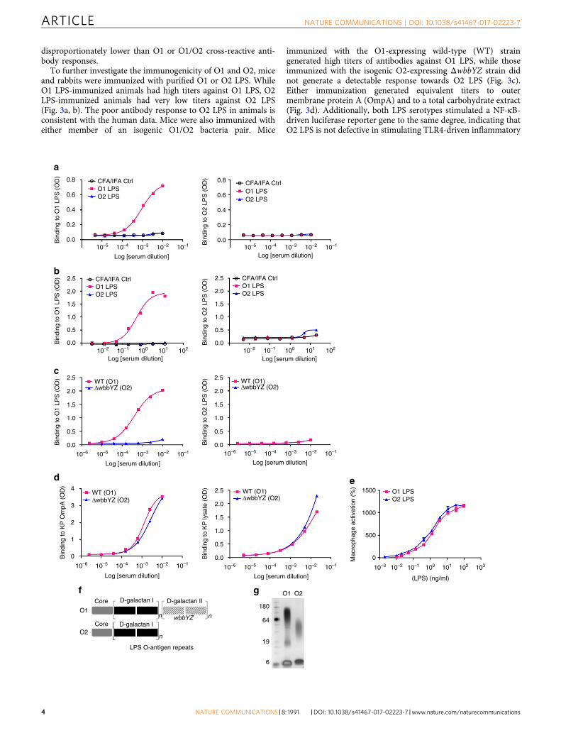
disproportionately lower than O1 or O1/O2 cross-reactive anti-body responses.
To further investigate the immunogenicity of O1 and O2, miceand rabbits were immunized with purified O1 or O2 LPS. WhileO1 LPS-immunized animals had high titers against O1 LPS, O2LPS-immunized animals had very low titers against O2 LPS(Fig. 3a, b). The poor antibody response to O2 LPS in animals isconsistent with the human data. Mice were also immunized witheither member of an isogenic O1/O2 bacteria pair. Mice
immunized with the O1-expressing wild-type (WT) straingenerated high titers of antibodies against O1 LPS, while thoseimmunized with the isogenic O2-expressing ΔwbbYZ strain didnot generate a detectable response towards O2 LPS (Fig. 3c).Either immunization generated equivalent titers to outermembrane protein A (OmpA) and to a total carbohydrate extract(Fig. 3d). Additionally, both LPS serotypes stimulated a NF-κB-driven luciferase reporter gene to the same degree, indicating thatO2 LPS is not defective in stimulating TLR4-driven inflammatory
0.8
0.6
0.4
Bin
ding
to O
1 LP
S (
OD
)B
indi
ng to
O1
LPS
(O
D)
Bin
ding
to O
2 LP
S (
OD
)
0.2
0.0
2.5
2.0
1.5
1.0
0.5
0.0
Bin
ding
to O
1 LP
S (
OD
) 2.5
2.0
1.5
1.0
0.5
0.0
Bin
ding
to K
P O
mpA
(O
D) 4
3
2
1
0
2.5
2.0
1.5
1.0
0.5
0.0
Bin
ding
to O
2 LP
S (
OD
) 2.5
2.0
1.5
1.0
0.5
0.0
Bin
ding
to K
P ly
sate
(O
D)
Mac
roph
age
activ
atio
n (%
)2.5 1500
1000
500
0
2.0
1.5
1.0
0.5
0.0
0.8
0.6
0.4B
indi
ng to
O2
LPS
(O
D)
0.2
0.0
CFA/IFA CtrlO1 LPSO2 LPS
CFA/IFA CtrlO1 LPSO2 LPS
CFA/IFA Ctrl
WT (O1)ΔwbbYZ (O2)
WT (O1)ΔwbbYZ (O2)
WT (O1)ΔwbbYZ (O2)
WT (O1) O1 LPSO2 LPSΔwbbYZ (O2)
O1 LPSO2 LPS
CFA/IFA CtrlO1 LPSO2 LPS
10–5 10–4 10–3 10–2 10–1
Log [serum dilution]
10–510–6 10–4 10–3 10–2 10–1
Log [serum dilution]
10–510–6
O1
O2
Core
Core
D-galactan I D-galactan II
D-galactan I
LPS O-antigen repeats
180
O1 O2
64
19
6
n
n
nwbbYZ
10–4 10–3 10–2 10–1
Log [serum dilution]10–510–6 10–4 10–3 10–310–2 10–210–1 10–1 100 101 102 103
Log [serum dilution] (LPS) (ng/ml)
10–510–6 10–4 10–3 10–2 10–1
Log [serum dilution]
10–5 10–4 10–3 10–2 10–1
Log [serum dilution]
10–2 10–1 100 101 102
Log [serum dilution]10–2 10–1 100 101 102
Log [serum dilution]
a
b
c
d e
f g
ARTICLE NATURE COMMUNICATIONS | DOI: 10.1038/s41467-017-02223-7
4 NATURE COMMUNICATIONS |8: 1991 |DOI: 10.1038/s41467-017-02223-7 |www.nature.com/naturecommunications
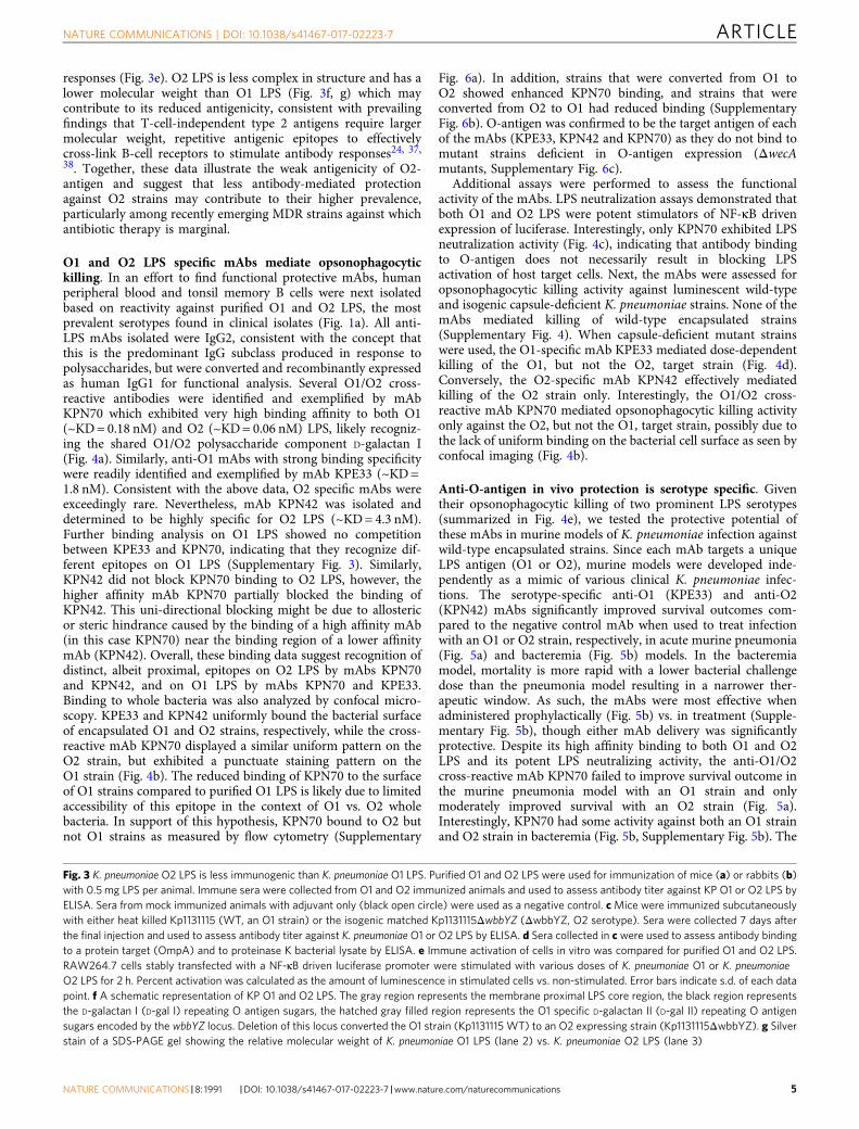
responses (Fig. 3e). O2 LPS is less complex in structure and has alower molecular weight than O1 LPS (Fig. 3f, g) which maycontribute to its reduced antigenicity, consistent with prevailingfindings that T-cell-independent type 2 antigens require largermolecular weight, repetitive antigenic epitopes to effectivelycross-link B-cell receptors to stimulate antibody responses24, 37,38. Together, these data illustrate the weak antigenicity of O2-antigen and suggest that less antibody-mediated protectionagainst O2 strains may contribute to their higher prevalence,particularly among recently emerging MDR strains against whichantibiotic therapy is marginal.
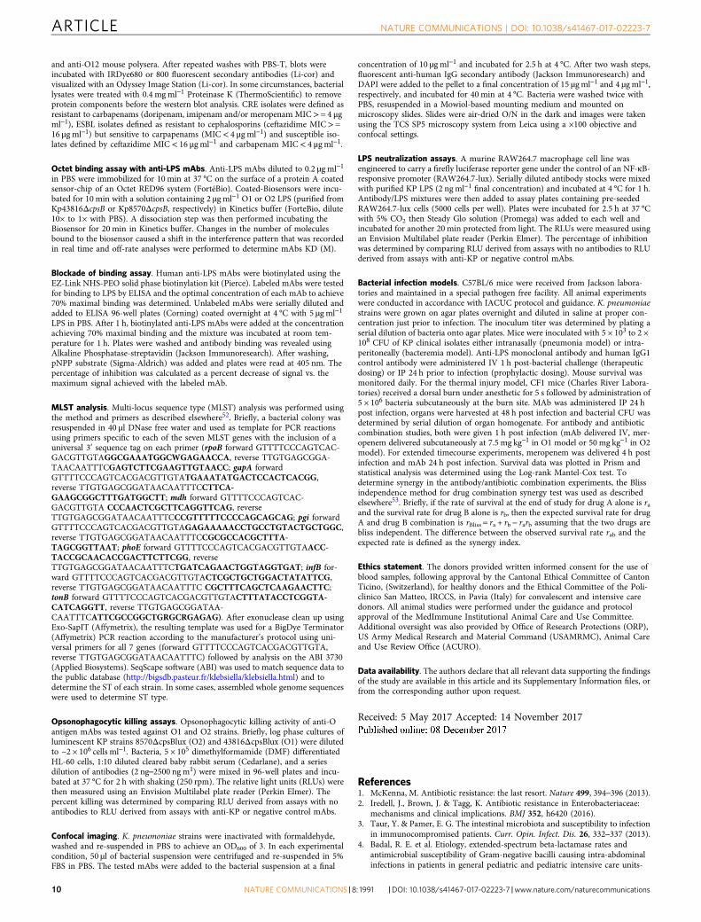
O1 and O2 LPS specific mAbs mediate opsonophagocytickilling. In an effort to find functional protective mAbs, humanperipheral blood and tonsil memory B cells were next isolatedbased on reactivity against purified O1 and O2 LPS, the mostprevalent serotypes found in clinical isolates (Fig. 1a). All anti-LPS mAbs isolated were IgG2, consistent with the concept thatthis is the predominant IgG subclass produced in response topolysaccharides, but were converted and recombinantly expressedas human IgG1 for functional analysis. Several O1/O2 cross-reactive antibodies were identified and exemplified by mAbKPN70 which exhibited very high binding affinity to both O1(~KD = 0.18 nM) and O2 (~KD = 0.06 nM) LPS, likely recogniz-ing the shared O1/O2 polysaccharide component D-galactan I(Fig. 4a). Similarly, anti-O1 mAbs with strong binding specificitywere readily identified and exemplified by mAb KPE33 (~KD =1.8 nM). Consistent with the above data, O2 specific mAbs wereexceedingly rare. Nevertheless, mAb KPN42 was isolated anddetermined to be highly specific for O2 LPS (~KD = 4.3 nM).Further binding analysis on O1 LPS showed no competitionbetween KPE33 and KPN70, indicating that they recognize dif-ferent epitopes on O1 LPS (Supplementary Fig. 3). Similarly,KPN42 did not block KPN70 binding to O2 LPS, however, thehigher affinity mAb KPN70 partially blocked the binding ofKPN42. This uni-directional blocking might be due to allostericor steric hindrance caused by the binding of a high affinity mAb(in this case KPN70) near the binding region of a lower affinitymAb (KPN42). Overall, these binding data suggest recognition ofdistinct, albeit proximal, epitopes on O2 LPS by mAbs KPN70and KPN42, and on O1 LPS by mAbs KPN70 and KPE33.Binding to whole bacteria was also analyzed by confocal micro-scopy. KPE33 and KPN42 uniformly bound the bacterial surfaceof encapsulated O1 and O2 strains, respectively, while the cross-reactive mAb KPN70 displayed a similar uniform pattern on theO2 strain, but exhibited a punctuate staining pattern on theO1 strain (Fig. 4b). The reduced binding of KPN70 to the surfaceof O1 strains compared to purified O1 LPS is likely due to limitedaccessibility of this epitope in the context of O1 vs. O2 wholebacteria. In support of this hypothesis, KPN70 bound to O2 butnot O1 strains as measured by flow cytometry (Supplementary
Fig. 6a). In addition, strains that were converted from O1 toO2 showed enhanced KPN70 binding, and strains that wereconverted from O2 to O1 had reduced binding (SupplementaryFig. 6b). O-antigen was confirmed to be the target antigen of eachof the mAbs (KPE33, KPN42 and KPN70) as they do not bind tomutant strains deficient in O-antigen expression (ΔwecAmutants, Supplementary Fig. 6c).
Additional assays were performed to assess the functionalactivity of the mAbs. LPS neutralization assays demonstrated thatboth O1 and O2 LPS were potent stimulators of NF-κB drivenexpression of luciferase. Interestingly, only KPN70 exhibited LPSneutralization activity (Fig. 4c), indicating that antibody bindingto O-antigen does not necessarily result in blocking LPSactivation of host target cells. Next, the mAbs were assessed foropsonophagocytic killing activity against luminescent wild-typeand isogenic capsule-deficient K. pneumoniae strains. None of themAbs mediated killing of wild-type encapsulated strains(Supplementary Fig. 4). When capsule-deficient mutant strainswere used, the O1-specific mAb KPE33 mediated dose-dependentkilling of the O1, but not the O2, target strain (Fig. 4d).Conversely, the O2-specific mAb KPN42 effectively mediatedkilling of the O2 strain only. Interestingly, the O1/O2 cross-reactive mAb KPN70 mediated opsonophagocytic killing activityonly against the O2, but not the O1, target strain, possibly due tothe lack of uniform binding on the bacterial cell surface as seen byconfocal imaging (Fig. 4b).
Anti-O-antigen in vivo protection is serotype specific. Giventheir opsonophagocytic killing of two prominent LPS serotypes(summarized in Fig. 4e), we tested the protective potential ofthese mAbs in murine models of K. pneumoniae infection againstwild-type encapsulated strains. Since each mAb targets a uniqueLPS antigen (O1 or O2), murine models were developed inde-pendently as a mimic of various clinical K. pneumoniae infec-tions. The serotype-specific anti-O1 (KPE33) and anti-O2(KPN42) mAbs significantly improved survival outcomes com-pared to the negative control mAb when used to treat infectionwith an O1 or O2 strain, respectively, in acute murine pneumonia(Fig. 5a) and bacteremia (Fig. 5b) models. In the bacteremiamodel, mortality is more rapid with a lower bacterial challengedose than the pneumonia model resulting in a narrower ther-apeutic window. As such, the mAbs were most effective whenadministered prophylactically (Fig. 5b) vs. in treatment (Supple-mentary Fig. 5b), though either mAb delivery was significantlyprotective. Despite its high affinity binding to both O1 and O2LPS and its potent LPS neutralizing activity, the anti-O1/O2cross-reactive mAb KPN70 failed to improve survival outcome inthe murine pneumonia model with an O1 strain and onlymoderately improved survival with an O2 strain (Fig. 5a).Interestingly, KPN70 had some activity against both an O1 strainand O2 strain in bacteremia (Fig. 5b, Supplementary Fig. 5b). The
Fig. 3 K. pneumoniae O2 LPS is less immunogenic than K. pneumoniae O1 LPS. Purified O1 and O2 LPS were used for immunization of mice (a) or rabbits (b)with 0.5 mg LPS per animal. Immune sera were collected from O1 and O2 immunized animals and used to assess antibody titer against KP O1 or O2 LPS byELISA. Sera from mock immunized animals with adjuvant only (black open circle) were used as a negative control. c Mice were immunized subcutaneouslywith either heat killed Kp1131115 (WT, an O1 strain) or the isogenic matched Kp1131115ΔwbbYZ (ΔwbbYZ, O2 serotype). Sera were collected 7 days afterthe final injection and used to assess antibody titer against K. pneumoniae O1 or O2 LPS by ELISA. d Sera collected in c were used to assess antibody bindingto a protein target (OmpA) and to proteinase K bacterial lysate by ELISA. e Immune activation of cells in vitro was compared for purified O1 and O2 LPS.RAW264.7 cells stably transfected with a NF-κB driven luciferase promoter were stimulated with various doses of K. pneumoniae O1 or K. pneumoniaeO2 LPS for 2 h. Percent activation was calculated as the amount of luminescence in stimulated cells vs. non-stimulated. Error bars indicate s.d. of each datapoint. f A schematic representation of KP O1 and O2 LPS. The gray region represents the membrane proximal LPS core region, the black region representsthe D-galactan I (D-gal I) repeating O antigen sugars, the hatched gray filled region represents the O1 specific D-galactan II (D-gal II) repeating O antigensugars encoded by the wbbYZ locus. Deletion of this locus converted the O1 strain (Kp1131115 WT) to an O2 expressing strain (Kp1131115ΔwbbYZ). g Silverstain of a SDS-PAGE gel showing the relative molecular weight of K. pneumoniae O1 LPS (lane 2) vs. K. pneumoniae O2 LPS (lane 3)
NATURE COMMUNICATIONS | DOI: 10.1038/s41467-017-02223-7 ARTICLE
NATURE COMMUNICATIONS |8: 1991 |DOI: 10.1038/s41467-017-02223-7 |www.nature.com/naturecommunications 5

0.4
0.3
0.2
Bin
ding
to L
PS
O1
(RU
)0.1
0.0
0.4
0.3
0.2
Bin
ding
to L
PS
O2
(RU
)
0.1
0.0
0 500
KPE33
100
50
0
O1
LPS
neu
tral
izat
ion
(%)
O1
OP
K a
ctiv
ity (
%)
O2
OP
K a
ctiv
ity (
%)
–50
100
80
60
40
20
0
–20
100
80
60
40
20
0
–20
10–3 10–2
10–2
10–1
10–1
100
100
mAb (μg ml)
Log (mAb) μg ml
KPE33KPN70KPN42c-IgG
KPE33KPN70KPN42c-IgG
101
101 10–2 10–1 100
Log (mAb) μg ml
101
102 103
100
50
0
O2
LPS
neu
tral
izat
ion
(%)
–50
10–3 10–2 10–1 100
mAb (μg ml)
101 102 103
O1
O2
KPN42 KPN70 KPE33 KPN42 KPN70
1000
Time (s)
1500 0 500 1000
Time (s)
1500
LPS mAb
O1O1
LPS Neut Δ Capsule OPK
O1/O2
O2
O2 O1 O2KPE33KPN70
KPN42
– ––
–
–
––
++ ++++++
+++
+++
KPE33KPN70KPN42
a
b
c
d
e
Fig. 4 In vitro characterization of anti-O antigen mAbs. a Association/dissociation curves of K. pneumoniae O1 or K. pneumoniae O2 LPS binding to mAbsKPE33, KPN70, and KPN42 were measured by ForteBio Octet using protein A capture probe pre-loaded with each mAb. b Confocal images of mAb bindingto an O1 (Kp43816) or O2 (Kp9148) strain (scale bar, 2 μm). c LPS neutralization was measured using RAW264.7 cells transfected with a NK-kB drivenluciferase promoter stimulated with 2 ngml−1 of purified O1 or O2 LPS in the presence of KPE33, KPN70, KPN42 or the isotype-matched control mAb (c-IgG). Percent inhibition was calculated as the amount of luminescence (RLU) in treated wells divided by the RLU in wells stimulated with LPS alonemultiplied by 100. d Opsonophagocytic killing assays were performed using various concentrations of mAbs in the presence of the phagocytic cell line HL-60. The capsule-deficient luminescent mutants Kp43816ΔcpsB and Kp8570ΔcpsB were used as the O1 and O2 serotype targets, respectively. Percentkilling was calculated based on the amount of luminescence after 2.5 h incubation in treated wells vs. wells with bacteria only. An isotype-matched mAb (c-IgG) is used as a negative control. e Summary of the in vitro functional activities against O1 and O2 K. pneumoniae for each mAb. All error bars indicate s.d.at each data point
ARTICLE NATURE COMMUNICATIONS | DOI: 10.1038/s41467-017-02223-7
6 NATURE COMMUNICATIONS |8: 1991 |DOI: 10.1038/s41467-017-02223-7 |www.nature.com/naturecommunications

activity of KPN70 against O1 suggests that LPS neutralization inthe absence of opsonophagocytic killing may preferentially pro-vide limited benefit in the bacteremia model vs. the pneumoniamodel. KPE33 also protected against dissemination to distalorgans from necrotic tissue at the burn site in a murine model ofthermal injury (Fig. 5c), suggesting the utility of anti-O-antigenmAbs to protect in multiple infection models. Further in vivotesting using additional strains (including Kp8045, a highlyvirulent, mucoid K1 strain39) in a pneumonia model (Supple-mentary Fig. 5a), confirmed the high level of serotype-specificprotection mediated by the KPE33 and KPN42 mAbs as well as
the inferior activity of the cross-reactive, LPS neutralizing mAbKPN70. Interestingly, KPE33 and KPN42 showed in vivo activityeven though mAb in vitro activity was restricted to capsule-deficient strains (Fig. 4d, Supplementary Fig. 4). The data fromthis panel of mAbs, in concert with recently published datashowing that an opsonophagocytic killing deficient mutantKPE33 loses activity in vivo40, supports opsonophagocytic killingactivity against specific epitopes as a major mechanism of in vivoprotection, at least in the pneumonia model. Conversely, a ben-eficial role for LPS neutralization may be more limited anddependent on the type of infection.
100
% S
urvi
val 75
50
25
00 24 48 72 96 120 144 168
Hrs post infection
100
% S
urvi
val 75
50
25
1011
1010
109
108
108
107
106
105
104
103
102
108109
107
106
105
104
103
102
101
108
107
106
105
104
103
102
101
CF
U/g
CF
U/g
CF
U/g
CF
U/g
00 24 48 72 96 120
Hrs post infection
100
% S
urvi
val 75
50
25
00 24 48 72 96 120
Hrs post infection
KPE33 (45 mpk)KPE33 (15 mpk)c-IgG (45 mpk)
O1 Pneumonia
O1 Bacteremia O2 Bacteremia
100
% S
urvi
val 75
50
25
00 24 48 72 96 120 144 168
Hrs post infection
O2 Pneumonia
KPE33 (10 mpk)
KPE33 (15 mpk)
KPE33 (5 mpk)
KPE33 (1 mpk)
KPN70 (15 mpk)
KPN70 (5 mpk)
KPN70 (1 mpk)
c-IgG (15 mpk)
KPN42 (10 mpk)KPN42 (2 mpk)
KPN42 (15 mpk)KPN42 (5 mpk)KPN42 (1 mpk)KPN70 (15 mpk)KPN70 (5 mpk)KPN70 (1 mpk)c-IgG (15 mpk)
KPN42 (0.4 mpk)KPN70 (10 mpk)c-IgG (10 mpk)
KPE33 (2 mpk)KPE33 (0.4 mpk)KPN70 (10 mpk)c-IgG (10 mpk)
***
************
***
*****
********
***
***
**
Liver Spleen
**
Skin Lung
**
**
a
b
c
Fig. 5 Anti-O1 and anti-O2 mAbs are protective in vivo. a Therapeutic activity of the mAbs was tested in C57BL/6 mice infected intranasally with a K.pneumoniae O1 (Kp1131115, 6 × 10⁷ CFU) or O2 (Kp961842, 2 × 10⁸ CFU) strain. KPE33 (anti-O1), KPN42 (anti-O2) and KPN70 (anti-O1/O2) mAbs wereadministered intravascular 1 h post infection at the doses indicated. An isotype-matched mAb (c-IgG) was used as a negative control. b Prophylacticactivity of the mAbs was tested in C57BL/6 mice infected intraperitoneal with the O1 strain Kp1131115 (3 × 10⁶ CFU) or the O2 strain Kp961842 (1 × 10⁷
CFU). mAbs were administered IP 24 h prior to infection at the doses indicated; survival was monitored for 5 days post infection. c CF1 mice received adorsal burn followed by K. pneumoniae O1 (Kp1131115, 5 × 10⁶) bacterial inoculation subcutaneously at the burn site. KPE33 (45 or 15 mpk) or isotype-matched control mAb (c-IgG, 45 mpk) were administered IP 24 h post infection, and organs were harvested 48 h post infection to determine bacterial CFU.All graphs are representative of at least three separate experiments. Mantel-Cox analysis was done to assess significant survival benefit compared to thecontrol mAb. For thermal injury CFU comparison, Kruskal Wallace one way Anova analysis was done to compare differences in CFU between KPE33treated vs. control IgG treated groups, ****p< 0.0001, ***p< 0.001, **p< 0.01
NATURE COMMUNICATIONS | DOI: 10.1038/s41467-017-02223-7 ARTICLE
NATURE COMMUNICATIONS |8: 1991 |DOI: 10.1038/s41467-017-02223-7 |www.nature.com/naturecommunications 7

Anti-O-antigen mAbs protection is synergistic with antibiotics.Clinical application of anti-KP antibodies would likely be incombination with current standard of care antibiotic treatments,including a carbapenem such as meropenem. We thereforeassessed the potential for the anti-O1 and O2 mAbs to comple-ment antibiotic therapy using sub-therapeutic doses of antibioticsto simulate insufficient drug exposure against drug-resistantstrains, as described previously10, 11, 41. These experiments wereperformed using identical conditions to the pneumonia modeldescribed in Fig. 5a with the additional administration of mer-openem. A sub-therapeutic dose of mAb KPE33 administered incombination with a sub-protective meropenem dose providedsignificant protection in the murine pneumonia model against aintermediately meropenem-resistant O1 strain (MIC = 2 µg ml−1).Statistical analysis confirmed synergistic adjunctive activitycompared to either drug administered alone (Fig. 6a). The effi-cacy of the mAb was maintained even when administered 24 hpost infection (Fig. 6b). Similarly, a sub-therapeutic dose of mAbKPN42 in combination with a dose approximating a human-equivalent level of meropenem, also provided significant protec-tion in this model against a highly meropenem-resistant O2 CREST258 strain (MIC = 32 µg ml−1, eight times more than thehuman breakpoint) (Fig. 6a). These data clearly illustrate thatmAbs targeting O-antigen could not only provide protectionwhen dosed sufficiently in monotherapy, but could also providecomplementary synergistic activity in adjunctive therapy withantibiotics. In conclusion, these results indicate that anti-O-antigen antibodies can provide additional and complementarybenefits to antibiotic therapy even against highly carbapenem-resistant KP strains and further substantiate the concept ofantibody help in antibiotic therapy10, 11, 41. Without this
additional antibody-mediated help, O2 strains may preferentiallypersist under antibiotic pressure.
DiscussionK. pneumoniae is a common enteric bacterial species in humangut flora, therefore basal levels of antibody to immunodominantantigens are to be expected and were indeed evident in our surveyof tonsillar and circulating memory B cells. In our efforts toidentify protective mAbs against K. pneumoniae, only a smallfraction of antibodies recognizing whole bacteria possessed potentopsonophagocytic killing activity, the majority of which recog-nized LPS O-antigen. It is reasonable to consider that humoralimmunity to O-antigen may offer some level of protection againstthe establishment of systemic infections in healthy individuals,and these antigens are currently under clinical investigation as avaccine target against other pathogens such as nontyphoidalSalmonella, Vibrio cholera and Shigella sonnei42–46, though ineach case the antibody activity was verified against unencapsu-lated strains only. Our study determined for the first time that theO2 serotype comprises a significantly larger fraction of ESBL(35%) and CRE (50%) strains in comparison to fully susceptible(17%) KP strains suggesting a selective advantage for MDRstrains of the O2 serotype despite their susceptibility to serumkilling. Much attention has been focused on the particularlysuccessful MDR carbapenem-resistant ST258 sequence type of K.pneumoniae, which we serotyped as almost exclusively O2.However, as ST258 only accounts for half of CRE strains and amuch smaller fraction of the earlier emerging ESBL strains,simple clonal outbreaks cannot completely account for theincreased prevalence of the O2 serotype.
100
75
50
% S
urvi
val
25
00 24 48 72 96 120 144 168
Hours post challenge
100
75
50
% S
urvi
val
25
00 24 48 72 96 120 144 168
Hours post challenge
O1 Pneumoniaa
b O1 Pneumonia
KPE33 + MEM
KPE33 (45 mpk) + MEMKPE33 (15 mpk) + MEMKPE33 (5 mpk) + MEMc-IgG (45 mpk) + MEM
****
****
***
******
***
100
75
50
% S
urvi
val
25
00 24 48 72 96 120 144 168
Hours post challenge
O2 Pneumonia
****
KPE33MEMc-IgG + MEMc-IgG
KPN42 + MEMKPN42MEMc-IgG + MEMc-IgG
Fig. 6 Antibody synergy with antibiotic. aMice were infected intranasally as in Fig. 5 with the K. pneumoniae O1 (Kp1131115, 6 × 10⁷ CFU) or O2 (Kp961842,2 × 108 CFU) strain then treated 1 h post infection with suboptimal doses of mAb (KPE33, 1 mg kg−1 or KPN42, 0.2 mg kg−1 given IV) and antibiotic(meropenem, 7.5 mg kg−1 for O1 strain; 50mg kg−1 for O2 strain, given subcutaneous) or a combination of both mAb and antibiotic. Graphs represent thecombined data from three separate experiments with a total of 24 mice in each group. Mantel-Cox analysis was done to assess significant survival benefitcompared to the control mAb (a) or control mAb +MEM (b), ****p< 0.0001, ***p< 0.001, **p< 0.01. The Bliss independence method was used todetermine synergistic activity of the combination therapy vs. either monotherapy alone; p= 2.5 × 10⁻⁶ for KPE33+MEM combination and p= 7.7 × 10⁻⁵ forKPN42+MEM combination. b Mice were infected intransal with a KP O1 strain (Kp8045, 1 × 10⁴ CFU). Meropenem (1.5 mpk) was administeredsubcutaneous 4 h post infection and mAb KPE33 was administered IV 24 h post infection at the indicated doses. All graphs are representative of at leastthree separate experiments. An isotype-matched mAb (c-IgG) was used as a negative control. Mantel-Cox analysis was done to assess significant survivalbenefit compared to the control mAb+MEM b, ****p< 0.0001, ***p< 0.001, **p< 0.01
ARTICLE NATURE COMMUNICATIONS | DOI: 10.1038/s41467-017-02223-7
8 NATURE COMMUNICATIONS |8: 1991 |DOI: 10.1038/s41467-017-02223-7 |www.nature.com/naturecommunications

A possible explanation for the unexpected prevalence of theserum-susceptible O2 serotype is the lower immunogenicity ofthe O-antigen and the consequent failure to induce antibodiesthat synergize with antibiotics. Thus, the combination of drugresistance and lack of immunogenicity observed in this study maycontribute to the success of O2 strains (including theST258 subset) particularly in the context of broad spectrumantibiotic pressure. It is notable that strains without a detectableO-antigen (non-serotypeable) were also more prevalent in CREisolates. This observation further supports the concept that strainswith less immunogenic O antigen or lacking O antigen expressionmay be selected under antibiotic pressure. However, immuno-genic O1 strains are also prevalent among ESBL isolates andslightly increased in prevalence compared to their representationin susceptible isolates. In this instance, other O1-specificmechanisms of immune evasion may play a role such as theirresistance to human serum killing (Fig. 1b) and/or the increasedpresence of virulence genes in O1 isolates vs. other serotypes16.Human serum sensitivity may play a role in bacterial clearance inpatients, but would not necessarily be expected to translate tomouse models as species-specific differences in serum sensitivityhave been clearly demonstrated for several pathogens, includingK. pneumoniae26–30.
A diminishing pipeline for new antibiotics and the nowrecognized negative impact of broad-spectrum antibiotics on thebeneficial microbiome highlight the necessity of developing newantibacterial strategies47. The diversity of capsular poly-saccharides of K. pneumoniae has been suggested by many to bean immunodominant feature that obscures the accessibility ofantibodies to other surface targets. Here, we demonstrate that it ispossible to identify highly protective mAbs to the less diverse O-antigen. Despite the relative rarity of O2 antigen-specific B cells, itwas possible to identify a mAb (KPN42) with high protectiveactivity. Potent mAbs targeting the O1 antigen were also identi-fied. Interestingly, these mAbs had no opsonophagocytic killingactivity against WT strains in vitro. However, in vitro activityagainst capsule knock-out strains was found to be predictive ofactivity against WT encapsulated strains in vivo, suggestingcapsule may be expressed at lower levels or less consistentlyin vivo. Consistent with previous studies with other bacterialpathogens10, 11, the anti-KP mAbs isolated in this study affordedsynergistic adjunctive protection to drug-resistant strains,including a representative strain of the problematic ST258 CREclone. These data further support an under-appreciated role forhumoral immunity in antibiotic therapy against KP and indeedthe background impact of immunity on bacterial drug resistanceobserved with vaccines9. Furthermore, these studies provide someencouragement that mAb-based strategies may offer futuresolutions for combatting antibiotic refractory bacteria.
MethodsK. pneumoniae strains. All K. pneumoniae isolates were purchased from AmericaType Culture Collection (ATCC), Eurofin or IHMA (a complete list of strains canbe found in Supplementary Table 2), and cultures were maintained in 2xYT mediaat 37 °C supplemented with antibiotics when appropriate. For capsule mutantstrain generation, approximately one kilobase of DNA flanking either side of thecpsB/manC gene was PCR amplified and cloned into the pir-dependent plasmidpDMS19748. This plasmid was then electroporated into Klebsiella strains andplasmid integrants selected on LB agar containing 10 μg ml−1 tetracycline or gen-tamycin. After propagation of recombinants in the absence of selection, clones thatresolved the integrated plasmid were selected by growth on LB agar containing 10%sucrose. Deletion of manC was confirmed by PCR on lysed colonies with primersspecific to the manC gene (AAACAGTTCCTCCGTCTGTTC, GGAA-TAAAGGTGGACTGGTTCT) using the following cycling conditions: 94° for 2min, 30 cycles of 94° for 30 s, 58° for 30 s, then 72° for 1 min.
Clinical isolates. For serotyping, clinical isolates were obtained from InternationalHealth Management Associates (Schaumburg, IL, USA). The isolates were selected
based on MIC profiles (to ensure relatively equal numbers of susceptible, ESBL andCRE strains), then chosen from 6 continents, 38 different countries and at least 162different hospitals in an attempt to generate a global snapshot of current K.pneumoniae infections as opposed to a sampling of any single outbreak. All isolateswere collected between 2012 and 2014 from various sites of infection (a breakdownof infection site can be found in Supplementary Table 1).
Isolation and production of LPS serotyping antibodies. Peripheral bloodmononuclear cells (PBMC) and sera were separated from buffy coats of healthyblood donors or convalescent patients after K. pneumoniae infection as previouslydescribed49. Alternatively, lymphocytes were obtained from tonsils after tissuehomogenization in the presence of DNase I and collagenase. The donors providedwritten informed consent for the use of these samples, following approval by theCantonal Ethical Committee of Canton Ticino, Switzerland (for healthy donors) andthe Ethical Committee of the Policlinico San Matteo, IRCCS, in Pavia (Italy) (forconvalescent ICU donors). Memory B cells were isolated from cryopreserved PMBCor from lymphocytes isolated from tonsils using PE-Cy7-labeled CD19 microbeads(BD Biosciences, clone SJ25C1), followed by isolation with anti PE-beads (MiltenyiBiotec) and by depletion of cells stained with anti-IgM (Jackson ImmunoResearch),anti-IgD (BD Biosciences), and anti-IgA (Novex) antibodies by cell sorting on aFACSAria (BD Biosciences). Memory B cells were immortalized with EBV asdescribed previously36, 50 Briefly, sorted memory B cells were infected with EBV andseeded into 384 well plates in the presence of CpG TRL9 agonist and allogenicirradiated PBMCs. Immortalized memory B cells were then screened for antibodybinding to purified LPS by ELISA. Total mRNA from positive B cell cultures wasreverse transcribed in 50 µl nuclease-free water (Ambion) using 15 µM specific pri-mer (IgG:5′-TCTTGTCCACCTTGGTGTTGCT; Igκ: 5′ACACTCTCCCCTGTT-GAAGCTCTT; Igλ: 5′-ACTGTCTTCTCCACGGTGCT), 1.8 µl of 25 mM dNTPmix (GE Healthcare), 5 µl of 0.1M DTT, 0.5% v/v Igepal CA-630 (Sigma), 60 URNAse OUT (Invitrogen), and 100 U Superscript III reverse transcriptase (Invitro-gen). Reverse transcription (RT) was performed at 42 °C 10min, 55 °C 60min, and94 °C 5min. IgH, Igκ, and Igλ genes were amplified by PCR using HotStar Taq DNApolymerase (Qiagen) and cloned into human Igγ1, Igκ, and Igλ expression vectors, asdescribed51. Recombinant mAbs were produced by transient transfection of EXPI293cells (Invitrogen) and purified by Protein A chromatography (GE Healthcare) anddesalted against PBS. Alternatively, mouse immune sera were generated by purifiedLPS or bacterial immunization. The specificity of the resulting mAbs and polyserawas confirmed by loading 1 μg of purified LPS from serotype reference strains ontoan sodium dodecyl sulfate-polyacrylamide gel electrophoresis (SDS-PAGE), followedby transfer onto a nitrocellulose membrane, and probed using our isolated anti-O-antigen mAbs or polyclonal mouse sera (Supplementary Fig. 1a).
Enzyme-linked immunosorbent assay. Spectraplate-384 with high proteinbinding treatment (custom made from Perkin Elmer) or Nunc 96-well highbinding plates were coated overnight at 4 °C with 5 µg ml−1 LPS in phosphate-buffered saline (PBS), pH 7.2, and plates were subsequently blocked with PBSsupplemented with 1% BSA (low endotoxin, Sigma-Aldrich). The coated plateswere incubated with serial dilutions of our isolated anti-O-antigen humanmonoclonal antibodies for 1 h at room temperature. The plates were then washedwith PBS containing 0.1% Tween-20 (PBS-T), and alkaline phosphatase-affiniPureF(ab’)2 Frag rabbit anti-human IgG, Fcg Frag Specific (Jackson ImmunoResearch,10,000× dilution) or alkaline phosphatase-goat anti-human IgG (Southern Biotech)were added and incubated for 1 h at room temperature. Plates were washed threetimes with PBS-T, and P-NitroPhenyl Phosphate (pNPP, Sigma-Aldrich) substrateswere added and the absorbance of 405 nm was measured by a microplate reader(Biotek or Molecular Devices). Data were plotted with GraphPad Prism software.
AMBRA of IgG and IgM antibodies from tonsillar B cells. Replicate cultures oftotal tonsillar lymphocytes were seeded at 20,000 cells per well in 96 U-bottomplates (Costar) and stimulated with 2.5 μg ml−1 R848 (3 M) and 1000 Uml−1
human recombinant IL-2 for 10 days at 37 °C 5% CO2. The cells culture super-natants were collected for further analysis. Spectraplate-384 with high proteinbinding treatment (Perkin Elmer) or Nunc 96-well high binding plates were coatedovernight at 4 °C with 5 µg ml−1 O1 or O2 LPS in phosphate-buffered saline (PBS).Plates were blocked with PBS 1% BSA (low endotoxin, Sigma-Aldrich) and thenincubated with AMBRA supernatant for 1 h at room temperature. After washingswith PBS 0.1% Tween the binding of IgG or IgM Abs was detected with goatAlkaline Phosphatase-anti human IgG (Southern Biotech) or AlkalinePhosphatase-goat anti human IgM (Southern Biotech), respectively. pNPP (Sigma-Aldrich) substrate was added and absorbance measured at 405 nm on a microplatereader (Biotek).
Serotyping of K. pneumoniae clinical isolates. The serotypes of the KP clinicalisolates were determined by western blot analysis with serotype-specific mono-clonal antibodies or mouse polysera. Briefly, purified LPS or bacterial lysates weresubjected to SDS-PAGE. Separated proteins and LPS were transferred from gels tonitrocellulose membranes with an iBlot apparatus (Life Technology). Membraneswere then blocked with Casein or Odyssey (Li-cor) blocking buffer before beingprobed with anti-O1, O1/O2, O3, O4, or O5 LPS monoclonal antibodies or anti-O7
NATURE COMMUNICATIONS | DOI: 10.1038/s41467-017-02223-7 ARTICLE
NATURE COMMUNICATIONS |8: 1991 |DOI: 10.1038/s41467-017-02223-7 |www.nature.com/naturecommunications 9
and anti-O12 mouse polysera. After repeated washes with PBS-T, blots wereincubated with IRDye680 or 800 fluorescent secondary antibodies (Li-cor) andvisualized with an Odyssey Image Station (Li-cor). In some circumstances, bacteriallysates were treated with 0.4 mg ml−1 Proteinase K (ThermoScientific) to removeprotein components before the western blot analysis. CRE isolates were defined asresistant to carbapenams (doripenam, imipenam and/or meropenam MIC > = 4 μgml−1), ESBL isolates defined as resistant to cephalosporins (ceftazidime MIC> =16 μg ml−1) but sensitive to carpapenams (MIC < 4 μg ml−1) and susceptible iso-lates defined by ceftazidime MIC < 16 μg ml−1 and carbapenam MIC< 4 μg ml−1.
Octet binding assay with anti-LPS mAbs. Anti-LPS mAbs diluted to 0.2 µg ml−1
in PBS were immobilized for 10 min at 37 °C on the surface of a protein A coatedsensor-chip of an Octet RED96 system (FortéBio). Coated-Biosensors were incu-bated for 10 min with a solution containing 2 µg ml−1 O1 or O2 LPS (purified fromKp43816ΔcpsB or Kp8570ΔcpsB, respectively) in Kinetics buffer (ForteBio, dilute10× to 1× with PBS). A dissociation step was then performed incubating theBiosensor for 20 min in Kinetics buffer. Changes in the number of moleculesbound to the biosensor caused a shift in the interference pattern that was recordedin real time and off-rate analyses were performed to determine mAbs KD (M).
Blockade of binding assay. Human anti-LPS mAbs were biotinylated using theEZ-Link NHS-PEO solid phase biotinylation kit (Pierce). Labeled mAbs were testedfor binding to LPS by ELISA and the optimal concentration of each mAb to achieve70% maximal binding was determined. Unlabeled mAbs were serially diluted andadded to ELISA 96-well plates (Corning) coated overnight at 4 °C with 5 µg ml−1
LPS in PBS. After 1 h, biotinylated anti-LPS mAbs were added at the concentrationachieving 70% maximal binding and the mixture was incubated at room tem-perature for 1 h. Plates were washed and antibody binding was revealed usingAlkaline Phosphatase-streptavidin (Jackson Immunoresearch). After washing,pNPP substrate (Sigma-Aldrich) was added and plates were read at 405 nm. Thepercentage of inhibition was calculated as a percent decrease of signal vs. themaximum signal achieved with the labeled mAb.
MLST analysis. Multi-locus sequence type (MLST) analysis was performed usingthe method and primers as described elsewhere52. Briefly, a bacterial colony wasresuspended in 40 μl DNase free water and used as template for PCR reactionsusing primers specific to each of the seven MLST genes with the inclusion of auniversal 3′ sequence tag on each primer (rpoB forward GTTTTCCCAGTCAC-GACGTTGTAGGCGAAATGGCWGAGAACCA, reverse TTGTGAGCGGA-TAACAATTTCGAGTCTTCGAAGTTGTAACC; gapA forwardGTTTTCCCAGTCACGACGTTGTATGAAATATGACTCCACTCACGG,reverse TTGTGAGCGGATAACAATTTCCTTCA-GAAGCGGCTTTGATGGCTT; mdh forward GTTTTCCCAGTCAC-GACGTTGTA CCCAACTCGCTTCAGGTTCAG, reverseTTGTGAGCGGATAACAATTTCCCGTTTTTCCCCAGCAGCAG; pgi forwardGTTTTCCCAGTCACGACGTTGTAGAGAAAAACCTGCCTGTACTGCTGGC,reverse TTGTGAGCGGATAACAATTTCCGCGCCACGCTTTA-TAGCGGTTAAT; phoE forward GTTTTCCCAGTCACGACGTTGTAACC-TACCGCAACACCGACTTCTTCGG, reverseTTGTGAGCGGATAACAATTTCTGATCAGAACTGGTAGGTGAT; infB for-ward GTTTTCCCAGTCACGACGTTGTACTCGCTGCTGGACTATATTCG,reverse TTGTGAGCGGATAACAATTTC CGCTTTCAGCTCAAGAACTTC;tonB forward GTTTTCCCAGTCACGACGTTGTACTTTATACCTCGGTA-CATCAGGTT, reverse TTGTGAGCGGATAA-CAATTTCATTCGCCGGCTGRGCRGAGAG). After exonuclease clean up usingExo-SapIT (Affymetrix), the resulting template was used for a BigDye Terminator(Affymetrix) PCR reaction according to the manufacturer’s protocol using uni-versal primers for all 7 genes (forward GTTTTCCCAGTCACGACGTTGTA,reverse TTGTGAGCGGATAACAATTTC) followed by analysis on the ABI 3730(Applied Biosystems). SeqScape software (ABI) was used to match sequence data tothe public database (http://bigsdb.pasteur.fr/klebsiella/klebsiella.html) and todetermine the ST of each strain. In some cases, assembled whole genome sequenceswere used to determine ST type.
Opsonophagocytic killing assays. Opsonophagocytic killing activity of anti-Oantigen mAbs was tested against O1 and O2 strains. Briefly, log phase cultures ofluminescent KP strains 8570ΔcpsBlux (O2) and 43816ΔcpsBlux (O1) were dilutedto ~2 × 10⁶ cells ml−1. Bacteria, 5 × 10⁵ dimethylformamide (DMF) differentiatedHL-60 cells, 1:10 diluted cleared baby rabbit serum (Cedarlane), and a seriesdilution of antibodies (2 ng–2500 ng m1) were mixed in 96-well plates and incu-bated at 37 °C for 2 h with shaking (250 rpm). The relative light units (RLUs) werethen measured using an Envision Multilabel plate reader (Perkin Elmer). Thepercent killing was determined by comparing RLU derived from assays with noantibodies to RLU derived from assays with anti-KP or negative control mAbs.
Confocal imaging. K. pneumoniae strains were inactivated with formaldehyde,washed and re-suspended in PBS to achieve an OD600 of 3. In each experimentalcondition, 50 µl of bacterial suspension were centrifuged and re-suspended in 5%FBS in PBS. The tested mAbs were added to the bacterial suspension at a final
concentration of 10 µg ml−1 and incubated for 2.5 h at 4 °C. After two wash steps,fluorescent anti-human IgG secondary antibody (Jackson Immunoresearch) andDAPI were added to the pellet to a final concentration of 15 µg ml−1 and 4 µg ml−1,respectively, and incubated for 40 min at 4 °C. Bacteria were washed twice withPBS, resuspended in a Mowiol-based mounting medium and mounted onmicroscopy slides. Slides were air-dried O/N in the dark and images were takenusing the TCS SP5 microscopy system from Leica using a ×100 objective andconfocal settings.
LPS neutralization assays. A murine RAW264.7 macrophage cell line wasengineered to carry a firefly luciferase reporter gene under the control of an NF-κB-responsive promoter (RAW264.7-lux). Serially diluted antibody stocks were mixedwith purified KP LPS (2 ng ml−1 final concentration) and incubated at 4 °C for 1 h.Antibody/LPS mixtures were then added to assay plates containing pre-seededRAW264.7-lux cells (5000 cells per well). Plates were incubated for 2.5 h at 37 °Cwith 5% CO2 then Steady Glo solution (Promega) was added to each well andincubated for another 20 min protected from light. The RLUs were measured usingan Envision Multilabel plate reader (Perkin Elmer). The percentage of inhibitionwas determined by comparing RLU derived from assays with no antibodies to RLUderived from assays with anti-KP or negative control mAbs.
Bacterial infection models. C57BL/6 mice were received from Jackson labora-tories and maintained in a special pathogen free facility. All animal experimentswere conducted in accordance with IACUC protocol and guidance. K. pneumoniaestrains were grown on agar plates overnight and diluted in saline at proper con-centration just prior to infection. The inoculum titer was determined by plating aserial dilution of bacteria onto agar plates. Mice were inoculated with 5 × 10³ to 2 ×10⁸ CFU of KP clinical isolates either intranasally (pneumonia model) or intra-peritoneally (bacteremia model). Anti-LPS monoclonal antibody and human IgG1control antibody were administered IV 1 h post-bacterial challenge (therapeuticdosing) or IP 24 h prior to infection (prophylactic dosing). Mouse survival wasmonitored daily. For the thermal injury model, CF1 mice (Charles River Labora-tories) received a dorsal burn under anesthetic for 5 s followed by administration of5 × 106 bacteria subcutaneously at the burn site. MAb was administered IP 24 hpost infection, organs were harvested at 48 h post infection and bacterial CFU wasdetermined by serial dilution of organ homogenate. For antibody and antibioticcombination studies, both were given 1 h post infection (mAb delivered IV, mer-openem delivered subcutaneously at 7.5 mg kg−1 in O1 model or 50 mg kg−1 in O2model). For extended timecourse experiments, meropenem was delivered 4 h postinfection and mAb 24 h post infection. Survival data was plotted in Prism andstatistical analysis was determined using the Log-rank Mantel-Cox test. Todetermine synergy in the antibody/antibiotic combination experiments, the Blissindependence method for drug combination synergy test was used as describedelsewhere53. Briefly, if the rate of survival at the end of study for drug A alone is raand the survival rate for drug B alone is rb, then the expected survival rate for drugA and drug B combination is rBliss = ra + rb − rarb assuming that the two drugs arebliss independent. The difference between the observed survival rate rab and theexpected rate is defined as the synergy index.
Ethics statement. The donors provided written informed consent for the use ofblood samples, following approval by the Cantonal Ethical Committee of CantonTicino, (Switzerland), for healthy donors and the Ethical Committee of the Poli-clinico San Matteo, IRCCS, in Pavia (Italy) for convalescent and intensive caredonors. All animal studies were performed under the guidance and protocolapproval of the MedImmune Institutional Animal Care and Use Committee.Additional oversight was also provided by Office of Research Protections (ORP),US Army Medical Research and Material Command (USAMRMC), Animal Careand Use Review Office (ACURO).
Data availability. The authors declare that all relevant data supporting the findingsof the study are available in this article and its Supplementary Information files, orfrom the corresponding author upon request.
Received: 5 May 2017 Accepted: 14 November 2017
References1. McKenna, M. Antibiotic resistance: the last resort. Nature 499, 394–396 (2013).2. Iredell, J., Brown, J. & Tagg, K. Antibiotic resistance in Enterobacteriaceae:
mechanisms and clinical implications. BMJ 352, h6420 (2016).3. Taur, Y. & Pamer, E. G. The intestinal microbiota and susceptibility to infection
in immunocompromised patients. Curr. Opin. Infect. Dis. 26, 332–337 (2013).4. Badal, R. E. et al. Etiology, extended-spectrum beta-lactamase rates and
antimicrobial susceptibility of Gram-negative bacilli causing intra-abdominalinfections in patients in general pediatric and pediatric intensive care units-
ARTICLE NATURE COMMUNICATIONS | DOI: 10.1038/s41467-017-02223-7
10 NATURE COMMUNICATIONS |8: 1991 |DOI: 10.1038/s41467-017-02223-7 |www.nature.com/naturecommunications

global data from the Study for Monitoring Antimicrobial Resistance Trends2008 to 2010. Pediatr. Infect. Dis. J. 32, 636–640 (2013).
5. van Duin, D. & Paterson, D. L. Multidrug-resistant bacteria in the community:trends and lessons learned. Infect. Dis. Clin. North Am. 30, 377–390 (2016).
6. Russotto, V. et al. Bloodstream infections in intensive care unit patients:distribution and antibiotic resistance of bacteria. Infect. Drug Resist. 8, 287–296(2015).
7. Wyres, K. L. et al. Extensive capsule locus variation and large-scale genomicrecombination within the Klebsiella pneumoniae clonal group 258. GenomeBiol. Evol. 7, 1267–1279 (2015).
8. Wyres, K. L. & Holt, K. E. Klebsiella pneumoniae population genomics andantimicrobial-resistant clones. Trends Microbiol. 24, 944–956 (2016).
9. Lipsitch, M. & Siber, G. R. How can vaccines contribute to solving theantimicrobial resistance problem? mBio 7, e00428-16 (2016).
10. DiGiandomenico, A. et al. A multifunctional bispecific antibody protectsagainst Pseudomonas aeruginosa. Sci. Transl. Med. 6, 262ra155 (2014).
11. Hua, L. et al. Assessment of an anti-alpha-toxin monoclonal antibody forprevention and treatment of Staphylococcus aureus-induced pneumonia.Antimicrob. Agents Chemother. 58, 1108–1117 (2014).
12. Baumgartner, J. D. et al. Prevention of gram-negative shock and death insurgical patients by antibody to endotoxin core glycolipid. Lancet 2, 59–63(1985).
13. Ziegler, E. J. et al. Treatment of gram-negative bacteremia and septic shock withHA-1A human monoclonal antibody against endotoxin. A randomized,double-blind, placebo-controlled trial. The HA-1A Sepsis Study Group. N. Engl.J. Med. 324, 429–436 (1991).
14. Ziegler, E. J. et al. Treatment of gram-negative bacteremia and shock withhuman antiserum to a mutant Escherichia coli. N. Engl. J. Med. 307, 1225–1230(1982).
15. Greenman, R. L. et al. A controlled clinical trial of E5 murine monoclonal IgMantibody to endotoxin in the treatment of gram-negative sepsis. The XOMASepsis Study Group. JAMA 266, 1097–1102 (1991).
16. Follador, R. et al. The diversity of Klebsiella pneumoniae surfacepolysaccharides. Microb. Genom. 2, e000073 (2016).
17. Whitfield, C., Richards, J. C., Perry, M. B., Clarke, B. R. & MacLean, L. L.Expression of two structurally distinct D-galactan O antigens in thelipopolysaccharide of Klebsiella pneumoniae serotype O1. J. Bacteriol. 173,1420–1431 (1991).
18. Vinogradov, E. & Perry, M. B. Structural analysis of the core region of thelipopolysaccharides from eight serotypes of Klebsiella pneumoniae. Carbohydr.Res. 335, 291–296 (2001).
19. Wang, Q. et al. Target-agnostic identification of functional monoclonalantibodies against Klebsiella pneumoniae multimeric MrkA fimbrial subunit. J.Infect. Dis. 213, 1800–1808 (2016).
20. Hansen, D. S. et al. Klebsiella pneumoniae lipopolysaccharide O typing:revision of prototype strains and O-group distribution among clinical isolatesfrom different sources and countries. J. Clin. Microbiol. 37, 56–62 (1999).
21. Trautmann, M. et al. O-antigen seroepidemiology of Klebsiella clinical isolatesand implications for immunoprophylaxis of Klebsiella infections. Clin. Diagn.Lab. Immunol. 4, 550–555 (1997).
22. Sahly, H. et al. Increased serum resistance in Klebsiella pneumoniae strainsproducing extended-spectrum beta-lactamases. Antimicrob. Agents Chemother.48, 3477–3482 (2004).
23. Merino, S., Camprubi, S., Alberti, S., Benedi, V. J. & Tomas, J. M. Mechanismsof Klebsiella pneumoniae resistance to complement-mediated killing. Infect.Immun. 60, 2529–2535 (1992).
24. Hsieh, P. F. et al. D-galactan II is an immunodominant antigen in O1lipopolysaccharide and affects virulence in Klebsiella pneumoniae: implicationin vaccine design. Front. Microbiol. 5, 608 (2014).
25. McCallum, K. L., Schoenhals, G., Laakso, D., Clarke, B. & Whitfield, C. A high-molecular-weight fraction of smooth lipopolysaccharide in Klebsiella serotypeO1:K20 contains a unique O-antigen epitope and determines resistance tononspecific serum killing. Infect. Immun. 57, 3816–3822 (1989).
26. Brown, G. C. The complementary activity of mouse-serum. J. Immunol. 46,319–323 (1943).
27. Muschel, L. H. & Muto, T. Bactericidal reaction of mouse serum. Science 123,62–64 (1956).
28. Warren, H. S. et al. Resilience to bacterial infection: difference between speciescould be due to proteins in serum. J. Infect. Dis. 201, 223–232 (2010).
29. Siggins, M. K. et al. Absent bactericidal activity of mouse serum against invasiveAfrican nontyphoidal Salmonella results from impaired complement functionbut not a lack of antibody. J. Immunol. 186, 2365–2371 (2011).
30. Szijarto, V. et al. Endotoxin neutralization by an O-antigen specific monoclonalantibody: A potential novel therapeutic approach against Klebsiella pneumoniaeST258. Virulence, http://dx.doi.org/10.1080/21505594.2017.1279778 (2017).
31. Lee, C. R. et al. Global dissemination of carbapenemase-producing klebsiellapneumoniae: epidemiology, genetic context, treatment options, and detectionmethods. Front. Microbiol. 7, 895 (2016).
32. Gaiarsa, S. et al. Genomic epidemiology of Klebsiella pneumoniae in Italy andnovel insights into the origin and global evolution of its resistance tocarbapenem antibiotics. Antimicrob. Agents Chemother. 59, 389–396 (2015).
33. Kitchel, B. et al. Molecular epidemiology of KPC-producing Klebsiellapneumoniae isolates in the United States: clonal expansion of multilocussequence type 258. Antimicrob. Agents Chemother. 53, 3365–3370 (2009).
34. Pitout, J. D., Nordmann, P. & Poirel, L. Carbapenemase-producing Klebsiellapneumoniae, a key pathogen set for global nosocomial dominance. Antimicrob.Agents Chemother. 59, 5873–5884 (2015).
35. Jean, S. S. et al. Carbapenemase-producing Gram-negative bacteria: currentepidemics, antimicrobial susceptibility and treatment options. Future Microbiol.10, 407–425 (2015).
36. Pinna, D., Corti, D., Jarrossay, D., Sallusto, F. & Lanzavecchia, A. Clonaldissection of the human memory B-cell repertoire following infection andvaccination. Eur. J. Immunol. 39, 1260–1270 (2009).
37. Vos, Q., Lees, A., Wu, Z. Q., Snapper, C. M. & Mond, J. J. B-cell activation byT-cell-independent type 2 antigens as an integral part of the humoral immuneresponse to pathogenic microorganisms. Immunol. Rev. 176, 154–170 (2000).
38. Mond, J. J., Lees, A. & Snapper, C. M. T cell-independent antigens type 2.Annu. Rev. Immunol. 13, 655–692 (1995).
39. Fang, C. T. et al. Klebsiella pneumoniae genotype K1: an emerging pathogenthat causes septic ocular or central nervous system complications frompyogenic liver abscess. Clin. Infect. Dis.: Off. Publ. Infect. Dis. Soc. Am. 45,284–293 (2007).
40. Cohen, T. et al. Anti-LPS antibodies protect against Klebsiella pneumoniae byempowering neutrophil-mediated clearance without neutralizing TLR4. JCIInsight 2, 92774 (2017).
41. Song, Y. et al. PcrV antibody-antibiotic combination improves survival inPseudomonas aeruginosa-infected mice. Eur. J. Clin. Microbiol. Infect. Dis.: Off.Publ. Eur. Soc. Clin. Microbiol. 31, 1837–1845 (2012).
42. Wu, Y. et al. Development of a live attenuated bivalent oral vaccine againstShigella sonnei shigellosis and typhoid fever. J. Infect. Dis. 215, 259–268 (2016).
43. Lin, J. et al. Monoclonal antibodies to shigella lipopolysaccharide are useful forvaccine production. Clin. Vaccine Immunol. 23, 681–688 (2016).
44. Leitner, D. R. et al. Lipopolysaccharide modifications of a cholera vaccinecandidate based on outer membrane vesicles reduce endotoxicity and reveal themajor protective antigen. Infect. Immun. 81, 2379–2393 (2013).
45. Watson, D. C., Robbins, J. B. & Szu, S. C. Protection of mice against Salmonellatyphimurium with an O-specific polysaccharide-protein conjugate vaccine.Infect. Immun. 60, 4679–4686 (1992).
46. Simon, R. et al. Sustained protection in mice immunized with fractional dosesof Salmonella Enteritidis core and O polysaccharide-flagellin glycoconjugates.PLoS ONE 8, e64680 (2013).
47. Fraenkel-Wandel, Y., Raveh-Brawer, D., Wiener-Well, Y., Yinnon, A. M. &Assous, M. V. Mortality due to blaKPC Klebsiella pneumoniae bacteraemia. J.Antimicrob. Chemother. 71, 1083–1087 (2016).
48. Edwards, R. A., Keller, L. H. & Schifferli, D. M. Improved allelic exchangevectors and their use to analyze 987P fimbria gene expression. Gene 207,149–157 (1998).
49. Beltramello, M. et al. The human immune response to Dengue virus isdominated by highly cross-reactive antibodies endowed with neutralizing andenhancing activity. Cell Host Microbe 8, 271–283 (2010).
50. Corti, D. & Lanzavecchia, A. Efficient methods to isolate human monoclonalantibodies frommemory B cells and plasma cells. Microbiol. Spectr. http://doi:10.1128/microbiolspec.AID-0018-2014 (2014).
51. Tiller, T. et al. Efficient generation of monoclonal antibodies from single humanB cells by single cell RT-PCR and expression vector cloning. J. Immunol.Methods 329, 112–124 (2008).
52. Diancourt, L., Passet, V., Verhoef, J., Grimont, P. A. & Brisse, S. Multilocussequence typing of Klebsiella pneumoniae nosocomial isolates. J. Clin.Microbiol. 43, 4178–4182 (2005).
53. Zhao, W. et al. A new bliss independence model to analyze drug combinationdata. J. Biomol. Screen. 19, 817–821 (2014).
AcknowledgementsThis work was funded by MedImmune, LLC, a wholly owned subsidiary of AstraZenecaPharmaceuticals and a grant from the Defense Advanced Projects Research Agency(DARPA). We thank Professor Piero Marone from Policlinico San Matteo in Pavia, Italyfor providing the sera samples from convalescent patients.
Author contributionsM.E.P., Q.W., P.W., J.S., D.C., E.C., A.D.M., M.B., A.L., and C.K.S. contributed to theexperimental design and writing of this manuscript. M.E.P., Q.W., M.P., A.D.M., J.B., R.C., M.B., X.X., E.C., W.Z., M.M.C., S.B., F.Z., A.D., and E.S. performed experiments orprovided critical reagents.
NATURE COMMUNICATIONS | DOI: 10.1038/s41467-017-02223-7 ARTICLE
NATURE COMMUNICATIONS |8: 1991 |DOI: 10.1038/s41467-017-02223-7 |www.nature.com/naturecommunications 11

Additional informationSupplementary Information accompanies this paper at https://doi.org/10.1038/s41467-017-02223-7.
Competing interests: M.E.P., Q.W., M.P.,J.B, R.C, E.S, X.X, W.Z., M.M.C., A.D., P.W., J.S., C.K.S. are current or former employees of MedImmune/AstraZeneca and may ownstock or stock options in the company. Patents describing the activity of the antibodies inthis work have been filed by MedImmune. A.D.M., M.B, E.C., S.B, F.Z, A.L, D.C. areemployees of HumAbs BioMed and may currently hold Humabs stocks or stock options.The remaining authors declare no competing financial interests.
Reprints and permission information is available online at http://npg.nature.com/reprintsandpermissions/
Publisher's note: Springer Nature remains neutral with regard to jurisdictional claims inpublished maps and institutional affiliations.
Open Access This article is licensed under a Creative CommonsAttribution 4.0 International License, which permits use, sharing,
adaptation, distribution and reproduction in any medium or format, as long as you giveappropriate credit to the original author(s) and the source, provide a link to the CreativeCommons license, and indicate if changes were made. The images or other third partymaterial in this article are included in the article’s Creative Commons license, unlessindicated otherwise in a credit line to the material. If material is not included in thearticle’s Creative Commons license and your intended use is not permitted by statutoryregulation or exceeds the permitted use, you will need to obtain permission directly fromthe copyright holder. To view a copy of this license, visit http://creativecommons.org/licenses/by/4.0/.
© The Author(s) 2017
ARTICLE NATURE COMMUNICATIONS | DOI: 10.1038/s41467-017-02223-7
12 NATURE COMMUNICATIONS |8: 1991 |DOI: 10.1038/s41467-017-02223-7 |www.nature.com/naturecommunications
Copyright © 2022 FDOKUMEN


![Synthesis and Structure of trans-[O2(Im)4Tc]Cl•2H,O, trans-[O2(1-meIm1)4Tc]Cl•3H2O and Related Compounds](https://static.fdokumen.com/doc/165x107/63398ee975188717130476f6/synthesis-and-structure-of-trans-o2im4tccl2ho-trans-o21-meim14tccl3h2o.jpg)