EFA6A encodes two isoforms with distinct biological activities in neuronal cells
Glycobiology vol. 20 no. 11 pp. 1410–1419, 2010doi: 10.1093/glycob/cwq105Advance Access publication on July 4, 2010
The α-galactosidase type A gene aglA from Aspergillus nigerencodes a fully functional α-N-acetylgalactosaminidase
Natallia Kulik2,3, Lenka Weignerová4,7, Tomáš Filipi4,Petr Pompach5,7, Petr Novák5,7, Hynek Mrázek6,7,Kristýna Slámová4, Karel Bezouška6,7, Vladimír Křen1,4,and Rüdiger Ettrich1,2,3
2Center for Biocatalysis and Biotransformation, Institute of Systems Biologyand Ecology, Academy of Sciences of the Czech Republic, Zámek 136, CZ 37333 Nové Hrady, Czech Republic; 3Institute of Physical Biology, University ofSouth Bohemia, Zámek 136, CZ 373 33 Nové Hrady, Czech Republic; 4Centerfor Biocatalysis and Biotransformation, and 5Laboratory of MolecularStructure Characterization, and 6Laboratory of Protein Architecture, Institute ofMicrobiology, Academy of Sciences of the Czech Republic, Vídeňská 1083,CZ 142 20 Praha 4, Czech Republic; and 7Department of Biochemistry,Faculty of Science, Charles University in Prague, Albertov 2030, CZ 128 40Praha 2, Czech Republic
Received on March 1, 2010; revised on June 28, 2010; accepted onJune 30, 2010
Two genes in the genome of Aspergillus niger, aglA andaglB, have been assigned to encode for α-D-galactosidasesvariant A and B. However, analyses of primary and 3Dstructures based on structural models of these two enzymesrevealed significant differences in their active centers sug-gesting important differences in their specificity for thehydrolyzed carbohydrates. To test this unexpected finding,a large screening of libraries from 42 strains of filamentousfungi succeeded in identifying an enzyme from A. nigerCCIM K2 that exhibited both α-galactosidase and α-N-acetylgalactosaminidase activities, with the latter activitypredominating. The enzyme protein was sequenced, andits amino acid sequence could be unequivocally assigned tothe enzyme encoded the aglA gene. Enzyme activity mea-surements and substrate docking clearly demonstrated thepreference of the identified enzyme for α-N-acetyl-D-galactosaminide over α-D-galactoside. Thus, we provideevidence that the α-galactosidase type A gene aglA fromA. niger in fact encodes a fully functional α-N-acetylgalac-tosaminidase using a retaining mechanism.
Keywords: α-N-acetylgalactosaminidase/Aspergillus niger /α-galactosidase /molecular modeling/substrate binding
Introduction
α-Galactosidases (α-D-galactopyranoside galactohydrolase,EC 3.2.1.22) occur widely in microorganisms, plants, and ani-mals and have considerable potential in practical applications(Weignerová et al. 2009). The enzymes encoded by the genesaglA and aglB from Aspergillus niger (Pel et al. 2007) belongto glycohydrolase (GH) family 27, which mainly comprisesα-galactosidases, α-N-acetylgalactosaminidases, and relatedenzymes. Fungal α-galactosidases are typically enzymes witha length of 380–430 amino acids and a highly conserved activesite (Hart et al. 2000; Ly et al. 2000).
Unlike α-galactosidases, the availability of α-N-acetylgalac-tosaminidases is somewhat lower. These enzymes have so farmostly been isolated and structurally characterized from ani-mal sources (Garman et al. 2002) and bacteria (Hsieh et al.2000, 2003). The first human crystal structure was reported on-ly recently (Clark and Garman 2009). With respect to thecatalytic mechanism of α-N-acetylgalactosaminidases, a dou-ble-displacement mechanism using two carboxylate groups asa catalytic pair is expected with the anomeric configuration ofthe leaving monosaccharide unit unchanged (Weignerová et al.2008; Clark and Garman 2009).
A deficiency or mutations of α-N-acetylgalactosaminidase inhumans lead to a lysozomal storage disorder causing Kanzaki dis-ease resulting in neurodegenerative pathologies (Clark andGarman 2009). α-N-Acetylgalactosaminidase has been used toconvert blood group epitope A to the universal blood group H(0)(Liu et al. 2007; Olsson and Clausen 2008). Recently, a promisingcandidate from fungi was reported, the α-N-acetylgalactosamini-dase from A. niger CCIM K2 (Weignerová et al. 2008).Although the aglA gene is assigned to encode a α-galactosidase,this study attempts to prove that the experimentally reported α-N-acetylgalactosaminidase is in fact identical with the encodedenzyme using biochemical, genetic, and structural characteriza-tion in combination with computational modeling.
Results
The recent sequencing of the entire genome of A. niger (Pelet al. 2007) opened the possibility of a targeted search forgenes encoding potential α-galactosidases. A BLAST searchfor α-galactosidase primary sequences within the A. niger ge-nome in the non-redundant protein database found fivedistinct protein-coding genes. Apart from genes aglA andaglB, there are three sequences with the sequence identitymore than 33%. However, these three sequences are not yet
© The Author 2010. Published by Oxford University Press. All rights reserved. For permissions, please e-mail: [email protected]
1To whom correspondence should be addressed: Tel: +420386361297; Fax:+420386361279; [email protected] (R. Ettrich), [email protected](V. Kren).
by guest on August 9, 2016
http://glycob.oxfordjournals.org/D
ownloaded from
characterized and, therefore, are assigned to hypothetical pro-teins with unknown function (gene IDs 4984860, 4978099,and 4987036). The lengths of the hypothetical open readingframes are similar to aglB and vary from 391 to 431 aminoacids.
Sequence analyses revealed that the enzymes coded by aglAand aglB genes differ in their size and in the amino acids of theiractive site. The sequence identity of the two enzymes estimatedby BLAST only reaches 28%. Whereas aglB has an average se-quence identity of more than 70% with known α-galactosidasesfrom the same GH 27 family, the enzyme encoded by aglA ismuch closer to another cluster of enzymes having 65–78% iden-tity with various putative α-galactosidases from fungi, as well as64% identity with the α-N-acetylgalactosaminidase from Acre-monium sp. No. 413. These relationships opened up thepossibility that this gene in fact encodes an α-N-acetylgalacto-saminidase, e.g., an enzyme characterized as an exoglycosidase(α-GalNAc-ase, EC 3.2.1.49, GH family 27) specific for thehydrolysis of terminal α-linked N-acetylgalactosamine in vari-ous sugar chains.
The issue has been addressed in a large screening studyaimed at obtaining a good producer of extracellular α-N-acetylgalactosaminidase activity. A library of filamentous fungi(42 strains) and a series of inducers and cultivation conditionswere examined (Weignerová et al. 2008). We have observedthe presence of at least four extracellular α-galactosidasesin A. niger culture extracts. However, only a single enzymedemonstrated α-N-acetylgalactosaminidase activity, too. The
purified protein from the best producer, A. niger CCIM K2,exhibited a dual enzyme activity and was able to hydrolyze both2-nitrophenyl-2-acetamido-2-deoxy-α-D-galactopyranoside(2-NP-α-GalNAc) and 4-nitrophenyl-α-D-galactopyranoside(4-NP-α-Gal) as its substrate. The specific activity of the puri-fied enzyme was 33.5 and 3.1 U/mg for the α-GalNAc and theα-Gal substrates, respectively, under the given experimentalconditions. Thus, the ability to hydrolyze α-GalNAc substratewas more than tenfold higher compared to the hydrolysis ofα-Gal substrate. Additional biochemical characteristics of theisolated enzyme were also provided. The native molecularweight was estimated to be approximately 440 kDa, and the ex-perimentally obtained pI was close to 4.8. The KM for 2-NP-α-GalNAc substrate was 0.73 mM, and the optimum of enzymeactivity was achieved at pH 1.8 and 55°C. The enzyme belongsto retaining glycosidases as proved by nuclear magnetic res-onance determination of the α/β pyranose forms of themonosaccharide formed during the hydrolysis of 2-NP-α-GalNAc (Weignerová et al. 2008).The purity of the isolated α-N-acetylgalactosaminidase was
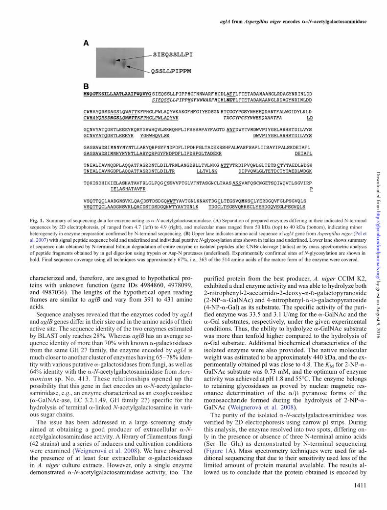
verified by 2D electrophoresis using narrow pI strips. Duringthis analysis, the enzyme resolved into two spots, differing on-ly in the presence or absence of three N-terminal amino acids(Ser–Ile–Glu) as demonstrated by N-terminal sequencing(Figure 1A). Mass spectrometry techniques were used for ad-ditional sequencing that due to their sensitivity used less of thelimited amount of protein material available. The results al-lowed us to conclude that the protein obtained is encoded by
1411
Fig. 1. Summary of sequencing data for enzyme acting as α-N-acetylgalactosaminidase. (A) Separation of prepared enzymes differing in their indicated N-terminalsequences by 2D electrophoresis, pI ranged from 4.7 (left) to 4.9 (right), and molecular mass ranged from 50 kDa (top) to 40 kDa (bottom), indicating minorheterogeneity in enzyme preparation confirmed by N-terminal sequencing. (B) Upper lane indicates amino acid sequence of aglA gene from Aspergillus niger (Pel etal. 2007) with signal peptide sequence bold and underlined and individual putative N-glycosylation sites shown in italics and underlined. Lower lane shows summaryof sequence data obtained by N-terminal Edman degradation of entire enzyme or isolated peptides after CNBr cleavage (italics) or by mass spectrometric analysisof peptide fragments obtained by in gel digestion using trypsin or Asp-N proteases (underlined). Experimentally confirmed sites of N-glycosylation are shown inbold. Final sequence coverage using all techniques was approximately 67%, i.e., 363 of the 514 amino acids of the mature form of the enzyme were covered.
aglA from Aspergillus niger encodes α-N-acetylgalactosaminidase
by guest on August 9, 2016
http://glycob.oxfordjournals.org/D
ownloaded from

the aglA gene in the A. niger genome. The overall sequencecoverage was 67% and thus led to an unambiguous assignment(Figure 1B).
Alignment of the primary sequences of the two enzymes en-coded by the aglA and aglB genes with the primary sequences ofthe available 3D structures determined by X-ray diffraction en-
1412
Fig. 2. Multiple sequence alignment of the α-N-acetylgalactosaminidase from chicken (pdb code: 1KTB), the α-N-acetylgalactosaminidase from humans(pdb code: 3H53), the α-galactosidase from rice (pdb code: 1UAS), the α-galactosidase Trichoderma reesei (pdb code: 1SZN) with enzymes encoded by aglA andaglB genes. Amino acids of α-N-acetylgalactosaminidases and α-galactosidases, which are responsible for substrate specificity, are highlighted in green andyellow respectively; amino acids common to both enzymes are cyan. Secondary structure was assigned with ProCheck (Laskowski et al. 1993).
N Kulik et al.
by guest on August 9, 2016
http://glycob.oxfordjournals.org/D
ownloaded from

abled us to construct amodel of each of these enzymes (Figures 2and 3). As a result, the protein encoded by the aglB gene showedmore than 50% sequence identity to the available solved α-galactosidase crystal structures (Golubev et al. 2004) and only33% sequence identity to the available α-N-acetylgalactosami-nidase crystal structure (enzyme from chicken, Garman et al.2002). In contrast, the enzyme encoded by the aglA gene is char-acterized by 29% sequence identity to the available crystalstructure of the α-galactosidase from Trichoderma reesei and35% sequence identity to the α-N-acetylgalactosaminidase fromchicken. Recently, the structure of human α-N-acetylgalactosa-minidase was determined (Clark and Garman 2009). Ourprimary structure shows an equal sequence identity of 35% tothis new human α-N-acetylgalactosaminidase and a similarityof 50%. The similarity is thus comparable to that between aglAand α-N-acetylgalactosaminidase from chicken describedabove. A phylogenetic analysis reveals a comparable evolution-ary distance to chicken α-N-acetylgalactosaminidase and to
human α-N-acetylgalactosaminidase for the aglA gene (0.764and 0.768, respectively). α-N-Acetylgalactosaminidases fromchicken and humans have a very close 3D structure with a rootmean square deviation of only 0.54 Å for Cα over 387 alignedresidues, with the active site not only fully conserved in the pri-mary sequence (see alignment in Figure 2) but also in 3D.Significant differences between both crystal structures (chickenand human) and our structural model of aglA could only befound in the loop regions on the enzyme surface, where ourmodel has three longer loops (Gly132-Glu145, Ala176-Tyr185, and Asp201-Ala210). Surprisingly, the three loops to-gether cover a similar 3D space as the one large loop seen in thecrystal structure of α-galactosidase from T. reesei (also presentin our α-galactosidase model of gene aglB), indicating that oc-cupation of this specific space could be a feature of all fungalα-galactosidases and α-N-acetylgalactosaminidases. In con-clusion, since both structures are very similar over the entirelength and identical within the active site, the inclusion of the
1413
Fig. 3. Structure of the α-N-acetylgalactosaminidase from Aspergillus niger. (A) Overall fold shows a TIM-barrel with the active site at the N-terminus, a smalldomain of eight antiparallel β-strands packed in β-sandwich in the middle, and a ricin-like domain on the right. The generated model (magenta) is overlaidwith the crystal structure of the homologs α-N-acetylgalactosaminidase from chicken (yellow), with α-N-acetylgalactosamine and the ricin-like domain fromthe xylanase from Streptomyces olivaceoviridis E-86 (green). (B) and (C) Molecular surface of the active site of aglA enzyme (magenta) and aglB enzyme (yellow)with oNP-α-GalNAc. The active site of aglA enzyme has extra space for accommodating the N-acetyl-group of the substrate, while in aglB enzyme this spaceis occupied by Trp205.
aglA from Aspergillus niger encodes α-N-acetylgalactosaminidase
by guest on August 9, 2016
http://glycob.oxfordjournals.org/D
ownloaded from
recently released crystal structure of the human enzyme wouldnot change the modeling results, nor would it affect the 3D ar-rangement in the active site. Therefore, at present we can statethat the closest probable structure for the enzyme encoded bygene aglB is unambiguously α-galactosidase, whereas for theenzyme encoded by the aglA gene the primary sequence analysisshows a slightly higher probability that its 3D structure is closerto that of α-N-acetylgalactosaminidase (Figure 2).
A BLAST domain search identified two domains in the en-zyme encoded by the aglA gene (NCBI reference sequence:XP_001390845.1): a melibiase domain at the N-terminal anda ricin-like domain at the C-terminal end (Figure 3). Comparisonof the aglA gene with amino acid sequences from non-redundantsequence databases identified α-N-acetylgalactosaminidasefrom Acremonium sp. No. 413 (Ashida et al. 2000) as anotherexample of glycosidase containing a ricin-like domain. Othersimilar sequences either do not contain a ricin-like domain orhave a sequence identity of less than 40%. The enzyme encodedby the aglB gene (GenBank: CAK44445.1) contains only amelibiase domain.
The theoretical correctness of the generated models is an im-portant issue in homology modeling. Analysis of the probabilitythat the given primary sequence adopts the predicted fold usingso-called z scores calculated by comparing the conformation en-ergies with ProSA demonstrates the principal correctness of ourtwo initial structural models with z scores of −7.35 (aglA) and −7.88 (aglB), while the local quality scores point to slightly prob-lematic regions for aglA (residues 220–230 and 300–320) and
1414
Table I. Changes in secondary structure as calculated by YASARA of the initialhomology models for enzymes encoded by genes aglA and aglB during themolecular dynamics refinement
Model
Secondary structure content (%)
α-Helix β-Sheet Turn Coil
aglA Before refinement 29.2 27.0 11.8 32.0After refinement 28.8 26.5 10.5 34.2
aglB Before refinement 26.3 23.7 16.0 34.0After refinement 26.1 24.4 16.6 32.9
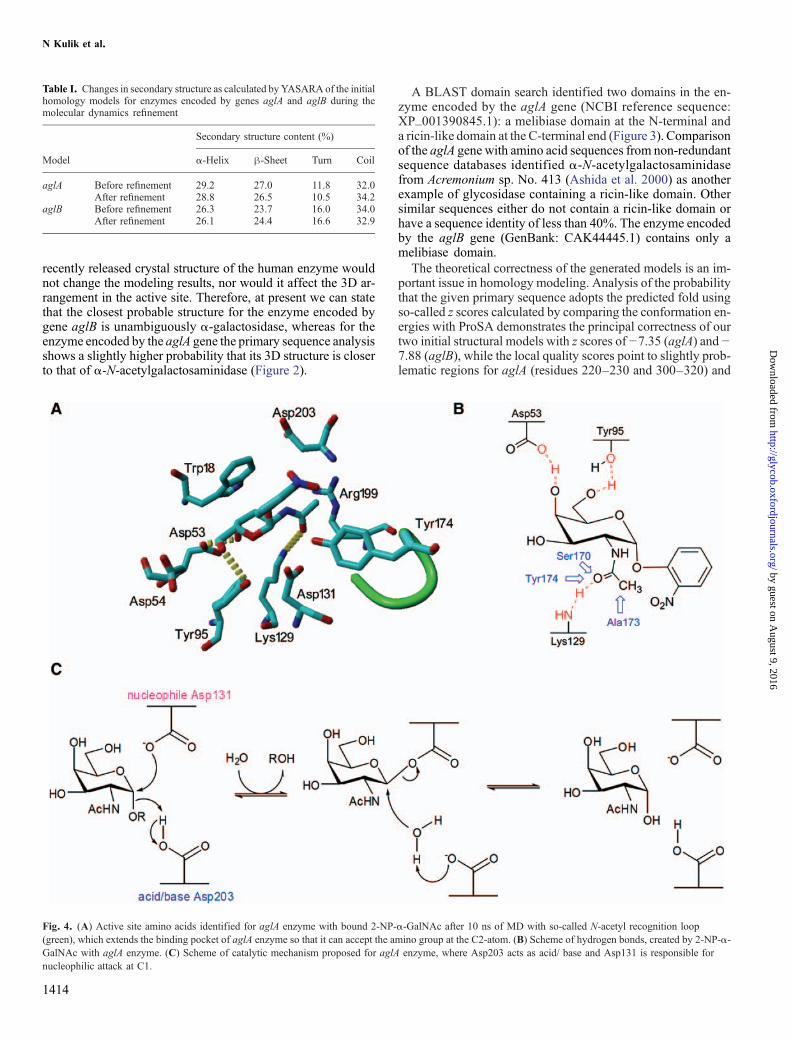
Fig. 4. (A) Active site amino acids identified for aglA enzyme with bound 2-NP-α-GalNAc after 10 ns of MD with so-called N-acetyl recognition loop(green), which extends the binding pocket of aglA enzyme so that it can accept the amino group at the C2-atom. (B) Scheme of hydrogen bonds, created by 2-NP-α-GalNAc with aglA enzyme. (C) Scheme of catalytic mechanism proposed for aglA enzyme, where Asp203 acts as acid/ base and Asp131 is responsible fornucleophilic attack at C1.
N Kulik et al.
by guest on August 9, 2016
http://glycob.oxfordjournals.org/D
ownloaded from
for aglB (residues 200–220 and 340–350). This was probablydue to the low resolution in the corresponding template crystalstructures, and, therefore, we must treat these regions as poor-ly resolved at this initial stage. Improvement was reached by arefinement using molecular dynamics simulation in explicitsolvent with both structural models reaching equilibrium con-formations after 2 ns of simulation. The root mean squaredeviation of Cα atoms reached a plateau with values the dif-ference of which from those of the initial structure was lessthan 0.25 nm. During the refinement, we observed only minorchanges (around 1%) in the secondary structure content of themodel (Table I).
Model analysis discovered significant differences betweenthe amino acid sequences in the active site of the enzymesencoded by the aglA and aglB genes. This result could ex-plain the difference in substrate specificity between bothenzymes (Figure 2). Reasonable structural models stable inmolecular dynamics simulations in explicit solvent can bebuilt only using α-galactosidase as a template for aglB andα-N-acetylgalactosaminidase as a template for aglA. Bothmodels, aglA and aglB, have similar folding: the main domainis a TIM-barrel, containing the active site at the N-terminus ofthe β-barrel; this structure is followed by eight antiparallelβ-strands folded into a β-sandwich. The α-N-acetylgalacto-saminidase isolated from A. niger CCIM K2 has an additionalC-terminal ricin-like domain, making this enzyme somewhatlarger on sodium dodecyl sulfate polyacrylamide gel electro-phoresis (57 vs 45 kDa). Its role is unknown, but its foldingmight indicate a potential interaction with monosaccharides,such as galactose or lactose (Figure 3A).
Automated docking of the corresponding substrates into theactive sites of both enzymes followed by simulations of the re-sulting enzyme–substrate complexes enabled us to analyze thestability of the ligand–protein interaction in aqueous solutionat room temperature. Although equilibration was alreadyreached after 2 ns, the simulations were continued for 10 nsin total to demonstrate the stability of the whole complex.Amino acid residues in the active site of aglA within 0.3 nmof 2-NP-α-GalNAc at the end of the simulation are shownin Figure 4A and B. A docking attempt of 2-NP-α-GalNAcinto the active site of aglB led to a fast repulsion of the 2-acetamido group due to Trp205 blocking the space needed toaccommodate the 2-acetamido group properly and the sidechain of Asp222 flipping in and pushing the N-acetyl-groupcompletely out of the active side towards water. As a result,the distance between the oxygen at the aglycon of the ligandand the oxygen of the catalytic residue, which acts as an acidto hydrolyze the C1-O bond, increased during the first 1.5 nsof molecular dynamics (MD) to an average value of 0.67 nm,a distance too far for the catalytic residue to attack the C1-Obond of the ligand. The same distance measured for aglBwith docked 4-NP-α-Gal after 10 ns of MD was 0.35 nm,allowing appropriate positioning of the substrate for eventualreaction. These results well demonstrate that the enzyme en-coded by gene aglB is a pure α-galactosidase unable toperform any α-N-acetylgalactosaminidase enzymatic function.Figures 3B and C show the active sites of both enzymes withbound substrates in a surface representation.The binding energies of the respective substrates during the
MD simulations were calculated by Yasara for the first 4 ns of
1415
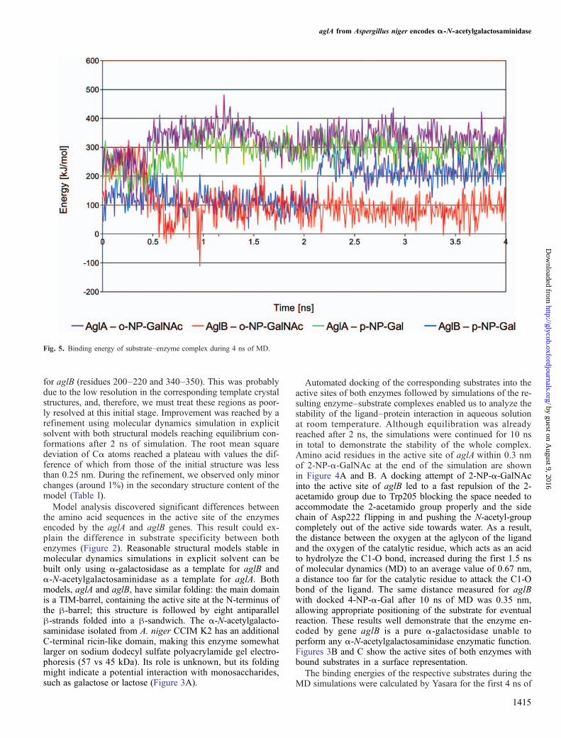
Fig. 5. Binding energy of substrate–enzyme complex during 4 ns of MD.
aglA from Aspergillus niger encodes α-N-acetylgalactosaminidase
by guest on August 9, 2016
http://glycob.oxfordjournals.org/D
ownloaded from

MD (Figure 5). The complexes reached equilibration after1.5 ns with an average binding energy for 2-NP-α-GalNAcdocked into aglA of 324 kJ/mol and only slightly lower valuesfor 4-NP-α-Gal, 287 kJ/mol. With aglB, the initial complexeshave similar values for both substrates; however, after 2 ns,we can see a clear separation in favor of the complex with4-NP-α-Gal, which reaches a higher average binding energyof 215 kJ/mol, in contrast to the complex with 2-NP-α-GalNAcwith average values below 100 kJ/mol. When estimating thebinding energy this way, positive values are used, and highernumbers indicate better binding. Positive numbers result fromthe calculation in which the sum of solvation and potential en-ergies of the complex is subtracted from the sum of theseenergies of the single components. As this method accordingto our experience shifts the absolute values to higher numbersand thus overestimates the binding energies when comparedwith experimental values, we also give the binding energies cal-cu la ted wi th Autodock, which should be c loser toexperimentally determined ΔG values: −24 kJ/mol for theaglA–2-NP-α-GalNAc complex and −18 kJ/mol for aglA–4-NP-α-Gal; −17 kJ/mol for aglB–4-NP-α-Gal. The aglB–2-NP-α-GalNAc complex was not ranked as a possible dockedconformation by Autodock, indicating that 2-NP-α-GalNAc
cannot be properly accommodated in the active site, and thebound position after the molecular dynamics simulation is notstable. Indeed, visual inspection confirms that the substrate isalready partially released to the solvent at the end of the simula-tion. Thus, our computational modeling results corroboratethe hypothesis that the enzyme encoded by gene aglA isan α-N-acetylgalactosaminidase that exhibits a dual enzymeactivity and is able to hydrolyze both 2-NP-α-GalNAc and4-NP-α-Gal, although at a different rate.
Discussion
The structural models constructed in this study were indispens-able in allowing to explain the inability of α-galactosidaseaglB from A. niger to accomodate the substrate containing a2-acetamido group in the active site in a stable and proper po-sition. Two reasons why the α-galactosidase aglB is unable tohydrolyze oNP-α-GalNAc have emerged: the lower bindingaffinity and the sterical hindrance connected with positioningof the C1-O bond close to the catalytic residue that makes itimpossible for the hydrolytic reaction to take place. The com-puter modeling experiments enabled us to assign the proteinpossessing the dual activity to the enzyme encoded by gene
1416
Fig. 6. Multiple sequence alignment of galactosidase/galactosaminidase homologs in the genome of Aspergillus niger, shaded according to their sequenceidentity (intensity depends on the frequency at same position in other aligned sequences). The region containing the N-acetyl recognition loop inα-N-acetylgalactosaminidases is marked with a black rectangle. Other potential active site residues in aglB are marked with a dot.
N Kulik et al.
by guest on August 9, 2016
http://glycob.oxfordjournals.org/D
ownloaded from
aglA. This is a completely new view of this gene since a possibleα-N-acetylgalactosaminidase activity has never been mentionedin the literature before. Our experimental results show that thisprotein exhibits dual enzyme activity and is able to hydrolyze 2-NP-α-GalNAc and 4-NP-α-Gal with 10 times better cleavage of2-NP-α-GalNAc than that of 2-NP-α-Gal. This contradicts withits original assignment as an α-D-galactopyranosidase belong-ing to GH family 27. Substrate docking into the structuralmodel of this enzyme confirmed similar binding energies forboth substrates with a clear preference for the α-N-acetyl-D-galactosaminide over the galactoside (Figure 3B and C), thusproviding evidence that this gene from A. niger does not encodeexclusively an α-galactosidase type A but that rather one com-bined with a fully functional α-N-acetylgalactosaminidase.
Figure 3B shows the binding pocket of aglAwith bound α-N-acetylgalactosamine, in comparison to the binding pocket ofaglBwith boundα-galactose in Figure 3C. The enlarged bindingpocket in aglA, which is able to accommodate an C2 acetamidogroup, is a result of the amino acid sequence Ser170, Ala171,Pro172, Ala173, and Tyr174, which forms a longer loop follow-ing β5 and connecting to α5 (Figure 2). Instead of Trp205,which blocks the part of the binding pocket accommodatingthe acetamido group in α-galactosidases, amino acid residuesSer170, Ala173, and Tyr174 (cf. Figure 4A and B) on this“N-acetyl recognition loop” create an open space that becomespart of the enlarged binding pocket of α-N-galactosaminindase.The presence of this loop explains the fact that the attempt tomodel aglA taking α-galactosidase as a template did not leadto a stable enzyme but induced distortions in the active site dur-ing molecular dynamics simulations. The multiple sequencealignment of the α-N-acetylgalactosaminindase/α-galactosi-dase homologs in A. niger (Figure 6) identified conservedresidues at positions corresponding to the active site of aglBfor all hypothetical proteins, with the exception of a tyrosineand cysteine in An14g01800 instead of Trp17 and Asp55 inaglB. AglA is the only homolog having the above described“N-acetyl recognition loop”. Lacking this loop, all otherfound potential α-galactosidases in the A. niger genomecould have α-galactosidase acitivity but definitely not α-N-galactosaminindase activity. This is in accordance with ourexperimental findings demonstrating that the other α-galato-sidases isolated from A. niger did not show dual activity butare pure α-galactosidases.
The fact that the enzyme, which possesses a pocket capa-ble of accommodating the N-acetyl group, shows dualactivity and is able to degrade substrates having an OHgroup in the same position, excludes the possibility thatthe N-acetyl recognition loop would be directly involved inthe reaction mechanism adopted by the glycoside hydrolasefamily 27. Instead, the N-acetyl recognition loop that is highlyconserved in α-N-acetylgalactosaminidases functions to struc-turally recognize and accommodate the N-acetyl group, butdoes not have additional contacts or interactions with the sub-strate, thus, the enzyme can bind equally well to 4p-NP- α-Gal.
Therefore, we propose that the α-N-acetylgalactosaminidase/α-galactosidase encoded by aglA utilizes a classical double in-version reaction mechanism (Figure 4C) analogous tohomologous α-galactosidases (Garman et al. 2002; Golubev etal. 2004) and that this mechanism is the same for both of its ac-tivities, e.g., α-N-acetylgalactosaminidase and α-galactosidase.
Materials and methodsEnzyme assayEnzyme activity was tested as described previously (Weignerováet al. 2008). All data were measured in triplicate.
Purification procedureThe protein was purified as described previously (Weignerováet al. 2008). 2D electrophoresis was performed using a drypolyacrylamide gel strip, gradient pH 4.0–5.0 (GE HealthBio-Sciences AB, Uppsala, SE).
N-Terminal sequencingN-Terminal sequencing was performed with the enzyme puri-fied in a C-4 reversed-phase column (Vydac C-4, Dionex,Sunnyvale, CA, USA) or electrotransferred onto PVDF blotsusing a Procise 491 Protein Sequencer (Applied BioSystems-Life Technologies, Carlsbad, CA, USA) using the Pulsed Liq-uid program. For CNBr cleavage, the dried enzyme wasincubated in 0.15 M CNBr (Aldrich, Buchs, CH) in 70% for-mic acid for 24 h in the dark, and the fragmented peptides wereseparated on a reversed-phase column (Vydac C-18) equilibrat-ed in 0.1% trif luoroacetic acid (TFA, LiChrosolv, Merck,Darmstadt, DE) and eluted with acetonitrile gradient (0–70%acetonitrile).
Sample preparation for mass spectrometryThe spot of interest was excised from the 2D gel. After com-plete destaining, the protein was reduced with 50 mM Tris-carboxyethyl phosphine (Fluka, Buchs, CH) for 15 min at80°C and alkylated using iodoacetamide (Sigma-Aldrich,Buchs, CH) for 40 min in the dark. The gel was then washedwith deionized water, shrunk by dehydration in acetonitrile,and re-dissolved in deionized water again. The supernatantwas removed and the gel partly dried in a SpeedVac concentra-tor (Savant, Holbrook, NY, USA). Gel pieces were thenreconstituted in deglycosylation buffer pH 5.5 containing,50 mM citrate buffer and 1000 units of Endo H (New EnglandBioLabs, Ipswich, MA, USA). After overnight deglycosyla-tion, the gel pieces were washed with deionized water, shrunkby dehydration in acetonitrile, and re-dissolved in deionized wa-ter again. Cleavage buffer containing 50mM 4-ethylmorpholineacetate pH 8.2, 10% acetonitrile, and sequencing-grade trypsin(5 ng/μL, Promega, Madison, USA) was added to the gel pieces.After overnight digestion at 37°C, the resulting tryptic peptideswere extracted using 30% acetonitrile in 1% acetic acid.
Mass spectrometry and data analysisThe peptide sample was dissolved in 200 μL of 0.1% TFA andthen desalted in a peptide MacroTrap (Michrom Bioresources,Auburn, CA, USA), eluted with 80% MeCN/0.5% TFA. Thepurified peptides were dried up on SpeedVac and redissolvedin 20 μL of 0.1% TFA/5% MeCN. Five microliters of samplewas loaded into a reversed-phase Magic C18 column (MichromBioresources, Auburn, USA; column size, 150 × 0.2 mm I.D.,particle size 5 μm; pore size 200 A) and separated with water/MeCN gradient using an Ultimate 1000 HPLC system (Dionex,Amsterdam, NL). The mobile phase consisted of the followingsolvents: 0.2% formic acid in water (A) and 0.16% formic acidin MeCN (B). Peptides were eluted under gradient conditions as
1417
aglA from Aspergillus niger encodes α-N-acetylgalactosaminidase
by guest on August 9, 2016
http://glycob.oxfordjournals.org/D
ownloaded from
follows: 0–1 min, 1% B; 5 min, 15% B; 30 min, 40% B;35 min, 95% B; 40 min, 95% B. Flow rate was 4 μL/min aftersplitting, and the column was kept at room temperature. Thecapillary column was directly connected to the mass analyzer.The mass spectrometric experiment was performed on a com-mercial APEX-Qe Fourier Transform Mass Spectrometry(FTMS) Lys/Arg instrument equipped with a 9.4-T supercon-ducting magnet and Combi ESI/MALDI ion source (BrukerDaltonics, Billerica, MA, USA). Mass spectra were obtainedby accumulating ions in the collision hexapole and runningthe quadrupole mass filter in non mass-selective (rf-only) modeso that a broad range of ions (300–2500 m/z) were passed to theFTMS analyzer cell. The accumulation time in the collision cellwas set at 0.5 s, the cell was opened for 4000 μs, and 256 ex-periments were collected for one liquid chromatography run,where one experiment consisted of the accumulation of fivespectra. The acquisition time was set to 512 k points at m/z300 a.m.u. The instrument was externally calibrated using triplyand doubly charged ions of angiotensin I and quadruply, quin-tuply, and sextuply charged ions of bovine insulin. Thiscalibration typically results in a mass accuracy below 2 ppm.
The acquired spectra were apodized and processed using a sinfunction with one zero-fill and run through the data-reducingmacro designed by Kruppa et al. (2003). A list of unique mono-isotopic masses was generated for each sample. The output ofthe macro (the list of unique monoisotopic masses) was matchedto a theoretical library of α-galactosidase tryptic peptides towithin 2 ppm. The library was created using the program ASAP(Automated Spectrum Assignment Program) (Young et al.2000; Kellersberger et al. 2004). The trypsin specificity wasset to Lys/Arg, and the degree of incomplete digestion was upto 1. Also, the algorithm was allowed to include a single oxida-tion of methionin (+15.9949 a.m.u.) and modification ofglycosylated asparagines by N-acetylglucosamine (+203.0794a.m.u.).
Molecular modelingMolecular models were generated by restrain-based homologymodeling followed by a structural refinement to test their ro-bustness using molecular dynamics simulations in explicitsolvent. In the first step, homologues were identified byBLAST search and the top scoring extracted from the ProteinData Bank (Berman et al. 2000): 1KTB, 1UAS, 1R46, 1SZN—for the melibiase domain; 2AAI, 1RZO, 1V6X—for the ricindomain. Different combinations of templates were used formultiple sequence and structural alignments. The multiplestructure-based sequence alignments were calculated withT-Coffee (http://www.tcoffee.org). Structural alignments ofknown 3D structures were performed with SHEBA in Yasara7.5.14 (Jung and Lee 2000). 3D models comprising non-hydrogen atoms were built and examined using Modeller 9.1package (Sali and Blundell 1993) and checked with ProSA(Wiederstein and Sippl 2007). The models were refined in YA-SARA (Krieger et al. 2004) by energyminimization followed by2 ns of MD simulation in water (pH 7, TIP3P water model) withperiodic boundary conditions. Substrates were built in YA-SARA, force field parameters were assigned using theAutoSMILES approach. The corresponding bond, angle, andtorsion potential parameters are taken from the General AMBERforce field. Initial ligand positions for docking experiments were
determined by comparison with homologous crystal structuresof α-galactosidase (Golubev et al. 2004) and α-N-acetylgalacto-saminidase (Garman and Garboczi 2004), respectively, whichwere co-crystallized with the ligands. The exact position of li-gands was set by Autodock 4.0 (Goodsell and Olson 1990)using local docking with the Lamarckian Genetic Algorithm,grid space 0.375. Substrate–enzyme complexes were refinedby minimization in a YAMBER 2 force field (Krieger et al.2004) and periodic boundary conditions. Sodium ions were iter-atively placed at the coordinates with the lowest electrostaticpotential until the cell was neutral. Molecular dynamics simula-tions were run in YASARA, using a multiple time step of 0.7 fsfor intramolecular and 1.4 fs for intermolecular forces, a 0.78 nmcutoff for Lennard Jones forces and the direct space portion ofthe electrostatic forces, which were calculated using the ParticleMesh Ewald method (Essman et al. 1995) with grid spacing 0.1 nm, fourth-order B-splines, and a tolerance of 10−4 for the di-rect space sum. The simulation of their interaction was run inthe following NPT ensemble: constant temperature (298 K),pressure and number of particles. The evaluation of ligand–enzyme complexes in time was analyzed on the basis of ge-ometry and energy parameters.
Interaction energies were calculated using a similar method tothat applied earlier for substrate–hexosaminidase complexes(Ettrich et al. 2007), considering the internal energy obtainedwith the specified force field, as well as the electrostatic energyand the entropy cost of fixing the ligand implicitly including Vander Waals solvation energy (Bultinck et al. 2003). The solvationenergy was calculated using the boundary fast methodimplemented in YASARA. The boundary between solvent(dielectric constant 80) and solute (dielectric constant 1)was formed by the latter's molecular surface, constructedwith a solvent probe radius of 1.4 Å and the following radiifor the solute elements: polar hydrogens 0.32 Å, other hydro-gens 1.017 Å, carbon 1.8 Å, oxygen 1.344 Å, nitrogen1.14 Å, and sulfur 2.0 Å. The solute charges were assignedbased on the YASARA 2 force field, using GAFF/AM1BCCfor the ligands. An estimate of the entropy cost of exposing0.01 nm2 to solvent was calculated as follows: Eentr =SSAS*solvation entropy, where SSAS is the solvent accessiblearea, and the solvation entropy characterizes the entropy costof exposing 0.01 nm2 of surface and can vary. For proteinstructures, the solvation entropy is usually estimated to be ap-proximately 0.65 kJmol−1A−2. It is almost impossible tocalculate this entropy cost accurately, but this is fortunatelynot needed, since it mainly depends on characteristics that areconstant during the simulation (ligand and protein size, sidechains on the surface, etc.) and thus is a constant factor. Bindingenergies calculated in this way might therefore be shifted by anunknown amount that depends on the protein; however, their rel-ative values are correct. The more positive interaction energy,the more favorable is the interaction in the context of the chosenforce field. Ligand–enzyme complexes after 10 ns of MD wereused for measuring binding energies by Autodock.
Acknowledgments
Support by the Academy of Sciences of the Czech Republic(AVOZ50200510, AVOZ60870520), the Ministry ofEducation of the Czech Republic (LC 06010, LC 7017,
1418
N Kulik et al.
by guest on August 9, 2016
http://glycob.oxfordjournals.org/D
ownloaded from
MSM6007665808, MSM21620808), and by the CzechScience Foundation grants P207/10/0321 to L.W., P207/10/1934 to R.E, P207/10/1040 to P.N., and 303/09/0477 and305/09/H008 to K.B. i s acknowledged. Access toMETACentrum supercomputing facilities was provided underthe research intent MSM6383917201.
Conflict of interest statement
None declared.
Abbreviations
2-NP-α-GalNAc, 2-nitrophenyl 2-acetamido-2-deoxy-α-D-galactopyranoside; 4-NP-α-Gal, 4-nitrophenyl α-D-galactopyr-anoside; aglA,α-galactosidase variant A in theA. niger genome;aglB, α-galactosidase variant B in the A. niger genome; GH,glycosylhydrolase; MD, molecular dynamics simulation; TFA,trif luoroacetic acid.
References
Ashida H, Tamaki H, Fujimoto T, Yamamoto K, Kumagai H. 2000. Molecularcloning of cDNA encoding α-N-acetylgalactosaminidase from Acremoniumsp. and its expression in yeast. Arch Biochem Biophys. 384:305–310.
Berman HM, Westbrook J, Feng Z, Gilliland G, Bhat TN, Weissig H,Shindyalov IN, Bourne PE. 2000. The Protein Data Bank. Nucleic AcidsRes. 28:235–242.
Bultinck P, De Winter H, Langenaeker W, Tollenare J. 2003. Computationalmedicinal chemistry for drug discovery. CRC Press.
Clark NE, Garman SC. 2009. The 1.9 Å structure of human α-N-acetylgalacto-saminidase: the molecular basis of Schindler and Kanzaki diseases. J MolBiol. 393:435–447.
Essman U, Perera L, Berkowitz ML, Darden T, Lee H, Pedersen LG. 1995. Asmooth particle mesh Ewald method. J Chem Phys. 103:8577–8593.
Ettrich R, KopeckýV, Hofbauerová K, Baumruk V, Novák P, Pompach P, Man P,Plíhal O, Kutý M, Kulik N, et al. 2007. Structure of the dimeric N-glycosy-lated form of fungal beta-N-acetylhexosaminidase revealed by computermodeling, vibrational spectroscopy, and biochemical studies. BMC StructBiol. 17:32.
Garman SC, Garboczi DN. 2004. The molecular defect leading to Fabry dis-ease: structure of human α-galactosidase. J Mol Biol. 337:319–335.
Garman SC, Hannick L, Zhu A, Garboczi DN. 2002. The 1.9 Å structure of α-N-acetylgalactosaminidase: molecular basis of glycosidase deficiency diseases.Structure. 10:425–434.
Golubev AM, Nagem RAP, Brandão Neto JR, Neustroev KN, Eneyskaya EV,Kulminskaya AA, Shabalin KA, Savel'ev AN, Polikarpov I. 2004. Crystalstructure of α-galactosidase from Trichoderma reesei and its complex withgalactose: implications for catalytic mechanism. J Mol Biol. 339:413–422.
Goodsell DS, Olson AJ. 1990. Automated docking of substrates to proteins bysimulated annealing. Proteins: Struct Funct Genet. 8:195–202.
Hart DO, He S, Chany CJ 2nd,Withers SG, Sims PF, Sinnott ML, Brumer H 3rd.2000. Identification of Asp-130 as the catalytic nucleophile in the main alpha-galactosidase from Phanerochaete chrysosporium, a family 27 glycosyl hy-drolase. Biochemistry. 39:9826–9836.
Hsieh HY, Calcutt MJ, Chapman LF, Mitra MD, Smith S. 2003. Purificationand characterization of a recombinant α-N-acetylgalactosaminidase fromClostridium perfringens. Protein Expression Purif. 32:309–316.
Hsieh HY, Mitra M, Wells DC, Smith DS. 2000. Purification and characteriza-tion of α-N-acetylgalactosaminidase from Clostridium perfringens. Life.50:91–97.
Jung J, Lee B. 2000. Protein structure alignment using environmental profiles.Protein Eng. 13:535–543.
Kellersberger KA, Yu E, Kruppa GH, Young MM, Fabris D. 2004. Top-downcharacterization of nucleic acids modified by structural probes using high-resolution tandem mass spectrometry and automated data interpretation.Anal Chem. 76:2438–2445.
Krieger E, Darden T, Nabuurs SB, Finkelstein A, Vriend G. 2004. Making op-timal use of empirical energy functions: force-field parameterization incrystal space. Proteins. 57:678–683.
Kruppa GH, Schoeniger J, Young MM. 2003. A top down approach to proteinstructural studies using chemical cross-linking and Fourier transform massspectrometry. Rapid Commun Mass Spectrom. 17:155–162.
Laskowski RA, MacArthur MW,Moss DS, Thornton JM. 1993. PROCHECK –a program to check the stereochemical quality of protein structures. J ApplCryst. 26:283–291.
Liu QP, Sulzenbacher G, Yuan H, Bennett EP, Pietz G, Saunders K, Spence J,Nudelman E, Levary SB, White T, et al. 2007. Bacterial glycosidasesfor the production of universal red blood cells. Nature Biotechnol.25:454–464.
Ly HD, Howard S, Shum K, He S, Zhu A, Withers SG. 2000. The synthesis,testing and use of 5-f luoro-a-D-galactosyl fluoride to trap an intermediate ongreen coffee bean α-galactosidase and identify the catalytic nucleophile.Carbohydr Res. 329:539–547.
Olsson ML, Clausen H. 2008. Modifying the red cell surface: towards an AB0-universal blood supply. Br J Haematol. 140:3–12.
Pel HJ, de Winde JH, Archer DB, Dyer PS, Hofmann G, et al. 2007. Genomesequencing and analysis of the versatile cell factory Aspergillus niger CBS513.88. Nat Biotechnol. 25:221–231.
Sali A, Blundell TL. 1993. Comparative protein modelling by satisfaction ofspatial restraints. J Mol Biol. 234:779–815.
Weignerová L, Filipi T, Manglová D, Křen V. 2008. Induction, purification andcharacterization of a α-N-acetylgalactosaminidase from Aspergillus niger.Appl Microbiol Biotechnol. 79:769–774.
Weignerová L, Simerská P, Křen V. 2009. α-Galactosidases and their applica-tion in biotransformation. Biocat Biotrans. 27:79–89.
Wiederstein M, Sippl MJ. 2007. ProSA-web: interactive web service for therecognition of errors in three-dimensional structures of proteins. NucleicAcids Res. 35:407–410.
Young MM, Tang N, Hempel JC, Oshiro CM, Taylor EW, Kuntz ID, GibsonBW, Dollinger G. 2000. High throughput protein fold identification by usingexperimental constraints derived from intramolecular cross-links and massspectrometry. Proc Natl Acad Sci USA. 97:5802–5806.
1419
aglA from Aspergillus niger encodes α-N-acetylgalactosaminidase
by guest on August 9, 2016
http://glycob.oxfordjournals.org/D
ownloaded from
Copyright © 2022 FDOKUMEN