Diversity of Marine-Derived Aspergillus from Tidal Mudflats ...
12
Full Terms & Conditions of access and use can be found at https://www.tandfonline.com/action/journalInformation?journalCode=tmyb20 Mycobiology ISSN: 1229-8093 (Print) 2092-9323 (Online) Journal homepage: https://www.tandfonline.com/loi/tmyb20 Diversity of Marine-Derived Aspergillus from Tidal Mudflats and Sea Sand in Korea Seobihn Lee, Myung Soo Park & Young Woon Lim To cite this article: Seobihn Lee, Myung Soo Park & Young Woon Lim (2016) Diversity of Marine- Derived Aspergillus from Tidal Mudflats and Sea Sand in Korea, Mycobiology, 44:4, 237-247, DOI: 10.5941/MYCO.2016.44.4.237 To link to this article: https://doi.org/10.5941/MYCO.2016.44.4.237 © The Korean Society of Mycology Published online: 19 Jun 2018. Submit your article to this journal Article views: 20 View Crossmark data
-
Upload
khangminh22 -
Category
Documents
-
view
1 -
download
0
Transcript of Diversity of Marine-Derived Aspergillus from Tidal Mudflats ...

Full Terms & Conditions of access and use can be found athttps://www.tandfonline.com/action/journalInformation?journalCode=tmyb20
Mycobiology
ISSN: 1229-8093 (Print) 2092-9323 (Online) Journal homepage: https://www.tandfonline.com/loi/tmyb20
Diversity of Marine-Derived Aspergillus from TidalMudflats and Sea Sand in Korea
Seobihn Lee, Myung Soo Park & Young Woon Lim
To cite this article: Seobihn Lee, Myung Soo Park & Young Woon Lim (2016) Diversity of Marine-Derived Aspergillus from Tidal Mudflats and Sea Sand in Korea, Mycobiology, 44:4, 237-247, DOI:10.5941/MYCO.2016.44.4.237
To link to this article: https://doi.org/10.5941/MYCO.2016.44.4.237
© The Korean Society of Mycology
Published online: 19 Jun 2018.
Submit your article to this journal
Article views: 20
View Crossmark data
237
Mycobiology
Diversity of Marine-Derived Aspergillus from Tidal Mudflats and Sea Sand in KoreaSeobihn Lee, Myung Soo Park and Young Woon Lim*
School of Biological Sciences and Institute of Microbiology, Seoul National University, Seoul 08826, Korea
Abstract Aspergillus (Trichocomaceae, Eurotiales, and Ascomycota) is a genus of well-defined asexual spore-forming fungi thatproduce valuable compounds such as secondary metabolites and enzymes; however, some species are also responsible fordiseases in plants and animals, including humans. To date, 26 Aspergillus species have been reported in Korea, with most specieslocated in terrestrial environments. In our study, Aspergillus species were isolated from mudflats and sea sand along the westernand southern coasts of Korea. A total of 84 strains were isolated and identified as 17 Aspergillus species in 11 sections on thebasis of both morphological characteristics and sequence analysis of the calmodulin gene (CaM) locus. Commonly isolated specieswere A. fumigatus (26 strains), A. sydowii (14 strains), and A. terreus (10 strains). The diversity of Aspergillus species isolated frommudflats (13 species) was higher than the diversity of those from sea sand (five species). Four identified species—A. caesiellus, A.montenegroi, A. rhizopodus, and A. tabacinus—are in the first records in Korea. Here, we provide detailed descriptions of themorphological characteristics of these four species.
Keywords Aspergillus, CaM, Marine environment, Morphology, Phylogeny
Genus Aspergillus consists of well-defined asexual spore-forming fungi and is classified as Trichocomaceae, Eurotiales,Eurotiomycetes, and Ascomycota. Approximately 64% ofthe 250 described species of aspergilli have not beenstudied regarding their sexual state [1]. Historically, speciesof Aspergillus were called as several names: Chaetosartorya,Cristaspora, Dichotomomyces, Emericella, Eurotium, Fennellia,Neocarpenteles, Neopetromyces, Neosartorya, and Petromyces[2]. However, genus Aspergillus has been recently unified bymycologists in an effort to simplify the taxonomy by using asingle genus name following ICN guidelines (InternationalCode of Nomenclature for algae, fungi, and plants) [3].
Since Aspergillus was first described in the 18th century[4], 339 species have been reported worldwide and havebeen categorized into four subgenera (Aspergillus, Circumdati,
Fumigati, and Nidulantes) and 20 sections [2]. Morphologicalfeatures of Aspergillus are crucial for initial identification[2]; however, morphology alone has proven unreliablebecause several morphological features are influencedby environmental conditions such as composition of themedium, pH, additives, or temperature [5]. To overcomethese limitations, mycologists recently proposed standardizedmethods involving polyphasic analysis including morphologicaland molecular analyses and extrolite profiling [2]. Molecularidentification methods were first introduced into fungaltaxonomy as sequencing of the internal transcribed spacer(ITS) region, and this locus has been proposed as theuniversal DNA barcode marker for all fungi. More recently,additional DNA markers for Aspergillus such as calmodulin(CaM), β-tubulin (BenA), and the RNA polymerase IIsecond largest subunit (RPB2) have been used in Aspergillustaxonomy [2]. In particular, CaM has proven a usefulmarker for Aspergillus identification because the CaMdatabase is well established, the locus is relatively easy toamplify, and adequate polymorphism allows for accurateidentification of Aspergillus species [2].
Aspergillus is known for plant pathogen and cause ofrespiratory diseases in humans, and produces variousmycotoxins such as aflatoxin and ochratoxin A [6], however,Aspergillus plays important ecological roles by degradingstarches, hemicelluloses, celluloses, and other polysaccharides[7]. Therefore, Aspergillus species have been industriallyexploited for potential applications of their enzymes [6, 8].Aspergillus species are isolated from various environments[2]. In addition, many Aspergillus species have been reported
Research Article
Mycobiology 2016 December, 44(4): 237-247https://doi.org/10.5941/MYCO.2016.44.4.237pISSN 1229-8093 • eISSN 2092-9323© The Korean Society of Mycology
*Corresponding authorE-mail: [email protected]
Received September 22, 2016Revised October 9, 2016Accepted October 29, 2016
This is an Open Access article distributed under the terms of theCreative Commons Attribution Non-Commercial License (http://creativecommons.org/licenses/by-nc/3.0/) which permits unrestrictednon-commercial use, distribution, and reproduction in any medium,provided the original work is properly cited.

238 Lee et al.
from marine environments including sponges and corals,but most Aspergillus studies have been focused on theirbioactive compounds rather than their diversity [9, 10].
There are various types of the intertidal zone dependingon various geographical factors [11, 12]. The intertidal zonein Korea usually appears as mudflats or sea sand. Intertidalzones are ecologically valuable and provide habitats andfeeding areas for diverse flora and fauna populations [13].In addition, many microbes live in intertidal zones andperform critical ecological roles such as degradation oforganic materials and synthesis of various bioactivecompounds [14, 15]. On the other hand, most researchesabout intertidal zones have been focused on bacterialcommunities [16, 17] rather than fungal communities.
To date, 26 species of Aspergillus have been reported inKorea [18]. Although some Aspergillus species were reportedfrom soil and inside buildings, most of them were isolated
from fermented foods and studied as a potential fermentationinitiator for production of foods and drinks such as meju,soy sauce, and traditional alcohol [19]. On the other hand,diversity of marine-derived Aspergillus has been poorly studied.Since the Ministry of Ocean and Fisheries established theMarine Fungal Resource Bank, marine-derived fungi havebeen studied to promote exploration of marine biodiversityand biological resources. We studied Aspergillus fromintertidal mudflats and sea sand on both the western andsouthern coasts of Korea from 2014 to 2015. Based onsequence analysis of the CaM locus, several Aspergillusstrains were identified at the species level. During thestudy, we found four previously unrecorded species inKorea (A. caesiellus, A. montenegroi, A. rhizopodus, and A.tabacinus). In this study, we describe macromorphologicaland micromorphological characteristics of these fourspecies.
Table 1. Information of isolated Aspergillus strains from mudflat and sea sand in different seasons
Section Species Strain No. Collection No.a
Substrate(No. of strains)
Season(No. of strains)
GenBankaccession No.
(CaM)Mudflat Sea sand Summer WinterAspergillus A. ruber 8M62 SFC20160317-M25 02 − 01 01 KX845511
P239 SFC20160805-M29 − − − − KX845512A. pseudoglacus 8M859 SFC20160317-M27 − 2 02 − KX845513
Circumdati A. westerdijikiae 8M709 SFC20160112-M05 01 − − 01 KX845514Clavati A. clvatus P6 SFC20150303-M01 06 − 05 01 KX845515
A. rhizopodus SP53 SFC20160407-M08 02 − 01 01 KX845516Flavi Aspergillus sp. 1 P102 SFC20160805-M30 03 2 − 05 KX845517
SEP13 SFC20160805-M31 − − − − KX845518Flavipedes Aspergillus sp. 2 SP112 SFC20160805-M32 01 − − 01 KX845519Fumigati A. fumigatus 8M56 SFC20160805-M33 23 3 11 15 KX845520
8M59 SFC20160805-M34 − − − − KX8455218M87 SFC20160805-M35 − − − − KX8455228M111 SFC20160805-M36 − − − − KX845523MT195 SFC20160805-M37 − − − − KX845524P230 SFC20160805-M38 − − − − KX845525
Aspergillus sp. 3 SP66 SFC20160805-M39 01 − − 01 KX845526Nidulantes A. nidulans 8M315 SFC20150812-M13 01 − 01 − KX845527
A. montenegroi P40 SFC20160805-M50 01 − − 01 KX845528Nigri A. welwischiae SA8 SFC20160805-M40 05 1 02 04 KX845529
SA13 SFC20160805-M41 − − − − KX845530SA14 SFC20160805-M42 − − − − KX845531SP100 SFC20160317-M20 − − − − KX845532
Restriciti A. caesiellus MT127 SFC20160112-M06 01 − 01 − KX845533Terrei A. terreus P212 SFC20160805-M45 09 1 03 07 KX845517
P213 SFC20160805-M43 − − − − KX845535Versicolores A. tabacinus P39 SFC20160407-M11 02 − − 02 KX845536
A. venenatus SP57 SFC20160407-M10 02 1 − 03 KX845537A. sydowii MT147 SFC20160407-M09 14 − 04 10 KX845538
MT172 SFC20160805-M44 − − − − KX845539MT349 SFC20160805-M46 − − − − KX845540P17 SFC20160805-M47 − − − − KX845541P49 SFC20160805-M48 − − − − KX845542P206 SFC20160805-M49 − − − − KX845543P207 SFC20160805-M51 − − − − KX845544
aSFC stands for abbreviation of ‘Seoul National University Fungus Collection’.
Aspergillus from Tidal Mudflats and Sea Sand in Korea 239
MATERIALS AND METHODS
Collection and isolation. Samples were collected fromboth the western and southern coasts of Korea. Collectionwas carried out during summer and winter from 2014 to2015. Each collection was repeated five times after removalof the top 0.5 to 1.0 cm of soil, and each collection site wasrandomly chosen within a 10 m distance. After collection,the samples were stored at 4oC prior to isolation. Eachsample was diluted 10-fold with artificial sea water [20].Next, 100 μL of each dilution was spread on three differentmedia plates, which contained artificial sea water: potatodextrose agar (PDA; Difco, Becton Dickinson, Sparks, MD,USA), glucose yeast extract agar (1 g/L glucose, 0.1 g/L yeastextract, 0.5 g/L peptone, and 15 g/L agar), and dichloranrose bengal chloramphenicol agar (Difco, Becton Dickinson).All plates were incubated at 25oC for 1 wk to confirmdistinguishable morphological features. Each Aspergillusstrain was transferred to a new PDA plate. Each strain wasthen stored in 20% glycerol at −80oC at the Seoul NationalUniversity Fungus Collection (SFC).
Molecular procedures and phylogenetic analysis.DNA was extracted using a modified cetyltrimethylammoniumbromide protocol [21]. The PCR amplification of the CaMlocus was conducted as described by Park et al. [22]. Gelelectrophoresis was carried out for each amplicon on a 1%agarose gel, and these PCR products were purified usingthe Expin PCR Purification Kit (Geneall Biotechnology,Seoul, Korea) following the manufacture’s instruction. Thepurified amplicons were sequenced using correspondingPCR primers by Macrogen (Seoul, Korea) in both forwardand reverse directions using an ABI Prism 3700 geneticanalyzer (Life Technologies, Gaithersburg, MD, USA).
Sequences were assembled, proofread, edited, and alignedusing MEGA v.5 [23] and were deposited in GenBank(Table 1). For multiple sequence alignments, MAFFTv.7 [24] was used, and each sequence was checked andadjusted by eye. After alignment, maximum likelihoodphylogenetic trees were constructed. The phylogenetic treeswere generated using RAxML 8.0.2 [25] and the GTR-GAMMA model of evolution with 1,000 bootstrap replicates.
Morphological analysis. To examine both macro- andmicromorphological characteristics of the unrecorded species,each strain was inoculated as a spore suspension at threepoints on plates with one of three different media: Czapekyeast autolysate agar (CYA, yeast extract; Difco), maltextract agar (MEA, Oxoid), and yeast extract sucrose agar(YES, yeast extract; Difco). All CYA, MEA, and YES plateswere incubated at 25oC for 7 days, and CYA plates wereincubated at 30oC, 37oC, and 50oC for additional 7 days. Allplates with media were prepared as described by Samsonet al. [2]. The color names and alphanumeric codes formacromorphological characteristics were assigned usingthe criteria of Kornerup and Wanscher [26]. Analysis of
micromorphological characteristics was performed followingthe methods of Samson et al. [2].
RESULTS
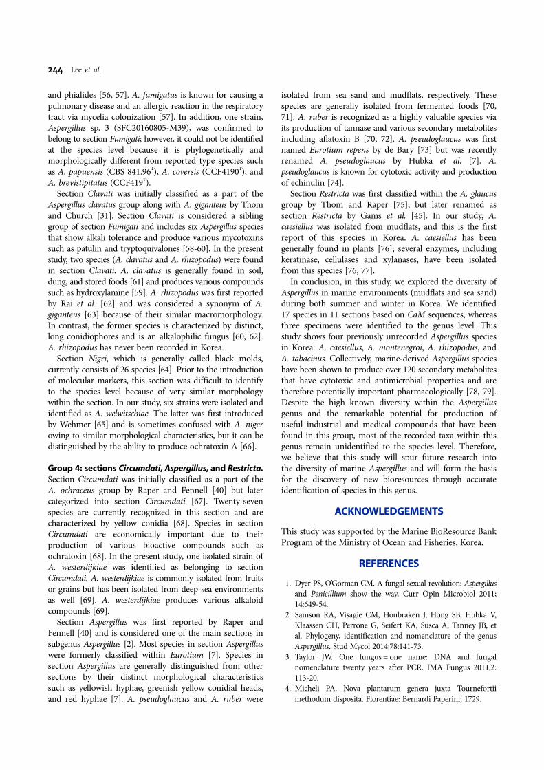
Totally, 84 Aspergillus strains were isolated from two intertidalsubstrates (mudflats and sea sand) at 30 sites from thewestern and southern coasts of Korea. More Aspergillusstrains were isolated (74 strains) from mudflats rather thanfrom sea sand (10 strains). Furthermore, higher diversity ofAspergillus was observed in mudflats (16 species) comparedto sea sand (six species). Five species (A. fumigatus, A.terreus, A. welwitschiae, A. venenatus, and Aspergillus sp. 1)were commonly found from both substrates, and A. sydowiiwas found only from mudflats. More Aspergillus strainswere isolated during winter (52 strains) than summer (32strains). Diversity of Aspergillus species was higher inwinter (14 species) than in summer (10 species), and sixspecies (A. clavatus, A. fumigatus, A. rhizopodus, A. ruber,A. sydowii, A. terreus, and A. welwitschiae) were commonlyfound during both seasons (Table 1). On the basis of CaMsequences, 84 isolates were identified as 17 species in 11sections, and three strains were identified as Aspergillus sp.due to ambiguous phylogenetic relationship (Table 1). Forconvenience, sections were grouped into four groupsregardless of phylogenetic relationship.
Three sections were included in group 1: Versicolores,Nidulantes, and Flavi. Three species (A. sydowii, A. tabacinus,and A. venenatus) were confirmed to belong to sectionVersicolores. Seven representative strains (SFC20160407-M09, SFC20160805-M44, SFC20160805-M46, SFC20160805-M47, SFC20160805-M48, SFC20160805-49, and SFC20160805-M51) were identified as A. sydowii (sequence similarity, 99%;bootstrap support, 99%) (Fig. 1). Although one isolatedstrain (SFC20160407-M11) formed a monophyletic groupwith low bootstrap support, its morphology and CaMsequence similarity were very similar to those of strains ofA. tabacinus (NRRL 4791T, NRRL5031, S823, and NRRLA-23173); thus, it was identified as A. tabacinus (sequencesimilarity, 99.8~100%; bootstrap support, 78%) (Fig. 1). Onestrain (SFC20160407-M10) was identified as A. venenatus(sequence similarity, 99.8%; bootstrap support, 99%) (Fig.1). In section Nidulantes, two strains (SFC20150812-M13and SFC20160805-M50) were isolated from mudflats (Table1), and each strain was identified as either A. nidulans(sequence similarity, 99.8%; bootstrap support, 100%) orA. montenegroi (sequence similarity, 99.3%; bootstrapsupport, 100%), respectively. Two strains (SFC20160805-M30 and SFC20160805-M31) in section Flavi were identicalto type strains of A. flavus (NRRL 1957T) and A. oryzae(NRRL 447T), with 100% sequence similarity (Fig. 1).However, we identified these specimens as Aspergillus sp. 1because it was difficult to distinguish these two species byCaM sequences.
Group 2 included sections Terrei and Flavipedes andshowed high bootstrap support. Two strains (SFC20160805-

240 Lee et al.
M43 and SFC20160805-M45) were identified as A. terreusin section Terrei (sequence similarity, 99.6~100%; bootstrapsupport, 76%) (Fig. 1). In section Flavipedes, SFC20160805-M32 formed a group with type strains of A. mangaliensis(CCF4698T: sequence similarity, 96.5%) and A. templicola(DTO270C6T: sequence similarity, 97.1%), but we identifiedit as Aspergillus sp. 2 because of unclear phylogeneticrelationship (Fig. 1).
Group 3 included three sections (Clavati, Fumigati, and
Nigri). Six strains (SFC20160805-M33, SFC20160805-M34,SFC20160805-M35, SFC20160805-M36, SFC20160805-M37,and SFC20160805-M38) were confirmed to belong to sectionFumigati and were identified as A. fumigatus (sequencesimilarity, 99.6~100%; bootstrap support, 96%) (Fig. 1).SFC20160805-M39 was found to be closely related to A.papuensis (CBS 841.96T), A. coversis (CCF4190T), and A.brevistipitatus (CCF419T) (sequence similarity, 93.1~95.3%),but it was named Aspergillus sp. 3 due to unresolved
Fig. 1. A maximum likelihood phylogenetic tree of Aspergillus on the basis of sequences of the calmodulin (CaM) locus.Bootstrap scores > 70 are presented at the nodes. The scale bar indicates the number of nucleotide substitutions per site. “T”indicates ex-type strains. Each group was designed depending on the arrangement of sections in the phylogenetic tree.

Aspergillus from Tidal Mudflats and Sea Sand in Korea 241
phylogeny. In section Clavati, the strains (SFC20150303-M01 and SFC20160407-M08) were identified as A. clavatus(sequence similarity, 99.8%; bootstrap support, 100%) andA. rhizopodus (sequence similarity, 99.9%; bootstrap support,100%), respectively (Fig. 1). In section Nigri, four strains(SFC20160317-M20, SFC20160805-M40, SFC20160805-M41,and SFC20160805M-42) were identified as A. welwitschiae(sequence similarity, 100%; bootstrap support, 97%) (Fig. 1).
Group 4 included three distantly related sections(Aspergillus, Restricta, and Circumdati). In section Aspergillus,two strains (SFC20160317-M25 and SFC20160805-M29)were identified as A. ruber (sequence similarity, 99.8~100%;bootstrap support, 99%), and the other strain (SFC20160317-M27) was identified as A. pseudoglaucus (sequence similarity,99.6%; bootstrap support, 94%) (Fig. 1). Strain SFC201601112-M06 was identified as A. caesiellus in section Restricta(sequence similarity, 100%; bootstrap support, 100%). Onestrain (SFC20160112-M05) was identified as A. westerdijkiaein section Circumdati (sequence similarity, 99.83%; bootstrapsupport, 100%).
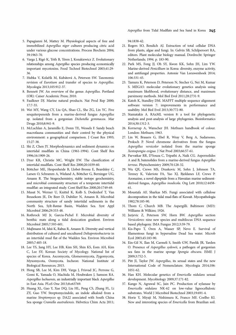
Taxonomy.Aspergillus caesiellus Saito, J. Coll. (1904) (Fig. 2A)Description: Colony diameter, at 25oC for 7 days, CYA8~13 mm; CYA 30oC: 14~15 mm; no growth at 37oC and50oC; MEA 13~14 mm; YES 29~31 mm.
Colonies on CYA, lightly sulcate, heavy sporulation,velutinous, central part gray (26C1) to grayish green(25C3) with 3-mm gray margin (26C1), no exudates, nopigmentation, reverse central part light brown (6D4) todull green (28E4).
Colonies on MEA, sporulation, floccus, central part dullgreen (25D3) to dark turquoise (24F5) with 1-mm whitemargin, no exudates, no pigmentation, reverse central partdull green (27D3) to dull green (30D4).
Colonies on YES, sporulation, central floccus, velutinous,central part greenish gray (25D2) to dark green (25F4)with 1 mm white margin, no exudates, no pigmentation,reverse grayish yellow (4C4).
Conidiophore smooth with slightly green wall (61~) 82~95 (~114) × 4.6~5.2 (~5.5) μm wide, vesicles spatulate (10.7~)11.1 × 13.5 (~15.4), conidial head uniseriate, metulae coveringhalf of upper surface, (4.4~) 4.5~5.6 (~5.8) × (2.6~) 2.8~3.4(~3.9) μm, phialide (2.4~) 2.5~2.6 × 7.0~8.2 (~8.6) μm,conidia ellipse and pyriform with greenish gray (4.4~)4.5~5.5 (~6) × (2.4~) 2.6~2.8 μm diam.Strain examined: SFC20160112-M06.Remarks: A. caesiellus is similar to A. penicillioides but isdistinguished by more vigorous growth and formation ofconidia on CYA and MEA media. A. caesiellus grows moreslowly and its cylinder becomes more round.
Aspergillus montenegroi Y. Horie, Miyaji & Nishim (1996)(Fig. 2B)Description: Colony diameter, at 25oC for 7 days, CYA52~58 mm; CYA 30oC 64~70 mm; CYA 37oC 66~72 mm;
no growth at 50oC; MEA 64~72 mm; YES 66~70 mm.Colonies on CYA, not sulcate, sporulation, floccus, and
velutinous, central part yellowish gray (3C2) to olive (3D3)and grayish green (28E5) with 3 mm white margin, noexudate, pigmentation grayish red (7B3), reverse centralpart reddish gray (7B2) to brownish orange (7C4) withreddish gray (8B2) margin.
Colonies on MEA, not sulcate, sporulation velutinous,dull green (27E3) with 1-mm white margin, no exudate,no pigmentation, reverse central part brown (6E4) to lightbrown (6D4).
Colonies on YES, sulcate, sporulation, velutinous, centralpart purplish gray (13B2) to greenish yellow (1B8) andgrayish green (27E6) with 2 mm white margin, no exudate,no pigmentation, reverse yellowish brown (5D5).
Conidiophore smooth and thick-walled with orange-brown(63.9~) 65~88.8 (~110.8) × (3.5~) 4.3~5.3 (~5.5) μm, vesicleshemispherical with orange-brown to dark brown 9.7~9.9 ×(10.8~) 11.8~13.5 (~13.8) μm, conidial heads biseriate,metulae covering over half of upper surface (4.4~) 4.5~5.6(~5.8) × (2.6~) 2.8~3.4 (~3.9) μm, phialide (4.7~) 5.2~5.6(~6.1) × (2~) 2.1~2.6 (~2.7) μm, conidia hyaline with grayishgreen to green (2.8~) 2.9~4.3 (~4.7) × (2.1~) 2.9~3.4 (~3.6)μm.Strain examined: SFC20160805-M50.Remarks: The spore of Aspergillus montenegroi has similarmorphology to that of the Aspergillus nidulans. However,the spore of A. montenegroi has reticulate ornamentationon the convex wall, whereas the spore of A. nidulans has asmooth wall.
Aspergillus rhizopodus J. N. Rai, Wadhwani & S. C.Agarwal (1975) (Fig. 2C)Description: Colony diameter, at 25oC for 7 days, CYA58~62 mm; CYA 30oC 65~68 mm; CYA 37oC 19~22 mm;no growth at 50oC; MEA 52~53 mm; YES 74~76 mm.
Colonies on CYA, weakly sulcate, heavy sporulation,velutinous with a floccus central part, dull green (25D3)with 2 mm white margin, no exudate, no pigmentation, reversecentral part light brown (5D4) to olive brown (4D4).
Colonies on MEA, weakly sulcate, heavy sporulationfloccus, dull green (25D3) with 7-mm white margin, slightexudates, no pigmentation, reverse central part brownishorange (5C6) to grayish orange (5B4).
Colonies on YES, weakly sulcate, heavy sporulation,floccus, central part grey (24C1) to grayish turquoise (24D3)with 2 mm white margin, no exudates, no pigmentation,reverse grayish yellow (4C5).
Conidiophore smooth-walled with light brown (210~)340~502 (~550) × (5.9~) 6.3~8.2 μm, vesicles clavate form14.9~15.9 (~16.4) × 19~20.8μm, and conidial heads biseriate,metulae covering entire vesicle surface, (4~) 4.9~5.5 (~5.8) ×(2.4~) 2.5~3.1 (~3.2) μm, phialide (5.2~) 5.6~5.9 (~6) ×(1.4~) 1.6~2 (~2.1) μm, conidia spherical or subsphericalwith a light green smooth wall (1.9~) 2.1~2.7 μm.Strain examined: SFC20160407-M08.

242 Lee et al.
Remarks: A. rhizopodus is similar to A. giganteus and A.longivesica but is distinguished by variously shaped footcells with fingerlike projections and large conidial heads inthe presence of light.
Aspergillus tabacinus Nakaz., Y. Takeda, Simo & A. Watan(1934) (Fig. 2D)Description: Colony diameter, at 25oC for 7 days, CYA25~26 mm; CYA 30oC 22~24 mm; no growth at 37oC and50oC; MEA 18~19 mm; YES 31~33 mm.
Colonies on CYA, sulcate, sporulation in central area,central part grayish beige (4C2) to greenish grey (27D2),
brownish orange (5C4) with 2-mm white margin, no exudate,no pigmentation, reverse central part grayish orange (5B3)to yellowish white (4A2). No growth at 37oC and 50oC.
Colonies on MEA, sulcate, sporulation moderate, dullgreen (26E2) with 2-mm white margin, no exudates, nopigmentation, reverse brownish orange (5C5).
Colonies on YES, lightly sulcate, rings in central area,central part yellowish white (2A2) to grayish green (27C3)with 3-mm white margin, no exudates, no pigmentation,reverse central part deep yellow (4A8) to pale yellow (4A3).
Conidiophore smooth-walled, light brown (353~) 368~448 (~514) × (5.2~) 5.4~6.3 μm wide, vesicles spatulate form
Fig. 2. Morphology of Aspergillus caesiellus (SFC201601112-M06) (A), A. montenegroi (SFC20160805-M50) (B), A. rhizopodus(SFC20160407-M08) (C), and A. tabacinus (SFC20160407-M11) (D). a~c, incubation for 7 days at 25oC. Upper: a, Czapek yeastautolysate agar (CYA); b, malt extract agar (MEA); c, yeast extract sucrose agar (YES). Lower: a, CYA reverse; b, MEA reverse;c, YES reverse. d, e, conidiophores; f, conidia (scale bars = 10 µm).

Aspergillus from Tidal Mudflats and Sea Sand in Korea 243
14.9~15.9 (~16.4) × 19~20.8 μm, conidial heads biseriate,metulae covering entire vesicle surface (4~) 4.9~5.5 (~5.8) ×(2.4~) 2.5~3.1 (~3.2) μm, phialide (5.2~) 5.6~5.9 (~6) ×(1.4~) 1.6~2 (~2.1) μm, conidia spherical or subsphericalwith light green smooth wall (1.9~) 2.1~2.7 μm.Strain examined: SFC20160407-M08.Remarks: A. tabacinus has smooth-walled conidia similar tothose of A. amoenus and A. austroafricanus, but A. tabacinusproduces no soluble pigment, whereas A. austroafricanusproduces a brown soluble pigment on the CYA medium.
DISCUSSION
In this study, we explored the diversity of Aspergillus derivedfrom marine environments in Korea and identified eachAspergillus strain using CaM sequences, a locus that haspreviously been shown to be an effective DNA marker forAspergillus identification [2]. A total of 17 species in 11sections were identified, while three species were leftunidentified (Aspergillus sp. 1~3) due to their ambiguity inmorphological and phylogenetic analyses. Several Aspergillusspecies (A. fumigatus, A. nidulans, A. sydowii, A. terreus, A.versicolor, and A. westerdijkiae) have been reported frommarine substrates such as deep sea sediments, marinesponges, corals, and mudflat [27-29], and particularly A.fumigatus and A. sydowii were dominant in mud substrates[30]. Similarly, in our study, higher diversity of Aspergilluswas detected in mudflats, and both A. fumigatus and A.sydowii were the most abundant species, as another studyhas shown [30]. The number of isolated strains anddiversity of Aspergillus were similar in winter and summer,and we could not find a significant difference between theseasons. A. montenegroi, A. venenatus, and A. tabacinus arereported for the first time in marine environments.
Group 1: sections Versicolores, Nidulantes, and Flavi.Section Versicolores was first introduced by Thom andChurch [31], and 13 species are included in this section[32]. Pathogenic species in section Versicolores are generallyisolated from various substrates including hypersalinewater [33]. In our study, A. sydowii, A. tabacinus, and A.venenatus were isolated from a marine environment. A.sydowii is commonly isolated from marine environmentsas well and is known as a serious pathogen of marineorganisms [34]. A. venenatus and A. tabacinus have previouslybeen isolated from plant materials [32], but our study isthe first report of these species in a marine environment.This study is the first report of A. tabacinus in Korea.
Species in section Nidulantes are usually found in soil,indoor environments, and foods, and are characterizedby red or purple pseudoparenchymatous cleistotheciumsurrounded by hülle cells and purple ascospores [35]. Inthe present study, we identified two strains as A. nidulans andA. montenegroi. A. nidulans is characterized by a “nestlike”fruiting body [36] and is known for producing xylanase[37]. A. montenegroi was first isolated from soil by Horie
et al. [38] (as Emericella montenegroi). A. montenegroi ischaracterized by ascospores with reticulate and convexwalls [38]. This is the first report of this species in amarine environment and in Korea.
Species in section Flavi have a relatively thin and roughcell wall as compared to species in other sections [39].Species in this section are found in soil, air, organic materials,and plants [40]. In our study, five isolated strains wereconfirmed within section Flavi and form a monophyleticgroup with type strains of A. flavus and A. oryzae. Bothspecies are generally found in terrestrial environments suchas soil and plant products, but are also found in marineenvironments [41, 42]. A. flavus is a representative speciesin that section and is known as an opportunistic pathogenof plants and animals due to aflatoxin production, whereasA. oryzae is used in food fermentation [42, 43]. However,it is difficult to distinguish A. flavus and A. oryzae becauseof their similar morphological characteristics and ambiguousphylogenetic relationship [44]. Therefore, we denoted fivestrains as Aspergillus sp. 1, which showed high sequencesimilarity to both A. flavus and A. oryzae; thus, furtherresearch is necessary to unambiguously identify these strains.
Group 2: sections Terrei and Flavipedes. Species insection Terrei were first described by Gams et al. [45] andcommonly have brown columnar conidial heads. They areubiquitous and considered economically important, especiallyin the fermentation industry [46]. In section Terrei, 10 strainswere identified as A. terreus. This species is commonlyisolated from terrestrial habitats such as deserts, grasslandsoils, and plant products but has also been isolated from asoft coral [28]. A. terreus is used for food fermentationbecause of its production of itaconic or itatartaric acid [47,48]; however, A. terreus is also a known human pathogenthat induces an inflammatory response [49]. A. terreusproduces aspernolides A and B as well as butenolides [28].In section Flavipedes, only one strain, Aspergillus sp. 2, ispresent, and it could not be clearly distinguished from eitherA. mangaliensis or A. templicola due to weak phylogeneticsupport and ambiguous morphological features.
Group 3: sections Fumigati, Clavati, and Nigri. SectionFumigati was first categorized as an A. fumigatus group byRaper and Fennel [40]. This identification was based onmorphology and has remained a controversial issue due tothe similarity of these morphological features among othertaxa [50, 51]. Most species in this section are pathogenicor allergenic to humans owing to mycotoxins [52, 53].In our study, two Aspergillus species (A. fumigatus andAspergillus sp. 3) were found in this section. A total of 26strains were identified as A. fumigatus in section Fumigati.A. fumigatus is the most abundant species isolated in thisstudy. A. fumigatus is generally found in forest soils andair. A. fumigatus is often the dominant fungal species inmudflats [30, 54, 55]. Like other species in section Fumigati,A. fumigatus is generally characterized by green conidia
244 Lee et al.
and phialides [56, 57]. A. fumigatus is known for causing apulmonary disease and an allergic reaction in the respiratorytract via mycelia colonization [57]. In addition, one strain,Aspergillus sp. 3 (SFC20160805-M39), was confirmed tobelong to section Fumigati; however, it could not be identifiedat the species level because it is phylogenetically andmorphologically different from reported type species suchas A. papuensis (CBS 841.96T), A. coversis (CCF4190T), andA. brevistipitatus (CCF419T).
Section Clavati was initially classified as a part of theAspergillus clavatus group along with A. giganteus by Thomand Church [31]. Section Clavati is considered a siblinggroup of section Fumigati and includes six Aspergillus speciesthat show alkali tolerance and produce various mycotoxinssuch as patulin and tryptoquivalones [58-60]. In the presentstudy, two species (A. clavatus and A. rhizopodus) were foundin section Clavati. A. clavatus is generally found in soil,dung, and stored foods [61] and produces various compoundssuch as hydroxylamine [59]. A. rhizopodus was first reportedby Rai et al. [62] and was considered a synonym of A.giganteus [63] because of their similar macromorphology.In contrast, the former species is characterized by distinct,long conidiophores and is an alkalophilic fungus [60, 62].A. rhizopodus has never been recorded in Korea.
Section Nigri, which is generally called black molds,currently consists of 26 species [64]. Prior to the introductionof molecular markers, this section was difficult to identifyto the species level because of very similar morphologywithin the section. In our study, six strains were isolated andidentified as A. welwitschiae. The latter was first introducedby Wehmer [65] and is sometimes confused with A. nigerowing to similar morphological characteristics, but it can bedistinguished by the ability to produce ochratoxin A [66].
Group 4: sections Circumdati, Aspergillus, and Restricta.Section Circumdati was initially classified as a part of theA. ochraceus group by Raper and Fennell [40] but latercategorized into section Circumdati [67]. Twenty-sevenspecies are currently recognized in this section and arecharacterized by yellow conidia [68]. Species in sectionCircumdati are economically important due to theirproduction of various bioactive compounds such asochratoxin [68]. In the present study, one isolated strain ofA. westerdijkiae was identified as belonging to sectionCircumdati. A. westerdijkiae is commonly isolated from fruitsor grains but has been isolated from deep-sea environmentsas well [69]. A. westerdijkiae produces various alkaloidcompounds [69].
Section Aspergillus was first reported by Raper andFennell [40] and is considered one of the main sections insubgenus Aspergillus [2]. Most species in section Aspergilluswere formerly classified within Eurotium [7]. Species insection Aspergillus are generally distinguished from othersections by their distinct morphological characteristicssuch as yellowish hyphae, greenish yellow conidial heads,and red hyphae [7]. A. pseudoglaucus and A. ruber were
isolated from sea sand and mudflats, respectively. Thesespecies are generally isolated from fermented foods [70,71]. A. ruber is recognized as a highly valuable species viaits production of tannase and various secondary metabolitesincluding aflatoxin B [70, 72]. A. pseudoglaucus was firstnamed Eurotium repens by de Bary [73] but was recentlyrenamed A. pseudoglaucus by Hubka et al. [7]. A.pseudoglaucus is known for cytotoxic activity and productionof echinulin [74].
Section Restricta was first classified within the A. glaucusgroup by Thom and Raper [75], but later renamed assection Restricta by Gams et al. [45]. In our study, A.caesiellus was isolated from mudflats, and this is the firstreport of this species in Korea. A. caesiellus has beengenerally found in plants [76]; several enzymes, includingkeratinase, cellulases and xylanases, have been isolatedfrom this species [76, 77].
In conclusion, in this study, we explored the diversity ofAspergillus in marine environments (mudflats and sea sand)during both summer and winter in Korea. We identified17 species in 11 sections based on CaM sequences, whereasthree specimens were identified to the genus level. Thisstudy shows four previously unrecorded Aspergillus speciesin Korea: A. caesiellus, A. montenegroi, A. rhizopodus, andA. tabacinus. Collectively, marine-derived Aspergillus specieshave been shown to produce over 120 secondary metabolitesthat have cytotoxic and antimicrobial properties and aretherefore potentially important pharmacologically [78, 79].Despite the high known diversity within the Aspergillusgenus and the remarkable potential for production ofuseful industrial and medical compounds that have beenfound in this group, most of the recorded taxa within thisgenus remain unidentified to the species level. Therefore,we believe that this study will spur future research intothe diversity of marine Aspergillus and will form the basisfor the discovery of new bioresources through accurateidentification of species in this genus.
ACKNOWLEDGEMENTS
This study was supported by the Marine BioResource BankProgram of the Ministry of Ocean and Fisheries, Korea.
REFERENCES
1. Dyer PS, O’Gorman CM. A fungal sexual revolution: Aspergillusand Penicillium show the way. Curr Opin Microbiol 2011;14:649-54.
2. Samson RA, Visagie CM, Houbraken J, Hong SB, Hubka V,Klaassen CH, Perrone G, Seifert KA, Susca A, Tanney JB, etal. Phylogeny, identification and nomenclature of the genusAspergillus. Stud Mycol 2014;78:141-73.
3. Taylor JW. One fungus = one name: DNA and fungalnomenclature twenty years after PCR. IMA Fungus 2011;2:113-20.
4. Micheli PA. Nova plantarum genera juxta Tournefortiimethodum disposita. Florentiae: Bernardi Paperini; 1729.
Aspergillus from Tidal Mudflats and Sea Sand in Korea 245
5. Papagianni M, Mattey M. Physiological aspects of free andimmobilized Aspergillus niger cultures producing citric acidunder various glucose concentrations. Process Biochem 2004;39:1963-70.
6. Varga J, Rigó K, Tóth B, Téren J, Kozakiewicz Z. Evolutionaryrelationships among Aspergillus species producing economicallyimportant mycotoxins. Food Technol Biotechnol 2003;41:29-36.
7. Hubka V, Kolařík M, Kubátová A, Peterson SW. Taxonomicrevision of Eurotium and transfer of species to Aspergillus.Mycologia 2013;105:912-37.
8. Bennett JW. An overview of the genus Aspergillus. Portland(OR): Caiser Academic Press; 2010.
9. Faulkner DJ. Marine natural products. Nat Prod Rep 2000;17:7-55.
10. Wei MY, Wang CY, Liu QA, Shao CL, She ZG, Lin YC. Fivesesquiterpenoids from a marine-derived fungus Aspergillussp. isolated from a gorgonian Dichotella gemmacea. MarDrugs 2010;8:941-9.
11. McLachlan A, Jaramillo E, Donn TE, Wessels F. Sandy beachmacrofauna communities and their control by the physicalenvironment: a geographical comparison. J Coast Res 1993;15:27-38.
12. Shi Z, Chen JY. Morphodynamics and sediment dynamics onintertidal mudflats in China (1961-1994). Cont Shelf Res1996;16:1909-26.
13. Dyer KR, Christie MC, Wright EW. The classification ofintertidal mudflats. Cont Shelf Res 2000;20:1039-60.
14. Böttcher ME, Hespenheide B, Llobet-Brossa E, Beardsley C,Larsen O, Schramm A, Wieland A, Böttcher G, Berninger UG,Amann R. The biogeochemistry, stable isotope geochemistry,and microbial community structure of a temperate intertidalmudflat: an integrated study. Cont Shelf Res 2000;20:1749-69.
15. Musat N, Werner U, Knittel K, Kolb S, Dodenhof T, VanBeusekom JE, De Beer D, Dubilier N, Amann R. Microbialcommunity structure of sandy intertidal sediments in theNorth Sea, Sylt-Rømø Basin, Wadden Sea. Syst ApplMicrobiol 2006;29:333-48.
16. Rothrock MJ Jr, Garcia‐Pichel F. Microbial diversity ofbenthic mats along a tidal desiccation gradient. EnvironMicrobiol 2005;7:593-601.
17. Muβmann M, Ishii K, Rabus R, Amann R. Diversity and verticaldistribution of cultured and uncultured Deltaproteobacteria inan intertidal mud flat of the Wadden Sea. Environ Microbiol2005;7:405-18.
18. Lee YS, Jung HY, Lee HB, Kim SH, Shin KS, Eom AH, KimC, Lee SY; Korean Society of Mycology. National list ofspecies of Korea. Ascomycota, Glomeromycota, Zygomycota,Myxomycota, Oomycota. Incheon: National Institute ofBiological Resources; 2015.
19. Hong SB, Lee M, Kim DH, Varga J, Frisvad JC, Perrone G,Gomi K, Yamada O, Machida M, Houbraken J, Samson RA.Aspergillus luchuensis, an industrially important black Aspergillusin East Asia. PLoS One 2013;8:e63769.
20. Huang XL, Gao Y, Xue DQ, Liu HL, Peng CS, Zhang FL, LiZY, Guo YW. Streptomycindole, an indole alkaloid from amarine Streptomyces sp. DA22 associated with South ChinaSea sponge Craniella australiensis. Helvetica Chim Acta 2011;
94:1838-42.21. Rogers SO, Bendich AJ. Extraction of total cellular DNA
from plants, algae and fungi. In: Gelvin SB, Schilperoort RA,editors. Plant molecular biology manual. Dordrecht: SpringerNetherlands; 1994. p. 183-90.
22. Park MS, Fong JJ, Oh SY, Kwon KK, Sohn JH, Lim YW.Marine-derived Penicillium in Korea: diversity, enzyme activity,and antifungal properties. Antonie Van Leeuwenhoek 2014;106:331-45.
23. Tamura K, Peterson D, Peterson N, Stecher G, Nei M, KumarS. MEGA5: molecular evolutionary genetics analysis usingmaximum likelihood, evolutionary distance, and maximumparsimony methods. Mol Biol Evol 2011;28:2731-9.
24. Katoh K, Standley DM. MAFFT multiple sequence alignmentsoftware version 7: improvements in performance andusability. Mol Biol Evol 2013;30:772-80.
25. Stamatakis A. RAxML version 8: a tool for phylogeneticanalysis and post-analysis of large phylogenies. Bioinformatics2014;30:1312-3.
26. Kornerup A, Wanscher JH. Methuen handbook of colour.London: Methuen; 1963.
27. Lin W, Brauers G, Ebel R, Wray V, Berg A, Sudarsono,Proksch P. Novel chromone derivatives from the fungusAspergillus versicolor isolated from the marine spongeXestospongia exigua. J Nat Prod 2003;66:57-61.
28. Parvatkar RR, D’Souza C, Tripathi A, Naik CG. AspernolidesA and B, butenolides from a marine-derived fungus Aspergillusterreus. Phytochemistry 2009;70:128-32.
29. Wu QX, Crews MS, Draskovic M, Sohn J, Johnson TA,Tenney K, Valeriote FA, Yao XJ, Bjeldanes LF, Crews P.Azonazine, a novel dipeptide from a Hawaiian marine sediment-derived fungus, Aspergillus insulicola. Org Lett 2010;12:4458-61.
30. Moustafa AF, Sharkas MS. Fungi associated with cellulosedecomposition in the tidal mud-flats of Kuwait. Mycopathologia1982;78:185-90.
31. Thom C, Church MB. The Aspergilli. Baltimore (MD):Williams & Wilkins; 1926.
32. Jurjevic Z, Peterson SW, Horn BW. Aspergillus sectionVersicolores: nine new species and multilocus DNA sequencebased phylogeny. IMA Fungus 2012;3:59-79.
33. Kis-Papo T, Oren A, Wasser SP, Nevo E. Survival offilamentous fungi in hypersaline Dead Sea water. MicrobEcol 2003;45:183-90.
34. Ein-Gil N, Ilan M, Carmeli S, Smith GW, Pawlik JR, YardenO. Presence of Aspergillus sydowii, a pathogen of gorgoniansea fans in the marine sponge Spongia obscura. ISME J2009;3:752-5.
35. Pitt JI, Taylor JW. Aspergillus, its sexual states and the newInternational Code of Nomenclature. Mycologia 2014;106:1051-62.
36. Han KH. Molecular genetics of Emericella nidulans sexualdevelopment. Mycobiology 2009;37:171-82.
37. Kango N, Agrawal SC, Jain PC. Production of xylanase byEmericella nidulans NK-62 on low-value lignocellulosicsubstrates. World J Microbiol Biotechnol 2003;19:691-4.
38. Horie Y, Miyaji M, Nishimura K, Franco MF, Coelho KI.New and interesting species of Emericella from Brazilian soil.

246 Lee et al.
Mycoscience 1996;37:137-44.39. Wang L, Yokoyama K, Takahasi H, Kase N, Hanya Y, Yashiro
K, Miyaji M, Nishimura K. Identification of species in Aspergillussection Flavi based on sequencing of the mitochondrialcytochrome b gene. Int J Food Microbiol 2001;71:75-86.
40. Raper KB, Fennell DI. The genus Aspergillus. Baltimore(MD): Williams & Wilkins; 1965.
41. Geiser DM, Dorner JW, Horn BW, Taylor JW. The phylogeneticsof mycotoxin and sclerotium production in Aspergillus flavusand Aspergillus oryzae. Fungal Genet Biol 2000;31:169-79.
42. Machida M, Asai K, Sano M, Tanaka T, Kumagai T, Terai G,Kusumoto K, Arima T, Akita O, Kashiwagi Y, et al. Genomesequencing and analysis of Aspergillus oryzae. Nature 2005;438:1157-61.
43. Hedayati MT, Pasqualotto AC, Warn PA, Bowyer P, DenningDW. Aspergillus flavus: human pathogen, allergen and mycotoxinproducer. Microbiology 2007;153(Pt 6):1677-92.
44. Payne GA, Nierman WC, Wortman JR, Pritchard BL, BrownD, Dean RA, Bhatnagar D, Cleveland TE, Machida M, Yu J.Whole genome comparison of Aspergillus flavus and A.oryzae. Med Mycol 2006;44(Suppl 1):S9-11.
45. Gams W, Christensen M, Onions AH, Pitt JI, Samson RA.Infrageneric taxa of Aspergillus. In: Samson RA, Pitt JI,editors. Advances in Penicillium and Aspergillus systematics.New York: Plenum Press; 1985. p. 55-62.
46. Samson RA, Peterson SW, Frisvad JC, Varga J. New species inAspergillus section Terrei. Stud Mycol 2011;69:39-55.
47. Kozakiewicz Z. Aspergillus species on stored products. MycolPap 1989;161:1-188.
48. Okabe M, Lies D, Kanamasa S, Park EY. Biotechnologicalproduction of itaconic acid and its biosynthesis in Aspergillusterreus. Appl Microbiol Biotechnol 2009;84:597-606.
49. Walsh TJ, Petraitis V, Petraitiene R, Field-Ridley A, Sutton D,Ghannoum M, Sein T, Schaufele R, Peter J, Bacher J, et al.Experimental pulmonary aspergillosis due to Aspergillus terreus:pathogenesis and treatment of an emerging fungal pathogenresistant to amphotericin B. J Infect Dis 2003;188:305-19.
50. Samson RA, Hong SB, Frisvad JC. Old and new concepts ofspecies differentiation in Aspergillus. Med Mycol 2006;44(Supp 1):133-48.
51. Alcazar-Fuoli L, Mellado E, Alastruey-Izquierdo A, Cuenca-Estrella M, Rodriguez-Tudela JL. Aspergillus section Fumigati:antifungal susceptibility patterns and sequence-basedidentification. Antimicrob Agents Chemother 2008;52:1244-51.
52. Hong SB, Shin HD, Hong J, Frisvad JC, Nielsen PV, Varga J,Samson RA. New taxa of Neosartorya and Aspergillus inAspergillus section Fumigati. Antonie Van Leeuwenhoek2008;93:87-98.
53. Samson RA, Hong S, Peterson SW, Frisvad JC, Varga J.Polyphasic taxonomy of Aspergillus section Fumigati and itsteleomorph Neosartorya. Stud Mycol 2007;59:147-203.
54. Mullins J, Harvey R, Seaton A. Sources and incidence ofairborne Aspergillus fumigatus (Fres). Clin Allergy 1976;6:209-17.
55. Gilna VV, Khaleel KM. Cellulase enzyme activity of Aspergillusfumigatus from mangrove soil on lignocellulosic substrate.Recent Res Sci Technol 2011;3:132-4.
56. Leslie CE, Flannigan B, Milne LJ. Morphological studies onclinical isolates of Aspergillus fumigatus. J Med Vet Mycol1988;26:335-41.
57. Latgé JP. Aspergillus fumigatus and aspergillosis. Clin MicrobiolRev 1999;12:310-50.
58. Clardy J, Springer JP, Buchi G, Matsuo K, Wightman R.Letter: tryptoquivaline and tryptoquivalone, two tremorgenicmetabolites of Aspergillus clavatus. J Am Chem Soc 1975;97:663-5.
59. Büchi G, Luk KC, Kobbe B, Townsend JM. Four newmycotoxins of Aspergillus clavatus related to tryptoquivaline. JOrg Chem 1977;42:244-6.
60. Varga J, Due M, Frisvad JC, Samson RA. Taxonomic revisionof Aspergillus section Clavati based on molecular, morphologicaland physiological data. Stud Mycol 2007;59:89-106.
61. Flannigan B, Pearce AR. Aspergillus spoilage: spoilage ofcereals and cereal products by the hazardous species A.clavatus. In: Powell KA, Renwick A, Peberdy JF, editors. Thegenus Aspergillus. New York: Plenum Press; 1994. p. 115-27.
62. Rai JN, Wadhwani K, Agarwal SC. Aspergillus rhizopodus sp.nov. from Indian alkaline soils. Trans Br Mycol Soc 1975;64:515-7.
63. Samson RA. A compilation of the Aspergilli described since1965. Stud Mycol 1979;18:1-38.
64. Varga J, Frisvad JC, Kocsubé S, Brankovics B, Tóth B, SzigetiG, Samson RA. New and revisited species in Aspergillussection Nigri. Stud Mycol 2011;69:1-17.
65. Wehmer C. Zur Kenntnis einiger Aspergillus arten. CentralbBakteriol Parasitenkd Infektionskr Hyg Abt II 1907;18:385-95.
66. Massi FP, Sartori D, de Souza Ferranti L, Iamanaka BT,Taniwaki MH, Vieira ML, Fungaro MH. Prospecting for theincidence of genes involved in ochratoxin and fumonisinbiosynthesis in Brazilian strains of Aspergillus niger andAspergillus welwitschiae. Int J Food Microbiol 2016;221:19-28.
67. Frisvad JC, Samson RA. Neopetromyces gen. nov. and anoverview of teleomorphs of Aspergillus subgenus Circumdati.Stud Mycol 2000;45:201-7.
68. Visagie CM, Varga J, Houbraken J, Meijer M, Kocsubé S,Yilmaz N, Fotedar R, Seifert KA, Frisvad JC, Samson RA.Ochratoxin production and taxonomy of the yellow aspergilli(Aspergillus section Circumdati). Stud Mycol 2014;78:1-61.
69. Peng J, Zhang XY, Tu ZC, Xu XY, Qi SH. Alkaloids from thedeep-sea-derived fungus Aspergillus westerdijkiae DFFSCS013.J Nat Prod 2013;76:983-7.
70. Kumar R, Sharma J, Singh R. Production of tannase fromAspergillus ruber under solid-state fermentation using jamun(Syzygium cumini) leaves. Microbiol Res 2007;162:384-90.
71. Hong SB, Kim DH, Samson RA. Aspergillus associated withMeju, a fermented soybean starting material for traditionalsoy sauce and soybean paste in Korea. Mycobiology 2015;43:218-24.
72. Leitao J, Le Bars J, Bailly JR. Production of aflatoxin B1 byAspergillus ruber Thom and Church. Mycopathologia 1989;108:135-8.
73. De Bary A. Eurotium, Erysiphe, Cicinnobolus. NebstBemerkungen über die Geschlechtsorgane der Ascomyceten.Abh Senckb Naturforsch Ges 1870;7:361-455.
74. Smetanina OF, Kalinovskii AI, Khudyakova YV, Slinkina NN,

Aspergillus from Tidal Mudflats and Sea Sand in Korea 247
Pivkin MV, Kuznetsova TA. Metabolites from the marinefungus Eurotium repens. Chem Nat Compd 2007;43:395-8.
75. Thom C, Raper KB. A manual of the Aspergilli. Soil Sci1945;60:333.
76. Batista-García R, Balcázar-López E, Miranda-Miranda E,Sánchez-Reyes A, Cuervo-Soto L, Aceves-Zamudio D, Atriztán-Hernández K, Morales-Herrera C, Rodríguez-Hernández R,Folch-Mallol J. Characterization of lignocellulolytic activitiesfrom a moderate halophile strain of Aspergillus caesiellusisolated from a sugarcane bagasse fermentation. PLoS One2014;9:e105893.
77. Marcondes NR, Taira CL, Vandresen DC, Svidzinski TI,Kadowaki MK, Peralta RM. New feather-degrading filamentousfungi. Microb Ecol 2008;56:13-7.
78. Lee YM, Kim MJ, Li H, Zhang P, Bao B, Lee KJ, Jung JH.Marine-derived Aspergillus species as a source of bioactivesecondary metabolites. Mar Biotechnol (NY) 2013;15:499-519.
79. Jin L, Quan C, Hou X, Fan S. Potential pharmacologicalresources: natural bioactive compounds from marine-derivedfungi. Mar Drugs 2016;14:E76.