Probing the Impact of Acidification on Spider Silk Assembly Kinetics
8
Probing the Impact of Acidification on Spider Silk Assembly Kinetics Dian Xu, † Chengchen Guo, † and Gregory P. Holland* ,‡ † Department of Chemistry and Biochemistry, Magnetic Resonance Research Center, Arizona State University, Tempe, Arizona 85287-1604, United States ‡ Department of Chemistry and Biochemistry, San Diego State University, 5500 Campanile Drive, San Diego, California 92182-1030, United States * S Supporting Information ABSTRACT: Spiders utilize fine adjustment of the phys- icochemical conditions within its silk spinning system to regulate spidroin assembly into solid silk fibers with out- standing mechanical properties. However, the exact mechanism about which this occurs remains elusive and is still hotly debated. In this study, the effect of acidification on spider silk assembly was investigated on native spidroins from the major ampullate (MA) gland fluid excised from Latrodectus hesperus (Black Widow) spiders. Incubating the protein-rich MA silk gland fluid at acidic pH conditions results in the formation of silk fibers that are 10−100 μm in length and ∼2 μm in diameter as judged by optical and electron microscope methods. The in vitro spider silk assembly kinetics were monitored as a function of pH with a 13 C solid-state MAS NMR approach. The results confirm the importance of acidic pH in the spider silk self-assembly process with observation of a sigmoidal nucleation-elongation kinetic profile. The rates of nucleation and elongation as well as the percentage of β-sheet structure in the grown fibers depend on the pH. These results confirm the importance of an acidic pH gradient along the spinning duct for spider silk formation and provide a powerful spectroscopic approach to probe the kinetics of spider silk formation under various biochemical conditions. ■ INTRODUCTION Spiders use up to seven types of silk with a diverse range of physical and mechanical properties to help them survive their environments. 1−3 Each type of silk is a proteinaceous material produced by a specific type of gland inside the spider’s abdomen. The dragline spider silk is an outstanding fiber with an extraordinary toughness exceeding man-made fibers such as Kevlar. 1−5 Collectively, spider silk has drawn considerable attention in the field of material science. 6−8 The dragline silk (or major ampullate silk) is particularly attractive because of its exceptional mechanical properties and ease of collection compared to other types of spider silk. To achieve the goal of producing synthetic spider silk with comparable mechanical properties to natural silk, great effort has been made to elucidate the secondary structures and higher order organ- ization of the dragline silk spidroins prior to 9−13 and after 14−28 fiber formation as well as the physicochemical conditions that are responsible for the structural transformation from soluble silk protein to insoluble super fiber. 29−34 The dragline silk contains primarily two proteins, major ampullate spidroin (MaSp) 1 and 2, which are both biomacromolecules with molecular weights exceeding 250 kDa. 14,15,35 The molecular architecture of the dragline silk spidroins includes a highly repetitive amphiphilic core region flanked by a nonrepetitive hydrophilic N- and C-terminal domain with sequences that are highly conserved across different spider species. 36,37 The dragline silk spidroins are stored in the major ampullate (MA) glands at high concentration (∼25 to 50 wt %), 3 and the core regions of the MaSp proteins were shown with NMR spectroscopy to have no discernible secondary structures, 9,10,12 exhibiting rapid backbone dynamics on the subnanosecond time scale. 13 A Raman spectromicroscopy and circular dichroisim (CD) study also indicates a random coil structure for spider MA silk proteins in the gland with a small population of polyproline II (PPII) and α-helicies. 11 In contrast, the terminal domains appear to be well-structured helical bundles and are proposed to facilitate spidroin assembly via dimerization at acidic pH. 38−43 During the silk protein self-assembly process, it is thought that the spidroins experience changes in physicochem- ical conditions along the elongated duct to drive silk formation in a controlled manner. 44−46 In the final spun spider silk fiber, the spidroins were shown with X-ray diffraction (XRD) and solid-state NMR methods to form nanocrystalline β-sheet structures aligned along the fiber axis interleaved with more disordered regions that form approximate 3 1 -helical and type II β-turn conformations. 16−28 Received: April 13, 2015 Revised: May 29, 2015 Article pubs.acs.org/Biomac © XXXX American Chemical Society A DOI: 10.1021/acs.biomac.5b00487 Biomacromolecules XXXX, XXX, XXX−XXX
-
Upload
independent -
Category
Documents
-
view
2 -
download
0
Transcript of Probing the Impact of Acidification on Spider Silk Assembly Kinetics
Probing the Impact of Acidification on Spider Silk Assembly KineticsDian Xu,† Chengchen Guo,† and Gregory P. Holland*,‡
†Department of Chemistry and Biochemistry, Magnetic Resonance Research Center, Arizona State University, Tempe, Arizona85287-1604, United States‡Department of Chemistry and Biochemistry, San Diego State University, 5500 Campanile Drive, San Diego, California 92182-1030,United States
*S Supporting Information
ABSTRACT: Spiders utilize fine adjustment of the phys-icochemical conditions within its silk spinning system toregulate spidroin assembly into solid silk fibers with out-standing mechanical properties. However, the exact mechanismabout which this occurs remains elusive and is still hotlydebated. In this study, the effect of acidification on spider silkassembly was investigated on native spidroins from the majorampullate (MA) gland fluid excised from Latrodectus hesperus(Black Widow) spiders. Incubating the protein-rich MA silkgland fluid at acidic pH conditions results in the formation ofsilk fibers that are 10−100 μm in length and ∼2 μm indiameter as judged by optical and electron microscopemethods. The in vitro spider silk assembly kinetics weremonitored as a function of pH with a 13C solid-state MAS NMR approach. The results confirm the importance of acidic pH inthe spider silk self-assembly process with observation of a sigmoidal nucleation-elongation kinetic profile. The rates of nucleationand elongation as well as the percentage of β-sheet structure in the grown fibers depend on the pH. These results confirm theimportance of an acidic pH gradient along the spinning duct for spider silk formation and provide a powerful spectroscopicapproach to probe the kinetics of spider silk formation under various biochemical conditions.
■ INTRODUCTIONSpiders use up to seven types of silk with a diverse range ofphysical and mechanical properties to help them survive theirenvironments.1−3 Each type of silk is a proteinaceous materialproduced by a specific type of gland inside the spider’sabdomen. The dragline spider silk is an outstanding fiber withan extraordinary toughness exceeding man-made fibers such asKevlar.1−5 Collectively, spider silk has drawn considerableattention in the field of material science.6−8 The dragline silk(or major ampullate silk) is particularly attractive because of itsexceptional mechanical properties and ease of collectioncompared to other types of spider silk. To achieve the goalof producing synthetic spider silk with comparable mechanicalproperties to natural silk, great effort has been made toelucidate the secondary structures and higher order organ-ization of the dragline silk spidroins prior to9−13 and after14−28
fiber formation as well as the physicochemical conditions thatare responsible for the structural transformation from solublesilk protein to insoluble super fiber.29−34
The dragline silk contains primarily two proteins, majorampullate spidroin (MaSp) 1 and 2, which are bothbiomacromolecules with molecular weights exceeding 250kDa.14,15,35 The molecular architecture of the dragline silkspidroins includes a highly repetitive amphiphilic core regionflanked by a nonrepetitive hydrophilic N- and C-terminaldomain with sequences that are highly conserved across
different spider species.36,37 The dragline silk spidroins arestored in the major ampullate (MA) glands at highconcentration (∼25 to 50 wt %),3 and the core regions ofthe MaSp proteins were shown with NMR spectroscopy tohave no discernible secondary structures,9,10,12 exhibiting rapidbackbone dynamics on the subnanosecond time scale.13 ARaman spectromicroscopy and circular dichroisim (CD) studyalso indicates a random coil structure for spider MA silkproteins in the gland with a small population of polyproline II(PPII) and α-helicies.11 In contrast, the terminal domainsappear to be well-structured helical bundles and are proposedto facilitate spidroin assembly via dimerization at acidicpH.38−43 During the silk protein self-assembly process, it isthought that the spidroins experience changes in physicochem-ical conditions along the elongated duct to drive silk formationin a controlled manner.44−46 In the final spun spider silk fiber,the spidroins were shown with X-ray diffraction (XRD) andsolid-state NMR methods to form nanocrystalline β-sheetstructures aligned along the fiber axis interleaved with moredisordered regions that form approximate 31-helical and type IIβ-turn conformations.16−28
Received: April 13, 2015Revised: May 29, 2015
Article
pubs.acs.org/Biomac
© XXXX American Chemical Society A DOI: 10.1021/acs.biomac.5b00487Biomacromolecules XXXX, XXX, XXX−XXX

Despite the fast progress on characterizing the structures ofspidroins in the silk fibers, it is still critical to understand thestructural transitions as well as how the self-assembly ofspidroins is regulated by changes in physicochemical conditionsduring the spinning process. In order to elucidate the importantphysicochemical condition responsible for spider silk spidroinassembly, studies have been done on different systemsincluding spidroin mimics, recombinant spidroins, spidroinsregenerated from spider silk or native spidroins from silkglands.29−34 For example, it has been indicated that the highconcentration of sodium chloride in the silk gland helpsincrease the solubility of the spidroins and prevents undesiredaggregation30,31,34 while, potassium and phosphate ions inducethe formation of β-sheet structures.29,30,34 Additionally, a pHgradient from neutral to acidic conditions was observed fromthe silk gland to the spinning duct indicating acidification is acritical parameter that drives the assembly of soluble spidroinsinto silk fibers.31,34,46 Further, structural information on therecombinant N-terminal domains for spidroins provides anexplanation for the mechanism of acidification-assisted spidroinassembly at the molecular level.39,41−43 In addition, althoughrelatively less studied, the effects of dehydration44 and shearingforce or elongation flow47 are believed to also be importantvariables that promote β-sheet formation as well as molecularalignment of the spidroins during the spinning process.In this work, the impact of acidification on spidroin assembly
is investigated for Latrodectus hesperus (Black Widow) nativeMA silk fluid with an in vitro 13C solid-state MAS NMRapproach. The spider silk assembly process was investigated at anumber of acidic pH conditions. Compared to other character-ization techniques, this solid-state NMR method is able toresolve and detect poly(Ala) present in both the liquid (asrandom coil) and the solid phase (as insoluble β-sheetstructures) during fiber assembly. The resulting time-dependentdata represents the first illustration of monitoring pH-dependent spider silk assembly kinetics in vitro at near nativeconditions with a NMR spectroscopic method. The assembly ofspider silk fibers from the native MA gland fluid was observedfor a range of pH from 3 to 6 with a long period for nucleationfollowed by a rapid pH-dependent elongation process.
■ EXPERIMENTAL SECTIONSample Preparation. Latrodectus hesperus, Black Widow (BW),
spiders were chosen because of their abundance in the southwesternUnited States and the complete primary amino acid sequence of bothspider silk proteins MaSp1 and MaSp2 is known for this species.35
Mature female spiders were forcibly silked at a speed of 2 cm/s for a30 min time period. During the silking process, each spider was fed∼20 μL 15% (w/v) aqueous solution of uniformly labeled U−13C-L-alanine or U-[13C, 15N]-L-alanine every other day. After 2 weeks ofisotope labeling, Ala, Gly, Gln, and Ser are enriched with 13C.48 Thespiders were then sacrificed and dissected under an optical microscope.The intact MA glands were carefully extracted from the spider’sabdomen and rinsed with ultrapure water (neutral pH), the ducts, tails,and outside membranes were then gently removed. The gland fluidfrom four intact MA glands from two isotope enriched BW spiderswere transferred into a Bruker Kel-F 4 mm rotor insert filled with anaqueous solution of 5% (w/v) sodium azide (to prevent bacterialgrowth), 10 mM sodium formate, and 10 mM piperazine buffersolution (as chemical pH indicator).49 The silk protein concentrationis near the native concentration (∼30 wt %) and is approximated to be∼23 wt % following addition of the buffer. Hydrochloric acid wasadded to adjust the pH of the buffer. For microscopy imaging, the silkprotein solution at a pH = 3 was extracted from the rotor insertfollowing silk fiber formation, as monitored by solid-state NMR
experiments. A 10 times diluted and nondiluted silk protein solutionwas used for optical microscopy and transmission electron microscopy(TEM), respectively.
Polarized Light Microscopy. A total of 1 μL of the silk proteinsolution was placed on a glass slide and imaged using an OlympusMVX10 Macroview microscope with a SC100 camera. The light waspolarized by Olympus polarizer SZX-PO and analyzed by SZX-ANanalyzer. Images were taken and analyzed by the Olympus cellSensv1.9 software.
Negative-Staining Transmission Electron Microscopy. A totalof 2 μL of the silk protein solution was deposited on a 200 meshcopper grid with a pure carbon support film. After 20 s of slow drying,10 μL of 2% (w/v) pH = 7.4 phosphotungstic acid (PTA, Ted PellaInc., Redding, CA) solution was slowly introduced onto the sample for60 s. The extra solution was gently removed from the side of the gridand the sample was dried at room temperature for 60 s. TEM imageswere acquired on a Philips CM200 TEM (Philips Electron Optics,Eindhoven, The Netherlands) at 200 kV.
Scanning Electron Microscopy. The dragline silk sample wastaped on the sample holder and coated with Au/Pd in a Dentonvacuum sputter coater desk II for 120 s under a pressure of 200 mTorr.The deposition rate was 5 nm/min with a current of 20 mA. The SEMimage was taken using a FEI XL30 Environmental SEM-FEG. TheSEM was operating at a vacuum pressure less than 9 × 10⁻⁵ mbar anda beam current of 10.00 kV.
Solid-State NMR. All solid-state NMR experiments wereconducted with a 400 MHz Bruker Avance III spectrometer equippedwith a 4 mm double-resonance (1H/13C) MAS probe at 25 °C. One-dimensional (1D) 13C direct-detect (DD) MAS NMR spectra wereobtained at a 4 kHz MAS frequency with a 90° 13C pulse and 55 kHzTPPM50 1H decoupling during acquisition with a 15° phase shift. Atotal of 256 scans were collected for each spectrum with a 7 s recycledelay (fully relaxed). In order to monitor silk fiber assembly kinetics,consecutive 13C DD-MAS experiments were collected for a set of BWMA gland fluid samples incubated at a pH = 3, 4, and 5 for 24, 28, and35 h, respectively. To test the impact of MAS on the fiber formationkinetics, a set of 1D 13C DD-MAS NMR spectra were collectedwithout MAS on a control sample incubated at pH = 4 (seeSupporting Information). 1D 1H−13C CP-MAS experiments werecollected with 4 kHz MAS, a 1 ms ramped (50%) 1H spin-lock pulsewith a radio frequency (rf) field of 56.5 kHz matched to the −1Hartmann−Hahn spinning sideband on the 13C channel. TPPM50 1Hdecoupling was applied during acquisition with a 15° phase shift. The1H−13C CP-MAS spectra were acquired right after collecting the 13CDD-MAS experiments for samples incubated at a pH = 3, 4, and 5.Another set of samples was incubated at pH = 6 and 7 for 7 daysbefore the 1H−13C CP-MAS experiments were collected.
Two-dimensional (2D) 13C−13C correlation experiments wereacquired at a 10 kHz MAS frequency with the DARR 13C−13Crecoupling method.51,52 150 ms of CW irradiation was applied on the1H channel at the n = 1 rotary-resonance condition. The acquisitionparameters were a 25 kHz sweep width in both dimensions with 512points in the direct dimension and 196 points in the indirectdimension, 128 scans were averaged with a 2.5 s recycle delay. The 2DDARR experiments were acquired on a BW MA silk gland fluid sampleincubated at a pH = 3 for 24 h and native spider dragline silk collectedfrom the same spiders by forcible silking.
Data Processing and Analysis. In order to monitor spider silkassembly kinetics, 48 1D 13C DD-MAS spectra for pH = 4 sample, 561D 13C direct spectra for pH = 3 sample and 70 1D 13C direct spectrafor pH = 5 sample were collected successively each 30 min followingsample preparation. During data processing, all spectra were zero filledto 16 k data points, and a 100 Hz line broadening was applied,followed by baseline correction. To accurately extract the peakintensity of the random coil peak for Ala Cβ, the region from 13 to 29ppm was deconvoluted into the following four peaks, with chemicalshifts extracted from the 2D DARR spectrum, Ala Cβ at 16.6 ppm(random coil), Ala Cβ at 20.5 ppm (antiparallel β-sheet), Ala Cβ at23.0 ppm (parallel β-sheet) and Gln Cβ at 26.8 ppm (representativedeconvolution of Ala Cβ resonance is shown in Figure S1). Spectrum
Biomacromolecules Article
DOI: 10.1021/acs.biomac.5b00487Biomacromolecules XXXX, XXX, XXX−XXX
B
process and deconvolution were accomplished by using theMestReNova v. 8.0.2 data process package.The kinetic curves were then plotted with the measured peak
intensities for the Ala Cβ random coil peak as a function of time (seeFigures S1 and S2 for justification of using random coil peakintensities). To quantify the kinetic behavior of spider silk fiberformation and better analyze the impact of different pH conditions, amodified Kolmogorov-Johnson-Mehl-Avrami (KJMA) kinetic model53
from the overall crystallization theory was applied to the experimentaldata. The general form of the analytical expression for the model is
αθ
= − −⎜ ⎟⎡⎣⎢
⎛⎝
⎞⎠
⎤⎦⎥(t) 1 exp
t n
where α represents the normalized experimentally observed intensityfor Ala Cβ random coil peak, θ is a time constant quantifying the timescale of fiber formation, and n is a kinetic index indicating the rate ofelongation. To accommodate the model to our experimental data, aslight modification as follows was made to enable applicability of fittinga decreasing sigmoidal curve with nonzero fraction at equilibrium:
αθ
= + − −⎜ ⎟⎡⎣⎢
⎛⎝
⎞⎠
⎤⎦⎥A A
t(t) (1 )exp
n
where an extra parameter, A, was added to better determine thepercentage of Ala that did not adopt a β-sheet conformation atequilibrium and remained random coil. The lag time, tl, and themaximal rate of elongation, k, can be determined based on thefollowing formulas:
θ= − − −−
−⎜ ⎟⎛⎝
⎞⎠
⎡⎣⎢
⎤⎦⎥t
nn
en
11
11
n n n
l
1/ 1/
θ= −
−− −⎜ ⎟⎛
⎝⎞⎠k n
nn
e( 1)1
1nn n
1/1/
■ RESULTSDuring the spider silk spinning process, acidic conditions areobserved in the duct region with reported pH values from 6.3to a range of 5 to 4 (or even lower near the spinneret).43,46,54,55
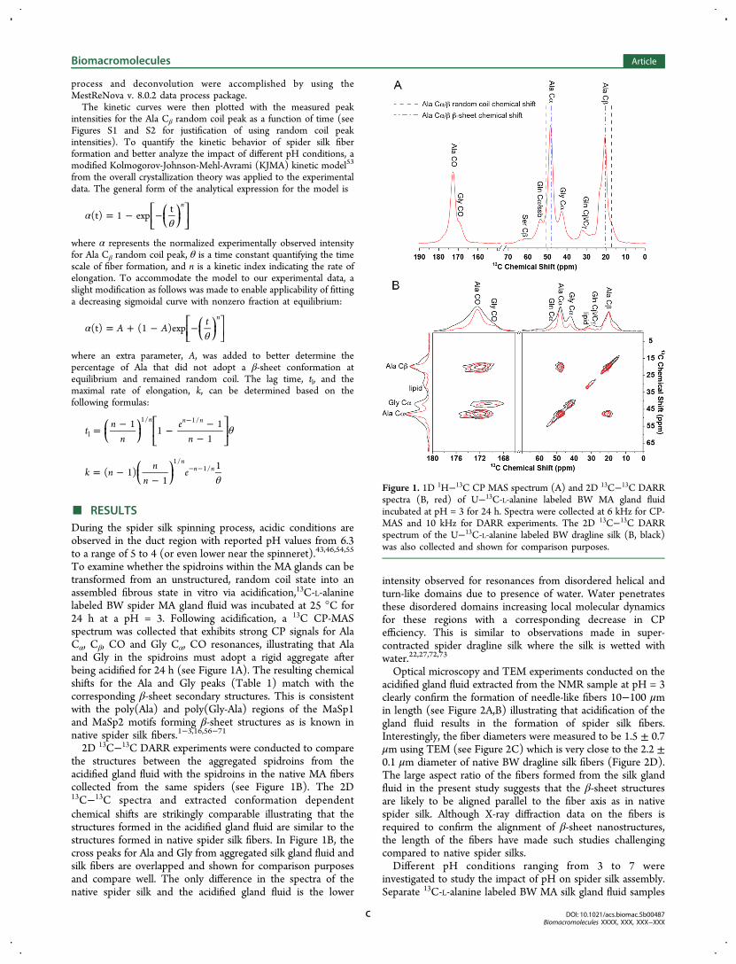
To examine whether the spidroins within the MA glands can betransformed from an unstructured, random coil state into anassembled fibrous state in vitro via acidification,13C-L-alaninelabeled BW spider MA gland fluid was incubated at 25 °C for24 h at a pH = 3. Following acidification, a 13C CP-MASspectrum was collected that exhibits strong CP signals for AlaCα, Cβ, CO and Gly Cα, CO resonances, illustrating that Alaand Gly in the spidroins must adopt a rigid aggregate afterbeing acidified for 24 h (see Figure 1A). The resulting chemicalshifts for the Ala and Gly peaks (Table 1) match with thecorresponding β-sheet secondary structures. This is consistentwith the poly(Ala) and poly(Gly-Ala) regions of the MaSp1and MaSp2 motifs forming β-sheet structures as is known innative spider silk fibers.1−3,16,56−71
2D 13C−13C DARR experiments were conducted to comparethe structures between the aggregated spidroins from theacidified gland fluid with the spidroins in the native MA fiberscollected from the same spiders (see Figure 1B). The 2D13C−13C spectra and extracted conformation dependentchemical shifts are strikingly comparable illustrating that thestructures formed in the acidified gland fluid are similar to thestructures formed in native spider silk fibers. In Figure 1B, thecross peaks for Ala and Gly from aggregated silk gland fluid andsilk fibers are overlapped and shown for comparison purposesand compare well. The only difference in the spectra of thenative spider silk and the acidified gland fluid is the lower
intensity observed for resonances from disordered helical andturn-like domains due to presence of water. Water penetratesthese disordered domains increasing local molecular dynamicsfor these regions with a corresponding decrease in CPefficiency. This is similar to observations made in super-contracted spider dragline silk where the silk is wetted withwater.22,27,72,73
Optical microscopy and TEM experiments conducted on theacidified gland fluid extracted from the NMR sample at pH = 3clearly confirm the formation of needle-like fibers 10−100 μmin length (see Figure 2A,B) illustrating that acidification of thegland fluid results in the formation of spider silk fibers.Interestingly, the fiber diameters were measured to be 1.5 ± 0.7μm using TEM (see Figure 2C) which is very close to the 2.2 ±0.1 μm diameter of native BW dragline silk fibers (Figure 2D).The large aspect ratio of the fibers formed from the silk glandfluid in the present study suggests that the β-sheet structuresare likely to be aligned parallel to the fiber axis as in nativespider silk. Although X-ray diffraction data on the fibers isrequired to confirm the alignment of β-sheet nanostructures,the length of the fibers have made such studies challengingcompared to native spider silks.Different pH conditions ranging from 3 to 7 were
investigated to study the impact of pH on spider silk assembly.Separate 13C-L-alanine labeled BW MA silk gland fluid samples
Figure 1. 1D 1H−13C CP MAS spectrum (A) and 2D 13C−13C DARRspectra (B, red) of U−13C-L-alanine labeled BW MA gland fluidincubated at pH = 3 for 24 h. Spectra were collected at 6 kHz for CP-MAS and 10 kHz for DARR experiments. The 2D 13C−13C DARRspectrum of the U−13C-L-alanine labeled BW dragline silk (B, black)was also collected and shown for comparison purposes.
Biomacromolecules Article
DOI: 10.1021/acs.biomac.5b00487Biomacromolecules XXXX, XXX, XXX−XXX
C

were prepared and incubated at different pH conditions and 25°C. In Figure 3, the 1H−13C CP-MAS spectra at different pHconditions are shown. At pH = 7, the spidroins remainedunstructured for more than 7 days, as no CP signal wasdetected from the silk proteins. At pHs below 7, the spidroinsare assembled into an aggregated fiber with a β-sheet richconformation. The overlay of the CP spectra at pH below 7shows no obvious difference in secondary structures. However,at pH = 6, the pH reported for the duct region of the spider silkgland45,46,55 and the dimerization of the N-terminal do-main,39,41−43 an extremely long period of 7 days is requiredfor the spidroins to self-assemble into the aggregated, fibrousstate. In contrast, at more acid environments, the time it takesfor the spidroins to aggregate can be greatly reduced to lessthan 24 h.The effect of pH on the spider silk spidroin assembly kinetics
was monitored with fully relaxed 13C DD-MAS solid-stateNMR. For the unstructured spidroins, the 13C DD-MAS
spectrum exhibits relatively sharp peaks because of the fastsubnanosecond silk protein backbone dynamics in the randomcoil state (see Figure 4A).13 However, after the silk fiber isformed, broader peaks are observed with 13C chemical shiftsthat correspond to β-sheet structures (see Figure 4A). A timeperiod of 24−35 h was required to record the full process ofspidroin assembly at different acidic pH conditions from 3 to 6.Consecutive 13C DD-MAS spectra were collected, with eachone requiring 30 min to obtain quantifiable 13C spectra withacceptable S/N ratio. The spider silk assembly transition isclearly monitored in the Ala Cβ region of the spectrum where asimultaneous increase for the broad β-sheet resonance occurswith a corresponding decrease of the narrow random coilresonance (Figure 4B). The composition of the spidroins withdifferent conformations at each sampling point was furtherextracted by deconvoluting the Ala Cβ region into three peaks(see Figure S1).The time dependence of the increase of β-sheet components
and the decrease of random coil components in the spidroins atpH = 4 is plotted in Figure 5. The trends of the twocomponents are very close, which is further confirmed byapplying the kinetic model to the data. The rate constant k forthe random coil peak decrease is 0.52 compared to 0.54 for theincrease of the β-sheet peak, while the lag times are 12.4 and12.1 h, respectively (see Table S1). This result indicates thedecrease of the random coil component is mainly due to theformation of β-sheet aggregates. Although the increase of the
Table 1. 13C Chemical Shift of Native Black Widow MA Gland Protein before and after Fiber Formation (Aggregated) At AcidicpH, the Chemical Shifts for Model Peptides with Known Secondary Structures Are Shown for Comparison Purposes56−71
13C chemical shifta
gland aggregated native silk α-helical β-sheet random coil
Ala Cα 50.3 48.3, 49.7 48.6, 49.4, 52.4 52.3−52.8 48.2−49.3 50.5Ala Cβ 16.5 20.5, 16.6 20.4, 17.0, 16.4 14.6−16.0 19.9−20.7 17.1Ala CO 175.5 172.2 172.4, 175.4 176.2−176.8 172.0−175.2 175.8Gly Cα 42.9 42.3 41.3, 43.0 43.2−44.3 43.1Gly CO 171.8 168.9, 171.9 168.8, 171.8 168.4−169.7 172.9Gln Cα 53.4 53.3 52.4 56.4−57.0 51.0−51.4 54.2Gln Cβ 26.8 26.9 27.4 25.6−26.3 29.0−29.9 27.4Gln Cγ 31.3 31.2 30.9 29.7−29.8 29.7−29.9 31.7Gln Cδ 177.8 177.7 177.5 178.5Gln CO 174.0 172.8 175.4−175.9 171.9−172.2 174.0
aAll chemical shifts are referenced to TMS.
Figure 2. Polarized light microscopy images with (A) and without (B)cross-polars and TEM (C) image for silk fibers formed by incubatingthe BW MA silk gland fluid at pH = 3 for 24 h. SEM image of BWdragline silk fibers collected from the same spiders (D).
Figure 3. 1H−13C CP MAS spectra of U−13C-L-alanine labeled BWMA silk gland fluid following incubation at pH = 3 (red), 4 (black), 5(blue), 6 (green), and 7 (magenta). Spectra were collected following a24 h incubation period for pH = 3 and 4, 48 h for pH = 5, and 7 daysfor pH = 6 and 7 from initial sample preparation.
Biomacromolecules Article
DOI: 10.1021/acs.biomac.5b00487Biomacromolecules XXXX, XXX, XXX−XXX
D

broad peak for β-sheet conformation can be observed by solid-state NMR spectroscopy, it is advantageous to indirectly trackthe spidroin assembly by monitoring the decrease of the muchnarrower and intense random coil resonance to gain better
sensitivity. In addition, it is worth noting that the agreementbetween the random coil decay and β-sheet build-up kineticsindicates that the vast majority of the NMR signal is observedin the 13C DD-MAS spectrum of native BW MA silk proteinsprior to fiber formation and corroborates previous interpreta-tions.13
The kinetic curves for spider silk assembly at different pHwere established by plotting the normalized peak intensities ofthe random coil peak for the Ala Cβ as a function of time. Asignature sigmoidal shaped kinetic curve was observed for thespider silk assembly process for the three acidic pHs tested (seeFigure 6). A long lag time was observed followed by rapid
elongation indicating that nucleation is a prerequisite. Theimpact of pH on the different phases of spidroin fiber formationcan be analyzed by applying the modified KJMA model to thekinetic curves (Table 2). At pH = 4, the shortest lag time (12.1h) is required for the spidroins to shift to the rapid elongationphase. However, an extra 5.5 or 7 h was observed for passingthe nucleation phase at pH = 3 or 5. Although the nucleationphase took a longer time at pH = 5, the β-sheet conformationyielded the largest percentage (80%) of Ala in a β-sheetstructure at equilibrium suggesting longer nucleation times isbeneficial for the complete formation of crystalline β-sheetcomponents. However, the difference in the fraction of Ala thatforms a β-sheet structure at different acidic pHs is small rangingfrom 70 to 80% and not far off from the fraction of Ala in β-sheet structures from solid-state NMR studies on native BWMA silk fibers where it was determined to be 88%.28 Theincreasing rate constants of the elongation phase from 0.24 to0.91 with decreasing pH values from 5 to 3 indicates that theelongation process is accelerated by more acidic conditions.
■ DISCUSSIONThe 13C DD-MAS NMR spectrum of the BW spidroins withinthe MA glands displays narrow resonances, with chemical shiftsmatching a random coil structure. Although the spidroins arestored in the silk glands at a near neutral but slightly acidicenvironment (pH ∼ 6.3), a high concentration of NaCl (∼180mM) is present and believed to prevent premature aggregationand maintain an unstructured, random coil state.45 It has beenproposed that an acidic pH gradient exists in the spinningsystem from the silk glands along the duct to the spinneret andis likely important for spider silk formation.46,54 With the
Figure 4. 13C DD-MAS NMR spectra of U−13C-L-alanine labeled BWMA silk gland fluid at pH = 4 before (A, black) and after spider silkassembly (A, red). The Ala Cβ region is framed by dashed lines andwill be analyzed to monitor spider silk assembly kinetics. Ala Cβ
regions from the 13C DD-MAS spectra collected at different time fromsample preparation were stacked showing that the decrease for therandom coil resonance and increase for the β-sheet resonance can beobserved simultaneously as a function of time (B).
Figure 5. Time dependence of normalized peak integrals for randomcoil peak (black circle) and β-sheet peak (red circle) from the kineticmeasurement of spider silk fiber formation for U−13C-L-alaninelabeled BW MA gland fluid at pH = 4 with 13C DD-MAS NMR. Thedata was fit to modified KJMA kinetic model and the extracted kineticparameters are shown in Table S1.
Figure 6. Kinetic curves for U−13C-L-alanine labeled BW MA silkgland fluid incubated at pH = 3 (red circle), 4 (blue circle) and 5(black circle). The solid curves were obtained by applying themodified KJMA model and the extracted kinetic parameters are shownin Table 2.
Biomacromolecules Article
DOI: 10.1021/acs.biomac.5b00487Biomacromolecules XXXX, XXX, XXX−XXX
E

limitation of experimental methods, accurate measurement tomap out the pH values in the spinning system is stillchallenging. However, the spidroins are expected to experiencemuch lower pH values near the spinneret.55 In addition, toinduce fiber formation in vitro on the native spider silk glandfluid with a high NaCl content, a lower pH condition may berequired compared to the in vivo spinning conditions.Therefore, to study the effect of acidic biochemical conditionson spider silk fiber formation, acidic conditions from pH = 6 to3 were investigated for the native BW MA spidroins. At pH = 7,the spidroins remain unstructured random coils for over 7 dayswith no evidence of aggregation, but when acidic conditions areapplied, the metastable state of the unstructured spidroins isdisturbed and aggregation to silk fibers occurs albeit withdifferent kinetics. The spidroins assemble into fibers at pH ≤ 6,with no observable difference in the conformational structuresof the folded spidroins for different pH values following fiberformation. However, distinguishable difference in the rates offiber formation from 7 days at pH = 6 to 12 h at pH = 4 areobserved leading to the study on the kinetics of spidroin fiberformation at acidic conditions with 13C DD-MAS NMR.The kinetic profile of spidroin fiber formation is a typical
sigmoidal curve. The long period of lag time indicatesnucleation is required to allow further elongation. Unlike aparabolic curve, the sigmoidal curve also indicates no seedsexisted in the system to help skip the nucleation phase.74
Because nucleation is a crucial prerequisite for fiber formation,it was important to determine that centrifugal and centripetalforces from MAS did not influence the kinetics to anyappreciable extent as confirmed by the static 13C NMRexperiments (see Figure S3 and Table 2). Comparison of theextracted kinetic parameter for static and 4 kHz MASconditions illustrates that MAS has little impact on the spidersilk assembly kinetics with the exception of a slightly higherremaining random coil fraction of 0.30 and 0.24 at equilibriumfor the two conditions, respectively.The lag times observed for spider silk fiber formation as a
function of pH in the present study are surprisingly long (>12h) indicating that pH may not be the biochemical variable thatcontrols nucleation. Previous studies on silkworm fibroins andspider spidroins indicate shorter lag times for fiberformation.29,30,34,75 These previous studies are likely notcomparable to the investigation here because of the dilute(∼0.1% by wt) protein concentrations used compared to themuch higher concentration of silk proteins (∼23% by wt) inthe silk glands used in this study. Even though the remainingNaCl in the silk gland may postpone nucleation, the over 12 hof lag time implies that other mechanisms may be responsiblefor nucleation. One possible explanation for this is the gel-likestate of spider silk spidroins inside the MA gland that canprevent or postpone nucleation. Thus, any approaches todissolve the dope inside silk glands will remove this stabilizingeffect of the gel-like state with high salt content leading to fasteraggregation due to increased hydrophobic interactions.
Similarly, one must consider the other physicochemicalinfluences such as dehydration, shearing forces and theinfluence of other ions that could be introduced in the ductto assist the nucleation process.29−31,34,44,46,47
As shown in Figure 6, the shortest nucleation phase wasobserved at a pH near the isoelectric point (pI = 4.25) of theN-terminal domains (NTD) of the spidroins.38 This resultindicates the nucleation process may be related to the NTD.The NTD is known to dimerize at a pH = 6 and thedimerization becomes more stable as pH decreases.39,41,43
Compared to a pH = 3 or 5, the solubility of the NTD dimer ismuch lower at a pH = 4. This is consistent with the resultspresented here where the shortest nucleation phase is observedat a pH = 4. In comparison, during the elongation process, thespidroins exhibit the fastest elongation rate at lower pH values.Since the pI of the entire silk protein was experimentallyestimated to be ∼4.2,30,31 the fastest rate of elongation phase atpH = 3 implies that the elongation phase is not governed by thesolubility change of the entire silk protein. In addition, a largerpercent of β-sheet structures is formed in the silk fibers withlonger nucleation time. Combining the information listedabove, it is reasonable to propose a possible model to explainthe function of the pH gradient in the spinning system. Thespidroins are initially stored in an unstructured, random coilstate at near neutral pH, where the spidroins can remain stable.At pH = 6, the NTD first starts to form dimers followed by silkprotein nucleation. Larger assemblies are formed at a pH ∼ 5 toensure higher percentage of β-sheet structures in the silk fiber,then the solubility of the silk protein drops dramatically at a pH= 4 leading to rapid fiber formation. When the dope is close tothe spinneret, decreasing the pH below 4 can accelerate theelongation rate even further. Thus, a pH gradient can be usedto regulate the speed of the nucleation and elongation as well asthe final β-sheet composition in the spider silk assemblyprocess. Lastly, although pH appears to be important inregulating and controlling spider silk assembly, the otherphysicochemical variables that have been discussed previ-ously29−31,34,44,46,47 will certainly contribute to the assemblyprocess.
■ CONCLUSIONS
The kinetics of spider silk assembly were tracked with solid-state NMR spectroscopy for native BW MA silk proteins as afunction of pH. The resulting kinetic profiles reveal that silkfiber formation is nucleation dependent and the rate ofnucleation reaches a maximum when the pH value is close tothe pI of the N-terminal domain and the silk protein. Inaddition, decreasing the pH value can accelerate the rate ofelongation while, a higher percentage of the spidroins wasobserved to exhibit a β-sheet conformation in the silk fiber atless acidic conditions. Therefore, the gradual pH decrease alongthe duct can be used to optimize the speed of silk production aswell as to achieve higher crystalline compositions in the finalfibers. We anticipate that this in vitro 13C solid-state NMR
Table 2. Fitting Parameters from the Kinetic Curves Using a Modified KJMA Model for Spider Silk Fiber Assembly at DifferentpH Conditions and Different MAS Frequencies
pH n θ A k tl
3 (4 kHz MAS) 45.5 ± 3.6 18.4 ± 0.1 0.22 ± 0.01 0.91 ± 0.07 17.7 ± 0.14 (4 kHz MAS) 19.8 ± 2.5 13.3 ± 0.1 0.24 ± 0.01 0.55 ± 0.07 12.1 ± 0.25 (4 kHz MAS) 14.2 ± 0.5 21.8 ± 0.1 0.20 ± 0.01 0.24 ± 0.01 19.2 ± 0.14 (0 kHz MAS) 19.7 ± 2.8 13.2 ± 0.1 0.30 ± 0.01 0.55 ± 0.08 12.1 ± 0.2
Biomacromolecules Article
DOI: 10.1021/acs.biomac.5b00487Biomacromolecules XXXX, XXX, XXX−XXX
F

approach will be a valuable tool moving forward to interrogate arange of biochemical conditions on spider silk assemblyincluding the effect of various salts. Such studies are alreadyunder way in our laboratory.
■ ASSOCIATED CONTENT*S Supporting InformationKJMA kinetic parameters for Ala Cβ random coil and β-sheetcomponent from 13C DD-MAS NMR kinetic curves obtainedduring acidification; an example of peak deconvolution for theAla Cβ region from the 13C DD-MAS NMR spectrum; kineticcurves for the spider silk protein assembly with and withoutMAS. The Supporting Information is available free of charge onthe ACS Publications website at DOI: 10.1021/acs.bio-mac.5b00487.
■ AUTHOR INFORMATIONCorresponding Author*E-mail: [email protected] authors declare no competing financial interest.
■ ACKNOWLEDGMENTSThis work was supported by grants from the Department ofDefense Air Force Office of Scientific Research (AFOSR)under Award No. FA9550-14-1-0014, the Defense UniversityResearch Instrumentation Program (DURIP) under Award No.FA2386-12-1-3031 DURIP 12RSL231, and the NationalScience Foundation, Division of Materials Research (NSF-DMR) under Award No. DMR-1264801. We thank Dr. BrianCherry for help with NMR instrumentation and studenttraining.
■ ABBREVIATIONSCP, cross-polarization; MAS, magic angle spinning; TPPM, twopulse phase modulation; DARR, dipolar assisted rotationalresonance; CW, continuous wave
■ REFERENCES(1) Gosline, J. M.; Demont, M. E.; Denny, M. W. Endeavour 1986,10, 37−43.(2) Altman, G. H.; Diaz, F.; Jakuba, C.; Calabro, T.; Horan, R. L.;Chen, J. S.; Lu, H.; Richmond, J.; Kaplan, D. L. Biomaterials 2003, 24,401−416.(3) Lewis, R. V. Chem. Rev. 2006, 106, 3762−3774.(4) Gosline, J. M.; Denny, M. W.; Demont, M. E. Nature 1984, 309,551−552.(5) Vollrath, F.; Porter, D. Soft Matter 2006, 2, 377−385.(6) Teule, F.; Cooper, A. R.; Furin, W. A.; Bittencourt, D.; Rech, E.L.; Brooks, A.; Lewis, R. V. Nat. Protoc. 2009, 4, 341−355.(7) Vollrath, F.; Porter, D. Polymer 2009, 50, 5623−5632.(8) Omenetto, F. G.; Kaplan, D. L. Science 2010, 329, 528−531.(9) Hijirida, D. H.; Do, K. G.; Michal, C.; Wong, S.; Zax, D.; Jelinski,L. W. Biophys. J. 1996, 71, 3442−3447.(10) Hronska, M.; van Beek, J. D.; Williamson, P. T. F.; Vollrath, F.;Meier, B. H. Biomacromolecules 2004, 5, 834−839.(11) Lefevre, T.; Leclerc, J.; Rioux-Dube, J. F.; Buffeteau, T.; Paquin,M. C.; Rousseau, M. E.; Cloutier, I.; Auger, M.; Gagne, S. M.;Boudreault, S.; Cloutier, C.; Pezolet, M. Biomacromolecules 2007, 8,2342−2344.(12) Jenkins, J. E.; Holland, G. P.; Yarger, J. L. Soft Matter 2012, 8,1947−1954.(13) Xu, D.; Yarger, J. L.; Holland, G. P. Polymer 2014, 55, 3879−3885.
(14) Xu, M.; Lewis, R. V. Proc. Natl. Acad. Sci. U.S.A. 1990, 87,7120−7124.(15) Hinman, M. B.; Lewis, R. V. J. Biol. Chem. 1992, 267, 19320−19324.(16) Simmons, A.; Ray, E.; Jelinski, L. W. Macromolecules 1994, 27,5235−5237.(17) Kummerlen, J.; van Beek, J. D.; Vollrath, F.; Meier, B. H.Macromolecules 1996, 29, 2920−2928.(18) Bram, A.; Branden, C. I.; Craig, C.; Snigireva, I.; Riekel, C. J.Appl. Crystallogr. 1997, 30, 390−392.(19) Grubb, D. T.; Jelinski, L. W. Macromolecules 1997, 30, 2860−2867.(20) Riekel, C.; Vollrath, F. Int. J. Biol. Macromol. 2001, 29, 203−210.(21) van Beek, J. D.; Hess, S.; Vollrath, F.; Meier, B. H. Proc. Natl.Acad. Sci. U.S.A. 2002, 99, 10266−10271.(22) Holland, G. P.; Lewis, R. V.; Yarger, J. L. J. Am. Chem. Soc. 2004,126, 5867−5872.(23) Holland, G. P.; Creager, M. S.; Jenkins, J. E.; Lewis, R. V.;Yarger, J. L. J. Am. Chem. Soc. 2008, 130, 9871−9877.(24) Holland, G. P.; Jenkins, J. E.; Creager, M. S.; Lewis, R. V.;Yarger, J. L. Chem. Commun. 2008, 5568−5570.(25) Holland, G. P.; Jenkins, J. E.; Creager, M. S.; Lewis, R. V.;Yarger, J. L. Biomacromolecules 2008, 9, 651−657.(26) Jenkins, J. E.; Creager, M. S.; Butler, E. B.; Lewis, R. V.; Yarger,J. L.; Holland, G. P. Chem. Commun. 2010, 46, 6714−6716.(27) Jenkins, J. E.; Creager, M. S.; Lewis, R. V.; Holland, G. P.;Yarger, J. L. Biomacromolecules 2010, 11, 192−200.(28) Jenkins, J. E.; Sampath, S.; Butler, E.; Kim, J.; Henning, R. W.;Holland, G. P.; Yarger, J. L. Biomacromolecules 2013, 14, 3472−3483.(29) Chen, X.; Knight, D. P.; Shao, Z. Z.; Vollrath, F. Biochemistry2002, 41, 14944−14950.(30) Dicko, C.; Kenney, J. M.; Knight, D.; Vollrath, F. Biochemistry2004, 43, 14080−14087.(31) Foo, C. W. P.; Bini, E.; Hensman, J.; Knight, D. P.; Lewis, R. V.;Kaplan, D. L. Appl. Phys. A: Mater. Sci. Process. 2006, 82, 223−233.(32) Lefevre, T.; Boudreault, S.; Cloutier, C.; Pezolet, M.Biomacromolecules 2008, 9, 2399−2407.(33) Lefevre, T.; Boudreault, S.; Cloutier, C.; Pezolet, M. J. Mol. Biol.2011, 405, 238−253.(34) Leclerc, J.; Lefevre, T.; Gauthier, M.; Gagne, S. M.; Auger, M.Biopolymers 2013, 99, 582−593.(35) Ayoub, N. A.; Garb, J. E.; Tinghitella, R. M.; Collin, M. A.;Hayashi, C. Y. PloS One 2007, 2, e514.(36) Beckwitt, R.; Arcidiacono, S. J. Biol. Chem. 1994, 269, 6661−6663.(37) Motriuk-Smith, D.; Smith, A.; Hayashi, C. Y.; Lewis, R. V.Biomacromolecules 2005, 6, 3152−3159.(38) Askarieh, G.; Hedhammar, M.; Nordling, K.; Saenz, A.; Casals,C.; Rising, A.; Johansson, J.; Knight, S. D. Nature 2010, 465, 236−U125.(39) Gaines, W. A.; Sehorn, M. G.; Marcotte, W. R. J. Biol. Chem.2010, 285, 40745−40753.(40) Hagn, F.; Eisoldt, L.; Hardy, J. G.; Vendrely, C.; Coles, M.;Scheibel, T.; Kessler, H. Nature 2010, 465, 239−U131.(41) Hagn, F.; Thamm, C.; Scheibel, T.; Kessler, H. Angew. Chem.2011, 50, 310−313.(42) Schwarze, S.; Zwettler, F. U.; Johnson, C. M.; Neuweiler, H.Nat. Commun. 2013, 4, 2815.(43) Kronqvist, N.; Otikovs, M.; Chmyrov, V.; Chen, G.; Andersson,M.; Nordling, K.; Landreh, M.; Sarr, M.; Jornvall, H.; Wennmalm, S.;Widengren, J.; Meng, Q.; Rising, A.; Otzen, D.; Knight, S. D.;Jaudzems, K.; Johansson, J. Nat. Commun. 2014, 5, 3254.(44) Tillinghast, E. K.; Chase, S. F.; Townley, M. A. J. Insect. Physiol.1984, 30, 591−596.(45) Knight, D. P.; Vollrath, F. Naturwissenschaften 2001, 88, 179−182.(46) Dicko, C.; Vollrath, F.; Kenney, J. M. Biomacromolecules 2004, 5,704−710.
Biomacromolecules Article
DOI: 10.1021/acs.biomac.5b00487Biomacromolecules XXXX, XXX, XXX−XXX
G

(47) Boulet-Audet, M.; Terry, A. E.; Vollrath, F.; Holland, C. ActaBiomater. 2014, 10, 776−784.(48) Shi, X. Y.; Yarger, J. L.; Holland, G. P. Anal. Bioanal. Chem.2013, 405, 3997−4008.(49) Baryshnikova, O. K.; Williams, T. C.; Sykes, B. D. J. Biomol.NMR 2008, 41, 5−7.(50) Bennett, A. E.; Rienstra, C. M.; Auger, M.; Lakshmi, K. V.;Griffin, R. G. J. Chem. Phys. 1995, 103, 6951−6958.(51) Takegoshi, K.; Nakamura, S.; Terao, T. Chem. Phys. Lett. 2001,344, 631−637.(52) Takegoshi, K.; Nakamura, S.; Terao, T. J. Chem. Phys. 2003, 118,2325−2341.(53) Auer, S.; Kashchiev, D. Proteins 2010, 78, 2412−2416.(54) Vollrath, F.; Knight, D. P.; Hu, X. W. Proc. R. Soc. B 1998, 265,817−820.(55) Andersson, M.; Chen, G.; Otikovs, M.; Landreh, M.; Nordling,K.; Kronqvist, N.; Westermark, P.; Jornvall, H.; Knight, S.;Ridderstrale, Y.; Holm, L.; Meng, Q.; Jaudzems, K.; Chesler, M.;Johansson, J.; Rising, A. PLoS Biol. 2014, 12, e1001921.(56) Kricheldorf, H. R.; Muller, D. Macromolecules 1983, 16, 615−623.(57) Saito, H.; Tabeta, R.; Asakura, T.; Iwanaga, Y.; Shoji, A.; Ozaki,T.; Ando, I. Macromolecules 1984, 17, 1405−1412.(58) Shoji, A.; Ozaki, T.; Saito, H.; Tabeta, R.; Ando, I.Macromolecules 1984, 17, 1472−1479.(59) Saito, H. Magn. Reson. Chem. 1986, 24, 835−852.(60) Ishida, M.; Asakura, T.; Yokoi, M.; Saito, H. Macromolecules1990, 23, 88−94.(61) Wishart, D. S.; Bigam, C. G.; Holm, A.; Hodges, R. S.; Sykes, B.D. J. Biomol. NMR 1995, 5, 67−81.(62) Asakura, T.; Demura, M.; Date, T.; Miyashita, N.; Ogawa, K.;Williamson, M. P. Biopolymers 1997, 41, 193−203.(63) Asakura, T.; Ashida, J.; Yamane, T.; Kameda, T.; Nakazawa, Y.;Ohgo, K.; Komatsu, K. J. Mol. Biol. 2001, 306, 291−305.(64) Zhao, C. H.; Asakura, T. Prog. Nucl. Magn. Reson. Spectrosc.2001, 39, 301−352.(65) Asakura, T.; Yao, J. M. Protein Sci. 2002, 11, 2706−2713.(66) Murata, K.; Kuroki, S.; Ando, I. Polymer 2002, 43, 6871−6878.(67) Ashida, J.; Ohgo, K.; Komatsu, K.; Kubota, A.; Asakura, T. J.Biomol. NMR 2003, 25, 91−103.(68) Kishi, S.; Santos, A.; Ishii, O.; Ishikawa, K.; Kunieda, S.; Kimura,H.; Shoji, A. J. Mol. Struct. 2003, 649, 155−167.(69) Wildman, K. A. H.; Wilson, E. E.; Lee, D. K.; Ramamoorthy, A.Solid State Nucl. Magn. Reson. 2003, 24, 94−109.(70) Asakura, T.; Nakazawa, Y.; Ohnishi, E.; Moro, F. Protein Sci.2005, 14, 2654−2657.(71) Asakura, T.; Yang, M. Y.; Kawase, T.; Nakazawa, Y.Macromolecules 2005, 38, 3356−3363.(72) Creager, M. S.; Jenkins, J. E.; Thagard-Yeaman, L. A.; Brooks, A.E.; Jones, J. A.; Lewis, R. V.; Holland, G. P.; Yarger, J. L.Biomacromolecules 2010, 11, 2039−2043.(73) Yang, Z. T.; Liivak, O.; Seidel, A.; LaVerde, G.; Zax, D. B.;Jelinski, L. W. J. Am. Chem. Soc. 2000, 122, 9019−9025.(74) Morris, A. M.; Watzky, M. A.; Finke, R. G. Biochim. Biophys.Acta 2009, 1794, 375−397.(75) Chen, X.; Shao, Z. Z.; Knight, D. P.; Vollrath, F. Proteins 2007,68, 223−231.
Biomacromolecules Article
DOI: 10.1021/acs.biomac.5b00487Biomacromolecules XXXX, XXX, XXX−XXX
H