UNIVERSITY OF CALIFORNIA, IRVINE PIAS1 modulates the ...
291
UNIVERSITY OF CALIFORNIA, IRVINE PIAS1 modulates the transcriptional landscape and DNA damage repair in Huntington’s disease DISSERTATION submitted in partial satisfaction of the requirements for the degree of DOCTOR OF PHILOSOPHY in Biological Sciences by Eva Louise Lombardi Morozko Dissertation Committee: Professor Leslie M. Thompson, Chair Professor Marcelo Wood Professor Peter Kaiser Professor Kyoko Yokomori Professor Mathew Blurton-Jones 2019
-
Upload
khangminh22 -
Category
Documents
-
view
2 -
download
0
Transcript of UNIVERSITY OF CALIFORNIA, IRVINE PIAS1 modulates the ...
UNIVERSITY OF CALIFORNIA,
IRVINE
PIAS1 modulates the transcriptional landscape and DNA damage repair in Huntington’s disease
DISSERTATION
submitted in partial satisfaction of the requirements for the degree of
DOCTOR OF PHILOSOPHY
in Biological Sciences
by
Eva Louise Lombardi Morozko
Dissertation Committee: Professor Leslie M. Thompson, Chair
Professor Marcelo Wood Professor Peter Kaiser
Professor Kyoko Yokomori Professor Mathew Blurton-Jones
2019

Portions of Chapter 1 © 2018 IOS Press All other materials © 2019 Eva L. Morozko

ii
DEDICATION
For
My mother whom may not be here physically but will always be in my heart. The fire and fight she exhibited during such unimaginable circumstances taught me to never give up, to love and support those around me, and to seek the truth. She will forever motivate me to
keep moving forward, no matter what obstacles may be placed before me, for the road goes ever on and on.
To my father for his unconditional support and belief in me and for the sacrifices he made for our family.
To my wife who reminds me that love knows no boundaries and no distance. Her constant love and belief in me made this journey possible.
To my cats, who always know.

iii
TABLE OF CONTENTS
Page LIST OF FIGURES iv LIST OF TABLES vii LIST OF ABBREVIATIONS viii ACKNOWLEDGMENTS ix CURRICULUM VITAE xi ABSTRACT OF THE DISSERTATION xix INTRODUCTION 1 A. Huntington’s disease genetics and pathology 1 B. Protein and genomic homeostasis in Huntington’s disease 8 C. SUMO modification in HD and neurodegeneration 11 D. SUMO E3 ligase PIAS1 may represent a therapeutic target for Huntington’s disease 17 E. DNA damage and SUMOylation; implications in HD and the Nervous System 22 F. Consequences of unrepaired DNA towards neuronal viability 36 G. Summary 38 CHAPTER 1: Longitudinal Biochemical Assay Analysis of Mutant Huntingtin 48 Exon 1 Protein in R6/2 Mice CHAPTER 2: PIAS1 SUMO-Interaction Motif May Modulate Accumulation of 84 Mutant Huntingtin Protein CHAPTER 3: PIAS1 Serves as a SUMO E3 Ligase for PNKP and Modulates its 107 Enzymatic Activity in Huntington’s Disease Patient iPSC-Derived Neurons CHAPTER 4: Pias1 Reduction Modulates DNA Damage Repair and Neuronal 139 transcription in the zQ175 Mouse Model of HD CHAPTER 5: Restoration of Pnkp Activity by Pias1 Reduction in R6/2 and 207 Potential Relationship to Somatic Repeat Expansion DISSERTATION CONCLUDING REMARKS 235 REFERENCES 243

iv
LIST OF FIGURES
Page Figure 1 The SUMO conjugation pathway 15 Figure 2 Accrual of damage and consequences in neurons 24 Figure 1.1 Progressive motor and transcriptional changes as 54
quality control for R6/2 mice Figure 1.2 Delta CT (dCT) values used to analyze transcriptional 56
alterations detected by qPCR in R6/2 mice Figure 1.3 Progressive mHTT inclusion body formation in cortex 58
and striatum of R6/2 mice Figure 1.4 Detection of mHTTex1p in striatum of R6/2 mice 60 Figure 1.5 Detection of mHTTex1p is highly variable using TRIzol 61
reagent Figure 1.6 Detection of mHTTex1p in cortex of R6/2 mice 62 Figure 1.7 Detection of mHTTex1p in hippocampus and cerebellum 64
of R6/2 mice varies depending on break method Figure 1.8 Detection of mHTTex1p in peripheral tissue varies depending 67
on break method. Figure 1.9 Detection of mHTTex1p on AGE gels varies based by break 68
method Figure 2.1 PIAS1 has numerous functional domains 88 Figure 2.2 PIAS1 phospho-mutants express at the same level 89 Figure 2.3 PIAS1 SIM may modulate mHTT solubility in Human cells 91 Figure 2.4 PIAS1 SIM may modulate mHTT solubility in striatal-like cells 93 Figure 2.5 PIAS1 Ligase-Dead mutants produce a dominant-negative 95
protein product Figure 2.6 PIAS1 LD mutants confound mHTT assessment 97
v
Page Figure 2.7 PIAS1 LD mutants confound mHTT assessment with MG132 98
treatment Figure 3.1 CRISPR-Cas9 was used to knockdown PIAS1 expression in iPSC 112 Figure 3.2 CRISPR edited HD-iPSC lines differentiate into neurons 114 Figure 3.3 PIAS1 is part of the TCR complex and modulates PNKP 115
enzymatic activity Figure 3.4 PNKP may be a substrate for SUMOylation 117 Figure 3.5 PNKP is SUMOylated and may be a substrate for STUbL 118
Activity in vitro Figure 3.6 SUMO1 PNKP modification is mediated by PIAS1 in vitro 120 Figure 3.7 SUMO2 PNKP modification is mediated by PIAS1 in vitro 121 Figure 3.8 mHTT may affect endogenous PNKP SUMOylation 122 Figure 3.9 PIAS1 may play a role in evicting the TCR complex 127 Figure 4.1 Experimental design for modulating Pias1 in zQ175 mice 145 Figure 4.2 Flow chart summary of zQ175 Pias1 KD experimental 146
groups, study progression, and overall findings Figure 4.3 Pre-symptomatic Pias1 KD may exacerbate motor 148
deficits in zQ175 mice Figure 4.4 Pre-symptomatic Pias1KD does not affect mHTT 152
accumulation at 13.5 months of age Figure 4.5 Pre-symptomatic Pias1 KD rescues neuronal 156
function-related transcriptional dysregulation in zQ175 mice at 13.5 months of age
Figure 4.6 qPCR analysis to validate mRNAseq identified genes for 158
pre-symptomatic miPias1.3 treatment assessed at 13.5 months of age
Figure 4.7 Pre-symptomatic Pias1 KD modulates immune response 160

vi
Page Figure 4.8 Symptomatic Pias1KD has no effect on behavior in zQ175 163
mice Figure 4.9 Symptomatic Pias1 KD does not affect mHTT accumulation 165
at 13.5 months of age Figure 4.10 Symptomatic Pias1 KD modulates immune response 167 Figure 4.11 Pre-symptomatic Pias1KD may rescue early motor deficits 169 in zQ175 mice Figure 4.12 Pre-symptomatic PIias1 KD does not affect mHTT 172
accumulation at 8 months of age Figure 4.13 Pre-symptomatic Pias1 KD does not modulate immune response 173
in younger mice Figure 4.14 Pre-symptomatic Pias1 KD has minimal effect on transcriptional 175
landscape at 8 months of age Figure 4.15 qPCR analysis on mRNAseq identified genes that were 179
differentially expressed at both 8 and 13.5 months of age with pre-symptomatic Pias1 KD
Figure 4.16 Pre-symptomatic Pias1 KD rescues disease-associated modules 182
from zQ175 allelic series
Figure 4.17 Pias1KD affects DNA damage repair in zQ175 mice 184 Figure 5.1 Pias1 KD does not rescue behavior in R6/2 mice 212 Figure 5.2 PIAS1 knockdown does not affect mHTT accumulation in R6/2 216
mice Figure 5.3 Pias1 modulated Pnkp enzymatic activity but not DNA damage 217
levels in R6/2 mice Figure 5.4 qPCR analysis on top mRNAseq identified genes identified in 220
Chapter 4 for R6/2 miPias1.3 treated mice Figure 5.5 Pias1 knock-down results in smaller expansion indexes in 221
R6/2 mice.

vii
LIST OF TABLES
Page Table 1 SUMO modification in neurodegenerative disease 42 Table 2 SUMO modification of neuronal transcription factors 43 Table 3 Synaptic SUMOylation substrates 44 Table 4 DNA damage SUMOylation substrates 46 Table 5 DDR genes associated with neuropathologies 47 Table 1.1 Break methods optimized for detection of specific 81
mHTT species. Table 1.2 mHTTex1p size fluctuation due to CAG repeat length 82 Table 1.3 Summary heat map of mHTTex1p detected using various 83
break methods and assays at 5, 7, 9, and 11 weeks of age. Table 3.1 Immunofluorescent staining protocols for iPSC QC 138 Table 4.1 Statistical outputs for zQ175 pre-symptomatic Pias1 KD cohorts 202 Table 4.2 Statistical outputs for zQ175 Symptomatic Pias1 KD cohorts 203 Table 4.3 Top Pias1 KD- modulated DEGs in pre-symptomatic miPias1.3 204
treated animals used for RT-qPCR Table 4.4 Summary heat map of Pias1 KD effect on transcriptional targets 205
in zQ175 heterozygote animals Table 4.5 disease associated transcriptional modules affected by Pias1 KD 206
in zQ175 mice Table 5.1 Summary heat map of Pias1 KD effect on transcriptional 234
readouts in R6/2 and zQ175 heterozygote animals

viii
LIST OF ABBREVIATIONS
AD Alzheimer’s disease AGE Agarose Gel Electrophoresis ALS Amyotrophic lateral sclerosis AO Age of onset BER Base excision repair DDR DNA Damage Repair DEG Differentially Expressed Gene DSB Double strand breaks FA Fanconi Anemia FL HTT Full Length HTT protein gDNA genomic DNA GO Gene Ontology GWAS Genome-wide association studies HD Huntington's disease Het Heterozygous mouse HMW High Molecular Weight HR Homologous Recombination HTT Huntingtin protein or gene ICL Interstrand crosslink IPA Ingenuity Pathway Analysis iPSC Induced Pluripotent Stem Cell KD Knock-down KI Knock-In LD Ligase dead mHTT mutant huntingtin mHTTex1p mutant huntingtin protein encoded by exon 1 only MMR Mismatch repair N17 17 N-terminal amino acids of HTT before CAG repeat region NER Nucleotide excision repair NHEJ Non-homologous end-joining NT Non-transgenic PCA Principal Component Analysis PD Parkinson’s disease PolyQ Polyglutamine tract PTM Post translational modification R-loop DNA:RNA hybrid structure ROS Reactive Oxygen Species SASP Senescence associated secretory phenotype SIM SUMO Interaction Motif SSB Single Strand Breaks STUbLs SUMO-targeted ubiquitin ligases TCR Transcription coupled repair UPS Ubiquitin Proteasome System WT Wild type

ix
ACKNOWLEDGMENTS
I would like to acknowledge and thank the members of my committee, Dr. Marcelo Wood, Dr. Peter Kaiser, and Dr. Kyoko Yokomori for all of their support and feedback towards my project. Their critiques and input were invaluable towards the evolution and success of this project. A special thanks to Dr. Mathew Blurton-Jones for his support. Thank you to Dr. Leslie Thompson who has been a supportive and encouraging mentor throughout my graduate studies. Thank you for tolerating my “devil’s advocate” scientific debates/discussions and helping me transform into the motivated and thoughtful scientist I am today. Your training, mentoring, and support were integral towards my own personal and scientific growth. To my collaborators and their labs, for which this project would not have been possible - their support and scientific input were priceless. To Dr. Beverly Davidson and her lab, especially Alejandro Mas Monteys for his work generating clones, miRNA, and viral vectors to knock-down PIAS1. To Dr. Partha Sarkar and his lab for their mechanistic contributions regarding DNA damage repair, especially Subrata Pradhan for his technical assistance. To Dr. William Yang and Peter Langfelder for their help analyzing transcriptional data. To Dr. Vanessa Wheeler, for her expertise on somatic repeat expansion. Thank you to Dr. Jack Reidling for his constant support and mentorship and for our shared intellectual approach. I am forever grateful for the support, criticisms, guidance, and feedback you provided me throughout my journey. I am entirely grateful to the Thompson Laboratory for their help and support towards this work. Specifically, I would like to thank Alice Lau for her extensive technical knowledge and laughs in the lab, Lexi Kopan and Illiana Orellana for their technical help and support, and Giana Fote for being an amazing friend and companion and providing sound, intellectual insights into my project. I also want to thank Dr. Charliene Smith-Geater for being an amazing teammate for team Unicorn aka PIAS1 project. I am so grateful that we got to work together to explore these exciting new mechanisms; thank you for sharing your story with me. These years would not have been possible without amazing friends and family. I want to thank my parents and my sister for maybe not understanding my work beyond “something gross with mice” but supporting me none-the-less. Thank you to my incoming graduate class for their camaraderie over the years – knowing we are all on this together made it possible. Specifically, I’d like to thank Maria Montchal and Susan Gil for being the dopest ladies and putting up with my crap. And Isabella Sanchez; it was amazing sharing my Thompson-lab graduate journey with you. I also want to thank my UCI Archery team and crew; for supporting me and making me laugh no matter what the situation and for teaching me that the most important arrow is in the bow.

x
Finally, this would not have been possible if not for the amazing support from my wife, Meaghan. We had no idea where we would end up when this all started but we only grew stronger over this journey and her support was my foundation for success. I’m so happy we get to share our life journey together. I’m looking forward to the adventures to come. This work was supported in parts by NSF GRFP 2016137353, the ARCs foundation, and NINDS RO1NS090390. I thank the IOS Press and the Journal of Huntington’s disease for permission to include copyrighted figures and text as part of Chapter 1 of my dissertation.

xi
CURRICULUM VITAE
Eva Louise Lombardi Morozko
EDUCATION 2014 – 2019 University of California, Irvine, CA
Doctor of Philosophy: Biological Sciences, Neurobiology and Behavior
2014 – 2017 University of California, Irvine, CA Master of Science: Biological Sciences
2008 – 2012 Seton Hall University, South Orange, NJ
Bachelor of Science: Biochemistry, Minor: Classical Studies Magna Cum Laude
HONORS AND AWARDS 2018 UCI, CNLM Renee Harwick Advanced Graduate Student Award 2018 UCI MIND, ReMIND Outstanding Predoctoral Scholar Award 2017 – 2019 ARCS Foundation, Inc. Scholar, UC, Irvine Graduate Division 2017 Fine Science Tools Graduate Travel Award in Neurobiology, School of
Biological Sciences 2017 Amgen Scholars Alumni Travel Award 2017, 18, 19 Graduate Student Travel Award, Office of the Associate Dean, School Of Biological Sciences 2016 – 2019 NSF Graduate Research Fellowship Program Awardee 2015 NSF Graduate Research Fellowship Program Honorable Mention 2012 New Jersey Institute of Chemists Biochemistry Award 2012 Best Senior Academy Poster Award, New Jersey Academy of Science 2011 Amgen Scholar 2010 – 2012 Clare Booth Luce Scholar 2008 – 2012 Seton Hall University Dean’s List, all semesters 2010 – 2012 Seton Hall University Department of Chemistry and Biochemistry
Honors Program 2010 ΑΦΔ (Alpha Phi Delta) Merit Scholarship Recipient 2008 – 2010 National M.S. Society Scholarship Recipient 2008 New Jersey Interscholastic Athletic Association Scholar Athlete GRADUATE FUNDING 2017 – 2019 ARCS Foundation Fellowship 2016 – 2019 NSF Graduate Research Fellowship (NSF 2016137353) 2015 – 2019 NIH NINDS (RO1-NS090390)

xii
RESEARCH EXPERIENCE 2015 – 2019 Graduate Student, Ph.D. Candidate, Lab of Dr. Leslie Thompson
University of California, Irvine, Dept. of Neurobiology and Behavior Project: PIAS1 functional mechanisms in Huntington’s disease
Utilized HeLa, HEK293T, and ST14A mammalian cell lines for
transfection with Lipofectamine 2000 to investigate protein functional domains, protein-protein interactions, post-translational modifications, and protein aggregation
Investigated the role of PIAS1 SUMO E3 ligase in Huntington’s disease (HD) pathology, protein homoestasis, and genomic homoestasis in HD zQ175 mice
Analyzed shifts in protein homeostasis associated with HD by immunohistochemistry and western blots on the HD R6/2 and zQ175 mouse models
Performed and analyzed Rotarod, Pole test, Elevated Plus maze, and Irwin test as behavioral analyses of the HD R6/2 and zQ175 mouse models and performed perfusions and tissue harvest on sacrificed animals
2015 Rotating Graduate Student, Lab of Dr. Dritan Agalliu
University of California, Irvine, Dept. of Developmental Biology Project: Investigating Apcdd1 Associated Angiogenesis and Barrier Properties Utilized immunohistochemistry to investigate neuronal cell
populations in the retinas of mice lacking the Wnt inhibitor Apcdd1
Investigated the effect of Apcdd1 loss on the blood brain barrier by using transendothelial electrical resistance (TEER) assays on endothelial cell cultures in vivo
2014 Rotating Graduate Student, Lab of Dr. John Weiss
University of California, Irvine, Dept. of Anatomy and Neurobiology Project: Zn+ excitotoxicity in ALS Cultured primary cortical neurons from neonatal and postnatal
mice to investigate the role of zinc in neurotoxicity Performed spinal cord dissections for use in tissue culture to
investigate immune responses associated with amyotrophic lateral sclerosis

xiii
2012 – 2014 Post-baccalaureate researcher, Lab of Dr. Thomas B. Friedman National Institutes of Health, National Institute on Deafness and Other Communication Disorders, Laboratory of Molecular Genetics Projects: 1) Taperin function and protein-protein interactions, 2) Characterization of an Ildr1 Knockout Mouse as a Model for DFNB42 Performed Auditory Brainstem Response (ABR) and Distortion
Product Otoacoustic Emission (DPOAE) testing to investigate hearing phenotypes of multiple mouse strains
Managed mouse colonies by maintaining inventories, breeding, and genotyping mice
Determined localization of proteins using immunofluorescent staining on microdissected sensory epithelia of mouse cochlea and confocal microscopy
Characterized protein binding motif by designing, cloning, and sequencing (Sanger sequencing) full length and fragments of proteins to generate cDNA libraries for use in cell-based protein-protein interaction assay
Cultured Cos-7 mammalian cells for transfection via electroporation and explant cultured organotypic inner ear sensory epithelial for transfection with Helios gene gun
2011 – 2012 Undergraduate Academic Researcher, Labs of Dr. David Sabatino and
Dr. Allan Blake Seton Hall University, Depts. of Chem. and Biochem. and Bio. Sci. Project: Synthesis and Characterization of GRP78-siRNAs for Potential Anti-Cancer Applications Synthesized lipid-conjugated branch-point amidites using solution
phase synthesis Utilized solid phase synthesis to generate siRNA sequences as
chemotherapeutic agents using synthesized branch-point amidites Cultured mammalian HEPG-2 liver cancer cells for biological
evaluation using western blots to determine protein expression 2011 Amgen Scholars Summer Program, Lab of Dr. Catherine F. Clarke
University of California, Los Angeles, Dept. of Chem. and Biochem. Project: Characterization of the Coenzyme Q Biosynthetic Complex
Investigated the Coenzyme Q biosynthetic pathway in S. cerevisiae and identified specific proteins in a mitochondria located complex using Co-Immunoprecipitation assays
Cloned and produced a novel yeast strain for use in Co-Immunoprecipitation assays

xiv
2010 – 2011 Undergraduate Academic Researcher, Lab of Dr. James E. Hanson Seton Hall University, Dept. of Chemistry and Biochemistry Project: Synthesis of meso-Tetrapyridylporphyrin Derivatives for DNA Quadraplex Binding Synthesized meso-Tetrapyridylporphyrin and quaternizing
nitrogens on pyridine rings to increase binding affinity for quadruplex DNA for use as cancer therapeutics
Purified and analyzed synthesized products using thin-layer and column chromatography, UV-Visible spectroscopy and Nuclear Magnetic Resonance
2009 Undergraduate Academic Researcher, Lab of Dr. George Turner
Seton Hall University, Dept. of Chemistry and Biochemistry Project: Mutation Effects on bR Expression in H. salinarium Researched genetic mutations of the Bacteriorhodopsin protein
gene in Halobacterium salinarium to understand how they affected protein expression
PUBLICATIONS Morozko E.L.*, Smith-Geater C.*, Pradhan S., Mas Monteys A., Lim R., Langfelder P,
Kachemov M., Wu J., Ochaba J., Miramontes R., , Lau A., Orellana I., Kopan L., Yeung S., Reidling J.C., Yang X.W., Steffan J.S., Davidson B.L.#, Sarkar P.S.#, Thompson L.M.#, ‘Neuronal Pias1 reduction improves Transcription Coupled Repair and PNKP activity in vivo and in HD iPSC-neurons.’ Manuscript in preparation. *Co-First authors, #Co-Corresponding Authors.
Morozko E.L., Ochaba J., Hernandez S., Lau A., Sanchez I., Orellana I., Kopan L., Overman
J., Yeung S., Steffan J.S., Reidling J., and Thompson L.M. ‘Longitudinal Biochemical Assay Analysis of Mutant Huntingtin Exon 1 Protein in R6/2 Mice.’ J. Huntingtins Dis., November 2018, 7(4):321-335.
Ochaba J., Morozko E.L., Thompson L.M. ‘Fractionation for Resolution of Soluble and
Insoluble Huntingtin Species’. JoVE, February. 2018. Grima J., Daigle J.G., Arbez N., Cunningham K.C., Zhang K., Ochaba J., Geater C., Morozko E.,
Stocksdale J., Glatzer J.C., Pham J.T., Ahmed I., Peng Q., Wadhwa H., Pletnikova O., Troncoso J.C., Duan W., Snyder S.H., Ranum L.P.W., Thompson L.M., Lloyd T.E., Ross C.A., Rothstein J.D. ‘Mutant Huntingtin Disrupts the Nuclear Pore Complex’. Neuron, April 2017, 94(1):93-107.

xv
Bird J.E., Barzik M., Drummond M.C., Sutton D.C., Goodman S.M., Morozko E.L., Cole S.M., Boukhvalova A.K., Skidmore J., Syam D., Wilson E.A., Fitzgerald T., Rehman A.U., Martin D.M., Boger E.T., Belyantseva I.A., Friedman T.B. ‘Harnessing molecular motors for nanoscale pulldown in live cells’. Molecular Biology of the Cell. February 2017, 28(3):463-475.
Morozko E.L.*, Nishio A*, Ingham, N.J., Chandra R, Fitzgerald T, Martelletti E, Borck G,
Wilson E, Riordan G.P., Wangemann P, Forge A, Steel K.P., Liddle R.A., Friedman T.B., Belyantseva I.A. ‘ILDR1 null mice, a model of human deafness DFNB42, show structural aberrations of tricellular tight junctions and degeneration of auditory hair cells’. Human Molecular Genetics, February 2015, 24(3):609-24. *Co-first authors.
Maina A, Blackman B.A., Parronchi C.J., Morozko E, Bender M.E., Blake A.D., Sabatino D.
‘Solid-phase synthesis, characterization and RNAi activity of branch and hyperbranch siRNAs’. Bioorganic & Medicinal Chemistry Letters, October 2013, 23(19):5270-4.
RESEARCH PRESENTATIONS Modulating Huntington’s disease through DNA damage repair April, 2019 Selected Lightning Talk and Poster Presentation for the Ubiquitin,
Autophagy and Disease Meeting. Cold Spring Harbor Laboratory, Cold Spring Harbor, NY
March, 2019 Oral presentation for the ARCS Scholars 19th Scholar Awards Dinner. Irvine, CA
February, 2019 Poster Presentation for the Research and Education in Memory Impairments and Neurological Disorders 10th Annual Emerging Scientists Symposium. University of California, Irvine, CA
January, 2019 Neuroblitz Seminar presentation, University of California, Irvine, Department of Neurobiology and Behavior, Irvine, CA
Contribution of SUMOylation towards Huntington's disease pathogenesis August, 2018 Poster Presentation for the Hereditary Disease Foundation HD2018
annual Conference: “The Milton Wexler Celebration of Life,” Cambridge, MA
Protein Balance in Neurodegenerative Disease April, 2018 Oral “TED-Style” presentation, UC, Irvine Associated Graduate
Students Annual Graduate Research Symposium. University of California, Irvine, CA– Winner, People’s Choice Award
Contribution of SUMOylation towards Huntington's disease pathogenesis March, 2018 Selected Abstract Oral Presentation and Poster Presentation for the
Research and Education in Memory Impairments and Neurological Disorders 9th Annual Emerging Scientists Symposium. University of California, Irvine, CA – Winner, Outstanding Predoctoral Scholar Award

xvi
PIAS1 Functional Mechanisms in Huntington's disease April, 2017 Poster Presentation for the Ubiquitin Family Meeting. Cold Spring
Harbor Laboratory, Cold Spring Harbor, NY May, 2017 Poster Presentation for the UCI, NSF-GRFP “Training for Tomorrow”
symposium. University of California, Irvine, CA June, 2017 Poster Presentation for the Gordon Research Seminar & Gordon
Research Conference, CAG Triplet Repeat Disorder. Mount Snow, West Dover, VT
Longitudinal Assessments of R6/2 Mice Aid in Evaluation of Preclinical Disease Interventions February, 2017 Poster Presentation for the Research and Education in Memory
Impairments and Neurological Disorders 8th Annual Emerging Scientists Symposium. University of California, Irvine, CA
PIAS1 functional mechanisms in Huntington's disease February, 2017 Neuroblitz Seminar presentation, University of California, Irvine,
Department of Neurobiology and Behavior, Irvine, CA Comprehensive Biochemical Analysis of Protein Homeostasis in R6/2 Mice August, 2016 Poster Presentation for the Hereditary Disease Foundation HD2016
annual Conference: “The Milton Wexler Celebration of Life,” Cambridge, MA
A Comprehensive Biochemical Analysis of HD Pathogenesis in the R6/2 Mouse Model April, 2016 Neuroblitz Seminar presentation, University of California, Irvine,
Department of Neurobiology and Behavior, Irvine, CA Investigating Apcdd1 Associated Angiogenesis and Barrier Properties April, 2015 Neuroblitz Seminar presentation, University of California, Irvine,
Department of Neurobiology and Behavior, Irvine, CA Characterization of an Ildr1 Knockout Mouse as a Model for DFNB42 June, 2014 Trainee Talk Oral Seminar, National Institutes of Health, National
Institute on Deafness and Other Communication Disorders, Irvine, CA An Ildr1 Knockout Mouse is a Model of Human Deafness DFNB42 February, 2014 Poster presentation for the Association for Research in
Otolaryngology’s 37th Annual Midwinter Meeting, San Diego, CA May, 2014 Poster presentation for National Institutes of Health Postbac Poster
Day, Bethesda, MD

xvii
Synthesis and Characterization of GRP78-siRNAs for Potential Anti-Cancer Applications April, 2012 Poster presentation for the 16th Annual Petersheim Academic
Expositions at Seton Hall University, South Orange, NJ April, 2012 Poster presentation for the 57th Annual New Jersey Academy of
Science Meeting at Seton Hall University, South Orange, NJ– Winner, Best Senior Academy Poster Presentation
Characterization of the Coenzyme Q Biosynthetic Complex in Saccharomyces cerevisiae August, 2011 Amgen Scholars Program Research Presentation, University of
California, Los Angeles. Los Angeles, CA Synthesis of meso-Tetrapyridylporphyrin Derivatives for DNA Quadraplex Binding April, 2010, 11 Poster presentation for the 14th and 15th Annual Petersheim
Academic Expositions at Seton Hall University. South Orange, NJ Mutation Effects on bR Expression in H. salinarium April, 2009 Poster and Oral presentation for the 13th Annual Petersheim
Academic Exposition at Seton Hall University. South Orange, NJ TEACHING EXPERIENCE 2016 – 2017 Teaching Assistant, Neurobiology Laboratory N113L Supervised by
Professor Andrea Nicholas University of California, Irvine, Dept. of Neurobiology and Behavior
2016 Teaching Assistant, Intro to Neurobiology N110 University of California, Irvine, Dept. of Neurobiology and Behavior
2015 – 2018 Guest Instructor, Neurodegeneration N150
Instructed by Dr. Leslie Thompson University of California, Irvine, Dept. of Neurobiology and Behavior
2010 – 2012 Teaching Fellow, Organic Chemistry, Nursing Biochemistry
Supervised by Dr. Stephen P. Kelty Seton Hall University, Dept. of Chemistry and Biochemistry

xviii
PROFESSIONAL DEVELOPMENT AND LEADERSHIP 2018 – 2019 Professional Development Committee Co-Chair
University of California, Irvine Center for the Neurobiology of Learning and Memory Ambassadors Program
2017 – 2018 Graduate Student Representative
NBB/MIND Assistant Professor Search Committee 2016 – 2018 Series Coordinator, NeuroBlitz Graduate Student Seminar University
of California, Irvine, Dept. of Neurobiology and Behavior 2015 – 2019 Member, GPS-BIOMED professional development program University
of California, Irvine 2015 – 2017 Graduate Representative, Interdepartmental Neuroscience Program,
University of California, Irvine 2016 Participant, Activate to Captivate Public Speaking workshop
University of California, Irvine 2015 Participant, Becoming an Effective Mentor workshop
School of Biological Sciences, University of California, Irvine 2008 – 2012 Secretary and Member, Biology Society
Seton Hall University

xix
ABSTRACT OF THE DISSERTATION
PIAS1 modulates the transcriptional landscape and DNA damage repair in
Huntington’s disease
By
Eva Louise Lombardi Morozko
Doctor of Philosophy in Biological Sciences
University of California, Irvine, 2019
Professor Leslie M. Thompson, Chair
Disruption of protein homeostasis, leading to accumulation of insoluble high molecular
weight protein complexes containing the Huntingtin (HTT) protein and SUMOylated
proteins, and transcriptional dysregulation are key features in Huntington’s disease (HD).
Genetic modifiers contributing to HD age of onset have recently been identified and have
critical roles in DNA damage repair (DDR) pathways. The mechanisms involved in DDR
rely strongly on signaling cascades and post-translational modifications such as SUMO to
maintain genomic integrity. Further, the Huntingtin (HTT) protein itself scaffolds DDR
proteins. We previously showed that striatal reduction of the E3 SUMO ligase PIAS1 was
neuroprotective and modulated disease associated pathologies including accumulation of
mutant HTT in a mouse model of HD. However, the exact mechanistic contributions of
PIAS1 towards HD pathogenesis have not yet been fully elucidated. To further evaluate
PIAS1 function in the context of HD, knock-down was investigated in human patient
medium spiny neurons differentiated from induced pluripotent stems cells and two disease

xx
mouse models. My findings suggest that PIAS1 functions as a key regulator of post-
translational modification and protein homeostasis in HD neurons and mediates the
functional activity of the transcription-coupled DNA damage repair complex in the
striatum. Reduction of PIAS1 facilitated DNA repair, normalized aberrant transcriptional
profiles related to synaptic function, and may stabilize the CAG-repeat within HTT. The
results of this research provide the first mechanistic link between SUMOylation and DNA
damage repair in the central nervous system. Specifically, they provide insight into how
DNA damage repair pathways and post-translational modifications might contribute
towards HD, and overall for targeting pathway mediators to restore homeostatic balance,
with broad implications for HD and other neurodegenerative diseases.

1
INTRODUCTION
Huntington’s disease genetics and pathology
Huntington’s disease (HD) is an autosomal-dominant, neurodegenerative disorder, caused
by a trinucleotide CAG repeat expansion within exon 1 of the huntingtin (HTT) gene (Group,
1993). Typical age of adult-onset for HD is between 35 and 50 years of age with an inverse
relationship existing between the length of the CAG repeat and age of onset (Langbehn et
al., 2004). A repeat length of 40 CAGs is associated with nearly complete penetrance of the
disease with unaffected individuals having between 6 and 34 repeats. Incomplete
penetrance exists with individuals carrying between 36 and 39 repeats and there is an
increased risk of offspring inheriting the disease due to somatic repeat expansion in
unaffected individuals with 27-35 repeats, particularly if the mutant allele is inherited from
the father (Norremolle et al., 1995; Semaka et al., 2006). A juvenile variant of the disease
with more than 60 repeats typically develops symptoms before the age of 20 with greater
severity and a more rapid progression of neuropathology (Nance and Myers, 2001).
However, the correlation between repeat length and age of onset (AO) is not fully
predictive for adult onset HD and only accounts for 40-50% of the variance in AO,
suggesting the influence of disease modifiers including genetic or environmental factors
(Wexler et al., 2004). Indeed, recent Genome-wide association studies (GWAS) identified
several genetic contributors to the observed variance in age of onset (GeM-HD, 2015,
2019).
Movement abnormalities, cognitive deficits, and personality changes are the hallmark
symptoms of HD (Goh et al., 2018). In addition to these clinically observed symptoms,
2
Huntington’s is associated with numerous pathogenic outcomes. The CAG-repeat expansion
codes for a polyglutamine (PolyQ) tract within the mutant HTT protein (mHTT). The
presence of this expansion results in aberrant misfolding, proteolysis, and accumulation of
mHTT associated with disease pathogenesis (Koyuncu et al., 2017; Sathasivam et al., 2013).
This disruption of protein homeostasis, including protein quality control networks, is also a
hallmark of other neurodegenerative disorders such as Alzheimer’s disease (AD),
Parkinson’s disease (PD) and Amyotrophic lateral sclerosis (ALS) (Hipp et al., 2014;
Kurtishi et al., 2019; Ross and Poirier, 2004). Neuropathology presents as notable
degeneration of GABAergic medium spiny neurons (MSNs) in the striatum (caudate and
putamen), cortical atrophy, and loss of striatal projection neurons (Ross and Tabrizi, 2011;
Rub et al., 2016; Zuccato et al., 2010). After clinical onset there is a mean survival rate of
about 20 years for individuals with HD with patients ultimately passing away due to falls or
swallowing difficulties leading to aspiration pneumonia (Bates et al., 2002; Zuccato et al.,
2010). To date, no disease modifying treatment exists for HD.
Homozygous loss of Htt in mice shows embryonic lethality prior to the formation of the
nervous system (Nasir et al., 1995) and conditional loss of WT Htt during development
results in neurodegenerative phenotypes in adult mice (Dragatsis et al., 2000). Mutant HTT
itself is functional and can rescue null mice (Leavitt et al., 2001), however normal function
appears to be impaired upon CAG repeat expansion. While not sufficient to drive disease in
the absence of mHTT, loss of normal HTT function appears to contribute to disease (Liu
and Zeitlin, 2017).

3
The HTT protein is involved in multiple normal cellular processes from its function as a
critical scaffold protein and loss of these functions may contribute to disease. For instance,
a key characteristic of HD is BDNF (Brain Derived Neurotrophic Factor) deficiency with
loss of trophic support potentially contributing to neuronal dysfunction and
neurodegeneration (Ferrer et al., 2000; Zuccato and Cattaneo, 2009). One of the first
functions for HTT was defined as a mediator of BDNF trafficking along microtubules. Loss
of this function could in part contribute to the observed regional vulnerability of MSNs due
to loss of trophic support from the cortex to the striatum (Gauthier et al., 2004). Further,
HTT binds numerous transcription factors, which initially suggested that HTT functions in
gene transcription (Cha, 2007; Steffan et al., 2000) and HTT has recently been defined as a
scaffold for selective autophagy by interacting with key autophagic proteins that are
important for the formation of autophagosomes (Ochaba et al., 2014; Rui et al., 2015).
Finally, recent studies have shown the HTT protein scaffolds DNA repair processes (Gao et
al., 2019; Maiuri et al., 2017). Therefore, scaffolding functions of HTT are required for
multiple cellular processes including selective autophagy and DNA damage repair and
these functions are compromised in disease. Together, reduced normal HTT function in
conjunction with a toxic gain of function for mHTT together appear to result in the
observed molecular hallmarks associated with HD.
Due to the genetic nature of HD, multiple cell and mouse models were developed in an
effort to understand the molecular progression that contributes to disease pathogenesis.
The design of these animals varies; from transgenic fragments to knock-in at the
endogenous murine locus and each provides a platform for studying different aspects of

4
the disease (Brooks and Dunnett, 2015). Summarized are the models that I used in for my
dissertation work.
The R6/2 model was the first HD model generated by a transgenic overexpression of the
first 1.9 kb of human HTT with a PolyQ tract of ~115-150 followed by the human proline-
rich region on a hybrid CBAxC57BL/6 background (Mangiarini et al., 1996). Originally, it
was developed to study repeat instability in vivo, however was found to exhibit robust and
progressive neurological deficits similar to features of HD. Phenotypes include early onset
of involuntary movements such as dyskinesia, impaired performance on motor tasks such
as pole test and Rotarod, seizures, and progressive biochemical pathology (Carter et al.,
1999). While motor deficits appear as early as 5 weeks of age, cognitive deficits are also
observed in R6/2 mice as early as 3.5 weeks of age and include decreased spatial
navigation capacity and deficits in visual recognition and discrimination tasks (Lione et al.,
1999). There is a progressive formation of mHTT intranuclear inclusions throughout the
brain in these animals along with substantial loss in overall brain volume (Davies et al.,
1997; Mangiarini et al., 1996). R6/2 mice have reduced levels of BDNF at both the
transcript and protein levels, recapitulating an important phenotype in human patients
(Zuccato et al., 2010). A notable loss of neurotransmitter receptors in R6/2 mice on striatal
projection neurons may in part account for observed neuronal dysfunction and
neurodegeneration (Cha et al., 1998). This is accompanied by electrophysiological
alterations in MSNs in the striatum causing excitotoxicity, affecting synaptic properties, and
resulting in decreased amplitude of action potentials (Klapstein et al., 2001). Progressive
genomic instability is notable in these animals with increased oxidative damage to genomic

5
DNA (Bogdanov et al., 2001) and strand breaks observed (Enokido et al., 2010; Illuzzi et al.,
2009).
Due to the rapid progression and shortened lifespan (10-15 weeks), presence of juvenile
onset repeat, and presence of a toxic fragment that is typically generated during the course
of disease, the R6/2 transgenic line is roughly representative of juvenile HD or later stage
HD. Taking advantage of its robust phenotype, it has been used for countless initial
preclinical studies to evaluate interventions in vivo (Gil and Rego, 2009). However, R6/2
mice and associated lines (e.g R6/1 that has transgene in a different integration site) may
be less suitable for investigating molecular mechanisms that are implicated early on in the
disease, prior to onset of motor or even cognitive phenotypes. HD mouse models carrying
full-length HTT with the expanded repeat mutation have relatively delayed onset of motor
phenotypes and additional cognitive neurological deficits have been detected (Brooks and
Dunnett, 2015; Menalled and Brunner, 2014). Full-length models tend to live longer and
cognitive tests, such as those testing learning and memory, can be conducted with less
performance confounds due to motor deficits. The zQ175 mouse model of HD is a knock-in
(KI) mouse model at the endogenous locus of murine Htt on a C57Bl/6 background derived
from a spontaneous expansion of a well-established Q140 knock-in line (Menalled et al.,
2012). Notably, this mouse has a chimeric full-length HTT with the human CAG-repeat
expansion and adjacent proline-rich region of human HTT within endogenous mouse Htt.
zQ175 mice show onset of motor and cognitive deficits in both homozygous and
heterozygous mice with heterozygous animals exhibiting later onset. Heterozygous mice

6
therefore mimic the autosomal dominant inheritance trait of HD. Behavioral deficits
include decreased body weight, hypoactivity, and motor deficits as assessed by Rotarod
behavioral task with phenotypes manifesting around 20 weeks of age in heterozygotes.
Transcriptional dysregulation including decreased expression of striatal gene markers such
as Darpp-32 is detected as early as 12 weeks of age (Heikkinen et al., 2012; Menalled et al.,
2012). More recently, zQ175 mice were included in an in-depth allelic series analysis of
transcriptional and proteomic profiling where transcriptional signatures were
characterized into disease-associated modules (Langfelder et al., 2016). Transcriptional
modules associated with disease included those which enriched for MSN identity genes,
cellular signaling, and DNA damage repair (DDR) pathways thereby establishing a baseline
for observed molecular changes in this mouse model of HD. In addition to DDR pathways
being upregulated transcriptionally (Langfelder et al., 2016), zQ175 animals have increased
levels of strand breaks and a corresponding decrease in genomic stability (Gao et al., 2019;
Gasset-Rosa et al., 2017).
MSNs in the striatum of zQ175 mice are hyperexcitable with smaller action potentials with
deficits in neurotransmitter release by 6-9 months of age in heterozygotes (Heikkinen et
al., 2012) accompanied by a decrease in striatal and cortical volumes by MRI as early as 4
months of age. Later studies have shown a considerable amount of striatal and cortical
atrophy in homozygous mice as early as 3 months of age, preceding behavioral deficits
(Peng et al., 2016). Decreased post-synaptic density is observed in zQ715 mice with
corresponding alterations in striatal metabolites such as decreases in glutamate and GABA
and an increase in glutamine (Peng et al., 2016). Further, these animals have altered

7
neurotransmission and synaptic vesicle release. Taken together, zQ175 mice exhibit
molecular dysregulation that recapitulates aspects of HD pathogenesis (Chen et al., 2018;
Heikkinen et al., 2012). Overall, mouse models of HD provide a powerful genetic tool to
help elucidate the pathogenic complexities of the disease. Utilizing these two specific
models allows us to investigate the contribution of the expanded PolyQ within a toxic
fragment (i.e. R6/2 transgene fragment) and full-length mHTT protein (i.e. KI mouse
models). Investigating the functional contributions of different molecular pathways in a
complex in vivo system such as the mouse brain, aids in elucidating mechanisms driving
disease pathogenesis in a translational context.
Cross-model investigation of therapeutic interventions will greatly aid in preclinical drug
design. In addition to utilizing multiple mouse models of HD, the ability to induce
pluripotency in human patient fibroblasts has become a powerful tool for studying human
diseases. Termed induced pluripotent stem cells (iPSCs), these cells are reprogrammed into
a pluripotent state allowing them to be differentiated into numerous cell types using a
variety of transcription factors, small molecules, and small RNAs that can then be used to
study specific human cell types in culture (Singh et al., 2015). These iPSCs can be derived
from human subjects with specific diseases and differentiated into relevant cell types to
study related pathologies. For HD, patient-derived iPSCs can be differentiated into MSNs,
other neurons or glia, allowing for experimental manipulation of an otherwise inaccessible
cell type (Geater et al., 2018). Therefore, for my dissertation I sought to utilize numerous
genetic models of HD, ranging from human, patient neurons to animal models, to elucidate
the molecular mechanisms of disease pathogenesis.

8
Protein and genomic homeostasis in Huntington’s disease
Investigations into the biological mechanisms underlying HD and identification of potential
therapeutic targets have shed light on the ways mHTT is processed throughout the disease.
Mutant HTT undergoes conformational flux and processing which leads to increased
protein fragmentation and formation of both soluble and insoluble aggregate species
(Hoffner and Djian, 2014, 2015; Ross et al., 2017). Intranuclear inclusions of aggregated
NH2-terminal fragments are one of the hallmarks of HD pathogenesis first described by
DiFiglia et al. in 1997 in post-mortem brain tissue from HD patients and by the Bates group
in R6/2 mice (Davies et al., 1997; DiFiglia et al., 1997). In later studies, the Bates group
showed that incomplete splicing can also result in the endogenous expression of a
pathogenic exon 1 fragment of mHTT that may contribute to disease progression in HD
(Neueder et al., 2017). Mutant HTT forms aggregates in a progressive manner
corresponding with stage of disease, likely serving as a surrogate marker of disease
progression and aberrant protein homeostasis (Davies et al., 1997; DiFiglia et al., 1997).
However, intranuclear inclusions and mHTT aggregates are an end-product of
dyshomeostasis and mHTT undergoes several changes in conformation prior to entering an
insoluble aggregate phase (Hoffner and Djian, 2014).
During the aggregation process, mHTT transitions from a soluble monomer stage through
soluble oligomeric species and β-sheet fibrils to insoluble aggregates and inclusions,
though in a non-linear path (Arndt et al., 2015; Hoffner and Djian, 2014). Soluble,
monomeric mHTT containing the expanded PolyQ region first forms prefibrillar or
protofibrilar soluble oligomers (Poirier et al., 2002). Proposed globular intermediates

9
bridge between soluble and insoluble fibrils but ultimately β-sheet amyloid-like fibers form
(Scherzinger et al., 1997) followed by insoluble accumulated species, aggregates, or
inclusions. However, the “toxic conformer” of mHTT is not yet defined and the temporal
formation of each form has not been systematically evaluated in parallel. A number of
assays are now available to follow mHTT protein species throughout disease progression
such as soluble oligomeric species (Sontag et al., 2012), insoluble accumulated species
(O'Rourke et al., 2013; Ochaba et al., 2016; Ochaba et al., 2018), and insoluble fibrillary
species (Wanker et al., 1999). Further, due to the chemically distinct nature of difference
conformers, establishing optimal cell lysis methods or biochemical assays to resolve mHTT
species to compare to behavioral phenotypes, peripheral effects, and cellular networks
would greatly assist researchers in preclinical studies.
Alterations in transcription and genome stability are also observed in HD. These may in
part be due to a loss of HTT function, an aberrant function of mHTT, or driven by aberrant
compensatory mechanisms to counterbalance the presence of mHTT protein.
Transcriptional dysregulation has been well documented in HD and other PolyQ diseases
(Hodges et al., 2006; Labadorf et al., 2015; Moumne et al., 2013; Xiang et al., 2018). Some of
this observed dysregulation has been attributed to mHTT binding to transcription factors,
directly binding DNA, or altering chromatin structure (Benn et al., 2008; Kumar et al.,
2014), although the emerging literature suggests highly complex mechanisms involved.
Overall, transcriptional dysregulation in HD has been associated with loss of neuronal
identity gene expression which may contribute to neuronal malfunction (Achour et al.,
2015; Hodges et al., 2006; Labadorf et al., 2015).

10
While aberrant transcriptional profiles are an indicator of altered genomic structure and
regulation, in HD, genomic instability is also observed. In HD patients, genetic variants
within DNA damage repair genes associate with disease progression and account for some
of the observed variability for AO, suggesting maintenance of genomic stability is a critical
component of disease pathogenesis (Bettencourt et al., 2016; GeM-HD, 2015; Moss et al.,
2017). Additionally, somatic repeat expansion of the disease-causing CAG-repeat is a
significant predictor of AO from analysis of HD patient post-mortem cortical samples
(Swami et al., 2009). This expansion is attributed to the instability of the CAG-repeat and is
seen primarily in the most overtly affected striatum (Telenius et al., 1994). Germline repeat
instability, which causes repeat expansions between generations, are primarily due to
instability during gametogenesis with the largest expansions arising in sperm (Telenius et
al., 1994; Wheeler et al., 1999) suggesting that expansion is related to DNA replication.
However, in neurons where DNA is not replicated but is highly transcribed, the observed
somatic repeat expansion has been primarily attributed to aberrant or inappropriate repair
of damaged DNA after misalignment or formation of other unusual DNA structures
(Schmidt and Pearson, 2016). Alternatively, a loss of adequate repair has also been
proposed to decrease genomic stability in the HD brain such that increased oxidative stress
and DNA damage have been reported in both human postmortem brain samples and mouse
models of HD (Bogdanov et al., 2001; Browne et al., 1997; Enokido et al., 2010; Giuliano et
al., 2003; Illuzzi et al., 2009). This suggested that a decrease in genomic stability may also
contribute to the observed transcriptional dysregulation in HD. Supporting this, evidence
from the zQ175 mouse striatum has shown a deficit in adequate DDR corresponded to
decreased genomic stability of key neuronal genes (Gao et al., 2019). Further, increased

11
levels of DNA damage correspond to hyperactivation of ATM signaling pathways which
may further contribute to disease pathogenesis (Gao et al., 2019; Giuliano et al., 2003).
Understanding the functional mechanisms that drive genomic instability throughout
disease pathogenesis will therefore aid in development of targeted therapeutics to delay
AO.
SUMO modification in HD and neurodegeneration
Mutant HTT-containing neuronal inclusions co-localize with ubiquitin suggesting that
mHTT is targeted for degradation but is unable to be cleared by the ubiquitin proteasome
system (UPS) or autophagy and implicates post-translational modifications (PTMs) in
disease-affected mechanisms (Hipp et al., 2014; Ravikumar et al., 2004; Waelter et al.,
2001). PTMs are additions onto translated proteins that can alter function, localization,
interactions or homeostasis. There are a variety of different PTMs ranging from
phosphorylation to conjugation of small peptides such as ubiquitin, with each impacting
cellular networks. Ubiquitination on target lysine residues canonically marks a misfolded
or unfolded protein for degradation by the proteasome as a recycling mechanism for
cellular components (Hershko and Ciechanover, 1998; Kocaturk and Gozuacik, 2018).
However, there are other functions that are dictated by the manner of ubiquitin
conjugation on lysines of the target protein, such as histone ubiquitination modulating gene
expression at the epigenetic level (Bonnet et al., 2014).
Another PTM, Small ubiquitin-like modifier (SUMO), also associates with protein inclusions
in neurodegenerative disorders including HD, suggesting a pathologic role for SUMO

12
modification in neurodegenerative pathogenesis (Anderson et al., 2017; Ma et al., 2019).
Further, many disease proteins themselves are SUMOylated, suggesting that the SUMO
modification directly regulates protein aggregation perhaps by mediating solubility or
protein-protein interactions ((Liebelt and Vertegaal, 2016), Table 1). Establishing the
precise molecular mechanisms of SUMO contribution in the disease state will help inform
how this PTM (and similar modifications) contribute towards maintaining protein
homeostasis.
HTT is post-translationally modified at multiple residues and by diverse PTMs, with each
influencing the function of normal HTT or the toxicity and clearance of mHTT (Sambataro
and Pennuto, 2017). In the presence of expanded mHTT, normal modification pathways
may be perturbed. There are three lysines in the 17 amino acid N-terminus of HTT
preceding the PolyQ tract (referred to as N17 from hereon); K6, K9, and K15. These lysines
were shown to be both ubiquitin and SUMO modified, indicating a possible competitive
homeostatic balance that is further modulated by phosphorylation (O'Rourke et al., 2013;
Steffan et al., 2004; Thompson et al., 2009). These studies focused on the pathogenic, N-
terminal fragment of mHTT and the group has since shown that HTT may be SUMOylated
further downstream with a number of potential SUMO consensus sites (O'Rourke et al.,
2013). Since SUMOylation and ubiquitination are associated with HD and accumulation of
mHTT, understanding the contribution of SUMO towards HD pathogenesis provided a
unique opportunity to understand protein homeostasis in HD as well as identify potential
targets for future disease-modifying interventions.

13
Like ubiquitin, SUMO is conjugated to lysine residues, typically within a ψKXE SUMO
consensus motif where ψ is a hydrophobic amino acid (Liebelt and Vertegaal, 2016).
Competition between SUMO and ubiquitin may mediate substrate stability and function
through a tightly regulated balance between conjugation and deconjugation. Further, SUMO
conjugation onto target lysines can act in concert with ubiquitin as SUMO-targeted
ubiquitin ligases (STUbLs) recognize SUMOylated substrates as targets for ubiquitination
and clearance (Liebelt and Vertegaal, 2016). SUMO, however, has independent roles from
ubiquitin as well. Addition of SUMO on a target protein alone can activate or repress
function, mediate interactions with different binding partners, or change subcellular
localization. SUMO modulates transcriptional activity of several transcription factors and at
the epigenetic level, modifies histones directly (Liebelt and Vertegaal, 2016). Epigenetic
SUMOylation is associated with transcriptional repression of biogenesis encoding genes
and serves as a global transcriptional repressor by mediating chromatin modifiers in
response to DNA damage (Hendriks et al., 2015; Neyret-Kahn et al., 2013). Further,
maintenance of genome stability through a key role in DDR pathways has been well
established for SUMO (Schwertman et al., 2016; Su et al., 2019). To utilize the diverse
functionality of SUMO, SUMO target specificity and subsequent functionality is orchestrated
by a cascade of processing enzymes that activate and covalently link SUMO to substrates.
The biochemical mechanism for SUMO conjugation is similar to that of ubiquitin (Figure 1).
SUMO isoforms have high sequence similarities but distinct functions. SUMO 2 and 3 are
95% identical in sequence, making it difficult to distinguish between these isoforms using
traditional biochemical approaches. Therefore, they are often referred to together as

14
SUMO2/3. SUMO modification is highly dynamic and reversible. To initiate the conjugation
cascade, SUMO proteins first undergo a maturation process by SUMO-specific proteases
(SENPs, (Kunz et al., 2018)). SENPs, which are also responsible for SUMO deconjugation,
first expose a C-terminal diglycine motif allowing for further downstream access for
enzymatic activity. This initial step allows for discrimination between different SUMO
isoforms, with different SENPs recognizing different SUMOs based on surface charge
complementarity, thereby allowing transcriptional and subcellular regulation of SUMO-
specific conjugation (Dorval and Fraser, 2007; Reverter and Lima, 2004). SUMO then
undergoes an activation reaction with specific SUMO activating enzymes (E1 enzymes SAE1
and SAE2). This step is an ATP-dependent series of chemical reactions in which the C-
terminus of SUMO is cleaved and a high-energy thioester bond formed between the E1
enzyme and the processed SUMO facilitated by a conformational change in E1 (Cappadocia
and Lima, 2017).
After activation, SUMO is transferred onto the SUMO conjugating enzyme (E2) through a
transthioesterification reaction with the E1-SUMO structure. There is only one known
SUMO E2 ligase: Ubc9. Ubc9 is capable of conjugating SUMO to substrate proteins by
interacting with the SUMOylation consensus motif but with no specificity (Liebelt and
Vertegaal, 2016). However, there are several families of SUMO E3 ligases in mammals
which increase substrate specificity, increase the rate of SUMOylation, and are necessary
for SUMO conjugation in vivo. SUMO is transferred from E2 to E3 ligases through another
transthioesterification reaction. With the E3 thioester-SUMO product, SUMO can be
efficiently transferred onto a lysine residue of the target protein (Cappadocia and Lima,

15
2017). Alternatively, E3 ligases can promote optimal conformation of E2-SUMO with the
substrate allowing for nucleophilic attack of the thioester bond. SUMO E3 ligases
canonically possess a RING domain which is required for the ligase activity (Yunus and
Lima, 2009). SUMOylation is a transient, reversible modification and SUMO can be rapidly
removed by SENPs and recycled to start the conjugation process over again (Kunz et al.,
2018).
Figure 1: The SUMO conjugation pathway. 1) SUMO processing by SUMO-specific proteases (SENPs) recycles conjugated or prepares free SUMO moieties for conjugation by exposing the diglycine motif. 2) Activation of SUMO by E1 activating enzymes is ATP dependent and generates a thioester bond. 3) SUMO transfer onto E2 conjugating enzyme followed by 4) transfer to E3 ligase through transthioesterification reactions to conjugate SUMO moiety onto target substrate.
The broad repertoire for SUMO as a regulatory PTM comes from the existence of multiple
E3 ligases and from the different ways substrates can be SUMOylated. Isoforms SUMO2 and

16
3 are known to form polySUMO chains and SUMO2 and SUMO3 may have prominent roles
in the cytoplasm and in nuclear bodies respectively (Su and Li, 2002). The sequence and
structure of SUMO1 prevents further conjugation of additional SUMO proteins. SUMO1
therefore is often conjugated to target proteins as a monomer or as a cap for polySUMO
chains of other SUMO isoforms (such as SUMO2 or 3, (Gareau and Lima, 2010)). However,
SUMO isoforms can be modified directly by ubiquitin, further increasing the functional
capabilities of this PTM (Sun et al., 2007).
SUMOylation can mediate protein-protein interactions and complex formation by non-
covalent binding through SUMO Interaction Motifs (SIMs, (Song et al., 2004)). SIMs are
composed of a conserved sequence of four amino acids containing three hydrophobic
residues (VVXV or VXVV where V=V/I) that are flanked by phosphorylatable acidic
residues (Song et al., 2004; Stehmeier and Muller, 2009). Binding of SUMO is facilitated by a
β-strand tertiary structure adopted by the SIM which then complements the hydrophobic
β-sheet groove within SUMO in either parallel or antiparallel orientations (Cappadocia and
Lima, 2017). Proteins containing a series of tandem SIMs have high affinity for polySUMO
chains such as the STUbL RNF4 which ubiquitinates SUMOylated proteins for degradation
by the UPS, converging SUMO and ubiquitin proteostatic pathways (Sun et al., 2007).
SUMOylation of disease-associated proteins often corresponds to a shift in solubility and
aggregation propensity (Table 1). However, whether this is a direct functional role for
SUMO on these target substrates or a consequence of disease-associated pathogenesis
requires additional study. One possibility is that SUMO conjugation mediates protein

17
solubility thereby affecting levels of toxic, soluble conformations of mutant or misfolded
proteins. For example, mHTT can be modified by SUMO2 which is associated with its
increased accumulation and aggregation potential (O'Rourke et al., 2013). Another
suggested mechanism is through sequestration of cellular components through non-
covalent SIM-mediated binding to SUMOylated proteins within protein aggregates (Dorval
and Fraser, 2007). A third possibility is that SUMOylation can affect subcellular localization
(Borden, 2002). This might drive aggregate formation such that SUMO is a significant
component of intranuclear inclusions (Ma et al., 2019). Indeed, phosphorylated N17 HTT
regulates both nuclear localization and poly-SUMOylation potential of HTT (Thompson et
al., 2009). Nuclear retention or subcellular mislocalization can greatly affect the
transcriptional landscape in affected neurons providing an explanation of the
transcriptional dysregulation often associated with neurodegenerative diseases (Dorval
and Fraser, 2007). Targeting SUMO and SUMOylation machinery will help elucidate the role
of SUMO in neurodegenerative diseases, including HD.
SUMO E3 ligase PIAS1 may represent a therapeutic target for Huntington’s disease
To begin understanding the molecular contributions of SUMO and SUMOylated mHTT in
HD and to develop a therapeutic target distinct from SUMO itself given its broad
functionality, the conjugation mechanism for SUMO was investigated and of the PIAS E3
ligases, PIAS1 was identified as the form which predominantly enhanced SUMOylation by
both SUMO1 and SUMO 2/3 of HTT (O'Rourke et al., 2013). PIAS1 was originally identified
and named for its ability to inhibit activated STAT1 (Protein inhibitor of activated STAT1
(Liu et al., 1998)). STAT proteins are transcription factors that, when activated by

18
cytokines, translocate to the nucleus to activate genes associated with immune response
pathways. However, PIAS1 and other PIAS isoforms (PIAS2/x, PIAS3, PIAS4/y) have
several functions including serving as SUMO E3 ligases (Rytinki et al., 2009). Specifically,
PIAS proteins are RING-domain containing ligases and PIAS1 interacts directly with Ubc9
through this domain to mediate the rate and substrate specificity for SUMO conjugation by
either increasing Ubc9 substrate binding or orienting Ubc9 in a beneficial position for
optimal transfer of SUMO (Mascle et al., 2013). As an E3 ligase, PIAS1 regulates
inflammatory processes (Liu and Shuai, 2008), recruits proteins to DNA damage break sites
(Galanty et al., 2009), and targets proteins for degradation by the UPS (Galanty et al., 2012).
For mHTT specifically, overexpression of PIAS1 increased accumulation of an insoluble
high molecular weight (HMW) aggregate species in cell culture (O'Rourke et al., 2013).
Inversely, knock-down (KD) of PIAS1 in cell culture using an siRNA against PIAS1
drastically reduced accumulated, insoluble mHTT (O'Rourke et al., 2013) indicating that
PIAS1 may be a viable therapeutic target for disease intervention in HD by modulating
protein accumulation. Whether PIAS1 modulation had a direct effect on SUMO modification
of HTT versus through impacting the SUMO network or PIAS1 function is not yet known.
In vivo KD of Pias1 in the striatum of the R6/2 mouse model of HD showed behavioral
rescue of grip strength, pole test, Rotarod, and clasping deficits, suggesting that
modulation of Pias1 is behaviorally beneficial in HD animals (Ochaba et al., 2016).
Overexpression of Pias1 in the striatum of a separate cohort of R6/2 mice exacerbated HD-
associated behavioral deficits highlighting the molecular balance affected in the disease

19
state. Biochemically, KD of Pias1 in R6/2 mice significantly reduced accumulation of
insoluble HMW mHTT and aggregate species of mHTT which were accompanied by
reductions in both global SUMO and ubiquitin insoluble conjugates with KD. Pias1
modulation also affected levels of synaptophysin in the striatum of treated mice; reduced
Pias1 restored synaptophysin levels to non-disease state, suggesting that Pias1 may be
influencing synaptic proteins as well. Further, reduction of Pias1 in R6/2 mice normalized
dysregulated inflammatory markers and reduced microglial activation suggesting that the
function of Pias1 in innate immunity may be altered in HD (Ochaba et al., 2016). This was
accompanied by restoring nuclear localization of NfκB, negatively regulated by Pias1,
potentially freeing NfκB to function as a transcription factor for proinflammatory
pathways, and providing a possible mechanism of action for Pias1 in HD for restoring
disease-associated dyshomeostasis. However, the exact mechanisms of Pias1 in HD
remained unresolved.
PIAS1 contains several functional domains (Figure 2.1). SUMO E3 ligase activity, as
discussed, is mediated through the RING domain. This domain is also crucial for protein-
protein interactions and specific ligase functionality includes the interaction with Ubc9
mediated through several cysteines within the RING domain (Kahyo et al., 2001). Mutation
of these cysteines produces a ligase-dead, functionally null PIAS1 protein (Lee et al., 2003;
Yurchenko et al., 2006). If direct SUMOylation of mHTT by PIAS1 mediates aggregation
propensity, it would therefore be hypothesized that a ligase-dead PIAS1 would not
promote HMW mHTT accumulation when overexpressed. In addition to the RING domain
mediating ligase activity, PIAS1 interaction with Ubc9 is mediated in part through the SIM
20
domain following the RING domain, in a ternary complex with a SUMO moiety (Mascle et
al., 2013). The SIM within PIAS1 may therefore aid in proper loading and stabilization of
the Ubc9-SUMO structure for optimal transfer of the SUMO moiety onto a substrate protein
(Cappadocia and Lima, 2017). It is therefore possible that the PIAS1 SIM is important for
mediating adequate SUMOylation of PIAS1 substrates such as HTT, thereby mediating
accumulation of mHTT protein.
While overall SUMOylation may contribute to protein stability and turnover, it is possible
that SUMOylation also may regulate proteostatic balance of transcriptional regulators.
SUMOylation of transcription factors often serves as a negative regulator of transcription
(Rosonina et al., 2017). In the nervous system specifically, numerous transcription factors
that drive neuronal identity and survival genes are regulated by SUMO (Table 2).
Therefore, SUMOylation may be an integral component of disease associated
transcriptional dysregulation. PIAS1 specifically has been identified as the E3 for many of
these factors (Estruch et al., 2016; Gregoire and Yang, 2005; Riquelme et al., 2006; Tai et al.,
2016) indicating that it could play a crucial role in mediating neuronal development,
survival, and identity.
In addition to directly modulating transcription of neuronal genes, SUMOylation also
contributes to the structure and function of neurons at the synapse (Henley et al., 2018;
Schorova and Martin, 2016). Specifically, purified synaptosomes are enriched for SUMO-
modified proteins (Feligioni et al., 2009) suggesting that SUMO may modify components of
the synapse directly. Indeed, dozens of synaptic proteins have been identified as putative

21
SUMO substrates although through unknown E3 ligases (Table 3). For some of these
substrates, SUMOylation modulates synaptic excitability suggesting that SUMO plays a
neuroprotective role to prevent excitotoxic events in response to neuronal injury (Peters et
al., 2017). Therefore, dysregulated SUMOylation at the synapse may contribute to neuronal
dysfunction observed in neurodegenerative disease. At the pre-synaptic terminal, PIAS1
serves as an E3 ligase for glutamate receptors though with unknown functional
consequences (Dutting et al., 2011; Tang et al., 2005). It is therefore possible that a direct
role for PIAS1 exists at the synapse, mediating localization or protein-protein interactions
to contribute to neuronal function.
Another well characterized function for SUMO and related PTMs is in mediating DNA
damage repair (DDR) pathways (Schwertman et al., 2016; Su et al., 2019). While the
majority of these functions have been defined in dividing cells with major implications for
cancer therapeutics (Han et al., 2018), little is known about their role in maintaining
neuronal genomic homeostasis. SUMOylation of DDR factors in dividing cells has numerous
functions ranging from nuclear localization and factor recruitment to degradation, and
often involves PIAS1 or other PIAS family members for SUMOylation (Table 4). While many
of these functions contribute to Homologous Recombination (HR), which requires the
presence of the sister chromatid during replication, several contribute to repair processes
that may be utilized in non-dividing cells. Neurons accrue DNA damage over time and lack
access to repair mechanisms that require DNA replication (Abner and McKinnon, 2004;
McKinnon, 2009). Further, inclusion bodies have been reported to contain DDR proteins
associated with SUMO (Ma et al., 2019). Therefore, it is critical to begin to elucidate the

22
possible roles for SUMOylation in neuronal DDR mechanisms, particularly given the
overwhelming current evidence linking DNA damage repair mechanisms to disease
pathogenesis in HD (Flower et al., 2019; GeM-HD, 2015; Massey and Jones, 2018; Moss et
al., 2017). Given that DDR factors modulate AO of HD, the HTT protein serves as a scaffold
for repair factors, and that PIAS1 also participates in repair mechanisms, it is possible that
SUMOylation by PIAS1 is modulating repair pathways in the brain.
DNA damage and SUMOylation; implications in HD and the Nervous System
DNA damage repair (DDR) pathways help guarantee the fidelity of our genetic code.
Mutations in repair factors involved in these pathways have been linked to numerous
diseases; particularly cancer but including anemias, developmental neuropathologies, and
neurodegenerative disorders ((McKinnon, 2017), Table 5). Further, DDR contributes to
pathogenesis of neurodegenerative diseases, including HD, even in absence of mutations of
repair factors (Maiuri et al., 2019). In replicating cells, viable and robust repair mechanisms
are crucial for passing genetic information onto sister cells and therefore have been
extensively studied in the context of immortalized or cancerous cell lines. However, non-
replicating cells such as neurons face a different type of genetic hurdle: maintaining genetic
stability without the chance of DNA being replenished. Post-mitotic cells are at risk for
generating and accumulating DNA damage overtime which needs to be repaired without a
template chromosomal copy that is available during mitosis. Neurons in particular are
especially vulnerable to damage as they are highly active and produce more DNA damaging
reactive oxygen species (ROS) compared to other cell types (Barzilai, 2007). The high
transcription rate of neurons also makes them more prone to damage and mutagenesis

23
(Jinks-Robertson and Bhagwat, 2014) leading to the hypothesis that sustained DNA
damage may be linked to aging and neurodegenerative disorders (Chow and Herrup,
2015). Direct evidence about how DNA repair mechanisms function in neurons and the
brain specifically is hard to come by due to the inaccessibility of these cell types for
experimental manipulation. However, numerous studies using genetic animal models have
characterized key components involved in maintaining genomic integrity in the brain
(Madabhushi et al., 2014) and have shed light on the repair mechanisms utilized by post-
mitotic neurons.
DDR pathways encompass a large array of intricate cellular response networks that have
evolved to respond to numerous types of DNA damage. Endogenous damage can include
double stranded breaks (DSB), single-stranded breaks (SSB), DNA:RNA hybrid structures
(R-loops), mismatches, slipped hairpin loops, or base modifications such as oxidation
(Figure 2A). Upon DNA damage accrual, a series of steps initiates to recognize and repair
the lesion in several distinct pathways. The initial damage site is first recognized by
“sensor” proteins which signal to “transducers” to propagate the response signal to
downstream “effectors”. The canonical transducers for DDR are ATM (ataxia-telangiectasia
mutated) and ATR (ataxia-telangiectasia and RAD3-related), with ATR primarily acting in
replication-related repair (Marechal and Zou, 2013). Through signaling cascades,
downstream “effectors” are recruited to process and repair the DNA (Barzilai, 2007). This
cascade relies heavily on protein-protein interactions and PTMs to activate, recruit, and
evict repair factors in a delicate balance. Disruption of this balance often leads to diseases
including cancer and a variety of neuropathies ((McKinnon, 2017), Table 5).

24
Figure 2: Accrual of damage and consequences in neurons. A) Several types of DNA damage can be caused by active transcription in neurons as well as from high production of ROS. Damage can prevent transcription leading to neuronal malfunction. Several pathways have been shown to repair these types of lesions, many of which have been linked to neurodegenerative diseases. B) In a healthy neuron, DNA is repaired, increasing genomic stability allowing transcription to resume. C) In a diseased neuron, repair is either impaired or aberrantly contributes to decreased genomic integrity. This leads to further blockage of transcription and hyperactivation of signaling pathways such as ATM. Potential consequences of unrepaired DNA include mitochondrial malfunction and apoptosis, persistence in a senescent like state which might trigger neuroinflammation, or attempted re-entry into the cell cycle which could result in abortive cell death.
25
Base and Nucleotide excision repair: Damage to individual bases due to oxidative
metabolism relies primarily on the base excision repair pathway (BER). The
overproduction of ROS in neurons leaves DNA at risk for oxidative damage which can serve
as a major road block for cellular processes if left unresolved (Crouch and Brosh, 2017).
The BER pathway functions to correct such damage through use of DNA glycosylases (each
serving as a sensor for a specific lesion type) to remove the modified base and repair the
remaining single strand DNA (Jacobs and Schar, 2012). After recognizing the specific
damaged nucleotide, glycosylases remove it, leaving a break behind. This break is
subsequently processed by the endonuclease APE1 to prepare it for ligation. PNKP is
another end processing enzyme that classically participates in BER to resolve oxidative
damage by phosphorylating the 5’ and dephosphorylating the 3’ nucleic acid at the break
site to prep for re-ligation (Jilani et al., 1999). This damage site is subsequently filled in and
ligated by DNA polymerases and ligases, scaffolded by XRCC1 (Krokan and Bjoras, 2013;
Svilar et al., 2011).
Longer stretches of nucleotides or larger structural, transcription-impeding DNA lesions
can be excised and repaired by the similar nucleotide excision repair (NER) pathway. NER
often functions in conjunction with transcription (Marteijn et al., 2014) as the main form of
transcription-coupled repair (TCR) which removes bulky adducts or lesions on the
template DNA strand that impede RNA polymerase progression (Spivak and Ganesan,
2014). After the damage is recognized, the NER effectors excise a stretch of 22-35
nucleotides surrounding the damage site leaving single strand DNA behind which is then
filled by components of the BER pathway such as PCNA, XRCC1, or a number of DNA

26
polymerases. XRCC1 is a SUMO substrate and SUMOylation of XRCC1 is necessary to recruit
DNA polymerase β for efficient repair (Hu et al., 2018). The yeast homolog of PIAS1, Siz1, is
known to SUMOylate PCNA requiring its PINIT domain to do so (Yunus and Lima, 2009)
indicating that SUMO is a key mediator of BER and NER pathways.
Base excision repair has been implicated in HD pathogenesis in several ways. Accumulation
of ROS in fibroblasts of HD patients leads to an increase in damaged DNA in an expanded
repeat dependent manner (Giuliano et al., 2003). Metabolic dysfunction and oxidative
stress is observed in HD post-mortem brain, with increased oxidative damage recorded in
caudate compared to age-matched control samples, suggesting a BER dysregulation under
HD-associated genotoxic stress (Browne et al., 1997). Notably, repair of oxidized
nucleotides through the BER pathway is linked to somatic expansion of the HD-associated
CAG repeat in mice (Kovtun et al., 2007; Mollersen et al., 2012). Additionally, HTT itself
functions as a scaffold for the ATM-mediated BER pathway in retinal epithelial cells and
human HD patient derived immortalized fibroblasts, mediated through phosphorylation of
HTT N17 as a functional consequence of sensing ROS (DiGiovanni et al., 2016). HTT
localization at the site of damaged chromatin under oxidative stress requires ATM activity
and HTT co-localizes with the BER endonuclease APE1. In addition to co-localizing with
APE1, HTT interacts with other components of the BER pathway: XRCC1, FEN1, and
phosphorylated ATM (Maiuri et al., 2017). Overall, recruitment of mHTT to damage sites
but with a decreased repair response suggested a dominant negative gain of function for
mHTT in DNA damage by failure to mediate adequate repair.

27
Transcription Coupled Repair: As mentioned, NER is utilized frequently to repair
transcription-induced DNA strand breaks (Spivak and Ganesan, 2014). However,
transcription-coupled repair (TCR) more broadly repairs several types of DNA lesions that
can arise from active transcription and often samples from numerous distinct repair
processes to do so. Actively transcribing genes are more exposed to endogenous damage
causing agents due to the unwound state of DNA bound to polymerases and DNA breaks
can form as a normal consequence of transcription (Jinks-Robertson and Bhagwat, 2014).
Specifically, topoisomerases serve to maintain the topology of DNA during transcription by
inducing breaks to remove torsional stress from the DNA and prevent hyper winding.
Different types of topoisomerases result in single strand (SSB) and double strand (DSB)
breaks which then need to be repaired to maintain genome integrity, with aberrant activity
contributing to cellular malfunction (McKinnon, 2016; Pommier et al., 2016; Vos et al.,
2011). In addition to being necessary to relieve torsional stress on DNA, strand breakage
mediated by topoisomerases induces expression of immediate early genes. In the brain,
these genes are necessary for synaptic strengthening in response to activity (Madabhushi
et al., 2015). Therefore, in addition to chemical insults on DNA, in actively transcribing and
terminally differentiated cells such as neurons, DNA breaks are acquired as a consequence
of normal transcription (Madabhushi et al., 2015; McKinnon, 2017) and TCR serves as a
response mechanism to combat accrual of these lesions and maintain genomic integrity.
Stalled RNA polymerase serves as the initial sensor for downstream TCR pathways. In an
ATP-dependent manner, TCR effectors are recruited by interacting with the stalled
polymerase (Laine and Egly, 2006). The first effectors to be recruited are the Cockayne

28
syndrome proteins (CSA and CSB) which serve to alter the topology of the DNA near the
damage site and recruit additional factors (Beerens et al., 2005). SUMO may be involved in
mediating this initial process as a potential intermediate degradation signal for CSB
(Lecona et al., 2016; Sin et al., 2016). Downstream repair factors from other pathways such
as NER and BER are then recruited to remove the lesion and repair the damaged DNA (Pani
and Nudler, 2017). To resolve SSBs that arise from active transcription, BER is primarily
utilized (Abbotts and Wilson, 2017). CSB can promote BER protein scaffold XRCC1 (Menoni
et al., 2018), which utilized end processing enzyme PNKP to facilitate repair (Chakraborty
et al., 2015).
Mutations in CSA and CSB result in the premature aging disease Cockayne syndrome, with
CSB mutations leading to syndrome-associated neuropathies (Table 5). While traditional
TCR utilizes NER proteins, CSA and CSB proteins can work to repair strand breaks (both
DSB and SSB) that arise from active transcription (Pascucci et al., 2018; Wei et al., 2015).
Therefore, TCR may more broadly be applied to any type of damage that arises as a
consequence of transcription and may sample repair factors from additional pathways to
maintain genome integrity, especially in the aging brain.
Double Strand Break repair: Two canonical and powerful pathways serve to repair DSBs:
Homologous recombination (HR) and Non-homologous end-joining (NHEJ). HR depends on
the presence of a sister chromatid, serving as a template for the site of DNA damage
(Prakash et al., 2015). Therefore, HR operates only in the S and G2 phases of the cell cycle
making this repair system inaccessible in senescent and post-mitotic cells. Unlike HR, NHEJ

29
does not require a template for the repair process. Instead, NHEJ brings each end of the
damaged site together and reseals the DNA by direct ligation, making this system more
error prone (Chiruvella et al., 2013). HR is utilized in proliferating or differentiating
neurons and defects in this pathway have been linked to neurodevelopmental disorders
(Abner and McKinnon, 2004). While there is a need of a template for HR, fully
differentiated neurons rely heavily on NHEJ over HR to combat DSBs.
Signaling cascades to repair DNA strand breaks are activated by initial epigenetic changes
at the site of damage. Breaks are first recognized by the sensor MRN complex which
activates ATM as a transducer (Marechal and Zou, 2013; Petrini and Stracker, 2003).
Activated ATM subsequently phosphorylates serine 139 on the histone H2AX (Rogakou et
al., 1998). Referred to as γH2AX, this epigenetic alteration marks damage sites as nuclear
foci, ultimately recruiting downstream repair factors. The phosphorylation of H2AX is
propagated along chromatin adjacent to the break site due to interaction of the protein
MDC1 (Lou et al., 2006). SUMOylation of MDC1 is necessary for recruitment of further
downstream repair factors and subsequent ubiquitination and clearance of MDC1 from
repair sites (Luo et al., 2012). MDC1 is also phosphorylated by ATM which allows for
additional epigenetic modifications near the DNA break site. These changes are key for
recruiting 53BP1 or BRCA1 which act as competing crossing guards for NHEJ over HR
respectively (Bunting et al., 2010). Recruitment of 53BP1 has been shown to be SUMO-
dependent and 53BP1 itself is SUMOylated (Galanty et al., 2009), suggesting that a
depletion in SUMO or SUMOylation of 53BP1 may mediate activation of HR repair.

30
In mammalian cells, NHEJ is initiated by the recruitment of the Ku complex to sites of DSB
marked by γH2AX (Mischo et al., 2005). The Ku complex consists of a heterodimer of Ku70
and Ku80 which protects broken DNA ends and recruits additional effectors. End-
processing factors including DNA-dependent protein kinase (DNA-PKc), which serves as an
additional transducer, and the nuclease Artemis are recruited to trim 5’ and 3’ overhangs at
the damage site, prepping the ends for ligation (Ma et al., 2002). PNKP has also been shown
to participate in end processing of NHEJ facilitated repair including those arising from
active transcription (Chakraborty et al., 2016; Chappell et al., 2002). After adequate end-
trimming, DNA binding proteins including XRCC4 bring the broken ends within mending
proximity. XRCC4 has been shown to be SUMOylated by PIAS1 and PIAS2 with
SUMOylation being critical for its nuclear localization (Yurchenko et al., 2006). XRCC4
forms a tight complex with DNA ligase IV to facilitate strand ligation (Chiruvella et al.,
2013).
Deficits in DSB repair are suggested in HD, based in part on the observed increased of
strand breaks in patient striatum as defined by elevated levels of γH2AX (Enokido et al.,
2010). This may be due to the interaction between Ku70 and HTT in a mutation dependent
manner, which leads to sequestration of Ku70 in mHTT intranuclear inclusions and
increased presence of DSBs (Enokido et al., 2010). Overexpression of Ku70 in R6/2 mice
rescued behavioral deficits and reduced the number of DSB detected in vivo. Therefore,
inability to repair DSB may contribute to HD pathogenesis.
31
Fanconi Anemia: Another potential consequence of transcription is the formation of
DNA:RNA hybrids, or R-loops. These are structures that form when the recently
transcribed RNA transcript anneals to the DNA template leaving the unannealed strand
vulnerable to additional damage and lead to genome instability if left unresolved (Aguilera
and Gomez-Gonzalez, 2017; Freudenreich, 2018; Jinks-Robertson and Bhagwat, 2014).
Both the Fanconi Anemia (FA) and TCR pathways can repair these structures (Garcia-Rubio
et al., 2015; Sollier et al., 2014). While the FA pathway was canonically defined to resolve
interstrand crosslinks (ICL) which can form from agents endogenously produced by
cellular processes such as aldehydes, recent work has characterized its contribution to
resolving R-loops, indicating this pathway contributes to preventing transcription-coupled
genome instability (Okamoto et al., 2019; Schwab et al., 2015).
The initiating component of the ICL-repair FA pathway is lesion sensing by a FANCM-
containing complex followed by ATR-mediated signaling transduction. This initiating factor
recruits a large core complex with ubiquitin ligase capability (Ceccaldi et al., 2016) which is
necessary for the monoubiquitination activation of effectors FANCD2 and FANCI to drive
the FA repair process (Miles et al., 2015). Activated FANCD2 is required for nucleolytic
incisions near the damage site and recruiting endonucleases necessary for unhooking
lesions (Klein Douwel et al., 2014; Knipscheer et al., 2009). Another key function of
monoubiquitinated FANCD2 is the interaction with FAN1, an additional nuclease which is
critical for locating the lesion, unwinding, and orienting the DNA to promote incisions
(Zhao et al., 2014). Monoubiquitinated FANCD2 and FANCI are SUMOylated by PIAS1/4
and subsequently recognized by the STUbL RNF4 and polyubiquitinated resulting in

32
eviction from repair sites (Gibbs-Seymour et al., 2015). After lesions are removed,
polymerases are recruited to the site to facilitate error prone repair by bypassing the lesion
and leaving behind possible point mutations, but generating an intact DNA duplex that
subsequently utilized proteins in the HR repair pathway to mend DNA (Long et al., 2011;
Prakash et al., 2015). Repair factors from the TCR and mismatch repair (MMR, below)
pathways are utilized by the FA pathway to repair transcription-associated lesions (Datta
and Brosh, 2019; Ravikumar et al., 2004; Williams et al., 2011).
The FA pathway was implicated in HD pathogenesis with the identification of the nuclease
FAN1 as a genetic modifier of HD onset. Two significant single nucleotide polymorphisms
at the FAN1 locus on chromosome 15 were uncovered using GWAS: one corresponded to
6.1 years early onset of clinical HD while the other delayed onset by 1.4 years (Bettencourt
et al., 2016; GeM-HD, 2015). The identification of inversely affecting variants within the
same genetic modifier suggests that a robust and functional DNA repair mechanism may
help carry the burden of DNA damage and delay clinical onset of disease. Indeed, FAN1
stabilizes trinucleotide repeat expansions, including that within HTT (Goold et al., 2019;
Zhao and Usdin, 2018). Therefore, a robust and accurate repair system serves a protective
role in post-mitotic cells by maintaining genomic stability. In disease, these pathways may
be out of balance and suggests that facilitating appropriate repair may be therapeutically
beneficial.
Mismatch Repair: Mismatch repair (MMR) is utilized to address structural abnormalities
that arise from misaligned or slipped DNA after replication or transcription. These

33
structures are similar to R-loops which MMR also serves to resolve (Freudenreich, 2018).
While mismatches can occur during replication, MMR is preferentially used to safeguard
stability of actively transcribing genes and can utilize TCR components to do so (Huang and
Li, 2018). Though in the case of repeat-expansions, may inadvertently contribute to
genome instability (Schmidt and Pearson, 2016).
MMR is initiated with recognition of the mismatch by sensors MutSα or MutSβ,
heterodimers composed of Msh proteins (Kunkel and Erie, 2005) which then facilitates
interaction with downstream transducers and effectors including PCNA and Mlh proteins.
MutLα (Mlh and Pms-containing protein) activates MutH to nick the nascent DNA strand at
the mismatch (Acharya et al., 2003; Au et al., 1992). Exonuclease Exo1 is recruited as an
effector to excise the mismatch and allow for DNA polymerases to fill the gap using the
parental DNA strand as a template (Liberti et al., 2013; Longley et al., 1997; Tishkoff et al.,
1998). DNA ligases then completes repair by ligating the newly synthesized DNA at the nick
site (Kunkel and Erie, 2005).
In HD, MMR facilitates somatic-repeat expansion within the HTT gene (Schmidt and
Pearson, 2016). Since expansion of the CAG-repeat in HD-patient cortex serves as a
predictor of disease onset (Swami et al., 2009) and greater expansion is observed in brain
regions most overly affected by disease (Telenius et al., 1994) the mechanism of somatic
repeat expansion has been extensively investigated. Nucleotide repeats are unstable: they
are prone to slip upon annealing (Pearson et al., 2002), leading to a misalignment that is
aberrantly targeted by MMR which further contributes to instability. Longer repeat regions

34
lead to larger slip-formed structures and therefore may be more mutagenic (Pearson et al.,
2002) with the probability of slippage increasing in a repeat-length associated manner
(Pearson et al., 1997). MMR protein Msh2 preferentially binds disease-relevant CAG-repeat
lengths providing a possible selective function for DDR in promoting trinucleotide
expansion through inappropriate repair in non-replicating cells (Pearson et al., 1997;
Shelbourne et al., 2007).
In addition to genetic variants within FAN1 associating with altered AO identified by GWAS,
a modifier locus within the MMR gene MLH1 also associated with a 0.7-0.9 year
exacerbation in HD AO (GeM-HD, 2015; Lee et al., 2017). While the functional consequences
of variants within the MLH1 locus are yet to be defined, earlier GWAS studies in HD mouse
models had identified Mlh1 as a genetic variant associated with HTT CAG repeat instability
(Pinto et al., 2013). Identification of genetic variants at the human MLH1 locus therefore
strengthened the negative association of MMR and disease pathogenesis driven by somatic
repeat expansion as genetic ablation of Mlh1 in mice stabilizes the HTT CAG (Pinto et al.,
2013). In a separate GWAS study, the variants within the MMR gene MSH3 were associated
with HD progression in human patients (Moss et al., 2017). An associated MSH3 variant
corresponded with decreased MSH3 expression, a reduced rate of somatic expansion,
delayed onset, and slower disease progression (Flower et al., 2019; Moss et al., 2017).
Several genetic studies in mouse models of HD define how MMR genes might mediate
somatic repeat expansion.
35
In HD knock-in mice with ~111 repeats, genetic ablation of MMR gene Msh3 (part of the
MutSβ sensor) prevented striatal somatic repeat expansion of the Htt CAG (Dragileva et al.,
2009). Deletion of MMR gene Msh2 (part of the MutSα sensor) in the same model similarly
prevented expansion and delayed appearance of intranuclear inclusions (Wheeler et al.,
2003). Removing Msh2 in MSNs specifically yielded similar results, providing evidence that
aberrant MMR in neurons may be driving the observed outcomes (Kovalenko et al., 2012).
In addition to slipping, repeat expansions that are GC rich are prone to forming R-loops
leading to decreased stability. This is observed in HD-associated CAG repeats and MMR
aberrantly repairs these structures (Freudenreich, 2018).Together these studies highlight
how some repair pathways may be hijacked in the presence of expanded repeats, leading to
inappropriate repair and further expansion. This suggests that mediating these repair
pathways to restore appropriate repair in neurons would be therapeutically beneficial.
It is notable that while many studies implicate DNA repair in mediating genomic instability
or disease pathogenesis in the brain, less is known about the exact mechanistic
contributions of repair factors towards the repair process. For example, PIAS isoforms have
several unique or overlapping roles in DNA damage signaling cascades but due to
expression profiles, PIAS-specific functionality may be mediated by distinct isoforms in
different cellular contexts. For instance, PIAS1 is the most abundantly expressed family
member in the brain and notably in the caudate (Human Protein Atlas) indicating that DNA
damage pathways in specific brain regions may rely heavily on one PIAS isoform over the
other. Further, since there have been few studies investigating the contribution of PIAS1
towards these pathways in non-dividing cells, it is possible that the exact functional

36
components may be tissue specific or recycled between canonically distinct repair
mechanisms. Therefore, it is important to understand what repair mechanism may be
being utilized by post-mitotic neurons. Determining how PIAS1 may be contributing to
maintaining genomic stability in the brain will begin to elucidate these mechanisms.
Consequences of unrepaired DNA towards neuronal viability
In normal, healthy neurons, repair pathways maintain genomic integrity to allow for
transcription and function (Figure 2B). Loss of adequate repair or facilitation of
inappropriate repair can lead to neuronal malfunction and potentially degeneration.
However, the exact mechanisms contributing towards these potential outcomes remain
unclear but several possibilities have been proposed (Figure 2C). Neurons with elevated
damage also have activated caspase-3 suggesting that accrual of DNA damage in neurons
can lead to an activation of cell-death pathways (Chechlacz et al., 2001). This is likely
caused by upstream ATM and p53 signaling leading to mitochondrial dysfunction and
activation of apoptosis (Chong et al., 2000; Gilman et al., 2003; Herzog et al., 1998; Martin
and Liu, 2002). Therefore, diseased neurons accruing damaged DNA may elicit aberrant
ATM signaling leading to pro-apoptotic signaling. Supporting this theory, in both HD
patient neurons and zQ175 mouse cortex and striatum with decreased repair, a
hyperactivation of ATM and p53-related pathways with accrual of damaged DNA was
recently observed (Gao et al., 2019). Caspase activation has long been considered a
contributor or marker of neurodegenerative associated cell death in HD (Wang et al.,
2015). Activation of caspases due to induction of DDR pathways therefore suggests a
potential direct consequence of damaged DNA in neurons (Figure 2C).
37
Activation of the ATM pathway upon accumulation of DNA damage may also activate the
cell-cycle in an effort for neurons to manage the detrimental load (Folch et al., 2012). DNA
damage is closely linked to cell-cycle control. Increased expression of cell-cycle specific
proteins is documented in multiple neurodegenerative diseases including HD (Chow and
Herrup, 2015; Fernandez-Fernandez et al., 2011; Pelegri et al., 2008), potentially
facilitating “re-entry” into the cell-cycle followed by delayed cellular death. Expression of
these cell-cycle proteins can be induced by DNA damage in the brain (Fielder et al., 2017)
and may be linked to the observed increased aneuploidy in aging and diseased brains
indicating active DNA replication in post-mitotic cells (Chow and Herrup, 2015; Fielder et
al., 2017; Katchanov et al., 2001). Cells attempting to replicate highly damaged DNA are
then at risk for replication stress, with genomic instabilities being incompatible with
normal cellular homeostasis resulting in abortive cell death (Yurov et al., 2011). Aberrant
cell-cycle re-entry triggered by DNA damage in post-mitotic cells would therefore be
unsustainable and lead to degeneration (Figure 2C).
Alternatively, neurons may be able to prevent cell-cycle re-entry through expression of
cell-cycle inhibitors even under persistent low-level DNA damage. Therefore, instead of
abortive cell death, neurons may enter a senescent-like state which allows for prolonged
survival but may unintentionally promote neuronal malfunction (Fielder et al., 2017).
Cellular senescence can be induced by persistent DNA damage mediated by ATM, p53 and
expression of cell-cycle inhibitor p21 by inhibiting p53-mediated apoptosis (Mallette and
Ferbeyre, 2007; Passos et al., 2010; van Deursen, 2014). Persisting in a senescence-like
state can lead to downstream mitochondrial and neuronal malfunction by compromising

38
cellular metabolism and synaptic function (Fielder et al., 2017). Additionally, senescing
neurons trigger the production of pro-inflammatory markers. A hallmark of senescence is
the presence of the senescence associated secretory phenotype (SASP) which includes
production of inflammatory cytokines, such as IL-6, which is mediated by NFkB signaling
(Fielder et al., 2017; Rodier et al., 2009). This can lead to neuronal malfunction as elevated
IL-6 results in an imbalance of excitatory and inhibitory synaptic transmission (Wei et al.,
2012). Meanwhile, SASP can signal to neighboring cells and has been suggested to be linked
to the observed neuroinflammatory profiles of AD and PD (Chinta et al., 2015). It is
therefore possible that persistent DDR signaling may be intimately linked to
neuroinflammatory-mediated cell death in the brain by activating microglia (Fielder et al.,
2017). Overall, while entering a senescent state may be initially pro-survival for neurons
accruing DNA damage, the downstream negative consequences of senescence can
contribute to neuronal malfunction and ultimately contribute to the observed
neuropathologies in degenerative diseases (Figure 2C).
SUMMARY
Huntington’s disease is caused by a CAG-repeat mutant expansion within the HTT gene,
leading to a plethora of downstream pathologies and molecular dyshomeostasis.
Accumulation of mHTT conformers associates with disease progression and may in part
contribute to the observed atrophy in HD patient brains. However, due to the biochemical
heterogeneity of different conformers such as soluble oligomers and insoluble fibrils,
preclinical assessments often exhibit variable results. Characterizing the temporal
formation of each form using a variety of established lysis buffers has not been

39
systematically evaluated in parallel. Establishing a baseline for different mHTT conformers
would be a valuable resource for researchers interpreting preclinical studies including
those modulating PIAS1.
The post translational modification SUMO is implicated in HD and PIAS1, serving as a
SUMO E3 ligase for the HTT protein, may serve as a potential therapeutic target through its
ability to mediate HD-associated phenotypes including accumulation of insoluble mHTT.
However, the exact function of PIAS1-associated SUMOylation in the context of HD is
unknown. SUMO may serve to mediate solubility and aggregation propensity of disease-
associated proteins.
Additionally, SUMO and PIAS1 may be contributing to DDR mechanisms in the brain.
Deficits or inaccuracies in repair pathways contribute to HD pathogenesis and AO,
suggesting that imbalances in maintaining genomic stability contribute to
neurodegenerative disease. In replicating cells, SUMO and PIAS proteins contribute
towards maintaining genomic homeostasis by mediating DNA damage response pathways.
Post-mitotic neurons can readily accrue damage, likely as a consequence of active
transcription and the increased metabolic activity of neurons resulting in ROS production
(Figure 2). With numerous pathways serving to repair damaged DNA, many have been
shown to be implicated in HD, driving disease pathogenesis either by contributing to
somatic repeat expansion or overall genomic instability in the brain. Decreased genomic
stability can lead to neurodegeneration or neuronal malfunction in several potential ways.
Hyperactivation of DDR signaling pathways may also lead to further imbalances and

40
contribute to detrimental downstream consequences. Defining a function for PIAS1 in
neuronal DDR will aid in understanding what repair pathways are contributing to disease
pathogenesis, and how.
Data provided in my dissertation describes a novel modulatory role for PIAS1 in regulating
DNA damage repair pathways. This may represent a separate function from contributions
to accumulation of mHTT, with baseline molecular landscapes driven by either disease
progression or inflammatory insults dictating the potential outcome of PIAS1 modulation
in the brain. This also supports a pleotropic role for PIAS1 which should be strongly
considered for timing and context of clinical intervention. However, my thesis provides
evidence that PIAS1 is a key component of transcription-coupled DNA damage repair by
serving as an E3 ligase for the end processing enzyme PNKP. Though the mechanistic
contribution of PNKP SUMOylation remains to be elucidated, reducing PIAS1 in HD patient
neurons and HD mouse striata restores PNKP enzymatic activity, normalizes aberrant
transcription, and may potentially stabilize the CAG-repeat within HTT. This suggests that
PIAS1-associated repair pathways may be aberrantly activated and out of balance in HD
with KD serving to restore accurate repair and transcription. Since both protein
accumulation and perturbed DNA damage repair are implicated in other
neurodegenerative diseases, data presented here may be applicable to overall degenerative
mechanisms in the brain.

41
INTRODUCTION
TABLES

42
Table 1: SUMO modification in neurodegenerative disease
Neurological disorder
SUMO substrate
Functional Consequence Reference(s)
Huntington’s disease
HTT Increased protein stability
(O'Rourke et al., 2013; Steffan et al., 2004)
Parkinson’s disease
α-synuclein Inhibits aggregation and toxicity
(Abeywardana and Pratt, 2015; Krumova et al., 2011; Shahpasandzadeh et al., 2014)
DJ-1
Promotes insolubility
(Shinbo et al., 2006)
Alzheimer’s disease
Amyloid-β Decreases production
(Takahashi et al., 2008; Yun et al., 2013)
Tau
Inhibits degradation
(Luo et al., 2014)
Ischemia Drp1 Prevents caspase-mediated cell death
(Guo et al., 2013)
DRPLA Atrophin-1 Enhanced aggregation
(Terashima et al., 2002)
SBMA Androgen Receptor
Unknown (Mukherjee et al., 2009)
SCA -1 Ataxin-1 Enhanced aggregation
(Ryu et al., 2010)
Ataxin-3
Increased stability, Decreased aggregation
(Zhou et al., 2013)
Ataxin-7
Increased protein stability, degradation
(Janer et al., 2010)
ALS SOD1 Increased protein stability and aggregation
(Fei et al., 2006; Niikura et al., 2014)
EAAT2
Promotes accumulation
(Foran et al., 2011)

43
Table 2: SUMO modification of neuronal transcription factors
Substrate E3 SUMO
isoform Pathway
Function of SUMOylation
Reference(s)
MEF-2A PIASx SUMO1 postsynaptic differentiation
Represses transcription
(Shalizi et al., 2007; Shalizi et al., 2006)
MEF-2A PIAS1 SUMO1, 2 postsynaptic differentiation
Represses transcription
(Gregoire and Yang, 2005; Riquelme et al., 2006),
MEF-2C PIASxB SUMO1, 2 Excitatory synapse repression
Represses transcription
(Gregoire and Yang, 2005; Kang et al., 2006)
MEF-2D Unknown SUMO1, 2 Neuronal differentiation
Represses transcription
(Gregoire and Yang, 2005)
c-Fos PIASxB All Transcription (proliferation, response to stress)
Represses transcription
(Bossis et al., 2005)
c-Jun Unknown SUMO1 Transcription reduces transactivation activity
(Muller et al., 2000)
p53 Unknown SUMO1 Transcription impairs apoptotic function
(Muller et al., 2000)
MeCP2 Unknown SUMO1 Transcription for Synaptic development
represses transcription
(Cheng et al., 2014)
MeCP2 PIAS1 SUMO1 Transcription for Synaptic development
K412 enhances Bdnf expression
(Tai et al., 2016)
CREB PIAS1 SUMO1 Transcription, memory formation
K290 enhances Bdnf expression
(Chen et al., 2014)
FOXP2 Unknown SUMO1, 3 Brain development of speech and language areas
Represses transcription
(Meredith et al., 2016)
FOXP2 PIAS1 All Brain development of speech and language areas
No mechanism determined
(Estruch et al., 2016)
FOXP2 PIAS3 SUMO1, 2 Brain development of speech and language areas
Promotes neuronal maturation
(Usui et al., 2017)

44
Table 3, part 1: Synaptic SUMOylation substrates
Substrate E3 SUMO
isoform Pathway
Function of SUMOylation
Reference(s)
GluR6/GluK2 PIAS3 SUMO1 Post-synaptic inhibitory
Stimulates endocytosis of the kainate receptor
(Chamberlain et al., 2012;
Martin et al., 2007)
CASK Unknown SUMO1
synaptic transmembrane protein anchoring, spine morphology
Inhibits spine formation (Chao et al.,
2008)
EAAT2 Unknown SUMO1 synaptic glutamate clearance
Intracellular localization (decrease in function)
(Foran et al., 2014; Gibb et
al., 2007)
K2P1 Unknown SUMO1 potassium channel modulation
Silences leak channel (Rajan et al.,
2005)
Kv1.5 KChAP? Unknown
All Voltage-gated potassium channel
prevents hyperpolarization
(Benson et al., 2007)
Kv2.1 Unknown SUMO1 Voltage-gated potassium channel
Inactivates channel (Dai et al.,
2009)
Kv2.1 Unknown SUMO1 Voltage-gated potassium channel
Increased sensitivity of channel
(Plant et al., 2011)
Kv1.1 Unknown SUMO1 SUMO2
Voltage-gated potassium channel
Increased excitability of channel
(Qi et al., 2014)
Kv7.2 Unknown SUMO2 Voltage-gated sodium channel
Increased excitability of channel
(Qi et al., 2014)
NaV1.2 Unknown SUMO1 Voltage-gated potassium channel
Increases sodium currents
(Plant et al., 2016)
mGluR8b PIAS1 SUMO1 Presynaptic transmission
unknown (Dutting et al.,
2011)
mGluR8a PIAS1 SUMO1 Presynaptic transmission
unknown (Tang et al.,
2005)
mGluR7 Unknown SUMO1 SUMO2
Presynaptic excitatory transmission
unknown, potentially receptor recycling
(Choi et al., 2016;
Wilkinson and Henley, 2011;
Wilkinson et al., 2008)

45
Table 3, part 2: Synaptic SUMOylation substrates continued
Substrate E3 SUMO
isoform Pathway
Function of SUMOylation
Reference(s)
TβRI Unknown SUMO1 transcription signaling
enhances receptor function
(Kang et al., 2008)
MEF-2A PIASx SUMO1 postsynaptic differentiation
Represses transcription
(Shalizi et al., 2007; Shalizi et
al., 2006)
PTB1B PIAS1 SUMO1
structural plasticity through regulation of phosphorylation
inhibits activity (Dadke et al.,
2007)
La Unknown SUMO1 SUMO2
axonal mRNA transport
retrograde transport (van Niekerk et
al., 2007)
NaV1.2 Unknown SUMO1 membrane depolarization
increases voltage-gated sodium current
(Plant et al., 2016)
Syn1 Unknown SUMO1 Synaptic vesicle release
Increases association with synaptic vesicles
(Tang et al., 2015)
Stx1 Unknown SUMO1 Synaptic vesicle release
Increases interaction with SNARE proteins
(Craig et al., 2015)
Syt1 Unknown SUMO1 Neurotransmitter release
Potentially, decrease in synaptic activity
(Matsuzaki et al., 2015)
RIM1α Unknown SUMO1 Presynaptic vesicle docking
Vesicle exocytosis (Girach et al.,
2013)
Arc Unknown SUMO1 Synaptic plasticity Unknown (Craig et al.,
2012)
Gephrin PIAS3, 2a SUMO1 SUMO2
Postsynaptic scaffold
Increase scaffolding (Ghosh et al.,
2016)

46
Table 4: DNA damage SUMOylation substrates
Substrate E3 Pathway Function of SUMOylation Reference(s)
Polη PIAS1 Trans-lesion Required for replication at forks
(Despras et al., 2016)
Ku70 PIAS1, PIASXB
NHEJ Stabilizes (Yurchenko et al., 2008)
XRCC4 PIAS1 NHEJ Recruitment to damage sites (Yurchenko et al., 2006)
XRCC1 TOPORS BER Recruits Polb for efficient repair
(Hu et al., 2018)
HERC2 PIAS4 DSB Stabilization with RNF8 (Danielsen et al., 2012)
MDC1 PIAS4 DSB Removal and Degradation from break sites
(Luo et al., 2012)
53BP1 PIAS1/4 NHEJ Maybe Localization/unknown
(Galanty et al., 2009)
RNF168 PIAS4 DSB Recruitment to damage sites (Danielsen et al., 2012)
BRCA1 PIAS1 DSB Enhances Ligase activity (Morris et al., 2009)
WRN p14 BER, HR, NHEJ Nuclear localization (Woods et al., 2004)
VCP Unknown DSB Localization of VCP to cellular compartments
(Wang et al., 2016)
MRE11 Ad5 E4-ORF3
DSB Unknown (Sohn and Hearing, 2012)
NBS1 Ad5 E4-ORF3
DSB Unknown (Sohn and Hearing, 2012)
CtIP CBX4 HR Promotes HR over NHEJ (Soria-Bretones et al., 2017)
EXO1 PIAS4 HR Reduces stability (Bologna et al., 2015)
RPA70 Unknown HR Repair foci formation/recruitment
(Dou et al., 2010)
RAD52 Unknown HR Nuclear Transport (Saito et al., 2010)
CSB Unknown TCR Unknown, maybe STUbL signal
(Sin et al., 2016)
XPC Unknown NER Stabilizes protein, Interaction necessary for efficient repair and clearance
(Akita et al., 2015; Wang et al., 2005)
Ataxin3 Unknown TCR Autophagy-mediated degradation
(Hwang and Lee, 2017; Zhou et al., 2013)
FANCD2 PIAS1/4 ICL STUbL signal for eviction (Gibbs-Seymour et al., 2015)
FANCI PIAS1/4 ICL STUbL signal for eviction (Gibbs-Seymour et al., 2015)

47
Table 5: DDR genes associated with neuropathologies
Gene Gene function repair
pathway(s) Associated disease(s)
Neuropathology Reference(s)
CSB DNA topology/repair complex scaffolding
NER/TCR Cockayne Syndrome
micrencephaly, neurodegeneration
(van der Horst et al., 1997; Weidenheim et al., 2009)
APTX
End Processing, diadenosine polyphosphate hydrolase
SSB Ataxia with Occulomotor Apraxia-1
ataxia, neurodegeneration
(El-Khamisy et al., 2009; Moreira et al., 2001)
ATM Serine/threonine protein kinase, DNA damage sensor
DSB Ataxia-telangiectasia
ataxia, neurodegeneration
(Choy and Watters, 2018; Herzog et al., 1998)
ATR Serine/threonine protein kinase, DNA damage sensor
DSB Seckel syndrome microcephaly, mental retardation
(Brown and Baltimore, 2003; O'Driscoll et al., 2003)
MRE11 Exonuclease DSB Ataxia-telangiectasia-like disorder
ataxia, neurodegeneration
(Sedghi et al., 2018; Xiao and Weaver, 1997)
LIG4 Ligase DSB LIG4 syndrome microcephaly
(Altmann and Gennery, 2016; Frank et al., 2000)
TDP1 End Processing/DNA Phosphodiesterase
SSB
Spinocerebellar ataxia with axonal neuropathy
ataxia, neurodegeneration
(Katyal et al., 2007; Takashima et al., 2002)
PNKP
End Processing/DNA 5'-Kinase/3'-Phosphatase
BER/TCR AOA, MCSZ Ataxia, Microcephaly (Bras et al., 2015; Shen et al., 2010)
NBN DNA damage sensor, Response to DSB
DSB Nijmegen breakage syndrome
microcephaly
(Chrzanowska et al., 2012; Frappart et al., 2005)

48
CHAPTER 1
Longitudinal Biochemical Assay Analysis of Mutant Huntingtin Exon 1 Protein in R6/2 Mice
SUMMARY OF CHAPTER 1
Our previous work has involved assessment of mutant huntingtin (mHTT) aggregated
intermediates as a readout for altered protein homeostasis in HD. For instance, we
evaluated the effect of Pias1 knock-down (KD) in R6/2 mice on the formation of an
insoluble, accumulated species of mHTT protein, which accumulated in human brain and
was reduced by Pias1 KD. However, the relevance of modulating this species is unknown as
the mHTT protein undergoes numerous changes in conformation over time. Further, while
biochemical analysis of mutant huntingtin (mHTT) aggregation species in HD mice is a
common measure to track disease, a longitudinal and systematic study of how tissue
processing affects detection of conformers had not yet been reported. Understanding the
homeostatic flux of mHTT over time and under different processing conditions would aid in
interpretation of pre-clinical assessments of disease interventions such as Pias1 KD.
Therefore, I systematically evaluated tissue lysis methods and biochemical assays in
parallel with behavioral readouts in R6/2 mice to establish a baseline for HTT exon1
protein accumulation. This portion of my dissertation explores the formation of the
insoluble, high-molecular weight (HMW) accumulated mHTT exon 1 protein species that
was modulated by Pias1 in R6/2 mice. Understanding the accumulation dynamics of this
species and overall flux of mHTT exon 1 protein conformational changes provided insight
into the possible functional consequences of reducing HMW mHTT with Pias1 KD. Further,
I established a toolbox for assessing mHTT conformers for pre-clinical assessments that I

49
could utilize throughout my dissertation and for other investigators in the field. Established
biochemical methods were utilized to process tissue from R6/2 mice of specific ages
following behavior tasks to track conformers associated with behavioral deficits.
Aggregation states and accumulation of mHTT exon 1 protein were evaluated using
multiple break and assay methods to determine potential conformational flux in detection
of mHTT species, and tissue specificity of conformers. Detection of mHTT exon 1 protein
species varied based on biochemical processing and analysis providing a baseline for
subsequent studies in R6/2 mice. Insoluble, HMW species of mHTT exon 1 protein
increased and tracked with onset of behavioral impairments in R6/2 mice using multiple
assay methods. Overall, conformational flux from soluble monomer to insoluble, HMW
species of mHTT exon 1 protein was generally consistent for multiple assay methods
throughout R6/2 disease progression. However, these results have important implications
for assessing pre-clinical interventions in protein-aggregation associated
neurodegenerative fields as data reveals inconsistencies between detecting mHTT exon 1
conformers depending on biochemical processing methods. Use of multiple methods or
maintaining consistent methods throughout studies is critical.
Portions of Chapter reprinted with permission from IOS Press from publication:
Morozko E.L., Ochaba J., Hernandez S., Lau A., Sanchez I., Orellana I., Kopan L., Overman J.,
Yeung S., Steffan J.S., Reidling J., and Thompson L.M. ‘Longitudinal Biochemical Assay
Analysis of Mutant Huntingtin Exon 1 Protein in R6/2 Mice.’ J. Huntingtins Dis.,
November 2018, 7(4):321-335. The publication is available at IOS Press through
http://dx.doi.org/10.3233/JHD-180329

50
INTRODUCTION
The CAG-coding polyglutamine repeat (polyQ) expansion within the Huntingtin (HTT) gene
results in aberrant misfolding, incomplete splicing, proteolysis, and accumulation of the
mutant huntingtin protein (mHTT), which may contribute to Huntington’s disease (HD)
pathogenesis (Koyuncu et al., 2017; Sathasivam et al., 2013). Investigations into the
biological mechanisms underlying HD and identification of pathways that impact disease
have implicated progressive mHTT accumulation as an indicator of pathogenesis. Accrued
evidence from multiple studies supports the following. Mutant HTT undergoes
conformational flux, modification, and processing resulting in increased protein
fragmentation and accumulation of insoluble aggregate species as the disease progresses
(Hoffner and Djian, 2014, 2015; Ross et al., 2017). Intranuclear inclusions of aggregated,
amino-terminal fragments and ubiquitin are one of the hallmarks of HD pathogenesis
(Davies et al., 1997; DiFiglia et al., 1997) with the degree of aggregation corresponding
with the stage of disease, likely as a surrogate marker of disease progression and aberrant
protein homeostasis. Juvenile onset cases of HD, which are caused by longer repeat lengths
above ~60 polyQs, have an even higher aggregate load (DiFiglia et al., 1997). Aggregates
accumulate in medium spiny neurons (MSNs) of the striatum and cortex, which are
significantly impacted by disease, but are notably absent from the cerebellum (DiFiglia et
al., 1997). However, mHTT undergoes changes in conformation prior to entering the visible
inclusion phase (Hoffner and Djian, 2014).
Many HD-associated studies have focused on mHTT exon 1 protein (mHTTex1p) due to its
robust effects on mHTT accumulation and aggregation, and on behavioral and biochemical

51
deficits associated with HD in mice and flies (Mangiarini et al., 1996; Steffan et al., 2001).
For in vivo studies, the first HD mouse model generated was an amino-terminal human
exon 1 transgenic mouse (Mangiarini et al., 1996). Recently, the Bates and Housman groups
showed that incomplete splicing can produce an endogenous, pathogenic mHTTex1p in
full-length knock-in mouse models and human patient fibroblasts, equivalent to the
transgene expressed in R6/2 mice (Mangiarini et al., 1996), that may contribute to disease
progression in HD (Neueder et al., 2017; Sathasivam et al., 2013). The process of
aggregation is representative of the progressive nature of the disease, beginning from
soluble monomers, assembly into soluble species such as oligomers and β-sheet fibrils and
ultimately forming insoluble aggregates and inclusions (Arndt et al., 2015; Hoffner and
Djian, 2014). Soluble oligomers can go on to form spherical prefibrillar or protofibrilar
soluble oligomers (Poirier et al., 2002) before maturing into amyloid-like aggregates
(Scherzinger et al., 1997) and fibrils (Wanker et al., 1999). However, aggregation of mHTT
and other amyloidgenic disease-associated proteins appears to progress in a non-linear
path (Arndt et al., 2015; Eisele et al., 2015).
While the precise contribution of aberrant accumulation and aggregation of protein to
disease pathogenesis is complex, altered proteostasis may serve as a useful surrogate in
evaluating preclinical efficacy of genetic and pharmacologic interventions on pathologies.
Therefore, systematic detection of mHTT protein and the overlap with disease phenotypes
is important for evaluating in vivo perturbations and their impact on disease onset and
progression. Conformational species of mHTT have been tracked over time previously in
HdhQ150 mice using uniform processing (Marcellin et al., 2012). However, due to
52
biochemical heterogeneity of mHTT conformers, we suggest that sample processing may
impact detection of different mHTT species. As a first step in systematically evaluating
conformational transitions throughout disease progression, we performed a longitudinal
analysis of tissues from transgenic R6/2 mice expressing human mHTTex1p using a battery
of biochemical cell lysis methods (Table 1.1) and several specific assays developed over the
years to detect and resolve mHTT protein species to investigate how they impact detection
of mHTTex1p. Conformational transitions we measured include soluble oligomeric species
(Legleiter et al., 2010; Sontag et al., 2012; Weiss et al., 2008), insoluble accumulated species
(Kim et al., 2011; O'Rourke et al., 2013; Ochaba et al., 2016; Ochaba et al., 2018), and
insoluble fibrillary species (Wanker et al., 1999). We tracked these species in parallel with
behavioral phenotypes to provide a baseline to guide studies, and also show that the ability
to detect specific species and their abundance is influenced by the tissue processing
techniques employed.
RESULTS
R6/2 mice exhibit classical behavioral deficits
A longitudinal assessment of mHTTex1p species and tissue processing methods was
performed in R6/2 transgenic mice based on their rapid progression of HD modeled
phenotypes, the formation of detergent-insoluble aggregated species of mHTT and the
extensive use of this model for preclinical studies (Li et al., 2005). These mice express the
first exon of human HTT (CAG repeat of ~125) encoding mHTTex1p and show
reproducible and rapidly progressing motor and metabolic symptoms at 6 weeks of age
and eventually develop tremors, lack of coordination, excessive weight loss, and early

53
death (~12 weeks). Animals were assessed in behavior tasks and tissue collected for
biochemical assays at time points of 5, 7, 9 and 11 weeks of age. Behavioral deficits were
consistent with reported data (Hickey et al., 2005; Mangiarini et al., 1996), allowing direct
comparison to molecular readouts. R6/2 mice had a significantly decreased latency to fall
in the Rotarod task compared to non-transgenic controls (NT) at 7, 9, and 11 weeks of age
(Figure 1.1A; p<0.0001). This latency to fall was progressive over time in R6/2 mice
(F2,49=5.62, p<0.01). Motor deficits were further characterized by a significantly increased
time to descend on the pole test task between NT and R6/2 mice at 8 (p<0.001) and 10
(p<0.01) weeks of age, which represented maximal dysfunction that did not progress
further in the later time points (Figure 1.1B; F2,51=2.86, p>0.05). At 6, 8, and 10 weeks of
age, R6/2 mice show significantly impaired forelimb strength as measured by the grip
strength test compared to NT mice (Figure 1.1C; p<0.0001), progressively worsening over
time in R6/2 mice (F2,51=10.99, p<0.0001). Additionally, clasping phenotype increased over
the course of the disease indicating progressive dyskinesia and beginning at week 7, R6/2
mice show plateaued weight gain as compared to their NT littermates (Figure 1.1D, E;
Genotype: F1, 272 =70.69, p<0.0001).

54

55
Figure 1.1: Progressive motor and transcriptional changes as quality control for R6/2 mice. A) R6/2 mice have significantly decreased latency to fall compared to non-transgenic controls (NT) that progressed over time. B) R6/2 mice display progressive and significantly increased time to descend on the pole test task through disease progression. C) R6/2 mice show significantly impaired forelimb strength as measured by the grip strength test compared to NT and progressively worsened over time. D) R6/2 mice show plateaued weight gain beginning at week 7 (Genotype: F1, 272 =70.69, p<0.0001). E) Clasping deficits increase over the course of the disease. F) Striatal RNA shows significant fold change in Darpp-32 (Genotype: F1,16=58.54, p<0.0001) and full length murine Htt (Genotype: F1,16=5.22, p<0.05) relative to NT week 5. An increase in the R6/2 Transgene (F3,8=4.41, p<0.05) fold change relative to R6/2 week 5 is observed. G) Cortical RNA shows significant changes in Darpp-32 (Genotype: F1,16=160.1, p<0.0001) but not Htt (Age: F1,16=2.69, p>0.05, Genotype: F1,16=0.02, p>0.05) relative to NT week 5 or R6/2 transgene relative to week 5 R6/2 animals (F3,8=0.62, p>0.05). Behavior: n= 36 (week 5); 27 (week 7); 18 (week 9); 9 (week 11). *P<0.05, **P<0.01, ***P<0.001, values represent means ± SEM. Statistical significance of genotypic differences was determined by unpaired, 2-tailed t-test for all behavioral analysis. 2-way ANOVA followed by Bonferroni post-hoc test for weight comparison. qPCR: n=3 for each gene and timepoint. Statistical analysis was completed using 2-way ANOVAs for comparing to NT animals followed by Sidak’s multiple comparison’s test and 1-way ANOVAs comparing R6/2 mice at different ages followed by Tukey’s multiple comparison test.
For quality control and to assess a molecular readout of disease progression supporting the
validity of protein fluctuations in our longitudinal cohort of R6/2 animals, qPCR analysis
was conducted on Ppp1r1b (Darpp-32), known to be dysregulated in R6/2 mice (Bibb et al.,
2000; Luthi-Carter et al., 2000), endogenous murine huntingtin (Htt), and the R6/2
transgene. Darpp-32 was significantly decreased at all ages in R6/2 mice compared to NT
littermates in both the striatum (Genotype: F1,16=58.54, p<0.0001) and the cortex
(Genotype: F1,16=160.1, p<0.0001, Figure 1.1F,G, Figure 1.2A,B) as reported (Bibb et al.,
2000; Luthi-Carter et al., 2000). In the striatum, endogenous Htt increased in R6/2 mice at
week 11 compared to week 11 NT littermates showing a significant genotype effect
(F1,16=5.22, p<0.05). A significant increase in R6/2 transgene by week 11 compared to
week 7 was detected in R6/2 mice (F3,8=4.41, p<0.05, Figure 1.1F, Figure 1.2A). In the
cortex there was no detectable difference in Htt in R6/2 compared to NT littermates at

56
each age tested (Age: F3,16=2.69, p>0.05, Genotype: F1,16=0.02, p>0.05, Figure 1.1G, Figure
1.2B). While there was a significant increase in R6/2 transgene between weeks 7 and 11 of
age in the striatum, there was no significant change in R6/2 transgene in the cortex
(F3,8=0.62, p>0.05). No genotype-related transcriptional changes were detected for Rplp0,
which was used as a normalization factor. Further, reverse-transcriptase-negative controls
showed no significant signal in any sample (data not shown).
Figure 1.2: Delta CT (dCT) values used to analyze transcriptional alterations detected by qPCR in R6/2 mice. Delta CT (dCT) values used to analyze transcriptional alterations detected by qPCR in R6/2 mice. A) Striatal gene dCT values relative to NT week 5 shows a significant progressive increase in full length murine Htt (Genotype: F1,16=5.22, p<0.05) and decrease in Darpp-32 (Genotype: F1,16=58.54, p<0.0001). Striatal gene dCT values relative to R6/2 week 5 shows a progressive increase of R6/2 Transgene expression (F3,8=4.41, p<0.05). B) Cortical gene dCT values relative to NT week 5 shows significant changes in Darpp-32 (Genotype: F1,16=160.1, p<0.0001) but not Htt (Age: F1,16=2.69, p>0.05, Genotype: F1,16=0.02, p>0.05) or R6/2 transgene relative to week 5 R6/2 animals (F3,8=0.62, p>0.05). *P<0.05, **P<0.01, ***P<0.001, ****P<0.0001, values represent mean fold change. n=3 for each gene and timepoint. Statistical analysis was completed using 2-way ANOVAs for comparing to NT animals followed by Sidak’s multiple comparison’s test and 1-way ANOVAs comparing R6/2 mice at different ages followed by Tukey’s multiple comparison test.

57
Mutant HTT undergoes conformational flux over time in R6/2 mice
A standard outcome measure used to measure mHTT flux is the detection of intranuclear
inclusion bodies by immunohistochemistry using aggregation specific antibodies (e.g.
EM48, (Gutekunst et al., 1999)), therefore, we evaluated inclusions in the R6/2 mouse
striatum and cortex over time. R6/2 mice showed a progressive increase in mHTT
inclusion body formation in both the striatum (F3,8=13.28, p<0.01) and the cortex
(F3,8=25.42, p<0.001) compared from 5 weeks of age to later time points (Figure 1.3). This
is consistent with a conformational flux towards insoluble, aggregate species of mHTT
beginning at the time of overt disease onset. Although striatum shows the most overt
degeneration at late stages, cortical regions have significantly more inclusion bodies by 9
and 11 weeks of age compared to the striatum (Region: F1, 16=34.69, p<0.0001, Age: F3,
16=37.89, p<0.0001), consistent with what has previously been reported (Figure 1.3)
(Davies et al., 1997).
We have previously described a Soluble/Insoluble Fractionation protocol and Western blot
analysis to distinguish soluble monomeric and accumulated insoluble mHTT species in late
stage R6/2 mice (Ochaba et al., 2016; Ochaba et al., 2018). This technique was used for this
longitudinal analysis of R6/2 mouse tissue and compared to other tissue lysis methods of
striatum, cortex, hippocampus, cerebellum, and peripheral tissues (liver and skeletal
muscle) using a HTT antibody that selectively detects human mHTTex1p.

58
Figure 1.3: Progressive mHTT inclusion body formation in cortex and striatum of R6/2 mice. A-D) 5-11 week R6/2 mice show progressive inclusion body formation in striatum. a'-d’) Corresponding cortical area at 5-11 weeks indicates this site shows early and progressive inclusion formation (Striatum: F3,8=13.28, p<0.01, Cortex: F3,8=25.42, p<0.001) with more inclusions in the cortex than striatum (Region: F1, 16=34.69, p<0.0001, Age: F3, 16=37.89, p<0.0001). mHTT inclusion bodies stained with EM48 (green). Striatal tissue marked with DARPP-32 (red) and nuclei of all cells stained with TOPRO3 (blue). All images taken from coronal slices at 10x magnification, striatal and cortical images were taken from the same slice, quantified by IMARIS software, and analyzed by 1-way ANOVA followed by Tukey’s multiple comparison test or 2-way ANOVA followed by Sidak’s multiple comparison test. Representative images shown. Scale bar 100 µm. *P<0.05, **P<0.01, ***P<0.001, values represent means ± SEM. n=3 for all time points.
Striatum: Following fractionation, the soluble protein fragment of human HTT encoded by
the transgene in R6/2 mice, mHTTex1p, was detectable as a monomeric protein species in
the Soluble Fraction, with highest levels at the first time point tested at 5 weeks of age in
the striatum (Figure 1.4A). Over the disease course the detectable amount of soluble
mHTTex1p monomer decreased significantly (F3,8=15.51, p<0.01) and appeared to be

59
accompanied by a corresponding increase in the insoluble, HMW species of mHTTex1p,
potentially reflecting the dynamic shift in mHTTex1p conformation from soluble monomer
into an insoluble, accumulated species. However, significance was only supported by 1-way
ANOVA and was not detected following post-hoc analysis (F3,8=4.274, p<0.05, Figure 1.4A).
The observed fluctuation in size of the detectable monomer is not likely due to a somatic
expansion in the striatum as the number of repeats present in the transgene was similar in
the striatum compared to tail genomic DNA (Table 1.2, CAG repeat sizes in tails and
striatum). Accompanying the apparent increase in insoluble HMW mHTTex1p resolved by
PAGE and western blotting was an observed increase in insoluble fibrils of mHTTex1p from
the Insoluble Fraction as detected by a filter retardation assay (Figure 1.4A). To examine
methods used to process samples then assess levels of mHTTex1p, a series of commonly
used tissue lysis buffers specific for given assays were tested to compare outcomes (Table
1.1). An inverse fluctuation from monomeric mHTTex1p towards progressive detection of
HMW mHTTex1p retained in the top of the gel were detected in striatal samples lysed in
either T-PER (Monomer: F3,8=5.85, p<0.05, HMW: F3,8=12.12, p<0.01) or RIPA (Monomer:
F3,8=25.44, p<0.001, HMW: F3,8=4.06, p>0.05) buffers as with the Soluble/Insoluble
Fractionation (Figure 1.4B, C) and summarized in Table 1.3. TRIzol reagent was also tested
since it has the advantage of collection of both RNA and protein from processed tissue.
However, TRIzol reagent showed extensive variability in detection of both soluble
monomer and HMW species of mHTTex1p compared to other methods, precluding its use
as an assay for shifts in solubility (Figure 1.5A).

60
Figure 1.4: Detection of mHTTex1p in striatum of R6/2 mice. A) Striatal time-course samples were fractionated into Soluble and Insoluble proteins. Insoluble fraction reveals an increase in a HMW species of mHTTex1p throughout disease progression (F3,8=4.274, p<0.05). Soluble fraction shows an inverse, significantly decreased monomeric form of mHTTex1p throughout disease progression (F3,8=15.51, p<0.01). Insoluble aggregates detected by filter retardation assay may show a slight increase throughout disease progression‡. Striatal tissue samples broken in B) T-PER (Monomer: F3,8=5.85, p<0.05, HMW: F3,8=12.12, p<0.01) and C) RIPA (Monomer: F3,8=25.44, p<0.001, HMW: F3,8=4.06, p>0.05) reagents show a significant reduction of monomeric mHTex1pT throughout disease progression. Fluctuations in soluble, monomeric mHTTex1p correspond to varying CAG repeats in R6/2 mice. Western blots quantified by mean pixel value. Soluble fraction normalized to actin and analyzed by 1-way ANOVA followed by Tukey’s multiple comparison test. *P<0.05, **P<0.01, values represent means ± SEM. n=3 for all time points. HTT antibody MAB5492 used to detect mHTTex1p. Panel (A) has been modified with permission (Grima et al., 2017). ‡Reprinted from Grima et al., 2017 (Grima et al., 2017) with permission from Elsevier.

61
Figure 1.5: Detection of mHTTex1p is highly variable using TRIzol reagent. A) Striatal and C) Cortical protein samples recovered from TRIzol preparations show highly variable detection of both soluble monomeric and soluble HMW mHTTex1p as analyzed by PAGE and western blot. AGE analysis of B) Striatal and samples show a significant change in oligomer levels (F3,8=16.02, p<0.001) while D) Cortical samples do not (F3,8=0.95, p>0.05). Western blots quantified by mean pixel value and normalized to actin. AGE blots were quantified by mean pixel value. Data analyzed by 1-way ANOVA followed by Tukey’s multiple comparison test. *P<0.05, **P<0.01, values represent means ± SEM. n=3 for all time points. HTT antibody MAB5492 used to detect mHTTex1p.

62
Cortex: Cortical tissue samples were processed using the same methods in parallel and
samples showed a similar dynamic shift from soluble, monomeric mHTTex1p to HWM
mHTTex1p as observed in the striatum (Table 1.3, Figure 1.6). Insoluble, HWM mHTTex1p
significantly increased (F3,8=9.88, p<0.01), while soluble, monomeric mHTTex1p
significantly decreased (F3,8=8.78, p<0.01, Figure 1.6A). This shift was also accompanied by
an observed increase in insoluble fibrils from the Insoluble Fraction similar to striatal
samples (Figure 1.6A). T-PER-processed samples showed significant reduction in
monomeric mHTTex1p and increase in HMW mHTTex1p (Monomer: F3,8=10.91, p<0.01,
HMW: F3,8=8.42, p<0.01, Figure 1.6B), similarly seen in RIPA-processed samples
(Monomer: F3,8=7.21, p<0.05, HMW: F3,8=10.85, p<0.01, Figure 1.6C). Extensive variability
in TRIzol-processed samples was again observed (Figure 1.5C).
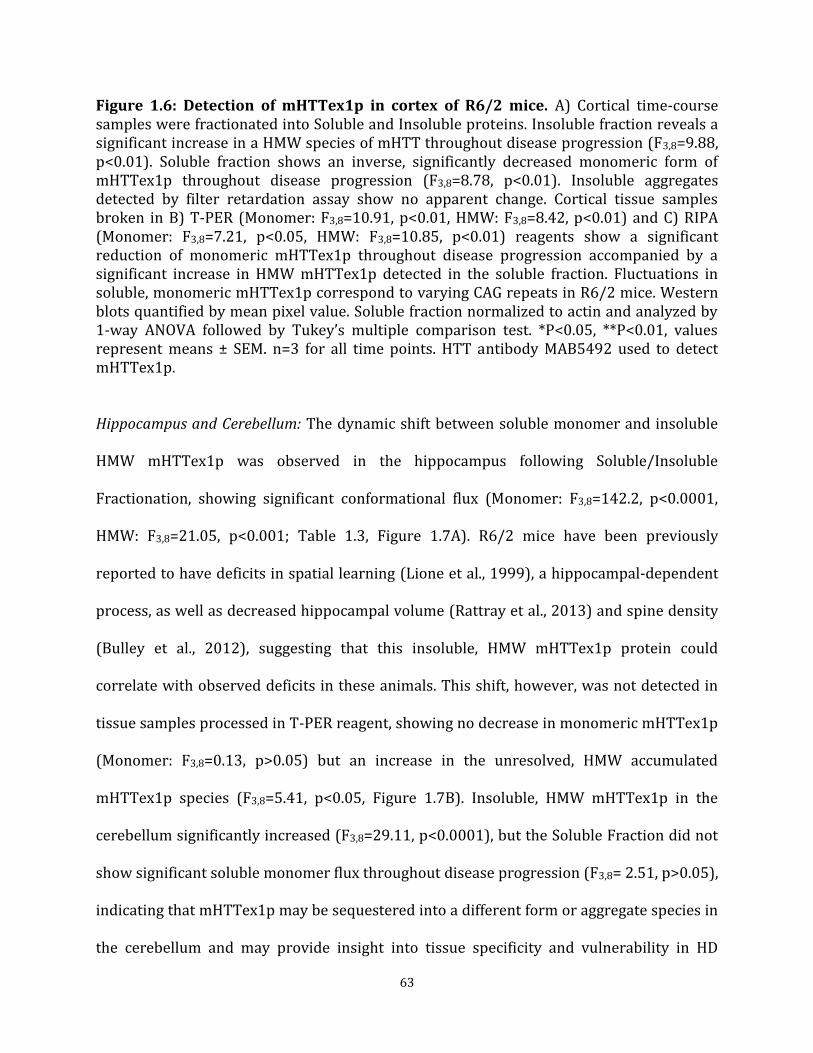
63
Figure 1.6: Detection of mHTTex1p in cortex of R6/2 mice. A) Cortical time-course samples were fractionated into Soluble and Insoluble proteins. Insoluble fraction reveals a significant increase in a HMW species of mHTT throughout disease progression (F3,8=9.88, p<0.01). Soluble fraction shows an inverse, significantly decreased monomeric form of mHTTex1p throughout disease progression (F3,8=8.78, p<0.01). Insoluble aggregates detected by filter retardation assay show no apparent change. Cortical tissue samples broken in B) T-PER (Monomer: F3,8=10.91, p<0.01, HMW: F3,8=8.42, p<0.01) and C) RIPA (Monomer: F3,8=7.21, p<0.05, HMW: F3,8=10.85, p<0.01) reagents show a significant reduction of monomeric mHTTex1p throughout disease progression accompanied by a significant increase in HMW mHTTex1p detected in the soluble fraction. Fluctuations in soluble, monomeric mHTTex1p correspond to varying CAG repeats in R6/2 mice. Western blots quantified by mean pixel value. Soluble fraction normalized to actin and analyzed by 1-way ANOVA followed by Tukey’s multiple comparison test. *P<0.05, **P<0.01, values represent means ± SEM. n=3 for all time points. HTT antibody MAB5492 used to detect mHTTex1p.
Hippocampus and Cerebellum: The dynamic shift between soluble monomer and insoluble
HMW mHTTex1p was observed in the hippocampus following Soluble/Insoluble
Fractionation, showing significant conformational flux (Monomer: F3,8=142.2, p<0.0001,
HMW: F3,8=21.05, p<0.001; Table 1.3, Figure 1.7A). R6/2 mice have been previously
reported to have deficits in spatial learning (Lione et al., 1999), a hippocampal-dependent
process, as well as decreased hippocampal volume (Rattray et al., 2013) and spine density
(Bulley et al., 2012), suggesting that this insoluble, HMW mHTTex1p protein could
correlate with observed deficits in these animals. This shift, however, was not detected in
tissue samples processed in T-PER reagent, showing no decrease in monomeric mHTTex1p
(Monomer: F3,8=0.13, p>0.05) but an increase in the unresolved, HMW accumulated
mHTTex1p species (F3,8=5.41, p<0.05, Figure 1.7B). Insoluble, HMW mHTTex1p in the
cerebellum significantly increased (F3,8=29.11, p<0.0001), but the Soluble Fraction did not
show significant soluble monomer flux throughout disease progression (F3,8= 2.51, p>0.05),
indicating that mHTTex1p may be sequestered into a different form or aggregate species in
the cerebellum and may provide insight into tissue specificity and vulnerability in HD

64
(Table 1.3, Figure 1.7C). Cerebellar tissue samples broken in T-PER reagent, however,
showed a significant increase in accumulated mHTTex1p (F3,8=6.32, p<0.05) and a
corresponding decrease in soluble monomer (F3,8=4.25, p<0.05, Figure 1.7D). These results
suggest that while accumulated HMW mHTTex1p tracks with disease pathogenesis and
may serve as a robust measure of disease progression, additional methods to detect
additional species of mHTT or levels of full-length endogenous HTT may be necessary to
investigate tissue specific vulnerabilities or normal HTT levels (Franich et al., 2018).

65
Figure 1.7: Detection of mHTTex1p in hippocampus and cerebellum of R6/2 mice varies depending on break method. Insoluble fraction in A) hippocampus reveals a significant increase in a HMW species of mHTTex1p throughout disease progression (F3,8=21.05, p<0.001). Soluble fraction shows an inverse, significantly decreased monomeric form of mHTTex1p throughout disease progression (F3,8=142.2, p<0.0001). B) T-PER processed hippocampal tissue samples did not show decrease in soluble monomeric mHTTex1p (F3,8=0.13, p>0.05) or HMW accumulated mHTTex1p (F3,8=5.41, p<0.05). Insoluble fraction in C) cerebellum reveals a significant increase in HMW species of mHTTex1p throughout disease progression (F3,8=29.11, p<0.0001). Soluble fraction did not detect a significant decreased monomeric form of mHTTex1p (F3,8= 2.51, p>0.05). However, soluble monomeric mHTTex1p decrease was detected in D) T-PER processed cerebellar tissue samples (HMW: F3,8=6.32, p<0.05, Monomer: F3,8=4.25, p<0.05). Fluctuations in soluble, monomeric mHTTex1p correspond to varying CAG repeats in R6/2 mice. Western blots quantified by mean pixel value. Soluble fraction normalized to actin and analyzed by 1-way ANOVA followed by Tukey’s multiple comparison test. *P<0.05, **P<0.01, values represent means ± SEM. n=3 for all time points. HTT antibody MAB5492 used to detect mHTTex1p. Liver and Skeletal Muscle: We also evaluated liver and skeletal muscle samples for
accumulation of HMW mHTTex1p in comparison to the brain tissue results. Liver samples
show significant accumulation of insoluble, HMW mHTTex1p (F3,8=83.34, p<0.0001). The
soluble monomer, however, was not detectable using Soluble/Insoluble Fractionation
(Figure 1.8A, data not shown). Significant accumulation of HMW mHTTex1p was detected
in samples broken in T-PER reagent (F3,8=12.79, p<0.01) and interestingly, soluble
monomeric mHTTex1p was also detectable in liver samples broken in T-PER reagent but
no change in protein abundance during disease progression was detected with this break
method (F3,8=2.95, p>0.05, Figure 1.8B). Like liver, significant accumulation of insoluble,
HMW mHTTex1p was detectable in skeletal muscle only using Soluble/Insoluble
Fractionation (F3,8=7.53, p<0.05). No soluble monomer of mHTTex1p was detectable in
skeletal muscle broken with either Soluble/Insoluble Fractionation or T-PER reagent.
HMW mHTTex1p was not detected in T-PER (Figure 1.8C, data not shown).

66
Oligomeric mHTT detection varies based on tissue processing
Agarose Gel Electrophoresis (AGE) has been used to resolve soluble, oligomeric species of
mHTT in RIPA buffer as described (Sontag et al., 2012; Weiss et al., 2008). Tissue samples
from striatum and cortex that were lysed using methods as above (Table 1.1) were also
analyzed using AGE to compare how biochemical processing affected detection of mHTT
species resolved by AGE. Detection of oligomeric mHTTex1p species on AGE gels
throughout the course of the disease in R6/2 mice varied greatly depending on break
method as summarized in Table 1.3 (Figure 1.9). Striatal tissue samples from R6/2 mice
broken in RIPA buffer showed an increase in oligomeric mHTTex1p through week 9
followed by a significant decrease by week 11 (F3,8=27.21, p<0.001). However, R6/2
striatal samples showed increased oligomeric mHTTex1p through week 11 in both
Insoluble (F3,8=27.31, p<0.001) and Soluble (F3,8=7.30, p<0.05) Fractions, and samples
broken in T-PER (F3,8=24.70, p<0.001, Figure 1.9A), and TRIzol (F3,8=16.02, p<0.001, Figure
1.5B). Cortical tissue samples broken using identical methods more consistently showed a
significant increase in oligomer formation (RIPA: F3,8=19.56, p<0.001, Insoluble: F3,8=29.95,
p<0.001, T-PER: F3,8=109.3, p<0.0001, Figure 1.9B) with the exception of the Soluble
Fraction (F3,8=2.291, p>0.05) and of TRIzol samples (F3,8=0.95, p>0.05, Figure 1.5D) which
showed no significant accumulation of mHTTex1p oligomers. Therefore, the variability in
detectable oligomeric mHTTex1p among tissue processing methods is an important
consideration when analyzing data in preclinical studies.
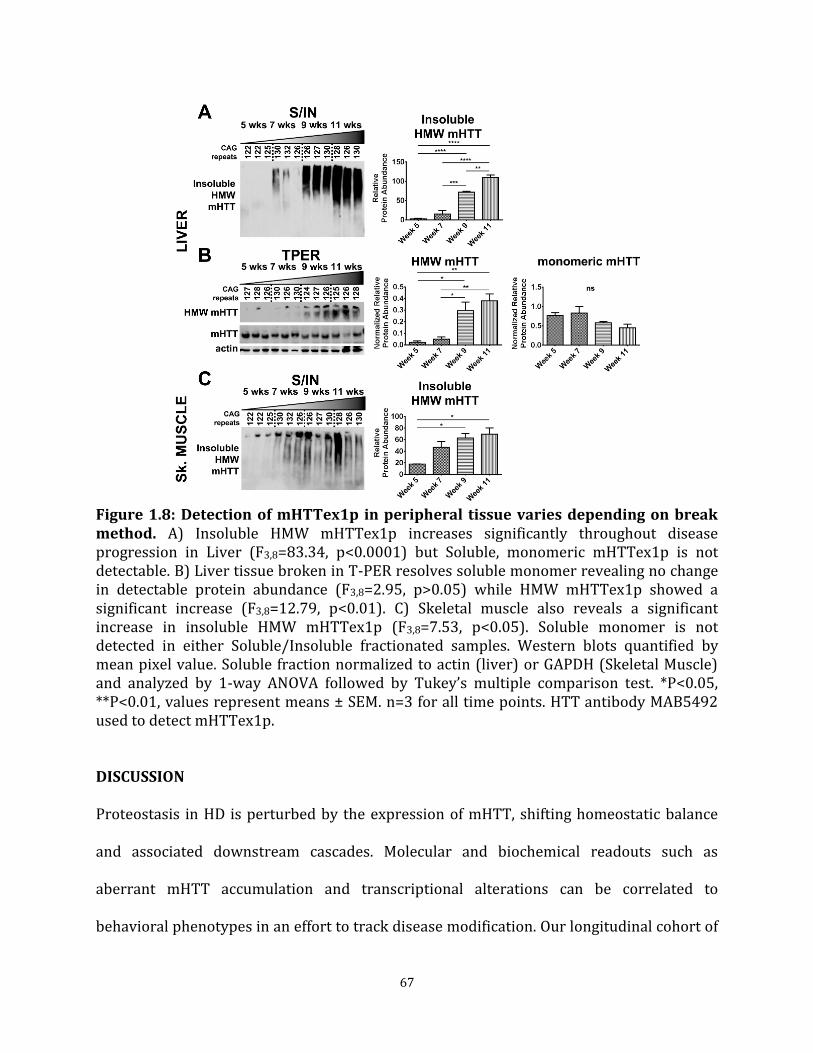
67
Figure 1.8: Detection of mHTTex1p in peripheral tissue varies depending on break method. A) Insoluble HMW mHTTex1p increases significantly throughout disease progression in Liver (F3,8=83.34, p<0.0001) but Soluble, monomeric mHTTex1p is not detectable. B) Liver tissue broken in T-PER resolves soluble monomer revealing no change in detectable protein abundance (F3,8=2.95, p>0.05) while HMW mHTTex1p showed a significant increase (F3,8=12.79, p<0.01). C) Skeletal muscle also reveals a significant increase in insoluble HMW mHTTex1p (F3,8=7.53, p<0.05). Soluble monomer is not detected in either Soluble/Insoluble fractionated samples. Western blots quantified by mean pixel value. Soluble fraction normalized to actin (liver) or GAPDH (Skeletal Muscle) and analyzed by 1-way ANOVA followed by Tukey’s multiple comparison test. *P<0.05, **P<0.01, values represent means ± SEM. n=3 for all time points. HTT antibody MAB5492 used to detect mHTTex1p.
DISCUSSION
Proteostasis in HD is perturbed by the expression of mHTT, shifting homeostatic balance
and associated downstream cascades. Molecular and biochemical readouts such as
aberrant mHTT accumulation and transcriptional alterations can be correlated to
behavioral phenotypes in an effort to track disease modification. Our longitudinal cohort of

68
R6/2 mice exhibited behavioral phenotypes as previously reported (Figure 1.1, (Bibb et al.,
2000; Hickey et al., 2005; Luthi-Carter et al., 2000; Mangiarini et al., 1996)), allowing for a
systematic comparison of changes in mHTTex1p conformations using selected cell lysis and
biochemical assays in an HD model over time.
Figure 1.9: Detection of mHTTex1p on AGE gels varies based by break method. A) Striatal tissue samples broken in indicated buffers and analyzed by Agarose Gel Electrophoresis (AGE). R6/2 mice show an increase in oligomeric mHTTex1p through week 9 followed by a significant decrease by week 11 in RIPA broken samples (F3,8=27.21, p<0.001). Increased, disease-endpoint mHTTex1p detected in Insoluble (F3,8=27.31, p<0.001), Soluble (F3,8=7.30, p<0.05), and T-PER (F3,8=24.70, p<0.001) broken samples showed a significant increase by week 7, followed by a decrease in week 9 before increased again by week 11. B) Cortical samples broken in RIPA buffer (F3,8=19.56, p<0.001), Insoluble (F3,8=29.95, p<0.001) and Soluble fractionation (F3,8=2.291, p>0.05), and T-PER buffer (F3,8=109.3, p<0.0001) show either no change or significant increase in formation of oligomers by week 11. AGE blots were quantified by mean pixel value and analyzed by 1-way ANOVA followed by Tukey’s multiple comparison test. *P<0.05, **P<0.01, ***P<0.001, ****P<0.0001 values represent means ± SEM. n=3 for all time points. HTT antibody MAB5492 used to detect mHTTex1p.

69
At the level of transcription, expression of Darpp-32 (Bibb et al., 2000; Luthi-Carter et al.,
2000) was decreased in R6/2 mice as expected, thus providing a reliable molecular quality
control marker (Figure 1.1, Figure 1.2). While previously reported that transgene
expression was within range of the endogenous murine gene in R6/2 mice (Mangiarini et
al., 1996), there was a moderate increase in the levels of endogenous Htt transcript in R6/2
mice in this study by 11 weeks of age in the striatum compared to age-matched NT
controls, perhaps indicating an upregulation to compensate for transgene genotoxic stress
or for the declining wild-type endogenous HTT protein levels we observed in HD knock-in
mice during aging (Franich et al., 2018). Intriguingly, this increase also extended to
expression of the R6/2 transgene in striatum, potentially reflecting compensatory
upregulation of Htt given that the transgene is expressed from the human Htt promoter
(Mangiarini et al., 1996). The upregulation of the transgene and of endogenous Htt was not
observed in cortex, potentially reflecting some aspect of tissue specificity. Thus, when
making assessments of protein levels, differential transcription may be considered in
addition to changes in protein clearance mechanisms, such that the observed increase in
R6/2 transgene expression in the striatum could also contribute to the detected increase in
accumulated species of mHTTex1p.
Given that mHTT conformations have different biochemical properties, it is important to
evaluate how tissue processing and analytical technique impact detection of conformers,
and we assessed this in vivo assaying mHTTex1p from transgenic R6/2 HD mice. We first
sought to resolve soluble and insoluble species of mHTTex1p in different brain regions and
peripheral tissues. Accumulation of insoluble, HMW mHTTex1p resolved from the

70
Insoluble Fraction was consistently detected in every tissue tested (Table 1.3, Figure 1.7,
1.8), and the appearance of the insoluble mHTTex1p accumulation tracked with onset of
disease phenotypes. In striatum, cortex, and hippocampus, this increase was accompanied
with a progressive decrease in the soluble mHTTex1p monomer associated with the
transgene. The decrease in soluble mHTTex1p and corresponding increase in insoluble
HMW species of mHTTex1p, tracking with disease progression, suggests that preventing
this flux through therapeutic modulation may be therapeutically beneficial. Supporting this,
our previous data showed that modulation of the insoluble, HMW species by reduction of
the E3 SUMO ligase, Protein Inhibitor of Activated Stat 1 (PIAS1) in the striatum of R6/2
mice corresponded with beneficial behavioral and molecular alterations (Ochaba et al.,
2016) and treatment with a compound that corresponds with worsened behavioral
phenotypes, JQ1, increased levels of this insoluble, HMW specifies (unpublished results,
Thompson). However, while this insoluble, HMW species tracked with disease progression
and observed behavioral deficits, it did not provide insight into tissue specificity involved
in disease pathogenesis. Therefore, this species may serve as a reliable readout for
molecular progression of disease but not of tissue-specific vulnerabilities. An alternative
methodology to further define levels of HTT species recently reported that levels of full-
length mHTT protein do not decline robustly in cerebellum with aging, unlike in cortex and
striatum, in two homozygous knock-in models, highlighting the importance of tracking
multiple full-length and protein fragment species of both HTT and mHTT (Franich et al.,
2018).

71
While accumulation of insoluble, HMW mHTTex1p was detectable in peripheral tissue
(skeletal muscle and liver), Soluble/Insoluble Fractionation was unable to resolve the
soluble, monomeric transgene protein product (Figure 1.8). This could indicate an
accelerated or differential aggregation pathway or preferential clearance of the monomer
in peripheral tissue. Being able to accurately detect mHTT conformers would aid in
establishing tissue intrinsic differences.
Whole-cell lysis techniques, such as RIPA and T-PER reported here, were able to resolve
soluble monomeric mHTTex1p albeit with variability compared to other techniques used
(Table 1.3). Interestingly, a HMW mHTTex1p species from whole-cell tissue processing
methods was retained at the top of the gel when analyzed by PAGE and Western blot. It is
possible that this HMW species contains both soluble and insoluble proteins and multiple
mHTTex1p conformers and therefore cannot be directly compared to the insoluble, HMW
mHTTex1p detected in the Insoluble Fraction that resolves in low-percentage PAGE gels.
However, the whole-cell lysis HMW species reliably increased in some brain regions
broken in both RIPA and T-PER buffers and therefore may also serve as a readout for
molecular progression of disease.
Soluble oligomeric mHTT was previously shown to be detectable at an early age in
HdhQ150 mice. Levels of this soluble species, detected by size exclusion chromatography,
diminished over time and corresponded with an increase in intranuclear inclusion
formation (Marcellin et al., 2012). Though our findings present a similar trend, detection of
an earlier oligomeric species of mHTTex1p is dependent on tissue processing (Table 1.3,
72
Figure 1.5B, D, Figure 1.9). Alternatively, due to the heterogeneity of oligomeric mHTT
(Hoffner and Djian, 2014), certain biochemical processing methods may favor one soluble
species over the other. Conformation-specific antibodies facilitate studies of various mHTT
species and their association with pathogenesis (e.g. 3B5H10 (Miller et al., 2011), 1C2
(Trottier et al., 1995), MW8 (Ko et al., 2001), EM48 (Gutekunst et al., 1999)). Therefore,
future in depth analysis using conformer specific antibodies may provide further insight
(Legleiter et al., 2009).
Taken together, the data presented here is consistent with the dynamic shift of the soluble
mHTT transgene protein product in R6/2 mice to an insoluble, HMW accumulated
mHTTex1p species accompanied by a change in oligomeric mHTTex1p over the time
course of the disease. Significantly, protein processing methods influence the detection and
temporal effects. Protein processed in TRIzol reagent yielded the greatest amount of
variation between assays for detecting different species of mHTTex1p (Figure 1.5),
precluding its use in these studies. However, TRIzol may be useful in detection of non-
aggregating proteins to enable parallel protein and RNA processing.
The shift to insoluble, HMW and aggregated mHTTex1p from soluble monomer is
associated with increasing behavioral dysfunction compared to NT mice and represents a
robust protein assay to track disease progression in R6/2 mice. Understanding the
outcomes of standard protein homeostasis assays to assess mHTTex1p flux in R6/2 mice
provides a baseline for cross comparisons of preclinical interventions. It will be beneficial
to conduct similar assessments of mHTTex1p flux in brain expressing full-length mutant

73
HTT, given that mHTTex1p is expressed in knock-in HD mouse models and in patient brain
(Neueder et al., 2017; Sathasivam et al., 2013), and may be relevant to the onset of HD
pathogenesis. A recent report showed a decrease in soluble mHTTex1p monomer in the
zQ175 HD knock-in mouse model over time (Neueder et al., 2017), suggesting that
detecting this species as well as the insoluble, mHTTex1p species may be powerful cross-
model molecular correlates. Indeed, in the assays here, decreasing monomeric mHTTex1p
from fractionation of striatal tissue or breaking in RIPA appeared qualitatively to track
most closely with the progression of phenotypes in the R6/2 mice. We have previously
published a decrease in full-length HTT with age in striatum and cortex of two HD
homozygous knock-in models and in wild-type controls, which may reflect a loss of full-
length HTT protein function over time (Franich et al., 2018). Due to the dynamic shift
between mHTTex1p conformations throughout the course of the disease we propose using
multiple and consistent, optimized biochemical analyses, potentially in combination with
high context histological approaches as described for knock-in zQ175 mice (Carty et al.,
2015) to determine the impact of potential therapeutics on the protein flux of both
mHTTex1p and full-length HTT protein.
EXPERIMENTAL PROCEDURES
Animals
R6/2 mice were obtained from Jackson Laboratories at 5 weeks of age and allowed to age
to 7, 9, or 11 weeks. CAG repeat sizing of genomic DNA harvested from tails was performed
by Laragen. Experiments were carried out in strict accordance with the Guide for the Care
and Use of Laboratory Animals of the NIH and an approved animal research protocol by the

74
Institutional Animal Care and Use Committee (IACUC) at the University of California, Irvine.
Animals were humanely euthanized by an injection of Euthasol at age 5, 7, 9 or 11 weeks
followed by cardiac perfusion with PBS then decapitation. Brain and peripheral tissues
used for biochemical assessment were flash-frozen on dry ice and stored at -80°C until
further use. Brain tissue used for immunofluorescence was drop fixed in 4%
paraformaldehyde for 48 hours then protected in 30% sucrose with 0.02% sodium azide.
Behavioral Paradigms: Motor deficits were assessed in pole test, rotarod, and grip strength
assays as previously described (Ochaba et al., 2016). For pole test, mice were tested at 6, 8,
and 10 weeks of age for their ability to descend a vertical pole (1 cm in diameter, 60 cm
high) by recording and averaging time to descend over four trials for analysis. Rotarod was
carried out using an accelerating apparatus (Dual Species Economex Rota-Rod; 0207-
003M; Columbus Instruments) at 7, 9, and 11 weeks of age. Animals were trained on
Rotarod for 5 minutes on day 1 and tested the following day by an accelerating assay
recording and averaging time to fall for three, 5-minute trials test day.
Grip strength was assessed 6, 8, and 10 weeks of age to measure forelimb strength using a
meshed force gauge which retained the peak force applied (IITC Life Science instrument,
Woodland Hills, CA) and averaging the top 4 strongest pulls of 5 trials in grams of force
applied to the meter. Dyskinesia was assessed by recording percent of animals clasping
from weeks 5-11 as described previously (Mangiarini et al., 1996) and body weights were
recorded at the same time daily from weeks 5-11.

75
Immunohistochemistry and Quantitation
Post-fixed brains were processed as 40 µm sections, used for immunohistochemistry and
imaged by confocal microscopy. The following primary antibodies were used: anti-HTT
mEM48 (Millipore MAB5374) and anti-DARPP-32 (Santa Cruz SC-11365). Following
antibody incubation, nuclei of slices were stained with TO-PROTM-3 iodide nuclear stain
(Thermo Fisher T3605) and mounted onto microscope slides using Fluoromount-G
(SouthernBiotech 0100-01). A LeicaDM2500 confocal microscope was used to acquire
images. Six representative sections were used per animal. Two comparable z-stack images
per section were acquired at 40x and used for quantification. EM48-positive puncta were
automatically counted using Imaris Bitplane 5.0 spots setting with a diameter threshold of
0.5 µm and consistent threshold between samples.
Tissue Lysis
Flash frozen brain and peripheral mouse tissue were lysed by douncing 30 times in a glass
2 mL vial. Soluble and Insoluble Fractionation was performed on tissue samples in lysis
buffer containing 10 mM Tris (pH 7.4), 1% Triton X-100, 150 mM NaCl, 10% glycerol, and
0.2 mM PMSF, and supplemented with protease inhibitors and NEM as previously
described (O'Rourke et al., 2013; Ochaba et al., 2016; Ochaba et al., 2018). Samples were
lysed for 60 minutes on ice followed by a 20-minute centrifugation at 15,000 xg at 4°C.
Supernatant served as Soluble Fraction and pellet was washed twice with lysis buffer
before being re-suspended in lysis buffer supplemented to 4% SDS and served as the
Insoluble Fraction. Insoluble samples were sonicated for 30 seconds at 40% amplitude and
boiled for 30 minutes. Protein fractions were quantified using Lowry protein assay (Bio-

76
Rad). Whole cell tissue lysis was performed in T-PER or RIPA buffers supplemented with
protease inhibitors protease inhibitors (Complete Mini, Roche Applied Science), 0.1 mM
PMSF, 25 mM NEM, 1.5 mM aprotinin, and 23.4 mM leupeptin by douncing as above then
sonicated for 10 seconds, 3 times at 40% amplitude on ice. T-PER samples were quantified
using Bradford protein assay and RIPA samples were analyzed using Lowry protein assay.
Protein fraction from tissue samples processed using TRIzolTM (Thermo Fisher A33251)
reagent was prepared according to manufacturer’s protocol to solubilize organic protein
phase and analyzed by Bradford protein assay.
Western Blot analysis
Soluble Fractions, T-PER, and RIPA lysates were resolved by reducing and running 30µg of
protein on 4-12% Bis-Tris Poly-Acrylamide gels (PAGE) at 150V and transferred onto
0.45µM nitrocellulose membrane. 30µg of reduced, insoluble protein from Insoluble
Fractions were resolved on 3-8% Tris-Acetate Poly-Acrylamide gels run at 150V and
transferred onto 0.45µM nitrocellulose membrane. Membranes were blocked in Starting
block (Invitrogen) for 20 minutes at room temperature and probed in primary antibody
overnight at 4°C. The following primary antibodies were used: anti-HTT (Millipore
MAB5492) to detect HTTex1p, anti-actin (Sigma A5060), and anti-GAPDH (Novus
Biologicals NB100-56875). Western blot data was quantified by determining the mean
pixel intensity using Scion Image processing software. Soluble Fractions, T-PER, and RIPA
lysates were normalized to house-keeping protein loading control (GAPDH or Actin) prior
to statistical analysis. Insoluble protein was quantified as relative protein abundance as
previous (Ochaba et al., 2016; Ochaba et al., 2018).

77
Agarose Gel Electrophoresis analysis
Oligomeric species of mHTTex1p were detected using Agarose Gel Electrophoresis (AGE)
as previously described (Sontag et al., 2012) using a 1%, 375mM Tris-HCl, pH 8.8, 1% SDS
agarose gel. 30µg of protein was suspended in a non-reducing loading buffer (4x, 300 mM
Tris-HCl pH 6.8, 66% glycerol, 2.4%SDS with BPB) and loaded onto an agarose gel without
boiling. Samples were run in a 192 mM glycine, 24.8 mM Tris base, 0.1% SDS running
buffer and transferred using an Invitrogen Novex Semi-Dry blotter onto 0.45 µm PVDF
membrane at 10V for 1 hour. Membranes were blocked in starting block (Invitrogen) for 1
hour at room temp and probed with primary antibody overnight at 4°C. mHTTex1p was
detected using anti-HTT (Millipore MAB5492) antibody. AGE data was quantified by
determining the mean pixel intensity using Scion Image processing software as a relative
protein abundance.
Filter Retardation assay
Insoluble fibrils of mHTTex1p were detected using a filter retardation assay (Wanker et al.,
1999) with modifications. 30µg of insoluble samples obtained from Soluble/Insoluble
Fractionation were suspended in 200 µl of 0.1% SDS, boiled for 5 minutes, loaded on to a
dot blot apparatus (BioRad), and blotted onto 0.2 µm Cellulose Acetate membrane.
Membrane was blocked for 1 hour at room temperature in 5% milk in TBST. Fibrils were
detected with anti-HTT antibody (Enzo PW0595).

78
RNA Isolation and Real-Time qPCR
Flash frozen brain regions harvested from R6/2 longitudinal animals were homogenized in
TRIzolTM (Thermo Fisher A33251) and RNA was collected using manufacturer’s procedures
then purified using RNEasy Mini kit (QIAGEN). Residual DNA was removed by DNase
treatment incorporated into RNEasy protocol as per manufacturer’s suggestion. Following
RNA isolation, reverse transcription was performed using SuperScript 3 First-strand
synthesis system according to manufacturer’s protocol (Invitrogen). Both oligo (dT) and
random hexamer primers were used in a 1:1 ratio with a total of 1 µg RNA per sample.
Final synthesized cDNA was diluted 1:10 in DEPC treated water and stored at -20°C until
use. The following primers were used to amplify and detect cDNA from transcripts: full-
length Htt (F: GCAGGG-AAAGAGCTTGAGACAC, R: CCTCATTCTCCTTGTGGCACTG), R6/2
transgene (F: CCGCTCAGGTTCTGCTTTTA, R: TGGAGGGACTTGAGGGACTC), Darpp-
32/Ppp1r1b (F: TCTCAGAGCACTCCTCACCAGA, R: CACTCAAGTTGCTAATGGTCTGC), Rplp0
(F: GCTTCGTGTTCACCAAGGAGGA, R: GTCCTAGACCAGTGTTCTGAGC).
Statistical analysis
All data represented as mean ± SEM with a p value of p<0.05 considered statistically
significant. Analyses were completed in GraphPad Prism software. Student’s two-tailed t-
tests, assuming equal variance were performed for behavioral comparisons at designated
time points between non-transgenic (NT) and R6/2 mice. Behavioral comparisons between
R6/2 animals over time were completed by performing a 1-way ANOVA. Densitrometry
and aggregate count from EM48 positive immunolabeling studies were analyzed by
performing a 1-way ANOVA to assess changes overtime or 2-way ANOVA to compare

79
between brain regions. Pixel intensity values were obtained from western blots and
subjected to 1-way ANOVA to assess changes in protein quantify overtime in R6/2 mice.
Statistical analysis was completed on the delta CT (dCT) values obtained from qPCR
reactions normalized to Rplp0. When comparing relative to NT week 5, a 2-way ANOVA
was used to assess changes in genotype and time. When comparing between R6/2 mice
only, a 1-way ANOVA was used to assess changes over time in transcription. All 1-way
ANOVAs were followed by Tukey’s multiple comparisons test. All 2-way ANOVAs were
followed by Sidak’s multiple comparisons test.

80
CHAPTER 1
TABLES
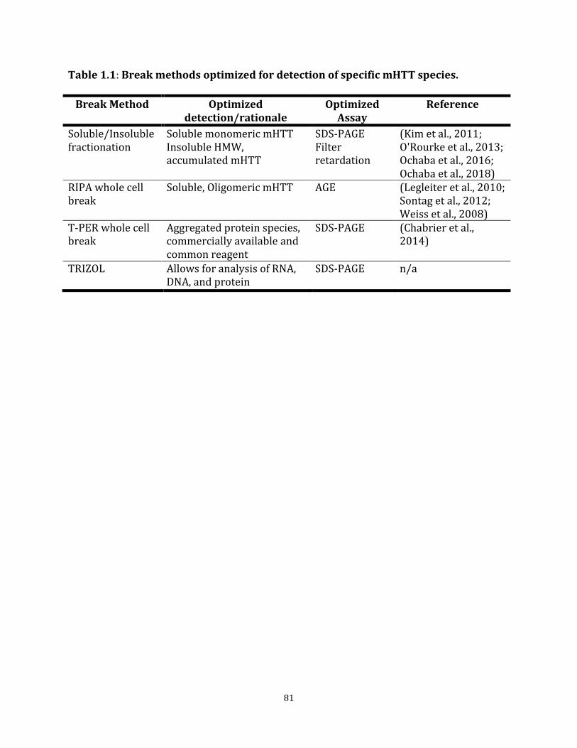
81
Table 1.1: Break methods optimized for detection of specific mHTT species.
Break Method Optimized detection/rationale
Optimized Assay
Reference
Soluble/Insoluble fractionation
Soluble monomeric mHTT Insoluble HMW, accumulated mHTT
SDS-PAGE Filter retardation
(Kim et al., 2011; O'Rourke et al., 2013; Ochaba et al., 2016; Ochaba et al., 2018)
RIPA whole cell break
Soluble, Oligomeric mHTT AGE (Legleiter et al., 2010; Sontag et al., 2012; Weiss et al., 2008)
T-PER whole cell break
Aggregated protein species, commercially available and common reagent
SDS-PAGE (Chabrier et al., 2014)
TRIZOL Allows for analysis of RNA, DNA, and protein
SDS-PAGE n/a

82
Table 1.2: mHTTex1p size fluctuation due to CAG repeat length.
Animal Age (weeks) (CAG)n Genomic DNA (Tail)
(CAG)n cDNA (Striatum)
1 5 125 125 2 5 129 129 3 5 123 123 4 7 128 128 5 7 131 131 6 7 131 130 7 9 126 126 8 9 126 126 9 9 126 126 10 11 129 129 11 11 126 127 12 11 128 127
Table 1.2: CAG repeats from genomic tail DNA comparable to counts obtained from striatal cDNA from the same animal showing lack of somatic expansion in these animals. cDNA generated from RNA harvested from striatum. CAG repeat sizing of cDNA was performed by Laragen.

83
Table 1.3: Summary heat map of mHTTex1p detected using various break methods and assays at 5, 7, 9, and 11 weeks of age.
Table 1.3: Representative gradient for each assay based on ease of detection for general comparison. Cannot quantify between assay methods for relative protein abundance.
84
CHAPTER 2
PIAS1 SUMO-Interaction Motif May Modulate Accumulation of Mutant Huntingtin Protein
SUMMARY OF CHAPTER 2
Disruption of protein homeostasis, leading to accumulation of insoluble high molecular
weight protein complexes containing the huntingtin protein and SUMOylated proteins, is a
key feature of Huntington’s disease (HD). We previously showed that exogenous
expression of the E3 SUMO ligase PIAS1 enhanced SUMO modification and accumulation of
the huntingtin protein (HTT) and that reduction of Pias1 in vivo reduced this accumulation.
However, the functional contributions of PIAS1 domains towards accumulation of mutant
HTT had not yet been studied. Structurally, PIAS1 contains a SUMO-interaction motif (SIM)
and a RING-ligase domain and the contribution of these domains towards HTT
SUMOylation and accumulation is unknown. Using informed mutagenesis combined with
cell culture, preliminary experiments investigating the effects of the PIAS1 SUMO-
interaction motif and RING domain on mutant HTT accumulation were carried out. The
data suggests that the PIAS1 SIM domain may be mediating formation of an insoluble
aggregation species of mutant HTT but results were confounded by issues related to
transient transfection and promoter effects. Future studies utilizing CRISPR directed
mutagenesis will greatly aid in elucidating the precise contribution of PIAS1 functional
domains on HTT accumulation. Identification of specific mutant HTT-modulating proteins
and the specific functional domains involved will inform future targeted drug design for
disease intervention with broad relevance to protein dyshomeostasis in other
neurodegenerative diseases.

85
INTRODUCTION
Neurodegenerative disorders such as Alzheimer’s disease (AD), Parkinson’s disease (PD)
and Amyotrophic lateral sclerosis (ALS) exhibit hallmark disruption of protein
homeostasis, including of protein quality control networks, similar to Huntington’s disease
(HD, (Hipp et al., 2014; Kurtishi et al., 2019; Ross and Poirier, 2004)). Disease is
accompanied by accumulation of disease-related proteins and post-translational modifiers
Ubiquitin and SUMO associate with protein inclusions (Anderson et al., 2017). In addition
to an association of SUMO with protein aggregates in disease, many disease proteins
themselves are SUMOylated, suggesting that the SUMO modification directly regulates
protein aggregation perhaps by mediating solubility, degradation, or protein-protein
interactions (Liebelt and Vertegaal, 2016). Therefore, understanding functional
components contributing towards protein accumulation and the contribution of
SUMOylation machinery, including the E3 ligases that regulate SUMO modification of target
proteins, will prove valuable with broad relevance to other neurodegenerative diseases.
The HTT protein itself is SUMOylated with SUMOylation of mHTT potentially affecting
subcellular localization and stability (Steffan et al., 2004). We identified PIAS1 as the E3
ligase (Figure 1) which enhanced SUMOylation (by SUMO1 and SUMO2/3) of HTT and
modulated mHTT accumulation in vitro (O'Rourke et al., 2013). In vivo knock-down of Pias1
in the striatum of the rapidly progressing R6/2 mouse model of HD significantly altered the
SUMO and ubiquitin landscape in these animals (Ochaba et al., 2016). Reduction of
accumulated, insoluble High-molecular weight (HMW) mHTT and aggregate species was
accompanied by reductions in both global SUMO and ubiquitin insoluble conjugates.

86
However, the exact contribution of PIAS1 domains towards mHTT accumulation remained
unknown as mHTT undergoes changes in conformation and solubility prior to entering
SUMO-positive inclusions ((Morozko et al., 2018), Chapter 1, unpublished data).
PIAS1 contains several functional domains (Figure 2.1). Of particular interest are the
SUMO-interaction motif (SIM) and the RING-ligase domain within PIAS1. SUMO E3 ligase
activity is mediated through the RING domain (Cappadocia and Lima, 2017). This domain is
crucial for protein-protein interactions and selective ligase functionality includes the
interaction with Ubc9 mediated through several cysteines within the RING domain (Kahyo
et al., 2001). Mutation of these cysteines produces a Ligase-dead (LD), functionally null
PIAS1 protein (Lee et al., 2003; Yurchenko et al., 2006). Further, this domain has been
implicated in PIAS1 autoSUMOylation, which in turn impacts the overall function of PIAS1
(Alagu et al., 2018).
SUMOylation can mediate protein-protein interactions and formation of protein complexes
by non-covalent binding through SIMs which are composed of a conserved sequence of four
amino acids containing three hydrophobic residues (VVXV or VXVV where V=V/I) that are
flanked by phosphorylatable acidic residues (Cappadocia and Lima, 2017).
Phosphorylation of these residues by the serine/threonine kinase CK2 acts as a molecular
switch for enhancing or preventing SUMO binding (Stehmeier and Muller, 2009). The SIM
within PIAS1 may therefore aid in proper loading and stabilization of the Ubc9-SUMO
structure for optimal transfer of the SUMO moiety onto a substrate protein mediated by
phosphorylation (Cappadocia and Lima, 2017; Stehmeier and Muller, 2009).

87
Whether PIAS1 has a direct effect on SUMO modification of HTT and accumulation of
insoluble substrates is unknown. Alternatively, modulating PIAS1 in the HD context may
impact the global SUMO network or a different mechanistic pathway to modify disease
indirectly. Investigating the functional impact of both the SIM and Ligase PIAS1 functional
domains towards mediating mHTT solubility and accumulation may provide insight into
the role of PIAS1 in the disease context. Therefore, these studies explore the underlying
mechanism whereby PIAS1 may modulate protein-protein interactions and HTT protein
accumulation.
RESULTS
PIAS1 phospho-SIM mutants express at the same levels
To evaluate how the PIAS1 SIM domain affects mHTT accumulation, we generated
constructs containing mutations in the SIM domain of PIAS1. Constructs harboring
mutations within the PIAS1 SIM or the phosphorylatable region directly downstream of the
PIAS1 SIM (Figure 2.1) were generated. PIAS1 SIM was interrupted using alanine
mutagenesis to replace VIDLT (AA459-463) of PIAS1 with alanines (∆SIM). A phospho-
resistant PIAS1 was created using alanine mutagenesis at serines 466-468 (S466-8A) as
preventing phosphorylation of these residues perturbs SUMO binding to the SIM
(Stehmeier and Muller, 2009). A complementary, phospho-mimetic PIAS1 mimicking
phosphorylation of serines 466-468 was also generated by substituting them with aspartic
acid residues (S466-8D), therefore facilitating SUMO binding to the PIAS1 SIM.

88
Figure 2.1 PIAS1 has numerous functional domains: PIAS1 contains numerous functional domains which aid in E3 ligase activity, protein-protein interactions, DNA binding, and post-translational modifications. Specifically, the RING domain and SIM of are particular interest due to their roles in modulating SUMOylation and associated machinery.
To evaluate whether PIAS1 SIM mutations alter PIAS1 protein levels, we first tested
whether the mutations influenced expression of the construct. Using qPCR analysis, there
were no differences in the levels of CMV-PIAS1 transcript expressed between WT and
phospho-SIM-mutant constructs (1-Way ANOVA, post-hoc p>0.05, Figure 2.2B). However,
the levels of CMV-PIAS1∆SIM were consistently elevated compared to WT transcript (1-
Way ANOVA, F(3,8)=9.08, p<0.01). CMV-PIAS1 constructs did not affect levels of
endogenous PIAS1 transcript (1-Way ANOVA, F(4,10)=0.72, p>0.05), providing a baseline
to allow us to draw conclusions regarding mHTT accumulation in the presence of
overexpressed PIAS1 phospho-SIM constructs. CMV-PIAS1 expression did not affect
transcription of a transfection control, CMV-myc-actin (1-Way ANOVA, F(4,10)=2.41,
p>0.05), but increased levels of exogenous CMV-myc-actin protein levels in samples
containing overexpressed PIAS1 constructs compared to endogenous PIAS1 control (1-Way
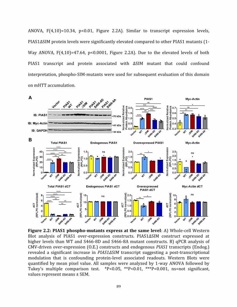
89
ANOVA, F(4,10)=10.34, p<0.01, Figure 2.2A). Similar to transcript expression levels,
PIAS1∆SIM protein levels were significantly elevated compared to other PIAS1 mutants (1-
Way ANOVA, F(4,10)=47.64, p<0.0001, Figure 2.2A). Due to the elevated levels of both
PIAS1 transcript and protein associated with ∆SIM mutant that could confound
interpretation, phospho-SIM-mutants were used for subsequent evaluation of this domain
on mHTT accumulation.
Figure 2.2: PIAS1 phospho-mutants express at the same level: A) Whole-cell Western Blot analysis of PIAS1 over-expression constructs. PIAS1∆SIM construct expressed at higher levels than WT and S466-8D and S466-8A mutant constructs. B) qPCR analysis of CMV-driven over-expression (O.E.) constructs and endogenous PIAS1 transcripts (Endog.) revealed a significant increase in PIAS1∆SIM transcript suggesting a post-transcriptional modulation that is confounding protein-level associated readouts. Western Blots were quantified by mean pixel value. All samples were analyzed by 1-way ANOVA followed by Tukey’s multiple comparison test. *P<0.05, **P<0.01, ***P<0.001, ns=not significant, values represent means ± SEM.

90
PIAS1 SIM modulates accumulation of mHTTex1p in human cells
To determine whether PIAS1 may modulate solubility of the mHTT exon-1 97QP protein
fragment (mHTTex1p) in a human cell-line, I first examined mHTT accumulation in human
HEK293T cells. This fragment rapidly forms an insoluble, HMW species that is modulated
by PIAS1 (O'Rourke et al., 2013). Control and phospho-mutant PIAS1 constructs were
transfected together with mHTTex1p expressing constructs in HEK293T cells to assess
mHTTex1p accumulation using Soluble/Insoluble fractionation (Ochaba et al., 2018).
Preventing phosphorylation of the PIAS1 SIM (S466-8A) increased accumulation of HMW,
insoluble mHTTex1p (1-Way ANOVA, F(3,7)=53.43, p<0.0001, Figure 2.3A, B), suggesting
that the phosphorylation status of the PIAS1 SIM is involved in modulating mHTTex1p
accumulation and solubility (Figure 2.3). This increase corresponded with a significant
decrease in both soluble, monomeric mHTTex1p (1-Way ANOVA, F(3,8)=19.87, p<0.001)
and insoluble, fibrillar mHTTex1p (Figure 2. 3A, B). For overexpression of WT PIAS1, one
sample had insufficient transfection efficiency (middle lane, Figure 2.3A of WT PIAS1) and
was therefore not included in analyses. PIAS1 was increased in the soluble fraction of
samples with phospho-resistant PIAS1 overexpression (Figure 2.A PIAS1 S466-8A lanes, 1-
Way ANOVA, F(3,8)=35.81, post-hoc p<0.05), compared to other PIAS1 constructs,
indicating a possible shift in subcellular localization of PIAS1 as the soluble fraction serves
as a crude cytosolic fraction (Insoluble as crude nuclear, (Ochaba et al., 2016)). Samples
with overexpressed PIAS1 phospho-mimetic (S466-8D) constructs did not show any shift
in mHTTex1p solubility or PIAS1 fractionation compared to control WT PIAS1.

91
Figure 2.3: PIAS1 SIM may modulate mHTT solubility in Human cells: A) Insoluble and Soluble Western Blot analysis and B) subsequent relative protein abundance from HEK293T (Human embryonic kidney cell type) cells co-transfected with PIAS1 phospho-SIM mutants and mHTTex1p shows an increase in PIAS1 S466-8A in the Insoluble fraction, corresponding to a decrease increase in HMW mHTTex1p. C) qPCR analysis of CMV-driven over-expression (O.E.) constructs and endogenous PIAS1 and HTT transcripts. Western Blots were quantified by mean pixel value. Red asterisk indicates sample with insufficient PIAS1 overexpression and was not included in analysis. All samples were analyzed by 1-way ANOVA followed by Tukey’s multiple comparison test. *P<0.05, **P<0.01, ***P<0.001, ns=not significant, values represent means ± SEM.

92
To determine if protein accumulation in the presence of the phospho-resistant mutant was
in part due to an impact on HTT expression, qPCR was carried out. We observed a
significant increase in both endogenous (1-Way ANOVA, F(3,8)=6.50, p<0.05) and
overexpressed (1-Way ANOVA, F(3,8)=5.58, p<0.05) HTT transcript levels with PIAS1
phospho-mutant expression (Figure 2.3C). The effect on exogenous HTT could in part be
due to influence on CMV-promoter as Myc-Actin transcript levels were also increased in
these samples (1-Way ANOVA, F(3,8)=5.76, p<0.05). However, this effect on Myc-Actin
transcription was not observed with overexpression of PIAS1 constructs alone (Figure 2.2)
and could indicate a compound effect with the presence of mHTTexp1 on expression of
CMV-driven overexpression constructs, though this possibility is not clear in this present
dataset. The results therefore may suggest that the inability to phosphorylate the SIM
domain causes an increase in endogenous expression and thus accumulation of HMW
mHTT species.
PIAS1 SIM modulates accumulation of mHTTex1p in striatal-derived cells
I next investigated whether the PIAS1 SIM was modulating mHTTex1p accumulation in an
immortalized striatal cell line to investigate accumulation dynamics in cells derived from a
neuronal linage. The experiment was therefore repeated in immortalized rat, striatal
ST14A cells (Ehrlich et al., 2001). In ST14A cells, overexpression of wild-type PIAS1 had an
inverse effect on mHTTex1p accumulation compared to HEK293T cells (Figure 2.4A);
overexpression of WT PIAS1 resulted in a significant decrease in HMW accumulated,
insoluble mHTTex1p (1-Way ANOVA, F(3,8)=23.83, post-hoc p<0.01, Figure 2.4A, B). PIAS1
phospho-resistant overexpression slightly prevented this reduction in HMW insoluble

93
Figure 2.4: PIAS1 SIM may modulate mHTT solubility in striatal-like cells: A) Insoluble and Soluble Western Blot analysis and B) subsequent relative protein abundance from ST14A (rat, immortalized striatal cell type) cells co-transfected with PIAS1 phospho-SIM mutants and mHTTex1p showed an increase in PIAS1 S466-8A in the Insoluble fraction, corresponding to a significant increase in HMW mHTTex1p. C) qPCR analysis of CMV-driven over-expression (O.E.) constructs and endogenous PIAS1 and HTT transcripts (Endog.) revealed a significant increase in endogenous HTT transcript. Western Blots were quantified by mean pixel value. All samples were analyzed by 1-way ANOVA followed by Tukey’s multiple comparison test. *P<0.05, **P<0.01, ***P<0.001, ns=not significant, values represent means ± SEM.

94
mHTTex1p but did not affect levels of fibrillary or soluble monomeric mHTTex1p (1-Way
ANOVA, post-hoc PIAS1vs.PIAS1 S466-8A p>0.05, Figure 2.4A, B). Interestingly, PIAS1 was
increased in the Insoluble fraction in these samples further suggesting that the PIAS1 SIM
is important for the subcellular localization or co-aggregation of PIAS1 (1-Way ANOVA
F(3,8)92.11, post-hoc PIAS1vsPIAS1 S466-8A p<0.01). While the phospho-mimetic form of
PIAS1 did not alter the levels of HMW mHTTex1p compared to WT PIAS1, it reduced levels
of soluble, monomeric mHTTex1p (1-Way ANOVA, F(3,8)=17.60, post-hoc PIAS1vs.PIAS1
S466-8D p<0.01) indicating that the PIAS1 SIM also modulates conformational flux in a
striatal cell line. Transcript levels assessed by qPCR analysis indicate that overexpression
of PIAS1 may decrease endogenous PIAS1 levels (1-Way ANOVA, F(3,8)=3.66, p>0.05, post-
hoc Vector vs. PIAS1 p<0.05) while PIAS1 phospho-mimetic slightly increased expression
of mHTTex1p construct (1-Way ANOVA, F(3,8)=5.58, p<0.05) and therefore may be
confounding accumulation results (Figure 2.4C).
PIAS1 Ligase domain may affect mHTTex1p accumulation in HEK293T cells
To begin investigating the consequence of ligase activity of PIAS1 on HTT accumulation,
two previously reported Ligase-dead (LD) constructs were generated. The first construct
contained a point mutation at cysteine 351 resulting in a serine substitution (C351S, (Lee et
al., 2003)). This cysteine was previously reported to be crucial for interaction of PIAS1 with
the SUMO E2 conjugating enzyme Ubc9 (Kahyo et al., 2001). Surrounding cysteine 351 are
two additional cysteines, which also contribute significantly to ligase activity of PIAS1
(Figure 2.1, (Yurchenko et al., 2006)). Therefore, a second construct with three cysteine-to-
serine substitutions was generated (3CS). To assess whether these mutations successfully
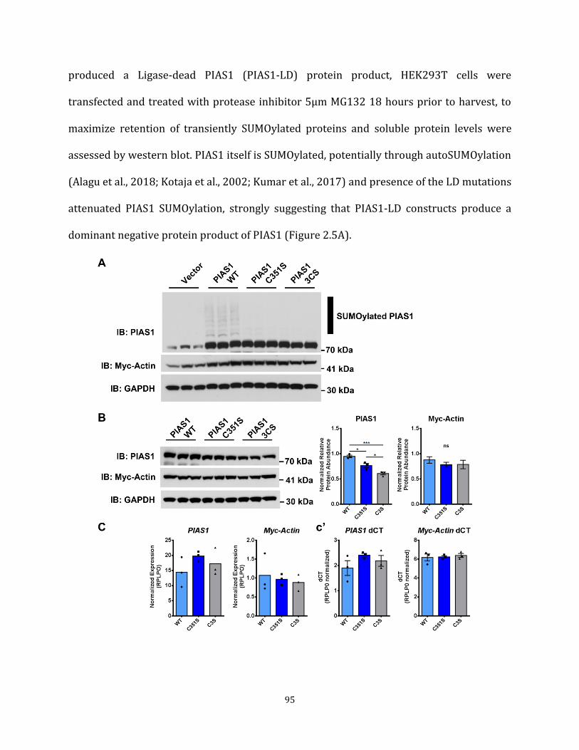
95
produced a Ligase-dead PIAS1 (PIAS1-LD) protein product, HEK293T cells were
transfected and treated with protease inhibitor 5µm MG132 18 hours prior to harvest, to
maximize retention of transiently SUMOylated proteins and soluble protein levels were
assessed by western blot. PIAS1 itself is SUMOylated, potentially through autoSUMOylation
(Alagu et al., 2018; Kotaja et al., 2002; Kumar et al., 2017) and presence of the LD mutations
attenuated PIAS1 SUMOylation, strongly suggesting that PIAS1-LD constructs produce a
dominant negative protein product of PIAS1 (Figure 2.5A).

96
Figure 2.5: PIAS1 Ligase-Dead mutants produce a dominant-negative protein product: A) Assessment of soluble PIAS1-LD constructs showed lack of autoSUMOylation for both PIAS1 C351S and PIAS1 3CS. B) Assessment of whole-cell protein lysate and transcript expression levels of PIAS1-LD constructs. PIAS1-LD protein detected at lower levels than WT. C) qPCR and c’) dCT values show constructs are transcriptionally expressed at the same level. Western Blots were quantified by mean pixel value. All samples were analyzed by 1-way ANOVA followed by Tukey’s multiple comparison test. *P<0.05, **P<0.01, ***P<0.001, ns=not significant, values represent means ± SEM.
To test whether the PIAS1 Ligase domain contributes to accumulation of insoluble HMW
mHTTex1p, HEK293T cells were transfected with either control or PIAS1-LD constructs
together with mHTTex1p expressing constructs and assessed using Soluble/Insoluble
fractionation. Preliminary data in HEK293T cells indicated that ligase activity may not
affect accumulation of Insoluble HMW mHTTex1p but may affect levels of soluble
monomeric mHTTex1p (Figure 2.6A, B). Overexpression of PIAS1-3CS, but not C351S,
resulted in a significant decrease in soluble mHTTex1p levels (1-Way ANOVA, F(3,8)=3.68,
post-hoc PIAS1vs.PIAS13CS p<0.05). However, levels of both PIAS1-LD protein products
were significantly lower in both the Soluble (1-Way ANOVA, F(3,8)=28.21, p<0.001) and
Insoluble (1-Way ANOVA, F(3,8)=63.59, p<0.0001) fractions indicating that PIAS1-LD
protein may be preferentially cleared by the cell. Assessing PIAS1 protein levels without
mHTTex1p overexpression in whole-cell lysate revealed a similar significant decrease with
PIAS1-LD expression (1-Way ANOVA, F(2, 6) = 27.79, p<0.001, Figure 2.5B). This was not
due to lower expression level as transcripts were assessed by qPCR and showed similar
levels of expression (1-Way ANOVA, F(2,6)=1.43, p>0.05, Figure 2.5C, c’). Therefore, it was
difficult to determine whether the PIAS1-LD mutants might have an impact on insoluble
HMW mHTTex1p levels.
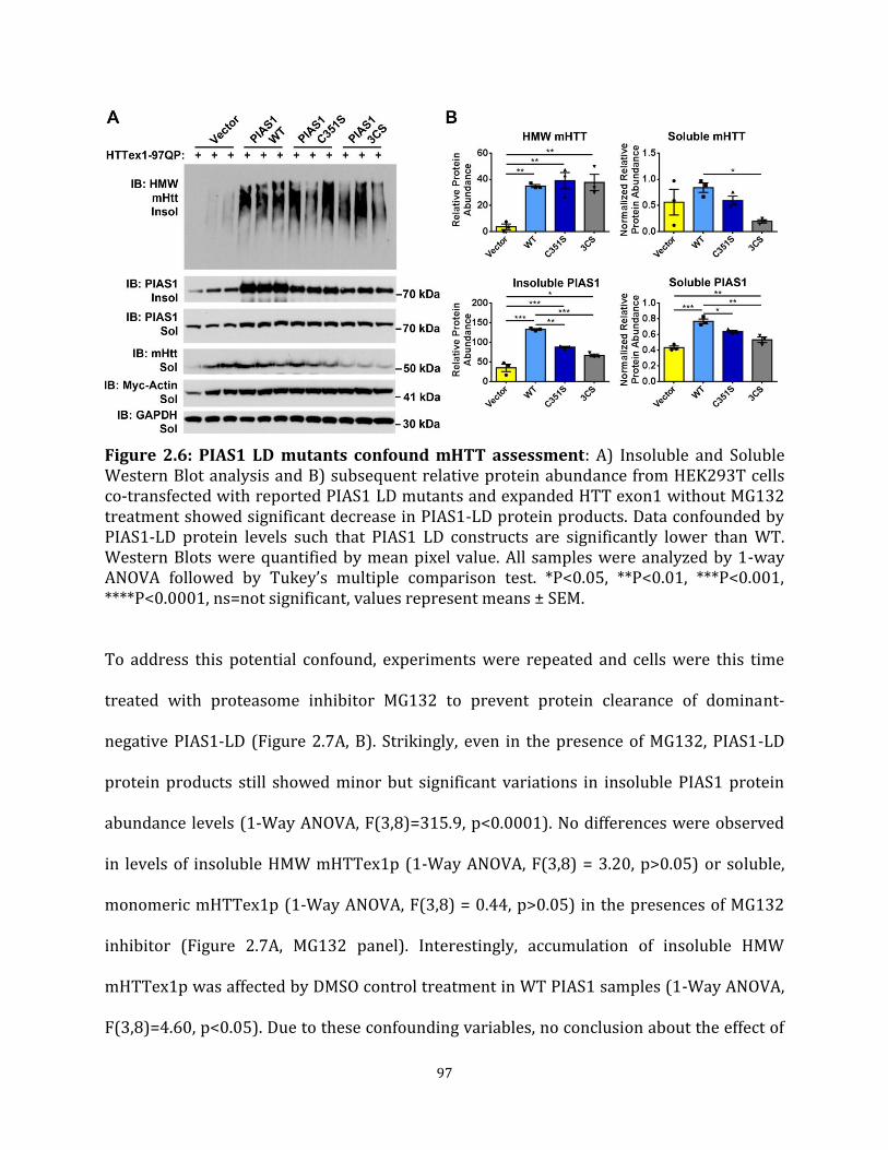
97
Figure 2.6: PIAS1 LD mutants confound mHTT assessment: A) Insoluble and Soluble Western Blot analysis and B) subsequent relative protein abundance from HEK293T cells co-transfected with reported PIAS1 LD mutants and expanded HTT exon1 without MG132 treatment showed significant decrease in PIAS1-LD protein products. Data confounded by PIAS1-LD protein levels such that PIAS1 LD constructs are significantly lower than WT. Western Blots were quantified by mean pixel value. All samples were analyzed by 1-way ANOVA followed by Tukey’s multiple comparison test. *P<0.05, **P<0.01, ***P<0.001, ****P<0.0001, ns=not significant, values represent means ± SEM.
To address this potential confound, experiments were repeated and cells were this time
treated with proteasome inhibitor MG132 to prevent protein clearance of dominant-
negative PIAS1-LD (Figure 2.7A, B). Strikingly, even in the presence of MG132, PIAS1-LD
protein products still showed minor but significant variations in insoluble PIAS1 protein
abundance levels (1-Way ANOVA, F(3,8)=315.9, p<0.0001). No differences were observed
in levels of insoluble HMW mHTTex1p (1-Way ANOVA, F(3,8) = 3.20, p>0.05) or soluble,
monomeric mHTTex1p (1-Way ANOVA, F(3,8) = 0.44, p>0.05) in the presences of MG132
inhibitor (Figure 2.7A, MG132 panel). Interestingly, accumulation of insoluble HMW
mHTTex1p was affected by DMSO control treatment in WT PIAS1 samples (1-Way ANOVA,
F(3,8)=4.60, p<0.05). Due to these confounding variables, no conclusion about the effect of
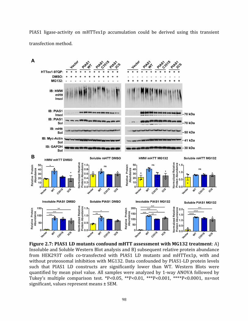
98
PIAS1 ligase-activity on mHTTex1p accumulation could be derived using this transient
transfection method.
Figure 2.7: PIAS1 LD mutants confound mHTT assessment with MG132 treatment: A) Insoluble and Soluble Western Blot analysis and B) subsequent relative protein abundance from HEK293T cells co-transfected with PIAS1 LD mutants and mHTTex1p, with and without proteosomal inhibition with MG132. Data confounded by PIAS1-LD protein levels such that PIAS1 LD constructs are significantly lower than WT. Western Blots were quantified by mean pixel value. All samples were analyzed by 1-way ANOVA followed by Tukey’s multiple comparison test. *P<0.05, **P<0.01, ***P<0.001, ****P<0.0001, ns=not significant, values represent means ± SEM.
99
DISCUSSION
While SUMOylated proteins as well as dysregulation of SUMO-associated proteostatic
pathways are associated with neurodegenerative diseases, the exact functional
contribution towards mediating homeostasis in disease states remains unclear. Specifically,
how SUMO and SUMO-related machinery promote or prevent protein accumulation (e.g.
mHTT) remains to be elucidated. Previously, we showed that SUMOylation with SUMO2 by
PIAS1 increased the accumulation and aggregation potential of mHTT species (O'Rourke et
al., 2013). Modulation of PIAS1 in cell culture and in the brain of HD mice significantly
altered levels of accumulated mHTT (O'Rourke et al., 2013; Ochaba et al., 2016). Therefore,
using PIAS1 and its SUMO-related functional domains as a target, I assessed if these
domains were mediating mHTT accumulation.
The RING-domain within PIAS1 is critical for its function as an E3 SUMO ligase. This
domain is also critical for the interaction between PIAS1 and E2 UBC9 (Kahyo et al., 2001).
PIAS1 interaction with Ubc9 is mediated in part through the SIM domain following the
RING domain. The creation of a ternary complex with a SUMO moiety through the PIAS1
SIM serves as an integral functional component in SUMOylation of target substrates
(Mascle et al., 2013). Further, both of these domains are integral for tethering other
SUMOylated proteins (Kotaja et al., 2002). Due to their significant role in mediating
SUMOylation of target substrates, PIAS1 Ligase and SIM domains were assessed for their
impact on mHTTex1p accumulation.

100
Overall, experiments presented here were confounded by effects of transient over-
expression driven by the CMV-promoter. While PIAS1 mutant constructs had equivalent
levels of transcript expression when expressed alone (Figures 2.2, 2.5), these constructs
affected expression of both exogenous expression of mHTTex1p as well as endogenous HTT
expression (Figures 2.3C, 2.4C). In turn, the increased expression of mHTTex1p could
account for the observed differences in mHTTex1p protein levels (both soluble and
insoluble levels). In the case of the PIAS1-LD constructs, protein products seemed to be
preferentially degraded by the cell, preventing sufficient overexpression of dominant-
negative PIAS1-LD, including when treated with proteasome inhibitor MG132 (Figure 2.6).
Follow-up experiments blocking autophagy may provide information whether this process
accounts for the increased clearance or whether some other mechanism is contributing to
the reduced protein levels. However, further development of a cell-based assay is
necessary to fully assess impact of PIAS1 functional domains on protein accumulation and
SUMOylation. Alternative approaches include testing additional promoters that may be less
sensitive to PIAS effects. However, the optimal approach would be to create endogenous
mutations through CRISPR-mediated genome editing of the endogenous PIAS locus in both
cell-based models and ultimately in vivo in mouse models of disease.
Although there are complications in interpretation, it appears that the PIAS1 SIM may be
involved in modulating solubility of mHTTex1p in multiple cell lines, such that preventing
phosphorylation of the SIM leads to an increase in insoluble mHTTex1p with a
corresponding decrease in soluble mHTTex1p in human HEK293T cells (Figure 2.3). This
may be a result of preventing SUMO binding to PIAS1 such that phosphorylation of SIMs
101
increases their affinity for SUMO (Anamika and Spyracopoulos, 2016; Cappadocia et al.,
2015). This increased affinity may be in part due to increasing electrostatic interactions of
SUMO moieties with the hydrophobic core of the SIM (Cappadocia et al., 2015) or
potentially through mediating polarity of SUMO-SIM binding interactions (Gareau and
Lima, 2010). For PIAS1 specifically, preventing phosphorylation of serines 466, 467, and
468 prevents SUMO binding (Stehmeier and Muller, 2009), which in turn may destabilize
the ternary complex of PIAS1 with Ubc9 and SUMO (Kahyo et al., 2001; Mascle et al., 2013).
Destabilizing this interaction could impair SUMOylation of PIAS1 substrates. This could
suggest that modulation of mHTTex1p accumulation was due to preventing SUMOylation of
PIAS1 target proteins (e.g. mHTT itself or another substrate). Alternatively, evidence exists
supporting a function for SIM phosphorylation in specification of protein interaction with
SUMO1 (Hecker et al., 2006). While this could suggest that our observations are due to
preferential binding of one SUMO isoform over another to mediate accumulation of
insoluble protein substrates, the overall selectivity of SIMs for specific SUMO isoforms
requires further characterization (Cappadocia et al., 2015).
Interestingly, in striatal-derived rat ST14A cells, preventing phosphorylation of the PIAS1
SIM abrogated a PIAS1 effect on insoluble mHTT, but increased soluble mHTTex1p levels
(Figure 2.4). The observed difference in PIAS1 effects between cell lines may in part be due
to cell lineage (e.g. striatal vs. kidney) or species (e.g. rat vs. human). Cell type and lineage
may result in a differential contribution of SUMO machinery towards maintaining
proteostasis. Additional experiments investigating differences in protein homeostatic

102
pathways (e.g. autophagy and proteasome) in these cell lines and in vivo may aid in
elucidating these differences.
SUMMARY AND NEXT STEPS
Together, data presented in this chapter present a first step to establish a system to
understand how SUMO machinery, specifically the E3 SUMO ligase PIAS1, is modulating
mHTTex1p solubility and aggregation. Using Soluble/Insoluble fractionation allows for a
quantitative assessment of mHTTex1p conformers (Morozko et al., 2018; Ochaba et al.,
2016), Chapter 1). This technique, coupled with informed mutagenesis of key residues or
domains involving SUMO (including within the HTT protein itself) could aid in
understanding how SUMO may be contributing to solubility dynamics or modulating
accumulation of target proteins. While aggregation dynamics observed in this chapter
appear to have been affected by CMV-driven promoter effects, the data provides
preliminary data showing that the SIM within PIAS1 may mediate HTT accumulation. Cell
lines that avoid the use of transient overexpression constructs by using targeted gene
editing techniques will need to be generated to fully assess the contribution of PIAS1
functional domains.
EXPERIMENTAL PROCEDURES
Cloning
Plasmids for PIAS1 functional domain studies were generated by Genscript. PIAS1 SIM
domain was modulated by mutating the phophorylatable series directly adjacent to the
PIAS1 SIM to either alanines (inactivating) or aspartic acids (activating) generating PIAS1

103
S466-8A or PIAS1 S466-8D constructs (Stehmeier and Muller, 2009). PIAS1 ring domain
was targeted by site-specific mutagenesis of cysteine 351 to serine (C351S) or cysteines
346, 351, and 356 to serines (3CS) to yield ligase-dead PIAS1 (PIAS1-LD) as previously
characterized (Lee et al., 2003; Yurchenko et al., 2006). Mutant huntingtin exon1 protein
(mHTTex1p) and myc-actin constructs were used as previous (O'Rourke et al., 2013). All
constructs were under CMV-driven promoter.
Cell Culture
Human, HEK293T cells were cultured in Dulbecco's Modified Eagle Medium (DMEM) with
High glucose supplemented to 10% FBS at 37°C. Cells were transfected using 1.5 µl
Lipofectamine 2000 at ~45% confluency with 2 µg of cDNA plus 1/10th myc-actin as a
transfection and expression control on day 2. Media was changed 24 hours post
transfection (day 3) and cells will be allowed to grow to ~95% confluency (~44 hours after
transfection, day 4). Rat, ST14A striatal-derived (Ehrlich et al., 2001) cells were similarly
cultured but grown at 33°C, transfected as above at ~45-50% confluency and allowed to
grow to ~90% confluency prior to harvest on day 4.
Soluble/Insoluble Fractionation
Soluble and Insoluble fractionation was performed on cell samples in lysis buffer
containing 10 mM Tris (pH 7.4), 1% Triton X-100, 150 mM NaCl, 10% glycerol, and 0.2 mM
PMSF, and supplemented with protease inhibitors and NEM as previous (O'Rourke et al.,
2013; Ochaba et al., 2016; Ochaba et al., 2018). Samples were lysed for 60 minutes on ice
followed by a 20 minute centrifugation at 15,000 xg at 4°C. Supernatant served as Soluble

104
fraction. Pellet was re-suspended in lysis buffer supplemented to 4% SDS as the Insoluble
fraction. Insoluble samples were sonicated for 30 seconds at 40% amplitude and boiled for
30 minutes. Protein fractions will be quantified using Lowry protein assay (Bio-Rad).
Western Blot and SDS-PAGE
Soluble protein lysates were resolved on 4-12% Bis-Tris Poly-Acrylamide gels while
Insoluble protein lysates were resolved on 3-8% Tris-Acetate Poly-Acrylamide followed by
transfer onto a nitrocellulose membrane. The following primary antibodies were used:
anti-HTT (Millipore MAB5492) to detect HTTex1p, anti-PIAS1 (Cell Signaling 3550S), anti-
PIAS1 (Invitrogen 39-6600), Anti-myc (Millipore 05-419), and anti-GAPDH (Novus
Biologicals NB100-56875) and assessed using HRP-conjugated secondaries, ECL reagent
(Thermo, 34580, 34076), and exposure to X-Ray film. Western blots were quantified by
determining the mean pixel intensity using Scion Image processing software. Soluble and
whole cell samples were normalized to house-keeping protein (GAPDH) prior to statistical
analysis. Insoluble protein was quantified as relative protein abundance.
Filter Retardation Assay
To analyze insoluble, fibrillar mHTT species, filter retardation assays were performed
(Wanker et al., 1999). 30 µg of Insoluble protein lysate was suspended in 200 µL of 2% SDS
and boiled for 5 minutes. Samples were applied to a dot blot apparatus (Bio-Rad) and
filtered through 0.2 µm cellulose-acetate membrane. Membrane was washed twice with
0.1% SDS solution and blocked in 5% non-fat milk dissolved in TBST for 1 hour at room
temperature followed by incubation in primary antibody (Anti-HTT Viva Biosciences

105
VB3130) to detect insoluble mHTT fibrils overnight at 4°C. Protein was detected using ECL
reagent and exposure to radiogram.
RNA purification and qPCR
Transfected cells were harvested on day 4 using 1 ml TRIzol reagent (Invitrogen,
15596026) per well of a 6-well dish. Cells were washed once with sterile 1x PBS prior to
harvest in TRIzol reagent. RNA was extracted according to manufacturer’s protocol and
purified using RNeasy Mini kit (Qiagen, 74106). Residual DNA was removed by DNase
treatment incorporated into RNEasy protocol as per manufacturer’s suggestion. Random
hexamer primers were used with 1 µg RNA per sample to reverse synthesize cDNA. Final
synthesized cDNA was diluted 1:5 in DEPC treated water and stored at -20°C until use. The
following primers were used to detect endogenous and exogenous transcripts:
endogenous human PIAS1 (F: CGAAGTTCACTGCGCTTGCG, R: GTTCGTGTTTGCGTCCGTGC),
endogenous human full-length HTT (F: CTCTGGTGTCAGATACTGCTGC, R:
CTCCTCTTCTCCAGACATCTGG), endogenous human RPLP0 (F:
TGGTCATCCAGCAGGTGTTCGA, R: ACAGACACTGGCAACATTGCGG), endogenous rat Pias1
(F: CAACAGTCTGAGGGAGAGCCAT, R: TTAATCTGGGATGGGGTGGGAG), endogenous rat full-
length Htt (F: GCAGGGCTTTCCAGTCTGTGTT, R: CCTCTCAGCCTCTTGTTCCACA),
endogenous rat Rplp0 (F: GGCCCGAGAAGACCTCTTTCTT, R:
CATGTTCAGCAGTGTGGCTTCG), exogenous PIAS1 (F: CGTGTACGGTGGGAGGTCTA, R:
TTAGTTCCGCACTGTCCGCC), exogenous myc-actin (F: CGTGTACGGTGGGAGGTCTA, R:
CCGCCAGATCCTCTTCTGAGA), exogenous HTT (F: CGTGTACGGTGGGAGGTCTA, R:

106
GCTTTTCCAGGGTCGCCATG). Data presented as Fold-Change relative to Vector control (or
PIAS1-WT for PIAS1 overexpression assessment).
Statistical analysis
All data represented as mean ± SEM with a p value of p<0.05 considered statistically
significant. Analyses were completed in GraphPad Prism™ software. Pixel intensity values
were obtained from western blots and subjected to 1-way ANOVA to assess differences in
protein levels. Statistical analysis using 1-way ANOVA was completed on the delta CT (dCT)
values obtained from qPCR reactions normalized to RPLP0. All 1-way ANOVAs were
followed by Tukey’s multiple comparisons test.

107
CHAPTER 3
PIAS1 Serves as a SUMO E3 Ligase for PNKP and Modulates its Enzymatic Activity in
Huntington’s Disease Patient iPSC-Derived Neurons
SUMMARY
As a SUMO E3 ligase, PIAS1 modulates mHTT accumulation, potentially through non-
covalent interactions mediated by the PIAS1-SIM (Chapter 2). While KD of Pias1 in the
rapidly progressing R6/2 Huntington’s disease (HD) mouse model reduced accumulation of
mHTT and was behaviorally beneficial, PIAS1 has multiple cellular roles and may
contribute to HD pathogenesis through alternative mechanisms. Previous studies relied on
overexpression in immortalized cell culture or mice expressing a pathogenic, toxic
fragment of human mHTT. Therefore, understanding the function of PIAS1 in the
endogenous genomic disease context may reveal additional molecular contributions
towards mediating disease pathogenesis. We therefore utilized human induced pluripotent
stem cells (iPSCs) from control and HD patients and CRISPR-mediated genome editing to
reduce endogenous PIAS1. These iPSCs were differentiated into medium spiny neurons
(MSNs), the most overtly affected cell type in HD, permitting experimental manipulation of
an otherwise inaccessible, post-mitotic cell type. This allowed us to begin elucidating
neuron-specific functions of PIAS1 and how these may be perturbed by endogenous full-
length mHTT in human cells. Here we show that PIAS1 is a component of the transcription-
coupled repair complex, a DNA damage repair mechanism that safe-guards the genomic
integrity of actively transcribing genes. Further, genetic reduction of PIAS1 in iPSC-derived
MSNs restored mHTT-perturbed enzymatic activity of the TCR enzyme PNKP. We provide

108
experimental evidence that PIAS1 is a SUMO E3 ligase for PNKP in vitro and may modulate
endogenous PNKP SUMOylation in neurons. Increased PNKP activity also corresponded to
increased genomic integrity. Overall, data presented in this chapter presents the first
evidence that PIAS1 serves as an E3 ligase mediating DNA damage repair processes in post-
mitotic neurons and suggests that therapeutic reduction of PIAS1 could increase genomic
stability in the HD brain.
INTRODUCTION
We previously showed that Small Ubiquitin-like Modifier (SUMO) modifies the huntingtin
(HTT) protein and potentially mediates solubility and degradation (Steffan et al.,
2004). SUMOylation plays an integral role in maintaining protein homeostasis
(proteostasis) which is dysregulated in numerous neurodegenerative diseases, including
HD (Liebelt and Vertegaal, 2016). SUMOylation is involved in many neuronal-specific
processes including localization of metabotropic glutamate receptors, functionality of
voltage-gated ion channels, and axonal trafficking by either directly modifying target
substrates or mediating interactions with binding partners (Henley et al., 2018; Schorova
and Martin, 2016). However, SUMOylation contributes broadly to overall cellular
homeostasis in dividing cells by modulating protein-protein interactions and maintaining
cellular homeostasis through mediating biological processes such as protein clearance
pathways and DNA damage repair (DDR) cascades (Liebelt and Vertegaal, 2016;
Schwertman et al., 2016). PIAS1 has defined roles in many different processes as a SUMO
E3 ligase, including DNA repair (Galanty et al., 2009; Gibbs-Seymour et al., 2015). For HTT,
PIAS1 serves as a SUMO E3 ligase by enhancing its SUMOylation which in part may

109
contribute to accumulation of insoluble mHTT protein (O'Rourke et al., 2013; Ochaba et al.,
2016). Importantly, PIAS1 is the most expressed PIAS isoform in the human caudate,
suggesting a potential region-specific function of this isoform in the striatum (Human
Protein Atlas). However, the specific functional contribution of PIAS1 towards disease
associated pathways in the brain is unknown.
An inverse relationship between the number of repeats within the HTT gene and age-of-
onset (AO) is observed, though with great variability (Langbehn et al., 2004). It was
recently shown that this variability in part is linked to genetic modifiers involved in DNA
damage repair (DDR) pathways (Bettencourt et al., 2016; Flower et al., 2019; GeM-HD,
2015; Moss et al., 2017). The HTT protein itself has been identified as a scaffold for DDR
proteins. Specifically, HTT interacts with components of the base excision repair (BER) and
transcription coupled repair (TCR) complexes, suggesting that mHTT protein may be
preventing appropriate repair in the brain (Gao et al., 2019; Maiuri et al., 2017). Indeed,
presence of mHTT protein perturbs enzymatic activity of the end-processing enzyme PNKP
with impaired activity corresponding with a decrease in genomic stability (Gao et al.,
2019). This suggested that modulating activity of TCR processes may be therapeutically
beneficial. Since DDR mechanisms rely heavily on post-translational modifications such as
SUMO, and HTT itself is a SUMO substrate, it is possible that PIAS1 may be participating in
these repair pathways in neurons as a SUMO E3 ligase.
In HD, the medium spiny neurons (MSNs) of the striatum and projection neurons of the
cerebral cortex are specifically affected and degenerate (Ross and Tabrizi, 2011; Rub et al.,
110
2016; Zuccato et al., 2010). While many animal models are available to study HD, induced
pluripotent stem cells (iPSC) allow us to study the disease within the human endogenous
genomic context. HD iPSCs have already been informative in elucidating early aspects of
transcriptional dysregulation (Consortium, 2017), nuclear/cytoplasmic transport deficits
(Gasset-Rosa et al., 2017; Grima et al., 2017), neurodevelopmental changes (Consortium,
2017; Mathkar et al., 2019; Mattis et al., 2015), and deficits in DDR mechanisms (Gao et al.,
2019) in HD. Therefore, they provide a powerful tool for studying HD-associated
mechanisms in a human, neuronal context.
To begin to identify cellular mechanisms utilizing PIAS1 that contribute to HD pathogenesis
we used HD patient iPSC-derived MSNs and CRISPR-Cas9 gene editing to knock-down (KD)
PIAS1. After identifying PIAS1 as a binding partner of HTT within the TCR complex, and
due to PIAS1’s previously established functions in DDR, we assessed if PIAS1 was
modulating repair activity in iPSC-derived MSNs. Genetic reduction of PIAS1 rescued
mHTT-perturbed enzymatic activity of the repair enzyme PNKP. We now show that PIAS1
is a SUMO E3 ligase for PNKP and that rescued enzymatic activity corresponded with an
increase in genomic stability of actively transcribing genes. Our data further supports a role
for SUMOylation in maintaining neuronal homeostasis in neurodegenerative disease and
defines PIAS1 as a key component of the DNA damage repair pathway in neurons.

111
RESULTS
Generation of PIAS1 KD in control and HD iPSCs using CRISPR-CAS9 genome editing
To investigate the impact of PIAS1 in HD neurons, KD of PIAS1 by creation of an in-del in
the PIAS1 locus of the genome was performed using CRISPR-Cas9 genome editing
technology. Clones were generated by targeting exon 2 of the PIAS1 locus (Figure 3.1A) and
tested by T7EI digestion of PCR fragments (data not shown). Several clones that were
positive in the T7EI assay were then validated for in-dels by genomic DNA sequencing. The
specific mutation was shown to be a deletion in the 33Q and an insertion in the 66Q iPSC
clones (Figure 3.1B, D) and both produced shortened transcripts that resulted in
significantly reduced PIAS1 protein product as assessed by western blot (p<0.05, Figure
3.1C, E). iPSC clones were assessed by immunofluorescence for pluripotency (Figure 3.2B).
Lines were determined to be karyotypically normal and selected for follow-up
differentiation and functional studies (data not shown). Next, the differentiation capability
into neurons was assessed by differentiation through a 37 day protocol to derive MSNs
(Figure 3.2A). Cell lines showed comparable DARPP32 and BCL-11B co-staining, and MAP-
2 staining at the end of differentiation indicating successful differentiation (Figure 3.2C)
and reveals that PIAS1 reduction does not influence differentiation into neurons.

112

113
Figure 3.1: CRISPR-Cas9 was used to reduce PIAS1 expression in iPSC. A) Schematic showing CRISPR-Cas9 design to create an in-del mutation at the PIAS1 locus in exon 2. B) 33n1 Clone 40 PIAS1 sequence highlighting the mutation a deletion in 33n1 Clone 40. C) Western blots showing PIAS1 expression in iPSCs that have been modified at the PIAS1 locus by CRISPR-Cas9 in 33Q and quantification of the PIAS1 expression normalized to α-tubulin. D) 66n4 Clone 22 PIAS1 sequence highlighting the mutation an insertion in 66n4 clone 22. E) Western blots showing PIAS1 expression in iPSCs that have been modified at the PIAS1 locus by CRISPR-Cas9 in 66Q and quantification of the PIAS1 expression normalized to α-tubulin. P = parental line. Arrows shows band quantified. 1-Way ANOVA, * p<0.05. Values represented as means ± SEM and individual values. Data and figure generated by Charlene Smith-Geater, PhD, Thompson lab.
PIAS1 is part of the TCR complex along with HTT and modulates PNKP activity
We previously showed that PIAS1 and HTT interact (O'Rourke et al., 2013) and that KD of
PIAS1 modulated accumulation of mHTT in vitro and in vivo as well as additional HD-
associated molecular readouts in vivo suggesting a functional, molecular association
between PIAS1 and HTT in the brain (O'Rourke et al., 2013; Ochaba et al., 2016). More
recently, we identified HTT as a member of the transcription-coupled repair complex (TCR)
together with PNKP and RNA Pol2A in collaboration with the Sarkar lab (Gao et al., 2019).
Presence of the CAG-repeat expansion perturbed function of the TCR complex, specifically
PNKP activity, leading to a decrease in genomic stability. Since PIAS1 has a well
characterized function in mediating DDR pathways through SUMOylation (Galanty et al.,
2009; Gibbs-Seymour et al., 2015; Schwertman et al., 2016), we hypothesized that PIAS1
may also be part of the TCR complex through its interaction with HTT. To test this
hypothesis, endogenous HTT was co-immunoprecipitated from nuclear extracts of SH-SY5Y
cells and precipitated complex was analyzed by western blot. PIAS1 was co-precipitated
with HTT along with PNKP and RNA Pol2A, suggesting that PIAS1 is a component of the
TCR complex in neuronal-like cells (Figure 3.3A). Therefore, given that mHTT perturbs
PNKP enzymatic activity in iPSC-derived MSNs (Gao et al., 2019) and PIAS1 is a component

114

115
Figure 3.2: CRISPR edited HD-iPSC lines differentiate into neurons. A) Schematic of 37 day differentiation paradigm to make neurons enriched for medium spiny neurons. B) Staining for pluripotency markers (NANOG, OCT4, SOX2 and SSEA4 in gene edited clones shows no effect on pluripotency with levels the same between parental line and PIAS1 KD. C) Immunofluorescence for medium spiny neuronal markers DARPP32 and BCL-11B showing no effect on differentiation in control or PIAS1 KD neurons Parental neurons or gene edited neurons PIAS1 KD. Cells became MAP-2 positive neurons after the 37 day differentiation Scalebar = 30 µm for top panels and 20 µm for bottom panels. Data and figure generated by Charlene Smith-Geater, PhD, Thompson lab.
of the TCR complex, we investigated whether activity was altered in the CRISPR-derived
PIAS1 KD iPSCs-derived MSNs. Excitingly, nuclear PNKP activity was increased in both the
control (33Q) and HD (66Q) iPSC derived neurons upon PIAS1 KD (Figure 3.3B) and in the
66Q line, PIAS1 KD restored PNKP activity to almost control levels. This impact on PNKP
enzymatic activity strongly suggested that PIAS1 is contributing to the TCR response to
DNA damage in neurons.
Figure 3.3: PIAS1 is part of the TCR complex and modulates PNKP enzymatic activity. A) Immunoprecipitation of the nuclear extract from SY5Y cells with HTT antibody identifies PIAS1 as an interactor of HTT as well as members of the transcription coupled repair complex, PNKP and RNApol2A. B) Nuclear PNKP activity assay shows increase of PNKP enzyme activity PIAS1 knockdown. These measurements were repeated 3-4 times and consistently showed increased PNKP activity. Data and figure generated in collaboration with Sarkar lab, UTMB.

116
PNKP is SUMOylated
The striking effect PIAS1 KD had on PNKP enzymatic activity suggested that PNKP may be a
substrate for SUMOylation. Presence of one or more SUMOylation consensus sites (ψKXE,
where ψ is a hydrophobic amino acid) can be used to predict if a protein may be modified
by SUMO (Cappadocia and Lima, 2017) with E3 ligases in the SUMOylation cascade
conferring specificity to target substrates (Figure 3.4A). The SUMOplot™ bioinformatics
tool from Abgent was used to identify probable receptor lysines that fall within predicted
SUMO-motifs. Several probable SUMO consensus motifs (11 total) in both the N and C
terminus of human PNKP were identified (Figure 3.4B). Many of these lysines were highly
conserved in mammals and lower vertebrates (Figure 3.4C), further supporting their
potential for SUMOylation and suggesting that PNKP SUMOylation may be an important
evolutionary contribution to PNKP function.
To test if PNKP is SUMOylated, a cell-based SUMOylation assay was used as described
(O'Rourke et al., 2013). A Myc-tagged PNKP construct was co-expressed with either His-
SUMO1 or His-SUMO2 constructs in HeLa cells. Lysates were processed using denaturing
conditions and protein analyzed by western blotting for predicted motility shift of modified
substrate. His-purification of SUMOylated proteins was completed using magnetic cobalt
Dynabeads in 6M Guanidine lysis buffer followed by stringent washes in 6M Urea. Data
shows that PNKP is SUMOylated by both SUMO1 and SUMO2 by at least two SUMO moieties
as observed by predicted motility shift detected by both anti-PNKP and anti-Myc antibodies
(Figure 3.5A, B). Additional laddering at higher molecular weights suggests further

117
polySUMOylation or subsequent polyUbiquitylation. Together with computational data, the
in-cell SUMOylation data supports PNKP as a bona fide SUMO substrate.
Figure 3.4: PNKP may be a substrate for SUMOylation. A) SUMO is covalently linked to target substrates through a four-step enzymatic pathway: 1) SUMO processing by SUMO-specific proteases (SENPs), 2) activation of SUMO by E1 activating enzymes, 3) SUMO transfer onto E2 conjugating enzyme followed by 4) transfer to E3 ligase to conjugate SUMO moiety onto target substrate. B) Human PNKP is predicted to be SUMOylated at numerous lysine residues by Abgent SUMOplot™. C) Several predicted PNKP SUMO consensus sites are highly conserved in mammals and lower vertebrates. Human lysines are listed with corresponding alignments from Clustal Omega™. Yellow highlights conserved residues (compared to human) if consensus site was predicted by SUMOplot™.

118
PIAS1 is a SUMO E3 Ligase for PNKP
Since PNKP is a substrate for SUMOylation and PIAS1 reduction modulated PNKP
enzymatic activity in HD neurons, we next tested whether PIAS1 may serve as a SUMO E3
ligase for PNKP. First, the interaction between PIAS1 and PNKP was confirmed in HeLa
cells used for the SUMOylation assay using co-immunoprecipitation. A specific interaction
between PNKP and endogenous PIAS1 in HeLa cells was observed (Figure 3.6A).
Figure 3.5: PNKP is SUMOylated and may be a substrate for STUbL activity in vitro. PNKP is SUMOylated by A) SUMO1 and B) SUMO2 as shown using in-cell SUMOylation assay HeLa cell-lysates assessed under denaturing conditions. Black arrowheads indicate corresponding molecular weight shift of SUMOylated substrate by numerous SUMO moieties. SUMOylated PIAS1 serves as a positive control (Alagu et al., 2018). High molecular weight laddering after denaturing pulldown suggests PNKP is modified beyond two SUMO moieties (Indicated by Asterisk). Overlay shows immunoreactivity of modified species with both anti-PNKP and anti-Myc antibodies.

119
Second, to assess if PIAS1 modulates PNKP SUMOylation, in-cell SUMOylation assays were
repeated, with increased or decreased PIAS1. An siRNA against PIAS1 was co-transfected
along with Myc-PNKP and His-SUMO1 to reduce PIAS1 (siPIAS1). Knock-down of PIAS1
(p<0.0001) caused a significant decrease in PNKP SUMOylation by SUMO1 (p<0.01, Figure
3.6B). To complement PIAS1 KD, PIAS1 overexpression under SUMO-limiting conditions
was assessed. To detect facilitation of SUMOylated PNKP by PIAS1, Myc-PNKP was co-
expressed in Hela cells with limited His-SUMO1 which reduced baseline levels of
SUMOylated PNKP (1-Way ANOVAs, 1xSUMO F (2, 6) = 24.34, p<0.01, 2xSUMO F(2, 6) =
369.40, p<0.0001, Figure 3.6C) to allow detection of an enhancing effect. With PIAS1
overexpression, a significant increase in PNKP SUMOylation was observed (1-Way
ANOVAs, 1xSUMO F (2, 6) = 24.34, p<0.01, 2xSUMO F(2, 6) = 369.40, p<0.0001). Together,
data suggests that PIAS1 SUMOylates PNKP with SUMO1.
To determine if PIAS1 similarly enhances PNKP SUMO-2 modification, the same
experiments were carried out with His-SUMO2 instead of His-SUMO1. With PIAS1 KD
(p<0.0001), a significant reduction in SUMO2 modified PNKP by two SUMO2 moieties was
detected (p<0.01, Figure 3.7A). Overexpression of PIAS1 under SUMO-limiting conditions
again showed a significant enhancement of PNKP SUMOylation by SUMO2 (1-Way ANOVAs,
1xSUMO F(2, 6) = 14.60, p<0.05; 2xSUMO F(2, 6) = 10.90, p<0.05, Figure 3.7B). Together,
these data support PIAS1 as a probable E3 ligase for PNKP by both SUMO1 and SUMO2.

120
Figure 3.6: SUMO1 PNKP modification is mediated by PIAS1 in vitro. A) HeLa cells transfected with myc-PNKP and subjected with co-immunoprecipitation assay with anti-myc antibody shows specific interaction between myc-PNKP and endogenous PIAS1 in this cell type. Arrowheads represent antibody heavy chains. B) Significant KD of PIAS1 with siRNA (siPIAS1) shows a reduction in SUMOylated PNKP by His-SUMO1. C) Under SUMO-limiting conditions (1/3 normal input), PIAS1 overexpression significantly increases PNKP SUMOylation by His-SUMO1. Astrisk represent SUMOylated PIAS1. *p<0.01, **p<0.001, ***p<0.001, ****p<0.0001, ns= not significant, values represent means ± SEM.

121
Figure 3.7: SUMO2 PNKP modification is mediated by PIAS1 in vitro. A) Significant knock-down of PIAS1 with siRNA (siPIAS1) shows a reduction in SUMOylated PNKP by His-SUMO2. B) Under SUMO-limiting conditions (1/3 normal input), PIAS1 over expression significantly increases PNKP SUMOylation by His-SUMO2. *p<0.01, **p<0.001, ***p<0.001, ****p<0.0001, ns= not significant, values represent means ± SEM.
mHTT may influence PNKP SUMOylation
To assess if the presence of the expanded CAG repeat in HTT impacts PNKP SUMOylation
and if PNKP is SUMOylated endogenously, protein extracts from iPSC-differentiated MSNs
were generated and evaluated using a co-immunoprecipitation assay capturing
endogenous SUMO2/3-modified proteins (Figure 3.8). Prior to harvest, cells were treated
with 50µm of inhibitor PR619 to prevent deSUMOylation and 200 µM hydrogen peroxide
for 2 hours to additionally assess if inducing DNA damage impacted PNKP SUMOylation.

122
However, assessment of damage levels by marker γH2AX indicated that hydrogen peroxide
treatment did not increase damage levels in this setting (Figure 3.8B), therefore treated
samples were considered as biological replicates for analysis. Immunoblot of PNKP
following co-immunoprecipitation of SUMO-2/3-ylated proteins from differentiated iPSCs
showed high molecular weight bands that were PNKP immunoreactive, consistent with
endogenous PNKP SUMOylation in human neurons (Figure 3.8A). This signal appeared
increased in HD 66Q neurons, suggesting that presence of expanded mHTT increased PNKP

123
Figure 3.8: mHTT may affect endogenous PNKP SUMOylation. A) SUMO 2/3 Co-Immunoprecipitation assay precipitated endogenous unmodified PNKP and higher molecular weight PNKP suggesting endogenous PNKP SUMOylation in iPSC-derived neurons. KD of PIAS1 suggests reduction in high molecular weight PNKP signal. Endogenous SUMOylated PIAS1 served as a control for enrichment of SUMOylated proteins. B) DNA damage levels indicated by γH2AX immunostaining suggest no increase in damage levels with H2O2 treatment in these cells. β-Actin served as a relative loading control for whole cell lysate input samples. Data and figure generated by Marketta Kachemov, Thompson lab.
SUMOylation which was then decreased in the 66Q PIAS1 KD neurons. It is therefore
possible that PIAS1 is modulating endogenous PNKP SUMOylaton in neurons, with KD
restoring mHTT-mediated aberrant SUMOylation. Endogenous SUMOylated PIAS1 was also
detected in SUMO-2/3 co-immunoprecipitation with a general increase in unmodified and
SUMOylated PIAS1 in HD iPSC-derived neurons compared to control. Importantly, an
overall accumulation in SUMOylated proteins was seen in HD neurons compared to control
33Q neurons, with a return towards control levels upon PIAS1 KD. Altogether, these
findings suggest that PNKP is SUMOylated endogenously, that this SUMOylation may be
modulated by PIAS1 in neurons, and that there may be aberrant SUMOylation in the
presence of full-length mHTT. Of note, co-immunoprecipitation was performed with an
antibody that preferentially recognizes SUMO2/3 over SUMO1, thus likely favoring the
capture of polySUMOylated substrates over monoSUMOylated proteins and suggesting
polySUMOylation of PNKP.
DISCUSSION
We previously reported that KD of PIAS1 has therapeutic potential for an HD mouse model
(Ochaba et al., 2016). Therefore, we further investigated the molecular mechanisms
associated with PIAS1 in HD by utilizing human iPSC derived cell models from a control

124
and HD patient. Using CRISPR-mediated gene editing, we provide evidence here that PIAS1
mediates DNA damage repair mechanisms in HD. DNA damage repair mechanisms have
recently been shown to contribute to age of onset (AO) of HD (Bettencourt et al., 2016;
Flower et al., 2019; GeM-HD, 2015; Moss et al., 2017) and affect somatic expansion of the
HTT CAG-repeat (Goold et al., 2019; Pinto et al., 2013; Tome et al., 2013). HTT itself has
been shown to play a role in repair mechanisms and we have recently reported that mHTT
impacts enzymatic activity of repair enzyme PNKP in iPSC-derived MSNs from HD patients
(Gao et al., 2019; Maiuri et al., 2017). Here we show that PNKP and PIAS1 interact in
numerous cell types, that PIAS1 is a component of a TCR complex, and that PIAS1 is a
SUMO E3 ligase for the repair enzyme PNKP and modulates its activity in neurons. Further,
we provide evidence that PNKP is SUMOylated in neurons endogenously and this may be
perturbed by HTT PolyQ expansion. Since decreased PNKP activity previously
corresponded with decrease genomic integrity (Gao et al., 2019), rescue of activity with
PIAS1 KD may serve to increase integrity of actively transcribing genes.
Other post-translational modifications for PNKP have been reported. For instance,
phosphorylation of PNKP at S114 and S126 by ATM or DNA-PK regulates activation of
PNKP enzymatic activity (Segal-Raz et al., 2011; Zolner et al., 2011). Ubiquitination of PNKP
may prevent phosphorylation and serve as a degron signal for clearance by the ubiquitin
proteasome system (UPS) suggesting that multiple PTMs may be affecting function and fate
of PNKP (Parsons et al., 2012). PNKP had also previously been reported as a SUMO
substrate in proteomic screens (Hendriks et al., 2017; Uzoma et al., 2018). Here we report

125
the first experimental validation that PNKP is SUMOylated endogenously in neurons and
identify PIAS1 as an E3 SUMO ligase.
Genetic reduction of PIAS1 in HD iPSCs rescued perturbed DNA damage repair enzymatic
activity of PNKP. PIAS1 and PIAS4 have well defined, canonical roles in the recruitment and
eviction of DDR factors through SUMOylation (Schwertman et al., 2016). It is therefore
possible that PIAS1 is recruiting PNKP to the sites of damaged DNA in a SUMO-dependent
manner as SUMOylation often serves to recruit DNA damage repair factors to the sites of
lesioned DNA. This might be in conjunction with activation of PNKP through ATM/DNA-PK
mediated phosphorylation (Segal-Raz et al., 2011; Zolner et al., 2011). Cross-talk between
phosphorylation and SUMOylation is well established, such that phosphorylation often
primes substrates for SUMOylation by providing the negatively charged surrounding
residues necessary for adequate SUMOylation and phosphorylation-dependent
SUMOylation motifs (PDSM) have been well characterized (Hendriks et al., 2017;
Hietakangas et al., 2006). However, while identified as a SUMO substrate, PNKP was not
shown to be a co-modified (dual Phospho-SUMO modified) substrate in a recent proteomic
screen and top probability consensus motifs (Figure 3.4) do not fit the characterized PDSM
(Hendriks et al., 2017; Hietakangas et al., 2006). Alternatively, PIAS1 SUMOylation may be
blocking phosphorylation of PNKP similar to reported ubiquitination, resulting in
premature clearance of PNKP (Parsons et al., 2012). Knock-down of PIAS1 would therefore
potentially facilitate PNKP phosphorylation and activation.

126
Instead of competing with ubiquitin, SUMOylation of PNKP could be serving as a primer for
subsequent ubiquitination. SUMOylation of repair factors at DNA lesions can serve as a
degradation signal in collaboration with ubiquitin signaling. The SUMO-targeted ubiquitin
ligase (STUbL) RNF4 targets DNA repair factors that have been polySUMOylated to mark
them for degradation with ubiquitin (Galanty et al., 2012; Kumar et al., 2017; Tatham et al.,
2008). Failure of RNF4 to ubiquitinylate SUMOylated substrates leads to delayed eviction of
DNA damage sites and inefficient repair (Galanty et al., 2012). Alternatively, premature
eviction would prevent adequate repair suggesting that an ideal balance for retention and
eviction needs to be maintained to ensure genomic stability. Therefore, due to the presence
of HMW modifications (e.g. polySUMOylation) identified in our in-cell SUMOylation assay
(Figure 3.5), PNKP SUMOylation may serve as a degradation signal for STUbL RNF4 as
observed for other PIAS SUMO substrates (Gibbs-Seymour et al., 2015; Luo et al., 2012).
Supporting this possibility is the observed immunoreactivity for PNKP from the SUMO2/3
co-immunoprecipitation assay such that modification by SUMO2/3 serves as a signal for
STUbL activity (Sun et al., 2007). Therefore, we hypothesize that PIAS1 imbalance in HD
may lead to expedited eviction from repair sites resulting in inefficient repair by removing
repair factors prematurely (Figure 3.9). PIAS1 reduction could therefore serve as a
molecular brake-pad, delaying PNKP eviction from repair sites long enough for enzymatic
activity to effectively prep lesioned DNA for repair. This may serve as an alternative
pathway for PNKP clearance to PNKP ubiquitination (Parsons et al., 2012), such that after
PNKP is activated by phosphorylation, subsequent SUMOylation may trigger further
signaling cascades for clearance after efficient repair. Further analysis testing these

127
hypotheses including cross-talk between other known PNKP post-translational
modifications are the focus of future studies.
Figure 3.9: PIAS1 may play a role in evicting the TCR complex. A) In control cells, PIAS1 may SUMOylate PNKP and potentially other members of the TCR complex including HTT to cause eviction from the DNA. B) In HD, PIAS1 may hyperSUMoylate PNKP and maybe mHTT in the complex and cause premature eviction from the DNA and halting of DNA TCR. C) We hypothesize that PIAS1 knockdown prevents this premature eviction in HD and TCR happens without interruption. Figure generated by Charlene Smith-Geater, PhD, Thompson lab.

128
SUMMARY AND NEXT STEPS
Consistent with the defined role of PIAS1 in DDR pathways, data presented here provide
the first evidence that PIAS1 is serving as an E3 SUMO ligase mediating DNA repair in post-
mitotic neurons. Rescue of perturbed enzymatic activity of PNKP suggests that activity of
PNKP is reduced by PIAS1 SUMOylation. The functional consequence of PNKP
SUMOylation, however, remains unknown. Therefore, studies addressing our hypothesis
that SUMOylation is mediating localization and clearance of PNKP and TCR proteins at
damage sites will be carried out in future studies. The functional interplay between PNKP
phosphorylation and SUMOylation can similarly be assessed by determining if one
modification requires the other. With an impact on activity of DNA repair in neurons, it is
possibly that PIAS1 is mediating repair pathways in the brain through SUMOylation. To
investigate this possibility, we assessed Pnkp activity and transcriptional profiles in the
knock-in, zQ175 mouse model with Pias1 KD, the results of which are detailed in Chapter 4.
Overall, data here presents the first evidence that PIAS1 is mediating repair pathways in
neurons through SUMOylation and highlight the importance of targeting disease-specific
homeostatic imbalances for effective therapeutic design.
EXPERIMENTAL PROCEDURES
iPSC culture and CRISPR modification
iPSCs were cultured in mTESR1 on hESC-qualified Matrigel and passaged using Versene
dissociation. Once at 70% confluence, iPSCs were pre-treated with 10 µM Y27632
dihydrochloride Rho kinase inhibitor for 1 hour to prevent cell death at single cell
suspension. Cells were transfected with the CRISPR/CAS9 RNP complex and a pEF1α-

129
puromycin plasmid to allow for selection of successfully transfected colonies. CRISPR-guide
RNA CAS9 ribonucleic acid protein complex was made as follows: both guide RNAs, Alt-R
CRISPR-CAS9 crRNA for targeting the PIAS1 locus and the Alt-R tracR RNA were mixed
with Nucleic acid duplex buffer (all IDT Technologies) to a final concentration of 1 µM and
heated to 95oC for 5 minutes in a thermocycler and allowed to cool to room temperature.
CAS9 protein (IDT) is diluted to 1 µM in Optimem medium (Life Technologies) and
combined with the duplex in the following amounts per transfection 2.4 µl of Alt-R Guide
RNA duplex, 1.7 µl of 1 µM CAS9 protein and made up to 5 µl with PBS (with Ca2+ and Mg2+)
and incubated at room temperature for 20 minutes. Cells were prepared to a single cell
suspension with Versene treatment for 5-10 minutes at 37oC and then removed and
replaced with mTESR1, cells were pipetted into a single cell suspension using a P1000 and
collected by centrifuge at 200 xg for 90 seconds. Cells were resuspended in 100 µl per
transfection of Nucleofector solution II for hESC 1 µg of pmaxGFP plasmid (Lonza) and 1 µg
of pEF1α-puro were added and 5 µl of the RNP complex were added to each cuvette with
100 µl of cell suspension. Cells were nucleofected using an AMAXA Nucleofector II device
on program B-016. Cells were transferred to a 15 ml conical and mTESR supplemented
with CloneR (Stem Cell Technologies) and 1 µM azidothymidine (AZT - Tocris) and 10 µM
Y27632 dihydrochloride. Cells were plated between 4 wells of a 6 well plate on hESC-
qualified Matrigel. One day after transfection, AZT was removed and cells were treated
with 200 ng/ml puromycin for 48 hours with a daily media change. Clones were picked 24
hours after puromycin was removed by removing medium and treating cells for 30 seconds
with Versene at 37oC. Cells were placed in mTESR and colonies were picked using a P200
pipette tip and replated in a 12 well plate 1 colony per well in mTESR1 supplemented with

130
CloneR. Cells were maintained in CloneR for 2 more days and then cells were maintained as
above to collect enough cells for genomic DNA isolation and freezing back stocks.
Validation of CRISPR clones
iPSCs were pelleted and snap frozen in liquid nitrogen. Genomic DNA was extracted from
the cells using the QIAGEN genomic DNA extraction kit (Qiagen). PCR across the region of
interest containing potential in-dels was performed using the KAPA Hi-Fi PCR kit
(VENDOR) following manufacturer’s protocol. Formation of DNA heteroduplexes was
performed by heating the double stranded DNA PCR product to 98oC and then cooling 1oC
every 30 seconds to re-anneal DNA strands. Annealed products were then digested with
the T7EI enzyme (NEB) and subsequently run on a 1% agarose TAE gel for assessment.
Protein was harvested from frozen cell pellets of the clones using RIPA lysis buffer and run
on a 4-12% Bis Tris polyacrylamide gel and transferred to 0.45 µm Nitrocellulose, blocked
with Starting block (Invitrogen) and probed for PIAS1 (Cell signaling #3550S) and α-
tubulin (Sigma T6079200). PCR products were cloned into the pGEM-T-easy cloning vector
(Promega) and sequenced at Eurofins using T7 and SP6 enzymes. Sequences were analyzed
on NCBI BLAST.
iPSC differentiation
iPSC were maintained as above. Once they reached 60-70% confluency, neuronal
differentiation was started. iPSC colonies were washed with PBS (without Ca2+/Mg2+) and
then switched into SLI medium (ADF supplemented with 2 mM Glutamax™, 2% B27
without vitamin A, 10 µM SB431542, 1 µM LDN 193189 and 1.5 µM IWR1) this is day 0.

131
Medium was changed daily and at day 4, cells were passaged 1:2. Cells were pre-treated for
1 hour at 37oC with 10 µM Y27632 dihydrochloride, then washed with PBS (without
Ca2+/Mg2+) and dissociated using Stempro Accutase (Life Technologies) at 37oC for 5
minutes. Cells were re-plated in SLI medium containing 10 µM Y27632 dihydrochloride
onto hESC-qualified Matrigel (Corning) and switched into SLI medium on day 5. Cells were
passaged on day 8 as above in a ratio of 1:2 and replated in LIA medium (ADF
supplemented with 2 mM Glutamax™, 2% B27 without vitamin A, 0.2 µM LDN 193189, 1.5
µM IWR1 and 20 ng/ml Activin A) containing 10 µM Y27632 dihydrochloride. Daily
medium changes were performed with LIA medium until day 16. Cells were replated as
before but plated into SCM1 (ADF supplemented with 2 mM Glutamax™, 2% B27
supplement, 2 µM PD 0332991 (Tocris, USA), a CDK4/6 inhibitor, 10 µM DAPT (Tocris,
USA), 10 ng/ml brain-derived neurotrophic factor (BDNF, Peprotech), 10 µM Forskolin, 3
µM CHIR 99021, 300 µM γ-amino butyric acid (GABA, all Tocris), supplemented with CaCl2
to final concentration of 1.8 mM) and 200 µM Ascorbic acid (both Sigma-Aldrich) for
neuronal differentiation on nitric acid (Fisher Scientific) washed borosilicate 13 mm glass
coverslips (VWR) pre-treated with 100 µg/ml poly-D-lysine hydrobromide (Sigma Aldrich)
and hESC-qualified Matrigel and plated at a density of 80 x 103 cells/cm2. Half medium
change was performed on day 21. On day 23 medium was changed to SCM2 medium (1:1
ADF:Neurobasal A supplemented with 2 mM Glutamax™, 2% B27 supplement, 2 µM PD
0332991, 10 ng/ml BDNF, 3 µM CHIR 99021, 1.8 mM CaCl2 and 200 µM Ascorbic acid).
50% media changes were then performed every 2-3 days until harvest at day 37.

132
PNKP enzymatic activity measurements
3’-phosphatase PNKP enzymatic activity from nuclear and mitochondrial extracts was
performed as previously described (Chakraborty et al., 2015; Gao et al., 2019). A 3’-
phosphate oligo (51-mer), 32P-labeled, containing a single strand break was used to assess
activity by measuring the amount of released 3’ phosphate from radio-labeled substrate as
analyzed by 20% Urea-PAGE and PhosphorImager. The percent of phosphate release was
represented as compared to total radiolabeled substrate (as 100).
Immunofluorescence
Cells for immunofluorescence were cultured on 13 mm borosilicate round coverslips and
fixed after being washed with PBS pH 7.4 in 4% paraformaldehyde (Electron microscopy)
for 10 minutes at room temperature. Coverslips were washed three times with PBS and
then stored at 4oC until staining. Primary antibody incubation was performed overnight at
4oC, antibodies and permeabilization and blocking solution was specific per stain and
detailed in Table 3.1. Secondary antibodies used were Alexa-Fluor IgG highly cross
adsorbed antibodies resuspended at 1:1000 (Life Technologies) in blocking solution and
used for 1 hour at room temperature in the dark. Nuclei were co-stained with Hoechst
(Sigma #33342) for 10 minutes in the dark in PBS. Washed with PBS and then mounted
using Fluoromount G (Fisher Scientific). Cells were imaged on either Nikon TE fluorescent
microscope or the Olympus FV3000RS confocal laser scanning microscope. For synapse
images, Z-stack images were obtained with 0.5 µm increments and maximum intensity
projections generated. They were analyzed using Cell Profiler for staining intensity per
image and number of cells were counted in ImageJ.

133
HeLa Cell Culture
HeLa cells were cultured in Dulbecco's Modified Eagle Medium (DMEM) with High glucose
supplemented to 10% fetal bovine serum (FBS) at 37°C. Cells were transfected with cDNA
plasmid and/or siRNA (PIAS1 knock-down) using Lipofectamine 2000 at ~60%
confluency. Media was changed 24 hours post transfection. Cells were harvested 44 hours
post transfection. PIAS1 siRNA sense sequence: AUCACCUCACUUGUCCGACUGUUU.
Plasmids
Myc-PNKP construct was generous gift from the Sarkar lab. PIAS1, His-SUMO-1 and SUMO-
2 were used as previously described (O'Rourke et al., 2013) as were pcDNA containing
HTTex1p with 25QP or 97QP (Steffan et al., 2004) and HTT 586aa fragment with 25QP or
137QP (O'Rourke et al., 2013).
Denaturing SUMOylation Assay
HeLa cells were lysed under denaturing conditions in a buffer containing 6M guanidine HCl,
100 mM NaH2PO4 pH 7.8, and 10 mM Tris-HCl pH 7.8. Lysed cells were sonicated for 30
seconds at 40% amplitude. 5% lysis volume was removed for TCA precipitation. Remaining
volume was used for His-purification under denaturing conditions by incubating lysed
samples in lysis buffer together with His-isolation Dynabeads (Invitrogen, 10103D) for 1
hour at room temperature while rotating. Samples were washed twice in 8M urea with 100
mM NaH2PO4 pH 7.8, and 10 mM Tris-HCl pH 7.8, once in 8M urea with 100 mM NaH2PO4
pH 6.3, and 10 mM Tris-HCl pH 6.3 and once in 1X PBS. Beads were resuspended in loading
buffer (1.6x loading dye, 1x reducing) and boiled for 10 minutes prior to western blot

134
analysis. Whole-cell, 5% loading input was assessed using TCA precipitation. Sample 5%
inputs in 6M guanidine lysis buffer were incubated in an equal volume of cold, 20% TCA
and incubated on ice for 30 minutes. Precipitates were centrifuged at 18,000xg for 15
minutes at 4°C. Supernatant was discarded and protein pellet was washed in ice-cold
acetone followed by subsequent centrifugation at 18,000xg for 15 minutes at 4°C. Protein
pellet was resuspended in loading buffer, pH was adjusted to basic with Tris-HCl pH 8, and
samples were boiled for 10 minutes prior to western blot analysis.
Co-Immunoprecipitation Assays
HeLa and differentiated iPS cells were lysed in buffer containing 20 mM Tris-HCl pH 7.5,
137 mM NaCl, 5 mM EDTA, 1% NP40 alternative, and 10% glycerol (Pierce Protease
Inhibitor tablet, 0.2 mM NaF, 1:1000 dilution of inhibitor cocktails 2 & 3, 0.2 mM butyric
acid, 5 mM nicotinamide, 1 mM PMSF, 1ug/uL aprotinin, 1ug/uL leupeptin, and 25 mM n-
ethylmaleimide). HeLa cell Co-immunoprecipitations were carried out using 1 ug α-Myc tag
antibody in lysis buffer without glycerol. Samples (400ug) were incubated for 1 hour
together with α-Myc tag antibody at 4°C while rotating. Dynabeads M280 (Invitrogen,
11201D) were then added to samples and rotated for 30 minutes at room temperature.
Beads were washed 3x using a magnetic rack and lysis buffer without glycerol. For
assessment of endogenous SUMOylated proteins in iPSCs, neuron pellets were lysed in lysis
buffer (above) containing several deSUMOylation and phosphatase inhibitors (above).
Pellets in lysis buffer were sonicated three times at 40% amplitude in 10 second intervals.
A Bradford assay was then performed to assess protein concentration levels in lysates. Co-
immunoprecipitation was carried out using an antibody against SUMO-2/3 (MBL M114-3).

135
Protein G Dynabeads (Invitrogen, 10004D) were pre-washed three times with lysis buffer
without glycerol (above) and 1 ug of SUMO2/3 antibody was pre-bound to 30 uL of
magnetic Dynabeads Protein G (Invitrogen, 10004D) for 1 hour at 4°C while rotating in
lysis buffer without glycerol. Replicates of each sample, omitting antibody, served as no-
antibody negative controls. Following antibody-bead pre-incubation, 200 ug of lysate was
added to each IP and incubated overnight at 4°C while rotating in 500 ul lysis buffer. The
following day, samples were washed three times each with 500 ul lysis buffer without
glycerol. Protein was eluted from the beads in 1x reducing loading dye and boiled for 5
minutes. SUMO elution was separated from the beads using a magnetic rack, collected, and
analyzed by western blot.
SDS-PAGE and Western Blot
Cell lysates and precipitated were assessed on 4-12% Bis-Tris PAGE gels and transferred
onto 0.45 µm PVDF-FL or PVDF. For LiCor assessed membranes, prior to blocking, total
protein was assessed using Revert total protein stain (LI-COR Biosciences 926-11016) and
imaged using Odyssey CLx imager. Membranes were blocked in Intercept starting block
(LI-COR Biosciences 927-60010) for 1 hour prior to incubation in primary antibodies
overnight at 4°C. Goat-α-Rabbit secondary (IRDye 800CW or 680LR, LI-COR), goat- α-
Mouse IgG secondary (IRDye 800CW or 680LR, LI-COR), and donkey- α-Goat (IRDye
800CW, LI-COR) were used to detect proteins. For assessment of PIAS1 KD and
SUMOylated Co-immunoprecipitated proteins, membranes were assessed using
chemiluminescence with either goat-anti-mouse (Jackson Laboratories #115035146) or

136
goat-anti-rabbit (Thermo scientific # 31460) HRP-conjugated secondaries and
SuperSignal™ West Pico substrate (Thermo Scientific #34580) captured on X-ray film.
Antibodies
α-PNKP (Origene AP16044PU-N), α-PNK (Novus NBP1-87257), α-Myc tag (Millipore 05-
419), α-Penta His tag (Qiagen 34600), α-PIAS1 (Cell Signaling #3550S), α-HTT (clone 5526,
Abcam ab109115), Anti-α-Tubulin (Sigma T6079200), α-DARPP-32 (Abcam ab40801), α-
CTIP2 (Abcam ab18465), α-FOXP1 (Abcam ab16645), and α-MAP2 (Millipore MAB3418)
primaries were used. Goat- α-Rabbit (LI-COR Biosciences IRDye 800CW 926-32211 or
680RD 926-68071), goat- α-Mouse (LI-COR Biosciences IRDye 800CW 926-32210 or
IRDye 680RD 926-68070), donkey- α -Goat (LI-COR Biosciences IRDye 800CW 926-32214),
goat- α-mouse HRP (Jackson Laboratories #115035146), and goat- α-rabbit HRP (Thermo
scientific # 31460) secondaries were used.
Statistical analysis
All data represented as mean ± SEM with a p value of p<0.05 considered statistically
significant. Analyses were completed in GraphPad Prism™ software. Chemiluminescent
pixel intensity values were obtained from western blots and normalized to α-Tubulin. IR-
fluorescent intensity values were obtained using Li-Cor imaging software, Image Studio
and normalized to total protein stain. SUMO-modified species of PNKP were quantified by
normalizing to abundance of 5% input from the same western and multiplying by 100. 1-
way ANOVA was used to assess differences in protein levels followed by Tukey’s multiple
comparisons test.

137
CHAPTER 3
TABLES

138
Table 3.1: Immunofluorescent staining protocols for iPSC QC
Antibody Host Source Cat number
Co-stain Permeabil-ization
Block Concen-tration
DARPP32 Rabbit Abcam ab40801 CTIP2 0.3%Triton-X in PBS 10
minutes
2% Goat serum
3% BSA 0.1%
Triton X PBS 1
hour RT
1:200
CTIP2 Rat Abcam ab18465 DARPP32 1:500
SYN Mouse Abcam ab8049 PSD95 N/A 2% Goat serum
3% BSA 0.05% Tween
PBS
1:200
PSD95 Goat Abcam ab12093 SYN 1:500
FOXP1 Rabbit Abcam ab16645 MAP2 0.3% Triton X PBS 10 minutes
10% Goat
serum 1% BSA
0.1% TritonX
PBS
1:1000
MAP2 Mouse Millipore MAB3418 FOXP1 1:1000
139
CHAPTER 4
Pias1 reduction modulates DNA damage repair and neuronal transcription in the zQ175
mouse model of HD
SUMMARY OF CHAPTER 4
Genetic modifiers that influence Huntington’s disease (HD) age of onset have recently been
identified. The majority of these modifiers have critical roles in DNA damage repair (DDR)
pathways, mechanisms which rely strongly on signaling cascades and post translational
modification such as ubiquitin and SUMO. Further, the Huntingtin (HTT) protein itself
scaffolds DDR proteins and this scaffolding function may be impaired in HD. We previously
showed that the E3 SUMO ligase PIAS1 enhances SUMOylation of the HTT protein and that
reduction of Pias1 modulated disease associated phenotypes in the rapidly progressing
R6/2 mouse model of HD. PIAS1 modifies other SUMO-substrates which participate in DDR
pathways, including PNKP which is dysregulated by mHTT and modulated by PIAS1 in HD-
patient iPSC-derived neurons, which may promote genomic stability (Chapter 3).
Therefore, we investigated whether Pias1 also modulates DNA repair pathways and
transcriptional networks in the striatum of a full-length, knock-in mouse model of HD
(zQ175) to further elucidate the functional contributions of Pias1, SUMO, and DDR towards
disease-associated mechanisms. Pias1 knock-down (KD) did not exert significant effects on
either behavior or accumulation of insoluble mHTT protein in zQ175 mice. However, pre-
symptomatic Pias1 KD did alter levels of the DNA damage marker γH2AX and restored
enzymatic activity of Pnkp in striatum of zQ175 mice at 8 months of age, similar to the
effect on PNKP observed in human HD neurons. Transcriptome analysis by mRNAseq

140
shows a significant normalization of disease-associated transcriptional networks and
modules encompassing DNA damage repair mechanisms, synaptic transmission, and mRNA
processing, suggesting that Pias1 may modulate neuronal health by mediating
transcription. Together, these data suggest that Pias1 may influence DDR pathways and
modulate molecular HD outcomes related to neuronal function in vivo.
INTRODUCTION
DNA damage repair (DDR) has been established as either an underlying cause of
neurodegenerative disease (McKinnon, 2017) or as a contributor towards
neurodegenerative disease pathogenesis as a disease modifier (Maiuri et al., 2019).
Recently, groundbreaking work in the HD field identified genetic variants in the DNA repair
genes FAN1, MSH3, and MLH1 in human Huntington’s disease (HD) patients associated with
altered age-of-onset (AO) and disease progression, (GeM-HD, 2015; Lee et al., 2017; Moss
et al., 2017). Huntington’s disease is a monogenic disorder, caused by an expanded
trinucleotide repeat expansion (CAG) within the Huntingtin (HTT) gene and inherited in an
autosomal dominant manner (Group, 1993). The presence of the mutation exhibits nearly
complete penetrance. The size of the repeat expansion is inversely correlated with AO in
adult-onset HD patients, with CAG-repeat length accounting for about 50% of the observed
AO (Langbehn et al., 2004). The identification of genetic variants that contribute to AO
variance seen between HD patients with the same repeat size provided insight into
mechanisms that may contribute to disease pathogenesis. Defining FAN1, MSH3, and MLH1
(Bettencourt et al., 2016; Flower et al., 2019; GeM-HD, 2015; Lee et al., 2017; Moss et al.,

141
2017) as disease modifiers in HD strengthened the link between DNA damage repair and
the neurodegenerative process.
We recently showed, in collaboration with the Sarkar lab, that transcription-coupled repair
(TCR) is impaired in HD, with the huntingtin protein (HTT) itself serving as a scaffold for
the TCR complex (Gao et al., 2019). In HD model systems, the presence of mutant expanded
huntingtin protein (mHTT) directly perturbed enzymatic end-processing activity of DNA
repair protein PNKP (Gao et al., 2019), a component of the base excision repair (BER) and
TCR pathways (Chakraborty et al., 2016; Chakraborty et al., 2015). This data suggested that
the loss of specific types of DNA repair mechanisms may enhance disease pathogenesis as a
direct consequence of mHTT expression. Given that DNA repair associated genetic variants
mediating AO of HD and dysfunctional DNA repair pathways are implicated in other
neurodegenerative diseases (McKinnon, 2017), there is a need to understand the
underlying mechanism of how either deficient or inappropriate repair might contribute to
disease pathogenesis.
Post-translational modifications, including by small ubiquitin like modifier (SUMO), are
integral signaling components that participate in numerous cellular processes and
dysregulation of SUMOylation is associated with neurodegenerative diseases including HD
(Liebelt and Vertegaal, 2016; Princz and Tavernarakis, 2019; Vijayakumaran and Pountney,
2018). The HTT protein is SUMOylated and SUMOylation may contribute to solubility and
aggregation potential of the mHTT protein (Steffan et al., 2004). We previously identified
PIAS1 as a specific E3 ligase that enhances SUMOylation of HTT (O'Rourke et al., 2013).

142
Insoluble Pias1 was found to aberrantly accumulate in the striatum of the R6/2 mouse
model of HD and viral miRNA-mediated knock-down (KD) of Pias1 in R6/2 striata was
beneficial to behavior and reduced accumulation of insoluble mHTT, SUMO-modified, and
Ubiquitin-modified proteins (Ochaba et al., 2016). In addition to mediating accumulation of
insoluble proteins, Pias1 KD in R6/2 mice increased levels of the pre-synaptic marker
synaptophysin and normalized the aberrant, inflammatory profile observed in the mice.
This in part may have been through Pias1’s function as a negative regulator of NfkB
signaling (Liu and Shuai, 2008; Ochaba et al., 2016). However, the precise mechanistic
contributions of Pias1 in HD pathogenesis are not yet defined.
In addition to SUMOylating the HTT protein (O'Rourke et al., 2013) and serving as a
negative regulator of inflammation (Liu and Shuai, 2008), PIAS1 is also a SUMO E3 ligase in
DDR pathways. SUMO serves as an integral signaling component for DDR pathways
(Schwertman et al., 2016). Specifically, SUMOylation by PIAS1 may serve to recruit repair
factors (Galanty et al., 2009) or mark them for eviction or clearance (Gibbs-Seymour et al.,
2015). More recently, we identified a function of PIAS1 in mediating repair-related
enzymatic activity of PNKP in HD patient iPSC-derived medium spiny neurons following
genome editing to reduce PIAS1 levels (Chapter 3). In collaboration with the Sarkar lab, the
TCR complex contained PIAS1 along with HTT, PNKP, and RNA polymerase 2A, defining a
new role for PIAS1 in mediating DDR mechanisms. PNKP is SUMOylated by PIAS1 in HD
neurons and this SUMOylation was elevated at baseline in patient neurons (Chapter 3).
Knock-down of PIAS1 rescued perturbed PNKP enzymatic activity and reduced
endogenous PNKP SUMOylation.

143
We next wished to investigate the possibility that DDR pathways (specifically Pnkp) and
other molecular readouts (e.g. mHTT accumulation), might be influenced by Pias1 KD in a
full-length and slowly progressing zQ175 knock-in mouse model of HD. The zQ175 mouse
expresses the human expanded repeat region encoded by exon 1 within the endogenous
murine Htt locus, producing a chimeric mHTT protein (Menalled et al., 2012). These
animals have a more subtle disease progression compared to the rapidly progressing,
fragment R6/2 mouse model. Due to their underlying molecular differences and the ability
to assess zQ175 animals in a pre-symptomatic stage and over longer time frames, we
utilized this model to assess the temporal contribution of Pias1 in disease. These animals
also have perturbed Pnkp enzymatic activity, similar to HD-patient derived neurons (Gao et
al., 2019). Further, zQ175 animals have a well-established transcriptional profile
(Langfelder et al., 2016). Pias1 regulates transcription including that mediated by NfkB and
p53 in addition to several neuronal specific transcription factors (Table 2, (Rytinki et al.,
2009)). Therefore, we assessed behavioral and molecular phenotypes including
transcription, Pnkp activity, and accumulation of insoluble high molecular weight (HMW)
mHTT with Pias1 KD in these animals.
Using the same viral approach as described for R6/2 mice (Ochaba et al., 2016) in
collaboration with the Davidson lab, we evaluated Pias1 reduction by intrastriatal injection
of zQ175 mice at pre-symptomatic and symptomatic disease stages and assessed the
impact of this reduction over time. Pias1 KD did not alter the majority of behavioral
readouts nor did it modulate accumulation of insoluble mHTT in most groups assessed,
suggesting that Pias1 may not primarily be modulating mHTT aggregation in these animals.

144
Of note, Pias1 did not aberrantly accumulate in the insoluble fraction in these mice.
However, we did observe a rescue in Pnkp enzymatic activity at 8 months of age with pre-
symptomatic KD of Pias1. Additionally, genotype-associated transcriptional profiles were
normalized with pre-symptomatic Pias1 KD, specifically in genes enriched for processes
involved in neuronal and synaptic function. Compared to established transcriptional
modules of a zQ175 allelic series (Langfelder et al., 2016), pre-symptomatic Pias1 KD also
rescued disease-associated DDR-related modules, suggesting that Pias1 mediates repair
pathways in vivo. Therefore, data presented in this chapter further defines Pias1 as a
modulatory component of the TCR pathway and regulator of transcriptional networks
associated with neuronal function in the context of HD in vivo.
RESULTS
Pre-symptomatic Pias1 KD may modulate behavioral phenotypes in zQ175 mice
Heterozygote zQ175 mice exhibit behavioral deficits starting around 7 months of age
(Menalled et al., 2012). For this study we designated 2.5 months of age as “pre-
symptomatic” and 7.5 months of age as “symptomatic” and a timeline for behavioral and
molecular assessments of miPias1.3 treatment at both pre-symptomatic and symptomatic
stages was developed (Figure 4.1B). Male and female heterozygote (Het/zQ175) or wild-
type (WT) littermates were treated with either a miRNA against Pias1 (miPias1.3) or a
control, scrambled miRNA (miSafe) through bilateral stereotaxic striatal injections of
AAV2/1 as described ((Ochaba et al., 2016), Figure 4.1C). The GFP viral reporter can be
detected 11 months after injection suggesting continued expression of transduced
constructs and long-term feasibility of viral delivery method (Figure 4.1D).

145
Figure 4.1: Experimental design for modulating Pias1 in zQ175 mice. A) HMW Insoluble mHTT is detectable by 6 months of age, and increases with age allowing for molecular assessment of mHTT accumulation after onset of symptoms in this model. B) Time-line for pre-symptomatic and symptomatic injections of AAV2/1 miRNA, behavior, and sacrifice dates. C) Schematic for bilateral stereotaxic injections into the striatum of zQ175 mice. D) GFP viral reporter is present 11 months post-injection, indicating continued presence of successful transduction. Scale bar 500µm First, pre-symptomatic miPias1.3 treated animals were assessed behaviorally until 13.5
months of age prior to sacrifice (Figure 4.2A, Direction 1). Open field, previously reported
to detect genotype differences in zQ175 mice, was used to assess motor and anxiety
measures (Menalled et al., 2012). A running wheel task was used to assess motor deficits as
described (Hickey et al., 2008). Accelerating Rotarod task was also performed as it was
previously reported to detect genotype differences in zQ175 heterozygous mice (Menalled
et al., 2012). Body weights were recorded. Statistical outputs for open field, Rotarod, and
body weights are detailed in Table 4.1. Overall, animals exhibited genotype-related deficits
with little to no impact observed for miPias1.3 treated animals (Figure 4.3).

146
Figure 4.2: Flow chart summary of zQ175 Pias1 KD experimental groups, study progression, and overall findings. A) Direction 1 pre-symptomatic KD assessed at 13.5 months of age had robust transcriptional changes but no reduced Pias1 protein. B) Direction 2 symptomatic KD assessed at 13.5 months of age had few molecular changes. C) Direction 3 informed from direction 1 for pre-symptomatic KD assessed at an earlier time point (8 months) to assess molecular readouts associated with reduced Pias1 protein.

147
Open field task: To assess motor deficits and anxiety, the open field task was performed at
7.5 and 12.5 months of age and analyzed by Ethovision™ video tracking software. Deficits
in total distance traveled were previously reported at 7.5 months of age for zQ175
heterozygous mice (Menalled et al., 2012). At 7.5 months of age, both zQ175 males and
females had significantly less distance traveled with slower velocity. This was retained
analyzing both genders combined with no effect of Pias1 KD treatment observed (Table 4.1,
Figure 4.3A). These data support a clear zQ175 motor deficit but miPias1.3 treatment had
no effect on motor behavior in these animals at 7.5 months of age. No effects were observed
for percent of time spent in center zone for any group indicating a lack of anxiety
phenotype in these animals that was not affected by miPias1.3 treatment. At 12.5 months of
age, zQ175 motor deficits were no longer observed and there was no miPias1.3 treatment
effect (Figure 4.3B). No anxiety phenotype was observed for percent time spent in center
zone. Loss of genotype effect is common in these animals at later ages (Menalled et al.,
2012) and overall Pias1 KD did not affect these behavioral readouts.
Rotarod: Rotarod detects genotype differences starting at 7.5 months of age in zQ175 mice
(Menalled et al., 2012). No overt miPias1.3 treatment effects were observed for either
males or females in latency to fall in this study (Figure 4.3C, Table 4.1). Specifically
assessing latency to fall at 9.5 months of age, both males and females showed a significant
genotype effect that was retained when combined, indicating that zQ175 mice had a
decreased latency to fall (Figure 4.3C). No treatment effect was observed for either sex
when analyzed separately or for males and females combined. Interestingly, at 12.5 months
of age, male heterozygote animals with miPias1.3 treatment may have had a slight rescue in
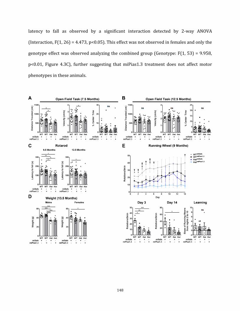
148
latency to fall as observed by a significant interaction detected by 2-way ANOVA
(Interaction, F(1, 26) = 4.473, p<0.05). This effect was not observed in females and only the
genotype effect was observed analyzing the combined group (Genotype: F(1, 53) = 9.958,
p<0.01, Figure 4.3C), further suggesting that miPias1.3 treatment does not affect motor
phenotypes in these animals.

149
Figure 4.3: Pre-symptomatic Pias1 KD may exacerbate motor deficits in zQ175 mice. A) open field task at 7.5 months of age for combined group shows significant genotype effect for distance traveled and velocity but not % center time. B) No differences were detected in open field task at 12.5 months of age. Rotarod at C) 9.5 months of age and 12.5 months of age shows significant genotype effects. D) Male mice show genotype effect in body weight while female heterozygotes treated with miPias1.3 have a deficit in weight gain. E) Running wheel task at 9 months of age in males suggests a negative impact on motor tasks with miPias1.3 treatment but with no effect on motor learning. Genotype effects were observed for day 1 (F(1, 15) = 21.620, p<0.001), day 2 (F(1, 15) = 19.320, p<0,001), day 3 (F(1, 15) = 22.230, p<0.001), day 4 (F(1, 15) = 12.230, p<0.01), day 5 (F(1, 15) = 7.879, p<0.05), day 6 (F(1, 15) = 9.265, p<0.001), day 7 (F(1, 15) = 6.986, p<0.05), day 12 (F(1, 15) = 4.887, p<0.05), day 13 (F(1, 15) = 5.751, p<0.05), and day 14 (F(1, 15) = 7.883, p<0.05). Treatment effects were observed for day 3 (F(1, 15) = 9.243, p<0.01) and Day 5 (F(1, 15) = 9.339, p<0.01) Open symbols represent female mice. All samples were analyzed by 2-way ANOVA followed by Tukey’s multiple comparison test. ns=not significant, *p<0.05, **p<0.01, ***p<0.001, ****p<0.0001, values represent means ± SEM. Shaded bars represent miPias1.3 treated animals. For running wheel: ‡ = p<0.05 WTmiSafe vs WTmiPias1.3, * = p<0.05 WTmiSafe vs HetmiSafe, † = p<0.05 WTmiSafe vs HetmiPias1.3
Body weight: zQ175 heterozygous animals reliably show decreased body weight (Menalled
et al., 2012). Male zQ175 animals had significantly lower body weights than their WT
counterparts with Pias1 KD not altering this genotype effect (Table 4.1, Figure 4.3D). In
female animals, only a decrease in weight was observed for miPias1.3 treated zQ175
animals, but with no significant treatment effect (Treatment: F(1, 23) = 2.592, p>0.05).
Overall, males showed a significant genotype effect while female zQ175 animals did not
show a genotype effect but may have decreased body weight with Pias1 KD.
Running Wheel: The running wheel task allows for uninterrupted, voluntary assessment of
motor function and motor learning (Hickey et al., 2008). Therefore, males were assessed at
9 months of age for two weeks. Females were not analyzed due to the influence of estrus
cycle on running wheel activity (Hickey et al., 2008). Running wheel data trends suggest a
promotion of motor deficits in mice treated with miPias1.3 in both WT and zQ175 mice
150
compared to miSafe-treated controls, but with no significant treatment effect over time
(Treatment: F(3, 15) = 3.257, p>0.05, Figure 4.3E). Overall, miPias1.3 treated zQ175 mice
performed worse throughout the task and WT animals with miPias1.3 treatment showed a
significant reduction in wheel-usage at days 3 and 5, suggesting a negative impact on this
task with miPias1.3 treatment on WT and HD mice at this age (Figure 4.3E).
The running wheel task can also be used to assess motor learning based on the slope of
mean running between days 2 and 10 (Hickey et al., 2008). Using this analysis, no
differences in motor learning were observed between our groups (Genotype, F(1, 15) =
0.510, p>0.05; Treatment, F(1, 15) = 0.741, p>0.05, Figure 4.3E). Overall, running wheel
data may suggest an exacerbation in motor deficits in zQ175 animals and a decreased
usage effect in WT animals with miPias1.3 pre-symptomatic treatment when assessed at 9
months of age, with no effect on motor learning.
Pias1 knock-down affects formation of HMW mHTT in pre-symptomatic treated
female zQ175 mice
Next, we assessed formation of insoluble HMW mHTT after sacrifice at 13.5 months of age,
a molecular readout modulated in cell culture and R6/2 mice by Pias1 (O'Rourke et al.,
2013; Ochaba et al., 2016). In zQ175 mice, this species can be detected at 6 months of age,
accumulating over time in zQ175 heterozygotes similar to the HMW insoluble mHTTex1p
species observed in R6/2 mice (Chapter 1, Figure 4.1A). First, to evaluate KD efficiency,
mRNA was harvested from GFP-positive microdisected striata to quantify levels of Pias1
transcript. Pias1 levels were significantly reduced in both zQ175 and WT animals with pre-

151
symptomatic KD as assessed by qPCR, being about -0.5 to -0.6 fold less than control groups
(Males: F(1, 11) = 52.800, p<0.0001, Females: F(1, 10) = 73.160, p<0.0001), indicating
successful reduction by Pias1 miRNA (Figure 4.4A, D). To assess KD effect on Pias1 protein
levels and levels of accumulated, insoluble mHTT species, Soluble/Insoluble fractionation
was performed as described (Ochaba et al., 2016; Ochaba et al., 2018) and assessed by
western blot. In males, no differences in Pias1 protein levels were detected in any group in
both Insoluble (Treatment: F(1, 8) = 3.709, p>0,05, Genotype: F(1, 8) = 0.069, p>0.05) and
Soluble (Treatment: F(1, 8) = 0.533, p>0.05, Genotype: F(1, 8) = 0.104, p>0,05) fractions
(Figure 4.4B, C). GFP was detected in all animals, suggesting successful viral transduction.
In zQ175 males, HMW mHTT was not modulated with miPias1.3 treatment, unlike that
observed previously in R6/2 animals (p>0.05, (Ochaba et al., 2016)). This may have been
due to insufficient reduction in Pias1 protein levels or lack of accumulation of Pias1 in the
insoluble fraction of zQ175 animals as reduction of insoluble Pias1 previously
corresponded with a decrease in HMW mHTT accumulation (Ochaba et al., 2016). A decline
in mHTT protein levels is associated with disease progression in knock-in HD mice
(Franich et al., 2018). To determine if Pias1 KD affected this genotype-associated decline,
Soluble, full-length HTT (FL HTT) protein was quantified using D7F7 antibody (Cell
signaling #5656S). FL-HTT was significantly reduced in zQ175 males compared to WT (F(1,
8) = 39.600, p<0.001), but no miPias1.3 treatment effect was observed (F(1, 8) = 0.935,
p>0.05, Figure 4.4B, b’), suggesting that Pias1 did not impact age-associated decline of
mHTT.

152
In female animals, Pias1 protein was also not significantly reduced in any group for either
the Insoluble (F(1, 10) = 4.464, p>0.05) or Soluble fractions (F(1, 10) = 2.512, p>0.05,
Figure 4.4E, F), however, insoluble HMW mHTT was significantly increased in females with
miPias1.3 treatment (p<0.05). No treatment effect was observed on levels of soluble, FL
HTT (F(1, 10) = 1.879, p>0.05) but similar to males, showed a significant reduction in
zQ175 mice (F(1, 10) = 43.570, p<0.0001, Figure 4.4E, F). Therefore, Pias1 KD may have
influenced the accumulation of insoluble, HMW mHTT in females only.

153
Figure 4.4: Pre-symptomatic Pias1KD does not affect mHTT accumulation at 13.5 months of age. A) qPCR analysis of mRNA harvested from male GFP+ microdisected striata shows significant reduction in levels of Pias1 transcript with miPias1.3 treatment. Solid symbols for qPCR represent samples used for later mRNAseq analysis. B) Western blot analysis of Soluble/Insoluble fractionation of striatal tissue and C) quantification from male mice shows insufficient reduction of Pias1 protein in both fractions. HMW insoluble mHTT remains unchanged with treatment. Soluble Full length (FL) HTT shows a significant genotype effect with no treatment effect, b’) *samples used for representative image and quantitative analysis due to observed batch effect. *Samples all processed from the same batch. D) qPCR analysis of mRNA harvested from female GFP+ microdisected striata shows significant reduction in levels of Pias1 transcript with miPias1.3 treatment. E) Western blot analysis of Soluble/Insoluble fractionation and F) quantification of striatal tissue from female mice shows insufficient reduction of Pias1 protein in both fractions. HMW insoluble mHTT significantly increased with treatment. Soluble Full length (FL) HTT shows a significant genotype effect with no treatment effect. Protein normalized to total protein stain. Normalized relative protein abundances for all samples were analyzed by 2-way ANOVA followed by Tukey’s multiple comparison test. ns = not significant, *p<0.05. **p<0.01, ***p<0.001, ****p<0.0001, values represent means ± SEM. Shaded bars represent miPias1.3 treated animals.
Reducing Pias1 differentially modulates the transcriptional landscape in zQ175 mice
Pias1 is well established to function as a regulator of transcription (Rytinki et al., 2009)
including serving as a negative regulator of inflammatory transcriptional response through
binding of NfkB (Liu et al., 2005). Pias1 also serves as an E3 SUMO ligase for several
transcription factors that regulate neuronal transcription, suggesting that KD of Pias1 could
have an impact on the transcriptional profile in the brain (Table 2, (Estruch et al., 2016;
Gregoire and Yang, 2005; Riquelme et al., 2006; Tai et al., 2016)). Therefore, with
significant KD at the transcript level, we hypothesized that miPias1.3 treatment would
impact the transcriptome in zQ175 mice.
Transcriptomic analysis was carried out on GFP-positive striatal regions harvested from
pre-symptomatic male mice representing four conditions: WT miSafe, WT miPias1.3, Het
miSafe, and Het miPias1.3 with three mice each per condition. Total RNA was isolated using

154
Trizol extraction and purified using RNeasy columns (Qiagen) for mRNAseq analysis. Data
of completed mRNAseq analysis will be deposited to GEO. After QC, mapping, and
quantification, normalized gene expression values were used for exploratory analysis.
Principal component analysis (PCA) revealed a clear separation between the groups; both
genotype and treatment conditions showed distinct separation along PC1 and PC2,
representing the maximal variance among samples (Figure 4.5A). These data recapitulate
known transcriptional deficits in HD mice and human patients and indicate that reduction
of Pias1 has a substantial, and reproducible, impact on gene expression in both HD and WT
mice. Statistical analysis of differentially expressed genes (DEGs) was performed using
DEseq2 (Love et al., 2014) and a significance threshold was set at a 10% FDR. Overall, WT
animals had a higher number of dysregulated genes with miPias1.3 treatment than zQ175
animals (1438 for WT and 759 for ZQ175, Figure 4.5B). Of these, 279 DEGs were in
common with miPias1.3 treatment between genotypes (Figure 4.5C). Interestingly, the
majority of these shared genes modulated by Pias1 were inversely regulated based on
genotype, supporting a unique role for Pias1 in the context of mHTT expression. These
shared DEGs were analyzed using Gene Ontology (GO, GOrilla (Eden et al., 2009))
enrichment analysis to assess specific biological processes (Figure 4.5D). Data revealed top
processes related to synaptic vesicle and neurotransmitter release, suggesting that Pias1 is
differentially modulating neuronal function based on disease context. Remaining DEGs
were unique to either WT or zQ175 mice when treated with miPias1.3. These genotype-
specific changes were further analyzed using GO and Ingenuity Pathway Analysis (IPA) for
upstream regulators by assessing all DEGs mediated by Pias1 KD for each genotype, with
each showing unique enrichment terms (Figure 4.5E, G). GO analysis of WT animals with

155
Pias1 KD revealed processes related to cellular defense mechanisms and immune
responses. Upstream regulators suggest inhibition of immune related processes with Pias1
KD (Figure 4.5E, F), consistent with the well-defined function of Pias1 as a negative
regulator of the NfkB inflammatory pathway (Shuai and Liu, 2005). GO analysis of zQ175
animals with Pias1 KD showed enrichment for genes involved in cellular communication,
neuronal transmission, and development (Figure 4.5G). IPA analysis revealed upstream
pathways associated with HTT and neuronal trophic support (Figure 4.5G). Overall,
analysis of genotype-specific transcriptional dysregulation with miPias1.3 treatment
suggests that Pias1 may be differentially mediating pathways in zQ175 mice compared to
WT mice. In WT animals, networks appear to be centered around immune and cellular
defense functions while in zQ175, networks are associated with neuronal function.
Due to the differential impact of miPias1.3 treatment in zQ175 mice compared to WT, we
next examined the effect of treatment on genotype-specific dysregulated genes. A unique
set of DEGs was generated when comparing zQ175 miPias1.3 DEGs to those from control
WT and zQ175 animals. Analyzing these genotype-filtered DEGs by GO and IPA revealed
that Pias1 KD normalized genotype-associated changes in genes involved in neuronal
function and health (Figure 4.5I, J). These included trans-synaptic signaling, synaptic
transmission, and nervous system development suggesting a normalizing effect for Pias1
KD on aberrant disease associated gene transcription with an inverse fold change observed
for most DEGs (Figure 4.5K). Together, transcriptomic profiling of miPias1.3 treated zQ175
animals suggests that Pias1 is contributing to neuronal function in HD mice.

156
Figure 4.5: Pre-symptomatic Pias1 KD rescues neuronal function-related transcriptional dysregulation in zQ175 mice at 13.5 months of age. A) PCA of normalized gene expression values shows separation by both treatment and genotype. B) Number of DEGs with miPias1.3 treatment in WT and zQ175 mice. C) Overlap of 279 genes dysregulated with miPias1.3 treatment in WT and zQ175 mice enrich for D) GO biological processes associated with neuronal function. E) GO enrichment analysis of WT miPias1.3 vs miSafe treated animals shows modulation of immune and cellular defense processes and corresponding F) IPA upstream analysis suggests inhibition of immune related regulators. G) GO enrichment analysis of Het miPias1.3 vs miSafe treated animals shows modulation of synaptic signaling and development related processes and corresponding H) IPA upstream analysis suggests modulation of neuronal and disease-related regulators with miPias1.3 treatment. I) GO enrichment analysis and fold change of rescued, genotype-associated DEGs reveals enrichment for synaptic and neuronal functional processes and J) corresponding IPA upstream analysis similarly shows modulation of neuronal and disease-related regulators. K) Fold change heatmap indicating normalizing effect of miPias1.3 treatment on genotype-associated DEGs. Sequencing completed by UCI GHTF, analysis assisted by Ryan Lim, PhD, and Jie Wu, PhD, Thompson Lab.

157
Transcriptional analysis was done on mRNA extracted from male striata. Therefore, to
determine if miPias1.3 treatment was similarly affecting transcriptional readouts in zQ175
females, mRNA was assessed using qPCR on several genes that were normalized with
miPias1.3 treatment in males (Table 4.3). The house-keeping gene Eif4a2 was used to
normalize gene expression as it does not have a genotype effect in zQ175 mice (Southwell
et al., 2016) and consistently remained unchanged in our mRNAseq dataset. In males, qPCR
confirmed data obtained by mRNAseq showing a significant normalization effect with Pias1
KD on genotype-associated DEGs for levels of Bdnf, NeuroD1, NeuroD2, Syn1, and Syn2,
(Figure 4.6A). In females, however, levels of Bdnf remained statistically unaffected by
miPias1.3 treatment with high variability within groups. Levels of NeuroD1 and NeuroD2
showed a significant increase in expression with Pias1 KD, unlike in males, which showed a
decrease with KD (Figure 4.6B). Levels of Syn1 and Syn2 were unaffected by miPias1.3
treatment or genotype in females (Figure 4.6B). While data suggests a differential effect on
transcription in a gender-specific manner, transcriptional qPCR data supports the
hypothesis that Pias1 has a regulatory role in synaptic and neuronal function by
modulating NeuroD1 and NeuroD2 in both sexes and other neuronal genes in males.
Gender-specific consequences of differential expression and accumulation of HMW mHTT
remain to be explored. It is noteworthy that the vast majority of transcriptomic studies in
R6/2 mice have been carried out in males, and previous transcriptional profiling in zQ175
mice removed gender-associated variation (Langfelder et al., 2016). Moreover, the majority
of the findings correspond with gene changes found in human HD brain (Hodges et al.,
2006).

158
Figure 4.6: qPCR analysis to validate mRNAseq identified genes for pre-symptomatic miPias1.3 treatment assessed at 13.5 months of age. A) Fold change and a’) dCT values for male mice show significant treatment and interaction effects for miPias1.3 treated Hets reproducing normalizing effect seen in mRNAseq data. Solid symbols represent animals used for mRNAseq analysis: Bdnf (Interaction: F(1, 11) = 15.090, p<0.01), NeuroD1 (Interaction: F(1, 11) = 5.740, p<0.05), NeuroD2 (Interaction: F(1, 11) = 17.340, p<0.01), Syn1 (Interaction: F(1, 11) = 48.480, p<0.0001), and Syn2 (Interaction: F(1, 11) = 22.780, p<0.001). B) Fold change and b’) dCT values for female mice show significant treatment effect for NeuroD1 and NeuroD2 but inverse of effect seen in males: Bdnf (Treatment: F(1, 10) = 3.886, p>0.05), NeuroD1 (Treatment: F(1, 10) = 8.699, p<0.05) and NeuroD2 (Treatment: F(1, 10) = 6.309, p<0.05), Syn1 (Treatment: F(1, 10) = 3.851, p>0.05), and Syn2 (Treatment: F(1, 10) = 0.410, p>0.05). All samples were analyzed by 2-way ANOVA followed by Tukey’s multiple comparison test. ns = not significant , *p<0.05, **p<0.01, ***P<0.0001, values represent means ± SEM. Shaded bars represent miPias1.3 treated animals.

159
Inflammation is impacted by pre-symptomatic Pias1 reduction
We previously showed that Pias1 KD decreased the number of microglia in the striatum of
R6/2 mice. Given the enrichment of pathways involved in defense-responses in WT
animals treated with miPias1.3, we evaluated inflammatory signatures. Microglia activation
in mouse striata was monitored by counting the number of cells and measuring cell-body
diameter of Iba1+ microglia using confocal microscopy and Imaris automatic analysis
software. Microglia with cell body diameters of greater than 7 µm were considered reactive
(Watson et al., 2012). Images were taken at 20x magnification from GFP+ regions of the
striatum, representing virally transduced, Pias1 reduced regions. Pias1 KD in zQ175 males
appeared to return cells towards resting state by 13.5 months of age (Diameter: F(13,
112)=194.900, p<0.0001). However, post-hoc analysis failed to reach significance for Het
miPias1.3 treated animals compared to other groups. Further, no difference in the number
of microglia was observed (Genotype: F (1, 8) = 0.023, p>0.05, Figure 4.7A, B).
Interestingly, miPias1.3 treatment in WT animals significantly increased the number of
reactive microglia in the striatum based on cell-body diameter (Figure 4.7B). This is
consistent with the observed mRNAseq GO analysis, with enrichment in pathways
associated with cellular defense in WT animals only compared to zQ175 mice. We further
measured inflammatory markers that we previously reported were affected by Pias1 KD in
R6/2 mice (NfkB and Il-6) by western blot (Figure 4.7C). Soluble levels of NfkB significantly
increased in zQ175 mice treated with miPias1.3 (Genotype: F(1, 8) = 14.820, p<0.01), with
no signal being detected in the Insoluble fraction (data not shown) unlike what was
observed in R6/2 mice with KD (Ochaba et al., 2016). Further, levels of total Il-6 remained
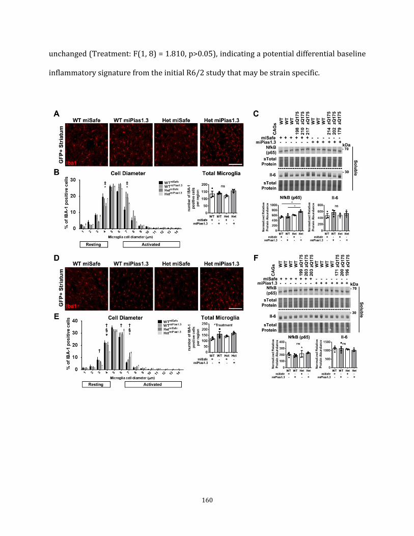
160
unchanged (Treatment: F(1, 8) = 1.810, p>0.05), indicating a potential differential baseline
inflammatory signature from the initial R6/2 study that may be strain specific.

161
Figure 4.7: Pre-symptomatic Pias1 KD modulates immune response. Confocal analysis of Iba1+ microglia in striatum of male mice with pre-symptomatic knock-down of Pias1 (n=3). A) Representative images from the GFP+ region of 13.5 mon. old male mice. B) Cell body diameter analysis and cell count from male mice quantified using Bitplane Imaris automated software to detect cell bodies. C) Western blot from soluble striatal fraction shows significant increase in inflammatory regulator NfkB but not in cytokine Il-6 in males. D) Representative images from the GFP+ region of 13.5 mon. old female mice. E) Cell body diameter analysis and cell count from female mice quantified using Bitplane Imaris automated software to detect cell bodies. F) Western blot from soluble striatal fraction shows no changes in inflammatory markers in females. Protein normalized to total protein stain. Normalized relative protein abundances for all samples were analyzed by 2-way ANOVA followed by Tukey’s multiple comparison test. ns = not significant, *p<0.05, values represent means ± SEM. For cell body diameter: ‡ = p<0.05 WTmiSafe vs WTmiPias1.3, * = p<0.05 WTmiSafe vs HetmiSafe, † = p<0.05 WTmiSafe vs HetmiPias1.3, # = p<0.05 WTmiPias1.3 vs HetmiSafe, § = p<0.05 WTmiPias1.3 vs HetmiPias1.3, ᵠ = p<0.05 HetmiSafe vs HetmiPias1.3
Interestingly, female zQ175 animals treated with miPias1.3 showed an increase in
microglial activation (Diameter: F(13, 112) = 397.100, p<0.0001), but with only a slight
treatment affect in total number of microglia (Treatment: F (1, 8) = 6.095, p<0.05, Figure
4.7D, E). Inflammatory markers remained unchanged by western blot (NfkB: Treatment,
F(1, 10) = 0.004, p>0.05; Il-6: Treatment, F(1, 9) = 0.013, p>0.05, Figure 4.7F). Taken
together, the data suggest that Pias1 influences molecular networks associated with
neuronal function in zQ175 mice, which may be independent of behavioral or protein
homeostasis readouts at the 13.5 month long-term time point.
Symptomatic Pias1 KD does not affect zQ175 behavior or HMW HTT accumulation
We next asked whether Pias1 miRNA treatment after onset of symptoms in zQ175 mice
could alter behavioral or molecular phenotypes (Figure 4.2B, Direction 2). Symptomatic
Pias1 KD animals (injections at 7.5 months of age in collaboration with the Davidson lab,
Figure 4.1B) were also subjected to a battery of assays as described above for pre-
symptomatic mice. Mice were evaluated for behavior following viral injections until they

162
were sacrificed at 13.5 months of age to match the pre-symptomatic cohort. Statistical
outputs for open field, Rotarod, and body weights are detailed in Table 4.2.
Behavior: For open field task at 12.5 months of age, no significant differences were detected
between groups for distance traveled or velocity (Figure 4.8A). This data is consistent with
the lack of a genotype effect in the pre-symptomatic cohort assessed with this task at 12.5
months of age and as observed previously for these animals (Menalled et al., 2012). No
differences were detected for males or females for percent of time in center separately but
combined, animals showed a significant difference, suggesting that this cohort of zQ175
mice may have had increased anxiety (F(2, 44) = 3.380, p<0.05, Figure 4.8A). However,
post-hoc analysis failed to reveal the source of significance.
To assess additional deficits in motor movements, accelerating Rotarod task was
performed at 9.5 and 12.5 months of age (Figure 4.8B). At 9.5 months of age, zQ175
animals had a significant decrease in latency to fall that was primarily observed in the
females only as males alone did not show any significant difference in latency to fall
between the groups (Table 4.2). Pias1 KD had no effect on latency to fall at 9.5 months of
age. At 12.5 months of age, no differences in latency to fall were detected for males and
females separately, but combined showed a significant difference suggesting a slight
decrease in latency to fall for zQ175 mice (Combined: F(2, 43) = 3.576, p<0.05). However,
post-hoc analysis failed to show significance between groups with combined animals
further suggesting that miPias1.3 treatment did not affect behavioral readouts in zQ175
mice (Figure 4.8B).

163
Figure 4.8: Symptomatic Pias1 KD has no effect on behavior in zQ175 mice. A) Open field task at 12.5 months of age for combined group shows no effect for distance traveled and velocity but with a significant difference in % center time by 1-Way ANOVA, with no post-hoc reaching significance. B) Rotarod at 9.5 months of age and 12.5 months of age shows significant genotype effects but no treatment effects. C) No differences were detected in body weight at 8 months of age. Open symbols represent female mice. All samples were analyzed by 1-way ANOVA followed by Tukey’s multiple comparison test. ns=not significant, *p<0.05, **p<0.01, ***p<0.001, values represent means ± SEM. Shaded bars represent miPias1.3 treated animals. Data generated in collaboration with Davidson lab, CHOP. Body weight was also recorded for these animals and assessed separately due to the
previously observed differences based on gender in these animals (Menalled et al., 2012).
Males did not show any differences in body weight at 12.5 months of age even though
decreased body weight is typical in zQ175 mice at this age (Menalled et al., 2012). For
females, 1-way ANOVA suggested a significant genotype effect indicating decreased body
weight (F(2, 24) = 4.226, p<0.05) but with post-hoc analysis again failing to reveal
significance (Figure 4.8C). Symptomatic Pias1 KD did not affect body weights. Taken

164
together, behavioral data suggest that symptomatic miPias1.3 treatment does not affect
behavioral readouts in zQ175 mice.
HMW mHTT: First, KD efficiency was assessed by measuring levels of Pias1 transcript from
GFP-positive microdissected striata at 13.5 months of age. A significant reduction by about
-0.5 to -0.6 fold in levels of Pias1 transcript, assessed by qPCR, in both females (F(2, 8) =
70.210, p<0.0001) and males (F(2, 9) = 13.410, p<0.01) was observed (Figure 4.9A, D),
indicating successful reduction by Pias1 miRNA. Similar to previous cohorts, striata from
symptomatic miPias1.3 treated animals were also assessed by Soluble/Insoluble
fractionation (Figure 4.9). A significant reduction of Pias1 was observed in the Soluble
fraction in females only (Figure 4.9E, F, Females: F(2,11)=14.510, p<0.001; Males: F(2, 9) =
1.165, p>0.05). Insoluble Pias1 was not altered (Females: F(2, 11) = 3.262, p>0.05; Males:
F(2, 9) = 0.730, p>0.05) and HMW mHTT was not affected in symptomatic miPias1.3-
treated animals (Females: p>0.05, males: p>0.05). There were significantly lower levels of
soluble FL HTT in zQ175 mice, independent of miPias1.3 treatment in both females (F(2,
11) = 19.830, p<0.001) and males (F(2, 9) = 12.060, p<0.01, Figure 4.9B, E), again
suggesting that Pias1 is not modulating age-associated decline of mHTT protein. Together,
these data suggest that targeting Pias1 symptomatically in these zQ175 mice does not
affect accumulation of insoluble mHTT species, even with successful reduction of Pias1
protein in females.

165
Figure 4.9: Symptomatic Pias1 KD does not affect mHTT accumulation at 13.5 months of age. A) qPCR analysis of mRNA harvested from male GFP+ microdisected striata shows significant reduction in levels of Pias1 transcript with miPias1.3 treatment. B) Western blot analysis and C) quantification of Soluble/Insoluble fractionation of striatal tissue from male mice shows insufficient reduction of Pias1 protein in both fractions. HMW insoluble mHTT remains unchanged with treatment. Soluble Full length (FL) HTT shows a significant genotype effect with no treatment effect. D) qPCR analysis of mRNA harvested from female GFP+ microdisected striata shows significant reduction in levels of Pias1 transcript with miPias1.3 treatment as well as a significant genotype effect compared to WT vehicle control mice. E) Western blot analysis and F) quantification of Soluble/Insoluble fractionation of striatal tissue from female mice shows insufficient reduction of insoluble Pias1 protein but significant reduction of soluble Pias1 levels with miPias1.3 treatment. HMW insoluble mHTT remains unchanged with treatment. Soluble Full length (FL) HTT shows a significant genotype effect with no treatment effect. Protein normalized to total protein stain. Normalized relative protein abundances for all samples were analyzed by 1-way ANOVA followed by Tukey’s multiple comparison test. ns = not significant, *p<0.05. **p<0.01, ***p<0.001, ****p<0.0001, values represent means ± SEM. Shaded bars represent miPias1.3 treated animals.

166
Microglial activation: Similar to pre-symptomatic Pias1 KD animals, symptomatic-treated
mice were assessed for microglial activation and inflammatory markers. Male zQ175
animals with symptomatic miPias1.3 treatment had a significant increase in activated
microglia based on cell body diameter (F(13,84)=150.500, p<0.0001) as well as an increase
in the number of microglia detected suggesting that miPias1.3 treatment in symptomatic
zQ175 animals increased microglial activity in the striatum (F(2,6)=7.870, p<0.05, Figure
4.10A, B). This suggested that targeting Pias1 symptomatically may have induced an
immune response as opposed to reducing one (compared to pre-symptomatic treated
males, Figure 4.7B). However, no changes in inflammatory markers were detected in the
Soluble protein fraction assessed by western blot (NfkB: F(2, 9) = 2.588, p>0.05; Il-6: F(2,
9) = 0.202, p>0.05, Figure 4.10C). For females, a significant increase in soluble levels of
NfkB was observed in zQ175 mice treated with miPias1.3 (NfkB: F(2, 9) = 9.610, p<0.01; Il-
6: F(2, 11) = 2.818, p>0.05, Figure 4.10D), which had not been observed with pre-
symptomatic KD (Figure 4.7F), also suggesting an increased inflammatory response.
Insoluble NfkB was not detected (data not shown). Taken together these data suggest that
Pias1 may have different functional consequences towards specific neuroinflammatory
responses during different disease stages.
In summary, for both pre-symptomatic and symptomatic mice, Pias1 reduction in general
did not modulate behavior or insoluble HMW HTT species. Modest modulatory effects on
inflammatory responses were observed with both symptomatic and pre-symptomatic KD
with symptomatic reduction potentially increasing an inflammatory response in zQ175
males. However, Pias1 protein levels were not significantly reduced with KD for most

167
groups when assessed at 13.5 months of age, potentially reflecting disruptions in protein
clearance or compensatory responses. Importantly, Pias1 did not accumulate in the
insoluble fraction as was observed in R6/2 mice (Ochaba et al., 2016). These data suggest
that either Pias1 modulation does not impact these outcomes in zQ175 animals or that the
concordant accumulation of insoluble Pias1 and activation of Pias1 specific inflammatory
responses is a necessary component for impacting either behavior or aberrant protein
homeostasis in HD animals.

168
Figure 4.10: Symptomatic Pias1 KD modulates immune response. Confocal analysis of Iba1+ microglia in striatum of male mice with symptomatic knock-down of Pias1 (n=3). A) Representative images from the GFP+ region of 13 mon. old male mice. B) Cell body diameter analysis and cell count from male mice quantified using Bitplane Imaris automated software to detect cell bodies. Western blot from C) male soluble striatal fraction effects on inflammatory regulator NfkB and cytokine Il-6 with miPias1.3 treatment. D) Female western blot analysis shows significant elevation in NfkB but no change in Il-6. Protein normalized to total protein stain. Normalized relative protein abundances for all samples were analyzed by 1-way ANOVA followed by Tukey’s multiple comparison test. ns = not significant, *p<0.05, values represent means ± SEM. Shaded bars represent miPias1.3 treated animals. For cell body diameter: ‡ = p<0.05 WTmiSafe vs WTmiPIAS1.3, * = p<0.05 WTmiSafe vs HetmiSafe, † = p<0.05 WTmiSafe vs HetmiPIAS1.3, # = p<0.05 WTmiPIAS1.3 vs HetmiSafe, § = p<0.05 WTmiPias1.3 vs HetmiPias1.3, ᵠ = p<0.05 HetmiSafe vs HetmiPias1.3
Assessing pre-symptomatic Pias1 KD at 8 months of age reveals additional molecular
modulation
To further understand molecular mechanisms impacted by Pias1KD and inform results
from the 13.5 month-old study, we evaluated mice at an earlier time point of 8 months of
age, just after symptomatic phenotypes emerge (Menalled et al., 2012). We repeated pre-
symptomatic treatment with miPias1.3 and miSafe with a small cohort of animals at 2.5
months of age and aged mice to 8 months, specifically to evaluate biochemical and
molecular readouts (Figure 4.2, Direction 3). A Saline/vehicle control was included in
female mice to control for viral delivery method. Behavior was evaluated primarily as a
mode of enrichment to ensure that molecular readouts were not confounded by lack of
trophic support (Mattson et al., 2004) and for consistency with the longer term studies.
Behavioral analysis of pre-symptomatic Pias1 KD at 8 months
The same battery of behavioral tasks was carried out as above. Open field was completed at
7.5 months of age with running wheel task performed at 7 months before sacrifice. Body
weights were also recorded. A summary of statistical analysis for open field and body

169
weights are shown in Table 4.1 (Figure 4.11A, B). The smaller cohort size may account, in
part, for the lack of observed genotype effects for these readouts.
Figure 4.11: Pre-symptomatic Pias1 KD may rescue early motor deficits in zQ175 mice. A) open field task at 7.5 months of age for combined group shows no effect for distance traveled and velocity but a significant genotype effect for % center time. B) No differences were detected in body weight at 8 months of age. C) Running wheel task at 7 months of age in males suggests a significant increase in motor activity with miPias1.3 treatment in at least WT animals but with no effect on motor learning. Significant treatment effects were observed on day 1 (F(1, 12) = 11.840, p<0.01), day 3 (F(1, 12) = 6.053, p<0.05), day 4 (F(1, 12) = 6.019, p<0.05), day 5 (F(1, 12) = 5.292, p<0.05), day 10 (F(1, 12) = 5.390, p<0.05), day 11 (F(1, 12) = 5.646, p<0.05), day 12 (F(1, 12) = 6.882, p<0.05), day 13 (F(1, 12) = 6.921, p<0.05), and day 14 (F(1, 12) = 7.148, p<0.5). A significant interaction was detected on day 3 (F(1, 12) = 5.045, p<0.5). A significant genotype effect was detected on day 4 (F(1, 12) = 5.566, p<0.05). Open symbols represent female mice. All samples were analyzed by 2-way ANOVA followed by Tukey’s multiple comparison test. ns=not significant, *p<0.05, **p<0.01, ***p<0.001, values represent means ± SEM. Shaded bars represent miPias1.3 treated animals. For running wheel: ‡ = p<0.05 WTmiSafe vs WTmiPias1.3, # = p<0.05 WTmiPias1.3 vs HetmiSafe

170
The running wheel apparatus has a finite number of animals that can be tested at a time
due to single housing. Therefore, groups of male mice were of comparable size to the
cohort assessed at 9 months of age and data was similarly processed (Figure 4.11C). In
contrast to mice tested at 9 months of age, which showed some additional impairment in
this task with Pias1KD, at this earlier time point, KD increased running wheel usage.
Specifically, a significant treatment effect (Treatment: F(3, 12) = 3.526, p<0.05) was
observed with WT animals treated with miPias1.3 showing an apparent increase in usage
of the running wheel over time compared to WT control treated animals. A similar trend
was observed between zQ175 miSafe and miPias1.3 treated animals over time. No
differences were detected in motor learning (Genotype: F (1, 12) = 1.339, p>0.05;
Treatment, F(1, 12) = 4.663, p>0.05). Overall, data suggests that miPias1.3 treatment led to
increased usage of the running wheel and increased motor capabilities in at least WT
animals and potentially zQ175 animals at this time point (7 months, Figure 4.11C).
Therefore, miPias1.3 pre-symptomatic treatment may have differential effects on behavior
during different stages of disease pathogenesis.
Bodyweight was recorded and assessed as in previous cohort until 8 months of age. No
differences in weight gain were detected for either genotype or treatment by 8 months of
age. However, at 8 months of age, zQ175 mice only exhibit minor deficits in body weight
(Menalled et al., 2012). It is possible that this cohort of animals had not begun showing
detectable genotype effects with this analysis by time of sacrifice.

171
Pias1 does not affect formation of HMW mHTT or alter microglial activation at 8
months of age
Striata from male mice were assessed at 8 months of age for levels of Pias1 and
accumulated mHTT using Soluble/Insoluble fractionation and western blotting (Figure
4.12) to determine whether an earlier time point might reveal changes in protein
homeostasis. Unlike pre-symptomatic animals assessed at 13.5 months of age, Pias1
protein levels were significantly reduced in the Soluble fraction in both WT and HD zQ175
males treated with miPias1.3 (Treatment: F(1, 10) = 76.120, p<0.0001). Insoluble Pias1
levels also showed a significant treatment effect suggesting reduction of this species (F (1,
10) = 12.920, p<0.01) but only between control WT animals and miPias1.3 treated zQ175
as revealed by post-hoc analysis. Levels of insoluble Pias1 were not elevated in untreated
zQ175 animals at this age, similar to 13.5 month old animals. We next assessed formation
of the insoluble HMW mHTT species in this younger cohort of pre-symptomatic miPias1.3
treated animals. Significant reduction of Pias1 still did not affect formation of insoluble
HMW mHTT accumulated species (p>0.05). zQ175 animals again had significantly reduced
levels of soluble FL HTT compared to WT animals (Genotype: F(1, 10) = 31.990, p<0.0001),
and a modest treatment effect of reduced FL HTT in zQ175 mice was observed with
miPias1.3 treatment (Treatment: F(1, 10) = 7.564, p<0.05).

172
Figure 4.12: Pre-symptomatic PIias1 KD does not affect mHTT accumulation at 8 months of age. A) Western blot analysis and B) quantification of Soluble/Insoluble fractionation of striatal tissue from male mice shows significant reduction of Pias1 protein in both fractions. HMW insoluble mHTT remains unchanged with treatment at this age. Soluble Full length (FL) HTT shows a significant genotype effect with a slight treatment effect observed with zQ175 animals having significantly less FL HTT than WT with miPias1.3 treatment. Protein normalized to total protein stain. Normalized relative protein abundances for all samples were analyzed by 2-way ANOVA followed by Tukey’s multiple comparison test. ns = not significant, *p<0.05. **p<0.01, ***p<0.001, ****p<0.0001, values represent means ± SEM. Shaded bars represent miPias1.3 treated animals.
Given the modest effect of miPias1.3 treatment on microglia in older animals, we assessed
cell body diameter and total number of microglia in females at 8 months of age. A saline
treatment control group was included in this analysis to determine if inflammatory effects
could arise from viral delivery. Using the same analysis as above on Iba1+ cells, there was
no effect of miPias1.3 treatment on microglial activation in these animals. Interestingly,
saline WT and zQ175 control groups appeared to have more reactive microglia than viral
treated WT and zQ175 groups based on cell body diameter (F(13, 168) = 477.500,
p<0.0001, Figure 4.13A, B), potentially confounding the data. No difference in number of
microglia was observed for either genotype (F(1, 12) = 0.026, p>0.05) or treatment (F(2,
12) = 0.234, p>0.05, Figure 4.13B). Due to these confounding variables, no conclusion

173
regarding microglial activation in these animals could be made using this analysis. Due to
cohort size, male animals were not assessed using this assay, rather, inflammatory markers
were analyzed by western blot on male tissues (Figure 4.13C). Soluble levels of NfkB were
significantly altered in males with miPias1.3 treatment in a genotype dependent manner
(Treatment: F(1, 10) = 6.564, p<0.05; Interaction: F(1, 10) = 30.850, p<0.001) such that,
WT animals with Pias1 KD had less soluble NfkB: an effect that was not observed at 13.5
months of age (Figure 4.7C). For levels of soluble Il-6, a slight increase in WT treated
animals and a slight decrease in zQ175 treated animals was observed (Interaction: F(1, 10)
= 8.932, p<0.05) but with no significant treatment effect (Treatment: F(1, 10) = 0.108,
p>0.05). Therefore, Pias1 appears to modulate some inflammatory responses in a
genotype-dependent manner but may have differential impact depending on disease stage.
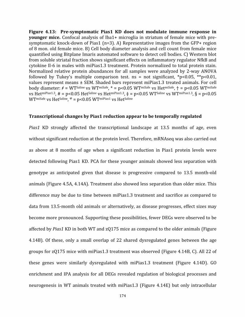
174
Figure 4.13: Pre-symptomatic Pias1 KD does not modulate immune response in younger mice. Confocal analysis of Iba1+ microglia in striatum of female mice with pre-symptomatic knock-down of Pias1 (n=3). A) Representative images from the GFP+ region of 8 mon. old female mice. B) Cell body diameter analysis and cell count from female mice quantified using Bitplane Imaris automated software to detect cell bodies. C) Western blot from soluble striatal fraction shows significant effects on inflammatory regulator NfkB and cytokine Il-6 in males with miPias1.3 treatment. Protein normalized to total protein stain. Normalized relative protein abundances for all samples were analyzed by 2-way ANOVA followed by Tukey’s multiple comparison test. ns = not significant, *p<0.05, **p<0.01, values represent means ± SEM. Shaded bars represent miPias1.3 treated animals. For cell body diameter: ҂ = WTSaline vs WTmiSafe, * = p<0.05 WTmiSafe vs HetmiSafe, † = p<0.05 WTmiSafe vs HetmiPias1.3, # = p<0.05 HetSaline vs HetmiPias1.3, ‡ = p<0.05 WTSaline vs WTmiPias1.3, § = p<0.05 WTmiSafe vs HetSaline, ᵠ = p<0.05 WTmiPias1 vs HetSaline
Transcriptional changes by Pias1 reduction appear to be temporally regulated
Pias1 KD strongly affected the transcriptional landscape at 13.5 months of age, even
without significant reduction at the protein level. Therefore, mRNAseq was also carried out
as above at 8 months of age when a significant reduction in Pias1 protein levels were
detected following Pias1 KD. PCA for these younger animals showed less separation with
genotype as anticipated given that disease is progressive compared to 13.5 month-old
animals (Figure 4.5A, 4.14A). Treatment also showed less separation than older mice. This
difference may be due to time between miPias1.3 treatment and sacrifice as compared to
data from 13.5-month old animals or alternatively, as disease progresses, effect sizes may
become more pronounced. Supporting these possibilities, fewer DEGs were observed to be
affected by Pias1 KD in both WT and zQ175 mice as compared to the older animals (Figure
4.14B). Of these, only a small overlap of 22 shared dysregulated genes between the age
groups for zQ175 mice with miPias1.3 treatment was observed (Figure 4.14B, C). All 22 of
these genes were similarly dysregulated with miPias1.3 treatment (Figure 4.14D). GO
enrichment and IPA analysis for all DEGs revealed regulation of biological processes and
neurogenesis in WT animals treated with miPias1.3 (Figure 4.14E) but only intracellular

175
signal transduction as a process significantly impacted by Pias1 reduction in zQ175 treated
animals (Figure 4.14G). Interestingly, IPA upstream analysis in WT treated animals showed
an enrichment for similar regulators compared to older zQ175 animals treated with
miPias1.3 such at HTT, CREB1, L-dopa, SNCA, and BDNF (Figure 4.5E, Figure 4.14F). While
some of these regulators were also enrichened in 8-month old zQ175 mice with treatment
(e.g. CREB1, BDNF), there was less overlap compared to 13.5 month-old treated zQ175
animals (Figures 4.5G, 4.14H) suggesting either a different or smaller impact on
transcription with less treatment time or stage of disease progression.

176
Figure 4.14: Pre-symptomatic Pias1 KD has minimal effect on transcriptional landscape at 8 months of age. A) PCA of normalized gene expression values shows separation of WT treated animals. B) 8 month-old zQ175 mice with miPias1.3 treatment have fewer measured DEGs compared to 13.5 month-old pre-symptomatic treated animals with C) minimal overlap of 22 DEGs observed between cohorts. D) Fold change heatmap of 22 shared DEGs between age groups shows similar modulation with miPias1.3 treatment. E) GO enrichment analysis of WT miPias1.3 vs miSafe treated animals shows modulation of biological processes including localization and development-related processes and F) corresponding IPA upstream analysis suggests modulation of neuronal and disease-related regulators with miPias1.3 treatment at this age in WT animals. G) GO enrichment analysis of Het miPias1.3 vs miSafe treated animals shows modulation of only intracellular signal transduction with corresponding H) IPA upstream analysis. H) GO enrichment analysis of rescued, genotype-associated DEGs reveals enrichment for intracellular signaling and mRNA processing only with J) corresponding IPA upstream analysis. K) Fold change heatmap indicating normalizing effect of miPias1.3 treatment on genotype-associated DEGs. Sequencing completed by UCI GHTF, analysis assisted by Ryan Lim, PhD, and Jie Wu, PhD, Thompson Lab.
Only 175 genes were dysregulated in zQ175 animals with miPias1.3 treatment at this age.
Of these, 119 were associated with genotype. When analyzing the effect of miPias1.3
treatment on these 119 genotype-specific DEGs, processing of mRNA was also significantly
enriched by GO analysis in the 8 month-old zQ175 animals (Figure 4.14I). Upstream IPA
regulator analysis of genotype-associated DEGs normalized with KD (119), revealed similar
upstream processes to those detected when all DEGs in zQ175 mice with KD were assessed
(175). This suggests that Pias1 may be primarily regulating disease-associated
transcriptional networks at this age (Figure 4.14H, J). The majority of genotype-
dysregulated genes that are altered in expression by miPias1.3 treatment again showed
inverse fold changes suggesting a normalizing effect on aberrant HD-associated
transcription (Figure 4.14K). Together, transcriptional analysis at 8 months of age revealed
a smaller impact of Pias1 KD in both WT and zQ175 mice suggesting that length of KD
treatment or stage of disease greatly influences modulation of transcriptional profiles in

177
these animals, but that Pias1 KD does appear to also have a normalizing effect on
transcriptionally dysregulated genes in HD mice.
Pias1 KD consistently modulates Bcl2l2 expression levels independent of age or
disease stage
As described above, 22 Pias1 KD-dysregulated genes in zQ175 animals overlapped between
age groups (Figure 4.14D). Within these overlapping genes, Rnf112 and Pde4a were of
interest, as these were both dysregulated in male zQ175 mice at 13.5 months of age and
normalized with pre-symptomatic Pias1 KD as determined by mRNAseq (Table 4.3).
Rnf112, also known as Neurolastin, encodes a GTPase that functions in neurogenesis
(Lomash et al., 2015). Pde4a is a SUMO substrate that mediates cAMP signaling cascades by
hydrolyzing cyclic nucleotides. Members of the Pde4 family play a role in mediating Darpp-
32 signaling cascades and memory in the brain (Havekes et al., 2016; Kuroiwa et al., 2012).
A third gene of interest that was modulated at both 8 months of age and 13.5 months of age
in males with pre-symptomatic Pias1 KD, but did not show a normalizing effect, was Bcl2l2.
Instead, pre-symptomatic Pias1 KD consistently resulted in an increase in Bcl2l2
expression, regardless of age assessed (Table 4.3). Bcl2l2, (also known as Bcl-w), is a
member of the Bcl-2 protein family. This family has multiple members that comprise two
self-antagonizing functions: pro-survival and pro-death pathways through maintenance of
mitochondrial dynamics (Aouacheria et al., 2017). Bcl2l2 is a pro-survival member of the
Bcl-2 family that has been implicated in neuronal survival after injury (Courchesne et al.,
2011; Simon et al., 2016). Therefore, due to their potential roles in mediating neuronal
function, development, and survival, transcript levels of gene targets Rnf112, Pde4a, and

178
Bcl2l2 were assessed in 13.5-month old cohorts of animals with either pre-symptomatic or
symptomatic Pias1 KD by qPCR (Table 4.4).
Pre-symptomatic KD, 13.5 months of age: Levels of Rnf112 were significantly reduced in
treated male zQ175 mice, similar to that observed in mRNAseq data (Figure 4.15A).
Interestingly, there was no significant difference in levels of Rnf112 transcript in pre-
symptomatic female mice (Figure 4.15B). For Pde4a, a significant reduction in transcript
levels was again observed in male zQ175 animals only, corresponding with mRNAseq data.
This affect was not observed in females and may be due to sex-related differences in Pde4
isoforms previously reported after neuroinflammatory insult (Johansson et al., 2012).
Bcl2l2 was significantly increased in males by mRNAseq and in females by qPCR (Figure
4.16B) with miPias1.3 treatment. Therefore, for pre-symptomatic KD, only Bcl2l2 was
modulated in both males and females at 13.5 months of age of the genes assessed.
Symptomatic KD, 13.5 month of age: We also evaluated this set of genes in symptomatically
treated mice. No differences were observed for gene targets Rnf112 and Pde4a (Figure
4.16C, D). Levels of Bcl2l2, however, showed a slightly rescued genotype effect in males
(F(2, 9) = 5.951, p<0.05), suggesting an increase in zQ175-treated mice consistent with
other cohorts (Figure 4.15A, B, C). In symptomatic treated females, levels of Bcl2l2
remained statistically unchanged though with a trend similar to that of males (F(2, 8) =
1.199, p>0.05) (Figure 4.15D). Overall, qPCR data suggests that Bcl2l2 is consistently
modulated by Pias1 independent of age and animal gender, potentially supporting a role
for Pias1 in this pro-survival pathway (Figure 4.15, Table 4.4).

179
Figure 4.15: qPCR analysis on top mRNAseq identified genes. A) Fold change and a’) dCT values for male mice validate mRNAseq data. Solid symbols represent animals used for mRNAseq analysis: Rnf112 (Interaction: F(1, 11) = 18.510, p<0.01), Bcl2l2 (Interaction: F(1, 11) = 12.600, p<0.01), Pde4a (Interaction: F(1, 11) = 6.553, p<0.05). B) Fold change and b’) dCT values for female mice show significant treatment effect for Bcl2l2: Rnf112 (Treatment: F(1, 10) = 3.017, p>0.05), Bcl2l2 (Treatment: F(1, 10) = 25.090, p<0.001), Pde4a (Treatment: F(1, 10) = 0.422, p>0.05). C) Fold change and c’) dCT values for symptomatic treated male mice show significant rescue effect on Bcl2l2 levels: Rnf112 (F(2, 9) = 1.561, p>0.05), Bcl2l2 (F(2, 9) = 5.951, p<0.05), Pde4a (F(2, 9) = 3.380, p>0.05). D) Fold change and d’) dCT values for symptomatic-treated zQ175 female mice show no significant differences but with trending increases in Bcl2l2 and Pde4a in Hets with miPias1.3 treatment: Rnf112 (F(2, 8) = 2.097, p>0.05), Bcl2l2 (F(2, 8) = 1.199, p>0.05), Pde4a (: F(2, 8) = 3.460, p>0.05). All samples for pre-symptomatic treated animals were analyzed by 2-way ANOVA followed by Tukey’s multiple comparison test. All samples for symptomatic treated animals were analyzed by 1-way ANOVA followed by Tukey’s multiple comparison test. ns = not significant, *p<0.05, **p<0.01, values represent means ± SEM. Shaded bars represent miPias1.3 treated animals.

180
Pias1 KD rescues disease-associated transcriptional modules including DNA damage
We next compared our zQ175 transcriptional profiles to a previously established
transcriptome from an allelic series derived from zQ175 mice (Langfelder et al., 2016) and
compared Pias1 signatures on defined disease-associated profiles. For this analysis, the
combined data from both the 13.5 and 8-month old pre-symptomatic KD cohorts were
compared to RNAseq data from the zQ175 allelic series. First, we looked at gene expression
changes between zQ175 and WT without treatment; a scatter plot of Z scores calculated
using the log2FC from each dataset showed strong concordance with previously
established transcriptional dysregulation (R=0.72, p<0.0001) supporting a robust
transcriptional phenotype for zQ175 animals between datasets (Figure 4.16A). To assess
the impact of Pias1 KD per a genotype, we employed the same scatterplot and correlation
analysis comparing zQ175 treated animals with WT treated animals. A significant, negative
concordance was observed (R=-0.02, p<0.05), further supporting a disease-specific
function and impact of Pias1 modulation in HD (Figure 4.16B).
To determine the impact of miPias1.3 treatment on previously established, disease gene
networks, our datasets were analyzed against disease correlated co-expression modules
identified in the allelic series (Langfelder et al., 2016). KD of Pias1 significantly rescued
M11, M20, and M39 modules while having a modest but not significant rescue in others
(M2, M25, M34, M1, M10, M43, M46) and slightly but not significantly exacerbating M7 and
M9 modules (Figure 4.16C, Table 4.5). Of interest, significant rescues in both M20 and M39
support a role for Pias1 regulating DNA damage repair pathways through p53 signaling and
ubiquitination pathways respectfully (Galanty et al., 2009). M11 module is glial and

181
immune response related, further implicating Pias1 KD in neuroinflammatory responses.
The remaining co-expression modules that are modestly rescued by Pias1 KD are enriched
for genes involved in synaptic function and Huntington’s disease associated pathways
(Table 4.5). The slightly exacerbated M52 module confirms the negative regulatory role of
Pias1 in modulating NfkB inflammatory pathways, with KD potentially further activating
this module (Liu et al., 2005). Interestingly, both M7 and M9 exacerbated modules suggests
a worsening of neuronal signaling in response to oxidative stress, synaptic transmission,
and cell death. This was unexpected due to the normalization effect observed in our
previous analysis for synaptic-related functions but may suggest Pias1 is serving to
counterbalance disease-associated signaling insults. Overall, data further supports that KD
of Pias1 impacts neuronal function and synaptic components, as well as inflammatory
regulation. Our data for genotype effects highly correlates with previously published zQ175
transcriptional data and the analysis revealed a significant impact on disease-associated
DNA damage repair pathways with Pias1 modulation in HD.
Pias1 modulates Pnkp enzymatic activity for effective DNA repair at 8 months of age
In Chapter 3, we showed that PIAS1 can serve as an E3 ligase for PNKP and that genetic
reduction of PIAS1 in human iPSCs differentiated into medium spiny neurons rescued
mHTT-perturbed enzymatic activity of PNKP. That data provided new evidence for PIAS1
as a SUMO E3 ligase that selectively modulates DNA damage repair pathways in neurons.
Given those results and transcriptomic data above showing that Pias1 KD rescues DNA
damage-associated disease modules in HD mice (M20 and M39), we analyzed enzymatic
activity of Pnkp in vivo. Activity was assessed using 32P-labeled 3’-phosphate-containin

182
oligo substrate and Pnkp-containing tissue lysates from GFP+ regions of 8-month old
zQ175 WT and zQ175 females treated with Saline, miSafe, or miPias1.3 pre-
symptomatically. HD animals recapitulated reduced enzymatic activity of Pnkp (Gao et al.,
2019), and excitingly, miPias1.3 treatment rescued this perturbed activity (Figure 4.17A)
similar to that observed in human neurons (Chapter 3). Together in vivo data provides
additional evidence for a role for Pias1 in DNA damage repair pathways in the brain.
Figure 4.16: Pre-symptomatic Pias1 KD rescues disease-associated modules from zQ175 allelic series. A) Disease signatures from our zQ175 data has high concordance with published allelic series (Langfelder et al., 2016). B) Slight anticorrelation is observed in genes changed between WT and Het miPias1.3 treated vs miSafe treated animals; suggesting disease-specific functional impact with modulation of PIAS1. C) Manhattan plot of rescue score from combined mRNAseq data sets shows rescue and exacerbation of disease-associated transcriptional modules. Analysis and figure completed by Peter Langfelder, PhD, Yang lab, UCLA.

183
Pias1 KD affects levels of DNA damage at different ages in zQ175 mice
Given the rescue of Pnkp enzymatic activity in vivo with miPias1.3 treatment and the
significant rescue in DNA damage disease-associated zQ175 transcriptional modules, we
assessed levels of the DNA damage marker γH2AX and Pnkp protein levels by western blot
in all cohorts of zQ175 pre-symptomatic and symptomatic treated animals. In the 8 month-
old animals, a significant interaction was detected in levels of γH2AX, suggesting a disease-
dependent functional consequence of modulating Pias1 (Interaction: F(1, 10) = 26.940,
p<0.001). Specifically, levels were increased at baseline in zQ175 control mice and
significantly decreased with miPias1.3 treatment. For WT animals, a potential increase in
levels of γH2AX was detected after KD (Figure 4.17B). Pnkp was detected in the Soluble
fraction only and was unchanged (Genotype: F(1, 9) = 0.040, p>0.05; Treatment: F(1, 9) =
0.970, p>0.05, Figure 4.17B).
In 13.5-month old mice with pre-symptomatic miPias1.3 treatment, no significant
treatment effects in levels of γH2AX were detected in either male (Treatment: F(1, 8) =
2.843, p>0.05) or female (Treatment: F(1, 10) = 1.675, p>0.05) animals (Figure 4.18C, D).
No genotype effects were observed (Males: F(1, 8) = 1.379, p>0.05; Females: F(1, 10) =
1.379, p>0.05). Levels of Pnkp were unchanged in male mice (Genotype: F(1, 8) = 0.001,
p>0.05; Treatment: F(1, 8) = 0.552, p>0.05) but showed a significant interaction in female
mice (Interaction: F(1, 9) = 8.001, p<0.05, Figure 4.17D). However, post-hoc analysis failed
to reveal source of this interaction.
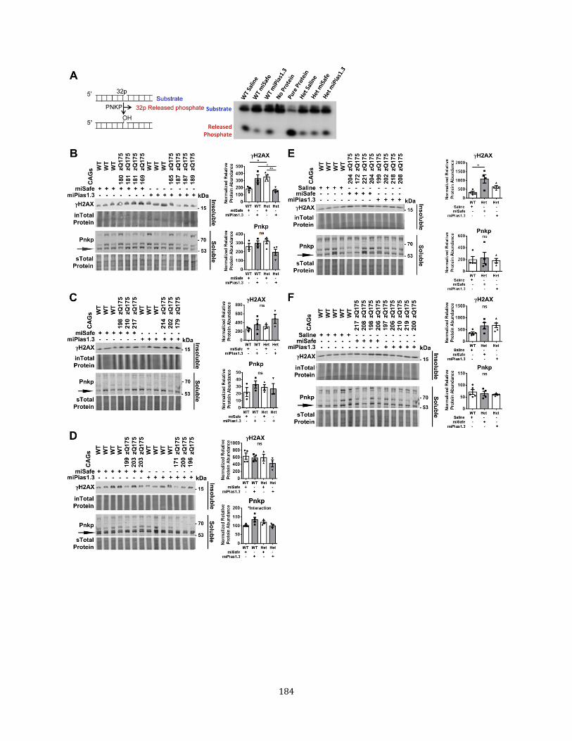
184

185
Figure 4.17: Pias1KD effects DNA damage repair in zQ175 mice. A) mHTT-perturbed enzymatic activity of repair enzyme PNKP in the striatum is rescued with Pias1 KD. Western blot analysis of Soluble/Insoluble fractionation of striatal tissue from B) 8 month-old male mice with pre-symptomatic miPias1.3 treatment. C) 13.5 month-old male mice with pre-symptomatic treatment. D) 13.5 month-old female mice with pre-symptomatic treatment. E) 13.5 month-old male mice with symptomatic treatment. F) 13.5 month-old female mice with symptomatic treatment. Black arrows indicated quantified Pnkp protein band which corresponds to unmodified positive control of overexpressed Pnkp (not shown). Higher bands are suggested to be post-translational modifications of Pnkp as observed in Chapter 3. Protein normalized to total protein stain. Normalized relative protein abundances for all samples for pre-symptomatic treated animals were analyzed by 2-way ANOVA followed by Tukey’s multiple comparison test. All samples for symptomatic treated animals were analyzed by 1-way ANOVA followed by Tukey’s multiple comparison test. ns = not significant, *p<0.05, **p<0.01, values represent means ± SEM. Shaded bars represent miPias1.3 treated animals. Panel A data and figure generated in collaboration with Sarkar lab, UTMB.
In 13.5-month old mice with symptomatic miPias1.3 treatment, more γH2AX was observed
in zQ175 males at 13.5 months of age (F(2, 9) = 6.742, p<0.05) with levels of damage in
miPias1.3 treated zQ175 mice being statistically insignificant from control WT animals,
suggesting a rescue in accrued DNA damage (Figure 4.17E). This affect was not observed in
female mice (F(2, 11) = 3.849, p>0.05, Figure 4.17F). Levels of Pnkp remained unchanged
in this cohort (Males: F(2, 9) = 0.119, p>0.05; Females: F(2, 11) = 0.490, p>0.05).
Symptomatic miPias1.3 treatment in zQ175 mice may therefore rescue accrued DNA strand
breaks in males only. Further assessment of DNA damage repair pathways remains to be
assessed as perturbation of PNKP enzymatic activity results in hyperactivation of ATM-
mediated signaling (Gao et al., 2019). Reducing Pias1 in these animals did not impact the
level of detectable, unmodified Pnkp protein.

186
DISCUSSION
Here we investigated the molecular impact of Pias1 reduction in the striatum of the knock-
in zQ175 mouse model of HD to address the temporal contribution of Pias1 towards HD
pathogenesis in different disease stages and in the context of full-length mHTT. Further, we
investigated the molecular contribution of Pias1 to DNA damage repair in vivo to compare
to our data obtained in human neurons (Chapter 3). We now show that Pias1 KD rescues
mHTT-associated perturbation of Pnkp enzymatic activity in two models of HD: Human
patient, iPSC-derived medium spiny neurons and a full-length KI mouse model (Figure 3.X,
Figure 4.18) and normalizes aberrant genotype-associated transcriptional profiles in vivo.
Since impaired Pnkp activity previously corresponded to decreased genomic integrity in
HD neurons (Gao et al., 2019), observed normalization effect may be due to increased
stability of normalized genes. Levels of Pnkp remained unchanged with KD however,
suggesting that SUMOylation may not mediate clearance. Pias1 may therefore either be
modulating subcellular localization or recruitment of Pnkp through SUMOylation or
SUMOylation may prevent activation without directly influencing clearance of this repair
factor (Chapter 3). While the functional contribution of PNKP SUMOylation remains to be
elucidated, Pias1 KD also rescued DNA damage related transcriptional modules associated
with disease. Together this data suggests that Pias1 is modulating a conserved DDR
mechanism in response to transcription-linked genomic instability in the brain.
Pias1 serves as a negative inhibitor of NfkB signaling (Liu et al., 2005). In R6/2 mice, Pias1
KD restored perturbed localization of NfkB and normalized inflammatory responses
(Ochaba et al., 2016). In our current study, Pias1 modestly impacted inflammatory

187
readouts in older zQ175 mice. With pre-symptomatic treatment, Pias1 KD may have in part
restored resting state of microglia in male mice with a corresponding increase in soluble
NfkB. With symptomatic treatment, an opposite effect was observed for microglial
activation, potentially suggesting a shift in inflammatory balance during symptomatic
disease stages that is exacerbated with Pias1 KD. However, baseline inflammatory profiles
may have influenced the impact of Pias1 KD in zQ175 mice. A key biochemical difference
observed between zQ175 mice and R6/2 mice was the lack of a HD-associated increase in
insoluble/nuclear Pias1 in zQ175 mouse striatum. In R6/2 mice, Pias1 was significantly
increased in the Insoluble fraction and miPias1.3 treatment specifically reduced insoluble
Pias1 protein levels only (Ochaba et al., 2016). Reduction in this species specifically
corresponded to normalization of inflammatory profiles and was accompanied by a
decrease in insoluble accumulated HMW mHTT. No increased insoluble Pias1 was observed
in zQ175 mice compared to WT controls in any group assessed (Figures 4.4, 4.9, 4.13).
Additionally, no insoluble NfkB was detected (data not shown). Therefore, it is possible that
zQ175 mice display a differential baseline of Pias1 activity/function based on localization
as compared to the R6/2 mouse model. This difference may be why levels of insoluble
Pias1 remained unchanged in zQ175 mice with KD and in part, might account for the lack of
impact observed on behavior and HMW mHTT accumulation. Further, this difference might
have potentially influenced the modest impact on inflammatory readouts in this study.
Future studies will investigate the functional contributions of insoluble versus soluble
Pias1 on molecular pathways, particularly inflammatory networks.

188
Modulating Pias1 in pre-symptomatic zQ175 mice strongly impacted synaptic networks at
13.5 months of age at the transcriptional level. Synaptic abnormalities in HD patients have
been well characterized, with changes often preceding neuronal degeneration and likely
accounting for some of the earliest, cognitive and psychiatry symptoms observed during
disease pathogenesis (Nithianantharajah and Hannan, 2013). As a result of an imbalance
between excitatory and inhibitory neurotransmission, HD mice have alterations in number
of GABAergic, glutamatergic, and dopaminergic receptors (Nithianantharajah and Hannan,
2013). zQ175 mice also have altered neurotransmission and synaptic vesicle release (Chen
et al., 2018; Heikkinen et al., 2012). This may be due to a loss of Htt function as conditional
cortical knock-out of Htt impairs synaptic formation and maturation (McKinstry et al.,
2014). Further, Htt is localized at post-synaptic spines, interacts with synaptic-associated
proteins, and functions in vesicular trafficking of Bdnf (Gauthier et al., 2004;
Nithianantharajah and Hannan, 2013). Indeed, loss of Bdnf decreases trophic support
necessary for activity-dependent dendritic spine strengthening thereby affecting synaptic
plasticity (Bramham and Messaoudi, 2005; Jia et al., 2010), with exogenous Bdnf providing
neuroprotective affects in HD animals (Lynch et al., 2007). In the current study, levels of
Bdnf transcript were elevated in control zQ175 mice compared to control WT animals at
13.5 months of age. This is consistent with the zQ175 allelic series transcriptional profile
(Langfelder et al., 2016) and may represent a compensatory overexpression for decreased
trophic support in HD animals relatively early in disease. With Pias1 KD, levels of Bdnf
transcript decreased, suggesting a normalizing impact on aberrant transcription.

189
Transcription of Bdnf was not the only normalizing affect observed for Pias1 KD in our
mRNAseq analysis. Looking further, biological processes enriched from GO analysis such as
regulation of trans-synaptic signaling and modulation of chemical synaptic transmission
were genotype-specific networks modulated by Pias1 KD. Specifically, the DEGs that were
contributing to these processes in our data set showed a majority with inverse fold changes
with Pias1 KD compared to control zQ175 mice, suggesting that miPias1.3 treatment
normalized aberrant transcription associated with disease progression in these animals
(Figure 4.5). We hypothesize this may be in part due to facilitated repair of key neuronal
genes. Therefore, increasing activity of Pnkp and thereby DNA repair with Pias1 KD may
have a direct impact on genomic stability in zQ175 mice. Increased genomic integrity
would then facilitate neuronal health and promote synaptogenesis or strengthen existing
synapses. Direct measurements of genomic integrity of normalized genes will allow testing
of this hypothesis. Additionally, assessing stability of the HTT CAG-repeat expansion in
zQ175 mice will determine if modulating Pias1 and subsequently TCR impacts expansion
propensity.
An alternative mechanism is that SUMOylation of transcription factors for synaptic genes
may be modulated by Pias1 KD. Pias1 serves as an E3 ligase for neuronal transcription
factors ((Estruch et al., 2016; Gregoire and Yang, 2005; Riquelme et al., 2006; Tai et al.,
2016), Table 2) including CREB whose activity is impaired by the mHTT containing TCR
complex (Chen et al., 2014; Gao et al., 2019). Indeed, CREB was consistently regulated with
Pias1 KD at both 8 and 13.5 months of age as determined by IPA upstream analysis (Figure
4.5, Figure 4.15), supporting this possibility. Mef2a is also SUMOylated by PIAS1, with
190
SUMOylation serving to repress transcription of genes such as synaptotagmin1 (Syt1)
which itself is SUMOylated (Gregoire and Yang, 2005; Matsuzaki et al., 2015; Riquelme et
al., 2006; Yamada et al., 2013). SUMOylated Mef2a increases the number of PSD95-positive
dendritic claws while also suppressing orphaned presynaptic sites, suggesting that
SUMOylated Mef2a contributes to synapse maturation during development (Shalizi et al.,
2006; Yamada et al., 2013). Interestingly, levels of Syt1 in our mRNAseq data were
decreased with Pias1 KD in zQ175 mice at 13.5 months of age, opposite to what would
have been expected if there was a decrease in Mef2a SUMOylation by Pias1 (Yamada et al.,
2013). No change in Mef2a transcription was observed in with Pias1 KD in any group,
indicating we may not have affected expression as opposed to function or localization
mediated through SUMOylation of transcription factors. Further, some transcription factors
may be uncharacterized SUMO substrates. Therefore, it remains possible that Pias1 KD is
mediating synaptic function in zQ175 striatum by directly affecting SUMOylation of
transcription factors required to facilitate production of protein products needed for
synaptic maturation. Directly targeting SUMOylation of these transcription factors may
account in part for the observed impact on transcriptional networks associated with
synaptic function (Figure 4.5, Figure 4.15). Analysis of transcription factors associated with
synaptic transcription will need to be assessed at the protein level to determine if this
mechanism is contributing to the observed transcriptional impact of Pias1 KD on synaptic
networks.
Finally, Pias1 may also directly modulate SUMOylation of proteins involved in synaptic
development, function, and health. SUMOylation can modulate synaptic activity by

191
dynamically mediating protein subcellular distribution and function (see (Schorova and
Martin, 2016) and (Henley et al., 2018) for comprehensive reviews). Several synaptic
proteins themselves are SUMOylated which often associates with repressive functional
consequences and therefore has been attributed to a potential neuroprotective effect to
prevent excitotoxic events in response to neuronal injury ((Peters et al., 2017), Table 3).
This highlights the importance of maintaining SUMO-related proteostatic balance to ensure
cellular survival. Supporting this, overexpression of SUMO1 in the mouse brain leads to
impairments in learning and memory associated with reduction in synaptic transmission
and presynaptic function (Matsuzaki et al., 2015). Though the direct mechanism is unclear,
imbalance in the SUMO landscape in the brain leads to neuronal malfunction and suggests a
potential direct impact on synaptic health with reduction of an E3 ligase promoting
SUMOylation. Signaling feedback may then result in observed transcriptional changes.
Whether or not Pias1 directly modulates localization or SUMOylation of these proteins
remains to be investigated. Overall, KD of Pias1 significantly impacted synaptic and
neuronal gene networks, highlighting a potential modulatory function of Pias1 towards
mediating HD-associated synaptic malfunction (Nithianantharajah and Hannan, 2013).
The most consistently regulated gene assessed with Pias1 KD in zQ175 mice was Bcl2l2,
also known as Bcl-w, which could suggest that Pias1 may mediate apoptotic and survival
pathways in the brain. Bcl2l2 is a member of the Bcl-2 family of genes that serve as fate
determinants by having dual, antagonizing functions (Youle and Strasser, 2008). Some
family members such as Bax and Puma are pro-apoptotic while others including Bcl-2,
Bcl2l2, and Bcl-xL have anti-apoptotic function. These family members antagonize each
192
other either directly or indirectly with the pro-death members preventing Bax-mediated
cytochrome-c release from the mitochondria (Wei et al., 2001; Yang et al., 1997; Youle and
Strasser, 2008). If the balance of pro and anti-apoptotic family members shifts towards the
former, Bax is free to bind to the mitochondrial outer membrane and trigger caspase-
mediated apoptosis (Dewson and Kluck, 2009; Jurgensmeier et al., 1998). Due to this
intricate balance of pro-death and pro-survival signals, the ratio of Bax-to-Bcl-2 is often
used as a metric for evaluating apoptosis potential (Kilbride and Prehn, 2013; Korsmeyer,
1999; Oltvai et al., 1993). Together, these family members in part dictate the fate of
neuronal development, connections, and survival (Akhtar et al., 2004). Future studies into
the balance of Bax and Bcl2l2 proteins in zQ175 mice with Pias1 KD would further test this
hypothesis.
It is exciting that of the genes assessed, Bcl2l2 specifically was elevated with miPias1.3
treatment in zQ175 mice since the presence of Bcl2l2 in the axons of sensory neurons is
critical for their survival under trophic deprivation (Courchesne et al., 2011; Simon et al.,
2016). This balances the observation that Bax mediates axonal degradation (Nikolaev et al.,
2009) as well as neuronal death during development (Deckwerth et al., 1996) with
decreases in trophic support. Indeed, there are neuroprotective roles for the pro-survival
side of the Bcl-2 family in nervous system development and brain or spinal cord after
injury, including loss of neurotrophic support (i.e. Bdnf, Ngf, etc., (Anilkumar and Prehn,
2014)). BDNF deficiency is a hallmark of HD (Ferrer et al., 2000; Zuccato and Cattaneo,
2009). Further, dysregulation of the Bcl-2 family has been well documented in numerous
models of HD (Sassone et al., 2013). Therefore, inducing expression of pro-survival Bcl2l2

193
with Pias1 KD may counterbalance the decreased trophic support seen within the HD
striatum, ultimately promoting neuronal survival and strengthening sensory axons.
The observed differences between pre-symptomatic and symptomatic KD of Pias1 in this
study suggest that Pias1 may have different functional contributions towards HD
pathogenesis dependent on disease stage. The characterized embryonic lethality of Pias1
knock-out mice highlights an important role in development, particularly in angiogenesis
(Constanzo et al., 2016), indicating that targeting Pias1 at different ages may have specific
functional outcomes. Our data suggests that Pias1 may contribute to compensatory
mechanisms attempting to maintain neuronal homeostasis under genotoxic stress in
neurodegenerative disease (Vanni et al., 2019). Overall, we saw significant Pias1 KD at all
stages tested at the transcript level. For symptomatic-treated animals, we only observed
significant Pias1 KD at the protein level in the soluble fraction of female striatal lysates. At
13.5 months of age, no reduction in Pias1 protein levels were observed for pre-
symptomatic treated animals, suggesting a compensatory proteostatic mechanism to
combat the reduced production of Pias1 transcript. These findings suggest that an optimal
window for targeting Pias1 mediated mechanisms may be at earlier disease stages. Even
with no observed changes in Pias1 protein at 13.5 months of age, reduced level of Pias1
transcript was observed along with robust transcriptional changes in both WT and zQ175
mice. Seeing that significant reduction in Pias1 protein was observed at our earlier time-
point, data suggests a long-term, rippling effect of Pias1 KD in these animals during aging
which could be considered for future therapeutic studies.

194
SUMMARY AND NEXT STEPS
Intrastriatal viral KD of Pias1 in the long term, knock-in zQ175 mouse model of HD
restored Pnkp enzymatic activity and normalized disease-associated transcriptional
dysregulation in these animals. Overall, Pias1 KD had little to no effect on behavioral
readouts in zQ175 animals. Pre-symptomatic KD specifically had the strongest effect on
molecular readouts, suggesting that targeting Pias1 in this disease stage could aid in
restoring homeostatic balance. Unexpectedly, Pias1 KD had little effect on accumulation of
HMW mHTT suggesting that Pias1 and SUMOylation may not be mediating solubility or
aggregation propensity of mHTT in this disease context, potentially due to lack of impact on
levels of insoluble Pias1 with KD. The robust and cross-model impact on Pnkp enzymatic
activity, however, provides evidence that Pias1 may be mediating DDR in vivo. Future
experiments to elucidate the exact modulatory mechanism of Pias1 on synaptic structure
and function remain to be completed. Such experiments would include high resolution
microscopy to visualize synaptic structures with Pias1 KD as well as proteomic analysis of
SUMO-containing synaptosomes to assess potential direct mechanisms. Further, assessing
levels and chromatin binding of SUMO-targeted transcription factors that mediate synaptic
transcription would identify indirect mechanisms for Pias1 mediating synaptic structure
and function. Finally, assessing the functional impact of modulating pro-survival gene
Bcl2l2, such as assessing caspase activation or the balance of Bcl-2 protein family members,
may further elucidate how Pias1 KD could be neuroprotective and a viable therapeutic
target for treatment of HD.

195
EXPERIMENTAL PROCEDURES
Animals
Knock-in, zQ175 mice were obtained from CHDI colony, bred in house, genotyped, and aged
to approximately 13.5 months of age. CAG repeat sizing of genomic DNA harvested from
tails was performed by Laragen. Experiments were carried out in strict accordance with
the Guide for the Care and Use of Laboratory Animals of the NIH and an approved animal
research protocol by the Institutional Animal Care and Use Committee (IACUC) at the
University of California, Irvine. Animals were humanely euthanized by an over injection of
Euthasol followed by whole body perfusion with 1x-PBS and cervical dislocation.
Surgery
At 2.5 months of age and 7.5 months of age, zQ175 mice were anesthetized using isoflurane
and underwent bilateral intrastriatal injections using a dual injection stereotaxic apparatus
(coordinates 0.01 mm caudal to bregma, 0.2 mm right/left of midline, 0.345 pocket to
0.325 mm ventral to pial surface) as previous (Ochaba et al., 2016). 5 µl of AAV2/1 virus
containing miPIAS1.3 CMVeGFP or miSAFE CMVeGFP at ~ 3e12 vg/ml was injected using a
Hamilton syringe at a rate of 0.5 µl/min. Syringe was left in place for 5 minutes after each
injection to ensure complete ejection of viral solution into brain region. Animals were
allowed to recover for 1 week under observation prior to beginning behavioral analysis.
Behavioral Paradigms
For zQ175 PIAS1 pre-symptomatic knock-down cohorts, animal body weights were
measured weekly over the course of the studies. Motor deficits were assessed by a running

196
wheel (0297-0521-D60; Columbus Instruments) at 7 or 9 months of age months of age
depending on date of sacrifice. Running wheel usage was constantly monitored over two
weeks and collected in three minute bins using Multi-Device System software 24-Channel
Version 1.55 (160640; Columbus instruments). Data from dark phase only was analyzed
for significance over the two week period. Motor learning was calculated as previously
described (Hickey et al., 2008). Briefly, slopes were calculated using formula (Y2-Y1)/(X2-
X1) where Y2 is wheel usage per bin on night 10 and X2 is 10 (night 10), and Y1 is wheel
usage per bin on night 2 with X1 as 2.
Rotarod was also used to assess motor deficits in these mice using an accelerating
apparatus (Dual Species Economex Rota-Rod; 0207-003M; Columbus Instruments) at 9.5
and 12.5 months of age under blinded conditions. Animals were tested over two days with
four trial runs each day consisting of 5 minute trial periods. The first trial for each test day
was used as a training trial and not used in final calculations. The latency to fall for the
three final trials on day 2 of testing were averaged and analyzed for significance.
To assess impairments in anxiety, and as an additional motor task, animals were subjected
to open field test at 7.5 months of age and 12.5 months of age when applicable. Mice were
allowed to explore an empty open field box for 5 minutes. Activity during these five
minutes was recorded and analyzed automatically using Ethovision™ Software by
determining time spent in the center and on the periphery of the open field in addition to
average velocity and distance traveled over the 5 minute test period. Velocity and distance

197
traveled during the open field trials were used as measures of motor performance while
percent time spent in center was used as a measure of anxiety.
Immunofluorescence protocol
Brain tissue samples used for immunofluorescence were from animals humanely sacrificed
as described above. 40µm coronal brain slices from flash frozen, previously preserved
hemispheres were washed three times in 1xPBS and then blocked in 5% goat serum with
0.2% Triton-X blocking solution for 1 hour at room temperature. After blocking, slices were
incubated in primary antibody in blocking solution overnight while rocking at 4°C. The
following day, slices were washed 3 times in 1xPBS followed by incubation in secondary
antibody in blocking solution for 1 hour at room temperature. Slices were washed a final 3
times with 1x PBS prior to mounting on slides using fluoromount mounting media and
imaged using confocal microscopy. The following antibodies were used: anti-Iba1 (Wako
Pure Chemicals Industries 27030) and DAPI was used as a nuclear stain. To measure
microglia number and cell body diameter, Imaris Bitplane analysis software was run using
region-growing spot detection analysis on Iba1+ cells.
Tissue and cell protein lysis, quantification, and Western Blot analysis
Flash frozen brain tissue was prepared as previous (Ochaba et al., 2016; Ochaba et al.,
2018). Protein fractions were quantified using detergent compatible (DC) protein assay
(Bio-Rad). Soluble protein lysates were resolved on 4-12% Bis-Tris Poly-Acrylamide gels
and transferred onto either 0.45µM or 0.2µM nitrocellulose membrane depending on size
of proteins of interest (smaller proteins on 0.2µM). Insoluble protein lysates were resolved

198
on 3-8% Tris-Acetate Poly-Acrylamide gels and transferred onto 0.45µM nitrocellulose
membrane. Membranes were blocked in Odessey starting block (Li-Cor) for 1 hour at room
temperature and probed in primary antibody overnight at 4°C in blocking supplemented to
0.5% tween. Secondary IR-fluorophore antibodies were used and detected using a Li-Cor
Odyssey CLx system. IR fluorescence imaging was used for quantitative analysis with
detected protein levels normalized to Revert Li-Cor whole-protein stain prior to statistical
analysis. Antibodies used for analysis were anti-PIAS1 (Cell Signaling 3550S), anti-HTT
(Millipore MAB5492, HMW mHTT), anti-HTT (Cell Signaling 5656S, clone D7F7 for FL-
HTT), anti-GFP (Living Colors, Clontech Laboratories 632381), anti-NfkB (Cell Signaling
8242S), anti-Il-6 (Mouse specific, Cell Signaling 12912T), anti-PNKP (Novus Biologicals
NBP1-87257), and anti-γH2AX (Millipore 05-636).
RNA purification, qPCR, mRNASeq
GFP+ flash frozen brain regions were homogenized in TRIzol reagent (Invitrogen) and RNA
was extracted according to manufacturer’s protocol and purified using RNEasy Mini kit
(QIAGEN). Residual DNA was removed by DNase treatment incorporated into RNEasy
protocol as per manufacturer’s suggestion. RNA was submitted for mRNAseq as described
previously (Vashishtha et al., 2013). Differentially expressed genes were generated using
DEseq2 (Love et al., 2014) and a significance threshold was set at a 10% FDR. Enrichment
analysis was completed using GOrilla (Eden et al., 2009) and Ingenuity Pathway Analysis
software. To confirm differentially regulated genes identified in mRNAseq studies, reverse
transcription was performed using SuperScript 3 First-strand synthesis system according
to manufacturer’s protocol (Invitrogen) from harvested and purified RNA. Both oligo (dT)

199
and random hexamer primers were used in a 1:1 ratio. Final synthesized cDNA was diluted
to 5ng/ul in DEPC treated water and stored at -20°C until use. Primers used for qPCR were:
Eif4a2 (F: GTGGACTGGCTCACGGAGAAAA, R: AGAACACGGCTTGACCCTGATC),
Pias1 (F: CTGCACAGACTGTGACGAGATAC, R: CGCTACCTGATGCTCCAATGTG),
NeuroD2 (F: GCTACTCCAAGACGCAGAAGCT, R: CACAGAGTCTGCACGTAGGACA),
NeuroD1 (F: CCTTGCTACTCCAAGACCCAGA, R: TTGCAGAGCGTCTGTACGAAGG),
Bdnf (F: TCGTTCCTTTCGAGTTAGCC, R: TTGGTAAACGGCACAAAAC),
Pde4a (F: CCGTGTTCACAGACCTGGAGAT, R: GGTGGTTCTCAAGCACAGACTC),
Syn1 (F: TATGCCACTGCTGAGCCCTTCA, R: ATGGCAATCTGCTCAAGCATAGC),
Syn2 (F: CCTGCTCTGAAATGTTTGGTGGC, R: TCTGTCCTCCACTTGGTGTTCC),
Rnf112 (F: AAGTGGAAGCCGCCAAGAAGGA, R: AGGAGGTTCCTCATCGTGTCAG),
Bcl2l2 (F: CAAGTGCAGGATTGGATGGTGG, R: CTGTCCTCACTGATGCCCAGTT).
Pnkp enzymatic activity
3’-phosphatase PNKP enzymatic activity from nuclear and mitochondrial extracts was
performed as previously described (Chakraborty et al., 2015; Gao et al., 2019). A 3’-
phosphate oligo (51-mer), 32P-labeled, containing a single strand break was used to assess
activity by measuring the amount of released 3’ phosphate from radio-labeled substrate as
analyzed by 20% Urea-PAGE and PhosphorImager. The percent of phosphate release was
represented as compared to total radiolabeled substrate (as 100).
200
Statistical Analysis
All data represented as mean ± SEM with a p value of p<0.05 considered statistically
significant. Analyses were completed in GraphPad Prism software. IR-fluorescent intensity
values were obtained from western blots using Li-Cor imaging software, Image Studio and
normalized to total protein stain, Revert. Normalized protein abundance levels were the
subjected to 2-way ANOVA to assess changes in protein levels with the exception of HMW
mHTT analysis which was with Student’s unpaired 2-tailed t-test. Statistical analysis was
completed of the delta CT (dCT) values obtained from qPCR reactions normalized to Eif4a2.
All 2-way ANOVAs were followed by Tukey’s multiple comparisons test.

201
CHAPTER 4
TABLES

202
Table 4.1: Statistical outputs for zQ175 pre-symptomatic Pias1 KD cohorts.
Age at Sac
Behavioral task
n (m,f,c) Genotype effect Treatment effect Interaction
13.5
Open Field 7.5 Mon. (Distance Traveled)
7-8, 5-9, 12-17
M: F(1, 26) = 6.420, p<0.05 F: F(1, 23) = 7.911, p<0.01
C: F(1, 51) = 13.080, p<0.001
M: F(1, 26) = 2.845, p>0.05 F: F(1, 23) = 2.814, p>0.05 C: F(1, 51) = 0.898, p>0.05
M: F(1, 26) = 0.012, p>0.05 F: F(1, 23) = 0.012, p>0.05
C: F(1, 51) = 0.0651, p>0.05
13.5 Open Field
7.5 Mon. (Velocity)
7-8, 5-9, 12-17
M: F(1, 26) = 6.420, p<0.05 F: F(1, 23) = 7.911, p<0.01
C: F(1, 53) = 11.900, p<0.01
M: F(1, 26) = 2.845, p>0.05 F: F(1, 23) = 2.814, p>0.05 C: F(1, 53) = 0.253, p>0.05
M: F(1, 26) = 0.012, p>0.05 F: F(1, 23) = 0.012, p>0.05 C: F (1, 53) = 0.013, p>0.05
13.5
Open Field 7.5 Mon. (Center
time)
7-8, 5-9, 12-17
M: F(1, 26) = 0.099, p>0.05 F: F(1, 23) = 0.204, p>0.05 C: F(1, 53) = 0.526, p>0.05
M: F(1, 26) = 1.039, p>0.05 F: F(1, 23) = 0.033, p>0.05 C: F(1, 53) = 0.850, p>0.05
M: F(1, 26) = 1.070, p>0.05 F: F(1, 23) = 0.049, p>0.05 C: F(1, 53) = 0.822, p>0.05
13.5
Open Field 12.5 Mon. (Distance Traveled)
7-8, 5-9, 12-17
M: F(1, 26) = 0.302, p>0.05 F: F(1, 23) = 1.311, p>0.05 C: F(1, 53) = 0.831, p>0.05
M: F(1, 26) = 0.156, p>0.05 F: F(1, 23) = 1.617, p>0.05 C: F(1, 53) = 1.091, p>0.05
M: F(1, 26) = 0.681, p>0.05 F: F(1, 23) = 0.146, p>0.05 C: F(1, 53) = 0.104, p>0.05
13.5 Open Field 12.5 Mon. (Velocity)
7-8, 5-9, 12-17
M: F(1, 26) = 0.302, p>0.05 F: F(1, 23) = 1.311, p>0.05 C: F(1, 53) = 0.831, p>0.05
M: F(1, 26) = 0.156, p>0.05 F: F(1, 23) = 1.617, p>0.05 C: F(1, 53) = 1.091, p>0.05
M: F(1, 26) = 0.681, p>0.05 F: F(1, 23) = 0.146, p>0.05 C: F(1, 53) = 0.104, p>0.05
13.5
Open Field 12.5 Mon.
(Center time)
7-8, 5-9, 12-17
M: F(1, 26) = 0.615, p>0.05 F: F(1, 23) = 3.092, p>0.05 C: F(1, 53) = 1.802, p>0.05
M: F(1, 26) = 0.553, p>0.05 F: F(1, 23) = 0.013, p>0.05 C: F(1, 53) = 0.388, p>0.05
M: F(1, 26) = 0.185, p>0.05 F: F(1, 23) = 0.202, p>0.05 C: F(1, 53) = 0.397, p>0.05
13.5 Rotarod
(9.5 Mon.) 7-8, 5-9,
12-17
M: F(1, 26) = 11.030, p<0.01 F: F(1, 23) = 9.185, p<0.01
C: F(1, 53) = 20.290, p<0.0001
M: F(1, 26) = 0.232, p>0.05 F: F(1, 23) = 0.007, p>0.05 C: F(1, 53) = 0.014, p>0.05
M: F(1, 26) = 0.748, p>0.05 F: F(1, 23) = 0.006, p>0.05 C: F(1, 53) = 0.135, p>0.05
13.5 Rotarod
(12.5 Mon.) 7-8, 5-9,
12-17
M: F(1, 26) = 6.476, p<0.05 F: F(1, 23) = 3.206, p>0.05 C: F(1, 53) = 9.958, p<0.01
M: F(1, 26) = 0.046, p>0.05 F: F(1, 23) = 0.040, p>0.05 C: F(1, 53) = 0.006, p>0.05
M:F(1, 26) = 4.473, p<0.05 F: F(1, 23) = 0.857, p>0.05 C: F(1, 53) = 0.590, p>0.05
13.5 Weight
(13.5 Mon) 7-8, 5-9
M: F(1, 26) = 47.110, p<0.0001
F: F(1, 23) = 7.078, p<0.05
M: F(1, 26) = 0.492, p>0.05 F: F(1, 23) = 2.592, p>0.05
M: F(1, 26) = 0.002, p>0.05 F: F(1, 23) = 0.075, p>0.05
8
Open Field 7.5 Mon. (Distance Traveled)
4, 5-7, 9-11
M: F(1, 12) = 0.655, p>0.05 F: F(1, 26) = 3.010, p>0.05 C: F(1, 34) = 1.750, p>0.05
M: F(1, 12) = 0.102, p>0.05 F: F(2, 26) = 0.638, p>0.05 C: F(1, 34) = 0.019, p>0.5
M: F(1, 12) = 0.062, p>0.05 F: F(2, 26) = 0.495, p>0.05 C: F (1, 34) = 0.099, p>0.05
8 Open Field
7.5 Mon. (Velocity)
4, 5-7, 9-11
M: F(1, 12) = 0.655, p>0.05 F: F(1, 26) = 3.010, p>0.05 C: F(1, 34) = 1.750, p>0.05
M: F(1, 12) = 0.102, p>0.05 F: F(2, 26) = 0.638, p>0.05 C: F(1, 34) = 0.019, p>0.5
M: F(1, 12) = 0.062, p>0.05 F: F(2, 26) = 0.495, p>0.05 C: F(1, 34) = 0.099, p>0.05
8
Open Field 7.5 Mon. (Center
time)
4, 5-7, 9-11
M: F(1, 12) = 7.958, p<0.05 F: F(1, 26) = 7.219, p<0.05
C: F(1, 34) = 13.090, p<0.001
M: F(1, 12) = 0.316, p>0.05 F: F(2, 26) = 0.220, p>0.05 C: F(1, 34) = 0.160, p>0.05
M: F(1, 12) = 6.434, p<0.05 F: F(2, 26) = 0.263, p>0.05 C: F(1, 34) = 5.259, p<0.05
8 Weight (8
Mon) 4, 5-7
M: F(1,12)=0.907, p>0.05 F: F(1,26)=2.143, p>0.05
M: F(1,12)=1.491, p>0.05 F: F(2,26)=0.846, p>0.05
M: F(1, 12) = 0.196, p>0.05 F: F(2, 26) = 1.687, p>0.05
All groups and tasks analyzed by 2-way ANOVA followed by Tukey’s multiple comparison test. p<0.05 considered statistically significant. m=males, f=females, c=combined.
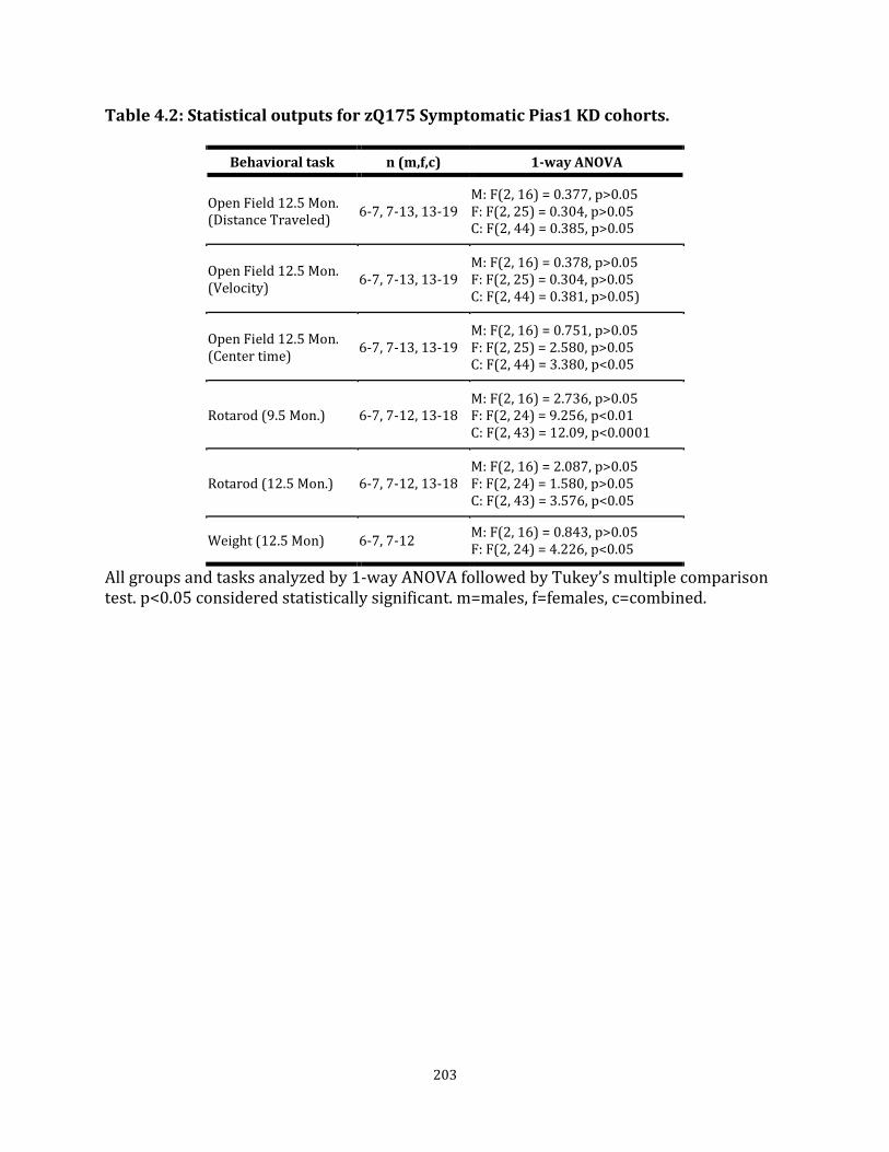
203
Table 4.2: Statistical outputs for zQ175 Symptomatic Pias1 KD cohorts.
Behavioral task n (m,f,c) 1-way ANOVA
Open Field 12.5 Mon. (Distance Traveled)
6-7, 7-13, 13-19 M: F(2, 16) = 0.377, p>0.05 F: F(2, 25) = 0.304, p>0.05 C: F(2, 44) = 0.385, p>0.05
Open Field 12.5 Mon. (Velocity)
6-7, 7-13, 13-19 M: F(2, 16) = 0.378, p>0.05 F: F(2, 25) = 0.304, p>0.05 C: F(2, 44) = 0.381, p>0.05)
Open Field 12.5 Mon. (Center time)
6-7, 7-13, 13-19 M: F(2, 16) = 0.751, p>0.05 F: F(2, 25) = 2.580, p>0.05 C: F(2, 44) = 3.380, p<0.05
Rotarod (9.5 Mon.) 6-7, 7-12, 13-18 M: F(2, 16) = 2.736, p>0.05 F: F(2, 24) = 9.256, p<0.01 C: F(2, 43) = 12.09, p<0.0001
Rotarod (12.5 Mon.) 6-7, 7-12, 13-18 M: F(2, 16) = 2.087, p>0.05 F: F(2, 24) = 1.580, p>0.05 C: F(2, 43) = 3.576, p<0.05
Weight (12.5 Mon) 6-7, 7-12 M: F(2, 16) = 0.843, p>0.05 F: F(2, 24) = 4.226, p<0.05
All groups and tasks analyzed by 1-way ANOVA followed by Tukey’s multiple comparison test. p<0.05 considered statistically significant. m=males, f=females, c=combined.

204
Table 4.3: Top Pias1 KD- modulated DEGs in pre-symptomatic miPias1.3 treated animals used for RT-qPCR
Gene 13.5 months FC Het w/KD
8 Months FC Het w/KD
Pathway/Function
BDNF -2.492* n/a Neurotrophic growth factor
Neurod2 -1.965* n/a Neuronal transcription factor
Neurod1 -1.133* n/a Neuronal transcription factor
Syn1 -0.456* n/a Synaptic vesicle pathway
Syn2 -0.295* n/a Synaptic vesicle pathway
Rnf112 -0.296* -0.629 Neurogenesis, protection against oxidative stress-induced damage
Pde4a -0.281* -0.303 cAMP signaling
Bcl2l2 0.315 0.506 Survival of NGF- and BDNF-dependent neurons
Asterisk indicates normalized genotype DEG with Pias1 KD

205
Table 4.4: Summary heat map of Pias1 KD effect on transcriptional targets in zQ175 heterozygote animals
Gene
8 Mon. zQ175
Pre-Symp miPias1.3
13.5 Mon. zQ175
Pre-Symp miPias1.3
male
13.5 Mon. zQ175
Pre-Symp miPias1.3
female
13.5 Mon. zQ175 Symp
miPias1.3 male
13.5 Mon. zQ175 Symp
miPias1.3 female
Rnf112 Treatment Treatment No Difference No Difference No Difference
Bcl2l2 Treatment Treatment Treatment Genotype No Difference
Pde4a Treatment Treatment No Difference No Difference No Difference
(Orange = elevated, blue = decreased)

206
Table 4.5: disease associated transcriptional modules affected by Pias1 KD in zQ175 mice
Module GO Term(s) IPA Term(s) Enrichment Term(s)
Pias1 KD effect
Significant
M11
Ensheathment of neurons Fatty acid biosynthetic process
Endothelin-1 signaling LPS/IL-1 inhibition of RXR Signaling by Rho family GTPase
Oligodendrocyte Astrocyte probable
rescue Y
M20 Cell division Protocadherin beta Zinc finger, CH2H-like
p53 signaling Brca1 in DNA damage response
Glial marker p53 immune pathway
rescue Y
M39
Protocadherin beta Microtubule cytoskeleton Ubl conjucation pathway
DNA break repair by NHEJ Signaling by Rho family GTPases Protein ubiquitination pathway
Oligodendrocyte rescue Y
M2
Protein kinase ATP binding site GTPase regulator activity Synapse
GPCR signaling cAMP signaling Huntington's disease signaling
Neuron probable Postsynaptic density proteins
rescue N
M25 Leucine-rich repeat Reg. neuron differentiation
Glutamate receptor signaling Gαi signaling Huntington's disease signaling
Autism associated Neuron probable
rescue N
M34
Regulation of transcription Chromatin modification Protocadherin gamma
LPS-stimulated MAPK signaling FGF signaling Huntington's disease signaling
Immune pathway rescue N
M1
Purine nucleotide binding Cellular protein localization Ubl conjugation pathway
Protein ubiquitination pathway tRNA charging Huntington's disease signaling
Neuron Postsynaptic density proteins
rescue N
M10
Ubl conjugation pathway Chaperonin TCP-1 conserved site Zinc finger, CH2H-like
Sonic hedgehog signaling RAN signaling Amyloid processing
Down in Alzheimer's CA1 Nuclear proteins
rescue N
M43 Fatty acid catabolic process
Fatty Acid β-oxidation I LPS/IL-1 Inhibition of RXR 14-3-3 mediated signaling
Postsynaptic density proteins Mitochondria
rescue N
M46 Protocadherin gamma Zinc finger, CH2H-like
IL-22 signaling Glucocorticoid signaling
Nuclear proteins rescue N
M52 Transmembrane region VEGF receptor activity
NF-κB signaling Protein kinase A signaling
JAX disease genes Astrocyte probable
Exacer-bation
N
M7 Cell death Oligodendrocyte cell fate
Death receptor signaling Phospholipase C signaling Axonal guidance signaling
Oligodendrocyte Putamen local marker
Exacer-bation
N
M9
Mitochondrion Protein transport Translation elongation factor
Nrf2 oxidative stress response Serotonin degradation
Postsynaptic density proteins Mitochondria
Exacer-bation
N

207
CHAPTER 5
Restoration of Pnkp Activity by Pias1 Reduction in R6/2 and Potential Relationship to
Somatic Repeat Expansion
SUMMARY OF CHAPTER 5
Pias1 is a SUMO E3 ligase that can modulate molecular and behavioral phenotypes in the
R6/2 transgenic mouse model of Huntington’s disease (HD). PIAS1 can also modulate DNA
damage repair pathways in human patient iPSC-derived medium spiny neurons and in the
striatum of the zQ175 knock-in mouse model of HD (Chapters 3 and 4). Specifically, PIAS1
SUMOylates the DNA repair factor PNKP, an enzyme in the transcription-coupled repair
complex scaffolded by the huntingtin protein. Mutant huntingtin-associated perturbation of
PNKP enzymatic activity was rescued with PIAS1 knock-down in both models and
normalized aberrant transcriptional profiles, suggesting an impact on genomic stability.
However, due to the extreme length of CAG-repeat in zQ175 mice, somatic repeat
expansion could not be assessed in these animals due to technical limitations for resolution
of repeat sizes. Therefore, given the previously reported impact on R6/2 pathogenesis with
Pias1 knock-down and our robust, cross-model modulatory effect on PNKP activity, Pias1
modulation of Pnkp and DNA damage-associated mechanisms in R6/2 mice was assessed.
Pias1 knock-down similarly rescued perturbed Pnkp activity in R6/2 mice, and data shows
a potential reduction of somatic CAG repeat expansion in the striatum. Taken together, data
in this dissertation suggest that Pias1 is an integral component of the transcription-coupled
repair complex and response in multiple models of HD, and may contribute towards
transcriptional dysregulation and somatic repeat expansion within the disease context.

208
INTRODUCTION
The typical age of adult-onset for HD is between 35 and 50 years of age with an inverse
relationship existing between the length of the CAG repeat and age of onset. However, CAG
length only accounts for 40-50% of the variance in the age of onset (AO) for adult-onset HD
patients; variance is also influenced by genetic or environmental factors (Langbehn et al.,
2004; Wexler et al., 2004).
Recent genome-wide association studies (GWAS) identified genetic variants that correlated
with either a delay of AO or exacerbation of AO as positive or negative modifiers
respectfully (Bettencourt et al., 2016; Flower et al., 2019; GeM-HD, 2015; Lee et al., 2017;
Moss et al., 2017). These variants were primarily identified as components of DNA damage
repair (DDR) pathways suggesting that genomic stability contributes to onset of HD.
Indeed, one of the potential pathogenic features of HD is a propensity for somatic CAG-
repeat instability in the striatum, observed in both human and mouse HD brain (Lee et al.,
2011; Shelbourne et al., 2007); e.g. the number of CAG-repeats in the brain exhibits
increased length mosaicism over time (Lee et al., 2011; Telenius et al., 1994) with greater
expansion in postmortem cortex significantly correlating with earlier onset of HD in human
patients (Swami et al., 2009). Expansion is attributed to decreased genomic stability of the
CAG-repeat and DNA damage repair (DDR) pathways are implicated in mediating this
instability (Jones et al., 2017; Schmidt and Pearson, 2016). Supporting this, two disease
modifiers with genetic variants found in GWAS from HD patients were MLH1 and MSH3,
genes involved in mismatch repair (MMR) and previously linked to somatic repeat
expansion (Flower et al., 2019; Lee et al., 2017; Moss et al., 2017). Another genetic modifier

209
of AO identified in the GWAS was FAN1, a DDR nuclease in the Fanconi Anemia, inter-
strand crosslink repair pathway (Bettencourt et al., 2016). Recently, FAN1 has been shown
to prevent somatic repeat expansion including that of HTT (Goold et al., 2019; Zhao and
Usdin, 2018), further linking DDR responses to somatic repeat expansion and disease
pathogenesis.
We recently showed that PNKP enzymatic activity is perturbed by expanded mutant HTT
protein (mHTT, (Gao et al., 2019)). PNKP is a DNA-end processing enzyme that is a
component of the base excision repair (BER) and transcription-coupled repair (TCR)
pathways (Chakraborty et al., 2016; Chakraborty et al., 2015; Jilani et al., 1999). Our recent
data shows that PIAS1 is a SUMO E3 ligase for PNKP and is a component of the TCR
complex together with HTT (Chapter 3). Further, reduction of PIAS1 in both human iPSC-
derived MSNs and striatum of zQ175 HD knock-in (KI) mice rescued mHTT-associated
perturbed enzymatic activity of PNKP (Chapter 3, Chapter 4). PIAS1 is an E3 SUMO ligase
that modulates DDR repair mechanisms, including the Fanconi Anemia pathway (Gibbs-
Seymour et al., 2015), through SUMOylation of repair factors (Schwertman et al., 2016; Su
et al., 2019) to either aid in recruitment or eviction of DDR components. PIAS1 was also
identified as an E3 ligase enhancing SUMOylation of the HTT protein itself, with
SUMOylation potentially modulating aggregation propensity of the mHTT protein
(O'Rourke et al., 2013; Steffan et al., 2004). HTT serves as a scaffold for DDR complexes
(Gao et al., 2019; Maiuri et al., 2017), with both PNKP and PIAS1 being binding partners
within this complex, potentially through a SUMO-mediated mechanism (Chapter 3).
Overall, PIAS1 knock-down (KD) in these models normalized aberrant transcriptional

210
profiles suggesting a neuroprotective role for modulating PIAS1-involved mechanisms at
key disease stages (Chapter 3, Chapter 4).
We previously reported that Pias1 can modulate disease progression in a rapidly
progressing mouse model of HD, the R6/2 model (Ochaba et al., 2016). R6/2 mice differ
from zQ715 in that the HD phenotype is caused by expression of a mutant exon-1 human
HTT fragment transgene instead of a knock-in (KI) mutation at the endogenous murine Htt
locus (zQ175, (Brooks and Dunnett, 2015; Menalled and Brunner, 2014)). The R6/2 mouse
model is overtly symptomatic at very early ages (e.g. 5-8 weeks) (Carter et al., 1999;
Mangiarini et al., 1996) compared to the more subtle, long term symptomatic progression
of the zQ175 model (Menalled et al., 2012). Previously, insoluble Pias1 was found to be
increased in R6/2 striatum and KD of Pias1 in the striatum decreased this species and
rescued HD-associated motor behavioral deficits, reduced accumulation of insoluble mHTT
protein, and normalized an aberrant inflammatory profile (Ochaba et al., 2016). Therefore,
because of the robust impact of Pias1 KD in the R6/2 mouse model (Ochaba et al., 2016)
and our data showing a cross-model, and conserved impact of PIAS1 KD on PNKP
enzymatic activity (Chapter 3, Chapter 4), we assessed whether Pias1 could also modulate
Pnkp activity in R6/2 mice. Further, we examined whether Pias1 had an effect on somatic
CAG repeat expansion of the R6/2 transgene to determine if modulating DNA repair
activity in vivo, via a Pias1-mediated mechanism, could affect instability of the HTT CAG
repeat.

211
Here I show that Pias1 KD in R6/2 mice rescued perturbed Pnkp enzymatic activity in both
the nuclear and mitochondrial fractions of R6/2 mouse striata. Excitingly, a significant but
modest reduction in somatic repeat expansion of the R6/2 transgene was observed. Due to
the modest degree of reduction in expansion, no conclusions could be made about
translational impact of stabilizing the CAG-repeat, however the data provides preliminary
evidence that modulating Pias1-mediated DNA repair in vivo may have direct consequences
on a key modifier of HD onset and progression. Interestingly, data also suggest that the
neuroinflammatory profile in HD animals may have been an important component of a
subset of previously observed phenotypic consequences of Pias1 KD in the R6/2 model.
RESULTS
miPias1.3 treatment does not reproduce behavioral rescue in R6/2 mice
To evaluate Pias1 reduction in R6/2 mice, we used the same parameters as our published
study (Ochaba et al., 2016) and previous zQ175 studies (Chapter 4), using bilateral
intrastriatal injections of the same AAV2/1 containing either miPias1.3 or control miSafe.
Animals were injected at 5 weeks of age and behavior was recorded until sacrifice at 10
weeks of age (Figure 5.1A). In this study, baseline measures differed and miPias1.3 treated
R6/2 mice did not show rescued behavior as previously recorded (Ochaba et al., 2016).
Only a significant genotype effect was observed for grip strength at 6 weeks (F (1, 36) =
36.660, p<0.0001) and 8 weeks of age (F(1, 35) = 31.730, p<0.0001) with no significant
treatment effect (Week 6: F(1, 36) = 4.054, p>0.05; Week 8: F(1, 35) = 0.722, p>0.05, Figure
5.1B). A slight genotype effect was observed for pole test with a significant interaction
detected at 6 weeks of age suggesting a partial rescue of time to descend with miPias1.3

212
treatment (Genotype: F (1, 36) = 4.268, p<0.05; Interaction: F(1, 36) = 5.192, p<0.05, Figure
5.1C). This interaction was lost by 8 weeks of age, with only a significant genotype effect
observed (Genotype: F(1, 35) = 16.510, p<0.001; Treatment: F(1, 35) = 2.846, p>0.05).
Figure 5.1: Pias1 KD does not rescue behavior in R6/2 mice. A) Experimental design and timeline for modulating Pias1 in R6/2 mice. Significant genotype effects observed for B) Grip strength, C) Pole test, and D) Rotarod at all ages tested. E) No difference in percent clasping with miPias1.3 treatment. F) Significant genotype for weight observed. All samples were analyzed by 2-way ANOVA followed by Tukey’s multiple comparison test. ns=not significant, *p<0.05, **p<0.01, ***p<0.001, ****p<0.0001, values represent means ± SEM. Shaded bars represent miPias1.3 treated animals.
213
Similar genotype-only results were observed for Rotarod at 7 weeks of age (Genotype: F(1,
36) = 93.120, p<0.0001; Treatment: F(1, 36) = 0.969, p>0.05) and 9 weeks of age
(Genotype: F(1, 36) = 66.490, p<0.0001; Treatment: F(1, 36) = 0.074, p>0.05, Figure 5.1D).
For Clasping, no differences were observed between control R6/2 animals and miPias1.3
treated animals in percent clasping (Figure 5.1E). A clear body weight genotype effect was
observed as described ((Ochaba et al., 2016); Genotype: F (1, 36) = 59.620, p<0.0001;
Treatment: F (1, 36) = 0.020, p>0.05, Figure 5.1F) with R6/2 mice exhibiting less weight
gain over time. Together, these data suggest that miPias1.3 treatment in this cohort of R6/2
animals did not rescue or modulate behavior as previously reported (Ochaba et al., 2016).
However, R6/2 behavior overall appeared more subtle compared to previous results
independent of treatment.
Molecular readouts not altered by Pias1 KD in R6/2 mice
We next evaluated whether we modulated the molecular landscape of R6/2 animals with
miPias1.3 treatment. We previously showed that levels of insoluble Pias1, but not soluble
PIAS1, were elevated in R6/2 mice and that knock-down of Pias1 restored these levels to
that of NT animals. (Ochaba et al., 2016). Therefore, soluble and insoluble levels of Pias1
were assessed using Soluble/Insoluble fractionation (Ochaba et al., 2016; Ochaba et al.,
2018) by western blot. Interestingly, in this cohort of R6/2 animals, no significant increase
in insoluble Pias1 protein levels was observed in untreated R6/2 mice (Figure 5.2A).
Instead, R6/2 mice had significantly less insoluble Pias1 (Genotype: F(1, 10) = 51.230,
p<0.0001) with only a KD treatment effect being observed in NT treated mice (Treatment:
F(1, 10) = 12.210, p<0.01), in contrast to what had previously been seen in NT mice. A
214
significant decrease in Pias1 levels with miPias1.3 treatment in R6/2 mice was observed in
the Soluble fraction with no effect observed in NT mice with treatment (Interaction: F(1,
10) = 25.360, p<0.001). Assessing levels of Pias1 transcript by qPCR showed successful,
significant reduction of Pias1 in both NT and R6/2 animals (Treatment: F(1, 11) = 49.91,
p<0.0001, Figure 5.2B). Unexpectedly, a significant genotype effect between NT and R6/2
mice was also observed (Genotype: F(1, 11) = 10.370, p<0.01) with R6/2 control animals
having less Pias1 expression at baseline compared to NT control animals. This effect was
not observed previously and may in part explain the baseline differences in Pias1 levels as
detected by western blot. Therefore, Soluble/Insoluble fractionation analysis reveals
differential levels of Pias1 at baseline between NT and R6/2 animals as well as a
differential impact of miPias1.3 treatment in a genotype-dependent manner as compared
to our previously published results modulating Pias1 in this animal model (Ochaba et al.,
2016). These differences in baseline may, in part, explain differences in behavioral and
molecular outcomes with Pias1 KD in these animals.
High molecular weight (HMW) insoluble mHTT exon-1 protein (mHTTex1p) species was
previously modulated by Pias1 both in vitro (O'Rourke et al., 2013) and in vivo in the R6/2
model (Ochaba et al., 2016). Assessing formation of this species, miPias1.3 treatment in this
cohort did not significantly modulate HMW mHTTex1p formation compared to miSafe
treated R6/2 mice, though with an observed trending decrease (p>0.05, Figure 5.2A). This
was consistent with the lack of change in insoluble Pias1 levels in these animals as
reduction in this Pias1 species previously corresponded to a significant decrease in HMW
mHTT (Ochaba et al., 2016). Levels of soluble monomeric transgene protein product from

215
R6/2 mice decrease with age ((Morozko et al., 2018), Chapter 1). Monomeric mHTTex1p
was not detected at 10 weeks of age (data not shown), indicating that the majority of
transgene product had shifted towards the insoluble fraction and that miPias1.3 treatment
did not affect levels of this mHTTex1p species similar to what was previously observed
(Ochaba et al., 2016).
Pias1 has an important role in inflammation. In our previous studies, inflammatory
responses were noted; in particular, changes in NfkB (p65) localization and increased Il-6
cytokine levels were observed. However, in this study, these inflammatory responses were
not mounted nor were they altered by Pias1 reduction. For instance, protein levels of NfkB
did not change in a genotype-dependent manner (Genotype: F(1, 10) = 0.050, p>0.05), and
were not affected by miPias1.3 treatment in this cohort (Treatment: F(1, 10) = 0.140,
p>0.05, Figure 5.2C). Further, NfkB was only detected in the soluble fraction suggesting a
different inflammatory profile in these animals. Il-6 levels were significantly reduced in
these R6/2 mice, regardless of treatment, as detected by western blot (Genotype: F(1, 10) =
16.940, p<0.01, Figure 5.2C). This cytokine was previously shown by a Meso Scale
Discovery panel to be elevated in R6/2 mice and restored to NT baseline levels by
miPias1.3 treatment (Ochaba et al., 2016). No treatment effect was observed in this current
cohort of R6/2 miPias1.3 treated animals using an antibody which similarly detects total
levels of soluble Il-6 (Treatment: F(1, 10) = 0.111, p>0.05). Together, these data suggest
that the baseline inflammatory background was distinctly different in this cohort of
animals compared to previously published studies.

216
Figure 5.2: Pias1 knockdown does not affect mHTT accumulation. A) Soluble/Insoluble fractionation of striatal tissue shows significant reduction of soluble Pias1 only. HMW insoluble mHTT remains statistically unchanged with treatment. B) qPCR analysis shows significant reduction in levels of Pias1 transcript with KD. C) Western blot shows no change in NfkB protein abundance with miPias1.3 treatment and reveals that Il-6 levels are significantly lower in R6/2 mice from this cohort. Protein normalized to total protein stain. Normalized relative protein abundances for all samples were analyzed by 2-way ANOVA followed by Tukey’s multiple comparison test. ns = not significant, *p<0.05. **p<0.01, ***p<0.001, ****p<0.0001, values represent means ± SEM. Shaded bars represent miPias1.3 treated animals.
Pnkp enzymatic activity is modulated by Pias1 in R6/2 mice
Although baseline behavior and HMW mHTTex1p accumulation were not affected by Pias1
KD, we assessed if the DDR function of Pias1 was modulated in these animals. We examined
the effect of Pias1 KD on Pnkp activity as described for zQ175 mice (Chapter 4). Enzymatic

217
activity of Pnkp was assessed from both the nuclear and mitochondrial extracts harvested
from GFP+ regions of microdissected striata. In R6/2 animals, a noticeable reduction in
Pnkp activity was observed in both extracts. Nuclear and mitochondrial activity deficits
were rescued with miPias1.3 treatment (Figure 5.3A, B), reproducing modulatory
phenotypes of Pias1 observed in zQ175 mice and human iPSC-derived neurons. These data
suggest a robust and conserved function for Pias1 in modulating transcription-coupled
repair mechanisms in distinctly different mouse models of HD and in human, patient cells.
Figure 5.3: Pias1 modulated Pnkp enzymatic activity but not DNA damage levels in R6/2 mice. Decreased Pnkp activity in R6/2 mice is rescued by miPias1.3 treatment in both the A) nucelar and B) mitochondrial extract. C) Western blot analysis of Soluble/Insoluble fractionation of striatal tissue showed not significant difference in levels of DNA damage marker γH2AX and unmodified Pnkp, with Pias1 KD. Black arrow indicated quantified Pnkp protein band which corresponds to unmodified positive control of overexpressed Pnkp (not shown). Higher bands are suggested to be post-translational modifications of Pnkp as observed in Chapter 3. Protein normalized to total protein stain. Normalized relative protein abundances for all samples were analyzed by 2-way ANOVA followed by Tukey’s multiple comparison test. ns = not significant, values represent means ± SEM. Shaded bars represent miPias1.3 treated animals. Panels A and B data and figure generated in collaboration with Sarkar lab, UTMB.

218
Rescued Pnkp activity previously corresponded to decreased levels of the DNA damage
marker γH2AX in 8-month old zQ175 mice, suggesting an impact on total levels of DNA
damage overall (Chapter 4). In R6/2 mice treated with miPias1.3, no significant difference
in abundance of γH2AX was observed by western blot (Genotype: F(1, 10) = 1.308, p>0.05;
Treatment: F(1, 10) = 0.388, p>0.05) in R6/2 mice (Figure 5.3C). The lack of genotype
effect was unexpected as levels of γH2AX have previously been shown to be elevated in
R6/2 mice as compared to NT in the striatum (Enokido et al., 2010; Illuzzi et al., 2009).
Levels of unmodified PNKP similarly were unaffected (Genotype: F(1, 10) = 1.625, p>0.05;
Treatment: F(1, 10) = 0.296, p>0.05, Figure 5.3C).
Pias1 modulates key transcriptional targets in R6/2 mice
A robust effect on specific dysregulated genes in zQ175 mice following miPias1.3 treatment
was observed (Chapter 4), therefore I assessed a subset of those genes following Pias1
modulation in R6/2 (Figure 5.4). Besides Bdnf, these genes were not previously detected as
altered in 12 week old R6/2 mice (Jacobsen et al., 2017; Vashishtha et al., 2013). The most
robustly modified gene in zQ175 was Bcl2l2; in R6/2 mice, miPias1.3 treatment resulted in
a significant increase in Bcl2l2 expression in the NT animals (Treatment: F(1, 11) = 10.660,
p<0.01) with a trending increase in R6/2 treated animals. Bcl2l2 was increased in pre-
symptomatically treated zQ175 animals by mRNASeq as well (Chapter 4, Table 5.1).
Synaptic vesicle component genes Syn1 and Syn2 were shown to be modulated by Pias1,
with pre-symptomatic KD reducing zQ175 genotype-associated aberrantly elevated
transcription (Chapter 4,). In R6/2 animals, levels of Syn1 were significantly increased with
219
miPias1.3 treatment (Treatment: F(1, 11) = 9.603, p<0.05) but showed no significant
genotype effect (Genotype: F(1, 11) = 1.336, p>0.05). For Syn2, both a significant genotype
and treatment effect was observed (Genotype: F(1, 11) = 10.820, p<0.01; Treatment: F(1,
11) = 6.749, p<0.05) but with post-hoc analysis only indicating a difference between NT
Pias1 KD and control R6/2 animals. Therefore, Pias1 may also modulate synaptic
transcriptional profiles in R6/2 mice.
The neurotrophic growth factor Bdnf was significantly modulated by Pias1 at the
transcriptional level in pre-symptomatic treated zQ175 animals. In R6/2 mice, miPias1.3
treatment significantly increased levels of Bdnf in NT animals and trended in R6/2 animals
(Treatment: F (1, 11) = 12.56, p<0.01) with a significant genotype effect also observed (F(1,
11) = 8.065, p<0.05). Additional genes arising from mRNASeq data in zQ175 mice were
evaluated (Table 5.1), however were not significantly altered by Pias1 KD in R6/2 mice.
Given that levels of Bcl2l2 were consistently elevated with Pias1 KD in the majority of
animal groups assessed, these data strongly suggest there may be an underlying pro-
survival pathway that is modulated by Pias1 in the brain ((Aouacheria et al., 2017), Figure
5.4, Table 5.1) and can be further explored.
Somatic repeat expansion is reduced in R6/2 mice with Pias1 KD
DNA damage repair genes modulate CAG repeat instability in animal models of HD
(Schmidt and Pearson, 2016). Further, the human GWAS-identified Age-of-Onset modifier
FAN1, a cross-link DDR gene, stabilizes the HTT CAG repeat to modify disease progression
in patient-derived neurons (Goold et al., 2019). Given that we have observed Pias1

220
modulating repair activity in Pnkp in three models of HD, in collaboration with Vanessa
Wheeler’s group, we evaluated whether increasing repair activity in R6/2 mice had any
impact on somatic repeat expansion in the striatum with miPias1.3 treatment.
Figure 5.4: qPCR analysis on top mRNAseq identified genes identified in Chapter 4 for R6/2 miPias1.3 treated mice. A) Fold change and a’) dCT values for mice show significant genotype and treatment effects for miPias1.3 treated animals. Mice show significant treatment effect for Bcl2l2, Syn1, Syn2, and Bdnf. Rnf112 (Genotype: F (1, 11) = 50.720, p<0.0001; Treatment: F(1, 11) = 0.562, p>0.05), Pde4a (Genotype: F (1, 11) = 10.990, p<0.01; Treatment: F(1, 11) = 3.350, p>0.05), NeuroD1 (Genotype: F (1, 11) = 0.032, p>0.05; Treatment: F(1, 11) = 3.803, p>0.05), NeuroD2 (Genotype: F(1, 11) = 4.215, p>0.05; Treatment: F(1, 11) = 1.099, p>0.05). All samples were analyzed by 2-way ANOVA followed by Tukey’s multiple comparison test. ns = not significant, *p<0.05, **p<0.01, ***P<0.0001, values represent means ± SEM. Shaded bars represent miPias1.3 treated animals.

221
Genomic DNA (gDNA) from R6/2 mice was harvested from the same GFP+ microdissected
striatal tissue used for mRNA purification and assessed as previously described (Lee et al.,
2010). Somatic repeat expansion was determined using ABI GeneMapper and expansion
indexes using threshold factors of 0%, 10%, and 20% were calculated. R6/2 mice with
Pias1 KD had significantly smaller expansion indexes at all peak thresholds measured (0%:
p<0.01, 10%: p<0.01, 20%: p<0.01) strongly suggesting a positive effect on expansion that
is sustained with more stringent analyses (Figure 5.5A, C). The significantly smaller
expansion indexes in miPias1.3 treated R6/2 mice compared to miSafe treated animals was
not due to general variation in CAG-repeat length as no significant difference was observed
in repeat size measured from tail gDNA (p>0.05) and from modal striatal allele from
expansion analysis (p>0.05, Figure 5.5B). Therefore, Pias1 KD lead to a modest but
significant decrease in somatic repeat expansion in the striatum of R6/2 mice that may be
associated with restoration of Pnkp activity. This supports previous observations that DDR
mechanisms are contributing to genomic stability and suggests that TCR may also be a
driving factor for promoting somatic repeat expansion in the striatum.
222
Figure 5.5: Pias1 knock-down results in smaller expansion indexes in R6/2 mice. A) R6/2 measured expansion indexes from PCR traces obtained from amplifying genomic HTT CAG repeat are significantly smaller in miPias1.3 treated R6/2 mice at all peak thresholds analyzed. B) Modal CAG repeat length not significantly different between groups in both tail and striatum. C) Representative PCR peak traces from ABI GeneMapper shows smaller expansion in miPias1.3 treated animal. Expansion indexes analyzed by Student’s unpaired 2-tailed t-test. ns = not significant, values represent means ± SEM. Shaded bars represent miPias1.3 treated animals. Data generated in collaboration with Wheeler lab, MGH.
DISCUSSION
We previously showed that PIAS1 KD rescued perturbed enzymatic activity of PNKP in two
models of HD: Human HD patient, iPCS-derived MSNs and zQ175 mouse striatum (Chapter
3, Chapter 4). Here we show that this modulatory effect is reproduced in a third model of
HD; the R6/2. Even though all three models have different molecular profiles and represent
different stages of disease progression and development, data presented here suggest a
robust, conserved mechanism in modulating DDR pathways in neurons for PIAS1.
Together, data from all three models supports a function for PIAS1 in DDR mechanisms in
the brain with high translational impact for targeting PIAS1 to modulate these pathways in
HD. With KD serving to normalize aberrant transcription associated with HD in the zQ175
mice and facilitating enzymatic activity of the TCR repair complex, it is possible that PIAS1
is participating in inappropriate DNA repair within the HD context (Shelbourne et al.,
2007). Therefore, homeostatic imbalance may result in initially beneficial or compensatory
mechanisms shifting to promote detrimental outcomes (Vanni et al., 2019). Supporting this
hypothesis is the observation that Pias1 KD differentially impacted transcriptional
networks in WT and zQ175 mice (Chapter 4), suggesting that normal homeostatic function
of Pias1 during aging may be different from that under neurodegenerative disease-
associated insults and that appropriately targeting Pias1 in HD may restore balance.

223
Excitingly, I provide preliminary evidence here for a potential direct consequence of Pias1
mediated DDR pathways in the brain and specifically within the context of CAG-repeat
associated neurodegenerative diseases. Together with rescue of Pnkp enzymatic activity, I
also observed a statistically significant decrease in the expansion indexes for transgene
CAG-repeat length with miPias1.3 treatment (Figure 5.3, Figure 5.5). This reduction in
expansion was significant, but modest, potentially representing a variation of only a few
repeats. The ultimate impact of this reduction is therefore not yet known and impact of the
modest change may be masked by the rapid progression of symptomology in the R6/2
mouse model. Assessing repeat length and stability in a long-term model of HD (i.e. the
zQ175 or Q140) may aid in elucidating the molecular impact of reducing somatic expansion
with Pias1 modulation.
DNA damage repair mechanisms have been previously linked to CAG repeat instability and
somatic repeat expansion (Kovtun et al., 2007; Mollersen et al., 2012). Notably, repair of
oxidized nucleotides through the base-excision repair (BER) pathway is linked to somatic
expansion of the HD-associated CAG repeat in mice, suggesting that normal repair
processes may contribute to detrimental outcomes in disease contexts when under
oxidative stress (Kovtun et al., 2007; Mollersen et al., 2012). Genes involved in mismatch
repair pathways (MMR) also promote somatic repeat expansion (Dragileva et al., 2009;
Gonitel et al., 2008; Kovalenko et al., 2012; Shelbourne et al., 2007; Wheeler et al., 2003),
likely as a consequence of DNA instability or inappropriate repair (Gonitel et al., 2008;
Shelbourne et al., 2007). This could be caused by slippage of DNA to produce unusual
structures or formation of DNA:RNA hybrid structures known as R-loops (Schmidt and

224
Pearson, 2016). While formation of unusual DNA structures is common in replicating cells,
in neurons they can form as a consequence of active transcription and subsequent
misalignment of the CAG-repeat or hybridization of the transcribing RNA strand (Reddy et
al., 2011; Slean et al., 2013). Indeed, MMR has been shown to have a preference for actively
transcribing genes (Huang and Li, 2018). Therefore, MMR may inappropriately repair these
structures in a manner that produces a few new repeat units at a time, leading to
incremental expansion over time (Schmidt and Pearson, 2016). Indeed, a genetic variant
within MMR gene MSH3 that resulted in decreased expression corresponded with reduced
somatic expansion, delayed onset, and slower disease progression (Flower et al., 2019;
Moss et al., 2017). It is important to note, however, that while active MMR facilitates
somatic repeat expansion, recent work investigating FAN1, a known HD genetic variant AO
modifier, supports a role for active and appropriate repair in stabilizing the CAG repeat
region (Goold et al., 2019; Zhao and Usdin, 2018). Therefore, a protective role for adequate
repair has also been established.
Interestingly, the most recent GWAS completed on HD patients showed that the length of
uninterrupted CAG repeats predominately predicts AO rather than translational length of
expanded polyglutamine stretch (with glutamine also being coded by CAA, (GeM-HD,
2019)). Therefore, the length of the uninterrupted CAG may dictate its propensity for
somatic expansion due to instability within the striatum as interruptions increase
trinucleotide repeat stability (Pearson et al., 1998). Indeed, somatic expansion has been
long been suggested to correlate with the regional vulnerability seen in diseases like HD
(i.e. MSNs, striatum) where the disease-associated mutation is ubiquitously expressed, yet

225
only specific tissues are affected (Pearson et al., 2005; van Eyk and Richards, 2012). In HD,
this effect was suggested early on where larger somatic repeat expansion was observed in
the striatum versus the cerebellum which is less affected by disease (Kennedy and
Shelbourne, 2000; Telenius et al., 1994). Tissue-specific expansion occurs early on in
disease pathogenesis, with brain regions showing earlier expansion being the most overtly
affected over time (Kennedy et al., 2003; Lee et al., 2011) and regional expression of DDR
genes corresponding to observed increases in repeat expansion (Mason et al., 2014).
Therefore, understanding the modest impact miPias1.3 treatment is having on somatic
expansion may have a novel and significant translational impact for modulating disease
progression or AO of HD.
Even though we saw a significant impact on DDR activity and CAG-repeat stability, we
unexpectedly observed significant differences in behavioral and molecular readouts in
R6/2 mice at baseline and with Pias1 KD compared to our previously published results
(Ochaba et al., 2016). This is likely due to the molecular differences at baseline between
these groups. The functional consequence of the lack of initially increased Pias1 in the
Insoluble fraction in these current animals is not clear, although suggests that Pias1
responses were altered to start and therefore did not change inflammatory readouts or
presence of HMW insoluble mHTTex1p. Further, levels of soluble Pias1 were previously not
affected by miPias1.3 treatment unlike our current observations (Figure 5.2A, (Ochaba et
al., 2016)). Insoluble HMW mHTTex1p species tracks with disease progression in R6/2
mice ((Morozko et al., 2018), Chapter 1) and rescued behavioral deficits corresponding to
reduced insoluble Pias1 were observed when this mHTTex1p species was significantly

226
reduced (Ochaba et al., 2016). Our current data therefore, may explain the lack of
behavioral impact with Pias1 KD as neither of these Insoluble species were altered in this
study (Figure 5.2A).
These differences in protein homeostasis and behavioral readouts between studies may in
part be explained by the striking differences in inflammatory markers observed. Pias1 has
a canonical function in modulating inflammation through NfkB signaling, such that Pias1
binds to NfkB to prevent transcription of pro-inflammatory gene targets (Liu and Shuai,
2008). Therefore, in R6/2 mice, perturbed translocation of NfkB was suggested to be
caused by Pias1 accumulation in the Insoluble/nuclear fraction with KD reducing insoluble
levels of Pias1 specifically. Previous observed results were associated with a restoration of
NfkB localization and subsequent lack of inhibition towards associated signaling cascades
which resulted in normalization of perturbed inflammatory cytokine profile (Ochaba et al.,
2016). In this current study, there was no detectable NfkB in the Insoluble fraction and
Pias1 KD did not alter this profile (data not shown), suggesting that even though mice
express mHTT, their Pias1-mediated inflammatory responses were altered. Similarly, levels
of soluble cytokine Il-6 in control R6/2 animals observed in this study were not elevated
and miPias1.3 had no significant effect (Figure 5.2B). With this current data, it is apparent
that previous cohorts of animals may have had an elevated inflammatory environment at
baseline, therefore utilizing Pias1’s inhibitory function in an attempt to counterbalance
neuroinflammation. While purely speculative, due to the difference in time when
experiments were completed, it is possible that environment and/or microbiome
differences in the cohorts of mice may have contributed to differences in inflammatory

227
baseline between studies (Castillo et al., 2019; Sherwin et al., 2018). Specifically,
composition of the microbiome has been reported to be highly variable between breeding
centers or vendors (Hufeldt et al., 2010) and it is possible that over the time between the
R6/2 studies, conditions changed within the vivarium.
Overall, the data presented here supports a function for Pias1 in mediating genome
integrity in the brain, especially in the context of HD. This function may be independent
from serving as an inhibitor of inflammatory cascades and reflect a dual role for Pias1 in
inflammation and as a SUMO E3 ligase for DDR pathways (Galanty et al., 2009; Gibbs-
Seymour et al., 2015). PIAS1 KD restored activity of repair protein PNKP in three models,
including human patient-derived neurons and in two drastically different mouse models of
HD. In R6/2 mice, this increased activity corresponded to a decrease in somatic repeat
expansion, suggesting that facilitating TCR stabilized the CAG repeat. And while future
studies remain to be carried out to investigate the specific functional consequence of PNKP
SUMOylation as well as the pathogenic impact of CAG-repeat stability in HD animal models
with Pias1 KD, we have defined a new role for Pias1 modulating HD pathogenesis through
DNA damage repair mechanisms.
EXPERIMENTAL PROCEDURES
Animals
R6/2 mice were obtained from Jackson Laboratories. Initial CAG repeat sizing of genomic
DNA harvested from tails was performed by Laragen. Experiments were carried out in
strict accordance with the Guide for the Care and Use of Laboratory Animals of the NIH and
228
an approved animal research protocol by the Institutional Animal Care and Use Committee
(IACUC) at the University of California, Irvine. Animals were humanely euthanized by an
injection of Euthasol at 10 weeks followed by cardiac perfusion with PBS then decapitation.
Brain tissue used for biochemical assessment was flash-frozen in liquid nitrogen and stored
at -80°C until further use. Striatal tissue was microdissected for GFP+ region using Night
Sea fluorescent lamp at time of harvest, prior to freezing.
Surgery
At 5 weeks of age, R6/2 mice were anesthetized using isoflurane and underwent bilateral
intrastriatal injections using a dual injection stereotaxic apparatus (coordinates 0.01 mm
caudal to bregma, 0.2 mm right/left of midline, 0.345 pocket to 0.325 mm ventral to pial
surface). 5 µl of AAV2/1 virus containing miPias1.3 CMVeGFP or miSAFE CMVeGFP at ~
3e12 vg/ml, purified as described (Ochaba et al., 2016), was injected using a Hamilton
syringe at a rate of 0.5 µl/min. Syringe was left in place for 5 minutes after each injection to
ensure complete ejection of viral solution into brain region. Animals were allowed to
recover for 1 week under observation prior to beginning behavioral analysis.
Behavioral Paradigms
Motor deficits were assessed in pole test, Rotarod, and grip strength assays as described
(Ochaba et al., 2016). For pole test, mice were tested at 6, and 8 weeks of age for their
ability to descend a vertical pole (1 cm in diameter, 60 cm high) by recording and averaging
time to descend over four trials for analysis. Rotarod was carried out using an accelerating
apparatus (Dual Species Economex Rota-Rod; 0207-003M; Columbus Instruments) at 7and

229
9 weeks of age. Animals were trained on Rotarod for 5 minutes on day 1 and tested the
following day by an accelerating assay recording and averaging time to fall for three, 5-
minute trials test day.
Grip strength was assessed 6 and 8 weeks of age to measure forelimb strength using a
meshed force gauge which retained the peak force applied (IITC Life Science instrument,
Woodland Hills, CA) and averaging the top 4 strongest pulls of 5 trials in grams of force
applied to the meter. Dyskinesia was assessed by recording percent of animals clasping
from weeks 5-10 as described (Mangiarini et al., 1996) and body weights were recorded at
the same time daily from weeks 5-10 and averaged for each week for statistical analysis.
Tissue and cell protein lysis, quantification, and Western Blot analysis
Flash frozen brain tissue was prepared as previous (Ochaba et al., 2016; Ochaba et al.,
2018). Protein fractions were quantified using detergent compatible (DC) protein assay
(Bio-Rad). Soluble and Insoluble protein lysates were resolved on either 3-8% or 4-12%
Bis-Tris Poly-Acrylamide gels and transferred onto either 0.45µM or 0.2µM nitrocellulose
membrane depending on size of proteins of interest (smaller proteins with 4-12% on
0.2µM). Insoluble protein lysates were resolved on 3-8% Tris-Acetate Poly-Acrylamide gels
and transferred onto 0.45µM nitrocellulose membrane to assess formation of HMW mHTT
species. Membranes were blocked in Odessey starting block (Li-Cor) for 1 hour at room
temperature and probed in primary antibody overnight at 4°C in blocking supplemented to
0.5% tween. Secondary IR-fluorophore antibodies were used and detected using a Li-Cor
Odyssey CLx system and Image Studio software. IR fluorescence imaging was used for

230
quantitative analysis with detected protein levels normalized to Revert Li-Cor whole-
protein stain prior to statistical analysis. Antibodies used for analysis were anti-Pias1 (Cell
Signaling 3550S), anti-HTT (Millipore MAB5492, HMW mHTT), anti-HTT (Cell Signaling
5656S, clone D7F7 for FL-HTT), anti-GFP (Living Colors, Clontech Laboratories 632381),
anti-NfkB (Cell Signaling 8242S), anti-Il-6 (Cell Signaling 12912T), anti-PNKP (Novus
Biologicals NBP1-87257), and anti-γH2AX (Millipore 05-636).
RNA purification and qPCR
GFP+ flash frozen brain regions were homogenized in TRIzol reagent (Invitrogen) and RNA
was extracted according to manufacturer’s protocol and purified using RNEasy Mini kit
(QIAGEN). Residual DNA was removed by DNase treatment incorporated into RNEasy
protocol as per manufacturer’s suggestion. Reverse transcription was performed using
SuperScript 3 First-strand synthesis system according to manufacturer’s protocol
(Invitrogen) from harvested and purified RNA. Both oligo (dT) and random hexamer
primers were used in a 1:1 ratio using a total of 500 ng RNA per sample. Final synthesized
cDNA was diluted to 5ng/ul in DEPC treated water and stored at -20°C until use. Rplp0 was
used as the normalizing house-keeping gene as previous (PMID: 30452420). qPCR primers:
Rplp0 (F: GCTTCGTGTTCACCAAGGAGGA, R: GTCCTAGACCAGTGTTCTGAGC),
Pias1 (F: CTGCACAGACTGTGACGAGATAC, R: CGCTACCTGATGCTCCAATGTG),
NeuroD2 (F: GCTACTCCAAGACGCAGAAGCT, R: CACAGAGTCTGCACGTAGGACA),
NeuroD1 (F: CCTTGCTACTCCAAGACCCAGA, R: TTGCAGAGCGTCTGTACGAAGG),
Bdnf (F: TCGTTCCTTTCGAGTTAGCC, R: TTGGTAAACGGCACAAAAC),
Pde4a (F: CCGTGTTCACAGACCTGGAGAT, R: GGTGGTTCTCAAGCACAGACTC),

231
Syn1 (F: TATGCCACTGCTGAGCCCTTCA, R: ATGGCAATCTGCTCAAGCATAGC),
Syn2 (F: CCTGCTCTGAAATGTTTGGTGGC, R: TCTGTCCTCCACTTGGTGTTCC),
Rnf112 (F: AAGTGGAAGCCGCCAAGAAGGA, R: AGGAGGTTCCTCATCGTGTCAG),
Bcl2l2 (F: CAAGTGCAGGATTGGATGGTGG, R: CTGTCCTCACTGATGCCCAGTT).
PNKP enzymatic activity
3’-phosphatase PNKP enzymatic activity from nuclear and mitochondrial extracts was
performed as previously described (Chakraborty et al., 2015; Gao et al., 2019). A 3’-
phosphate oligo (51-mer), 32P-labeled, containing a single strand break was used to assess
activity by measuring the amount of released 3’ phosphate from radio-labeled substrate as
analyzed by 20% Urea-PAGE and PhosphorImager. The percent of phosphate release was
represented as compared to total radiolabeled substrate (as 100).
Genomic DNA purification and Somatic Expansion Measurement
Genomic DNA (gDNA) was harvested for TRIzol interphase from tissue samples processed
for RNA extraction as per manufacturers protocol. Bulk striatal gDNA was subjected to PCR
amplification as previously described (Lee et al., 2010) using Htt CAG repeat–specific
primers. Products were analyzed with ABI GeneMapper. PCR product traces were
evaluated for somatic expansion by assessing cluster of peaks that differ by a single CAG
repeat unit with the modal allele representing the designated base striatal CAG repeat
length. Expansion indexes were calculated as previously described using threshold factors
of 0%, 10%, and 20% to distinguish peaks from noise (Lee et al., 2010). Data was collected
and analyzed blind.

232
Statistical Analysis
All data represented as mean ± SEM with a p value of p<0.05 considered statistically
significant. Analyses were completed in GraphPad Prism software. IR-flourescent intensity
values were obtained from western blots using Li-Cor imaging software (Image Studio) and
subjected to 2-way ANOVA to assess changes in protein levels with the exception of HMW
mHTT analysis which was with Student’s unpaired 2-tailed t-test. Statistical analysis was
completed of the delta CT (dCT) values obtained from qPCR reactions normalized to Rplp0.
All 2-way ANOVAs were followed by Tukey’s multiple comparisons test. Somatic expansion
was analyzed by comparing the expansion indexes with designated peak thresholds
between miSafe and miPias1.3 treated R6/2 animals using Student’s unpaired 2-tailed t-
test.

233
CHAPTER 5
TABLES

234
Table 5.1: Summary heat map of Pias1 KD effect on transcriptional readouts in R6/2 and
zQ175 heterozygote animals
Gene R6/2
miPias1
8 Mon.
zQ175
Pre-Symp
miPias1.3
13.5 Mon.
zQ175
Pre-Symp
miPias1.3
male
13.5 Mon.
zQ175
Pre-Symp
miPias1.3
female
13.5 Mon.
zQ175
Symp
miPias1.3
male
13.5 Mon.
zQ175
Symp
miPias1.3
female
NeuroD1 No
Difference
No
Difference Treatment Treatment n/a n/a
NeuroD2 No
Difference
No
Difference Treatment Treatment n/a n/a
Syn1 Treatment Genotype Treatment No
Difference n/a n/a
Syn2 Treatment Genotype Treatment No
Difference n/a n/a
Bdnf Treatment Genotype Treatment No
Difference n/a n/a
Rnf112 Genotype Treatment Treatment No
Difference
No
Difference
No
Difference
Bcl2l2 Treatment Treatment Treatment Treatment Genotype No
Difference
Pde4a No
Difference Treatment Treatment
No
Difference
No
Difference
No
Difference
(Orange = elevated, blue = decreased)

235
DISSERTATION CONCLUDING REMARKS
Data presented in this dissertation provides the first mechanistic evidence for PIAS1 in
modulating DNA damage repair pathways in the brain. In a normal, aging neuron, cellular
processes exist to actively maintain cellular homeostasis and genomic integrity. Under
neurodegenerative insults, these processes may be overcompensating or acting
inappropriately in an attempt to maintain balance. Eventually they may become
overwhelmed by disease insult and these normally neuroprotective mechanisms may tip
the scale and become detrimental (Vanni et al., 2019). Therefore, my findings help
elucidate how normal cellular processes are altered in HD and lay the foundation for
understanding how targeting PIAS1 and DDR may restore balance, genomic integrity, and
normalize aberrant transcription.
A long standing question exists as to how PIAS1 and SUMOylation may modulate mHTT
solubility and accumulation. I provide preliminary evidence that this accumulation may be
modulated through non-covalent interactions through the PIAS1 SIM (Chapter 2). This may
occur through binding to HTT itself or from interaction with a highly SUMOylated complex.
However, while promising, the use of transient overexpression constructs resulted in
promoter-driven artifacts that precluded final interpretation. Moving forward, the
generation of endogenously edited PIAS1 mutations will allow for investigation into the
specific mechanistic contributions of different functional domains of PIAS1 towards protein
accumulation and other readouts, without the impact of overexpression promoters.

236
Pias1 KD previously reduced accumulation of an insoluble HMW mHTT species in the
rapidly progressing, symptomatic R6/2 mouse model (Ochaba et al., 2016). This species
accumulates over time in R6/2 mice and tracks with behavioral deficits ((Morozko et al.,
2018), Chapter 1). Interestingly, in zQ175 mice, Pias1 KD did not consistently modulate
accumulation of this species which similarly accumulates over time (Chapter 4). In
replicate R6/2 studies, baseline readouts differed but PIAS1 KD also did not have an effect
on HMW species (Chapter 5). This was unexpected but may in part have been due to the
lack of insoluble Pias1 protein accumulation at baseline or modulation of insoluble Pias1
with miPias1.3 treatment in these mice. While the exact function of insoluble Pias1 versus
soluble Pias1 is unknown, these differences at baseline suggest that there were
fundamentally different molecular processes utilizing Pias1 in these current animal cohorts
and may, in part, explain the lack of impact on accumulation of insoluble HMW mHTT
protein and HD-associated behavioral deficits. This functional difference may be related to
Pias1’s role in mediating inflammatory pathways since reduction in insoluble Pias1
previously corresponded to normalized inflammatory markers in R6/2 mice (Ochaba et al.,
2016).
PIAS1 functions to counterbalance inflammatory cascades by inhibiting STAT1 and binding
to NfkB in the nucleus. Indeed, reduction of Pias1 in WT animals showed enrichment for
inflammatory and cellular defense related mechanisms in the brain at the mRNA level
(Chapter 4). However, this was not observed in HD zQ175 animals. Further, repeating our
previous study in R6/2 mice, animals displayed inherently different baseline inflammatory
profiles between cohorts based on levels of NfkB, Il-6, and insoluble Pias1 at baseline

237
(Chapter 5). This raises the possibility that Pias1 may have several and separable functions
in the brain, which may be differentially accessed depending on the pre-existing molecular
landscape. In neurodegenerative diseases, neuroinflammation may be an integral
pathological process that drives disease progression. Therefore, targeting PIAS1 to
modulate immune responses may be therapeutically beneficial in that way. However, this
landscape may be influenced in part by genetic background, the surrounding environment,
and diet. There is an emerging awareness in the neurodegeneration field that environment
may impact observed phenotypic differences.
Recent insights have been gained into the link between inflammatory diseases, including
neurodegenerative diseases with neuroinflammatory components, and microbiome
dysbiosis (Keshavarzian et al., 2015; Sherwin et al., 2018). Several pathways exist between
the gastrointestinal tract and the nervous systems, providing an infrastructure for
communication or the exchange of signaling molecules between the two (Bhattacharjee
and Lukiw, 2013). The vascular system can also serve as a highway to deliver neurotoxic
bacterial biproducts to the brain through the blood brain barrier. Therefore, microbiome
dysbiosis may result in production of endotoxins (i.e. LPS) which circulate through the
blood to reach the brain, triggering glial activation and protein aggregation (Castillo et al.,
2019). These changes in microbiome composition may in part be due vivarium differences
as laboratory mouse microbiome composition can vary based on environment (Hufeldt et
al., 2010). Since dysbiosis has been related to neurodegenerative diseases (Castillo et al.,
2019), different environments between the timing of experiments may have influenced the
neuroinflammatory profile of individual animal cohorts. For future studies, it may be
238
beneficial to collect intestinal tissue or fecal matter at time of sacrifice to track differences
in microbiome composition that could influence behavioral or molecular readouts.
While levels of insoluble Pias1 were unchanged with KD in zQ175 mice, and accumulation
of HMW mHTT was not affected, soluble levels of Pias1 protein as well as Pias1 transcript
levels were significantly reduced. This corresponded with profound effects on
transcriptional networks in mouse striatum (Chapter 4). Therefore, it is possible that
modulating levels of soluble Pias1 specifically revealed additional molecular functions of
Pias1 in the brain that had not been previously observed. However, the functional
consequences of modulating these genes remain to be evaluated. At older ages, these genes
were largely related to neuronal health and synaptic function. These genes could be
affected by Pias1 mediated SUMOylation of transcription factors, thus altering their
activity. Binding of transcription factors to transcription start sites with Pias1 KD can be
assessed using chromatin-immunoprecipitation assays. Alternatively, Pias1 may be having
a direct impact on SUMOylation of synaptic proteins. Assessing the SUMOylation status of
synaptic compartments may help elucidate this direct effect. This is currently being
pursued through use of SUMO-enrichment from striatum and striatal synaptosomes
followed by proteomic profiling with and without Pias1 KD in HD animals. High-resolution
microscopy will also help determine the structural impact of specific synaptic proteins with
Pias1 KD. Finally, differentiated MSNs are electrophysiologically active (Telezhkin et al.,
2016). Completing electrophysiology on these cells with PIAS1 KD, coupled with slice
cultures from HD mice with Pias1 KD, could further elucidate the functional consequences
of modulating these synaptic-related transcriptional networks in HD neurons and brain.
239
The most consistently regulated gene with Pias1 KD was Bcl2l2, part of the Bcl-2 family.
The pro-apoptotic protein Bax is also a member of this family but instead contributes
towards neuronal death under numerous sources of stress including excitoxic, oxidative,
and trophic deprivation stressors (D'Orsi et al., 2012; Deckwerth et al., 1996). Seeing as
upstream regulator ATM mediates DNA damage response as well as Bax-mediated pro-
death pathways in conjunction with p53, it is possible that hyperactivation of ATM
signaling due to sustained DNA damage in the HD brain might be triggering Bax mediated
pro-death pathways (Chong et al., 2000; Gao et al., 2019; Gilman et al., 2003; Herzog et al.,
1998; Martin and Liu, 2002). Bcl2l2/Bcl-w is a pro-survival bcl-2 family member, counter
balancing Bax mediated mitochondrial malfunction and cytochrome c release (Youle and
Strasser, 2008). With the profound impact Pias1 KD had on Bcl2l2 transcript levels in
zQ175 and R6/2 mice, it is possible that reducing Pias1 is supporting pro-survival
pathways by increasing Bcl2l2 expression. Several experiments can be carried out to assess
this possibility. First, assessment of protein balance between Bax and Bcl2l2 protein levels
will determine pro-survival balance in neurons with Pias1 KD. As the balance between pro-
death and pro-life Bcl-2 family members is often used as a marker of apoptotic potential
(Kilbride and Prehn, 2013; Korsmeyer, 1999; Oltvai et al., 1993), determining if the
observed transcriptional effect of Pias1 KD on Bcl2l2 affects protein level balance may
further support a pro-survival mechanism. Second, levels of cytochrome c release can be
assessed either by western blot or by immunofluorescent staining. Increased levels of
cytochrome c could indicate a shift towards pro-death pathways. Third, experiments can
assess mitochondrial integrity or malfunction can also be carried out. An ATP assay can be
completed on neuronal tissue extract. Alterations in ATP production could indicate

240
mitochondrial malfunction, suggesting a deficit in mitochondrial integrity. Together, these
experiments would help determine if Pias1 is mediating neuronal survival by regulating
the bcl-2 family balance and dynamics.
Excitingly, reduction of PIAS1 rescued mHTT-impaired enzymatic activity of the end-
processing enzyme PNKP in several models of HD (Chapters 3-5). This was likely due
PIAS1’s role as a SUMO E3 ligase for PNKP as I have now shown that PNKP is SUMOylated
by PIAS1 (Chapter 3). The direct functional consequence of PNKP SUMOylation remains to
be elucidated. Since KD of PIAS1 restored activity, it can be hypothesized that PNKP
SUMOylation was preventing activity. This could be either by preventing activating
phosphorylation of PNKP (Segal-Raz et al., 2011; Zolner et al., 2011) or by serving as an
eviction signal for PNKP similar to other DDR repair factors that are PIAS1 SUMO
substrates (Gibbs-Seymour et al., 2015; Luo et al., 2012). To investigate this, several
experiments are currently underway. First, assessment of in vivo PNKP SUMOylation is
currently being assessed. While endogenously SUMOylated PNKP was detected in human
neurons, detecting PNKP SUMOylation in the complex environment of the brain will
provide further evidence that PNKP SUMOylation may be modulating activity. Second, the
target lysine(s) for PNKP SUMOylation will be identified. This will then allow for follow-up
activity assays to assess the functional contribution of PNKP SUMOylation towards
enzymatic activity. Third, as PNKP is phosphorylated to induce enzymatic activity (Segal-
Raz et al., 2011; Zolner et al., 2011), the interplay and relationship between
phosphorylation and SUMOylation will be assessed. This will be completed by assessing
SUMOylation of non-phosphorylatable PNKP compared to phosphorylated PNKP. Fourth,

241
PNKP will be assessed for STUbL activity to determine if SUMOylation is serving as an
eviction signal similar to what has previously been seen for other DDR proteins. The STUbL
RNF4 is a likely candidate as it has been shown to work in close harmony with PIAS1
within repair processes (Gibbs-Seymour et al., 2015; Luo et al., 2012). Experiments
knocking-down RNF4 will be completed to assess impact on PNKP SUMOylation,
recruitment to repair sites, and clearance. These experiments will greatly aid in elucidating
the functional consequences of PNKP SUMOylation.
Decreased PNKP activity previously corresponded to decreased genomic integrity of
actively transcribing neuronal genes (Gao et al., 2019). With Pias1 KD in zQ175 mice,
aberrant transcriptional profiles were normalized and in R6/2 mice, the CAG-repeat was
moderately stabilized. This supports the hypothesis that facilitating appropriate repair of
unstable genes, or genes obtaining DNA lesions arising from active transcription, may be
neuroprotective. However, due to the rapidly progressing nature of the R6/2 mouse model,
the physiological consequences of preventing somatic repeat expansion by only a few
repeats remained elusive. Therefore, to understand the long-term contribution of PIAS1
towards stabilizing the HTT CAG-repeat, somatic repeat expansion will be assessed in
knock-in HD animal models to compare to overall molecular landscape. For zQ175 mice,
long-read sequencing technologies will be utilized as the length of the CAG-repeat in these
animals is too long and unstable to assess expansion by conventional methods. Follow-up
studies will include the use of PacBio™ Single Molecule, Real-Time Sequencing technology
which allows for highly accurate, targeted sequencing of long regions including those of
low complexity such as repeat expansions with high GC content (e.g. CAG-repeats). Further,

242
genomic integrity of genes normalized by pre-symptomatic Pias1 knock-down in zQ175 is
currently being assessed by long-amplification qPCR. These genes are primarily synaptic
and suggest that neuronal function may, in part, be modulated by maintaining genomic
integrity of key genes required for neuronal function. These genes are likely accruing
damage from active transcription and therefore may be overexpressed in a compensatory
manner in an attempt to maintain function. With Pias1 KD serving to reduce this apparent,
aberrant transcriptional profile in zQ175 mice, facilitating Pnkp activity may be promoting
repair similar to what was observed in HD human neurons. Therefore, experiments directly
assessing genomic integrity of several Pias1 KD-modulated genes are currently underway.
Together, these studies will further establish how PIAS1 may be maintaining genomic
stability and thereby neuronal function in vivo.
Overall, data presented in this dissertation sheds light on functional contributions of Pias1
towards HD pathogenesis in several different models of disease. These functional
contributions may in part be modulated by baseline biochemical and inflammatory profiles
and strongly suggest an ideal pre-symptomatic window for targeting HD-associated
molecular readouts. Rescued activity of DNA damage repair processes and enhanced
genomic integrity with PIAS1 KD defines a new function for PIAS1 in neurons and the
brain. Given that modulating Pias1 at different disease stages differentially impacted
molecular readouts, this data further highlights the importance of understanding the
temporal effects on maintaining proteostatic balance that is perturbed by
neurodegenerative processes.

243
REFERENCES
Abbotts, R., and Wilson, D.M., 3rd (2017). Coordination of DNA single strand break repair. Free radical biology & medicine 107, 228-244.
Abeywardana, T., and Pratt, M.R. (2015). Extent of inhibition of alpha-synuclein aggregation in vitro by SUMOylation is conjugation site- and SUMO isoform-selective. Biochemistry 54, 959-961.
Abner, C.W., and McKinnon, P.J. (2004). The DNA double-strand break response in the nervous system. DNA repair 3, 1141-1147.
Acharya, S., Foster, P.L., Brooks, P., and Fishel, R. (2003). The coordinated functions of the E. coli MutS and MutL proteins in mismatch repair. Molecular cell 12, 233-246.
Achour, M., Le Gras, S., Keime, C., Parmentier, F., Lejeune, F.X., Boutillier, A.L., Neri, C., Davidson, I., and Merienne, K. (2015). Neuronal identity genes regulated by super-enhancers are preferentially down-regulated in the striatum of Huntington's disease mice. Human molecular genetics 24, 3481-3496.
Aguilera, A., and Gomez-Gonzalez, B. (2017). DNA-RNA hybrids: the risks of DNA breakage during transcription. Nature structural & molecular biology 24, 439-443.
Akhtar, R.S., Ness, J.M., and Roth, K.A. (2004). Bcl-2 family regulation of neuronal development and neurodegeneration. Biochimica et biophysica acta 1644, 189-203.
Akita, M., Tak, Y.S., Shimura, T., Matsumoto, S., Okuda-Shimizu, Y., Shimizu, Y., Nishi, R., Saitoh, H., Iwai, S., Mori, T., et al. (2015). SUMOylation of xeroderma pigmentosum group C protein regulates DNA damage recognition during nucleotide excision repair. Scientific reports 5, 10984.
Alagu, J., Itahana, Y., Sim, F., Chao, S.H., Bi, X., and Itahana, K. (2018). Tumor Suppressor p14ARF Enhances IFN-gamma-Activated Immune Response by Inhibiting PIAS1 via SUMOylation. J Immunol 201, 451-464.
Altmann, T., and Gennery, A.R. (2016). DNA ligase IV syndrome; a review. Orphanet journal of rare diseases 11, 137.
Anamika, and Spyracopoulos, L. (2016). Molecular Basis for Phosphorylation-dependent SUMO Recognition by the DNA Repair Protein RAP80. The Journal of biological chemistry 291, 4417-4428.
Anderson, D.B., Zanella, C.A., Henley, J.M., and Cimarosti, H. (2017). Sumoylation: Implications for Neurodegenerative Diseases. Advances in experimental medicine and biology 963, 261-281.
Anilkumar, U., and Prehn, J.H. (2014). Anti-apoptotic BCL-2 family proteins in acute neural injury. Frontiers in cellular neuroscience 8, 281.
Aouacheria, A., Baghdiguian, S., Lamb, H.M., Huska, J.D., Pineda, F.J., and Hardwick, J.M. (2017). Connecting mitochondrial dynamics and life-or-death events via Bcl-2 family proteins. Neurochemistry international 109, 141-161.
Arndt, J.R., Chaibva, M., and Legleiter, J. (2015). The emerging role of the first 17 amino acids of huntingtin in Huntington's disease. Biomolecular concepts 6, 33-46.
Au, K.G., Welsh, K., and Modrich, P. (1992). Initiation of methyl-directed mismatch repair. The Journal of biological chemistry 267, 12142-12148.
Barzilai, A. (2007). The contribution of the DNA damage response to neuronal viability. Antioxidants & redox signaling 9, 211-218.

244
Bates, G., Harper, P.S., and Jones, L. (2002). Huntington's disease, 3rd edn (Oxford ; New York: Oxford University Press).
Beerens, N., Hoeijmakers, J.H., Kanaar, R., Vermeulen, W., and Wyman, C. (2005). The CSB protein actively wraps DNA. The Journal of biological chemistry 280, 4722-4729.
Benn, C.L., Sun, T., Sadri-Vakili, G., McFarland, K.N., DiRocco, D.P., Yohrling, G.J., Clark, T.W., Bouzou, B., and Cha, J.H. (2008). Huntingtin modulates transcription, occupies gene promoters in vivo, and binds directly to DNA in a polyglutamine-dependent manner. The Journal of neuroscience : the official journal of the Society for Neuroscience 28, 10720-10733.
Benson, M.D., Li, Q.J., Kieckhafer, K., Dudek, D., Whorton, M.R., Sunahara, R.K., Iniguez-Lluhi, J.A., and Martens, J.R. (2007). SUMO modification regulates inactivation of the voltage-gated potassium channel Kv1.5. Proceedings of the National Academy of Sciences of the United States of America 104, 1805-1810.
Bettencourt, C., Hensman-Moss, D., Flower, M., Wiethoff, S., Brice, A., Goizet, C., Stevanin, G., Koutsis, G., Karadima, G., Panas, M., et al. (2016). DNA repair pathways underlie a common genetic mechanism modulating onset in polyglutamine diseases. Annals of neurology 79, 983-990.
Bhattacharjee, S., and Lukiw, W.J. (2013). Alzheimer's disease and the microbiome. Frontiers in cellular neuroscience 7, 153.
Bibb, J.A., Yan, Z., Svenningsson, P., Snyder, G.L., Pieribone, V.A., Horiuchi, A., Nairn, A.C., Messer, A., and Greengard, P. (2000). Severe deficiencies in dopamine signaling in presymptomatic Huntington's disease mice. Proceedings of the National Academy of Sciences of the United States of America 97, 6809-6814.
Bogdanov, M.B., Andreassen, O.A., Dedeoglu, A., Ferrante, R.J., and Beal, M.F. (2001). Increased oxidative damage to DNA in a transgenic mouse model of Huntington's disease. Journal of neurochemistry 79, 1246-1249.
Bologna, S., Altmannova, V., Valtorta, E., Koenig, C., Liberali, P., Gentili, C., Anrather, D., Ammerer, G., Pelkmans, L., Krejci, L., and Ferrari, S. (2015). Sumoylation regulates EXO1 stability and processing of DNA damage. Cell Cycle 14, 2439-2450.
Bonnet, J., Devys, D., and Tora, L. (2014). Histone H2B ubiquitination: signaling not scrapping. Drug discovery today Technologies 12, e19-27.
Borden, K.L. (2002). Pondering the promyelocytic leukemia protein (PML) puzzle: possible functions for PML nuclear bodies. Molecular and cellular biology 22, 5259-5269.
Bossis, G., Malnou, C.E., Farras, R., Andermarcher, E., Hipskind, R., Rodriguez, M., Schmidt, D., Muller, S., Jariel-Encontre, I., and Piechaczyk, M. (2005). Down-regulation of c-Fos/c-Jun AP-1 dimer activity by sumoylation. Molecular and cellular biology 25, 6964-6979.
Bramham, C.R., and Messaoudi, E. (2005). BDNF function in adult synaptic plasticity: the synaptic consolidation hypothesis. Progress in neurobiology 76, 99-125.
Bras, J., Alonso, I., Barbot, C., Costa, M.M., Darwent, L., Orme, T., Sequeiros, J., Hardy, J., Coutinho, P., and Guerreiro, R. (2015). Mutations in PNKP cause recessive ataxia with oculomotor apraxia type 4. American journal of human genetics 96, 474-479.
Brooks, S.P., and Dunnett, S.B. (2015). Mouse Models of Huntington's Disease. Current topics in behavioral neurosciences 22, 101-133.
Brown, E.J., and Baltimore, D. (2003). Essential and dispensable roles of ATR in cell cycle arrest and genome maintenance. Genes & development 17, 615-628.

245
Browne, S.E., Bowling, A.C., MacGarvey, U., Baik, M.J., Berger, S.C., Muqit, M.M., Bird, E.D., and Beal, M.F. (1997). Oxidative damage and metabolic dysfunction in Huntington's disease: selective vulnerability of the basal ganglia. Annals of neurology 41, 646-653.
Bulley, S.J., Drew, C.J., and Morton, A.J. (2012). Direct Visualisation of Abnormal Dendritic Spine Morphology in the Hippocampus of the R6/2 Transgenic Mouse Model of Huntington's Disease. Journal of Huntington's disease 1, 267-273.
Bunting, S.F., Callen, E., Wong, N., Chen, H.T., Polato, F., Gunn, A., Bothmer, A., Feldhahn, N., Fernandez-Capetillo, O., Cao, L., et al. (2010). 53BP1 inhibits homologous recombination in Brca1-deficient cells by blocking resection of DNA breaks. Cell 141, 243-254.
Cappadocia, L., and Lima, C.D. (2017). Ubiquitin-like Protein Conjugation: Structures, Chemistry, and Mechanism. Chemical reviews.
Cappadocia, L., Mascle, X.H., Bourdeau, V., Tremblay-Belzile, S., Chaker-Margot, M., Lussier-Price, M., Wada, J., Sakaguchi, K., Aubry, M., Ferbeyre, G., and Omichinski, J.G. (2015). Structural and functional characterization of the phosphorylation-dependent interaction between PML and SUMO1. Structure 23, 126-138.
Carter, R.J., Lione, L.A., Humby, T., Mangiarini, L., Mahal, A., Bates, G.P., Dunnett, S.B., and Morton, A.J. (1999). Characterization of progressive motor deficits in mice transgenic for the human Huntington's disease mutation. The Journal of neuroscience : the official journal of the Society for Neuroscience 19, 3248-3257.
Carty, N., Berson, N., Tillack, K., Thiede, C., Scholz, D., Kottig, K., Sedaghat, Y., Gabrysiak, C., Yohrling, G., von der Kammer, H., et al. (2015). Characterization of HTT inclusion size, location, and timing in the zQ175 mouse model of Huntington's disease: an in vivo high-content imaging study. PloS one 10, e0123527.
Castillo, X., Castro-Obregon, S., Gutierrez-Becker, B., Gutierrez-Ospina, G., Karalis, N., Khalil, A.A., Lopez-Noguerola, J.S., Rodriguez, L.L., Martinez-Martinez, E., Perez-Cruz, C., et al. (2019). Re-thinking the Etiological Framework of Neurodegeneration. Frontiers in neuroscience 13, 728.
Ceccaldi, R., Sarangi, P., and D'Andrea, A.D. (2016). The Fanconi anaemia pathway: new players and new functions. Nature reviews Molecular cell biology 17, 337-349.
Cha, J.H. (2007). Transcriptional signatures in Huntington's disease. Progress in neurobiology 83, 228-248.
Cha, J.H., Kosinski, C.M., Kerner, J.A., Alsdorf, S.A., Mangiarini, L., Davies, S.W., Penney, J.B., Bates, G.P., and Young, A.B. (1998). Altered brain neurotransmitter receptors in transgenic mice expressing a portion of an abnormal human huntington disease gene. Proceedings of the National Academy of Sciences of the United States of America 95, 6480-6485.
Chabrier, M.A., Cheng, D., Castello, N.A., Green, K.N., and LaFerla, F.M. (2014). Synergistic effects of amyloid-beta and wild-type human tau on dendritic spine loss in a floxed double transgenic model of Alzheimer's disease. Neurobiology of disease 64, 107-117.
Chakraborty, A., Tapryal, N., Venkova, T., Horikoshi, N., Pandita, R.K., Sarker, A.H., Sarkar, P.S., Pandita, T.K., and Hazra, T.K. (2016). Classical non-homologous end-joining pathway utilizes nascent RNA for error-free double-strand break repair of transcribed genes. Nature communications 7, 13049.
246
Chakraborty, A., Wakamiya, M., Venkova-Canova, T., Pandita, R.K., Aguilera-Aguirre, L., Sarker, A.H., Singh, D.K., Hosoki, K., Wood, T.G., Sharma, G., et al. (2015). Neil2-null Mice Accumulate Oxidized DNA Bases in the Transcriptionally Active Sequences of the Genome and Are Susceptible to Innate Inflammation. The Journal of biological chemistry 290, 24636-24648.
Chamberlain, S.E., Gonzalez-Gonzalez, I.M., Wilkinson, K.A., Konopacki, F.A., Kantamneni, S., Henley, J.M., and Mellor, J.R. (2012). SUMOylation and phosphorylation of GluK2 regulate kainate receptor trafficking and synaptic plasticity. Nature neuroscience 15, 845-852.
Chao, H.W., Hong, C.J., Huang, T.N., Lin, Y.L., and Hsueh, Y.P. (2008). SUMOylation of the MAGUK protein CASK regulates dendritic spinogenesis. The Journal of cell biology 182, 141-155.
Chappell, C., Hanakahi, L.A., Karimi-Busheri, F., Weinfeld, M., and West, S.C. (2002). Involvement of human polynucleotide kinase in double-strand break repair by non-homologous end joining. The EMBO journal 21, 2827-2832.
Chechlacz, M., Vemuri, M.C., and Naegele, J.R. (2001). Role of DNA-dependent protein kinase in neuronal survival. Journal of neurochemistry 78, 141-154.
Chen, S., Yu, C., Rong, L., Li, C.H., Qin, X., Ryu, H., and Park, H. (2018). Altered Synaptic Vesicle Release and Ca(2+) Influx at Single Presynaptic Terminals of Cortical Neurons in a Knock-in Mouse Model of Huntington's Disease. Frontiers in molecular neuroscience 11, 478.
Chen, Y.C., Hsu, W.L., Ma, Y.L., Tai, D.J., and Lee, E.H. (2014). CREB SUMOylation by the E3 ligase PIAS1 enhances spatial memory. The Journal of neuroscience : the official journal of the Society for Neuroscience 34, 9574-9589.
Cheng, J., Huang, M., Zhu, Y., Xin, Y.J., Zhao, Y.K., Huang, J., Yu, J.X., Zhou, W.H., and Qiu, Z. (2014). SUMOylation of MeCP2 is essential for transcriptional repression and hippocampal synapse development. Journal of neurochemistry 128, 798-806.
Chinta, S.J., Woods, G., Rane, A., Demaria, M., Campisi, J., and Andersen, J.K. (2015). Cellular senescence and the aging brain. Experimental gerontology 68, 3-7.
Chiruvella, K.K., Liang, Z., and Wilson, T.E. (2013). Repair of double-strand breaks by end joining. Cold Spring Harbor perspectives in biology 5, a012757.
Choi, J.H., Park, J.Y., Park, S.P., Lee, H., Han, S., Park, K.H., and Suh, Y.H. (2016). Regulation of mGluR7 trafficking by SUMOylation in neurons. Neuropharmacology 102, 229-235.
Chong, M.J., Murray, M.R., Gosink, E.C., Russell, H.R., Srinivasan, A., Kapsetaki, M., Korsmeyer, S.J., and McKinnon, P.J. (2000). Atm and Bax cooperate in ionizing radiation-induced apoptosis in the central nervous system. Proceedings of the National Academy of Sciences of the United States of America 97, 889-894.
Chow, H.M., and Herrup, K. (2015). Genomic integrity and the ageing brain. Nature reviews Neuroscience 16, 672-684.
Choy, K.R., and Watters, D.J. (2018). Neurodegeneration in ataxia-telangiectasia: Multiple roles of ATM kinase in cellular homeostasis. Developmental dynamics : an official publication of the American Association of Anatomists 247, 33-46.
Chrzanowska, K.H., Gregorek, H., Dembowska-Baginska, B., Kalina, M.A., and Digweed, M. (2012). Nijmegen breakage syndrome (NBS). Orphanet journal of rare diseases 7, 13.

247
Consortium, H.i. (2017). Developmental alterations in Huntington's disease neural cells and pharmacological rescue in cells and mice. Nature neuroscience 20, 648-660.
Constanzo, J.D., Deng, M., Rindhe, S., Tang, K.J., Zhang, C.C., and Scaglioni, P.P. (2016). Pias1 is essential for erythroid and vascular development in the mouse embryo. Developmental biology 415, 98-110.
Courchesne, S.L., Karch, C., Pazyra-Murphy, M.F., and Segal, R.A. (2011). Sensory neuropathy attributable to loss of Bcl-w. The Journal of neuroscience : the official journal of the Society for Neuroscience 31, 1624-1634.
Craig, T.J., Anderson, D., Evans, A.J., Girach, F., and Henley, J.M. (2015). SUMOylation of Syntaxin1A regulates presynaptic endocytosis. Scientific reports 5, 17669.
Craig, T.J., Jaafari, N., Petrovic, M.M., Jacobs, S.C., Rubin, P.P., Mellor, J.R., and Henley, J.M. (2012). Homeostatic synaptic scaling is regulated by protein SUMOylation. The Journal of biological chemistry 287, 22781-22788.
Crouch, J.D., and Brosh, R.M., Jr. (2017). Mechanistic and biological considerations of oxidatively damaged DNA for helicase-dependent pathways of nucleic acid metabolism. Free radical biology & medicine 107, 245-257.
D'Orsi, B., Bonner, H., Tuffy, L.P., Dussmann, H., Woods, I., Courtney, M.J., Ward, M.W., and Prehn, J.H. (2012). Calpains are downstream effectors of bax-dependent excitotoxic apoptosis. The Journal of neuroscience : the official journal of the Society for Neuroscience 32, 1847-1858.
Dadke, S., Cotteret, S., Yip, S.C., Jaffer, Z.M., Haj, F., Ivanov, A., Rauscher, F., 3rd, Shuai, K., Ng, T., Neel, B.G., and Chernoff, J. (2007). Regulation of protein tyrosine phosphatase 1B by sumoylation. Nature cell biology 9, 80-85.
Dai, X.Q., Kolic, J., Marchi, P., Sipione, S., and Macdonald, P.E. (2009). SUMOylation regulates Kv2.1 and modulates pancreatic beta-cell excitability. Journal of cell science 122, 775-779.
Danielsen, J.R., Povlsen, L.K., Villumsen, B.H., Streicher, W., Nilsson, J., Wikstrom, M., Bekker-Jensen, S., and Mailand, N. (2012). DNA damage-inducible SUMOylation of HERC2 promotes RNF8 binding via a novel SUMO-binding Zinc finger. The Journal of cell biology 197, 179-187.
Datta, A., and Brosh, R.M., Jr. (2019). Holding All the Cards-How Fanconi Anemia Proteins Deal with Replication Stress and Preserve Genomic Stability. Genes 10.
Davies, S.W., Turmaine, M., Cozens, B.A., DiFiglia, M., Sharp, A.H., Ross, C.A., Scherzinger, E., Wanker, E.E., Mangiarini, L., and Bates, G.P. (1997). Formation of neuronal intranuclear inclusions underlies the neurological dysfunction in mice transgenic for the HD mutation. Cell 90, 537-548.
Deckwerth, T.L., Elliott, J.L., Knudson, C.M., Johnson, E.M., Jr., Snider, W.D., and Korsmeyer, S.J. (1996). BAX is required for neuronal death after trophic factor deprivation and during development. Neuron 17, 401-411.
Despras, E., Sittewelle, M., Pouvelle, C., Delrieu, N., Cordonnier, A.M., and Kannouche, P.L. (2016). Rad18-dependent SUMOylation of human specialized DNA polymerase eta is required to prevent under-replicated DNA. Nature communications 7, 13326.
Dewson, G., and Kluck, R.M. (2009). Mechanisms by which Bak and Bax permeabilise mitochondria during apoptosis. Journal of cell science 122, 2801-2808.

248
DiFiglia, M., Sapp, E., Chase, K.O., Davies, S.W., Bates, G.P., Vonsattel, J.P., and Aronin, N. (1997). Aggregation of huntingtin in neuronal intranuclear inclusions and dystrophic neurites in brain. Science 277, 1990-1993.
DiGiovanni, L.F., Mocle, A.J., Xia, J., and Truant, R. (2016). Huntingtin N17 domain is a reactive oxygen species sensor regulating huntingtin phosphorylation and localization. Human molecular genetics 25, 3937-3945.
Dorval, V., and Fraser, P.E. (2007). SUMO on the road to neurodegeneration. Biochimica et biophysica acta 1773, 694-706.
Dou, H., Huang, C., Singh, M., Carpenter, P.B., and Yeh, E.T. (2010). Regulation of DNA repair through deSUMOylation and SUMOylation of replication protein A complex. Molecular cell 39, 333-345.
Dragatsis, I., Levine, M.S., and Zeitlin, S. (2000). Inactivation of Hdh in the brain and testis results in progressive neurodegeneration and sterility in mice. Nature genetics 26, 300-306.
Dragileva, E., Hendricks, A., Teed, A., Gillis, T., Lopez, E.T., Friedberg, E.C., Kucherlapati, R., Edelmann, W., Lunetta, K.L., MacDonald, M.E., and Wheeler, V.C. (2009). Intergenerational and striatal CAG repeat instability in Huntington's disease knock-in mice involve different DNA repair genes. Neurobiology of disease 33, 37-47.
Dutting, E., Schroder-Kress, N., Sticht, H., and Enz, R. (2011). SUMO E3 ligases are expressed in the retina and regulate SUMOylation of the metabotropic glutamate receptor 8b. The Biochemical journal 435, 365-371.
Eden, E., Navon, R., Steinfeld, I., Lipson, D., and Yakhini, Z. (2009). GOrilla: a tool for discovery and visualization of enriched GO terms in ranked gene lists. BMC bioinformatics 10, 48.
Ehrlich, M.E., Conti, L., Toselli, M., Taglietti, L., Fiorillo, E., Taglietti, V., Ivkovic, S., Guinea, B., Tranberg, A., Sipione, S., et al. (2001). ST14A cells have properties of a medium-size spiny neuron. Experimental neurology 167, 215-226.
Eisele, Y.S., Monteiro, C., Fearns, C., Encalada, S.E., Wiseman, R.L., Powers, E.T., and Kelly, J.W. (2015). Targeting protein aggregation for the treatment of degenerative diseases. Nature reviews Drug discovery 14, 759-780.
El-Khamisy, S.F., Katyal, S., Patel, P., Ju, L., McKinnon, P.J., and Caldecott, K.W. (2009). Synergistic decrease of DNA single-strand break repair rates in mouse neural cells lacking both Tdp1 and aprataxin. DNA repair 8, 760-766.
Enokido, Y., Tamura, T., Ito, H., Arumughan, A., Komuro, A., Shiwaku, H., Sone, M., Foulle, R., Sawada, H., Ishiguro, H., et al. (2010). Mutant huntingtin impairs Ku70-mediated DNA repair. The Journal of cell biology 189, 425-443.
Estruch, S.B., Graham, S.A., Deriziotis, P., and Fisher, S.E. (2016). The language-related transcription factor FOXP2 is post-translationally modified with small ubiquitin-like modifiers. Scientific reports 6, 20911.
Fei, E., Jia, N., Yan, M., Ying, Z., Sun, Q., Wang, H., Zhang, T., Ma, X., Ding, H., Yao, X. , et al. (2006). SUMO-1 modification increases human SOD1 stability and aggregation. Biochemical and biophysical research communications 347, 406-412.
Feligioni, M., Nishimune, A., and Henley, J.M. (2009). Protein SUMOylation modulates calcium influx and glutamate release from presynaptic terminals. The European journal of neuroscience 29, 1348-1356.

249
Fernandez-Fernandez, M.R., Ferrer, I., and Lucas, J.J. (2011). Impaired ATF6alpha processing, decreased Rheb and neuronal cell cycle re-entry in Huntington's disease. Neurobiology of disease 41, 23-32.
Ferrer, I., Goutan, E., Marin, C., Rey, M.J., and Ribalta, T. (2000). Brain-derived neurotrophic factor in Huntington disease. Brain research 866, 257-261.
Fielder, E., von Zglinicki, T., and Jurk, D. (2017). The DNA Damage Response in Neurons: Die by Apoptosis or Survive in a Senescence-Like State? Journal of Alzheimer's disease : JAD 60, S107-S131.
Flower, M., Lomeikaite, V., Ciosi, M., Cumming, S., Morales, F., Lo, K., Hensman Moss, D., Jones, L., Holmans, P., Monckton, D.G., and Tabrizi, S.J. (2019). MSH3 modifies somatic instability and disease severity in Huntington's and myotonic dystrophy type 1. Brain : a journal of neurology.
Folch, J., Junyent, F., Verdaguer, E., Auladell, C., Pizarro, J.G., Beas-Zarate, C., Pallas, M., and Camins, A. (2012). Role of cell cycle re-entry in neurons: a common apoptotic mechanism of neuronal cell death. Neurotoxicity research 22, 195-207.
Foran, E., Bogush, A., Goffredo, M., Roncaglia, P., Gustincich, S., Pasinelli, P., and Trotti, D. (2011). Motor neuron impairment mediated by a sumoylated fragment of the glial glutamate transporter EAAT2. Glia 59, 1719-1731.
Foran, E., Rosenblum, L., Bogush, A., Pasinelli, P., and Trotti, D. (2014). Sumoylation of the astroglial glutamate transporter EAAT2 governs its intracellular compartmentalization. Glia 62, 1241-1253.
Franich, N.R., Basso, M., Andre, E.A., Ochaba, J., Kumar, A., Thein, S., Fote, G., Kachemov, M., Lau, A.L., Yeung, S.Y., et al. (2018). Striatal Mutant Huntingtin Protein Levels Decline with Age in Homozygous Huntington's Disease Knock-In Mouse Models. Journal of Huntington's disease 7, 137-150.
Frank, K.M., Sharpless, N.E., Gao, Y., Sekiguchi, J.M., Ferguson, D.O., Zhu, C., Manis, J.P., Horner, J., DePinho, R.A., and Alt, F.W. (2000). DNA ligase IV deficiency in mice leads to defective neurogenesis and embryonic lethality via the p53 pathway. Molecular cell 5, 993-1002.
Frappart, P.O., Tong, W.M., Demuth, I., Radovanovic, I., Herceg, Z., Aguzzi, A., Digweed, M., and Wang, Z.Q. (2005). An essential function for NBS1 in the prevention of ataxia and cerebellar defects. Nature medicine 11, 538-544.
Freudenreich, C.H. (2018). R-loops: targets for nuclease cleavage and repeat instability. Current genetics 64, 789-794.
Galanty, Y., Belotserkovskaya, R., Coates, J., and Jackson, S.P. (2012). RNF4, a SUMO-targeted ubiquitin E3 ligase, promotes DNA double-strand break repair. Genes & development 26, 1179-1195.
Galanty, Y., Belotserkovskaya, R., Coates, J., Polo, S., Miller, K.M., and Jackson, S.P. (2009). Mammalian SUMO E3-ligases PIAS1 and PIAS4 promote responses to DNA double-strand breaks. Nature 462, 935-939.
Gao, R., Chakraborty, A., Geater, C., Pradhan, S., Gordon, K.L., Snowden, J., Yuan, S., Dickey, A.S., Choudhary, S., Ashizawa, T., et al. (2019). Mutant huntingtin impairs PNKP and ATXN3, disrupting DNA repair and transcription. eLife 8.
Garcia-Rubio, M.L., Perez-Calero, C., Barroso, S.I., Tumini, E., Herrera-Moyano, E., Rosado, I.V., and Aguilera, A. (2015). The Fanconi Anemia Pathway Protects Genome Integrity from R-loops. PLoS genetics 11, e1005674.

250
Gareau, J.R., and Lima, C.D. (2010). The SUMO pathway: emerging mechanisms that shape specificity, conjugation and recognition. Nature reviews Molecular cell biology 11, 861-871.
Gasset-Rosa, F., Chillon-Marinas, C., Goginashvili, A., Atwal, R.S., Artates, J.W., Tabet, R., Wheeler, V.C., Bang, A.G., Cleveland, D.W., and Lagier-Tourenne, C. (2017). Polyglutamine-Expanded Huntingtin Exacerbates Age-Related Disruption of Nuclear Integrity and Nucleocytoplasmic Transport. Neuron 94, 48-57 e44.
Gauthier, L.R., Charrin, B.C., Borrell-Pages, M., Dompierre, J.P., Rangone, H., Cordelieres, F.P., De Mey, J., MacDonald, M.E., Lessmann, V., Humbert, S., and Saudou, F. (2004). Huntingtin controls neurotrophic support and survival of neurons by enhancing BDNF vesicular transport along microtubules. Cell 118, 127-138.
Geater, C., Hernandez, S., Thompson, L., and Mattis, V.B. (2018). Cellular Models: HD Patient-Derived Pluripotent Stem Cells. Methods Mol Biol 1780, 41-73.
GeM-HD (2015). Identification of Genetic Factors that Modify Clinical Onset of Huntington's Disease. Cell 162, 516-526.
GeM-HD (2019). CAG Repeat Not Polyglutamine Length Determines Timing of Huntington's Disease Onset. Cell 178, 887-900 e814.
Ghosh, H., Auguadri, L., Battaglia, S., Simone Thirouin, Z., Zemoura, K., Messner, S., Acuna, M.A., Wildner, H., Yevenes, G.E., Dieter, A., et al. (2016). Several posttranslational modifications act in concert to regulate gephyrin scaffolding and GABAergic transmission. Nature communications 7, 13365.
Gibb, S.L., Boston-Howes, W., Lavina, Z.S., Gustincich, S., Brown, R.H., Jr., Pasinelli, P., and Trotti, D. (2007). A caspase-3-cleaved fragment of the glial glutamate transporter EAAT2 is sumoylated and targeted to promyelocytic leukemia nuclear bodies in mutant SOD1-linked amyotrophic lateral sclerosis. The Journal of biological chemistry 282, 32480-32490.
Gibbs-Seymour, I., Oka, Y., Rajendra, E., Weinert, B.T., Passmore, L.A., Patel, K.J., Olsen, J.V., Choudhary, C., Bekker-Jensen, S., and Mailand, N. (2015). Ubiquitin-SUMO circuitry controls activated fanconi anemia ID complex dosage in response to DNA damage. Molecular cell 57, 150-164.
Gil, J.M., and Rego, A.C. (2009). The R6 lines of transgenic mice: a model for screening new therapies for Huntington's disease. Brain research reviews 59, 410-431.
Gilman, C.P., Chan, S.L., Guo, Z., Zhu, X., Greig, N., and Mattson, M.P. (2003). p53 is present in synapses where it mediates mitochondrial dysfunction and synaptic degeneration in response to DNA damage, and oxidative and excitotoxic insults. Neuromolecular medicine 3, 159-172.
Girach, F., Craig, T.J., Rocca, D.L., and Henley, J.M. (2013). RIM1alpha SUMOylation is required for fast synaptic vesicle exocytosis. Cell reports 5, 1294-1301.
Giuliano, P., De Cristofaro, T., Affaitati, A., Pizzulo, G.M., Feliciello, A., Criscuolo, C., De Michele, G., Filla, A., Avvedimento, E.V., and Varrone, S. (2003). DNA damage induced by polyglutamine-expanded proteins. Human molecular genetics 12, 2301-2309.
Goh, A.M., Wibawa, P., Loi, S.M., Walterfang, M., Velakoulis, D., and Looi, J.C. (2018). Huntington's disease: Neuropsychiatric manifestations of Huntington's disease. Australasian psychiatry : bulletin of Royal Australian and New Zealand College of Psychiatrists 26, 366-375.

251
Gonitel, R., Moffitt, H., Sathasivam, K., Woodman, B., Detloff, P.J., Faull, R.L., and Bates, G.P. (2008). DNA instability in postmitotic neurons. Proceedings of the National Academy of Sciences of the United States of America 105, 3467-3472.
Goold, R., Flower, M., Moss, D.H., Medway, C., Wood-Kaczmar, A., Andre, R., Farshim, P., Bates, G.P., Holmans, P., Jones, L., and Tabrizi, S.J. (2019). FAN1 modifies Huntington's disease progression by stabilizing the expanded HTT CAG repeat. Human molecular genetics 28, 650-661.
Gregoire, S., and Yang, X.J. (2005). Association with class IIa histone deacetylases upregulates the sumoylation of MEF2 transcription factors. Molecular and cellular biology 25, 2273-2287.
Grima, J.C., Daigle, J.G., Arbez, N., Cunningham, K.C., Zhang, K., Ochaba, J., Geater, C., Morozko, E., Stocksdale, J., Glatzer, J.C., et al. (2017). Mutant Huntingtin Disrupts the Nuclear Pore Complex. Neuron 94, 93-107 e106.
Group, H.C.R. (1993). A novel gene containing a trinucleotide repeat that is expanded and unstable on Huntington's disease chromosomes. The Huntington's Disease Collaborative Research Group. Cell 72, 971-983.
Guo, C., Hildick, K.L., Luo, J., Dearden, L., Wilkinson, K.A., and Henley, J.M. (2013). SENP3-mediated deSUMOylation of dynamin-related protein 1 promotes cell death following ischaemia. The EMBO journal 32, 1514-1528.
Gutekunst, C.A., Li, S.H., Yi, H., Mulroy, J.S., Kuemmerle, S., Jones, R., Rye, D., Ferrante, R.J., Hersch, S.M., and Li, X.J. (1999). Nuclear and neuropil aggregates in Huntington's disease: relationship to neuropathology. The Journal of neuroscience : the official journal of the Society for Neuroscience 19, 2522-2534.
Han, Z.J., Feng, Y.H., Gu, B.H., Li, Y.M., and Chen, H. (2018). The post-translational modification, SUMOylation, and cancer (Review). International journal of oncology 52, 1081-1094.
Havekes, R., Park, A.J., Tolentino, R.E., Bruinenberg, V.M., Tudor, J.C., Lee, Y., Hansen, R.T., Guercio, L.A., Linton, E., Neves-Zaph, S.R., et al. (2016). Compartmentalized PDE4A5 Signaling Impairs Hippocampal Synaptic Plasticity and Long-Term Memory. The Journal of neuroscience : the official journal of the Society for Neuroscience 36, 8936-8946.
Hecker, C.M., Rabiller, M., Haglund, K., Bayer, P., and Dikic, I. (2006). Specification of SUMO1- and SUMO2-interacting motifs. The Journal of biological chemistry 281, 16117-16127.
Heikkinen, T., Lehtimaki, K., Vartiainen, N., Puolivali, J., Hendricks, S.J., Glaser, J.R., Bradaia, A., Wadel, K., Touller, C., Kontkanen, O., et al. (2012). Characterization of neurophysiological and behavioral changes, MRI brain volumetry and 1H MRS in zQ175 knock-in mouse model of Huntington's disease. PloS one 7, e50717.
Hendriks, I.A., Lyon, D., Young, C., Jensen, L.J., Vertegaal, A.C., and Nielsen, M.L. (2017). Site-specific mapping of the human SUMO proteome reveals co-modification with phosphorylation. Nature structural & molecular biology 24, 325-336.
Hendriks, I.A., Treffers, L.W., Verlaan-de Vries, M., Olsen, J.V., and Vertegaal, A.C. (2015). SUMO-2 Orchestrates Chromatin Modifiers in Response to DNA Damage. Cell reports.
Henley, J.M., Carmichael, R.E., and Wilkinson, K.A. (2018). Extranuclear SUMOylation in Neurons. Trends in neurosciences 41, 198-210.

252
Hershko, A., and Ciechanover, A. (1998). The ubiquitin system. Annual review of biochemistry 67, 425-479.
Herzog, K.H., Chong, M.J., Kapsetaki, M., Morgan, J.I., and McKinnon, P.J. (1998). Requirement for Atm in ionizing radiation-induced cell death in the developing central nervous system. Science 280, 1089-1091.
Hickey, M.A., Gallant, K., Gross, G.G., Levine, M.S., and Chesselet, M.F. (2005). Early behavioral deficits in R6/2 mice suitable for use in preclinical drug testing. Neurobiology of disease 20, 1-11.
Hickey, M.A., Kosmalska, A., Enayati, J., Cohen, R., Zeitlin, S., Levine, M.S., and Chesselet, M.F. (2008). Extensive early motor and non-motor behavioral deficits are followed by striatal neuronal loss in knock-in Huntington's disease mice. Neuroscience 157, 280-295.
Hietakangas, V., Anckar, J., Blomster, H.A., Fujimoto, M., Palvimo, J.J., Nakai, A., and Sistonen, L. (2006). PDSM, a motif for phosphorylation-dependent SUMO modification. Proceedings of the National Academy of Sciences of the United States of America 103, 45-50.
Hipp, M.S., Park, S.H., and Hartl, F.U. (2014). Proteostasis impairment in protein-misfolding and -aggregation diseases. Trends in cell biology 24, 506-514.
Hodges, A., Strand, A.D., Aragaki, A.K., Kuhn, A., Sengstag, T., Hughes, G., Elliston, L.A., Hartog, C., Goldstein, D.R., Thu, D., et al. (2006). Regional and cellular gene expression changes in human Huntington's disease brain. Human molecular genetics 15, 965-977.
Hoffner, G., and Djian, P. (2014). Monomeric, oligomeric and polymeric proteins in huntington disease and other diseases of polyglutamine expansion. Brain sciences 4, 91-122.
Hoffner, G., and Djian, P. (2015). Polyglutamine Aggregation in Huntington Disease: Does Structure Determine Toxicity? Molecular neurobiology 52, 1297-1314.
Hu, L.Y., Chang, C.C., Huang, Y.S., Chou, W.C., Lin, Y.M., Ho, C.C., Chen, W.T., Shih, H.M., Hsiung, C.N., Wu, P.E., and Shen, C.Y. (2018). SUMOylation of XRCC1 activated by poly (ADP-ribosyl)ation regulates DNA repair. Human molecular genetics 27, 2306-2317.
Huang, Y., and Li, G.M. (2018). DNA mismatch repair preferentially safeguards actively transcribed genes. DNA repair 71, 82-86.
Hufeldt, M.R., Nielsen, D.S., Vogensen, F.K., Midtvedt, T., and Hansen, A.K. (2010). Variation in the gut microbiota of laboratory mice is related to both genetic and environmental factors. Comparative medicine 60, 336-347.
Hwang, S.P., and Lee, D.H. (2017). Autophagy mediates SUMO-induced degradation of a polyglutamine protein ataxin-3. Animal cells and systems 21, 169-176.
Illuzzi, J., Yerkes, S., Parekh-Olmedo, H., and Kmiec, E.B. (2009). DNA breakage and induction of DNA damage response proteins precede the appearance of visible mutant huntingtin aggregates. Journal of neuroscience research 87, 733-747.
Jacobs, A.L., and Schar, P. (2012). DNA glycosylases: in DNA repair and beyond. Chromosoma 121, 1-20.
Jacobsen, J.C., Erdin, S., Chiang, C., Hanscom, C., Handley, R.R., Barker, D.D., Stortchevoi, A., Blumenthal, I., Reid, S.J., Snell, R.G., et al. (2017). Potential molecular consequences of transgene integration: The R6/2 mouse example. Scientific reports 7, 41120.

253
Janer, A., Werner, A., Takahashi-Fujigasaki, J., Daret, A., Fujigasaki, H., Takada, K., Duyckaerts, C., Brice, A., Dejean, A., and Sittler, A. (2010). SUMOylation attenuates the aggregation propensity and cellular toxicity of the polyglutamine expanded ataxin-7. Human molecular genetics 19, 181-195.
Jia, Y., Gall, C.M., and Lynch, G. (2010). Presynaptic BDNF promotes postsynaptic long-term potentiation in the dorsal striatum. The Journal of neuroscience : the official journal of the Society for Neuroscience 30, 14440-14445.
Jilani, A., Ramotar, D., Slack, C., Ong, C., Yang, X.M., Scherer, S.W., and Lasko, D.D. (1999). Molecular cloning of the human gene, PNKP, encoding a polynucleotide kinase 3'-phosphatase and evidence for its role in repair of DNA strand breaks caused by oxidative damage. The Journal of biological chemistry 274, 24176-24186.
Jinks-Robertson, S., and Bhagwat, A.S. (2014). Transcription-associated mutagenesis. Annual review of genetics 48, 341-359.
Johansson, E.M., Sanabra, C., and Mengod, G. (2012). Sex-related differences of cAMP-specific PDE4B3 mRNA in oligodendrocytes following systemic inflammation. Glia 60, 1815-1825.
Jones, L., Houlden, H., and Tabrizi, S.J. (2017). DNA repair in the trinucleotide repeat disorders. The Lancet Neurology 16, 88-96.
Jurgensmeier, J.M., Xie, Z., Deveraux, Q., Ellerby, L., Bredesen, D., and Reed, J.C. (1998). Bax directly induces release of cytochrome c from isolated mitochondria. Proceedings of the National Academy of Sciences of the United States of America 95, 4997-5002.
Kahyo, T., Nishida, T., and Yasuda, H. (2001). Involvement of PIAS1 in the sumoylation of tumor suppressor p53. Molecular cell 8, 713-718.
Kang, J., Gocke, C.B., and Yu, H. (2006). Phosphorylation-facilitated sumoylation of MEF2C negatively regulates its transcriptional activity. BMC biochemistry 7, 5.
Kang, J.S., Saunier, E.F., Akhurst, R.J., and Derynck, R. (2008). The type I TGF-beta receptor is covalently modified and regulated by sumoylation. Nature cell biology 10, 654-664.
Katchanov, J., Harms, C., Gertz, K., Hauck, L., Waeber, C., Hirt, L., Priller, J., von Harsdorf, R., Bruck, W., Hortnagl, H., et al. (2001). Mild cerebral ischemia induces loss of cyclin-dependent kinase inhibitors and activation of cell cycle machinery before delayed neuronal cell death. The Journal of neuroscience : the official journal of the Society for Neuroscience 21, 5045-5053.
Katyal, S., el-Khamisy, S.F., Russell, H.R., Li, Y., Ju, L., Caldecott, K.W., and McKinnon, P.J. (2007). TDP1 facilitates chromosomal single-strand break repair in neurons and is neuroprotective in vivo. The EMBO journal 26, 4720-4731.
Kennedy, L., Evans, E., Chen, C.M., Craven, L., Detloff, P.J., Ennis, M., and Shelbourne, P.F. (2003). Dramatic tissue-specific mutation length increases are an early molecular event in Huntington disease pathogenesis. Human molecular genetics 12, 3359-3367.
Kennedy, L., and Shelbourne, P.F. (2000). Dramatic mutation instability in HD mouse striatum: does polyglutamine load contribute to cell-specific vulnerability in Huntington's disease? Human molecular genetics 9, 2539-2544.

254
Keshavarzian, A., Green, S.J., Engen, P.A., Voigt, R.M., Naqib, A., Forsyth, C.B., Mutlu, E., and Shannon, K.M. (2015). Colonic bacterial composition in Parkinson's disease. Movement disorders : official journal of the Movement Disorder Society 30, 1351-1360.
Kilbride, S.M., and Prehn, J.H. (2013). Central roles of apoptotic proteins in mitochondrial function. Oncogene 32, 2703-2711.
Kim, Y.M., Jang, W.H., Quezado, M.M., Oh, Y., Chung, K.C., Junn, E., and Mouradian, M.M. (2011). Proteasome inhibition induces alpha-synuclein SUMOylation and aggregate formation. Journal of the neurological sciences 307, 157-161.
Klapstein, G.J., Fisher, R.S., Zanjani, H., Cepeda, C., Jokel, E.S., Chesselet, M.F., and Levine, M.S. (2001). Electrophysiological and morphological changes in striatal spiny neurons in R6/2 Huntington's disease transgenic mice. Journal of neurophysiology 86, 2667-2677.
Klein Douwel, D., Boonen, R.A., Long, D.T., Szypowska, A.A., Raschle, M., Walter, J.C., and Knipscheer, P. (2014). XPF-ERCC1 acts in Unhooking DNA interstrand crosslinks in cooperation with FANCD2 and FANCP/SLX4. Molecular cell 54, 460-471.
Knipscheer, P., Raschle, M., Smogorzewska, A., Enoiu, M., Ho, T.V., Scharer, O.D., Elledge, S.J., and Walter, J.C. (2009). The Fanconi anemia pathway promotes replication-dependent DNA interstrand cross-link repair. Science 326, 1698-1701.
Ko, J., Ou, S., and Patterson, P.H. (2001). New anti-huntingtin monoclonal antibodies: implications for huntingtin conformation and its binding proteins. Brain research bulletin 56, 319-329.
Kocaturk, N.M., and Gozuacik, D. (2018). Crosstalk Between Mammalian Autophagy and the Ubiquitin-Proteasome System. Frontiers in cell and developmental biology 6, 128.
Korsmeyer, S.J. (1999). BCL-2 gene family and the regulation of programmed cell death. Cancer research 59, 1693s-1700s.
Kotaja, N., Karvonen, U., Janne, O.A., and Palvimo, J.J. (2002). PIAS proteins modulate transcription factors by functioning as SUMO-1 ligases. Molecular and cellular biology 22, 5222-5234.
Kovalenko, M., Dragileva, E., St Claire, J., Gillis, T., Guide, J.R., New, J., Dong, H., Kucherlapati, R., Kucherlapati, M.H., Ehrlich, M.E., et al. (2012). Msh2 acts in medium-spiny striatal neurons as an enhancer of CAG instability and mutant huntingtin phenotypes in Huntington's disease knock-in mice. PloS one 7, e44273.
Kovtun, I.V., Liu, Y., Bjoras, M., Klungland, A., Wilson, S.H., and McMurray, C.T. (2007). OGG1 initiates age-dependent CAG trinucleotide expansion in somatic cells. Nature 447, 447-452.
Koyuncu, S., Fatima, A., Gutierrez-Garcia, R., and Vilchez, D. (2017). Proteostasis of Huntingtin in Health and Disease. International journal of molecular sciences 18.
Krokan, H.E., and Bjoras, M. (2013). Base excision repair. Cold Spring Harbor perspectives in biology 5, a012583.
Krumova, P., Meulmeester, E., Garrido, M., Tirard, M., Hsiao, H.H., Bossis, G., Urlaub, H., Zweckstetter, M., Kugler, S., Melchior, F., et al. (2011). Sumoylation inhibits alpha-synuclein aggregation and toxicity. The Journal of cell biology 194, 49-60.
Kumar, A., Vaish, M., and Ratan, R.R. (2014). Transcriptional dysregulation in Huntington's disease: a failure of adaptive transcriptional homeostasis. Drug discovery today 19, 956-962.

255
Kumar, R., Gonzalez-Prieto, R., Xiao, Z., Verlaan-de Vries, M., and Vertegaal, A.C.O. (2017). The STUbL RNF4 regulates protein group SUMOylation by targeting the SUMO conjugation machinery. Nature communications 8, 1809.
Kunkel, T.A., and Erie, D.A. (2005). DNA mismatch repair. Annual review of biochemistry 74, 681-710.
Kunz, K., Piller, T., and Muller, S. (2018). SUMO-specific proteases and isopeptidases of the SENP family at a glance. Journal of cell science 131.
Kuroiwa, M., Snyder, G.L., Shuto, T., Fukuda, A., Yanagawa, Y., Benavides, D.R., Nairn, A.C., Bibb, J.A., Greengard, P., and Nishi, A. (2012). Phosphodiesterase 4 inhibition enhances the dopamine D1 receptor/PKA/DARPP-32 signaling cascade in frontal cortex. Psychopharmacology 219, 1065-1079.
Kurtishi, A., Rosen, B., Patil, K.S., Alves, G.W., and Moller, S.G. (2019). Cellular Proteostasis in Neurodegeneration. Molecular neurobiology 56, 3676-3689.
Labadorf, A., Hoss, A.G., Lagomarsino, V., Latourelle, J.C., Hadzi, T.C., Bregu, J., MacDonald, M.E., Gusella, J.F., Chen, J.F., Akbarian, S., et al. (2015). RNA Sequence Analysis of Human Huntington Disease Brain Reveals an Extensive Increase in Inflammatory and Developmental Gene Expression. PloS one 10, e0143563.
Laine, J.P., and Egly, J.M. (2006). Initiation of DNA repair mediated by a stalled RNA polymerase IIO. The EMBO journal 25, 387-397.
Langbehn, D.R., Brinkman, R.R., Falush, D., Paulsen, J.S., and Hayden, M.R. (2004). A new model for prediction of the age of onset and penetrance for Huntington's disease based on CAG length. Clinical genetics 65, 267-277.
Langfelder, P., Cantle, J.P., Chatzopoulou, D., Wang, N., Gao, F., Al-Ramahi, I., Lu, X.H., Ramos, E.M., El-Zein, K., Zhao, Y., et al. (2016). Integrated genomics and proteomics define huntingtin CAG length-dependent networks in mice. Nature neuroscience 19, 623-633.
Leavitt, B.R., Guttman, J.A., Hodgson, J.G., Kimel, G.H., Singaraja, R., Vogl, A.W., and Hayden, M.R. (2001). Wild-type huntingtin reduces the cellular toxicity of mutant huntingtin in vivo. American journal of human genetics 68, 313-324.
Lecona, E., Rodriguez-Acebes, S., Specks, J., Lopez-Contreras, A.J., Ruppen, I., Murga, M., Munoz, J., Mendez, J., and Fernandez-Capetillo, O. (2016). USP7 is a SUMO deubiquitinase essential for DNA replication. Nature structural & molecular biology 23, 270-277.
Lee, J.M., Chao, M.J., Harold, D., Abu Elneel, K., Gillis, T., Holmans, P., Jones, L., Orth, M., Myers, R.H., Kwak, S., et al. (2017). A modifier of Huntington's disease onset at the MLH1 locus. Human molecular genetics 26, 3859-3867.
Lee, J.M., Kang, H.J., Lee, H.R., Choi, C.Y., Jang, W.J., and Ahn, J.H. (2003). PIAS1 enhances SUMO-1 modification and the transactivation activity of the major immediate-early IE2 protein of human cytomegalovirus. FEBS letters 555, 322-328.
Lee, J.M., Pinto, R.M., Gillis, T., St Claire, J.C., and Wheeler, V.C. (2011). Quantification of age-dependent somatic CAG repeat instability in Hdh CAG knock-in mice reveals different expansion dynamics in striatum and liver. PloS one 6, e23647.
Lee, J.M., Zhang, J., Su, A.I., Walker, J.R., Wiltshire, T., Kang, K., Dragileva, E., Gillis, T., Lopez, E.T., Boily, M.J., et al. (2010). A novel approach to investigate tissue-specific trinucleotide repeat instability. BMC systems biology 4, 29.
256
Legleiter, J., Lotz, G.P., Miller, J., Ko, J., Ng, C., Williams, G.L., Finkbeiner, S., Patterson, P.H., and Muchowski, P.J. (2009). Monoclonal antibodies recognize distinct conformational epitopes formed by polyglutamine in a mutant huntingtin fragment. The Journal of biological chemistry 284, 21647-21658.
Legleiter, J., Mitchell, E., Lotz, G.P., Sapp, E., Ng, C., DiFiglia, M., Thompson, L.M., and Muchowski, P.J. (2010). Mutant huntingtin fragments form oligomers in a polyglutamine length-dependent manner in vitro and in vivo. The Journal of biological chemistry 285, 14777-14790.
Li, J.Y., Popovic, N., and Brundin, P. (2005). The use of the R6 transgenic mouse models of Huntington's disease in attempts to develop novel therapeutic strategies. NeuroRx : the journal of the American Society for Experimental NeuroTherapeutics 2, 447-464.
Liberti, S.E., Larrea, A.A., and Kunkel, T.A. (2013). Exonuclease 1 preferentially repairs mismatches generated by DNA polymerase alpha. DNA repair 12, 92-96.
Liebelt, F., and Vertegaal, A.C. (2016). Ubiquitin-dependent and independent roles of SUMO in proteostasis. American journal of physiology Cell physiology 311, C284-296.
Lione, L.A., Carter, R.J., Hunt, M.J., Bates, G.P., Morton, A.J., and Dunnett, S.B. (1999). Selective discrimination learning impairments in mice expressing the human Huntington's disease mutation. The Journal of neuroscience : the official journal of the Society for Neuroscience 19, 10428-10437.
Liu, B., Liao, J., Rao, X., Kushner, S.A., Chung, C.D., Chang, D.D., and Shuai, K. (1998). Inhibition of Stat1-mediated gene activation by PIAS1. Proceedings of the National Academy of Sciences of the United States of America 95, 10626-10631.
Liu, B., and Shuai, K. (2008). Targeting the PIAS1 SUMO ligase pathway to control inflammation. Trends in pharmacological sciences 29, 505-509.
Liu, B., Yang, R., Wong, K.A., Getman, C., Stein, N., Teitell, M.A., Cheng, G., Wu, H., and Shuai, K. (2005). Negative regulation of NF-kappaB signaling by PIAS1. Molecular and cellular biology 25, 1113-1123.
Liu, J.P., and Zeitlin, S.O. (2017). Is Huntingtin Dispensable in the Adult Brain? Journal of Huntington's disease 6, 1-17.
Lomash, R.M., Gu, X., Youle, R.J., Lu, W., and Roche, K.W. (2015). Neurolastin, a Dynamin Family GTPase, Regulates Excitatory Synapses and Spine Density. Cell reports 12, 743-751.
Long, D.T., Raschle, M., Joukov, V., and Walter, J.C. (2011). Mechanism of RAD51-dependent DNA interstrand cross-link repair. Science 333, 84-87.
Longley, M.J., Pierce, A.J., and Modrich, P. (1997). DNA polymerase delta is required for human mismatch repair in vitro. The Journal of biological chemistry 272, 10917-10921.
Lou, Z., Minter-Dykhouse, K., Franco, S., Gostissa, M., Rivera, M.A., Celeste, A., Manis, J.P., van Deursen, J., Nussenzweig, A., Paull, T.T., et al. (2006). MDC1 maintains genomic stability by participating in the amplification of ATM-dependent DNA damage signals. Molecular cell 21, 187-200.
Love, M.I., Huber, W., and Anders, S. (2014). Moderated estimation of fold change and dispersion for RNA-seq data with DESeq2. Genome biology 15, 550.

257
Luo, H.B., Xia, Y.Y., Shu, X.J., Liu, Z.C., Feng, Y., Liu, X.H., Yu, G., Yin, G., Xiong, Y.S., Zeng, K., et al. (2014). SUMOylation at K340 inhibits tau degradation through deregulating its phosphorylation and ubiquitination. Proceedings of the National Academy of Sciences of the United States of America 111, 16586-16591.
Luo, K., Zhang, H., Wang, L., Yuan, J., and Lou, Z. (2012). Sumoylation of MDC1 is important for proper DNA damage response. The EMBO journal 31, 3008-3019.
Luthi-Carter, R., Strand, A., Peters, N.L., Solano, S.M., Hollingsworth, Z.R., Menon, A.S., Frey, A.S., Spektor, B.S., Penney, E.B., Schilling, G., et al. (2000). Decreased expression of striatal signaling genes in a mouse model of Huntington's disease. Human molecular genetics 9, 1259-1271.
Lynch, G., Kramar, E.A., Rex, C.S., Jia, Y., Chappas, D., Gall, C.M., and Simmons, D.A. (2007). Brain-derived neurotrophic factor restores synaptic plasticity in a knock-in mouse model of Huntington's disease. The Journal of neuroscience : the official journal of the Society for Neuroscience 27, 4424-4434.
Ma, L., Herren, A.W., Espinal, G., Randol, J., McLaughlin, B., Martinez-Cerdeno, V., Pessah, I.N., Hagerman, R.J., and Hagerman, P.J. (2019). Composition of the Intranuclear Inclusions of Fragile X-associated Tremor/Ataxia Syndrome. Acta neuropathologica communications 7, 143.
Ma, Y., Pannicke, U., Schwarz, K., and Lieber, M.R. (2002). Hairpin opening and overhang processing by an Artemis/DNA-dependent protein kinase complex in nonhomologous end joining and V(D)J recombination. Cell 108, 781-794.
Madabhushi, R., Gao, F., Pfenning, A.R., Pan, L., Yamakawa, S., Seo, J., Rueda, R., Phan, T.X., Yamakawa, H., Pao, P.C., et al. (2015). Activity-Induced DNA Breaks Govern the Expression of Neuronal Early-Response Genes. Cell 161, 1592-1605.
Madabhushi, R., Pan, L., and Tsai, L.H. (2014). DNA damage and its links to neurodegeneration. Neuron 83, 266-282.
Maiuri, T., Mocle, A.J., Hung, C.L., Xia, J., van Roon-Mom, W.M., and Truant, R. (2017). Huntingtin is a scaffolding protein in the ATM oxidative DNA damage response complex. Human molecular genetics 26, 395-406.
Maiuri, T., Suart, C.E., Hung, C.L.K., Graham, K.J., Barba Bazan, C.A., and Truant, R. (2019). DNA Damage Repair in Huntington's Disease and Other Neurodegenerative Diseases. Neurotherapeutics : the journal of the American Society for Experimental NeuroTherapeutics.
Mallette, F.A., and Ferbeyre, G. (2007). The DNA damage signaling pathway connects oncogenic stress to cellular senescence. Cell Cycle 6, 1831-1836.
Mangiarini, L., Sathasivam, K., Seller, M., Cozens, B., Harper, A., Hetherington, C., Lawton, M., Trottier, Y., Lehrach, H., Davies, S.W., and Bates, G.P. (1996). Exon 1 of the HD gene with an expanded CAG repeat is sufficient to cause a progressive neurological phenotype in transgenic mice. Cell 87, 493-506.
Marcellin, D., Abramowski, D., Young, D., Richter, J., Weiss, A., Marcel, A., Maassen, J., Kauffmann, M., Bibel, M., Shimshek, D.R., et al. (2012). Fragments of HdhQ150 mutant huntingtin form a soluble oligomer pool that declines with aggregate deposition upon aging. PloS one 7, e44457.
Marechal, A., and Zou, L. (2013). DNA damage sensing by the ATM and ATR kinases. Cold Spring Harbor perspectives in biology 5.
258
Marteijn, J.A., Lans, H., Vermeulen, W., and Hoeijmakers, J.H. (2014). Understanding nucleotide excision repair and its roles in cancer and ageing. Nature reviews Molecular cell biology 15, 465-481.
Martin, L.J., and Liu, Z. (2002). Injury-induced spinal motor neuron apoptosis is preceded by DNA single-strand breaks and is p53- and Bax-dependent. Journal of neurobiology 50, 181-197.
Martin, S., Nishimune, A., Mellor, J.R., and Henley, J.M. (2007). SUMOylation regulates kainate-receptor-mediated synaptic transmission. Nature 447, 321-325.
Mascle, X.H., Lussier-Price, M., Cappadocia, L., Estephan, P., Raiola, L., Omichinski, J.G., and Aubry, M. (2013). Identification of a non-covalent ternary complex formed by PIAS1, SUMO1, and UBC9 proteins involved in transcriptional regulation. The Journal of biological chemistry 288, 36312-36327.
Mason, A.G., Tome, S., Simard, J.P., Libby, R.T., Bammler, T.K., Beyer, R.P., Morton, A.J., Pearson, C.E., and La Spada, A.R. (2014). Expression levels of DNA replication and repair genes predict regional somatic repeat instability in the brain but are not altered by polyglutamine disease protein expression or age. Human molecular genetics 23, 1606-1618.
Massey, T.H., and Jones, L. (2018). The central role of DNA damage and repair in CAG repeat diseases. Disease models & mechanisms 11.
Mathkar, P.P., Suresh, D., Dunn, J., Tom, C.M., and Mattis, V.B. (2019). Characterization of Neurodevelopmental Abnormalities in iPSC-Derived Striatal Cultures from Patients with Huntington's Disease. Journal of Huntington's disease 8, 257-269.
Matsuzaki, S., Lee, L., Knock, E., Srikumar, T., Sakurai, M., Hazrati, L.N., Katayama, T., Staniszewski, A., Raught, B., Arancio, O., and Fraser, P.E. (2015). SUMO1 Affects Synaptic Function, Spine Density and Memory. Scientific reports 5, 10730.
Mattis, V.B., Tom, C., Akimov, S., Saeedian, J., Ostergaard, M.E., Southwell, A.L., Doty, C.N., Ornelas, L., Sahabian, A., Lenaeus, L., et al. (2015). HD iPSC-derived neural progenitors accumulate in culture and are susceptible to BDNF withdrawal due to glutamate toxicity. Human molecular genetics 24, 3257-3271.
Mattson, M.P., Duan, W., Wan, R., and Guo, Z. (2004). Prophylactic activation of neuroprotective stress response pathways by dietary and behavioral manipulations. NeuroRx : the journal of the American Society for Experimental NeuroTherapeutics 1, 111-116.
McKinnon, P.J. (2009). DNA repair deficiency and neurological disease. Nature reviews Neuroscience 10, 100-112.
McKinnon, P.J. (2016). Topoisomerases and the regulation of neural function. Nature reviews Neuroscience 17, 673-679.
McKinnon, P.J. (2017). Genome integrity and disease prevention in the nervous system. Genes & development 31, 1180-1194.
McKinstry, S.U., Karadeniz, Y.B., Worthington, A.K., Hayrapetyan, V.Y., Ozlu, M.I., Serafin-Molina, K., Risher, W.C., Ustunkaya, T., Dragatsis, I., Zeitlin, S., et al. (2014). Huntingtin is required for normal excitatory synapse development in cortical and striatal circuits. The Journal of neuroscience : the official journal of the Society for Neuroscience 34, 9455-9472.
259
Menalled, L., and Brunner, D. (2014). Animal models of Huntington's disease for translation to the clinic: best practices. Movement disorders : official journal of the Movement Disorder Society 29, 1375-1390.
Menalled, L.B., Kudwa, A.E., Miller, S., Fitzpatrick, J., Watson-Johnson, J., Keating, N., Ruiz, M., Mushlin, R., Alosio, W., McConnell, K., et al. (2012). Comprehensive behavioral and molecular characterization of a new knock-in mouse model of Huntington's disease: zQ175. PloS one 7, e49838.
Menoni, H., Wienholz, F., Theil, A.F., Janssens, R.C., Lans, H., Campalans, A., Radicella, J.P., Marteijn, J.A., and Vermeulen, W. (2018). The transcription-coupled DNA repair-initiating protein CSB promotes XRCC1 recruitment to oxidative DNA damage. Nucleic acids research 46, 7747-7756.
Meredith, L.J., Wang, C.M., Nascimento, L., Liu, R., Wang, L., and Yang, W.H. (2016). The Key Regulator for Language and Speech Development, FOXP2, is a Novel Substrate for SUMOylation. Journal of cellular biochemistry 117, 426-438.
Miles, J.A., Frost, M.G., Carroll, E., Rowe, M.L., Howard, M.J., Sidhu, A., Chaugule, V.K., Alpi, A.F., and Walden, H. (2015). The Fanconi Anemia DNA Repair Pathway Is Regulated by an Interaction between Ubiquitin and the E2-like Fold Domain of FANCL. The Journal of biological chemistry 290, 20995-21006.
Miller, J., Arrasate, M., Brooks, E., Libeu, C.P., Legleiter, J., Hatters, D., Curtis, J., Cheung, K., Krishnan, P., Mitra, S., et al. (2011). Identifying polyglutamine protein species in situ that best predict neurodegeneration. Nature chemical biology 7, 925-934.
Mischo, H.E., Hemmerich, P., Grosse, F., and Zhang, S. (2005). Actinomycin D induces histone gamma-H2AX foci and complex formation of gamma-H2AX with Ku70 and nuclear DNA helicase II. The Journal of biological chemistry 280, 9586-9594.
Mollersen, L., Rowe, A.D., Illuzzi, J.L., Hildrestrand, G.A., Gerhold, K.J., Tveteras, L., Bjolgerud, A., Wilson, D.M., 3rd, Bjoras, M., and Klungland, A. (2012). Neil1 is a genetic modifier of somatic and germline CAG trinucleotide repeat instability in R6/1 mice. Human molecular genetics 21, 4939-4947.
Moreira, M.C., Barbot, C., Tachi, N., Kozuka, N., Uchida, E., Gibson, T., Mendonca, P., Costa, M., Barros, J., Yanagisawa, T., et al. (2001). The gene mutated in ataxia-ocular apraxia 1 encodes the new HIT/Zn-finger protein aprataxin. Nature genetics 29, 189-193.
Morozko, E.L., Ochaba, J., Hernandez, S.J., Lau, A., Sanchez, I., Orellana, I., Kopan, L., Crapser, J., Duong, J.H., Overman, J., et al. (2018). Longitudinal Biochemical Assay Analysis of Mutant Huntingtin Exon 1 Protein in R6/2 Mice. Journal of Huntington's disease 7, 321-335.
Morris, J.R., Boutell, C., Keppler, M., Densham, R., Weekes, D., Alamshah, A., Butler, L., Galanty, Y., Pangon, L., Kiuchi, T., et al. (2009). The SUMO modification pathway is involved in the BRCA1 response to genotoxic stress. Nature 462, 886-890.
Moss, D.J.H., Pardinas, A.F., Langbehn, D., Lo, K., Leavitt, B.R., Roos, R., Durr, A., Mead, S., Holmans, P., Jones, L., and Tabrizi, S.J. (2017). Identification of genetic variants associated with Huntington's disease progression: a genome-wide association study. The Lancet Neurology 16, 701-711.
Moumne, L., Betuing, S., and Caboche, J. (2013). Multiple Aspects of Gene Dysregulation in Huntington's Disease. Frontiers in neurology 4, 127.

260
Mukherjee, S., Thomas, M., Dadgar, N., Lieberman, A.P., and Iniguez-Lluhi, J.A. (2009). Small ubiquitin-like modifier (SUMO) modification of the androgen receptor attenuates polyglutamine-mediated aggregation. The Journal of biological chemistry 284, 21296-21306.
Muller, S., Berger, M., Lehembre, F., Seeler, J.S., Haupt, Y., and Dejean, A. (2000). c-Jun and p53 activity is modulated by SUMO-1 modification. The Journal of biological chemistry 275, 13321-13329.
Nance, M.A., and Myers, R.H. (2001). Juvenile onset Huntington's disease--clinical and research perspectives. Mental retardation and developmental disabilities research reviews 7, 153-157.
Nasir, J., Floresco, S.B., O'Kusky, J.R., Diewert, V.M., Richman, J.M., Zeisler, J., Borowski, A., Marth, J.D., Phillips, A.G., and Hayden, M.R. (1995). Targeted disruption of the Huntington's disease gene results in embryonic lethality and behavioral and morphological changes in heterozygotes. Cell 81, 811-823.
Neueder, A., Landles, C., Ghosh, R., Howland, D., Myers, R.H., Faull, R.L.M., Tabrizi, S.J., and Bates, G.P. (2017). The pathogenic exon 1 HTT protein is produced by incomplete splicing in Huntington's disease patients. Scientific reports 7, 1307.
Neyret-Kahn, H., Benhamed, M., Ye, T., Le Gras, S., Cossec, J.C., Lapaquette, P., Bischof, O., Ouspenskaia, M., Dasso, M., Seeler, J., et al. (2013). Sumoylation at chromatin governs coordinated repression of a transcriptional program essential for cell growth and proliferation. Genome research 23, 1563-1579.
Niikura, T., Kita, Y., and Abe, Y. (2014). SUMO3 modification accelerates the aggregation of ALS-linked SOD1 mutants. PloS one 9, e101080.
Nikolaev, A., McLaughlin, T., O'Leary, D.D., and Tessier-Lavigne, M. (2009). APP binds DR6 to trigger axon pruning and neuron death via distinct caspases. Nature 457, 981-989.
Nithianantharajah, J., and Hannan, A.J. (2013). Dysregulation of synaptic proteins, dendritic spine abnormalities and pathological plasticity of synapses as experience-dependent mediators of cognitive and psychiatric symptoms in Huntington's disease. Neuroscience 251, 66-74.
Norremolle, A., Sorensen, S.A., Fenger, K., and Hasholt, L. (1995). Correlation between magnitude of CAG repeat length alterations and length of the paternal repeat in paternally inherited Huntington's disease. Clinical genetics 47, 113-117.
O'Driscoll, M., Ruiz-Perez, V.L., Woods, C.G., Jeggo, P.A., and Goodship, J.A. (2003). A splicing mutation affecting expression of ataxia-telangiectasia and Rad3-related protein (ATR) results in Seckel syndrome. Nature genetics 33, 497-501.
O'Rourke, J.G., Gareau, J.R., Ochaba, J., Song, W., Rasko, T., Reverter, D., Lee, J., Monteys, A.M., Pallos, J., Mee, L., et al. (2013). SUMO-2 and PIAS1 modulate insoluble mutant huntingtin protein accumulation. Cell reports 4, 362-375.
Ochaba, J., Lukacsovich, T., Csikos, G., Zheng, S., Margulis, J., Salazar, L., Mao, K., Lau, A.L., Yeung, S.Y., Humbert, S., et al. (2014). Potential function for the Huntingtin protein as a scaffold for selective autophagy. Proceedings of the National Academy of Sciences of the United States of America 111, 16889-16894.
Ochaba, J., Monteys, A.M., O'Rourke, J.G., Reidling, J.C., Steffan, J.S., Davidson, B.L., and Thompson, L.M. (2016). PIAS1 Regulates Mutant Huntingtin Accumulation and Huntington's Disease-Associated Phenotypes In Vivo. Neuron 90, 507-520.

261
Ochaba, J., Morozko, E.L., O'Rourke, J.G., and Thompson, L.M. (2018). Fractionation for Resolution of Soluble and Insoluble Huntingtin Species. Journal of visualized experiments : JoVE.
Okamoto, Y., Hejna, J., and Takata, M. (2019). Regulation of R-loops and genome instability in Fanconi anemia. Journal of biochemistry 165, 465-470.
Oltvai, Z.N., Milliman, C.L., and Korsmeyer, S.J. (1993). Bcl-2 heterodimerizes in vivo with a conserved homolog, Bax, that accelerates programmed cell death. Cell 74, 609-619.
Pani, B., and Nudler, E. (2017). Mechanistic insights into transcription coupled DNA repair. DNA repair 56, 42-50.
Parsons, J.L., Khoronenkova, S.V., Dianova, II, Ternette, N., Kessler, B.M., Datta, P.K., and Dianov, G.L. (2012). Phosphorylation of PNKP by ATM prevents its proteasomal degradation and enhances resistance to oxidative stress. Nucleic acids research 40, 11404-11415.
Pascucci, B., Fragale, A., Marabitti, V., Leuzzi, G., Calcagnile, A.S., Parlanti, E., Franchitto, A., Dogliotti, E., and D'Errico, M. (2018). CSA and CSB play a role in the response to DNA breaks. Oncotarget 9, 11581-11591.
Passos, J.F., Nelson, G., Wang, C., Richter, T., Simillion, C., Proctor, C.J., Miwa, S., Olijslagers, S., Hallinan, J., Wipat, A., et al. (2010). Feedback between p21 and reactive oxygen production is necessary for cell senescence. Molecular systems biology 6, 347.
Pearson, C.E., Eichler, E.E., Lorenzetti, D., Kramer, S.F., Zoghbi, H.Y., Nelson, D.L., and Sinden, R.R. (1998). Interruptions in the triplet repeats of SCA1 and FRAXA reduce the propensity and complexity of slipped strand DNA (S-DNA) formation. Biochemistry 37, 2701-2708.
Pearson, C.E., Ewel, A., Acharya, S., Fishel, R.A., and Sinden, R.R. (1997). Human MSH2 binds to trinucleotide repeat DNA structures associated with neurodegenerative diseases. Human molecular genetics 6, 1117-1123.
Pearson, C.E., Nichol Edamura, K., and Cleary, J.D. (2005). Repeat instability: mechanisms of dynamic mutations. Nature reviews Genetics 6, 729-742.
Pearson, C.E., Tam, M., Wang, Y.H., Montgomery, S.E., Dar, A.C., Cleary, J.D., and Nichol, K. (2002). Slipped-strand DNAs formed by long (CAG)*(CTG) repeats: slipped-out repeats and slip-out junctions. Nucleic acids research 30, 4534-4547.
Pelegri, C., Duran-Vilaregut, J., del Valle, J., Crespo-Biel, N., Ferrer, I., Pallas, M., Camins, A., and Vilaplana, J. (2008). Cell cycle activation in striatal neurons from Huntington's disease patients and rats treated with 3-nitropropionic acid. International journal of developmental neuroscience : the official journal of the International Society for Developmental Neuroscience 26, 665-671.
Peng, Q., Wu, B., Jiang, M., Jin, J., Hou, Z., Zheng, J., Zhang, J., and Duan, W. (2016). Characterization of Behavioral, Neuropathological, Brain Metabolic and Key Molecular Changes in zQ175 Knock-In Mouse Model of Huntington's Disease. PloS one 11, e0148839.
Peters, M., Wielsch, B., and Boltze, J. (2017). The role of SUMOylation in cerebral hypoxia and ischemia. Neurochemistry international 107, 66-77.
Petrini, J.H., and Stracker, T.H. (2003). The cellular response to DNA double-strand breaks: defining the sensors and mediators. Trends in cell biology 13, 458-462.

262
Pinto, R.M., Dragileva, E., Kirby, A., Lloret, A., Lopez, E., St Claire, J., Panigrahi, G.B., Hou, C., Holloway, K., Gillis, T., et al. (2013). Mismatch repair genes Mlh1 and Mlh3 modify CAG instability in Huntington's disease mice: genome-wide and candidate approaches. PLoS genetics 9, e1003930.
Plant, L.D., Dowdell, E.J., Dementieva, I.S., Marks, J.D., and Goldstein, S.A. (2011). SUMO modification of cell surface Kv2.1 potassium channels regulates the activity of rat hippocampal neurons. The Journal of general physiology 137, 441-454.
Plant, L.D., Marks, J.D., and Goldstein, S.A. (2016). SUMOylation of NaV1.2 channels mediates the early response to acute hypoxia in central neurons. eLife 5.
Poirier, M.A., Li, H., Macosko, J., Cai, S., Amzel, M., and Ross, C.A. (2002). Huntingtin spheroids and protofibrils as precursors in polyglutamine fibrilization. The Journal of biological chemistry 277, 41032-41037.
Pommier, Y., Sun, Y., Huang, S.N., and Nitiss, J.L. (2016). Roles of eukaryotic topoisomerases in transcription, replication and genomic stability. Nature reviews Molecular cell biology 17, 703-721.
Prakash, R., Zhang, Y., Feng, W., and Jasin, M. (2015). Homologous recombination and human health: the roles of BRCA1, BRCA2, and associated proteins. Cold Spring Harbor perspectives in biology 7, a016600.
Princz, A., and Tavernarakis, N. (2019). SUMOylation in Neurodegenerative Diseases. Gerontology, 1-9.
Qi, Y., Wang, J., Bomben, V.C., Li, D.P., Chen, S.R., Sun, H., Xi, Y., Reed, J.G., Cheng, J., Pan, H.L., et al. (2014). Hyper-SUMOylation of the Kv7 potassium channel diminishes the M-current leading to seizures and sudden death. Neuron 83, 1159-1171.
Rajan, S., Plant, L.D., Rabin, M.L., Butler, M.H., and Goldstein, S.A. (2005). Sumoylation silences the plasma membrane leak K+ channel K2P1. Cell 121, 37-47.
Rattray, I., Smith, E., Gale, R., Matsumoto, K., Bates, G.P., and Modo, M. (2013). Correlations of behavioral deficits with brain pathology assessed through longitudinal MRI and histopathology in the R6/2 mouse model of HD. PloS one 8, e60012.
Ravikumar, B., Vacher, C., Berger, Z., Davies, J.E., Luo, S., Oroz, L.G., Scaravilli, F., Easton, D.F., Duden, R., O'Kane, C.J., and Rubinsztein, D.C. (2004). Inhibition of mTOR induces autophagy and reduces toxicity of polyglutamine expansions in fly and mouse models of Huntington disease. Nature genetics 36, 585-595.
Reddy, K., Tam, M., Bowater, R.P., Barber, M., Tomlinson, M., Nichol Edamura, K., Wang, Y.H., and Pearson, C.E. (2011). Determinants of R-loop formation at convergent bidirectionally transcribed trinucleotide repeats. Nucleic acids research 39, 1749-1762.
Reverter, D., and Lima, C.D. (2004). A basis for SUMO protease specificity provided by analysis of human Senp2 and a Senp2-SUMO complex. Structure 12, 1519-1531.
Riquelme, C., Barthel, K.K., and Liu, X. (2006). SUMO-1 modification of MEF2A regulates its transcriptional activity. Journal of cellular and molecular medicine 10, 132-144.
Rodier, F., Coppe, J.P., Patil, C.K., Hoeijmakers, W.A., Munoz, D.P., Raza, S.R., Freund, A., Campeau, E., Davalos, A.R., and Campisi, J. (2009). Persistent DNA damage signalling triggers senescence-associated inflammatory cytokine secretion. Nature cell biology 11, 973-979.

263
Rogakou, E.P., Pilch, D.R., Orr, A.H., Ivanova, V.S., and Bonner, W.M. (1998). DNA double-stranded breaks induce histone H2AX phosphorylation on serine 139. The Journal of biological chemistry 273, 5858-5868.
Rosonina, E., Akhter, A., Dou, Y., Babu, J., and Sri Theivakadadcham, V.S. (2017). Regulation of transcription factors by sumoylation. Transcription 8, 220-231.
Ross, C.A., Kronenbuerger, M., Duan, W., and Margolis, R.L. (2017). Mechanisms underlying neurodegeneration in Huntington disease: applications to novel disease-modifying therapies. Handbook of clinical neurology 144, 15-28.
Ross, C.A., and Poirier, M.A. (2004). Protein aggregation and neurodegenerative disease. Nature medicine 10 Suppl, S10-17.
Ross, C.A., and Tabrizi, S.J. (2011). Huntington's disease: from molecular pathogenesis to clinical treatment. The Lancet Neurology 10, 83-98.
Rub, U., Seidel, K., Heinsen, H., Vonsattel, J.P., den Dunnen, W.F., and Korf, H.W. (2016). Huntington's disease (HD): the neuropathology of a multisystem neurodegenerative disorder of the human brain. Brain Pathol 26, 726-740.
Rui, Y.N., Xu, Z., Patel, B., Chen, Z., Chen, D., Tito, A., David, G., Sun, Y., Stimming, E.F., Bellen, H.J., et al. (2015). Huntingtin functions as a scaffold for selective macroautophagy. Nature cell biology 17, 262-275.
Rytinki, M.M., Kaikkonen, S., Pehkonen, P., Jaaskelainen, T., and Palvimo, J.J. (2009). PIAS proteins: pleiotropic interactors associated with SUMO. Cellular and molecular life sciences : CMLS 66, 3029-3041.
Ryu, J., Cho, S., Park, B.C., and Lee, D.H. (2010). Oxidative stress-enhanced SUMOylation and aggregation of ataxin-1: Implication of JNK pathway. Biochemical and biophysical research communications 393, 280-285.
Saito, K., Kagawa, W., Suzuki, T., Suzuki, H., Yokoyama, S., Saitoh, H., Tashiro, S., Dohmae, N., and Kurumizaka, H. (2010). The putative nuclear localization signal of the human RAD52 protein is a potential sumoylation site. Journal of biochemistry 147, 833-842.
Sambataro, F., and Pennuto, M. (2017). Post-translational Modifications and Protein Quality Control in Motor Neuron and Polyglutamine Diseases. Frontiers in molecular neuroscience 10, 82.
Sassone, J., Maraschi, A., Sassone, F., Silani, V., and Ciammola, A. (2013). Defining the role of the Bcl-2 family proteins in Huntington's disease. Cell death & disease 4, e772.
Sathasivam, K., Neueder, A., Gipson, T.A., Landles, C., Benjamin, A.C., Bondulich, M.K., Smith, D.L., Faull, R.L., Roos, R.A., Howland, D., et al. (2013). Aberrant splicing of HTT generates the pathogenic exon 1 protein in Huntington disease. Proceedings of the National Academy of Sciences of the United States of America 110, 2366-2370.
Scherzinger, E., Lurz, R., Turmaine, M., Mangiarini, L., Hollenbach, B., Hasenbank, R., Bates, G.P., Davies, S.W., Lehrach, H., and Wanker, E.E. (1997). Huntingtin-encoded polyglutamine expansions form amyloid-like protein aggregates in vitro and in vivo. Cell 90, 549-558.
Schmidt, M.H.M., and Pearson, C.E. (2016). Disease-associated repeat instability and mismatch repair. DNA repair 38, 117-126.
Schorova, L., and Martin, S. (2016). Sumoylation in Synaptic Function and Dysfunction. Frontiers in synaptic neuroscience 8, 9.

264
Schwab, R.A., Nieminuszczy, J., Shah, F., Langton, J., Lopez Martinez, D., Liang, C.C., Cohn, M.A., Gibbons, R.J., Deans, A.J., and Niedzwiedz, W. (2015). The Fanconi Anemia Pathway Maintains Genome Stability by Coordinating Replication and Transcription. Molecular cell 60, 351-361.
Schwertman, P., Bekker-Jensen, S., and Mailand, N. (2016). Regulation of DNA double-strand break repair by ubiquitin and ubiquitin-like modifiers. Nature reviews Molecular cell biology 17, 379-394.
Sedghi, M., Salari, M., Moslemi, A.R., Kariminejad, A., Davis, M., Goullee, H., Olsson, B., Laing, N., and Tajsharghi, H. (2018). Ataxia-telangiectasia-like disorder in a family deficient for MRE11A, caused by a MRE11 variant. Neurology Genetics 4, e295.
Segal-Raz, H., Mass, G., Baranes-Bachar, K., Lerenthal, Y., Wang, S.Y., Chung, Y.M., Ziv-Lehrman, S., Strom, C.E., Helleday, T., Hu, M.C., et al. (2011). ATM-mediated phosphorylation of polynucleotide kinase/phosphatase is required for effective DNA double-strand break repair. EMBO reports 12, 713-719.
Semaka, A., Creighton, S., Warby, S., and Hayden, M.R. (2006). Predictive testing for Huntington disease: interpretation and significance of intermediate alleles. Clinical genetics 70, 283-294.
Shahpasandzadeh, H., Popova, B., Kleinknecht, A., Fraser, P.E., Outeiro, T.F., and Braus, G.H. (2014). Interplay between sumoylation and phosphorylation for protection against alpha-synuclein inclusions. The Journal of biological chemistry 289, 31224-31240.
Shalizi, A., Bilimoria, P.M., Stegmuller, J., Gaudilliere, B., Yang, Y., Shuai, K., and Bonni, A. (2007). PIASx is a MEF2 SUMO E3 ligase that promotes postsynaptic dendritic morphogenesis. The Journal of neuroscience : the official journal of the Society for Neuroscience 27, 10037-10046.
Shalizi, A., Gaudilliere, B., Yuan, Z., Stegmuller, J., Shirogane, T., Ge, Q., Tan, Y., Schulman, B., Harper, J.W., and Bonni, A. (2006). A calcium-regulated MEF2 sumoylation switch controls postsynaptic differentiation. Science 311, 1012-1017.
Shelbourne, P.F., Keller-McGandy, C., Bi, W.L., Yoon, S.R., Dubeau, L., Veitch, N.J., Vonsattel, J.P., Wexler, N.S., Arnheim, N., and Augood, S.J. (2007). Triplet repeat mutation length gains correlate with cell-type specific vulnerability in Huntington disease brain. Human molecular genetics 16, 1133-1142.
Shen, J., Gilmore, E.C., Marshall, C.A., Haddadin, M., Reynolds, J.J., Eyaid, W., Bodell, A., Barry, B., Gleason, D., Allen, K., et al. (2010). Mutations in PNKP cause microcephaly, seizures and defects in DNA repair. Nature genetics 42, 245-249.
Sherwin, E., Dinan, T.G., and Cryan, J.F. (2018). Recent developments in understanding the role of the gut microbiota in brain health and disease. Annals of the New York Academy of Sciences 1420, 5-25.
Shinbo, Y., Niki, T., Taira, T., Ooe, H., Takahashi-Niki, K., Maita, C., Seino, C., Iguchi-Ariga, S.M., and Ariga, H. (2006). Proper SUMO-1 conjugation is essential to DJ-1 to exert its full activities. Cell death and differentiation 13, 96-108.
Shuai, K., and Liu, B. (2005). Regulation of gene-activation pathways by PIAS proteins in the immune system. Nature reviews Immunology 5, 593-605.
Simon, D.J., Pitts, J., Hertz, N.T., Yang, J., Yamagishi, Y., Olsen, O., Tesic Mark, M., Molina, H., and Tessier-Lavigne, M. (2016). Axon Degeneration Gated by Retrograde Activation of Somatic Pro-apoptotic Signaling. Cell 164, 1031-1045.

265
Sin, Y., Tanaka, K., and Saijo, M. (2016). The C-terminal Region and SUMOylation of Cockayne Syndrome Group B Protein Play Critical Roles in Transcription-coupled Nucleotide Excision Repair. The Journal of biological chemistry 291, 1387-1397.
Singh, V.K., Kumar, N., Kalsan, M., Saini, A., and Chandra, R. (2015). Mechanism of Induction: Induced Pluripotent Stem Cells (iPSCs). Journal of stem cells 10, 43-62.
Slean, M.M., Reddy, K., Wu, B., Nichol Edamura, K., Kekis, M., Nelissen, F.H., Aspers, R.L., Tessari, M., Scharer, O.D., Wijmenga, S.S., and Pearson, C.E. (2013). Interconverting conformations of slipped-DNA junctions formed by trinucleotide repeats affect repair outcome. Biochemistry 52, 773-785.
Sohn, S.Y., and Hearing, P. (2012). Adenovirus regulates sumoylation of Mre11-Rad50-Nbs1 components through a paralog-specific mechanism. Journal of virology 86, 9656-9665.
Sollier, J., Stork, C.T., Garcia-Rubio, M.L., Paulsen, R.D., Aguilera, A., and Cimprich, K.A. (2014). Transcription-coupled nucleotide excision repair factors promote R-loop-induced genome instability. Molecular cell 56, 777-785.
Song, J., Durrin, L.K., Wilkinson, T.A., Krontiris, T.G., and Chen, Y. (2004). Identification of a SUMO-binding motif that recognizes SUMO-modified proteins. Proceedings of the National Academy of Sciences of the United States of America 101, 14373-14378.
Sontag, E.M., Lotz, G.P., Yang, G., Sontag, C.J., Cummings, B.J., Glabe, C.G., Muchowski, P.J., and Thompson, L.M. (2012). Detection of Mutant Huntingtin Aggregation Conformers and Modulation of SDS-Soluble Fibrillar Oligomers by Small Molecules. Journal of Huntington's disease 1, 119-132.
Soria-Bretones, I., Cepeda-Garcia, C., Checa-Rodriguez, C., Heyer, V., Reina-San-Martin, B., Soutoglou, E., and Huertas, P. (2017). DNA end resection requires constitutive sumoylation of CtIP by CBX4. Nature communications 8, 113.
Southwell, A.L., Smith-Dijak, A., Kay, C., Sepers, M., Villanueva, E.B., Parsons, M.P., Xie, Y., Anderson, L., Felczak, B., Waltl, S., et al. (2016). An enhanced Q175 knock-in mouse model of Huntington disease with higher mutant huntingtin levels and accelerated disease phenotypes. Human molecular genetics 25, 3654-3675.
Spivak, G., and Ganesan, A.K. (2014). The complex choreography of transcription-coupled repair. DNA repair 19, 64-70.
Steffan, J.S., Agrawal, N., Pallos, J., Rockabrand, E., Trotman, L.C., Slepko, N., Illes, K., Lukacsovich, T., Zhu, Y.Z., Cattaneo, E., et al. (2004). SUMO modification of Huntingtin and Huntington's disease pathology. Science 304, 100-104.
Steffan, J.S., Bodai, L., Pallos, J., Poelman, M., McCampbell, A., Apostol, B.L., Kazantsev, A., Schmidt, E., Zhu, Y.Z., Greenwald, M., et al. (2001). Histone deacetylase inhibitors arrest polyglutamine-dependent neurodegeneration in Drosophila. Nature 413, 739-743.
Steffan, J.S., Kazantsev, A., Spasic-Boskovic, O., Greenwald, M., Zhu, Y.Z., Gohler, H., Wanker, E.E., Bates, G.P., Housman, D.E., and Thompson, L.M. (2000). The Huntington's disease protein interacts with p53 and CREB-binding protein and represses transcription. Proceedings of the National Academy of Sciences of the United States of America 97, 6763-6768.
Stehmeier, P., and Muller, S. (2009). Phospho-regulated SUMO interaction modules connect the SUMO system to CK2 signaling. Molecular cell 33, 400-409.

266
Su, H.L., and Li, S.S. (2002). Molecular features of human ubiquitin-like SUMO genes and their encoded proteins. Gene 296, 65-73.
Su, S., Zhang, Y., and Liu, P. (2019). Roles of Ubiquitination and SUMOylation in DNA Damage Response. Current issues in molecular biology 35, 59-84.
Sun, H., Leverson, J.D., and Hunter, T. (2007). Conserved function of RNF4 family proteins in eukaryotes: targeting a ubiquitin ligase to SUMOylated proteins. The EMBO journal 26, 4102-4112.
Svilar, D., Goellner, E.M., Almeida, K.H., and Sobol, R.W. (2011). Base excision repair and lesion-dependent subpathways for repair of oxidative DNA damage. Antioxidants & redox signaling 14, 2491-2507.
Swami, M., Hendricks, A.E., Gillis, T., Massood, T., Mysore, J., Myers, R.H., and Wheeler, V.C. (2009). Somatic expansion of the Huntington's disease CAG repeat in the brain is associated with an earlier age of disease onset. Human molecular genetics 18, 3039-3047.
Tai, D.J., Liu, Y.C., Hsu, W.L., Ma, Y.L., Cheng, S.J., Liu, S.Y., and Lee, E.H. (2016). MeCP2 SUMOylation rescues Mecp2-mutant-induced behavioural deficits in a mouse model of Rett syndrome. Nature communications 7, 10552.
Takahashi, K., Ishida, M., Komano, H., and Takahashi, H. (2008). SUMO-1 immunoreactivity co-localizes with phospho-Tau in APP transgenic mice but not in mutant Tau transgenic mice. Neuroscience letters 441, 90-93.
Takashima, H., Boerkoel, C.F., John, J., Saifi, G.M., Salih, M.A., Armstrong, D., Mao, Y., Quiocho, F.A., Roa, B.B., Nakagawa, M., et al. (2002). Mutation of TDP1, encoding a topoisomerase I-dependent DNA damage repair enzyme, in spinocerebellar ataxia with axonal neuropathy. Nature genetics 32, 267-272.
Tang, L.T., Craig, T.J., and Henley, J.M. (2015). SUMOylation of synapsin Ia maintains synaptic vesicle availability and is reduced in an autism mutation. Nature communications 6, 7728.
Tang, Z., El Far, O., Betz, H., and Scheschonka, A. (2005). Pias1 interaction and sumoylation of metabotropic glutamate receptor 8. The Journal of biological chemistry 280, 38153-38159.
Tatham, M.H., Geoffroy, M.C., Shen, L., Plechanovova, A., Hattersley, N., Jaffray, E.G., Palvimo, J.J., and Hay, R.T. (2008). RNF4 is a poly-SUMO-specific E3 ubiquitin ligase required for arsenic-induced PML degradation. Nature cell biology 10, 538-546.
Telenius, H., Kremer, B., Goldberg, Y.P., Theilmann, J., Andrew, S.E., Zeisler, J., Adam, S., Greenberg, C., Ives, E.J., Clarke, L.A., and et al. (1994). Somatic and gonadal mosaicism of the Huntington disease gene CAG repeat in brain and sperm. Nature genetics 6, 409-414.
Telezhkin, V., Schnell, C., Yarova, P., Yung, S., Cope, E., Hughes, A., Thompson, B.A., Sanders, P., Geater, C., Hancock, J.M., et al. (2016). Forced cell cycle exit and modulation of GABAA, CREB, and GSK3beta signaling promote functional maturation of induced pluripotent stem cell-derived neurons. American journal of physiology Cell physiology 310, C520-541.
Terashima, T., Kawai, H., Fujitani, M., Maeda, K., and Yasuda, H. (2002). SUMO-1 co-localized with mutant atrophin-1 with expanded polyglutamines accelerates intranuclear aggregation and cell death. Neuroreport 13, 2359-2364.

267
Thompson, L.M., Aiken, C.T., Kaltenbach, L.S., Agrawal, N., Illes, K., Khoshnan, A., Martinez-Vincente, M., Arrasate, M., O'Rourke, J.G., Khashwji, H., et al. (2009). IKK phosphorylates Huntingtin and targets it for degradation by the proteasome and lysosome. The Journal of cell biology 187, 1083-1099.
Tishkoff, D.X., Amin, N.S., Viars, C.S., Arden, K.C., and Kolodner, R.D. (1998). Identification of a human gene encoding a homologue of Saccharomyces cerevisiae EXO1, an exonuclease implicated in mismatch repair and recombination. Cancer research 58, 5027-5031.
Tome, S., Manley, K., Simard, J.P., Clark, G.W., Slean, M.M., Swami, M., Shelbourne, P.F., Tillier, E.R., Monckton, D.G., Messer, A., and Pearson, C.E. (2013). MSH3 polymorphisms and protein levels affect CAG repeat instability in Huntington's disease mice. PLoS genetics 9, e1003280.
Trottier, Y., Devys, D., Imbert, G., Saudou, F., An, I., Lutz, Y., Weber, C., Agid, Y., Hirsch, E.C., and Mandel, J.L. (1995). Cellular localization of the Huntington's disease protein and discrimination of the normal and mutated form. Nature genetics 10, 104-110.
Usui, N., Co, M., Harper, M., Rieger, M.A., Dougherty, J.D., and Konopka, G. (2017). Sumoylation of FOXP2 Regulates Motor Function and Vocal Communication Through Purkinje Cell Development. Biological psychiatry 81, 220-230.
Uzoma, I., Hu, J., Cox, E., Xia, S., Zhou, J., Rho, H.S., Guzzo, C., Paul, C., Ajala, O., Goodwin, C.R., et al. (2018). Global Identification of Small Ubiquitin-related Modifier (SUMO) Substrates Reveals Crosstalk between SUMOylation and Phosphorylation Promotes Cell Migration. Molecular & cellular proteomics : MCP 17, 871-888.
van der Horst, G.T., van Steeg, H., Berg, R.J., van Gool, A.J., de Wit, J., Weeda, G., Morreau, H., Beems, R.B., van Kreijl, C.F., de Gruijl, F.R., et al. (1997). Defective transcription-coupled repair in Cockayne syndrome B mice is associated with skin cancer predisposition. Cell 89, 425-435.
van Deursen, J.M. (2014). The role of senescent cells in ageing. Nature 509, 439-446. van Eyk, C.L., and Richards, R.I. (2012). Dynamic mutations: where are they now? Advances
in experimental medicine and biology 769, 55-77. van Niekerk, E.A., Willis, D.E., Chang, J.H., Reumann, K., Heise, T., and Twiss, J.L. (2007).
Sumoylation in axons triggers retrograde transport of the RNA-binding protein La. Proceedings of the National Academy of Sciences of the United States of America 104, 12913-12918.
Vanni, S., Colini Baldeschi, A., Zattoni, M., and Legname, G. (2019). Brain aging: A Ianus-faced player between health and neurodegeneration. Journal of neuroscience research.
Vashishtha, M., Ng, C.W., Yildirim, F., Gipson, T.A., Kratter, I.H., Bodai, L., Song, W., Lau, A., Labadorf, A., Vogel-Ciernia, A., et al. (2013). Targeting H3K4 trimethylation in Huntington disease. Proceedings of the National Academy of Sciences of the United States of America 110, E3027-3036.
Vijayakumaran, S., and Pountney, D.L. (2018). SUMOylation, aging and autophagy in neurodegeneration. Neurotoxicology 66, 53-57.
Vos, S.M., Tretter, E.M., Schmidt, B.H., and Berger, J.M. (2011). All tangled up: how cells direct, manage and exploit topoisomerase function. Nature reviews Molecular cell biology 12, 827-841.

268
Waelter, S., Boeddrich, A., Lurz, R., Scherzinger, E., Lueder, G., Lehrach, H., and Wanker, E.E. (2001). Accumulation of mutant huntingtin fragments in aggresome-like inclusion bodies as a result of insufficient protein degradation. Molecular biology of the cell 12, 1393-1407.
Wang, Q.E., Zhu, Q., Wani, G., El-Mahdy, M.A., Li, J., and Wani, A.A. (2005). DNA repair factor XPC is modified by SUMO-1 and ubiquitin following UV irradiation. Nucleic acids research 33, 4023-4034.
Wang, T., Xu, W., Qin, M., Yang, Y., Bao, P., Shen, F., Zhang, Z., and Xu, J. (2016). Pathogenic Mutations in the Valosin-containing Protein/p97(VCP) N-domain Inhibit the SUMOylation of VCP and Lead to Impaired Stress Response. The Journal of biological chemistry 291, 14373-14384.
Wang, X.J., Cao, Q., Zhang, Y., and Su, X.D. (2015). Activation and regulation of caspase-6 and its role in neurodegenerative diseases. Annual review of pharmacology and toxicology 55, 553-572.
Wanker, E.E., Scherzinger, E., Heiser, V., Sittler, A., Eickhoff, H., and Lehrach, H. (1999). Membrane filter assay for detection of amyloid-like polyglutamine-containing protein aggregates. Methods in enzymology 309, 375-386.
Watson, M.B., Richter, F., Lee, S.K., Gabby, L., Wu, J., Masliah, E., Effros, R.B., and Chesselet, M.F. (2012). Regionally-specific microglial activation in young mice over-expressing human wildtype alpha-synuclein. Experimental neurology 237, 318-334.
Wei, H., Chadman, K.K., McCloskey, D.P., Sheikh, A.M., Malik, M., Brown, W.T., and Li, X. (2012). Brain IL-6 elevation causes neuronal circuitry imbalances and mediates autism-like behaviors. Biochimica et biophysica acta 1822, 831-842.
Wei, L., Nakajima, S., Bohm, S., Bernstein, K.A., Shen, Z., Tsang, M., Levine, A.S., and Lan, L. (2015). DNA damage during the G0/G1 phase triggers RNA-templated, Cockayne syndrome B-dependent homologous recombination. Proceedings of the National Academy of Sciences of the United States of America 112, E3495-3504.
Wei, M.C., Zong, W.X., Cheng, E.H., Lindsten, T., Panoutsakopoulou, V., Ross, A.J., Roth, K.A., MacGregor, G.R., Thompson, C.B., and Korsmeyer, S.J. (2001). Proapoptotic BAX and BAK: a requisite gateway to mitochondrial dysfunction and death. Science 292, 727-730.
Weidenheim, K.M., Dickson, D.W., and Rapin, I. (2009). Neuropathology of Cockayne syndrome: Evidence for impaired development, premature aging, and neurodegeneration. Mechanisms of ageing and development 130, 619-636.
Weiss, A., Klein, C., Woodman, B., Sathasivam, K., Bibel, M., Regulier, E., Bates, G.P., and Paganetti, P. (2008). Sensitive biochemical aggregate detection reveals aggregation onset before symptom development in cellular and murine models of Huntington's disease. Journal of neurochemistry 104, 846-858.
Wexler, N.S., Lorimer, J., Porter, J., Gomez, F., Moskowitz, C., Shackell, E., Marder, K., Penchaszadeh, G., Roberts, S.A., Gayan, J., et al. (2004). Venezuelan kindreds reveal that genetic and environmental factors modulate Huntington's disease age of onset. Proceedings of the National Academy of Sciences of the United States of America 101, 3498-3503.

269
Wheeler, V.C., Auerbach, W., White, J.K., Srinidhi, J., Auerbach, A., Ryan, A., Duyao, M.P., Vrbanac, V., Weaver, M., Gusella, J.F., et al. (1999). Length-dependent gametic CAG repeat instability in the Huntington's disease knock-in mouse. Human molecular genetics 8, 115-122.
Wheeler, V.C., Lebel, L.A., Vrbanac, V., Teed, A., te Riele, H., and MacDonald, M.E. (2003). Mismatch repair gene Msh2 modifies the timing of early disease in Hdh(Q111) striatum. Human molecular genetics 12, 273-281.
Wilkinson, K.A., and Henley, J.M. (2011). Analysis of metabotropic glutamate receptor 7 as a potential substrate for SUMOylation. Neuroscience letters 491, 181-186.
Wilkinson, K.A., Nishimune, A., and Henley, J.M. (2008). Analysis of SUMO-1 modification of neuronal proteins containing consensus SUMOylation motifs. Neuroscience letters 436, 239-244.
Williams, S.A., Wilson, J.B., Clark, A.P., Mitson-Salazar, A., Tomashevski, A., Ananth, S., Glazer, P.M., Semmes, O.J., Bale, A.E., Jones, N.J., and Kupfer, G.M. (2011). Functional and physical interaction between the mismatch repair and FA-BRCA pathways. Human molecular genetics 20, 4395-4410.
Woods, Y.L., Xirodimas, D.P., Prescott, A.R., Sparks, A., Lane, D.P., and Saville, M.K. (2004). p14 Arf promotes small ubiquitin-like modifier conjugation of Werners helicase. The Journal of biological chemistry 279, 50157-50166.
Xiang, C., Zhang, S., Dong, X., Ma, S., and Cong, S. (2018). Transcriptional Dysregulation and Post-translational Modifications in Polyglutamine Diseases: From Pathogenesis to Potential Therapeutic Strategies. Frontiers in molecular neuroscience 11, 153.
Xiao, Y., and Weaver, D.T. (1997). Conditional gene targeted deletion by Cre recombinase demonstrates the requirement for the double-strand break repair Mre11 protein in murine embryonic stem cells. Nucleic acids research 25, 2985-2991.
Yamada, T., Yang, Y., Huang, J., Coppola, G., Geschwind, D.H., and Bonni, A. (2013). Sumoylated MEF2A coordinately eliminates orphan presynaptic sites and promotes maturation of presynaptic boutons. The Journal of neuroscience : the official journal of the Society for Neuroscience 33, 4726-4740.
Yang, J., Liu, X., Bhalla, K., Kim, C.N., Ibrado, A.M., Cai, J., Peng, T.I., Jones, D.P., and Wang, X. (1997). Prevention of apoptosis by Bcl-2: release of cytochrome c from mitochondria blocked. Science 275, 1129-1132.
Youle, R.J., and Strasser, A. (2008). The BCL-2 protein family: opposing activities that mediate cell death. Nature reviews Molecular cell biology 9, 47-59.
Yun, S.M., Cho, S.J., Song, J.C., Song, S.Y., Jo, S.A., Jo, C., Yoon, K., Tanzi, R.E., Choi, E.J., and Koh, Y.H. (2013). SUMO1 modulates Abeta generation via BACE1 accumulation. Neurobiology of aging 34, 650-662.
Yunus, A.A., and Lima, C.D. (2009). Structure of the Siz/PIAS SUMO E3 ligase Siz1 and determinants required for SUMO modification of PCNA. Molecular cell 35, 669-682.
Yurchenko, V., Xue, Z., Gama, V., Matsuyama, S., and Sadofsky, M.J. (2008). Ku70 is stabilized by increased cellular SUMO. Biochemical and biophysical research communications 366, 263-268.
Yurchenko, V., Xue, Z., and Sadofsky, M.J. (2006). SUMO modification of human XRCC4 regulates its localization and function in DNA double-strand break repair. Molecular and cellular biology 26, 1786-1794.

270
Yurov, Y.B., Vorsanova, S.G., and Iourov, I.Y. (2011). The DNA replication stress hypothesis of Alzheimer's disease. TheScientificWorldJournal 11, 2602-2612.
Zhao, Q., Xue, X., Longerich, S., Sung, P., and Xiong, Y. (2014). Structural insights into 5' flap DNA unwinding and incision by the human FAN1 dimer. Nature communications 5, 5726.
Zhao, X.N., and Usdin, K. (2018). FAN1 protects against repeat expansions in a Fragile X mouse model. DNA repair 69, 1-5.
Zhou, Y.F., Liao, S.S., Luo, Y.Y., Tang, J.G., Wang, J.L., Lei, L.F., Chi, J.W., Du, J., Jiang, H., Xia, K., et al. (2013). SUMO-1 modification on K166 of polyQ-expanded ataxin-3 strengthens its stability and increases its cytotoxicity. PloS one 8, e54214.
Zolner, A.E., Abdou, I., Ye, R., Mani, R.S., Fanta, M., Yu, Y., Douglas, P., Tahbaz, N., Fang, S., Dobbs, T., et al. (2011). Phosphorylation of polynucleotide kinase/ phosphatase by DNA-dependent protein kinase and ataxia-telangiectasia mutated regulates its association with sites of DNA damage. Nucleic acids research 39, 9224-9237.
Zuccato, C., and Cattaneo, E. (2009). Brain-derived neurotrophic factor in neurodegenerative diseases. Nature reviews Neurology 5, 311-322.
Zuccato, C., Valenza, M., and Cattaneo, E. (2010). Molecular mechanisms and potential therapeutical targets in Huntington's disease. Physiological reviews 90, 905-981.