The peptidoglycan recognition proteins (PGRPs
13
Genome Biology 2006, 7:232 comment reviews reports deposited research interactions information refereed research Protein family review The peptidoglycan recognition proteins (PGRPs) Roman Dziarski and Dipika Gupta Address: Indiana University School of Medicine-Northwest, Gary, IN 46408, USA. Correspondence: Roman Dziarski. Email: [email protected] Summary Peptidoglycan recognition proteins (PGRPs) are innate immunity molecules present in insects, mollusks, echinoderms, and vertebrates, but not in nematodes or plants. PGRPs have at least one carboxy-terminal PGRP domain (approximately 165 amino acids long), which is homologous to bacteriophage and bacterial type 2 amidases. Insects have up to 19 PGRPs, classified into short (S) and long (L) forms. The short forms are present in the hemolymph, cuticle, and fat-body cells, and sometimes in epidermal cells in the gut and hemocytes, whereas the long forms are mainly expressed in hemocytes. The expression of insect PGRPs is often upregulated by exposure to bacteria. Insect PGRPs activate the Toll or immune deficiency (Imd) signal transduction pathways or induce proteolytic cascades that generate antimicrobial products, induce phagocytosis, hydrolyze peptidoglycan, and protect insects against infections. Mammals have four PGRPs, which are secreted; it is not clear whether any are directly orthologous to the insect PGRPs. One mammalian PGRP, PGLYRP-2, is an N-acetylmuramoyl-L-alanine amidase that hydrolyzes bacterial peptidoglycan and reduces its proinflammatory activity; PGLYRP-2 is secreted from the liver into the blood and is also induced by bacteria in epithelial cells. The three remaining mammalian PGRPs are bactericidal proteins that are secreted as disulfide-linked homo- and hetero-dimers. PGLYRP-1 is expressed primarily in polymorphonuclear leukocyte granules and PGLYRP-3 and PGLYRP-4 are expressed in the skin, eyes, salivary glands, throat, tongue, esophagus, stomach, and intestine. These three proteins kill bacteria by interacting with cell wall peptidoglycan, rather than permeabilizing bacterial membranes as other antibacterial peptides do. Direct bactericidal activity of these PGRPs either evolved in the vertebrate (or mammalian) lineage or is yet to be discovered in insects. Published: 23 August 2006 Genome Biology 2006, 7:232 (doi:10.1186/gb-2006-7-8-232) The electronic version of this article is the complete one and can be found online at http://genomebiology.com/2006/7/8/232 © 2006 BioMed Central Ltd Gene organization and evolutionary history Peptidoglycan recognition proteins (PGRPs) are innate immunity molecules that contain a conserved peptidoglycan- binding type 2 amidase domain that is homologous to bacte- riophage and bacterial type 2 amidases [1-6]. PGRPs are ubiquitous in most animals. Insects have multiple PGRP genes that are classified into short (S) and long (L) transcripts and are often alternatively spliced into up to 19 different pro- teins (Table 1) [1-5]. PGRPs have also been identified in mol- lusks, echinoderms, and vertebrates (Table 1), but plants and lower metazoa, including nematodes such as Caenorhabditis elegans, do not have PGRPs. PGRP genes usually form clusters that suggest their origin by gene duplication. Mammals have a family of four PGRPs, which were initially named PGRP-S, PGRP-L, and PGRP-I and PGRP-I (for ‘short’, ‘long’, or ‘intermediate’ transcripts, respectively), by analogy to insect PGRPs [3]. Subsequently, the Human Genome Organization Gene Nomenclature Committee changed their symbols to PGLYRP-1, PGLYRP-2, PGLYRP-3, and PGLYRP-4, respectively. This terminology is also used for mouse PGRPs, and is beginning to be adopted for all
-
Upload
independent -
Category
Documents
-
view
3 -
download
0
Transcript of The peptidoglycan recognition proteins (PGRPs

Genome Biology 2006, 7:232
com
ment
reviews
reports
deposited research
interactions
inform
ation
refereed research
Protein family reviewThe peptidoglycan recognition proteins (PGRPs)Roman Dziarski and Dipika Gupta
Address: Indiana University School of Medicine-Northwest, Gary, IN 46408, USA.
Correspondence: Roman Dziarski. Email: [email protected]
Summary
Peptidoglycan recognition proteins (PGRPs) are innate immunity molecules present in insects,mollusks, echinoderms, and vertebrates, but not in nematodes or plants. PGRPs have at least onecarboxy-terminal PGRP domain (approximately 165 amino acids long), which is homologous tobacteriophage and bacterial type 2 amidases. Insects have up to 19 PGRPs, classified into short (S)and long (L) forms. The short forms are present in the hemolymph, cuticle, and fat-body cells, andsometimes in epidermal cells in the gut and hemocytes, whereas the long forms are mainlyexpressed in hemocytes. The expression of insect PGRPs is often upregulated by exposure tobacteria. Insect PGRPs activate the Toll or immune deficiency (Imd) signal transduction pathwaysor induce proteolytic cascades that generate antimicrobial products, induce phagocytosis,hydrolyze peptidoglycan, and protect insects against infections. Mammals have four PGRPs, whichare secreted; it is not clear whether any are directly orthologous to the insect PGRPs. Onemammalian PGRP, PGLYRP-2, is an N-acetylmuramoyl-L-alanine amidase that hydrolyzes bacterialpeptidoglycan and reduces its proinflammatory activity; PGLYRP-2 is secreted from the liver intothe blood and is also induced by bacteria in epithelial cells. The three remaining mammalianPGRPs are bactericidal proteins that are secreted as disulfide-linked homo- and hetero-dimers.PGLYRP-1 is expressed primarily in polymorphonuclear leukocyte granules and PGLYRP-3 andPGLYRP-4 are expressed in the skin, eyes, salivary glands, throat, tongue, esophagus, stomach,and intestine. These three proteins kill bacteria by interacting with cell wall peptidoglycan, ratherthan permeabilizing bacterial membranes as other antibacterial peptides do. Direct bactericidalactivity of these PGRPs either evolved in the vertebrate (or mammalian) lineage or is yet to bediscovered in insects.
Published: 23 August 2006
Genome Biology 2006, 7:232 (doi:10.1186/gb-2006-7-8-232)
The electronic version of this article is the complete one and can befound online at http://genomebiology.com/2006/7/8/232
© 2006 BioMed Central Ltd
Gene organization and evolutionary history Peptidoglycan recognition proteins (PGRPs) are innate
immunity molecules that contain a conserved peptidoglycan-
binding type 2 amidase domain that is homologous to bacte-
riophage and bacterial type 2 amidases [1-6]. PGRPs are
ubiquitous in most animals. Insects have multiple PGRP
genes that are classified into short (S) and long (L) transcripts
and are often alternatively spliced into up to 19 different pro-
teins (Table 1) [1-5]. PGRPs have also been identified in mol-
lusks, echinoderms, and vertebrates (Table 1), but plants and
lower metazoa, including nematodes such as Caenorhabditis
elegans, do not have PGRPs. PGRP genes usually form
clusters that suggest their origin by gene duplication.
Mammals have a family of four PGRPs, which were initially
named PGRP-S, PGRP-L, and PGRP-I� and PGRP-I� (for
‘short’, ‘long’, or ‘intermediate’ transcripts, respectively), by
analogy to insect PGRPs [3]. Subsequently, the Human
Genome Organization Gene Nomenclature Committee
changed their symbols to PGLYRP-1, PGLYRP-2, PGLYRP-3,
and PGLYRP-4, respectively. This terminology is also used
for mouse PGRPs, and is beginning to be adopted for all

232.2 Genome Biology 2006, Volume 7, Issue 8, Article 232 Dziarski and Gupta http://genomebiology.com/2006/7/8/232
Genome Biology 2006, 7:232
Table 1
Accession numbers, chromosomal locations, and functions of PGRPs
Organism (abbreviation) Protein name* Accession number† Gene ID Chromosome PDB ID‡ Function§
InsectsAnopheles gambiae, PGRP-LA XM_314105 1274911 2L - -mosquito (Ag) PGRP-LB XM_321943 1281956 2R - Predicted amidase
PGRP-LC1 XM_314103 1274909 2L - -PGRP-LC2 XM_558599 1274909 2L - -PGRP-LC3 XM_558600 1274909 2L - -PGRP-S1 XM_310547 1271702 X - -PGRP-S2 XM_557000 3290146 2L - -PGRP-S3 XM_316359 1276947 2L - Predicted amidasePGRP-SC2 XM_316360 1276948 2L - Predicted amidase
Apis mellifera, PGRP-L XM_392452 408924 LG7 - -honey bee (Am) PGRP-S XM_395941 412484 LG13 - Predicted amidase
Bombyx mori, domestic BTL-LP1 AB017519 - - - Predicted amidasesilkworm (Bm) BTL-LP2 AB017520 - - - -
PGRP AF441723 - - - -PGRP-S AB016249 - - - PPO activation [36]
Calpodes ethlius, Brazilian PGRP-S AF035445 - - - -skipper butterfly (Ce)
Drosophila melanogaster, PGRP-LA-C NM_206306 39062 3L 67A7 - -fruit fly (Dm) PGRP-LA-D(a) NM_206305 39062 3L 67A7 - -
PGRP-LA-E NM_206304 39062 3L 67A7 - -PGRP-LA-F(b) NM_206307 39062 3L 67A7 - -PGRP-LB-A NM_141822 41379 3R 86E8 1OHT Amidase [7,40]PGRP-LB-B NM_169393 41379 3R 86E8 - Predicted amidasePGRP-LB-C NM_169392 41379 3R 86E8 - Predicted amidasePGRP-LC-A(x) NM_168324 39063 3L 67A8 2F2L Imd activation [19,25,29-34],
phagocytosis [31]PGRP-LC-B(a) NM_140041 39063 3L 67A8 1Z6I, 2F2L Imd activation [19,29-34]PGRP-LC-C(y) NM_206308 39063 3L 67A8 - Imd activation [33]PGRP-LD-A NM_001031942 3771920 3L 67A8 - - PGRP-LE NM_132850 32534 X 13F1 2CB3 Imd and PPO activation [35]PGRP-LF NM_140042 39064 3L 67A8-67A9 - -PGRP-SA NM_132499 32099 X 10C6 1SXR, 1S2J Toll activation [8], carboxypeptidase
[12], phagocytosis [24]PGRP-SB1 NM_140660 39870 3L 73C1 - Predicted amidasePGRP-SB2 NM_140659 39869 3L 73C1 - Predicted amidasePGRP-SC1a¶ NM_136563 35859 2R 44E2 - Amidase [14], Toll activation
[24], phagocytosis [24]PGRP-SC1b¶ NM_136565 35861 2R 44E2 - Amidase [14]PGRP-SC2 AJ55662 - 2R 44E2 - Predicted amidasePGRP-SD AJ556628 - 3L 66A8 - Toll activation [23]
Glossina morsitans, PGRP-LB DQ307160 - - - Predicted amidasetsetse fly (Glm) PGRP-LC DQ307161 - - - -
Galleria mellonella, PGRP-A AF394583 - - - -greater wax moth (Gm) PGRP-B AF394587 - - - -
Holotrichia diomphalia, PGRP-1 AB115774 - - - PPO activation [38]beetle (Hd) PGRP-2 AB115775 - - - -
PGRP-3 AB115776 - - - -
Manduca sexta, PGRP-1A AF413068 - - - -tobacco hornworm (Ms) PGRP-1B AF413061 - - - -
Tenebrio molitor, PGRP-SA AB219970 - - - PPO activation [37]yellow mealworm (Tm)
Trichoplusia ni, PGRP-S AF076481 - - - -cabbage looper (Tn)
MollusksArgopecten irradians, PGRP AY437875 - - - Predicted amidasebay scallop (Ai)
Euprymna scolopes, PGRP-1 AY956811 - - - Predicted amidaseHawaiian bobtail squid (Es) PGRP-2 AY956812 - - - Predicted amidase

com
ment
reviews
reports
deposited research
interactions
inform
ation
refereed research
http://genomebiology.com/2006/7/8/232 Genome Biology 2006, Volume 7, Issue 8, Article 232 Dziarski and Gupta 232.3
Genome Biology 2006, 7:232
Table 1 (continued)
Organism (abbreviation) Protein name* Accession number† Gene ID Chromosome PDB ID‡ Function§
PGRP-3 AY956813 - - - Predicted amidasePGRP-4 AY956814 - - - -
EchinodermsAsterias rubens, PGRP-S1a DQ222477 - - - Predicted amidaseEuropean starfish (Ar) PGRP-S2a DQ222478 - - - Predicted amidase
Strongylocentrotus PGRP-S XM_781925 581948 - - Predicted amidasepurpuratus, purple sea urchin (Sp)
FishDanio rerio, zebrafish (Dr) PGLYRP-2 DQ447202 568634 8 - Predicted amidase
PGLYRP-5 DQ447203 553387 18 - Predicted amidasePGLYRP-6 DQ447204 571817 - - Predicted amidase
Tetraodon nigroviridis, PGLYRP-2 CAG06114 - - - Predicted amidasespotted green pufferfish (Ten)
AmphibiansXenopus laevis, PGLYRP-5 BC087429 496035 - - Predicted amidaseAfrican clawed frog (Xl)
Xenopus tropicalis, PGLYRP-1 NM_001030455 595014 - - Predicted amidaseWestern clawed frog (Xt) PGLYRP-5 NM_001015775 548492 - - Predicted amidase
BirdsGallus gallus, chicken (Gg) PGLYRP-2 AY740510 - - - Predicted amidase
MammalsBos taurus, cow (Bt) PGLYRP-1 NM_174573 282305 18 - Bactericidal [46,47]
PGLYRP-2 XM_588006 510803 7 - Predicted amidasePGLYRP-3 XM_611696 532575 3 - Predicted bactericidal¥
Camelus dromedaries, PGLYRP-1 AJ409286 - - - Predicted bactericidalcamel (Cd)
Canis familiaris, dog (Cf) PGLYRP-1 XM_849945 612209 1 - Predicted bactericidalPGLYRP-2 XM_847906 610405 20 - Predicted amidase
Homo sapiens, human (Hs) PGLYRP-1 NM_005091 8993 19q13.2-q13.3 1YCK Bactericidal [17]PGLYRP-2 NM_052890 114770 19p13.12 - Amidase [9,16]PGLYRP-3 NM_052891 114771 1q21 1SK3, 1SK4, Bactericidal [17]
1TWQ, 2APHPGLYRP-4 NM_020393 57115 1q21 - Bactericidal [17]
Mus musculus, mouse (Mm) PGLYRP-1 NM_009402 21946 7 A3 - Antibacterial [45,48]PGLYRP-2 AY282722 57757 17 - Amidase [15]PGLYRP-3 NM_207247 242100 3 F1 - Predicted bactericidalPGLYRP-4 NM_207263 384997 3 F1 - Predicted bactericidal
Pan troglodytes, PGLYRP-2 XM_512455 455797 19 - Predicted amidasechimpanzee (Pt)
Rattus norvegicus, rat (Rn) PGLYRP-1 NM_053373 84387 1q21 - Predicted bactericidalPGLYRP-2 BC088306 299567 7q11 - Predicted amidasePGLYRP-3 XM_57498 499658 2q34 - Predicted bactericidalPGLYRP-4 XM_227383 310611 2q34 - Predicted bactericidal
Sus scrofa, pig (Ss) PGLYRP-1 NM_001001260 397213 - - Predicted bactericidalPGLYRP-2A AF541955 - - - Amidase [44]PGLYRP-2B AF541956 - - - Amidase [44]
*Vertebrate PGRPs were initially named PGRP-S, PGRP-L, and PGRP-I� and PGRP-I� (for short, long, and intermediate transcripts). The human andmouse PGRPs have been renamed PGLYRP-1, PGLYRP-2, PGLYRP-3, and PGLYRP-4, respectively, and this new nomenclature is followed here for allvertebrate PGRP orthologs. Current nomenclature of D. melanogaster PGRP-LA, -LB, and -LC isoforms (-A, -B, and so on) is indicated. Previous namesare also included, indicated by lower case letters in parentheses. For D. melanogaster PGRP-LD, isoforms -A, -B, and -C have the same amino-acidsequence, and only isoform A is shown. †Accession numbers starting with XM are predicted proteins. ‡A dash in the PBD ID column indicates that astructure or function has not been determined. §Amidase activities were predicted on the basis of the presence of all four Zn2+-binding amino acids andother amino acids required for the amidase activity, as described [9,14,15]. PPO, prophenol-oxidase. ¶D. melanogaster PGRP-SC1a and PGRP-SC1b areencoded by two adjacent genes translated into proteins with identical amino acid sequences. ¥Bactericidal activities were predicted on the basis ofhomology to human PGLYRPs.

vertebrate PGRPs. In this article, the abbreviation PGRP will
be used for all invertebrate members and PGLYRP for all
vertebrate members of the PGRP family.
Phylogenetic analysis of insect PGRPs reveals an early separa-
tion of PGRPs into enzyme-active amidases and the remaining
PGRPs, which activate signal transduction pathways and pro-
teolytic cascades (Figure 1). PGRPs from other animals cannot
easily be grouped with any individual insect PGRPs, so they
are considered separately here. The non-insect PGRPs also
evolved into two groups. The first group are all amidases,
which in echinoderms, mollusks, fish, and amphibians are
evolutionarily older and which more recently evolved into the
mammalian amidases (PGLYRP-2; Figure 2). The second
group are mammalian bactericidal proteins, which separated
into two well defined branches: PGLYRP-1 (present in phago-
cytic granules) and PGLYRP-3 and PGLYRP-4 (present on
skin and mucous membranes; Figure 2). The only probable
orthologs between non-insect and insect PGRPs are the
amidase-active PGRPs (Figures 1,2 and Table 1).
Characteristic structural features Most PGRPs have one carboxy-terminal type 2 amidase
domain (approximately 165 amino acids-long; Figure 3),
which is homologous to bacteriophage and bacterial type 2
amidases [1-4]. It is also called a PGRP domain, because it is
longer at its amino terminus than a type 2 amidase domain
and contains a PGRP-specific segment not present in type 2
amidases [7]. Across all animals, the PGRP domains are
approximately 42% identical and about 55% similar. The
short PGRPs (invertebrate PGRP-S and vertebrate
PGLYRP-1) are about 200 amino acids long, have a signal
peptide and one PGRP domain, and have a molecular weight
232.4 Genome Biology 2006, Volume 7, Issue 8, Article 232 Dziarski and Gupta http://genomebiology.com/2006/7/8/232
Genome Biology 2006, 7:232
Figure 1A phylogenetic tree of insect PGRPs, indicating their known and deduced functions. For branches supported by bootstrap analysis with the proportion of1,000 replications higher than 70%, the percentage is indicated. The bar indicates the p-distance. Abbreviations: Ag, Anopheles gambiae; Am, Apis mellifera;Bm, Bombyx mori; Ce, Calpodes ethlius; Dm, Drosophila melanogaster; Glm, Glossina morsitans; Gm, Galleria mellonella; Hd, Holotrichia diomphalia; Ms, Manducasexta; Tm, Tenebrio molitor; Tn, Trichoplusia ni. Accession numbers and references are listed in Table 1. PPO, prophenol-oxidase.
Dm PGRP-LD
Ag PGRP-LC2Ag PGRP-LC3
Ag PGRP-LC1Ag PGRP-S1
Ag PGRP-LA
Ag PGRP-SC2Ag PGRP-S3
Ag PGRP-LB
Ag PGRP-S2
Dm PGRP-LF
Dm PGRP-LE - Imd activation, PPO activationDm PGRP-SA - Toll activation, carboxypeptidase activity, phagocytosis
Dm PGRP-LA-CDm PGRP-LA-F(b)
Dm PGRP-LA-EDm PGRP-LA-D(a)
Dm PGRP-LC-A(x)Dm PGRP-LC-B(a)
Dm PGRP-LC-C(y)
Dm PGRP-LB-ADm PGRP-LB-C
Dm PGRP-LB-B
Dm PGRP-SC2
Tm PGRP-SA - PPO activationHd PGRP-1 - PPO activation
Bm PGRP-S - PPO activation
Imd activation, phagocytosis
Dm PGRP-SC1 - Toll activation
Dm PGRP-SD - Toll activationDm PGRP-SB1
Dm PGRP-SB2
Amidase activity
Amidase activity
Hd PGRP-2Hd PGRP-3
Bm PGRPBm BTL-LP2
Gm PGRP-AGm PGRP-B
Ce PGRP-S
Tn PGRP-SMs PGRP-1BMs PGRP-1A
Glm PGRP-LC
Glm PGRP-LB
Am PGRP-S
Am PGRP-L
Bm BTL-LP1
100
100
100
100
100
100
100
100
100
10086
84
95
9990
99
89
76
80
86
0.2
100

of about 18-20 kDa. Most long or intermediate-sized PGRPs
(invertebrate PGRP-L and vertebrate PGLYRP-2) are at
least twice as large and have one carboxy-terminal PGRP
domain and an amino-terminal sequence of variable length
that is not conserved and is unique for a given PGRP. These
amino-terminal sequences have no homology to other
PGRPs or any other proteins, and they lack easily identifi-
able functional motifs. Some PGRPs, such as Drosophila
PGRP-LC, are transmembrane molecules, whereas most
other PGRPs have a signal peptide and are secreted, or do
not have a signal peptide and therefore are either intracellu-
lar or are secreted by another mechanism. Some PGRPs,
most notably all mammalian PGLYRP-3 and PGLYRP-4 and
some insect PGRPs (such as Drosophila PGRP-LF), have
two PGRP domains, but these are not identical (for
example, in human PGLYRP-3 and PGLYRP-4 they have
only 37-43% identity).
Almost all PGRPs have two closely spaced conserved cys-
teines in the middle of the PGRP domain that form a disulfide
bond, which is needed for the activity of PGRPs. A mutation
in one of these cysteines in Drosophila PGRP-SA (Cys80Tyr)
abolishes the ability of PGRP-SA to activate the Toll pathway
and to induce a protective response against Gram-positive
bacteria [8], whereas a mutation in one of these cysteines in
human PGLYRP-2 (Cys419Ala) abolishes its amidase activity
[9]. Most vertebrate PGLYRPs and some invertebrate PGRPs
have two additional conserved cysteines that form a second
disulfide bond, and many mammalian PGLYRPs (PGLYRP-1
and the carboxy-terminal PGRP domain of PGLYRP-3 and
PGLYRP-4) have another conserved pair of cysteines that
form a third disulfide (Figure 3).
The crystal structures of PGRPs reveal a general design similar
to type 2 bacteriophage amidases: they all have three periph-
eral � helices and several central �-sheet strands (Figure 3)
[7,10-13]. The front face of the molecule has a cleft that forms
a peptidoglycan-binding groove (Figure 3), and the back of the
molecule has a PGRP-specific segment (not present in bacte-
riophage amidases), which is often hydrophobic and is also
com
ment
reviews
reports
deposited research
interactions
inform
ation
refereed research
http://genomebiology.com/2006/7/8/232 Genome Biology 2006, Volume 7, Issue 8, Article 232 Dziarski and Gupta 232.5
Genome Biology 2006, 7:232
Figure 2A phylogenetic tree of mollusk, echinoderm, and vertebrate PGRPs, indicating their known and deduced functions . Bootstrap analysis and p-distance areindicated as in Figure 1. Abbreviations: Ai, Argopecten irradians; Ar, Asterias rubens; Bt, Bos taurus; Cd, Camelus dromedaries; Cf, Canis familiaris; Dr, Danio rerio;Es, Euprymna scolopes; Gg, Gallus gallus; Hs, Homo sapiens; Mm, Mus musculus; Pt, Pan troglodytes; Rn, Rattus norvegicus; Sp, Strongylocentrotus purpuratus; Ss,Sus scrofa; Ten, Tetraodon nigroviridis; Xl, Xenopus laevis; Xt, Xenopus tropicalis. Accession numbers and references are listed in Table 1. The asteriskindicates that Es PGRP-4 is not a predicted amidase.
88100
100100
100
100100
100
100100
100
100
100 95
89
7992
0.1
100
9985
83
73
85
8182
Amidaseactivity
Amidase activity
Mammals
Fish
Mammals
Bactericidalactivity
*
Xt PGLYRP-1Xl PGLYRP-5Xt PGLYRP-5
Amphibians
Ar PGRP-S1a - Echinoderm
Ar PGRP-S2a - Echinoderm
Hs PGLYRP-4
Hs PGLYRP-1
Hs PGLYRP-2Pt PGLYRP-2
Mm PGLYRP-2
Dr PGLYRP-6Dr PGLYRP-2
Dr PGLYRP-5Ten PGLYRP-2
Rn PGLYRP-2
Cf PGLYRP-1
Cf PGLYRP-2
Cd PGLYRP-1Ss PGLYRP-1
Ss PGLYRP-2ASs PGLYRP-2B
Hs PGLYRP-3
Mm PGLYRP-4
Mm PGLYRP-1
Mm PGLYRP-3Rn PGLYRP-4
Rn PGLYRP-1
Rn PGLYRP-3Bt PGLYRP-3
Bt PGLYRP-1
Bt PGLYRP-2
Es PGRP-4Es PGRP-3
Es PGRP-1Es PGRP-2
Ai PGRP
Sp PGRP-S - Echinoderm
Gg PGLYRP-2 - Bird
Mollusks

more diverse among various PGRPs. All amidase-active
PGRPs (invertebrate and vertebrate) have a conserved Zn2+-
binding site in the peptidoglycan-binding groove, which is
also present in bacteriophage type 2 amidases and consists
of two histidines, one tyrosine, and one cysteine (Cys168 in
Drosophila PGRP-SC1 and Cys530 in human PGLYRP-2). In
non-amidase PGRPs, this cysteine is substituted with serine;
the presence of this cysteine can therefore be used to predict
the amidase activity of PGRPs (Figures 1,2 and Table 1)
[9,14,15].
All mammalian PGLYRPs are secreted, and PGLYRP-1,
PGLYRP-3, and PGLYRP-4 form disulfide-linked homo-
dimers [16,17]. Moreover, if PGLYRP-3 and PGLYRP-4 are
expressed in the same cells, they almost exclusively form
disulfide-linked heterodimers [17]. Insect PGRPs have not
been shown to form disulfide-linked dimers, but binding to
their ligands may induce dimerization [18,19].
Localization and function Insect PGRPsBoth invertebrate and vertebrate PGRPs function as pattern-
recognition and effector molecules in innate immunity.
Consistent with their role in insect immunity, most insect
PGRPs are expressed in immune-competent organs [1,2,20-
22]. Insect PGRP-S and other short PGRPs are present in the
hemolymph and cuticle and are constitutively synthesized or
induced, mainly in the fat-body cells, and some also in the
epidermal cells, in the gut, and to a lesser extent in hemo-
cytes. Long insect PGRPs are expressed mainly in hemo-
cytes, although some are also present in the hemolymph (for
example Drosophila PGRP-LE). The expression of several
short and long insect PGRPs is upregulated by exposure to
bacteria or purified bacterial peptidoglycan, which is an
essential cell wall component of virtually all bacteria. Differ-
ential induction of expression of different PGRPs by differ-
ent stimuli suggests specificity of induction and effector
function of different PGRPs [21,22].
Insect PGRPs have recognition, signaling, and effector func-
tions, all of which are important for antimicrobial innate
immunity (Figure 4). Three Drosophila PGRPs - PGRP-SA,
PGRP-SD, and PGRP-SC1 - recognize bacterial peptidogly-
can and activate proteases that cleave Spaetzle, an extracel-
lular cytokine-like protein present in insect hemolymph,
which in turn serves as an endogenous activator of Toll
[8,23,24] (Figure 4a). Activation of Toll initiates a signal
232.6 Genome Biology 2006, Volume 7, Issue 8, Article 232 Dziarski and Gupta http://genomebiology.com/2006/7/8/232
Genome Biology 2006, 7:232
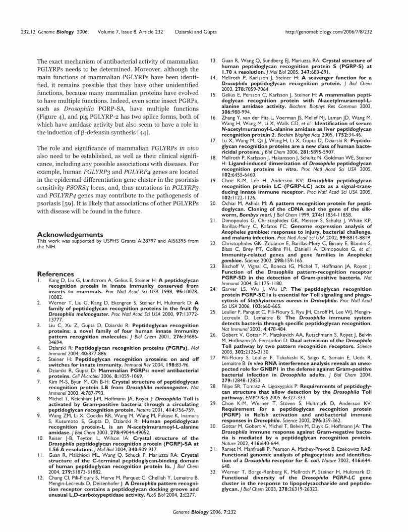
Figure 3The structures of (a) Lys-type peptidoglycan and (b) the carboxy-terminal PGRP domain of human PGLYRP-3 complexed with MurNAc-pentapeptide.(a) Lys-type peptidoglycan; two repeating disaccharide units crosslinked by a peptide are shown; the MurNAc-pentapeptide is in red; the arrowsrepresent the direction of the peptide bond; D-isoGln, D-isoglutamine. (b) The PGRP domain has three � helices (red), five � strands (yellow) and coils(cyan); the three disulfide bonds are in purple; MurNAc-pentapeptide is drawn in stick representation, with carbon, nitrogen, and oxygen atoms in green,blue, and red, respectively. N, amino terminus; C, carboxyl terminus. Reproduced with permission from [58].
(a) (b)
α1
α2
α3
β7
β3
β6
β4
β5
C
N
D-isoGln
L-AlaL-Lys
D-Ala
D-Ala

transduction pathway that results in the activation of the
Dorsal and Dif transcription factors (which are similar to
mammalian nuclear factor NF-�B), which translocate into
the nucleus, bind to the NF�B sites in the genome, and initi-
ate transcription of drosomycin and other antimicrobial pep-
tides, which are mainly active against Gram-positive bacteria
and fungi (Figure 4a). This pathway is essential for
Drosophila immunity to Gram-positive bacteria: mutations
in recognition or signal-transduction molecules for this
pathway make the flies highly susceptible to infections with
Gram-positive, but not Gram-negative, bacteria [8,23,24].
Peptidoglycan is a polymer of �(1-4)-linked N-acetyl-
glucosamine (GlcNAc) and N-acetylmuramic acid (MurNAc),
crosslinked by short peptides containing alternating L- and
D-amino acids (Figures 3a, 4d and 5c). In position 3, the
peptide has either diaminopimelic acid (DAP-type peptidogly-
can, found in all Gram-negative bacteria and in Gram-positive
bacilli; Figure 4d) or L-lysine (Lys-type peptidoglycan, found
in most other Gram-positive bacteria, Figures 3a and 5c).
The Toll pathway is preferentially triggered by the Lys-type
peptidoglycan and only weakly by the DAP-type peptidoglycan
com
ment
reviews
reports
deposited research
interactions
inform
ation
refereed research
http://genomebiology.com/2006/7/8/232 Genome Biology 2006, Volume 7, Issue 8, Article 232 Dziarski and Gupta 232.7
Genome Biology 2006, 7:232
Figure 4Functions of insect PGRP proteins. In response to peptidoglycan (PGN) from bacteria or other stimulants (yellow), insect PGRPs activate the (a) Toll and(b) Imd pathways and (c) the prophenol-oxidase cascade, which results in the production of antimicrobial products. (d) The structure of DAP-typepeptidoglycan, indicating the positions at which proinflammatory peptidoglycan can be hydrolyzed by some PGRPs, reducing inflammation. Drosophila PGRPsare shown (green) unless otherwise indicated (Bm, Bombyx mori; Hd, Holotrichia diomphalia; Tm, Tenebrio molitor). Multiple arrows signify multiple steps;question marks signify unconfirmed or controversial functions. PGN, peptidoglycan; m-DAP, meso-DAP. See text for more details of the pathways shown.
Proteases
PGRP-SA
PGRP-SD
PGRP-LE
PG
RP
-LC
Co-
rece
ptor
Gram-negative bacteriaGram-positive rods
PGN
GNBP-3GNBP-1
Spaetzle
Cell membrane
Phagocytosis
Phagocytosis?
Cytoplasm
Hemolymph
Nucleus
Cell membrane
Cytoplasm
Nucleus
FungalBacteria
PGN
PGRP-SC1
? ?
Dorsal Dif
Antimicrobialpeptide genes
Antimicrobialpeptide genes
Toll
Activation of Tolland phagocytosis
Activation of Imdand phagocytosis
Activation of prophenol-oxidase cascade
? ?
?
Imd
Relish
Melanin
Hemolymph
Reactiveoxygenspecies
Phenol oxidase
Prophenol oxidase
BmPGRP-S
PGRP-LE HdPGRP-1
TmPGRP-SA
BacteriaPGN
Fungalβ-glucan
(a) (b)
Enzymatic activity(d)
(c)
(—GlcNAc—MurNAc—)n
(—GlcNAc—MurNAc—)n
Peptidoglycan
N-acetylmuramoyl-L-Ala amidases
Carboxypeptidase
L-Ala
D-Glu
D-Ala
m-DAP D-Ala L-AlaD-Glum-DAP

232.8 Genome Biology 2006, Volume 7, Issue 8, Article 232 Dziarski and Gupta http://genomebiology.com/2006/7/8/232
Genome Biology 2006, 7:232
Figure 5 (see legend on following page)
PGLYRP-3:4 dimer
PGLYRP-4 dimerPGLYRP-3 dimer
PGLYRP-1dimer
PMNs
Bactericidal on the skin, in themouth, saliva, intestinal tract
and eyes
Bactericidal on the skin, in themouth, intestinal tract and eyes
(a) (b)
Bone marrowLiver
(—GlcNAc—MurNAc—)n
(—GlcNAc—MurNAc—)n
N-acetylmuramoyl-L-Ala amidase
L-Ala
D-Glu
L-Lys
D-Ala
L-Ala
D-Glu
L-Lys
D-Ala(Gly)5
PeptidoglycanPGLYRP-2
Serum (and skin and intestine)
Enzymatic activity in serum(c) Bactericidal effect in PMNs(d)

[25], although both types of peptidoglycan bind to PGRP-SA
[12]. The probable reason for the weak Toll-activating capac-
ity of DAP-type peptidoglycan is that this peptidoglycan, but
not Lys-type peptidoglycan, is the substrate for the car-
boxypeptidase activity of PGRP-SA [12] (Figure 4d). Effi-
cient triggering of the Toll pathway by PGRP-SA requires
cooperation (and probably formation of a complex) with
another pattern-recognition molecule, Gram-negative
binding protein (GNBP)-1 [26,27] (Figure 4a). GNBP-1
digests peptidoglycan and generates free reducing ends of
MurNAc, which are then recognized by PGRP-SA [28].
Drosophila PGRP-SC1 and PGRP-SD [23,24], as well as
other pattern-recognition molecules such as GNBP-3, also
activate the Toll pathway (Figure 4a). Both PGRP-SA and
PGRP-SC1 are required for the activation of Toll pathway,
whereas PGRP-SD is not essential but enhances Toll activa-
tion. Recognition of bacteria by PGRP-SC1 and PGRP-SA
may also trigger phagocytosis by an as yet unidentified
mechanism [24].
Activation of Drosophila PGRP-LC by Gram-negative bacte-
ria and Gram-positive bacilli (also called rods) triggers
another signal transduction pathway, the Imd pathway
[19,25,29-34] (Figure 4b). Binding of peptidoglycan to
Drosophila PGRP-LC induces its oligomerization and
recruitment and activation of the death-domain-containing
Imd protein [19]. The Imd pathway is Toll-independent and
results in the activation of Relish transcription factor (which
is also similar to mammalian NF-�B) and induction of tran-
scription of diptericin and other antimicrobial peptides that
are active primarily against Gram-negative bacteria [29-31].
PGRP-LC responds primarily to DAP-type peptidoglycan. It
is a transmembrane protein and has three alternative splice
forms (LC-A, LC-B, and LC-C), which differ in the extracellu-
lar PGRP domains; they probably cooperate with each other
and have somewhat different recognition specificities
[25,29,32-34]. PGRP-LC activates the Imd pathway in coop-
eration with PGRP-LE [35] and also probably with another,
as yet unidentified co-receptor (Figure 4b). Drosophila PGRP-
LC may also have a role in phagocytosis of Gram-negative
bacteria, because inhibition of PGRP-LC expression in
Drosophila S-2 cells diminishes phagocytosis of Escherichia
coli, but not of Staphylococcus aureus [31]; the mechanism
of this phenomenon is still unclear, however.
Silkworm (Bombyx mori) and mealworm (Tenebrio molitor)
PGRP-S are present in the hemolymph and cuticle, bind bac-
teria and Lys- and DAP-peptidoglycan, and activate the
prophenol-oxidase cascade (Figure 4c) [36,37]. This gener-
ates antimicrobial products, such as melanin and reactive
oxygen species, surrounds the infection site with melanin,
and contains the infection. Drosophila PGRP-LE [35] and
beetle (Holotrichia diomphalia) PGRP-1 [38] (and probably
other PGRPs) also activate the prophenol-oxidase cascade,
but H. diomphalia PGRP-1 responds to 1,3-�-D-glucan, a
common constituent of fungal cell walls.
Drosophila PGRP-SC1 and PGRP-LB are N-acetylmuramoyl-
L-alanine amidases [7,14], which hydrolyze the amide bond
between MurNAc and L-alanine and thus remove stem pep-
tides from peptidoglycan (Figure 4d). Stem peptides are the
four to five amino acids directly bound to MurNAc. Diges-
tion of peptidoglycan with amidase reduces or eliminates the
ability of polymeric peptidoglycan to stimulate insect cells
[14], and thus the function of amidase PGRPs in vivo may be
to prevent excessive activation of the immune system by bac-
teria [39,40]. On the basis of the conserved structure of the
active site of the amidase, several other insect PGRPs are
predicted to have amidase activity, whereas several others
are not [9,14,15] (Figure 1 and Table 1). One PGRP that is not
an amidase, Drosophila PGRP-SA, has an L,D-carboxypepti-
dase activity with specificity for the bond between DAP and
D-Ala of the stem peptide present in peptidoglycan of Gram-
negative bacteria and Gram-positive rod bacteria [12]
(Figure 4). The biological significance of this carboxypepti-
dase activity is not certain.
Mammalian PGLYRPsMammalian PGLYRPs are differentially expressed in various
organs and tissues and have two major functions: amidase
com
ment
reviews
reports
deposited research
interactions
inform
ation
refereed research
http://genomebiology.com/2006/7/8/232 Genome Biology 2006, Volume 7, Issue 8, Article 232 Dziarski and Gupta 232.9
Genome Biology 2006, 7:232
Figure 5 (see figure on previous page)Functions and expression of mammalian PGLYRP proteins. The diagram in the center shows the regions of the human body where each PGLYRP isexpressed; note that the information shown applies to other mammals as well as humans. (a) Mammalian PGLYRP-3 has direct bactericidal activity and isexpressed in the skin, eyes, tongue, esophagus, stomach, and intestines. (b) PGLYRP-4 and the PGLYRP-3:4 dimer also have direct bactericidal activity inthe same tissues; PGLYRP-4 is also expressed in the salivary gland, mucus-secreting glands in the throat and also in saliva. (c) PGLYRP-2, which isconstitutively produced in the liver and secreted into the blood, is also induced in the skin and intestine. It is an N-acetylmuramoyl-L-alanine amidase thathydrolyzes proinflammatory peptidoglycan. The structure of Lys-type peptidoglycan is shown, to indicate where in the molecule PGLYRP-2 hydrolyzes it.(d) PGLYRP-1 is present in the granules of the polymorphonuclear leukocytes (PMNs) which are produced in the bone marrow. PGLYRP-1 isbactericidal for phagocytosed bacteria; the images show killing of bacillus by PMNs. The images of scanning electron micrographs of Bacillus in (a) and (b)are copyright Dennis Kunkel Microscopy, Inc and are reproduced with permission. PGLYRP structures were rendered by RasMol and arranged ashomodimers or heterodimers. The structure of PGLYRP-1 is based on PDB entry 1yckA; the structure of the carboxy-terminal PGRP domain ofPGLYRP-2 was predicted by Swiss-Model on the basis of the crystal structure of D. melanogaster PGRP-SA (PDB entry 1s2jB); the amino-terminal portionof PGLYRP-2 cannot be predicted and hence is shown as an oval; the structures of PGLYRP-3 and PGLYRP-4 were predicted by Swiss-Model based onthe crystal structure of carboxy-terminal half of PGLYRP-3 (PDB entry 1SK3A).

activity and antibacterial activity. Mammalian PGLYRP-2
(and probably other vertebrate PGLYRP-2s) is an N-acetyl-
muramoyl-L-alanine amidase that hydrolyzes the lactyl bond
between the MurNAc and L-alanine in bacterial peptidogly-
can (Figure 5c) [9,15]. PGLYRP-2 is constitutively produced
in the liver and is secreted from the liver into the blood [16].
This liver PGLYRP-2 and serum N-acetylmuramoyl-L-alanine
amidase (which was identified earlier but not cloned) are the
same protein, encoded by the PGLYRP2 gene [16]. The func-
tion of this amidase is probably to eliminate the proinflam-
matory peptidoglycan and thus to prevent overactivation of
the immune system and excessive inflammation.
Mammalian PGLYRP-2 is also expressed in the intestinal
follicle-associated epithelial cells [41]. PGLYRP-2 is not
expressed in healthy human skin, but its expression is
induced in keratinocytes and other epithelial cells by expo-
sure to bacteria and cytokines [42,43]. Some mammals
express multiple splice forms of PGLYRP-2 that may have
different expression and possibly multiple functions. For
example, pigs have two PGLYRP-2 splice forms, short and
long. They both have N-acetylmuramoyl-L-alanine amidase
activity, and the long form has similar expression to human
PGLYRP-2, whereas the short form is constitutively
expressed in several tissues, including bone marrow, intes-
tine, liver, spleen, kidney, and skin [44].
Mammalian PGLYRP-1 is highly expressed in the bone
marrow [1,3], and the protein is almost exclusively present in
the granules of polymorphonuclear leukocytes [45-49]
(Figure 5d). Mammalian PGLYRP-3 and PGLYRP-4 proteins
are selectively expressed in the skin epidermis, hair follicles,
sebaceous glands and sweat glands; in the eye’s ciliary body
(which produces aqueous humor that fills the anterior and
posterior chambers of the eye); in the eye’s corneal epithe-
lium; in the mucus-secreting cells of the main salivary (sub-
mandibular) gland and in mucus-secreting glands in the
throat (both mucus-secreting glands selectively express
PGLYRP-4, but not PGLYRP-3); in the tongue and esophagus
in squamous epithelial cells; in the stomach in acid-secreting
parietal cells (PGLYRP-3) and glycoprotein-secreting neck
mucous cells (PGLYRP-4); and in the small and large intes-
tine in the columnar absorptive cells, but not in mucus-
secreting goblet cells and not in Paneth cells in the crypts,
which produce antimicrobial peptides [17,50] (Figure 5a,b).
Bacteria and their products increase the expression of
PGLYRP-3 and PGLYRP-4 in keratinocytes [17] and oral
epithelial cells [51], probably through activation of the Toll-
like receptors TLR2, TLR4, Nod1, and Nod2.
Human PGLYRP-1, PGLYRP-3, PGLYRP-4, the heterodimer
formed by PGLYRP-3 and PGLYRP-4, (PGLYRP-3:4), and
bovine PGLYRP-1 are bactericidal for many pathogenic and
nonpathogenic Gram-positive and Gram-negative bacteria
[17,46,47] (Figure 5a,b,d). PGLYRP-1, PGLYRP-3, and
PGLYRP-4 from other mammalian species are also likely to
have similar bactericidal activity. Bovine PGLYRP-1 also has
some microbicidal activity against a fungus, Cryptococcus
neoformans [46,47]. This broader spectrum of microbicidal
activity of bovine PGLYRP-1 could reflect a true difference
between the human and bovine orthologs, or it might simply
reflect a difference in the protein purification methods and
assay conditions.
MechanismCrystallographic analysis of human PGLYRP-1 and the
carboxy-terminal PGRP domain of PGLYRP-3, as well as
insect PGRP-LB, -SA, -LC and -LE, show that all these PGRPs
have a ligand-binding groove that binds peptidoglycan and is
specific for MurNAc bound to three peptide-bonded amino
acids (muramyl-tripeptide), which is the minimum peptido-
glycan fragment hydrolyzed by PGLYRP-2 [7,9,10-13,52-55].
It can accommodate a larger structure, such as GlcNAc-
MurNAc-tetrapeptide or MurNAc-pentapeptide (Figure 3),
but it does not bind muramyl-dipeptide or a peptide
without MurNAc [56-58]. These results are consistent with
the specificity of human PGLYRP-2 for muramyl-tripeptide
and with the specificity and high affinity (Kd = 13 nM) of
murine PGLYRP-1 for uncrosslinked polymeric peptidogly-
can but not muramyl-dipeptide or pentapeptide [45]. The
high-affinity binding of peptidoglycan to PGLYRP is
achieved by burying both the peptide and MurNAc portions
of peptidoglycan in a deep cleft that completely excludes
solvent [52].
Human PGLYRP-1 and a carboxy-terminal fragment of
PGLYRP-3 bind muramyl-tetrapeptide and muramyl-pen-
tapeptide with higher affinity than muramyl-tripeptide
[56,58]. Moreover, binding of muramyl-pentapeptide (but
not muramyl-tripeptide) to the carboxy-terminal fragment
of PGLYRP-3 induces a conformational change in the
PGLYRP-3 molecule that locks the ligand in the binding
groove (Figure 3) [58]. Some PGRPs (such as a carboxy-
terminal fragment of human PGLYRP-3) have a preference
for binding the Lys-type over the DAP-type peptidoglycan,
whereas others (such as human PGLYRP-1 or Drosophila
PGRP-LCx and PGRP-LE) bind DAP-type peptidoglycan
with higher affinity than Lys-type peptidoglycan [54-57].
The only difference between Lys and DAP is the presence of
an additional carboxylate at carbon 1 of DAP. Discrimination
between Lys- and DAP-type peptidoglycan is based on three
amino acids in the peptidoglycan-binding groove, corre-
sponding to Asn236, Phe237, and Val256 in human
PGLYRP-3 for binding Lys, or Gly68, Trp69, and Arg88 in
human PGLYRP-1 in the same position for binding DAP, or
Gly234, Trp235 and Arg254 in Drosophila PGRP-LE for
binding DAP [54-57]. The importance of these Asn and Phe
or Gly and Trp for binding Lys and DAP is verified by muta-
tions in these positions that can change the specificity of the
binding from Lys to DAP or DAP to Lys [57]. This allows pre-
diction of binding specificity of various PGRP domains for
Lys- or DAP-type peptidoglycan. Moreover, both human and
232.10 Genome Biology 2006, Volume 7, Issue 8, Article 232 Dziarski and Gupta http://genomebiology.com/2006/7/8/232
Genome Biology 2006, 7:232

insect PGRPs have a dual strategy for discrimination among
different types of peptidoglycan, using detection of Lys or
DAP in the stem peptide together with the type of peptide
crossbridge [57]. Detection of peptide-crosslinked peptido-
glycan would require engagement of two peptidoglycan-
binding sites in two PGRP domains, which could be
accomplished by PGRPs with two PGRP domains and/or by
dimeric PGRPs, which is consistent with recent demonstra-
tion of dimeric PGRPs in mammals [17] and insects [18,19].
There is likely, however, to be considerable variation in the
fine specificity of different PGRPs, because the residues in
and around the peptidoglycan-binding groove are relatively
variable; they are less than 50% conserved among PGRPs
[7,11,52]. This structural variation may correspond to differ-
ent ligand specificities of different PGRPs. Mammalian
PGLYRPs bind to both Gram-positive and Gram-negative
bacteria and also some fungi [17,47], and some insect PGRPs
(such as H. diomphalia PGRP-1) bind fungal �-glucan [38].
Therefore, binding to peptidoglycan is not always responsi-
ble for PGRP binding, and even with bacteria there are indi-
cations that some PGRPs may also bind to other polymers,
such as lipoteichoic acid and lipopolysaccharide [17,45,47].
Human and mouse PGLYRPs have the highest affinity for
peptidoglycan, however, and much lower affinities for lipo-
teichoic acid and lipopolysaccharide [17,45], whereas bovine
PGLYRP-1 seems to have high affinity for lipoteichoic acid
and lipopolysaccharide [47]. It is not clear, however,
whether these other ligands bind to the peptidoglycan-
binding groove or to another portion of the PGLYRP mole-
cule, such as the hydrophobic region on the opposite side of
the molecule. Binding of peptidoglycan outside the peptido-
glycan-binding groove was recently shown, which con-
tributes to the formation of PGRP-LE oligomers [54] or
PGRP-LCx:PGRP-LCa dimers [55].
The diversity of PGRP specificities is also increased by dupli-
cation of PGRP domains and dimerization. PGLYRP-3 and
PGLYRP-4 both have two PGRP domains, and each PGRP
domain has one ligand-binding site [52]. Thus, whereas
PGLYRP-1 monomers and dimers have one and two identi-
cal ligand-binding sites, respectively, PGLYRP-3 and
PGLYRP-4 monomers and dimers have two and four ligand-
binding sites, respectively (Figure 5). Because these PGRP
domains in PGLYRP-3 and PGLYRP-4 are not identical (they
have 37-43% identity), however, the fine binding specificity
or affinity of each PGRP domain in these PGLYRP molecules
is probably different. For example, the carboxy-terminal and
amino-terminal PGRP domains in human PGLYRP-3 are
specific for DAP-type and Lys-type peptidoglycan, respec-
tively [57]. The diversification of PGLYRP specificities is
then further increased by formation of PGLYRP-3:4 het-
erodimers, which have four different binding sites. In this
way, the host can fine-tune the specificities of PGLYRPs by
expressing PGLYRP-3 and PGLYRP-4 either in the same or in
separate cells, to form hetero- or homodimers, respectively. In
addition, PGRPs have hydrophobic domains on the opposite
side of the molecule from the ligand-binding groove, which
were previously hypothesized to interact with signal transduc-
tion molecules [7]. In mammalian PGLYRPs, however, these
hydrophobic domains may either have a role in the interaction
of PGLYRPs with bacteria, or in the formation of dimers.
Mammalian PGLYRP-1, PGLYRP-3, and PGLYRP-4 form a
new class of bactericidal proteins that have a different
structure, mechanism of action, and expression from those
of currently known mammalian antimicrobial peptides
[6,17]. PGLYRPs are much larger than all currently known
vertebrate antibacterial peptides: PGLYRP-1, PGLYRP-3,
PGLYRP-3:4, and PGLYRP-4 proteins are disulfide-linked
glycosylated 44 kDa, 89 kDa, 98 kDa, and 115 kDa dimers
[17], and vertebrate antimicrobial peptides are typically
3 kDa to 15 kDa. PGLYRPs require divalent cations and
N-glycosylation for bactericidal activity, which are not
usually required by membrane-permeabilizing antibacterial
peptides, such as defensins or magainin [17]. Mammalian
PGLYRPs also differ from antimicrobial peptides in their
mechanism of bactericidal activity: they kill bacteria by
interacting with cell-wall peptidoglycan, whereas antimicro-
bial peptides do so by permeabilizing bacterial membranes
[17]. Furthermore, the expression patterns of mammalian
PGLYRPs and antimicrobial peptides are different, and
some cells that produce large amounts of these peptides,
such as Paneth cells (which produce defensins, phospholi-
pase A2, and lysozyme), do not express PGLYRPs [17].
FrontiersDespite enormous progress since the discovery of PGRPs in
1996 [36], much remains to be done. The structures and speci-
ficities of many insect and mammalian PGRPs still need to be
determined. For example, the PGRP/amidase domain of
mammalian PGLYRP-2 or many insect long PGRPs is located
in the carboxy-terminal one third of the molecule, but the role
and the structure of the remaining amino-terminal two thirds
of PGLYRP-2 or several insect long PGRPs is unknown, as this
portion has no homology to any other PGRPs or to any other
known proteins [3,9]. These amino-terminal portions of
PGLYRP-2 and several insect long PGRPs may therefore have
unique and so far unidentified functions.
The functions of many insect PGRPs and their mechanisms of
action also still need to be determined (Figure 1 and Table 1).
It should be especially interesting to look for direct antimi-
crobial activity of insect PGRPs, which will establish whether
this function developed in mammalian or vertebrate
PGLYRPs or whether it was already present in their common
ancestor with insects. PGRPs in other invertebrates and in
nonmammalian vertebrates (fish, amphibians, reptiles, and
birds) are beginning to be discovered and nothing is known
about their functions, although most of them are predicted
to have amidase activity (Figure 2 and Table 1).
com
ment
reviews
reports
deposited research
interactions
inform
ation
refereed research
http://genomebiology.com/2006/7/8/232 Genome Biology 2006, Volume 7, Issue 8, Article 232 Dziarski and Gupta 232.11
Genome Biology 2006, 7:232
The exact mechanism of antibacterial activity of mammalian
PGLYRPs needs to be determined. Moreover, although the
main functions of mammalian PGLYRPs have been identi-
fied, it remains possible that they have other unidentified
functions, because many mammalian proteins have evolved
to have multiple functions. Indeed, even some insect PGRPs,
such as Drosophila PGRP-SA, have multiple functions
(Figure 4), and pig PGLYRP-2 has two splice forms, both of
which have amidase activity but also seem to have a role in
the induction of �-defensin synthesis [44].
The role and significance of mammalian PGLYRPs in vivo
also need to be established, as well as their clinical signifi-
cance, including any possible associations with diseases. For
example, human PGLYRP3 and PGLYRP4 genes are located
in the epidermal differentiation gene cluster in the psoriasis
sensitivity PSORS4 locus, and, thus mutations in PGLYRP3
and PGLYRP4 genes may contribute to the pathogenesis of
psoriasis [59]. It is likely that associations of other PGLYRPs
with disease will be found in the future.
AcknowledgementsThis work was supported by USPHS Grants AI28797 and AI56395 fromthe NIH.
References 1. Kang D, Liu G, Lundstrom A, Gelius E, Steiner H: A peptidoglycan
recognition protein in innate immunity conserved frominsects to mammals. Proc Natl Acad Sci USA 1998, 95:10078-10082.
2. Werner T, Liu G, Kang D, Ekengren S, Steiner H, Hultmark D: Afamily of peptidoglycan recognition proteins in the fruit flyDrosophila melanogaster. Proc Natl Acad Sci USA 2000, 97:13772-13777.
3. Liu C, Xu Z, Gupta D, Dziarski R: Peptidoglycan recognitionproteins: a novel family of four human innate immunitypattern recognition molecules. J Biol Chem 2001, 276:34686-34694.
4. Dziarski R: Peptidoglycan recognition proteins (PGRPs). MolImmunol 2004, 40:877-886.
5. Steiner H: Peptidoglycan recognition proteins: on and offswitches for innate immunity. Immunol Rev 2004, 198:83-96.
6. Dziarski R, Gupta D: Mammalian PGRPs: novel antibacterialproteins. Cell Microbiol 2006, 8:1059-1069.
7. Kim M-S, Byun M, Oh B-H: Crystal structure of peptidoglycanrecognition protein LB from Drosophila melanogaster. NatImmunol 2003, 4:787-793.
8. Michel T, Reichhart J-M, Hoffmann JA, Royet J: Drosophila Toll isactivated by Gram-positive bacteria through a circulatingpeptidoglycan recognition protein. Nature 2001, 414:756-759.
9. Wang ZM, Li X, Cocklin RR, Wang M, Wang M, Fukase K, InamuraS, Kusumoto S, Gupta D, Dziarski R: Human peptidoglycanrecognition protein-L is an N-acetylmuramoyl-L-alanineamidase. J Biol Chem 2003, 278:49044-49052.
10. Reiser J-B, Teyton L, Wilson IA: Crystal structure of theDrosophila peptidoglycan recognition protein (PGRP)-SA at1.56 Å resolution. J Mol Biol 2004, 340:909-917.
11. Guan R, Malchiodi ML, Wang Q, Schuck P, Mariuzza RA: Crystalstructure of the C-terminal peptidoglycan-binding domainof human peptidoglycan recognition protein I��. J Biol Chem2004, 279:31873-31882.
12. Chang CI, Pili-Floury S, Herve M, Parquet C, Chelliah Y, Lemaitre B,Mengin-Lecreulx D, Deisenhofer J: A Drosophila pattern recogni-tion receptor contains a peptidoglycan docking groove andunusual L,D-carboxypeptidase activity. PLoS Biol 2004, 2:E277.
13. Guan R, Wang Q, Sundberg EJ, Mariuzza RA: Crystal structure ofhuman peptidoglycan recognition protein S (PGRP-S) at1.70 A resolution. J Mol Biol 2005, 347:683-691.
14. Mellroth P, Karlsson J, Steiner H: A scavenger function for aDrosophila peptidoglycan recognition protein. J Biol Chem2003, 278:7059-7064.
15. Gelius E, Persson C, Karlsson J, Steiner H: A mammalian pepti-doglycan recognition protein with N-acetylmuramoyl-L-alanine amidase activity. Biochem Biophys Res Commun 2003,306:988-994.
16. Zhang Y, van der Fits L, Voerman JS, Melief MJ, Laman JD, Wang M,Wang H, Wang M, Li X, Walls CD, et al.: Identification of serumN-acetylmuramoyl-L-alanine amidase as liver peptidoglycanrecognition protein 2. Biochim Biophys Acta 2005, 1752:34-46.
17. Lu X, Wang M, Qi J, Wang H, Li X, Gupta D, Dziarski R: Peptido-glycan recognition proteins are a new class of human bacte-ricidal proteins. J Biol Chem 2006, 281:5895-5907.
18. Mellroth P, Karlsson J, Hakansson J, Schultz N, Goldman WE, SteinerH: Ligand-induced dimerization of Drosophila peptidoglycanrecognition proteins in vitro. Proc Natl Acad Sci USA 2005,102:6455-6460.
19. Choe K-M, Lee H, Anderson KV: Drosophila peptidoglycanrecognition protein LC (PGRP-LC) acts as a signal-trans-ducing innate immune receptor. Proc Natl Acad Sci USA 2005,102:1122-1126.
20. Ochiai M, Ashida M: A pattern recognition protein for pepti-doglycan. Cloning of the cDNA and the gene of the silk-worm, Bombyx mori. J Biol Chem 1999, 274:11854-11858.
21. Dimopoulos G, Christophides GK, Meister S, Schultz J, White KP,Barillas-Mury C, Kafatos FC: Genome expression analysis ofAnopheles gambiae: responses to injury, bacterial challenge,and malaria infection. Proc Natl Acad Sci USA 2002, 99:8814-8819.
22. Christophides GK, Zdobnov E, Barillas-Mury C, Birney E, Blandin S,Blass C, Brey PT, Collins FH, Danielli A, Dimopoulos G, et al.:Immunity-related genes and gene families in Anophelesgambiae. Science 2002, 298:159-165.
23. Bischoff V, Vignal C, Boneca IG, Michel T, Hoffmann JA, Royet J:Function of the Drosophila pattern-recognition receptorPGRP-SD in the detection of Gram-positive bacteria. NatImmunol 2004, 5:1175-1180.
24. Garver LS, Wu J, Wu LP: The peptidoglycan recognitionprotein PGRP-SC1a is essential for Toll signaling and phago-cytosis of Staphylococcus aureus in Drosophila. Proc Natl AcadSci USA 2006, 103:660-665.
25. Leulier F, Parquet C, Pili-Floury S, Ryu JH, Caroff M, Lee WJ, Mengin-Lecreulx D, Lemaitre B: The Drosophila immune systemdetects bacteria through specific peptidoglycan recognition.Nat Immunol 2003, 4:478-484.
26. Gobert V, Gottar M, Matskevich AA, Rutschmann S, Royet J, BelvinM, Hoffmann JA, Ferrandon D: Dual activation of the DrosophilaToll pathway by two pattern recognition receptors. Science2003, 302:2126-2130.
27. Pili-Floury S, Leulier F, Takahashi K, Saigo K, Samain E, Ueda R,Lemaitre B: In vivo RNA interference analysis reveals an unex-pected role for GNBP1 in the defense against Gram-positivebacterial infection in Drosophila adults. J Biol Chem 2004,279:12848-12853.
28. Filipe SR, Tomasz A, Ligoxygakis P: Requirements of peptidogly-can structure that allow detection by the Drosophila Tollpathway. EMBO Rep 2005, 6:327-333.
29. Choe K-M, Werner T, Stoven S, Hultmark D, Anderson KV:Requirement for a peptidoglycan recognition protein(PGRP) in Relish activation and antibacterial immuneresponses in Drosophila. Science 2002, 296:359-362.
30. Gottar M, Gobert V, Michel T, Belvin M, Duyk G, Hoffmann JA: TheDrosophila immune response against Gram-negative bacte-ria is mediated by a peptidoglycan recognition protein.Nature 2002, 416:640-644.
31. Ramet M, Manfruelli P, Pearson A, Mathey-Prevot B, Ezekowitz RAB:Functional genomic analysis of phagocytosis and identifica-tion of a Drosophila receptor for E. coli. Nature 2002, 416:644-648.
32. Werner T, Borge-Renberg K, Mellroth P, Steiner H, Hultmark D:Functional diversity of the Drosophila PGRP-LC genecluster in the response to lipopolysaccharide and peptido-glycan. J Biol Chem 2003, 278:26319-26322.
232.12 Genome Biology 2006, Volume 7, Issue 8, Article 232 Dziarski and Gupta http://genomebiology.com/2006/7/8/232
Genome Biology 2006, 7:232

33. Kaneko T, Goldman WE, Mellroth P, Steiner H, Fukase K, KusumotoS, Harley W, Fox A, Golenbock D, Silverman N: Monomeric andpolymeric gram-negative peptidoglycan but not purifiedLPS stimulate the Drosophila IMD pathway. Immunity 2004,20:637-649.
34. Stenbak CR, Ryu JH, Leulier F, Pili-Floury S, Parquet C, Herve M,Chaput C, Boneca IG, Lee WJ, Lemaitre B, et al.: Peptidoglycanmolecular requirements allowing detection by theDrosophila immune deficiency pathway. J Immunol 2004,173:7339-7348.
35. Takehana A, Yano T, Mita S, Kotani A, Oshima Y, Kurata S: Pepti-doglycan recognition protein (PGRP)-LE and PGRP-LC actsynergistically in Drosophila immunity. EMBO J 2004, 23:4690-4700.
36. Yoshida H, Kinoshita K, Ashida M: Purification of peptidoglycanrecognition protein from hemolymph of the silkworm,Bombyx mori. J Biol Chem 1996, 271:13854-13860.
37. Park JW, Je BR, Piao S, Inamura S, Fujimoto Y, Fukase K, KusumotoS, Ha NC, Soderhall K, Ha NC, et al.: A synthetic peptidoglycanfragment as a competitive inhibitor of the melanizationcascade. J Biol Chem 2006, 281:7747-7755.
38. Lee MH, Osaki T, Lee JY, Baek MJ, Zhang R, Park JW, Kawabata S,Soderhall K, Lee BL: Peptidoglycan recognition proteinsinvolved in 1,3-��-D-glucan-dependent prophenoloxidaseactivation system of insect. J Biol Chem 2004, 279:3218-3227.
39. Bischoff V, Vignal C, Duvic B, Boneca IG, Hoffmann JA, Royet J:Downregulation of the Drosophila immune response by pep-tidoglycan-recognition proteins SC1 and SC2. PLoS Pathog2006, 2:e14.
40. Zaidman-Remy A, Herve M, Poidevin M, Pili-Floury S, Kim MS,Blanot D, Oh BH, Ueda R, Mengin-Lecreulx D, Lemaitre B: TheDrosophila amidase PGRP-LB modulates the immuneresponse to bacterial infection. Immunity 2006, 24:463-473.
41. Lo D, Tynan W, Dickerson J, Mendy J, Chang HW, Scharf M, ByrneD, Brayden D, Higgins L, Evans C, et al.: Peptidoglycan recogni-tion protein expression in mouse Peyer’s Patch follicle asso-ciated epithelium suggests functional specialization. CellImmunol 2003, 224:8-16.
42. Wang H, Gupta D, Li X, Dziarski R: Peptidoglycan recognitionprotein 2 (N-acetylmuramoyl-L-ala amidase) is induced inkeratinocytes by bacteria through the p38 kinase pathway.Infect Immun 2005, 73:7216-7225.
43. Li X, Wang S, Wang H, Gupta D: Differential expression of pep-tidoglycan recognition protein 2 in the skin and liverrequires different transcription factors. J Biol Chem 2006,281:20738-20748.
44. Sang Y, Ramanathan B, Ross CR, Blecha F: Gene silencing andoverexpression of porcine peptidoglycan recognitionprotein long isoform: involvement in ��-defensin-1 expres-sion. Infect Immun 2005, 73:7133-7141.
45. Liu C, Gelius E, Liu G, Steiner H, Dziarski R: Mammalian peptido-glycan recognition protein binds peptidoglycan with highaffinity, is expressed in neutrophils, and inhibits bacterialgrowth. J Biol Chem 2000, 275:24490-24499.
46. Tydell CC, Yount N, Tran D, Yuan J, Selsted M: Isolation, charac-terization, and antimicrobial properties of bovine oligosac-charide-binding protein. J Biol Chem 2002, 277:19658-19664.
47. Tydell CC, Yuan J, Tran P, Selsted ME: Bovine peptidoglycanrecognition protein-S: antimicrobial activity, localization,secretion, and binding properties. J Immunol 2006, 176:1154-1162.
48. Dziarski R, Platt KA, Gelius E, Steiner H, Gupta D: Defect in neu-trophil killing and increased susceptibility to infection withnon-pathogenic Gram-positive bacteria in peptidoglycanrecognition protein-S (PGRP-S)-deficient mice. Blood 2003,102:689-697.
49. Cho JH, Fraser IP, Fukase K, Kusumoto S, Fujimoto Y, Stahl GL,Ezekowitz, RA: Human peptidoglycan recognition protein S isan effector of neutrophil-mediated innate immunity. Blood2005, 106:2551-2558.
50. Mathur P, Murray B, Crowell T, Gardner H, Allaire N, Hsu YM,Thill G, Carulli JP: Murine peptidoglycan recognition proteinsPglyrpI�� and PglyrpI�� are encoded in the epidermaldifferentiation complex and are expressed in epidermal andhematopoietic tissues. Genomics 2004, 83:1151-1163.
51. Uehara A, Sugawara Y, Kurata S, Fujimoto Y, Fukase K, Kusumoto S,Satta Y, Sasano T, Sugawara S, Takada H: Chemically synthesized
pathogen-associated molecular patterns increase theexpression of peptidoglycan recognition proteins via toll-like receptors, NOD1 and NOD2 in human oral epithelialcells. Cell Microbiol 2005, 7:675-686.
52. Guan R, Roychowdhury A, Ember B, Kumar S, Boons G-J, MariuzzaRA: Structural basis for peptidoglycan binding by peptidogly-can recognition proteins. Proc Natl Acad Sci USA 2004,101:17168-17173.
53. Chang CI, Ihara K, Chelliah Y, Mengin-Lecreulx D, Wakatsuki S,Deisenhofer J: Structure of the ectodomain of Drosophila pep-tidoglycan-recognition protein LCa suggests a molecularmechanism for pattern recognition. Proc Natl Acad Sci USA2005, 102:10279-10284.
54. Lim J-H, Kim M-S, Kim H-E, Yano T, Oshima Y, Aggarwal K,Goldman WE, Silverman N, Kurata S, Oh B-H: Structural basis forpreferential recognition of diaminopimelic acid-type pepti-doglycan by a subset of peptidoglycan-recognition proteins.J Biol Chem 2006, 281:8286-8295.
55. Chang CI, Chelliah Y, Borek D, Mengin-Lecreulx D, Deisenhofer J:Structure of tracheal cytotoxin in complex with a het-erodimeric pattern-recognition receptor. Science 2006,311:1761-1764.
56. Kumar S, Roychowdhury A, Ember B, Wang Q, Guan R, MariuzzaRA, Boons G-J: Selective recognition of synthetic lysine andmeso-diaminopimelic acid-type peptidoglycan fragments byhuman peptidoglycan recognition proteins I�� and S. J BiolChem 2005, 280:37005-37012.
57. Swaminathan CP, Brown PH, Roychowdhury A, Wang Q, Guan R,Silverman N, Goldman WE, Boons GJ, Mariuzza RA: Dual strate-gies for peptidoglycan discrimination by peptidoglycanrecognition proteins (PGRPs). Proc Natl Acad Sci USA 2006,103:684-689.
58. Guan R, Brown PH, Swaminathan CP, Roychowdhury A, Boons GJ,Mariuzza RA: Crystal structure of human peptidoglycanrecognition protein I�� bound to a muramyl pentapeptidefrom Gram-positive bacteria. Protein Sci 2006, 15:1199-1206.
59. Sun C, Mathur P, Dupuis J, Tizard R, Ticho B, Crowell T, Gardner H,Bowcock AM, Carulli J: Peptidoglycan recognition proteinsPglyrp3 and Pglyrp4 are encoded from the epidermal differ-entiation complex and are candidate genes for the Psors4locus on chromosome 1q21. Hum Genet 2006, 119:113-125.
com
ment
reviews
reports
deposited research
interactions
inform
ation
refereed research
http://genomebiology.com/2006/7/8/232 Genome Biology 2006, Volume 7, Issue 8, Article 232 Dziarski and Gupta 232.13
Genome Biology 2006, 7:232