Structures, Separation, Characterization, Biosynthesis, Bioac
54
Citation: Omar, A.M.; Mohamed, G.A.; Ibrahim, S.R.M. Chaetomugilins and Chaetoviridins—Promising Natural Metabolites: Structures, Separation, Characterization, Biosynthesis, Bioactivities, Molecular Docking, and Molecular Dynamics. J. Fungi 2022, 8, 127. https://doi.org/10.3390/ jof8020127 Academic Editor: Laurent Dufossé Received: 21 December 2021 Accepted: 24 January 2022 Published: 27 January 2022 Publisher’s Note: MDPI stays neutral with regard to jurisdictional claims in published maps and institutional affil- iations. Copyright: © 2022 by the authors. Licensee MDPI, Basel, Switzerland. This article is an open access article distributed under the terms and conditions of the Creative Commons Attribution (CC BY) license (https:// creativecommons.org/licenses/by/ 4.0/). Fungi Journal of Review Chaetomugilins and Chaetoviridins—Promising Natural Metabolites: Structures, Separation, Characterization, Biosynthesis, Bioactivities, Molecular Docking, and Molecular Dynamics Abdelsattar M. Omar 1,2, * , Gamal A. Mohamed 3 and Sabrin R. M. Ibrahim 4,5, * 1 Department of Pharmaceutical Chemistry, Faculty of Pharmacy, King Abdulaziz University, Jeddah 21589, Saudi Arabia 2 Center for Artificial Intelligence in Precision Medicines, King Abdulaziz University, Jeddah 21589, Saudi Arabia 3 Department of Natural Products and Alternative Medicine, Faculty of Pharmacy, King Abdulaziz University, Jeddah 21589, Saudi Arabia; [email protected] 4 Department of Chemistry, Preparatory Year Program, Batterjee Medical College, Jeddah 21442, Saudi Arabia 5 Department of Pharmacognosy, Faculty of Pharmacy, Assiut University, Assiut 71526, Egypt * Correspondence: [email protected] (A.M.O.); [email protected] or [email protected] (S.R.M.I.); Tel.: +966-56-768-1466 (A.M.O.); +966-58-118-3034 (S.R.M.I.) Abstract: Fungi are recognized as luxuriant metabolic artists that generate propitious biometabolites. Historically, fungal metabolites have largely been investigated as leads for various therapeutic agents. Chaetomugilins and the closely related chaetoviridins are fungal metabolites, and each has an oxygenated bicyclic pyranoquinone core. They are mainly produced by various Chaetomaceae species. These metabolites display unique chemical features and diversified bioactivities. The current review gives an overview of research about fungal chaetomugilins and chaetoviridins regarding their structures, separation, characterization, biosynthesis, and bioactivities. Additionally, their antiviral potential towards the SARS-CoV-2 protease was evaluated using docking studies and molecular dynamics (MD) simulations. We report on the docking and predictive binding energy estimations using reported crystal structures of the main protease (PDB ID: 6M2N, 6W81, and 7K0f) at variable resolutions—i.e., 2.20, 1.55, and 1.65 Å, respectively. Chaetovirdin D (43) exhibited highly negative docking scores of -7.944, -8.141, and -6.615 kcal/mol, when complexed with 6M2N, 6W81, and 7K0f, respectively. The reference inhibitors exhibited the following scores: -5.377, -6.995, and -8.159 kcal/mol, when complexed with 6M2N, 6W81, and 7K0f, respectively. By using molecular dynamics simulations, chaetovirdin D’s stability in complexes with the viral protease was analyzed, and it was found to be stable over the course of 100 ns. Keywords: chaetomugilins; chaetoviridins; fungi; Chaetomaceae; characterization; bioactivities; molecular docking; COVID-19; protease; biosynthesis 1. Introduction Fungi are a wealthy and substantial pool of many secondary metabolites with many different structures and diversified bioactivities [1–11]. These metabolites attract much attention as lead metabolites for pharmaceutical agents, and for plant protection [1,8–16]. Fungal polyketides (FPKs) represent one of the largest and most structurally diverse groups of fungal metabolites. They range from simple and aromatic to highly macrocyclic and complex [10,13,17]. Their backbone is biosynthesized by the condensation of acyl-CoA thioesters [18]. Their structural variations originate from differences in the starting and extending units, methylation pattern, chain length, degree of reduction, and modifications by tailoring enzymes [19]. Mycotoxins and pigments are among the FPKs that have had J. Fungi 2022, 8, 127. https://doi.org/10.3390/jof8020127 https://www.mdpi.com/journal/jof
-
Upload
khangminh22 -
Category
Documents
-
view
0 -
download
0
Transcript of Structures, Separation, Characterization, Biosynthesis, Bioac

�����������������
Citation: Omar, A.M.; Mohamed,
G.A.; Ibrahim, S.R.M.
Chaetomugilins and
Chaetoviridins—Promising Natural
Metabolites: Structures, Separation,
Characterization, Biosynthesis,
Bioactivities, Molecular Docking, and
Molecular Dynamics. J. Fungi 2022, 8,
127. https://doi.org/10.3390/
jof8020127
Academic Editor: Laurent Dufossé
Received: 21 December 2021
Accepted: 24 January 2022
Published: 27 January 2022
Publisher’s Note: MDPI stays neutral
with regard to jurisdictional claims in
published maps and institutional affil-
iations.
Copyright: © 2022 by the authors.
Licensee MDPI, Basel, Switzerland.
This article is an open access article
distributed under the terms and
conditions of the Creative Commons
Attribution (CC BY) license (https://
creativecommons.org/licenses/by/
4.0/).
FungiJournal of
Review
Chaetomugilins and Chaetoviridins—Promising NaturalMetabolites: Structures, Separation, Characterization,Biosynthesis, Bioactivities, Molecular Docking,and Molecular DynamicsAbdelsattar M. Omar 1,2,* , Gamal A. Mohamed 3 and Sabrin R. M. Ibrahim 4,5,*
1 Department of Pharmaceutical Chemistry, Faculty of Pharmacy, King Abdulaziz University,Jeddah 21589, Saudi Arabia
2 Center for Artificial Intelligence in Precision Medicines, King Abdulaziz University,Jeddah 21589, Saudi Arabia
3 Department of Natural Products and Alternative Medicine, Faculty of Pharmacy, King Abdulaziz University,Jeddah 21589, Saudi Arabia; [email protected]
4 Department of Chemistry, Preparatory Year Program, Batterjee Medical College, Jeddah 21442, Saudi Arabia5 Department of Pharmacognosy, Faculty of Pharmacy, Assiut University, Assiut 71526, Egypt* Correspondence: [email protected] (A.M.O.); [email protected] or
[email protected] (S.R.M.I.); Tel.: +966-56-768-1466 (A.M.O.); +966-58-118-3034 (S.R.M.I.)
Abstract: Fungi are recognized as luxuriant metabolic artists that generate propitious biometabolites.Historically, fungal metabolites have largely been investigated as leads for various therapeuticagents. Chaetomugilins and the closely related chaetoviridins are fungal metabolites, and each hasan oxygenated bicyclic pyranoquinone core. They are mainly produced by various Chaetomaceaespecies. These metabolites display unique chemical features and diversified bioactivities. The currentreview gives an overview of research about fungal chaetomugilins and chaetoviridins regarding theirstructures, separation, characterization, biosynthesis, and bioactivities. Additionally, their antiviralpotential towards the SARS-CoV-2 protease was evaluated using docking studies and moleculardynamics (MD) simulations. We report on the docking and predictive binding energy estimationsusing reported crystal structures of the main protease (PDB ID: 6M2N, 6W81, and 7K0f) at variableresolutions—i.e., 2.20, 1.55, and 1.65 Å, respectively. Chaetovirdin D (43) exhibited highly negativedocking scores of −7.944, −8.141, and −6.615 kcal/mol, when complexed with 6M2N, 6W81, and7K0f, respectively. The reference inhibitors exhibited the following scores: −5.377, −6.995, and−8.159 kcal/mol, when complexed with 6M2N, 6W81, and 7K0f, respectively. By using moleculardynamics simulations, chaetovirdin D’s stability in complexes with the viral protease was analyzed,and it was found to be stable over the course of 100 ns.
Keywords: chaetomugilins; chaetoviridins; fungi; Chaetomaceae; characterization; bioactivities;molecular docking; COVID-19; protease; biosynthesis
1. Introduction
Fungi are a wealthy and substantial pool of many secondary metabolites with manydifferent structures and diversified bioactivities [1–11]. These metabolites attract muchattention as lead metabolites for pharmaceutical agents, and for plant protection [1,8–16].Fungal polyketides (FPKs) represent one of the largest and most structurally diverse groupsof fungal metabolites. They range from simple and aromatic to highly macrocyclic andcomplex [10,13,17]. Their backbone is biosynthesized by the condensation of acyl-CoAthioesters [18]. Their structural variations originate from differences in the starting andextending units, methylation pattern, chain length, degree of reduction, and modificationsby tailoring enzymes [19]. Mycotoxins and pigments are among the FPKs that have had
J. Fungi 2022, 8, 127. https://doi.org/10.3390/jof8020127 https://www.mdpi.com/journal/jof

J. Fungi 2022, 8, 127 2 of 54
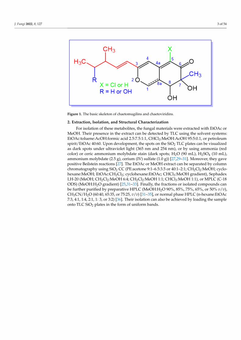
remarkable contributions in the field of drug discovery [20]. Azaphilones (azaphilonoidsor isochromenes) are fungal pigments belonging to FPKs. Structurally, they have anisochromene skeleton that contains an oxygenated bicyclic pyrano-quinone core and a qua-ternary carbon center [21]. Biosynthetically, the O atom in the pyran chromophore could beexchanged by an N atom in the existence of primary amines, and accordingly, the pigmentcolor will shift to red [22]. They are produced by various basidiomycetous and ascomyce-tous fungi, including Chaetomium, Penicillium, Aspergillus, Talaromyces, Phomopsis, Monascus,Emericella, Epicoccum, Hypoxylon, and Pestalotiopsis, where they are accountable for thegreen, red, or yellow color of mycelia and/or fruiting bodies [23]. They possess myriadbioactivities: antitumor, cytotoxic, antimicrobial, anti-inflammatory, antioxidant, enzymeinhibitory, antiviral, insecticidal, and antileishmanial [24]. Chaetomugilins and closelyrelated chaetoviridins are azaphilones featuring a C-7-methyl group and C-5 chlorine—except for chaetomugilins T (29) and U (30)—and a C-3-branched pentenyl chain (Figure 1).However, chaetomugiline P (24) differs from the others in that it has no substituent at C-7and a methyl group at C-5. 3-Methyl-4-hydroxy-1-pentyl chains at C-3 are found in somechaetomugilins and chaetoviridins. Sometimes, they bear a five-membered lactone and/ora fused tetrahydrofuran/δ-lactone [25]. The 7-OH group can have (S) or (R) configuration,though (7S) isomers are the most common among these metabolites. On the other hand,the 7-hydroxyl can be part of a furanone ring [26]. These fungal metabolites are producedby various Chaetomium species. Chaetoviridins were firstly reported by Takahashi et al.from Chaetomium globosum var. flavoviride [27]. Chaetomugilins are known as cytotoxicmetabolites, whereas chaetoviridins have antifungal and antibiotic activities [28]. Recently,these metabolites have been recognized as a unique family of fungal metabolites in viewof their interesting structural features and prominent bioactivities, which could provokeenormous attention from natural products chemists and pharmacologists. The currentreview focuses on chaetomugilins and chaetoviridins from fungal sources, including isola-tion, structural characterization, biosynthesis, and bioactivities (Tables 1 and 2). Some ofthe metabolites have been reported with the same names, despite having different struc-tures and molecular formulae—e.g., chaetomugilin S, chaetoviridin B, and chaetoviridinG. Moreover, the structures of some compounds have been revised and renamed: in suchcases, both structures have been drawn in the figures, highlighting the new names andcorresponding references. Additionally, the emergence of the COVID-19 pandemic mo-tivated us to investigate the potential of these metabolites as antiviral agents towardsSARS-CoV-2 using docking studies and molecular dynamics (MD) simulations. Literaturesearching was carried out using diverse databases—Web of Science, PubMed (MedLine),GoogleScholar, Scopus, and SciFinder—and different publishers— Springer-Link (Cham,Switzerland), Wiley (New York, NY, United States), Taylor & Francis (London, England),Bentham (Sharjah, United Arab Emirates), and ACS (Washington, DC, USA) Publications.
J. Fungi 2022, 8, 127 3 of 54J. Fungi 2022, 8, x FOR PEER REVIEW 3 of 53
Figure 1. The basic skeleton of chaetomugilins and chaetoviridins.
2. Extraction, Isolation, and Structural Characterization For isolation of these metabolites, the fungal materials were extracted with EtOAc or
MeOH. Their presence in the extract can be detected by TLC using the solvent systems: EtOAc:toluene:AcOH:formic acid 2.5:7.5:1:1, CHCl3:MeOH:AcOH 95:5:0.1, or petroleum spirit/EtOAc 40:60. Upon development, the spots on the SiO2 TLC plates can be visualized as dark spots under ultraviolet light (365 nm and 254 nm), or by using ammonia (red color) or ceric ammonium molybdate stain (dark spots; H2O (90 mL), H2SO4 (10 mL), ammonium molybdate (2.5 g), cerium (IV) sulfate (1.0 g)) [27,29–31]. Moreover, they gave positive Beilstein reactions [27]. The EtOAc or MeOH extract can be separated by column chroma-tography using SiO2 CC (PE:acetone 9:1–6.5:3.5 or 40:1–2:1; CH2Cl2:MeOH; cyclohex-ane:MeOH; EtOAc:CH2Cl2; cyclohexane:EtOAc; CHCl3:MeOH gradient), Sephadex LH-20 (MeOH; CH2Cl2:MeOH 6:4; CH2Cl2:MeOH 1:1; CHCl3:MeOH 1:1), or MPLC (C-18 ODS) (MeOH:H2O gradient) [25,31–33]. Finally, the fractions or isolated compounds can be fur-ther purified by preparative HPLC (MeOH:H2O 90%, 85%, 75%, 65%, or 50% v/v), CH3CN/H2O (60:40, 65:35, or 75:25, v/v) [31–35], or normal phase HPLC (n-hexane:EtOAc 7:3, 4:1, 1:4, 2:1, 1: 3, or 3:2) [36]. Their isolation can also be achieved by loading the sample onto TLC SiO2 plates in the form of uniform bands.
Figure 1. The basic skeleton of chaetomugilins and chaetoviridins.
2. Extraction, Isolation, and Structural Characterization
For isolation of these metabolites, the fungal materials were extracted with EtOAc orMeOH. Their presence in the extract can be detected by TLC using the solvent systems:EtOAc:toluene:AcOH:formic acid 2.5:7.5:1:1, CHCl3:MeOH:AcOH 95:5:0.1, or petroleumspirit/EtOAc 40:60. Upon development, the spots on the SiO2 TLC plates can be visualizedas dark spots under ultraviolet light (365 nm and 254 nm), or by using ammonia (redcolor) or ceric ammonium molybdate stain (dark spots; H2O (90 mL), H2SO4 (10 mL),ammonium molybdate (2.5 g), cerium (IV) sulfate (1.0 g)) [27,29–31]. Moreover, they gavepositive Beilstein reactions [27]. The EtOAc or MeOH extract can be separated by columnchromatography using SiO2 CC (PE:acetone 9:1–6.5:3.5 or 40:1–2:1; CH2Cl2:MeOH; cyclo-hexane:MeOH; EtOAc:CH2Cl2; cyclohexane:EtOAc; CHCl3:MeOH gradient), SephadexLH-20 (MeOH; CH2Cl2:MeOH 6:4; CH2Cl2:MeOH 1:1; CHCl3:MeOH 1:1), or MPLC (C-18ODS) (MeOH:H2O gradient) [25,31–33]. Finally, the fractions or isolated compounds canbe further purified by preparative HPLC (MeOH:H2O 90%, 85%, 75%, 65%, or 50% v/v),CH3CN/H2O (60:40, 65:35, or 75:25, v/v) [31–35], or normal phase HPLC (n-hexane:EtOAc7:3, 4:1, 1:4, 2:1, 1: 3, or 3:2) [36]. Their isolation can also be achieved by loading the sampleonto TLC SiO2 plates in the form of uniform bands.

J. Fungi 2022, 8, 127 4 of 54
Table 1. List of chaetomugilins and chaetoviridins. (Molecular weight and formula, fungal source, host, and place of discovery).
Compound Name Mol. Wt. Mol. Formula Fungal Source Host (Part, Family) Place Refs.
Chaetomugilin 106B-6 XXVIII (1) 328 C16H21ClO5 C. globosum Mugii cephalus (Fish bora stomachcontent, Mugilidae)
Katsuura, Nachi,Wakayama, Japan [37]
Chaetomugilin A (2) 450 C23H27ClO7 C. globosum OUPS-T106B-6 Mugil cephalus(Marine fish, Mugilidae) Katsuura Bay, Japan [38]
C. globosum OUPS-T106B-6 Mugil cephalus(Marine fish, Mugilidae) Katsuura Bay, Japan [39]
C. globosum OUPS-T106B-6 Mugil cephalus(Marine fish, Mugilidae) Katsuura Bay, Japan [40]
C. globosum OUPS-T106B-6 Mugil cephalus(Marine fish, Mugilidae) Katsuura Bay, Japan [41]
C. globosum OUPS-T106B-6 Mugil cephalus(Marine fish, Mugilidae) Katsuura Bay, Japan [35]
C. globosum Ginkgo biloba(Leaves, Ginkgoaceae) Linyi, Shandong, China [42]
C. globosum OUPS-T106B-6 Mugil cephalus(Marine fish, Mugilidae) Katsuura Bay, Japan [43]
C. globosum Z1 Broussonetia papyrifera(Barks, Moraceae) Nanjing, Jiangsu, China [44]
C. globosum TY1 Ginkgo biloba(Barks Ginkgoaceae) Linyi, Shandong, China [45]
C. globosum CBS148.51 Cultured China [46]
C. globosum HDN151398 Sediment sea South China Sea [22]
C. globosum TY-2 Polygonatum sibiricum,(Root, Convallariaceae) Linan, Zhejiang, China [32]
C. globosum TY1 Ginkgo biloba (Barks Ginkgoaceae) Linyi, Shandong, China [47]
Seco-chaetomugilin A (3) 482 C24H31ClO8 C. globosum OUPS-T106B-6 Mugil cephalus(Marine fish, Mugilidae) Katsuura Bay, Japan [48]
11-Epi-chaetomugilin A (4) 450 C23H27ClO7 C. globosum Mugii cephalus (Fish bora stomachcontent, Mugilidae)
Katsuura, Nachi,Wakayama, Japan [37]
C. globosum OUPS-T106B-6 Mugil cephalus(Marine fish, Mugilidae) Katsuura Bay, Japan [35]
C. globosum TW1-1 Armadillidium vulgare(Pillbugs, Armadillidiidae)
Tongji Medical College,Hubei, China [33]
C. globosum CBS148.51 Culture China [46]

J. Fungi 2022, 8, 127 5 of 54
Table 1. Cont.
Compound Name Mol. Wt. Mol. Formula Fungal Source Host (Part, Family) Place Refs.
4′-Epi-chaetomugilin A (5) 450 C23H27ClO7 C. globosum Mugii cephalus (Fish bora stomachcontent, Mugilidae)
Katsuura, Nachi,Wakayama, Japan [37]
C. globosum OUPS-T106B-6 Mugil cephalus(Marine fish, Mugilidae) Katsuura Bay, Japan [35]
Chaetomugilin B (6) 464 C24H29ClO7 C. globosum OUPS-T106B-6 Mugil cephalus(Marine fish, Mugilidae) Katsuura Bay, Japan [38]
C. globosum OUPS-T106B-6 Mugil cephalus(Marine fish, Mugilidae) Katsuura Bay, Japan [39]
C. globosum OUPS-T106B-6 Mugil cephalus(Marine fish, Mugilidae) Katsuura Bay, Japan [40]
C. globosum Z1 Broussonetia papyrifera(Barks, Moraceae) Nanjing, Jiangsu, China [44]
Chaetomugilin C (7) 432 C23H25ClO6 C. globosum OUPS-T106B-6 Mugil cephalus(Marine fish, Mugilidae) Katsuura Bay, Japan [38]
C. globosum OUPS-T106B-6 Mugil cephalus(Marine fish, Mugilidae) Katsuura Bay, Japan [39]
C. globosum OUPS-T106B-6 Mugil cephalus(Marine fish, Mugilidae) Katsuura Bay, Japan [40]
C. globosum HDN151398 Sediment sea South China Sea [22]
Chaetomugilin D (8) 434 C23H27ClO6 C. globosum Adiantum capillus-veneris(Plant, Pteridaceae)
Saint Katherine Protectorate,Sinai, Egypt [49]
C. globosum OUPS-T106B-6 Mugil cephalus(Marine fish, Mugilidae) Katsuura Bay, Japan [39]
C. globosum OUPS-T106B-6 Mugil cephalus(Marine fish, Mugilidae) Katsuura Bay, Japan [40]
C. globosum Ginkgo bilob (Leaves, Ginkgoaceae) Linyi, Shandong province, China [42]
C. globosum OUPS-T106B-6 Mugil cephalus(Marine fish, Mugilidae) Katsuura Bay, Japan [41]
C. globosum OUPS-T106B-6 Mugil cephalus(Marine fish, Mugilidae) Katsuura Bay, Japan [43]
C. globosum DAOM 240359 Indoor air samplesor building materials
Ontario, Alberta, Saskatchewan,Nova Scotia, Canada [30]
C. globosum DAOM 240359 Indoor air samplesor building materials
Ontario, Alberta, Saskatchewan,Nova Scotia, Canada [50]
J. Fungi 2022, 8, 127 6 of 54
Table 1. Cont.
Compound Name Mol. Wt. Mol. Formula Fungal Source Host (Part, Family) Place Refs.
C. globosum Amaranthus viridis(Leaves, Amaranthaceae) Central Province of Sri Lanka [51]
C. globosum TW1-1 Armadillidium vulgare(Pill bugs, Armadillidiidae)
Tongji Medical College, HubeiProvince, China [33]
C. globosum DAOMC 240359 Damp building materials Ontario, Alberta, Saskatchewan,Nova Scotia, Canada [52]
C. globosum TY1 Ginkgo biloba (Barks Ginkgoaceae) Linyi, Shandong, China [45]
C. globosum CBS148.51 Cultured China [46]
Chaetomium sp. NA-S01-R1 Deep-sea West Pacific Ocean, China [25]
C. cochliodes Indoor buildings Finland [53]
C. globosum Seawater and marine deposits Jeju, Korea [54]
C. globosum DAOM 240359 Damp and moldy buildings Canada [55]
Seco-chaetomugilin D (9) 466 C24H31ClO7 C. globosum OUPS-T106B-6 Mugil cephalus(Marine fish, Mugilidae) Katsuura Bay, Japan [48]
C. globosum Wikstroemia uva-ursi(Leaves, Thymelaeaceae) Hawaiian Islands, USA [56]
C. cupreum National Fungal CultureCollection
India, Agharkar ResearchInstitute, Pune, India [29]
Epi-chaetomugilin D (10) 434 C23H27ClO6 C. globosum Adiantum capillus-veneris(Plant, Pteridaceae)
Saint Katherine Protectorate,Sinai, Egypt [49]
Chaetomugilin E (11) 448 C24H29ClO6 C. globosum OUPS-T106B-6 Mugil cephalus(Marine fish, Mugilidae) Katsuura Bay, Japan [39]
C. globosum OUPS-T106B-6 Mugil cephalus(Marine fish, Mugilidae) Katsuura Bay, Japan [40]
C. globosum Wikstroemia uva-ursi(Leaves, Thymelaeaceae) Hawaiian Islands, USA [56]
Chaetomugilin EA-4 (12) 406 C22H27ClO5 C. globosum Kunze ex. 5157 Soils of wheat field New Delhi, India [57]
Chaetomugilin F (13) 416 C23H25ClO5 C. globosum OUPS-T106B-6 Mugil cephalus(Marine fish, Mugilidae) Katsuura Bay, Japan [39]
C. globosum OUPS-T106B-6 Mugil cephalus(Marine fish, Mugilidae) Katsuura Bay, Japan [40]
C. globosum Wikstroemia uva-ursi(Leaves, Thymelaeaceae) Hawaiian Islands, USA [56]

J. Fungi 2022, 8, 127 7 of 54
Table 1. Cont.
Compound Name Mol. Wt. Mol. Formula Fungal Source Host (Part, Family) Place Refs.
Chaetomugilin G (14) 464 C24H29ClO7 C. globosum OUPS-T106B-6 Mugil cephalus(Marine fish, Mugilidae) Katsuura Bay, Japan [39]
C. globosum OUPS-T106B-6 Mugil cephalus(Marine fish, Mugilidae) Katsuura Bay, Japan [41]
Chaetomugilin H (15) 448 C24H29ClO6 C. globosum OUPS-T106B-6 Mugil cephalus(Marine fish, Mugilidae) Katsuura Bay, Japan [39]
C. globosum OUPS-T106B-6 Mugil cephalus(Marine fish, Mugilidae) Katsuura Bay, Japan [41]
Chaetomugilin I (16) 406 C22H27ClO5 C. globosum Mugii cephalus (Fish bora stomachcontent, Mugilidae)
Katsuura, Nachi,Wakayama, Japan [37]
C. globosum Mugil cephalus(Marine fish, Mugilidae) Katsuura Bay, Japan [58]
C. globosum Mugil cephalus(Marine fish, Mugilidae) Katsuura Bay, Japan [58]
C. globosum Wikstroemia uva-ursi(Leaves, Thymelaeaceae) Hawaiian Islands, USA [56]
C. globosum TY1 Ginkgo biloba (Barks Ginkgoaceae) Linyi, Shandong, China [45]
C. globosum TY-2 Polygonatum sibiricum,(Roots, Convallariaceae) Linan, Zhejiang, China [32]
C. globosum TY1 Ginkgo biloba (Barks, Ginkgoaceae) Linyi, Shandong, China [47]
C. globosum Kunze ex. 5157 Soils of wheat field New Delhi, India [57]
11-Epi-chaetomugilin I (17) 406 C22H27ClO5 C. globosum OUPS-T106B-6 Mugil cephalus (Marine fish,Mugilidae) Katsuura Bay, Japan [59]
C. globosum Wikstroemia uva-ursi (Leaves,Thymelaeaceae) Hawaiian Islands, USA [56]
Chaetomugilin J (18) 390 C22H27ClO4 C. globosum Mugil cephalus(Marine fish, Mugilidae) Katsuura Bay, Japan [58]
C. globosum Adiantum capillus-veneris(Plant, Pteridaceae)
Saint Katherine Protectorate,Sinai, Egypt [49]
C. globosum Mugii cephalus (Fish bora stomachcontent, Mugilidae)
Katsuura, Nachi,Wakayama, Japan [37]
C. globosum Amaranthus viridis(Leaves, Amaranthaceae) Central Province of Sri Lanka [51]
C. globosum Wikstroemia uva-ursi(Leaves, Thymelaeaceae) Hawaiian Islands, USA [56]

J. Fungi 2022, 8, 127 8 of 54
Table 1. Cont.
Compound Name Mol. Wt. Mol. Formula Fungal Source Host (Part, Family) Place Refs.
C. globosum TY1 Ginkgo biloba (Barks, Ginkgoaceae) Linyi, Shandong, China [45]
C. globosum TY-2 Polygonatum sibiricum(Root, Convallariaceae) Linan, Zhejiang, China [32]
C. globosum Polygonatum sibiricum,(Root, Convallariaceae) China [34]
C. globosum TY1 Ginkgo biloba (Barks, Ginkgoaceae) Linyi, Shandong, China [47]
C. globosum Kunze ex. 5157 Soils of wheat field New Delhi, India [57]
Chaetomugilin K (19) 420 C23H29ClO5 C. globosum Mugil cephalus(Marine fish, Mugilidae) Katsuura Bay, Japan [58]
Chaetomugilin L (20) 404 C23H29ClO4 C. globosum Mugil cephalus(Marine fish, Mugilidae) Katsuura Bay, Japan [58]
Chaetomugilin M (21) 450 C23H27ClO7 C. globosum Mugil cephalus(Marine fish, Mugilidae) Katsuura Bay, Japan [58]
Chaetomugilin N (22) 432 C23H25ClO6 C. globosum Mugil cephalus(Marine fish, Mugilidae) Katsuura Bay, Japan [58]
C. globosum Wikstroemia uva-ursi(Leaves, Thymelaeaceae) Hawaiian Islands, USA [56]
Chaetomugilin O (23) 416 C23H25ClO5 C. globosum Mugil cephalus(Marine fish, Mugilidae) Katsuura Bay, Japan [58]
C. globosum TY1 Ginkgo biloba(Barks, Ginkgoaceae) Linyi, Shandong, China [45]
C. globosum TY-2 Polygonatum sibiricum,(Root, Convallariaceae) Linan, Zhejiang, China [32]
Chaetomugilin P (24) 406 C22H27ClO5 C. globosum OUPS-T106B-6 Mugil cephalus(Marine fish, Mugilidae) Katsuura Bay, Japan [59]
Chaetomugilin Q (25) 424 C22H29ClO6 C. globosum OUPS-T106B-6 Mugil cephalus(Marine fish, Mugilidae) Katsuura Bay, Japan [59]
C. globosum TW1-1 Armadillidium vulgare(Pill bugs, Armadillidiidae)
Tongji Medical College, HubeiProvince, China [33]
C. globosum TY1 Ginkgo biloba (Barks, Ginkgoaceae) Linyi, Shandong,China [45]
C. globosum TY1 Ginkgo bilob (Leaves, Ginkgoaceae) Linyi, Shandong province, China [47]
C. globosum TY-2 Polygonatum sibiricum,(Root, Convallariaceae) Linan, Zhejiang, China [32]

J. Fungi 2022, 8, 127 9 of 54
Table 1. Cont.
Compound Name Mol. Wt. Mol. Formula Fungal Source Host (Part, Family) Place Refs.
Chaetomugilin R (26) C. globosum OUPS-T106B-6 Mugil cephalus (Marine fish,Mugilidae) Katsuura Bay, Japan [59]
Chaetomugilin S (27) 434 C23H27ClO6 C. globosum OUPS-T106B-6 Mugil cephalus (Marine fish,Mugilidae) Katsuura Bay, Japan [43]
C. globosum TW1-1 Armadillidium vulgare (Pillbugs,Armadillidiidae)
Tongji Medical College, HubeiProvince, China [33]
C. globosum TY1 Ginkgo biloba (Barks, Ginkgoaceae) Linyi, Shandong,China [45]
Chaetomugilin S (28) 420 C23H29ClO5 C. elatum No. 89-1-3-1 Ramalina calicaris (Lichen,Ramalinaceae)
Zixishan Mountain,Yunnan, China [31]
Chaetomugilin T (29) 416 C23H28O7 C. globosum OUPS-T106B-6 Mugil cephalus (Marine fish,Mugilidae) Katsuura Bay, Japan [43]
Chaetomugilin U (30) 406 C23H28O6 C. globosum OUPS-T106B-6 Mugil cephalus (Marine fish,Mugilidae) Katsuura Bay, Japan [43]
Chaetoviridin A (31) 432 C23H25ClO6 C. globosum DAOM 240359 Indoor air samples or buildingmaterials
Ontario, Alberta, Saskatchewan,Nova Scotia, Canada [30,50]
Chaetomium sp. NA-S01-R1 Deep sea West Pacific Ocean, China [25]
C. cochliodes VTh01C. cochliodes CTh05 Soil
Ubon Rajathanee province,Bangkok, ThailandChiangrai province,Bangkok, Thailand
[60]
C. globosum Adiantum capillus-veneris (Plant,Pteridaceae)
Saint Katherine Protectorate,Sinai, Egypt [49]
C. globosum 5157 Soils of wheat field New Delhi, India [57]
C. globosum var. flavo-viride Culture - [27]
C. globosum F0142 Echinochloa crusgalli (Stems,Poaceae) Korea [61]
C. globosum Viguiera robusta (Leaves,Asteraceae) Spain [36]
C. siamense Soil Bangkok, Thailand [62]
C. globosum Cucumber soil (Rhizosphere) Egypt [63]
C. globosum CIB-160 - China [64]
C. subafine Culture Japan [65]
C. globosum DAOM 240359 Damp and moldy buildings Canada [55]

J. Fungi 2022, 8, 127 10 of 54
Table 1. Cont.
Compound Name Mol. Wt. Mol. Formula Fungal Source Host (Part, Family) Place Refs.
C. globosum Sea water and marine deposits Jeju, Korea [54]
C. globosum F211_UMNG Protium heptaphyllum(Leaves, Burseraceae)
Foothill of the west ColombianAndes mountains, Aguazul,
Casanare, Colombia[66]
C. globosum 22-10 Soil PaLong ZangBu Brook,Tibet, China [67]
C. globosum Artemisia desterorum(Roots, Asteraceae)
Tengger Desert,Ningxia Province, China [68]
C. globosum CEF-082 Gossypium arboreum(Plant, Malvaceae) China [69]
C. globosum E-C-2 Apostichopus japonicas (Surfacemuscle, Stichopodidae)
Chengshantou Island, Weihai City,the Yellow Sea, China [70]
C. globosum Indoor buildings Finland [53]
C. cochliodes indoor buildings Finland [53]
C. globosum MP4-S01-7 Sea water West Pacific Ocean, China [71]
4′-Epi-chaetoviridin A (32) 432 C23H25ClO6 C. globosum Viguiera robusta(Leaves, Asteraceae) Spain [36]
C. globosum F211_UMNG Protium heptaphyllum(Leaves, Burseraceae)
Foothill of the west ColombianAndes mountains, Aguazul,
Casanare, Colombia[66]
5′-Epi-chaetoviridin A (33) 432 C23H25ClO6C. cochliodes VTh01C. cochliodes CTh05 Soil
Ubon Rajathanee province,Bangkok, ThailandChiangrai province,Bangkok, Thailand
[60]
C. globosum Viguiera robusta(Leaves, Asteraceae) Spain [36]
C. globosum TY1 Ginkgo biloba (Barks, Ginkgoaceae) Linyi, Shandong province, China [72]
C. globosum CDW7, Ginkgo biloba(Leaves, Ginkgoaceae) China [73]
C. globosum 22-10 Soil Palong Zangbu Brook,Tibet, China [67]
C. globosum F211_UMNG Protium heptaphyllum(Leaves, Burseraceae)
Foothill of the west ColombianAndes mountains, Aguazul,
Casanare, Colombia[66]

J. Fungi 2022, 8, 127 11 of 54
Table 1. Cont.
Compound Name Mol. Wt. Mol. Formula Fungal Source Host (Part, Family) Place Refs.
7,5′-Bis-epi-chaetoviridin A (34) 432 C23H25ClO6 C. elatum No. 89-1-3-1 Ramalina calicaris(Lichen, Ramalinaceae)
Zixishan Mountain,Yunnan, China [31]
N-Glutarylchaetoviridin A (35) 603 C31H38ClNO9 C. globosum HDN151398 Sea sediment South China Sea [22]
4′-Epi-N-2-Hydroxyethyl-azachaetoviridin A (36)
475 C25H30ClNO6 C. globosum DAOM 240359 Indoor air samplesor building materials
Ontario, Alberta, Saskatchewan,Nova Scotia, Canada [50]
Chaetoviridin B (37) 452 C23H29ClO7 C. globosum var. flavo-viride Culture - [27]
C. globosum F0142 Echinochloa crusgalli(Stems, Poaceae) Korea [61]
C. globosum Viguiera robusta(Leaves, Asteraceae) Spain [36]
C. globosum Cucumber soil (Rhizosphere) Egypt [63]
Chaetoviridin B (38) C. globosum 5157 Soils of wheat field New Delhi, India [57]
C. globosum E-C-2 Apostichopus japonicas (Surfacemuscle, Stichopodidae)
Chengshantou Island, Weihai City,the Yellow Sea, China [70]
C. globosum Z1 Broussonetia papyrifera(Barks, Moraceae) Nanjing, Jiangsu, China [44]
C. globosum TY1 Ginkgo biloba (Barks, Ginkgoaceae) Linyi, Shandong, China [47]
Chaetomium sp. Dromaius novaehollandiae(Scat, Casuariidae) Australia [74]
C. globosum Adiantum capillus-veneris(Plant, Pteridaceae)
Saint Katherine Protectorate,Sinai, Egypt [49]
C. globosum TY1 Ginkgo biloba (Barks, Ginkgoaceae) Linyi, Shandong province, China [72]
N-Glutarylchaetoviridin B (39) 543 C28H30ClNO8 C. globosum HDN151398 Sea sediment South China Sea [22]
Chaetoviridin C (40) 434 C23H27ClO6 C. globosum var. flavo-viride Culture - [27]
C. globosum Viguiera robusta(Leaves, Asteraceae) Spain [36]
Chaetomium globosumOUPS-T106B-6
Mugil cephalus(Marine fish, Mugilidae) Katsuura Bay, Japan [41]
C. globosum Indoor buildings Finland [53]
12β-Hydroxychaetoviridin C (41) 450 C23H27ClO7 C. globosum Viguiera robusta(Leaves, Asteraceae) Spain [36]
N-Glutarylchaetoviridin C (42) 571 C30H34ClNO8 C. globosum HDN151398 Sea sediment South China Sea [22]
J. Fungi 2022, 8, 127 12 of 54
Table 1. Cont.
Compound Name Mol. Wt. Mol. Formula Fungal Source Host (Part, Family) Place Refs.
Chaetoviridin D (43) 486 C23H29ClO8 C. globosum var. flavo-viride Culture Spain [27]
C. globosum Viguiera robusta(Leaves, Asteraceae) Spain [36]
Chaetomium sp. Dromaius novaehollandiae(Scat, Casuariidae) Australia [74]
Chaetoviridin E (44) 414 C23H23ClO5 C. globosum 5157 Soils of wheat field New Delhi, India [57]
C. globosum MP4-S01-7 Sea water West Pacific Ocean, China [71]
C. globosum Artemisia desterorum(Roots, Asteraceae) Tengger Desert in Ningxia, China. [68]
C. globosum E-C-2 Apostichopus japonicas (Surfacemuscle, Stichopodidae)
Chengshantou Island, Weihai City,the Yellow Sea, China [70]
Chaetomium sp. NA-S01-R1 Deep sea West Pacific Ocean, China [25]
C. globosum 22-10 Soil PaLong ZangBu Brook,Tibet, China [67]
C. globosum Sea water and marine deposits Jeju, Korea [54]
C. cochliodes CTh05 Soil Ubon Rajathanee province,Bangkok, Thailand [60]
C. globosum Adiantum capillus-veneris(Plant, Pteridaceae)
Saint Katherine Protectorate,Sinai, Egypt [49]
C. globosum Viguiera robusta(Leaves, Asteraceae) Spain [36]
Chaetomium siamense Soil Bangkok, Thailand [62]
Chaetomium sp. Dromaius novaehollandiae(Scat, Casuariidae) Australia [74]
C. globosum TY1 Ginkgo biloba (Barks, Ginkgoaceae) Linyi, Shandong province, China [72]
7-Epi-chaetoviridin E (45) 414 C23H23ClO5 C. elatum No. 89-1-3-1 Ramalina calicaris(Lichen, Ramalinaceae)
Zixishan Mountain,Yunnan, China [31]
N-2-Butyric-azochaetoviridin E(46) 499 C27H30ClNO6 C. globosum DAOM 240359 Indoor air samples
or building materialsOntario, Alberta, Saskatchewan,
Nova Scotia, Canada [50]
Chaetoviridin F (47) 416 C23H25ClO5C. cochliodes VTh01C. cochliodes CTh05 Soil
Ubon Rajathanee province,Bangkok, ThailandChiangrai province,Bangkok, Thailand
[60]

J. Fungi 2022, 8, 127 13 of 54
Table 1. Cont.
Compound Name Mol. Wt. Mol. Formula Fungal Source Host (Part, Family) Place Refs.
C. globosum Viguiera robusta(Leaves, Asteraceae) Spain [36]
4′-Epi-chaetoviridin F (48) 416 C23H25ClO5 C. globosum Viguiera robusta(Leaves, Asteraceae) Spain [36]
Chaetoviridin G (49) 416 C23H25ClO5 C. globosum Viguiera robusta(Leaves, Asteraceae) Spain [36]
Chaetoviridin G (50) 420 C23H29ClO5 C. siamense Soil Bangkok, Thailand [62]
Chaetoviridin H (51) 398 C23H26ClO6 C. globosum CBS148.51 Cultured China [46]
C. globosum Viguiera robusta(Leaves, Asteraceae) Spain [36]
Chaetoviridin I (52) 466 C23H27ClO8 C. globosum Viguiera robusta(Leaves, Asteraceae) Spain [36]
Chaetoviridin J (53) 408 C22H29ClO5 C. globosum TY1 Ginkgo biloba (Barks Ginkgoaceae) Linyi, Shandong, China [47]
C. globosum Wikstroemia uva-ursi(Leaves, Thymelaeaceae) Hawaiian Islands, USA [56]
C. globosum Seawater and marine deposits Jeju, Korea [54]
Chaetoviridin K (54) 450 C23H27ClO7 C. globosum Wikstroemia uva-ursi(Leaves, Thymelaeaceae) Hawaiian Islands, USA [56]
Table 2. Biological activities of chaetomugilins and chaetoviridines.
Compound Name Biological Activity Assay, Organism, or Cell Line Biological Results Positive Control Refs.
Chaetomugilin 106B-6 XXVIII (1) Cytotoxicity MTT/P388 32.0 µM (IC50) 5-FU 1.7 µM (IC50) [37,58]
MTT/HL-60 51.8 µM (IC50) 5-FU 2.7 µM (IC50) [37,58]
MTT/L1210 67.1 µM (IC50) 5-FU 1.1 µM (IC50) [37,58]
MTT/KB 58.8 µM (IC50) 5-FU 7.7 µM (IC50) [37,58]
Chaetomugilin A (2) Cytotoxicity MTT/P388 8.7 µM (IC50) 5-FU 1.7 µM (IC50) [38,40]
MTT/HL-60 7.3 µM (IC50) 5-FU 2.7 µM (IC50) [38,40]
MTT/HL-60 6.4 µM (IC50) Adriamycin 0.1 µM (IC50) [22]
MTT/K562 11.1 µM (IC50) Adriamycin 0.3 µM (IC50) [22]

J. Fungi 2022, 8, 127 14 of 54
Table 2. Cont.
Compound Name Biological Activity Assay, Organism, or Cell Line Biological Results Positive Control Refs.
SRB/BEL-7402 17.9 µM (IC50) Adriamycin 0.4 µM (IC50) [22]
SRB/HCT-116 6.1 µM (IC50) Adriamycin 0.2 µM (IC50) [22]
SRB/HeLa 20.3 µM (IC50) Adriamycin 0.6 µM (IC50) [22]
SRB/L-02 15.2 µM (IC50) Adriamycin 0.4 µM (IC50) [22]
SRB/MGC-803 15.3 µM (IC50) Adriamycin 0.2 µM (IC50) [22]
SRB/HO8910 12.1 µM (IC50) Adriamycin 0.4 µM (IC50) [22]
SRB/SH-SY5Y 23.4 µM (IC50) Adriamycin 0.2 µM (IC50) [22]
SRB/NCI-H1975 18.3 µM (IC50) Adriamycin 0.3 µM (IC50) [22]
SRB/U87 27.1 µM (IC50) Adriamycin 0.1 µM (IC50) [22]
SRB/MDA-MB-231 22.7 µM (IC50) Adriamycin 0.2 µM (IC50) [22]
11-Epi-chaetomugilin A (4) Cytotoxicity MTT/P388 88.9 µM (IC50) 5-FU 1.7 µM (IC50) [35]
MTT/HL-60 66.7 µM (IC50) 5-FU 2.7 µM (IC50) [35]
MTT/P388 88.9 µM (IC50) 5-FU 1.7 µM (IC50) [37,58]
MTT/HL-60 66.7 µM (IC50) 5-FU 2.7 µM (IC50) [37,58]
MTT/L1210 80.2 µM (IC50) 5-FU 1.1 µM (IC50) [37,58]
Chaetomugilin B (6) Cytotoxicity MTT/P388 18.7 µM (IC50) 5-FU 1.7 µM (IC50) [38,40]
MTT/HL-60 16.5 µM (IC50) 5-FU 2.7 µM (IC50) [38,40]
Chaetomugilin C (7) Cytotoxicity MTT/P388 3.6 µM (IC50) 5-FU 1.7 µM (IC50) [38,40]
MTT/HL-60 2.7 µM (IC50) 5-FU 2.7 µM (IC50) [38,40]
MTT/HL-60 6.6 µM (IC50) Adriamycin 0.1 µM (IC50) [22]
MTT/K562 12.3 µM (IC50) Adriamycin 0.3 µM (IC50) [22]
SRB/BEL-7402 16.8 µM (IC50) Adriamycin 0.4 µM (IC50) [22]
SRB/HCT-116 5.7 µM (IC50) Adriamycin 0.2 µM (IC50) [22]
SRB/HeLa 13.2 µM (IC50) Adriamycin 0.6 µM (IC50) [22]
SRB/L-02 9.1 µM (IC50) Adriamycin 0.4 µM (IC50) [22]

J. Fungi 2022, 8, 127 15 of 54
Table 2. Cont.
Compound Name Biological Activity Assay, Organism, or Cell Line Biological Results Positive Control Refs.
SRB/MGC-803 9.6 µM (IC50) Adriamycin 0.2 µM (IC50) [22]
SRB/HO8910 8.8 µM (IC50) Adriamycin 0.4 µM (IC50) [22]
SRB/SH-SY5Y 19.4 µM (IC50) Adriamycin 0.2 µM (IC50) [22]
SRB/NCI-H1975 12.1 µM (IC50) Adriamycin 0.3 µM (IC50) [22]
SRB/U87 17.6 µM (IC50) Adriamycin 0.1 µM (IC50) [22]
SRB/MDA-MB-231 26.6 µM (IC50) Adriamycin 0.2 µM (IC50) [22]
Chaetomugilin D (8) Cytotoxicity MTT/P388 7.5 µM (IC50) 5-FU 1.7 µM (IC50) [38,40]
MTT/HL-60 6.8 µM (IC50) 5-FU 2.7 µM (IC50) [38,40]
Phytotoxic activity Lettuce seed germinationbioassay/Root growth inhibition 24.2 ppm (IC50) - [51]
Lettuce seed germinationbioassay/Shoot growth inhibition 27.8 ppm (IC50) - [51]
Antimicrobial Microplate assay/Vibrio vulnificus 32.4 µg/mL (MIC) Erythromycin 2.0 µg/mL (MIC) [25]
Microplate assay/Vibrio rotiferianus 15.3 µg/mL (MIC) Erythromycin 3.9 µg/mL (MIC) [25]
Microplate assay/MRSA 1 32.2 µg/mL (MIC) Chloramphenicol7.6 µg/mL (MIC) [25]
Microplate assay/MRSA 2 32.4 µg/mL (MIC) Chloramphenicol7.5 µg/mL (MIC) [25]
Seco-chaetomugilin D (9) Cytotoxicity MTT/P388 38.6 µM (IC50) 5-FU 1.7 µM (IC50) [48]
MTT/HL-60 47.2 µM (IC50) 5-FU 2.7 µM (IC50) [48]
MTT/L1210 53.6 µM (IC50) 5-FU 3.0 µM (IC50) [48]
MTT/KB 47.2 µM (IC50) 5-FU 6.0 µM (IC50) [48]
Chaetomugilin E (11) Cytotoxicity MTT/P388 15.7 µM (IC50) 5-FU 1.7 µM (IC50) [38,40]
MTT/HL-60 13.2 µM (IC50) 5-FU 2.7 µM (IC50) [38,40]
Inhibition TNF-α TNF-α activated NF-kB assay 11.6 µM (IC50) TPCK 3.8 µM (IC50)BAY-11 2.0 µM (IC50) [56]
Inhibition NO Nitrite assay 5.8 µM (IC50) L-NMMA 25.1 µM (IC50) [56]

J. Fungi 2022, 8, 127 16 of 54
Table 2. Cont.
Compound Name Biological Activity Assay, Organism, or Cell Line Biological Results Positive Control Refs.
Chaetomugilin F (13) Cytotoxicity MTT/P388 3.3 µM (IC50) 5-FU 1.7 µM (IC50) [38,40]
MTT/HL-60 1.3 µM (IC50) 5-FU 2.7 µM (IC50) [38,40]
Inhibition TNF-α TNF-α activated NF-kB assay 5.1 µM (IC50) TPCK 3.8 µM (IC50)BAY-11, 2.0 µM (IC50) [56]
Inhibition NO Nitrite assay 1.9 µM (IC50) L-NMMA 25.1 µM (IC50) [56]
Chaetomugilin G (14) Cytotoxicity MTT/P388 24.1 µM (IC50) 5-FU 1.7 µM (IC50) [38]
MTT/HL-60 19.8 µM (IC50) 5-FU 2.7 µM (IC50) [38]
MTT/P388 24.1 µM (IC50) 5-FU 1.7 µM (IC50) [41]
MTT/HL-60 19.8 µM (IC50) 5-FU 2.7 µM (IC50) [41]
MTT/L1210 123.6 µM (IC50) 5-FU 3.0 µM (IC50) [41]
MTT/KB 137.8 µM (IC50) 5-FU 6.0 µM (IC50) [41]
Chaetomugilin H (15) Cytotoxicity MTT/P388 12.3 µM (IC50) 5-FU 1.7 µM (IC50) [38]
MTT/HL-60 10.3 µM (IC50) 5-FU 2.7 µM (IC50) [38]
MTT/P388 12.3 µM (IC50) 5-FU 1.7 µM (IC50) [41]
MTT/HL-60 10.3 µM (IC50) 5-FU 2.7 µM (IC50) [41]
MTT/L1210 93.3 µM (IC50) 5-FU 3.0 µM (IC50) [41]
MTT/KB 18.8 µM (IC50) 5-FU 6.0 µM (IC50) [41]
Chaetomugilin I (16) Cytotoxicity MTT/P388 1.1 µM (IC50) 5-FU 1.7 µM (IC50) [37,58,59]
MTT/HL-60 1.1 µM (IC50) 5-FU 2.7 µM (IC50) [37,58,59]
MTT/L1210 1.9 µM (IC50) 5-FU 1.1 µM (IC50) [37,58,59]
MTT/KB 2.3 µM (IC50) 5-FU 7.7 µM (IC50) [37,58,59]
Inhibition TNF-α TNF-α activated NF-kB assay 0.9 µM (IC50) TPCK 3.8 µM (IC50)BAY-11 2.0 µM (IC50) [56]
Inhibition NO Nitrite assay 0.3 µM (IC50) L-NMMA 25.1 µM (IC50) [56]
11-Epi-chaetomugilin I (17) Cytotoxicity MTT/P388 0.7 µM (IC50) 5-FU 1.7 µM (IC50) [59]
MTT/HL-60 1.0 µM (IC50) 5-FU 2.7 µM (IC50) [59]

J. Fungi 2022, 8, 127 17 of 54
Table 2. Cont.
Compound Name Biological Activity Assay, Organism, or Cell Line Biological Results Positive Control Refs.
MTT/L1210 1.6 µM (IC50) 5-FU 1.1 µM (IC50) [59]
MTT/KB 1.2 µM (IC50) 5-FU 7.7 µM (IC50) [59]
Inhibition TNF-α TNF-α activated NF-kB assay 0.9 µM (IC50) TPCK 3.8 µM (IC50)BAY-11 2.0 µM (IC50) [56]
Inhibition NO Nitrite assay 0.8 µM (IC50) L-NMMA 25.1 µM (IC50) [56]
Chaetomugilin J (18) Phytotoxic activity Lettuce seed germinationbioassay/Root growth inhibition 22.6 ppm (IC50) - [51]
Lettuce seed germinationbioassay/Shoot growth inhibition 21.9 ppm (IC50) - [51]
Cytotoxicity MTT/P388 12.6 µM (IC50) 5-FU 1.7 µM (IC50) [37,58]
MTT/HL-60 12.6 µM (IC50) 5-FU 2.7 µM (IC50) [37,58]
MTT/L1210 2.8 µM (IC50) 5-FU 1.1 µM (IC50) [37,58]
MTT/KB 8.5 µM (IC50) 5-FU 7.7 µM (IC50) [37,58]
Inhibition TNF-α TNF-α activated NF-kB assay 7.6 µM (IC50) TPCK 3.8 µM (IC50)BAY-11 2.0 µM (IC50) [56]
Inhibition NO Nitrite assay 4.2 µM (IC50) L-NMMA 25.1 µM (IC50) [56]
Chaetomugilin K (19) Cytotoxicity MTT/P388 8.2 µM (IC50) 5-FU 1.7 µM (IC50) [58]
MTT/HL-60 14.1 µM (IC50) 5-FU 2.7 µM (IC50) [58]
MTT/L1210 11.2 µM (IC50) 5-FU 1.1 µM (IC50) [58]
MTT/KB 18.7 µM (IC50) 5-FU 7.7 µM (IC50) [58]
Chaetomugilin L (20) Cytotoxicity MTT/P388 10.9 µM (IC50) 5-FU 1.7 µM (IC50) [58]
MTT/HL-60 13.1 µM (IC50) 5-FU 2.7 µM (IC50) [58]
MTT/L1210 15.6 µM (IC50) 5-FU 1.1 µM (IC50) [58]
MTT/KB 20.1 µM (IC50) 5-FU 7.7 µM (IC50) [58]
Chaetomugilin N (22) Cytotoxicity MTT/P388 2.3 µM (IC50) 5-FU 1.7 µM (IC50) [58]
MTT/HL-60 2.3 µM (IC50) 5-FU 2.7 µM (IC50) [58]
MTT/L1210 10.6 µM (IC50) 5-FU 1.1 µM (IC50) [58]

J. Fungi 2022, 8, 127 18 of 54
Table 2. Cont.
Compound Name Biological Activity Assay, Organism, or Cell Line Biological Results Positive Control Refs.
MTT/KB 10.6 µM (IC50) 5-FU 7.7 µM (IC50) [58]
Chaetomugilin O (23) Cytotoxicity MTT/P388 11.1 µM (IC50) 5-FU 1.7 µM (IC50) [58]
MTT/HL-60 11.1 µM (IC50) 5-FU 2.7 µM (IC50) [58]
MTT/L1210 10.1 µM (IC50) 5-FU 1.1 µM (IC50) [58]
MTT/KB 7.2 µM (IC50) 5-FU 7.7 µM (IC50) [58]
Chaetomugilin P (24) Cytotoxicity MTT/P388 0.7 µM (IC50) 5-FU 1.7 µM (IC50) [59]
MTT/HL-60 1.2 µM (IC50) 5-FU 2.7 µM (IC50) [59]
MTT/L1210 1.5 µM (IC50) 5-FU 1.1 µM (IC50) [59]
MTT/KB 1.8 µM (IC50) 5-FU 7.7 µM (IC50) [59]
Chaetomugilin Q (25) Cytotoxicity MTT/P388 49.5 µM (IC50) 5-FU 1.7 µM (IC50) [59]
MTT/HL-60 47.2 µM (IC50) 5-FU 2.7 µM (IC50) [59]
MTT/L1210 80.2 µM (IC50) 5-FU 1.1 µM (IC50) [59]
Chaetomugilin R (26) Cytotoxicity MTT/P388 32.0 µM (IC50) 5-FU 1.7 µM (IC50) [59]
MTT/HL-60 51.8 µM (IC50) 5-FU 2.7 µM (IC50) [59]
MTT/L1210 67.1 µM (IC50) 5-FU 1.1 µM (IC50) [59]
MTT/KB 67.1 µM (IC50) 5-FU 7.7 µM (IC50) [59]
Chaetomugilin S (27) Caspase-3 inhibitory Caspase-3 enzymatic assay 20.6 µM (IC50) Ac-DEVD-CHO 13.7 nM (IC50) [31]
Chaetomugilin T (29) Cytotoxicity MTT/P388 62.4 µM (IC50) 5-FU 1.9 µM (IC50) [43]
MTT/HL-60 67.2 µM (IC50) 5-FU 2.3 µM (IC50) [43]
Chaetomugilin U (30) Cytotoxicity MTT/P388 57.4 µM (IC50) 5-FU 1.9 µM (IC50) [43]
MTT/HL-60 57.4 µM (IC50) 5-FU 2.3 µM (IC50) [43]
MTT/L1210 94.8 µM (IC50) 5-FU 2.2 µM (IC50) [43]
Chaetoviridin A (31) Antifungal Alternaria mali 33.3 µg/mL (MIC) - [61]
Botrytis cinerea 33.3 µg/mL (MIC) - [61]
Colletotrichum gloeosporioides 33.3 µg/mL (MIC) - [61]
Fusarium oxysporum 33.3 µg/mL (MIC) - [61]

J. Fungi 2022, 8, 127 19 of 54
Table 2. Cont.
Compound Name Biological Activity Assay, Organism, or Cell Line Biological Results Positive Control Refs.
Phytophthora capsici 33.3 µg/mL (MIC) - [61]
Phytophthora infestans 33.3 µg/mL (MIC) - [61]
Pythium ultimum 1.23 µg/mL (MIC) - [61]
Magnaporthe grisea 1.23 µg/mL (MIC) - [61]
Sclerotinia sclerotiorum 97.5 (% inhibition) - [73]
Botrytis cinerea 69.1 (% inhibition) - [73]
Fusarium graminearum 77.0 (% inhibition) - [73]
Phytophthora capsici 60.7 (% inhibition) - [73]
Fusarium moniliforme 59.2 (% inhibition) - [73]
5′-Epi-chaetoviridin A (33) Cytotoxicity SRB assay/HepG-2 35.3 µM (IC50) Camptothecin 32.3 µM (IC50) [72]
7,5′-Bis-epi-chaetoviridin A (34) Caspase-3 inhibitory Caspase-3 enzymatic assay 10.9 µM (IC50) Ac-DEVD-CHO 13.7 nM (IC50) [31]
N-glutarylchaetoviridin A (35) Cytotoxicity MTT/HL-60 10.3 µM (IC50) Adriamycin 0.1 µM (IC50) [22]
MTT/K562 20.3 µM (IC50) Adriamycin 0.3 µM (IC50) [22]
SRB/BEL-7402 23.9 µM (IC50) Adriamycin 0.4 µM (IC50) [22]
Chaetoviridin B (37) Antifungal Pythium ultimum 33.3 µg/mL (MIC) - [61]
Magnaporthe grisea 33.3 µg/mL (MIC) - [61]
Chaetoviridin B (38) α-Glucosidase inhibiory Spectrophotometric assay 6.328 µM (IC50) Acarbose 54.74 µM (IC50) [47]
N-Glutarylchaetoviridin C (42) Cytotoxicity MTT/HL-60 11.1 µM (IC50) Adriamycin 0.1 µM (IC50) [22]
MTT/K562 11.7 µM (IC50) Adriamycin 0.3 µM (IC50) [22]
SRB/BEL-7402 10.9 µM (IC50) Adriamycin 0.4 µM (IC50) [22]
SRB/HCT-116 11.3 µM (IC50) Adriamycin 0.2 µM (IC50) [22]
SRB/HeLa 22.1 µM (IC50) Adriamycin 0.6 µM (IC50) [22]
SRB/L-02 18.2 µM (IC50) Adriamycin 0.4 µM (IC50) [22]
SRB/MGC-803 6.6 µM (IC50) Adriamycin 0.2 µM (IC50) [22]
SRB/HO8910 9.7 µM (IC50) Adriamycin 0.4 µM (IC50) [22]

J. Fungi 2022, 8, 127 20 of 54
Table 2. Cont.
Compound Name Biological Activity Assay, Organism, or Cell Line Biological Results Positive Control Refs.
SRB/NCI-H1975 11.2 µM (IC50) Adriamycin 0.3 µM (IC50) [22]
SRB/U87 18.3 µM (IC50) Adriamycin 0.1 µM (IC50) [22]
SRB/MDA-MB-231 13.2 µM (IC50) Adriamycin 0.2 µM (IC50) [22]
Chaetoviridin E (44) Cytotoxicity SRB assay/BC1 5.6 µg/mL (IC50) Ellipticine 0.26 µg/mL (IC50) [60]
Cytotoxicity SRB assay/NCl-H187 3.5 µg/mL (IC50) Ellipticine 0.32 µg/mL (IC50) [60]
Cytotoxicity SRB assay/HepG-2 40.6 µM (IC50) Camptothecin 32.3 µM (IC50) [72]
Antimalarial Microculture radioisotope assay/P. falciparum (K1, MDR) 2.9 µg/mL (IC50) - [60]
7-Epi-chaetoviridin E (45) Caspase-3 inhibitory Caspase-3 enzymatic assay 7.9 µM (IC50) Ac-DEVD-CHO 13.7 nM (IC50) [31]
Chaetoviridin F (47) Cytotoxicity SRB assay/NCl-H187 4.5 µg/mL (IC50) Ellipticine 0.32 µg/mL (IC50) [60]
Cytotoxicity MTT/P388 46.0 µM (IC50) 5-FU 1.9 µM (IC50) [43]
MTT/HL-60 39.1 µM (IC50) 5-FU 2.3 µM (IC50) [43]
MTT/L1210 43.7 µM (IC50) 5-FU 2.2 µM (IC50) [43]
MTT/KB 34.5 µM (IC50) 5-FU 20.6 µM (IC50) [43]

J. Fungi 2022, 8, 127 21 of 54
The plates were developed by using toluene/EtOAc/formic acid (7:3:1), CH2Cl2/MeOH (20:1), benzene/ethyl acetate (8:2), EtOAc/CH2Cl2 (5:95), n-hexane/ethyl acetate(4:1), or EtOAc/CH2Cl2 (2:8) [29,42,60,63]. The isolated metabolites can be purified byrecrystallization from MeOH or CHCl3:MeOH until they show constant melting points.
The structures of isolated metabolites were determined through extensive spectro-scopic analyses, including UV, IR, MS, and 1D (1H, 13C NMR, and DEPT) and 2D NMR(COSY, NOESY, ROESY, HMQC, HSQC, or HMBC).
The absolute configurations of these metabolites have been established with theaid of optical rotation sign, X-ray crystallography, CD (circular dichroism), the modifiedMosher’s method, and chemical transformation studies, including derivatization anddegradation [22,27,35,41,48,60,75]. It has been reported that the absolute configuration atC(7) controls signs of the specific rotation [35]. Compounds with (S) C-11 and C-7 hadnegative optical rotation values; however, when C-7 was (R), the sign switched to positivewith the same magnitude [50]. The absolute (S) configuration at C-7 was determined bythe negative Cotton effect in the CD spectrum [58]. Mass spectra of these compoundsdisplayed an isotopic peak [M+H]+/[M+H+2]+ in a ratio 3:1, characterizing the presence ofa single chlorine atom. Moreover, their IR spectrum exhibited characteristic bands for ahydroxyl group (3405–3450 cm−1), lactone (1718–1780 cm−1), and α,β-unsaturated ketone(1616–1684 cm−1). Characteristic UV bands of a highly extended conjugation system wereobserved at 283–429 nm.
3. Biological Activities3.1. Cytotoxic Activity
Yamada et al. reported the isolation of chaetomugilins A (2), B (6), C (7), D (8), E (11),F (13), G (14), and H (15) from the culture broth of C. globosum associated with marinefish Mugil cephalus and assessed for their cytotoxic effects on P388 and HL-60 cell linesin the MTT assay (Figures 2 and 3; Table 2). It is noteworthy that compounds 7 and 13exhibited remarkable cytotoxicity towards P388 and HL-60 cell lines (IC50 3.6 and 3.3 µMand 2.7 and 1.3 µM, respectively), nearly equal to that of 5-fluorouracil (IC50 1.7 and2.7 µM, respectively). While other compounds had moderate to weak cytotoxicity (IC50ranging from 6.8 to 24.1 µM) [38,39]. Further, 2, 7, and 13 displayed selective cytotoxicitytowards a panel of 39 disease-related human cell lines, including breast, CNS, colon,lung, melanoma, ovary, kidney, stomach, and prostate cancer cells with range and deltavalues of 2 (1.24 and 1.13, respectively), 7 (1.19 and 0.71, respectively), and 13 (1.21 and 1.97,respectively) [38,40]. It was suggested that the existence of C-12-hydroxyl and C-3-methoxylgroups had little effect on the activity [40]. Evaluation of the differential cytotoxicitypatterns using COMPARE revealed that the modes of action for 2, 7, and 13 might bedifferent from those of other anticancer drugs [40]. On the other hand, chaetomugilins A (2)biosynthesized by C. globosum Z1 isolated from Broussonetia papyrifera bark had no in vitroeffectiveness towards SMMC-7721, MG-63 and A-549 cell lines (IC50 > 50 µg/mL) in theMTT assay in comparison to doxorubicin [44].

J. Fungi 2022, 8, 127 22 of 54J. Fungi 2022, 8, x FOR PEER REVIEW 18 of 53
Figure 2. Structures of compounds 1–8. Figure 2. Structures of compounds 1–8.

J. Fungi 2022, 8, 127 23 of 54J. Fungi 2022, 8, x FOR PEER REVIEW 19 of 53
Figure 3. Structures of compounds 9–16. Figure 3. Structures of compounds 9–16.

J. Fungi 2022, 8, 127 24 of 54
C. globosum OUPS-T106B-6 isolated from M. cephalus yielded two new metabolitesthat demonstrated moderate cytotoxicity towards HL-60 and P388 cell lines (IC50 rangedfrom 10.3 to 24.1 µM, respectively), compared to 5-FU (IC50 2.7 and 1.7 µM) in the MTTassay [41]: chaetomugilins G (14) and H (15).
In another study by Yamada et al. on the same fungus, two new compounds namedseco-chaetomugilins A (3) and D (8) were separated. Compound 8 exhibited weak ac-tivity (IC50 38.6–53.6 µM) towards HL-60, P388, KB, and L1210, compared with 5-FU(IC50 1.7–6.0 µM); however, 8 was inactive. It was suggested that the C-12-hydroxyl groupdecreased the activity [48].
Chaetomugilins I (16), J (18), 11-epichaetomugilin A (4), 4′-epichaetomugilin A (5),and 106B-6 XXVIII (1) were separated from C. globosum 106B-6 and assessed for cytotoxicactivity towards P388, HL-60, L1210, and KB cell lines. Interestingly, compounds 16 and 18had significant cytotoxicity (IC50 1.1–2.3 µM for 16 and 2.8–12.8 µM for 18) towards all celllines equal to that of 5-FU (IC50 1.1–7.7 µM). In addition, 16 showed potent and selectivecytotoxic activity towards a panel of 39 human cell lines. The other compounds exhibitedmoderate to marginal activity toward the tested cell lines [37].
Muroga et al. assessed the cytotoxicity of the new metabolites, chaetomugilins I–O (16and 18–23), towards P388, HL-60, L1210, and KB cell lines using the MTT assay. Compounds16, 18–20, 22, and 23 revealed remarkable cytotoxicity (IC50 ranged from 1.1 to 20.1 µM)towards all cell lines, compared to 5-FU (IC50 1.1–7.7 µM), whereas chaetomugilin M (21)was inactive. Particularly, compound 16 was more potent than 5-FU. Further, 16 hadselective and potent cytotoxicity towards a panel of 39 human cell lines [58].
Furthermore, the new metabolites, 11- and 4′-epichaetomugilin A (4 and 5) purifiedfrom C. globosum isolated from M. cephalus, displayed moderate to weak cytotoxicity towardKB, P388, HL-60, and L1210 cell lines [35].
The new metabolites, chaetomugilins P–R (24–27) and 11-epi-chaetomugilin I (17),along with the formerly separated chaetomugilin I (16), were purified by the marinefish-associated C. globosum (Figures 4 and 5) [59]. Compounds 24, 17, and 16 possessedstronger cytotoxicity towards HL-60, P388, KB, and L1210 (IC50 1.1, 1.1, 2.3, and 1.9 µM,respectively, for 16; 1.0, 0.7, 1.2, and 1.6 µM, respectively, for 17; and 1.2, 0.7, 1.8, and 1.5 µM,respectively, for 24) cell lines than 5-FU (IC50 2.7, 1.7, 7.7, and 1.1 µM, respectively). Theresults indicated that the C-2′–C-4′ enone moiety is essential for activity. On the other hand,compounds 25–27 were weakly to moderately active towards all tested cancer cell lines(IC50 32.0–80.2 µM) [59].
The new metabolites, chaetomugilin S (28), T (29), and U (30), separated fromC. globosum derived from M. cephalus, revealed moderate to high growth inhibition to-wards HL-60, P388, KB, and L1210 cell lines (IC50 ranging from 34.4 to 94.9 µM), relative to5-FU (IC50 1.9–20.6 µM) [43].
In 2019, Sun et al. evaluated the cytotoxicity of the new glutamine-containing deriva-tives, N-glutarylchaetoviridins A–C (35, 39, and 42), in addition to chaetomugilins A (2)and C (7) from the extract of deep-sea sediment-associated C. globosum HDN151398 towardBEL-7402, HeLa, HCT-116, L-02, HO8910, MGC-803, SH-SY5Y, U87, NCI-H1975, and MDA-MB-231 cancer cells using the SRB method and towards HL-60 and K562 using MTT method(Figure 6). Chaetomugilins A (2) and C (7) and N-glutarylchaetoviridin C (42) exhibitedpowerful cytotoxicity towards all tested cell lines (IC50 5.7–27.1 µM for 2, 6.6–26.6 µMfor 7, and 6.6–26.5 µM for 42) compared to adriamycin (IC50 0.1–0.6 µM) [22]. Amongthem, N-glutarylchaetoviridin C (42) had a remarkable cytotoxicity toward MGC-803 andHO-8910 (IC50 6.6 and 9.7 µM, respectively) [22].

J. Fungi 2022, 8, 127 25 of 54J. Fungi 2022, 8, x FOR PEER REVIEW 21 of 53
Figure 4. Structures of compounds 17–24. Figure 4. Structures of compounds 17–24.

J. Fungi 2022, 8, 127 26 of 54J. Fungi 2022, 8, x FOR PEER REVIEW 22 of 53
Figure 5. Structures of compounds 25–31. (*Revised by Makrerougras et al.)
Chaetomugilin A (2), 11-epi-chaetomugilin A (4), and chaetomugilin D (8) displayed no noticeable cytotoxic activity (IC50 ˃ 40 µM) toward HepG-2, A549, and HeLa in the MTT assay compared to etoposide (IC50 16.11, 16.46, and 15.00 µM, respectively) [46].
Figure 5. Structures of compounds 25–31 (* Revised by Makrerougras et al).

J. Fungi 2022, 8, 127 27 of 54J. Fungi 2022, 8, x FOR PEER REVIEW 23 of 53
Figure 6. Structures of compounds 32–36 (*Revised and renamed by Makrerougras et al.).
Wani et al. reported that seco-chaetomugilin D (9) isolated from C. cupreum had cyto-toxicity towards MCF-7 (inhibition ranging from 25.25% to 75.25% after 24 h and from 41.5% to 99% after 38 h at 3.12–50 µg/mL). Further, it increased mitochondrial membrane depolarization (16.45% and 32.25% at 5 and 15 µg/mL, respectively) and induced ROS production (19.6% and 26.2% at 5 and 15 µg/mL, respectively) in comparison to untreated
Figure 6. Structures of compounds 32–36 (* Revised and renamed by Makrerougras et al.).
J. Fungi 2022, 8, 127 28 of 54
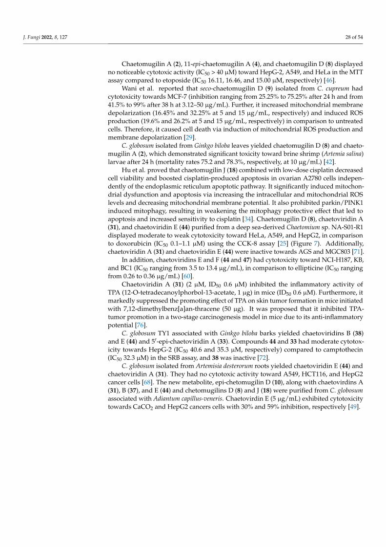
Chaetomugilin A (2), 11-epi-chaetomugilin A (4), and chaetomugilin D (8) displayedno noticeable cytotoxic activity (IC50 > 40 µM) toward HepG-2, A549, and HeLa in the MTTassay compared to etoposide (IC50 16.11, 16.46, and 15.00 µM, respectively) [46].
Wani et al. reported that seco-chaetomugilin D (9) isolated from C. cupreum hadcytotoxicity towards MCF-7 (inhibition ranging from 25.25% to 75.25% after 24 h and from41.5% to 99% after 38 h at 3.12–50 µg/mL). Further, it increased mitochondrial membranedepolarization (16.45% and 32.25% at 5 and 15 µg/mL, respectively) and induced ROSproduction (19.6% and 26.2% at 5 and 15 µg/mL, respectively) in comparison to untreatedcells. Therefore, it caused cell death via induction of mitochondrial ROS production andmembrane depolarization [29].
C. globosum isolated from Ginkgo biloba leaves yielded chaetomugilin D (8) and chaeto-mugilin A (2), which demonstrated significant toxicity toward brine shrimp (Artemia salina)larvae after 24 h (mortality rates 75.2 and 78.3%, respectively, at 10 µg/mL) [42].
Hu et al. proved that chaetomugilin J (18) combined with low-dose cisplatin decreasedcell viability and boosted cisplatin-produced apoptosis in ovarian A2780 cells indepen-dently of the endoplasmic reticulum apoptotic pathway. It significantly induced mitochon-drial dysfunction and apoptosis via increasing the intracellular and mitochondrial ROSlevels and decreasing mitochondrial membrane potential. It also prohibited parkin/PINK1induced mitophagy, resulting in weakening the mitophagy protective effect that led toapoptosis and increased sensitivity to cisplatin [34]. Chaetomugilin D (8), chaetoviridin A(31), and chaetoviridin E (44) purified from a deep sea-derived Chaetomium sp. NA-S01-R1displayed moderate to weak cytotoxicity toward HeLa, A549, and HepG2, in comparisonto doxorubicin (IC50 0.1–1.1 µM) using the CCK-8 assay [25] (Figure 7). Additionally,chaetoviridin A (31) and chaetoviridin E (44) were inactive towards AGS and MGC803 [71].
In addition, chaetoviridins E and F (44 and 47) had cytotoxicity toward NCI-H187, KB,and BC1 (IC50 ranging from 3.5 to 13.4 µg/mL), in comparison to ellipticine (IC50 rangingfrom 0.26 to 0.36 µg/mL) [60].
Chaetoviridin A (31) (2 µM, ID50 0.6 µM) inhibited the inflammatory activity ofTPA (12-O-tetradecanoylphorbol-13-acetate, 1 µg) in mice (ID50 0.6 µM). Furthermore, itmarkedly suppressed the promoting effect of TPA on skin tumor formation in mice initiatedwith 7,12-dimethylbenz[a]an-thracene (50 µg). It was proposed that it inhibited TPA-tumor promotion in a two-stage carcinogenesis model in mice due to its anti-inflammatorypotential [76].
C. globosum TY1 associated with Ginkgo biloba barks yielded chaetoviridins B (38)and E (44) and 5′-epi-chaetoviridin A (33). Compounds 44 and 33 had moderate cytotox-icity towards HepG-2 (IC50 40.6 and 35.3 µM, respectively) compared to camptothecin(IC50 32.3 µM) in the SRB assay, and 38 was inactive [72].
C. globosum isolated from Artemisia desterorum roots yielded chaetoviridin E (44) andchaetoviridin A (31). They had no cytotoxic activity toward A549, HCT116, and HepG2cancer cells [68]. The new metabolite, epi-chetomugilin D (10), along with chaetovirdins A(31), B (37), and E (44) and chetomugilins D (8) and J (18) were purified from C. globosumassociated with Adiantum capillus-veneris. Chaetovirdin E (5 µg/mL) exhibited cytotoxicitytowards CaCO2 and HepG2 cancers cells with 30% and 59% inhibition, respectively [49].

J. Fungi 2022, 8, 127 29 of 54J. Fungi 2022, 8, x FOR PEER REVIEW 25 of 53
Figure 7. Structures of compounds 37–44 (*Revised Kingsland and Barrow).
In addition, chaetoviridins E and F (44 and 47) had cytotoxicity toward NCI-H187, KB, and BC1 (IC50 ranging from 3.5 to 13.4 µg/mL), in comparison to ellipticine (IC50 rang-ing from 0.26 to 0.36 µg/mL) [60].
Chaetoviridin A (31) (2 µM, ID50 0.6 µM) inhibited the inflammatory activity of TPA (12-O-tetradecanoylphorbol-13-acetate, 1 µg) in mice (ID50 0.6 µM). Furthermore, it mark-edly suppressed the promoting effect of TPA on skin tumor formation in mice initiated with 7,12-dimethylbenz[a]an-thracene (50 µg). It was proposed that it inhibited TPA-
Figure 7. Structures of compounds 37–44 (* Revised Kingsland and Barrow).

J. Fungi 2022, 8, 127 30 of 54
3.2. Antimicrobial Activity
The liquid culture of C. globosum DAOM-240359 isolated from an indoor air samplecollected from Ottawa, Ontario, Canada produced new nitrogen-containing chaetoviridins,4′-epi-N-2-hydroxyethyl-azachaetoviridin A (36) and N-2-butyric-azochaetoviridin E (46),along with chaetoviridin A (31) and chaetomugilin D (8). Compound 36 is a nitrogenousderivative of 31 with an N-2-hydroxy ethyl chain and (R) configuration at C-4′ instead ofthe (S) configuration of 31. Compound 46 is a nitrogenous derivative of chaetoviridin E(44) with a C-2 γ-aminobutyric acid moiety. This represents the first reported azaphilonewith a 3-methyl-1-pentyl group and an N-2 side chain. Compounds 8 and 46 signifi-cantly reduced the growth of Pseudomonas putida and B. subtilus (conc. 20 µM) in thequantitative growth inhibition assay. Additionally, they showed the same effectivenessas chloramphenicol at 200 µM. On the other hand, 8 and 46 showed antifungal activitytowards Saccharomyces cerevisiae at 2 mM and 200 µM, respectively. However, 31 exertedits antibacterial activity at 200 µM [50]. Chaetovirdin A (31) possessed weak inhibitorypotential towards induction of chlamydospore-like cells of the plant pathogen Cochliobo-lus lunatus (40–50% at 100 µg/disc), and prohibition of the rice-blast fungus Pyricuraliaoryzae’s growth (IC50 2.5 µg/mL) [27]. The new metabolites, 5′-epichaetoviridin A (33),4′-epichaetoviridin F (48), 12β-hydroxychaetoviridin C (41), and chaetoviridins G-I (49,51, and 52), along with chaetoviridins A–E (31,37, 40, 43, and 44) and 4′-epichaetoviridinA (32) separated from the endophytic fungus C. globosum, were assessed in an in vivopathogenicity assay that involved the infection of Caenorhabditis elegans with Enterococcusfaecalis (Figure 8). None of them possessed a significant ability to promote nematode sur-vival [36]. Park et al. stated that chaetoviridins A (31) and B (37) also possessed growthinhibitory activity against Magnaporthe grisea (rice blast) and Pythium ultimum (wheat leafrust) mycelia (MICs 1.23 and 33.3 and 1.23 and 33.3 µg/mL, respectively) in vitro [61]. Theyalso exhibited strong in vivo antifungal activity against M. grisea and Puccnicia recondite(wheat leaf rust) [36]. Chaetoviridin A (Conc. 62.5 µg/mL) inhibited rice blast developmentby >80%, but it had moderate control (50%) of tomato late blight at 125 µg/mL. Therefore,they can control wheat leaf rust rice blast and tomato late blight [36,61]. ChaetoviridinA (31), chaetoviridin E (44), and chaetomugilin D (8) separated from a deep sea-derivedChaetomium sp. NA-S01-R1 had weak to moderate antibacterial effectiveness against Vibriostrains (V. vulnificus, V. rotiferianus, and V. campbellii) (MIC 15.4–32.3 µg/mL) and MRSA(MICs 15.2–32.4 µg/mL), compared to erythromycin (MIC 2.0–7.7 µg/mL) and chloram-phenicol (MIC 7.5–7.6 µg/mL) [25].
Chaetoviridins B (38) and E (44) displayed antibacterial activity towards E. faecalisand S. aureus [74]. Chaetoviridins A (31) and B (34) also had antimicrobial activity againstB. subtilis, Rhizoctonia solani, and E. coli (IZDs 15 and 14 mm, respectively, towards allstrains) [63].
Yan et al. reported that chaetoviridin A (31) exhibited significant antifungal potential(EC50 1.97 µg/mL) towards Sclerotinia sclerotiorum, which causes rape Sclerotinia rot (RSR).Further, 31 displayed in vivo protective efficacy (64.3%, dose 200 µg/mL) towards RSR,comparable to that of carbendazim (69.2%). Additionally, it had antifungal activity towardsBotrytis cinerea, Phytophthora capsici, Fusarium graminearum, and F. moniliforme (inhibitionrates 69.1, 60.7, 77.0, and 59.2%, respectively) [73].
C. globosum CEF-082, isolated from cotton plants, produced chaetoviridin A (31), whichpossesses significant antifungal activity towards Verticillium dahlia, which causes cottonVerticillium wilt (CVW). It induced mycelial deformation and cell necrosis, increased NOand ROS production, prohibited the germination of microsclerotia of V. dahliae, and boostedthe cotton defensive response [69].
Chaetoviridin A (31), 5′-epichaetoviridin A (33), and chaetoviridin E (44) were sep-arated from the soil-associated C. globosum 22–10. They showed significant inhibitoryeffectiveness (inhibition 32.31%, 15.38%, and 13.85%, respectively) at 100 µg/mL towardsBipolaris sorokiniana, a soil-borne pathogen that commonly causes wheat root rot. It isnoteworthy that chaetoviridin A had the same inhibitory efficiency as the carbendazim

J. Fungi 2022, 8, 127 31 of 54
(3 mg/mL), suggesting its potential to be a biocontrol agent for B. sorokiniana [67]. Further,chaetoviridin A identified from an EtOAc extract of C. globosum F211_UMNG isolated fromP. heptaphyllum was active towards F. oxysporum [66]. Chaetomugilin D (8) and chaetoviridinA (31) (200 µM) significantly reduced the growth of B. subtilis and Psuedomonas putida [55].
J. Fungi 2022, 8, x FOR PEER REVIEW 27 of 53
Figure 8. Structures of compounds 45–54.
Chaetoviridins B (38) and E (44) displayed antibacterial activity towards E. faecalis and S. aureus [74]. Chaetoviridins A (31) and B (34) also had antimicrobial activity against B. subtilis, Rhizoctonia solani, and E. coli (IZDs 15 and 14 mm, respectively, towards all strains) [63].
Yan et al. reported that chaetoviridin A (31) exhibited significant antifungal potential (EC50 1.97 µg/mL) towards Sclerotinia sclerotiorum, which causes rape Sclerotinia rot (RSR). Further, 31 displayed in vivo protective efficacy (64.3%, dose 200 µg/mL) towards RSR, comparable to that of carbendazim (69.2%). Additionally, it had antifungal activity to-wards Botrytis cinerea, Phytophthora capsici, Fusarium graminearum, and F. moniliforme (in-hibition rates 69.1, 60.7, 77.0, and 59.2%, respectively) [73].
C. globosum CEF-082, isolated from cotton plants, produced chaetoviridin A (31), which possesses significant antifungal activity towards Verticillium dahlia, which causes cotton Verticillium wilt (CVW). It induced mycelial deformation and cell necrosis,
Figure 8. Structures of compounds 45–54.
3.3. Phytotoxic Activity
The EtOAc extract of C. globosum associated with Amaranthus viridis yielded chaeto-mugilin D (8) and chaetomugilin J (18), which exhibited phytotoxic potential against lettuce(Lactuca sativa) seed germination with IC50 24.2 and 22.6 ppm, respectively, for root growthinhibition, and IC50 27.8 and 21.9 ppm, respectively, for shoot growth inhibition. The resultsrevealed the potential of these metabolites as herbicides or weedicides that can replace

J. Fungi 2022, 8, 127 32 of 54
hazardous synthetic compounds [51]. Chaetomugilin A (2), D (8), S (28), I (16), J (18), Q (25),and O (23) isolated from C. globosum TY1 exhibited allelopathic activity towards Brassicacampestris, Cucumis sativus, Eruca sativa, Daucus carota, Lactuca sativa, Scrophularia ningpoen-sis, Brassica rapa, and Spinacia oleracea. Among them, 23 exhibited higher germination androot and shoot elongation inhibitory potential with lower IC50 values and higher responseindexes than glyphosate (positive control). Moreover, 2, 8, and 28 exhibited similar orbetter inhibitory effects than glyphosate. At the same time, 2, 8, and 23 were more power-ful growth inhibitors than 16, 18, and 25, which could be attributed to the existence of atetrahydrofuran moiety. On the other hand, 23 had a higher growth-suppression effect thanthose of 2, 8, and 28, suggesting that the lactone rings may reduce the inhibitory effects [45].
3.4. Antimalarial and Antimycobacterial Activities
Phonkerd et al. isolated new derivatives, chaetoviridins E and F (44 and 47) and5′-epi-chaetoviridin A (33), together with chaetoviridin A (31) from C cochliodes VTh01and C. cochliodes CTh05. Compound 44 showed antimalarial activity against P. falciparum(IC50 2.9 µg/mL)—and was compared to artemisinin (IC50 0.001 µg/mL)—using the micro-culture radioisotope technique [60]. Additionally, 44 and 47 displayed weak antimycobacte-rial potential towards M. tuberculosis (MIC 50 and 100 µg/mL, respectively), in comparisonto isoniazid and kanamycin sulfate in the microplate Alamar Blue assay (MABA) [60].
3.5. Anti-Inflammatory Activity
Chaetomugilin D (8) from C. globosum isolated from damp building materials notablyincreased TNF-α production in RAW 264.7 murine macrophages. Therefore, 8 contributesto the non-allergy-linked respiratory disorders such as non-allergic asthma and rhinitis forpeople working and living in damp buildings [52]. Two new derivatives, chaetoviridins J(53) and K (54), along with 11-epi-chaetomugilin I (17) and chaetomugilins E (11), F (13),I (16), J (18), and N (22) biosynthesized by C. globosum were isolated from Wikstroemiauva-ursi leaves. Their structures were verified by NMR, Mosher’s method, X-ray diffraction,and CD. Chaetoviridin K (54) was separated as a mixture of diastereoisomers that couldnot be purified by chiral columns. Their potential to inhibit TNF-α-induced NF-κB and NO(nitric oxide) production in the LPS-stimulated RAW 264.7 cells was assessed. Compounds16 and 17 remarkably suppressed TNF-α-induced NF-κB activity (IC50 0.9 µM, at 50 µM),in comparison to BAY-11 (IC50 2.0 µM) and TPCK (IC50 3.8 µM), whereas 11, 13, and 18possessed moderate inhibitory activity (IC50 ranging from 5.1 to 11.6 µM, conc. 50 µM).In addition, 11, 13, 16–18, and 53 strongly prohibited NO production (74.2–99.9%). Itis noteworthy that 16 and 17 powerfully suppressed NO release (IC50 0.3 and 0.8 µM,respectively) more than N-monomethyl-L-arginine (IC50 25.1 µM). On the other hand, 11,13, and 18 also considerably inhibited NO production (IC50 values ranging from 1.9 to5.8 µM); however, 54 exhibited a weak effect [56].
3.6. Antidiabetic, Antioxidant, and Antiviral Activities
Chaetomugilins A (2), I (16), J (18), and Q (25) and chaetoviridins B (38) and J (53)purified from the EtOAc extract of C. globosum TY-1 isolated from Ginkgo biloba barkwere tested for α-amylase and α-glucosidase inhibitory activity. Chaetoviridin B (38)had promising α-glucosidase inhibitory potential (IC50 6.328 µM), compared to acarbose(IC50 54.7 µM). The other compounds displayed no α-amylase or α-glucosidase inhibitoryactivity (IC50 > 50 µM) compared to acarbose (IC50 13.7 and 54.7 µM, respectively) [47].Chaetoviridin B (38) and chaetomugilin A (2) have the same skeleton except for the 2having one more hydroxyl group in the side chain, revealing that the group is detrimentalfor α-glucosidase inhibition [47]. Chaetoviridins A (31) and B (37) purified from an EtOAcextract of C. globosum exhibited noticeable antioxidant potential on TLC using DPPH [63].Additionally, chetomugilin D (8) and its analog epi-chetomugilin D (10) possessed antiviralactivity towards HSV-2 (inhibition 33.3% and 40.7%, respectively, at 25µg/mL [49].

J. Fungi 2022, 8, 127 33 of 54
3.7. Caspase-3 and Monoamine Oxidase (MAO) Inhibitory Activities
Caspase-3 (cysteine aspartyl-specific protease-3) is one of the executioners in caspase-linked apoptosis that is activated in nearly every apoptosis model [77]. It is a prominenttherapeutic target for excessive apoptosis-associated disorders, such as ischemic damageand neurodegenerative disorders (e.g., Huntington’s and Alzheimer’s diseases) and au-toimmune disorders [78]. New azaphilones, chaetomugilin S (27), 7,5′-bis-epi-chaetoviridinA (34), and 7-epi-chaetoviridin E (45), purified from the lichen-associated C. elatum 89-1-3-1,were isolated from Ramalina calicaris. Their absolute configurations were assigned by CDexperiments and X-ray crystallography. They exhibited caspase-3 inhibitory potential (IC5020.6, 10.9, and 7.9 µM, respectively) in the cysteine aspartyl-specific protease-3 enzymaticassay compared with Ac-DEVD-CHO (IC50 13.7 nM) [31]. On the other hand, 31 possessedweak MAO inhibitory potential (IC50 1.2 × 10⁻² g/mL) [27].
3.8. Cholesteryl Ester Transfer Protein Inhibitory Activity
CETP allows the transfer and exchange of neutral lipids such as CE (cholesteryl ester)and TG (triacylglycerol) between plasma and lipoproteins. It is proven to play importantrole in atherosclerosis [79]. Tomoda et al. reported that chaetoviridin B (37) showedCETP (cholesteryl ester transfer protein) inhibitory activity with an IC50 < 6.3 µM, whereaschaetoviridin A (31) had moderate inhibitory activity (IC50 31.6 µM) [80]. It was indicatedthat the existence of an electrophilic enone(s) and/or ketone(s) at both C-8 and C-6 ofisochromane core is substantial for eliciting activity [80].
3.9. Anti-SARS-CoV-2 Activity
The COVID-19 pandemic has affected global health since 2019. COVID-19 can leadto acute respiratory distress syndrome [81]. It is produced by a novel type of coronavirus(CoV) called SARS-CoV-2 that was first found in Wuhan City, China, and then spreadworldwide [82,83]. It is considered a highly pathogenic CoV in the human population. TheSARS-CoV-2 genome encodes two polyproteins which are processed by a 3C-like protease(3CLpro) and a papain-like protease [84]. 3CLpro (3C-like protease) and PLpro (papain-likeprotease) are needed for processing the polyproteins into mature nonstructural proteins,such as helicase and RdRp (RNA-dependent RNA polymerase), which are substantialfor viral replication and transcription [85]. 3CLpro has high substrate specificity and isalso referred to as Mpro (main protease) [86]. 3CLpro’s substrate specificity makes thisenzyme an ideal target for developing broad-spectrum antiviral agents [87]. Its inhibitorsare expected to have selective toxicity towards the virus [88].
Fungi are a treasure that can provide a remarkable pool of secondary metabolites withantiviral activities [89]. The characterization and discovery of antiviral fungal metabolitesis an emerging and promising research field. Recently, many reports have been pub-lished on the structure-based virtual screening approach for the repurposing of naturalmetabolites, hoping to accelerate and assist in the discovery of agents for COVID-19 treat-ment [82]. We carried out a computational study on the reported fungal chaetomugilinsand chaetoviridins was carried out to identify their 3CLpro inhibitory potential, usingdocking calculations and MD simulations (Tables 3–7). Three crystal structures containingnon-covalent inhibitors for the protease (PDB entry: 6W81, 6M2N, and 7K0F) were selected.All the listed metabolites were docked with extra precision for maximum accuracy. Thedocking method was validated by redocking the inhibitors that co-crystallized with 6W81,6M2N, and 7K0F; and RMSD values were within an acceptable range and less than 1.50 Å.All the redocked inhibitors revealed the same binding interaction with the active site inthe original pose. Further, in silico ADMET (drug absorption, distribution, metabolism,excretion, and toxicity) predictions of the properties of the investigated compounds werecarried out. Finally, a molecular dynamics simulation was conducted to evaluate the natureof the ligand–target interaction under simulated physiological conditions for the mostcompatible drug-like molecule that could be used in pursuit of a truly adequate medicationfor COVID-19.

J. Fungi 2022, 8, 127 34 of 54
Table 3. In silico screening results of top candidates of the reported chaetomugilins and chaetoviridins against SARS-CoV-2 3CL protease (PDB: 6W81, 6M2N,and 7K0F).
Name & StructureFor 6W81 For 6M2N For 7K0F
DockingScore
GlideGscore
GlideEmodel
XPGScore
DockingScore
GlideGscore
GlideEmodel
XPGscore
DockingScore
GlideGscore
GlideEmodel
XPGscore
J. Fungi 2022, 8, x FOR PEER REVIEW 34 of 53
Table 3. In silico screening results of top candidates of the reported chaetomugilins and chaetoviridins against SARS-CoV-2 3CL protease (PDB: 6W81, 6M2N, and 7K0F).
Name & Structure For 6W81 For 6M2N For 7K0F
Docking Score Glide
Gscore Glide
Emodel XP GScore
Docking Score
Glide Gscore
Glide Emodel
XP Gscore Docking
Score Glide
Gscore Glide Emodel XP Gscore
−6.901 −6.918 −45.952 −6.918 −6.862 −6.879 −40.382 −6.879 −6.428 −6.445 −45.402 −6.445
−6.634 −6.933 −53.592 −6.933 −6.657 −7.205 −59.649 −7.205 −6.214 −6.762 −57.163 −6.762
−7.293 −7.294 −52.497 −7.294 −7.851 −7.851 −46.384 −7.851 −6.886 −6.904 −44.124 −6.904
−6.901 −6.918 −45.952 −6.918 −6.862 −6.879 −40.382 −6.879 −6.428 −6.445 −45.402 −6.445
J. Fungi 2022, 8, x FOR PEER REVIEW 34 of 53
Table 3. In silico screening results of top candidates of the reported chaetomugilins and chaetoviridins against SARS-CoV-2 3CL protease (PDB: 6W81, 6M2N, and 7K0F).
Name & Structure For 6W81 For 6M2N For 7K0F
Docking Score Glide
Gscore Glide
Emodel XP GScore
Docking Score
Glide Gscore
Glide Emodel
XP Gscore Docking
Score Glide
Gscore Glide Emodel XP Gscore
−6.901 −6.918 −45.952 −6.918 −6.862 −6.879 −40.382 −6.879 −6.428 −6.445 −45.402 −6.445
−6.634 −6.933 −53.592 −6.933 −6.657 −7.205 −59.649 −7.205 −6.214 −6.762 −57.163 −6.762
−7.293 −7.294 −52.497 −7.294 −7.851 −7.851 −46.384 −7.851 −6.886 −6.904 −44.124 −6.904
−6.634 −6.933 −53.592 −6.933 −6.657 −7.205 −59.649 −7.205 −6.214 −6.762 −57.163 −6.762
J. Fungi 2022, 8, x FOR PEER REVIEW 34 of 53
Table 3. In silico screening results of top candidates of the reported chaetomugilins and chaetoviridins against SARS-CoV-2 3CL protease (PDB: 6W81, 6M2N, and 7K0F).
Name & Structure For 6W81 For 6M2N For 7K0F
Docking Score Glide
Gscore Glide
Emodel XP GScore
Docking Score
Glide Gscore
Glide Emodel
XP Gscore Docking
Score Glide
Gscore Glide Emodel XP Gscore
−6.901 −6.918 −45.952 −6.918 −6.862 −6.879 −40.382 −6.879 −6.428 −6.445 −45.402 −6.445
−6.634 −6.933 −53.592 −6.933 −6.657 −7.205 −59.649 −7.205 −6.214 −6.762 −57.163 −6.762
−7.293 −7.294 −52.497 −7.294 −7.851 −7.851 −46.384 −7.851 −6.886 −6.904 −44.124 −6.904 −7.293 −7.294 −52.497 −7.294 −7.851 −7.851 −46.384 −7.851 −6.886 −6.904 −44.124 −6.904

J. Fungi 2022, 8, 127 35 of 54
Table 3. Cont.
Name & StructureFor 6W81 For 6M2N For 7K0F
DockingScore
GlideGscore
GlideEmodel
XPGScore
DockingScore
GlideGscore
GlideEmodel
XPGscore
DockingScore
GlideGscore
GlideEmodel
XPGscore
J. Fungi 2022, 8, x FOR PEER REVIEW 35 of 53
Name & Structure For 6W81 For 6M2N For 7K0F
Docking Score Glide
Gscore Glide
Emodel XP GScore
Docking Score
Glide Gscore
Glide Emodel
XP Gscore Docking
Score Glide
Gscore Glide Emodel XP Gscore
−6.838 −7.269 −59.268 −7.269 −7.4 −7.791 −61.032 −7.791 −6.599 −7.03 −50.901 −7.03
−6.94 −6.958 −40.421 −6.958 −6.921 −6.939 −42.35 −6.939 −6.886 −6.904 −44.124 −6.904
−6.48 −6.48 −57.388 −6.48 −7.519 −7.519 −67.872 −7.519 −7.311 −7.311 −65.197 −7.311
−7.944 −7.944 −51.069 −7.944 −8.141 −8.141 −48.92 −8.141 −6.615 −6.615 −60.199 −6.615
−6.838 −7.269 −59.268 −7.269 −7.4 −7.791 −61.032 −7.791 −6.599 −7.03 −50.901 −7.03
J. Fungi 2022, 8, x FOR PEER REVIEW 35 of 53
Name & Structure For 6W81 For 6M2N For 7K0F
Docking Score Glide
Gscore Glide
Emodel XP GScore
Docking Score
Glide Gscore
Glide Emodel
XP Gscore Docking
Score Glide
Gscore Glide Emodel XP Gscore
−6.838 −7.269 −59.268 −7.269 −7.4 −7.791 −61.032 −7.791 −6.599 −7.03 −50.901 −7.03
−6.94 −6.958 −40.421 −6.958 −6.921 −6.939 −42.35 −6.939 −6.886 −6.904 −44.124 −6.904
−6.48 −6.48 −57.388 −6.48 −7.519 −7.519 −67.872 −7.519 −7.311 −7.311 −65.197 −7.311
−7.944 −7.944 −51.069 −7.944 −8.141 −8.141 −48.92 −8.141 −6.615 −6.615 −60.199 −6.615
−6.94 −6.958 −40.421 −6.958 −6.921 −6.939 −42.35 −6.939 −6.886 −6.904 −44.124 −6.904
J. Fungi 2022, 8, x FOR PEER REVIEW 35 of 53
Name & Structure For 6W81 For 6M2N For 7K0F
Docking Score Glide
Gscore Glide
Emodel XP GScore
Docking Score
Glide Gscore
Glide Emodel
XP Gscore Docking
Score Glide
Gscore Glide Emodel XP Gscore
−6.838 −7.269 −59.268 −7.269 −7.4 −7.791 −61.032 −7.791 −6.599 −7.03 −50.901 −7.03
−6.94 −6.958 −40.421 −6.958 −6.921 −6.939 −42.35 −6.939 −6.886 −6.904 −44.124 −6.904
−6.48 −6.48 −57.388 −6.48 −7.519 −7.519 −67.872 −7.519 −7.311 −7.311 −65.197 −7.311
−7.944 −7.944 −51.069 −7.944 −8.141 −8.141 −48.92 −8.141 −6.615 −6.615 −60.199 −6.615
−6.48 −6.48 −57.388 −6.48 −7.519 −7.519 −67.872 −7.519 −7.311 −7.311 −65.197 −7.311

J. Fungi 2022, 8, 127 36 of 54
Table 3. Cont.
Name & StructureFor 6W81 For 6M2N For 7K0F
DockingScore
GlideGscore
GlideEmodel
XPGScore
DockingScore
GlideGscore
GlideEmodel
XPGscore
DockingScore
GlideGscore
GlideEmodel
XPGscore
J. Fungi 2022, 8, x FOR PEER REVIEW 35 of 53
Name & Structure For 6W81 For 6M2N For 7K0F
Docking Score Glide
Gscore Glide
Emodel XP GScore
Docking Score
Glide Gscore
Glide Emodel
XP Gscore Docking
Score Glide
Gscore Glide Emodel XP Gscore
−6.838 −7.269 −59.268 −7.269 −7.4 −7.791 −61.032 −7.791 −6.599 −7.03 −50.901 −7.03
−6.94 −6.958 −40.421 −6.958 −6.921 −6.939 −42.35 −6.939 −6.886 −6.904 −44.124 −6.904
−6.48 −6.48 −57.388 −6.48 −7.519 −7.519 −67.872 −7.519 −7.311 −7.311 −65.197 −7.311
−7.944 −7.944 −51.069 −7.944 −8.141 −8.141 −48.92 −8.141 −6.615 −6.615 −60.199 −6.615 −7.944 −7.944 −51.069 −7.944 −8.141 −8.141 −48.92 −8.141 −6.615 −6.615 −60.199 −6.615
J. Fungi 2022, 8, x FOR PEER REVIEW 36 of 53
Name & Structure For 6W81 For 6M2N For 7K0F
Docking Score Glide
Gscore Glide
Emodel XP GScore
Docking Score
Glide Gscore
Glide Emodel
XP Gscore Docking
Score Glide
Gscore Glide Emodel XP Gscore
Ref_6W81
−5.377 −5.377 −86.001 −5.377
Ref_6M2N
−6.995 −6.995 −52.4 −6.995
Ref_K0F
−8.159 −8.159 −113.296 −8.159
Ref_6W81
−5.377 −5.377 −86.001 −5.377
J. Fungi 2022, 8, x FOR PEER REVIEW 36 of 53
Name & Structure For 6W81 For 6M2N For 7K0F
Docking Score Glide
Gscore Glide
Emodel XP GScore
Docking Score
Glide Gscore
Glide Emodel
XP Gscore Docking
Score Glide
Gscore Glide Emodel XP Gscore
Ref_6W81
−5.377 −5.377 −86.001 −5.377
Ref_6M2N
−6.995 −6.995 −52.4 −6.995
Ref_K0F
−8.159 −8.159 −113.296 −8.159
Ref_6M2N
−6.995 −6.995 −52.4 −6.995
J. Fungi 2022, 8, x FOR PEER REVIEW 36 of 53
Name & Structure For 6W81 For 6M2N For 7K0F
Docking Score Glide
Gscore Glide
Emodel XP GScore
Docking Score
Glide Gscore
Glide Emodel
XP Gscore Docking
Score Glide
Gscore Glide Emodel XP Gscore
Ref_6W81
−5.377 −5.377 −86.001 −5.377
Ref_6M2N
−6.995 −6.995 −52.4 −6.995
Ref_K0F
−8.159 −8.159 −113.296 −8.159
Ref_K0F
−8.159 −8.159 −113.296 −8.159

J. Fungi 2022, 8, 127 37 of 54
Table 4. In silico screening results of the reported chaetomugilins and chaetoviridins against SARS-CoV-2 3CL protease (PDB: 6W81).
Fungal Chaetomugilins and Chaetoviridins Docking Score Glide Gscore Glide Emodel XP Gscore
Chaetovirdin D (43) −7.944 −7.944 −51.069 −7.944Chaetomugilin P (24) −7.293 −7.294 −52.497 −7.294
N-Glutrylchaetovirdin A (35) −7.101 −7.101 −66.739 −7.101Chaetovirdin B (37) −6.959 −6.959 −37.203 −6.959
Chaetomugilin R (26) −6.94 −6.958 −40.421 −6.958Chaetomugilin 106B-6 XXVIII (1) −6.901 −6.918 −45.952 −6.918
N-Glutarylchaetovirdin C (42) −6.881 −6.881 −60.87 −6.881Chaetomugilin Q (25) −6.838 −7.269 −59.268 −7.269
Chaetomugilin EA-4 (12) −6.634 −6.933 −53.592 −6.9337,4′,5′-Tris-Epi-chaetovirdin A (34) −6.519 −6.519 −56.68 −6.519
4′-Epi-N-2-Hydroxyethyl-azachaetovirdin A (36) −6.48 −6.48 −57.388 −6.48Chaetomugilin T (26) −6.407 −6.407 −45.87 −6.407
Chaetovirdin J (53) −6.295 −6.673 −54.688 −6.67312β-Hydroxychaetovirdin C (41) −6.074 −6.08 −54.03 −6.08
Chaetomugilin M (21) −5.985 −6.143 −53.589 −6.143Chaetovirdin G (50) −5.925 −5.925 −50.419 −5.925Chaetomugilin I (16) −5.919 −6.467 −53.609 −6.467
N-2-Butyric-azochaetovirdin E (46) −5.862 −5.863 −58.955 −5.86311-Epi-chaetomugilin A (4) −5.771 −5.774 −45.965 −5.774
Chaetovirdin C (40) −5.752 −5.752 −48.221 −5.752Chaetomugilin H (15) −5.741 −5.741 −40.295 −5.741
N-Glutarylchaetovirdin B (39) −5.733 −5.735 −57.615 −5.735Chaetomugilin U (30) −5.622 −5.622 −40.195 −5.622
4′-Epi-chaetovirdin A (5) −5.561 −5.561 −47.473 −5.561Chaetomugilin S (27) −5.539 −5.539 −45.867 −5.539
4′,5′-Bis-chaetovirdin A (33) −5.507 −5.507 −48.38 −5.507Chaetovirdin A (31) −5.493 −5.493 −51.732 −5.493
Ref_6W81 −5.377 −5.377 −86.001 −5.377Chaetovirdin I (52) −5.354 −5.419 −42.206 −5.419
Seco-Chaetomugilin D (9) −5.345 −5.347 −45.22 −5.347Chaetomugilin L (20) −5.25 −5.25 −39.261 −5.25Chaetomugilin B (6) −5.248 −5.248 −41.211 −5.248Chaetomugilin A (2) −5.241 −5.243 −44.852 −5.243
11-Epi-chaetomugilin I (17) −5.099 −5.647 −54.765 −5.647Chaetomugilin D (8) −5.093 −5.095 −41.171 −5.095
7-Epi-chaetovirdin E (45) −5.089 −5.089 −40.702 −5.089Chaetovirdin K (54) −4.987 −4.989 −45.166 −4.989
Chaetomugilin N (22) −4.977 −4.977 −3454.68 −4.977Seco-chaetomugilin A (3) −4.926 −4.928 −54.168 −4.928
Chaetomugilin K (19) −4.903 −4.903 −38.494 −4.903Chaetovirdin H (51) −4.898 −4.898 −51.406 −4.898
4′-Epi-chaetovirdin F (X48) −4.873 −4.873 −47.708 −4.873Chaetovirdin B (38) −4.792 −4.794 −42.458 −4.794Chaetovirdin E (44) −4.61 −4.61 −48.663 −4.61
Chaetomugilin G (14) −4.516 −4.516 −50.66 −4.516Chaetomugilin S (28) −4.391 −4.393 −48.292 −4.393Chaetomugilin F (13) −4.28 −4.28 −40.132 −4.28Chaetovirdin F (47) −4.271 −4.271 −50.793 −4.271
Epi-chaetomugilin D (10) −4.137 −4.139 −36.43 −4.139Chaetomugilin E (11) −3.597 −3.597 −40.424 −3.597Chaetomugilin C (7) −3.509 −3.509 −44.435 −3.509Chaetovirdin G (49) −3.492 −3.492 −48.133 −3.492

J. Fungi 2022, 8, 127 38 of 54
Table 5. In silico screening results of the reported chaetomugilins and chaetoviridins against SARS-CoV-2 3CL protease (PDB: 6M2N).
Fungal Chaetomugilins and Chaetoviridins Docking Score Glide Gscore Glide Emodel XP Gscore
Chaetovirdin D (43) −8.141 −8.141 −48.92 −8.141Chaetovirdin B (37) −7.851 −7.851 −46.384 −7.851
4′-Epi-N-2-Hydroxyethyl-azachaetovirdin A (36) −7.519 −7.519 −67.872 −7.519Chaetomugilin Q (25) −7.4 −7.791 −61.032 −7.791
12β-Hydroxychaetovirdin C (41) −7.045 −7.051 −58.607 −7.051Ref_6M2N −6.995 −6.995 −52.4 −6.995
Chaetomugilin R (26) −6.921 −6.939 −42.35 −6.939Chaetomugilin P (24) −6.9 −6.901 −48.711 −6.901
Chaetomugilin 106B-6 XXVIII (1) −6.862 −6.879 −40.382 −6.879Chaetomugilin S (27) −6.785 −6.785 −50.138 −6.785
Chaetomugilin EA-4 (12) −6.657 −7.205 −59.649 −7.205Chaetomugilin M (21) −6.655 −6.813 −56.59 −6.813
7,4′,5′-Tris-Epi-chaetovirdin A (34) −6.44 −6.44 −55.163 −6.44Chaetovirdin I (52) −6.439 −6.504 −53.296 −6.504Chaetovirdin J (53) −6.362 −6.741 −54.219 −6.741Chaetovirdin G (50) −6.312 −6.312 −48.248 −6.312
Chaetomugilin G (49) −6.272 −6.272 −56.423 −6.272Chaetovirdin C (40) −6.219 −6.219 −59.734 −6.219
N-Glutarylchaetovirdin B (39) −6.178 −6.179 −53.868 −6.17911-Epi-chaetomugilin I (17) −6.077 −6.625 −61.469 −6.625
Chaetovirdin H (51) −6.071 −6.071 −45.006 −6.071Chaetovirdin A (31) −5.93 −5.93 −47.164 −5.93
4′,5′-Bis-chaetovirdin A (33) −5.896 −5.896 −46.971 −5.896Seco-chaetomugilin A (3) −5.89 −5.892 −44.484 −5.892
Chaetovirdin E (44) −5.871 −5.871 −48.647 −5.871Chaetomugilin I (16) −5.87 −6.418 −55.891 −6.418Chaetovirdin F (47) −5.866 −5.866 −54.419 −5.866
4′-Epi-chaetovirdin F (48) −5.859 −5.859 −51.775 −5.859Chaetomugilin F (13) −5.759 −5.759 −43.54 −5.759
N-2-Butyric-azochaetovirdin E (46) −5.453 −5.454 −54.064 −5.454N-Glutrylchaetovirdin A (35) −5.405 −5.405 −59.359 −5.405
Seco-Chaetomugilin D (9) −5.376 −5.379 −41.076 −5.379Chaetomugilin H (15) −5.293 −5.293 −52.628 −5.293
Chaetovirdin B (38) −5.217 −5.219 −38.105 −5.21911-Epi-chaetomugilin A (4) −5.166 −5.168 −41.465 −5.168
Chaetovirdin K (54) −5.11 −5.113 −38.902 −5.113Chaetomugilin A (2) −5.09 −5.092 −36.177 −5.092Chaetomugilin T (29) −5.012 −5.012 −39.774 −5.012Chaetomugilin C (7) −4.987 −4.987 −44.839 −4.987
4′-Epi-chaetovirdin A (32) −4.905 −4.905 −58.103 −4.905Chaetomugilin S (28) −4.627 −4.629 −36.966 −4.629Chaetovirdin G (49) −4.612 −4.612 −42.176 −4.612
7-Epi-chaetovirdin E (45) −4.485 −4.485 −51.758 −4.485Epi-chaetomugilin D (10) −4.341 −4.343 −31.912 −4.343
Chaetomugilin U (30) −4.248 −4.249 −40.187 −4.249Chaetomugilin O (23) −4.208 −4.208 −1767.6 −4.208Chaetomugilin N (22) −4.07 −4.07 −31.442 −4.07Chaetomugilin B (6) −4.068 −4.068 −41.99 −4.068
Chaetomugilin K (19) −3.867 −3.867 −34.284 −3.867Chaetomugilin D (8) −3.667 −3.669 −35.613 −3.669Chaetomugilin L (20) −3.622 −3.622 −38.371 −3.622Chaetomugilin E (11) −3.506 −3.506 −41.116 −3.506
N-Glutarylchaetovirdin C (42) −1.412 −1.412 −62.951 −1.412

J. Fungi 2022, 8, 127 39 of 54
Table 6. In silico screening results of the reported chaetomugilins and chaetoviridins against SARS-CoV-2 3CL protease (PDB: 7K0F).
Fungal Chaetomugilins and Chaetoviridins Docking Score Glide Gscore Glide Emodel XP Gscore
Ref_7K0F −8.159 −8.159 −113.296 −8.1594′-Epi-N-2-Hydroxyethyl-azachaetovirdin A (36) −7.311 −7.311 −65.197 −7.311
Chaetomugilin R (26) −6.886 −6.904 −44.124 −6.904Chaetovirdin D (43) −6.615 −6.615 −60.199 −6.615
Chaetomugilin Q (25) −6.599 −7.03 −50.901 −7.03Chaetomugilin 106B-6 XXVIII (1) −6.428 −6.445 −45.402 −6.445
Chaetovirdin I (52) −6.381 −6.446 −60.443 −6.446Chaetomugilin EA-4 (12) −6.214 −6.762 −57.163 −6.762Seco-chaetomugilin A (3) −6.164 −6.166 −54.533 −6.166
Chaetomugilin T (29) −6.003 −6.003 −44.936 −6.003Chaetomugilin A (2) −5.937 −5.939 −49.214 −5.939
4′-Epi-chaetovirdin F (48) −5.781 −5.781 −53.249 −5.781Chaetovirdin G (50) −5.73 −5.73 −46.915 −5.73
11-Epi-chaetomugilin A (4) −5.634 −5.636 −45.502 −5.636Chaetomugilin M (21) −5.56 −5.718 −41.62 −5.718
Chaetovirdin K (54) −5.521 −5.523 −43.983 −5.523Chaetovirdin H (51) −5.487 −5.487 −53.211 −5.487Chaetovirdin J (53) −5.475 −5.853 −56.559 −5.853
Chaetomugilin D (8) −5.457 −5.459 −40.705 −5.459Chaetomugilin S (28) −5.409 −5.411 −37.945 −5.411Chaetovirdin A (31) −5.356 −5.356 −55.812 −5.356Chaetomugilin J (18) −5.262 −5.758 −44.768 −5.758Chaetovirdin F (47) −5.214 −5.214 −51.016 −5.214Chaetovirdin E (44) −5.207 −5.207 −57.958 −5.207
Chaetomugilin U (30) −5.17 −5.17 −39.245 −5.17Chaetomugilin G (14) −5.154 −5.154 −52.872 −5.154
4′,5′-Bis-chaetovirdin A (33) −5.147 −5.147 −56.898 −5.147Chaetomugilin H (15) −5.144 −5.144 −51.211 −5.144Chaetovirdin C (40) −5.119 −5.119 −51.53 −5.119
Epi-chaetomugilin D (10) −5.11 −5.112 −41.394 −5.112Chaetomugilin P (24) −5.083 −5.085 −47.434 −5.085Chaetomugilin S (27) −5.005 −5.005 −53.874 −5.005
N-2-Butyric-azochaetovirdin E (46) −4.926 −4.927 −58.939 −4.9277,4′,5′-Tris-Epi-chaetovirdin A (34) −4.883 −4.883 −50.65 −4.883
11-Epi-chaetomugilin I (17) −4.785 −5.084 −57.348 −5.084Chaetomugilin B (6) −4.732 −4.732 −44.687 −4.732Chaetomugilin I (16) −4.534 −4.834 −53.036 −4.834Chaetomugilin C (7) −4.469 −4.469 −45.833 −4.469
12β-Hydroxychaetovirdin C (41) −4.352 −4.357 −51.515 −4.357Chaetomugilin E (11) −4.268 −4.268 −39.678 −4.268
N-Glutarylchaetovirdin C (42) −4.247 −4.247 −59.534 −4.247Chaetomugilin K (19) −4.245 −4.245 −47.953 −4.245Chaetovirdin G (59) −4.218 −4.218 −46.464 −4.218
Seco-Chaetomugilin D (9) −4.205 −4.207 −48.935 −4.207N-Glutrylchaetovirdin A (39) −4.171 −4.171 −55.598 −4.171
Chaetomugilin F (13) −3.88 −3.88 −42.311 −3.88Chaetomugilin R (26) −3.808 −5.942 −41.217 −5.942
7-Epi-chaetovirdin E (45) −3.699 −3.699 −46.38 −3.699Chaetomugilin L (20) −3.612 −3.612 −43.218 −3.612
4′-Epi-chaetovirdin A (32) −3.552 −3.552 −58.379 −3.552Chaetomugilin O (23) −2.951 −2.951 31.679 −2.951Chaetovirdin B (38) −2.642 −2.644 −45.702 −2.644
Chaetomugilin N (22) −0.921 −0.921 −3319.51 −0.921N-Glutarylchaetovirdin B (39) −0.451 −0.453 −57.257 −0.453

J. Fungi 2022, 8, 127 40 of 54
Table 7. In silico predicted ADME properties of the selected fungal chaetomugilins and chaetoviridins.
Title Mol MW #Stars Dipole SASA DonorHB accptHB QPlogPo/w QPlogS QPlogKhsa #Metab QPlogBB
PercentHuman OralAbsorption
QPlogHERG CNS #rtvFG
Recommended Range (130–725) (0.0–5.0) (1–12.50) (300–1000) (0–6) (2.0–20.0) (−2–6.5) (−6.5–0.5) (−1.5–1.5) (1–8) (−3–1.2) (<25% Poor;>80% High)
ConcernBelow −5
(−2 Inactive)(+2 Active) (0–2)
Chaetovirdin K (54) 450.915 0 6.534 733.38 2 7.75 3.725 −6.195 0.507 3 −0.978 100 −5.072 −1 2Chaetovirdin J (53) 408.921 0 9.823 723.174 1 5.95 4.444 −6.019 0.636 6 −1.162 100 −4.971 −2 1Chaetovirdin I (52) 466.914 0 8.017 729.53 3 9.7 2.323 −5.013 0.029 5 −1.839 77.834 −4.801 −2 1Chaetovirdin H (51) 398.455 0 10.648 693.648 0 8.2 2.632 −3.929 −0.18 3 −1.388 87.791 −4.859 −2 0Chaetovirdin G (50) 420.932 0 11.089 718.605 0 6.2 4.554 −5.704 0.494 4 −0.639 100 −4.912 0 1Chaetovirdin G (49) 416.9 0 9.346 698.669 0 4.5 4.914 −6.381 0.89 5 −0.882 100 −4.698 −1 1
4′-Epi-chaetovirdin F (48) 416.9 0 11.445 723.973 0 7.5 3.722 −5.295 0.155 2 −0.793 100 −5.072 −1 0Chaetovirdin F (47) 416.9 0 11.992 704.483 0 7.5 3.502 −4.926 0.089 2 −0.858 100 −4.864 −1 0
N-2-Butyric-azochaetovirdin E (46) 499.99 1 13.922 827.776 1 9.5 4.264 −6.466 0.317 6 −2.11 77.59 −3.438 −2 17-Epi-chaetovirdin E (45) 414.885 0 11.745 707.051 0 7.5 3.482 −5.136 0.123 3 −0.816 100 −4.941 −1 1
Chaetovirdin E (44) 414.885 0 11.253 703.096 0 7.5 3.481 −5.061 0.115 3 −0.776 100 −4.877 −1 1Chaetovirdin D (43) 468.93 0 11.587 715.44 3 8.4 3.631 −4.999 0.09 5 −1.572 78.707 −2.816 −2 1
N-Glutarylchaetovirdin C (42) 572.053 0 12.347 840.463 0 11.5 3.737 −4.755 0.074 7 −1.845 73.79 −4.594 −2 312β-Hydroxychaetovirdin C (41) 450.915 0 2.581 723.069 1 6.9 3.938 −5.652 0.514 6 −1.528 92.976 −4.818 −2 0
Chaetovirdin C (40) 434.916 1 9.724 758.733 0 5.7 4.847 −6.568 0.718 5 −1.201 100 −5.436 −2 0Chaetovirdin C (40) 434.916 0 8.19 737.905 0 5.7 4.746 −6.172 0.662 5 −1.084 100 −5.134 −2 0
N-Glutarylchaetovirdin B (39) 544 0 12.329 793.894 2 11.5 3.491 −5.111 −0.161 7 −2.718 36.417 −0.961 −2 1Chaetovirdin B (38) 434.916 0 5.891 722.451 1 7 4.223 −6.419 0.644 3 −0.608 100 −5.051 0 2Chaetovirdin B (37) 452.931 0 11.095 702.548 2 6.7 4.584 −5.524 0.416 4 −1.112 90.886 −2.679 −2 1
4′-Epi-N-2-Hydroxyethyl-azachaetovirdin A (36)
475.968 1 14.774 776.474 1 9.9 3.056 −5.251 0.144 6 −2.061 80.997 −4.863 −2 0
N-Glutrylchaetovirdin A (35) 604.095 0 10.792 927.971 0 13.2 3.721 −5.198 −0.147 7 −2.026 76.425 −5.269 −2 27,4′ ,5′-Tris-Epi-chaetovirdin A (34) 432.9 0 12.018 729.829 0 8.2 3.205 −4.877 −0.033 3 −1.26 92.089 −5.078 −2 0
4′ ,5′-Bis-chaetovirdin A (33) 432.9 0 10.874 710.174 0 8.2 3.028 −4.504 −0.097 3 −1.245 90.472 −4.825 −2 04′-Epi-chaetovirdin A (32) 432.9 1 10.365 711.04 0 8.2 3.114 −4.52 −0.071 3 −1.186 91.808 −4.763 −2 0
Chaetovirdin A (31) 432.9 0 11.776 730.571 0 8.2 3.204 −4.891 −0.033 3 −1.263 92.089 −5.091 −2 0Chaetomugilin U (30) 400.471 0 4.358 680.73 1 7 3.638 −5.427 0.501 3 −0.724 100 −4.736 −1 2Chaetomugilin T (29) 416.47 0 5.838 708.003 2 8.7 2.784 −5.198 0.232 4 −1.243 89.439 −5.03 −2 2Chaetomugilin S (28) 434.916 1 3.205 728.386 1 7 4.315 −6.524 0.659 3 −0.522 100 −5.07 0 2Chaetomugilin S (27) 420.932 0 10.429 713.238 0 6.2 4.401 −5.611 0.469 4 −0.769 100 −4.83 −1 1Chaetomugilin R (26) 328.792 0 6.746 578.157 3 6.9 1.802 −3.7 −0.18 5 −1.075 83.055 −4.136 −2 0Chaetomugilin Q (25) 424.92 0 10.445 738.393 2 7.65 3.474 −5.495 0.295 7 −1.67 90.232 −5.126 −2 1Chaetomugilin P (24) 406.905 0 7.876 701.865 2 6.95 3.532 −5.371 0.361 7 −1.223 95.462 −4.879 −2 1Chaetomugilin O (23) 416.9 1 15.406 699.173 0 5.25 4.493 −6.001 0.677 4 −0.949 100 −4.786 −1 0Chaetomugilin N (22) 432.9 1 14.072 676.058 1 6.95 3.558 −5.135 0.461 5 −1.296 90.388 −4.184 −2 0Chaetomugilin M (21) 450.915 1 12.895 720.323 1 9.9 2.623 −4.609 −0.057 6 −1.517 84.164 −4.712 −2 2Chaetomugilin L (20) 404.933 1 8.629 737.101 0 4 6.159 −7.539 1.184 4 0.149 100 −5.329 1 1Chaetomugilin K (19) 420.932 1 6.053 732.931 1 5.7 5.186 −6.78 0.887 5 −0.327 100 −5.087 0 1Chaetomugilin J (18) 390.906 0 7.403 711.543 1 5.25 4.761 −6.404 0.766 6 −0.712 100 −5.115 −1 1
11-Epi-chaetomugilin I (17) 406.905 0 8.987 705.067 2 6.95 3.831 −5.476 0.389 7 −0.942 100 −4.938 −1 1Chaetomugilin I (16) 406.905 0 8.14 722.387 2 6.95 3.744 −5.781 0.415 7 −1.23 96.783 −5.14 −2 1Chaetomugilin H (15) 448.942 1 5.803 788.745 1 6.95 4.895 −7.125 0.798 4 −0.859 100 −5.48 −1 0Chaetomugilin G (14) 464.942 0 2.574 796.89 2 8.65 3.897 −6.478 0.455 5 −1.373 96.768 −5.491 −2 0Chaetomugilin F (13) 416.9 0 5.154 721.74 0 6.25 4.481 −6.393 0.6 3 −0.338 100 −5.109 0 0
Chaetomugilin EA-4 (12) 406.905 0 7.867 720.199 2 6.95 3.729 −5.743 0.405 7 −1.214 96.92 −5.132 −2 1Chaetomugilin E (11) 448.942 0 6.895 737.896 0 7 4.607 −6.146 0.54 3 −0.279 100 −5.022 0 2
Epi-chaetomugilin D (10) 434.916 1 5.857 727.72 1 7 4.222 −6.512 0.645 3 −0.622 100 −5.102 0 2Seco-Chaetomugilin D (9) 466.958 1 8.323 773.281 1 6.7 4.99 −6.784 0.844 4 −0.967 100 −5.112 −1 2
Chaetomugilin D (8) 434.916 1 5.813 727.059 1 7 4.221 −6.501 0.645 3 −0.621 100 −5.091 0 2Chaetomugilin C (7) 432.9 0 6.592 728.877 1 7.95 3.666 −6.164 0.441 4 −0.828 100 −5.118 −1 0Chaetomugilin B (6) 464.942 0 8.241 745.54 1 8.7 3.831 −6.002 0.415 4 −0.756 100 −5.038 −1 2
11-Epi-chaetomugilin A (4) 450.915 0 4.613 735.43 2 8.7 3.255 −5.915 0.322 4 −1.091 93.153 −5.126 −2 2

J. Fungi 2022, 8, 127 41 of 54
Table 7. Cont.
Title Mol MW #Stars Dipole SASA DonorHB accptHB QPlogPo/w QPlogS QPlogKhsa #Metab QPlogBB
PercentHuman OralAbsorption
QPlogHERG CNS #rtvFG
Recommended Range (130–725) (0.0–5.0) (1–12.50) (300–1000) (0–6) (2.0–20.0) (−2–6.5) (−6.5–0.5) (−1.5–1.5) (1–8) (−3–1.2) (<25% Poor;>80% High)
ConcernBelow −5
(−2 Inactive)(+2 Active) (0–2)
Seco-chaetomugilin A (3) 482.957 0 7.988 774.455 2 8.4 3.951 −6.021 0.479 5 −1.427 95.73 −5.066 −2 2Chaetomugilin A (2) 450.915 0 4.464 734.849 2 8.7 3.266 −5.905 0.335 4 −1.106 92.834 −5.084 −2 2
Chaetomugilin 106B-6 XXVIII (1) 328.792 0 6.746 578.786 3 6.9 1.79 −3.71 −0.186 5 −1.081 82.981 −4.167 −2 0
#Stars: Number of property or descriptor values that fall outside the 95% range of similar values for known drugs. A large number of stars suggests that a molecule is less drug-like thanmolecules with few stars. Dipole: Computed dipole moment of the molecule. SASA: Total solvent accessible surface area (SASA) in square angstroms using a probe with a 1.4 Å radius.Donor H-bond: Estimated number of hydrogen bonds that the solute would donate to water molecules in an aqueous solution. Acceptor H-bond: Estimated number of hydrogen bondsthat the solute would accept from water molecules in an aqueous solution. QPlogPo/w: Predicted octanol/water partition coefficient. QPlogS: Predicted aqueous solubility, log S.QPlogkhsa: Prediction of binding to human serum albumin. No. of Metabolites: Number of likely metabolic reactions. QplogBB: Predicted brain/blood partition coefficient. % HumanOral Absorption: Predicted human oral absorption on 0 to 100% scale. QPlogHERG: Predicted IC50 value for blockage of HERG K+ channels. CNS: Predicted central nervous systemactivity on a –2 (inactive) to +2 (active) scale. #rtvFG: Number of reactive functional groups; the specific groups are listed in the jobname out file. The presence of these groups can lead tofalse positives in HTS assays and to decomposition, reactivity, or toxicity problems in vivo.

J. Fungi 2022, 8, 127 42 of 54
3.9.1. Preparations of Ligands and Proteins
By using LigPrep, the conversion of 2D structures to 3D, tautomerization, and ion-ization yielded 254 minimized 3D structures. The minimized 3D structures were usedfor docking with the crystal structure of the 3CL hydrolase (Mpro). Preparation of theviral protease (6M2N, 6W81, and 7K0f) by the Protein Preparation Wizard tool optimizedthe H-bonding network and minimized the geometry. Assurance of assigning the properformal charges and force field treatments was achieved by adding missing hydrogens andcorrect ionization states (Figure 9).
J. Fungi 2022, 8, x FOR PEER REVIEW 31 of 53
Figure 9. The 3CL pro-hydrolase prepared via Protein Preparation Wizard, Maestro. H-bonding network optimized and geometry minimized structures of 2019-nCOV 3CL hydrolase (Mpro)—PDB ID: (a) 7K0F, (b) 6W81, (c), and 6M2N. Represented as 3D molecular surfaces and ribbon structures.
3.9.2. Molecular Docking Studies After defining the grid box in the prepared viral protease via the Receptor Grid Gen-
eration tool of Glide in Maestro, the prepared 3D molecular structures were docked into the co-crystallized inhibitor binding site of the viral protease. Table 3 shows the results of the top-score docked ligands chosen based on the most negative docking scores. These scores represent the best bound ligand conformations and relative binding affinities. Chaetovirdin D (43) exhibited the highly negative docking scores of −7.944, −8.141, and −6.615 kcal/mol, complexed with 6M2N, 6W81, and 7K0f, respectively. The reference in-hibitor exhibited the following scores: −5.377, −6.995, and −8.159 kcal/mol, complexed with 6M2N, 6W81, and 7K0f, respectively.
The docking analysis was updated by re-docking the selected chaetomugilins and chaetoviridins with the three crystal structures of the 3CL pro-hydrolase (PDB ID: 6M2N, 6W81, and 7K0f) at variable resolutions, 2.20, 1.55, and 1.65A, respectively. Analysis of the docking scores of these compounds with the inhibitor binding sites of 2019-nCoV main protease (PDB ID: 6M2N, 6W81, and 7K0f) revealed different docking scores, and hence binding affinities (Tables 4–6). These differences can be attributed to the differences in the grid formation due to the presence of different inhibitors in the binding sites of the three crystal structures.
Analysis of the docking of 43 revealed that it interacted through hydrogen bonds (Figure 10) with the binding site residues of SARS-CoV-2 main protease (6W81). The
Figure 9. The 3CL pro-hydrolase prepared via Protein Preparation Wizard, Maestro. H-bondingnetwork optimized and geometry minimized structures of 2019-nCOV 3CL hydrolase (Mpro)—PDBID: (a) 7K0F, (b) 6W81, (c), and 6M2N. Represented as 3D molecular surfaces and ribbon structures.
3.9.2. Molecular Docking Studies
After defining the grid box in the prepared viral protease via the Receptor GridGeneration tool of Glide in Maestro, the prepared 3D molecular structures were dockedinto the co-crystallized inhibitor binding site of the viral protease. Table 3 shows theresults of the top-score docked ligands chosen based on the most negative docking scores.These scores represent the best bound ligand conformations and relative binding affinities.Chaetovirdin D (43) exhibited the highly negative docking scores of −7.944, −8.141, and−6.615 kcal/mol, complexed with 6M2N, 6W81, and 7K0f, respectively. The referenceinhibitor exhibited the following scores: −5.377, −6.995, and −8.159 kcal/mol, complexedwith 6M2N, 6W81, and 7K0f, respectively.

J. Fungi 2022, 8, 127 43 of 54
The docking analysis was updated by re-docking the selected chaetomugilins andchaetoviridins with the three crystal structures of the 3CL pro-hydrolase (PDB ID: 6M2N,6W81, and 7K0f) at variable resolutions, 2.20, 1.55, and 1.65A, respectively. Analysis of thedocking scores of these compounds with the inhibitor binding sites of 2019-nCoV mainprotease (PDB ID: 6M2N, 6W81, and 7K0f) revealed different docking scores, and hencebinding affinities (Tables 4–6). These differences can be attributed to the differences in thegrid formation due to the presence of different inhibitors in the binding sites of the threecrystal structures.
Analysis of the docking of 43 revealed that it interacted through hydrogen bonds(Figure 10) with the binding site residues of SARS-CoV-2 main protease (6W81). The bindingsite residues Asn141, Gly142, and Thr189 of the viral protease exhibited hydrogen bondingwith the various hydroxyl groups of 43. Additionally, 43 interacted through hydrogenbonds (Figure 11) with the binding site residues of SARS-CoV-2 main protease (6M2N). Thebinding site residues Asn142 and Thr190 of the viral protease showed hydrogen bondingwith the various hydroxyl groups of 43. Its interactions through hydrogen bonds withthe binding site residues of SARS-CoV-2 main protease (7K0F) are shown in Figure 12.The binding site residues Thr24, His41, Gly143, His 164, and Glu166 of the viral proteasedisplayed H-bonding with the various hydroxyl groups of 43.
J. Fungi 2022, 8, x FOR PEER REVIEW 32 of 53
binding site residues Asn141, Gly142, and Thr189 of the viral protease exhibited hydrogen bonding with the various hydroxyl groups of 43. Additionally, 43 interacted through hy-drogen bonds (Figure 11) with the binding site residues of SARS-CoV-2 main protease (6M2N). The binding site residues Asn142 and Thr190 of the viral protease showed hy-drogen bonding with the various hydroxyl groups of 43. Its interactions through hydro-gen bonds with the binding site residues of SARS-CoV-2 main protease (7K0F) are shown in Figure 12. The binding site residues Thr24, His41, Gly143, His 164, and Glu166 of the viral protease displayed H-bonding with the various hydroxyl groups of 43.
Figure 10. (A) Putative binding mode of chaetovirdin D in the binding site of 2019-nCOV 3CL hy-drolase (Mpro), PDB: 6W81. Chaetovirdin D is displayed as green sticks. The amino acids of the binding site are represented as grey sticks, and H-bonds are represented as yellow dotted lines. (B) 2D depiction of the ligand–protein interactions.
Figure 11. (A) Putative binding mode of Chaetovirdin D in the binding site of 2019-nCOV 3CL hy-drolase (Mpro), PDB: 6M2N. Chaetovirdin D is displayed as green sticks. The amino acids of the
Figure 10. (A) Putative binding mode of chaetovirdin D in the binding site of 2019-nCOV 3CLhydrolase (Mpro), PDB: 6W81. Chaetovirdin D is displayed as green sticks. The amino acids ofthe binding site are represented as grey sticks, and H-bonds are represented as yellow dotted lines.(B) 2D depiction of the ligand–protein interactions.
3.9.3. In Silico ADMET Properties of Selected Ligands
Fifty-three compounds from among the reported fungal chaetomugilins and chaetoviridinswere selected and processed using LigPrep of the Schrodinger suite (Schrödinger Release2021-4: LigPrep, Schrödinger, LLC, New York, NY, USA, 2021; making 3D models withionization states at pH 7.0± 0.2 generated by the OPLS3 force field. The QikProp module ofthe Schrodinger suite (Schrödinger Release 2021-4: QikProp, Schrödinger, LLC, New York,NY, USA, 2021; was used to predict the ADME properties. The predicted ADMET proper-ties are summarized in Table 7. ADMET analysis describes and determines the biologicalfunction, drug-likeness, physicochemical characters, and expected toxicity of compounds.This is meant to evaluate the usefulness of the molecules. The examined descriptors, such asdrug-likeness, molecular weight, solvent accessible surface area, dipole moment, hydrogenbond acceptors, donor traits, aqueous solubility, octanol/water coefficient, binding to hu-

J. Fungi 2022, 8, 127 44 of 54
man serum albumin, number of likely metabolic reactions, brain–blood partition coefficient,human oral absorption, IC50 value for blockage of HERG K+ channels, central nervoussystem activity, and number of reactive functional groups, were predicted for the reportedmetabolites. The values obtained for all the compounds are in the recommended ranges.
J. Fungi 2022, 8, x FOR PEER REVIEW 32 of 53
binding site residues Asn141, Gly142, and Thr189 of the viral protease exhibited hydrogen bonding with the various hydroxyl groups of 43. Additionally, 43 interacted through hy-drogen bonds (Figure 11) with the binding site residues of SARS-CoV-2 main protease (6M2N). The binding site residues Asn142 and Thr190 of the viral protease showed hy-drogen bonding with the various hydroxyl groups of 43. Its interactions through hydro-gen bonds with the binding site residues of SARS-CoV-2 main protease (7K0F) are shown in Figure 12. The binding site residues Thr24, His41, Gly143, His 164, and Glu166 of the viral protease displayed H-bonding with the various hydroxyl groups of 43.
Figure 10. (A) Putative binding mode of chaetovirdin D in the binding site of 2019-nCOV 3CL hy-drolase (Mpro), PDB: 6W81. Chaetovirdin D is displayed as green sticks. The amino acids of the binding site are represented as grey sticks, and H-bonds are represented as yellow dotted lines. (B) 2D depiction of the ligand–protein interactions.
Figure 11. (A) Putative binding mode of Chaetovirdin D in the binding site of 2019-nCOV 3CL hy-drolase (Mpro), PDB: 6M2N. Chaetovirdin D is displayed as green sticks. The amino acids of the Figure 11. (A) Putative binding mode of Chaetovirdin D in the binding site of 2019-nCOV 3CLhydrolase (Mpro), PDB: 6M2N. Chaetovirdin D is displayed as green sticks. The amino acids ofthe binding site are represented as grey sticks, and H-bonds are represented as yellow dotted lines.(B) 2D depiction of the ligand–protein interactions.
J. Fungi 2022, 8, x FOR PEER REVIEW 33 of 53
binding site are represented as grey sticks, and H-bonds are represented as yellow dotted lines. (B) 2D depiction of the ligand–protein interactions.
Figure 12. (A) Putative binding mode of Chaetovirdin D in the binding site of 2019-nCOV 3CL hy-drolase (Mpro), PDB: 7K0F. Chaetovirdin D is displayed as green sticks. The amino acids of the binding site are represented as grey sticks, and H-bonds are represented as yellow dotted lines. (B) 2D depiction of the ligand–protein interactions.
Figure 12. (A) Putative binding mode of Chaetovirdin D in the binding site of 2019-nCOV 3CLhydrolase (Mpro), PDB: 7K0F. Chaetovirdin D is displayed as green sticks. The amino acids of thebinding site are represented as grey sticks, and H-bonds are represented as yellow dotted lines. (B) 2Ddepiction of the ligand–protein interactions.

J. Fungi 2022, 8, 127 45 of 54
3.9.4. Molecular Dynamics Simulation
The docking studies took a static view for the binding of each molecule in the activesite of the protein. A molecular dynamics (MD) simulation computes the atoms movementsover time. By using Desmond software, the stability and frequency of the 43 complex withthe proteases—PDB ID 6M2N, 6W81, and 7K0f—were studied. Three MD simulations wererun for complexes with 43 for 100 ns of simulated time; the complexes’ structures wereoptimized at pH 7.0 ± 2.0. Complex stability was checked by analysis of the interactionmap and the RMSD (root mean square deviation) plot of the ligand and protein.
The RMSD plots in Figure 13a for the chaetovirdin D-SARS-CoV-2 main protease (PDBID 6W81) complex, Figure 13b for the chaetovirdin D-SARS-CoV-2 main protease (PDB ID7K0F) complex, and Figure 13c for the chaetovirdin D-SARS-CoV-2 main protease (PDBID 6M2N) indicate that the complexes tended to stabilize during the simulations (100 ns)with respect to a reference frame at time 0 ns. There were slight fluctuations during thesimulations, but within the permissible range of 1–3 Å; hence, they can be considerednon-significant. Since the RMSD plots of 43 and protein backbone lie over each other, theformation of a stable complex can be inferred.
J. Fungi 2022, 8, x FOR PEER REVIEW 41 of 53
Figure 13. RMSD analysis of MD simulation trajectory. The RMSD plot obtained for (a) chaetovirdin D-SARS-CoV-2 main protease complex (PDB ID 6W81), (b) chaetovirdin D-SARS-CoV-2 main pro-tease complex (PDB ID 7K0F), and (c) chaetovirdin D-SARS-CoV-2 main protease complex (PDB ID 6M2N). The simulated time of 100 ns shows the formation of a stable complex without any signifi-cant conformational changes in protein structure.
Figure 13. RMSD analysis of MD simulation trajectory. The RMSD plot obtained for (a) chaetovirdinD-SARS-CoV-2 main protease complex (PDB ID 6W81), (b) chaetovirdin D-SARS-CoV-2 main proteasecomplex (PDB ID 7K0F), and (c) chaetovirdin D-SARS-CoV-2 main protease complex (PDB ID 6M2N).The simulated time of 100 ns shows the formation of a stable complex without any significantconformational changes in protein structure.
Figure 14a shows the residue interactions of chaetovirdin D with SARS-CoV-2 mainprotease (PDB ID 6W81) (the docked poses were retained during the simulation of 100 ns)—i.e., molecular interactions with His41, Asp141, and Glu165 residues. Moreover, a hy-drophobic interaction was also established between the Leu165 and aliphatic hydrocarbonsof 43. In Figure 14b, the viral protease (PDB ID 7K0F)-chaetovirdin D contacts over thecourse of 100 ns are categorized into water bridges, hydrogen bonds, and hydrophobic

J. Fungi 2022, 8, 127 46 of 54
interactions. The initial docked pose of 43 shows that the important hydrogen bonds (Thr24,His41, Asn142, Gly143, Ser144, Cys145) did not change during the MD simulation. Hydro-gen bonding with residue Cys145 was retained for more than 70% of the simulation time.Figure 14c reveals that the viral protease (PDB ID 6M2N)-chaetovirdin D contacts over thecourse of 100 ns were categorized into water bridges, hydrogen bonds, and hydrophobicinteractions. The bar chart shows that hydrogen bonds, hydrophobic contacts, and waterbridges prevailed during the course of the simulation. The important hydrogen bondsobserved in the initial docked pose of 43 (Thr26, His41, Glu166, Thr190, Gln192) did notchange during the MD simulation. Hydrogen bonding with residue Thr26 was retained formore than 5% of the simulation time.
J. Fungi 2022, 8, x FOR PEER REVIEW 45 of 53
Figure 14. Analysis of molecular interactions and types of contacts with 3CL pro-hydrolase during the MD simulations. Detailed schematic interactions of chaetovirdin D atoms with binding site res-idues of hydrolase crystal structures (a) 6W81, (b) 7K0F, and (c) 6M2N. Interactions happening dur-ing more than 30% of the simulation is shown. Normalized stacked bar charts show the viral prote-ase binding site residues interacting with chaetovirdin D via hydrogen bonds, hydrophobic and ionic interactions, and water bridges.
3.9.5. Materials and Methods
Preparation of PDB Structures
Figure 14. Analysis of molecular interactions and types of contacts with 3CL pro-hydrolase duringthe MD simulations. Detailed schematic interactions of chaetovirdin D atoms with binding siteresidues of hydrolase crystal structures (a) 6W81, (b) 7K0F, and (c) 6M2N. Interactions happeningduring more than 30% of the simulation is shown. Normalized stacked bar charts show the viralprotease binding site residues interacting with chaetovirdin D via hydrogen bonds, hydrophobic andionic interactions, and water bridges.

J. Fungi 2022, 8, 127 47 of 54
3.9.5. Materials and MethodsPreparation of PDB Structures
The three PDB structures (PDB ID: 6M2N, 6W81, and 7K0f) were downloaded fromthe Protein Data Bank (Protein Data Bank; available online, prepared, and optimized byusing “Protein preparation wizard” tool of Schrödinger suite (Schrödinger Release 2021-4:LigPrep, Schrödinger, LLC, New York, NY, USA, 2021) [90]. For this purpose, the bondorders for untemplated residues and known HET groups were assigned and hydrogenswere added. Bonds to metals were broken, zero-order bonds between metals and nearbyatoms were added, and formal charges to metals and neighboring atoms were corrected.Disulfide bonds were created. Water molecules beyond 5 Å from HET groups were deleted.For ligands, cofactors and metals het states were generated at pH 7.0 ± 2.0 using LigPrep(Schrödinger Release 2021-4: LigPrep, Schrödinger, LLC, New York, NY, USA, 2021). Finally,H-bonds were optimized by using PROPKA [91] at pH 7.0, water molecules beyond 3 Åfrom HET groups were removed, and restrained minimization was done using the OPLS4force field.
Predictions of ADME Properties
The ADME properties and drug-likeness of selected compounds were determined interms of distribution, absorption, metabolism, excretion, etc., via the QikProp module ofMaestro Schrodinger (Schrödinger Release 2021-4: QikProp, Schrödinger, LLC, New York,NY, USA, 2021).
Receptor Grids Generation and Docking
Glide (Schrödinger Release 2021-4: Glide, Schrödinger, LLC, New York, NY, USA,2021) was utilized for both grid generation and ligands docking. For docking of fifty-three fungal chaetomugilins and chaetoviridins, three grids were generated using the PDB:6M2N, 6W81, and 7K0f. For the first grid of PDB 6M2N, the binding region was defined byselecting 3WL. For the second grid PDB 6W81, the binding region was defined by selectingX77. For the third grid PDB 7K0f, the binding region was defined by selecting VR4. Thenon-polar atoms were set for the VdW radii scaling factor to 1.0, and the partial chargecut-off was 0.25. The ligands docking was performed by using the “ligand docking” toolof Schrödinger suite [92]. The selected protocol was standard precision (SP), the ligandsampling method was flexible, and all the other settings were maintained as default.
MD Simulations of Chaetovirdin D in Complexes with 6M2N, 6W81, and 7K0f
MD simulations were run using Schrödinger suite (Schrödinger Release 2021-4:Desmond Molecular Dynamics System, D. E. Shaw Research, New York, NY, USA, 2021.Maestro-Desmond Interoperability Tools, Schrödinger, New York, NY, USA, 2021). Thesystems of 43 in complexes with 6M2N, 6W81, and 7K0f were retrieved from docking re-sults and first tuned through the “System Builder” tool. The solvent model TIP3P and thenorthorhombic shape box shape were selected. The side distances box was set to 10 Å, andthe system was neutralized by adding Na+ ions. The MD calculations were run for 100 nsper trajectory, with the number of atoms, pressure, and the temperature kept maintainedconstant (NPT ensemble). Pressure was set to 1.01325 bar and temperature 300.0 K, and theforce field was set as OPLS4.
4. Biosynthesis of Chaetomugilins and Chaetoviridins
The biosynthetic studies revealed that these metabolites are generated via a polyketidepathway [93–96]. Their main polyketide chain was produced from malonate and acetateunits in a conventional way [93,95]. Briefly, a reduced triketide chain (I) was the starting unitto give the aromatic intermediate II, which was processed by the halogenase, followed byhydroxylation-stimulated cyclization catalyzed by monooxygenase to yield cazisochromene(III, chloropyranoquinone) [96]. Then, the addition of an oxidized triketide unit (IV) toIII by acyltransferase formed the pyranoquinone V. The latter underwent a Knoevenagel
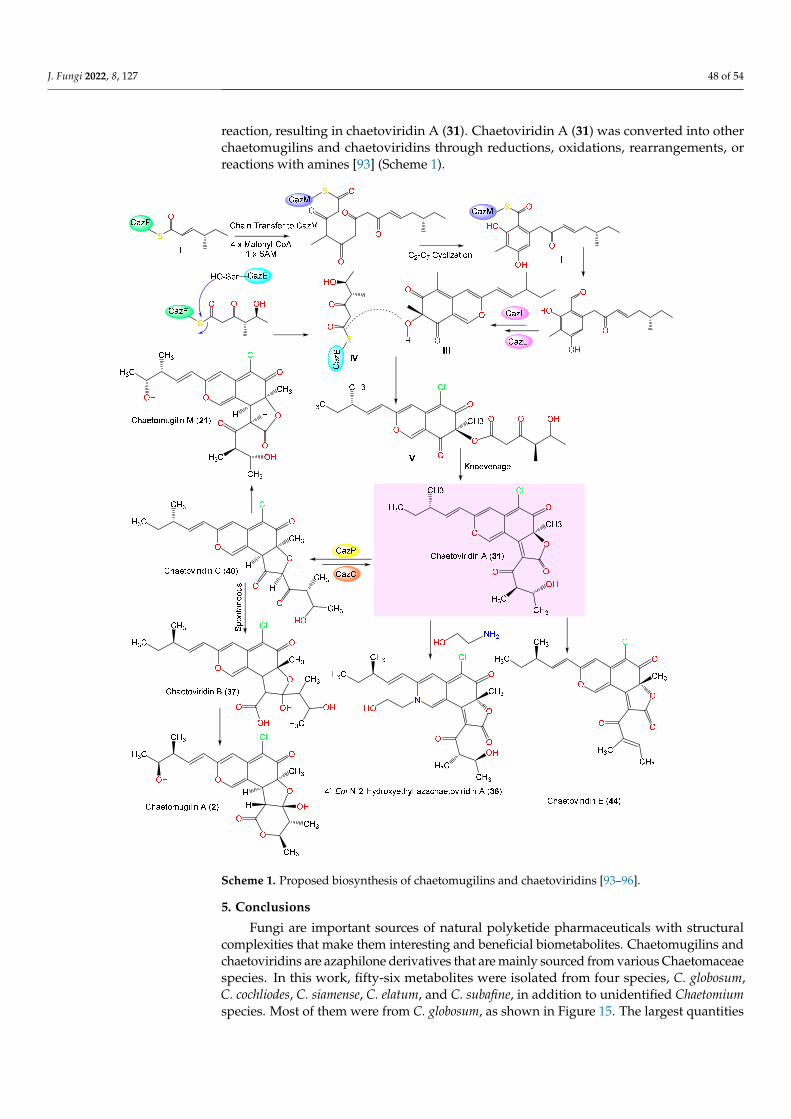
J. Fungi 2022, 8, 127 48 of 54
reaction, resulting in chaetoviridin A (31). Chaetoviridin A (31) was converted into otherchaetomugilins and chaetoviridins through reductions, oxidations, rearrangements, orreactions with amines [93] (Scheme 1).
J. Fungi 2022, 8, x FOR PEER REVIEW 47 of 53
converted into other chaetomugilins and chaetoviridins through reductions, oxidations, rearrangements, or reactions with amines [93] (Scheme 1).
Scheme 1. Proposed biosynthesis of chaetomugilins and chaetoviridins [93–96].
5. Conclusions Fungi are important sources of natural polyketide pharmaceuticals with structural
complexities that make them interesting and beneficial biometabolites. Chaetomugilins and chaetoviridins are azaphilone derivatives that are mainly sourced from various Chae-tomaceae species. In this work, fifty-six metabolites were isolated from four species, C. globosum, C. cochliodes, C. siamense, C. elatum, and C. subafine, in addition to unidentified Chaetomium species. Most of them were from C. globosum, as shown in Figure 15. The larg-est quantities of these metabolites were found in 2011 (18 compounds), 2009 (16 com-pounds), and 2017 (14 compounds) (Figure 16).
Scheme 1. Proposed biosynthesis of chaetomugilins and chaetoviridins [93–96].
5. Conclusions
Fungi are important sources of natural polyketide pharmaceuticals with structuralcomplexities that make them interesting and beneficial biometabolites. Chaetomugilins andchaetoviridins are azaphilone derivatives that are mainly sourced from various Chaetomaceaespecies. In this work, fifty-six metabolites were isolated from four species, C. globosum,C. cochliodes, C. siamense, C. elatum, and C. subafine, in addition to unidentified Chaetomiumspecies. Most of them were from C. globosum, as shown in Figure 15. The largest quantities
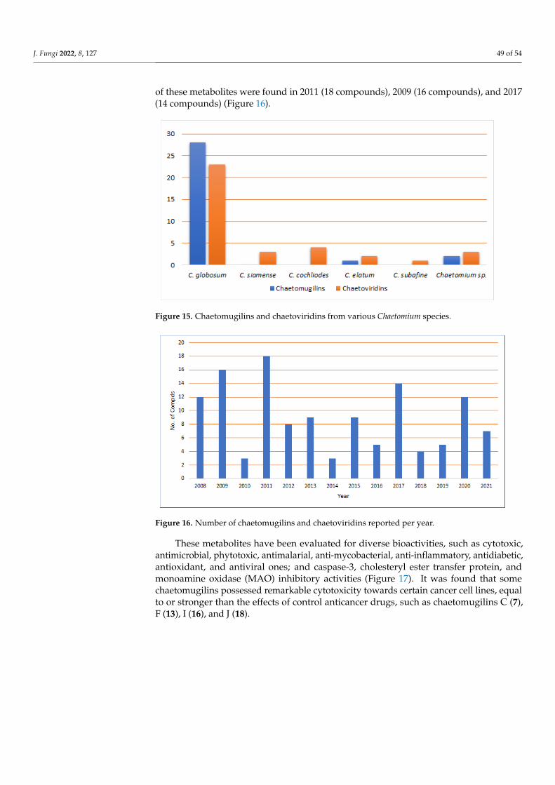
J. Fungi 2022, 8, 127 49 of 54
of these metabolites were found in 2011 (18 compounds), 2009 (16 compounds), and 2017(14 compounds) (Figure 16).
J. Fungi 2022, 8, x FOR PEER REVIEW 48 of 53
Figure 15. Chaetomugilins and chaetoviridins from various Chaetomium species.
Figure 16. Number of chaetomugilins and chaetoviridins reported per year.
These metabolites have been evaluated for diverse bioactivities, such as cytotoxic, antimicrobial, phytotoxic, antimalarial, anti-mycobacterial, anti-inflammatory, antidia-betic, antioxidant, and antiviral ones; and caspase-3, cholesteryl ester transfer protein, and monoamine oxidase (MAO) inhibitory activities (Figure 17). It was found that some chae-tomugilins possessed remarkable cytotoxicity towards certain cancer cell lines, equal to or stronger than the effects of control anticancer drugs, such as chaetomugilins C (7), F (13), I (16), and J (18).
Figure 15. Chaetomugilins and chaetoviridins from various Chaetomium species.
J. Fungi 2022, 8, x FOR PEER REVIEW 48 of 53
Figure 15. Chaetomugilins and chaetoviridins from various Chaetomium species.
Figure 16. Number of chaetomugilins and chaetoviridins reported per year.
These metabolites have been evaluated for diverse bioactivities, such as cytotoxic, antimicrobial, phytotoxic, antimalarial, anti-mycobacterial, anti-inflammatory, antidia-betic, antioxidant, and antiviral ones; and caspase-3, cholesteryl ester transfer protein, and monoamine oxidase (MAO) inhibitory activities (Figure 17). It was found that some chae-tomugilins possessed remarkable cytotoxicity towards certain cancer cell lines, equal to or stronger than the effects of control anticancer drugs, such as chaetomugilins C (7), F (13), I (16), and J (18).
Figure 16. Number of chaetomugilins and chaetoviridins reported per year.
These metabolites have been evaluated for diverse bioactivities, such as cytotoxic,antimicrobial, phytotoxic, antimalarial, anti-mycobacterial, anti-inflammatory, antidiabetic,antioxidant, and antiviral ones; and caspase-3, cholesteryl ester transfer protein, andmonoamine oxidase (MAO) inhibitory activities (Figure 17). It was found that somechaetomugilins possessed remarkable cytotoxicity towards certain cancer cell lines, equalto or stronger than the effects of control anticancer drugs, such as chaetomugilins C (7),F (13), I (16), and J (18).

J. Fungi 2022, 8, 127 50 of 54J. Fungi 2022, 8, x FOR PEER REVIEW 49 of 53
Figure 17. Biological activities of chaetomugilins and chaetoviridins.
Some studies revealed that the use of some additional compounds alongside anti-cancer drugs increased the sensitivity of the cancer cell lines towards these drugs. These metabolites could be further developed into anticancer agents; however, extensive in vivo studies and explorations of their mechanisms of action are needed. Chaetoviridins, par-ticularly chaetoviridin A (31), have substantial activity towards different plant pathogens; thus, they might be used as biocontrol agents. Additionally, some reports revealed that chaetomugilins (e.g., A (2), D (8), O (23), and S (28)) have serious phytotoxicity, more than the positive controls; therefore, they could be utilized for developing natural eco-friendly herbicides or weedicides that can replace hazardous synthetic compounds. However, ex-tensive studies and field trials should be conducted.
In molecular docking studies, chaetovirdin D exhibited the highly negative docking scores of −7.944, −8.141, and −6.615 kcal/mol, in complexes with 6M2N, 6W81, and 7K0f respectively. The reference inhibitor exhibited the following scores: −5.377, −6.995, and −8.159 kcal/mol, in complexes with 6M2N, 6W81, and 7K0f, respectively. By using molec-ular dynamics simulations, chaetovirdin D’s stability in complexes with the viral protease was analyzed, and it was found to be stable over the course of 100 ns simulation time.
Undoubtedly, chaetomugilins and chaetoviridins are fungi-derived metabolites that have multiple biological activities. They meet all the requirements for becoming drug leads in their respective therapeutic categories. They have varied chemical compositions that might provide the basis for the synthesis and design of novel and effective pharma-ceutical agents. The different substituents at various positions in their skeleton play criti-cal roles in the determination of some of their bioactivities. Finally, studies of the possible mechanisms, biosynthetic pathways, structure–activity relationships, and/or derivatiza-tion of these metabolites should be the focus of future research.
Author Contributions: Conceptualization, S.R.M.I. and G.A.M.; resources, G.A.M. and S.R.M.I., molecular docking and molecular dynamic, A.M.O.; discussion of the contents, G.A.M., S.R.M.I. and A.M.O., writing—original draft preparation, S.R.M.I., G.A.M. and A.M.O., writing—review and editing, S.R.M.I., G.A.M. and A.M.O. All authors have read and agreed to the published version of the manuscript.
Figure 17. Biological activities of chaetomugilins and chaetoviridins.
Some studies revealed that the use of some additional compounds alongside anti-cancer drugs increased the sensitivity of the cancer cell lines towards these drugs. Thesemetabolites could be further developed into anticancer agents; however, extensive in vivostudies and explorations of their mechanisms of action are needed. Chaetoviridins, par-ticularly chaetoviridin A (31), have substantial activity towards different plant pathogens;thus, they might be used as biocontrol agents. Additionally, some reports revealed thatchaetomugilins (e.g., A (2), D (8), O (23), and S (28)) have serious phytotoxicity, more thanthe positive controls; therefore, they could be utilized for developing natural eco-friendlyherbicides or weedicides that can replace hazardous synthetic compounds. However,extensive studies and field trials should be conducted.
In molecular docking studies, chaetovirdin D exhibited the highly negative dockingscores of −7.944, −8.141, and −6.615 kcal/mol, in complexes with 6M2N, 6W81, and 7K0frespectively. The reference inhibitor exhibited the following scores: −5.377, −6.995, and−8.159 kcal/mol, in complexes with 6M2N, 6W81, and 7K0f, respectively. By using molec-ular dynamics simulations, chaetovirdin D’s stability in complexes with the viral proteasewas analyzed, and it was found to be stable over the course of 100 ns simulation time.
Undoubtedly, chaetomugilins and chaetoviridins are fungi-derived metabolites thathave multiple biological activities. They meet all the requirements for becoming drug leadsin their respective therapeutic categories. They have varied chemical compositions thatmight provide the basis for the synthesis and design of novel and effective pharmaceuticalagents. The different substituents at various positions in their skeleton play critical roles inthe determination of some of their bioactivities. Finally, studies of the possible mechanisms,biosynthetic pathways, structure–activity relationships, and/or derivatization of thesemetabolites should be the focus of future research.
Author Contributions: Conceptualization, S.R.M.I. and G.A.M.; resources, G.A.M. and S.R.M.I.,molecular docking and molecular dynamic, A.M.O.; discussion of the contents, G.A.M., S.R.M.I.and A.M.O., writing—original draft preparation, S.R.M.I., G.A.M. and A.M.O., writing—review andediting, S.R.M.I., G.A.M. and A.M.O. All authors have read and agreed to the published version ofthe manuscript.
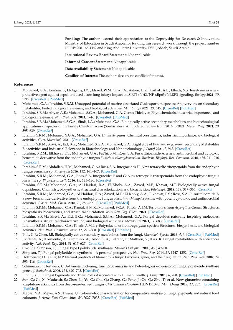
J. Fungi 2022, 8, 127 51 of 54
Funding: The authors extend their appreciation to the Deputyship for Research & Innovation,Ministry of Education in Saudi Arabia for funding this research work through the project numberIFPRP: 200-166-1442 and King Abdulaziz University, DSR, Jeddah, Saudi Arabia.
Institutional Review Board Statement: Not applicable.
Informed Consent Statement: Not applicable.
Data Availability Statement: Not applicable.
Conflicts of Interest: The authors declare no conflict of interest.
References1. Mohamed, G.A.; Ibrahim, S.; El-Agamy, D.S.; Elsaed, W.M.; Sirwi, A.; Asfour, H.Z.; Koshak, A.E.; Elhady, S.S. Terretonin as a new
protective agent against sepsis-induced acute lung injury: Impact on SIRT1/Nrf2/NF-κBp65/NLRP3 signaling. Biology 2021, 10,1219. [CrossRef] [PubMed]
2. Mohamed, G.A.; Ibrahim, S.R.M. Untapped potential of marine associated Cladosporium species: An overview on secondarymetabolites, biotechnological relevance, and biological activities. Mar. Drugs 2021, 19, 645. [CrossRef] [PubMed]
3. Ibrahim, S.R.M.; Altyar, A.E.; Mohamed, S.G.A.; Mohamed, G.A. Genus Thielavia: Phytochemicals, industrial importance, andbiological relevance. Nat. Prod. Res. 2021, 1–16. [CrossRef] [PubMed]
4. Ibrahim, S.R.M.; Mohamed, S.G.A.; Sindi, I.A.; Mohamed, G.A. Biologically active secondary metabolites and biotechnologicalapplications of species of the family Chaetomiaceae (Sordariales): An updated review from 2016 to 2021. Mycol. Prog. 2021, 20,595–639. [CrossRef]
5. Ibrahim, S.R.M.; Mohamed, S.G.A.; Mohamed, G.A. Humicola genus: Chemical constituents, industrial importance, and biologicalactivities. Curr. Microbiol. 2021. [CrossRef]
6. Ibrahim, S.R.M.; Sirwi, A.; Eid, B.G.; Mohamed, S.G.A.; Mohamed, G.A. Bright Side of Fusarium oxysporum: Secondary MetabolitesBioactivities and Industrial Relevance in Biotechnology and Nanotechnology. J. Fungi 2021, 7, 943. [CrossRef]
7. Ibrahim, S.R.M.; Elkhayat, E.S.; Mohamed, G.A.; Fat’hi, S.M.; Ross, S.A. Fusarithioamide A, a new antimicrobial and cytotoxicbenzamide derivative from the endophytic fungus Fusarium chlamydosporium. Biochem. Biophys. Res. Commun. 2016, 479, 211–216.[CrossRef]
8. Ibrahim, S.R.M.; Abdallah, H.M.; Mohamed, G.A.; Ross, S.A. Integracides H.-New tetracyclic triterpenoids from the endophyticfungus Fusarium sp. Fitoterapia 2016, 112, 161–167. [CrossRef]
9. Ibrahim, S.R.M.; Mohamed, G.A.; Ross, S.A. Integracides F and G: New tetracyclic triterpenoids from the endophytic fungusFusarium sp. Phytochem. Lett. 2016, 15, 125–130. [CrossRef]
10. Ibrahim, S.R.M.; Mohamed, G.A.; Al Haidari, R.A.; El-Kholy, A.A.; Zayed, M.F.; Khayat, M.T. Biologically active fungaldepsidones: Chemistry, biosynthesis, structural characterization, and bioactivities. Fitoterapia 2018, 129, 317–365. [CrossRef]
11. Ibrahim, S.R.M.; Mohamed, G.A.; Al Haidari, R.A.; Zayed, M.F.; El-Kholy, A.A.; Elkhayat, E.S.; Ross, S.A. Fusarithioamide B,a new benzamide derivative from the endophytic fungus Fusarium chlamydosporium with potent cytotoxic and antimicrobialactivities. Bioorg. Med. Chem. 2018, 26, 786–790. [CrossRef] [PubMed]
12. Ibrahim, S.R.M.; Mohamed, G.A.; Kamal, H.M.K.; Mohamed, S.G.A.; Khedr, A.I.M. Terretonins from Aspergillus Genus: Structures,biosynthesis, bioactivities, and structural elucidation. Mini Rev. Org. Chem. 2021. [CrossRef]
13. Ibrahim, S.R.M.; Sirwi, A.; Eid, B.G.; Mohamed, S.G.A.; Mohamed, G.A. Fungal depsides naturally inspiring molecules:Biosynthesis, structural characterization, and biological activities. Metabolites 2021, 11, 683. [CrossRef]
14. Ibrahim, S.R.M.; Mohamed, G.A.; Khedr, A.M.I. γ-Butyrolactones from Aspergillus species: Structures, biosynthesis, and biologicalactivities. Nat. Prod. Commun. 2017, 12, 791–800. [CrossRef] [PubMed]
15. Bills, G.F.; Gloer, J.B. Biologically active secondary metabolites from the fungi. Microbiol. Spectr. 2016, 4, 6. [CrossRef] [PubMed]16. Evidente, A.; Kornienko, A.; Cimmino, A.; Andolfi, A.; Lefranc, F.; Mathieu, V.; Kiss, R. Fungal metabolites with anticancer
activity. Nat. Prod. Rep. 2014, 31, 617–627. [CrossRef]17. Cox, R.J.; Simpson, T.J. Fungal type I polyketide synthases. Methods Enzymol. 2009, 459, 49–78.18. Simpson, T.J. Fungal polyketide biosynthesis—A personal perspective. Nat. Prod. Rep. 2014, 31, 1247–1252. [CrossRef]19. Hoffmeister, D.; Keller, N.P. Natural products of filamentous fungi: Enzymes, genes, and their regulation. Nat. Prod. Rep. 2007, 24,
393–416. [CrossRef]20. Schümann, J.; Hertweck, C. Advances in cloning, functional analysis and heterologous expression of fungal polyketide synthase
genes. J. Biotechnol. 2006, 124, 690–703. [CrossRef]21. Lin, L.; Xu, J. Fungal Pigments and Their Roles Associated with Human Health. J. Fungi 2020, 6, 280. [CrossRef] [PubMed]22. Sun, C.; Ge, X.; Mudassir, S.; Zhou, L.; Yu, G.; Che, Q.; Zhang, G.; Peng, J.; Gu, Q.; Zhu, T.; et al. New glutamine-containing
azaphilone alkaloids from deep-sea-derived fungus Chaetomium globosum HDN151398. Mar. Drugs 2019, 17, 253. [CrossRef][PubMed]
23. Mapari, S.A.; Meyer, A.S.; Thrane, U. Colorimetric characterization for comparative analysis of fungal pigments and natural foodcolorants. J. Agric. Food Chem. 2006, 54, 7027–7035. [CrossRef] [PubMed]

J. Fungi 2022, 8, 127 52 of 54
24. Zu, W.Y.; Tang, J.W.; Hu, K.; Zhou, Y.F.; Gou, L.L.; Su, X.Z.; Lei, X.; Sun, H.D.; Puno, P.T. Chaetolactam A, an azaphilone derivativefrom the endophytic fungus Chaetomium sp. g1. J. Org. Chem. 2021, 86, 475–483. [CrossRef]
25. Wang, W.; Liao, Y.; Chen, R.; Hou, Y.; Ke, W.; Zhang, B.; Gao, M.; Shao, Z.; Chen, J.; Li, F. Chlorinated azaphilone pigments withantimicrobial and cytotoxic activities isolated from the deep sea derived fungus Chaetomium sp. NA-S01-R1. Mar. Drugs 2018,16, 61. [CrossRef]
26. Makrerougras, M.; Coffinier, R.; Oger, S.; Chevalier, A.; Sabot, C.; Franck, X. Total Synthesis and Structural Revision ofChaetoviridins, A. Org. Lett. 2017, 19, 4146–4149. [CrossRef]
27. Takahashi, M.; Koyama, K.; Natori, S. Four New Azaphilones from Chaetomium globosum var. flavo-viridae. Chem. Pharm. Bull.1990, 38, 625. [CrossRef]
28. Gao, J.M.; Yang, S.X.; Qin, J.C. Azaphilones: Chemistry and biology. Chem. Rev. 2013, 113, 4755–4811. [CrossRef]29. Wani, N.A.; Khanday, W.I.; Tirumale, S. Evaluation of anticancer activity of Chaetomium cupreum extracts against human breast
adenocarcinoma cell lines. Matrix Sci. Pharma. 2020, 4, 31–40.30. McMullin, D.R.; Sumarah, M.W.; Miller, J.D. Chaetoglobosins and azaphilones produced by Canadian strains of Chaetomium
globosum isolated from the indoor environment. Mycotoxin Res. 2013, 29, 47–54. [CrossRef]31. Chen, G.D.; Li, Y.J.; Gao, H.; Chen, Y.; Li, X.X.; Li, J.; Guo, L.D.; Cen, Y.Z.; Yao, X.S. New azaphilones and chlorinated phenolic
glycosides from Chaetomium elatum with caspase-3 inhibitory activity. Planta Med. 2012, 78, 1683–1689. [CrossRef] [PubMed]32. Song, C.; Ding, G.; Wu, G.; Yang, J.; Zhang, M.; Wang, H.; Wei, D.; Qin, J.; Guo, L. Identification of a unique azaphilone produced
by Chaetomium globosum isolated from Polygonatum sibiricum. Chem. Biodivers. 2020, 17, e1900744. [CrossRef] [PubMed]33. Chen, C.; Wang, J.; Zhu, H.; Wang, J.; Xue, Y.; Wei, G.; Guo, Y.; Tan, D.; Zhang, J.; Yin, C.; et al. Chaephilones A and B, two new
azaphilone derivatives isolated from Chaetomium globosum. Chem. Biodivers. 2016, 13, 422–426. [CrossRef] [PubMed]34. Hu, X.; Wang, J.; Chai, J.; Yu, X.; Zhang, Y.; Feng, Y.; Qin, J.; Yu, H. Chaetomugilin J enhances apoptosis in human ovarian cancer
A2780 cells induced by cisplatin through inhibiting pink1/parkin mediated mitophagy. Onco. Targets Ther. 2020, 13, 9967–9976.[CrossRef]
35. Muroga, Y.; Yamada, T.; Numata, A.; Tanaka, R. 11-and 4-epimers of chaetomugilin a, novel cytostatic metabolit marinefish-derived fungus Chaetomium globosum. Helv. Chim. Acta 2010, 93, 542–549. [CrossRef]
36. Borges, W.S.; Mancilla, G.; Guimarães, D.O.; Durán-Patrón, R.; Collado, I.G.; Pupo, M.T. Azaphilones from the endophyteChaetomium globosum. J. Nat. Prod. 2011, 74, 1182–1187. [CrossRef]
37. Jinno, M.; Muroga, Y.; Yamada, F.; Tanaka, R. Absolute stereostructures of novel azaphiloness produced by a marine fish-derivedfungus. Symp. Chem. Nat. Prod. 2009, 51, 563–568.
38. Yamada, T.; Doi, M.; Shigeta, H.; Muroga, Y.; Hosoe, S.; Numata, A.; Tanaka, R. Absolute stereostructures of cytotoxic metabolites,chaetomugilins A–C, produced by a Chaetomium species separated from a marine fish. Tetrahedron Lett. 2008, 49, 4192–4195.[CrossRef]
39. Yamada, T.; Muroga, Y.; Doi, M.; Shiget, H.; Numata, A.; Tanaka, R. The structures of the cytotoxic metabolites produced by amarine fish-derived fungus. Symp. Chem. Nat. Prod. 2008, 50, 345–350.
40. Yasuhide, M.; Yamada, T.; Numata, A.; Tanaka, R. Chaetomugilins, new selectively cytotoxic metabolites, produced by a marinefish derived Chaetomium species. J. Antibiot. 2008, 61, 615–622. [CrossRef]
41. Yamada, T.; Yasuhide, M.; Shigeta, H.; Numata, A.; Tanaka, R. Absolute stereostructures of chaetomugilins G and H produced bya marine-fish-derived Chaetomium species. J. Antibiot. 2009, 62, 353–357. [CrossRef] [PubMed]
42. Qin, J.C.; Zhang, Y.M.; Gao, J.M.; Bai, M.S.; Yang, S.X.; Laatsch, H.; Zhang, A.L. Bioactive metabolites produced by Chaetomiumglobosum, an endophytic fungus isolated from Ginkgo biloba. Bioorg. Med. Chem. Lett. 2009, 19, 1572–1574. [CrossRef] [PubMed]
43. Yamada, T.; Jinno, M.; Kikuchi, T.; Kajimoto, T.; Numata, A.; Tanaka, R. Three new azaphilones produced by a marine fish derivedChaetomium globosum. J. Antibiot. 2012, 65, 413–417. [CrossRef] [PubMed]
44. Zhang, C.-Y.; Ji, X.; Gui, X.; Huang, B.-K. Chemical constituents from an endophytic fungus Chaetomium globosum Z1. Nat. Prod.Commun. 2013, 8, 1217–1218. [CrossRef] [PubMed]
45. Wang, D.; Zhang, Y.; Li, X.; Pan, H.; Chang, M.; Zheng, T.; Sun, J.; Qiu, D.; Zhang, M.; Wei, D.; et al. Potential allelopathicazaphilones produced by the endophytic Chaetomium globosum TY1 inhabited in Ginkgo biloba using the one strain-manycompounds method. Nat. Prod. Res. 2017, 31, 724–728. [CrossRef]
46. Wang, M.H.; Jiang, T.; Ding, G.; Niu, S.B.; Wang, X.W.; Yu, M.; Gu, Y.C.; Zhang, Q.B.; Chen, J.H.; Jia, H.M.; et al. Molecularepigenetic approach activates silent gene cluster producing dimeric bis-spiro-azaphilones in Chaetomium globosum CBS148.51. J.Antibiot. 2017, 70, 801–804. [CrossRef] [PubMed]
47. Qi, J.; Wang, D.; Yin, X.; Zhang, Q.; Gao, J.-M. New metabolite with inhibitory activity against α-glucosidase and α-amylase fromendophytic Chaetomium globosum. Nat. Prod. Commun. 2020, 15, 1–9. [CrossRef]
48. Yamada, T.; Muroga, Y.; Tanaka, R. New azaphilones, seco-chaetomugilins A and D, produced by a marine-fish-derivedChaetomium globosum. Mar. Drugs 2009, 7, 249–257. [CrossRef]
49. Selim, K.A.M.A. Biological Evaluation and Chemical Identification of Secondary Metabolites of Endophytic Fungi from EgyptianFlora. Master’s Thesis, Faculty of Science, Cairo University, Cairo, Egypt, 2008.
50. McMullin, D.R.; Sumarah, M.W.; Blackwell, B.A.; Miller, J.D. New azaphilones from Chaetomium globosum isolated from the builtenvironment. Tetrahedron Lett. 2013, 54, 568–572. [CrossRef]

J. Fungi 2022, 8, 127 53 of 54
51. Piyasena, K.G.N.P.; Wickramarachchi, W.A.R.T.; Kumar, N.S.; Jayasinghe, L.; Fujimoto, Y. Two phytotoxic azaphilone derivativesfrom Chaetomium globosum, a fungal endophyte isolated from Amaranthus viridis leaves. Mycology 2015, 6, 158–160. [CrossRef]
52. Rand, T.G.; Chang, C.T.; McMullin, D.R.; Miller, J.D. Inflammation-associated gene expression in RAW 264.7 macrophagesinduced by toxins from fungi common on damp building materials. Toxicol. In Vitro 2017, 43, 16–20. [CrossRef] [PubMed]
53. Salo, J.M.; Kedves, O.; Mikkola, R.; Kredics, L.; Andersson, M.A.; Kurnitski, J.; Salonen, H. Detection of Chaetomium globosum,Ch. Cochliodes and Ch. Rectangulare during the diversity tracking of mycotoxin-producing Chaetomium-like isolates obtained inbuildings in Finland. Toxins 2020, 12, 443. [CrossRef] [PubMed]
54. Ryu, Y. Cytochalasan and Azaphilone Derivatives from a Marine-Derived Fungus Chaetomium globosum. Master’s Thesis, SeoulNational University, Seoul, Korea, 2017.
55. McMullin, D.R. Structural Characterization of Secondary Metabolites Produced by Fungi Obtained from Damp CanadianBuildings. Ph.D. Thesis, Carleton University, Ottawa, ON, Canada, 2014.
56. Youn, U.J.; Sripisut, T.; Park, E.J.; Kondratyuk, T.P.; Fatima, N.; Simmons, C.J.; Wall, M.M.; Sun, D.; Pezzuto, J.M.; Chang, L.C.Determination of the absolute configuration of chaetoviridins and other bioactive azaphilones from the endophytic fungusChaetomium globosum. Bioorg. Med. Chem. Lett. 2015, 25, 4719–4723. [CrossRef]
57. Kumar, R.; Kundu, A.; Dutta, A.; Saha, S.; Das, A.; Bhowmik, A. Chemo-profiling of bioactive metabolites from Chaetomiumglobosum for biocontrol of Sclerotinia rot and plant growth promotion. Fungal Biol. 2021, 125, 167–176. [CrossRef] [PubMed]
58. Muroga, Y.; Yamada, T.; Numata, A.; Tanaka, R. Chaetomugilins I–O, new potent cytotoxic metabolites from a marine-fish-derivedChaetomium species. Stereochemistry and biological activities. Tetrahedron 2009, 65, 7580–7586. [CrossRef]
59. Yamada, T.; Muroga, Y.; Jinno, M.; Kajimoto, T.; Usami, Y.; Numata, A.; Tanaka, R. New class azaphilone produced by a marine fishderived Chaetomium globosum. The stereochemistry and biological activities. Bioorg. Med. Chem. 2011, 19, 4106–4113. [CrossRef]
60. Phonkerd, N.; Kanokmedhakul, S.; Kanokmedhakul, K.; Soytong, K.; Prabpai, S.; Kongsearee, P. Bio-spiro-azaphilones andazaphilones from the fungi Chaetomium cochliodes VTh01 and C. cochliodes Cth05. Tetrahedron 2008, 64, 9636–9645. [CrossRef]
61. Park, J.H.; Choi, G.J.; Jang, K.S.; Lim, H.K.; Kim, H.T.; Cho, K.Y.; Kim, J.C. Antifungal activity against plant pathogenic fungi ofchaetoviridins isolated from Chaetomium globosum. FEMS Microbiol. Lett. 2005, 252, 309–313. [CrossRef]
62. Pornsuriya, C.; Soytong, K.; Poeaim, S.; Kanokmedhakul, S.; Khumkomkhet, P.; Lin, F.-C.; Wang, H.K.; Hyde, K.D. Chaetomiumsiamense sp. nov., a soil isolate from Thailand, produces a new chaetoviridin, G. Mycotaxon 2011, 115, 19–27. [CrossRef]
63. Awad, N.E.; Kassem, H.A.; Hamed, M.A.; El-Naggar, M.A.; El-Feky, A.M. Bioassays guided isolation of compounds fromChaetomium globosum. J. Mycol. Med. 2014, 24, e35–e42. [CrossRef]
64. Xu, G.-B.; Liu, J.; Xiao, X.; Zhou, M.; Yan, T.; Li, G.-Y. Isolation and identification of secondary metabolites from Chaetomiumglobosum CIB-160 and their immunological activity. Nat. Prod. Res. Dev. 2016, 28, 1562–1567.
65. Koyama, K.; Takahashi, K.; Natori, S.; Udagawa, S. Production of mycotoxins by Chaetomium species. Proc. Jpn. Assoc. Mycotoxicol.1991, 33, 40–43. [CrossRef]
66. Fierro-Cruz, J.E.; Jiménez, P.; Coy-Barrera, E. Fungal endophytes isolated from Protium heptaphyllum and Trattinnickia rhoifolia asantagonists of Fusarium oxysporum. Rev. Argent. Microbiol. 2017, 49, 255–263. [CrossRef] [PubMed]
67. Yue, H.M.; Wang, M.; Gong, W.F.; Zhang, L.Q. The screening and identification of the biological control fungi Chaetomium spp.against wheat common root rot. FEMS Microbiol. Lett. 2018, 365, 242. [CrossRef] [PubMed]
68. Zhang, X.Y.; Tan, X.M.; Yu, M.; Yang, J.; Sun, B.D.; Qin, J.C.; Guo, L.P.; Ding, G. Bioactive metabolites from the desert plant-associated endophytic fungus Chaetomium globosum (Chaetomiaceae). Phytochemistry 2021, 185, 112701. [CrossRef] [PubMed]
69. Zhang, Y.; Zhu, H.; Ye, Y.; Tang, C. Antifungal activity of chaetoviridin A from Chaetomium globosum CEF-082 metabolites againstVerticillium dahliae in cotton. Mol. Plant Microbe Interact. 2021, 34, 758–769. [CrossRef]
70. Qi, J.; Jiang, L.; Zhao, P.; Chen, H.; Jia, X.; Zhao, L.; Dai, H.; Hu, J.; Liu, C.; Shim, S.H.; et al. Chaetoglobosins and azaphilonesfrom Chaetomium globosum associated with Apostichopus japonicus. Appl. Microbiol. Biotechnol. 2020, 104, 1545–1553. [CrossRef]
71. Wang, W.; Yang, J.; Liao, Y.Y.; Cheng, G.; Chen, J.; Cheng, X.D.; Qin, J.J.; Shao, Z. Cytotoxic nitrogenated azaphilones from thedeep-sea-derived fungus Chaetomium globosum MP4-S01-7. J. Nat. Prod. 2020, 83, 1157–1166. [CrossRef]
72. Li, X.; Tian, Y.; Yang, S.X.; Zhang, Y.M.; Qin, J.C. Cytotoxic azaphilone alkaloids from Chaetomium globosum TY1. Bioorg. Med.Chem. Lett. 2013, 23, 2945–2947. [CrossRef]
73. Yan, W.; Cao, L.L.; Zhang, Y.Y.; Zhao, R.; Zhao, S.S.; Khan, B.; Ye, Y.H. New metabolites from endophytic fungus Chaetomiumglobosum CDW7. Molecules 2018, 23, 2873. [CrossRef]
74. Kingsland, S.R.; Barrow, R.A. Identification of chaetoviridin E from a cultured microfungus, Chaetomium sp. and structuralreassignment of chaetoviridins B and D. Aust. J. Chem. 2009, 62, 269–274. [CrossRef]
75. Steyn, P.S.; Vleggaar, R. The Structure of Dihydrodeoxy-8-epi-austdiol and the Absolute Configuration of the Azaphilones. J.Chem. Soc. Perkin Trans. 1976, 7, 204–206. [CrossRef]
76. Yasukawa, K.; Takahashi, M.; Natori, S.; Kawai, K.; Yamazaki, M.; Takeuchi, M.; Takido, M. Azaphilones inhibit tumor promotionby 12-O-tetradecanoylphorbol-13-acetate in two-stage carcinogenesis in mice. Oncology 1994, 51, 108–112. [CrossRef] [PubMed]
77. Mehmet, H. Caspases find a new place to hide. Nature 2000, 403, 29–30. [CrossRef]78. Howley, B.; Fearnhead, H.O. Caspases as therapeutic targets. J. Cell. Mol. Med. 2008, 12, 1502–1516. [CrossRef]79. Taheri, H.; Filion, K.B.; Windle, S.B.; Reynier, P.; Eisenberg, M.J. Cholesteryl ester transfer protein inhibitors and cardiovascular
outcomes: A systematic review and meta-analysis of randomized controlled trials. Cardiology 2020, 145, 236–250. [CrossRef]

J. Fungi 2022, 8, 127 54 of 54
80. Tomoda, H.; Matsushima, C.; Tabata, N.; Namatame, I.; Tanaka, H.; Bamberger, M.J.; Arai, H.; Fukazawa, M.; Inoue, K.; Omura, S.Structure-specific inhibition of cholesteryl ester transfer protein by azaphilones. J. Antibiot. 1999, 52, 160–170. [CrossRef]
81. Acter, T.; Uddin, N.; Das, J.; Akhter, A.; Choudhury, T.R.; Kim, S. Evolution of severe acute respiratory syndrome coronavirus2 (SARS-CoV-2) as coronavirus disease 2019 (COVID-19) pandemic: A global health emergency. Sci. Total Environ. 2020, 730,138996. [CrossRef]
82. Abdallah, H.M.; El-Halawany, A.M.; Sirwi, A.; El-Araby, A.M.; Mohamed, G.A.; Ibrahim, S.R.M.; Koshak, A.E.; Asfour, H.Z.;Awan, Z.A.; Elfaky, M.A. Repurposing of some natural product isolates as SARS-COV-2 main protease inhibitors via in vitrocell-free and cell-based antiviral assessments and molecular modeling approaches. Pharmaceuticals 2021, 14, 213. [CrossRef]
83. Vellingiri, B.K.; Jayaramayya, M.; Iyer, A.; Narayanasamy, V.; Govindasamy, B.; Giridharan, S.; Ganesan, A.; Venugopal, D.;Venkatesan, H.; Ganesan, K.; et al. Subramaniam, COVID-19: A promising cure for the global panic. Sci. Total Environ. 2020, 725,138277. [CrossRef]
84. Zarei, A.; Fardood, S.T.; Moradnia, F.; Ramazani, A. A review on coronavirus family persistency and considerations of novel type,COVID-19 features. Eurasian Chem. Commun. 2020, 2, 798–811. [CrossRef]
85. Anand, K.; Ziebuhr, J.; Wadhwani, P.; Mesters, J.R.; Hilgenfeld, R. Coronavirus main proteinase (3CLpro) structure: Basis fordesign of anti-SARS drugs. Science 2003, 300, 1763–1767. [CrossRef] [PubMed]
86. Vandyck, K.; Abdelnabi, R.; Gupta, K.; Jochmans, D.; Jekle, A.; Deval, J.; Misner, D.; Bardiot, D.; Foo, C.S.; Liu, C.; et al.ALG-097111, a potent and selective SARS-CoV-2 3-chymotrypsin-like cysteine protease inhibitor exhibits in vivo efficacy in aSyrian Hamster model. Biochem. Biophys. Res. Commun. 2021, 555, 134–139. [CrossRef] [PubMed]
87. Yang, H.; Xie, W.; Xue, X.; Yang, K.; Ma, J.; Liang, W. Design of wide-spectrum inhibitors targeting coronavirus main proteases.PLoS Biol. 2005, 3, e324.
88. Fiorucci, D.; Milletti, E.; Orofino, F.; Brizzi, A.; Mugnaini, C.; Corelli, F. Computational drug repurposing for the identification ofSARS-CoV-2 main protease inhibitors. J. Biomol. Struct. Dyn. 2021, 39, 6242–6248. [CrossRef]
89. Linnakoski, R.; Reshamwala, D.; Veteli, P.; Cortina-Escribano, M.; Vanhanen, H.; Marjomäki, V. Antiviral agents from fungi:Diversity, mechanisms and potential applications. Front. Microbiol. 2018, 9, 2325. [CrossRef]
90. Sastry, M.G.; Adzhigirey, M.; Day, T.; Annabhimoju, R.; Sherman, W. Protein and ligand preparation: Parameters, protocols, andinfluence on virtual screening enrichments. J. Comput. Aided Mol. Des. 2013, 27, 221–234. [CrossRef]
91. Olsson, M.H.M.; Søndergaard, C.R.; Rostkowski, M.; Jensen, J.H. PROPKA3: Consistent Treatment of Internal and SurfaceResidues in Empirical pK a Predictions. J. Chem. Theory Comput. 2011, 7, 525–537. [CrossRef]
92. Friesner, R.A.; Banks, J.L.; Murphy, R.B.; Halgren, T.A.; Klicic, J.J.; Mainz, D.T.; Repasky, M.P.; Knoll, E.H.; Shelley, M.; Perry, J.K.;et al. Glide: A New Approach for Rapid, Accurate Docking and Scoring. 1. Method and Assessment of Docking Accuracy. J. Med.Chem. 2004, 47, 1739–1749. [CrossRef]
93. Pavesi, C.; Flon, V.; Mann, S.; Leleu, S.; Prado, S.; Franck, X. Biosynthesis of azaphilones: A review. Nat. Prod. Rep. 2021, 38,1058–1071. [CrossRef]
94. Winter, J.M.; Sato, M.; Sugimoto, S.; Chiou, G.; Garg, N.K.; Tang, Y.; Watanabe, K. Identification and characterization of thechaetoviridin and chaetomugilin gene cluster in Chaetomium globosum reveals dual functions of an iterative highly reducingpolyketide synthase. J. Am. Chem. Soc. 2012, 134, 17900–17903. [CrossRef] [PubMed]
95. Winter, J.M.; Cascio, D.; Dietrich, D.; Sato, M.; Watanabe, K.; Sawaya, M.R.; Vederas, J.C.; Tang, Y. Biochemical and structuralbasis for controlling chemical modularity in fungal polyketide biosynthesis. J. Am. Chem. Soc. 2015, 137, 9885–9893. [CrossRef][PubMed]
96. Sato, M.; Winter, J.M.; Kishimoto, S.; Noguchi, H.; Tang, Y.; Watanabe, K. Combinatorial generation of chemical diversity by redoxenzymes in chaetoviridin biosynthesis. Org. Lett. 2016, 18, 1446–1449. [CrossRef] [PubMed]