Regulation of paxilline biosynthesis in Penicillum paxilli
281
Copyright is owned by the Author of the thesis. Permission is given for a copy to be downloaded by an individual for the purpose of research and private study only. The thesis may not be reproduced elsewhere without the permission of the Author.
-
Upload
khangminh22 -
Category
Documents
-
view
2 -
download
0
Transcript of Regulation of paxilline biosynthesis in Penicillum paxilli

Copyright is owned by the Author of the thesis. Permission is given for a copy to be downloaded by an individual for the purpose of research and private study only. The thesis may not be reproduced elsewhere without the permission of the Author.
Regulation of Paxilline Biosynthesis in Penicillium paxilli
A Thesis presented in partial fulfilment of the requirements for the degree of
Doctor of Philosophy in
Molecular Genetics
at Massey Univ�rsity, Palmerston North, New Zealand
Emily lane Tetfer
2000

For Ally & Ian
My best frienas ana my inspiration.
Abstract
Production of the indole-diterpenoid paxilline was examined in the filamentous
ascomycete Penicillium paxilli. Paxilline is a secondary metabolite, that is synthesised
via a specific secondary metabolite biosynthetic pathway. The primary precursors of
paxiUine biosynthesis, mevalonate and isopentenyl pyrophosphate, are synthesised via
the isoprenoid pathway and the paxilline biosynthetic pathway branches from
isoprenoid biosynthesis after the synthesis of farnesyl pyrophosphate. The enzyme 3-
hydroxy-3-methylglutaryl co enzyme A (HMG Co) reductase is the rate limiting step of
the isoprenoid biosynthesis. Genes for (hmg) and �-tubulin (tub-2), were isolated from
a genomic DNA libray and characterised by DNA sequencing and RT-PCR. The steady
state mRNA levels of hmg and tub-2 were compared with genes isolated from the
paxilline biosynthetic gene cluster, using. a semi-quantitative RT-PCR gene expression
assay. A distinct pattern of expression was identified for genes involved in the
biosynthesis of paxilline. Increased expression of these genes occurs 36 h prior to the
detection of paxilline in liquid culture.
P. paxilli physiology and paxilline production was analysed in liquid culture after the
development of reproducible growth conditions that results in the formation of
homogeneous loose hyphal fragments and detectable paxilline after 72 h. The
morphology of P. paxilli in paxilline-inducing media was examined microscopically and
key physiological markers, culture pH and biomass accumulation, were also analysed.
Paxilline levels in both mycelia and culture supernatant were analysed with HPLC and
TLC. This confirmed that paxilline is not released into the media until 144 h when large
scale autolysis is observed. Initial experiments to examine paxilline production in
cultures supplemented with a biological buffer suggest that phase switching between
primary growth and secondary growth may be triggered by changes in ambient pH. The
presence of alternative carbon sources also affected the rate of paxilline production and
preliminary results indicate that biosynthesis of paxilline may be under carbon catabolite
repression by glucose.
Levels of HMG CoA reductase are known to be regulated at many levels, including
mRNA transcription, protein inactivation and protein degradation, in response to excess
sterols. A number of putative sterol response elements (SRE) , which control
transcription of hmg in higher eukaryotes, where identified in the 5' UTR of hmg from
P. paxilli. In higher eukaryotes, the extremely complex 5' UTR of hmg has been
proposed as the site of regulation for biosynthesis of non-sterol end-products. This
complexity appears to be conserved in the 5' UTR of hmg from P. paxilli and another
filamentous fungus Neotyphodium lolii Lp19. lntronic sequences are spliced from the 5'
UTR of both genes and there are additional intronic sequences present that could
produce alternative transcripts. At least two different hmg transcripts were identified
from P. paxilli with 5' RACE. The mechanism by which these alternative transcripts
arise is unclear at present, but could involve alternative splicing of the 5' UTR intron or
initiation of transcription from alternative start sites.
11
Acknowledgments
First and foremost, I would like to thank my supervisor, Barry Scott, for his experience
and guidance. Barry, the first thing I heard about you, before I came to Massey, was
your reputation as an excellent supervisor. It will certainly be the first thing anybody
hears about you from me. You have made sure that not just my work, but my finances
were on track, guided me through all the unexpected difficulties at the beginning of my
research and provided immeasurable assistance during the final writing process. I was
honoured that you trusted me to speak about DNA technology in Wellington last year.
Thanks also to my co-supervisor Rosie Bradshaw, for guidance during my research and
assistance with editing the final draft.
Carolyn and Lisa, how could this have been done without you. I sincerely appreciate
the technical help, the brain-storming sessions, and the paxilline cluster genes to perform
expression analysis on, but most of all, your friendship. You have certainly had your
hands full with me from time to time, hopefully more so at the beginning than at the end.
You never know Lisa, I may have time for one more Telferism' before I go.
To Jo Mudford and Dick Poll, when the HPLC was doing its best to drive me insane,
your experience and assistance got me through the problems I could never have solved
on my own. Thank you for all your help. I would also like to thank Jan Schrnid for
assistance taking the photographs of P. paxilli in liquid culture and for helping me
understand what I was seeing. It certainly added another dimension to the analysis of
culture physiology. I would also like to say that I have enjoyed working with both you
and Neville Honey as an undergraduate demonstrator. This too was a learning
experience.
To all the past and present members of the Molecular Genetics Unit, I've enjoyed
working and socialising with you all. At times I've been a student, at other times, a
teacher and I know I've greatly appreciated the expertise and perspectives that can be
gained from working in a large group of scientists such as this. That said, I must add
that being part of the Institute of Molecular BioSciences, is humbling to say the least.
ill

The wealth of knowledge that this amalgamation has brought together is world class, and
I fee l honoured to have been associated with it.
To the guys at 'Crapston Manor', I know I'm not the easiest person to live with, but
somehow you've put up with me, especially these last few months. The lengthy
philosophical and home brew evaluation sessions will be long remembered. I don't think
I 'll ever forget that Halloween party as long as I live.
The best part of moving to a new c ity has been all the friends I have made. A couple of
you moved with me to Palmerston North (the VUW invasion of '96), and many more
have since moved on, but all of you are remembered. Carolyn, David and Patrick,
you've made me feel like part of the family. Lisa, like the ad says, 'good things take
time' and I feel like our friendship is one of those good things. Bek, although we never
planned it that way, so many of the new things I've had to do in the past five years, I've
done them with you. Moving to a new city, starting a PhD, going to the States for our
first big conference. It will definitely feel weird entering this new stage of my life
without you there for moral support. Austen, you seem unflappable and I've learnt a
lot from your outlook on life. 10, you took me under your wing when I first moved here
and introduced me to Palmerston North. Marcus, you've been my sanity at times, and
seem to know what I'm going to say even before I do. Richard, Leon and Matt, thanks
for giving me a bolt-hole to go to, on Friday nights, I appreciate it more than you realise.
Finally, to my family, Ian, Ally, lames and Steve, without your support, both fmancial
and emotional, none of this would have been possible. lames, you don't always take the
easy road in life, but each challenge seems to make you stronger and I'm proud of the
man my brother has become. Mum and Dad, you are more than my parents, you're my
best friends and even though we don't get to see each other as much as we would like,
you couldn't have picked a nicer place for me to come home to.
iv
Table of Contents
Abstract .......................................................................... .
Acknowledgments.... ......................... ....... ............................ 111
Table of Contents............................. ......... . .... ...................... v
List of Tables........ . ...... . . .... ..... .................. .................. .......... XlI
List of Figures...................................................................... XlIl
Chapter 1 Introduction
1 . 1 Secondary metabolism................................................... 1
1 .2 Biosynthetic pathways.................................................... 2
1 .3 Gene clusters................................................................... 2
1 .4 Evolution . . . . . . . . . . . . . . . . . . . . . . . . . . . . . . . . . . . . . . . . . . . . . . . . . . . . . . . . . . . . . . '" . . . . . . . . . 4
1 .5 Regulation ............ ...... .................................................. '" 6
1 .5. 1 1 .5.2 1 .5.3 1 .5.4-1 .5.5
Nitrogen .......................................................... . Carbon ............................................................ . pH .................................................................. . Pathway·specific regulators ............ ..................... . Post transcriptional regulation of gene expression ... .
6 7 7 8 9
1 .6 Growth of filamentous fungi in liquid culture................ 1 0
1 . 7 Secondary metabolite structures................................... 1 2
1 . 7. 1 1 . 7.2 1 . 7.3 1 . 7.4-1 . 7.5 1 . 7.6
Peptides .. ............................... ......................... . f3-lactam antibiotics .......................................... . Polyketides ........................... ............................ . Aflatoxin .......................................................... . Terpenes .......................................................... . PaxiWne and lolitrem B ....................................... .
1 2 1 2 1 3 1 4 1 5 1 7
1 .8 3,Hydroxy,3,methylglutaryl CoenzymeA reductase 20
1 .8. 1 1 .8.2
Isoprenoid biosynthesis ...................................... . Regulation of HMGR ........................................... .
v
20 2 1
1.8.2.1 1.8.2.2 1.8.2.3
Transcriptional control of sterol b iosynthetic genes . . . . ... . Post-transcriptional control of sterol b iosynthetic genes .. A mechanism for regulation by non-sterol end-products ?.
24 24 25
1 . 9 Fungal endophytes.......................................................... 26
1 . 9. 1 Mycotoxins ................................................................ . 27
1 . 1 0 Penicillium pnxilli.. . . . .. . . . . . . . . . . . . . . . . . . . . . . . . . . . . . . . . . . . . . . . . . . . . . . . . . . . . 28
1 . 1 0. 1 1 . 1 0.2
P. pnxilli paxil/ine negative mutants ....................... . The paxilline biosynthetic gene cluster .................... .
28 29
1 . 1 1 Analysis of gene expression............................................. 32
1 . 1 2 Objectives........................................................................ 34
Chapter 2 Materials ana Methods
2. 1 Fungal and Bacterial strainsJ A Clones and Plasmids...... 35
2. 1 . 1 2. 1 .2 2. 1 .3 2. 1 .4 2.1 .5 2. 1 .6 2. 1 . 7 2. 1 . 8
Stocks and strains used ....................................... . Growth of cultures .............................................. . Glycerol stocks ................................................... . A Phage Iysates ................................................... . Spore suspensions ............................................... . Glassware preparation ......................................... . Spore suspension inoculation ................................ . Seed culture inoculation ...................................... .
35 35 40 40 40 4 1 4 1 4 1
2.2 Media............................................................................... 42
2.3 Solutions.......................................................................... 43
2.3. 1 2.3.2 2.3.3 2.3.4 2.3.5 2.3.6 2.3. 7 2.3.8 2.3. 9 2.3. 1 0 2.3. 1 1
Stock solutions .................................................. . Acrylamide mix .................................................. . 10 x Denhard�s solution ...................................... . DNA extraction buffer ......................................... . DNase 1 buffer ................................................... . PEG solution ....................................................... . SOS loading dye .................................................. . SM buffer ......................................................... . Southern blotting solutions .................................. . Tris-based buffers ............................................... . Tris-equilibrated phenol ...................................... .
43 43 44 44 44 44 44 45 45 45 46
2.4 Enzymes and Biological solutions.................................... 46
VI
2.5 Isolation of DNA.............................................................. 47
2.5. 1 2.5.2 2.5.3 2.5.4-2.5.5
Fungal DNA . . . . . . . . . . . . . . . . . . . . . . . . . . . . . . . . . . . . . . . . . . . . . . . . . . . . . . . . A Phage DNA (medium scale) . . . . . . . . . . . . . . . . . . . . . . . . . . . . . . . . . Plasmids DNA by rapid boil method . . . . . . . . . . . . . . . . . . .. ... . . DNA from agarose gel slices . . . . . . . . . . . . . . . . . . . . . . . . . . . . . . . . . . . Plasmid DNA by Alkaline lysis-based miniprep kits . . . ' "
47 48 49 49 50
2.6 Quantification of Nucleic Acids........ . . . . . . . . . . . . . . . . . . . . . . . . . . . . . . . . . 50
2.6.1 2.6.2 2.6.3
Intensity of ethidium bromide fluorescence . . . . . . . . . . . . . . Fluorometric determination . . . . . . . . . . . . . . . . . . . . . . . . . . . . . . . . . . . . UV Spectrophotometric determination . . . . . . . . . . . . . . . . . . . . . .
50 5 1 5 1
2. 7 Restriction Endonuclease Digestion of DNA.................. .... 5 1
2.8 Purification of DNA by Phenol/Chloroform...................... 5 1
2. 9 Precipitation of DNA with Ethanol and Isopropanol........ 52
2. 1 0 Agarose Gel Electrophoresis............................................... 52
2. 1 0.1 2. 1 0.2 2.1 0.3
Mini-gel . . . . . . . . . . . . . . . . . . . . . . . . . . . . . . . . . . . . . . . . . . . . . . . . . . . . . . . . . . . . . . . Submarine gel . . . . . . . . . . . . . . . . . . . . . . . . . . . . . . . . . . . . . . . . . . . . . . . . . . . . . . Staining DNA with ethidium bromide . . . . . . . . . . . . . . . . . . . . . . .
52 52 53
2. 1 1 Polymerase Chain Reaction (PCR) . . . . . . . . . . . . . . . . . . . . . . . . . . . . . . . . . . . . . 54
2. 1 1. 1 2.1 1 .2 2. 1 1.3 2.1 1 .4-2. 1 1 .5
Primer design . . . . . . . . . . . . . . . . . . . . . . . . . . . . . . . . . . . . . . . . . . . . . . . . . . . . . . . Protocol for routine PCR . . . . . . . . . . . . . . . . . . . . . . . . . . . . . . . . . . . . . . . Bacterial colony PCR . . . . . . . . . . . . . . . . . . . . . . . . . . . . . . . . . . . . . . . . . . . . . .. Gel stab PCR . . . . . . . . . . . . . . . . . . . . . . . . . . . . . . . . . . . . . . . . . . . . . . . . . . . . . . . . Purification of PCR products . . . . . . . . . . . . . . . . . . . . . . . . . . . . . . . . . . .
54 54 54 59 59
2. 12 Sequencing....... . . . . . . . . . . . . . . . . . . . . . . . . . . . . . . . . . . . . . . . . . . . . . . . . . . . . . . . . . . . . . . . . . . . 60
2.1 2. 1 2.1 2.2 2. 1 2.3
Cycle sequencing with the AmpliCycle™ kit . . . . . . . . . . . . . . . . Denaturing polyacrylamide gel electrophoresis . . . . . . . . . Automated sequencing . . . . . . . . . . . . . . . . . . . . . . . . . . . . . . . . . . . . . . . . . .
60 60 6 1
2. 13 Southern Blotting and Hybridisation................................ 6 1
2. 13.1 2.13.2 2. 1 3.3 2. 1 3.4-
Capillary transfer of DNA to nylon membranes . . . . . . . . . . Labelling DNA with [a.32Pl dCTP . . . . . . . . . . . . . . . . . . . . . . . . . . . . . .. Hybridisation of [a.32Pl dCTP-labelled probe . . . . . . . . . . . . . . Stripping labelled probe from Nylon membranes . . . . . . .
6 1 6 1 62 62
2. 14 Library Screening... . . . . . . . . . . . . . . . . . . . . . . . . . . . . . . . . . . . . . . . . . . . . . . . . . . . . . . . . . . 63
Vll
2.14 .1 2.14.2
2. 1 4.3 2. 1 4. 4 2. 1 4. S 2.1 4.6 2.1 4 . 7 2. 1 4.B
Titre of a AGEM®l l genomic DNA library .................. . Calculating library concentration required for isolating clone ............................................... . Library plating .................................................... . Transfer of plaques to Nylon membranes ................ . Hybridisation of [a.32p] dCTP-label/ed probe ............ . Isolation of positive clones .................................... . First round library screening ................................ .. Second and third round library screening ................ .
63
63 63 64 64 64 65 65
2. 15 Sub·cloning.............. . . . . . . . . . . . . . . . . . . . . . . . . . . . . . . . . . . . . . . . . . . . . . . . . . . . . . . . . . . . 65
2.1 S. 1
2.1 S.2 2. 1 S.3 2. 1 S.4 2. 1 S.S 2. 1 S.6
Ligation into CArd pUC1 1 B .................................. .. Ligation into pGEM®-T and pGEM®-T EASy ........ , ... . . . . Electrocompetent cel/s ........................................ .. Electroporation ................................................... .
Selection of transformants .................................. . Drop-outs and mapping ....................................... .
65 66 66 66 67 67
2. 1 6 Paxilline Analysis......................... . . . . . . . . . . . . . . . . . . . . . . . . . . . . . . . . . . . . . . . 68
2. 1 6. 1 2. 1 6.2 2. 1 6.3 2. 1 6.4
Sampling liquid cultures for analysis of paxilline ...... . Isolation of paxilline from freeze dried samples ...... .. HPLC .................................................................. . TLC . . . . . . . . . . . . . . . . . . . . . . . . . . . . . . . . . . . . . . . . . . . . . . . . . . . . . . . . . . . . . . . . . . . . .
68 68 69 69
2. 1 7 Isolation of RNA................................................................. 69
2. 1 7. 1 2. 1 7.2 2.1 7.3 2.1 7.4
Sampling liquid cultures for isolation of RNA ........... .. RNase-free solutions and equipment .............. , ....... . Isolation of total RNA with TRlzol"" reagent ............ .
DNase I treatment ............................................. .
69 70 70 7 1
2. 1 8 Reverse Transcription·PCR................................................. 72
2. 1 B. 1
2. 1 B.2
Synthesis of cDNA with Expand"" reverse transcriptase ......................................... .. PCR amplification of cDNA .................................... .
72 72
2. 1 9 RACE................. ........ . . . . . . . . . . . . . . . . . . . . . . . . . . . . . . . . . . . . . . . . . . . . . . . . . . . . . . . . . . . 73
2. 1 9. 1
2. 1 9.2 2. 1 9.3
2. 1 9.4 2. 1 9.S
Synthesis of cDNA with C. therm reverse transcriptase ......................................... .
Degradation of RNA ............................................. . Addition of 3" terminal dCTP with terminal transferase .......................................... . Second strand synthesis ........................................ . Isolation and characterisation of RACE products ...... .
V1ll
73 73
74 74 74
Chapter 3 RESULTS
3. 1 Isolation and Characterisation of P. pnxilli hmg........... 75
3.1 . 1 3 .1 .Z
3. 1 .3
3.1 .11-3. 1 .5 3. 1 .6 3. 1 . 7 3. 1 .8 3.1 . 9
3.1 . 1 0
3. 1 . 1 1
3. 1 . 1 Z
Isolation o f a unique hmg with degenerate primers. . . 75 Sequencing the 700 bp and 359 bp products from P. paxilli . . . . . . . . . . . . . . . . . . . . . . . . . . . . . . . . . . . . . . . . . . . . . . . . . . . . 80 Confirmation the unique hmg exists in the
P. paxilli genome. . . . . . . . . . . . . . . . . . . . . . . . . . . . . . . . . . . . . . . . . . . . . . . . . . 82 Library screening. . . . . . . . . . . . . . . . . . . . . . . . . . . . . . . . . . . . . . . . . . . . . . . . . 86 Physical mapping of A clones. . . . . . . . . . . . . . . . . . . . . . . . . . . . . . . . . . . 86 Sub-cloning AETZ into a sequencing vector... . . . . .. . .. ... . 86 Sequencing AETZ sub-clones . . . . . . . . . . . . . . . . . . . . . . , . . . . . . . . . . . . . 92 Confirmation of introns. . . . . . . . . . . . . . . . . . . . . . . . . . . . . . . . . . . . . . . . . 1 1 0 Estimation of transcription start site
with RT-PCR. . . . . . .. . . . . . . . . . . . . . . . . . . . . . . . . . . . . . . . . . . . . . . . . . . . . . . . . . . 1 1 8 Confirmation of transcription start site
with 5" RACE. . . . . .. . . . . . . . . . . . . . . . . . . . . . . . . . . . . . . .. . . . . . . . . . . . . . . . . . 1 1 9 Confirmation of transcription stop site
with 3" RACE . . . . . . . , . . . . . . . . . . . . . . . . . . . . . . . . . . . . . . . . . . . . . . . . . . . . . . . . 1 25 Chromosomal location of P. pnxilli hmg. . . . . . . . . . . . . . . . . . . . . 1 25
3.2 Isolation and Characterisation of P. pnxilli tub-2.......... 1 28
3.Z. 1 Preparation of tuo-2 genomic probe from pBT6. . . . . . . . . 1 28 3.Z.Z Confirmation a single tuo-2 exists in the
P. paxilli genome. . . . . . . . . . . . . . . . . . . . . . . . . . . . . . . . . . . . . . . . . . . . . . . . . . 1 28 3.Z.3 Library screening. . . . . . . . . . . . . . . . . . . . . . . . . . . . . . . . . . . . . . . . . . . . . . . . 1 32 3.Z.11- Physical mapping of tub-2 A clones. . . . . . . . . . . . . . . . . . . . . . . . . 1 32 3.Z.5 Sub-cloning AETZZ into a sequencing vector. . . . . . . . . . . . . . 1 32 3.Z.6 Sequencing AETZZ sub-clones. . . ... .. . .. ... ... . . . .. . . . . . . . . ... 1 3 8 3.Z. 7 Confirmations of introns. . . . . . . . . . . . . . . . . . . . . . . . . . . . . . . . . . . . . . . 1 3 8 3.Z.8 Chromosomal location of P. pnxilli tuo-2.................. 146
3.3 P. pnxilli physiology and conditions for induction of paxilIine . . . . . . . . , . . . . . . . . . . . . . . . . . . . . . . . . . . . . . . . . . . . . . . . . . , . . . . 1 5 1
3.3. 1 3.3.Z 3.3.3 3.3.11-
3.3.4.1 3.3.4.2 3.3.4.3 3.3.4.4
3.3.5 3.3.5.1 3.3.5.2
Spore viability ....... ... . . .. ........ . . .. .. .. . . ... . . . . . . . . . .. . ... . . Cultures inoculated from spore suspensions . . . . . . . . . . . . . . Cultures inoculated from seed culture . . . . . . . . . . . . . . . . . . . . Growth of P. pnxilli in paxilline-inducing media . . . . . . . . . . Experiment 1 ....................................................... . Experiment 2 ....................................................... . Analysis o f paxilline in broth ............ ' " . . . . . . . . , .. . . . . . . . . . . Microscopic analysis of culture morphology ................. .
Growth of P. pnxilli in supplemented media . . . . . . . . . . . . . . .
Calcium . . . . . . . . . . . . . . . . . . . . . . . . . . . . . . . . . . . . . . . . . . . . . . . . . . . . . . . . . . . . . .
Yeast Extract ...................................................... .
IX
1 5 1 1 5 1 1 54 1 55 1 55 1 55 1 62 1 63
1 70 1 70 1 73
3.3.5.3
3.3.6 3.3.7
Biological buffer.. . . . . . . . . . . . . . . . . . . . . . . . . . . . . . . . . . . . . . . . . . . . . . . . . . 1 73
Growth of P. paxilli in variable carbon sources. . . . . . . . . . . . 1 74 Growth of P. paKilli during carbon and nitrogen
starvation. . . . . . . . . . . . . . . . . . . . . . . . . . . . . . . . . . . . . . . . . . . . . . . . . . . . . . . . . 1 77
3.4 Analysis of Paxilline Biosynthetic Gene Expression........... 1 80
3.4. 1 RT-PCR gene expression assay. . . . . . . . . . . . . . . . . . . . . . . . . . . . . . . . . 1 80 3.4.2 Standardisation of the assay with tub-2.................. 1 80 3.4.3 Identification of the Pax pattern of expression. . . . . . . . . 1 82
Chapter 4 DISCUSSION
4. 1 Isolation ana Characterisation of hmg from P. pnxilli.... 1 9 1
4. 1 . 1 4 .1 .2 4 .1 .3
4.1.3.1 4.1.3.2 4.1.3.3 4.1.3.4
4.1.4 4 .1 .5
P. paxilli hmg. . . . . . . . . . . . . . . . . . . . . . . . . . . . . . . . . . . . . . . . . . . . . . . . . . . . . . 1 9 1
P. paxilli hmg nucleotide sequence. . . . . . . . . . . . . . . . . . . . . . . . . . . 1 92
P. paxilli hmg 5-' sequence analysis. . . . . . . . . . . . . . . . . . . . . . . . . . 1 93 Initiation o f transcription. . . . . . . . . . . . . . . . . . . . . . . . . . . . . . . . . . . . . . . 1 93 5' um introns . . . . . . . . . . . . . . . . . . . . . . . . . . . . . . '" . . . . . . . . . . . . . . . . . . . . . 1 94 5'um heterogeneity... . . . . . . . . . . . . . . . . . . . . . . . . . . . . . . . . . . . . . . . . . . . . . 1 95 Putative promoter elements... . . . . . . . . . . . . . . . . . . . . . . . . . . . . . . . . . . 1 99
P. paxilli hmg 3-' sequence analysis. . . . . . . . . . . . . . . . . . . . . . . . . . 203
P. paKilli hmg polypeptide sequence . . . . . , . . . . . . . . . . . . . . . . . . 203
4.1 Isolation ana Characterisation of tub,2 from P. pnxilli... 206
4.2.1 P. paxilli tub-2................. . . . . . . . . . . . . . . . . . . . . . . . . . . . . . . . . . . . . 206 4.2.2 P. paKilli tub-2 nucleotide sequence. . . . . . . . . . . . . . . . . . . . . . . . . 206 4.2.3 P. paxilli tub-2 polypeptide sequence. . . . . . . . . . . . . . . . . . . . . . . 207
4.3 Physiology of P. pnxilli in submergea liquia culture........ 208
4.3.1
4.3.2 4.3.2.1 4.3.2.2 4.3.2.3 4.3.2.4
4.3.3
4.3 .4
4.3.5
Development of cultures with homogeneous morphology and reproducible induction of paxilline. . . 208 Wild,type P. paxilli in paxilline,inducing media. . . . . . . . . . . 209 Biomass accumulation . . . . . . . , . . . . . . . . . . . . . '" . . . . . . . . . . . . . . . . . . . . . 2 1 0 Culture pH. . . . . . . . . . . . . . . . . . . . . . . . . . . . . . . . . . . . . . . . . . . . . . . . . . . . . . . . . . . 2 1 0 Paxilline.. . . . . . . . . . . . . . . . . . . . . . . . . . . . . . . . . . . . . . . . . . . . . . . . . . . . . . . . . . . . 2 1 1 Microscopy . . . . . . . . . . . . . . . . . . . . . . . . . . . . . . . . . . . . . . . . . . . . . . . . . . . . . . . . . , 2 1 1
Effect of sporulation on paxilline production. . . . . . . . . . . . . . . . . . . . . . . . . . . . . . . . . . . . . . . . . . . . . . . . . . . . . . . . 2 1 2
Effect of yeast extract supplement on paxilline production. . . . . . . . . . . . . . . . . . . . . . . . . . . . . . . . . . . . . . . . . . . . . . . . . . . . . . . . . 2 1 2
Effect of pH on paxilline production. . . . . . . . . . . . . . . . . . . . . . . . 2 1 3
x
4.3.6 Effect of carbon source on paxilline production......... 2 1 4 4.3. 7 Effect of carbon and nitrogen stalVation on
paxilline production.......... . . . . . . . . . . . . . . . . . . . . . . . . . . . . . . . . . . . 2 1 5
4.'1 Expression of Paxilline Biosynthetic Genes...................... 2 1 6
4.4.1 Development of a gene expression assay... ... . .. ......... 2 1 6 4.4.1 Expression of paxilline biosynthetic genes. ..... . . . . . . . . . . . 2 1 6 4.4.3 Expression of a putative pathway regulator.............. 2 1 8 4.4.4 Expression of primary metabolic genes..................... 2 1 9
Appendix
A 1 .0 Vector maps..................................................................... 221
A 1 .1 pUCl 1 8............... . . . . . . . . . . . . . . . . . . . . . . . . . . . . . . . . . . . . . . . . . . . . . . 22 1 A 1.1 pBT6 . . . . . . . . . . . . . . . . . . . . . . . . . . . . . , ' " . . . . . . . . . . . . . . . . . . . . . . . . . . . . . . . . 222 A 1 .3 pGEM®-T . . . . . . . . . . . . . . . . . . . . . . . . . . . . . . . . . . . . '" . . . . . . . . . . . . . . . . . . . . . 223 A 1.11 pGEM®-T Easy............... . . . . . . . . . . . . . . . . . . . . . . . . . . . . . . . . . . . . . . . 224 A 1 .5 Lambda GEM®-l l............ ............ . . . . . . . . . . . . . . . . . . . . . . . . 225
A Z.O Sequence data......... . . . . . . . . . . . . . . . . . . . . . . . . . . . . . . . . . . . . . . . . . . . . . . . . . . . . . . . . . 226
A 2.1 Sequence data of 359 bp product ... '" . . . . . . . . . . . . . . . . . . . . . 226 A 2.1 Sequence data of 700 bp product .......................... , 227 A 2.3 Sequencher™ alignment of hmg contig. . . . . . . . . . . . . . . . . . . . 228 A 2.4 Sequencher™ alignment of tub-2 contig... . . . . . . . . . . . . . . . 229
A.3.0 Calculations..................................................................... 230
A 3.1 Calculating paxilline content from HPLC analysis........ 230
References.............................................................................. 232
Xl
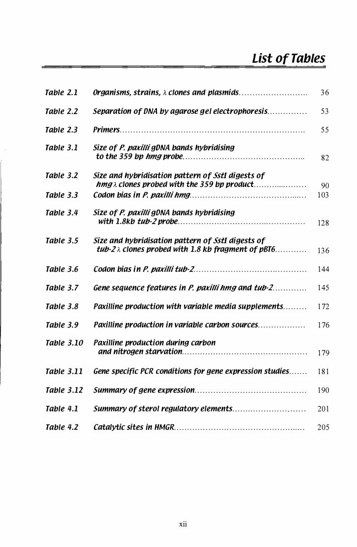
List of Tables
Table 1. 1 Organisms., strains., A clones and plasmids.......................... 3 6
Table 1.2 Separation of ONA by agarose gel electrophoresis... . . . . . . . . . . . . 5 3
Table 1.3 Primers...................................................................... 55
Table 3.1 Size of P. paxilli gDNA bands hybridising to the 359 bp hmg probe.................................... . . . . . . . . . . . 82
Table 3.1 Size and hybridisation pattern of Ss tI digests of hmg A clones probed with the 359 bp product......... . . . . . . . . . . . . 90
Table 3.3 Codon bias in P. paxilli hmg............... .................. . . . . . . . . . . . . 1 03
Table 3.4 Size of P. paxilli gONA bands hybridising with 1 .8kb tub-2 probe.. . . . . . . . . . . . . . . . . . . . . . . . . . . . . . . . . . . . . . . . . . . . . . . . 128
Table 3.5 Size and hybridisation pattern of Ss tI digests of tub-2 A clones probed with 1.8 kb fragment of pBT6............ 1 3 6
Table 3.6 Codon bias in P. paxilli tub-2........................................... 1 44
Table 3. 7 Gene sequence features in P. paxilli hmgand tub,l............. 1 45
Table 3.8 Paxilline production with variable media supplements..... . . . . 1 72
Table 3. 9 Paxilline production in variable carbon sources.................. 1 76
Table 3.10 Paxilline production during carbon and nitrogen starvation................... . . . . . . . . . . . . . . . . . . . . . . . . . . . . . 1 79
Table 3.1 1 Gene specific PCR conditions for gene expression studies....... 1 8 1
Table 3.11 Summary of gene expression.......................................... 1 90
Table 4.1 Summary of sterol regulatory elements . . . . . . . . . . . . . . . . . . . . " . . . . . . 20 1
Table 4.1 Catalytic sites in HMGR............ .......... . . . . . . . . . . . . . . . . . . . . . . . . . . . . 205
Xll
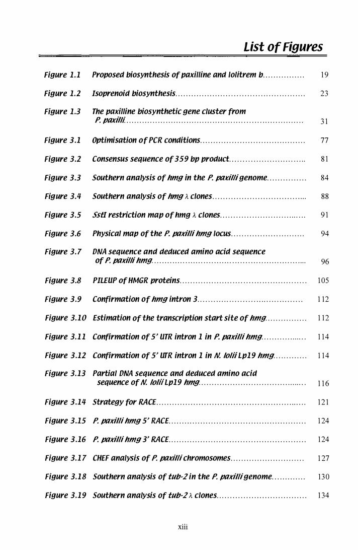
List of Figures
Figure 1 .1 Proposed biosynthesis of paxilline and lolitrem b................ 1 9
Figure 1.2 Isoprenoid biosynthesis..................... . . . . . . . . . . . . . . . . . . . . . . . . . . . . 23
Figure 1 .3 The paxilline biosynthetic gene cluster from P. paxil li .... . . . . . . . . . . . . . . . . . . . . . . . . . . . . . . . . . . . . . . . . . . . . . . . . . . . . . . . . . . . . . . . . 3 1
Figure 3.1 Optimisation of PCR conditions............ . . . . . . . . . . . . . . . . . . . . . . . . . . . . 77
Figure 3.2 Consensus sequence of 359 bp product...... . . . . . . . . . . . . . . . . . . . . . . . 8 1
Figure 3.3 Southern analysis of hmg in the P. ptlKilli genome............... 84
Figure 3.4 Southern analysis of hmg A clones.................................... 88
Figure 3.5 Ss tI restriction map of hmg A clones...... . . . . . . . . . . . . . . . . . . . . . . . . . . . 9 1
Figure 3.6 Physical map of the P. ptlKilli hmg locus.................. . . . . . . . . . . 94
Figure 3. 7 DNA sequence and deduced amino acid sequence of P. ptlKi lli hmg. . . . . . . . . . . . . . . . . . . . . . . . . . . . . . . . . . . . . . . . . . . . . . . . . . . . . . . . . . . . 96
Figure 3.8 PILEUP of HMGR proteins.................... . . . . . . . . . . . . . . . . . . . . . . . . . . . . 1 05
Figure 3.9 Confirmation of hmg intron 3. . . . . . . . . . . . . . . . . . . . . . . .. .. . . . . .. ..... .. 1 12
Figure 3.1 0 Estimation of the transcription start site of hmg................ 1 1 2
Figure 3.1 1 Confirmation of 5� UTR intron 1 in P. ptlKilli hmg.................. 1 1 4
Figure 3.12 Confirmation of 5� UTR intron 1 in N. lo liiLp1 9 hmg. . . . . . . . . . . . . 1 1 4
Figure 3.13 Partial DNA sequence and deduced amino acid sequence of N. lo liiLp19 hmg.......................................... 1 1 6
Figure 3.14 Strategy for RACE....................................... ................. . 1 2 1
Figure 3.15 P. ptlKilli hmg 5� RACE.................................................... 1 24
Figure 3.16 P. ptlKilli hmg 3� RACE.................................................... 1 24
Figure 3.1 7 CHEF analysis of P. ptlKillichromosomes............................ 1 27
Figure 3.1 8 Southern analysis of tub-2 in the P. ptlKilli genome............. 1 3 0
Figure 3.1 9 Southern analysis of tub-2 A clones.................................. 1 34
Xlll
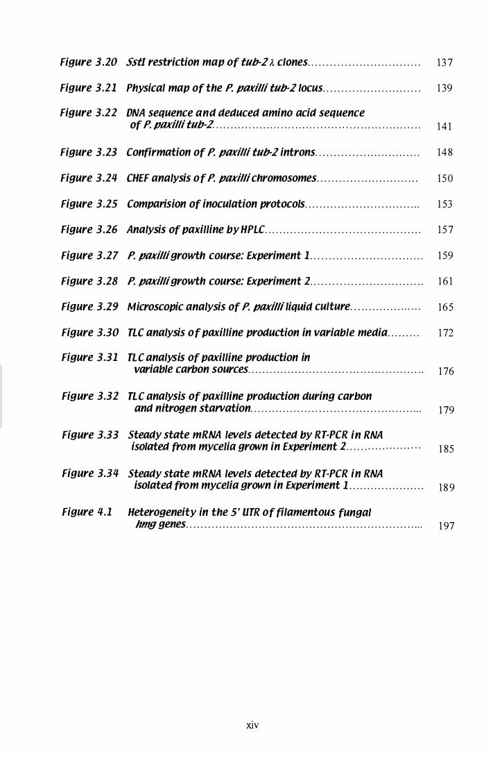
Figure 3.Z0 SstI restriction map of tub·2 A clones............................... 1 3 7
Figure 3.Z1 Physical map of the P. paxilli tub·2 Iocus.. . . . . . . . . . . . . . . . . . . . . . . . . . 1 39
Figure 3.ZZ DNA sequence and deduced amino acid sequence of P. paxilli tub· 2. . . . . . . . . . . . . . . . . . . . . . . . . . . . . . . . . . . . . . . . . . . . . . . . . . . . . . . . . . 1 4 1
Figure 3.Z3 Confirmation of P. paxilli tub·2 introns............................. 148
Figure 3.Z4 CHEF analysis of P. paxilli chromosomes............................ 1 50
Figure 3.Z5 Comparision of inoculation protocols................................ 1 53
Figure 3.Z6 Analysis of paxilline by HPLC...... . . . . . . . . . . . . . . . . . . . . . . . . . . . . . . . . . . . . . 1 5 7
Figure 3.Z7 P. paxilligrowth course: Experiment 1 . . . . . . . . . . . . . . . . . . . . . . . . . . . . . . . 1 59
Figure 3.Z8 P. paxilligrowth course: Experiment Z. . . . . . . . . .. . . . . . . . . . . . . . . . . . . . . 1 6 1
Figure 3.Z9 Microscopic analysis of P. paxilli liquid culture.............. ...... 165
Figure 3.30 nc analysis of paxilline production in variable media......... 1 72
Figure 3.31 nc analysis of paxilline production in variable carbon sources.................... . . . . . . . . . . . . . . . . . . . . . . . . . . . . . 1 76
Figure 3.3Z nc analysis of paxilline production during carbon and nitrogen starvation................... . . . . . . . . . . . . . . . . . . . . . . . . . . . . . 1 79
Figure 3.33 Steady state mRNA levels detected by RT-PCR in RNA isolated from mycelia grown in Experiment Z. . . . . . . . . . . . . . . . . . . . . 1 85
Figure 3.34 Steady state mRNA levels detected by RT-PCR in RNA isolated from mycelia grown in Experiment 1 . . . . . . . . . . . . . . . . . . . . . 1 8 9
Figure 4.1 Heterogeneity in the 5' UTR of filamentous fungal hmggenes.................................................................. 1 97
XlV

Introduction
Chapter 1
1 .1 Secondary metabolism
The phenomenon of secondary metabolite formation is of great importance to many
fields of human endeavour. Secondary metabolites, also referred to as natural products
or products arising from dispensable pathways, are products non-essential for the
primary metabolism, survival and reproduction of the producing organism. The
organism is able to exist and reproduce in the complete absence of secondary metabolic
functions. Examples of secondary metabolites include antibiotics, antifungal, antitumor
and anti-AIDS agents, plant growth factors, insecticides and herbicides. Many
secondary metabolites are beneficial, but many toxic molecules can also be produced,
including compounds that contaminate important human and agricultural stock food
resources and products that act as virulence factors during pathogenesis of many plants.
The effects on other species, caused by secondary metabolites are obvious, however, the
benefits to the producing organism are not always apparent.
There can be a selective advantage from synthes ising some secondary metabolites.
Antibiotics and antifungal agents help the producing organism compete for resources,
whilst toxins such as the trichothecene 4, 1 5 -diacetoxyscirpenol (DAS) from Gibberella
pulicaris (Desjardins et al. , 1 992) and dothistromin from Dothistroma pini (Shain &
Franich, 1 98 1 ) may be essential during fungal invasion of some host species.
Aspergillus species, that produce the carcinogenic aflatoxins, appear to have no
biological advantage over their atoxigenic counterparts. Evidence has shown that
atoxigenic strains of A. flavus can out compete toxigenic strains in field trials as a
biocontrol agent in both maize and cotton (Brown et al. , 1 99 1 ; Cotty, 1 994) . However,
almost all strains of A. parasiticus isolated to date, produce aflatoxins, and it appears
that A. parasiticus may be able to out-compete A. flavus in peanut crops in Australia
(Carter et al., 1 997). Penicillium paxilli strains, that lack the genes for paxilline
biosynthesis, have identical growth rates and morphology to wild-type paxilline
producing strains (Young et al. , 1 998) . Reasons for the prevalence of dispensable
pathways amongst fungi is still unknown.
1
Chapter 1
1.2 Biosynthetic pathways
Many of the natural products produced, and the biosynthetic pathways that have
evolved to produce them, are highly complex. It has been estimated that on average,
approximately ten to thirty different structural genes, representing enzymes that perform
successive transformations to a specific substrate, can be required for the production of
a single secondary metabolic product. The structure and function of secondary
metabolites can be as complex as the pathways that produce them and many organisms
synthesise more than one product. The streptomycetes alone produce approximately
half the known microbial antibiotics (Bibb, 1 996). For example, Streptomyces
coelicolor A3(2) produces a number of antibiotics via l inked pathways (Hopwood,
1 988) . The sophisticated pathways required for secondary metabolite production, can
be branched, causing the production of a range of closely related products.
Alternatively, a primary biosynthetic pathway can branch into a secondary pathway at a
point where an intermediate of the primary biosynthetic pathway becomes the precursor
of a secondary biosynthetic pathway (Drew & Demain, 1 977).
1.3 Gene clusters
Genes for secondary metabolite biosynthesis are frequently found in a single cluster.
For a time, the D.A. Hopwood quote "there are currently no proven exceptions to the
generalisation that genes controlling successive steps in antibiotic biosynthesis are
clustered" could be assumed for other secondary metabolites as well, although a number
of exceptions do appear in the paper by Martin & Liras ( 1 989) . The prevalence of
fungal metabolic gene clusters was reviewed by Keller & Hohn ( 1 997). Genes required
for both primary and secondary metabolic functions, are frequently grouped together. A
c luster of three genes involved in nitrate utilisation was located on a single genomic
fragment of Aspergillus Jumigatus (Amaar & Moore, 1 998) . In some bacterial species,
genes are found clustered on a plasmid and can even form an operon, such as the eight
genes for lantibiotic lactocin S from Lactobacillus sake (Skaugen et al. , 1 997).
P lasmids are mobile genetic elements that can be transferred horizontally between
species, therefore resistance and regulatory genes for the specific antibiotic are
2
Chapter 1
frequently located adjacent to the biosynthetic gene cluster. If the transfer of structural
genes were to occur without the concurrent transfer of a resistance mechanism, the
recipient organism commits " suicide" as soon as it starts producing the antibiotic . The
same argument can be applied to vertical transmission of secondary metabolite
pathways . If a resistance mechanism is separated from the structural genes during
sexual recombination and is not inherited, the effect will be the same. Therefore, strong
natural selective forces exist to keep not only the genes for the resistance mechanism
l inked, but the structural and regulatory genes as well (Martin & Liras, 1 989) . The
synthesis of some products could prove deleterious if the timing of biosynthesis is not
regulated . Examples of resistance mechanisms include the gene for �-lactamase, which
gives protection against the �-lactam antibiotics, and transporter pumps, which export
toxins. A gene recently isolated from the Fusarium sporotrichioides trichothecene gene
cluster, tril2, has homology to toxin efflux pumps. Tri 12 may be involved in protecting
the producing organism from trichothecenes (Alexander et al. , 1 999). In the rice blast
fungus Magnaporthe grisea, an ABC transporter was i solated and shown to give
protection against plant defence compounds, but did not play a role in transporting
fungal compounds (Urban et al. , 1 999). In some instances, the resistance mechanism
alone can be transmissible, by way of a mobile gene cassette called an integron.
Recombination takes place at a 59 bp DNA element associated with the integron
(Stokes et al., 1 997) .
Clustering of these secondary metabolite biosynthetic genes is extremely useful during
the isolation these pathway (Kleinkauf & von Dohr en, 1 990). Once one gene has been
identified, the other genes can usually been found on the same section of DNA (Gaisser
et al., 1 997; Hohn et al. , 1 993a; Tudzynski et al., 1 999) . In fungi, gene c lusters can be
located on dispensable chromosomes, such as the c luster of Nectaria haematococca
genes involved in pathogenic ity to peas. The presence of transposable elements
adjacent to the cluster has lead to the hypothesis that horizontal transfer of these
'pathogenicity islands' has occurred (Kistler et al. , 1 999) . In other cases, gene clusters
have been located on non-dispensable chromosomes, which appear to contain certain
regions that are dispensable. In P. paxilli, strains that contain large deletions of
chr omosome Va, encompassing the paxilline biosynthetic cluster, have normal growth
and morphology (Young et al. , 1 998) . Conserved hexanucleotide sequences in
P. chrysogenum mutants, frequently flank deletions in the penicillin biosynthetic gene
3
Chapter 1
c luster (Fierro et al., 1 996). Conserved repeat sequences can increase the likelihood of
a recombination event and could be a mechanism for the transfer of genetic material .
1 .4 Evolution
If the products of secondary metabolism are potentially auto-toxic, there is a selective
advantage for grouping resistance and regulatory mechanism with the biosynthetic
genes . However, the clustering of biosynthetic genes that synthesise products of
unknown function, is less wel l understood. Many hypotheses have been proposed to
explain the clustering of secondary metabolite genes. The first theory, 'that genetic
transfer promotes clustering' , refers to the relatedness of fungal and prokaryote
Isopenicillin N synthetase genes, cited in a number of examples by Keller & Hohn
( 1 997). There is also similarity between genes involved in phenazine and polyketide
biosynthetic pathways, which suggests that gene transfer between secondary metabolite
producing species, has influenced the evolution of these pathways as well (Vining,
1 992). The hypothesis, that horizontal transfer of pathway genes has occurred (and
presumably stil l occurs) and confers a selective advantage, results in the selection of
pathways that become clustered in a single region of a genome. Another theory predicts
that the regulation of many biosynthetic pathways requires the structural genes to be
clustered, so that a precise cascade of expression can be achieved and that chromatin
structures may be involved (Cavall i & Thoma, 1 993) . This becomes increasingly
important when a resistance mechanism is required prior to the production of potentially
auto-toxic antibiotics.
Maplestone et al. ( 1 992) and Stone & Williams ( 1 992) presented an alternative
hypothesis on the evolution of secondary metabolites. Clustering of biosynthetic genes
and the products of biosynthesis must have a selective advantage. They proposed a
mechanism for the evolution of secondary metabolite biosynthetic pathways. A gene
for a primary metabolic enzyme duplicates, mutates, and becomes capable of catalysing
the conversion of an intermediate product into a novel product. If this new product is
beneficial to the producing organism, the mutation will be selected for. Subsequent
duplication and further random mutation in the initially mutated gene may result in an
4
Chapter 1
enzyme capable of converting the first novel product into a second product of increased
benefit to the producing enzyme. Repeated gene duplications may be of no additional
benefit or of adverse effect and that l ineage would not be selected for, but some could
be beneficial and the process could continue to give rise to the gene clusters present
today. This would explain the sequentially ordered clusters we see in some secondary
metabolic pathways (Martin & Liras, 1 989). In the paxilline biosynthetic cluster from
P. paxilli, there are two sets of genes with similar functions located adjacent to each
other. PaxP 1 and paxP2, which have homology to P450 monooxygenases, and paxRI
and paxR2, which have regions of homology to Zn(Il)2Cys6 binuclear cluster DNA
binding motifs (Young et al. , 2000). Stone & Williams ( 1 992) also review the work of
Marsh et al. ( 1 989) that examines homology between two gene products of the
propionic acid fermentation pathway in Propionibacterium shermanii, a methyl
malonyl-CoA mutase, and a transcarboxylase. A method for testing this theory is also
discussed and examines the ability of sequential intermediates to bind to the specific
receptor in the target organism. According to the "function-evolution" hypothesis,
researchers would see increasing binding affinity with the subsequent pathway
intermediates. Further evidence would be seen, as a high degree of sequence homology
between genes of a single biosynthetic cluster.
A final theory on the evolution of secondary metabolites, specifically antibiotics,
proposes that antibiotics were prebiotic effector molecules . That is, they were able to
catalyse the synthesis of, and stabilise the structures of, such macromolecules as DNA
and proteins. This would explain why modem antibiotics are able to bind to these
structures and cause inhibition (Davies, 1 990). However, once ribosomal and protein
based synthesis of macromolecules had evolved, these prebiotic effector molecules
would become redundant and evolve new functions to j ustify the evolution of their
modem biosynthetic pathways. This, Stone & Williams ( 1 992) argue, provides further
support for their function-evolution hypothesis. Interesting speculations could be made
with respect to the peptide toxins discussed by Kleinkauf & von Dohren ( 1 993) that are
synthesised via an RNA-independent, non-ribosomal process. Although the protein
synthetases that catalyse the formation of the peptide toxins are themselves the product
of standard ribosomal function.
5
Chapter 1
1.5 Regulation
Regulation of gene expreSSIOn can occur at many levels. Active repres sion or
promotion of transcriptional machinery by regulatory proteins can affect the level of
mRNA transcription. Post-transcriptional regulation can affect mRNA stability and
splicing. Once a protein has been translated, it can be transported to different
organelles, stored in an inactive form, phosphorylated or rapidly degraded (Day &
Tuite, 1 998). However, gene regulation in filamentous fungal is usually at the level of
transcription (Gurr et al., 1 987). Biosynthesis of secondary metabolites is frequently
seen after the cessation of primary biomass accumulation. Environmental cues often
trigger the switch from primary to secondary metabolism, through global regulators.
With respect to secondary metabolite gene cluster, there are frequently two levels of
regulation, the fIrst by environmental global regulators and the second by pathway
specific regulators (Keller & Hohn, 1 997).
1.5. 1 Nitrogen
A vailability of a preferred nitrogen source regulates many different pathways in
filamentous fungi, including genes for the assimilation of alternative nitrogen sources.
The best examples of nitrogen global regulators are AREA from A . nidulans and NIT2
from Neurospora crassa. These positive-acting transcription factors have a CyS2/CyS2
zinc finger motif and are members of the GA TA family of transcription factors
(Marzluf, 1 993). These proteins bind to GAT A motifs in the promoters of nitrogen
regulated genes with a DNA binding domain located next to the zinc finger (Wilson &
Arst Jr., 1 998). Binding sites for the homologous A. fumigatus nitrogen regulator
AREA, are found in the promoters of the nitrate assimilation gene cluster (Amaar &
Moore, 1 998). High levels of nitrate are correlated with decreased aflatoxin production
in A . parasitic us (Liu & Chu, 1998). AREA was shown to bind to the intergenic region
of aflR and aflJ in A. parasiticus, suggesting that suppression of aflatoxin biosynthesis
by nitrate may be abrogated by binding of AREA (Chang et al., 2000). In Penicillium
chrysogenum, the homologue of AREA and NIT2, NRE, regulates both nitrate
assimilation and penicillin biosynthesis (Haas & Marzluf, 1995b) although there is no
6
Chapter 1
evidence for similar regulation of penicillin biosynthesis in A. nidulans (Brakhage,
1 998). A second global regulator protein, NMR, isolated from N. crassa, is also
required for nitrogen metabolite repression. Analysis of protein-protein interactions
showed that while NIT2 binds the promoter of nitrogen regulated genes, NMR binds to
NIT2 (Pan et al., 1 997). A homologue of the nmr-J gene was recently isolated from
A. n idulans (Andrianopoulos et al. , 1 998), indicating that a two-component system for
global nitrogen regulation may exist in other fungi as well.
1 .5.2 Carbon
Carbon catabolite repression (CCR) also regulates many genes via global regulatory
proteins. CREA, from A. nidulans is a negative-acting regulator that represses gene
expression in the presence of a preferred carbon source. CREA binds to the sequence
motif 5'-SYGGRG-3' in the promoters of repressed genes (Panozzo et al. , 1 998). In
Saccharomyces cerevisiae, the equivalent protein Mig l , binds the consensus sequence
5' -SYGGGG-3' with a Cys2/His2 zinc finger (Gancedo, 1 998). In N. crassa, the CREA
homologue is called Cre- l (de la Serna et al. , 1 999). Positive-acting carbon source
regulatory proteins in yeast include the Hap2/3/4/5 complex, which binds to
5'-CCAAT-3' promoter motifs, Gal4 and Ma163. Gal4 and Mal63 activate genes
involved in the catabolism of galactose and maltose and bind pairs of CG triplets
separated by 1 1 and 9 nucleotides respectively (Gancedo, 1 998). The presence of
glucose often imposes CCR on secondary metabolite gene clusters (Drew & Demain,
1 977), including the penicillin biosynthetic gene cluster from P. chrysogenum
(Gutierrez et al., 1999). Expression of penicillin biosynthetic genes is also under CCR
control, as is the level of isopenicillin N synthetase transcript present under repressing
carbon sources (Espeso & Pefialva, 1 992). However, this repression is independent of
the global carbon source regulator CREA (Pefialva et al. , 1 998). Glucose i s not always
the preferred carbon source, even though filamentous fungi are only able to take up
monosaccharides (Nielsen, 1 992). If disaccharides are present, the enzyme invertase is
released. Breakdown of invertase can lead to a temporary increase in nitrogen once
disaccharides have been converted to monosaccharides. Sugar a1cohols like mannitol,
which functions as carbon storage molecules in the phloem of plants, can be the
preferred carbon source of saprophytic or endophytic fungi. For example, substitution
7
Chapter 1
of mannitol for glycerol resulted in 4-fold increase of the antibiotic pneumocandins Ao
by the fungus Zalerion arboricola in liquid culture (Tkacz et al. , 1 993).
1.5.3 pH
Ambient pH is another environmental trigger that can affect gene expression via a
global regulator protein. PacC, found in A. nidulans, A. niger and P. chrysogenum has
three zinc finger motifs of the Cys2iHis2 type and is known to increase transcription of
genes under alkaline conditions (Espeso & Pefialva, 1996; Then Bergh & Brakhage,
1 998). Penicillin biosynthetic genes in both A. nidulans and P. chrysogenum, have
binding sites for PacC (Espeso & Pefialva, 1 996; Suarez & Pefialva, 1 996). The
signalling cascade that induces pace transcription, involves the pal genes in A. nidulans
(Negrete-Urtasun et al. , 1 999). Homologous genes have also been identified in
S. cerevisiae (Denison et al., 1 998).
1.5.4 Pathway-specific regulators
Secondary metabolite pathways can also be regulated in a pathway-specific manner,
usually by a product encoded within the cluster itself (Keller & Hohn, 1 997). A good
example of this is the regulation of aflatoxin and sterigmatocystin biosynthesis by ajlR
(previously apa2 from A. parasiticus and ajl2 from A. jlavus) which belongs to the
Zn(II)2Cys6 binuclear cluster Gal4 family of transcription factors (Woloshuk et al. ,
1 994). Induced expression of A. jlavus ajlR, has been shown to initiate transcription of
sterigmatocystin biosynthetic genes in A. n idulans grown under conditions that
normally suppress toxin biosynthesis (Yu et al. , 1 996). ALCR, the pathway specific
regulator of the ethanol utilisation pathway in A. nidulans, is also a Zn(II)2Cys6
binuclear cluster transcription factor. Transcription of alcR, is repressed by CREA in
the presence of a preferred carbon source (Panozzo et al., 1 998). The putative pathway
regulators (paxRI and paxR2) of the paxilline biosynthetic pathway in P. paxilli also
have homology to the Zn(II)2Cys6 binuclear cluster DNA-binding motifs (Young et al. ,
2000). In Streptomyces grise us, expression of streptomycin biosynthetic genes is
controlled by strR, which is in turn controlled by the global regulator A-factor (Ohnishi
8
Chapter 1
et al., 1 999). A regulatory gene has · also been isolated from the trichothecene
biosynthetic pathway in F. sporotrichioides. Tri6 has been shown to be temporally
expressed in coincidence with trichothecene structural biosynthetic genes, and Tri6-
mutants are unable to produce trichothecene, even when supplied with six different
trichothecene intermediates. The transcription level of structural genes tri4 and tri5 was
greatly reduced in the Tri6- mutant. The 2 1 7 residue protein specified by tri6 was
found to have regions of homology with the Cys2/His2 group of zinc finger proteins
(Procter et al., 1 995). In filamentous fungi, the Zn(II)2CyS6 binuclear cluster group of
transcription factors are extremely common, but not exclusively of this type (Schj erling
& Holmberg, 1 996). For example, PacC contains three Cys2/His2 type zinc finger
domains (Tilbum et al., 1 995) and the nitrogen responsive regulatory proteins NIT2
from N. crassa and AREA from Aspergillus possess zinc finger motifs of the Cys2/CYS2
type (Marzluf, 1 993).
1 .5.5 Post transcriptional regulation of gene expression
Gene expression in filamentous fungi is generally regulated at the level of transcription,
however, heterogeneity in the 5' UTR makes many post-transcriptional an<:! post
translational regulatory mechanisms possible. The inclusion of certain sequences in the
5' UTR could alter mRNA stability. Translation initiation from alternate ATG codons
could result in premature termination of translation, or the inclusion of specific
sequences for the targeting of proteins to different cellular organelles. MUltiple
transcripts are produced from the arginase gene in N. crassa. One appears to be
regulated by a basal promoter and a second transcript is produced in an arginine
dependent manner (Marathe et al., 1 998). Translation of the two transcripts is initiated
from different ATG co dons in each case. The genes for 3 -hydroxy-3-methylglutaryl
coenzyme A reductase (HMGR), found i n other species also produce multiple
transcripts. Sixteen different forms of hmg mRNA were detected in the UT -1 cell line
from Chinese hamsters. The multiple messages, which differ in their 5' UTR, are
transcribed from a single gene at 8 different transcription initiation sites. Nine A TG
codons are present in the genomic copy of this gene (Reynolds et al., 1 985). Primer
extension also revealed heterogeneity in the 5' UTR of the A. nidulans isopenicillin N
9

Chapter 1
synthase gene (Perez-Esteban et al. , 1 993). Introns in the 5' UTR of transcribed genes
are rare, but when present, they appear to play a role in the regulation of gene
expression. Multiple introns and alternative splicing can determine the choice of ATG
codon for translation initiation as is the case in the hmg gene of Chinese hamster
(Reynolds et al. , 1985). In the human tenascin-C gene, distinct regions of an
untranslated ex on were shown to affect transcription (Gherzi et al. , 1 995). Alternatively
spliced messages from the rat estrogen receptor gene are found in different tissues
(Hirata et al. , 1 996). Sometimes, the alternative products are found in different ratios as
is the case for the human a2 (VI) collagen gene (Saitta et al., 1 992), or in equal amounts
in all tissues like the HMG CoA synthase gene from both humans and Chinese hamster
(Gil et al. , 1987). The presence of alternative splicing mechanisms in the 5' UTR of
genes is often highly conserved between homologous genes in different species,
indicating an important role in regulation. A 5' UTR exon from the rat Fgf-J gene was
used to isolate the homologous gene in mouse (Hackshaw et al. , 1 996).
1.6 Growth of filamentous fungi in liquid culture
Reproducible physiology of filamentous fungi in liquid culture is notoriously hard to
achieve. However, as a huge number of commercially important products can be
isolated from fungi, a great deal of research has been directed at solving this problem.
S ome products, such as antibiotics are only produced under certain physiological or
developmental conditions. Biosynthesis of primary products, such as citric acid has
been easier to optimise. As discussed above, carbon, nitrogen and pH can affect the
type and rate at which secondary metabolites are produced. However, another factor
that relates directly to filamentous fungi in liquid cultures, is the availability of oxygen.
Two factors that affect the availability of oxygen are culture aeration and culture
morphology. Essentially there are two extreme morphologies that can appear in liquid
cultures, discrete circular pellets, which are hollow (Clark, 1 962) and homogeneous
loose mycelia. Studies of culture kinetics show that as the diameter of mycelial pellets
decreases, culture physiology resembles that seen in loose hyphal fragments and vice
versa. Mycelial pellets produce a mass transfer gradient across the radius, of nutrients
and oxygen into the peliet and of waste products and secreted metabolites out of the
10
Chapter 1
pellet (Prosser & Tough, 1 99 1 ) . Using a miniature oxygen electrode, the concentration
of dissolved oxygen was shown to exist in a gradient across the radius of mycelial
pellets (Huang & Bungay, 1 973). Prosser & Tough ( 1 99 1 ) list a number of factors that
can affect pellet formation including agitation and shear, growth medium composition,
culture viscosity, inoculum concentration and type, specific growth rate, aeration,
surfactants, pH and suspended solids. At higher shake speeds, mycelia are sheared from
the outer surface of the pellet, reducing the maximum diameter of each individual pellet
(Cui et al. , 1 998). Larger culture volumes also decrease the pellet diameter presumably
by increasing pellet interaction and shear effect. Mycelia on the outer surface of the
pellet have greater access to nutrients and oxygen, while mycelia closer to the centre
face a build up of waste products. Therefore, individual hyphae in pelleted cultures can
have different physiological environments. Potentially, mycelia toward the centre of the
pellets would cease primary growth much faster. With no access to nutrients and
oxygen, autolysis occurs in the centre and the pellets become hollow (Cui et al., 1 998).
However, in large-scale continuous cultures, pellets are frequently preferred, as loose
mycelia can clog outlet lines and stirrers (Moreira et aI. , 1 996) .
Loose filamentous hyphae produce very viscous cultures that need increased aeration to
provide adequate oxygen, although all hyphae are in a homogeneous environment. The
oxygen concentration in cultures of Streptomyces parvulus, was shown to affect the
amount and type of secondary metabolites formed (Kaiser et al. , 1 994). Pellet
morphology, and thus oxygenation, has also' been shown to affect penicillin
biosynthesis in cultures of A. nidulans, with pelleted cultures producing no penicillin
(Moore & Bushell, 1 997). Under normal growth conditions, many natural products are
only produced when the secondary growth phase or idiophase has commenced.
However, the induction of secondary metabolism in liquid culture can be manipulated in
numerous species with media supplements. Studies have revealed that different carbon
sources can alter the growth kinetics of a culture. By growing cultures in optimal
conditions, Liao et al. ( 1 995) showed that a sufficient biomass is produced during
trophophase to support secondary metabolite production after the switch to idiophase.
Growth in sub-optimal conditions resulted in overlapping biomass accumulation and
secondary metabolism, with lower levels of natural product biosynthesis.
1 1

1. 7 Secondary metabolite structures
1 . 7. 1 Peptides
Chapter 1
Fungi are known to produce a range of peptidyl secondary metabolites that are toxic to
plants, insects and nematodes . The interesting aspect of these products is the way in
which they are produced. Biosynthesis is via a nonribosomal peptide system that
completely bypasses the need for mRNA and tRNA and is catalysed by a single multi
functional synthetase enzyme, often encoded by large intronless genes (Kleinkauf &
von Dohren, 1 993). For example the proteins Hts- l and Hts-2, involved in the synthesis
of He-toxin in Coehliobolus earbonum, were found to be part of a single protein. This
single gene locus, designated hts contained no introns (Walton et al., 1 993). Sometimes
further modifications, such as epimerisation or N-methylations, are required to produce
the mature product. The genes encoding proteins that perform these modifications are
often clustered with the peptide synthetase gene.
1 .7.2 f3·lactam antibiotics
Of the more common peptidyl secondary metabolites, the �-lactam antibiotics are the
most widely recognised. Much work has gone into the elucidation and optimisation of
the biosynthetic pathways that produce penicillins and cephalosporins (Pefialva et al. ,
1 998). The mode of action of the �-lactam antibiotics involves the inhibition of cell
wall formation in Gram positive bacteria (Chopra et al. , 1998). Resistance to �-lactam
antibiotics can be gained by expression of �-lactamase, which breaks the central lactam
ring. This type of resistance can be thought of as aggressive, as opposed to passive
resistance mechanisms that bypass the site of action of the attacking antibiotic. One of
the initial steps in the biosynthesis of penicillin, is the conversion of the tripeptide 8-(L
a-aminoadipyl)-L-cysteinyl-D-valine (ACV), into isopenicillin N (IPN), by the enzyme
i sopenicillin N synthetase (IPNS). Espeso & Pefialva ( 1 992) showed that in the
presence of repressing sugars, that is sugars that induce the carbon catabolite repressed
state, the amount of IPNS gene transcript was noticeably decreased. Further work
showed that the ipnS promoter was predominantly regulated by three eis-acting negative
12
Chapter 1
elements, one of which has a role in sucrose repression, that effect the levels of basal
transcription (Perez-Esteban et al., 1 993). In A. nidulans, the gene for isopenicillin N
synthase is called ipnA , the promoter of which possesses three binding sites for the
Pace zinc-finger, transcription factor. Interestingly, one of the controlling factors that
regulate the activity of ipnA in A. nidulans, is the pH of the environment. Thus, ipnA is
an alkaline-expressed gene (Espeso & Pefialva, 1 996).
1 . 7.3 Polyketiaes
Another clas s of secondary metabolites is the polyketides. These include the
mycotoxins sterigmatocystin and aflatoxins B l and B2 of Aspergillus species, a number
of antibiotics, including actinorhodin, granaticin, oxytetracyclin, frenolicin, and
tetracenomycin, immunosuppressants, and spore pigments, including a grey pigment
from Streptomyces coelicolor A3(2) that is under the control of the whiE gene cluster
(Yu & Hopwood, 1 995). Polyketide biosynthesis is very similar to fatty acid synthesis,
such that many structural similarities exist between the polyketide synthase (PKS) and
fatty acid synthase (F AS) complexes. There are two classes of PKS and F AS
complexes. Type I synthases (bacterial and fungal PKS and vertebrate F AS) are single
large proteins with multiple catalytic domains and are divided into two sUbtypes. One
type is transcribed from a single gene and the single product performs all the rounds of
polyketide elongation. A second type, known as a modular P KS, is translated from
several transcripts, resulting in a set of synthase units. A single synthase unit is
proposed to catalyse a single round of polyketide chain elongation. (Yang et al., 1 996).
For example the biosynthesis of the polyketide erythromycin is controlled by genes
arranged as a set of six units, each unit responsible for a single round of polyketide
elongation (Donadio et al. , 1 99 1 ). Type II synthases (plant and bacterial F AS and PKS)
are complexes of several proteins, with one or two functions per protein.
There appears to be a high degree of sequence homology between genes of the
polyketide biosynthetic pathways, especially genes coding for polyketide synthases. In
fact, probes designed on the basis of previously sequenced polyketide synthase genes
are frequently used to isolate genes in the uncharacterised polyketide biosynthetic
pathways of different species. The act! gene locus of the modular polyketide synthase
1 3
Chapter 1
cluster, and the act!II polyketide reductase gene, both from the actinorhodin
biosynthetic pathway in Streptomyces coelicolor A3(2), have been used singularly and
in tandem to isolate a number of polyketide synthases from a range of species. Using an
act! probe, the griseusin polyketide synthase gene cluster was isolated from S. griseus
(Yu et al., 1 994) and polyketide synthase genes from the glycopeptide antibiotic ardacin
biosynthetic gene cluster of Kibdelosporangium aridum (Piecq et al. , 1994). I n
combination, act! and actIII probes have been used to isolate polyketide synthase genes
from the daunomycin-producing Streptomyces sp. strain C5 (Ye et al. , 1 994) and the
monensin producer S. cinnamonensis (Arrowsmith et al., 1 992).
1 . 7.4 Aflatoxin
The polyketides sterigmatocystin and aflatoxins B 1 and B2 are three of the most potent
carcinogenic toxins produced by fungi. Sterigmatocystin, the precursor of aflatoxins B 1
and B2 is converted into aflatoxin in A. flavus, A. nomius and A. parasitic us via the
pathways : sterigmatocystin (ST) � � O-methyl-sterigmatocystin (OMST) � �
Aflatoxin B 1 (AFB 1 ) ; and dihydrosterigmatocystin (DHST) � � dihydro-O
methylsterigmatocystin (DHOMST) � � Aflatoxin B2 (AFB2) (Bhatnagar et al. ,
1 99 1 ). The biosynthetic pathway stops at ST in A. nidulans and many other Aspergillus
species (Trail et al., 1995). In A. niduians, a F AS gene homologue was found linked to
the ST biosynthetic cluster. Mutational analysis showed that this gene, in addition to a
P KS, is required for ST biosynthesis. The F AS gene is specific for secondary
metabolism and primary requirements are met by a second unlinked F AS gene (Brown
et al. , 1 996b). In P. paxilli, another example of gene duplication is seen. A copy of the
gene for geranylgeranyl pyrophosphate (GGPP) synthase (pax G) was found within the
paxilline biosynthetic gene cluster. A second, unlinked copy (ggs - i ) fulfils the
requirements of primary metabolism and is unable to complement paxG deletion
mutants (Young et al., 2000). The biosynthetic genes for AF and ST biosynthesis are
clustered and the order of genes within the cluster often corresponds to the order of
enzymatic steps. However, the sequential ordering of genes is more conserved in the
AF pathway than the ST pathway (Brown et al., 1 996a; Woloshuk & Prieto, 1 998).
Gene expression is regulated by a pathway-specific, Zn(II)2Cys6 binuclear cluster,
transcription factor designated AFLR (Woloshuk et aI. , 1 994)
1 4
1.7.5 Terpenes
Chapter 1
Terpenes or terpenoids are another large group of secondary metabolites synthesised by
a variety of organisms, particularly plants. The range o f structures includes,
monoterpenes, diterpenes and triterpenes, cyclic and noncyclic forms. Specific
structural moieties can be included, such as indole-diterpenoids lolitrem B and paxiIline,
produced by the endophytic filamentous fungi Neotyphodium loW (previous ly
Acremonium lolii and A . loliae (Glenn et al. , 1 996)). The functions of this class of
metabolite are wide-ranging, and their synthesis involves both primary and secondary
metabolic pathways. A number of important primary metabolites are synthesised via
the isoprene pathway, including the steroid and retinoid hormones, insect juvenile
hormones, and two important plant hormones, gibberellic acid and abscissic acid
(Moore, 1 990). The filamentous fungus Gibberella fujikuroi also produces the plant
hormone gibberellic acid (GA) during secondary metabolism. The gibberellins are
tetracyclic diterpenes that are synthesised via the isoprenoid biosynthetic pathway. The
first committed step specific for GA biosynthesis is the cyclisation of GGPP by the
enzyme copalyl diphosphate synthase. The gene, cps was recently cloned, and Northern
analysis confirmed that a dramatic increase in expression of cps is associated with the
onset of GA production (Tudzynski et al., 1 998). The primary isoprenoid biosynthetic
genes for HMGR (Woitek et al. , 1 997), GGPP synthase (Homann et al. , 1 996) and
farnesylpyrophosphate (FPP) synthase (Mende et al. , 1 997) were also cloned from
G. fujikuroi. Expression levels of the genes for HMGR and GGPPS were not affected
by glucose or ammonia, while expression of the gene for FPPS was unaffected by light
regimes.
Many of the terpene structures are toxic. An inhibitor of acyl CoA cholesterol
acyltransferase was isolated from the liver and identified as a novel pentacyc1ic
triterpene ester. It is hypothesised that the source of this inhibitor was plant triterpenes
in the diet (Tabas et al. , 1 990). Another terpene, the monoterpene d-limonene, inhibits
small G proteins, thus affecting cell proliferation (Crowell et al., 1 995). Toxic terpenes
are presumed to effect the cell membranes of microorganisms. Along with a vast range
of other cyclic hydrocarbons, the lipophilic terpenes are capable of penetrating the lipid
bilayer of biological membranes. Studies investigating this phenomenon reported that
the membrane was seen to swell and have increased fluidity, which allowed for
1 5
Chapter 1
uncontrolled flux of primarily protons across the membrane (Sikkema et al., 1 994).
Cyclic monoterpenes can also cause damage to skin, as proliferation and survival o f
epidermal keratinocytes is greatly reduced b y the addition o f monocyclic terpenes, in a
dose-dependent manner (Kitahara et al., 1 993).
The sesquiterpenoid a1cohols are another group of toxic secondary metabolites. These
toxins are produced in several genera of fungi, as well as by the higher plant species
Baccharis. A group of these toxins, the trichothecenes , are synthesised via the
cyclisation of the isoprenoid pathway intermediate FPP, into trichodiene, by the enzyme
trichodiene synthase. Mutants of the fungal species F. sporotrichioides, that are
deficient in the production of the trichothecene T-2 toxin, have been analysed by
complementation studies with cosmids isolated from a F. sporotrichioides genomic
DNA library. These studies show that a number of genes involved in the biosynthesis
of T-2 toxin, are clustered in a single 30kb stretch of DNA upstream of a previously
identified gene tox5 (Hohn et al., 1993a). The gene tox5 encodes a trichodiene synthase
and is now called tri5 (Hohn & Desjardins, 1 992). To further characterise the regulation
of tox5, the promoter regions of tox5 from high and low trichothecene-producing strains
were compared. Promoters from the high-producing strains were found to contain a
42 nt tandem repeat. Low-producing strains only carried a single copy of this repeat in
their promoters. However, once transformed into reporter constructs, expression of tox5
was found to be independent of the 42 nt repeat (Hohn et al. , 1 993b). Another gene
from the trichothecene biosynthetic pathway, tri4, has recently been isolated from
F. sporotrichioides. The predicted amino acid sequence of tri4 shows significant
homology to cytochrome P450 monooxygenases. Because Tri4- mutants o nly
accumulate the unoxygenated intermediate trichodiene, tri4 has been placed in a new
cytochrome P450 subfamily, CYP58. Tri4- mutants were however, able to produce
trichothecenes when supplied with oxygenated pathway intermediates, meaning it is
unlikely to be involved in regulation of the pathway (Hohn et al. , 1 995). The catalytic
activities of cytochrome P450s are utilised in a vast range of metabolic capacities, and
are thought to play an important role in the synthesis of many secondary metabolites,
including isoflavonoids, terpenoids and alkaloids (Schuler, 1996). Similarities are also
seen between the cytochrome P450s of different fungal species. For example, the
eburicol 1 4-a-demethylase (P45014DM) was isolated from Penicillium italicum by
16
Chapter 1
heterologous hybridisation with the corresponding gene from Candida tropicalis. The
P45014DM proteins from yeast and fungi are the targets of antimycotics and fungicides
(van Nistelrooy et al., 1 996).
There are also examples of terpenes that are beneficial. The plant Celastrus hindsii
produces a number of triterpenes. Of four isolated recently (celasdin-A, celasdin-C,
celasdin-B, and maytenfolone-A), one, maytenfolone-A was considerably cytotoxic to
hepatoma and nasopharynx carcinoma. Another, celasdin-B was found to inhibit HIV
replication in H9 lymphocyte cells (Kuo & Kuo, 1 997).
1. 7.6 Paxilline ana Lolitrem B
It is the alkaloid indole-diterpenoids, lolitrem B and a proposed precursor paxilline,
produced by N lolii and P. paxilli respectively that are the main focus of this research.
However, other species also produce similar structures such as the endophytic
Phomopsis which synthesises paspalitrem A and C (Bills et al. , 1 992). Other
tremorgenic mycotoxins include the penitrems and janthitrems from Penicillium spp.
and the aflatrems from Aspergillus spp. (Cole et al., 1 974; GaUagher et al. , 1 98 0) .
These lipophilic toxins, capable o f crossing the blood brain barrier, are known as
tremorgens , because they cause the neurological disorder ryegrass staggers in
agricultural ruminants. When injected into the gastrointestinal smooth muscle of sheep,
paxilline induced tremors which lasted for 1 -2 h and lolitrem B induced tremors for
over 24 h (Smith et al., 1 997) . It is thought that the effect may be a result of toxin
interactions with Ca2+-activated K+ (maxi-K) channels and neurotransmitter release at
both central and peripheral levels (Knaus et al., 1 994) . Binding of paxilline to maxi-K
channels is reversible (Sanchez & McManus, 1 996). There is also evidence that the
tremorgenic mycotoxins affect 'Y-aminobutyric acid (GABA) receptors by binding close
to the Cl- channel (Gant et al., 1 987). Although the biosynthetic pathway is unknown,
attempts to elucidate a potential pathway, by examining the chemistry required for the
production of potential precursors through to the end product, lolitrem B, have provided
vital information. Enzymatic functions predicted to be involved include; cyclases,
oxygenases, P450 monooxygenases and dehydrogenases. The primary substrates of the
1 7
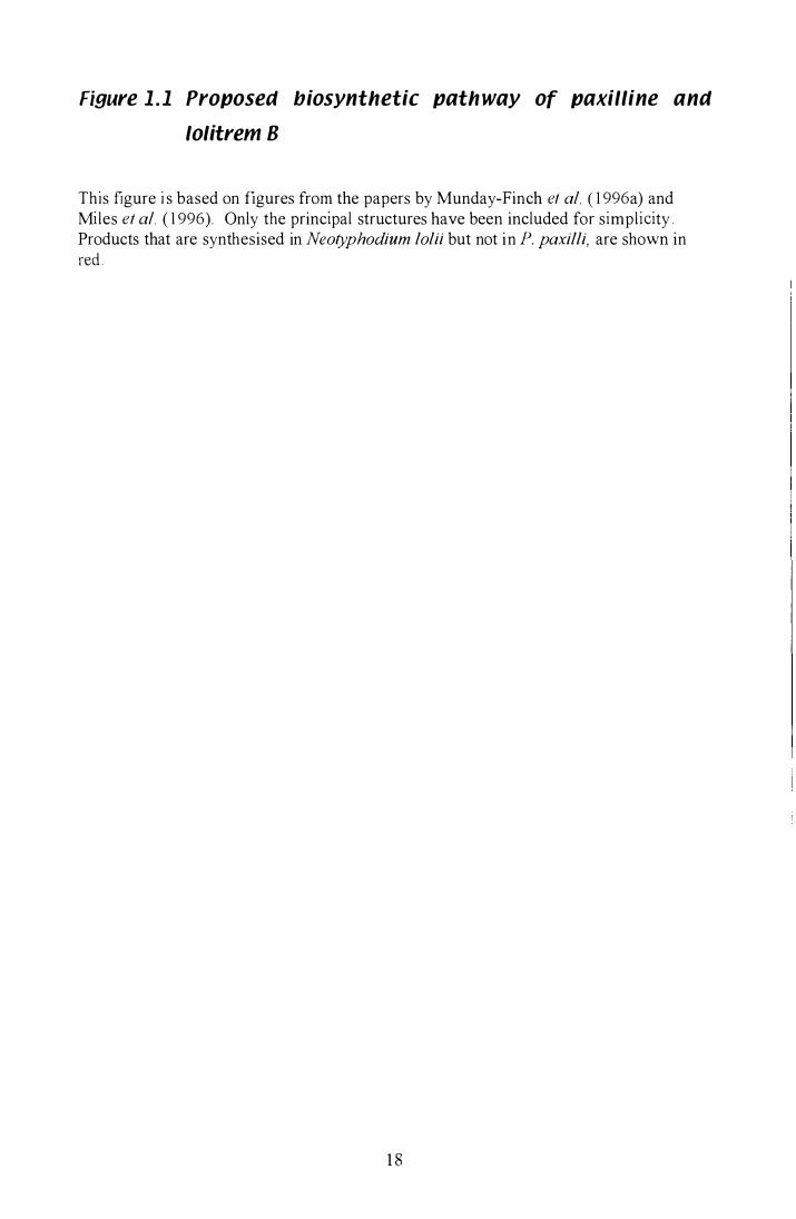
Figure 1 . 1 Proposed biosynthetic path way of paxilline ana
lolitrem B
This figure i s based on figures from the papers by Munday-Finch et af. ( 1 996a) and Miles et af. ( 1 996). Only the principal structures have been included for simplicity . Products that are synthesised in Neotyphodium lolii but not in P. paxilli, are shown in red .
1 8
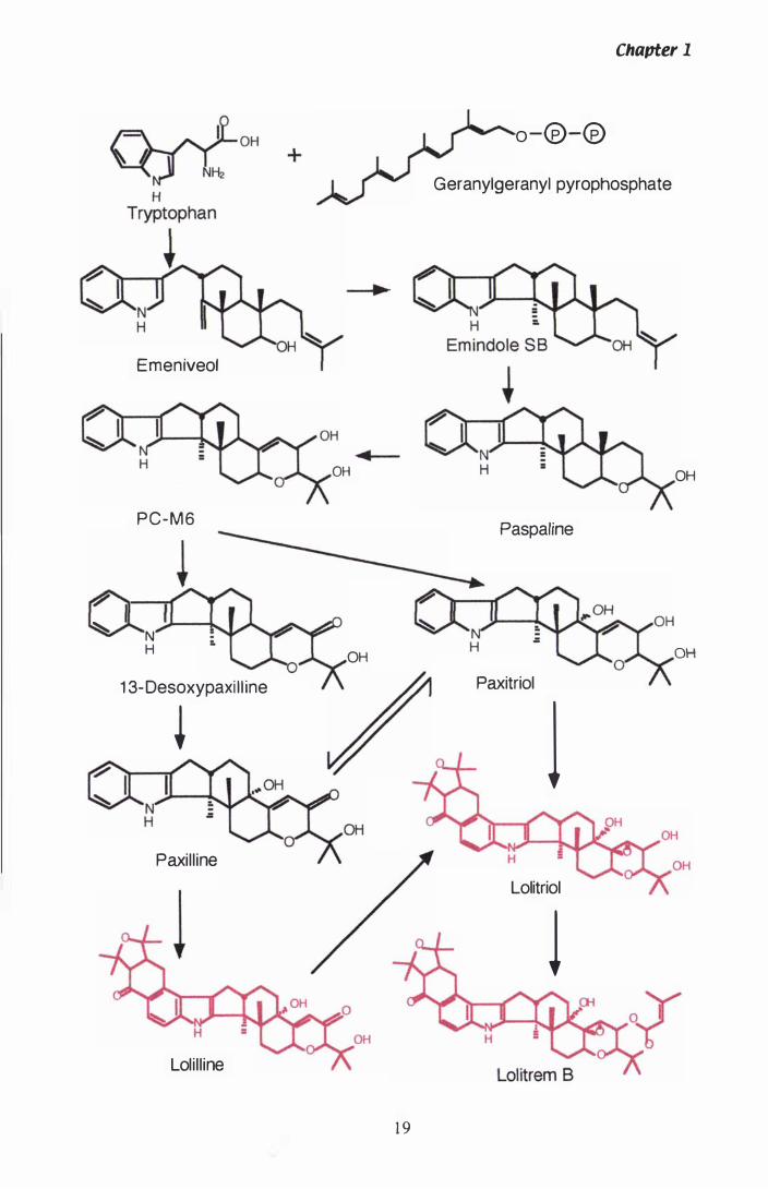
Chavter 1
O -@-@
Geranylgeranyl pyrophosphate
Emeniveol
� OH OH
P C - M 6 Paspaline
�
et l � N
H OH OH
1 3- Desoxypaxi l l ine Paxitriol
* 1 OH
Paxill ine OH
Lolitriol
l Loli l l ine
19
Chapter 1
lolitrem B biosynthetic pathway are tryptophan, which provides the indole moiety
(Laws & Mantle, 1 989) and mevalonic acid, which provides the C5 prenyI units (Acklin
et al., 1 977). There are two pieces of evidence that support paxilline as the biological
precursor of lolitrem B. Firstly, the spatial positioning of core structural atoms in
paxilline are conserved with lolitrem B. Secondly, paxilline has been isolated from
N. lolii grown in liquid culture (Weedon & Mantle, 1 987). A more immediate precursor
of lolitrem B, may be lolitriol, which differs from lolitrem B by only a single acetal
moiety. Intermediates between paxilline and lolitriol, appear to be the diastereoisomeric
allylic a1cohols CJ.- and �-paxitriol. Interestingly, although paxilline and lolitrem B are
toxic tremorgens, the paxitriols and lolitriol are toxic, but lack tremorgenicity (Miles et
al. , 1 992). Precursors of paxilline are proposed to be paspaline, paspaline B and
1 3-desoxypaxilline (Munday-Finch et al. , 1 996a). Recently a new indole-diterpenoid,
called lolilline, because of its similarities to both paxilline and lolitrem B, has been
isolated from N. loW infected seeds of Lolium perenne. This proposed new
intermediate was also found to be nontremorgenic (Munday-Finch et al. , 1 997). The
biosynthesis of both paxilline and lolitrem B forms an interrelated grid of compounds
once the initial indole-diterpenoid structure has formed (Figure 1 . 1 ). In this way,
isomers of the main structures can be formed, like the isomer of lolitrem B, lolitrem F
(Munday-Finch et al., 1996b).
1.8 3-Hydroxy-3-methylglutaryl Coenzyme A reductase
1.S. 1 Isoprenoid biosynthesis
The enzyme 3-hydroxy-3-methylglutaryl Coenzyme A reductase (HMGR) produces
mevalonate and is the rate-limiting step of many primary and secondary metabo lic
pathways. During the enzymatic reduction, HMG CoA is converted through the
intermediates mevaldyl-CoA and mevaldehyde to mevalonate. Frimpong & Rodwell
( 1 994) used these intermediates to determine the role of catalytically active residues
His86\ Glu558 and ASp766 in Syrian hamster HMGR. The proposed reaction, including
the intermediate substrates, is shown below.
20
Chapter 1
ASp766 Glu558 + His865 ASp766
HMG CoA � [mevaldyl-CoA � mevaldehyde] -----1�. mevalonate
Further phosphorylation and decarboxylation converts mevalonate to isopentenyl
pyrophosphate (IPP). IPP and Dimethylallylpyrophosphate (DMAPP), an isomer of
IPP, are the starting point for isoprenoid biosynthesis (Chappell, 1 995) . The se
pathways and their end products in filamentous fungi are depicted in Figure 1 .2 .
Synthesis of ergosterol (and cholesterol in mammalian cell membranes) involves the
condensation of two, 1 5 carbon (C), FPP units to form the 30 C squalene (Brown &
Goldstein, 1 980; Lees et al., 1 995). Alternatively, the addition of a single IPP unit to
FPP, by the enzyme GGPP synthase, results in the synthesis of GGPP, the starting
substrate for diterpene biosynthesis. GGPP has an important primary metabolic
function and it is the most common prenyl group added to prenylated proteins (Jiang et
al. , 1 995) . Prenylated proteins have increased hydrophobicity and the ability to
associate with lipid membranes (Parmryd & Dallner, 1 996) .
1.S.2 Regulation of HMGR
Internal synthesis of sterols, particularly cholesterol, has to be carefully balanced with
the external cholesterol available. Although the majority of work has focused on
cholesterol biosynthesis in mammals , levels of "ergosterol production in fungi are
presumably maintained by similar mechanisms. Cholesterol enters cells via L ow
Density Lipoproteins (LDLs), which are endocytosed after interaction with the LDL
receptor (Stryer, 1 995) . Sterol homeostasis can be achieved by regulating the
expression of cholesterogenic genes such as HMGR and LDL Receptors. Experiments
to examine this phenomenon have made use of competitive inhibitors of HMGR, such
as compactin and pravastatin (Brown & Goldstein, 1 980; Imbault et al., 1 996) and
showed that expression of these genes is repressed under conditions of excess sterols.
Addition of excess LDL to fibroblast cell culture media represses HMGR activity by
98%. Total repression was never achieved, as other non-sterol products produced from
mevalonate are also required. This type of repression was termed multivalent feedback
regulation (Brown & Goldstein, 1 980) .
2 1
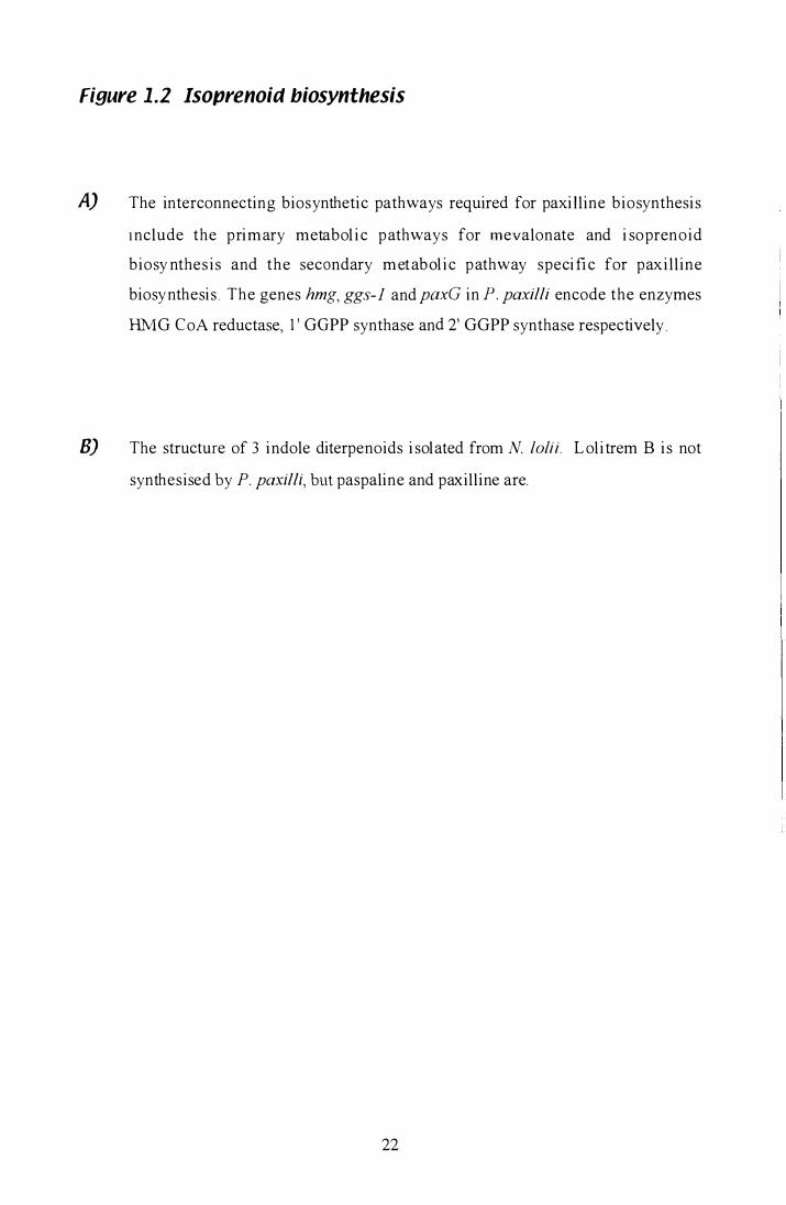
Figure 1 .2 Isoprenoid biosynthesis
A) The interconnecting biosynthetic pathways required for paxilline biosynthesi s
I nclude the primary metabol i c pathways for mevalonate and i soprenoid
biosy nthes i s and the secondary metabol i c pathway speci fi c for paxi l l ine
biosynthesi s . The genes hmg, ggs-l and paxG in P. paxilli encode the enzymes
HM G C oA reductase, l ' GGPP synthase and 2' GGPP synthase respecti vel y .
B) The structure of 3 indole diterpenoids i solated from N. lolii. L olitrem B i s not
synthesised by P. paxilli, but paspaline and paxilline are.
22
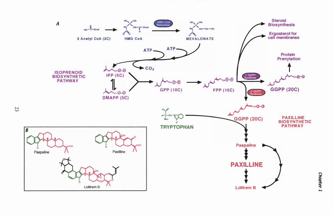
N W
A Steroid Biosynthesis
3 Acetyl CoA (2C) HMG CoA Ergosterol for
cell membranes
ISO P R E N O I D B IOSYNTHETIC
PATHWAY
OH
ATP
�_® C O2
Protein Prenylatlon
I P P (5C) >--�®-0 �-® G PP ( 1 0C )
_-I.��®-0 FPP ( 1 5C)
t
� �� GGPP (20C)
D M A P P (SC)
-Go.t H""L�� -----__
H
TRYPTOPHAN
OH
/�� GGPP (20C)
Paspallne
* PAXILLINE
t Lolitrem B
PAXILLlNE BIOSYNTHETIC
PATHWAY
� �
� "" ....

Chapter 1
1.8.2.1 Transcriptional control of sterol biosynthetic genes
Sterols affect transcription of cholesterogenic genes (Osborne et al. , 1 985) through
sterol response e lements (SREs) in their promoters (Goldstein & Brown, 1 990) .
B inding of the ubiquitous transcription factor, Nuclear Factor Y (NF-V), is also
required before increased transcription of cholesterogenic genes is seen (Jackson et al. ,
1 995). NF-Y, which binds to the inverted CCAAT box, may have a role in recruiting
the sterol response element-binding proteins (SREBP) (Ericsson et al. , 1 996b). In
Syrian hamster, the gene for HMGR has numerous transcription start sites which lack
TA TA boxes and CAA T boxes, the core promoter elements found in most eukaryotic
genes (Reynolds et al., 1 985).
1 .8.2.2 Post-transcriptional control of sterol biosynthetic genes
In the presence of compactin, enzyme activity was increased in three ways. Firstly,
increased transcription of the HMGR gene (called hrng by convention), secondly,
increased translation and thirdly, decreased degradation (Goldstein & Brown, 1 990).
Analysis of a fusion protein of HMGR and green fluorescent protein (GFP), showed
that HMGR is localised in the endoplasmic reticulum in S. cerevisiae (Hampton et al. ,
1 996b), as are the mammalian HMGR proteins (Liscum et al., 1 985) . Overexpression
of both hrng genes from S. cerevisiae resulted in the formation of a novel membrane
structure, possibly to provide a matrix for the abundant protein to be inserted into
(Wright et al. , 1 988). The regulatory degradation of HMGR in response to sterols,
involves the transmembrane domain (Luskey & Stevens, 1 985). A truncated isoform of
the protein expressed in Chinese hamster ovary cells was degraded five times slower
than the membrane bound holoenzyme (Gil et al. , 1 985) . The enzyme is held in the
endoplasmic reticulum membrane by 2-8 membrane spanning hydrophobic domains,
with the number of membrane spans varying between species. In archaebacteria, there
are no membrane spanning domains, plants have two domains, yeast and filamentous
fungi have 7 putative domains (Dobson, 1 997; Woitek et al. , 1 997) and higher
mammals have 7 domains (Hampton et al., 1 996b). Work by Roitelman et al. ( 1 992)
used a method that predicted 8 not 7 transmembrane domains in hamster HMGR.
Replacement of the seventh transmembrane domain with a transmembrane span from
24

Chapter 1
bacteriorhodopsin abolished the degradative response to sterols. HMGR found in sea
urchin embryos is not degraded in the presence of excess sterols, however a mutant
generated to contain the second transmembrane span from hamster HMGR, became
sterol-responsive (Kumagai et al., 1 995) . Expression of a mutant form of one of the
S. cerevisiae HMGR genes, which lacked the transmembrane domain, caused an
increase in squalene synthesis, but not of ergosterol, indicating that many levels of
regulation exist in sterol biosynthetic pathways (Donald et al. , 1 997; Polakowski et al. ,
1 998) . Overexpression of both native and transgenic Arabidopsis thaliana hmg did not
result in accumulation of isoprenoid products (Re et al., 1 995) .
HMGR can also be reversibly inactivated by phosphorylation at a conserved serine
residue in the C-terminal catalytic domain (Friesen & Rodwell, 1 997; Kennelly &
Rodwell , 1 985) . The response was observed when hormones involved in sterol
regulation were added to rat hepatocytes (Ingebritsen et al. , 1 979). The original report
showed HMGR activity was decreased when treated with Mg2+ and ATP (Beg et al. ,
1 973) . The inactivation was shown to be independent of the membrane-spanning
domain and dependent on protein phosphatases for reversal (Kennelly & Rodwell ,
1 985) . HMGR in rat hepatocytes was phosphorylated and inactivated in response to
fructose. Studies with 32p revealed that the protein was phosphorylated at the conserved
Ser87l residue (Gillespie & Hardie, 1 992). HMGR from P. mevalonii is not regulated
by phosphorylation. Friesen & Rodwel l ( 1 997) created a site for AMP-dependent
protein kinase inactivation by replacing Arg387 in P. mevalonii HMGR with a serine.
The activity of the mutant enzyme could then be reversibly phosphorylated in a manner
analogous to mammalian HMGR.
1.8.2.3 A mechanism for regulation by non-sterol end-products?
While regulation of mRNA transcription, protein inactivation and degradation appear to
be sterol dependent, another post-transcriptional mechanism, involving heterogeneity in
the 5 ' untranslated region (UTR) of hmg, could be a target of non-sterol end-product
regulation (Goldstein & Brown, 1 990) . Sixteen different transcripts are produced from
the hmg gene in Chinese hamster. These arise from a combination of mUltip le
transcription start sites and alternative splicing of an intron in the 5 ' UTR. A cluster of
25
Chapter 1
four transcription start sites, upstream of the most 5 ' intron splice site, initiate 70% of
the transcripts. The remaining 30% of transcripts were initiated from 3 alternative
transcription start sites closer to the translation start site (Reynolds et al. , 1 985) .
However, two alternatively spliced products from the HMG CoA Synthase gene were
found in equal abundance in numerous tissues from Chinese hamster and humans (Gil
et al., 1 987) .
1 .9 Fungal endophytes
Neotyphodium lolii, an asexual fi lamentous fungus of the taxonomic groupmg
Ascomycetes, forms a mutualistic symbiosis with the perennial ryegrass Lolium
perenne, a crop of maj or agricultural importance. From this association, the endophyte
is provided with a nutrient supply and, in the case of this asexual endophyte, seed
dissemination by maternal transmission. Benefits to the host include; drought tolerance,
and resistance to fungal pathogens, nematodes, insects and mammalian herbivores (Ball
et al., 1 995). There is a potential for these endophytes to be used as natural biocontrol
agents against insect pests, if the problems of associated livestock toxicosis can be
solved (Clay, 1 989). N. lolii and N. coenophialum, which infects tall fescue (Festuca
arundinacea) are anamorphs of the Clavicipitaceous choke pathogen Epichloe spp
(Schardl & An, 1 993) . Strains of Epichloe can vary in their degree of pathogenicity,
some will always form the sexual spore structure called a stroma which ' chokes' the
ryegrass inflorescence, others remain asexual and are passed on by seed dissemination
(Buchel i & Leuchtmann, 1 996). N. lolii and N. coenophialum never produce spores
while in symbiosis with the host plant (Siegel et al. , 1 985) . Another interesting feature
of these fungal-plant symbioses, has been the formation of interspecific hybrids. An
endophyte isolated from L. perenne, LpTG-2 CL. perenne Taxonomic grouping 2) was
shown to be a hybrid derived from Epichloe typhina (isolate E8) and N. loW (isolate
Lp5) (Schardl et al., 1 994). The formation of interspecific hybrids may be a potential
mechanism for the horizontal transfer of secondary metabolite biosynthetic gene
clusters between asexual and sexual species.
26
1.9. 1 Mycotoxins
Chapter 1
Toxins produced by N lolii include peramine, lolitrem B and ergovaline, whilst N.
coenophialum produces peramine, ergo valine and loline (Siegel et al., 1 990). Peramine,
a pyrrolopyrazine alkaloid isolated from ryegrass infected with N lolii, was shown to
deter feeding by adult Argentine stem weevil (Listronotus bonariensis) (Rowan &
Gaynor, 1 986). It has also proved to be a potent inhibitor of many other herbivorous
insect species (Rowan & Latch, 1 994). Unlike lolitrem B and the ergot alkaloids,
peramine does not appear to affect mammals (Scott & Schardl, 1 993 ; van Heeswijck &
McDonald, 1 992). Analysis of the distribution of peramine and lolitrem B within
L. perenne, revealed that whilst lolitrem B concentrations are highest in the leaf blade,
peramine was highest in the leaf sheath. Concentrations of peramine were highest in
younger leaves, compared to lolitrem B, which increased with the age of leaves (Keogh
et al., 1 996).
Ergovaline, an ergopeptine alkaloid, is produced via the same precursors as lolitrem B
and paxilline. Mevalonate is converted to DMAPP through four enzymatic steps
(Figure 1 .2) and is then condensed with tryptophan by the enzyme DMAT synthase to
produce the first product of ergovaline biosynthesis dimethylallyltryptophan (DMAT).
Due to the common precursors of the lolitrem B and ergovaline biosynthetic pathways,
there is potential for downstream effects if one or the other pathway becomes blocked.
Ergovaline has also been shown to affect herbivorous insect feeding, particularly
Argentine stem weevil (Rowan & Latch, 1 994). However, it has also been correlated to
the livestock disorder, fescue toxicosis, though no direct cause and effect has been
established (van Heeswijck & McDonald, 1 992).
Lolines pyrrolizidine alkaloids, are not produced by N [olii, but they have been shown
to deter feeding of an aphid species (Rhopalosiphum padi) (Siegel et al. , 1 990). Until
recently, lolines, like lolitrem B could only be isolated from the producing endophyte in
planta (van Heeswijck & McDonald, 1 992). However, lolines have since been isolated
from cultures of N uncinatum (Wilkinson et al., 1 997).
27
Chapter 1
1 .10 Penicillium ptlxilli
Penicillium paxilli i s a filamentous fungi of the subdivision Ascomycotina and
reproduces asexually by way of conidia (Kendrick, 1 992) . The tremorgenic indole
diterpenoid paxilline i s the major secondary metabolite produced by P. paxilli (Cole et
al., 1 974) and is also synthesised by the endophytic fungus N. lolii (Weedon & Mantle,
1 987). Paxilline has been predicted as the precursor of lolitrem B . Due to the adverse
effects of mammalian tremorgenic toxins present in agricultural grazing pastures, there
has been interest in isolating and characterising the genes responsible for toxin
biosynthesis from fungal endophytes. I solation of lolitrem B biosynthetic genes from
N. lolii would be difficult due to the inability of the fungus to produce sufficient
lol itrem B in l iquid culture together with a slow growth rate. N. lolii i s only
disseminated through the host seed and does not sporulate on solid media. P. paxilli
produces large amounts of paxilline in liquid culture (Ibba et al., 1 987) and spores can
be purified from solid media within 4-7 days. Therefore P. paxilli has been adopted as
a more suitable organism for the isolation of the b iosynthetic gene cluster respons ible
for paxilline biosynthesis.
1. 10.1 P. pOKilli paxilline negative mutants
A paxilline negative mutant of P. paxilli was first generated by heterologous plasmid
tagging (Hoh et al., 1 994a). The mutant YI-20 was found to have a 1 20- 1 50 kb deletion
and partial translocation of chromosome Va (Young et al., 1 998). The left boundary of
this deletion was isolated by plasmid rescue, however the right boundary has yet to be
isolated. Using the sequence isolated from the site of pAN7- 1 integration in YI-20,
homologous plasmid integration was used to generate three further mutants. CY-2 and
CY- I 02 were deleted in a similar fashion to YI-20, but without the associated
chromosomal translocation. Both mutants were shown to be paxilline negative as
determined by high performance l iquid chromatography (HPLC) and competitive
enzyme-linked immunosorbant assay (ELISA). A third mutant, CY-35 , has a smaller
deletion of 7 .6 kb, and produces wild type levels of paxilline (Young et al., 1 998). The
28
Chapter 1
chromosome walking technique was then used to isolate sequence from the CY- 1 02
deletion.
An additional Pax- mutant was generated using the REMI technique (restriction
enzyme-mediated integration), which uses a restriction enzyme, in conjunction with a
linear plasmid to generate double stranded breaks in the DNA (Schiestl & Petes, 1 99 1 ).
After screening 1 000 transformants by thin layer chromatography (TLC) for their ability
to produce paxilline, a single Pax- mutant, LM-662, was identified. The sequences
adjacent to the site of plasmid integration were isolated by plasmid rescue. However
DNA sequencing of these regions failed to identify any ORFs that could logically be
associated with the biosynthesis of paxilline. Southern analysis of this mutant revealed
that the pAN 7 - 1 tagged region was located away from the original deletion found in
YI-20, CY-2 and CY- 1 02 . A second un-tagged deletion was detected in the same
location as the original deletion mutants . This deletion resulted from the addition of the
restriction enzyme HindIII, which caused a 22.3 kb section of DNA to be removed.
Sequencing of this locus identified the first gene, paxG, a geranylgeranyl pyrophosphate
synthase. 'Knockout' mutants, constructed in this laboratory, were generated by
replacing the paxG gene with a hygromycin resistance gene (hph) CL. McMillan pers.
comm.) When the paxG- mutants were screened with HPLC neither paxilline, or any of
the predicted precursors C 1 3-desoxypaxilline, paspaline or paspaline B) were produced.
In addition to the replacement of paxG by a double cross-over recombination event, a
small deletion mutant was also created that left paxG intact, but removed all or part of
three additional genes located next to paxG. This mutant, called LM -G 1 30 is also
paxilline negative and lacks paxMl Ca putative monooxygenase), paxUl Ca putative
cyclase with prenyl transferase motifs) and part of paxP 1 Ca putative cytochrome P450
monooxygenase ) .
1.10.2 The paxilline biosynthetic gene cluster
Subsequent sequencing of this locus has revealed 1 7 genes, with putative functio ns
associated with paxilline biosynthesis, in a 50 kb region of chromosome Va (Figure
1 .3 ) . They are; pax G , a GGPPS ; paxMl and pax M2, two FAD-dependent
monooxygenases; paxPl and paxP2, cytochrome P450 monooxygenases; paxD, a
29
Figure 1.3 Paxilline biosynthetic gene cluster from P. paKilli
This figure contains information from the paper by Young et al. (2000) and is adapted
from Figure 1 in the same paper.
A restriction map of the 50 kb paxilline biosynthetic gene cluster and flanking sequence
is shown below the individual A clones isolated during chromosome walking by
Carolyn Young. The 17 cluster genes are shown with red arrows and genes not
predicted to function in paxilline biosynthesis are shown as black arrows. The direction
of each arrow indicates the orientation of individual genes within the cluster.
The cluster genes show homology to the fol lowing proteins M2) paxM2, a
monooxygenase, A) paxA, a pH responsive protein, R2) paxR2, a Zn(II)2Cys6 binuclear
cluster DNA-binding protein, RI ) paxRl , a Zn(II)2Cys6 binuclear cluster DNA-binding
protein, US) pax U5 , unknown function , H) paxH, a dehydrogenase, U4) pax U4,
unknown function, U3) paxU3, unknown function, T) paxT, a transporter, U2) paxU2,
unknown function, G) paxG, a geranylgeranyl pyrophosphate synthase, M I ) paxMl, a
mono oxygenase, U I ) paxUl , unknown possible prenyl transferase, P I ) paxP 1 , a P450
monooxygenase, P2) paxP2, a P450 monooxygenase, D) paxD, a dimethylallyl
tryptophan synthase and 0) paxO, an oxidoreductase.
Non-cluster genes show homology to the fol lowing proteins 1 ) dec, a phenylacrylic
decarboxylase 2) lipl, a lipase, 3 ) amy, an a-amylase, 4) gal, a glucoamylase, 5) trn, an
ABC transporter, 6) kin , a kinesin, 7) adh , an alcoholdehydrogenase, 8) perm , a
permease, 9) red, a reductase, 1 0) lip2, a lipase and 1 1 ) arb , an arabinase.
Strains of P. paxilli, containing deletions within the paxilline biosynthetic cluster are
shown, below the restriction map. Mutants created by Yasuo !toh are l abelled YI,
mutants created by Carolyn Young are labelled CY and mutants created by Lisa
McMillan are labelled LM. Mutants that produce paxilline are shown in green.
3 0
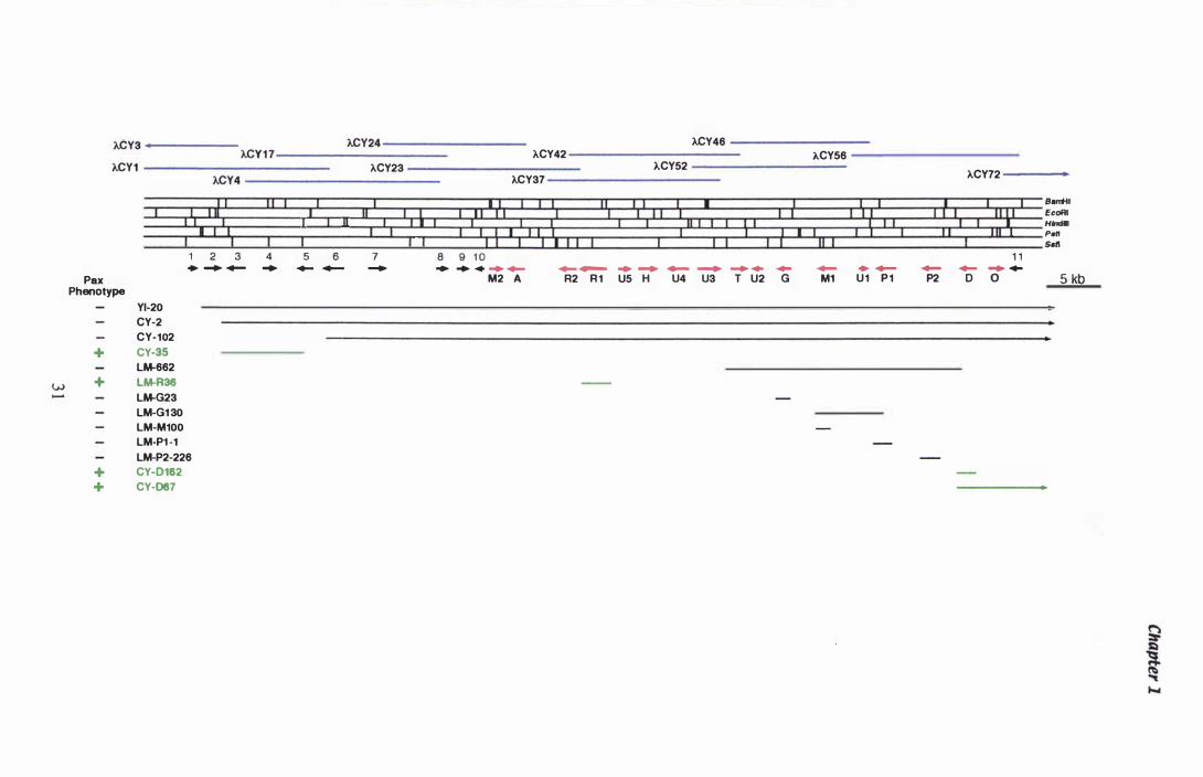
w ......
ACY42 ACY24-----------------ACY3 ... ---------
ACY 1 7-------------------kCY1 ---------------------- ACY23 -------------------
ACY4 -----------------------I I � I
I I : 1 1
1 2 3 4
1 I 1 11
5 6 1 1
I! 1 1 , I 1 I 7 8 9
kCY37
I I I I I 11: ; I 1 ) I I i. 1 1 1 1 0
1 1
11
+ - _ .. _ - - .. .. ... - - -Pax
Phenotype
YI-20
CY-2
CY-102
+ CY-35
+ LM-662
LM-R36
LM-G23
LM-G1 30
LM-M100
LM-PH
LM-P2-226
+ CY-D162
+ CY-D67
M2 A R2 R 1
I
ACY46
ACY56 kCY52
ACY72
I I I 11 I : III I I I
I I : I I I I I 11 1 ( !I I 1 I I 11 1
I I I 8_1 1 � 11 ::p EcoRl
I H_I I 1 I I Pen Sell
1 1 .. .... - - - - - - + - - - ... -U5 H U4 U3 T U2 G M1 U 1 P 1 P2 0 0 5 kb
Chapter 1
DMATS; paxRI and paxR2, with Zn(II)2Cys6 binuclear cluster DNA binding motifs;
paxH, a dehydrogenase; paxT, a transporter protein; paxA, a pH-responsive protein;
paxO, an oxidoreductase; paxUI , a possible cyclase with prenyl transferase motifs and
four other genes, named pax U2 -5, of unknown function. Gene replacement 'knockout'
mutants of paxD, paxRI paxMI, paxP I and paxP2 have been analysed as well .
Paxilline i s produced by replacement mutants of both paxD (Carolyn Young pers.
comm.) and paxRI (Lisa McMillan pers. comm.). The mutants missing paxPI (created
by Lisa McMil1an) and paxP2 (Carr, 1 999) have been shown to accumulate paspaline
and 1 3-desoxypaxilline respectively, but not paxilline (Lisa McMillan pers. comm.) .
1 . 11 Analysis of Gene expression
A number of techniques exist for the analysis of gene expression. Levels of protein can
be measured directly or as a result of gene fusions between a native promoter of interest
and a reporter product, such as �-Glucuronidase (GUS) and Green fluorescent protein
(GFP). This technique was used to examine expression of the verI gene from A. flavus,
involved in aflatoxin biosynthesis. Increased GUS activity was seen in concert with an
increase in aflatoxin biosynthesis. Northern analysis showed a correlation between an
increase in native verI mRNA and GUS activity (Flaherty et al., 1 995) . In filamentous
fungi, the majority of regulation occurs at the level of transcription (Gurr et al. , 1 9 87),
therefore the direct measurement of steady state mRNA levels is a good measure of the
rate of gene expression.
Traditionally, mRNA levels have been determined by Northern blotting. This
technique, based in theory on the method of Southern ( 1 975), separates either total RNA
of purified mRNA by electrophoresis, before transferring them to a nylon membrane.
Using a labelled gene specific probe, the mRNA of interest can be identified.
Messenger RNA from different time points, developmental stages or tissues can then be
compared.
With the development of the PCR, a number of techniques have evolved to quantify
mRNA based on the amount of product amplified from cDNA. Total RNA or purified
32
Chapter 1
mRNA from different time points, developmental stages or tissues is reverse transcribed
into cDNA and a PCR performed with gene specific primers. Product yields are
compared by the intensity of ethidium bromide fluorescence after e lectrophoretic
separation. This technique, known as RT-PCR can be performed both qualitatively and
quantitatively . An added benefit of the RT-PCR technique over Northern analysis, i s
that once cDNA has been synthesised, templates are no longer sensitive to RNase
degradation.
Competitive RT-PCR uses known concentrations of a competitive template to titre the
unknown concentration of the native mRNA species. Frequently a mutant form of the
mRNA is constructed so that the amplified product is a different size from the native
product (Riedy et al., 1 994). Simple PCR-based techniques exist for the construction of
such RNA internal controls (Vanden Heuvel et al. , 1 993). Alternatively gDNA can be
amplified as a competitor, provided intron splicing has occurred between the two gene
specific primers (Harting & Wiesner, 1 997). Messenger RNA from another species has
also been used as a successful competitor for quantification of mRNA in RT -PCR
(Levesque et al. , 1 994) . With techniques such as these, it is possible to calculate the
exact concentration of mRNA in a particular sample. This can be a powerful tool when
studying subtle changes in the level of gene expression.
With both Northern analysis and RT-PCR techniques, the amount of total RNA added
to each reaction needs to be standardised, so that the differences recorded can accurately
reflect changes in the original sample . This is usually done by comparing the
expression of the gene of interest to the gene expression of a constitutively expressed
housekeeping gene. Frequently ribosomal RNA genes or (J..- and �-tubulin genes are
used. D ifferent methods for standardising competitive quantitative RT-PCR were
examined in a paper by Haberhausen et al. ( 1 998). They found that an internal control,
where both the standard and test gene were amplified in a multiplex RT-PCR reaction,
was the most reliable. However, this results in a very complex PCR reaction, where
multiple templates and multiple primer combinations are required.
A simple qualitative RT-PCR technique can also be used to reveal qualitative patterns
of expression, rather than quantitative data. If a single pool of cDNA is generated with
random primers, subsequent PCR reactions can be performed with gene specific
33
Chapter 1
primers, which include amplification of a control gene. In this way the steady state
mRNA profile of a range of cDNA templates can be compared .
While the RT-PCR technique has greater sensitivity than Northern analysis, a method
more sensitive than RT-PCR has also been devised. Femino et al. ( 1 998) used
fluorescent in situ hybridisation (FISH) to detect single RNA molecules from a single
cell.
1 .11 Objectives
The aim of this study has been to begin to understand how paxilline biosynthesis is
regulated in P, paxilli. To achieve this, a gene from the primary isoprenoid biosynthetic
pathway, specifically hmg, was isolated and characterised. Expression of this gene was
then compared with the genes involved in the secondary metabolic biosynthesis of
paxilline. The gene tub-2 was also isolated to provide an independent control for these
experiments. In order to identify reproducible patterns of expression, the physiological
conditions for growth in liquid media were optimised. In the experimental procedure
developed, the induction of paxilline biosynthesis and homogeneous loose hyphal
morphology were observed in a reproducible manner. These conditions were then used
together with a RT-PCR gene expression assay to determine patterns of gene expression
during onset of paxilline biosynthesis.
34

Materials and Methods
2.1 Fungal ana Bacterial strains., A Clones ana Plasmias
2.1 .1 Stocks and strains used
Chapter 2
All fungal and bacterial strains, A clones and plasmids used are shown in Table 2 . 1 .
2. 1.2 Growth of cultures
Bacterial strains grown on solid media were streaked onto LB agar plates (section 2 .2)
with a sterile wire loop and grown at 3TC overnight. When required, antibiotic was
added to a final concentration of 1 00 J.lg/mL for ampicillin or 1 0 J.lg/mL for tetracycline.
Bacterial cells grown in liquid media were transferred to LB media (section 2.2) from a
single colony using a sterile wire loop. Cultures were grown overnight at 3TC and
220 rpm. When required, antibiotic was added to a final concentration of 1 00 Ilg/mL
for ampicillin or 1 0 Ilg/mL for tetracycline.
P. paxilli strains grown on solid media were inoculated from a spore suspension
(section 2 . 1 . 5) . Spores were streaked onto solid media (Potato Dextrose (PD) or
Czapex Dox (CD)) (section 2.2) with a sterile wire loop to produce individual colonies .
PD plates were incubated at 22°C for 4-5 days until sporulation was seen. CDYE plates
were used to determine if any yeast or bacterial contamination was present in large scale
liquid cultures . Approximately 1 00 ilL of culture was spread over the surface with a
sterile glass spreader and the plates were incubated at 30°C for 24 h.
For i solation of DNA, fungi were grown in PD liquid media. Six 250 mL Erlenmeyer
flasks containing 25 mL of PD media (section 2.2), were inoculated with 5 x 1 06
spores/mL from a fresh P. paxilli WT spore suspension (section 2 . 1 .5) . Cultures were
grown at 22° C with shaking at 230 rpm for 3 days. The broth was decanted, the pellets
washed with sterile Milli Q water and divided between two 1 5 mL falcon tubes.
Samples were frozen at -20°C and freeze-dried overnight.
3 5
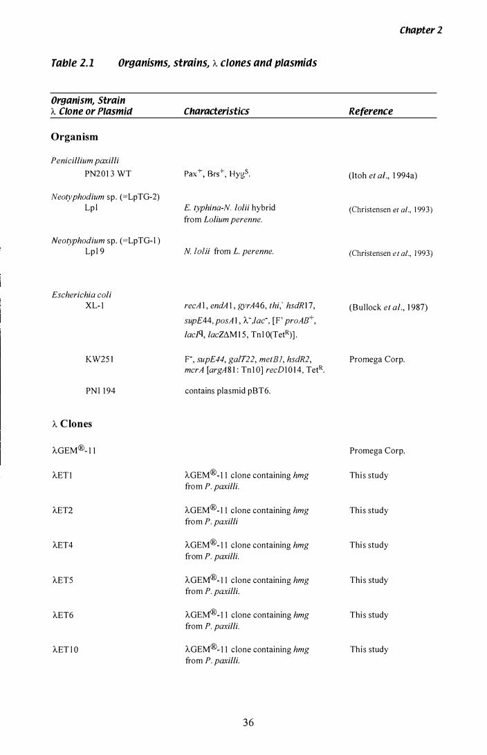
Table 2.1 Organisms .. strains .. A clones anti plasmitls
Organism .. Strain A Clone or Plasmia
Organism
Penicillium paxilli
PN2 0 1 3 WT
Neotyphodium sp. (=LpTG-2) Lp l
Neotyphodium sp. (=LpTG- l ) Lp l 9
Escherichia coli XL- l
KW25 1
PN l 1 94
A Clones
AGEM®- l l
AET l
AET2
AET4
AET5
AET6
AET l O
Characteristics
E. typhina-N. lolii hybrid from Lolium perenne.
N. lolii from L. perenne.
recA I , endA l , gyrA46, thi,' hsdR1 7,
supE44, posA 1 , A-,lac-, [F' proAB+,
lacI'l, lacZtlM 1 5 , Tn l O(TetR)] .
F-, supE44, ga1T22, metB], hsdR2, mcrA [argA 8 1 : Tn t o] recDl 0 1 4, TetR.
contains plasmid pBT6.
AGEM®- l l clone containing hmg from P. paxilli.
AGEM®- l l clone containing hmg from P. paxilli
AGEM®- l l clone containing hmg from P. paxilli.
AGEM®- l l clone containing hmg from P. paxilli.
AGEM®- l l clone containing hmg from P. paxilli.
AGEM®- l l clone containing hmg from P. paxilli.
36
Chapter 2
Reference
(Itoh et aI., 1 994a)
(Christensen et al., 1 993)
(Christensen et al., 1 993)
(Bullock et al., 1 987)
Promega Corp.
Promega Corp.
Thi s study
Thi s study
Thi s study
Thi s study
Thi s study
Thi s study
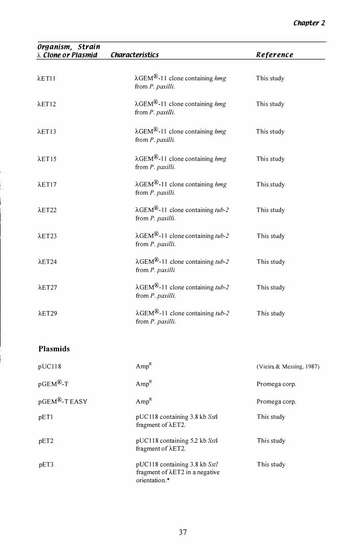
Chavter 2
Organism, Strain A Clone or Plasmid Characteristics Reference
AETl l AGEM® -1 1 clone containing hmg This study from P. paxilli.
AET l 2 AGEM®- 1 1 clone containing hmg Thi s study from P. paxilli.
AET l 3 AGEM®- l l clone containing hmg This study from P. paxilli.
AET I 5 AGEM®- l I clone containing hmg Thi s study from P. paxilli.
AETl 7 AGEM®- 1 1 clone containing hmg This study from P. paxilli.
AET22 AGEM®- l l clone containing tub-2 This study from P. paxilli.
AET23 AGEM®- l l clone containing tub-2 This study from P. paxilli.
AET24 AGEM®- l l clone containing tub-2 This study from P. paxilli
AET27 AGEM®- l l clone containing tub-2 This study from P. paxilli.
AET29 AGEM®- l l clone containing tub-2 This study from P. paxilli.
Plasmids
p UC I l S AmpR (Vieira & Messing. 1 987)
pGEM®-T AmpR Promega corp.
pGEM®-T EASY AmpR Promega corp.
pET l pUC l l S containing 3 . S kb SstI This study fragment of AET2.
pET2 pUC 1 1 S containing 5.2 kb Ss!I Thi s study fragment of AET2.
pET3 pUC l l S containing 3 . S kb Sstl This study fragment of AET2 in a negative orientation. *
37
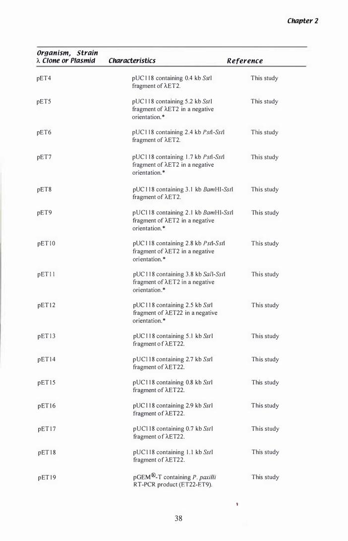
Or9anism� Strain A Clone or Plasmid Characteristics Reference
pET4 pUC 1 1 8 containing 0.4 kb Sstl This study fragment of AET2.
pETS pUC 1 1 8 containing 5 .2 kb Sst I This study fragment of AET2 in a negative
orientation. *
pET6 pUC 1 1 8 containing 2 .4 kb Pstr-SstI This study fragment of AET2.
pET7 pUC I 1 8 containing 1 .7 kb Pstl -SstI Th is study fragment of AET2 in a negative
pET8
pET9
pET I O
pET I I
pET l 2
pET I 3
pET I 4
pET I 5
pET l 6
pET I 7
pET I 8
pET 1 9
orientation. *
pUC 1 1 8 containing 3 . 1 kb BamH I -Sstl fragment of AET2.
pUCI 1 8 containing 2 . 1 kb BamH I-Sstl fragment of AET2 in a negative
orientation. *
pUC 1 1 8 containing 2.8 kb Pstl-Sstl fragment of AET2 in a negative
ori entation. *
pUC 1 1 8 containing 3 . 8 kb Sa/l-Sstl fragment of AET2 in a negative
orientation . *
pUC 1 1 8 containing 2.5 kb Sstl fragment of AET22 in a negative
orientation. *
pUC 1 1 8 containing 5 . 1 kb Sst l fragment o f AET22.
pUCI 1 8 containing 2.7 kb Sstl fragment of AET22.
pUC 1 1 8 containing 0.8 kb Sst l fragment of AET22.
pUC 1 1 8 containing 2.9 kb Sst l fragment of AET22.
pUCI 1 8 containing 0.7 kb Sst l fragment o f AET22.
pUC I 1 8 containing I . l kb Sst l fragment of AET22.
pGEM ®-T containing P. paxilli RT-PCR product (ET22-ET9).
38
Th is study
Th is study
This study
Th is study
This study
Th is study
Th is study
This study
This study
Th is study
This study
This study
Chapter 2

OrganismJ Strain 'A Clone or Plasmid Characteristics
pET20 pGEM®-T containing P. paxilli RT-PCR product (Etub5-Etub6).
pET2 1 pGEM®-T containing P. paxilli 5 'RACE product (UAP-ET3).
pET22 pGEM®-T containing P. paxilli RT-PCR product (ET1 2-ET I 5).
pET23 pGEM®-T containing Lp1 9 RT-PCR product (hmgeco-hmgnco).
pET24 pGEM®-T containing P. paxilli 3 ' RACE product (ET22-UAP).
Chapter 2
Reference
This study
This study
This study
This study
This study
pBT6 Modified pUC 1 2 containing a 3 . 1 kb (Orbach et al., 1 986) HindIlI fragment of the N. crassa �-TubuJin gene.
*NB: Fragments have the 3 ' end of the gene sequence adjacent to the forward primer annealing site in pUC l 1 8 .
39
Chapter 2
For growth experiments, fungi were grown in CDYE based media (section 2 .2), under
paxil line-inducing conditions. Large scale l iquid cultures, from which numerous
samples were removed, were grown in 2 L Erlenmeyer flasks containing 300 mL of
media. Small scale cultures, from which only one or two samples were removed, were
grown in 500 mL Erlenmeyer flasks, containing 1 00 mL of media. Cultures were
inoculated from either a spore suspension (section 2 . 1 .7) or a seed culture (section
2 . 1 .8) . All cultures were grown in a 28T incubator with shaking at 3 50 rpm.
2.1.3 Glycerol stocks
Bacterial cultures (5 mL) were grown overnight at 3TC (section 2 . 1 .2) and the
following day harvested by centrifugation ( 1 0 min, 5 000 rpm) in a 1 5 mL falcon tube.
The media was decanted off and the cells resuspended in 3 mL of sterile 50% (v/v)
glycerol. Aliquots of 750 ilL were transferred to four 1 mL Nunc cryotubes and the
samples snap frozen in an ice-box. Two samples (Table 2. 1 ) were stored at -70°C and
the remaining two stored at -20°C.
2. 1.4 A Phage Iysates
Lambda stocks were grown to confluent lysis (section 2. 1 4.3) , overlaid with 5 mL of
SM buffer (section 2 .3 .8) and stored at 4 °C overnight. The lysate was transferred to a
sterile 1 5 mL falcon tube and approximately 1 00 ilL of chloroform was added. The
lysates were kept at 4°C for long term storage.
2.1 .5 Spore suspensions
Glycerol stocks of P. paxilli (Table 2 . 1 ) stored at -20oC, were streaked for single
colonies on PD agar (section 2.2) as described in section 2 . 1 .2 . When the cultures had
sporulated, 6 mL of 0.0 1 % (v/v) Tween 80 (BDH) was transferred to the surface of the
plate, and spores collected using a glass spreader. The spore suspension was transferred
to a sterile 1 .5 mL Eppendorf tube and centrifuged in a microcentrifuge to pellet the
40
Chapter 2
spores ( 1 3 000 rpm, 3 min). The detergent was decanted and the pellet re suspended in
sterile Mill i Q water. The concentration was determined using haemocytometer
counting.
2.1.6 Glassware preparation
F or large scale liquid cultures, Erlenmeyer flasks were siliconised to prevent excess
build-up of mycelia around the rim of the culture vessel. The 2 L flasks were scrubbed
clean, rinsed with 95% (v/v) ethanol and left to air dry at 3TC. When the glassware
was completely dry, approximately 5 mL of S igmacote® (Sigma) was poured into the
vessel and coated over the entire inner surface. The excess was then drained off and the
flasks left to dry in a fumehood. Si liconised glassware was reused several times before
a new application of Sigmacote was needed. The required volume of test media was
transferred to the clean flasks and a cheese-cloth/cotton wool bung placed in the neck of
the vessel. The glassware and media was then sterilised by autoclaving for 1 5 min at
1 2 1 ° C with 1 5 psi. For smaller scale preparations the media and glassware was
steril ised as above although the smaller flasks were not siliconised.
2.1 . 7 Spore suspension inoculation
Liquid P. paxilli cultures were inoculated directly from a spore suspension (section
2 . 1 . 5 ) . Once the concentration had been determined, spore suspensions could be stored
for up to 6 weeks at 4°C. Liquid cultures were inoculated to a final concentration of 1 x
1 06 spores/mL and grown as described in section 2 . 1 .2.
2.1.8 Seed culture inoculation
Replica cultures of wild type P. paxilli were inoculated using a seed culture. A I L
Erlenmeyer flask (section 2 . 1 . 6) containing 1 00 mL CDYE broth (section 2 .2) was
inoculated with a spore suspension (section 2 . 1 .5) . The culture was grown in a shaking
incubator (3 50 rpm, 28 °C) for 1 2 h. After this time, a small sample was removed and
4 1
Chapter 2
examined microscopically for the presence of germinating spores. Replica cultures
were inoculated with 8 mL of seed culture per 1 00 mL of media. Where the test media
varied from the standard CDYE broth, the seed culture was pelleted by centrifugation
( 1 0 min, 3 1 00 rpm) and re suspended in an equal volume of sterile Milli Q water. The
resuspension was then checked microscopically for the presence of germinating spores.
A sample of the seed culture was plated on CDYE to check for the presence of any
contaminants (section 2 . 1 .2).
2.2 Media
All media and solutions were sterilised for 1 5 min at 1 2 1 °C and 1 5 psi.
Czapek Vox salts
CD salts contain per 1 L : Dipotassium phosphate 1 g, magnesium sulfate 0 . 5 g,
potassium chloride 0 .5 g, ferrous sulfate 0.0 1 g, sodium nitrate 2 g and Milli Q water to
l L.
Czapek Vox Er yeast extract media
CDYE media contains per 1 L : Czapek Dox media (Oxoid) 3 3 .4 g, yeast extract
(Oxoid) 5 g and Milli Q water to 1 L. For solid media, 1 5 g/L agar (Davis) was added
prior to sterilisation.
Luria-Bertani broth
LB media contains per 1 L : Sodium chloride 5 g, tryptone 1 0 9 and yeast extract
(Merck) 5 g and Milli Q water to 1 L. The pH was adjusted to 7-7 .5 prior to
sterilisation. For solid media, 1 5 g/L agar (Davis) or agarose (BDH) was added prior to
sterilisation.
Potato dextrose media
PD media contains per 1 L: Potato dextrose media (Difco) 24 g and Milli Q water to
1 L . For solid media, 1 5 g/L agar (Davis) was added prior to steril isation.
42
Chapter 2
SOC media
SOC media contains per 1 L: Glucose 2 0 mM, KCl 2 . S mM, MgCh 1 0 mM,
MgS04.7H20 1 0 mM, NaCl 1 0 mM, tryptone (Difco) 2% (w/v), yeast extract (Difco)
O.S% (w/v) and Milli Q water to 1 L .
Top agarose
Top agarose contains per 1 L: Tryptone (Difco) 1 0 g, NaCl 5 g, and agarose (BDH) 8 g.
2.3 Solutions
2.3. 1 Stock solutions
Name
Ethanol
Sodium Acetate
Tween 80
Sodium dodecyl sulfate (SDS)
Tris-HCI
NA2EDTA
Glycerol
Maltose
MgS04.7H2O
2.3.2 Acrylamide mix
Concentration pH
70% (v/v), 95% (v/v)
3 M
0 .01 % (v/v)
1 0% (w/v)
1 M 7.2, 7 .S , 8 .0
250 mM 8 .0
SO% (v/v)
20% (w/v)
1 M
Acrylamide mix contains per 1 L: Urea 480 g, acrylamide 57 g and bis-acrylamide 3 g.
These reagents were dissolved in 800 mL of Milli Q water and deionised by mixing
with Amberlite MB-3 (Sigma). The solution was filtered through a s intered glass
funnel and 1 0 mL of 1 0 x TBE sequencing buffer (section 2 .3 . 1 0) added. Milli Q water
was added to a final volume of 1 L .
43
2.3.3 1 0 x Oenhani-rs solution
Chapter 2
Denhardt' s solution ( 1 0 x) (Southern, 1 975) contains per 1 L : 5 0 mL of 1 M HEPES
(BRL) pH 7.0, 1 50 mL of 20 x SSC (section 2 .3 .9) , 6 mL of phenol extracted herring
sperm DNA (3 mg/mL, Sigma), 5 mL of 20% (w/v) sodium dodecyl sui fate (SDS), 2 g
of Ficoll (Type 70, Sigma), 2 mL of Escherichia coli transfer RNA ( l 0 mg/mL, S igma),
2 g of Bovine Serum Albumin (BSA) (Sigma), 2 g of polyvinylpyrrolidone (PVP- 1 0,
Sigma), and Milli Q water to 1 L.
2.3.4 DNA extraction buffer
1 50 mM Na2EDTA, 50 mM Tris-HCl pH 8 .0 , and 2% (w/v) Sodium Lauroyl Sarcosine
(Byrd et al., 1 990).
2.3.5 DNase I buffer
1 00 mM sodium acetate and 5 mM MgS04 pH 5 .0, in DEPC-treated water (section
2 . 1 7.2).
2.3.6 PEG solution
20% (w/v) PEG 6000 in SM buffer (section 2 .3 .8) without gelatin.
2.3. 7 SOS loading dye
1 % (w/v) SDS, 0 .02% (w/v) bromophenol blue, 20% (w/v) sucrose and 5 mM
Na2EDTA (pH 8 .0).
44
2.3.8 SM buffer
Chapter 2
1 00 mM NaCl, 8 mM MgS04.7H20, 50 mM Tris-HCI (pH 7 .5) and 0 . 0 1 % (w/v)
gelatin.
2.3.9 Southern blotting solutions (Southern 1915)
Solution 1 : 0.25 M HCl.
Solution 2: 0.5 M NaOH and 0.5 M NaCl.
Solution 3 : 2.0 M NaCI and 0.5 M Tris-HCI (pH 7.4).
SSC x 20: 5 M NaCI and 0.3 M sodium citrate.
SSC x 2: a 1 0 fold dilution of 20 x SSc.
2.3.10 Tr;s based buffers
STE buffer
1 00 mM NaCl, 10 mM Tris-HCI, and 1 mM Na2EDTA (pH 8 .0).
STET buffer
8% (w/v) Sucrose, 5% (v/v) Triton X-l OO, 5 0 mM Na2EDTA (pH 8 .0), and 5 0 mM
Tris-HCl (pH 8 .0) .
1 x TAE
40 mM Tris, 20 mM Acetic acid and 2 mM Na2EDTA (pH 8 .5).
1 x TBE
89 mM Tris, 89 mM boric acid and 2.5 mM Na2EDTA (pH 8 .2) .
10 X TBE for sequencing
1 .3 M Tris-HCI, 4 .4 M boric acid and 0.25 M Na2EDTA.
TE (10:0.1)
1 0 mM Tris-HCI, 0. 1 mM Na2EDTA.
45

Chapter Z
1 x TNE
1 0 mM Tris-HCl, 1 00 mM NaCl and 1 mM Na2EDTA (PH 7.4).
2.3.1 1 Tris-equilibrated phenol
Tris-equilibrated phenol was supplied by Amersham and stored at 4°C. Prior to use,
0.47 g/ 500 mL of 8- Hydroxyquinone was added to keep the phenol reduced.
1.4 Enzymes ana Biological Solutions
Ampicillin
An ampicillin stock solution was prepared with sterile MiIli Q water to a final
concentration of 1 00 mg/mL and stored at -20°e.
DNase (RNase free)
A stock solution of DNase I was prepared in 1 50 mM NaCl and 50% (v/v) glycerol to a
final concentration of 1 0 mg/mL and stored at _20° C.
IPTG
A stock solution of isopropyl-�-D-galactoside (IPTG) was prepared in sterile Milli Q
water to a final concentration of 20% (w/v) and stored at _20DC.
Lysozyme
A stock solution of Lysozyme was prepared in 1 0 mM Tris-HCI (pH 8 . 0) to a final
concentration of 1 0 mg/mL and stored at -20°C.
RNase (DNase-free)
RNase A (Sigma) was prepared at a stock concentration of 1 0 mg/mL in 1 0 mM Tris
HCl (pH 7.5), 1 5 mM NaCl. RNase was boiled for 1 0 min to inactivate any DNase.
46
Chapter Z
Tetracycline
A tetracycline stock was prepared in methanol to a final concentration of 1 0 mg/mL,
and stored at -20QC.
X·GAL
A stock solution of 5-bromo-4-chloro-3-indoyl-�-D-galactoside (X-GAL) was prepared
in dimethylformamide to a final concentration of 2% (w/v) and stored at -20°C .
2.5 Isolation of DNA
2.5.1 Fungal DNA
Based on the method of Byrd et al. ( 1 990), the freeze dried mycelia was weighed and
0 .5 - 1 g ground to a fine powder under liquid nitrogen in a chilled mortar and pestle.
The powdered mycelia was transferred to a fresh mortar at room temperature,
resuspended in 1 0 mL extraction buffer (section 2 .3 .4) and Proteinase K (BRL) added to
a final concentration of 2 mg/mL. The solution was transferred to a polypropylene tube
and centrifuged at 2 000 x g (Sorval SS34, 4 000 rpm, 1 0 min, 4°C), to remove cellular
debris . The cleared supernatant was transferred to a new tube and 1 volume of Tris
equilibrated phenol (section 2 .3 . 1 1 ) added. The solution was mixed by inversion and
separated by centrifugation (Sorval SS34, 1 3 000 rpm, 1 5 min, 4°C) . Two volumes of
chloroform/phenol ( 1 : 1 ) were added to the aqueous phase and mixed by inversion. The
aqueous phase was separated as before and a final volume of chloroform added. After
centrifugation, DNA was precipitated by the addition of one volume of isopropanol and
the solution incubated on ice for 3 0 min. The DNA pellet was col lected by
centrifugation (Sorval SS34, 8 000 rpm, 1 0 min, 4 QC), the supernatant removed and the
pellet carefully washed with 1 volume of 70% (v/v) ethanol . I f the pellet became
dislodged a second centrifugation step was performed, before decanting off the 70%
(v/v) ethanol . The DNA pellet was dried at 3 TC and resuspended in 500 ilL TE
(section 2 .3 . 1 0) .
47
2.5.2 Iv Phage DNA (medium scale)
Chapter 2
Lambda clones (section 2 . 1 4) were diluted with SM buffer (section 2 .3 . 8) in a sterile
Eppendorf tube and plated (section 2 . 14 .3) onto LB agarose plates (section 2.2) . Plates
were incubated at 3TC until they reached confluent lysis (�8h), overlaid with 5 mL of
SM buffer (section 2 .3 .8) and stored at 4°C overnight. The lysate was transferred to a
sterile 1 5 mL corex tube and the volume made up to 5 mL with SM buffer (section
2 . 3 . 8). KW2 5 1 cell debris was removed by centrifugation (Sorval SS34, 5 000 rpm, 1 0
min, 4°C), and the c leared supernatant transferred to a clean 1 5 m L corex tube .
Contaminating Escherichia coli RNA and DNA was removed with the addition of
RNase and DNase (section 2 .4) to final concentrations of 1 Ilg/mL and the reaction
incubated at 3 T C for 30 min. To precipitate the phage, PEG solution ( 5 mL) (section
2 . 3 . 6) was added and the solution incubated on ice for 1 hr. Phage were harvested by
centrifugation (Sorval SS34, 7 000 rpm, 20 min, 4 °C), the pellet resuspended in 0 .5 mL
SM buffer (section 2 . 3 .8) and transferred to a sterile Eppendorf tube . Phage coat
proteins were dissolved by the addition of 5 ilL 1 0% (w/v) SDS and 1 0 ilL 250 mM
Na2EDTA (pH 8 .0) and the solution incubated at 68°C for 1 5 min. To separate protein
and DNA an equal volume of Tris-equilibrated phenol (2.3 . 1 1 ) was added and mixed
thoroughly by vortexing. The aqueous phase was separated by centrifugation in a
microcentrifuge ( 1 3 000 rpm, S min) and transferred to a fresh Eppendorf tube. Equal
volumes of both Tris-equilibrated phenol ( section 2 .3 . 1 1 ) and chloroform were added
and the aqueous phase separated by centrifugation as above. Finally, an equal volume
of chloroform was added and the aqueous phase containing phage DNA was separated
as above. Phage DNA was precipitated by adding an equal volume of isopropanol and
the solution incubated at -200C for 20 min. The DNA pellet was collected in a
microcentrifuge ( 1 3 000 rpm, 1 0 min), and washed with 1 mL 70% (v/v) ethanol. After
drying, DNA was re suspended in 50 ilL TE (section 2 .3 . 1 0) and 5 ilL 200 Ilg/mL
RNase (section 2 .4) added. Uncut Iv DNA (2 ilL) was examined as in sections 2 . 1 0 . 1
and 2. 1 0.3 .
48
2.5.3 Plasmid DNA by rapid boil method
Chapter Z
Based on method of Holmes & Quigley ( 1 98 1 ) . Plasmid containing colonies were
grown in 3 mL of LB media (section 2 .2) containing ampicillin ( section 2 .4) as
described in section 2 . 1 .2 . Cells were harvested from 1 . 5 mL of culture in a
microcentrifuge ( 1 3 000 rpm, 3 min) and re suspended in 3 5 0 J.1L of STET buffer
(section 2.3 . 1 0) . Lysozyme (25 J.1L) (section 2 .4) was added and the mixture boiled for
40 sec. Precipitated material was pelleted in a microcentrifuge for 1 0 min at
1 3 000 rpm and the resulting gelatinous pellet was removed with a sterile toothpick.
DNA was precipitated by adding of 300 J.1L of isopropanol and the solution incubated at
-20°C for 20 min. The DNA was pelleted in a microcentrifuge ( 1 3 000 rpm, 5 min), the
supernatant discarded and the pellet washed with 350 J.1L of 95% (v/v) ethanol. The
pellet was reconstituted in a microcentrifuge ( 1 3 000 rpm, 2 min) and allowed to dry at
3 T C for approximately 1 5 min. DNA was re suspended in 50 J.1L of TE (section 2.3 . 1 0)
and stored at 4 °C until required. Plasmid DNA was examined by restriction enzyme
digestion (section 2 .7) and mini-gel electrophoresis (section 2. 1 0. 1 and 2 . 1 0.3 ) .
2.5.4 DNA from agarose gel slices
SeaPlaque agarose gel slices containing DNA were excised under long wave UV with a
sterile scalpel blade. The gel slices were placed in a sterile 1 .5 mL Eppendorf tube and
melted at 65 °C . Tris-equilibrated phenol (section 2 .3 . 1 1 ) (500 J.1L) was added, mixed
thoroughly for 1 5 sec and the mixture left to incubate at -20°C for two and a half hours.
The aqueous phase was separated in a microcentrifuge ( 1 3 000 rpm, l a min) and
transferred to two sterile 1 . 5 mL Eppendorf tubes. One volume of Tris-equilibrated
phenol :chloroform was added, mixed by vortexing and the aqueous phase separated in a
microcentrifuge ( 1 3 000 rpm, 3 min). Traces of phenol were removed with the addition
of one volume of chloroform and centrifugation as above. The aqueous phases of both
tubes were then pooled into a single sterile 1 . 5 mL Eppendorf tube and DNA
precipitated by the addition of 1 1 1 0 x volume 3 M sodium acetate and 2 volumes of
isopropanol . The reaction was incubated overnight at -20°C, and the DNA pelleted in a
microcentrifuge ( 1 3 000 rpm, 1 5 min). The supernatant was decanted off and the pellet
washed with 700 J.1L of 95% (v/v) ethanol, then centrifuged ( 1 3 000 rpm, 2 min) . The
49
Chapter 2
clean pellet was left to dry briefly at 3TC, resuspended in 20 ilL of sterile Milli Q water
and stored at 4°C.
2.5.5 Plasmid DNA by Alkaline lysis-based minil1rel1 kits
A number of small scale plasmid isolation kits have been used in the process of this
work including the Quantum Prep® Plasmid Miniprep kit (Biorad), the QIAprep Spin
Miniprep kit (Qiagen) and the CONCERTTM Rapid Plasmid Miniprep System (BRL).
In all cases the general protocol was based on the basic alkaline lysis technique.
Bacterial cultures were grown overnight in 5 mL of LB media (section 2 .2) containing
ampicillin (section 2.4) as described in section 2 . 1 .2. Cells were harvested from 3 mL
of culture and re suspended according to the protocol outlined in the particular kit being
used.
2.6 Quantification of Nucleic Acids
There are a number of methods available for the quantification of nucleic acids.
Genomic DNA or DNA isolated from agarose gel was frequently compared to an
appropriate DNA mass standard by agarose gel electrophoresis (section 2 . 1 0 . 1 ) and
ethidium bromide staining (section 2 . 1 0 .3) . Plasmid DNA, and samples where only a
small amount of material was available, were determined fluorometrically using
Hoechst dye . UV spectrophotometry was used to determine the concentration and
quality of RNA and DNA.
2.6.1 Intensity of ethiaium bromide fluorescence
Genomic DNA was quantified by running on a 0.7% (w/v) agarose gel (section 2. 1 0 . 1 )
alongside A concentration standards (range 1 0-200 ng) . Smaller DNA fragments
isolated from agarose gels were run alongside the Low DNA Mass™ ladder (BRL)
(range 5- 1 00 ng) on appropriate % (w/v) agarose gel. DNA was visualised with
ethidium bromide (section 2 . 1 0.3) .
5 0
2.6.2 Fluorometeric determination
Chapter Z
Using the Hoefer Scientific TKO 1 00 fluorometer, extremely small quantities of DNA
can be quantifi ed. The standards and samples were quantified in 1 x TNE buffer
(section 2 . 3 . 1 0) containing 0 . 1 /lg/mL Hoechst 33258 dye. A Calf thymus DNA ( 1 00
/lg/mL) standard was used to calibrate the machine.
2.6.3 UV spectrophotometric determination
Nucleic acid concentrations were determined on a Shimadzu UV spectrophotometer by
readings at A260 . Samples were diluted so that absorbency readings fell between the
range of 0 . 1 to 1 .0 . Sample purity was determined by calculating the ratio between A260
and A28o .
2. 7 Restriction Endonuclease Digestion of DNA
Restriction endonucleases (RE) were used with the specific buffer recommended by the
manufacturer. Incubations were performed in a 3TC water bath for 1 -2 h. After this
time a small sample was run on an agarose gel (section 2 . 1 0 . 1 and 2 . 1 0 .3) to determine
if digestion was complete. For incomplete digestions, additional enzyme was added and
the digestion continued for another hour. Digestion was terminated with the addition of
1 15 volume SDS loading dye (section 2 .3 .7) . In cases where two RE were used, the
enzyme requiring the lower salt buffer was used first. The salt concentration was then
altered by the addition ofNaCI and the second digestion performed.
2.8 Purification of DNA with Phenol/Chloroform
DNA was routinely purified by phenOl/chloroform extraction in equal volumes of Tris
equilibrated phenol (section 2 .3 . 1 1 ) and chloroform. Reagents were mixed by vortexing
for at least 1 0 seconds. The aqueous phase containing the DNA was separated in a
microcentrifuge ( 1 3 000 rpm, 2 min) . The clear upper phase was transferred to a new
5 1
Chapter Z
Eppendorf tube and an equal volume of chloroform added. The aqueous phase was
again separated as above and transferred to a new Eppendorf tube.
2.9 Precipitation of DNA with Ethanol or Isopropanol
DNA in solution was precipitated with e ither 95% (v/v) ethanol or isopropanol .
Precipitation with ethanol required the addition of 1 1 1 0 volume 3 M sodium acetate and
two and a half volumes of 95% (v/v) ethanol. Precipitation with isopropanol required
the addition of 0 .6 volumes of isopropanol. The reactions were then incubated either on
ice or at -20°C for approximately 3 0 min. The precipitated DNA was pelleted by
centrifugation in a microcentrifuge ( 1 3 000 rpm, 5 min) and the supernatant removed.
The pellet was washed gently with an equal volume of 75% (v/v) ethanol then left to air
dry for approximately 1 5 min. Samples were then resuspended in either sterile Milli Q
water or TE (section 2 .3 . 1 0) .
2.1 0 Agarose Gel Electrophoresis
2.10.1 Mini-gel
For most DNA separations, gels were prepared and run in 1 x TBE buffer (section
2 . 3 . 1 0) , with the concentration of agarose appropriate for the size of fragments to be
separated (Table 2.2). Molecular biology grade agarose (BRL) was melted using a
microwave then allowed to cool to �50 °C before pouring into the mini-gel apparatus.
For separation of low molecular weight bands, such as PCR products, NuSieve agarose
(FMC) was used. When DNA was purified from agarose gel slices, the gels were
prepared and run in 1 x T AE (section 2 .3 . 1 0) and a low melting temperature grade
agarose such as SeaPlaque agarose (FMC) was used. Gels were run between
60- 1 00 volts. In general, 1 0 f.tL samples, containing 1 15 volume SDS loading dye
( section 2 . 3 .7) , were transferred into each well and run alongside 5 !JL of the
appropriate size ladder.
52
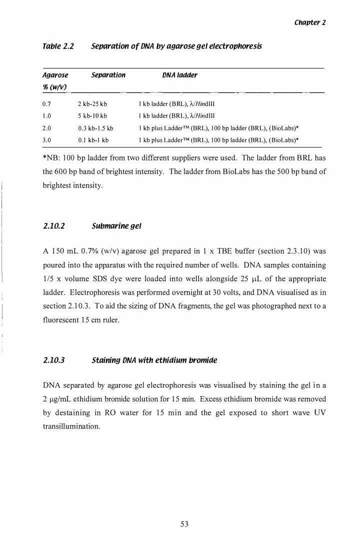
Chapter 2
Table 2.2 Separation of DNA by agarose gel electrophoresis
Agarose Separation DNA ladder
% (w/v)
0.7 2 kb�25 kb 1 kb ladder (BRL), AlHindIII
1.0 5 kb- l O kb I kb ladder (BRL), NHindJII
2 .0 0.3 kb- 1 .5 kb 1 kb plus LadderTM (BRL), 1 00 bp ladder (BRL), ( B ioLabs)*
3 . 0 0 . 1 kb- l kb 1 kb plus Ladder™ (BRL), 1 00 bp ladder (BRL), ( B ioLabs)*
*NB: 1 00 bp ladder from two different suppliers were used. The ladder from BRL has
the 600 bp band of brightest intensity. The ladder from BioLabs has the 500 bp band of
brightest intensity.
2.10.2 Submarine gel
A l SO mL 0 .7% (w/v) agarose gel prepared in 1 x TBE buffer (section 2.3 . 1 0) was
poured into the apparatus with the required number of wells. DNA samples containing
1 15 x volume SDS dye were loaded into wells alongside 25 ilL of the appropriate
ladder. Electrophoresis was performed overnight at 30 volts, and DNA visualised as in
section 2 . 1 0. 3 . To aid the sizing of DNA fragments, the gel was photographed next to a
fluorescent 1 5 cm ruler.
2.10.3 Staining DNA with ethidium bromide
DNA separated by agarose gel electrophoresis was visualised by staining the gel in a
2 Ilg/mL ethidium bromide solution for 1 5 min. Excess ethidium bromide was removed
by destaining in RO water for 1 5 min and the gel exposed to short wave DV
transillumination.
5 3
2.1 1 Polymerase Chain Reaction (PCR)
2.11 .1 Primer design
Chapter 2
Degenerate primers HMG 1 and HMG3 (Dobson, 1 997), designed to conserved regions
of the 3 ' end of hmg genes , contained a number of Inosine residues . Gene specific
primers were designed to have a GC content of between 40-60% and the 3' position was
always a G or C nucleotide. Primers were ordered from either Life Technologies or
S igma Genosys (Table 2.3) .
2.11 .2 Protocol for routine PCR
For routine PCR, the following protocol was used. A cocktail was prepared containing
(n+ 1 ) x [ 1 x Taq DNA polymerase Buffer (Roche), 50 !-LM dNTPs , 200 nM Primer 1 ,
200 nM Primer 2 , 0.5 U Taq DNA polymerase (Roche) and sterile Milli Q water t o a
final volume of 20 !-LL] . The reaction mix was (20 !-LL) was aliquoted into n x 0.2 mL
PCR tubes and stored on ice. To one of these tubes 5 !-LL of sterile water was added , to
serve as a negative control . Where possible a positive control was included as well .
The DNA template was diluted to a suitable concentration and 5 !-LL added to the
remaining tubes. Reactions were placed in a Corbett thermocycler, preset to 94°C, and
the following PCR programme performed; 1 cycle of 2 min at 94°C, 25 cycles of 45 sec
at 94°C, 45 sec at 60°C and 90 sec at 72 °C, 1 cycle of 5 min at 72 °C. Reactions were
terminated by the addition of 1 15 volume of SDS loading dye (section 2 . 3 . 7) and
separated by mini-gel electrophoresis (section 2. 1 0 . 1 and 2. 1 0.3) . The primers used are
listed in Table 2 .2 .
2.11 .3 Bacterial colony PCR
Where specific primers were available, transformed bacterial clones (section 2 . 1 5)
could be identified directly using PCR. A routine PCR cocktail (section 2 . 1 1 .2) was
prepared to a final volume of 25 !-LL. Primer 1 and Primer 2 were specific for the DNA
insert. Using a sterile P l O pipette tip a small amount of the ampicillin resistant colonies
54
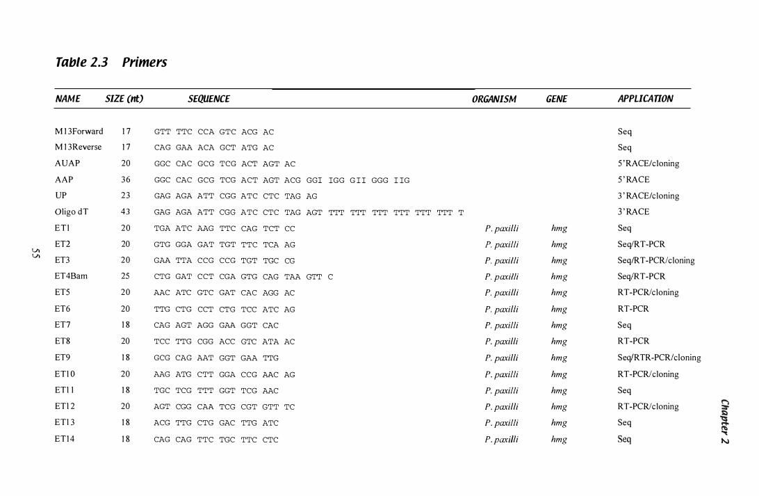
Table 2.3 Primers
NAME SIZE (nt) SEQUENCE
M 1 3Forward 1 7 GTT TTC CCA GTC
M 1 3Reverse 1 7 CAG GAA ACA GCT
AUAP 20 GGC CAC GCG TCG
AAP 3 6 GGC CAC GCG TCG
UP 23 GAG AGA ATT CGG
Oligo dT 43 GAG AGA ATT CGG
E T l 20 TGA ATC AAG TTC
ETI 20 GTG GGA GAT TGT VI VI ET3 20 GAA TTA CCG CCG
ET4Bam 25 CTG GAT CCT CGA
ET5 20 AAC ATC GTC GAT
ET6 20 TTG CTG CCT CTG
ET7 1 8 CAG AGT AGG GAA
ET8 20 TCC TTG CGG ACC
ET9 1 8 GCG CAG AAT GGT
ETl O 20 AAG ATG CTT GGA
ETl l 1 8 TGC TCG TTT GGT
ETl 2 20 AGT CGG CAA TCG
ETl 3 1 8 ACG TTG CTG GAC
ET1 4 1 8 CAG CAG TTC TGC
ACG
ATG
ACT
ACT
ATC
ATC
CAG
TTC
TGT
GTG
CAC
TCC
GGT
AC
AC
AGT
- - - ---------- -- - - -- --- �- - -
ORGANISM
AC
AGT ACG GGI IGG G I I GGG I IG
CTC TAG AG
CTC TAG AGT TTT TTT TTT TTT TTT TTT T
TCT CC P. paxilli
TCA AG P. paxilli
TGC CG P. paxilli
CAG TAA GTT C P. paxilli
AGG AC P. paxilli
ATC AG P. paxilli
CAC P. paxilli
GTC ATA AC P. paxilli
GAA TTG P. paxilli
CCG AAC AG P. paxilli
TCG AAC P. paxilli
CGT GTT TC P. paxilli
TTG ATC P. paxilli
TTC CTC P. pa..·dlli
GENE APPLICATION
Seq
Seq
5 ' RACE/c1oning
5 ' RACE
3 ' RACE/cloning
3 ' RACE
hmg Seq
hmg SeqIRT-PCR
hmg Seq/RT-PCRlcloning
hmg SeqIRT-PCR
hmg RT-PCRlcloning
hmg RT-PCR
hmg Seq
hmg RT-PCR
hmg SeqIRTR-PCRlc\oning
hmg RT-PCRlcloning
hmg Seq
hmg RT-PCRlcloning � hmg Seq ! hmg Seq
"'t N
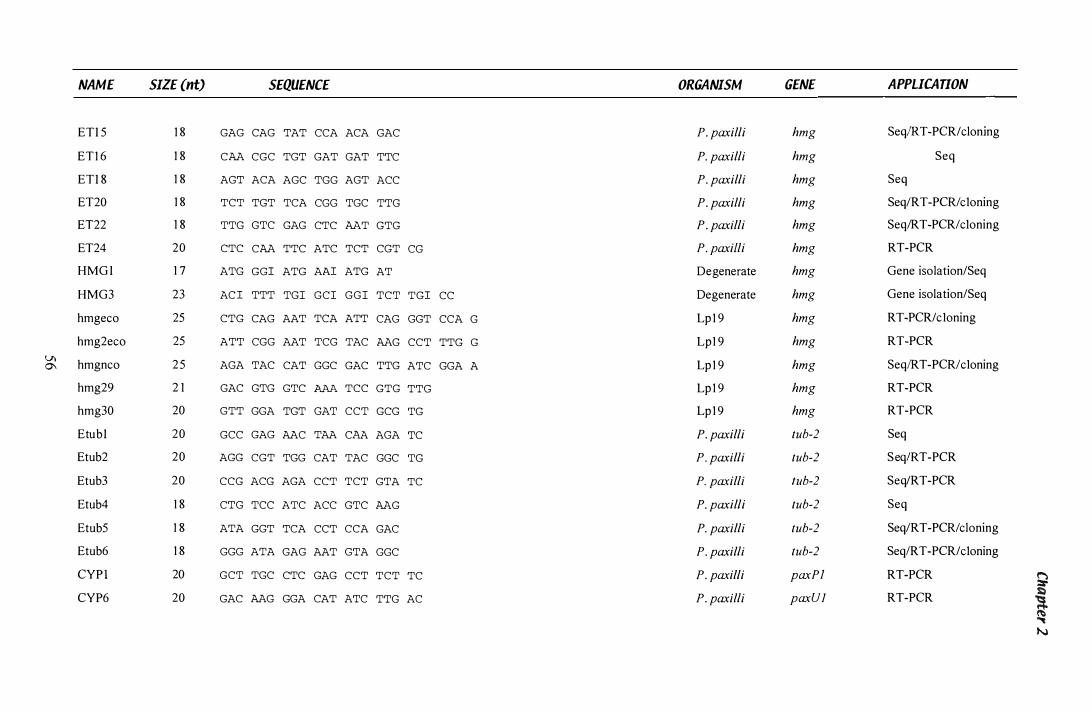
�. _.- ----- - -- --
NAME SIZE (nt) SEQUENCE ORGANISM GENE APPLICATION
ETl 5 1 8 GAG CAG TAT CCA ACA GAC P. paxilli hmg SeqlR T -PCR/cloning
ET1 6 1 8 CM CGC TGT GAT GAT TTC P. paxilli hmg Seq
ETl 8 1 8 AGT ACA AGC TGG AGT ACC P. paxilli hmg Seq
ET20 1 8 TCT TGT TCA CGG TGC TTG P. paxilli hmg Seq/RT-PCR/c1oning
ET22 1 8 TTG GTC GAG CTC MT GTG P. paxilli hmg Seq/RT-PCR/c1oning
ET24 20 CTC CM TTC ATC TCT CGT CG P. paxi/li hmg RT-PCR
HMG l 1 7 ATG GGI ATG AAI ATG AT Degenerate hmg Gene isolation/Seq
HMG3 23 AC I TTT TGl GCl GGl TCT TGI CC Degenerate hmg Gene isolation/Seq
hmgeco 25 CTG CAG AAT TCA ATT CAG GGT CCA G Lp 1 9 hmg RT-PCR/c1oning
hmg2eco 25 ATT CGG AAT TCG TAC MG CCT TTG G Lp1 9 hmg RT-PCR 'J)
hmgnco 2 5 0\ AGA TAC CAT GGC GAC TTG ATC GGA A Lp 1 9 hmg SeqlRT -PCR/c1oning
hmg29 2 1 GAC GTG GTC AAA TCC GTG TTG Lp 1 9 hmg RT-PCR
hmg30 20 GTT GGA TGT GAT CCT GCG TG Lp 1 9 hmg RT-PCR
Etubl 20 GCC GAG MC TAA CAA AGA TC P. paxilli tub-2 Seq
Etub2 20 AGG CGT TGG CAT TAC GGC TG P. paxilli tub-2 SeqIRT-PCR
Etub3 20 CCG ACG AGA CCT TCT GTA TC P. paxilli tub-2 Seq/RT-PCR
Etub4 1 8 CTG TCC ATC ACC GTC MG P. paxilli tub-2 Seq
Etub5 1 8 ATA GGT TCA CCT CCA GAC P. paxilli tub-2 Seq/RT-PCR/cloning
Etub6 1 8 GGG ATA GAG AAT GTA GGC P. paxilli tub-2 SeqIRT-PCR/c1oning
CYP I 20 GCT TGC CTC GAG CCT TCT TC P. paxilli paxP] RT-PCR 9 CYP6 20 GAC MG GGA CAT ATC TTG AC P. paxilli paxUl RT-PCR �
.., N
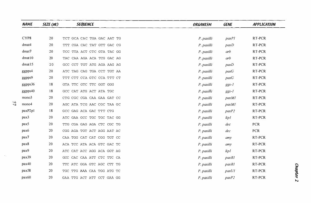
NAME
CYP8
dmat4
dmat?
dmat l O
dmat 1 5
ggpps4
ggpps9
ggpps36
ggpps40
mono3
� mono4
paxP2p l
pax3
pax5
pax6
pax?
pax8
pax9
pax39
pax40
pax58
pax60
-------------------� � -� -
SIZE (nt) SEQUENCE
20 TCT GCA CAC TGA GAC AAT TG
20 TTT CGA CAC TAT GTT GAC CG
20 TCC TTA ACT CTC GTA TAC GG
20 TAC CAA AGA ACA TCG GAC AG
2 0 GCC CCT TGT ATG AGA AAG AG
20 ATC TAG CAG TGA CCT TGT AA
20 TTT CTT CCA GTC CCA TTT CT
1 8 GTA TTC GTC TTC GGT GGG
1 8 GCC CAT ATG ACT ATA TGC
20
20
1 8
20
20
20
20
20
20
20
20
20
20
CTG CGC CGA CAA GAA GAT CC
AGC ATA TCG AAC CGC TAA GC
GCC GAG ACA GAC TTT CTG
ATC GAA GCC TGC TGC TAC GG
TTG CGA GAG AGA CTC CGC TG
CGG AGA TGT ACT AGG AAT AC
CAA TGG CAT CAT CGG TGT CC
ACA TCC ATA ACA GTC GAC TC
ATC CAT ACC AGG ACA GGT AG
GCC CAC CAA ATT CTC TTC CA
TTC ATC GGA GTC AGC CTT TG
TGC TTG AAA CAA TGG ATG TC
GAA TTG ACT ATT CCT GAA GG
ORGANISM
P. paxilli
P. paxilli
P. paxilli
P. paxilli
P. paxilli
P. paxilli
P. paxilli
P. paxilli
P. paxil/i
P. paxilli
P. paxilli
P. paxilli
P. paxilli
P. paxi/li
P. paxilli
P. paxilli
P. paxilli
P. paxilli
P. paxilli
P. paxilli
P. paxilli
P. paxilli
GENE
paxP]
paxD
arb
arb
paxD
paxG
paxG
ggs- l
ggs-l
paxMl
paxMl
paxP2
lipl
dec
dec
amy
amy
lip]
paxRl
paxRl
paxUl
paxP2
APPLICATION
RT-PCR
RT-PCR
RT-PCR
RT-PCR
RT-PCR
RT-PCR
RT-PCR
RT-PCR
RT-PCR
RT-PCR
RT-PCR
RT-PCR
RT-PCR
PCR
PCR
RT-PCR
RT-PCR
RT-PCR
RT-PCR
RT-PCR
RT-PCR
RT-PCR
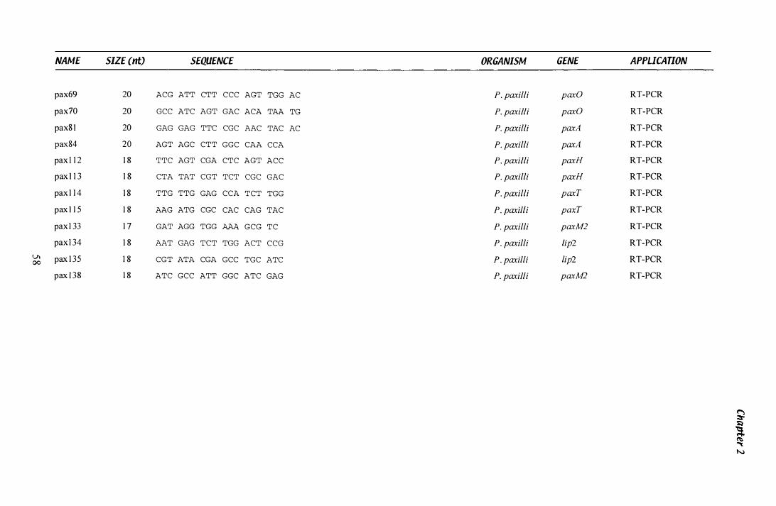
NAME SIZE (nt) SEQUENCE ORGANISM GENE A PPLlCAUON
pax69 20 ACG ATT CTT CCC AGT TGG AC P. paxilli paxO RT-PCR
pax70 20 GCC ATC AGT GAC ACA TAA TG P. paxilli paxO RT-PCR
paxS l 20 GAG GAG TTC CGC AAC TAC AC P. paxilli paxA RT-PCR
paxS4 20 AGT AGC CTT GGC CAA CCA P. paxilli paxA RT-PCR
pax ! 1 2 1 8 TTC AGT CGA CTC AGT ACC P. paxilli paxH RT-PCR
pax l 1 3 1 8 CTA TAT CGT TCT CGC GAC P. paxilli paxH RT-PCR
pax 1 1 4 1 8 TTG TTG GAG CCA TCT TGG P. paxilli paxT RT-PCR
pax l 1 5 1 8 AAG ATG CGC CAC CAG TAC P. paxilli paxT RT-PCR
pax 1 33 1 7 GAT AGG TGG AAA GCG TC P. paxilli paxM2 RT-PCR
pax 1 34 1 8 AAT GAG TCT TGG ACT CCG P. paxilli /ip2 RT-PCR
V't pax 1 35 1 8 CGT ATA CGA GCC TGC ATC P. paxilli lip2 RT-PCR 00 pax I 38 1 8 ATC GCC ATT GGC ATC GAG P. paxilli paxM2 RT-PCR
Chapter 2
were transferred to each reaction tube. A water control was left without any template,
and a positive control containing 2 �L of gDNA (0.5 ng/�L) was included. Tubes were
placed in a Corbett thermocycler preset to 95 °C and the following PCR programme
performed; 1 cycle of 3 min at 95 °C, 30 cycles of 3 0 sec at 95"C, 3 0 sec at 60°C and
90 sec at 72°C, and 1 cycle of 5 min at 72 °C . PCR products were visualised as in
section 2 . 1 1 .2.
2.1 1.4 Gel stab PCR
When multiple PCR products were produced or a single product was weakly amplified,
a second round of amplification could be performed on a small sample of DNA i solated
directly from the agarose gel . Under long wave UV transillumination, a disposable P20
pipette tip was used to remove a small piece of gel from each product and transfer it to
an Eppendorf containing 1 00 �L of sterile Milli Q water. The agarose plug was melted
at 65 °C . Second round PCR amplification was performed with serial dilutions ( 1 0- ,
1 02-, and 1 03-fold) of the isolated plug solution from each product. The new reactions
were separated on an appropriate agarose gel (section 2 . 1 0. 1 and 2 . 1 0 .3) . Purified
products could then be isolated directly from the agarose gel (section 2.5 .4) or cleaned
using a PCR purification kit (section 2 . 1 1 .5).
2.1 1.5 Purification of PCR products
When PCR reactions yielded a single product, they were purified with the
CONCERTTM Rapid PCR purification system (BRL). This was done according to the
instructions provided with the kit, except for step 5 (DNA elution), where DNA was
eluted with 50 �L warm sterile Milli Q water instead of TE.
59

Chapter 2
2. 12 DNA Sequencing
2.12.1 Cycle sequencing with the AmpliCycle™ kit
Sequencing was performed with the AmpliCycle™ sequencing kit (Perkin-Elmer)
which uses the Sanger dideoxy chain termination method (Sanger et al. , 1 977). The
following, 3 0 ilL cocktail was prepared; ( 1 -3 Ilg template DNA, 3 .2 pM of appropriate
primer, 4 ilL 1 0 x Cycling mix, 1 ilL [a_33p] dATP (Amersham) and sterile Milli Q
water). Four 0 .2 mL PCR tubes were prepared for individual cocktail reactions, each
containing 2 ilL of one of the four chain terminating dideoxy nucleotides (ddA TP,
ddGTP, ddTTP, or ddCTP). Then 6 ilL of cocktail reaction was added to each of the
four tubes. Reactions were placed in a Corbett thermocycler preset to 95 ° C and the
following PCR programme performed; 1 cycle of 1 min at 95°C, 25 cycles of 30 sec at
95 °C, 3 0 sec at 48 °C, and 1 min at 72°C, and 1 cycle of 1 min at 72°C. Reactions were
terminated with the addition of 4 ilL of stop solution and stored at -20°C until required.
2.12.2 Denaturing polyacrylamide gel electrophoresis
Polyacrylamide gels were poured with 60 mL of acrylamide mix (section 2 .3 .2) to
which 36 ilL of TEMED and 360 ilL of 1 0% (w/v) ammonium persulfate had been
added. The gel apparatus was assembled in 1 x sequencing TBE buffer (section 2 .3 . 1 0)
and run for 1 5 min at 1 500 Volts, prior to sample loading. The radioactively labelled
sequencing reactions (section 2 . 1 2 . 1 ) were denatured for 2 min at 75°C, and 3 ilL
loaded into each well . The gel was run at 1 500 volts, l imited by approximately 65
watts, with duplicate samples being run through either one (short run) or three (long
run) dye fronts. The gel was transferred to blotting paper, dried for 1 hr at 80°C and
exposed to x-ray film for several days. The order of bands revealed was manually
recorded.
60

Chapter 2
2.12.3 Automated sequencing
Automated sequencing using the Sanger dideoxy chain termination method (Sanger et
al . , 1 977) was carried out by the MuSeq DNA analysis faci lity at Massey University,
according to the recommendations of the facility operator Lorraine Berry. Sequencing
reactions incorporating ABI PRISMTM dye or ABI PRISMTM BigDye™ chemistry (PE
Applied Biosystems, Foster City, Calif.) were separated on either an ABI 3 73 or 3 77
DNA Sequencer (Perkin-Elmer Technologies) , and viewed using the Edit View
programme (Version 1 .0 . 1 , Perkin-Elmer Technologies).
2. 13 Southern Blotting ana Hybriaisafion
2.13.1 Capillary transfer of DNA to nylon membrane
DNA was transferred from agarose gel to a nylon membrane using the Southern
protocol (Southern, 1 975) . The submarine gel (section 2 . 1 0 .2) was carefully washed in
solution 1 for 1 5 min, solution 2 for 30 min, solution 3 for 30 min, and finally for 2 min
in 2 x SSC (section 2.3 .9) . Two sheets of Whatman 3 MM paper soaked in 20 x SSC
were placed in the blotting apparatus, to act as a wick between two reservoirs of blotting
buffer (20 x SSC). A sheet of glad-wrap plastic film, with a hole cut in it slightly
smaller than the dimensions of the gel, was positioned between the gel and the wick, to
ensure uni-directional capillary transfer through the gel . A pre-soaked sheet of
Hybond™-N+ Nylon membrane (Amersham), cut to the appropriate size, was place on
the gel, followed by two sheets of wet Whatman 3MM paper, two sheets of dry
Whatman 3MM paper and a stack of paper towels . Finally, a weight was added to the
top and the apparatus left overnight. The membrane was washed in 2 x SSC for 5 min
and baked at 80°C in a vacuum oven for 2 h to fix the DNA to the membrane.
2.13.2 Labelling DNA with [a..3ZPj dCTP
Purified DNA (20 ng) was made up to a final volume of 1 1 ilL with sterile Milli Q
water in a 1 .5 mL Eppendorf tube. Using a 22 gauge syringe needle, a hole was pierced
6 1
Chapter Z
in the top of the tube and the DNA denatured by boiling for 3 min. The single stranded
DNA was stored on ice, following the addition of 4 ilL of High prime reagent (Roche).
Radioisotope (5 ilL of [a_32p] dCTP (Amersham)) was added and the mixture incubated
for 1 hr at 3TC. A Probequant micro column G-5 0 (Amersham) was prepared by
plac ing it on top of a 1 . 5 mL Eppendorf tube and spinning in a microcentrifuge for
1 min at 2 000 rpm. The compacted column was transferred to a new Eppendorf tube
and the probe mixture transferred to the top of the column. Unincorporated dNTPs
were separated from the labelled probe by centrifugation at 2 000 rpm for 2 min.
Labelled probe was collected in the new Eppendorf, made up to a final volume of 50 ilL
with STE buffer (section 2 .3 . 1 0) and stored at -20°e.
1.13.3 Hybridisation of [a3ZPl dCTP-labelled probe
Nylon membranes were pre-hybridised in 25 mL of 1 0 x Denhardt' s solution (section
2 .3 .3 ) at 6YC for 2 h. Labelled probe (section 2 . 1 3 .2) was removed from -20°C and
denatured by boiling for 3 min. The single stranded DNA probe was stored on ice
before being transferred to the hybridisation tube. Labelled probe homologous to
A DNA was also added when A/HindU I ladder had been included on the submarine gel .
The membranes were incubated overnight at 65 °C with gentle shaking in a Bachofer
rotating oven. The following morning, excess probe was removed by washing once in
2 x SSC (section 2.3 .9) for 5 min, four times in 2 x SSC for 1 0 min, and once in
0.5 x SSC for 1 0 min. The washed membranes were positioned between Glad-wrap
p lastic film, and exposed to x-ray film ( l OONIF, Fuji) for several days at -70°C. F ilm
was developed manually according to manufacturers instructions.
1.13.4 Stripping labelled probe from nylon membrane
After the exposed x-ray film had been developed, labelled probe was stripped from the
nylon membranes by repeated washes in boiling 0 . 1 % (w/v) SDS that was allowed to
cool to room temperature. The washed membranes were then re-exposed to x-ray film
to ensure the complete removal of the labelled probe.
62
Chapter 2
2.14 Library Screening
2.1'1.1 Titre of a 'AGEM@-11 genomic DNA library
An Eppendorf containing an aliquot of a 'AGEM®- l l P. paxilli genomic DNA library
(Itoh & Scott, 1 994b) was removed from _70°C storage and a drop of chloroform added.
A set of serial dilutions ( 1 02 - 1 08) were made with SM Buffer (section 2 . 3 . 8), and 2 x
1 00 ilL of each transferred to duplicate sterile Eppendorf tubes. The library was plated
as described in section 2 . 14 . 3 . Plaque forming units were counted and the library titre
calculated.
2.1'1.2 Calculating library concentration required for isolating clone
To maximise the probability of i solating a gene of interest, the following equation was
used.
N = In( 1 -P) I In [ l -(I/G)]
Where N = the number of plaques needed to screen, I = Insert size ( 1 5 kb), G Genome
size (30 000 kb), and P = Probability of success (0.999).
N is equal to 1 3 8 1 2 plaques. Maximum density of plaques per plate is 3 000, therefore
5 plates containing 3 000 plaques per plate should be sufficient. To ensure success, 1 0
plates, each containing approximately 3 000 plaques, were screened in the first round.
2.1'1.3 Library plating
Two sterile 20 mL tubes containing 5 mL of LB media (section 2 .2) , 0 .2% (w/v)
Maltose, and 1 0 mM MgS04, were inoculated with KW25 1 cells (Table 2 . 1 ) and grown
overnight (section 2. 1 .2) . The appropriate dilution of l ibrary in a final volume of
1 00 ilL was transferred to a sterile 1 .5 mL Eppendorf tube. To each tube, 1 00 ilL of the
K W25 1 culture was added. One tube, containing 1 00 ilL of KW25 1 cells only, was
also included as a control, and all tubes were incubated at 3TC for 3 0 min. The l ibrary
dilution and KW25 1 cells were transferred to sterile 5 mL tubes pre-equilibrated at 50°C
63
Chapter 2
and 3 mL of Top agarose (section 2 .2) containing 1 0 mM MgS04 was added with
gentle vortexing. The top agarose was quickly transferred to LB agar plates (section
2 .2) and incubated for approximately 6-8 hours at 3TC. Plates were then stored at 4°C
until the following moming.
2.14.4 Transfer of plaques to nylon membranes
Plaques were transferred to Hybond™-N+ circular membranes (Amersham). Where
duplicate lifts were performed, the first membrane was overlaid for 1 min and the
second membrane for 2 min. For single lifts, the membrane was overlaid for 1 min.
The membranes were left to air dry, then washed in solution 2 for 2 min, in solution 3
for 5 min and finally in 2 x SSC (section 2 .3 .9) for 2 min. DNA was permanently fixed
to the membrane by baking for 2 h at 80°C in a vacuum oven.
2.14.5 Hybridisation of [a.32Pl dCTPllabelled probe
Membranes were pre-hybridised in approximately 25 mL of 1 0 x Denhardt' s solution
(section 2 .3 .3) at 6YC for 2 h. The radio-labelled probe (section 2. 1 3 .2) was hybridised
as described in section 2 . 1 3 .3 in a shaking water bath. After the final wash in 0 . 5 x
SSC, the membranes were carefully orientated between two layers of plastic wrap and
exposed to x-ray film ( l OONIF, Fuj i) at -70°C. Phosphorescent Tracker Tape™
(Amersham) was used to orientate the films for future reference.
2.14.6 Isolation of positive clones
After developing the exposed x-ray film, plaques that appeared on duplicate membranes
were isolated by aligning the appropriate plate to the x-ray film. An approximate 5 mm
plug was removed and transferred to a sterile Eppendorf containing 500 ilL SM buffer
(section 2 .3 .8) and a few drops of chloroform.
64
Chapter 2
2.14. 7 First round library screening
The AGEM®- l l P. paxilli genomic DNA l ibrary was diluted to the required
concentration [3 ilL l ibrary and 97 ilL SM Buffer (section 2 . 3 . 8)] and plated as
described in section 2 . 1 4 . 3 . Plaques were transferred to duplicate nylon membranes
(section 2 . 1 4 .4), hybridised to the gene specific probe ( section 2 . 1 4 .5) and positive
clones isolated (section 2 . 1 4.6).
2.14.8 Second and third round library screening
A 1 02-, 1 03-, and 1 04-fold dilution of the isolated clones were prepared with SM buffer
(section 2 . 3 . 8) in sterile Eppendorf tubes, and plated as described in section 2 . 1 4 . 3 .
Duplicate lifts were performed for second round screening and single lifts for third
round screening (section 2 . 1 4 .6). Lifts were taken from whichever dilution plate had
produced between 100 and 300 plaques in second round or 50 and 200 plaques in third
round. Membranes were hybridised with the labelled probe as in section 2 . 1 4.5 . After
development of the exposed x-ray film, two positive plaques were isolated from each
clone as described in section 2. 1 4 .6.
2. 15 Sub-Cloning
2. 1 5.1 Ligation into CArd pUCl 18
DNA to be inserted was purified prior to ligation. Lambda DNA was digested with SstI
as described in section 2 .7 and the DNA purified by phenol/chloroform (section 2 . 8)
and ethanol precipitation (section 2.9). The ligation reactions contained; 2 ilL of 1 0 x
Ligase buffer (BioLabs), 1 .5 ilL of CAP' d SstI digested pUC 1 1 8, SstI digested A clone
in a 2-3 molar excess of insert:vector, 0. 1 ilL of T4 DNA Ligase (BioLabs) and sterile
Milli Q water to a final volume of 20 ilL. The l igation mixes, including a vector only
control, were incubated overnight at 4°C. A sample (3 ilL) was removed before the
addition of enzyme and after the incubation period to confirm ligation was successful.
The samples were run on a 0 .7% (w/v) agarose gel (section 2 . 1 0. 1 and 2 . 1 0 .3).
65
Chapter 2
2.1 5.2 Ligation into PGEM®-T ana pGEM®-T Easy
Purified PCR products (section 2 . 1 1 .5 ) were cloned into either the pGEM®-T or the
pGEM®-T Easy vector from Promega. The ligation reactions were set up according to
the recommendations provided and contained; 5 ilL of ligation buffer x 2, 0 .5 J.!,L of
pGEM®-T or pGEM®-T Easy vector, 3 . 5 ilL of purified PCR product and 1 J.!,L of
T4 Ligase. The reactions were incubated at 4 °C overnight.
2.15.3 Electrocompetent cells
Escherichia coli strain XL- l (Table 2 . l ) was grown in 5 mL LB media (section 2 .2)
with tetracycline as described in section 2 . 1 .2 . The 5 mL E. coli culture was sub
cultured into 500 mL LB media (section 2.2) containing tetracycline. The cultures were
grown with vigorous shaking (300 rpm) at 3TC to mid-log phase (optical density of
0 .5 - 1 .0 at 600nm), which usually occurred after 3 h growth. The cells were chilled on
ice for 20 min before being pelleted by centrifugation at 4 000 x g (Sorval GSA, 10 min,
5 000 rpm, 4 °C). After the supernatant was decanted, the cells were re suspended in 1 L
of chilled water. The cells were harvested as above and re suspended in SOO mL o f
chilled water. The cells were harvested a third and fourth time as above and
resuspended in firstly, 20 mL of chilled 1 0% (v/v) glycerol and finally 4 mL of chilled
1 0% (v/v) glycerol. Aliquots of 40 J.!,L were transferred to sterile I .S mL Eppendorf
tubes, and stored at -70°C.
2.1 5.4 Electroporation
Based on the method of Dower et al. ( 1 988) . Individual Eppendorf tubes containing
40 J.!,L of electrocompetent XL- l cells (section 2 . 1 5 .3) were removed fro m -70° C and
2 J.!,L of l igation mixture (section 2 . 1 5 . 1 and 2 . l 5 .2) added. The two controls for
electroporation transformation were 2 ilL of pUC 1 l 8 (20 ng/IlL) added to 40 ilL XL-l
cells and a tube containing 40 ilL of XL- l cells only. The transformation mixes were
transferred to chil led e lectroporation cuvettes (Bio-Rad) and placed in the
electroporation bracket. The samples were pulsed with 2 .SkV, 251lFD and 200n in a
66
Chapter 2
Bio-Rad Gene pulser, and 1 mL of SOC media (section 2 .2) added. The transformation
mixes were transferred to a new sterile 1 .5 mL Eppendorf tube and incubated for 1 hr at
3TC.
2.15.5 Selection of transformants
Transformation mixes ( 1 00 ilL and 50 ilL) were plated onto LB agar plates (section
2 .2), containing ampicillin and 40 ilL of both IPTG and X-GAL (section 2.4 .) . Colonies
containing recombinant pUC 1 1 8, pGEM®-T or pGEM®-T EASY were identified as
ampicillin resistant white colonies. Serial dilutions of the pUC 1 1 8 only transformation
mix were also transferred to the selection media, as was 1 00 ilL of the XL- 1 cells only
transformation mix. Serial dilutions of the XL-l cells only transformation mix were
then plated onto LB agar plates (section 2 .2) to determine the concentration of viable
cells. All plates were incubated overnight (section 2 . 1 .2).
2.1 5.6 Drop-outs ana mapping
Where DNA inserts were particularly large, further sequence information could be
obtained be removing restriction enzyme fragments from the insert and religating novel
sequence next to the vector's forward primer site. Restriction enzyme sites in the
pUC 1 1 8 multiple cloning cassette include; EcoRl, SstI, Kpnl, SmaI, XmaI, BamHI, Sail,
Ace!, HincII, PstI, SphI and HindIII. Once successful sub-clones containing the gene of
interest had been identified by automated sequencing (section 2 . 1 2.3) with the universal
forward and reverse primers (Table 2.2), these restriction enzymes were used to map the
insert (section 2 .7, 2. 1 0 . 1 and 2 . 1 0.3) . Enzymes that cut once within the insert and once
in the vector were used to remove a section of insert DNA. The shortened vector was
then religated back together (section 2 . 1 5 . 1 ). Although blue/white selection was no
longer possible, ampicillin resistant sub-clones were selected and screened by the rapid
boil method (section 2 .5 . 3 ) until the required clones were identified.
67
Chapter 2
2. 16 Paxilline Analysis
1. 1 6. 1 Sampling liquid cultures for analysis of paxilline
F or High Performance Liquid Chromatography (HPLC) analysis, a 5 mL sample was
removed from fungal l iquid cultures (section 2 . 1 .2) and transferred to a pre-weighed 1 5
mL falcon tube. The mycelia were pelleted by centrifugation ( 1 0 min 3 1 00 rpm), and
fol lowing removal of the excess broth, snap frozen in l iquid nitrogen and stored at
-70°C . For Thin Layer Chromatography (TLC), a 1 0 mL sample was removed except
where the biomass was extremely low. In this case the entire 1 00 mL biomass was
harvested and stored as above.
Broth removed from mycelia samples isolated for extraction of paxilline and RNA
(section 2 . 1 7 . 1 ) was collected in a sterile 1 5 mL falcon tube, snap frozen in liquid
nitrogen and stored at -70°C .
1. 16.1 Isolation of paxilline from freeze-dried samples
Freeze dried mycelia was weighed to determine the biomass per sample. Paxilline was
extracted into chloroform:methanol (2 : 1 ) using a Virtis homogeniser. Samples were
homogenised in chloroform and then methanol was added to a final volume of 1 .2 mL
chloroform:methanol (2 : 1 ) per 1 mL of culture removed. Samples were shaken gently
for 2 h and the mycelia debris pelleted by centrifugation ( l 0 min, 3 1 00 rpm). Finally,
1 .5 mL was removed and evaporated overnight in a fumehood.
Samples to be analysed by HPLC were filtered to remove any remaining mycelia debris
capable of blocking the HPLC column. A Waterman Sep-Pak® silica cartridge was
pre-wet with 3 mL of Dichloromethane (DCM) . Dried paxilIine samples ( 1 .5 mL)
extracted from mycelia were re suspended in 1 mL of DCM and transferred to the
column. The column was washed with 1 mL of DCM:Acetonitrile (ACN) (80:20), then
the sample collected in 3 mL of ACN :DCM (80:20). Cartridges were reused after
inverting the column and flushing through with 4 mL of DCM:ACN (80:20) up to a
maximum of four times.
68
2.1 6.3 HPLC
Chapter Z
Standards and sample s ( 1 0 ilL) were separated with a Shimadzu LC-4A HPLC in
normal phase [DCM:ACN (80:20) containing 0.4% (v/v) Methanol] using a Zorbax-SIL
Silica column (4.6 mm x 25 cm) at a rate of 1 .2 mLlmin. DV absorbency at 230 nm
was determined with a Shimadzu SPD-2AS Spectrophotometric Detector. Several
paxil line standards of 25 J.l.g/mL were run as an internal standard for each set of
samples. Peak areas were calculated by the Shimadzu C-R3A Chromatopac printer, and
using peak areas from the paxilline standards, an average paxilline content/peak area
unit was calculated (Appendix 3 .0).
2.16.4 nc
The 1 .5 mL dried samples extracted from mycelia (section 2 . 1 6.2) were resuspended in
50 ilL of chloroform:methanol (2: 1 ) and spotted onto a Polygram Silica gel TLC plate
(Macherey-Nagel) at 1 cm intervals. The plate was run through two solvent fronts in
chloroform:acetone ( 1 0 : 1 ). When the plate was dry, indole-diterpene moieties were
identified by spraying with alcoholic Erhlich' s reagent (2% (w/v) p-dimethyl
aminobenzaldehyde in 1 2% (v/v) HCI and 50% (v/v) ethanol) and heated to 1 20°C for
5-7 min. After cooling the plates were laminated and stored away from light to prevent
fading.
2. 1 7 Isolation of RNA
2. 1 1. 1 Sampling liquid cultures for isolation o f RNA
Two samples of 5 mL were removed from fungal liquid cultures (section 2 . 1 .2) and
strained through sterile mira-cloth. The mycelia was washed with sterile Milli Q water
and transferred to sterile aluminium foil packets. The mycelia were snap frozen in
liquid nitrogen and stored at -70°C.
69
Chapter Z
2.1 1.2 RNase-free solutions anet equipment
All solutions except those containing Tris, EDTA and MOPS were made with DEPC
treated water. Milli Q water ( 1 L) was treated with diethyl pyrocarbonate (DEPC) to a
final concentration of 0 . 0 1 % (v/v). The water was incubated at 3 TC overnight then
autoc1aved twice for 1 5 min at 1 2 1 °C with 1 5 psi. All solutions were stored in prepared
glassware.
Where possible glassware was soaked overnight in a chromic acid bath and baked for
2 h at 1 80°C. Lids for Schott bottles were autoclaved for 1 5 min at 1 2 1 °C with 1 5 psi in
a solution of DEPC-treated water. Any equipment unable to be acid-washed and oven
baked, was soaked overnight in 0.3% (v/v) H202, rinsed in 95% (v/v) ethanol and left to
air-dry in a fume hood. The working area was scrubbed with Virkon and rinsed with
95% (v/v) ethanol and allowed to air-dry. All Eppendorf tubes, PCR tubes and 1 -5 mL
pipette tips were autoc laved for 1 5 min at 1 2 1 T with 1 5 psi and stored in RNase-free
glassware. Aerosol Resistant, RNase-free, DNase-free pipette tips ( 1 0 ilL, 20 !lL,
200 !lL, and 1 000 ilL) were used at all times. Latex gloves were used when handling
any equipment.
2.1 1.3 Isolation of total RNA with TRIzofY reagent
Frozen mycelia in foi l packets was removed from the -70°C freezer and transported in
liquid nitrogen. Mycelia ( 1 .25 g) was removed, re-wrapped in tin foil and emersed in
l iquid nitrogen. The remaining mycelia were returned to -70°C freezer. Mycelia were
ground to a fine powder under liquid nitrogen in a mortar and pestle. Powdered mycelia
was then mixed to a paste with 1 0 mL of TRlzol® reagent (BRL) and transferred to an
3 0 mL tube. The tube was sealed with parafilm and aluminium foil and left to stand at
room temperature for 5 min. The homogenate was centrifuged at 1 2 000 x g (Sorval
SS34, 1 0 min, 9 000 rpm, 4°C) and the cleared supernatant transferred to a fresh 30 mL
tube. Chloroform (2 mL) was added and the tube covered with parafilm and aluminium
foil . Samples were shaken vigorously for 1 5 sec, then incubated at room temperature
for 3 min. The aqueous phase was separated by centrifugation (Sorval SS34, 1 5 min,
9 000 rpm, 4°C) and transferred to a fresh tube. Total RNA was precipitated by adding
70
Chapter 2
5 mL of isopropanol and incubating the solution at room temperature for 1 0 min. RNA
was pelleted by centrifugation (Sorval SS34, 1 0 min, 9 000 rpm, 4°C) and washed with
1 0 mL of 75% (v/v) ethanol. The pellet was reconstituted by centrifugation at 7,5 00 x g
(Sorval SS34, 5 min, 6 700 rpm, 4°C) and air-dried in a fumehood for approximately
1 5 min before resuspending in 200-500 I-LL of DEPC-treated water (section 2 . 1 7 .2) .
Samples were quantified spectrophotometrically (section 2 .6 .3), and stored at -70°C.
2.1 7.4 DNase 1 treatment
Based on method described by Bradshaw & Pillar ( 1 992). Reactions were performed in
1 5 mL tubes (section 2. 1 7.2) . Total RNA, equivalent to 1 20 I-Lg was combined with
60 U DNase I (Roche), 80 U RNase Inhibitor (Roche), 500 I-LM 1 ,4-dithiothreitol (DTT)
(Roche) and 1 x DNase I Buffer (section 2 .3 .5) to a final volume of 2.4 mL. The
mixture was incubated at 37°C for 30 min, at which time another 60 U of DNase I was
added and the incubation continued for a further hour. The sample was re-extracted in
2 volumes of chloroform and Tris-equilibrated phenol (section 2 . 3 . 1 1 ) with gentle
vortexing. The aqueous phase was separated by centrifugation (Sorval SS34, 1 0 min,
1 1 500 rpm, 4°C) and transferred to a clean corex tube. Two volumes of chloroform
were added, mixed briefly by vortexing and the aqueous phase separated as above.
RNA was precipitated with the addition of 240 I-LL 3 M sodium acetate and 6 mL of
95% (v/v) ethanol. The precipitation was incubated on ice for 30 min, and the RNA
pelleted by centrifugation (Sorval SS34, 1 5 min, 9 000 rpm, 4°C). The resulting pellet
was carefully washed with 6 mL of 75% (v/v) ethanol and air dried at room temperature
for approximately 1 5 min, before resuspending in 1 00 I-LL of DEPC-treated Milli Q
water (section 2 . 1 7 .2) . The concentration of RNA was determined spectrophoto-
metrically (section 2.6.3) . Samples were stored at -70°C.
7 1
Chapter 2
2. 18 Reverse Transcription-peR
2.1 8. 1 Synthesis of cONA with Expanct™ reverse transcriptase
DNase I treated RNA ( 1 Ilg) and 0.6 ilL of random hexamer primer (3 1lg/IlL) (BRL)
were combined with DEPC-treated water (section 2 . 1 7.2) to a final volume of 1 1 .95 ilL
in sterile 0 .2 mL peR tubes. The primers and template were denatured at 900C for
5 min, cooled on ice, then briefly centrifuged in a microcentrifuge to collect the
reagents to the bottom of the tube. A reaction cocktail containing n x ( 1 x Expand buffer
(Roche), 1 0 mM (OTT), 5 0 IlM dNTPs, and 1 0 U RNase Inhibitor (Roche)) was
prepared and 7 .05 ilL added to each reaction tube. Duplicate reactions for each RNA
sample and a water only control were prepared. Expand™ Reverse Transcriptase
(50 U) (Roche) was added to the water control and one of the RNA reactions . To the
other RNA reaction, 1 ilL of DEPC-treated water (section 2 . 1 7.2) was added to confirm
the absence of any contaminating DNA. The reactions were gently mixed, centrifuged
to collect all reagents in the bottom of the tube, and incubated at room temperature for
1 0 min to allow primer annealing. The cDNA synthesis was performed at 42°C for 45
min in a Corbett thermocycler. Reactions were collected by centrifugation, diluted 1 0-,
1 02- and 1 03-fold with sterile Milli Q water and stored at -20°C.
2.18.2 peR amplification of cONA
A routine PCR cocktail (section 2 . 1 1 .2) was prepared to a final volume of 20 ilL. The
1 0-fold dilutions of cDNA, the undiluted 'no reverse transcriptase' controls, undiluted
' no RNA' control, genomic DNA (0.5 ng/IlL) and water controls (all 5 ilL) were added
to the reaction tubes . In a Corbett thermocycler preset to 94°C, the following PCR
programme was performed; 1 cycle of 2 min at 94°C, 30 cycles of 45 sec at 94°C,
45 sec at 60°C, and 1 min at 72°C, and 1 cycle of 5 min at 72°C. For the initial control
PCR to confirm cDNA synthesis and absence of DNA contamination, primers Etub2
and Etub3 (Table 2.2) for the tub-2 gene were used. These primers flank a 62 bp intron,
so cDNA PCR products are 62 bp smaller than gDNA PCR products, and
distinguishable by mini-gel electrophoresis (section 2 . 1 0. 1 and 2 . 1 0.3) .
72
Chapter 2
2. 1 9 RACE
Rapid Amplification of £DNA Ends (RACE) was used to determine the position of
transcription initiation and termination sites, after an initial estimation was made with
RT-PCR. The general strategy is outlined in Figure 3 . 1 4. The initial steps ( section
2 . 1 9 . 1 ) of the RACE were conducted under RNase free conditions as described in
section 2. 1 7.2.
2.19.1 Synthesis of cDNA with C. therm. reverse transcriptase
DNase I treated RNA (section 2. 1 7 .4) was removed from -70°C. Duplicate reactions
containing, 500 ng RNA, I pM gene specific primer for 5 ' RACE or oligo dT primer for
3 ' RACE and DEPC-treated water (section 2 . 1 7.2) to a final volume of 1 1 .3 j.1L, were
set up in 0 .2 mL PCR tubes. A tube containing no RNA was included as a control. The
reactions were incubated at 70°C for 1 0 min then stored on ice. A cocktail containing
n x ( 1 x C. therm Reverse Transcriptase buffer, S mM DTT, 1 00 j.1M dNTPs and 3%
(w/v) dimethylsulfoxide (DMSO» was transferred to each of the tubes in 7 .2 j.1L
aliquots. To one of the RNA reactions and the water control , 1 . 5 mL of C. therm
Reverse Transcriptase (Roche) was added. DEPC-treated water (section 2 . 1 7 .2) was
used to bring the remaining control reactions to a final volume of 20 j.1L. The reaction
was incubated for 30 min at 60°C in a Corbett thermocycler. The reverse transcriptase
was then heat inactivated at 9YC for 2 min.
2.19.2 Degraaation Of RNA
The RACE reactions were collected by centrifugation and placed at 3TC. To all tubes
except no RNA control , 1 j.1L of RNase mix (Roche), containing both RNase H and
RNase A, was added, gently mixed and incubated at 3TC for 30 min. The reactions
were again collected by centrifugation and a 1 j.1L sample (sample A) removed and
diluted 500-fold with TE (section 2 .3 . 1 0) . The remaining reaction was purified using
the Life Technologies CONCERTTM PCR clean-up kit (BRL) (section 2 . 1 1 .5) and
73
Chapter 2
resuspended in 50 ilL of warm sterile Milli Q water. A 5 ilL sample ( sample B) was
removed and diluted 1 0z-fold with TE (section 2.3 . 1 0) .
2.1 9.3 Addition of 3' terminal dCTP with terminal transferase
For 5 ' RACE, an extra step i s required to enable the AAP primer to bind to the 3 ' end of
newly synthesised cDNA. Duplicate reactions containing; 1 0 ilL of purified cDNA,
1 x Tailing buffer, 1 00 IlM dCTP, and 0.75 mM CoClz were prepared in 0 .2 mL PCR
tubes . To one set of reactions 50 U of Terminal transferase was added. To the
remaining control tubes, sterile Milli Q water was added to a final volume of 20 ilL .
The reactions were incubated at 3TC for 20 min, then the enzyme was heat inactivated
at 65°C for 1 0 min.
2. 19.11 Secontt strantt synthesis
Two sets of nested primers combined with the AUAP and UP primers for 5 ' RACE and
the UP primer for 3 ' RACE were used to purify the RACE products further (Figure
3 . 1 4) . Routine PCR reactions (section 2 . 1 1 .2 ) were performed and the products
separated by mini-gel electrophoresis (section 2. 1 0 . 1 and 2. 1 0 .3) .
2.19.5 Isolation and characterisation of RACE products
Bands were purified as described in section 2 . 1 1 . 5 and the second round PCR
amplification reaction from the gel stab cleaned using the CONCERTTM PCR clean up
kit (BRL) (section 2. 1 1 .5) . The products were c loned using the Promega pGEM®-T
system (section 2. 1 5 .2) and sequenced (section 2. 1 2 .3) .
74

Results
3.1 Isolation and Characterisation of hmg from P. pnxilli.
3.1.1 Isolation of a unique hmg with degenerate primers
Chapter 3
Optimal conditions for amplifying P. paxilli genomic DNA (gDNA) were determined
with primers pax5 and pax6 (Table 2.3) using a routine PCR protocol (section 2 . 1 1 .2).
These homologous primers were designed to sequence a specific site in the P. paxilli
genome. The plasmid pAN7- 1 , was heterologously integrated into this site during the
generation of paxilline negative mutants (Itoh et al. , 1 994a) . This l o cus i s
approximately 25 kb away from the left boundary o f the paxilline biosy nthetic cluster.
When used in combination, pax5 and pax6 amplify a 45 1 bp product, encompassing part
of an ORF with similarity to a phenylacrylic acid decarboxylase (Young et al., 1 998).
The product yield from the range of DNA concentrations used, is shown in Figure 3 . 1
A . Lane 5 , containing 5 ng o f template gDNA, is the optimal DNA concentration.
The same primers were used to determine the optimal concentration of Mg2+ in the
reaction buffer. A routine peR cocktail was prepared (section 2. 1 1 .2), with a Roche
Mg2+-free buffer replacing the standard buffer. The concentration of Mg2+ in individual
reaction tubes was adjusted by addition of MgCh. Within the range of concengations
te�ted, the product yield was unaffected by Mg2+ (Figure 3 . 1 B). The standard buffer
(containing 1 .5 mM Mg2+), supplied with Roche Taq DNA polymerase, was used in all
subsequent reactions.
Degenerate primers, designed to conserved regions of hmg genes, were used to isolate a
unique hmg gene from P. paxilli.. The optimal temperature for annealing degenerate
primers HMG 1 and HMG3 (Table 2.3) to the P. paxilli gDNA was determined using a
routine PCR reaction (section 2. 1 1 .2). Annealing temperatures from 42° C to 46°C were
trialed and the �ptimal result, primer annealing at 44°C, is shown in Figure 3 . 1 C.
Previously, a unique hmg was isolated from N. lolii Lp 1 9 using the same primers
(Dobson, 1 997) and a single specific product is also amplified from Saccharomyces
cerevisiae with HMG 1 and HMG3 . Therefore both species were used as controls to
identify a product of the expected size from P. paxilli gDNA. For comparison, N. loW
Lp ! gDNA was also included. Genomic DNA from the four species was amplified with
primers pax5 and pax6 as a positive control, as amplification with the homologous
75
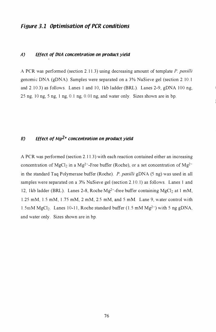
Figure 3. 1 Optimisation of peR conditions
A) Effect of DNA concentration on product yield
A PCR was performed (section 2 . 1 1 . 3 ) using decreasing amount of template P. paxilli
genomi c DNA (gDNA). Samples were separated on a 3 % NuSieve gel (section 2 . 1 0 . 1
and 2 . 1 0 . 3 ) as follows. Lanes 1 and 1 0, 1 kb l adder (BRL). Lanes 2-9, gDNA 1 00 ng,
25 ng, 1 0 ng, 5 ng, 1 ng, 0 . 1 ng, 0 . 0 1 ng, and water only. Sizes shown are in bp.
B) Effect of Mg2+ concentration on product yield
A PCR was performed (section 2 . 1 1 .3 ) with each reaction contained either an increasing
concentration of MgCb in a Mg2 i--Free buffer (Roche), or a set concentration of Mg2+
in the standard Taq Polymerase buffer (Roche) . P. paxilli gDNA (5 ng) was used in all
samples were separated on a 3% NuSieve gel (section 2 . 1 0 . 1 ) as follows. Lanes 1 and
1 2, 1 kb ladder (BRL) . Lanes 2-8, Roche Mg2+-free buffer containing MgCb at 1 mM,
1 25 mM, 1 . 5 mM, 1 .75 mM, 2 mM, 2.5 mM, and 5 mM. Lane 9, water control with
1 . 5mM MgCb Lanes 1 0- 1 1 , Roche standard buffer ( 1 . 5 mM Mg2+) with 5 ng gDNA,
and water only . Sizes shown are in bp.
76
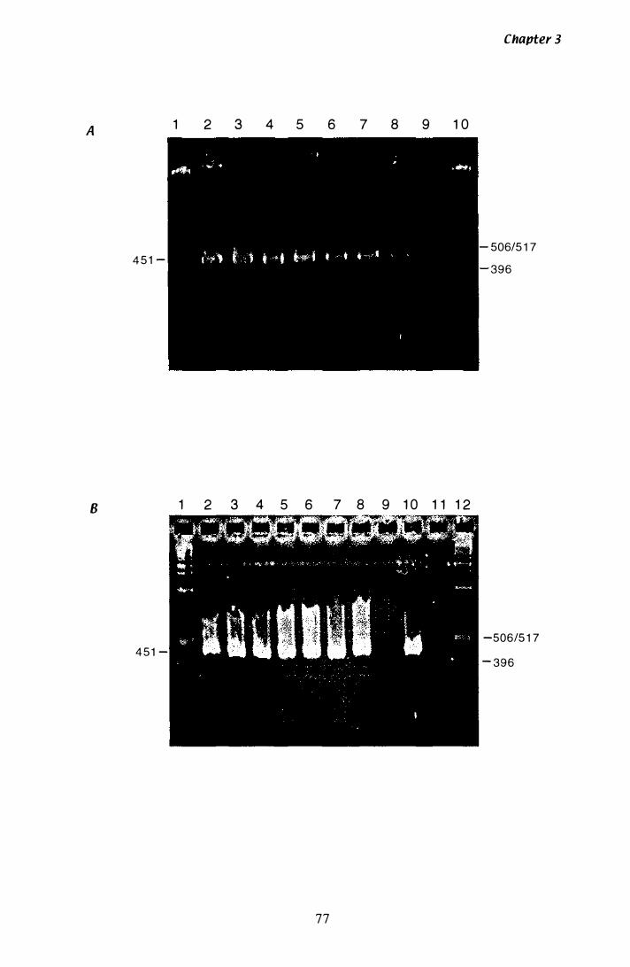
A 1 2 3 4 5 6 7 8 9 1 0
4 5 1 -
B 1 2 3 4 5 6 7 8 9 1 0 1 1 1 2
4 5 1 -
77
Chapter 3
506/5 1 7 396
-506/5 1 7
- 396
C) Proauct yiela at 44 °C annealing temperature
A PCR was performed (section 2 . 1 1 . 3 ) at 44°C annealing temperature, with 5 ng of
gDNA from various species . Samples were separated on a 3% NuSieve gel (secti on
2 . 1 0 . 1 and 2 . 1 0 . 3 ) as follows . Lanes 1 and 1 2, l kb ladder (BRL). Lanes 2-6, gDNA
from Lp 1 , Lp 1 9, P. paxilli, S. cerevisiae, and water control amplified with pax5 and
pax6. Lanes 7- 1 1 , gDNA from Lp l , Lp 1 9, P. paxilli, S. cerevisiae, and water control
amplified with HMGl and HMG3 . Sizes shown are in bp.
DJ Optimal PCR for purification of proaucts
A PCR was performed (section 2 . 1 1 .2) at 44°C annealing temperature. Samples were
separated on a 3% NuSieve gel (section 2 1 0 . 1 ) as follows. Lanes I and 7, l kb ladder
(BRL) . Lanes 2-5 , P. paxilli gDNA amplified with HMGI and HMG3, Lane 6, S.
cerevisiae amplified with HMGI and HMG3 . The 3 5 9 bp and 700 bp product were
isolated and sequenced. Sizes shown are in bp.
78
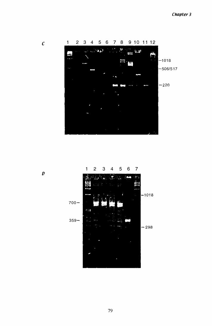
C 1 2 3 4 5 6 7 8 9 1 0 1 1 1 2
1 2 3 4 5 6 7 o
7 0 0 -
359-
79
Chapter 3
6/5 1 7
Chapter 3
primers would confirm a successful PCR. Primers pax5 and pax6 were not expected to
amplifY a product from S. cerevisiae. A product amplified from N. loW Lp 1 and N. loW
Lp 1 9 gDNA, with pax5 and pax6, can be seen in lanes 2 and 3 . /
The second set of
reactions was amplified with HMGl and HMG3 (lanes 7- 1 1 ) . A product of 3 59 bp is
amplified from N. loW Lp1 9, P. paxilli and S. cerevisiae. Lane 9, containing P. paxilli
template, shows two strong products and several medium to faint products, including
the 359 bp product. A product of 200 bp is present in lanes 7 - 1 1 , including the water
control. This product was not seen in any subsequent reactions (Figure 3 . 1 D). At
44°C, the 359 bp product from P. paxilli is at its greatest intensity. A large scale PCR
using all the optimal conditions and S. cerevisiae as a control was performed (Figure 3 . 1
D ) to enable the 700 b p product and the 3 5 9 bp products to be purified from the gel
(section 2 . 1 1 .4) . Both products were isolated (section 2 . 5 .4) and sequenced to
determine if a unique hmg gene was present.
3.1.2 Sequencing the 700 bp and 359 bp products from P. paKilli
The 700 bp and 359 bp products isolated from P. paxilli gDNA with the degenerate
primers HMG1 and HMG3, were sequenced (section 2. 1 2 . 1 and 2 . 1 2 .2) using HMG1
and HMG3 as primers. The smaller product was sequenced in both directions
(Appendix A.2 . 1 ) with sufficient results to produce a consensus overlap (Figure 3 .2) . A
BLAST search (Altschul et al. , 1 990) of the GenBank protein database was perforn1ed
at the NCBr website. This confirmed that a unique hmg had been isolated, and that the
novel sequence most closely resembled 3-hydroxy-3-methylglutaryl Coenzyme A
reductase from Gibberellafujikuroi (E value of 1 x 1 0-28). The 359 bp hmg product was
then labelled with [a_32p] and used to probe both a P. paxilli genomic Southern blot and
a P. paxilli genomic library.
The 700 bp product was also sequenced in both directions with primers HMG 1 and
HMG3, however due to the size of the template, there was no double stranded overlap
(Appendix A.2.2) . A BLAST search (Altschul et al., 1 990) of the GenBank database
was performed with the two sequences isolated from the 700 bp product. No significant
homology was found for either sequence.
80
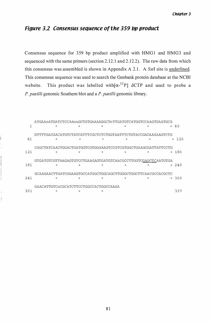
Chapter 3
Figure 3.2 Consensus sequence of the 359 bp product
Consensus sequence for 3 59 bp product amplified with HMG 1 and HMG3 and
sequenced with the same primers (section 2 . 1 2 . 1 and 2 . 1 2.2). The raw data from which
this consensus was assembled is shown in Appendix A 2 . 1 . A SstI site is underlined.
This consensus sequence was used to search the Genbank protein database at the NCBI
website . This product was label l ed with[a-32p] dCTP and used to probe a
P. paxilli genomic Southern blot and a P. paxilli genomic library.
ATGAAnATGATCTCCAAnnGCTGTGAAAAGGCTnTTGATGTCATGGTCCAAGTGAATGCG 1 + + + + + + 6 0
GTTTTGACGACATGTCTATCATTTCGCTCTCTGGTAATTTCTGTACCGACAAGAAGTCTG 6 1 + + + + + + 1 2 0
CGGCTATCAACTGGACTGATGGTCGTGGAAAGTCCGTCGTGGCTGAAGCGATTATTCCTG 1 2 1 + + + + + + 1 8 0
GTGATGTCGTTAAGAGTGTCCTGAAGAGTGATGTCAACGCCTTGGTCGAGCTCAATGTGA 1 8 1 + + + + + + 2 4 0
GCAAGAACTTGATCGGAAGTGCCATGGCTGGCAGCTTGGGCTGGCTTCAACGCCACGCTC 2 4 1 + + + + + + 3 0 0
GAACATTGTCnCGCATCTTCCTGGCCACTGGCCAAGA 3 0 1 + + + 3 3 7
8 1

Chapter 3
3.1.3 Confirmation the unique hmg exists in the P. paKilli genome
A unique hmg had been isolated using peR, from P. paxilli with the degenerate
primers. Southern analysis was performed on genomic DNA (section 2 .5 . 1 ) to confirm
the origin of the unique product and to determine the size and number of fragments
homologous to the 359 bp hmg probe in the P. paxilli genome. DNA was digested with
a range of restriction endonuc1eases (section 2 .7 and 2. 1 0.2), transferred to Nylon
membrane (section 2 . 1 3) (Figure 3 . 3 A), and probed with the 359 bp hmg product. The
autoradiograph (Figure 3 . 3 B) confirms a single copy of the unique hmg is present in the
P. paxilli genome.
Table 3.1 Size of P. paxilli gDNA bancls hYbridising to the 359 bp hmg probe
Size of Bands (kb)
Restriction enzyme
BamHI BglI EcoRI HindIII PstI SalI SstI
2.2 6.2 +23 . 1 1 9. 1 4.9 1 5 .6 1 3 .9
5 .5 5 .6
Two bands are seen in lanes digested with SalI (Figure 3 .3 B, lane 7) and SstI (lane 8) .
In the case of the SstI digestion, sequencing later revealed a SstI site between primers
HMG 1 and HMG3 (Figure 3 .2) . The smaller band seen in the SalI digestion could
result from a cryptic SaIl site that is cut rarely to produce a faint 5 .2 kb band, although
there are no sequences similar to the SalI recognition site in this area. It remains
unexplained at this time.
82
Figure 3.3 Southern analysis of hmg in the P. pnxilli genome
A) Restriction enctonuclease ctigestion of P. pnxilli gDNA
P. paxilli genom I c DNA (2 Jlg) ( secti on 2 . 5 . 1 ) was digested with restri ct ion
end onucl eases (secti on 2 . 7) . Each reaction was separated on a 0 . 7% agarose gel
(secti on 2 . 1 0 . 2 and 2 . 1 0 . 3 ) as fol lows. Lane 1 , AIHindIII. Lanes 2-8, P. paxiLLi gDNA
cut with Bamill, BglII, EcoRI, HindIII, PstI, san, and Sst! . Lane 9, 1 kb ladder (BRL).
S izes shown are in kb .
B) Confirmation that the 359 bp proctuct is part of the P. pnxilli genome
Autoradiograph showing hybri disation of the 359 bp product to the P. paxiLLi genome .
Eth i di um bromi de stained gel seen i n Figure 3 . 3 A was S outhern blotted and probed
with the [32P]-: !abelled 3 5 9 bp hmg product (section 2 . 1 3 ) . Sizes shown are in kb .
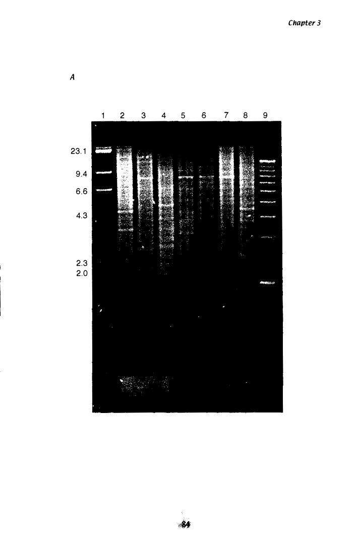
A
23. 1
9 .4
6.6
4. 3
2 .3
2 . 0
1 2 3 4 5 6 7 8 9
Chapter 3
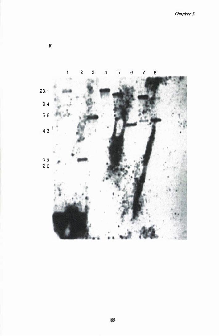
B
23. 1
9 .4
6.6
4.3 t
2.3 2.0
1 2 3 4 5
85
6 7 8
Chapter 3
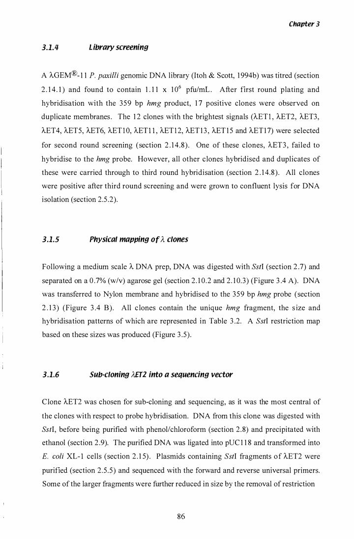
3.1.4 Library screening
Chapter 3
A AGEM®- 1 1 P. paxilli genomic DNA library (Itoh & Scott, 1 994b) was titred (section
2 . 1 4 . 1 ) and found to contain 1 . 1 1 x 1 06 pfu/mL . After first round plating and
hybridisation with the 359 bp hmg product, 1 7 positive clones were observed on
duplicate membranes . The 1 2 clones with the brightest signals (AETI , AET2, AET3,
AET4, AET5, AET6, AETI O, AETl l , AETI 2, AET 1 3 , AET 1 5 and AET17) were selected
for second round screening (section 2 . 1 4 .8) . One of these clones, AET3 , failed to
hybridise to the hmg probe. However, all other clones hybridised and duplicates of
these were carried through to third round hybridisation (section 2 . 1 4.8) . All clones
were positive after third round screening and were grown to confluent lysis for DNA
isolation (section 2.5 .2).
3.1.5 Physical mapping of A clones
Following a medium scale A DNA prep, DNA was digested with SstI (section 2 .7) and
separated on a 0 .7% (w/v) agarose gel (section 2 . 1 0 .2 and 2 . 1 0.3) (Figure 3 .4 A). DNA
was transferred to Nylon membrane and hybridised to the 359 bp hmg probe (section
2 . 1 3 ) (Figure 3 .4 B) . All c lones contain the unique hmg fragment, the s ize and
hybridisation patterns of which are represented in Table 3 .2. A SstI restriction map
based on these sizes was produced (Figure 3 .5) .
3.1.6 Sub-cloning AfT2 into a sequencing vector
Clone AET2 was chosen for sub-cloning and sequencing, as it was the most central of
the clones with respect to probe hybridisation. DNA from this clone was digested with
SstI, before being purified with phenol/chloroform (section 2 .8) and precipitated with
ethanol (section 2 .9). The purified DNA was ligated into pUC 1 1 8 and transformed into
E. coli XL- l cells (section 2 . 1 5) . P lasmids containing SstI fragments of AET2 were
purified (section 2 .5 .5) and sequenced with the forward and reverse universal primers.
Some of the larger fragments were further reduced in size by the removal of restriction
86
Figure 3.4 Southern analysis of hmg A Clones
A) Restriction enaonuclease aigestion of hmg AclonesJ cut with SstI
DNA (�500 ng) from medium scale A preps (section 2. 5 . 2) was digested with Ss!I
(section 2 . 7) and run on a 0 .7% agarose gel (section 2 . 10 .2 and 2 . 1 0 . 3 ) as fol lows. Lane
1 , A/HindUI. Lanes 2- 1 2, S.stI digestion of AET l , AET2, AET4, AET5, AET6, AE 1 0,
AET I I , AET 1 2, AET I 3 , AET 1 5 , and AET1 7 . Lane 1 3 , I kb ladder (BRL). Sizes shown
are in kb .
B) Mapping the position of hmg on M/ones
Autoradiograph showing hybridi sation of the 3 59 bp product to the digested A clones .
Ethidium b romide stained gel seen if Figure 3 .4 A was S outhern blotted and probed
with [32P]-labelled 3 5 9 bp hmg product (section 2 . 1 3 ) . Sizes shown are in kb .
87
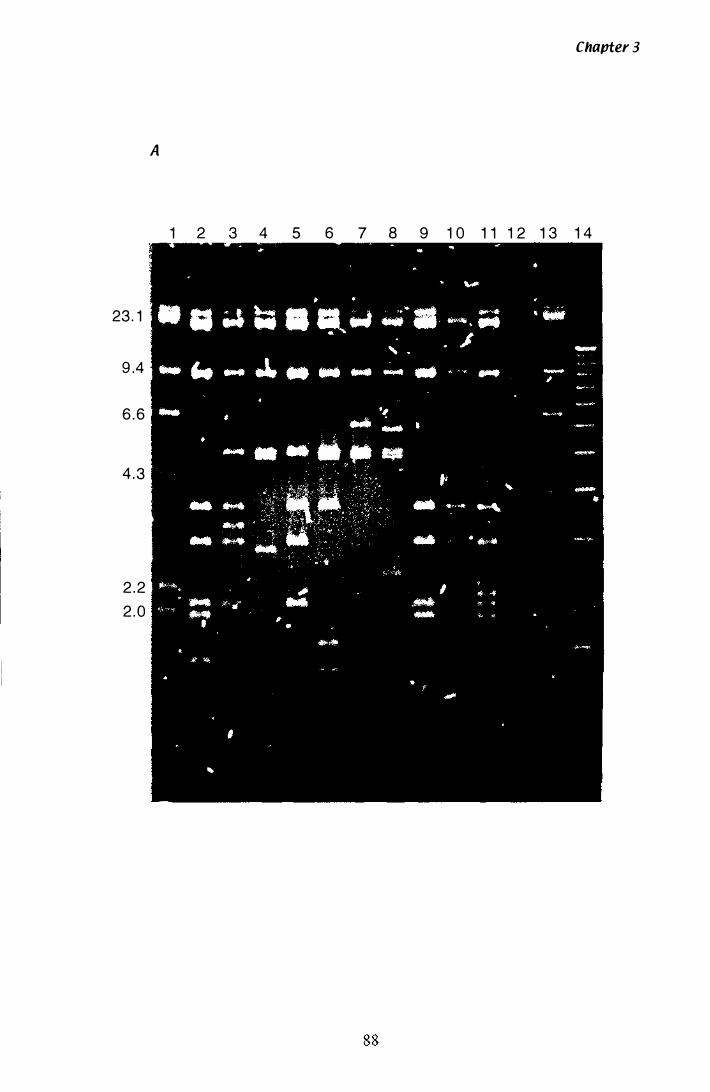
23 . 1
9 .4
6 . 6
4 .3
2 .2
2 . 0
Chapter 3
A
1 2 3 4 5 6 7 8 9 1 0 1 1 1 2 1 3 1 4

8
23. 1
9 . 4
6 . 6
4.3
2 .2
2 .0
Chapter 3
1 2 3 4 5 6 7 8 9 1 0 1 1
89
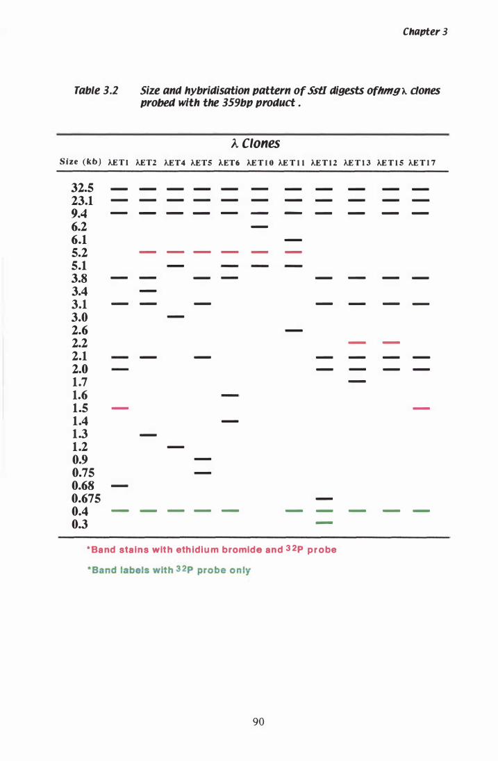
Table 3.2
S ize (kb) !..E T l
32.5 -
23.1 -
9.4 -
6.2 6.1 5.2 5.1 3.8 -
3.4 3.1 -
3.0 2.6 2.2 2.1 2.0 1.7 1.6 1.5 -
1.4 1.3 1.2 0.9 0.75 0.68 -
0.675 0.4 -
0.3
Chapter 3
Size ana hybridisation pattern of SstI digests o(hmgA. dones probed with the 359bp product .
A Clones
!..ET2 !..ET4 !..ETS AET6 !..E T I O !..E T l l !..E T l 2 !..E T 1 3 !..E T l S !..E T 1 7
- - - - - - - - - -
- - - - - - - - - -
- - - - - - - - -
-
-
- - - - - -
-
- - - - - - -
-
- - - - - -
-
-
- -
- - - - - -
- - - -
-
-
-
-
-
-
-
-
- - - - - - - - -
-
* Ba nd stains wit h eth ldl u m bromide and 3 2p p r o be
- Band labels wit h 3 2p probe on ly
90
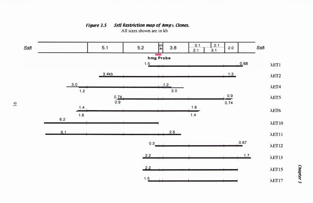
\0 .......
6.2
Figure 3.5 SstI Restriction map of hmg').. Qones.
All sizes shown are in kb
3.4kb 1 1 1 1 .3
3 .0 � 1 11 .2 1 .2 3 .0
014 1 1 0.9
0.9 0 .74 1 .4 1 1 1 .6 1 .6 1 .4
0 .3 1 1 1 0. 67
2.2 1 . 7
2.2 1 1 1 . 5 1 1
AEf I
AEf2
AEf4
AEf5
AEf6
AEf lO
AEf I I
AEf I2
AEf 13
AEf IS � i
AEf I7 "" w
Chapter 3
fragments between the multiple cloning cassette and unique sites within the fragment
(section 2 . 1 5 .6). The new sequence repositioned next to the multiple cloning cassette
was then isolated with the forward primer. All sub-clones used for sequencing are listed
in Table 2 . 1 . The restriction map of the P. paxilli hmg locus and relative position of the
sub-clones used for sequencing are shown in Figure 3 .6 A. Restriction fragments match
fragment sizes seen in the genomic digest of P. paxilli DNA (Table 3 . 1 ).
3.1 . 7 Sequencing AET2 sub-clones
The pUC 1 1 8 sub-clones, containing fragments of P. paxilli hmg, were sequenced at the
MuSeq DNA Analysis facility (IMBS, Massey University, NZ) ( section 2 . 1 2 .3 ) . As
much sequence as possible was determined using the forward and reverse universal
primers, but specific primers (Table 2 .3) needed to be designed (section 2 . 1 1 . 1 ) to
complete the double stranded sequence of this gene. Junctions between individual
fragments were sequenced directly from clones AET2 and AET4 with the appropriate
primers. Individual sequences were viewed using the Edit View programme (Version
1 .0 . 1 , Perkin Elmer), and assembled into a contig using both the GCG package and
Sequencher™ software. The templates used to assemble the hmg contig, are displayed
in Appendix A.2.3 with the Sequencher™ software. Sequencing analysis and deduced
polypeptide sequence revealed a 1 1 3 0 amino acid protein with three open reading
frames CORF) separated by two introns (introns 2 and 3) (section 3 . 1 . 8). The first ORF
extends from + 1 to +33 6 bp, ORF 2 from +408 to +3 1 27 bp, and ORF 3 from +3 1 96 to
+3529 bp (Figure 3 .6 B). A third intron (intron 1 ) was identified in the 5 ' untranslated
region (UTR) (section 3 . 1 .8) . The position of transcription initiation and termination
was determined with RACE (section 3 . 1 . 1 0 and 3 . 1 . 1 1 ) . The translation start and stop
sites were determined by the FRAMES programme of the GCG package, and the
sequence surrounding the translation start site (Ballance, 1 986 ; Kozak, 1 984) . The
deduced polypeptide sequence was separated into three domains by comparison to
existing hmg sequences. The transmembrane domain (residues 1 to 633) was analysed
by the TMPRED programme at the I SREC Bioinformatics group EMB.net website and
was predicted to contain 7 membrane-spanning regions. The linker domain (residues
634 to 683) has low conservation, which is seen in Figure 3 .8, a comparison of HMGR
proteins using the PILE UP programme from the GCG package. The catalytic domain
92
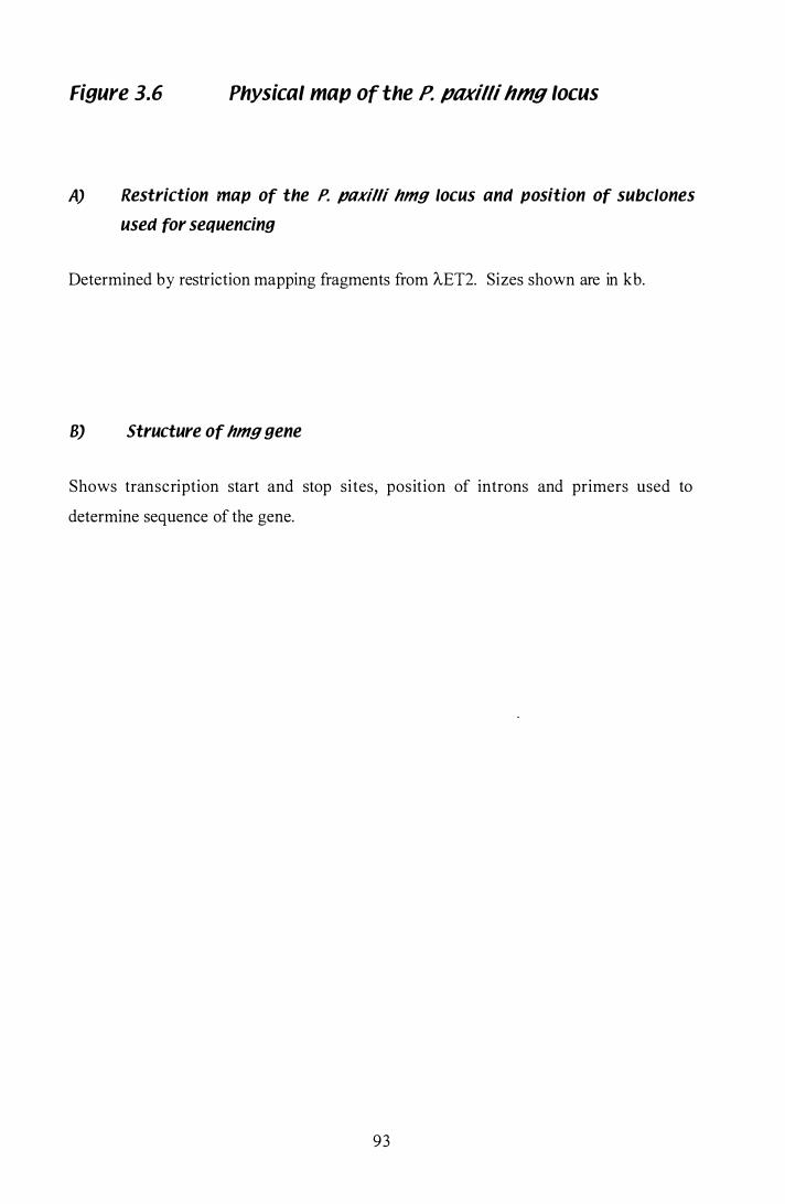
Figure 3.6 Physical map of the P. pnxilli hmg locus
A) Restriction map of the P. paxilli hmg locus and position of subclones
used for sequencing
Determined by restriction mapping fragments from AEI2. Sizes shown are in kb.
8) Structure of hmg gene
Shows transcription start and stop sites, position of introns and primers used to
determine sequence of the gene.
93

A
Ssft Pstl
8amH I Salt
•
� I
- 4 3 6 b p Tra nscription
� ....
I
� ....
AET2
5 ., I
I 9 . 8
4 .9 I �'4 '
2.2
DET2 ( DETS pET4
pE T9
DE;': pET 1 9
R�I.r _______ ....IiPioiiiE_T_1_1 _______ _
.... .... � � � � .... ET4Bam E T 1 E T 1 2 E T3 E T 1 S ETS E T 1 4 E T 1 ' E T29 H M G 1 H M G 3
� � E T2 ET8
�
� E T 1
� o ET6 E T2 4
.... .... .... � E T 1 3 E T 1 1 ET7 E T2 2
I
DE!J ( DET3
\. pE T7
+ 3 6 0 2 b p
A
.... � E T9 E T 1 6
I
1 k b I
+ 4 0 9 5 b p Tra nscription
op
1 k b I
� 1::)
l Vo./
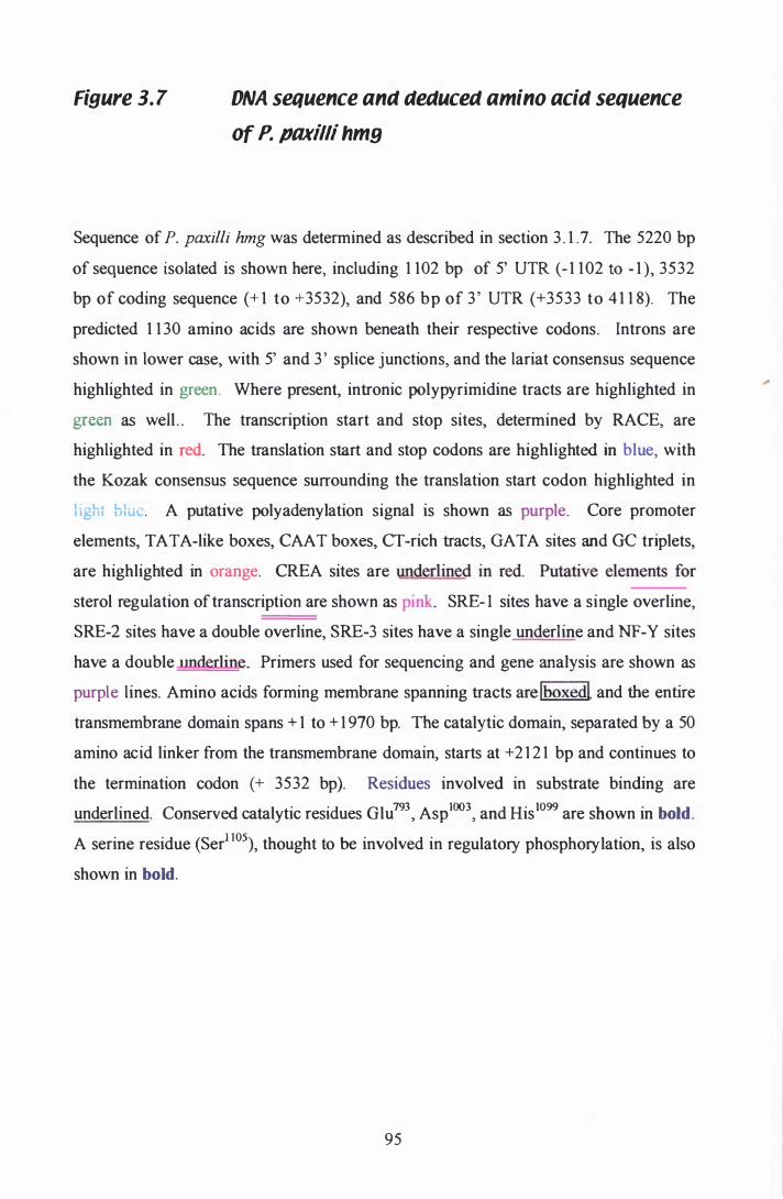
Figure 3.7 DNA sequence ana aeaucea amino acia sequence
of P. paxilli hmy
Sequence of P. paxilli hmg was determined as described in section 3 . 1 .7. The 5220 bp
of sequence isolated is shown here, including 1 102 bp of 5' UTR (- 1 102 to - 1 ), 3532
bp of coding sequence (+ 1 to +3532), and 586 bp of 3' UTR (+3533 to 4 1 1 8). The
predicted 1 1 30 amino acids are shown beneath their respective codons. Introns are
shown in lower case, with 5' and 3 ' splice junctions, and the lariat consensus sequence
highlighted in green. Where present, intronic polypyrimidine tracts are highlighted in
green as well . . The transcription start and stop sites, determined by RACE, are
highlighted in red. The translation start and stop codons are highlighted in blue, with
the Kozak consensus sequence surrounding the translation start codon highlighted in
l ight blue. A putative polyadenylation signal is shown as purple. Core promoter
elements, TA TA-like boxes, CAA T boxes, CT -rich tracts, GA TA sites and GC triplets,
are highlighted in orange. CREA sites are _==
sterol regulation of transcription are shown as pink. SRE- 1 sites have a single overline,
SRE-2 sites have a double overline, SRE-3 sites have a single underline and NF-Y sites
have a double Jlnderline. Primers used for sequencing and gene analysis are shown as
purple lines. Amino acids forming membrane spanning tracts are lboxedl and the entire
transmembrane domain spans + 1 to + 1 970 bp. The catalytic domain, separated by a 50
amino acid linker from the transmembrane domain, starts at +2 1 2 1 bp and continues to
the termination codon (+ 3532 bp). Residues involved in substrate binding are
underlined. Conserved catalytic residues Glu793, Asploo3, and His1099 are shown in bold.
A serine residue (Ser1 I05), thought to be involved in regulatory phosphorylation, is also
shown in bold.
95
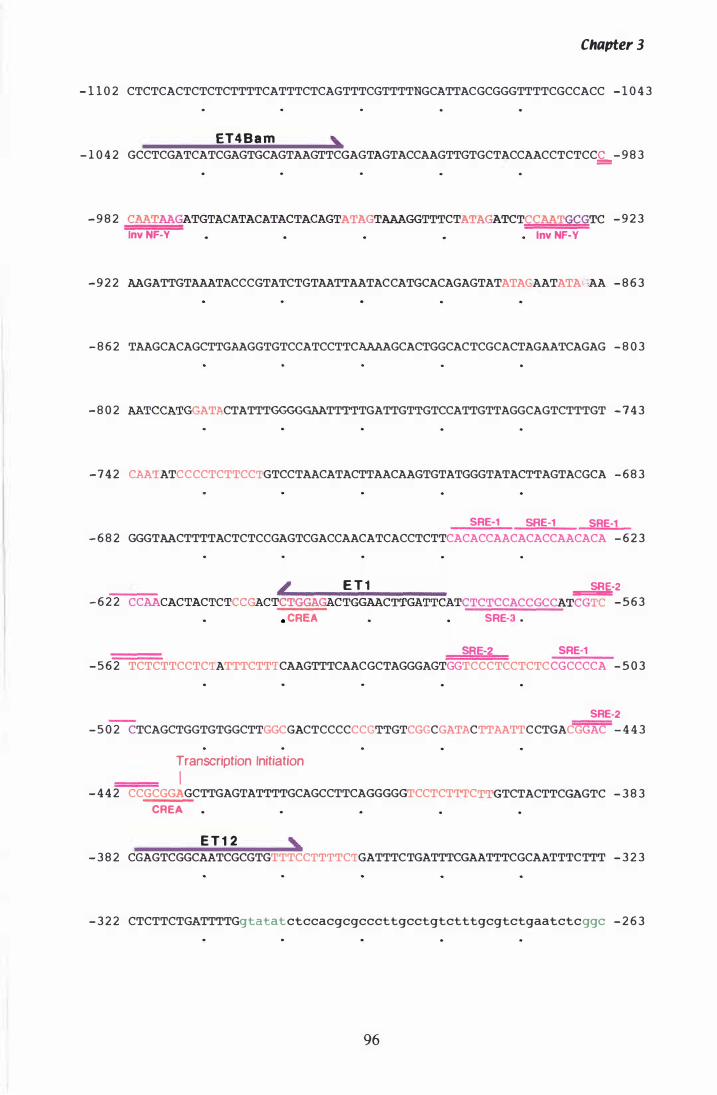
Chapter 3
- 1 1 0 2 CTCTCACTCTCTCTTTTCATTTCTCAGTTTCGTTTTNGCATTACGCGGGTTTTCGCCACC - 1 0 4 3
ET4 B a m , - 1 0 4 2 GCCTCGATCATCGAGTGCAGTAAGTTCGAGTAGTACCAAGTTGTGCTACCAACCTCTCC£:- 9 8 3
- 9 8 2 CAATAAGATGTACATACATACTACAGTATAGTAAAGGTTTCTATA TCTCCAATGCGTC - 9 2 3
Inv NF-Y • Inv NF-Y
- 9 2 2 AAGATTGTAAATACCCGTATCTGTAATTAATACCATGCACAGAGTATATAGAATATAGAA - 8 6 3
- 8 6 2 TAAGCACAGCTTGAAGGTGTCCATCCTTCAAAAGCACTGGCACTCGCACTAGAATCAGAG - 8 0 3
- 8 0 2 AATCCAT ATACTATTTGGGGGAATTTTTGATTGTTGTCCATTGTTAGGCAGTCTTTGT - 7 4 3
- 7 4 2 CAATATCCCCTCTTCCTGTCCTAACATACTTAACAAGTGTATGGGTATACTTAGTACGCA - 6 8 3
SRE-l SRE-l SRE-l - 6 8 2 GGGTAACTTTTACTCTCCGAGTCGACCAACATCACCTCTTCACACCAACACACCAACACA - 6 2 3
L E T 1 �2 - 6 2 2 CCAACACTACTCTCC CTCTGGA C TGGAACTTGATTCATCTCTCCACCGCCATCGTC - 5 6 3
. CREA SRE-3 .
SRE-2 SRE-l - 5 6 2 TCTCTTCCTCTATTTCTTTCAAGTTTCAACGCTAGGGAGTGGTCCCTCCTCTCCGCCCCA - 5 0 3
SRE-2 - 5 0 2 CTCAGCTGGTGTGGCTTGGCGACTCCCCCCGTTGTCGGCGATACTTAATTCCTGACGGAC - 4 4 3
Transcription Initiation I
- 4 4 2 CCGCGGAGCTTGAGTATTTTGCAGCCTTCAGGGGGTCCTCTTTC
CREA
E T 1 2 ,
TCTACTTCGAGTC - 3 8 3
- 3 8 2 CGAGTCGGCAATCGCGTGTTTCCTTTTCTGATTTCTGATTTCGAATTTCGCAATTTCTTT - 3 2 3
- 3 2 2 CTCTTCTGATTTTGgtatatctccacgcgcccttgcctgtctttgcgtctgaatctcggc - 2 6 3
96
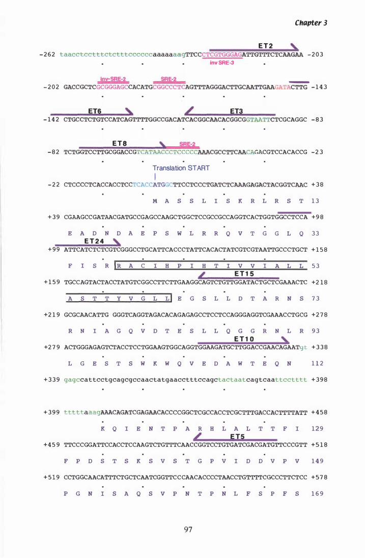
Chapter 3
E T 2 , - 2 6 2 taacctcctttctctttccccccaaaaaaagTTCCCTCGTGGGAGATTGTTTCTCAAGAA - 2 0 3
Inv SRE-3
Inv-SRE-2 SRE-2 _ - 2 0 2 GACCGCTCGCGGGAGCCACATGCGGCCCTCAGTTTAGGGACTTGCAATTGAAGATACTTG - 1 4 3
ET6 ' / ET3 - 1 4 2 CTGCCTCTGTCCATCAGTTTTGGCCGACATCACGGCAACACGGCGGTAA TCGCAGGC - 8 3
ET8 , SRE-2 - 8 2 TCTGGTCCTTGCGGACCGTCATAACCCTCCCCCAAACGCCTTCAACA CGTCCACACCG -2 3
Translation START I
- 2 2 CTCCCCTCACCACCTCCTCACCATGGCTTCCTCCCTGATCTCAAAGAGACTACGGTCAAC + 3 8
M A S S L I S K R L R S T 1 3
+ 3 9 CGAAGCCGATAACGATGCCGAGCCAAGCTGGCTCCGCCGCCAGGTCACTGGTGGCCTCCA +9 8
E A D N D A E P S W L R R Q v T G G L Q 3 3 E T 2 4 �
+ 9 9 ATTCATCTCTCGTCGGGCCTGCATTCACCCTATTCACACTATCGTCGTAATTGCCCTGCT + 1 5 8
F I S R IR A C I H P I H T I V V I A L L 5 3
� E T 1 5 + 1 5 9 TGCCAGTACTACCTATGTCGGCCTTCTTGAAGGCAGTCTGTTGGATACTGCTCGAAACTC +2 1 8
A S T T Y V G L LI E G S L L D T A R N S 7 3
+2 1 9 GCGCAACATTGCGGGTCAGGTAGACACAGAGAGCCTCCTCCAGGGAGGTCGAAACCTGCG +2 7 8
R N I A G Q v D T E S L L Q G G R N L R 9 3 E T 1 0 ,
+2 7 9 ACTGGGAGAGTCTACCTCCTGGAAGTGGCAGGTGGAAGATGCTTGGACCGAACAGAATgt + 3 3 8
L G E S T S W K W Q v E D A W T E Q N 1 1 2
+ 3 3 9 gagccattcctgcagcgccaactatgaacctttccagctactaatcagtcaattcctttt + 3 9 8
+ 3 9 9 tttttaaagAAACAGATCGAGAACACCCCGGCTCGCCACCTCGCTTTGACCACTTTTATT + 4 5 8
K Q I E N T P A R H L A L T T F I 1 2 9 L E TS
+ 4 5 9 TTCCCGGATTCCACCTCCAAGTCTGTTTCAACCGGTCCTGTGATCGACGATGTTCCCGTT + 5 1 8
F P D S T S K S V S T G P V I D D V P v 1 4 9
+ 5 1 9 CCTGGCAACATTTCTGCTCAATCGGTTCCCAACACCCCTAACCTGTTTTCGCCCTTCTCC + 5 7 8
P G N I S A Q S v P N T P N L F S P F S 1 6 9
97

Chapter 3
+5 7 9 CATGACTCTGCTCTCGCCTATGCCGTCCCCTTTGAACAAATTTCTGAATTTCTGAGGGCC +6 3 8
H D S A L A Y A V P F E Q I S E F L R A 1 8 9
+ 6 3 9 GTTCAGGAGATCCCAGATTCGTCGGACTACAAAGATGAAAAGGAATCGAAGAAATGGATC + 6 9 8
v Q E I P D S S D Y K D E K E S K K W I 2 0 9
+ 6 9 9 ATGAGGGCAGCTCGTGGAGCCACAGGCTCGCGCACTGCTCTCAAGCTCTGGTTGACCGAT +7 5 8
M R A A R G A T G S R T A L K L W L T D 2 2 9
+ 7 5 9 GCCTGGGGCTCGTTCGTTGATCTGATCAAACATGCCGAGACCATTGACATTGTCATCATG +8 1 8
A W G S F V D L I K H A E T I D Ir v I M 2 4 9
+ 8 1 9 GTTCTTGGTTACATCTCGATGCATCTGAGCTTCGTCTCCCTCTTCTTCTCCATGAGACGC +8 7 8
V L G Y I S M H L S F V S L F F I S M R R 2 6 9
+8 7 9 TTGGGATCCAACTTCTGGCTCGCTGCCACTGTGCTTTTCTCCGGCTCCTTCGCGTTCCTG + 9 3 8
L G S N IF W L A A T V L F S G S F A F L 2 8 9
+ 9 3 9 TTCGGTCTTTTGGTCACTACCAAACTTGGCGTGCCAATTAATCTACTCCTGTTGTCGGAG + 9 9 8
F G L � vi T TO
K IL G V P I N L L L L S E 3 0 9
+ 9 9 9 GGACTTCCTTTCCTGGTTGTCACTATTGGGTTTGAGAAGCCGATCATGTTCACTCGTGCA + 1 0 5 8
G L P F L V V T I G F E l K P I M F T R A 3 2 9
+ 1 0 5 9 GTTCTCAATGCCTCTGTGGACAATCGCCGACCTCATCCCGGTGCCGCACCCCGTCCCCTC + 1 1 1 8
V L N A S V D N R R P H P G A A P R P L 3 4 9
+ 1 1 1 9 GCTTCTAGCACCCCATCCTCCATTCAGGACTCTATCGCAACTGCGATCAAGCAGCAGGGC + 1 1 7 8
A S S T P S S I Q D S I A T A I K Q Q G 3 6 9
+1 1 7 9 TTCGAGATTATTCAGCACTACTGTATCGAGATTGGTCTTTTGACCATTGGTGCTGCTTCT + 1 2 3 8
F E I I Q H Y C I E I G L L T I G A A S 3 8 9 E T 1 4 ,
+ 1 2 3 9 GGTGTTCAGGGCGGTCTTCAGCAGTTCTGCTTCCTCGCGGCCTGGATTTTGTTCTTTGAC + 1 2 9 8
G V Q G G L Q Q F C iF L A A W I L F F D 4 0 9
+ 1 2 9 9 TGTGTGCTCCTGTTCACCTTCTACACCACCATTCTCTGCATCAAGCTCGAGATTACACGT + 1 3 5 8
C y L L F T F
O
Y TI T I L C I K L E I T R 4 2 9
+ 1 3 5 9 ATCAAGCGTCATGTTGCGCTGCGCAAGGCCCTTGAAGAGGATGGCATCACCCACAGCGTG + 1 4 1 8
I K R H V A L R K A L E E D G I T H S V 4 4 9
98
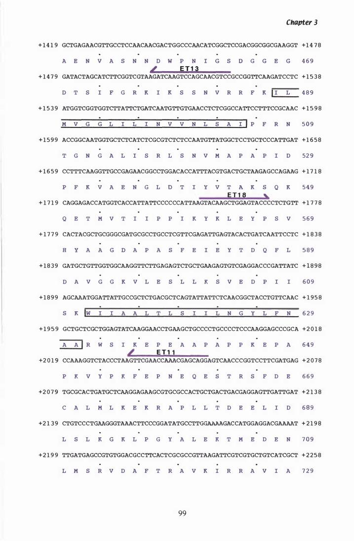
Chapter 3
+ 1 4 1 9 GCTGAGAACGTTGCCTCCAACAACGACTGGCCCAACATCGGCTCCGACGGCGGCGAAGGT +1 4 7 8
A E N V A S N N D W P N I G S 0 G G E G 4 6 9 L E T 1 3
+ 1 4 7 9 GATACTAGCATCTTCGGTCGTAAGATCAAGTCCAGCAACGTCCGCCGGTTCAAGATCCTC + 1 5 3 8
D T S I F G R K I K S S N V R R F K I I L 4 8 9
+ 1 5 3 9 ATGGTCGGTGGTCTTATTCTGATCAATGTTGTGAACCTCTCGGCCATTCCTTTCCGCAAC + 1 5 9 8
M V G G L I L I N V V N L S A I I P F R N 5 0 9
+ 1 5 9 9 ACCGGCAATGGTGCTCTCATCTCGCGTCTCTCCAATGTTATGGCTCCTGCTCCCATTGAT + 1 6 5 8
T G N G A L I S R L S N V M A P A P I D 52 9
+ 1 6 5 9 CCTTTCAAGGTTGCCGAGAACGGCCTGGACACCATTTACGTGACTGCTAAGAGCCAGAAG + 1 7 1 8
P F K V A E N G L O T I Y V T A K S Q K 5 4 9 E T 1 S ""
+ 1 7 1 9 CAGGAGACCATGGTCACCATTATTCCCCCCATTAAGTACAAGCTGGAGTACCCCTCTGTT + 1 7 7 8
Q E T M V T I I P P I K Y K L E Y P S V 5 6 9
+ 1 7 7 9 CACTACGCTGCGGGCGATGCGCCTGCCTCGTTCGAGATTGAGTACACTGATCAATTCCTC + 1 8 3 8
H Y A A G O A P A S F E I E Y T D Q F L 5 8 9
+ 1 8 3 9 GATGCTGTTGGTGGCAAGGTTCTTGAGAGTCTGCTGAAGAGTGTCGAGGACCCGATTATC + 1 8 9 8
D A V G G K V L E S L L K S V E D P I I 6 0 9
+ 1 8 9 9 AGCAAATGGATTATTGCCGCTCTGACGCTCAGTATTATTCTCAACGGCTACCTGTTCAAC + 1 9 5 8
S K Iw I I A A L T L S I I L N G Y L F N 6 2 9
+ 1 9 5 9 GCTGCTCGCTGGAGTATCAAGGAACCTGAAGCTGCCCCTGCCCCTCCCAAGGAGCCCGCA +2 0 1 8
A A I R W S I K E P E A A P A P P K E P A 6 4 9 L E T "
+2 0 1 9 CCAAAGGTCTACCCTAAGTTCGAACCAAACGAGCAGGAGTCAACCCGGTCCTTCGATGAG + 2 0 7 8
P K V Y P K F E P N E Q E S T R S F D E 6 6 9
+2 0 7 9 TGCGCACTGATGCTCAAGGAGAAGCGTGCGCCACTGCTGACTGACGAGGAGTTGATTGAT + 2 1 3 8
C A L M L K E K R A P L L T D E E L I D 6 8 9
+2 1 3 9 CTGTCCCTGAAGGGTAAACTTCCCGGATATGCCTTGGAAAAGACCATGGAGGACGAAAAT + 2 1 9 8
L S L K G K L P G Y A L E K T M E D E N 7 0 9
+2 1 9 9 TTGATGAGCCGTGTGGACGCCTTCACTCGCGCCGTTAAGATTCGTCGTGCTGTCATCGCT + 2 2 5 8
L M S R V D A F T R A V K I R R A V I A 7 2 9
99
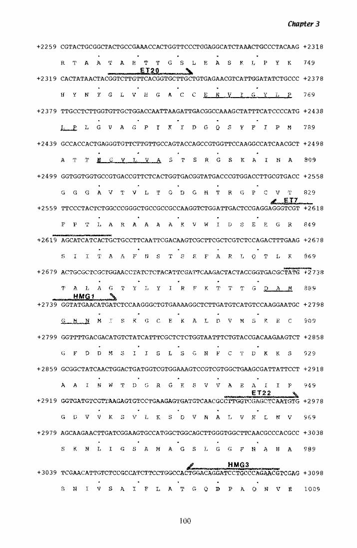
Chapter 3
+2 2 5 9 CGTACTGCGGCTACTGCCGAAACCACTGGTTCCCTC�GGCATCTAAACTGCCCTACAAG + 2 3 1 8
y
+2 3 1 9 CACTATAACTACGGTCTTGTTCACGGTGCTTGCTGTGAGAACGTCATTGGATATCTGCCC +2 3 7 8
+ 2 3 7 9 TTGCCT�TTGGTGTTGCTGGACCAATTAAGATTGACGGCCAAAGCTATTTCATCCCCATG + 2 4 3 B
G A p
+ 2 4 3 9 GCCACCACTGAGGGTGTTCTTGTT�CCAGTACCAGCCGTGGTTCCAAGGCCATCAACGCT + 2 4 9 8
G K A
+ 2 4 9 9 GGTGGTGGTGCCGTGACCGTTCTCACTGGTGACGGTATGACCCGTGGACCTTGCGTGACC + 2 5 5 8
+2 5 5 9 TTCCCTACTCTGGCCCGGGCTGCCGCCGCCAAGGTCTGGATTGACTCCGAGGAGGGTCGT +2 6 1 8
F E
AGCATCATCACTGCTGCCTTCAATTCGACAAGTCGCTTCGCTCGTCTCCAGACTTTGAAG +2 6 7 8
+ 2 6 7 9
+ 2 7 3 9 GGTATGAACATGATCTCCAAGGGCTGTGAAAAGGCTCTTGATGTCATGTCCAAGGAATGC + 2 7 9 8
A
+ 2 7 9 9 GGTTTTGACGACATGTCTATCATTTCGCTCTCTGGTAATTTCTGTACCGACAAGAAGTCT + 2 8 5 8
+ 2 8 5 9 GCGGCTATCAACTGGACTGATGGTCGTGGAAAGTCCGTCGTGGCTGAAGCGATTATTCCT + 2 9 1 8
+ 2 9 1 9 +2 9 7 8
v v A
+ 2 9 7 9 AGCAAGAACTTGATCGGAAGTGCCATGGCTGGCAGCTTGGGTGGCTTCAACGCCCACGCC + 3 0 3 8
G A 9
+ 3 0 3 9 + 3 0 9 8
L p
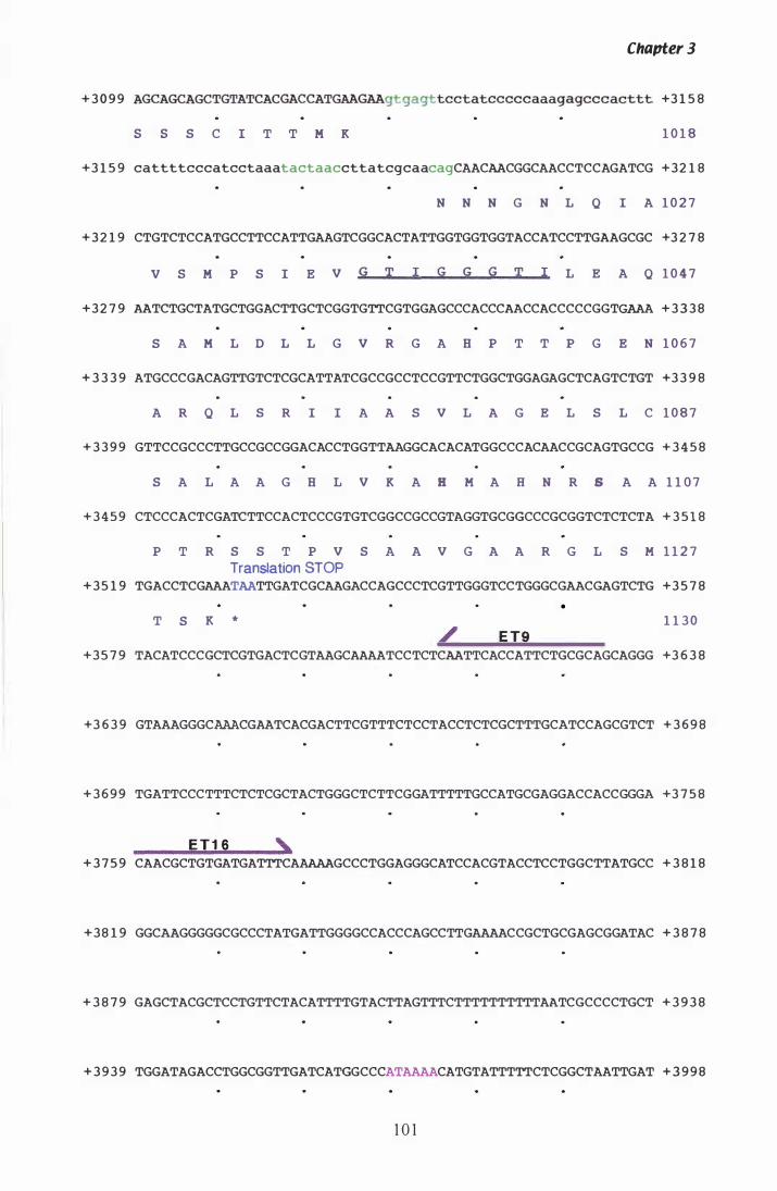
Chapter 3
+ 3 0 9 9 AGCAGCAGCTGTATCACGACCATGAAGAAgtgagttcctatcccccaaagagcccacttt +3 1 5 8
s s S C I T T M K 1 0 1 8
+3 1 5 9 cattttcccatcctaaatactaaccttatcgcaacagCAACAACGGCAACCTCCAGATCG +32 1 8
N N N G N L Q I A 1 0 2 7
+ 3 2 1 9 CTGTCTCCATGCCTTCCATTGAAGTCGGCACTATTGGTGGTGGTACCATCCTTGAAGCGC +3 2 7 8
v S M P S I E V G T I G G G T I L E A Q 1 0 4 7
+3 2 7 9 AATCTGCTATGCTGGACTTGCTCGGTGTTCGTGGAGCCCACCCAACCACCCCCGGTGAAA + 3 3 3 8
S A M L D L L G V R G A B P T T P G E N 1 0 6 7
+ 3 3 3 9 ATGCCCGACAGTTGTCTCGCATTATCGCCGCCTCCGTTCTGGCTGGAGAGCTCAGTCTGT +3 3 9 8
A R Q L S R I I A A S V L A G E L S L C 1 0 8 7
+ 3 3 9 9 GTTCCGCCCTTGCCGCCGGACACCTGGTTAAGGCACACATGGCCCACAACCGCAGTGCCG + 3 4 5 8
S A L A A G B L V K A H M A H N R S A A 1 1 0 7
+ 3 4 5 9 CTCCCACTCGATCTTCCACTCCCGTGTCGGCCGCCGTAGGTGCGGCCCGCGGTCTCTCTA + 3 5 1 8
P T R S S T P V S A A V G A A R G L S M 1 1 2 7
Translation STOP + 3 5 1 9 TGACCTCGAAATAATTGATCGCAAGACCAGCCCTCGTTGGGTCCTGGGCGAACGAGTCTG +3 5 7 8
T S K *
/ E T9
•
1 1 3 0
+3 5 7 9 TACATCCCGCTCGTGACTCGTAAGCAAAATCCTCTCAATTCACCATTCTGCGCAGCAGGG +3 6 3 8
+3 6 3 9 GTAAAGGGCAAACGAATCACGACTTCGTTTCTCCTACCTCTCGCTTTGCATCCAGCGTCT + 3 6 9 8
+ 3 6 9 9 TGATTCCCTTTCTCTCGCTACTGGGCTCTTCGGATTTTTGCCATGCGAGGACCACCGGGA +3 7 5 8
ET 1 6 , + 3 7 5 9 CAACGCTGTGATGATTTCAAAAAGCCCTGGAGGGCATCCACGTACCTCCTGGCTTATGCC + 3 8 1 8
+38 1 9 GGCAAGGGGGCGCCCTATGATTGGGGCCACCCAGCCTTGAAAACCGCTGCGAGCGGATAC + 3 8 7 8
+ 3 8 7 9 GAGCTACGCTCCTGTTCTACATTTTGTACTTAGTTTCTTTTTTTTTTAATCGCCCCTGCT + 3 9 3 8
+ 3 9 3 9 TGGATAGACCTGGCGGTTGATCATGGCCCATAAAACATGTATTTTTCTCGGCTAATTGAT + 3 9 9 8
1 0 1

Chapter 3
+ 3 9 9 9 TGGTTTTCCGCCCTCGAATAGCAGTCAAGCTAATGGTTTGGTGCACATTTAGATGAGTGT +4 0 5 8
Transcription Stop I
+ 4 0 5 9 ATTTCATATCAATTAGTCAAGTTAGCAAACAGGTACTATGCTTTCGCAGGGCTGCCTCCA +4 1 1 8
1 02
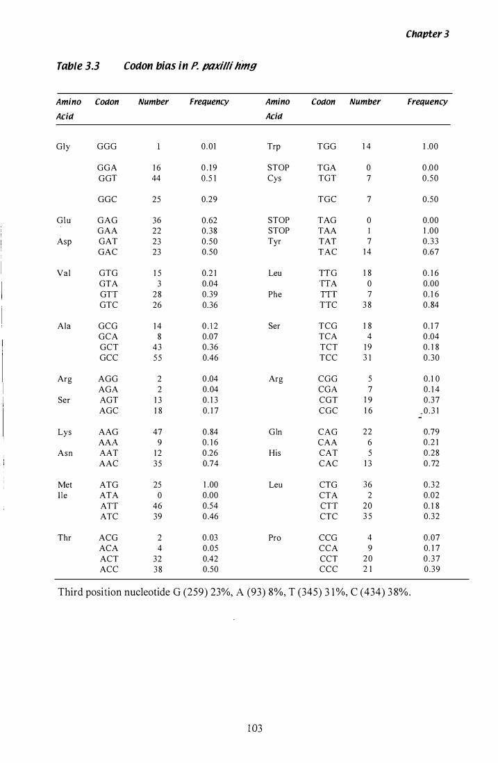
Chapter 3
Table 3.3 Codon bias in P. p(1xi/li hmg
Amino Codon Number Frequency Amino Codon Number Frequency
Acid Acid
Gly GGG 0.01 Trp TGG 1 4 1 .00
GGA 1 6 0 . 1 9 STOP TGA 0 0 .0 0 GGT 44 0.5 1 Cys TGT 7 0 .50
GGC 25 0.29 TGC 7 0.50
Glu GAG 36 0.62 STOP TAG 0 0.00 GAA 22 0 .38 STOP TAA 1 .00
Asp GAT 23 0 .50 Tyr TAT 7 0 . 3 3 GAC 23 0.50 TAC 1 4 0.67
Val GTG 1 5 0.2 1 Leu ITG 1 8 0. 1 6 GTA 3 0.04 ITA 0 0.00 GTI 2 8 0.39 Phe TIT 7 0. 1 6 GTC 26 0.36 TIC 3 8 0 . 84
Ala GCG 1 4 0 . 1 2 Ser TCG 1 8 0 . 1 7 GCA 8 0.07 TCA 4 0 .04 GCT 43 0.36 TCT 1 9 0. 1 8 GCC 5 5 0.46 TCC 3 1 0 .30
Arg AGG 2 0 .04 Arg CGG 5 0 . 1 0 AGA 2 0.04 CGA 7 0. 1 4
Ser AGT 1 3 0 . 1 3 CGT 1 9 0 .37 AGC 1 8 0 . 1 7 CGC 1 6 _ 0 . 3 1
Lys AAG 47 0 .84 G In CAG 22 0.79 AAA 9 0. 1 6 CAA 6 0.2 1
Asn AAT 1 2 0 .26 His CAT 5 0.28 AAC 3 5 0.74 CAC 1 3 0 . 72
Met ATG 25 1 .00 Leu CTG 36 0 .32 I I e ATA 0 0.00 CTA 2 0.02
ATI 46 0.54 CTI 20 0. 1 8 ATC 39 0 .46 CTC 3 5 0.32
Thr ACG 2 0.03 Pro CCG 4 0.07 ACA 4 0.05 CCA 9 0. 1 7 ACT 32 0.42 CCT 2 0 0 . 3 7 ACC 3 8 0.50 CCC 2 1 0 .39
Third position nucleotide G (259) 23%, A (93) 8%, T (345) 3 1 %, C (434) 3 8%.
103
Figure 3.8 Alignment of hmg polypeptide sequences
Protein sequences were aligned with the PILEUP programme from the GCG package
and are coloured with respect to similarity and identity with the P. paxilli HMGR
sequence. Amino acids in all species present are also highlighted.
Abbreviations for the species names are; Ppax, Penicillium paxilli; Lp1 9, Neotyphodium
loW i solate Lp1 9; Gfuj, Gibberella !ujikuroi; Ater, Aspergillus terreus; Spom,
Schizosaccharomyces pombe; Hsap, Homo sapiens; Atha, Arabidopsis thaliana.
1 04
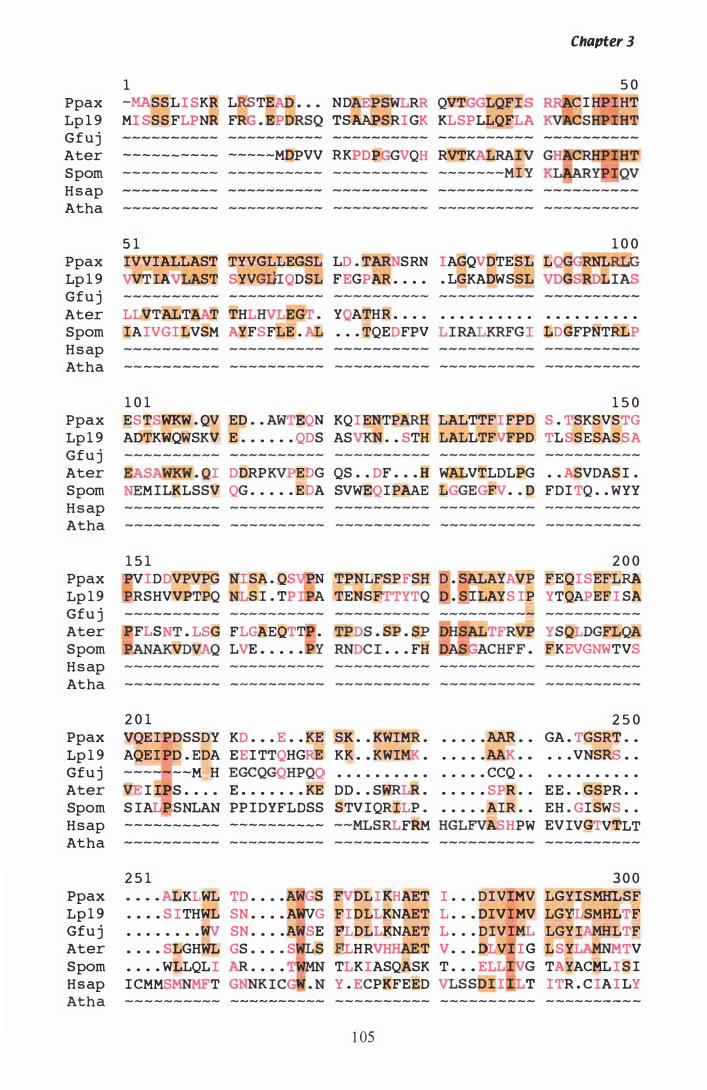
Ppax Lp 1 9 Gfuj Ater Spom H s ap Atha
Ppax Lp 1 9 Gfu j Ater Spom H s ap Atha
Ppax Lp 1 9 Gfu j Ater Spom H s ap Atha
Ppax Lp 1 9 Gfu j Ater Spom H s ap Atha
Ppax Lp 1 9 Gfu j Ater Spom H s ap Atha
Chapter 3
1 5 0 -MASSLI SKR LRST.E ND PSWL QVTGGLQFI S C I HP I HT MI SSSFLPNR FR . EP ORSQ TSAAPSRI GK KLSPLLQFLA C'SHlUHT
---------- -----MOPVV RKPDP VQH RVT ------ ---- ---------- ---------- -------MIY
5 1 1 0 0 IVVIALLAST TYVGLLEGSL LD . T SRN IAGQVDTESL L RNLRLG
lA ST SYVGL�QDSL FEGPAR . . . . . LGKADWSSL VDGSRDL IAS
LLVTALT T THL EGT . YQATHR . . . . • • . . • • . . • • . • • . . • . . • •
IAIVGI LVSM AYFSFLE . • • . TQEDFPV LlRALKRFGI �DGFPNTRLP
1 0 1 1 5 0 ESTSWKW . QV ED . . A EQN KQI ENTPARH LALTTFI FPD S . TSKSVSTG ADTKWQWSKV E . . . . . . QDS AS . • STH LALLTF PD TLSSESASSA
EASAWKW . QI DDRP EDG QS . . DF . . . H WALVTLDLPG . . ASVDAS I . MILKLSSV QG . . . . . EDA SVWEQIPAAE L EG . • D FDI TQ . . WYY
1 5 1 2 0 0 PVI DDVPVPG NI SA . QS P� TPNLFSPFSH D . SALAYAVP FEQI SEFLRA PRSHVVPTPQ S I . TP A TENSFTTYTQ . S[LAYS IP YTQ EF I SA
BFLS . LSG FLGAEQT� . �AKVDVAQ LVE . . • . . � RNDC I . . . FH
YS QLDGFLQA FKEVGNWTVS
2 0 1 2 5 0 VQE I PDSSDY KD . • . E . . KE SK . • KWIMR . • • . • • AAR • • GA . TGSRT • •
AQEI PD . EDA EEI TTQHG KK • • KWI • . . • . . • . • VNSRS • . --MOH EGCQ HPQQ • • • • • • • • • • • • • • • CCQ . . • • • • • • • • • •
I IP.S . . . • E . . . . . . . KE DD . . S • . . . • • SPR • • EE . . GSPR . . S IA P SNLAN PP I DYFLDSS STV I QRILP • . . . • . AIR . • EH . GI SWS . . ---------- ---------- --MLS FRM HGLFV HPW EVIVGTVTLT
2 5 1 3 0 0 Ppax KLWL TD . . • . A GS DLI KHAET I . • • DIVIMV LGYISMHLSF Lp 1 9 . • . • S ITHWL SN • . • • AWVG FI DLLKNAET L • . • DIVIMV LGYLSMHLTF Gfu j • • . • . • • • SN . • . • A SE FLDLLKNAET L . . . D IVI LGYI LTF Ater . • . • SLGHWL GS • . . . S S FLHRVH T V • • • DLVI I G LSY TV Spom • . . . WLLQL I . . . . T� TLK IASQASK T • • • EL �G TAYACML I S I H s ap I CMMS T I CG . N Y . ECPKFEED T ITR . C IAI LY Atha ---------- - - - - ----- - ---------- ---------- - ------ - - -
1 05
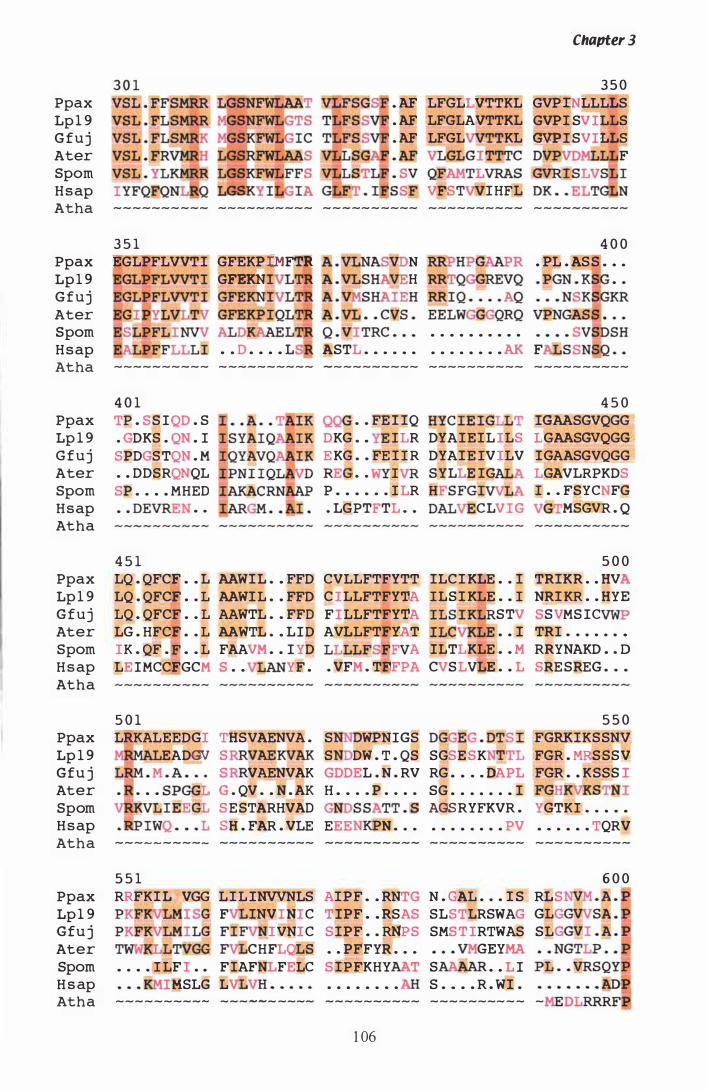
Ppax Lp 1 9 G f u j Ater Spom H s ap
. AF
. AF
. AF
. AF
LFGLLVTTKL LFGLAVTTKL LFGL KL
GLGlTTTC QFAMTLVRAS VFST I HFL
Chapter 3
3 5 0 GVP lNLLLLS GVPl SV ILLS GVPI SVILLS DVPVD F
Atha ---------- ---------- ----- - ---- ---------- ----------
3 5 1 4 0 0 Ppax EGLPFLVVTl GFEKPlMFTR A . VLNAS N . PL . AS S . . .
Lp 1 9 GFEKN A . VLSH� H . PGN . KS . . G f u j Ater Spom • • • • • • • • • •
H s ap . . D • • . • L ASTL . • . . . • • • . . • . . . AK F Atha ---------- ---------- - --------- ---------- ----------
Ppax Lp 1 9 G f u j Ater Spom H s ap Atha
Ppax Lp 1 9 G f u j Ater Spom H s ap Atha
4 0 1 TP . S S IQD . S . GDKS . QN . I SPDGS TQN . M
. • DDSRQNQL SP • . . • MHED . . DEVREN . .
4 5 1 LQ . QFCE . • L AAWlL . . FFD LQ . QFCE . . L AAWlL . . FFD LQ . QFCFi . . L AAWTL . . FFD LG . HFCE . . L AAWTL . • LID IK . QF . • . L FAAVM . . I YD LE I MCCEGCM S . . YF .
4 5 0 . . FE l lQ HYC l E I GLLT lGAASGVQGG
DKG . • ILR DYAlElL ILS LGAASGVQGG E KG . • FE l I R DYAlE IVILV lGAASGVQGG REG . . I SYLLE lG LGAVLRPKDS P . • • • . . I LR HFSFGl I • . FSYCNFG . LGPTFTL . . DAL CLVIG V MSGVR . Q
5 0 0 CVLLFTFYTT lLCl KLE . . l TRlKR . • HVA C ILLFTFYTA ILS l KLE . • I NRl KR . . HYE F ILLFTFYTA AVLLFTEYAT
STV SSVMS IC . . I TRI . . . . . . .
LLLLFS A ILTLKLE . . M RRYNAKD • . D . VFM . TEFPA CVSLVLE . . L SRE SREG • . •
5 0 1 5 5 0 Ppax LRKALEEDGI THSVAENVA . S WPNIGS DGG EG . DTS l FGRKlKSSNV Lp 1 9 EADGV S VAEKVAK S DW . T . QS SGSE SK TL FGR . �S SSV Gfu j S VAENVAK GDDEL . N . RV RG • • . . DAPL FGR . . KSSS I Ater • . • • S PGGL G . QV . • N . AK H • • . . P . . . . SG • • . . • . • l FGHKVKSTNI Spom S E STARHVAD GNDS SATT . S AGSRYFKVR . YGTKI . . . . •
H s ap sa . FAR . VLE EEE N • . • • . . • . . . . PV • . . • • . TQRV Atha ---------- ---------- ---------- ---- - ----- ----------
5 5 1 Ppax RRFKl LMVGG L l L INVVNLS AlPF • • RNTG N . GAL . . . l S RLS Lp 1 9 PKFKVLMI SG F lNVINIC TIPF • • RSAS SLSTLRSWAG GL Gfu j PKFK�LMlLG IVNIC S lPF • . RNPS SMS T IRTWAS SL Ater T LLTVGG CHFLQLS . . PFFYR . . . GEYMA Spom . . • . I LF I . . F lAF FELC S lPFKHY T S . . L I H s ap IMSLG L . • • . • . . . S . . • • R . Wl . Atha ---------- --- - - - ---- ---------- -------- - - -
1 06
6 0 0
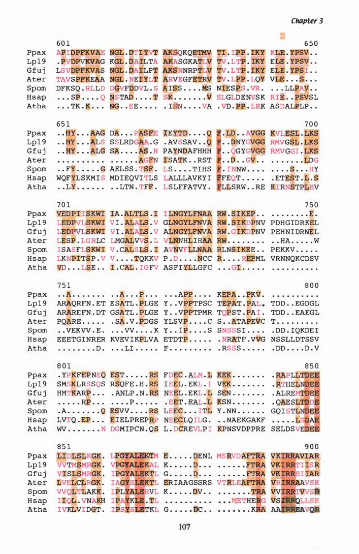
Ppax Lp 1 9 G f u j Ater Spom H s ap Atha
Ppax Lp 1 9 G f u j Ater S pom H s ap Atha
Ppax Lp 1 9 G f u j Ater S pom H s ap Atha
Ppax Lp 1 9 Gfu j Ater Spom H s ap Atha
Ppax Lp 1 9 Gfu j Ater Spom H s ap Atha
Ppax Lp 1 9 Gfu j Ater Spom H s ap Atha
Chapter 3
6 0 1 6 5 0 I DPFKV NGL . DT I T AKSQKQETMV TI . I PP . IKY . YPSV • .
. P PVKVAG KGL . O . LTA . L . . IKY ELE . YPSV . . LS PFKVAS NGL . OAILPT AKS . L . I KY ELE . YPS I . . TAVSPfKEAA NGL . I YLT . LPP . LQY VLE . • • S • . .
DFKSQ . RLLD D GV FOpVL . S A I S S • • • • MS NIESPS . VR . • • • LLPAV • • • . • SE • . • • Q NSTAD . . . . T SK • • • • • • • � SLGLDE S K RIE . . PSVSL · . • TK . K • • • NG . . EE . . . . • I SN • . . . VA . . PP . L ASDALPLP . .
6 5 1 • . HY . . • AAG DA . . . P AS E IEYTD . • . • Q . LD • • A,.VGG
. . HY . • . ALS SSLRD . G . AVSSAV . . Q • . DNYGVGG · . HY • . . ALG SA . . . . AS • N PAYNDAFHHH . . QGYGVGG · . . . . . . . . . . . . . . . AGFN I SATK . • RST • • D • • GV • •
• . FY . . . • • G AELSS . TSF . LS . • • . TIHS . 1 WQFYLS KMI S MD IEQVI TLS LLAVKY I EFEQT • . • • .
· • LY • • • • • . . . LTN . TFF . LS LFFATVY . LLSRW . . RE
7 0 1 VED P I I SKWI IA . ALTLS . I I LNGYLFNAA RW . S I KEP • .
LEDPVLSKWI VI . S . V GLNGYLFNVA RW . S I KDPNV
LEDPVLSKWI S . V ALNGYLFNVA RW . G I KDPNV LESP . LG C S . L NNHL I HAA RW . . • . • . . .
ISASFLS KWI V . CALSLS . I FLLNAA RLNS l KEE . .
LK I TSP . V V • . • . TQKKV P . D . • • . NCC R . . . . PML
Vp . . . LSE . . I . CAL . IGFV AS FI YLLGFC . . . GI . . . . .
7 5 1 . . A . • . . . . . . . A . . . P . . . . . • APP • • • • KEPA • • PKV .
7 0 0 LESL . LKS
RMVGSL . LKS
RMV I . LKS
• • • • • • • LOG • • • • S • • • HY • ETEST . L . S
KI RNSTPL
7 5 0 • • • • • • • • E .
PDHGIDRKEL PEHNI DRNEL
• • HA • • • • • W PEKKV . . . . .
VRNNQKCDSV · . . . . . . . . .
8 0 0
ARAQRFN . ET E SATL . PLGE Y . • VPPTPSC TEPAT . P • TDD . . EGDGL ARARE FN . DT GSATL . PLGE Y . . VPPTPMR TQPST . PAI . TDD . . EAEGL PQARE . . • . . . SA . V . PDGS YLSVP . . . • C S • . ATAPEVC T • . . . . . . . •
• . VEKVV . E . . . . VV . . . . K Y . . I P • • . . S SNSSS I . . • . . DD . IQKDE I EEETGINRER KVEV I KPLVA ETDTP . . . . . . TF . VVG NS SLLDTSSV . . . . . . . . 0 . . . . LI . . . . . F . . . . . . . . . . RS SS . . . . . . 00 . . . . D . V
8 0 1 . YPKFEPNEQ EST . . . • . RS FDEC . . L KEK • . . . . • . .
SMSKLRS SQS RSQFE . H . RS I EE L . EKL . I VEK . . . • . . .
HMTKARP • . . • ANLP . N . RS EL . EKL . L SEN . . . • . • .
• • • • • RP . • • • • • • P . • • • • • EET • E • L KSN . • • . . • •
. A • . • . • . . Q ESVV • . . . RS LEEC • • . ITL Y . NN • • . • • .
LVTQ . EP • • . E I ELPREP ECLQ ILG . . . NAEKGAKF W • • • • • • • N DGMIPCN . QS L . DCREVLP I KPNSVDPPRE SELDS
8 5 0
8 5 1 LIDLSLKGK . LPGYALEKTM VVTMSMRGK . GYALE VI SLSMRGK . IPGYALEKTL
9 0 0 E • • . . . DENL MSRVDAFTRA VKIRRAVIAR
K . • . . . D . . • . . . • . . FTRA VR I I ISR
G • . • • • D . . • . . . • • . FTRA VK IRR l IAR L VELCI;RGK .
VV 'DLAKK .
I IQL . H I LVI DGT .
ER lAAGSSRS VT EAFTRA K • • • • • DV • •
G • • • • • . . . • . • . KRA �&���
107
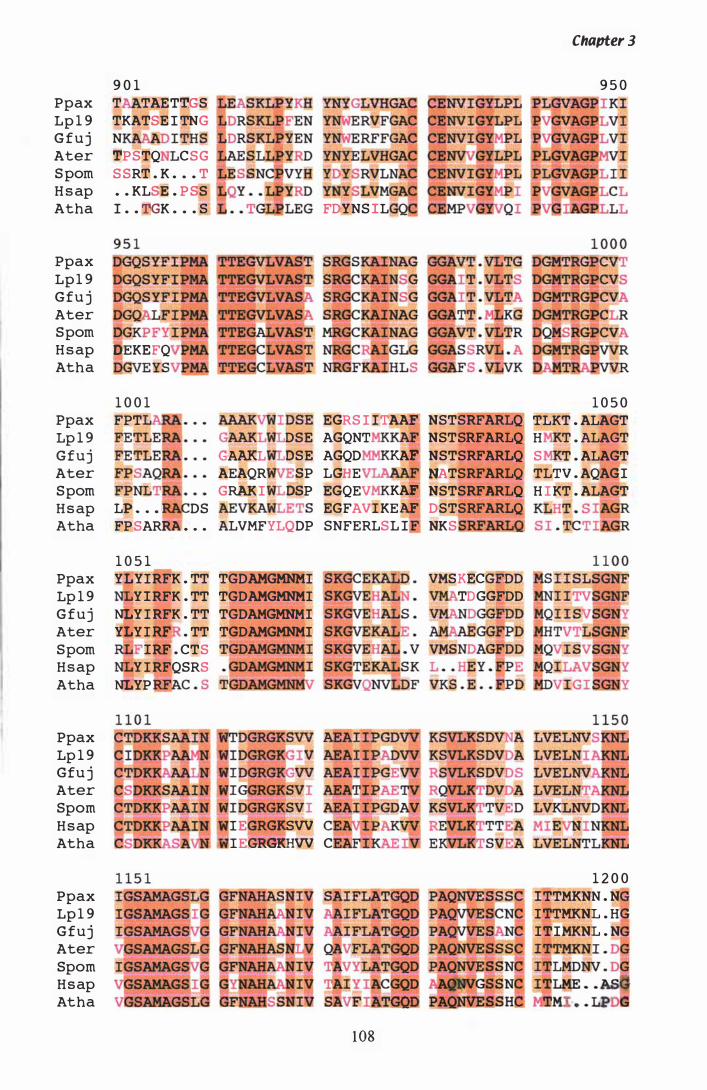
Ppax Lp 1 9 G f u j Ater S pom H s ap Atha
Ppax Lp 1 9 Gfu j Ater Spom H s ap Atha
Ppax Lp 1 9 Gfuj Ater Spom H s ap Atha
Ppax Lp 1 9 G f u j Ater Spom H s ap Atha
Ppax Lp 1 9 Gfu j Ater Spom H s ap Atha
Ppax Lp 1 9 Gfuj Ater Spom H s ap Atha
9 0 1 T TAETTGS TKATSE l TNG N Dl THS 'I'PSTQ CSG S SRT . K . . . T . . KLSE . P SS I . . TGK . . . S
�EKEFQ QGvE YSV
1 0 0 1 FPT ,
LP • . • 1 DS FPSARRA: . . .
1 0 5 1
YD YS RVLNA YNYpLVMGAI FDYNS lL
AAAKVWI DSE EGRS I ITAAF NSTSRFARLQ GAAKL DSE AGQNT NSTSRFARLQ GAAKL OSE AGQD RAF NSTSRFARLQ AEAQR SP LGHE NATSRFARLQ GRAKI DSP EGQEV KA!1 NSTSRFARLQ AEVKA EGFAVIKEAF. DSTSRFARLQ ALVMFYLQDP SNFERLSL I NKS
YLYIRFK . TT TGDAMGMNMI Y lRFK . TT TGDAMGMNMI
NLY I RFK . TT TGDAMGMNMI YLYI RFi • TT TGDAMGMNMI
VMSKECGFDD TDGGFDD
l� . CTS Y I RFQSRS
NLYP 'AC . S
lE RGJ(SW lEGRGKlHW C
1 08
Chapter 3
9 5 0
1 0 5 0 TLKT . ALAGT H . T . ALAGT S T . ALAGT TLTV . AQAGI H IKT . ALAGT KLHT . S GR
1 2 0 0 lTTMKNN . N I�TMKNL . HG ITIMKNL . N I TTMKN I . D

Ppax Lp 1 9 G f u j Ater Spom H s ap
1 2 0 1
Atha . • . 0
Ppax Lp 1 9 Gfu j Ater Spom H s ap Atha
Ppax Lp 1 9 G f u j Ater Spom H s ap Atha
1 3 0 1 • • P • • • • • • •
· . PAPPPMTP • • PG • . • • s . · • EYGAIRTP KKPATDALKS KKTA------
• • PSS • • • • Q
VS . . A . • AVG VSL . A . MTIA . SHOARLT . G
V . YVVILEHA VNSRVPGR------------
VN . R------
G LSM TSK-------
QOKSSKS . . AQQRSKR-
HDQCP NNVDER . RRY EYKNTYLRRK
---------- -------------------- - ---------- - - - - - - - - - - - - - - - - - - -
1 09
Chapter 3
1 3 0 0 . • AP . SS T · . APSR . $TT • . APSR . T · • KOAI • roT N . TP SSA NLQOLQGACT
· • . . SRD I G .
1 3 5 0 ----------- ---------
S EVKAIDE--
VPTLSCNLGR - --- - ---------- --- --- - - - - - - - - -

Chapter 3
(residues 684 to 1 1 39) is highly conserved and catalytically active residues were
predicted by comparison to other hmg genes. The complete nucleotide and polypeptide
sequence, including the position of the introns, transcription and translation start and
stop sites, is shown in figure 3 .7. Codon bias in the P. paxilli hmg gene is shown in
Table 3 .3 . A summary of gene features in P. paxilli is shown in Table 3 .7 .
3.1.8 Confirmation of introns
Total RNA was isolated from samples of mycelia, removed at a number of time points,
from a P. paxilli liquid culture (section 2 . 1 .2 and section 2 . 1 7). The RNA was reverse
transcribed into cDNA with random hexamer primers (section 2 . 1 8). RT-PCR was
performed on cDNA to characterise the structure of the hmg mRNA. The removal of
introns can be confirmed when products amplified from cDNA are compared to
products amplified from gDNA with the same primers. Intron 3 was identified when
cDNA, synthesised from RNA present in mycelia at 48 h, was amplified with primers
ET22 and ET9 (Table 2 .2). A routine PCR (section 2 . 1 1 .2) was performed and the
gDNA and cDNA products separated on a 2% (w/v) agarose gel (section 2 . 1 0 . 1 and
2 . 1 0 .3) (Figure 3 .9). The cDNA product was purified (section 2 .S .4) and sequenced
(section 2 . 1 2.3) to confirm the exact location of 5' donor and 3' acceptor sites. Intron 2
was identified and utilised as a control when the transcription start site was estimated
(section 3 . 1 .9) . Primer ET5 is downstream of intron 2 and when used in combination
with primers ET2, ET6 and ET8, results in a 7 1 bp size difference between the genomic
PCR product and the cDNA product (Figure 3 . 1 0, lanes 1 0- 1 2) . The product amplitled
with ETS and ET8 was purified from agarose gel (section 2.5 .4), and sequenced (section
2 . 1 2 . 3 ) to confirm the exact intronlexon boundary of intron 2. The size difference
between cDNA and gDNA products increases further when ETS is paired with ET1 2
(Figure 3 . 1 0, lanes 6-8), indicating that a second intron has been removed. The position
of this intron was confirmed when cDNA, synthesised from RNA present in mycelia at
60 h, was amplified with primers ET1 2 and ET1 5 (Figure 3 . 1 1 ) . The product was
cloned into a pGEM®-T vector (section 2 . 1 5) and sequenced (2. 1 2 .3) to show the exact
location of the intron one 5 ' donor and 3 ' acceptor sites. On closer examination, intron
consensus sequences were present in the Lp 1 9 hmg 5 'UTR sequence as well . RNA
isolated from Lp1 9 mycelia grown in liquid culture for 1 4 days (McGill, 2000 In Prep)
1 10
Figure 3.9 Confirmation of hmg intron 3
RT-PCR (section 2.20) was performed on a II I 00 dilution of cDNA from 48 h cultures,
using primers ET22 and ET9 (Table 2.2). Samples were separated on a 2% agarose gel
(section 2 . 1 0 . 1 and 2. 1 0 . 3 ) as follows. Lane 1 , 1 00bp ladder (BioLabs). Lane 2,
gDNA. Lane 3 , 48 h cDNA. Lane 4, water control . Sizes shown are in bp.
Figure 3. 10 Estimation of the transcription start site of hmg
RT-PCR (section 2.20) was performed on 1 1 100 dilutions of cDNA from 36 h and 60 h
cultures. Standard PCR reactions (section 2 . 1 1 .2) were performed with the 3 ' anchored
primer ET5 (Table 2 .2) and a set of 5' nested primers (ET4Bam, ETI 2, ET2, ET6 and
ET8), on templates; gDNA, 36 h cDNA, 60 h cDNA and a water control . Products
were separated on a 2% agarose gel (section 2. 1 0. 1 and 2 . 1 0. 3 ) as fol lows. Lanes 1 and
22, 1 00bp ladder (BRL). Lanes 2-5, ET4Bam and ET5 . Lanes 6-9, ET1 2 and ET5 .
Lanes 1 0- 1 3 . ET2 and ET5 . Lanes 14- 1 7, ET6 and ET5 . Lanes 1 8-2 1 , ET8 and ET5 .
Sizes shown are in bp.
1 1 1
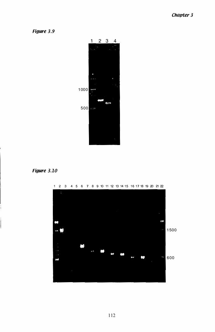
Chapter 3
Figure 3.9
1 2 3 4
1 00 0
500
Figure 3.1 0
1 2 3 4 5 6 7 8 9 10 1 1 12 13 14 1 5 1 6 1 7 18 19 20 21 22
1 50 0
6 0 0
1 1 2
Figure 3. 1 1 Confirmation of 5� UTR intron 1 in P. paxilli hmg
RT -PCR (section 2 .20) was performed on a 1 1 1 0 dilution of cDNA. from a 60 h culture,
with primers ET1 2 and ET1 5 in a standard PCR reaction (section 2 . 1 1 .2) . Samples
were separated on a 2% agarose gel (section 2. 1 0. 1 and 2 . 1 0 .3) as follows. Lanes I and
5, 1 00bp ladder (BioLabs). Lane 2, gONA. Lane 3 , 60 h cDNA. Lane 4, water control .
Sizes shown are in bp.
Figure 3.12 Confirmation of 5� UTR intron 1 in N. lolii Lp1 9
hmg
RT-PCR (section 2.20) was performed on a 1 1 1 0 dilution of cDNA from a 1 4 day
N lolii Lp1 9 culture. The standard PCR reactions (section 2 . 1 1 .2) were set up using
combinations of primers hmgeco, hmg2eco and hmgnco (Table 2.2) . Samples were
separated on a 2% agarose gel (section 2 . 1 0 . 1 and 2 . 1 0. 3 ) as follows. Lanes 1 and 1 3 ,
l kb+ ladder (BRL). Lane 2-6, (bmgeco and hmgnco) 1 4 day N. lolii Lp1 9 cDNA, 1 4
day N lolii Lp1 9 RNA (-RT), Reverse transcriptase water control , N lolii Lp1 9 gDNA,
and a water control . Lanes 7-9, (hmg2eco and hmgnco) 1 4 day N. lolii Lp1 9 cDNA,
N lolii Lp 1 9 gDNA, and a water control . Lanes 1 0- 1 2, (hmg29 and hmg30) 1 4 day
N lolii Lp1 9 cDNA, N. lolii Lp1 9 gONA, and a water control . Sizes shown are in bp.
1 13

Figure 3.1 1
Figure 3.12
1 650
300
1 00 0
5 0 0
1 2 3 4 5
1 2 3 4 5 6 7 8 9 1 0 1 1 1 2 1 3
1
Chapter 3
Fi9ure 3.13 Partial DNA sequence and deduced amino acid
sequence of N. loliiLp19 hmg
The 5 ' UTR Sequence of N. lolii Lp1 9 hmg was determined to -287 bp by Dobson
( 1 997), and further extended to - 1 1 80 bp by Lisa McMillan. The 5' UTR intron of 282
bp is shown in lower case, with the 5' and 3' splice junctions highlighted in red. The
putative 3 ' and 5' splices for alternative splicing are shown in orange. The single lariat
consensus sequence is shown in green. Primers used to determine the size of the 5 '
UTR are shown as purple lines. The translation start codon i s shown in blue.
1 1 5
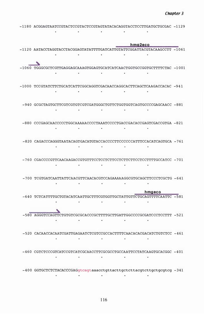
Chapter 3
- 1 1 8 0 ACGGAGTAATCCGTACTCCGTACTCCGTAGTATACACAGGTACCTCCTTGATGCTGCGAC - 1 1 2 9
bmg2eco - 1 1 2 0 AATACCTAGGTACCTACGGAGTATATTTTGATCATTGTATTCGGATTACGTACAAGCCTT - 1 0 6 1
� - 1 0 6 0 TGGGCGCTCGTTGAGGAGCAAAGTGGAGTGCATCATCAACTGGTGCCGGTGCTTTTCTAC - 1 0 0 1
- 1 0 0 0 TCCGTATCTTCTGCATCATTCGGCAGGTCGACAACCAGGCACTTCAGCTCAAGACCACAC -9 4 1
- 9 4 0 GCGCTAGTGCTTCGTCGTGTCGTCGATGGGCTGTTCTGGTGGTCAGTGCCCCGAGCAACC - 8 8 1
- 8 8 0 CCCGAGCAACCCCCTGGCAAAAACCCCTAAATCCCCTGACCGACACCGAGTCGACCGTGA - 8 2 1
- 8 2 0 CAGACCCAGGGTAATACAGTGACATGTACCACCCCTTCCCCCCATTTCCACATCAGTGCA - 7 6 1
- 7 6 0 CGACCCCGTTCAACAAGACCGTGTTTCCTCCTCTTCCTCTTCTTCCTCCTTTTGCCATCC - 7 0 1
- 7 0 0 TCGTGATCAATTATTCAACGTTCAACACGTCCAGAAAAAGGCGTGCAGCTTCCCTCGCTG - 6 4 1
b mpeco - 6 4 0 TCTCATTTTGCTGTACATCAATTGCTTTCGTGGTTGCTATTGTTCTGCAGTTTTCAATTC - 5 8 1
, - 5 8 0 AGGGTCCAGTTCTGTGTCGCGCACCCGCTTTTGCTTGATTGGCCCCGCGATCCCTCCTTT - 5 2 1
- 5 2 0 CACAACCACAATCGATTGAGAATCTCGTCCGCCACTTTTCAACACACGACATCTGTCTCC - 4 6 1
- 4 6 0 CGTCTCCCGTCATCCGTCATCGCAACCTTCGCGCCTGCCAATTCCTATCAAGTGCACGGC - 4 0 1
- 4 0 0 GGTGCTCTCTACACCCGAGgtcagtaaacctgttacttgctcttacgtcttgctgcgtcg - 3 4 1
1 1 6
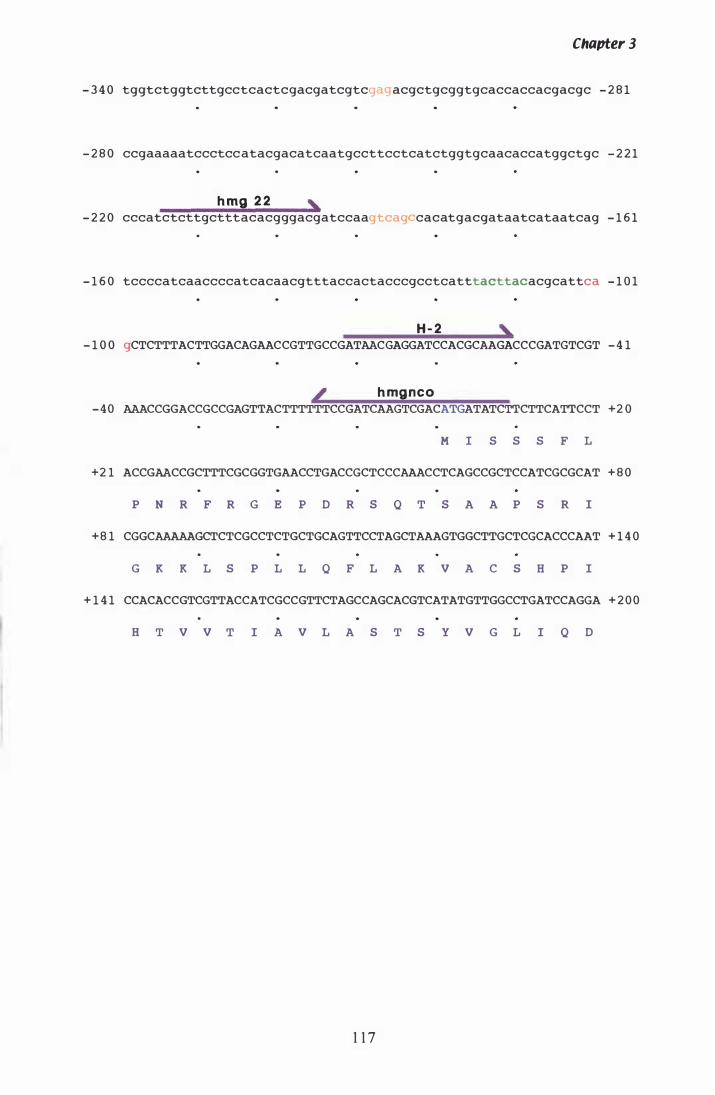
Chapter 3
- 3 4 0 tggtctggtcttgcctcactcgacgatcgtcg acgctgcggtgcaccaccacgacgc - 2 8 1
- 2 8 0 ccgaaaaatccctcc atacgacatcaatgccttcctcatctggtgcaacaccatggctgc - 2 2 1
h mg 2 2 , - 2 2 0 cccatctcttgctttacacgggacgatccaagtcaq cacatgacgataatcataatcag - 1 6 1
- 1 6 0 tccccatcaaccccatcacaacgtttaccactacccgcctcatttacttacacgcattca - 1 0 1
H - 2 , - 1 0 0 gCTCTTTACTTGGACAGAACCGTTGCCGATAACGAGGATCCACGCAAGACCCGATGTCGT - 4 1
� h mgnco - 4 0 AAACCGGACCGCCGAGTTACTTTTTTCCGATCAAGTCGACATGATATCTTCTTCATTCCT +2 0
M I S S S F L
+2 1 ACCGAACCGCTTTCGCGGTGAACCTGACCGCTCCCAAACCTCAGCCGCTCCATCGCGCAT + 8 0
P N R F R G E P 0 R S Q T S A A P S R I
+ 8 1 CGGCAAAAAGCTCTCGCCTCTGCTGCAGTTCCTAGCTAAAGTGGCTTGCTCGCACCCAAT + 1 4 0
G K K L S P L L Q F L A K V A C S H P I
+ 1 4 1 CCACACCGTCGTTACCATCGCCGTTCTAGCCAGCACGTCATATGTTGGCCTGATCCAGGA + 2 0 0
H T V V T I A V L A S T S Y V G L I Q 0
1 1 7

Chapter 3
was reverse transcribed into cDNA, using random hexamer primers. RT-PCR was
performed on this cDNA with a set of Lp1 9 hmg specific primers (hmgeco, hmg2eco
and hmgnco (Table 2.3» , and the products separated on a 2% (w/v) agarose gel (section
2 . 1 0. 1 and 2 . 1 0.3) . The size difference between gDNA and cDNA products confirms a
large intron or several small introns had been removed between primers hmgeco and
hmgnco (Figure 3 . 1 2, lanes 2-6). No cDNA products were amplified with primers
hmg2eco and hmgnco (lanes 7-9), indicating that the transcription start site is between
primers hmgeco and hmg2eco at this time point. Control reactions with primers hmg29
and hmg30 (lanes 1 0- 1 2) , amplified hmg cDNA in the coding region of the gene,
confirming that hmg mRNA was present in mycelia at thi s time point. The cDNA
product amplified with hmgnco and hmgeco was cloned into a pGEM®-T vector
(section 2 . 1 5) and sequenced (section 2 . 1 2 .3) to determine the number of introns
removed and the exact location of intron/exon boundaries. Sequencing revealed a
single intron of 282 bp had been removed (Figure 3 . 1 3 ) . It is possible this may
represent an alternatively spliced product encompassing the 5 ' donor site of one intron
and the 3 ' acceptor site of a downstream intron. Putative 3 ' acceptor and 5 ' donor sites
are present between the sites utilised in this instance.
3.1 .9 Estimation of transcription start site with RT�PCR
Copy DNA synthesised from the RNA present in mycelia at 36 h and 60 h, was
compared to determine if transcription of hmg mRNA is initiated at different sites
during paxilline biosynthesis. During early growth experiments, paxilline was detected
in mycelia after 60 h by HPLC (Figure 3 .25 A). Therefore, if a different hmg transcript
i s present during paxilline biosynthesis, it should be detected at 60 h. Primer ET5
(Table 2 .3) downstream of intron 2, was paired with a number of primers (ET4Bam,
ET1 2, ET2, ET6 and ET8) upstream of the translation start site. A routine PCR reaction
(section 2 . 1 1 .2) was performed and products separated on a 2% (w/v) agarose gel
( section 2 . 1 0 . 1 and 2 . 1 0 .3) (Figure 3 . 1 0) . Products were amplified only when the 5 '
primer sequence was present i n the cDNA. The gDNA control included with each
primer set confirms a successful PCR was achieved, and the size difference between
gDNA and cDNA products confirms the origin of amplified products . Both templates
produce a cDNA product with primer ET 1 2, but not with ET4Bam, indicating that
1 1 8
Chapter 3
transcription is initiated between these two primer sites at both time points. The level of
hmg expression at 36 h is lower than that seen at 60 h, even though the concentration of
the cDNA templates at both time points is equivalent. This was confirmed when tub-2
specific primers were used to amplify products of equal intensity from the same two
samples of cDNA (Figure 3 . 34).
3.1.10 Confirmation of transcription start site with 5� RACE
Although the approximate location of hmg transcription initiation had been estimated
with RT-PCR (section 3 . 1 .9), 5 ' RACE was used to pinpoint the exact location. As
described above, RNA iso lated from mycelia at two different time points was used to
determine if transcription of hmg mRNA is initiated at a different site during paxil line
biosynthesis. Analysis of expression levels from genes involved in paxil line
biosynthesis, performed after the initial estimation of the transcription start site, showed
transcription of pax genes as early as 36 h (section 3 .4) . Therefore, the time points
selected were 24 h and 60 h. RACE was performed as described in section 2 . 1 9 (Figure
3 . 1 4 A), and after the terminal transferase reaction had been completed several rounds
of nested PCR were performed. The final PCR products amplified with primers AUAP
and ET3 (Table 2.3) were separated on a 2% (w/v) agarose gel (section 2 . 1 0. 1 and
2 . 1 0 . 3 ) (Figure 3 . 1 5) . Only the 60 h RNA produced any detectable RACE products.
Gel stab purifications (section 2 . 1 1 .4) were performed on the two products visible in
Figure 3 . 1 5 . However, after second round amplification, only the smaller 3 1 4 bp
product was seen. The purified product was cleaned (section 2 . 1 1 .5 ) and cloned into a
pGEM®-T vector (section 2 . 1 5) for sequencing (section 2 . 1 2 .3) . The sequence data
obtained confirmed the position of the transcription start site that had previously been
estimated with RT-PCR (section 3 . 1 .9). Due to time constraints further attempts to
isolate the larger 60 h RNA RACE product, or a product from 24 h RNA were not
possible . It is possible that other transcription start sites for hmg exist, but only one,
shown in Figure 3 .7, has been identified with 5' RACE at this time.
1 19
Figure 3.14 Strategy for RACE
A) 5' RACE
The transcription start site was determined using the primers indicated (Table 2 .3)
8) 3' RACE
The transcription termination site was determined using the primers indicated (Table
2.3) ( section 2 . 1 9).
1 20
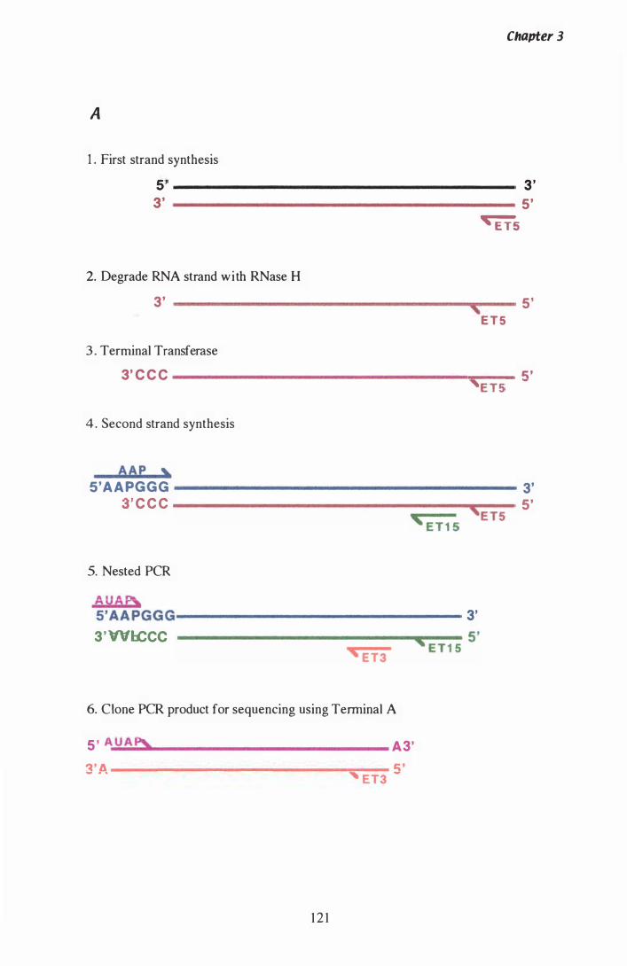
Chapter 3
A
1 . First strand synthesis
5' ------------------------------------------ 3' 3' ------------------------------------------ 5 '
�5
2. Degrade RNA strand with RNase H 3' ------------------------------------._---- 5' , ET5
3 . Terminal Transferase
3' C C C --------------__ -- 5' 'ET5
4 . Second strand synthesis
AAP , 5'AAPGGG ---------------- 3'
3' C C C 5' 'E T5 ' E T 1 5
5. Nested PCR
AUAp\, 5'AAPGGG-------------- 3' 3'WlCCC ' E T 1 5 5'
' E T3
6. Clone PeR product for sequencing using Tenninal A
5 ' A U Ap)" A3' 3'A � ' -----------------------------'�E-T
-3 -
1 2 1
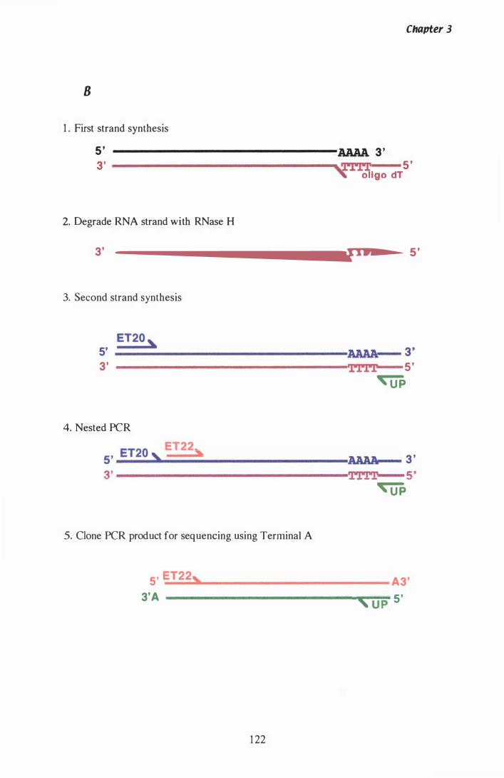
Chapter 3
8
1 . First strand synthesis
5 ' AAAA 3 ' 3' --------------------------------��-rrl 5 '
, ollgo dT
2. Degrade RNA strand with RNase H
3'
--------------------------------�T·�r�r�X----
5 '
3. Second strand synthesis
ET20 , 5' ====�------------------� 3' 3' i'PPl 5'
4. Nested PeR
5' ET20 , ET22,
�
3 ' 3'-----------------------------------� 5'
�
5. Clone PeR product for sequencing using Terminal A
5' ET22, A3'
' U P 5' 3'A ----------------------------__ ��
1 22
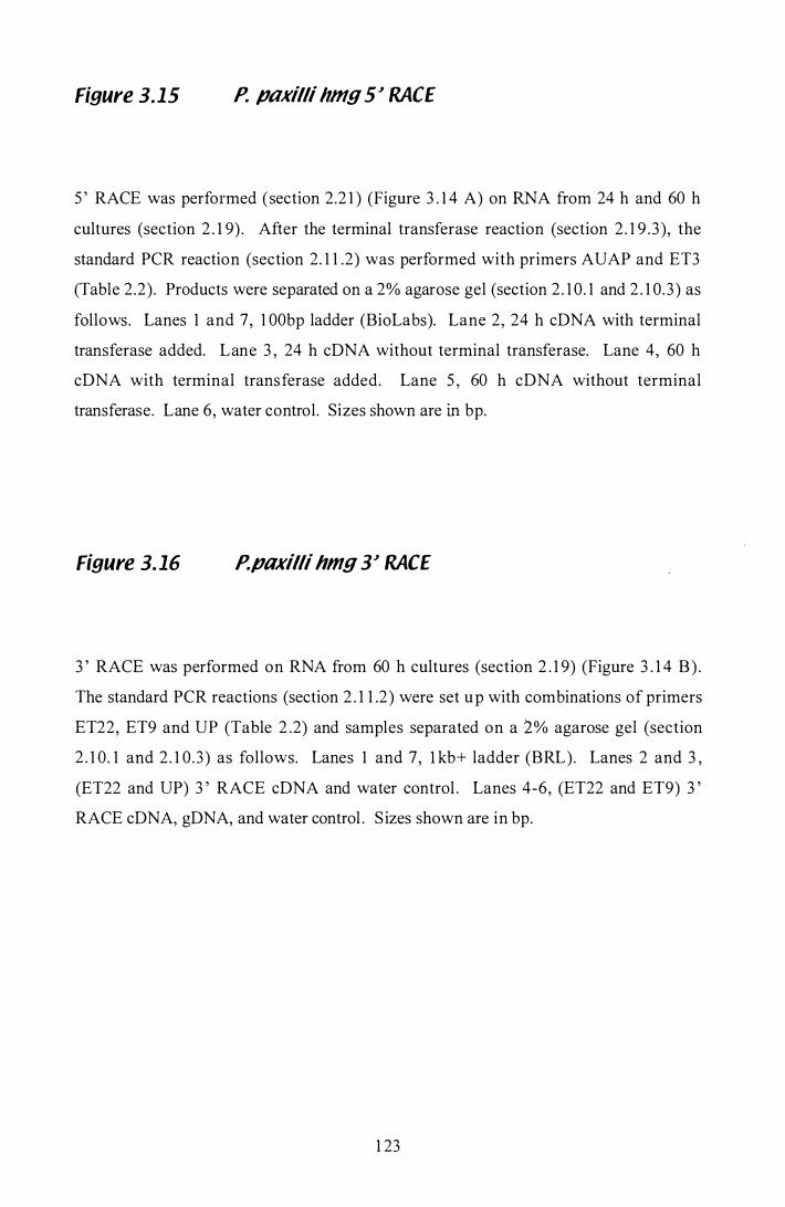
Figure 3.15 P. pOKilli hmg 5:1 RACE
5 ' RACE was performed (section 2.2 1 ) (Figure 3 . 1 4 A) on RNA from 24 h and 60 h
cultures (section 2 . 1 9) . After the terminal transferase reaction (section 2 . 1 9 .3 ), the
standard PCR reaction (section 2. 1 1 .2) was performed with primers AUAP and ET3
(Table 2.2). Products were separated on a 2% agarose gel (section 2 . 1 0. 1 and 2 . 1 0.3 ) as
follows. Lanes 1 and 7, 1 00bp ladder (BioLabs). Lane 2, 24 h cDNA with terminal
transferase added. Lane 3 , 24 h cDNA without terminal transferase. Lane 4, 60 h
cDNA with terminal transferase added. Lane 5 , 60 h cDNA without terminal
transferase. Lane 6, water control. Sizes shown are in bp.
Figure 3. 16 P.paxilli hmg 3:1 RACE
3 ' RACE was performed on RNA from 60 h cultures (section 2 . 1 9) (Figure 3 . 1 4 B) .
The standard PCR reactions (section 2 . 1 1 .2) were set up with combinations of primers
ET22, ET9 and UP (Table 2 .2) and samples separated on a 2% agarose gel (section
2 . 1 0 . 1 and 2 . 1 0 .3) as follows. Lanes 1 and 7, l kb+ ladder (BRL). Lanes 2 and 3 ,
(ET22 and UP) 3 ' RACE cDNA and water control . Lanes 4-6, (ET22 and ET9) 3 '
RACE cDNA, gDNA, and water control . S izes shown are in bp.
1 23
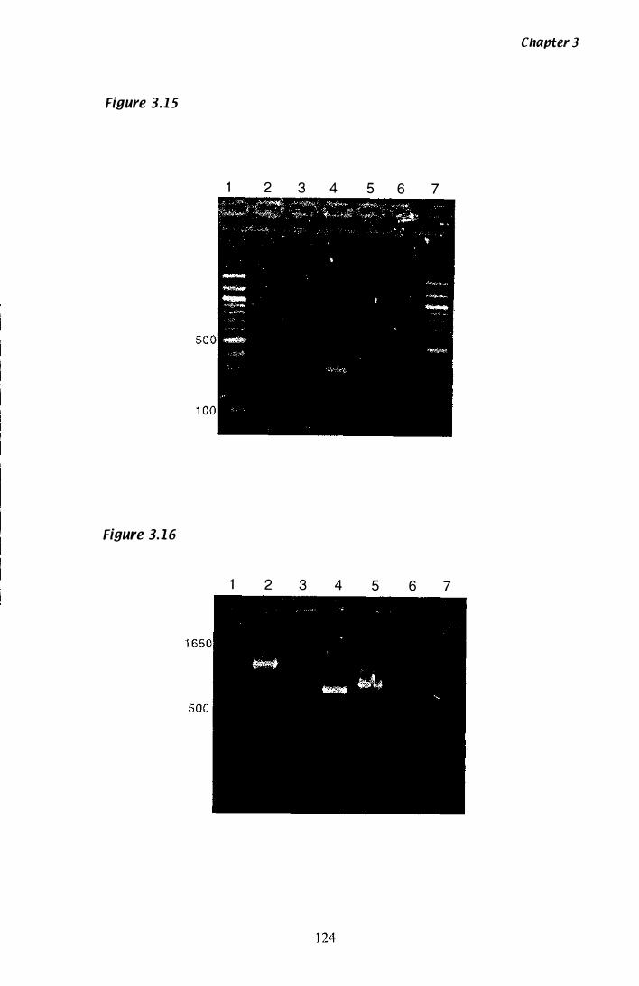
Figure 3.15
1
Figure 3.1 6
1
2 3 4
2 3 4
5 6 7
5 6 7
Chapter 3
Chapter 3
3.1.1 1 Confirmation of the transcription stop site with 33 RACE
Many filamentous fungal genes are transcribed past the signal for polyadenylation (Gurr
et al., 1 9 87). The signal for polyadenylation in filamentous fungal genes is frequently
the consensus 5 '-AAUAAA-3 ' . The closest match to this sequence in the P. paxilli hmg
3 ' UTR, is the motif 5'-ATAAAA-3' , found 483 bp downstream of the translation
termination codon (Figure 3 .7). The RACE technique was used to determine the 3' end
of transcription using RNA isolated from mycelia grown in liquid culture for 60 h. The
first round of 3 ' RACE amplification is non specific, and uses an oligo dT primer
(Table 2 .3 ) to reverse transcribe a pool of mRNA (Figure 3 . 1 4 B) . As 3 ' RACE was
being used to determine the transcription stop site of a number of P. paxilli genes, a
single first round reaction was performed by Lisa McMillan and stored at -2We. Second round amplification was then performed twice with a set of nested gene specific
5' primers and the UP primer. Amplification with gene specific internal primers ET22
and ET9, and separation of the products on a 2% (w/v) agarose gel (section 2 . 1 0 . l and
2 . 1 0 . 3 ) confirmed the specificity of the product (Figure 3 . 1 6). The single product
shown (lane 2), was purified (section 2 . 1 1 .5) and cloned (section 2 . 1 5) into a pGEM®
T EASY vector for sequencing (section 2 . 1 2 .3). The 3 'UTR region was 563 bp long
(Figure 3 .7) .
3.1 .12 Chromosomal location of P. paxilli hmg
The chromosomal location of hmg was identified by probing a nylon membrane (section
2 . 1 3 ) , containing a contour-clamped homogenous e lectri c field (CHEF)-gel
electrophoresis separation (Figure 3 . 1 7 A) previously performed by Carolyn Young,
with a PCR product of hmg amplified with ET5 and ET6. Chromosomes of P. paxilli
were separated by CHEF electrophoresis, and transferred to nylon membrane by
Southern blotting. The conditions for CHEF separation are described in the legend of
F igure 6, in the paper by Young et al. ( 1 998). This gel included several Pax- mutants of
P. paxilli as well as wild type, but in all cases, the hmg probe hybridised to chromosome
Vb (Figure 3 . 1 7 B).
1 25
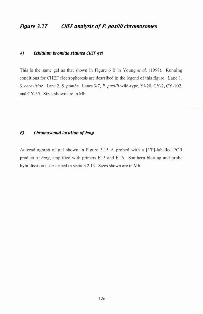
Figure 3. 1 7 CHEF analysis of P. ptlxilli chromosomes
A) Ethidium bromide stained CHEF gel
This is the same gel as that shown in Figure 6 B in Young et al. ( 1 998). Running
conditions for CHEF e lectrophoresis are described in the legend of this figure. Lane 1 ,
S. cerevisiae . Lane 2, S. pombe. Lanes 3-7, P. paxilli wild-type, YI-20, CY-2, CY- 1 02,
and CY-35 . Sizes shown are in Mb.
B) Chromosomal location of hmg
Autoradiograph of gel shown in Figure 3 . 1 5 A probed with a [32P] -label led PCR
product of hmg, amplified with primers ET5 and ET6. Southern blotting and probe
hybridisation is described in section 2. 1 3 . Sizes shown are in Mb.
1 26
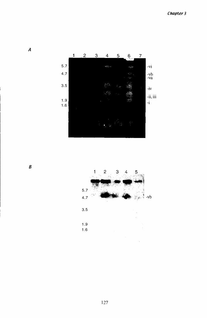
A
8
5 . 7
4 .7
3 . 5
1 . 9 1 . 6
1 2
5 . 7
4 . 7
3 . 5
1 . 9 1 . 6
3 4
1
Chapter 3
-VI
-vb -va
-IV
-ii, iii -1
-vb
3.2 Isolation ana Characterisation of P. pllKilli tub-2
3.2.1 Preparation of tub-2 genomic probe from pBT6
Chapter 3
A bacterial culture containing plasmid pBT6 (Table 2 . 1 Appendix A 1 .2) was plated for
single colonies (section 2 . 1 .2) and DNA isolated using an alkaline lysis based mini-prep
kit (section 2 .5 .5) . After quantification by fluorometry (section 2.6.2), the plasmid was
cut with EcoRI (section 2.7) and separated on a 0.7% (w/v) agarose gel (section 2. 1 0. 1
and 2 . 1 0.3) . The 1 .8 kb fragment, containing part of the Neurospora crassa �-tubulin
gene, was i solated from the gel (section 2.5 .4), purified with phenol/chloroform (section
2.8) and precipitated with ethanol (section 2.9) .
3.2.2 Confirmation a single tub-2 exists in the P. pl1xilli genome
The P. paxilli genomic blot (Figure 3 . 1 8 A) originally probed with the 359 bp hmg
product was stripped (section 2 . 1 3 .4), and re-probed with the [a-32P]-labeIled 1 .8 kb
EcoRI fragment of pBT6 as described in section 2 . 1 3 (Figure 3 . 1 8 B). The tub-2 gene
is extremely conserved, therefore, the conditions for heterologous probing with the
N crassa tub-2 were identical to the homologous probing of this blot with the unique
hmg discussed in section 3 . 1 .4
Table 3.4 Size of P. pl1xilli gDNA bands hybridising with 1 .8 kb tub-2 probe
BamHI BglII
6.2 20.5 5 .6
Size of Bands (kb)
Restriction enzyme
EcoRI HindUI PstI SalI SstI
1 4. 1 3 .2 7 .7 1 3 .9 0.7 2 .5
1 28
Figure 3.18 Southern analysis of tub-2 in the P. paxilli
genome
A) Restriction endonuclease digestion of P. paxilli gONA
This is the same gel seen in Figure 3 . 3 A. P. paxilli genomic DNA (2 Jlg) (section
2.5 . 1 ) was digested with restriction endonucleases (section 2 .7) . Each reaction was
separated on a 0 .7% agarose gel (section 2 . 1 0 .2 and 2 . 1 0 .3 ) as follows. Lane 1 ,
tJHindIII . Lanes 2-8, P. paxilli gDNA cut with BamHI, BglII , EcoRI, HindIII, PstI,
SaD, and SstI . Lane 9, lkb ladder (BRL). Sizes shown are in kb.
B) Confirmation that sequence heterologous to the 1 .8kb EeoRI fragment
of pBT6 is part of the P. paxilli genome
Autoradiograph, showing hybridisation of N. crassa tub-2 to the P. pax ill i genome.
Ethidium bromide stained gel seen in Figure 3 .3 A was Southern blotted and probed
with the [32P]-labelled 1 . 8 kb EcoRl fragment of pBT6 (section 2 . 1 3). Sizes shown are
in kb.
129
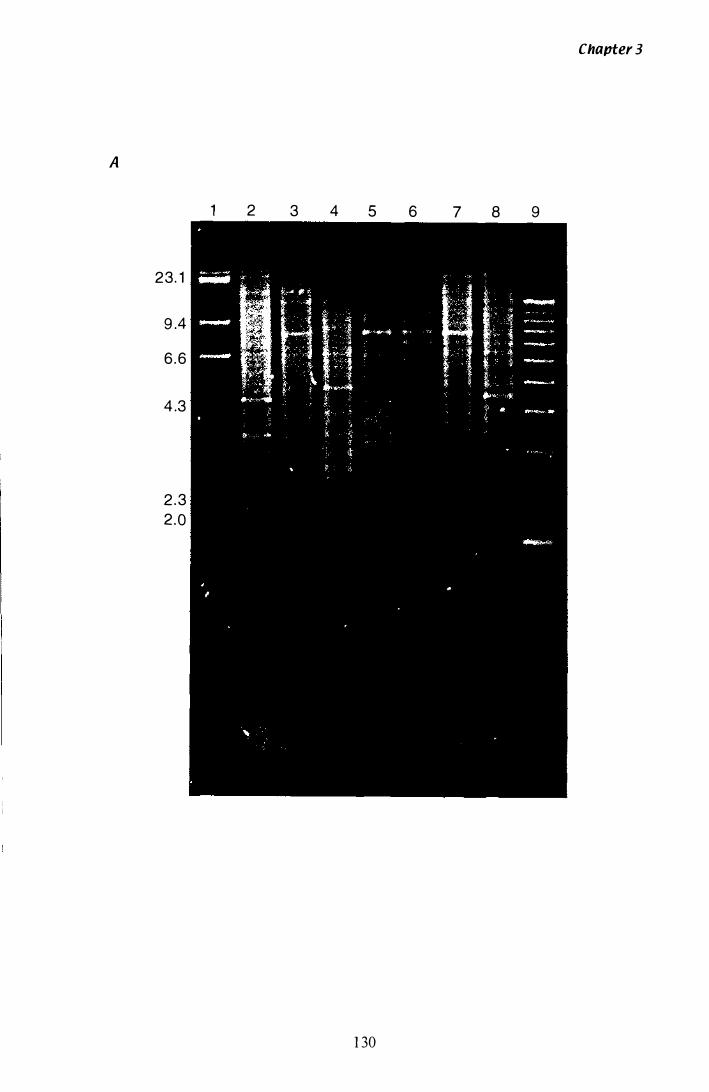
A
23. 1
9 .4
6 .6
4 .3
2 . 3
2 .0
1 2 3 4 5 6
1 30
Chapter 3
7 8 9
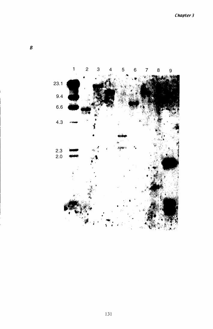
Chapter 3
8
1 2 3 4 5 6 7 8 9
23 . 1
9 .4
6 .6
4 .3 �
2 3 � .
2.0
1 3 1

Chavter 3
The single bands hybridising in most lanes confirm the presence of a single copy of
tub-2 in the P. paxilli genome. The multiple bands present in the BamHI and HindIII
digests (Figure 3 . 1 6 B, lanes 2 and 5) result from the presence of these restriction
enzyme sites within the tub-2 sequence.
3.Z.3 Library screening
The first round library filters used to isolate hmg were stripped (section 2 . 1 3 .4) and re
probed with the 1 .8 kb EcoRI fragment of pBT6 (section 2 . l 4.5) . Ten positive clones
(AET20, AET2 1 , AET22, AET23 , AET24, AET25, AET26, AET27, AET28, and AET29)
were isolated (section 2 . 1 4 .6) and plated for second round screening (section 2 . 1 4.8) .
Only six of these clones (AET22, AET23, AET24, AET25 , AET27, and AET29) were
positive after second round screening. Duplicates clones were isolated (section 2 . 1 4.6)
and one of these was plated for further screening. All six clones were positive after
third round screening, and duplicate samples of each clone were isolated and stored at
4 °C .
3.Z.'I Physical mapping of tub-2 A clones
DNA was isolated (section 2 . 5 .2) from each clone, digested with SstI (section 2 .7) and
separated by gel electrophoresis (section 2 . 1 0 .2 and 2 . 1 0 . 3 ) (Figure 3 . 1 9 A) . A
Southern blot of this gel was probed with the 1 .8 kb EcoRI fragment of pBT6 (section
2 . 1 3) (Figure 3 . l 9 B). Band sizes and patterns of hybridisation are represented in Table
3 .5 . A SstI restriction map based on these sizes was produced (Figure 3 .20), and used to
determine the best clone to continue sequencing of tub-2.
3.Z.5 Sub-cloning AfTZZ into a sequencing vector
Clone AET22 was chosen for sub-cloning and sequencing, as it was the most central of
the clones with respect to probe hybridisation. DNA from this clone was digested with
SstI, purified with phenol!chloroform (section 2 .8) and precipitated with ethanol
1 32
Figure 3. 1 9 Southern analysis of tub-2 lambaa clones
A) Restriction endonuclease ct;gestion of tub-2lambda clones with SstI
DNA (�500 ng) from medium scale A preps (section 2.5 .2) was digested with SstI
(section 2 .7) and separated on a 0 .7% agarose gel (section 2. 1 0.2 and 2 . 1 0.3) as follows.
Lane 1 , AlHindIII . Lanes 2-7, SstI digestion of, AET22, AET23 , AET24, AET25 , AET27
and AE29. Lane 8, 1 kb ladder (BRL). Sizes shown are in kb.
B) Mapping the position of tub2 on lambda clones
Autoradiograph showing hybridisation of N. crassa tub-2 to the digested A clones.
Ethidium bromide stained gel seen if Figure 3 .4 A was Southern blotted and probed
with [32P]-labeUed 1 .8kb EeoRI fragment of pBT6 (section 2 . 1 3) . Sizes shown are in
kb
1 3 3
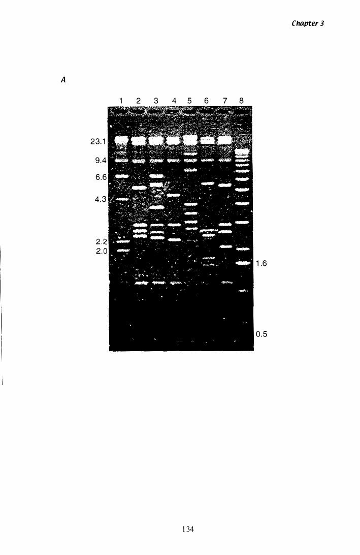
A
23. 1
9 .4
6 .6
4 .3
1 2 3 4 5 6 7 8
1 34
Chapter 3
1 .6
0 . 5
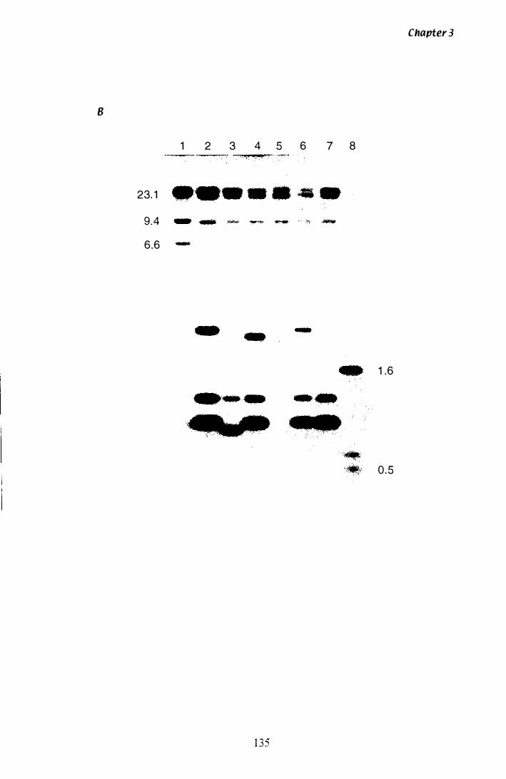
B
1 2 3 4 5 6 7 8
23. 1
9.4
6 .6 ..
et
Chapter 3
1 .6
0 .5
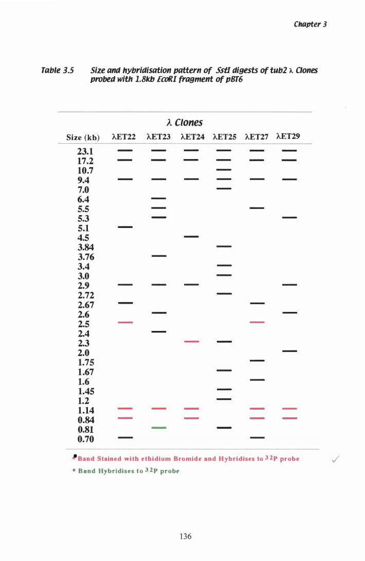
Chapter 3
Table 3.5 Size and hybridisation pattern of SstI digests of tubZ A Dones probed with 1.8kb EeaR! fragment of pBT6
A Clones
Size (kb) AET22 AET23 AET24 AET2S AET27 AET29
23.1 - -
17.2 - -
10.7 9.4 - - -
7.0 -
6.4 5.5 5.3 - -
5.1 -
4.5 -
3.84 3.76 3.4 -
3.0 2.9 -
2.72 2.67 -
2.6 -
2.5 -
2.4 -
2.3 -
2.0 -
1.75 -
1.67 1.6 -
1.45 -
1.2 -
1.14 - - - - -
0.84 - - - -
O.Sl - -
0.70
Band Stained with ethidium Bromide and Hybridises to 3 2p probe
* Band Hybridises to 3 2 p probe
1 36
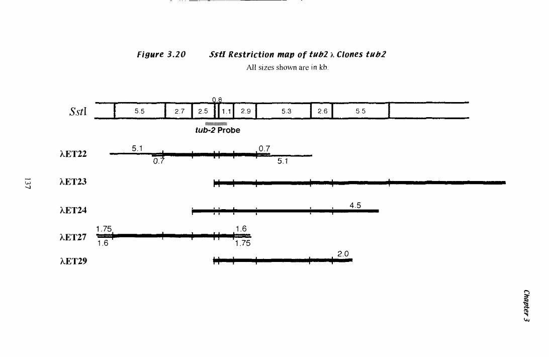
....... W -....l
figure 3.20 Sstl Restriction map of tub2 A Clones tub2 All sizes shmvn are
_ __ QJL __ _
SstI 1 --- 5 .5 I 2 .7 I 2 .5 1 1 1 . 1 1 2 .9 I 5 .3 I 2 .6 1 --�--�----I �-----
AET22
AET23
-Probe
5 . 1 --'-----_ _ • , L-..._.L......- • 0 . 7 (f.f
1 1
i .. _�_ 1__ 1--_____ I 4 . 5 AET24 .--- - -----�,----- - ---
1 75 1 .6 AET27 1 '.6 I " '1 .7; AET29 1 1 1
2.0 I I
� � � 1..1..1
Chapter 3
(section 2.9). The purified DNA was ligated into pUC 1 1 8 and transformed into E. coli
XL- 1 (section 2 . 1 5). Due to the size of the tub-2 gene, there was no need to reduce sub
clones further with restriction enzymes as was the case with sequencing of hmg (section
3 . 1 . 7) . Only a few novel primers (Table 2 .3) were required to complete double stranded
sequencing. The sub-clones used for sequencing are l isted in Table 2 . 1 . The restriction
map of P. paxilli tub-2 locus and relative position of the sub-clones used for sequencing
is shown in Figure 3 .2 1 .
3.2.6 Sequencing AET22 sub-clones
P. paxilli tub-2 was sequenced and analysed as described in section 3 . 1 . 7. Junctions
between individual fragments were sequenced directly from AET22 and AET27 with the
appropriate primers. Templates used to assemble the tub-2 contig, are displayed in
Appendix A.2.4 with the Sequencher™ software. Sequence analysis and polypeptide
sequence of tub-2 translation revealed a 447 amino acid protein comprised of 7 ORF.
Open reading frame 1 extends from + 1 to + 1 2 bp, 0 RF 2 extends from + 1 20 to + 1 40
bp, ORF 3 extends from +26 1 to + 286 bp, ORF 4 extends from +355 to +38 6 bp, ORF
5 extends from +463 to +5 1 7 bp, ORF 6 extends from +588 to + 1044 bp, and ORF 7
extend from + 1 1 07 to a termination codon at + 1 896 bp. This overall gene structure i s
depicted in Figure 3 .2 1 . The complete nucleotide and polypeptide sequence, including
the position of introns and translation start and stop sites, is shown in Figure 3 .22. The
translation start and stop sites were determined by homology to other tub-2 genes and
sequence surrounding the translation start codon (Ballance, 1 986; Kozak, 1 984). Codon
bias in P. paxilli tub-2 is shown in Table 3 .6. A summary of gene features in P. paxilli
is shown in Table 3 .7.
3.2. 7 Confirmations of introns
As described previously (section 3 . 1 . 8), cDNA was synthesised from RNA isolated
from mycelia at different time points. The presence of introns was determined by
comparing products amplified with the same primers fro m cDNA and gDNA. The
standard PCR reaction (section 2 . 1 1 .2) was set up with primers Etub5 and Etub6 (Table
1 38
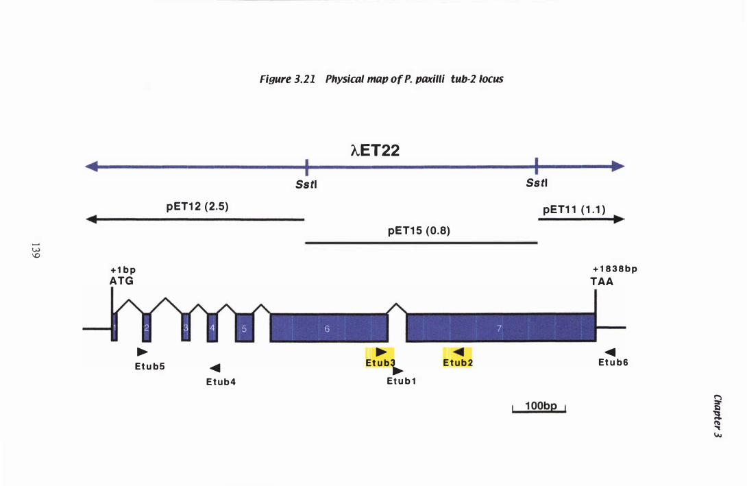
....... UJ \0
Figure 3.21 Physical map of P. paxilll tub-2 locus
AET22 � I I .
...... ........
+ 1 b p
ATG
� Et u b 5
pET1 2 (2 .5)
...... E t u b 4
�� ��
pET1 1 ( 1 . 1 ) ....
pET1 5 (0.8)
� Et u b�
Et u b 1
...... E t u b 2
1 00bp
.....
+ 1 8 3 8 b p
TAA
...... Et u b 6
g
t I.A.I
Figure 3.22 DNA sequence and deduced amino acid sequence
of P. paxilli tub·2
Sequence of P. paxilli tub-2 was determined as described in section 3 . I .7. The 2220 bp
of sequence isolated is shown here, including 305 bp of 5' UTR (-305 to - 1 ), 1 839 bp of
coding sequence (+ 1 to + 1 838), and 76 bp of 3 ' UTR (+ 1 839 to 1 9 1 5). The predicted
447 amino acids are shown beneath their respective codons. Introns are shown in lower
case, with 5 ' and 3 ' splice junctions, and the lariat consensus sequence highlighted in
green. Where present, intronic polypyrimidine tracts are highlighted in green as welL
The translation start and stop codons are highlighted in blue, with the Kozak consensus
sequence surrounding the translation start codon highlighted in light blue Primers used
for sequencing and gene analysis are shown as purple lines.
140
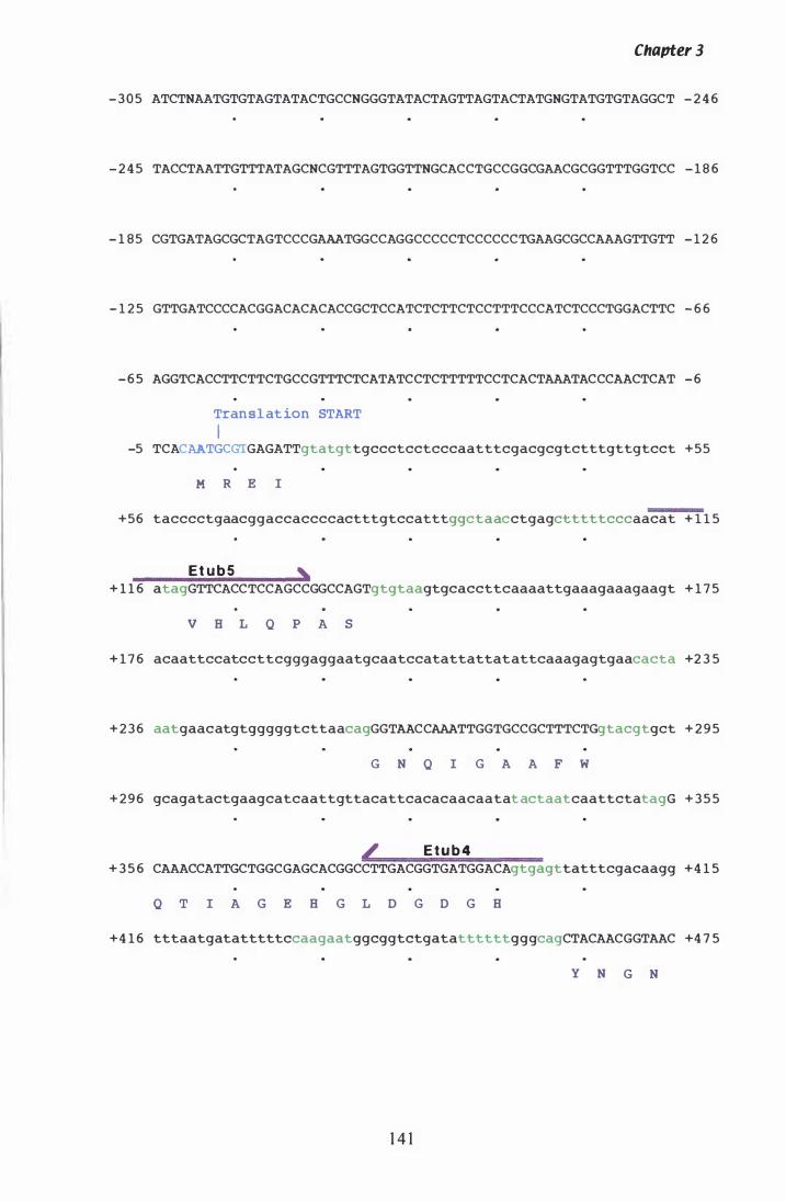
Chapter 3
- 3 0 5 ATCTNAATGTGTAGTATACTGCCNGGGTATACTAGTTAGTACTATGNGTATGTGTAGGCT - 2 4 6
- 2 4 5 TACCTAATTGTTTATAGCNCGTTTAGTGGTTNGCACCTGCCGGCGAACGCGGTTTGGTCC - 1 8 6
- 1 85 CGTGATAGCGCTAGTCCCGAAATGGCCAGGCCCCCTCCCCCCTGAAGCGCCAAAGTTGTT - 1 2 6
- 1 2 5 GTTGATCCCCACGGACACACACCGCTCCATCTCTTCTCCTTTCCCATCTCCCTGGACTTC - 6 6
- 6 5 AGGTCACCTTCTTCTGCCGTTTCTCATATCCTCTTTTTCCTCACTAAATACCCAACTCAT - 6
Translat ion START
I -5 TCACAATGCGTGAGATTgtatgttgccctcctcccaatttcgacgcgtctttgttgtcct +55
M R E I
+56 tacccctgaacggaccaccccactttgtccatttggctaacctgagctttttcccaacat + 1 1 5
Et u b 5 , + 1 1 6 atagGTTCACCTCCAGCCGGCCAGTgtgtaagtgcaccttcaaaattgaaagaaagaagt + 1 7 5
V B L Q P A S
+ 1 7 6 acaattccatccttcgggaggaatgcaatccatattattatattcaaagagtgaacacta +2 3 5
+ 2 3 6 aatgaacatgtgggggtcttaacagGGTAACCAAATTGGTGCCGCTTTCTGgtacgtgct + 2 9 5
G N Q I G A A F W
+ 2 9 6 gcagatactgaagcatcaattgttacattcacacaacaatat actaatcaattctatagG + 3 5 5
4' E t u b 4 + 3 5 6 CAAACCATTGCTGGCGAGCACGGCCTTGACGGTGATGGACAgtgagttatttcgacaagg +4 1 5
Q T I A G E B G L D G D G B
+4 1 6 tttaatgatatttttccaagaatggcggtctgatattttttgggcagCTACAACGGTAAC +4 7 5
1 4 1
Y N G N
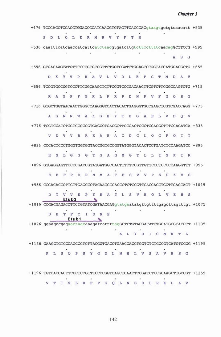
Chapter 3
+4 7 6 TCCGACCTCCAGCTGGAGCGCATGAACGTCTACTTCACCCACgtaagtgctgtcaacatt + 5 3 5
S D L Q L E R M N V Y F T H
+ 5 3 6 caatttcatcaaccatcattcatctaacgtgatcttgtcttccttttcaacagGCTTCCG +5 9 5
A S G
+ 5 9 6 GTGACAAGTATGTTCCCCGTGCCGTTCTGGTCGATCTGGAGCCCGGTACCATGGACGCTG + 6 5 5
D K Y V P R A V L V 0 L E P G T M D A V
+ 6 5 6 TCCGTGCCGGTCCCTTCGGCAAGCTCTTCCGTCCCGACAACTTCGTCTTCGGCCAGTCTG + 7 1 5
•
R A G P F G K L F R P D N F V F G 0 S G
+ 7 1 6 GTGCTGGTAACAACTGGGCCAAGGGTCACTACACTGAGGGTGCCGAGCTCGTCGACCAGG +7 7 5
A G N N W A K G H Y T E G A E L V 0 Q v
+ 7 7 6 TCGTCGATGTCGTCCGCCGTGAGGCTGAGGCTTGCGACTGCCTCCAGGGTTTCCAGATCA + 8 3 5
V 0 v V R R E A E A C D C L Q G F 0 I T
+ 8 3 6 CCCACTCCCTGGGTGGTGGTACCGGTGCCGGTATGGGTACACTCCTGATCTCCAAGATCC + 8 9 5
H S L G G G T G A G M G T L L I S K I R
+ 8 9 6 GTGAGGAGTTCCCCGACCGTATGATGGCCACTTTCTCCGTTGTTCCCTCCCCCAAGGTTT + 9 5 5
E E F P 0 R M M A T F S V v p S P K V S
+ 9 5 6 CCGACACCGTTGTTGAGCCCTACAACGCCACCCTCTCCGTTCACCAGCTGGTTGAGCACT + 1 0 1 5
o T V V E P Y N A T L S V H 0 L V E H S
Etub3 , + 1 0 1 6 CCGACGAGACCTTCTGTATCGATAACGA tatgaatatgttgttttgagcttagtttgt + 1 0 7 5
D E T F C I D N E
Etub1 , + 1 0 7 6 ggaagccgagaactaacaaagatcattttagGCTCTGTACGACATCTGCATGCGCACCCT + 1 1 3 5
A L Y D I C M R T L
+ 1 1 3 6 GAAGCTGTCCCAGCCCTCTTACGGTGACCTGAACCACCTGGTCTCTGCCGTCATGTCCGG + 1 1 9 5
K L S O P S Y G 0 L N H L V S A V M S G
+ 1 1 9 6 TGTCACCACTTCCCTCCGTTTCCCCGGTCAGCTCAACTCCGATCTCCGCAAGCTTGCCGT + 1 2 5 5
V T T S L R F P G 0 L N S D L R K L A V
1 42
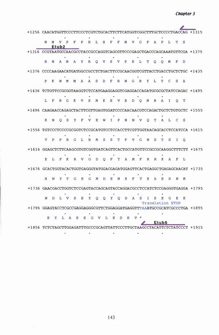
Chapter 3
+ 1 2 5 6 CAACATGGTTCCCTTCCCTCGTCTGCACTTCTTCATGGTCGGCTTTGCTCCCCTGACCAG + 1 3 1 5
N M V P F P R L H F F M V G F A P L T S
Etub2 + 1 3 1 6 CCGTAATGCCAACGCCTACCGCCAGGTCAGCGTTCCCGAGCTGACCCAGCAAATGTTCGA + 1 3 7 5
R N A N A Y R Q V S v P E L T Q Q M F D
+ 1 3 7 6 CCCCAAGAACATGATGGCCGCCTCTGACTTCCGCAACGGTCGTTACCTGACCTGCTCTGC + 1 4 3 5
P K N M M A A S D F R N G R Y L T C S A
+ 1 4 3 6 TCTGTTCCGCGGTAAGGTCTCCATGAAGGAGGTCGAGGACCAGATGCGCGCTATCCAGAC + 1 4 9 5
L F R G K V S M K E V E D Q M R A I Q T
+ 1 4 9 6 CAAGAACCAGAGCTACTTCGTTGAGTGGATCCCCAACAACGTCCAGACTGCTCTGTGCTC + 1 5 5 5
K N Q S Y F V E W I P N N V Q T A L C S
+ 1 5 5 6 TGTCCCTCCCCGCGGTCTCCGCATGTCCTCCACCTTCGTTGGTAACAGCACCTCCATCCA + 1 6 1 5
v P P R G L R M S S T F V G N S T S I Q
+ 1 6 1 6 GGAGCTCTTCAAGCGTGTCGGTGATCAGTTCACTGCCATGTTCCGCCGCAAGGCTTTCTT + 1 6 7 5
E L F K R V G D Q F T A M F R R K A F L
+ 1 6 7 6 GCACTGGTACACTGGTGAGGGTATGGACGAGATGGAGTTCACTGAGGCTGAGAGCAACAT + 1 7 3 5
H W Y T G E G M D E M E F T E A E S N M
+ 1 7 3 6 GAACGACCTGGTCTCCGAGTACCAGCAGTACCAGGACGCCTCCATCTCCGAGGGTGAGGA + 1 7 9 5
N D L V S E Y Q Q Y Q D A S I S E G E E
Translation STOP
+ 1 7 9 6 GGAGTACCTCGCCGAGGAGGGCGTTCTGGAGGATGAGGTTTAAATGCCGCATCGCCCTGA + 1 8 5 5
E Y L A E E G V L E D E V *
L Etub§ + 1 8 5 6 TCTCTAGCTTGGAGATTTGCCCGCAGTTATTCCCTTGCTAAGCCTACATTCTCTATCCCT + 1 9 1 5
1 43
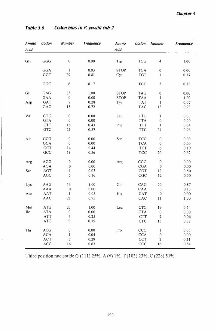
Chapter 3
Table 3.6 Coaon bias in P. pnxilli tub-2
Amino Coaon Number Frequency Amino Coaon Number Frequency
Acia Acia
G ly GGG 0 0.00 Trp TGG 4 1 .00
GGA 1 0.03 STOP TGA 0 0 .00 GGT 29 0 . 8 1 Cys TGT 0 . 1 7
GGC 6 0. 1 7 TGC 5 0 .83
Glu GAG 33 1 .00 STOP TAG 0 0.00 GAA 0 0.00 STOP TAA 1 1 .00
Asp GAT 7 0.28 Tyr TAT 1 0.07 GAC 1 8 0 .72 TAC 1 3 0.93
Val GTG 0 0 .00 Leu TIG 1 0 .0 3 GTA 0 0.00 TIA 0 0 .00 GIT 1 6 0.43 Phe TIT 1 0.04 GTC 2 1 0.57 TIC 24 0 .96
A la GCG 0 0.00 Ser TCG 0 0 .00 GCA 0 0.00 TCA 0 0 .00 GCT 1 4 0.44 TCT 6 0 . 1 9 GCC 1 8 0.56 TCC 20 0 .62
Arg AGG 0 0.00 Arg CGG 0 0 .00 AGA 0 0.00 CGA 0 0 .00
Ser AGT 1 0.03 CGT 1 2 0 . 5 0 AGC 5 0. 1 6 CGC 1 2 0 .50
Lys AAG 13 1 .00 Gln CAG 20 0.87 AAA 0 0.00 CAA 3 0. 1 3
Asn AAT I 0.05 His CAT 0 0.00 AAC 2 1 0.95 CAC 1 1 1 .00
Met ATG 20 1 .00 Leu CTG 1 9 0 .54 I Ie ATA 0 0.00 CTA 0 0 .00
AIT 3 0.25 CIT 2 0 .06 ATC 9 0.75 CTC 1 3 0 .37
Thr ACG 0 0.00 Pro CCG I 0 .0 5 ACA 1 0.04 CCA 0 0 .00 ACT 7 0.29 CCT 2 0 . 1 1 ACC 1 6 0.67 CCC 1 6 0 .84
Third position nucleotide G ( 1 1 1 ) 25%, A (6) 1 %, T ( 1 03) 23 %, C (228) 5 1 % .
1 44
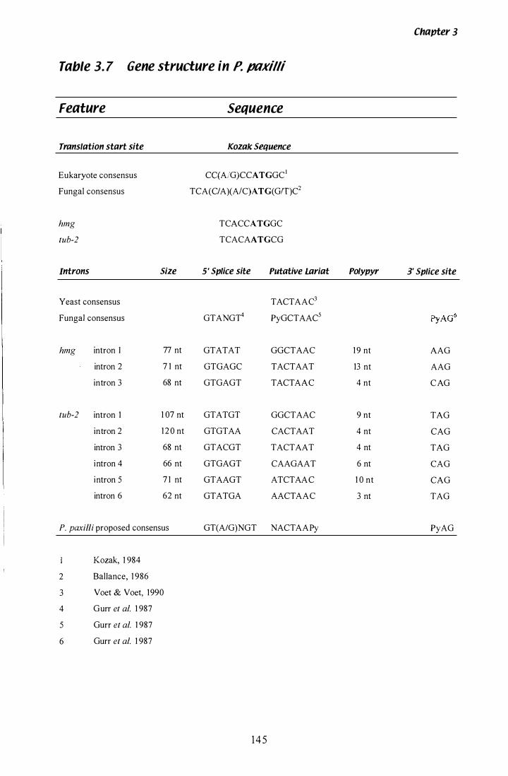
Table 3. 7 Gene structure in P. ptlKilli
Feature Sequence
Translation start site
Eukaryote consensus
Fungal consensus
hmg
tub-2
Introns
Yeast consensus
Fungal consensus
hmg intron I
intron 2
intron 3
tub-2 intron 1
intron 2
intron 3
intron 4
intron 5
intron 6
Size
77 nt
7 1 nt
68 nt
1 07 nt
1 2 0 nt
68 nt
66 nt
7 1 nt
62 nt
P. paxilli proposed consensus
Kozak, 1 984
2 BalIance, 1 986
3 Voet & Voet, 1990
4 Gurr et al. 1 987
5 Gurr et al. 1 987
6 Gurr et al. 1 987
Kozak Sequence
CqAJG)CCATGGC1
TCA( CIA )(A/C)A TG( GIT)C2
TCACCATGGC TCACAATGCG
5' Splice site Putative Lariat
TACTAAC3
GTANGT4 PyGCTAAC5
GTATAT GGCTAAC GTGAGC TACTAAT GTGAGT TACTAAC
GTATGT GGCTAAC GTGTAA CACTAAT GTACGT TACTAAT GTGAGT CAAGAAT GTAAGT ATCTAAC GTATGA AACTAAC
GT(A/G)NGT NACT AAPy
145
Polypyr
1 9 nt
13 nt
4 nt
9 nt
4 nt
4 nt
6 nt
1 0 nt
3 nt
Chapter 3
3' Splice site
AAG AAG C AG
TAG CAG TAG CAG CAG TAG
PyAG
Chapter 3
2 .2) and a 1 0-fold dilution of cDNA from 60 h RNA as a template. The products were
separated on a 1 % (w/v) agarose gel (section 2. 1 0. 1 and 2 . 1 0.3) . In Figure 3 .23 , the
cDNA product is the predicted 490 bp smaller than the gDNA product. The cDNA
peR reaction was then purified (section 2 . 1 1 .5) , c loned into a pGEM®-T vector
(section 2 . 1 5) and sequenced (section 2 . 1 2 . 3 ) to confirm the exact location of
intronlexon boundaries. No sequence data was available for the intronlexon boundary
for intron 1 and 2 , as the initial ORF is upstream of the sequencing primer, and the
second intron is too close to the sequencing primer. However, this is an extremely
conserved gene, and these ORF have been predicted from the tub-2 sequence of other
specIes.
3.2.8 Chromosomal location of P. paxilli tub-2
The chromosomal location of tub-2 was determined by probing a nylon membrane
containing a CHEF separation of P. paxilli chromosomes (Figure 3 .24 A), previously
discussed in section 3 . 1 . 1 2, with a PCR product of tub-2 amplified with Etub2 and
Etub3 . All strains, including the Pax- mutants and wild type P. paxilli, show the tub-2
probe hybridising to chromosome IV (Figure 3 .24 B).
146

Figure 3.23 Confirmation of P. pllKilli tUb-2 introns
RT-PCR (section 2.20) was performed on 1 1 1 00 dilution of cDNA from a 60 h culture,
using primers Etub5 and Etub6 (Table 2,2). Products were separated on a 1 % agarose
gel (section 2 . 1 0 . 1 and 2 . 1 0 .3) as follows. Lanes 1 and 4, 1 kb ladder (BRL). Lane 2,
gDNA. Lane 3, 60 h cDNA. Lane 3, water control . Sizes shown are in kb.
1 47
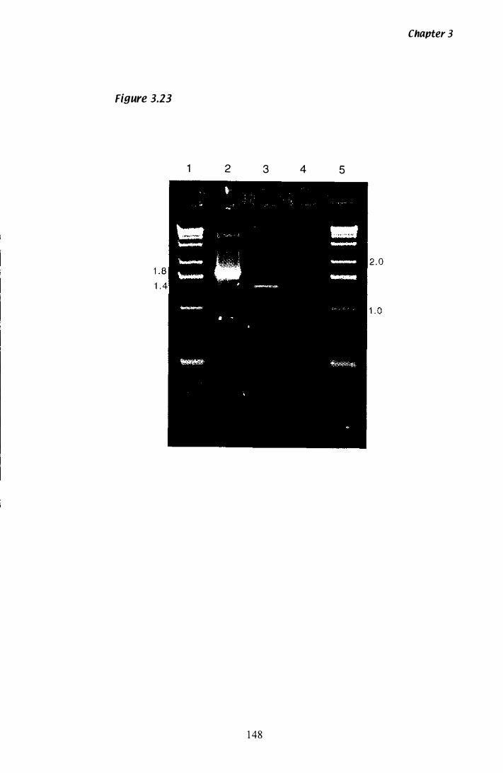
Chapter 3
Figure 3.23
1 2 3 4 5
2 .0
1 48
Figure 3.24 CHEF analysis of P. paxilli chromosomes
A) Ethidium bromide stained CHEF gel
This is the same gel as that shown in Figure 6 B in Young et al. ( 1 998) . Running
conditions for CHEF electrophoresis are described in the legend of this figure. Lane 1 ,
S. cerevisiae. Lane 2, S. pombe. Lanes 3-7, P. paxilli wild-type, YI-20, CY-2, CY- I 02,
and CY-35 . Sizes shown are in Mb.
B) Chromosomal location of tu�2
Autoradiograph of gel shown in Figure 3 .22 A probed with a [32P] -labelled PCR
product of tub-2, amplified with primers Etub2 and Etub3 . Southern blotting and probe
hybridisation as described in section 2 . l 3 . S izes shown are in Mb.
149
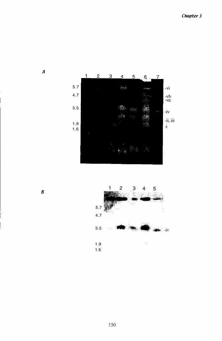
Chapter 3
A
5 . 7 -VI
4 . 7 -vb -va
3 . 5 -iv
-ii, iii 1 . 9 -i 1 . 6
1 2 B
3 4 5
5 . 7 4 .7
3 . 5 -IV
1 . 9 1 .6
1 50
Chapter 3
3.3 P. paxi"i physiology and conditions for induction of paxilline
The growth of filamentous fungi in submerged liquid culture is an important process for
both researchers and commercial enterprises. However, the optimal conditions for
production of fungal secondary metabolites in liquid culture can vary with different
species and even different strains. To study the regulation of paxilline biosynthesis in
liquid culture, three fundamental requirements had to be met. Firstly, paxill ine must be
produced at some time in the course of the experiment. Secondly, experiments must be
reproducible, with a limited level of inherent variability. Thirdly, the gross morphology
of mycelia in l iquid culture should be such that a small sample volume is stil l
representative of the entire culture, and that all mycelia in the culture have equal access
to nutrients and oxygen.
3.3.1 Spore viability
Liquid cultures of P. paxilli were inoculated from a spore suspension. Once a fresh
spore suspension had been prepared (section 2 . 1 .5), the concentration of spores was
determined by haemocytometer counting and the spore suspension stored at 4 °C .
Although spores are hardy structures, the percentage of viable spores can drop over
time . To ensure that spore viability did not overly affect the growth rate of liquid
cultures, the viability of spores over time was determined. Serial dilutions of each spore
suspension were plated onto PD plates (section 2 . 1 .2) on a regular basis, until a
significant drop in viability was recorded. In all spore suspensions tested, the initial
percentage of viable spores fel l by 50% in approximately 60 days . For future
experiments, spore suspensions were stored at 40 C for no more than 6 weeks, before a
new suspension was prepared.
3.3.2 Cultures inoculated from spore suspensions
In the initial growth experiments, cultures were inoculated directly from a spore
suspension (section 2 . 1 .7). Typical results for growth rate ( dry weight) and paxilline
content are shown in Figure 3 .25 A. Using these conditions there was considerable
1 5 1

Figure 3.25 Comparision of inoculation protocols
All cultures were grown in a 2 L Erlenmeyer flask in 300 rnL of CDYE media (section
2. 1 .2). Samples of mycelia ( 1 0 mL) were removed every 1 2 h (section 2 . 1 6. 1 ), freeze
dried and the dry weight detennined. PaxilIine content was determined by HPLC
(section 2. 1 6). An example of a typical HPLC trace is shown in Figure 3 .26.
A) Inoculation by spore suspension
The replica cultures were inoculated from a spore suspension (section 2. 1 .7). Flask A,
dry weight t ). Flask B, dry weight·(·{}.. ). Flask A, paxilline content t · . . . ).
Flask B, paxilline content t-O - ).
B) Inoculation by seea culture
The replica cultures were inoculated from a seed culture (section 2. 1 . 8). Flask A, dry weight C--- ). Flask B, dry weight·(·Q·· ). Flask A, paxilline content * " ' ). Flask
B, paxilline content to - ).
1 52
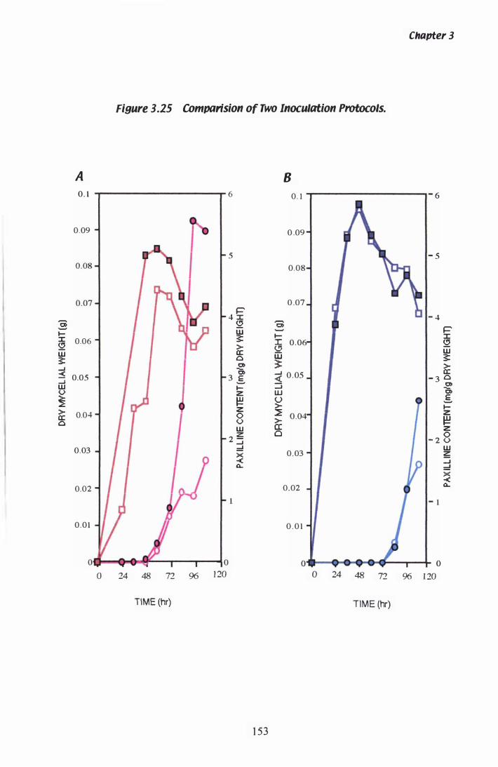
A
0 1
J 0.09
0.08 �
0.07 �
:§ I-x 0.06 52 w $: ....J
Figure 3.25 COmparlsion of Two Inoculation Protocols.
B 6 0. 1
� 0 .09
5 I " I , 0.08
I , " � 0 .07 �
4 x 52 0; W $: l-
G 0.0 > a: iD 0 0 �
Chapter 3
.- 6
1- 5
... - 4
� X 52 w $: > a: -< "0 � 0.05 :3 0.05 3 .§. - 3 0
:::i 0 w <.> � > a: 0
0.04 I
0.03 � I I
0.02 -4 I I A I
0.01
O. • • er 10 o 24 48 72 96 1 20
TIME (hr)
I-z � Z 0 <.> w
2 ;5; ....J =' x -< 0..
w "0 u .§. > I-� Z
� > er: z Cl 0 - 2 <.>
0.03 w ;5;
0 ....J =' X -<
0.02 I • .. 0..
0 .0 1
0l1li • • • • • , 0 o 24 48 72 96 120
TIME (hr)
1 53
Chapter 3
variation between both the time of paxilline induction and the yield of paxilline. This
variability was considered unsatisfactory for identifying real differences between
cultures due to variations in growth conditions or strain genealogies. The source of
variation was thought to be the spore suspension itself as the concentration of spores
was very high. Therefore, small variations in inoculum volume could result in a
dramatic increase in the final concentration of viable spores.
The morphology of mycelia in l iquid culture was also variable. A number of factors
were identified as sources of variation, including the culture volume, the shake speed of
the incubator and the concentration of the inoculum. Small 25 mL cultures grown at
230 rpm have large mycelial pellets, which settle rapidly when removed from the
shaker. In the large 300 mL cultures grown at 350 rpm, only loose mycelial fragments
were present. Decreasing the culture volume, the shaker speed, or the inoculum
concentration resulted in the formation of pellets. The larger pellets are hollow, which
results in a mass transfer gradient across the pellet, of nutrients and oxygen in, and of
waste and secreted metabolites out of the pellet (Prosser & Tough, 1 99 1 ) . A number of
protocols were trialed to improve the reproducibility of replica cultures with respect to
morphology (loose mycelia fragments) and gross physiological markers (dry weight and
paxilline induction).
3.3.3 Cultures inoculated from seed culture
A number of seed culture protocols were trialed to el iminate the variation seen in
cultures inoculated directly from a spore suspension. Seed cultures of 1 00 mL were
grown for 24 h, and used to inoculate 300 mL CDYE (section 2.2) cultures (section
2 . 1 . 8), but this resulted in the large pellet morphology. Pellets had formed in the
smaller culture volume, even at 350 rpm. Oat flour has been used in filamentous fungal
seed cultures to inhibit the formation of pellets (Tkacz et al. , 1 993). However, seed
cultures containing 1 % (w/v) oat flour still formed pellets . It appears that once the
pel lets have formed , sub-culturing in to a larger volume does not change the
morphology. The number of pellets is also fixed. Therefore if 24 mL of seed culture is
sub-cultured into a 3 00 mL volume, the number of pellets does not increase . The
growth rate and subsequent accumulation of biomass is severely hampered by
1 54
Chapter 3
inoculation with mycelial pellets. Finally the incubation period of the seed culture was
reduced from 24 h to 1 2 h . At this time spores have begun to germinate, but no gross
culture morphology has been established (Figure 3 .29, photograph A and B). Replica
cultures, inoculated from the same seed culture showed a significant decrease in
variability with respect to both growth rate and paxilline induction (Figure 3 .25 B),
when compared to cultures inoculated by spore suspensions (Figure 3 .25 A). This
particular protocol (section 2 . 1 . 8) was subsequently used for all other growth
experiments.
3.3.4 Growth of P. pl1Killi in paxilline,inducing media
3.3.4. 1 Experiment 1
After establishing a reliable and reproducible inoculation protocol (section 3 .3 .3) , a
large scale physiological investigation was conducted in two separate experiments.
CDYE cultures of 300 mL were inoculated from a 1 00 mL seed culture (section 2 . 1 .8)
and grown as described in section 2 . 1 .2. Analysis of culture physiology in Experiment
1 is shown in Figure 3 .27. Biomass accumulation, peaked after approximately 42 h (A),
at which time the average dry weight in a 5 mL sample of culture was 0 . 1 g or 20 g/L.
The pH of the broth declined after 48 h (B) in both cultures, 24 h before paxilline was
first detected. However, a slight increase in pH is detected in both flasks after 96 h.
Paxilline was first detected by HPLC at 72 h (C), and reaches an average content of
3 .25 mg/g of mycelia after 96 h. Production of paxilline by p, paxilli grown in CDYE
is equivalent to 65 mg/L.
3.3.4.2 Experiment 2
The growth experiment was repeated (Experiment 2) and the results are shown in Figure
3 .28 . Although the seed culture inoculation protocol was used, there is stil l evidence of
variation between cultures . This was most apparent when dry weight was analysed.
When both cultures are averaged, the biomass accumulation peaks after 45 h (A), is
comparable to the 42 h peak seen in Experiment 1 . At this time the average maximum
1 5 5
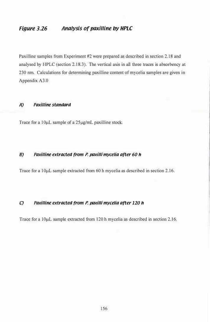
Figure 3.26 Analysis of paxilline by HPLC
Paxilline samples from Experiment #2 were prepared as described in section 2. 1 8 and
analysed by HPLC (section 2. 1 8 . 3 ) . The vertical axis in all three traces is absorbency at
230 nm. Calculations for determining paxilline content of mycelia samples are given in
Appendix A3.0
A) Paxilline standard
Trace for a 1 0llL sample of a 25 1lg/mL paxill ine stock.
B) Paxilline extracted from P. pllKilli mycelia after 60 h
Trace for a 1 0llL sample extracted from 60 h mycelia as described in section 2. 1 6 .
C) Paxilline extracted from P. pllKilli mycelia after 120 h
Trace for a l OllL sample extracted from 1 20 h mycelia as described in section 2. 1 6.
1 56
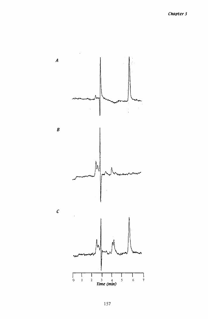
A
8
c
o 2 3 4 5
Time (min)
1 57
Chapter 3
6 7
Figure 3.27 Growth analysis ana paxilline proauction of
P. paxilli in Experiment 1
Liquid cultures of P. paxilli were grown (section 2. 1 .2) in a 2 L Erlenmeyer flask
containing 300 rnL of CDYE media, inoculated from a seed culture (section 2. 1 .8).
Samples of 10 rnL were removed for analysis of dry weight and paxilline content.
A) Analysis of mycelial dry weight
The dry weight of 10 rnL of freeze-dried mycelia was determined. Flask A ( . ),
Flask B ( . . . . . . . ).
8) Analysis of broth pH
The pH was determined on the mycelia-free supematant of the samples removed for
paxilline analysis and RNA isolation, with an Orion research digital ionalyzerlSO l .
Flask A ( ), Flask B ( . . . " ' ).
C) Analysis of mycelial paxllllne content
The paxilline content of freeze-dried 1 0 rnL mycelia samples was determined by HPLC
(section 2 . 1 6). Flask A ( ), Flask B ( . . . ' ' ' ) .
1 58
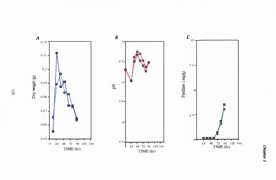
A 8 0. 1 2
0. 1 1 n
0. 1 -I " �
�
0.09 ::r: - � �
v. \0 �
Cl 0.08
0.07
0.05 "11-.,.....,..-,...---....... ....--1 o 24 48 72 96 1 20 1 44
TIME (hr)
7 i
6.5
l I V 6 \l
5.5
I 5
4.5 �I �-"-"--r-�""" 24 48 72 96 1 20 144
TIME (hr)
i ""
t p..
C 1 0 �-
-------------�
7 . 5
2 . 5
2 4 48 72 9 6 1 20 144 TIME (hr) �
t w
Figure 3.28 Growth analysis ana paxilline proouction of
p. pl1Killi in Experiment 2
Liquid cultures of P. paxilli were grown (section 2 . 1 .2) in a 2 L Erlenmeyer flask
containing 300 mL of CDYE media, inoculated from a seed culture (section 2 . 1 . 8).
Samples of 1 0 mL were removed for analysis of dry weight and detennination of
paxilline content.
A) Analysis of mycelial dry weight
The dry weight of 1 0 mL of freeze-dried mycelia was determined. Flask A ( . ),
Flask B C' " .. . . ).
B) Analysis of broth pH
The pH was determined on the mycelia-free supematant of the samples removed for
paxilline analysis and RNA isolation, with an Orion research digital ionalyzerlS0 1 .
Flask A ( ), Flask B ( . . . " ' ).
C) Analysis of mycelial paxllline content
The paxilline content of freeze-dried 1 0 mL mycelia samples was detennined by HPLC
(section 2. 1 6). Flask A ( ), Flask B ( . . . " ' ).
1 60

-0\ -
A 0.09 .----------.
0 .08
� 0.07
1 \ � � C 0
0.06
0 .05 -tl-""T""......,r----r-�__r-..,..... o 24 48 72 96 1 20 144
TIME (HR)
8
:r: P.
7 -
6.5
6
5 .5
5
4.5 -tl-T--r---r-....... ..,.....,-I o 24 48 72 96 1 20 1 44
TIME (HR)
C 1 0 ,-----------------�
7 .5
i 5 ......."
t p..
2 .5
o 24 48 72 96 1 20 144 TIME (HR)
�
t �
Chapter 3
dry weight in a 5 mL sample was 0.09 g, or 1 8 g/L. The pH of the broth declined after
33 h (B), with a more dramatic decrease apparent after 84 h. The increase in pH,
recorded at 96h in Experiment 1 , was not seen in Experiment 2 and remains
unexplained at this time. Paxilline was first detected at 72 h (C). For comparison with
Experiment 1 , where the final paxilline sample was taken at 96 h, the average paxilline
content at this time in Experiment 2 is 2 .35 mg/g of mycelia. However, the paxilline
content of flask B, registered a decrease in paxilline after 96 h. In all paxilline analyses
conducted by HPLC, this is the only example where a drop in paxilline was recorded. If
this result is discarded, the paxilline production in flask A after 96 h is 3 .49 mg/g of
mycelia. The overall paxilline production is then equivalent to 6 1 mg/L, comparable
with that seen in Experiment 1 . A typical set of results for paxilline analysis by HPLC
(section 2 . 1 6) is shown in Figure 3 .26. Authentic paxilline was eluted from the HPLC
column after 5 .8 min (Figure 3 .26 A). When P. paxilli mycelial extracts were analysed,
a second peak, with an elution time of 4.25 min was seen. This peak has been identified
as ergosterol (Sarah Munday-Finch pers. comm.) . By comparing the 60 h mycelia
sample (B) with the 1 20 h mycelia sample (C) a dramatic increase in paxilline is seen,
but the peak of ergosterol increased only slightly. The method for calculating the
concentration ofpaxilline from the peak area is described in appendix A.3 .0 .
3.3.4.3 Analysis of paxilline in broth
An analysis of paxilline in broth was performed during Experiment 2 . Broth removed
from mycelia samples was collected, freeze-dried and the paxilline content determined
by HPLC. Paxilline was not detected until 1 44 h. There was no steady increase in
paxilline over time, although a small peak was present in all samples from 84 h to
1 20 h. The elution time of this peak was the same as the elution time for paxilline, but
it was too small to register as a true peak with the Shimadzu C-R3A Chromatopac
detector. If paxilline was being actively secreted into the media, a steady increase in
broth paxilline should have been detected.
1 62

Chapter 3
3.3.'1.)1 Microscopic analysis of culture morphology
The culture morphology was also monitored in Experiment 2 . Mycelia were removed
every 24 h, examined microscopically with a Nikon Diaphot inverted microscope and
photographed with a Nikon F-60 1 camera using Kodak TMY 400 film. Examples of
the morphology observed are shown in Figure 3 .29. Photographs A and B show the
morphology of mycelia, 4 h after inoculation by seed culture. The germinating spores,
both singly or in aggregates, can be seen and are also an example of the typical
morphology seen in seed cultures after 1 2 h. The clumps of mycelia are presumed to be
the initial structure that eventually forms a mycelial pellet. After 28 h, the cultures
appear very viscous with fine micropellets (0 .5- 1 mm diameter) suspended in loose
mycelial fragments. Pellets in photograph C appear quite dense , especially when
compared to the fragmented pellets seen in the culture after 1 00 h (photograph J).
Previous reports had stated that P. paxilli does not sporulate when grown in CDYE
media (Ibba et aI. , 1 987) , however, a conidiophore structure, with a s ingle conidia still
attached is seen in photograph D. Conidia are seen in the broth after 28 h. These must
be a result of conidiation that has occurred after inoculation, as all the conidia seen in
photographs A and B are germinating, whereas ungerminated conidia are present in
photograph C. After 56 h, ungerminated conidia are still visible in the broth
(photograph E). A 'peniciIlius ' , the conidiophore structure typical of Penicillium spp
(Bold et al . , 1 987) , i s shown in photograph F . After 76 h (photograph G), discrete
hyphal fragments can be seen suspended in the broth, some of which undergo cryptic
growth or regeneration, (photograph H). Vacuolation is also occurring at this time, and
can be identified by the banding pattern seen on hyphae in photograph I. By 1 00 h, the
culture has become less viscous, and enters what i s known as the death phase (Prosser
& Tough, 1 99 1 ) . Pellets are less dense, and have begun to fragment (Photograph J) .
Vacuolation becomes more evident (photographs K and L) and there is evidence of
autolysis, as hyphae appear to shrink and break (photographs M and N).
1 63
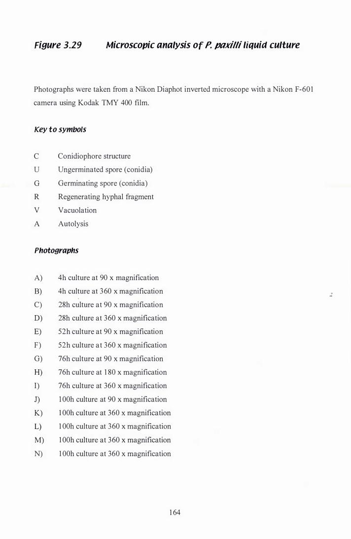
Figure 3.29 Microscopic analysis of P. paxilli liquid culture
Photographs were taken from a Nikon Diaphot inverted microscope with a Nikon F -60 1
camera using Kodak TMY 400 film.
Key to symbols
C Conidiophore structure
U Ungerminated spore (conidia)
G Germinating spore (conidia)
R Regenerating hyphal fragment
V Vacuolation
A Autolysis
Photographs
A) 4h culture at 90 x magnification
B) 4h culture at 360 x magnification
C) 28h culture at 90 x magnification
D) 28h culture at 360 x magnification
E) 52h culture at 90 x magnification
F) 52h culture at 360 x magnification
G) 76h culture at 90 x magnification
H) 76h culture at 1 80 x magnification
I) 76h culture at 360 x magnification
J) 1 00h culture at 90 x magnification
K) 1 00h culture at 360 x magnification
L) 1 OOh culture at 360 x magnification
M) 1 OOh culture at 360 x magnification
N) 1 00h culture at 360 x magnification
1 64
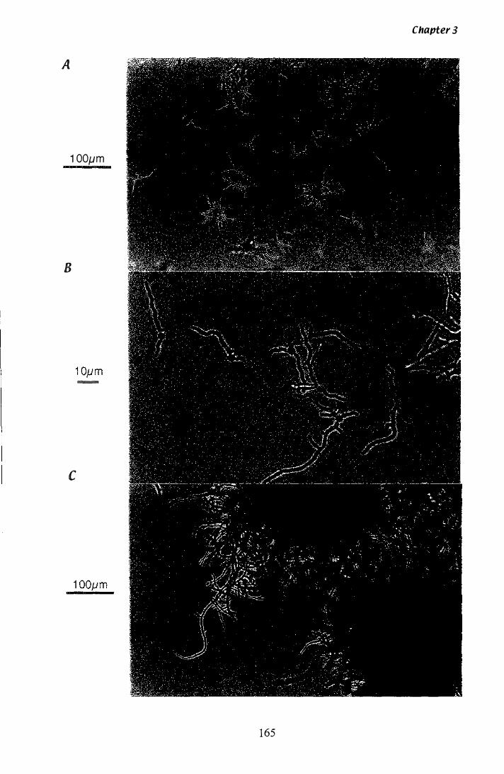
Chapter 3
A
1 00jJm
B
-
c
1 65
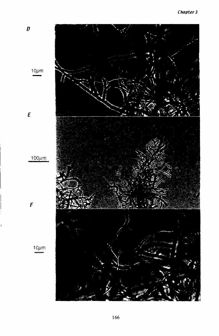
Chapter 3
D
E
F
-
1 66
Chapter 3
G
1 00pm
H
50p m
I
1 0p m --
1 67
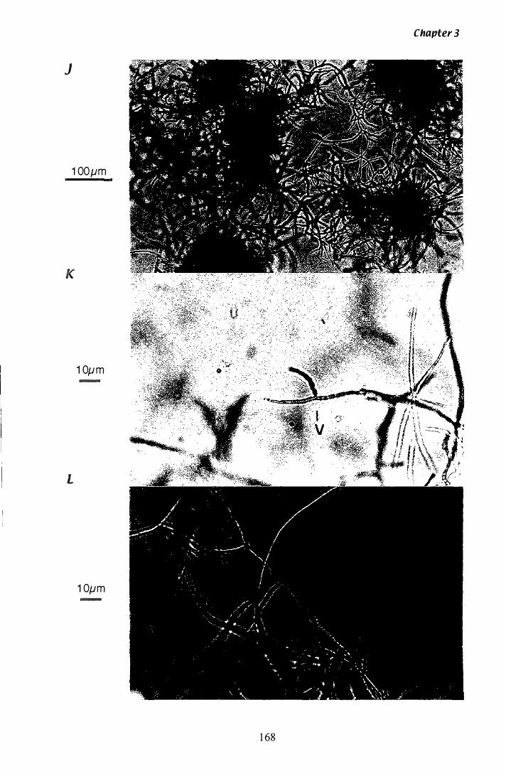
Chapter 3
J
1 00pm
K
1 0p m --
L
1 0pm -
1 68
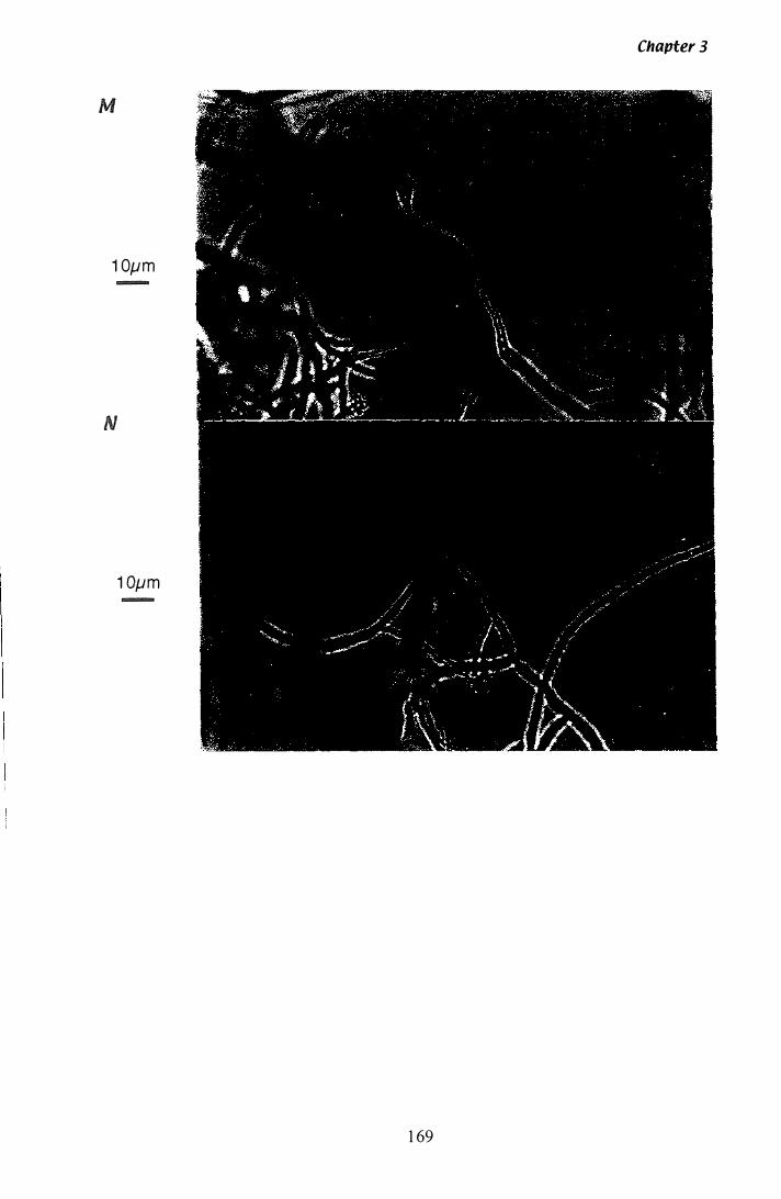
Chapter 3
M
1 0pm -
N
1 0pm -
1 69
3.3.5 Growth of P. pnxilli in supplemented media
Chapter 3
A number of experiments were conducted to determine the effect of various media
supplements on the growth and paxilline production of wild type P. paxilli in liquid
culture. The scale of the experiments was reduced to maximise the number of variables
that could be tested. The culture volume was reduced to 1 00 mL and all experiments
were inoculated with 8 mL of a 1 00 mL seed culture (section 2. 1 .8) . Samples ( 1 0 mL)
were taken at two time points (60 h and 1 20 h) except in cultures grown in CD and
CD + 0.25% tryptophan, where biomass accumulation was significantly reduced. A
single sample, of 1 00 mL was removed from these two cultures after 1 20 h . , and the
excess broth removed to a final volume of 1 0 mL. Paxilline was extracted from 1 0 mL
samples (section 2 . 1 6.2) and separated by TLC (section 2. 1 6.4). The gross morphology
of mycelia, grown in a 1 00 mL volume at 350 rpm, was fine pellets of approximately
1 -2 mm diameter.
3.3.5.1 Calcium
Calcium-induced sporulation was reported to inhibit the synthesis of paxilline (Ibba et
al. , 1 987). However, microscopic analysis of P. paxilli in the paxiIline-inducing media
CDYE, confirmed that sporulation was occurring (Figure 3 .29 D and F) . To test this
theory, P. paxilli was grown in the sporulation medium PD (section 2 .2) and in a CDYE
media (section 2 .2) containing 2% (w/v) CaCh. The concentration of spores in the
broth after 1 20 h was determined by haemocytometer counting. In the control CDYE
media, the spore concentration was 1 .4 x 1 05 spores/mL. At this concentration, the
culture is cream coloured. When P. paxilli was grown in PD or C DYE + 2% (w/v)
CaCh, the culture is coloured dark green. The spore concentration in PD media and
CDYE + 2% (w/v) CaCl2 after 1 20 h was 2 .5 x 1 07 spores/mL and 9.3 x 1 07 spores/mL
respectively. The paxilline content of the cultures was determined by TLC (section
2 . 1 6 .4). Results are qualitative only, and are shown in Figure 3 .30. The control culture
grown in CDYE is seen in lanes 2 and 3 , with paxilline staining dark green when
exposed to alcoholic Erhlich' s reagent. Paxilline was detected at 60 h and increases at
1 70
Figure 3.30 TLC analysis of paxilline production with media
supplements
P. paxilli cultures were grown as described in section 2 . 1 .2, in SOOmL Erlenmeyer
flasks containing 1 00mL of test media. The test media used the Czapek Dox medium
from Oxoid as a base. The constituents of CD medium from Oxoid are slightly
different from those in the CD salts used for other experiments and therefore should not
be compared. Paxilline was extracted from 1 0 mL of culture into 1 2 mL of
chloroform:methanol (2 : 1 ) (section 2. 1 6 . 1 ) . An aEquot of I .S mL was removed and
evaporated overnight. The dried sample was then resuspended in SO ilL of
chloroform:methanol (2 : 1 ) and run on a TLC plate (section 2 . 1 6.4). Lanes 1 and 1 2,
40llg of Paxilline standard. Lanes 2-3 , CDYE culture at 60 h and 1 20 h. Lane 4, CD
culture at 1 20 h. Lane S , CD + 0.2S% Tryptophan culture at 1 20 h. Lanes 6-7, PD
culture at 60 h and 1 20 h . Lanes 8-9, CDYE + 2% CaCl2 culture at 60 h and 1 20 h .
Lanes 1 0- 1 1 , CDYE + SOrnM MOPS culture at 60 h and 1 20 h.
1 7 1
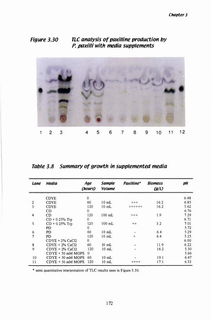
Figure 3.30 ne analysis of paxilline production by P. paxilli with media supplements
Chapter 3
1 2 3 4 5 6 7 8 9 1 0 1 1 1 2
Table 3.8 Summary of growth in supplemented media
Lane Media Age Sample Padlline* Biomass
(hours) Volume (gIL) COYE 0
2 COYE 60 1 0 mL +++ 1 6.2 3 COYE 1 20 l O mL ++++++ 1 6. 2
CO 0 4 CD 1 20 1 00 mL +++ 1 .9
CO + 0.25% Trp 0 5 CO + 0.25% Trp 120 lOO mL ++ 3 .2
PO 0 6 PO 60 1 0 mL 6.4 7 PO 1 20 1 0 mL + 6.4
COYE + 2% CaCl2 0 8 COYE + 2% CaCl2 60 10 mL 1 1 .9 9 COYE + 2% CaC12 1 20 1 0 mL 1 6.2
COYE + 50 mM MOPS 0 1 0 COYE + 50 mM MOPS 60 1 0 mL 1 9. 1 1 1 COYE + SO mM MOPS 1 20 1 0 mL ++++ 1 7. 1
* semi quantitative interpretation of TLe results seen in F igure 3 .30.
1 72
pH
6.48 6.85 5 .62 6.76 7 .29 6 .71 7 .01 5 .72 5 .29 5 .25 6.00 6 .22 5 .30 5 .66 6.47 6.33
Chapter 3
1 20 h. In lanes 6 and 7 the samples taken at 60 h and 1 20 h, from the culture grown in
PD, are shown. Paxil line was not detected at 60 h, although a very faint band is
discernible at 1 20 h. Cultures grown in CDYE + 2% (w/v) CaCl2 showed no trace of
paxilline, even after 1 20 h of growth (lanes 8 and 9) . Interestingly, the biomass
accumulation in PD is quite low (6 .4 glL), compared to both the control CDYE culture
( 1 6 .2 g/L) and the CDYE + 2% (w/v) CaCh culture ( 1 6 .2 g/L) (Table 3 . 8). However,
even with the lowered biomass, a faint trace of paxilIine was still detected. The cause
of this decrease in biomass accumulation could be the absence of yeast extract in the PD
media.
3.3.5.2 Yeast Extract
The putative source of the indole moiety in paxilline i s tryptophan (Laws & Mantle,
1 989). The yeast extract supplement added to CD media, although undefined, is a
source of many amino acids. To examine the effect of yeast extract on paxil line
production, a P. paxilli culture was grown in CD only media. A second culture was
supplemented with 0.25% (w/v) tryptophan, to determine if yeast extract was the source
of tryptophan during paxilline biosynthesis . B iomass accumulation in b oth
experimental media, was severely reduced, therefore the entire culture volume was
collected after 1 20 h and analysed for paxilline content. Both cultures were pink in
colour, although the colour was more intense in the CD + 0.25% (w/v) tryptophan
culture. Occasionally, cultures grown in CD YE are coloured pink, but this is usually in
cultures of less than 1 00 mL. Paxilline was detected in both cultures after 1 2 0 h,
however, the paxilline seen in Figure 3 .30, lanes 4 and 5 represents a sample volume
1 0 times that of the other lanes.
3.3.5.3 Biological buffer
Changes in pH have been associated with the onset of secondary metabol ite
biosynthesis in a number of filamentous fungi . During the growth experiments
discussed in section 3 .3 .4, the pH of the culture broth was seen to change over time
173

Chapter 3
(Figure 3 .27 B and 3 .28 B) . A drop in pH is seen approximately 3 0 h before paxilline
was detected in the culture. The biological buffer MOPS , was supplemented to 50 mM
in CDYE to examine the effect of pH on paxilline biosynthesis. Cultures grown in
CDYE + 5 0 mM MOPS achieved the greatest biomass ( 1 9. 1 g/L) of all the supplements
tested, including the control culture grown in CDYE, which had a biomass of 1 6 .2 g/L.
The pH of the broth from both CDYE cultures and CDYE + 50 mM MOPS is shown in
Table 3 .8 . The cultures grown in CDYE and CDYE + 50 mM MOPS both reach a pH
of over 6 . 5 after 60 h. Between 60 h and 1 20 h, pH in the c ontrol culture decreased
1 8%, while the pH in the culture supplemented with MOPS only decreased 2% during
the same time. The paxiIline content of the culture grown in CDYE + 50 mM MOPS is
shown in Figure 3 .30, lanes 1 0 and 1 1 . Even with a greater overall biomass, paxilline
was not detected until 1 20 h, at which time it is approximately equivalent to the amount
ofpaxilline seen in the control culture at 60 h (Figure 3 .30, lane 2). The results from all
these experiments are summarised in Table 3 .8 .
3.3.6 Growth of P. paxilli in variable carbon sources
Utilisation of a preferred carbon source can effect the growth rate and biosynthesis of
secondary metabolites by fungal species in liquid culture. To examine the effect of
carbon source on production of paxilline, P. paxilli was grown in CD salts (section 2 .2)
supplemented with 0.5% (w/v) YE and a range of carbon sources at 3% (w/v) . The
standard CDYE media from Oxoid contains 3% sucrose as the carbon source. The
culture grown in 3% (w/v) sucrose accumulated the highest biomass after 60 h and was
the only culture to record a drop in biomass after 1 20 h. At the end of 1 20 h, the culture
grown in 3 % (w/v) glucose, had accumulated the greatest biomass ( 1 1 .9 g/L) (Table
3 .9). The paxilline content was determined by TLC (section 2. 1 6.4) and the results are
shown in Figure 3 . 3 1 and Table 3 .9 . The most interesting result is the paxilline content
of the culture grown in 3% (w/v) glucose (lane 6-7) . The level of paxilline in this
culture was less than the control culture, containing 3% (w/v) sucrose, after 60 h (lanes
2-3), but surpassed it after 1 20 h. The number of bands seen in lane 6 indicates that the
biosynthesis of other indole-diterpenoid moieties has also increased. Four indole
diterpenoid products (stained green with alcoholic Erhlich's reagent), with a Rf greater
than paxilline are less polar than paxilline, and may represent precursors of paxilline.
1 74
Fi9ure 3.31 TLC analysis of paxilline production in variable
carbon sources
P. paxilli cultures were grown as described i n section 2 . 1 .2, in 5 00mL Erlenmeyer
flasks containing 1 00mL of test media. The basic media contained CD salts (section
2.2.) + 0.5% Yeast Extract (Oxoid), + 3% (w/v) carbon source. The constituents of the
CD salts medium used, vary from those in the CD medium supplied by Oxoid, which
was used for the experiments shown in figure 3 . 3 0 and should not be compared.
Paxilline was extracted from 1 0 mL of culture into 1 2 mL of chlorofonn:methanol (2: 1 )
(section 2 . 1 6 . 1 ) . An aliquot of 1 .5 mL was removed and evaporated overnight. The
dried sample was then resuspended in 50 ilL of chlorofonn:methanol (2 : 1 ) and run on a
TLC plate (section 2. 1 6.4). Lanes 1 and 1 0, 51lg of paxilline standard. Lanes 2-5, 60 h
cultures with 3 % sucrose, 3% glucose, 3% glycerol, and 3 % mannitol . Lanes 6-9, 1 20 h
cultures with 3 % sucrose, 3% glucose, 3% glycerol, and 3 % mannitol.
1 7 5
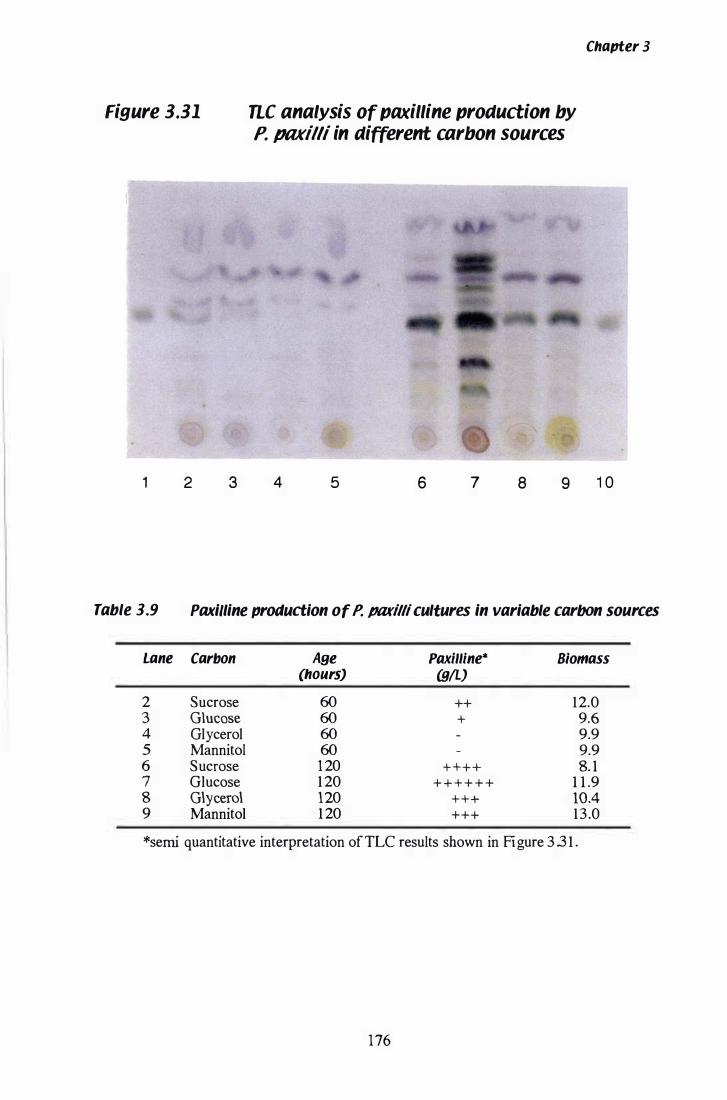
Figure 3.31
1 2 3
Chapter 3
ne analysis of paxilline production by P. paxilli in different carbon sources
4 5 6 7 8 9 1 0
Table 3.9 Paxilline production of P. JIIlfilli cultures in variable carbOn sources
Lane Carbon Age Paxilline* B/omass (hours) (gIL)
2 Sucrose 60 ++ 12.0 3 Glucose 60 + 9.6 4 Glycerol 60 9.9 5 Mannitol 60 9.9 6 Sucrose 120 + + + + 8. 1 7 Glucose 1 20 + + + + + + 1 1 .9 8 Glycerol 120 + + + lOA 9 Mannitol 1 20 +++ 13 .0
*semi quantitative interpretation of TLC results shown in Figure 3 3 1 .
176
Chapter 3
Two indole-diterpenoid products seen below paxilline, are structures more polar than
paxilline that may represent products from additional oxidation steps after the synthesis
ofpaxil line. Paxilline was not detected in cultures supplemented with 3% (v/v) glycerol
or 3 % (w/v) mannitol ( lane s 7 and 8) until 1 20 h, although the rate of biomass
accumulation was equivalent to that seen in the culture supplemented with 3 % (w/v)
glucose.
3.3. 7 Growth of P. /1tlKilli during carbon and nitrogen starvation
Biosynthesis of paxilline occurs during secondary metabolism. Regulation of many
secondary metabolite pathways is under nitrogen or carbon repression (Drew &
Demain, 1 977; Marzluf, 1 98 1 ) . Experiments were performed to examine the effect of
nitrogen and carbon repression on paxiIIine biosynthesis in P. paxiIli. It was postulated
that cultures prematurely released from repression by nitrogen or carbon catabolite
regulatory proteins would initiate biosynthesis of paxilline earlier, resulting in an
increased amount of paxilline at both 60 h and 120 h.
Test media, [CD salts + 0.5% (w/v) YE + 3% (w/v) sucrose] ( l OO mL), was inoculated
from a seed culture (section 2 . 1 . 8) and allowed to grow for 24 h. The mycelia were
then harvested and transferred to media lacking either nitrogen or carbon. The media
was supplemented with 0 .5% (w/v) yeast extract. The standard CD YE media contains
3% (w/v) sucrose and 0 .2% (w/v) sodium nitrate. Samples were collected at 60 h and
1 20 h. After 60 h, the culture grown in the absence of nitrate had a typical biomass
accumulation of 1 3 . 5 g/L (Table 3 . 1 0), compared to the control CD salts + YE +
sucrose culture which had a b iomass of 1 1 .9 g/L after 60 h (Table 3 .9). Unfortunately
the culture lacking nitrogen became contaminated and a final sample was not prepared.
PaxiIIine was detected after 60 h (Figure 3 .32, lane 2) and is a similar quantity to that
seen in controls CDYE (Figure 3 .3 0, lane 2) and CD salts + YE + sucrose cultures
(Figure 3 .3 1 , lane 2). Lack of nitrogen did not seem to effect the growth or production
of paxil line significantly, although yeast extract is probably a compensatory source of
nitrogen. Carbon starved cultures failed to gain biomass after being transferred into the
experimental media (Table 3 . 1 0) . Paxilline was not detected at either 60 h or 1 20 h
177
Figure 3.32 TLe analysis of paxilline production under
conditions of nitrogen and carbon stalVation
P. paxilli cultures were grown as described in section 2 . 1 .2 . After 24 h in CDYE media,
the mycelia were transferred to media lacking either carbon or nitrogen. The
constituents of the CD salts medium used in the second day of the experiment, vary
slightly from those in the CD medium supplied by Oxoid. Paxilline was extracted from
1 0 mL of culture into 1 2 mL of chloroform:methanol (2 : 1 ) (section 2. 1 6 . 1 ) . An aliquot
of 1 . 5 mL was removed and evaporated overnight. The dried sample was then
resuspended in 50 ilL of chloroform:methanol (2 : 1 ) and run on a TLC plate (section
2 . 1 6.4). Lanes 1 and 5, 5 1lg of paxilline standard. Lane 2, CD salts (-NaN03) + 0 .5%
YE + 3% sucrose culture at 60 h . Lanes 3-4. CD salts + 0.5% YE at 60 h and 1 20 h .
178
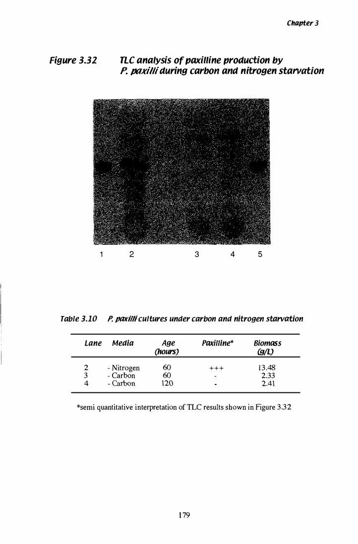
Chapter 3
Figure 3.32 nc analysis of paxilline production by P. ptlKilli during carbon and nitrogen starvation
1 2 3 4 5
Table 3.10 P. fJIIKilli cultures under carbon and nitrogen starvation
Lane Media Age Paxilline* Biomass (hours) WIL)
2 - Nitrogen 60 + + + 13 .48 3 - Carbon 60 2.33 4 - Carbon 120 2.41
*semi quantitative interpretation of TLC results shown in Figure 3 .32
1 79
Chapter 3
(Figure 3 .32, lanes 3 and 4 ) . Yeast extract does not appear to compensate for a lack of
carbon in the media.
3.4 Analysis of Paxilline Biosynthetic Gene Expression
3.4.1 RT-peR gene expression assay
The level of gene expression in P, pax illi , grown in paxilline-inducing media, was
determined with a semi-quantitative RT-PCR assay. Total RNA was isolated from
mycelia, sampled at different time points from Experiment 1 and 2 (section 2 . l 7) . In
both cases, flask B was used as the source of mycelia. The RNA was then reverse
transcribed into cDNA (section 2. 1 8) and stored at -20°C until required. Gene specific
primers for each gene of interest were selected with two main requirements. Firstly, to
distinguish cDNA products from gDNA products, primers were selected to ampl ify
regions where introns were present. Several genes of interest lacked introns, however,
stringent controls during reverse transcription (section 2. 1 8 .2 ) eliminated the likelihood
of any gDNA contaminating these pools of cDNA. Secondly, the average size of the
cDNA amplicon was approximately 400 bp, so that the amplification efficiency of each
gene product would be roughly equivalent. The number of peR amplification cycles
was optimised for each gene and the individual specifications for each gene are
summarised in Table 3 . 1 1 . Products were separated on a 2 ,5% (w/v) agarose gel
(section 2 . 1 0 . 1 and 2. 1 0 .3) and are shown in Figures 3 .33 and 3 .34. A frequent problem
was the failure of an individual peR reaction, which meant that the complete set (all
time points at three dilutions) had to be repeated. In some cases (Figure 3 .3 3 e), if the
pattern of expression could be determined, a single set of reactions has been shown.
3.4.2 Standardisation of the assay with tub-2
The first gene analysed for the level of steady state RNA was tub-2. The design of this
assay is semi-quantitative, as RNA levels are standardised after RNA isolation ( 1 20 llg
removed for DNase I treatment) and after removal of DNA ( l llg is reverse transcribed) .
1 80
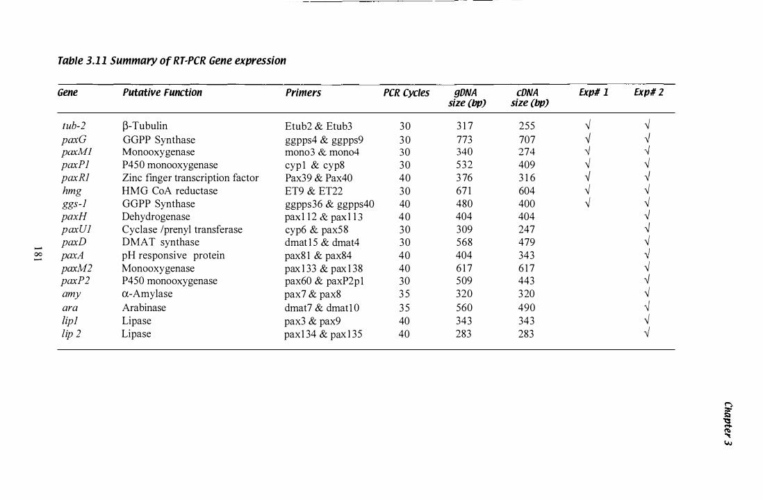
Table 3.1 1 Summary of RT·PCR Gene expression
Gene Putative Function Primers PCR Cycles gONA cONA Expl l Expl Z size (bp) size (bp)
tub-2 �-Tubulin Etub2 & Etub3 30 3 1 7 255 ,j ,j paxG GGPP Synthase ggpps4 & ggpps9 30 773 707 ,j ,j paxMl Monooxygenase mono3 & mono4 30 340 274 -j -j paxPl P450 monooxygenase cyp l & cyp8 30 532 409 ,j ,j paxRl Zinc finger transcription factor Pax39 & Pax40 40 3 76 3 1 6 ,j ,j hmg HMG CoA reductase ET9 & ET22 30 67 1 604 ,j ,j ggs-l GGPP Synthase ggpps36 & ggpps40 40 480 400 ,j ,j paxH Dehydrogenase pax1 1 2 & pax 1 1 3 40 404 404 -.J paxUl Cyclase /prenyl transferase cyp6 & pax58 30 309 247 -.J paxD D MA T synthase dmat 1 5 & dmat4 30 568 479 ,j
- ,j 00 paxA pH responsive protein pax8 1 & pax84 40 404 343 -'
paxM2 Monooxygenase pax 1 33 & pax 1 38 40 6 1 7 6 1 7 ,j paxP2 P450 mono oxygenase pax60 & paxP2pl 30 509 443 -j amy a-Amylase pax7 & pax8 3 5 320 320 ,j ara Arabinase dmat7 & dmatl 0 3 5 560 490 ,j lipl Lipase pax3 & pax9 40 343 343 ,j lip 2 Lipase pax 1 34 & pax 1 35 40 283 283 �

Chapter 3
Therefore the gene expression visualised by ethidium bromide staining, reflects the
amount of a particular mRNA species in a fixed amount of total RNA. It is possible to
think of the expression levels as a percentage of the total expression. For a gene such as
tub-2, the percentage of expression does not change over time. This is highlighted in
Figure 3 .3 3 A and 3 .34, where the tub-2 product fluoresces with equal intensity in all
lanes.
3.4.3 Iaenti(ication of the Pax pattern of expression
The expression pattern of genes involved in paxilline biosynthesis is best exemplified in
Experiment 2 by paxG (Figure 3.33 A) and reproduced using RNA isolated from
Experiment 1 (Figure 3 .34). Even at the highest cDNA concentration, there is no cDNA
product visible until 3 6 h. At lower cDNA concentrations, the expression of paxG is
seen to peak at 60 h, and then slowly decreases again. This pattern of expression is also
seen when paxP J cDNA is amplified (Figure 3 .33 A and 3 .34), although the decrease
after 60 h is not apparent. Gene replacement knockout mutants of both paxG and paxP]
have a Pax' phenotype (Young et al. , 2000). Therefore this pattern of expression was
established as the paxiIIine biosynthetic expression pattern (Pax pattern). An interesting
example of this is paxM] (Figure 3 . 33 A and 3 .34), although expression of paxM] is
detected as early as 24 h, the lower cDNA dilutions clearly reveal an increase in
expression at 36 h. Due to this increase, occurring at the same time as the onset of
expression of paxG and paxP ] , paxlld} was considered to have the Pax pattern of
expression. Additional genes that exhibit the Pax pattern of expression are paxU], lip2,
and ara in Figure 3 . 33 B, and paxD, paxP2 and possibly paxM2 in Figure 3 .3 3 C.
Some genes that were not predicted to be involved in paxilIine biosynthesis have shown
the Pax pattern of expression. For example, lip2 and ara do not have predicted
functions required for paxilline biosynthesis, although they clearly exhibit the Pax
pattern of expression. Interestingly, lip2 and ara are found c lose to the predicted
boundaries of the cluster. Genes lip] and amy have predicted functions similar to lip2
and ara, although they are located further away from the predicted boundaries of the
cluster. The expression patterns of amy and lip] were included for comparison and
neither exhibited the Pax pattern of expression (Figure 3 .33 B and 3 .33 C).
1 82
Chapter 3
Not all genes predicted to function in the paxilline biosynthetic cluster exhibit the Pax
pattern of expression. Genes paxH, a putative dehydrogenase and paxA, a putative
pH-responsive protein, were proposed to function during paxilline biosynthesis .
However, the expression of both these genes, seen in Figure 3 .3 3 C, showed no change
over time. The gene expression pattern of paxRl, the putative transcription regulator, i s
seen in Figure 3 . 33 A and 3 .34. Expression of this gene does not conform to the Pax
pattern, but does appear to change over time. Expression of paxR I was very faint at
24 h and increases to a peak level from 30 to 48 h and then decreases.
The expression pattern of hmg from Experiment 1 and 2 is shown in Figure 3 . 3 3 B and
3 .34 . In Figure 3 .34, the expression of hmg appears to resemble the Pax pattern of
expression, with an increase in expression after 36 h. However, when the pattern from
Experiment 2 is examined (Figure 3 . 3 3 B) and the time points 24 h and 3 0 h are
included, a new pattern emerges. Expression ofhrng is high at 24 h, and then decreases
at 3 0 h. After 36 h, the familiar pattern of increasing expression is seen, with
expression decreasing again after 72 h. Therefore hmg was considered to have a
modified form of the Pax pattern of expression.
The final gene to consider is ggs-l , the gene for primary geranylgeranyl pyrophosphate
synthase. This was included for comparison with paxG, the secondary geranylgeranyl
pyrophosphate synthase . Although these genes encode products of homologous
function, the patterns of expression are quite different (Figure 3 . 3 3 A) . Gene
replacement knockout mutants of paxG do not synthesis paxilline, and the product of
ggs-l does not appear to compensate the loss of the paxG product (Young et al., 2000).
Expression of ggs-l does not change significantly over time. A summary of the gene
expression patterns of all the genes analysed is shown in Table 3 . 1 2.
1 8 3

Figure 3.33 Steady state mRNA levels detected by RT·PCR in
RNA isolated from mycelia grown in Experiment 2
Total RNA was isolated from mycelia harvested at different time points (section 2. 1 7)
and reverse transcribed with random hexamer primers (section 2 . 1 8) . Gene specific
primers were used to amplify cDNA from serial dilutions of template and peR
conditions were optimised for individual reactions (Table 3 . 1 1 ) . Samples were run on a
2 .5% (w/v) agarose gel (section 2 . 1 0. 1 and 2 . 1 0 .3) . The time (h) at which the original
mycelia sample was removed is shown above the corresponding lanes. The marker (M)
used in all experiments was the 1 00 bp ladder (BioLabs) . Lanes containing products
amplified from a gDNA template are indicated with a 'G'. A 'W' indicates lanes that
contain the water controls for each set of reactions.
A) Steady state levels of mRNA from tub-2, paxG, paxM1, paxP 1 , paxR1 and
ggs-1 .
8) Steady state levels of mRNA frompaxU1, hmg, amy, /ip2 and ara.
C) Steady state levels of mRNA frompaxH, paxA, paxD, paxP2, paxM2 and /ip1
1 84
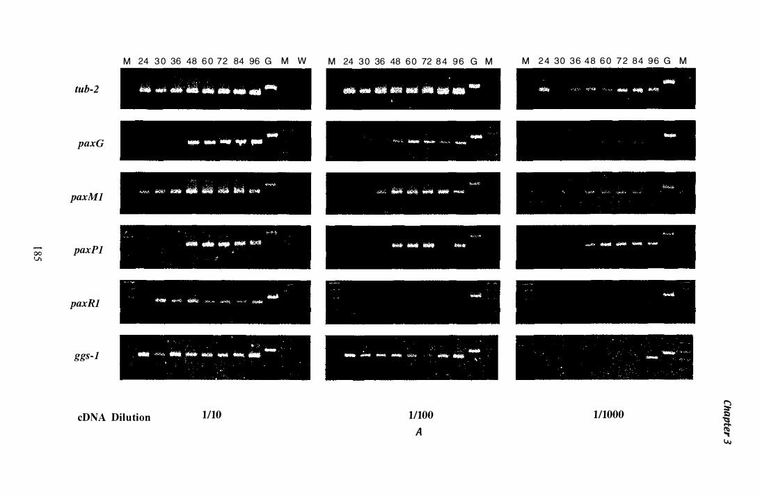
00 v.
tub-2
paxG
paxMl
paxPl
paxRl
ggs-l
M 24 3 0 36 48 6 0 72 84 96 G M W
cDNA Dilution 1110
M 24 3 0 36 48 6 0 72 8 4 9 6 G M
11100 A
M 2 4 30 3 6 48 6 0 7 2 84 96 G M
111000 � s:'.)
� "'« I./.J
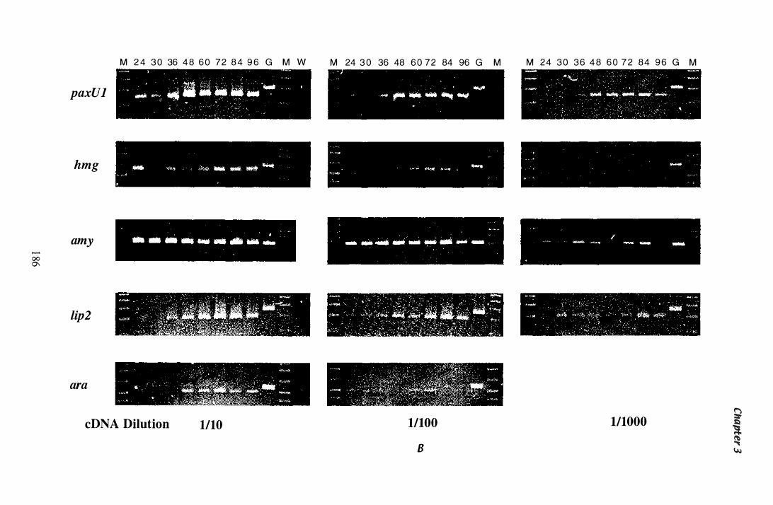
...... 00 0\
M 2 4 3 0 36 4 8 6 0 72 8 4 9 6 G M W
paxUl
hmg
amy
lip2
ara
cDNA Dilution 1110
M 24 3 0 36 48 6 0 72 84 96 G M M 24 3 0 3 6 4 8 6 0 7 2 84 9 6 G M
11100
B
111000 g
t Y.J
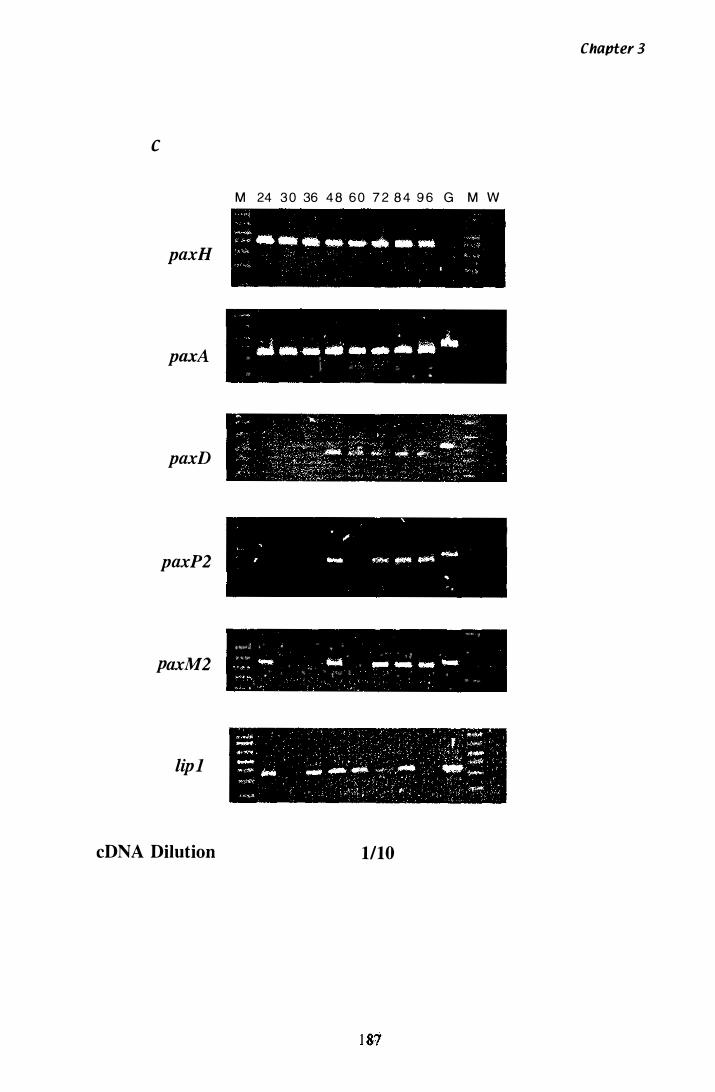
c
paxH
paxA
paxD
paxP2
paxM2
lipJ
cDNA Dilution
M 24 3 0 36 4 8 6 0 7 2 8 4 9 6 G M W
� �� � * '
�� II_"·._--.· '* z "%w " } k �t?0'7�ir »: w » » /, :t ¥ ,.,
<l!'0:'«"* * Q '" � * �
"t,X,", '"
¥¥;y� . ", ,, « � 1 $ , '" "'_ � " '" o . " .. � .. � .
" ( � ;; 1"'0 ¥ � ' " =[;;o;,�,*"�;w",MJhl;�Pd� V",� ¥t�Jk % x'� 9':" " x y.�; fe> � 'O r
1/10
Chapter 3
Figure 3.34 Steady state mRNA levels aetectea by RT-PCR in
RNA isolated from mycelia grown in Experiment 1
Total RNA was isolated from mycelia harvested at different time points (section 2. 1 7)
and reverse transcribed with random hexamer primers (section 2 . 1 8) . Gene specific
primers were used to amplify cDNA from serial dilutions of template and peR
conditions were optimised for individual reactions (Table 3 . 1 1 ). Samples were run on a
2 .5% (w/v) agarose gel (section 2 . 1 0 . 1 and 2 . 1 0 .3) . The time (h) at which the original
mycelia sample was removed is shown above the corresponding lanes. The marker (M)
used in all experiments was the 1 00 bp ladder (BioLabs) . Lanes containing products
amplified from a gDNA template are indicated with a 'G'. A 'W' indicates lanes that
contain the water controls for each set of reactions.
Steady state levels of mRNA from tub-2, paxG, paxM1, paxP 1, paxR1 and hmg are
shown.
1 88
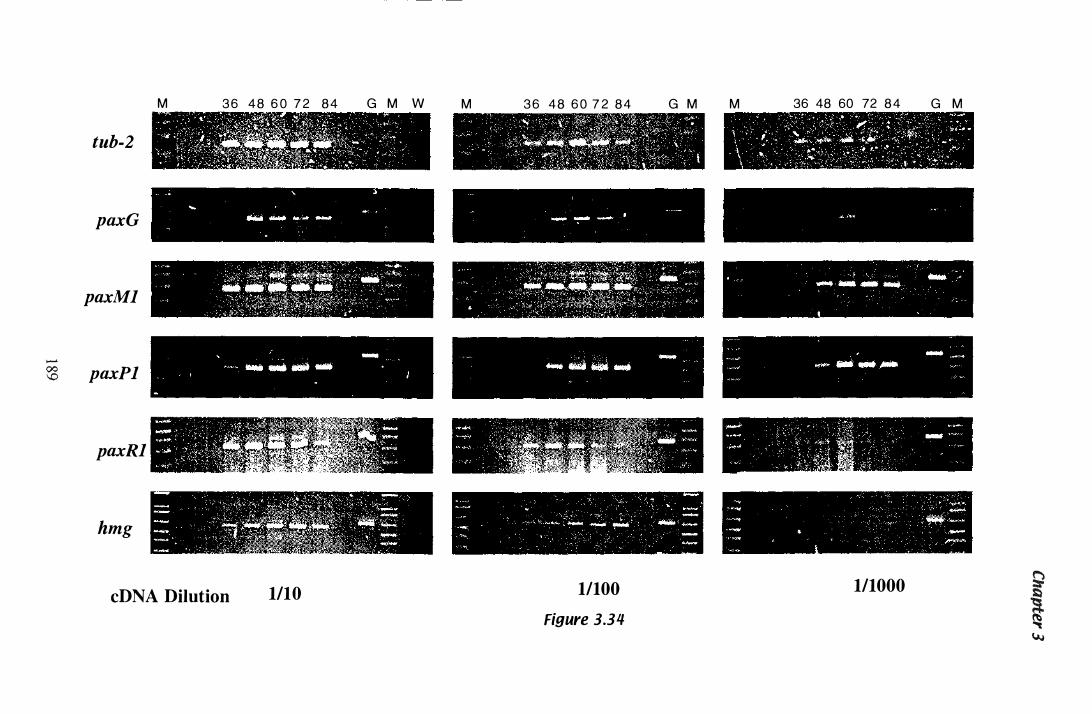
tub-2
paxG
paxMl
.......
?8 paxPl
paxRl
hmg
M 3 6 48 6 0 7 2 84 G M W
j
cDNA Dilution 1/10
M 3 6 48 6 0 7 2 84 G M M
1/100
Figure 3.34
36 48 60 72 84 G M
1/1000 g � � Y..i
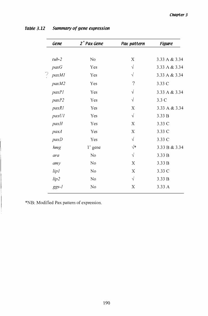
Chapter 3
Table 3.1Z Summary of gene expression
Gene Z " Pax Gene Pax pattern Figure
tub-2 No X 3 .3 3 A & 3.34 paxG Yes ,j 3 .3 3 A & 3 .3 4 paxMl Yes ,j 3 . 3 3 A & 3 .34
paxM2 Yes ? 3 .3 3 C
paxPI Yes ,j 3 . 3 3 A & 3 .34 paxP2 Yes ,j 3 . 3 C paxRI Yes X 3 .33 A & 3 .34 paxUl Yes I 3 .3 3 B "
paxH Yes X 3 .3 3 C pax:A Yes X 3 . 3 3 C paxD Yes ,j 3 .3 3 C hmg 1 0 gene -�* 3 .3 3 B & 3 .3 4
No I 3 .3 3 B ara " amy No X 3 .3 3 B lip] No X 3 .33 C lip2 No I 3 .3 3 B "
ggs-l No X 3 .3 3 A
*NB: Modified Pax pattern of expression.
190

Discussion
4.1 Isolation anti Characterisation of P. pnxilli hmg
'1.1 .1 P. paxilli hmg
Chapter 4
A unique hmg was successfully isolated from P. paxilli genomic DNA with degenerate
primers designed to the conserved catalytic region of hmg genes (Figure 3 .2) . Isolation
of genes with PCR is only possible when sufficient sequence data is available from
other species to design PCR primers. When designing degenerate primers, a number of
factors can enhance the probability of i solating a true product. These include primer
length and the percentage of degenerate nucleotides (inosines). The presence of non
conserved introns can also affect the size of genuine products. Controls of a known size
increase the l ikelihood of identifying the correct product. For example, six different
products were amplified with HMG 1 and HMG3 (Figure 3 . 1 C). It is interesting to
note, the specific 359 bp hmg product isolated was not the most abundant product. The
synthesis of additional products may be the result of inosine nucleotides in a primer and
lowered annealing temperature.
Southern analysis of the P. paxilli genome indicates that a single copy of hmg is present
(Figure 3 .3) , therefore, the mevalonate requirements for primary and secondary
metabolism, are met by a single enzyme. Once secondary metabolism is initiated,
enzyme activity may be increased to meet the new demands of paxilline biosynthesis , as
well as the requirements for primary metabolism. This may occur via increased
transcription, increased translation, decreased degradation or dephosphorylation of
specific residues in the catalytic domain. The primary metabolic requirement for
mevalonate may also be substantially l ower after the onset of secondary metabolism,
due to the cessation of exponential growth.
The chromosomal location of the hmg locus was identified by CHEF analysis of
P. paxilli chromosomes. A gene specific probe for hmg, hybridised to chromosome Vb
(Figure 3 . 1 8 A and B), indicating that hmg is not spatially linked to the paxi l l ine
biosynthetic gene cluster on chromosome Va (Young et al., 1 998a).
1 9 1
4.1.2 P. ptlKilli hmg nucleotide sequence
Chapter 4
Double stranded sequence (4.6 kb) was determined for the hmg locus, including 1 . 1 kb
of sequence upstream of the translation initiation codon. As discussed, hmg has a role
in both primary and secondary metabolism, and this may necessitate separate promoter
elements to regulate the dual requirements of this enzyme.
Sequencing of both genomic and copy DNA clones revealed three introns, one in the
5 ' UTR, and two within the coding region of the gene. These introns are labelled,
intron 1 , 2 and 3, and a summary of the sequence features is shown in Table 3.7. All
introns are within the size range for filamentous fungal introns which are generally no
greater than 1 00 bp in size (GUIT et al. , 1 987). The GT/AG rule for intron splice sites is
also conserved. At the 5 ' splice site the general consensus is GT, followed by ANGT
(BaIlance, 1 986; GUIT et al., 1 987) although conservation of the last four nucleotides is
less prominent in hmg.
At the 3 ' splice site, the consensus i s a pyrimidine (Py), fol lowed by AG. However,
variations from this sequence invariably have an adenosine nucleotide in place of a
pyrimidine (Ballance, 1 986; GUIT et al. , 1 987). In hmg, introns 1 and 2 have the
sequence AAG at the 3' splice site, and intron 3 has the more conservative CAG
sequence.
The internal consensus sequence, provides the branch site for lariat formation (V oet &
Voet, 1 990) and in yeast, the sequence 5'-TACTAAC-3 ' fulfils this function. The
adenosine nucleotide in position 6 is absolutely required for intron splicing to occur.
The branch site i s located closer to the 3 ' splice site than the 5' splice site. Ballance
( 1 986) reported that this exact sequence was rarely seen in filamentous fungi (only 3 out
of 54 introns). However, the exact sequence is found in intron 3 of the P. paxilli hmg
and a motif with a single mismatch was found in intron 2. There are no experimental
data to show that these are functional branch sites, they have been identified only by
homology to published consensus sequences. In filamentous fungi the consensus
proposed by GUIT et al. ( 1 987) is 5 '-PyGCT AAC-3' with the most conserved nucleotides
being the adenosines in position 5 and 6.
1 92
Chapter 'I
Another prominent feature of higher eukaryotic introns i s the presence o f a
poly pyrimidine tract located between the internal lariat sequence and the 3 ' branch site.
In higher eukaryotes, the polypyrimidine tract is important for correct lariat formation
and spliceosome assembly (Coolidge et al. , 1 997). Ballance ( 1 986) reported that most
filamentous fungal introns lack a polypyrimidine tract. However, polypyrimidine tracts
are present in intron 1 and intron 2. A smaller tract of 4 pyrimidines is seen in intron 3 .
Filamentous fungi contain fewer introns than higher eukaryotes, for example, the hmg
gene from humans and Chinese hamster, both contain 1 9 introns, including 1 intron in
the 5' UTR (Luskey & Stevens, 1 985 ; Reynolds et al. , 1 984). In, P. paxilli, intron 3 is
pos itionally conserved with introns in the hmg of N. loW Lp 1 9 and G. fujikuroi
(Dobson, 1 997; Woitek et al., 1 997), but is not conserved with any of the introns
present in humans or Chinese hamster hmg. The N. lolii Lp 1 9 intron is 73 bp in length,
and has a small tract of 5 pyrimidines . In G. fujikuroi, the intron is 47 bp in length and
lacks a distinctive poly pyrimidine tract.
Codon preference in P. paxilli hmg, follows trends seen in the established filamentous
fungal model organism, Neurospora crassa (Ballance, 1 986; Gurr et al. , 1 987). There
is a preference for pyrimidines in the third position, particularly C nucleotides, which
appear in 38% of all codons. The purine nucleotides, particularly adenosine, are rarely
seen in the third position and occurred in only 8% of codons in hmg (Table 3 .3) . The
coding sequence of P. paxilli hmg, from translation initiation to termination, including
introns, has a GC content of 54%.
'1.1 .3 P. paxilli hmg 5� sequence analysis
'1.1 .3.1 Initiation oftranscripfion
The transcription start site in the P. paxilli hmg was determined by 5 ' RACE, after first
establishing the approximate position by RT-PCR. Positional techniques like 5' RACE
and primer extension rely on the presence of a full-length 5 ' UTR and efficient reverse
transcription. Multiple transcription initiation sites are often found in filamentous
fungal genes (Gurr et al. , 1 987) and given the dual role hmg has in both primary and
1 93
Chapter "
secondary metabolism, it is possible that this gene also has multiple transcription start
sites. The RT-PCR estimation of the transcription start site was performed with RNA
isolated from cultures grown for 36 h and 60 h. Nested RT-PCR gave a product with
primers ET5 and ET1 2, but not with primers ET5 and ET4Bam, indicating that
transcription is initiated between ET 1 2 and ET4Bam (Figure 3 . 1 0) . No apparent
difference in 5 ' UTR length was seen between these two time points with the primers
available. However, as will be discussed in section 4 .4, the expression of genes
involved in paxil1ine biosynthesis i s seen at 36 h, and an earlier time point may have
been required to show differences in the transcription of hmg for primary and secondary
metabolism. In addition, the distance between primers ET4Bam and ET1 2 is l arge
enough that multiple transcripts could initiate between them and go undetected.
In this study, 5 ' RACE was performed on RNA isolated from cultures grown for 24 h
and 60 h. Unfortunately, 5 ' RACE products were only obtained from the 60 h RNA,
and time constraints meant a second attempt to obtain a 24 h 5 ' RACE product was not
possible . Two 5 ' RACE products were detected at 60 h (Figure 3 . 1 5), an abundant
product of 285 bp and a less abundant product of 345 bp. Due to the weak
amplification of the larger product, it was unable to be purified and sequenced in the
time available . However, the presence of the two products would indicate either the
presence of a second transcription initiation site or an alternatively spliced mRNA from
the same transcription initiation site. The smaller product was sequenced and confirmed
to start 436 bp upstream of the ATG. A putative TATA motif is located 22 bp upstream
from this site. This 5' UTR sequence has a GC content of 5 1 .3 % .
4.1.3.2 5' UTR introns
The cloning and sequencing of the 5 ' RACE product confirmed the presence of intron 1
in the 5 ' UTR. A second 63 bp 5 ' UTR intron is predicted in the P. paxilli hmg, due to
the presence of consensus splice sites (Figure 3 .7). At position -97 bp there i s a
5 ' splice motif (5 '-GTAATT-3 ') , at position -64 bp a lariat consensus sequence
(5 '-TCATAAC-3 ') is present, and at position -37 bp there is a 3' splice site (5 '-CAG-3 ') .
A 9 bp polypyrimidine tract is also present at position -58 bp. Many potential 5' and
3 ' splice sites are present in the 5 ' UTR of P. paxilli hmg (Figure 4 . 1 ), however, the
1 94
Chapter 4
presence of all four consensus motifs in the correct spatial organisation, suggests that an
unspliced intron is present in this position. Splicing of this intron was not detected and
may be extremely rare or developmentaIly regulated. Alternatively spliced products
may be present at earlier time points.
After the discovery of an intron in the 5 ' UTR of hmg from P. paxilli, the 5 ' UTR of
h mg from N. lolii Lp1 9 was examined in closer detai l . The site of transcription
initiation had been estimated using RT-PCR, and was predicted to fall between primers
hmg22 and H-2. Nested RT -PCR gave a cDNA product with H-2, but not with hmg22
(Dobson, 1 997). A number of intron consensus sequences are present in this area,
including a predicted intron that encompasses the s ite where hmg22 anneals (Figure
3 . 1 3) . The 5 ' UTR sequence of Lp1 9 hmg has been extended out to - 1 1 80 bp beyond
the translation initiation codon by Lisa McMillan, and a number of new primers were
designed to this region. Using these new primers, a single large intron of 280 bp was
detected by RT-PCR and confirmed by sequencing of a N lolii Lp 1 9 cDNA c lone
(Figure 3 . 1 2 and 3 . 1 3). Filamentous fungal introns are usually under 1 00 bp in length
(Gurr et al. , 1 987), although Ballance ( 1 986) reports a range of 48-240 bp for
filamentous fungi. As was seen in P. paxilli, there are multiple 5' and 3' intron splice
sites in the 5' UTR of the N. lolii Lp 1 9 hmg gene (Figure 4. 1 ), including sites within the
280 bp intron (Figure 3 . 1 3) . It is possible that the splice product isolated encompasses
two introns and an intervening exon. A 3' splice site was identified 73 bp downstream
from the confirmed 5' splice site, however there is no lariat branch site upstream of this
site. This site may have been lost over time, and spliceosome machinery has moved to
the next available lariat consensus and 3 ' splice junction at position - 1 1 6 bp and - 1 03 bp
respectively. A second 5' splice site was also identified 87 bp upstream of the
confirmed 3 ' splice site, provided further evidence that two separate introns may be
present. While 5'UTR introns have been observed in hmg genes from higher eukaryotic
systems, this is the first report of a 5' UTR intron in hmg genes from fungi.
4.1.3.3 5' UTR heterogeneity
When present, S' UTR introns are frequently involved in regulation. It is interesting that
the 5 ' UTR introns in P. paxilli and N lolii Lp1 9 hmg are the only splicing feature that
195
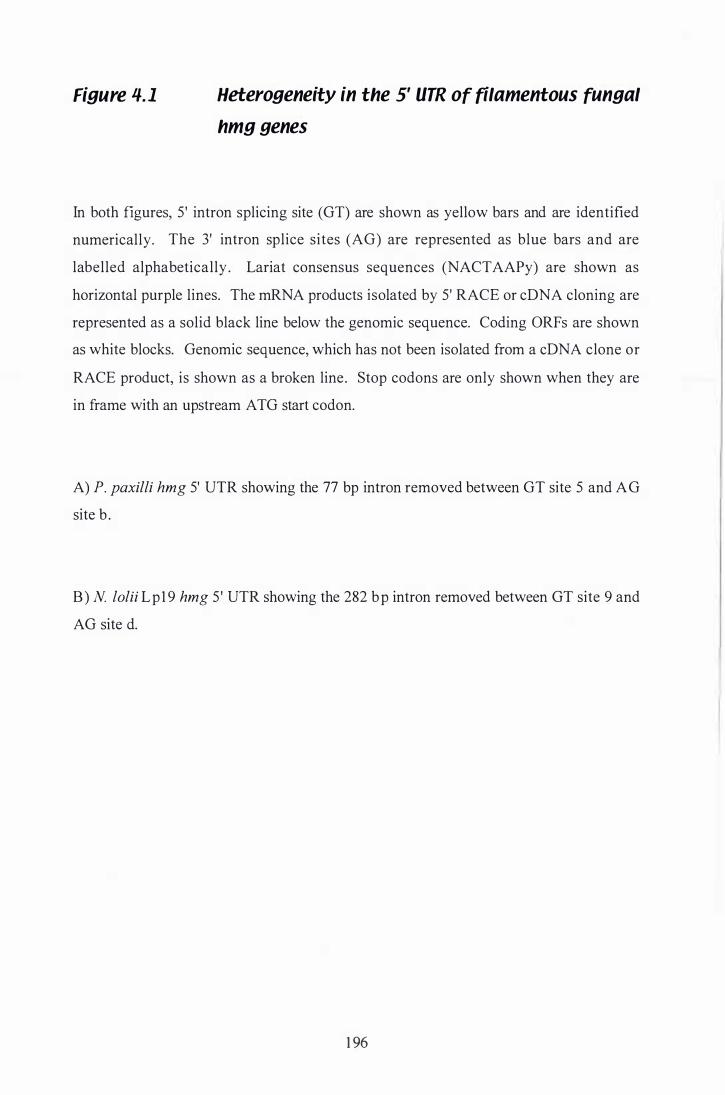
Figure 4. 1 Heterogeneity in the 5' UTR of filamentous fungal
hmg genes
In both figures, 5' intron splicing site (GT) are shown as yellow bars and are identified
numerically. The 3' intron splice sites (AG) are represented as blue bars and are
labelled alphabetically . Lariat consensus sequences (NACTAAPy) are shown as
horizontal purple lines. The mRNA products isolated by 5' RACE or cDNA cloning are
represented as a solid black line below the genomic sequence. Coding ORFs are shown
as white blocks. Genomic sequence, which has not been isolated from a cDNA clone or
RACE product, is shown as a broken line. Stop codons are only shown when they are
in frame with an upstream A TG start codon.
A) P. paxilli hmg 5' UTR showing the 77 bp intron removed between GT site 5 and AG
site b .
B) N lolii Lp 1 9 hmg 5 ' UTR showing the 282 bp intron removed between GT site 9 and
AG site d.
1 96
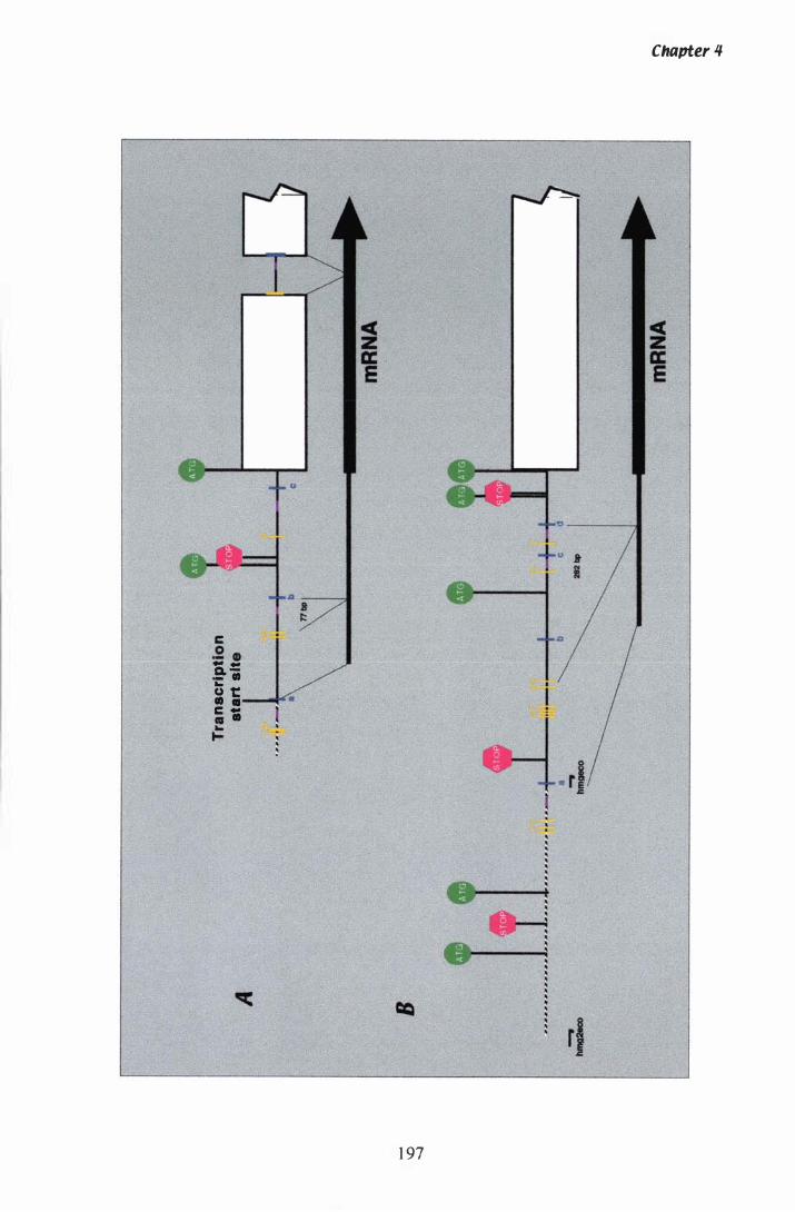
c .2 • .. .. 0. -- ti t: - . c ; • .. to-
.8 r:::
1 97
Chapter 1#
rJ

Chapter 4
is in common with hmg genes from higher eukaryotes, although the exact position of
the intron i s slightly different in the two filamentous fungi species. An optional
untrans lated exon in the 5' UTR of the HMG CoA synthase gene, is highly conserved
between humans and Chinese hamsters, and even appears to be present in the HMG
CoA reductase gene from Chinese hamster as well (Gil et al., 1 987). Even though the
intervening intronic sequences are much longer in the higher eukaryotes, the 5 'UTR
exons in P. paxilli and N lolii Lp 1 9 hmg are similar in size to those found in the 5 '
UTR o f Chinese hamster and human HMG CoA synthase. The heterogeneity present in
the S ' UTR of the Chinese hamster hmg has been extensively characterised. Primer
extension showed that 1 6 different transcripts are synthesised, using a combination of
multiple transcription start sites and alternative splicing of the S' UTR intron. Multiple
sites of initiation of translation are also present (Reynolds et al., 1 985). Ratios of these
multiple transcripts were affected by lovastatin (an inhibitor of HMGR), mevalonate
and 25-hydroxycholesterol. Transcripts with shorter 5 ' UTR (40- 1 00 nt) were isolated
from a polysome cellular fraction, while transcripts with longer S ' UTR (300-400 nt)
were found in the monosome fraction (Gayen & Peffley, 1 995). One of the Arabidopsis
thaliana hmg genes, HMG 1 , i s also transcribed from two different promoters, one of
which includes an in-frame alternative translation start site. Proteins translated from the
upstream translation start site have 50 additional residues in the N-terminus, and have
been isolated from microsomes (Lumbreras et al. , 1 995) . Therefore, 5' UTR
heterogeneity could have a role in targeting HMGR to different subcellular
compartments. There is a second ATG upstream of the major translational start site, but
this ORF is quickly terminated by a stop codon. In human hmg, alternative splicing of
the 5 ' UTR intron does not occur, although several different transcripts are produced,
presumably from mUltiple transcription start sites (Luskey, 1 987). Figure 4. 1 highlights
the potential for alternate splicing and translation initiation in hmg genes from P. paxilli
and N lolii Lp 1 9, including some short ORFs, that are in frame with the major
translational start site. Several mechanisms are already known to regulate the
rnevalonate pathway, including feedback repression of transcription by the end products
of cholesterol biosynthesis (Goldstein & Brown, 1 990) and phosphorylation of the
HMG Co A reductase enzyme (Kennelly & Rodwell, 1 985) . In Saccharomyces
cerevisiae and A. thaliana, two copies of the HMG CoA reductase gene are
differentially expressed to fulfil regulatory requirements (Enjuto et al., 1 994; Hampton
et al. , 1 996a). Given the biological importance of the primary metabolic products
1 98
Chapter 'I
produced from mevalonate and the need for regulation of their levels, it is not surprising
that multiple regulatory mechanisms exist. Whether the regulatory requirements of the
secondary metabolic pathways that use mevalonate as a substrate, such as the paxilline
biosynthetic pathway in P. paxilli, are met by these primary mechanisms is unclear at
present. The complexity seen in the 5 ' UTR of P. paxilli and Lp1 9 hmg genes provides
an interesting alternative regulatory mechanism, worthy of further investigation.
4.1.3.4 Putative promoter elements
In higher eukaryotes, the TA TA box is frequently found 1 0-30 bp upstream from the
site of transcription initiation (Stryer, 1 995) and is essential for assembly of the
transcriptional machinery. In filamentous fungi, the TAT AAA motif or simply an AT
rich tract is frequently present (Gurr et al. , 1 987), although the position and exact
sequence of the motif is more variable in filamentous fungi (Ballance, 1 986) . In
P. paxilli hmg, the motif 'TT AA TT' is found 22 bp upstream from the proposed
transcription start site and may fulfil the role of this core promoter element. A number
of pyrimidine-rich tracts are present in the P. paxilli hmg 5 ' UTR. These motifs are
frequently a feature of highly expressed genes (Gurr et al. , 1 987), although the
pyrimidine rich tracts in hmg do not directly precede the transcription start site
determined by 5 ' RACE as is usually the case for filamentous fungi (Ballance, 1 986).
Tomato (Lycopersicon esculentum), like many plants, contains two copies of the hmg
gene. Expression of HMG2 is correlated to the synthesis of carotenoid during ripening.
Interestingly, while the TAT A motif, upstream of the transcription start site, was not
required for expression, a pyrimidine-rich tract in the 5' UTR was (Daraselia et al. ,
1 996). In Chinese hamster, the hmg gene also lacks a TA TA box, although the gene for
HMG CoA synthase does have a TAT A box approximately 30 bp upstream from the
maj or transcription start (Goldstein & Brown, 1 990). The core promoter e lement
CAA T is generally found 70-90 bp upstream of the transcription start site, however
nothing resembling this sequence is found in the P. paxilli hmg promoter at this
position. Several CAAT motifs exist between the proposed transcription and translation
start sites.
1 99
Chapter 4
Many sterol-regulated genes have the binding motifs for Nuclear Factor Y and Sterol
regulated element binding proteins SREBPs in their promoter. Ericsson et al. ( l 996b)
showed that both NF-Y and SREB-3 were required for sterol regulation and the spacing
between the two binding sites was critical for the correct protein-protein interaction .
The sequence motif for NF-Y binding is an inverted CCAAT box (5 ' -ATTGG-3 ' ) ,
although mutation of a 5'-CCAAT-3' motif in the Human squalene synthase gene
(Inv-Y -Box) resulted in diminished promoter activity (Guan et al., 1 998). This motif i s
located downstream of the transcription start site and i s therefore distinct from the
CAAT box discussed above. There are two 5'-CCAAT-3 ' boxes in the P. paxilli hmg
gene at position -932 bp and -984 bp (Figure 3 . 7) . Examples of the sterol responsive
elements (SRE-1 , -2 and -3), known to regulate the transcription of cholesterogenic
genes are shown in Table 4 . 1 . A putative SRE- l motif (5 ' -CGCCCCAC-3 ' ) was
identified in the 5' UTR of P. paxilli hmg at position -509 bp (Figure 3 . 7) , 73 bp
upstream from the confirmed transcription start site. Three other putative SRE- l sites,
each containing 2 mismatches (5' CACACCAA-3') , occur in a mini repeat sequence at
pos ition -642 bp. Six different sites with homology to SRE-2 were identified at
positions, -566 bp, -522 bp, -446 bp, - 1 94 bp, - 1 88 bp and -57 bp respectively. The
motif at position - 1 94 bp is inverted and is listed with the other putative SRE-2 motifs
in Table 4 . 1 . Two putative SRE-3 sites were identified in the P. paxilli hmg 5 ' UTR,
although one of these, at position -508 bp (Figure 3 .7), contains 5 mismatches (5'
CTCTCCACCG-3 '). However, a second inverted SRE-3 motif (5 '-CTCCCACGAG-3'),
at position -227 bp (Figure 3 .7), has only a single mismatch. In the human squalene
synthase gene, a functional, inverted SRE-3 element was also identified. Mutants
generated with variations in this site showed decreased sterol activation (Guan et al. ,
1 998) . Therefore, even amongst higher eukaryotes, there i s some divergence between
the sequence motifs involved in sterol regulation. Similar motifs in filamentous fungi
may need to be identified experimentally.
N itrogen repression often regulates genes involved in many aspects of fungal growth
and development, including secondary metabolism and pathogenicity. Major nitrogen
regulatory proteins from fungi include NRE from P. chrysogenum (Haas et al., 1 995a),
NIT2 from N crassa (Fu & Marzluf, 1 987) , AREA from A. nidulans (Caddix et al. ,
1 986), NRRl from Metarhizium anisopliae (Screen et al. , 1 998) . AREA-GF from
200
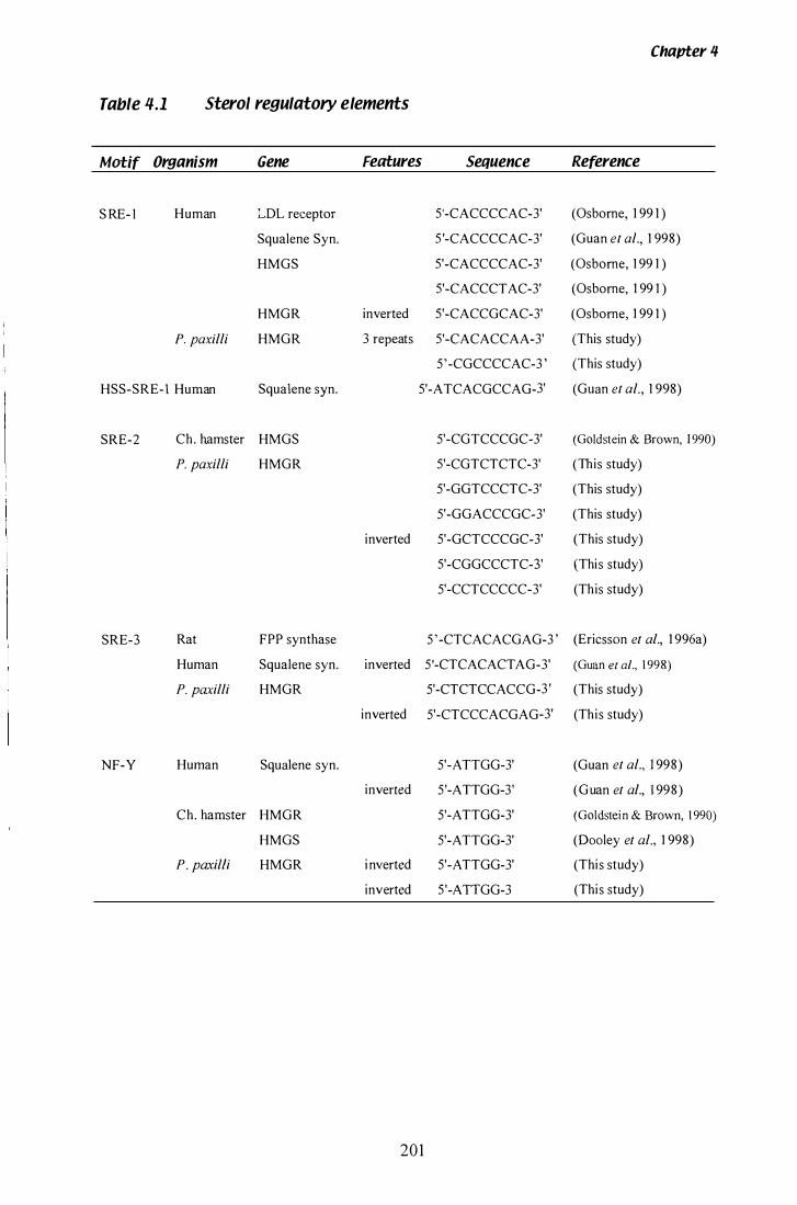
Chapter 4
Table 4.1 Sterol regulatory elements
Motif Organism Gene Features Sequence Reference
S RE- l Human LDL receptor S'-CACCCCAC-3' (Osbome, 1 99 1 )
Squalene Syn. S '-CACCCCAC-3' (Guan et al., 1 998)
HMGS S'-CACCCCAC-3' (Osbome, 1 99 1 )
S'-CACCCTAC-3' (Osbome, 1 99 1 )
HMGR inverted S'-CACCGCAC-3' (Osbome, 1 99 1 )
P. paxilli HMGR 3 repeats S'-CACACCAA-3' (This study)
5 ' -CGCCCCAC-3 ' (This study)
HSS-SRE-I Human Squalene syn. S'-ATCACGCCAG-3' (Guan et al., 1 998)
SRE-2 Ch. hamster HMGS 5'-CGTCCCGC-3' (Goldstein & Brown, 1 990)
P. paxilli HMGR S'-CGTCTCTC-3' (This study)
5'-GGTCCCTC-3' (This study)
5'-GGACCCGC-3' (This study)
inverted 5'-GCTCCCGC-3' (This study)
S'-CGGCCCTC-3' (This study)
5'-CCTCCCCC-3' (This study)
SRE-3 Rat FPP synthase 5 ' -CTCACACGAG-3 ' (Ericsson et al., 1 996a)
Human Squalene syn. inverted 5'-CTCACACT AG-3' (Guan et al., 1 998)
P. paxilli HMGR 5'-CTCTCCACCG-3 ' (This study)
inverted 5'-CTCCCACGAG-3' (This study)
NF-Y Human Squalene syn. 5'-A TTGG-3' (Guan et al., 1 998)
inverted 5'-A TTGG-3' (Guan et al., 1 99 8 )
Ch. hamster HMGR 5'-A TTGG-3' (Goldstein & Brown, 1 990)
HMGS 5'-A TTGG-3' (Dooley et al., 1 998)
P. paxilfi HMGR i nverted S'-A TTGG-3' (This study)
inverted S'-ATTGG-3 (This study)
201

Chapter 4
G. fujikuroi (Tudzynski et al., 1 999) and NUT1 from Magnaporthe grisea. (Froeliger &
Carpenter, 1 996). These all belong to the GA TA family of transcription factors which
have a CyS2/CyS2 zinc finger motif, and bind the GA TA motif in the promoters of
nitrogen regulated genes (Wilson & Arst Jr. , 1 998). In N crassa, nitrate utilisation
genes have pairs of 5'-ATAG-3' or 5'-GATA-3' motifs separated by a variable number
of nuc1eotides (Marzluf, 1 993) . There are two 5'-GATA-3' motifs in the P. paxilli hmg
promoter, one 27 bp upstream of the transcription start site and a second 358 bp
upstream. There are also two pairs of A TAG motifs, one pair at position -955 bp
separated by 1 1 bp, and a second pair at position -875 bp separated by 3 bp (Figure 3 .7).
Carbon catabolite repression (CCR) can also regulate expressIOn of secondary
metabolic genes . The negative-acting, regulatory protein CREA binds the consensus
5 '-(G/C)YGGRG-3' in A. nidulans (Panozzo et al. , 1 998). Two binding sites, matching
this consensus, were detected in the P. paxilli hmg 5 ' UTR at positions -603 bp
(5'-CTGGAG-3') and -440 bp (5'-GCGGAG-3'). This second sequence overlaps the
transcription start determined by 5 ' RACE. Two CCAAT motifs, which b ind the
positive-acting Hap2/3/4/5 complex in yeast, appear in the P. paxilli hmg promoter
548 bp and 496 bp upstream of the transcription start site. Several CG triplets are
present next to the transcription start site, but there is no consensus for the number of
nuc1eotides that separate them. GC triplets are involved in binding the positive acting
proteins Gal4 and Ma163, responsible for specific regulation of galactose and maltose
utilisation genes in yeast (Gancedo, 1 998).
The regulatory element PacC, has been shown to be involved in the pH dependent
regulation of the isopenicillin N synthetase gene of A. nidulans (Espeso & Pefialva,
1 996) . PacC i s a Cys2iHis2 type zinc finger protein that b inds the consensus
5'-GCCARG-3 ' . There are no binding sites for PacC in the P. paxilli hmg promoter,
although two motifs were found approximately 60 bp downstream of the A TG start
codon.
Two final features in the promoter region of P. paxilli hmg, are mini repeat sequences at
position -972 bp and -642 bp. The first repeat has four copies of the sequence
5'-TACA-3' , although the third repeat is imperfect. The second repeat has three copies
of the sequence 5'-CACACCAA-3 ' and has some homology to the SRE- 1 sequence
202
Chapter 'I
motif discussed above. There are no experimental data to confirm that any of these
putative promoter elements are functional, except for the intron splicing sites that were
confirmed by cDNA sequencing.
4.1 .4 P. ptlxilli hmg 3� sequence analysis
Filamentous fungal genes are frequently transcribed beyond the signal sequence for
polyadenylation (Gurr et al. , 1 987) and this may certainly be the case for P. paxilli hrng.
Using 3 ' RACE, the 3 ' UTR was shown to be 563 bp long and, in contrast to the
5 ' UTR and coding sequence, the 3 ' UTR has a GC content of only 49.7%. In higher
eukaryotes, the sequence 5 ' -AATAAA-3 , is the signal for polyadenylation in 90% of
mRNA (Colgan & Manley, 1 997). Adherence to this sequence varies in filamentous
fungi and often the exact site for polyadenylation is difficult to locate. However, there
is a tendency towards AT nucleotides in the 3 ' UTR (Bal1ance, 1 986), that may serve
the same function. In P. paxilli hmg, the sequence ' ATAAAA" is found 438 bp
downstream from the translation stop codon. S I nuclease mapping of the 3 ' UTR of
Chinese hamster hmg showed three functional polyadenylation sites, although 5 signal
sequences for polyadenylation were present (Reynolds et al., 1 984). A microsatellite,
present in the 3 ' UTR of N lolii Lp 1 9 hrng, was not seen in the P. paxilli hrng gene.
4.1.5 P. ptlxilli hmg polypeptide sequence
Translation is initiated at the A TG codon in the majority of eukaryotic genes. Sequence
surrounding the preferred translation start sites was determined by Kozak ( 1 984) and
shown to be, 5 ' -CC(A/G)CCATGGC-3 ' . The consensus sequence propo sed for
filamentous fungal genes was 5 '-TCA(CIA)(AIC)ATG(G/T)C-3 ' (Ballance , 1 986).
Translation of P. paxilli hrng i s assumed to initiate at an A TG surrounded by the
sequence 5 ' -TCACCATGGC-3 ' which supports the filamentous fungal consensus
sequence. Translation is assumed to terminate at a T AA codon, which is the established
trend in filamentous fungal genes (GUIT et al., 1 987).
203
Chapter 4
The peptide sequence of P. paxilli hmg is most similar to hmg genes from G. fujikuroi
(74% similarity), N. lolii Lp1 9 (74%), A. terre us (69% similarity) and S. pombe (64%
similarity) . An alignment of HMGR protein sequence from a number of species was
performed with the PILEUP programme of the GCG package, using a gap weight
penalty of 2 and a gap length penalty of 1 (Figure 3 .8). The N-terminal domain is most
similar to N lolii Lp 1 9 hmg. The published sequence for G. fujikuroi hmg has a
truncated transmembrane domain (Woitek et al. , 1 997), although evidence presented by
Do bson ( 1 997) predicts that the start of this gene is further upstream than that proposed
by Woitek et al. ( 1 997). Upstream of the G. fujikuroi translation codon, an ORF was
identified with similarity to the N-terminal domain of both N lolii Lp 1 9 and P. paxilli
hmg.
A model for the structure of the HMGR protein has been discussed by Liscum et al.
( 1 985). Proteolysis experiments on the Chinese hamster HMGR protein (Chin et al. ,
1 984) showed a membrane bound N-terminal domain and a cytosolic, catalytically
active C-terminal domain, with a less conserved linker domC;lin separating the two. The
structure of the N-terminal domain is less conserved amongst disparate species. In
plants, two or more genes are present, each contains two membrane spanning domains
in the N-terminus (Campos & Boronat, 1 995 ; Denbow et al., 1 996; Enjuto et al. , 1 994).
In S. cerevisiae, two genes are present, but the N-terminal domains contain
7 membrane-spanning domains (Basson et al. , 1 986). In mammals, single genes are
present, that contain 7 or 8 transmembrane domains (Chin et al. , 1 984; Hampton et al. ,
1 996a; Liscum e t al., 1 985 ; Roitelman e t al. , 1 992). Thus far, in filamentous fungi, the
number of h mg genes and organisation in the N-terminal domain most closely
resembles mammals. A single hmg gene is present in N lolii Lp 1 9 (Dobson, 1 997),
G. fujikuroi, Sphaceloma manihoticola (Woitek et al., 1 997) and P. paxilli. However,
Southern analysis of two zygomycetes, Absidia glauca and Blakeslea trispora, predicts
two copies of hmg (Burmester & Czempinski, 1 994). Hydropathy analysis predicts
7 transmembrane domains in the HMGR of N loW Lp 1 9 and G. fujikuroi. Using the
TMPRED programme (Hofmann & Stoffel , 1 993) at the website of the I SREC
Bioinformatics group, the predicted N-terminal domain of P. paxilli hmg was analysed
using the method of Kyte & Doolittle ( 1 982) and predicted to contain 7 transmembrane
domains in the N-terminal portion of the protein.
204
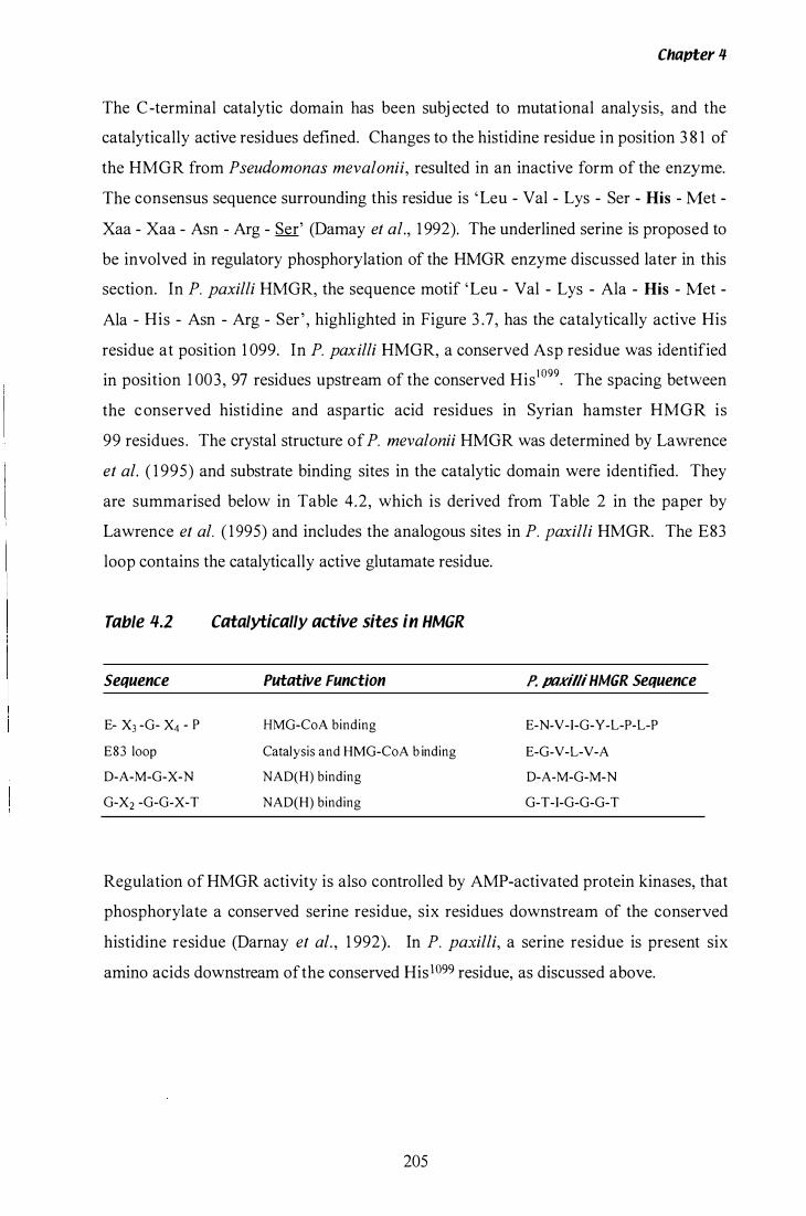
Chapter il
The C-terminal catalytic domain has been subj ected to mutational analysis, and the
catalytically active residues defined. Changes to the histidine residue in position 3 8 1 of
the HMGR from Pseudomonas mevalonii, resulted in an inactive form of the enzyme.
The consensus sequence surrounding this residue is 'Leu - Val - Lys - Ser - His - Met
Xaa - Xaa - Asn - Arg - Ser' (Damay et al., 1 992). The underlined serine is proposed to
be involved in regulatory phosphorylation of the HMGR enzyme discussed later in this
section. In P. paxilli HMGR, the sequence motif 'Leu - Val - Lys - Ala - His - Met -
Ala - His - Asn - Arg - Ser ' , highlighted in Figure 3 .7, has the catalytically active His
residue at position 1 099. In P. paxilli HMGR, a conserved Asp residue was identified
in position 1 003, 97 residues upstream of the conserved His I 099. The spacing between
the conserved histidine and aspartic acid residues in Syrian hamster HMGR is
99 residues. The crystal structure of P. mevalonii HMGR was determined by Lawrence
et al. ( 1 995) and substrate binding sites in the catalytic domain were identified. They
are summarised below in Table 4.2, which is derived from Table 2 in the paper by
Lawrence et al. ( 1 995) and includes the analogous sites in P. paxilli HMGR. The E83
loop contains the catalytically active glutamate residue.
Table '1.2
Sequence
E- X3 -G- X4 - P
E83 loop
D-A-M-G-X-N
G-X2 -G-G-X-T
Catalytically active sites in HMGR
Putative Function
HMG-CoA binding
Catalysis and HMG-CoA b inding
NAD(H) binding
NAD(H) binding
P. ptl%illi HMGR Sequence
E-N-V -I-G-Y -L-P-L-P
E-G-V-L-V-A
D-A-M-G-M-N
G-T-I-G-G-G-T
Regulation of HMGR activity is also controlled by AMP-activated protein kinases, that
phosphorylate a conserved serine residue, six residues downstream of the conserved
histidine residue (Darnay et al. , 1 992). In P. paxilli, a serine residue is present six
amino acids downstream of the conserved His l099 residue, as discussed above.
205
4.2 Isolation ana Characterisation of P. pUKilli ttlb-2
4.2.1 P. pnxilli tub�2
Chapter lf.
The gene for �-tubulin i s highly conserved, and together with a-tubulin, the gene
products form the micro tubules essential for chromosome separation during mitosis
(Stryer, 1 995). Expression of tub-2 is an excellent control for studying gene expression
in most organisms. A unique tub-2 gene was isolated from P. paxilli and the pattern of
gene expression used as a control for the experiments discussed in section 4.4. Due to
the high sequence conservation, this gene was isolated from a P. paxilli genomic library
by heterologous probing with the N. crassa tub-2 gene. The N. crassa probe used for
Southern analysis (Figure 3 . 1 8), confirmed a single copy of tub-2 is present in the
genome. A single copy of tub-2 is found in N. crassa (Orbach et al. , 1 986), Epichloe
typhina (Byrd et al. , 1 990) and the homobasidiomycete Schizophyllum commune,
although there is some evidence for a highly divergent second copy in this species
(Russo et al. , 1 992). A. nidulans contains two �-tubulin genes (benA and tubC) which
show significant sequence divergence (May et al. , 1 987). CHEF analysis showed that
the tub-2 gene is located on chromosome IV in P. paxilli (Figure 3 .24).
4.2.2 P. pnxilli tub*2 nucleotide sequem;e
The coding sequence of tub-2 is separated by 6 introns, five of which are clustered in
the 5' end of the gene. The sizes of the six introns are 1 07 bp, 1 20 bp, 68 bp, 66 bp,
7 1 bp, and 62 bp respectively. This is within the range (48-240 bp) reported by
Ballance ( 1 986). As discussed in section 4 . 1 .2, the GTI AG rule for intron splicing is
conserved, with the sequence 'ANGT' fol lowing the GT 5 ' splice junction. The PyAG
consensus is conserved at all 3 ' splice junctions. Introns 3-6 were confirmed by
sequencing cDNA clones, and the splice j unctions for introns 1 and 2 were predicted by
sequence similarity to other tub-2 genes. The number of introns in tub-2 genes varies
amongst filamentous fungal species. In A. nidulans, benA has 8 introns and tube 5
introns (May et al. , 1 987) E. typhina tub2 contains 4 introns (Byrd et al. , 1 990), the
206
Chapter 4
N crassa �-tubulin gene contains 6 introns (Orbach et al., 1 986) and S. commune tub-2
has 8 introns (Russo et al. , 1 992).
Codon bias in P. paxilli tub-2 is shown in Table 3 .6, and again follows the trends for
filamentous fungal genes (Ballance, 1 986; GUIT et al., 1 987). A cytosine nucleotide was
seen in the third position in 5 1 % of all codons and adenosine nucleotides were seen in
the third position in only 1 % of all codons. The coding sequence of the tub-2, from
translation initiation to termination, including introns, has a GC content of 55 .3%.
4.2.3 P. pnxilli tulJ-2 polypeptide sequence
The tub-2 sequence isolated from P. paxilli predicts a 447 amino acid protein comprised
of 7 ORF. The protein sequence is most similar to �-tubulin genes from P. digitatum,
A . flavus and A. parasitic us (all 93% similarity) and Emericella nidulans (92%
similarity ) .
r
207
4.3 Physiology of P. pnxilli in liquid culture
Chapter 4
1/-.3.1 Development of cultures with homogeneous morphology ana
reproaucible inauction of paxilline
There are numerous techniques for inoculating filamentous fungi into liquid culture,
including spore suspensions, seed cultures, and macerated mycelia fragments. Two of
these techniques (spore suspension and seed culture) were compared for their ability to
induce i) a homogeneous morphology and ii) paxilline bio synthesis in a reproducible
manner. The growth rate was determined by dry weight, which reflects biomass
accumulation rather than cellular division. Paxilline content was determined by HPLC
(section 2 . 1 6) and reflects paxilline production per unit of biomass. Accumulation of
biomass and paxilline showed considerable variation in cultures inoculated from a spore
suspension (Figure 3 .25 A). Distinguishing experimental differences from the inherent
variability in experiments performed with this inoculation protocol would have proved
difficult.
A number of papers describe the use of a seed cultures to inoculate replica flasks (Ibba
et al., 1 987; Kaiser et al., 1 994; Tkacz et al., 1993) so several variations of this method
were tried with P. paxilli. Seed culture volumes of 25 mL to 1 00 mL were examined
after 24 h growth and all cultures were found to have produced the large pellet
morphology, even when grown at 3 50 rpm. When a sample of each seed culture was
subsequently transferred to a larger culture volume, both the size and number of pellets
remained constant. Growth experiments performed with mycelia grown in the pellet
morphology were not desired for two reasons. Firstly, sample volumes removed from
P. paxilli cultures with the pellet morphology are not representative of the entire culture.
This is because large pellets sediment rapidly once a culture is removed from the
shaker. The loose homogeneous morphology was also preferred during labelling
experiments to determine the origin of the paxilline indole moiety in P. paxilli (Laws &
Mantle, 1 989). Loose mycelia fragments do not sediment and samples taken from these
cultures are representative of the entire culture. Secondly, individual hyphae in a
culture grown with the loose mycelia morphology have equal access to nutrients and
oxygen and are therefore in a uniform physiological state. It was important to develop
an inoculation protocol that decreased the inherent variability seen in Figure 3 .25 A, as
208
Chapter .q.
well as maintaining the preferred loose mycelial morphology. A paper by Tkazc et al.
1 993 recommended the use of oat flour in the seed culture to inhibit the formation of
pellets. However pellets were still present when this method was tested on P. paxilli.
The main problem appeared to be that once pellets had formed, they remained,
irrespective of culture volume or shake speed. To solve this problem, the seed culture
was sub-cultured before the gross morphology became fixed. After 1 2 h, viable spores
have germinated, and germination pegs are clearly visible under a light microscope
(Figure 3 . 29, photograph A and B). No pellets are visible, although clumps of 20 or
more spores are frequently seen at this time and may represent the start of a pellet. The
large 300 mL cultures inoculated from a 1 2 h seed culture showed an almost ideal
morphology . The majority of the culture grew as loose mycelia fragments with fine
micropellets dispersed throughout. The nutrient and oxygen gradients that exist in
larger pellets are less marked in smaller pellets. The biomass accumulation and
paxilline content of cultures inoculated from a 1 2 h seed culture is shown in Figure 3 .25
B. Using this protocol, the growth rates and period of paxilline induction are
reproducible (Figure 3 .27, Figure 3 .28).
4.3.2 Wild-type P. pnxilli in paxilline-inttucing media
Production of secondary metabolites in filamentous fungal l iquid cultures has been
discussed in a number of papers. In particular, the induction of alkaloid biosynthesis in
l iquid culture can be extremely difficult to achieve. In Penicillium nigricans l iquid
cultures, biosynthesis of the tremorgenic mycotoxin penitrem, was stimulated by
calcium induced sporulation (Mantle et al. , 1 984), while production of lolitrem B in
N loW l iquid cultures has not been achieved (Weedon & Mantle , 1 987). Conditions for
the production of paxilline by P. paxilli in liquid culture were determined by Ibba et al.
( 1 9 87) . The aim of this thesis was to determine the rate of growth and paxilline
production in l iquid cultures of wild-type P. paxilli. These growth patterns, once
determined, would serve as controls for experiments where the growth conditions were
varied or when P. paxilli mutants were analysed. Two separate growth experiments
were performed using the seed culture inoculum protocol . The first experiment
identified time points that required more detailed examination, hence the inclusion of
additional time points in Experiment 2. The second experiment examined many
209
Chapter 4
parameters of culture physiology, including culture morphology, and paxil I ine content
of the culture supernatant.
'1.3.2.1 8iomass accumulation
The majority of biomass accumulation occurs during the primary growth or
trophophase. Growth at this stage is essentially exponential (Prosser & Tough, 1 99 1 ).
However, additional biomass, in the form of storage products can still accumulate after
the cessation of exponential cellular division. In some cases, the biomass is maintained
at a steady level through balanced autolysis and regeneration (Prosser & Tough, 1 99 1 ) .
In P. paxilli, the primary growth phase and accumulation of biomass ceased between 48
and 60 h after seed culture inoculation (Figure 3 .27 A and 3 .28 A) , with an average
biomass accumulation of 20 g/L. During the late secondary phase, the biomass begins
to drop dramatically rather than remaining at a constant level. The rate of autolysis is
presumably greater than the rate of regenerative growth at this time. Possibly, the
biomass is being broken down to provide carbon for the production of paxilline. A
dramatic decrease in biomass and viscosity was observed in P. paxilli cultures after
1 44 h of growth. This final phase is known as the death phase (Prosser & Tough, 1 99 1 ).
I t i s at this time that paxilline was first detected in the culture broth, presumably as a
result of increased autolysis during the death phase. Microscopy showed that increased
autolysis was occurring after 1 00 h (Figure 3 .29, photographs M and N), but only trace
amounts of paxilline were detected between 84 h and 1 20 h. The amount of autolysis
must increase drastically after 1 20 h to account for the presence of broth paxilline and
the decreased culture viscosity.
'1.3.2.2 Culture pH
P. paxilli l iquid cultures exhibited two distinct changes in pH, which were correlated
with distinct phases of growth (Figure 3 .27 B and 3 .2 8 B). During the initial
exponential growth phase, culture pH increases in parallel with biomass accumulation.
The pH then begins to slowly decline, until 96 h, when a dramatic decrease was
2 10
Chapter 'I
recorded in Experiment 2 . Whether the pH drop i s a cause or consequence of the
transition from primary growth phase to secondary growth phase is unclear. An initial
experiment to investigate this question is discussed in section 4 .3 .5 .
4.3.2.3 Paxilline
Paxilline isolated from mycelia was detected after 72 h, and continued to accumulate for
the duration of the experiment. Paxilline was not detected in the broth until 1 44 h,
although an extremely small peak was observed from 84 h onwards, that corresponded
with the elution time of paxilline. The size of this peak did not change until 144 h,
when a dramatic increase was observed; indicating that paxilline is not actively secreted
into the media. The rapid release of paxilline could result from massive cellular lysis
after the culture enters the death phase. Interestingly, a steady decrease in biomass was
recorded from 60 h onwards, with no equivalent increase in broth paxilline. Paxilline
contained within mycelia undergoing autolysis at earlier time points, does not appear to
be released into the culture supernatant until large scale autolysis at 1 44 h.
4.3.2.4 Microscopy
Microscopic analysis of P. paxilli liquid cultures revealed information relevant to the
biosynthesis of paxilline. Firstly, sporulation does occur in the paxilline-inducing
media CDYE. However, the appearance of spore-bearing structures is less apparent
after 76 h, when paxilline i s first detected in mycelia. Spore production in the paxilline
inducing media was also significantly lower than that recorded in culture media
containing high concentrations of the sporulation-inducing supplement, calcium.
Secondly, despite the decrease in overall biomass, the majority of hyphae showed no
sign of vacuolation or autolysis, until 1 00 h. Vacuoles were observed in hyphae after
76 h, although limited vacuolation and autolysis might have gone undetected in earlier
samples. Vacuolation is a feature of hypha I age, and therefore increases over time (Cui
et al. , 1 998). The decrease in biomass could reflect the increasing size of vacuoles and
decreasing amount of cytoplasm and storage products in individual hyphae.
2 1 1
4.3.3 Effect of sporulation on paxilline production
Chapter 4
In contrast to many filamentous fungi (Guzman-de-Pefia & Ruiz-Herrera, 1 996; Mantle
et al. , 1 984), P. paxilli production of paxilline is not correlated with sporulation, as
documented in a paper by Ibba et al. ( 1 987). Microscopic analysis of cultures showed
that minimal sporulation does occur in the paxil line-inducing media, however in
comparison to the spore-inducing media, this is significantly reduced. The effect of
increased sporulation on paxilline production i s even more pronounced. Paxilline was
not detected in cultures supplemented with 2% CaCh (Table 3 .8, Figure 3 .3 0) and only
trace amounts of paxilline were detected in mycelia grown in PD media for 1 20 h.
There is also a significant difference in biomass accumulation between CDYE based
media and PD media. Biomass accumulation was not affected during calcium induced
sporulation in CDYE media, in comparison to the approximately 50% decrease that was
observed in PD cultures (Table 3 .8). This result confirms that the secondary production
of paxilline is a distinctly separate process from sporulation. In cases where sporulation
and secondary metabolism are temporally related, similarities are frequently seen in the
general structure of spore p igments and the particular secondary metabolite. In
Aspergillus, spore pigments are similar in structure to the polyketide-based metabolite
aflatoxin. Non-sporulating mutants of A. parasitic us were unable to make aflatoxins
(Guzman-de-Pefia & Ruiz-Herrera, 1 996). In another Penicillium species, two spore
pigments belonging to the melanin family were identified; a green chromoprotein and
an insoluble b lack pigment (Ha-Huy-Ke & Luckner, 1 979). Paxilline, an indole
diterpenoid, is not structurally related to the Penicillium spore pigments, and there is no
apparent link between sporulation and secondary metabolite production in this case.
Mutants of P. paxilli, that produced brown spores, were also shown to synthesis wild
type levels of paxilline (Hoh & Scott, 1 994b). The opposite scenario is observed in
P. nigricans, where synthesis of penitrem, also an indole-diterpenoid, was shown to be
sporulation-dependent (Mantle et al., 1 984).
4.3.4 Effect of yeast extract supplement on paxilline production
The indole moiety that i s part of the basic paxilline structure is proposed to be derived
from tryptophan (Laws & Mantle, 1 989). In C. purpurea, the preferred source of
2 1 2
Chavter 11
nitrogen during production of clavine-alkaloids was asparagine. I n response to
asparagine, both alkaloid production and concentrations of intracellular tryptophan were
increased (Rehacek et al., 1 977). P. paxilli cultures, supplemented with yeast extract
(YE), have access to a large pool of amino acids, which may enhance paxilline
production. Cultures grown in CD only, or supplemented with tryptophan, exhibited
diminished paxilline production (Figure 3 .30 , lane 4-5) . However, the growth of
P. paxilli i n these media was also severely reduced (Table 3 . 8), such that the entire
culture volume was needed for paxilline analysis. Therefore, the paxilline detected in
Figure 3 .30 , lanes 4 and 5 , represents 10 times the culture volume analysed in lane 2
and is still considerably less than the paxilline detected in the control culture (lane 2-3) .
The biomass accumulation in the non-paxilline-inducing media PD is also reduced,
although a small amount of paxilline was detected. I t appears that the addition of YE
affects biomass accumulation more than the paxilline production. Presumably the
lowered paxilline content is due to the decreased biomass of these cultures .
4.3.5 Effect of pH on paxilline production
Changes in ambient pH affect secondary metabol ism in a number of fungal species.
Pace, a pH-dependent transcription factor, binds the promoters of penici l l in
biosynthetic genes in P. chrysogenum (Suarez & Pefialva, 1 996), and A. nidulans
(Espeso & Pefialva, 1 996; Then Bergh & Brakhage, 1 998). This pH regulation was
shown to be independent of mechanisms that control glucose repression of penicillin
biosynthesis in P. chrysogenum (Gutierrez et al. , 1 999). Therefore, regulation of
switching from primary to secondary metabolism can involve multiple environmental
triggers. One of the genes (paxA) isolated from the paxilline biosynthetic gene cluster,
showed significant homology to a pH responsive gene. There is also a significant
decrease in pH, during cessation of biomass accumulation (Figure 3 .27 B and 3 .28 B) .
These two factors prompted an additional experiment to examine the effect of a
biological buffer on the production of paxilline. The decreases in pH, observed in a
control CDYE culture and a culture supplemented with MOPS, were significantly
different between 60 h and 1 20 h (Table 3 .8) with a greater decrease being observed in
CDYE. Increased biomass accumulation was also observed in the culture supplemented
with MOPS, however, paxilline accumulation was decreased. Paxil l ine was not
2 1 3
Chapter 4
detected after 60 h, and after 1 20 h, the level of paxilline was comparable to that seen in
the control culture at 60 h (Figure 3 .30, lanes 2-3 and 1 0- 1 1 ). Although only two time
points were analysed, preliminary results indicate that addition of a biological buffer to
l iquid cultures of P. paxilli has two effects. Firstly, biomass accumulation is increased,
and secondly, paxilIine production is decreased. A similar result would be seen, if the
switch from primary growth phase to secondary growth phase were delayed. Changes
to ambient pH may act as an environmental trigger for phase switching in P. paxilli.
4.3.6 Effect of carbon source on paxilline production
Many species are specialised to utilise a preferred carbon source. Production of
metabolites can be affected by the availability of preferred carbon. Production of the
antibiotic pnuemocandin Ao in submerged cultures of Zalerion arboricola, was
improved when mannitol was used as the major carbon source (Tkacz et al., 1 993) . The
presence of glucose or carbon sources, that give rise to carbon catabolite repression
(CCR), can have an inhibitory effect on production of secondary metabolites. CCR,
which arises when a preferred carbon source is available, is known to affect penicillin
biosynthesis in A. nidulans (Espeso & Pefialva, 1 992). The standard paxilline-inducing
media contains 3% sucrose and it was postulated that alternative carbon sources might
affect the rate of paxilline production. Therefore, P. paxilli was grown in media
containing 3% sucrose, glucose, mannitol or glycerol . The growth rate and biomass
accumulation in all carbon sources were not s ignificantly different (Table 3 . 9) .
However, paxil line content after 1 20 h, was significantly higher in 3% glucose,
compared to the other carbon sources (Figure 3 .3 1 ). Interestingly, the paxilline content
of the glucose culture was lower than the control at 60 h. This may result from a
distinct separation of primary growth phase and secondary growth phase in the presence
of a preferred carbon source (Liao et al. , 1 995). The presence of glucose could exert
CCR on genes involved in paxilIine biosynthesis. In the presence of non-repressing
carbon sources, paxilline biosynthesis would be initiated earlier. A number of
additional indole-diterpene moieties, staining green with alcoholic Erhlich's reagent,
were seen to increase in cultures grown in glucose. Bands with a greater Rf than
paxilline are probably intermediate products, as two of these bands were identified as
paspaline and 1 3 -desoxypaxilIine (Lisa McMillan pers . Comm.) . TLC separates
2 1 4
Chapter 4
molecules based on their polarity, therefore products with fewer oxygenated side-groups
travel further. Two further indole-diterpene bands are seen below paxilline, indicating
they have greater polarity than paxilline. There is evidence for a grid-like biosynthetic
pathway during synthesis of paxil line (Munday-Finch et al. , 1 996a) . Therefore,
paxilline is not necessarily the final product of biosynthesis and further oxidation steps
may occur. Oxidation can also occur during preparation and storage of samples,
although steps are taken to limit exposure. Glucose was the optimal carbon source for
paxilline production in P. paxilli, although during the initial growth stage, paxil line
biosynthesis may be affected by CCR.
4.3. 7 Effect of carbon and nitrogen starvation on paxilline production
Secondary metabolism in many fungal species is triggered by the depletion of preferred
carbon and nitrogen source from the media. By prematurely depleting carbon or
nitrogen from the media, it was postulated that secondary metabolism and production of
paxilline might be initiated sooner. The culture transferred to a media depleted of
carbon had a severely reduced rate of growth and no detectable paxilline (Figure 3 .32,
lanes 3-4 and Table 3 .9) . A normal level of biomass accumulation and paxil l ine
production was observed in the culture transferred to media depleted of nitrogen.
Although carbon (as 3% sucrose) and nitrogen (as 0.2% NaN03) were removed, YE
was still present in the media. Yeast extract appears to compensate for the lack of
nitrogen, but not for the lack of carbon.
2 1 5

11.4 Expression of Paxilline Biosynthetic Genes
4.4.1 Oevelopment of a gene expression assay
Chapter 4
A qualitative technique was devised to detect the steady state level of mRNA from a
single gene of interest. The cDN A was reverse transcribed using random hexamer
primers, which creates a pool of cDNA, representative of mRNA in the original sample.
The technique i s extremely sensitive and a single reverse transcription reaction
generates sufficient cDNA template for 40 subsequent peR reactions . In this way,
experimental variation due to efficiency of reverse transcription was e liminated . The
synthesis of cDNA was standardised by peR amplification with tub-2 gene specific
primers and peR conditions for individual genes were optimised. Standard protocols
used 3 0 cycles of amplification, however, genes expressed in low abundance needed up
to 40 cycles of amplification before they could be visualised by ethidium bromide
staining. peR saturation can be reached when excess template is present and this
results in ethidium bromide staining of a uniform intensity. Each gene specific peR
was performed on serial dilutions of the cDNA template. At the lower dilutions,
saturation was avoided and differences could be seen. This also served as an internal
control for peR, with the same expression patterns being recorded from multiple
reactions.
4.4.2 Expression of paxi/line biosynthetic genes
The paxilline biosynthetic gene cluster i s located in a 50 kb region of chromosome Va
(Young et al. , 1 998). Open reading frames from this locus, whose trans lation products
were predicted to function in paxiIIine biosynthesis, were labelled pax genes . Genes,
whose predicted function could not be associated with paxiIline biosynthesis, were used
to define the cluster boundaries. To date, all genes within the cluster have a predicted
function that could be associated with the requirements of paxil line biosynthesis, except
where no significant homology to any other proteins has yet been identified. Genes
with unrelated functions have not appeared within the cluster to date (Young et al. ,
2000).
2 1 6
Chapter 'I
One question I hoped to answer through analysis of gene expression, was whether the
pax genes would show a coregulated pattern of transcription, similar to the pattern seen
in the aflatoxinlsterigmatocystin biosynthetic gene clusters found in Aspergillus spp.
(Brown et al. , 1 996a). Many secondary metabolite gene clusters have coregulated
expression. The paper by Martin & Liras ( 1 9 89) discusses a number of mechanisms by
which this can be achieved. Biosynthesis of secondary metabolites usually occurs
during the secondary or stationary growth phase, when nutrients become scarce, but
primary metabolites and storage compounds are abundant. Coregulation of gene
expression would avoid the synthesis of excess proteins during the secondary growth
phase. In the case of the paxilline biosynthetic c luster, the observed patterns of
expression, only partially fit this expected model . Coregulation of some genes is
apparent and this pattern of expression has been called the Pax pattern. Expression of
these genes is first detected after 36 h, some 36 h before paxilline is detectable in the
culture, and when biomass is generally stil l accumulating. Genes that exhibit this
expression pattern included paxG, paxP 1, paxP2 and paxD. Gene replacement
'knockout' mutants of paxG (Young et al., 2000), paxP 1 (Lisa McMillan pers. comm.)
and paxP 2 (Carr, 1 999) have a Pax· phenotype, indicating that these genes are required
for the synthesis of paxilline. During the creation of a paxG gene replacement mutant, a
deletion mutant was created that left paxG intact, but deletedpaxMl, paxUl and part of
paxPl . This mutant, called LM-G1 30, is also Pax· (Young et al. , 2000). A gene
replacement knockout mutant of paxD constructed by Carolyn Young, was shown to
have a Pax+ phenotype, indicating it is not essentiai for paxilline biosynthesis (Carolyn
Young Pers . Comm.). However, in wild type P. paxilli, paxD exhibited the pax pattern
of expression. Two genes with variations on the Pax pattern of expression are paxMl
and paxUl . Transcripts from both these genes are present from 24 h onwards, however,
a significant increase in expression is seen after 36 h. Evidence from a gene
replacement mutant constructed by Lisa McMillan, has shown paxMl to be essential for
paxilline biosynthesis (Young et al., 2000). A function for paxUl has yet to be defined,
although there is some homology with prenyl transferases. Two other genes that have
shown the Pax pattern of expression are /ip2 and ara, located outside the putative
boundaries of the paxilline biosynthetic gene cluster. These genes are not predicted to
function in paxilline biosynthesis, yet they exhibit the same pattern of expression as
genes shown to be essential for paxilline biosynthesis. It is possible that the increase in
their expression is due to changes in chromosome conformation that allows the access
2 1 7
Chapter 'I
of transcriptional machinery to the pax genes. However, if this were the case, all the
pax genes, would be expected to have the Pax pattern of expression. PaxA, is located
close to the left boundary of the paxilline biosynthetic gene cluster, yet expression of
this gene does not change over time. A putative dehydrogenase, paxH, also shows no
change in expression over time. However, until gene replacement mutants of these
genes have been analysed, their role in paxilline biosynthesis can not be confirmed on
the basis of gene expression alone.
4.4.3 Expression of a putative pathway regulator
Regulation of secondary metabolite pathways seems to occur at two levels . Global
regulators, such as those involved in nitrogen and carbon repression, and ambient pH,
are known to interact with the promoters of secondary biosynthetic genes. Secondary
metabolite pathways can also be regulated in a pathway-specific manner, usually by a
product encoded within the cluster itself (Keller & Hohn, 1 997). The best example of
this , is the regulation of aflatoxin and sterigmatocystin biosynthesis by ajlR (Woloshuk
et al. , 1 994). I nduced expression of A. jlavus ajlR, has been shown to initiate
transcription of sterigmatocystin biosynthetic genes in A. nidulans (Yu et al., 1 996). In
Streptomyces griseus, expression of streptomycin biosynthetic genes is controlled by
strR, which is in turn controlled by A-factor (Ohnishi et al., 1 999). The trichothecene
biosynthetic gene cluster from Fusarium sporotrichioides (Hohn et al. , 1 993a) i s
regulated by a zinc finger protein encoded by Tri6. Mutants lacking Tri6, were unable
to synthesise trichothecenes, despite being supplemented with a number of pathway
intermediates (Procter et al., 1 995). Two genes, paxRl and paxR2, isolated from within
the paxilline b iosynthetic gene cluster contain regions with homology to Zn(II)2Cys6
binuclear cluster DNA-binding motifs . This motif has been found in numerous
secondary metabolism pathway regulators (Baumgartner et al. , 1 999; Cazelle et al. ,
1 998 ; Panozzo e t al. , 1 998 ; Todd et al. , 1 997; Woloshuk et al. , 1 994). Gene
replacement knockout mutants of paxRl created by Lisa McMillan were able to
synthesise paxilline (Lisa McMillan Pers. Comm.). Similar analysis of paxR2, only
recently isolated, is in progress. Expression of paxRl does not follow the Pax pattern of
expression, but peaks at 48 h, then slowly declines. Transcription of a pathway
transcription factor would be expected to increase before the subsequent increase in
2 1 8

Chapter 4
transcription of enzymatic genes was seen. Alternatively, the gene product of paxRl
may function as a pathway specific repressor, inhibiting expression of pathway genes at
earlier time points, although expression of paxRl is low at 24 h .
4.'1..4 Expression of primary metabolic genes
HMGR provides mevalonate, the essential precursor of many essential primary products
and of paxilline biosynthesis. Analysis of the P. paxilli genome confirmed that a single
copy of hmg is present. The gene expression pattern of hmg reflects this dual role.
Transcription of hmg is high at 24 h, but then drops at 30 h and increases steadily after
3 6 h. One of the primary products synthesised from mevalonate is ergosterol, an
important component of fungal cell walls . Synthesis of ergosterol would initially be
high during the primary growth phase, when biomass accumulation was occurring.
Other primary functions, such as the synthesis of prenyl groups, which are attached to
some proteins, would require an ongoing source of mevalonate. Regulation of HMGR
occurs at many levels, but chiefly by the feed-back inhibition of transcription by sterols
and other end-products (Goldstein & Brown, 1 990; Hampton et al. , 1 996a) . When
paxill ine is being synthesised, the majority of the available mevalonate may be
channelled towards secondary metabolism. The end-products of primary mevalonate
metabolism would not build up to levels capable of inhibiting hmg transcription.
Therefore, transcription may be increased to compensate for the demand for
mevalonate. Alternatively, a regulator, specific for the paxilline biosynthetic pathway,
may increase transcription of hmg independent of the primary feed-back inhibition
mechanism. This could involve the alternative splicing of the hmg 5 ' UTR, and
targeting of HMGR to organelles specific for paxilline biosynthesis.
Expression of another primary metabolic gene ggs-l was also examined. This gene
encodes a second geranylgeranyl pyrophosphate synthase that is proposed to fulfil the
primary metabolism requirements for GGPP. GGPPS- l functions downstream of
HMGR in the i soprenoid biosynthetic pathway, of which mevalonate is the primary
substrate. Expression of ggs-l i s highest at 24 h, and then decreases slightly to an
almost steady state. It would be interesting to see if expression of hmg, III non
paxilline-inducing media, resembled this pattern.
2 1 9
Chapter il
The aim of this analysis was to establish the normal patterns of expression for genes
involved in the biosynthesis of paxiIline. In the future, experiments involving specific
pax gene replacement mutants or media supplements could use gene expression patterns
as a key indicator of changes in pathway regulation. In particular, the expression of
hmg, a gene spatially isolated from the paxilline biosynthetic gene cluster could provide
information about the way paxilline biosynthesis is regulated.
220

Appendix
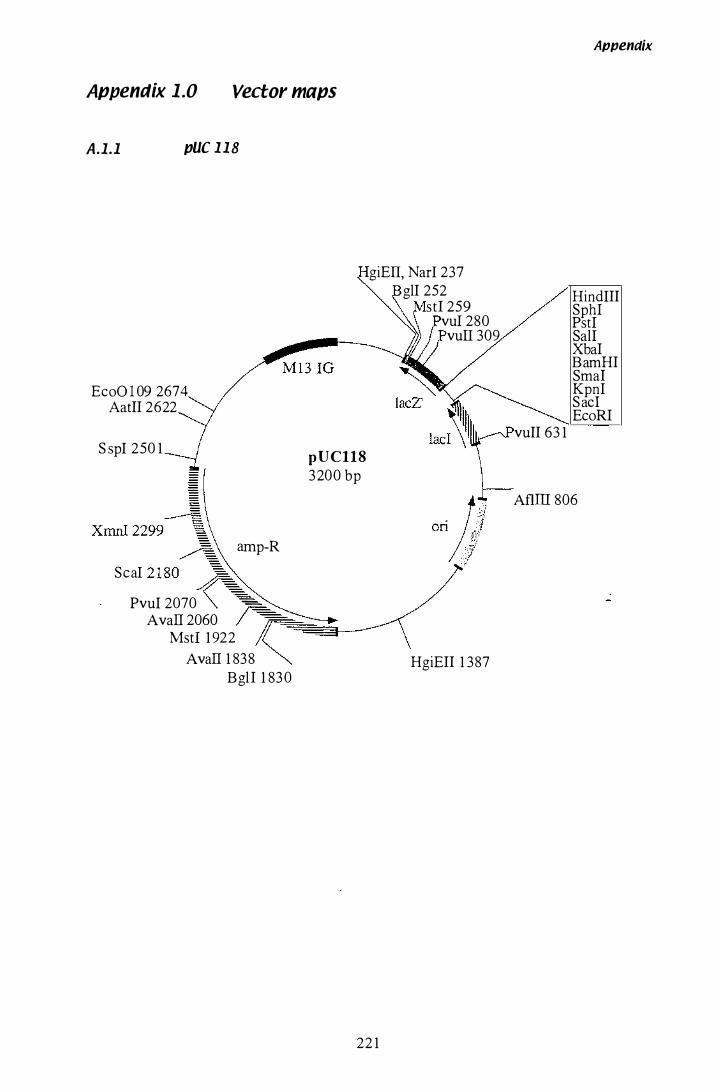
Appendix 1.0 Vector maps
A.1.1 pUC 1 1 8
giEII, Narl 237
EeoO l 09 2674 AatII 2622
S spl 250 1
XrnnI 2299� Seal 2 1 8�
amp-R
pUC118 3200 bp
Pvul 2070 '""==-AvaIl 2060 r��3--_
Mst1 1 922
AvaIl 1 838 BglI 1 830
22 1
glI 252 stI 259
vul 280 vuII 309
HgiEII 1 387
vuII 63 1
Appendix
HindlII Sphl Pstl Sall XbaI BamHI SmaI Kpnl Sac! EeoRI
AflIII 806

A.1 .2 pBT6
Sac
HindUI PstI SalI XhoI BamHI BgIl S stI EcoRI
NcoLXhoLKpnI
tub2
pBT 6
5 .80 Kb
222
Appendix
SaIl
L/XhOI
HindIU
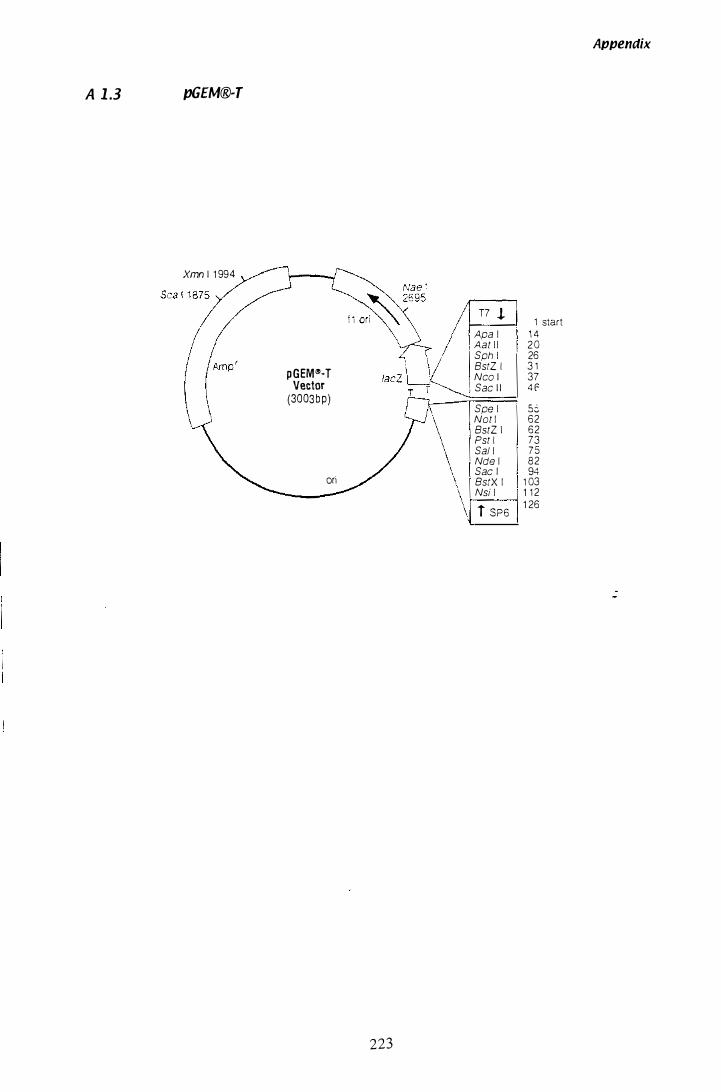
Appenciix
A 1 .3 pG£M®-T
Xmn 1 1 994
T7 J. 1 start
Apa l 1 4 Aat l l 2 0 Sph l 26 BstZ I 3 1 pGEM"-T Nco 1 37
Vector Sac 1 1 4 f (3003bp)
Spe l 5:'-Not I 62 BstZ I 62 Pst I 73 Sai l 75 Nde l 82 Sac I 94
ori BstX I 1 03 Nsi l 1 1 2
i SP6 1 26
223
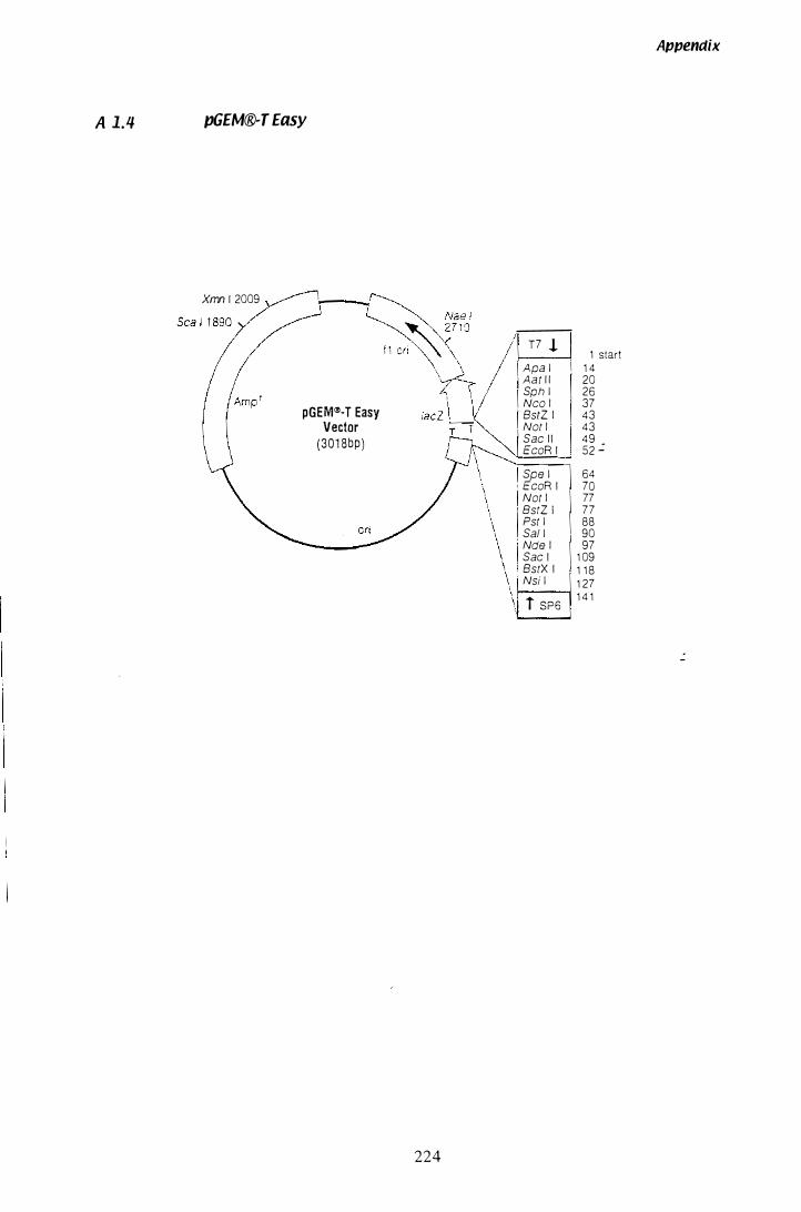
A 1.4 pGEM®-T Easy
Xmn 1 2009
pGEW�-T Easy Vector
(301 8bp)
224
T7 ! Apa l Aat 1 1 Sph l Nco I BstZ I Not I Sac 1 I EcoR I
1 start 1 4 20 26 37 43 43 4 9 52 .:
64 70 77 77 88 90 97
1 09 1 1 8
Spe l EcoR I Not I BstZ 1 Pst l Sai l Nde I Sac I BstX I Nsi l 1 27
1------; 1 4 1 i SP6
Appendix
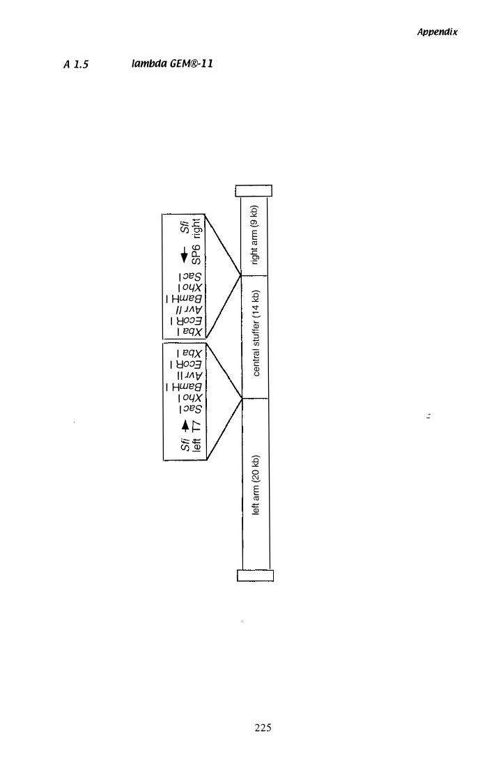
A 1.5 lambda GEM®-l l
, � 1 013S 1 0L/X
I HW138 " J/\ \7'
I Ho03 I 13QX
I 13QX I Ho03
1 1 J/\ \7' I HW138
1 0L/X 1 013S
. r::: '+::: .t:: C/) <D
Appendix
::E:l � e?-E .... (1j 1: Cl
";::
::E:l � � .-----.... .2 '5 Ul � c (]) ()
--
::E:l � 0 8-E .... (1j
.t: �
225
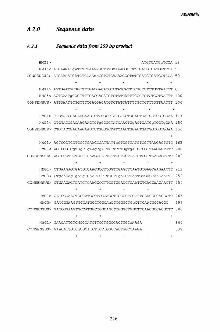
A 2.0 Sequence data
A 2. 1 Sequence data from 359 bl1 l1roduct
HMG 1 > ATGTCATGgTCCA 1 3
HMG 3 < ATGAANATgATCTCCAANNGCTGTGaAAAGGC TNt TGATGTCATGGTCCA 5 0
CONS ENSU S > ATGAAnATGATCTCCAAnnGCTGTGAAAAGGCTnTTGATGTCATGGTCCA 5 0
+ + + + +
Appendix
HMG 1 > AGTGAATGCGGTTTTGACGACATGTCTATCATTTCGCTCTCTGGTAATTT 6 3
HMG3 < AGTGAATgCGGTTTTGACGACATGTCTATCATTTCGCTCTCTGGTAATTT 1 0 0
CONSENSUS> AGTGAATGCGGTTTTGACGACATGT CTATCATTTCGCTCTCTGGTAATTT 1 0 0
+ + + + +
HMG 1 > CTGTACCGACl'.AGAAGTCTGCGGCTATCAF.CTGGACTGA'rGGTCGTGGAA 1 1 3
HMG 3 < CTGTACCGACAAGAAGTCTgCGGCTATCAACTGgACTGATGgTCGTgGAA 1 5 0
CONS ENSU S > C TGTACCGACAAGAAGTCTGCGGCTATCAACTGGACTGATGGTCGTGGAA 1 5 0
+ + + + +
HMG1 > AGTCCGTCGTGGCTGAAGCGATTATTcCTGGTGATGTCGTTAAGAGTGTC 1 6 3
HMG3 < AGTCCGTCgTGgCTgAAgCgATTATTCCTGgTgATGTCGTTAAGAGTGTC 2 0 0
CONS ENSUS> AGTCCGTCGTGGCTGAAGCGATTATTCCTGGTGATGTCGTTAAGAGTGTC 2 0 0
+ + + + +
HMG 1 > CTGAF.GAGTGATGTCAACGCCTTGGTCGAGCTCAATGTGAGCAAGAF.CTT 2 1 3
HMG 3 < CTgA�GAgTgATgTCAACGCCTTGGTCgAGCTCAATGTGAGCAAGAACT T 2 5 0
CONSEN S U S > CT��GAGTGATGTCAACGCCTTGGTCGAGCTCAATGTGAGCF.AGAACTT 2 5 0
+ + + + +
HMG 1 > GATCGGAAGTGCCATGGCTGGCAGCTTGGGCTGGCTTCAACGCCACGCTC 2 6 3
HMG 3 < GATCGGAAGTGCCATGGCTGGCAgC TTGGGCTGgCTTCAACGCCACGC 2 9 8
CONSENSUS> GATCGGAAGTGCCATGGCTGGCAGCTTGGGCTGGCTTCAF.CGCCACGCTC 3 0 0
+ + +
HI1G1 > GAACATTGTCNCGCATCTTCCTGGCCACTGGCcAAGA
CONSENSUS > GAACATTGTCnCGCATCTTCCTGGCCACTGGCCAAGA
+ + +
226
+
+
+
+
3 0 0
3 3 7
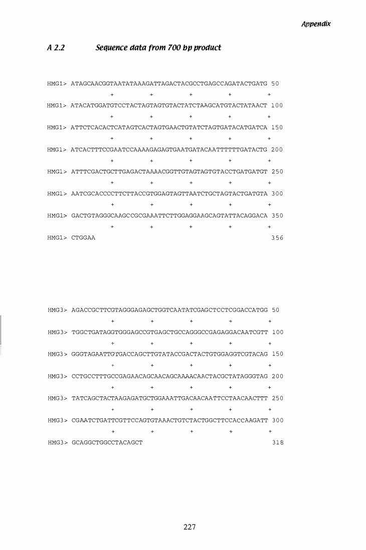
APpendix
A 2.2 Sequence data from 700 bl' I'roduct
HMG 1 > ATAGCAACGGTAATATAAAGATTAGACTACGCCTGAGCCAGATACTGATG 5 0
+ + + + +
HMG 1 > ATACATGGATGTCCTACTAGTAGTGTACTATCTAAGCATGTACTATAACT 1 0 0
+ + + + +
HMG 1 > ATTCTCACACTCATAGTCACTAGTGAACTGTATCTAGTGATACATGATCA 1 5 0
+ + + + +
HMG 1 > ATCACTTTCCGAATCCAAAAGAGAGTGAATGATACAATTTTTTGATACTG 2 0 0
+ + + + +
HMG1 > ATTTCGACTGCTTGAGACTAAAACGGTTGTAGTAGTGTACCTGATGATGT 2 5 0
+ + + + +
HMG1 > AATCGCACCCCTTCTTACCGTGGAGTAGTTAATCTGCTAGTACTGATGTA 3 0 0
+ + + + +
HMG1> GACTGTAGGGCAAGC CGCGAAATTCTTGGAGGAAGCAGTATTACAGGACA 3 5 0
+ + + + +
HMG1 > CTGGAA 3 5 6
HMG 3 > AGACCGCTTCGTAGGGAGAGCTGGTCAATATCGAGCTCCTCGGACCATGG 5 0
+ + + + +
HMG 3 > TGGCTGATAGGTGGGAGCCGTGAGCTGCCAGGGCCGAGAGGACAATCGTT 1 0 0
+ + + + +
HMG3 > GGGTAGAATTG'rGACCAGCTTGTATACCGACTACTGTGGAGGTCGTACAG 1 5 0
+ + + + +
HMG 3 > CCTGCCTTTGCCGAGAACAGCAACAGCk�CAACTACGCTATAGGGTAG 2 0 0
+ + + + +
HMG 3 > TATCAGCTACTAAGAGATGCTGGAAATTGACAACAATTCCTAACk�CTTT 2 5 0
+ + + + +
HMG 3 > CGAA.TCTGATTCGTTCCAGTGTAAACTGTCTACTGGCTTCCACCAAGATT 3 0 0
+ + + + +
HMG3 > GCAGGCTGGCCTACAGCT 3 1 8
227
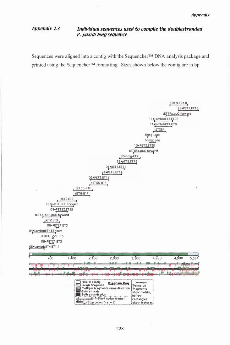
Appendix 2.3
Appendix
Individual sequences used to compile the doublestranded P. paxilli hmg sequence
Sequences were aligned into a contig with the SequencherTM DNA analysis package and
printed using the Sequencher™ fonnatting. Sizes shown below the contig are in bp .
• 1 0e pET24.RC �5ePET1 .ET 1 6.
t�T1Fa.pUC forwlrd 1 1 e,LambdaET4.�22 1 1 e lambdaET4.ET9
•
• C tET28F
5�eq 5h�g3'feq
1 0ePET2.ET20 , . tE�6Fa.pUC forwrrd
.22ehmg-ET7 C �6epET5.ET1 �
°lepET5.ET\1 �4ePET5·ET11
qgePET2.ET1 � tET55-B1 F
, . tET5S-P1 F
, . tET6-B1 F • C
, pET2.ET2 •
tET.6-P 1 F. pUC for�a rd 0gePET22.ET 1 5 -
tETSi-S3F.pUC f�rward
.pET2.ET3 C
OSePET2 1 .ET3 H 04eLambdaET 4.ET 4bam , .
0gePET�2.ET1 5 OSePE�1 .ET3
�5eLamb�ET4.ET1 1
700 1 .400 2. 1 00 2.800 3.500 4.200 4.900 5.56 1"
i I '" :� , = ' � = ': = ' � , a 1 t �;=!' i
i' , 11ft ha , a iI I I III 11111' III a I &11 i I I I �18 I'" , I 'i i :�-.......................................................... " .................................. . ............................... : i 0 H�le in contig Di.gnlll K�!I � l !Zl Smgle fragment - -_ . Bumps on � � Multiple fragments same direction fragments ! I!ii!f Both strand;; shoW" motifs, i � . Both strands plus hollow i 1 " j ... '; '1" ;" f' '';:Start' c()don' fra'm'� '{ " rectangles � [ .... � ..... ! .. t.::::�.t�.p. .. ����.� .. !r.�'!.'� .. � ............................ ��.o.� . . f.����!:.��.l
228
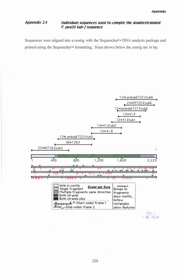
Appendix 2.4
Appenaix
Individual sequences used to compile the doublestranded P. paxilli tllb-2 sequence
Sequences were aligned into a contig with the SequencherrM DNA analysis package and
printed using the SequencherrM formatting. Sizes shown below the contig are in bp.
•
, 1 3 ·LambdaET22 .EtubS , .
06· 1 3 9.F
3 7 ·PET 1 8.Etub4 . •• --����----�C
• • •
(
1 2 ·LambdaET2 2.Etub6
2 4 ·PET20.Etub6 • c 1 2 · lambdaET27.Etub6 • c
1 3 ·4 1 .F • c
1 2 · 4 1 .Etubl , . 1 3 · 4 1 .Etub2 (
1 4 · 4 1 .R •
. .
c
. 1 400 800 1 , 2 00 1 , 600 2 , 2 2 0
'11 � I � , III ii I I I'
1 1 1 1 " ' I , I 11 I I I I I Hnl� • JJ j j J J I 1 1.111 12 .. • ...
i, .: � ; = �,;
i I B 11 I i , I 11 '1I1i 1"cj" H?i;'i�"���ti9"""'''''''���;���''���'''''''''';''=':
. . � .......... .
� f2J Smgle fragment - . Bumps on � � Multiple fragments same direction fragments � Iii Both strands show motifs I I .... ����. ��:.���. p.l�.� . . . . . . . . . . . . . . . . . . ho How : ' f I I I I f Start codon fr ame 1 rectangles ] &:::::i......- Stop codon frame 2 show features 1 .................................................. .................................................... . . . . . . . . . . . . . . . . . . . . . . .
229
1 1 11 I II ,
I , . A I
I , r
A 3.0 Calculations
A 3.1 Calculating paxilline content of mycelia
Appendix
The standard used to calibrate HPLC analysis of paxilline contained 25 ).lg/mL of
authentic paxilline. The standard was injected three times, standard A, B and C .
1 0 ).lL injected = 0.25 ).lg paxilline in 10 ).l1.
The chromatopac recorder calculated the area for each peak.
Average peak area (peak area of A + peak area of B + peak area of C) 13
(I)
The average paxilline lunit area was calculateed.
= 0 .25 ).lg 1 (I)
= (H)
A 1 0 ).lL sample of paxilline, isolated from mycelia, was injected into the HPLC.
Paxilline in 1 0).lL = peak area x (n) = (Ill)
The 1 0).lL sample was removed from the 3 mL extract collected from the Sepak column.
Sample paxilline in 3 mL (DCM:ACN)
230
= (Ill) x 300
= (IV)
Appendix
The 3 mL filtered extract was isolated from a 1 .5 mL aliquot of the 6 mL CHCb :
Methanol extraction, and is therefore equivalent to 1 14 of the original sample volume.
Sample paxilline in 6 mL (CHC13 :Methanol) = (IV) x 4
= (V) (/lG)
The paxil line was extracted into CHCb:methanol from a s mL sample of culture. These
samples were freeze-dried and the dry weight recorded before paxilline was extracted.
Paxilline (/lg) I g of mycelia = (V) IDry weight (g)
23 1

References
References
Acklin W, Weibel F and Arigoni D ( 1 977). Zur biosynthese von paspalin und
verwandten metaboliten aus Claviceps paspali. Chimia 31 : 63 .
Alexander NJ, McCormick SP and Hohn TM ( 1 999). TRI l 2, a trichothecene efflux
pump from Fusarium sporotricioides : gene isolation and expression in yeast. Mol.
Gen. Genet. 261 : 977-984.
Altschul SF, Gish W, Miller W, Myers EW and Lipman DJ ( 1 990). Basic local
alignment search tool . J. Mol. BioI. 215: 403-4 10.
Amaar YG and Moore MM ( 1 998). Mapping of the nitrate-assimilation gene cluster
(crnA-niiA-niaD) and characterization of the nitrate reductase gene (niiA ) in the
opportunistic fungal pathogen Aspergillusfumigatus. Curr. Genet. 33: 206-2 1 5 .
Andrianopoulos A, Kourambas S , Sharp JA, Davis MA and Hynes MJ ( 1 998) .
Characterization of the Aspergillus nidulans nmrA Gene Involved in Nitrogen
Metabolite Repression. J. Bacteriol. 180: 1 973-1 977.
Arrowsmith TJ, Malpartida F, Sherman DH, Birch A, Hopwood DA and Robinson JA
( 1 992). Characterisation of actI-homologous DNA encoding polyketide synthase genes
from the monensin producer Streptomyces cinnamonensis. Mol. Gen. Genet. 234: 254-
264.
Ball OJP, Prestidge RA and Sprosen JM ( 1 995) . Interrelationships between
Acremonium 10 lii , peramine, and lolitrem B in perennial ryegrass . Appl. Environ.
Microbiol. 61 : 1 527- 1 533 .
Ballance DJ ( 1 986). Sequences of Importance for Gene Expression in Filamentous
Fungi. Yeast 2: 229-236.
Basson ME, Thorsness M and Rine J ( 1 986). Saccharomyces cerevisiae contains two
functional genes encoding 3-hydroxy-3-methylglutaryl-coenzyme A reductase. Proc.
Natl. Acad. Sci. USA 83 : 5563-5567.
232
References
Baumgartner U, Hamilton B, Piskacek M, Rui s H and Rottensteiner H ( 1 999) .
Functional analysis of the Zn2Cys6 Transcription Factors Oaf! p and Pip2p. Differeant
roles in fatty acid induction in Saccharomyces cerevisiae. J. BioI. Chem. 2 74: 22208-
222 1 6.
Beg ZH, Allman DW and Gibson DM ( 1 973) . Modulation of 3 -hydroxy-3-
methylglutaryl coenzymeA reductase activity with cAMP and with protein fractions of
rat liver cytosol. Biochem. Biophys. Res. Commun. 54: 1 362- 1 369.
Bhatnagar D, Cleveland TE and Kingston DG ( 1 99 1 ) . Enzymological evidence for
separate pathways for aflatoxin B l and B2 biosynthesis. Biochemistry 30: 4343-4350.
Bibb M ( 1 996). The regulation of antibiotic production in Streptomyces coelicolor
A3(2) . Microbiology 142: 1 335 - 1 344.
B i lls GF, Giacobbe RA, Lee SH, Pelaez F and Tkacz JS ( 1 992) . Tremorgenic
mycotoxins, paspalitrem A and C, from a tropical Phomopsis. Mycol. Res. 96: 977-983 .
B old HC, Alexopoulos CJ and Delevoryas T ( 1 987). Morphology of Plants and
Fungi, 5 Edition (New York: HarperCollinsPublishers, Inc .) .
Bradshaw RE and Pillar TM ( 1 992). Isolation and northern blotting of RNA from
Aspergillus nidulans. J. MicrobioI. Methods 15: 1 -5 .
Brakhage AA ( 1 998 ) . Molecular Regulation of beta-Lactam B iosynthesis i n
Filamentous Fungi. Microbiol. Mol. BioI. Rev. 62: 547-585 .
Brown DW, Adams TH and Keller NP ( 1 996b). Aspergillus has distinct fatty acid
synthases for primary and secondary metabolism . Proc. Natl. Acad. Sci. USA 93 :
1 4873 - 14877.
Brown DW, Yu J-H, Kelkar HS, Fernandes M, Nesbitt TC, Keller NP, Adams TH and
Leonards TJ ( 1 996a). Twenty-five coregulated transcripts define a sterigmatocystin
gene cluster in Aspergillus nidulans. Proc. Natl. Acad. Sci. USA 93 : 1 4 1 8- 1 422.
233
References
Brown MS and Goldstein IL ( 1 980). Multivalent feedback regulation of HMG CoA
reductase, a control mechanism coordinating isoprenoid synthesis and cell growth. J.
L ipid Res. 2 1 : 505-5 1 7.
B rown RL, Cotty PJ and Cleveland TE ( 1 99 1 ) . Reduction in Aflatoxin Content of
Maize by Atoxigenic Strains of Aspergillus flavus. J. Food Protection 54: 623-626.
Bucheli E and Leuchtmann A ( 1 996). Evidence for Genetic Differentiation between
Choke-inducing and Asymptomatic strains of the Epichloe grass endophyte from
Brachpodium sylvaticum. Evolution 50: 1 879- 1 887.
Bullock WO, Fernandez JM and Short JM ( 1987). XLI -Blue: a high efficiency plasmid
transforming recA Escherichia coli strain with beta-galactosidase selection.
BioTechniques 5: 376-378.
B urmester A and Czempinski K ( 1 994). Sequence comparision of a segment of the gene
for 3 -hydroxy-3-methylglutaryl-coenzyme A reductase in zygomycetes . Eur. J.
Biochem. 220: 403-408.
Byrd AD, Schardl CL, Songlin PI, Mogen KL and Siegel MR ( 1 990). The p-tubulin
gene of Epichloe typhina from perennial rye grass (Lolium perenne) . Curr. Genet. 18:
347-354.
Caddix MX, Arst HN, Taylor LH, Johnson RI and Brownlee AG ( 1 986). Cloning of the
regulatory gene areA mediating nitrogen metabolite repression in Aspergillus nidulans.
EMBO J. 5 : 1 087-1 090.
Campos N and Boronat A ( 1 995). Targeting and Topology in the Membrane of Plant
Differential I nduction of 3 -hydroxy-3-methylglutaryl coenzyme A Reductase. The
Plant Cell 7: 2 163-2 1 74.
Carr R ( 1 999). Investigation of paxP2, a gene involved in paxilline production in
Penicillium paxilli. Institute of Molecular BioSciences (Palmerston North: Massey
University), pp. 1 25.
234
References
Carter D, Bui T, Walsh S and Tran Dinh N ( 1 997). RAPD analysis of Aspergillus
parasitic us isolates from peanut growing soils in Queensland , Australia. J. Food
Mycol. l : 3 1 -39.
Cavalli G and Thoma F ( 1 993). Chromatin transitions during activation and repression
of galactose-regulated genes in yeast. EMBO J. 12: 4603-461 3 .
Cazelle B, Pokorska A, Hull E, Green PM, Stanway G and Scazzocchio C ( 1 998) .
Sequence, exon-intron organization, transcription and mutational analysis of prnA, the
gene encoding the transcriptional activator of the prn gene c luster in Aspergillus
nidulans. Mol. Microbiol. 28: 355-370.
Chang P, Yu J, Bhatanagar D and Cleveland TE (2000) . Characterization of the
Aspergillus parasiticus major nitrogen regulatory gene areA . Biochim. Biophys. Acta.
25: 263-266.
Chappell J ( 1 995). The Biochemistry and Molecular Biology of Isoprenoid metabolism.
Plant Physiol. 1 07: 1 -6.
Chin DJ, Gil G, Russel DW, Liscum L, Luskey KL, Basu SK, Okayama H, Berg P,
Goldstein JL and Brown MS ( 1 984) . Nucleotide sequence of 3 -hydroxy-3 -
methylglutaryl coenzyme A reductase, a glycoprotein of endoplasmic reticulum. Nature
308: 6 1 3-6 1 7.
Chopra I, Storey C, Falla TJ and Pearce JH ( 1 998). Antibiotics, peptidoglycan synthesis
and genomics: the chlamydial anomaly revisited. Microbiology 144: 2673-2678 .
Christensen MJ, Leuchtmann A, Rowan DD and Tapper BA ( 1 993) . Taxonomy of
A cremonium endophytes of tall fescue (Festuca arundinacea), meadow fescue (F.
pratensis) and perennial ryegrass (Lolium perenne). Mycol. Res. 9 7: 1 083- 1 092.
Clark DS ( 1 962). Submerged citric acid fermentation of ferrocyanide treated beet
molasses: Morphology of pellets of Aspergillus nidulans. Can. J. Microbiol. 8: 1 33 -
1 36.
235
References
Clay K ( 1 989). Claviciptaceous endophytes of grasses: their potential as biocontrol
agents. Mycol. Res. 92: 1 - 1 2.
Cole RJ, Kirksey JW and Wells JM ( 1 974). A new tremorgenic metabolite from
Penicillium paxilli. Can. J. Microbiol. 20: 1 1 59-1 1 62 .
Colgan, DF and Manley JL ( 1 997) . Mechanisms and regulation o f mRNA
polyadenylation. Genes Dev. 1 1 : 2755-2766.
Coolidge CJ, Seely RJ and Patton JG ( 1 997). Functional analysis of the polypyrimidine
tract in pre-mRNA splicing. Nucleic Acids Res. 25: 888-896.
Cotty PJ ( 1 994). Influence of Field Application of an Atoxigenic Strain of Aspergillus
flavus on the Populations of A. flavus Infecting Cotton Bolls and on the Aflatoxin
Content of Cotton seed. Phytopathology 84: 1 270- 1 277.
Crowell PL, Ren Z, L in S , Vedejs E and Gould MN ( 1 995) . Structure-activity
relationships among monoterpene inhibitors of protein isoprenylation and cell
proliferation. Biochem. Pharmacol. 4 7: 1 405- 1 4 1 5 .
Cui YQ, Okkerse WJ, van der Lans RGJM and Luyben KCAM ( 1 998). Modelling and
Measurements of Fungal Growth and Morphology in Submerged Ferrnentations.
Biotechnol. Bioeng. 60: 2 16-229.
Daraselia ND, Tarchevskaya S and Narita JO ( 1 996). The promoter for tomato 3 -
hydroxy-3-methylglutaryl coenzyme A reductase gene 2 has unusual regulatory
elements that direct high-level expression. Plant Physiol. 1 12 : 727-733 .
Darnay BG, Wang Y and Rodwell V W ( 1 992). Identification o f the Catalytically
Important Histidine of 3 -Hydroxy-3-methylglutaryl-coenzyme A Reductase . J. BioI.
Chem. 267: 1 5064- 1 5070.
Davies J ( 1 990). What are antibiotics ? Archaic functions for modern activities. Mol.
Microbiol. 4: 1 227- 1 232.
236
References
Day DA and Tuite MF ( 1 998). Post-transcriptional gene regulatory mechanisms in
eukaryotes: an overview. J. EndocrinoI. 1 5 7: 36 1 -37 1 .
de la Serna I, Ng D and Tyler BM ( 1 999). Carbon Regulation of Ribosomal Genes in
Neurospora crassa occurs by a Mechanism Which Does Not Require CRE- I , the
Homologue of the Aspergillus Carbon Catabolite Repressor, CreA. Fungal Genet. BioI.
26: 253-269.
Denbow CJ, Lang S and Cramer CL ( 1 996) . The N-Terminal Domain of Tomato 3-
Hydroxy-3-Methylglutaryl-CoA Reductases. Sequence, microsomal targeting and
glycosylation. J. BioI. Chem. 271 : 97 1 0-97 1 5 .
Denison SH, Negret-Urtasun S , Mingot JM, Tilburn J , Mayer WA, Goel A, Espeso EA,
Pefiavala MA and Arst Jr. HN ( 1 998). Putative membrane components of signal
transduction pathways for ambient pH regulation in Aspergillus and meiosis in
Saccharomyces are homologous. Mol. Microbiol. 30: 259-264.
Desjardins AE, Hohn TM and McCormick SP ( 1 992). Effect of Gene Disruption of
Trichodiene Synthase on the Virulance of Gibberella pulicaris. Mol. Plant-Microbe
Interact. 5; 2 14-222.
Dobson JM ( 1 997). MSc Thesis ; Molecular Characterisation of the HMG CoA
Reductase Gene from Neotyphodium lolii. Department of Microbiology and Genetics
(Palmerston North: Massey), pp. 1 36 .
Donadio S , Staver MJ, McAlpine JB, Swanson SJ and Katz L ( 1 99 1 ) . Modular
organization of genes required for complex polyketide biosynthesis. Science 252: 675-
679.
Donald KAG, Hampton RY and Fritz IB ( 1 997). Effects of Overproduction of the
catalytic domain of 3-hydroxy-3-methylglutaryl Coenzyme A reductase on Squalene
synthesis in Saccharomyces cerevisiae. Appl. Environ. MicrobioI. 63 : 334 1 -3344.
237
References
Dooley KA, Millinder S and Osborne TF ( 1 998). Sterol Regulation of 3 -Hydroxy-3-
Methylglutaryl-coenzyme A synthase Gene through a Direct Interaction Between Sterol
Regulatory Element Binding Protein and the Trimeric CCAAT -binding FactorlNuc1ear
Factor Y. J. BioI. Chem. 2 73; 1 349- 1 356 .
Dower WJ, Miller JF and Ragsdale CW ( 1 988). High efficiency transformation of E.
coli by high voltage electroporation. Nucleic Acids Res. 16: 6 1 2 7-6 145 .
Drew SW and Demain AL ( 1 977). Effect of primary metabolites on secondary
metabolism. Annu. Rev. Microbiol. 31 : 3 43-356.
Enjuto M, Balcells L, Campos N, Caelles C, Arro M and Boronat A ( 1 994).
A rabidopsis thaliana contains two differentially expressed 3-hydroxy-3-methylglutaryl
CoA reductase genes, which encode microsomal forms of the enzyme. Proc. Natl.
Acad. Sci. USA 91: 927-93 l .
Ericsson J, Jackson SM and Edwards PA ( 1 996b). Synergistic Binding of Sterol
Regulatory Element-binding Protein and NF-Y to the Farnesyl Diphosphate Synthase
Promoter Is Critical for Sterol-regulated Expression of the Gene. J. BioI. Chem. 2 71 :
24359-24364.
Ericsson J, Jackson SM, Lee BC and Edwards PA ( 1 996a). Sterol regulatory element
binding protein binds to a cis element in the promoter of the farnesyl diphosphate
synthase gene. Proc. Natl. Acad. Sci. USA 93 : 945-950.
Espeso EA and Pefialva MA ( 1 992). Carbon catabolite repression can account for the
temporal pattern of expression of a penicilin biosynthetic gene in Aspergillus nidulans.
Mol. Microbiol. 6: 1 457- 1 465.
E speso EA and Pefialva MA ( 1 996). Three Binding Sites for the Aspergillus nidulans
PacC Zinc-finger Transcription Factor are Necessary and Sufficient for Regulation by
Ambient pH of the Isopenicillin N Synthase Gene Promoter. J. BioI. C hem. 2 71 :
28825-28830.
238
References
Femino AM, Fay FS, Fogarty K and Singer RH ( 1 998). Visualization of Single RNA
Transcripts in Situ. Science 280: 585-590.
Fierro F , Montenegro E, Gutierrez S and Martin JF ( 1 996). Mutants blocked in
penicillin biosynthesis show a deletion of the entire penicillin gene cluster at a specific
site within a conserved hexanucleotide sequence. Appl. Environ. Microbiol. 44: 597-
604.
Flaherty JE, Weaver MA, Payne GA and Woloshuk CP ( 1 995). A p-Glucuronidase
Reporter Gene Construct for Monitoring Aflatoxin Biosynthes is in Aspergillus
nidulans. Appl. Environ. Microbiol. 61 : 2482-2486.
Friesen JA and Rodwell VW ( 1 997) . Identification of Elements Critical for
Phosphorylation of 3-Hydroxy-3-methylglutaryl Coenzyme A Reductase by Adenosine
Monophosphate-Activated Protein Kinase : Protein Engineering of the Naturally
Nonphosphorylatable 3 -Hydroxy-3-methylglutaryl Coenzyme A Reductase from
Pseudomonas mevaloii. Biochemistry 36: 1 1 57-1 1 62 .
Frimpong K and Rodwell VW ( 1 994). Catalysis by Syrian Hamster 3 -Hydroxy-3-
Methylglutaryl coenzyme A reductase . Proposed roles of histidine 865, glutamate 558
and aspartate 766. J. BioI. Chem. 269: 1 1 478- 1 1 483 .
Froeliger EH and Carpenter BE ( 1 996). NUT1 , a major nitrogen regulatory gene in
Magnaporthe grisea, is dispensable for pathogenicity. Mol. Gen. Genet. 251 : 647-656.
Fu Y -H and Marzluf GA ( 1 987). Characterization of n it-2, the major nitrogen
regulatory gene of Neurospora crassa. Mol. Cell. BioI. 7: 1 69 1 - 1 696.
Gaisser S , Trefzer A, Stockert S, Kirschning A and Bechthold A ( 1 997). Cloning of an
A vilamycin Biosynthetic Gene Cluster from Streptomyces viridochromogenes Tu57 . J.
Bacteriol. 1 79: 6271 -6278.
239
References
GaUagher RT, Latch GCM and Keogh RG ( 1 980). The Janthitrems: Fluorescent
Tremorgenic Toxins Produced by Penicillium janthinellum I solates fro Ryegrass
Pastures. AppI. Environ. MicrobioI. 39: 272-273 .
Gancedo JM ( 1 998). Yeast Carbon Catabolite Repression. Microbiol. Mol. BioI. Rev.
62: 3 34-36 1 .
Gant DB, Cole RJ, Valdes JJ, Eldefrawi ME and Eldefrawi AT ( 1 987). Action of
Tremorgenic Mycotoxins on GABAA Receptor. Life Sci. 41 : 2207-22 1 4.
Gayen AK and Peffley DM ( 1 995) . The length of 5 ' -untranslated leader sequence
influences distribution of 3-hydroxy-3-methylglutaryl-coenzyme A reductase mRNA in
polysomes: effects of lovastatin, oxysterols, and mevalonate. Arch. Biochem. Biophys.
322: 475-485 .
Gherzi R, Ponassi M, Gaggero B and Zardi L ( 1 995). The first untranslated exon of the
human tenascin-C gene plays a regulatory role in gene expression. FEBS Lett. 3 69:
335-339.
Gil G , Faust JR, Chin DJ, Goldstein JL and Brown MS ( 1 985) . Membrane-Bound
Domain of HMG CoA Reductase I s Required for Sterol-Enhanced Degradation of the
Enzyme. Cell 41 : 249-258 .
Gi l G , Smith JR, Goldstein JL and Brown MS ( 1 987) . Optional exon in the 5 '
untranslated region of 3 -hydroxy-3-methylglutaryl coenzyme A synthase gene:
Conserved sequence and splicing patterns in humans and hamsters. Proe. Natl. Acad.
Sei. USA 84: 1 863-1 866.
Gillespie JG and Hardie DG ( 1 992). Phosphorylation and inactivation of HMG-CoA
reductase at the AMP-activated protein kinase site in response to fructose treatment of
isolated rat hepatocytes. FEBS Lett. 306: 59-62.
Glenn AE, Bacon CW, Price R and Hanlin RT ( 1 996) . Molecular phylogeny of
Acremonium and its taxonomic implications. Myeologia 88: 369-383 .
240
References
Goldstein JL and Brown MS ( 1 990). Regulation of the Mevalonate Pathway. Nature
343: 425-430 .
Guan G, Dai P and Shechter I ( 1 998). Differential transcriptional regulation of the
human squalene synthase gene by sterol regulatory element-binding proteins (SREBP)
l a and 2 and involvement of 5' DNA sequence elements in the regulation. J. BioI.
Chem. 2 73: 1 2526- 1 2535 .
Gurr SJ, Unkles SE and Kinghorn JR ( 1 987). The structure and organization of
nuclear genes of filamentous fungi, Volume 23, J. R. Kinghom, ed.
Gutierrez S, Marcos AT, Casqueiro J, Kosalkova K, Fernandez FJ, Velasco J and
Martin JF ( 1 999). Transcription of the pcbAB, pcbC and penDE genes of Penicillium
chrysogenum AS-P-78 is repressed by glucose and the repression is not reversed by
alkaline pHs. Microbiology 145: 3 1 7-324.
Guzman-de-Pefia D . , and Ruiz-Herrera J ( 1 996). Relationship between Aflatoxin
Biosynthesis and Sporulation in Aspergillus parasiticus. Fungal Genet. BioI. 21 : 1 98-
205 .
Ha-Huy-Ke and Luckner M ( 1 979). Structure and function of the condiospore pigments
of Penicillium cyclopium. Z. Allg Mikrobiol 19: 1 1 7-22.
Haas H, Bauer B, Redl B, Stoffker G and Marzluf GA ( 1 995a). Molecular cloning and
analysis of nre, the major nitrogen regulatory gene of Penicillium chrysogenum. Curr.
Genet. 2 7: 1 50- 1 58.
Haas H and Marzluf GA ( 1 995b). NRE, the major nitrogen regulatory protein of
Penicillium chrysogenum, binds specifically to elements in the intergenic promoter
regions of nitrate assimilation anp penici llin biosynthetic gene clusters. Curr. Genet.
28: 1 77- 1 83 .
r
24 1
References
Haberhausen G, Pinsl J, Kuhn C-C and Markert-Hahn C ( 1 998) . Comparative Study of
Different Standardization Concepts in Quantitative Competitive Reverse Transcription
PCR Assays. J. Clin. Microbiol. 36: 628-633 .
Hackshaw KV, Furlow ZM and Chiu I ( 1996). Cloning and Characterisation of a novel
upstream untranslated exon of the mouse Fgf-l gene. Gene 180: 1 3 1 - 1 3 5 .
Hampton R, Dimster-Denk D and Rine J ( l 996a). The Biology of HMG-CoA reductase :
the pros of contra-regulation. Trends Biochem. Sci. 21 : 1 40- 1 45 .
Hampton RY, Koning A, Wright R and Rine J ( l 996b) . In vivo examination of
membrane protein localization and degradation with green fluorescent protein. Proc.
Natl. Acad. Sci. USA 93: 828-833 .
Harting I and Wiesner RJ ( 1 997). Quantification of Transcript-to-Template Ratios as a
Measure of Gene Expression Using RT-PCR. BioTechniques 23 : 450-455.
Hirata S , Koh T, Yamada-Mouri N, Hoshi K and Kato J ( 1 996). The untranslated first
exon 'exon OS' of the rat estrogen receptor (ER) gene. FEBS Lett. 394; 37 1 -373 .
Hofmann K and Stoffel W ( 1 993) . TMBase - A database o f membrane spanning protein
segments. BioI. Chem. 347: 1 66 .
Hohn TM and Desjardins AE ( 1 992). I solation and gene disruption of the Tox5 gene
encoding Trichodiene synthase in Gibberella pulicaris. Mol. Plant-Microbe Interact.
5: 249-256.
Hohn TM, Desjardins AE and McCormick SP ( 1 993b). Analysis of Tox5 gene
expression in Gibberella pulicaris strains with different Trichothecene production
phenotypes. Appl. Environ. MicrobioI. 59: 2359-2363.
Hohn TM, Desjardins AE and McCormick SP ( 1 995). The Tri4 gene of Fusarium
sporotrichioides encodes a cytochrome P450 monooxygenase involved in trichothecene
biosynthesis. Mol. Gen. Genet. 248: 95- 1 02.
242
References
Hohn TM, McCormick SP and Desjardins AE ( 1 993a) . Evidence for a gene cluster
involving trichothecene-pathway biosynthetic genes in Fusarium sporotrichioides.
Curr. Genet. 24; 291 -295 .
Holmes DS and Quigley M ( 1 98 1 ) . A rapid boiling method for the preparation of
bacterial plasmids. Anal. Biochem. 1 1 4: 1 93- 1 97.
Homann V, Mende K, Amtz C , Ilardi V, Macino G, Morelli G, Bose G, and Tudzynski
B ( 1 996) . The isoprenoid pathway: cloning and characterisation of fungal FPPS genes.
Curr. Genet. 30: 232-239.
Hopwood DA ( 1 988). Towards an understanding of gene switching in Streptomyces, the
basis of sporulation and antibiotic production. Proc. R. Soc. Lond. BioI. Sci. 235; 1 2 1 -
1 38.
Huang MY and Bungay HR ( 1 973). Microprobe measurements of oxygen concentration
in mycelial pellets. Biotechnol. Bioeng. 15 : 1 1 93-1 1 97.
Ibba M, Taylor SJC, Weedon CM and Mantle PG ( 1 987). Submerged fermentation of
Penicillium paxilli biosynthesizing paxilline, a process inhibited by calcium induced
sporulation. J. Gen. Microbiol. 133: 3 1 09-3 1 1 9.
Imbault N, Thiersault M, Duperon P, Benabdelmouna A and Doireau P ( 1 996) .
Pravastatin: A tool for investigating tha availability of mevalonate metabolites for
primary and secondary metabolism in Catharanthus roseus cell suspensions.
P hysiologica Plantarum 98: 803-809.
I ngebritsen TS, Geelen MJH, Parker RA, Evenson KJ and Gibson DM ( 1 979) .
Modulation of hydroxy-methylglutaryl-CoA reductase activity, reductase kinase
activity, and cholesterol synthesis in rat hepatocytes in response to insulin and
glucagon. J. BioI. Chem. 254: 9986-9989.
Hoh Y, Johnson R and Scott B ( 1 994a). Intergrative transformation of the mycotoxin
producing fungus Penicillium paxilli. Curr. Genet. 25: 508-5 1 3 .
243

References
1toh Y and Scott B ( 1 994b). Heterologous and homologous plasmid intergration at a
spore-pigment locus in Penicillium paxilli generates large deletions. Curr. Genet. 26:
468-476.
Jackson SM, Ericsson J , Osborne TF and Edwards PA ( 1 995). NF-Y Has a Novel Role
in Sterol-dependent Transcription of Two Cholesterogenic Genes. J. BioI. Chem. 2 70:
2 1 445-2 1 448.
Jiang Y, Proteau P , Poulter D and Ferro-Novick S ( 1 995) . B TS1 Encodes a
Geranylgeranyl Diphosphate Synthase in Saccharomyces cerevisiae. J. BioI. C hem.
270: 2 1 793-2 1 799.
Kaiser D, Onken U, Sattler I and Zeeck A ( 1 994). Influence of increased dissolved
oxygen concentration on the formation of secondary metabolites by manumycin
producing Streptomyces parvulus. AppI. Microbiol. Biotechnol. 41 : 309-3 1 2 .
KeIler NP and Hohn TM ( 1 997). Metabolic Pathway Gene Clusters in Filamentous
Fungi. Fungal Genet. BioI. 21 : 1 7-29.
Kendrick B ( 1 992). The Fifth Kingdom, 2 Edition (Newburyport MA: Mycologue
Publications).
Kennelly PJ and Rodwell VW ( 1 985) . Regulation of 3-hydroxy-3-methylglutaryl
coenzyrne A reductase by reversible phosphorylation and dephosphorylation. J. Lipid
Res. 26: 903-9 14.
Keogh RG, Tapper BA and Fletcher RH ( 1 996) . D istributions of fungal endophyte
Acremonium lolii, and of the alkaloids lolitrem B and peramine, within perennial
ryegrass . NZ J. Agric. Res. 39: 1 2 1 - 1 27.
Kistler HC, Han Y, Benny U, Liu X, Temporini E and VanEtterr H ( 1 999). A
pathogenicity island in Nectaria haematococca. Fungal Genetics Newsletter 46-
Supplement: 33 .
244
References
Kitahara M, Ishiguro F, Takayama K, Isowa K and Nagai T. ( 1 993) . Evaluation of skin
damage of cyclic monoterpenes, percutaneous absorption enhancers, by using cultured
human skin cells. BioI. Pharm. Bull. 1 6: 9 1 2-9 1 6.
Kleinkauf H and von Dohren H ( 1 990). Antibiotics-cloning of biosynthetic pathways.
FEBS Lett. 268: 405-407.
Kleinkauf H and von Dohren H ( 1 993). Biosynthesis of Microbial Peptidic Effectors.
Pro ceedings of the second Tottori University International Symposium on Host
Specific Toxin: Biosynthesis, Receptor and Molecular Biology Sep 4-8 1993 : 1 85-
1 96.
Knaus H-G, McManus OB, Seok HL, Schmalhofer W A, Garcia-Calvo M, Helms LMH,
Sanchez M, Giangiacomo K, Reuben JP, Smith AB, Kaczorowski GJ and Garcia ML
( 1 994) . Tremorgenic indole alkaloids potently inhibit smooth muscle high-conductance
calcium-activated potassium channels. Biochemistry 33: 5 8 1 9-5828.
Kozak M ( 1 984) . Complilation and analysis of sequences upstream from the
translational start site in eukaryotic mRNAs. Nucleic Acids Res. 12: 857-872.
Kumagai H, Chun KT and Simoni RD ( 1 995). Molecular Dissection of the Role of the
Membrane Domain in the Regulated Degradation of 3 -Hydroxy-3-methylglutaryl
Coenzyme A reductase. J. BioI. Chem. 2 70: 1 9 1 07- 1 9 1 1 3 .
Kuo Y-H and Kuo L-MY ( 1 997). Antitumour and anti-AIDS triterpenes from Celastrus
hindsii. Phytochemistry 44: 1 275- 1 28 1 .
Kyte J and Doolittle RF ( 1 982). A S imple Method for Displaying the Hydropathic
Character of a Protein. J. Mol. BioI. 1 5 7: 1 05 - 1 32 .
Lawrence CM, Rodwell VW and Stauffacher CV ( 1 995) . Crystal Structure of
Pseudomonas mevalonii HMG-CoA Reductase at 3 .0 Angstrom Resolution. Science
268: 1 758- 1 762.
245

References
Laws I and Mantle PG ( 1 989) . Experimental Constraints in the study of the
Biosynthesis of Indole Alkaloids in Fungi . J. Gen. Microbiol. 1 35: 2679-2692.
Lees ND, Skaggs B, Kirsch DR and Bard M ( 1 995). Cloning of the Late Genes in the
Ergosterol Biosynthetic Pathway of Saccharomyces cerevisiae-A Review. Lipids 30:
22 1 -226.
Levesque G, Lamarche B, Murthy MRV, Julien P, Despres J-P and Deshaies Y ( 1 994).
Determination of changes in specific gene expression by reverse transciption PCR using
interspecies mRNAs as internal standards . Biotechniques 1 7: 738-74 1 .
L iao X, V ining LC and Doull JL ( 1 995) . Physiological control of trophophase
idiophase separation in streptomycete cultures producing secondary metabolites . C an .
J. Microbiol. 41 : 309-3 1 5 .
L iscum L, Finer-Moore J, Stroud RM, Luskey KL, Brown MS and Goldstein JL ( 1 9 85).
Domain Structure of 3 -Hydroxy-3-methylglutaryl Coenzyme A Reductase, a
Glycoprotein of the Endoplasmic reticulum. J. BioI. Chem. 260: 522-530 .
Liu B-H and Chu FS ( 1 998). Regulation of ajlR and Its Product, AflR, Associated with
Aflatoxin Biosynthesis. Appl. Environ. Microbiol. 64: 3 7 1 8-3 723 .
Lumbreras V, Campos N and Boronat A. ( 1 995). The use of an alternative promoter in
the A rabidopsis thaliana HMG 1 gene generates an mRNA that encodes a novel 3 -
hydroxy-3-methylglutaryl coenzyme A reductase isoform with an extended N-terminal
region. Plant J. 8: 541 -549.
Luskey KL ( 1 987). Conservation of promoter sequence but not complex intron splicing
pattern in human and hamstr genes for 3 -hydroxy-3-methylglutaryl co enzyme A
reductase. Mol. Cell. BioI. 7: 1 88 1 - 1 893.
Luskey KL and Stevens B ( 1 985). Human 3 -Hydroxy-3-methylglutaryl Coenzyme A
Reductase. Conserved domains responsible for catalytic activity and sterol-regulated
degradation. J. BioI. Chem. 260: 1 027 1 - 1 0277.
246
References
Mantle PG, Laws I, Tan MJL and Tizard M ( 1 984) . A Novel Process for the Production
of Pen it rem Mycotoxins by Submerged Fermentation of Penicillium nigricans. J. G en.
Microbiol. 130: 1 293- 1 298.
Maplestone RA, Stone MJ and Williams DH ( 1 992). The evolutionary role of secondary
metabolites - a review. Gene 1 15: 1 5 1 - 1 57 .
Marathe S , Yu VG, Turner GE, Palmier C and Weiss RL ( 1 998). Multiple forms of
Arginase are Differentially Expressed from a single Locus in Neurospora craSSG. J .
BioI. Chem. 2 73 : 29776-29785 .
Marsh EN, McKie N, Davis NK and Leadlay PF ( 1 989) . Cloning and structural
characterisation of the genes coding for adenosylcobalamin-dependent
methylmalonyl-CoA mutase from Propionibacterium shermanii. Biochem. J. 260: 345-
3 52.
Martin JF and Liras P ( 1 989). Organization and expression of genes involved in the
biosynthesis on antibiotics and other secondary metabolites. Annu. Rev. Microbiol. 43:
1 73-206.
Marzluf GA ( 1 98 1 ). Nitrogen metabolism and gene expression in fungi. Microbiol.
Rev. 45 : 437-46 1 .
Marzluf GA ( 1 993). Regulation of sulfur and nitrogen metabolism in filamentous fungi.
Annu. Rev. MicrobioI. 4 7: 3 1 -55 .
May GS, Tsang ML-S, Smith H, Fidel S and Morris NR ( 1 987). Aspergillus nidulans �
tubulin genes are unusually divergent. Gene 55, 23 1 -243 .
McGil l M (2000 In Prep) . MSc Thesis . Institute of Molecular B ioSciences
(Palmerston North: Massey University).
247
References
Mende K, Homann V and Tudzynski B ( 1 997). The geranylgeranyl diphosphate
synthase gene of Gibberellafujikuroi : Isolation and expression. Mol. Gen. Genet. 255:
96- 1 05 .
Miles CO, Munday-Finch SC, Meagher LP and Wilkins AL ( 1 996). AgResearch
Toxinology and Food Safety Report 1 992- 1 995 (Hamilton: AgResearch).
Miles CO, Wilkins AL, Gallagher RT, Hawkes AD, Munday SC and Towers NR
( 1 992). Synthesis and Tremorgenicity of Paxitriols and Lolitriols: Possible biosynthetic
precusors of Lolitrem B . J. Agric. Food Chem. 40: 234-238.
Moore DD ( 1 990). Diversity and unity in the nuclear hormone receptors: a terpenoid
receptor superfamily. New BioI. 2 : 1 00- 1 05 .
Moore J and BusheIl ME ( 1 997). The effect of morphology and oxygen uptake on
penicillin production by Aspergillus nidulans in submerged culture. MycoI. Res. 101 ;
1 237- 1 24 1 .
Moreira MT, Sanroman A, Fe ijoo G and Lema J M ( 1 996) . Control of pellet
morphology of filamentous fungi in fluidized bed bioreactors by means of a puls ing
flow. Application to Aspergillus niger and Phanerochaete chrysoporium. Enzyme
Microb. Technol. 19: 26 1 -266.
Munday-Finch SC, Wilkins AL,and Miles CO ( 1 996a) . Isolation of Paspaline B, an
indole-diterpenoid from Penicillium paxilli. Phytochemistry 41 : 327-332.
Munday-Finch SC, Wilkins AL, Miles CO, Ede RM and Thomson RA ( 1 996b) .
Structure Elucidation of Lolitrem F, a Naturally Occurring Stereoisomer of the
Tremorgenic Mycotoxin Lolitrem B, I solated from Lolium perenne Infected with
Acremonium lolii. J. Agric. Food Chem. 44: 2782-2788.
Munday-Finch SC, Wilkins AL, Miles CO, Tomoda H and Omura S ( 1 997). Isolation
and Structure Elucidation of Lolilline, a Possible Biosynthetic Precursor of the Lolitrem
Family of Tremorgenic Mycotoxins . J. Agric. Food Chem. 45: 1 99-204.
248
References
Negrete-Urtasun S, Reiter W, Diez E, Denison SH, Tilburn J, Espeso EA, Penalva MA,
and Arst Jr. HN ( 1 999). Ambient pH signal transduction in Aspergillus: Completion of
gene characterization. Mol. Microbiol. 33: 994- 1 003 .
Nielsen, J. ( 1 992). Modelling the Growth of Filamentous Fungi. Adv. Biochem. Eng.
Biotechnol. 46, 1 87-223.
Ohnishi Y, Kameyama S, Onaka H and Horinouchi S ( 1 999). The A-factor regulatory
cascade leading to streptomycin biosynthesis in Streptomyces griseus: identification of a
target gene of the A-factor receptor. Mol. Microbiol. 34: 1 02- 1 1 1 .
Orbach MJ, Porro EB and Yanofsky C ( 1 986). Cloning and Characterization of the
Gene for p-Tubulin from a Benomyl-Resistant Mutant of Neurospora crassa and Its
Use as a Dominant Selectable Marker. Mol. Cell. BioI. 6: 2452-246 1 .
Osborne TF ( 1 99 1 ). S ingle Nucleotide Resolution of S terol Regulatory Region in
Promoter for 3 -Hydroxy-3-methylglutaryl Coenzyme A Reductase. J. BioI. Chem. 266:
1 3947- 1 395 1 .
Osborne TF, Goldsterin JL and Brown MS ( 1 985). 5 ' end of HMG CoA reductase gene
contains sequences responsible for the cholesterol-mediated inhibition of transcription.
Cell 42: 203-2 12 .
Pan H, Feng B and Marzluf GA ( 1 997). Two distinct protein-protein interactions
between the NIT2 and NMR proteins are required to establish nitrogen metabolite
repression in Neurospora crassa. Mol. Microbiol. 26: 72 1 -729.
Panozzo C, Comillot E and Felenbok B ( 1 998). The CreA Repressor Is the Sole DNA
binding Protein Responsible for Carbon Catabolite Repression of the alcA Gene in
Aspergillus nidulans via Its binding to a Couple of Specific Sites. J. BioI. Chem. 2 73;
6367-6372.
Parmryd I and Dallner G ( 1 996). Organization of isoprenoid biosynthesis. Biochem.
Soc. Trans. 24; 677-682.
249
References
Pefialva MA, Rowlands RT and Turner G ( 1 998). The optimization of penici ll in
biosynthesis in fungi. Trends Biotechnol. 1 6: 483-489.
Perez-Esteban B, Orejas M., Gomez-Pardo E and Pefialva MA ( 1 993) . Molecular
characterisation of a fungal secondary metabolism promoter: transcription of the
Aspergillus n idulans i sopenicillin N synthetase gene i s modulated by upstream
negative elements. Mol. Microbiol. 9: 88 1 -895.
Piecq M, Dehottay P, Biot A and Dusart J ( 1 994). Cloning and nucleotide sequence of a
region of the Kibdelosporangium aridum genome homologous to polyketide
biosynthetic genes. DNA Seq 4: 2 1 9-229.
Polakowski T, Stahl U and Lang C ( 1 998) . Overexpression of a cytosol ic
hydroxymethylglutaryl-CoA reductase leads to squalene accumulation in yeast. Appl.
Microbiol. Biotechnol. 49: 66-7 1 .
Procter RH , Hohn TM, McCormick S P and Desjardins A E ( 1 995). Tri6 encodes an
unusual zinc finger protein involved in regulation of trichothecene biosynthesis in
Fusarium sporotrichioides. Appl. Environ. Microbiol. 61 : 1 923-1 930.
Prosser J1 and Tough AJ ( 1 99 1 ). Growth mechanisms and growth k inetics of
filamentous microorganisms. Crit. Rev. BiotechnoI. 1 0: 253-274.
Re EB, Jones D and Learned RM ( 1 995). Co-expression of native and introduced genes
reveals cryptic regulation of HMG CoA reductase expression in Arabidopsis . Plant J.
7: 77 1 �784.
Rehacek Z, Desai JD, Sajdl P and Pazoutova S ( 1 977). The cellular role of nitrogen in
the biosynthesis of alkaloids by submerged culture of Claviceps purpurea (Fr.) Tul .
Can. J. Microbiol. 23 : 5 96-600.
Reynolds GA, Basu SK, Osborne TF, Chin DJ, Gil G, Brown MS, Goldstein JL and
Luskey KL ( 1 984). HMG CoA Reductase: A Negatively Regulated Gene with Unusual
Promoter and 5' Untranslated Regions. Cell 38: 275-285 .
250
References
Reynolds GA, Goldstein JL and Brown MS ( 1 985). Multiple mRNAs for 3 -Hydroxy-3-
methylglutaryl Coenzyme A Reductase Determined by Multiple Transcription Initiation
Sites and Intron Splicing Sites in the 5 '-Untranslated Region. J. BioI. Chem. 260:
1 0369- 1 0377.
Riedy MC, Timm Jr. EA and Stew art CC ( 1 994). Quantitative RT -PCR for Measuring
Gene Expression. BioTechniques 18: 70-75.
Roitelman J, Olender EH, Bar-Nun S , Dunn Jr. WA and Simoni RD ( 1 992) .
Immunological Evidence for Eight Spans in the Memebrane Domain of 3 -Hydroxy-3 -
Methylglutaryl Coenzyme A Reductase: Implications for Enzyme Degradation in the
Endoplasmic Reticulum. J. Cell BioI. 1 1 7: 959-973 .
Rowan DD and Gaynor DL ( 1 986). Isolation of feeding deterrents against Argentine
stem weevil from ryegrass infected with the endophyte Acremonium loliae. J.Chem.
E col. 12: 647-658.
Rowan DD and Latch GCM ( 1 994) . Utilization of endophyte-infected perennial
ryegrasses for increased insect resistance. In Biotechnology of endophytic fungi of
grasses, C. W. Bacon and J. J. F. White, eds. (USA: CRC Press), pp. 1 69- 1 83 Chapter
1 2 .
Russo P, Juuti JT and Raudaskoski M ( 1 992). Cloning, sequence and expression of a �
tubulin-encoding gene in the homobasidiomycete Schizophyllum commune. Gene 1 1 9:
1 75-1 82.
Saitta B, Timpl R and Chu M ( 1 992). Human a2(VI) Collagen Gene. Heterogeneity at
the 5'-untranslated region generated by an alternate exon. J. BioI. Chem. 267; 6 1 88-
6 1 96 .
Sanchez M and McManus OB ( 1 996). Paxilline inhibition of the alpha-subunit of the
high-conductance calcium-activated potassium channel. Neuropharmacology 35: 963-
968.
25 1

References
Sanger F, Nicklen S and Coulson AR ( 1 977). DNA Sequencing with chain-terminating
inhibitors. Proc. Natl. Acad. Sei. USA 74: 5463-5467.
Schardl CL and An Z ( 1 993) . Molecular biology and genetics of protective fungal
endophytes of grasses . Genetic Engineering 15; 1 9 1 -2 1 2.
Schardl CL, Leuchtmann A, Tsai H-F, Collett MA, Watt DM and Scott DB ( 1 994).
Origin of a fungal symbiont of perennial ryegrass by interspecific hybridization of a
mutualist with the rye grass choke pathogen. G enetics 136, 1 307- 1 3 1 7.
Schiestl RH and Petes TD ( 1 99 1 ) . Integration of DNA fragments by illegitimate
recombination in Saccharomyces cerevisiae. Proc. Natl. Acad. Sci. USA 88: 7585-
7589.
Schjerling P and Holmberg S ( 1 996). Comparative amino acid sequence analysis of the
C6 zinc cluster family of transcriptional regulators. Nucleic Acids Res. 24: 4599-4607.
Schuler MA ( 1 996). The role of Cytochrome P450 monooxygenases in plant-insect
interactions. Plant Physiol. 112 : 1 4 1 1 - 1 4 1 9.
Scott B and Schardl C ( 1 993). Fungal symbionts of grasses: evolutionary insights and
agricultural potential. Trends Mircrobiol. 1 ; 1 97-22.
Screen N, Bailey A, Charnley K, Cooper R and Clarkson 1 ( 1 998) . Isolation of a
nitrogen response regulator gene (nnrl ) from Metarhizium anisopliae. Gene 221 ; 1 7-
24.
Shain L and Franich RA ( 1 98 1 ) . Induction of dothistroma blight symptoms with
dothistromin. Physi. Mol. Plant Path. 19: 49-55 .
Siegel MR, Latch GCM, Bush LP, Fannin FF, Rowan DD, Tapper BA, Bacon CW and
lohnson MC ( 1 990). Fungal endophyte-infected grasses: alkaloid accumulation and
aphid response. Journal of Chemical Ecology 1 6: 3 3 0 1 -3 3 1 5 .
252
References
Siegel MR, Latch GCM and lohnson MC ( 1 985). Acremonium Fungal Endophytes of
Tall Fescue and Perennial Ryegrass : Significance and Control . Plant Disease 69; 1 79-
1 83 .
Sikkema l, de Bont lA and Poolman B (1 994). Interactions o f cyclic hydrocarbons with
biological membranes. J. BioI. Chem. 269: 8022-8028.
Skaugen M, Abildgaard CIM and Nes IF ( 1 997). Organization and expression of a gene
cluster involved in the biosynthesis of the lantibiotic lactocin S. Mol. Gen. Genet. 253:
674-686.
Smith BL, McLeay LM and Embling PP ( 1 997). Effect of the mycotoxins penitrem,
paxi l l ine and lolitrem B on the electromyographic activity o f skeletal and
gastrointestinal smooth muscle of sheep. Res. Vet. Sci. 62: 1 1 1 - 1 1 6.
Southern EM ( 1 975). Detection of specific sequences among DNA fragments separated
by gel electrophoresis. J. Mol. BioI. 98; 503-5 1 7.
Stokes HW, O 'Gorman DB, Recchia GD, Parsekhian M and Hall RM ( 1 997). Structure
and function of 59-base element recombination sites associated with mobile gene
cassettes. Mol. Microbiol. 26: 73 1 -745.
Stone MJ and Williams DH ( 1 992). On the evolution of functional secondary
metabolites (natural products) . Mol. Microbiol. 6: 29-34.
Stryer L ( 1 995). Biochemistry, 4 Edition: (W.H.Freeman and Company).
Suarez T and Peftalva MA ( 1 996). Characterization of a Penicillium chrysogenum gene
encoding a PacC transcription factor and its binding sites in the divergent pcbAB-pcbC
promoter of he penicillin biosynthetic cluster. Mol. Microbiol. 20: 529-540.
253
References
Tabas I , Chen LL, Clader JW, McPhail AT, Burnett DA, Bartner P , Das PR, Pramanik
BN, Puar M S and Feinmark SJ ( 1 9 90). Rabbit and human liver contain a novel
pentacyc lic triterpene ester with acyl-CoA: cholesterol acyltransferase inhibitory
activity. J. BioI. Chem. 265: 8042-805 1.
Then Bergh K and Brakhage AA ( 1 998) . Regulation of the Aspergillus nidulans
Penicil l in B iosynthesi s Gene acvA (pc b A B ) by amino acids : Implications for
Involvement of Transcription Factor PACC. Appl. Environ. Microbiol. 64: 843-849.
Tilburn J, Sarkar S, Widdick DA, Espeso EA, Orejas M, Mungroo J , Pefialva MA and
Arst RN ( 1 995) . The Aspergillus Pace zinc finger transcription factor mediates
regulation of both acid- and alkaline-expressed genes by ambient pH. EMBO J. 14 :
779-790.
Tkacz JS , Giacobbe RA and Monaghan RL ( 1 993) . Improvement in the titer of
echinocandin-type antibiotics : a magnesium-limited medium supporting the biphasic
production of pneumocandins Ao and Bo . J. Ind. Microbiol. 1 1 : 95- 1 03 .
Todd RB, Murphy RL, Martin HM, Sharp JA, Davis MA, Katz ME and Hynes MJ
( 1 997). The acetate regulatory gene facB of Aspergillus nidulans encodes a
ZN(IIhCys6 transcriptional activator. Mol. Gen. Genet. 254: 495-504.
Trail F , Mahanti N and Linz J ( 1 995). Molecular biology of aflatoxin biosynthesis.
Microbiology 1 41 : 755-765.
Tudzynski B, Homann V, Feng B and Marzluf GA ( 1 999). Isolation, characterization
and disruption of the areA nitrogen regulatory gene of Gibberella fujikuroi. Mol. Gen.
Genet. 261 : 1 06- 1 1 4.
Tudzynski B , Kawaide H. and Kamiya Y ( 1 998). Gibberellin biosynthesis in Gibberella
fujikuroi: cloning and characterisation of the copalyl diphosphate synthase gene. Curr.
Genet. 34: 234-240.
254
References
Tudzynski P , Holter K, Correia T, Arntz C, GrammeI N and Keller U ( 1 999). Evidence
for an ergot alkaloid gene cluster in Claviceps purpurea. Mol. Gen. Genet. 261 : 1 33-
1 4 1 .
Urban M , Bhargava T and Hamer lE ( I 999). An ATP-driven efflux pump i s a novel
pathogenicity factor in rice blast disease. EMBO J. 18: 5 1 2�52 1 .
van Heeswij ck R and McDonald G ( 1 992). Acremonium Endophytes in Perennial
Ryegrass and other Pasture Grasses In Australia and New Zealand. Aust. J. Agric. Res.
43: 1 683 - 1 709.
van Nistelrooy JGM, van den Brink JM, van Kan JAL, van Gorcom RPM and de Waard
MA ( 1 996). Isolation and molecular characterisation of the gene encoding eburicol 1 4a
demethylase (CYP5J) from Penicillium italicum. Mol. Gen. Genet. 250: 725-733 .
Vanden Heuvel JP, Tyson FL and Bell DA ( 1 993) . Construction of re comb in ant RNA
templates for use as internal standards in Quantitative RT-PCR. Biotechniques 14: 395-
398 .
Vieira J and Messing J ( 1 987) . Production of single-stranded plasmid DNA. Methods
Enzymol. 153 : 3 - 1 1 .
Vining L C ( 1 992). Roles o f secondary metabolites from microbes. Ciba Found. Sym p.
1 71 : 1 95 - 1 98 .
Voet 0 and Voet JG ( 1 990). Biochemistry: John Wiley & Sons, Inc .
Walton JD, Akimitsu K, Ahn JH and Pitkin JW ( 1 993) . Towards an understanding of
the Tox 2 gene of Cochliobolus carboum . I n Proceedings of the Second Tottori
University International Symposium on Host-specific Toxin : B iosynthesis,
Receptor and Molecular Biology, pp. 227-237.
Weedon CM and Mantle PG ( 1 987). Paxilline biosynthesis by Acremonium loUae; A
step towards defining the origin of Lolitrem neurotoxins. Phytochemistry 26: 969-971 .
255
References
Wilkinson HH, Bush LP, Burton HR, Siegel MR and Schardl CL ( 1 997). Lolines are
Fungal Metabolites. In International NeotyphodiumlGrass Interactions Symposium
(Athens, Georgia, USA).
Wilson RA and Arst Jr. HN ( 1 998). Mutational Analysis of AREA, a Transcriptional
Activator Mediating Nitrogen Metabolite Repression in Aspergillus nidulans and a
member of the " Streetwise" GA TA family of Transcription Factors. MicrobioI. Mol.
B ioI. Rev. 62: 5 86-596.
Woitek S, Unkles SE, Kinghorn JR and Tudzynski B ( 1 997) . 3 -Hydroxy-3-
methylglutaryl-CoA reductase gene of Gibberella Ju}ikuroi i so lation and
characterisation. Curr. Genet. 31 : 38-47.
Woloshuk CP, Foutz KR, Brewer JF, Bhatnagar D, Cleveland TE and Payne GA
( 1 994). Molecular characterisation of ajlR, a regulatory locus for aflatoxin biosynthesis.
Appl. Environ. Microbiol. 60: 2408-241 4.
Woloshuk CP and Prieto R ( 1 998). Genetic organization and function of the aflatoxin
B 1 biosynthetic genes. FEMS MicrobioI. Lett. 1 60: 1 69- 1 76 .
Wright R , Basson M and Rine J ( 1 988) . Increased Amounts of HMG�CoA Reductase
Induce "Karmellae": A Proliferation of Stacked Membrane Pairs Surrounding the Yeast
Nucleus. J. Cell BioI. 107: 1 0 1 - 1 1 4.
Yang G, Rose MS, Turgeon BG and Y oder QC ( 1 996). A polyketide synthase is
required for fungal virulence and production of the polyketide T -toxin. Plant C ell 8:
2 1 39-2 1 50 .
Ye JML, Dickens ML, Plater R, Li Y, Lawrence J and Strohl WR ( 1994). Isolation and
sequence analysis of polyketide synthase genes from the daunomycin-producing
Streptomyces sp. strain C5. J. BacterioI. 1 76: 6270-6280.
256

References
Young C, Itoh Y, Johnson R, Garhwaite I , Miles CO, Munday-Finch se and Se ott B
( 1 998) . Paxilline-negative mutants of Penicillium paxilli generated by heterologous and
homologous plasmid intergration. Curr. Genet. 33: 368-377.
Young C, McMilIan L, Telfer E and Scott B (2000) . Molecular cloning and genetic
analysis of an indole-diterpene gene cluster from Penicillium paxilli. Submitted to
Molecular Microbiology.
Yu J-H, Butchko RAE, Femandes M, Keller NP, Leonard TJ and Adams TH ( 1 996).
Conservation of structure and function of the aflatoxin regulatory gene ajlR from
Aspergillus nidulans and A. flavus. Curr. Genet. 29: 549-555 .
Yu TW, Bibb MJ, Revil WP and Hopwood DA ( 1 994). Cloning, sequencing, and
analysis of the griseusin polyketide synthase gene cluster from Streptomyces griseus. J.
Bacteriol. 1 76: 2627-2634.
Yu TW and Hopwood DA ( 1 995) . Ectopic expression of the Streptomyces coelicolor
whiE genes for the polyketide spore pigment synthesis and their interaction with the act
genes for actinorhodin biosynthesis. Microbiology 141 : 2779-279 1 .
257
I'm a bit busy this weekend Barry.1 but I'll get onto that first thing
Monday morning.