Preamylopectin Pmcessing: A Mandatory Step for Starch Biosynthesis in Plants
15
The Plant Cell, Vol. 8, 1353-1366, August 1996 O 1996 American Society of Plant Physiologists Preamylopectin Pmcessing: A Mandatory Step for Starch Biosynthesis in Plants Gregory Mouille,' Marie-Lise Maddelein,' Nathalie Libessart,b Phiiippe Talaga,' André Decq,' Brigitte Delrue,' and Steven Ball a,' a Laboratoire de Chimie Biologique, Unité Mixte de Recherche du Centre National de Ia Recherche Scientifique No. 111, Université des Sciences et Technologie de Lille, 59655 Villeneuve d'Ascq Cedex, France Roquette Frères, F62136 Lestrem, France It has been generally assumed that the a-(l+4)-linked and a-(l-%)-branched glucans of starch are generated by the coordinated action of elongation (starch synthases) and branching enzymes. We have identified a novel Chlamydomonas locus (STA7) that when defective leads to a wipeout of starch and its replacement by a small amount of glycogen-like material. Our efforts to understand the enzymological basis of this phenotype have led us to determine the selective disappearance of an 88-kD starch hydrolytic activity. We further demonstrate that this enryme is a debranching enzyme. Cleavage of the a++) linkage in a branched precursor of amylopectin (preamylopectin)has provided us with the ground rules for understanding starch biosynthesis in plants. Therefore, we propose that amylopectin clusters are synthesized by a discontinuous mechanism involving a highly specific glucan trimming mechanism. INTRODUCTION Starch accumulates as a complex granular structure made of a glucans (a-[1-4] linked and a#-%] branched) in both the leaf cell chloroplast (transient starch) and the amyloplast of the plant storage tissue cell (storage starch). The storage poly- saccharide is usually defined as a mix of two distinct fractions: amylopectin and amylose. Amylopectin is by far the major com- pound. It is composed of intermediate-size a-(1 -4)-linked glucans that are clustered together by a-(l-%) linkages (Robin et al., 1974; reviewed in French, 1984; Manners, 1989). Plant starch can be further distinguished from glycogen by the pres- ente of highly ordered parallel arrays of double helical glucans (reviewed in lmberty et al., 1991). The origin of these arrays resides in the close packing of the a-(l-%) linkages at the root of the unit amylopectin cluster. The 9-nm size of each repeti- tive unit or cluster is conserved throughout the plant kingdom (Jenkins et al., 1993). This allows the growth of semicrystal- line granules whose dimensions often exceed those of a standard bacterial cell. It has been generally assumed that the clusters are generated by the coordinated action of elon- gation (starch synthases) and branching enzymes (reviewed in Preiss, 1991; Caspar, 1994; Müller-Rober and Kossmann, 1994; Ball, 1995; Nelson and Pan, 1995; Preiss &d Sivak, 1996). The recent finding of a debranching enzyme defect in sugaryl maize mutants hinted that there might be more com- plexity in amylopectin synthesis (Pan and Nelson, 1984; James To whom correspondence should be addressed. et al., 1995). Maize accumulates a novel polysaccharide frac- tion whose structure is reminiscent of animal glycogen and has therefore been called phytoglycogen (Sumner and Somers, 1944). Granular starch synthesis in sugaryl maize mutants, however, is only slightly reduced (reviewed in Shannon and Garwood, 1984). This observation brings into question the ab- solute requirement for this enzyme activity to obtain mature starch granules. Therefore, some researchers have assumed that debranching enzymes might be required only to balance the excess branching activity present at the surface of the gran- ule (Pan and Nelson, 1984). In that sense, phytoglycogen would not be a natural precursor for starch biosynthesis. We now report the characterization of a single Chlamydo- monas locus (STA7) responsible for switching the eukaryotic plant cell from glycogen to starch synthesis. Mutants in this gene contain <0.1% of the wild-type amount of granular starch and up to 8% of a novel species of soluble glucan. The fine structure of this polysaccharide proved to be similar to that of animal glycogen. Our efforts to understand the enzymolog- ical basis of phytoglycogen synthesis have led us to uncover a selective defect in a starch hydrolytic enzyme activity. This enzyme behaves as a debranching enzyme on zymograms. The defect cosegregated in crosses with the mutant pheno- type and was found in all sta%carrying mutants. The dextrins produced from amylopectin by this hydrolytic activity were characterized by mass spectroscopy and proton and 13C nu- clear magnetic resonance (NMR). We demonstrate selective disappearance of the a-(1 -6) linkage in the purified dextrins and conclude that this protein is a debranching enzyme. Thus,
-
Upload
independent -
Category
Documents
-
view
1 -
download
0
Transcript of Preamylopectin Pmcessing: A Mandatory Step for Starch Biosynthesis in Plants

The Plant Cell, Vol. 8, 1353-1366, August 1996 O 1996 American Society of Plant Physiologists
Preamylopectin Pmcessing: A Mandatory Step for Starch Biosynthesis in Plants
Gregory Mouille,' Marie-Lise Maddelein,' Nathalie Libessart,b Phiiippe Talaga,' André Decq,' Brigitte Delrue,' and Steven Ball a,'
a Laboratoire de Chimie Biologique, Unité Mixte de Recherche du Centre National de Ia Recherche Scientifique No. 111, Université des Sciences et Technologie de Lille, 59655 Villeneuve d'Ascq Cedex, France
Roquette Frères, F62136 Lestrem, France
It has been generally assumed that the a-(l+4)-linked and a-(l-%)-branched glucans of starch are generated by the coordinated action of elongation (starch synthases) and branching enzymes. We have identified a novel Chlamydomonas locus (STA7) that when defective leads to a wipeout of starch and its replacement by a small amount of glycogen-like material. Our efforts to understand the enzymological basis of this phenotype have led us to determine the selective disappearance of an 88-kD starch hydrolytic activity. We further demonstrate that this enryme is a debranching enzyme. Cleavage of the a++) linkage in a branched precursor of amylopectin (preamylopectin) has provided us with the ground rules for understanding starch biosynthesis in plants. Therefore, we propose that amylopectin clusters are synthesized by a discontinuous mechanism involving a highly specific glucan trimming mechanism.
INTRODUCTION
Starch accumulates as a complex granular structure made of a glucans (a-[1-4] linked and a#-%] branched) in both the leaf cell chloroplast (transient starch) and the amyloplast of the plant storage tissue cell (storage starch). The storage poly- saccharide is usually defined as a mix of two distinct fractions: amylopectin and amylose. Amylopectin is by far the major com- pound. It is composed of intermediate-size a-(1 -4)-linked glucans that are clustered together by a-(l-%) linkages (Robin et al., 1974; reviewed in French, 1984; Manners, 1989). Plant starch can be further distinguished from glycogen by the pres- ente of highly ordered parallel arrays of double helical glucans (reviewed in lmberty et al., 1991). The origin of these arrays resides in the close packing of the a-(l-%) linkages at the root of the unit amylopectin cluster. The 9-nm size of each repeti- tive unit or cluster is conserved throughout the plant kingdom (Jenkins et al., 1993). This allows the growth of semicrystal- line granules whose dimensions often exceed those of a standard bacterial cell. It has been generally assumed that the clusters are generated by the coordinated action of elon- gation (starch synthases) and branching enzymes (reviewed in Preiss, 1991; Caspar, 1994; Müller-Rober and Kossmann, 1994; Ball, 1995; Nelson and Pan, 1995; Preiss &d Sivak, 1996).
The recent finding of a debranching enzyme defect in sugaryl maize mutants hinted that there might be more com- plexity in amylopectin synthesis (Pan and Nelson, 1984; James
To whom correspondence should be addressed.
et al., 1995). Maize accumulates a novel polysaccharide frac- tion whose structure is reminiscent of animal glycogen and has therefore been called phytoglycogen (Sumner and Somers, 1944). Granular starch synthesis in sugaryl maize mutants, however, is only slightly reduced (reviewed in Shannon and Garwood, 1984). This observation brings into question the ab- solute requirement for this enzyme activity to obtain mature starch granules. Therefore, some researchers have assumed that debranching enzymes might be required only to balance the excess branching activity present at the surface of the gran- ule (Pan and Nelson, 1984). In that sense, phytoglycogen would not be a natural precursor for starch biosynthesis.
We now report the characterization of a single Chlamydo- monas locus (STA7) responsible for switching the eukaryotic plant cell from glycogen to starch synthesis. Mutants in this gene contain <0.1% of the wild-type amount of granular starch and up to 8% of a novel species of soluble glucan. The fine structure of this polysaccharide proved to be similar to that of animal glycogen. Our efforts to understand the enzymolog- ical basis of phytoglycogen synthesis have led us to uncover a selective defect in a starch hydrolytic enzyme activity. This enzyme behaves as a debranching enzyme on zymograms. The defect cosegregated in crosses with the mutant pheno- type and was found in all sta%carrying mutants. The dextrins produced from amylopectin by this hydrolytic activity were characterized by mass spectroscopy and proton and 13C nu- clear magnetic resonance (NMR). We demonstrate selective disappearance of the a-(1 -6) linkage in the purified dextrins and conclude that this protein is a debranching enzyme. Thus,

1354 The Plant Cell
sta7-5::ARG7
sta7-4::ARG7
sta7-3::ARG7
sta7-2::ARG7
sta3-l
stal-l
sta2-27::ARG7
sta7-l::ARG7Figure 1. Wild-Type and Mutant Iodine-Staining Phenotype.
Cell patches incubated for 7 days on solid nitrogen-deprived medium were stained with iodine. Genotypes with respect to starch are indicated.sta1-T, sta2-27::ARG7, and sfa3-7 correspond to highly specific defects in ADP-glucose pyrophosphorylase (Ball et al., 1991) and granule-boundand soluble starch synthase (Delrue et al., 1992; Fontaine et al., 1993), respectively. Five of the original mutant strains (sta7-1::ARG7 throughsta7-5::ARG7) display the typical yellow-to-orange stain of low-starch mutants. (+) indicates the wild type.
we propose that preamylopectin, a branched precursor ofamylopectin, is a natural precursor of starch synthesis, thatthe amorphous lamella of the amylopectin clusters (the rootof the cluster) is generated by selective debranching ofpreamylopectin, and that amylose is synthesized downstreamfrom amylopectin. Such a mechanism implies that in additionto the function of the debranching enzyme, other functions maybe required to prevent further branching and to trim the crys-talline lamella to generate mature 9-nm amylopectin clusters.
RESULTS
STA7, a Novel Chlamydomonas Locus Required forStarch Biosynthesis
Random integration of plasmid DMA in the algal nuclear ge-nome yielded five new independent mutants that displayedsimilar phenotypes. Cell patches incubated under nitrogen star-vation conditions gave a typical orange-to-yellow stain (Figure1). All mutants harbored <0.1% granular starch by compari-son with the wild-type reference. However, these strainscontained up to 8% of water-soluble polysaccharide (WSP)(Figure 2). The mutants were then crossed with a suitable wild-type strain to generate both vegetative diploids and meioticprogeny. Some of the original mutants yielded too few zygotes
to perform standard random spore analysis. In such instances,heterozygous vegetative diploids were crossed to an mt^ wild-type haploid, and the triploid zygotes were successfully ger-minated. In all cases, meiotic segregation indicated thepresence of a single nuclear defect. Cosegregation of the low-
1 2 3 4 5 6 7
| 0.5oroCL
i
Figure 2. WSP Content of Mutant Strains.Two assays were performed for each strain. The five original mutantstrains carrying sfa7-7.v/\flG7tosfa7-5.v,4flG7alleles are shown in bars1 to 5; the two mutant meiotic segregants carrying sta7-1::ARG7 andsta7-2::ARG7, respectively, are shown in bars 6 and 7. Wild-type strainsare devoid of WSP and contain from 20 to 50 ng of 10 6 cells of granu-lar starch.

Starch Synthesis in Chlamydomonas 1355
5 4 5 2 5 0 4 8
A
\
C
r . , . I . I . I . , . I ' I
Dpm 105 100 95 90 85 80 75 70 65 60
Figure 3. NMR Analysis of WildType and Mutated Polysaccharides.
lH and 13C spectra were made in DMSO-D20 (80/20 [v/v]) at 90°C (see Methods). The carbon-13 of all of the glucose carbons are displayed and numbered from 1 to 6. C4b and C6b indicate the C4 and C6 carbons engaged in a-(1-+4) or a(l+6) linkages. C6, indicates C6 free carbons, those not engaged in an a-(l+) linkage. The proton resonances that were used to estimate the amount of branching are inset and displayed by arrows. These signals at 4.9 and 5.1 ppm correspond, respectively, to a lH from the carbon atoms engaged in an a-(1+6) linkage and an a-(1 -4) lin kage. (A) Wild-type amylopectin type 1 . (E) Rabbit liver glycogen. (C) wsi?

1356 The Plant Cell
starch phenotype, the presence of WSP, and the yellow iodine stain were always observed.
The heterozygous diploids were then analyzed to determine the amount of starch and WSP produced. Moreover, their starch structure and composition were further analyzed by using gel permeation chromatography on TSK HW-75(S) or Sepharose CL2B columns. By all of these criteria, the defects were proven completely recessive.
We then subjected the haploid mutant progeny to standard complementation analysis. None of the five independent mu- tants complemented each other. However, they never failed to complement mutants carrying defects in granule-bound (sta2) (Delrue et al., 1992) or soluble starch (sta3) (Fontaine et al., 1993) synthases. To our surprise, they also complemented mutations affecting phosphoglucomutase (sta5) and both subunits of ADP-glucose pyrophosphorylase (stal and sta6) (Ball et al., 1991; Van den Koornhuyse et al., 1996). Thus, we conclude that the plasmid-tagged strains define a single nove1 Chlamydomonas locus (sta7) that is absolutely required to ob- tain significant starch biosynthesis. Of all mutants that we had selected previously, only those carrying the sta3-7 mutant al- lele accumulated substantial amounts of WSP (Maddelein et al., 1994). This material consisted of a very low molecular weight branched dextrin. Thus, we embarked on structural characterizations of the WSP fraction accumulated by all sta7 mutants.
The Water-Soluble Glucans Accumulated by Sta7 Mutants Define a High Molecular Weight Polysaccharide Whose Structure 1s Reminiscent of Rabbit Liver Glycogen
The WSP was purified from two mutant strainscarrying, respec- tively, the sta7-7::ARG7 and sta7-2::ARG7 alleles. The purified material consisted of both a high and a low molecular weight fraction. The low molecular weight fraction was always minor and consisted of small dextrins, with a major component con- sisting of a degree of polymerization (DP) of 8. The high molecular weight fraction gave an iodine-polysaccharide in- teraction with a h,,, at 490 nm that can be compared with the 480-nm value that typifies rabbit liver glycogen and with the 550- and 620-nm values that typify amylopectin and amy- lose, respectively.
The purified high molecular weight material was subjected to NMR analysis (Figure 3C) and compared with rabbit liver glycogen (Figure 38) and Chlamydomonas amylopectin (Fig- ure 3A). The proton spectra enabled us to estimate the leve1 of branching at 9%. We then debranched the polysaccharide with isoamylase and determined the chain length distribution. The results displayed in Figures 4E and 4F clearly point to a distribution of glucan size that is much closer to that of ani- mal glycogen (Figures 4C ,and 4D) than to that of plant amylopectin (Figures 4A and 48). By analogy with maize, we called this fraction phytoglycogen. In normal Coa-limited, un- starved Chlamydomonas cells, starch synthesis occurs
predominantly around the pyrenoid, a protein-dense structure containing mainly ribulose 1,5-bisphosphate carboxylasel oxygenase (Ramazanov et al., 1994); while under nitrogen starvation, the entire cell volume is filled with starch granules and large lipid droplets. Electron microscopy of the mutated cells shows the disappearance of the starch sheath surround- ing the pyrenoid (Figure 58). Moreover, during nitrogen starvation, we found no traces of granular starch in the mu- tants (Figure 5D).
Sta7-Carrying Mutants Are Defective for an 88-kD Starch Hydrolase
Next, we proceeded to uncover the enzymatic defect respon- sible for the disappearance of starch and its replacement by phytoglycogen. ADP-glucose pyrophosphorylase, phospho- glucomutase, starch synthases, branching enzyme, amylases, and phosphorylase were assayed in the original mutants and in their meiotic offspring. We found no difference in enzyme activity levels, kinetics, or zymogram patterns in any of the en- zyme activities tested. However, we did observe the disappearance of an 88-kD starch hydrolytic activity in all of the sta7-carrying mutants (Figure 6A). On starch-containing zymograms, the activity bands stained light blue, the hallmark of debranching activities (Figure 6B; Kakefuda and Duke, 1984; Rammesmayer and Praznik, 1992). We then tested cosegre- gation in crosses between loss of enzyme activity and the mutant phenotype. As displayed in Figure 6C, loss of the 88- kD hydrolase cosegregated perfectly with the mutant trait.
The 88-kD Starch Hydrolase 1s a Debranching Enzyme
Because cleavage of the a-(1 -6) linkage had not been demon- strated in the case of either the 88-kD hydrolase or the sugaryl gene product, we turned our attention to the nature of the dex- trins produced by this starch hydrolase. Partia1 (10-fold) purification of the enzyme activity was achieved, followed by preparative electrophoresis in amylopectin-containing poly- acrylamide gels. Slicing of these gels and incubation of the strips in buffer allowed us to produce 1 to 2 mg of eluted dex- trins for each enzyme activity (see Methods). We then compared the structures of the dextrins produced by the white- staining putative 53-kD a-amylase band to that of the blue- staining hydrolase. The dextrins that eluted from equal amounts of sliced gel without enzyme activities were used as standards. The size distribution of the reaction products was determined by standard thin-layer chromatography or m a s spectroscopy (Figure 7). In addition, we measured both the h,,, and the extinction coefficient at h,,, for the different dextrins (Table 1). As expected from dextrins generated by an a-amylase, the A,,, values of the iodine-oligosaccharide complex and the extinction coefficient decreased after treatment with the 53- kD white-staining hydrolase.

Starch Synthesis in Chlamydomonas 1357
3 -
2.5
2
1,s .
1
0.5
o
A
300
200
100
--I1 --t 46 66 86 3 4 5 6 7 8 8 10 11 12 13 14 15
3 -
2.5
2
1.5 .
1
0.5
o 1 11
C
300
200
100
O 46 66 86 3 4 5 6 7 8 9 10 11 12 13 14 15
1 E 3
46 66 86 3 4 5 6 7 8 9 10 11 12 13 14 15
Figure 4. Determination of Chain Length Distributions of Wild-Type and Mutated Polysaccharides
In panels (A), (C), and (E), the optical density'(0) of the iodine-polysaccharide complex was measured for each fraction at A,,,. The optical density xa le is shown on the outer side of the left y-axis, and the amount of glucose (unbroken boldface line) (micrograms per milliliter) is scaled on the inner side of the left y-axis. The y-axis at right represents the wavelength (in nanometers). The x-axis shows the elution volume x a l e (in milliliters). The I,,, values are displayed for all fractions for which they could be determined (dashed line). The DP scale (O) was generated by using the AmaX values of the debranched glucans as interna1 standards according to Banks et al. (1971). In (B), (D), and (F), the results are given in relative-frequency histograms of chains ranging from a DP of 3 to a DP of 15. The numbers on the y-axis are percentages. These frequen- cies were computed from the chromatogram peak surfaces, with the total number of chains from 3 to 15 adjusted to 100%. (A) Results of TSK HW-50(F) chromatography of debranched glucans from wild-type Chlamydomonas type 1 amylopectin. (B) High-performance anion exchange chromatography with pulsed amperometric detection of debranched glucans from wild-type type 1 Chlamydo- monas amylopectin. (C) Results of TSK HW-50(F) chromatography of debranched glucans from rabbit liver glycogen. (D) High-performance anion exchange chromatography with pulsed amperometric detection of debranched glucans from rabbit liver glycogen. (E) Results of TSK HW-50(F) chromatography of debranched glucans from Chlamydomonas mutated WSP (F) High-performance anion exchange chromatography with pulsed amperometric detection of debranched glucans from Chlamydomonas mu- tated WSP.

1358 The Plant Cell
lii'iiii'iiiiiiiiiiliiiiifiiiisi ^
Figure 5. Electron Microscopy of Wild-Type and Mutated Cells.
(A) The pyrenoid (P) of a wild-type cell grown in undepleted medium, showing a typical starch sheath surrounding the pyrenoid. Magnification x14,700.(B) The pyrenoid of a mutated cell grown in undepleted medium. Thylakoid membranes and the pyrenoid can be clearly seen with no tracesof pyrenoidal or stromal starch. Magnification x20,500.(C) A wild-type nitrogen-starved cell filled with starch granules and lipid bodies (L). Magnification x10,500.(D) A mutated nitrogen-starved cell filled with lipid bodies but lacking granular starch. Magnification x9100.

Starch Synthesis in Chlamydomonas 1359
On the other hand, as expected for a debranching activity,little modification was witnessed after treatment with the 88-kD hydrolase. Although both hydrolases generated abundantsmall glucans, the size distribution of the dextrins generatedby the 88-kD protein was identical to that of debranchedamylopectin (Figure 7). We then subjected the samples to pro-ton NMR analysis. The spectrum displayed in Figure 8B showsthat the 88-kD enzyme has cleaved the a(1->6) linkages from5% down below the level of detection (1%). Moreover, in do-ing so, it has clearly generated detectable reducing end signals.The 53-kD hydrolase (Figure 8A), on the other hand, yieldedlow molecular weight-branched oligosaccharides with clear
B
1 2 3 4 5 6
88kD
<— 53kD
Figure 6. Zymogram Detection of Starch Hydrolytic Activities in Poly-acrylamide Gels Containing Soluble Potato Starch.
(A) Crude extracts (100 ng) from the five original mutants carrying sfa7-1::ARG7 to sta7-5::ARG7 (lanes 2, 3, 4, 6, and 7) and wild-type strains(lanes 1, 5, and 8) were loaded on a denaturing polyacrylamide gel.(B) Isoamylase (I) and pullulanase (P) were loaded on a native starch-containing polyacrylamide gel.(C) Crude extracts from 10 meiotic wild-type (lanes 1, 5, and 9) andmutant (lanes 2, 3, 4, 6, 7, 8, and 10) segregants were loaded on adenaturing polyacrylamide gel containing soluble potato starch. Iden-tical results were obtained with a total of 16 recombinants (seven wildtype and nine mutants with respect to starch accumulation) from twocrosses involving sta7-1::ARG7and sta7-2::ARG7. The molecular masses(in kilodaltons) are scaled at right. The structure of the dextrin prod-ucts from the 53-kD white-staining and 88-kD blue-staining hydrolaseswere investigated further (see text).
100
0 25-IIo:
B
500 1000 1500 2000 2500 3000 3500m / z
lOOn
75-
50-
5 250)tt
500 1000 1500 2000 2500 3000 3500m/z
1 2 3 4
Figure 7. Matrix-Assisted Laser Desorption Mass Spectra (MALD-MS)and Thin-Layer Chromatography of Purified Dextrin Products.
(A) MALD-MS (sodium adducts) of the dextrins resulting from diges-tion of maize amylopectin by the 88-kD hydrolase. The number aboveeach peak indicates its degree of polymerization.(B) MALD-MS of the dextrins resulting from digestion of maizeamylopectin by the 53-kD hydrolase. The number above each peakindicates its degree of polymerization.(C) Thin-layer Chromatography of autoclaved undigested maizeamylopectin (lane 2) and of the dextrins produced by the 53-kD hydro-lase (lane 3) and the 88-kD protein (lane 4). The weight standard usedis Glucidex 19 (lane 1), a fully characterized commercial dextrin (Ro-quette Freres, Lestrem, France).

1360 The Plant Cell
redundance, could explain the differences in behavior between the Chlamydomonas and the maize mutants.
The absence of both starch fractions (amylose and amylo- pectin) in the Chlamydomonas mutants can be understood only if both require debranching for their synthesis. This observa- tion is at variance with those reported for all of the other starch structure mutants in which only one of the two starch fractions is affected by a single mutation (reviewed by Shannon and Garwood, 1984): In contrast to Erlander's initial hypothesis im- plying that amylose is produced by debranching amylopectin (Erlander, 1958), we believe that, for amylose, this requirement is expressed indirectly through its dependence on amylopec- tin synthesis. The dependence on amylopectin could result
Table 1. Structure of the Purified Product Dextrins
Enzymea
Reducing Endsd
Branch- ingd Average
(kD) XmaXb ec (010) a (Vo) 0 (Vo) DPd
- 526 22,800 5 - - >100 88 536 20,300 <I 2.4 3.8 16 53 481 3,900 6.8 2.2 2.8 20
a Corresponds to the molecular mass of the enzyme used for dex- trin elution. The control consists of a gel section in the absence of enzyme activity. The dash indicates no enzyme activity.
h,,, is expressed in nanometers. Extinction coefficient at 526 nm of the iodine-polysaccharide com-
plex (moles per centimeter). Branching, reducing ends, and average DP were calculated by in-
tegration of l H NMR signals, as described in Methods. The dashes indicate that no a and p reducing ends were generated.
reducing end signals, as expected from an a-amylase type of activity. Table 1 summarizes the average structure of the puri- fied dextrins. We have therefore demonstrated that the 88-kD starch hydrolase missing in all sta7-carrying strains is a debranching enzyme.
DISCUSSION
The Absence of Starch in Mutants Defective for the Debranching Enzyme: Definit ion of a Nove1 Mandatory Step for Starch Biosynthesis in Plants
The only mutants obtained to date that lead to dramatic decreases in the rates of starch synthesis and contents are those involved in the supply of the ADP-glucose substrate. All of the plants containing <10% of the wild-type polysaccharide amounts were defective for either plastidic phosphoglucomu- tase (Caspar et al., 1985; Hanson and McHale, 1988) or ADP-glucose pyrophosphorylase (Lin et al., 1988; Müller-Rober et al., 1992). The Chlamydomonas mutants described in this work are starchless. The phytoglycogen accumulated by the debranching enzyme-defective strains ranged between 1 and 8% of the usual starch amounts. Therefore, we conclude that polysaccharide debranching is a mandatory step to obtain starch biosynthesis in plants.
The presence of substantial amounts of starch in the analo- gous mutants from maize and those that were very recently characterized in rice (Nakamura et al., 1986a, 1996b) can be explained easily by the presence of multiple debranching en- zyme forms that are encoded by distinct genes. Expression of certain forms at the beginning of endosperm development and of other forms at the end, together with possible functional
----.A-
I , I ' I ' I ' I ' I , I , I , I r I , I , , I'PM 5.4 5.3 5.2 5.1 5.0 4.9 4.8 4.7 4.6 4.5
Figure 8. NMR Analysis of Purified Dextrin Products.
Shown is part of the lH NMR spectra of the purified dextrins that resulted from different hydrolytic activities in DMSO-D20 (80/20 [v/v]) at 90OC. The chemical shifts for the a and p anomers of the reducing end are at 5.1 and 4.5 ppm, respectively. The arrows indicate the sig- na1 corresponding to the anomeric proton of carbons engaged in an a-(l+6) linkage. (A) Dextrins generated from autoclaved maize amylopectin by diges- tion with the 53-kD protein. (6) Dextrins generated from autoclaved maize amylopectin by diges- tion with the 88-kD protein. (C) Control (section of the gel containing no enzyme activity) corre- sponding to autoclaved maize amylopectin.

Starch Synthesis in Chlamydomonas 1361
from the requirement for the presence of an organized gran- ule structure to obtain significant amylose synthesis. This would suggest that amylose is synthesized downstream from amylopectin. This hypothesis seems to correlate with the ab- sence of reports describing plant mutants accumulating only amylose. It is also consistent with the finding that many differ- ent species of mutants synthesize normal amounts of granules containing only amylopectin.
The spectacular decrease in starch contents in the phyto- glycogen-containing mutants described in this work further suggests that glycogen-like molecules are not adapted to the storage of large amounts of glucose in the chloroplast. Be- cause of the regularity of the branching in animal glycogen, theoretically it is possible to predict the maximal molecular weight attainable by the unit glycogen particle before steric hindrance impairs further growth. Those calculations are in good agreement with the 107 value that is indeed measured for these particles (Geddes, 1985). Because of their smaller size, the number of glycogen granules required to pack amounts of glucose similar to those found in single starch gran- ules will have to be considerably higher. Because granule synthesis priming events are expected to occur at a low fre- quency in plants, we propose ihat polysaccharide synthesis becomes selectively limited b y granule initiation.
1s the Debranching Enzyme Required Only to Balance an Excess of Branching Enzyme Activity?
Because originally it was thought that phytoglycogen and glyco- gen differ markedly from amylopectin in their ratio of A to B chains, it was proposed that phytoglycogen is not a precursor of starch biosynthesis in plants. However, recent estimates of this ratio (Manners, 1989) do not confirm these differences (Fig- ure 9). Pan and Nelson (1984) reasoned that the debranching enzyme was needed only to balance the excess of branching enzyme activity and prevent phytoglycogen synthesis. There- fore, if precise activity balances are involved, we would expect that heterozygous diploids or triploids containing one copy of the STA7 wild-type gene would display some change in their phenotype with respect to phytoglycogen production, struc- ture of the residual starch, or quantity of polysaccharide produced. This is clearly not the case, and the wild-type allele is completely dominant for the three different phenotypes. Therefore, debranching enzyme activity does not appear to be rate limiting for starch synthesis. Unless both enzymes are coordinately up- or downregulated, this observation is difficult to explain with the balance hypothesis. We believe that to some extent phytoglycogen can be taken as a precursor of starch synthesis. However, phytoglycogen per se exists only in the mutants, whereas in wild-type cells, the growing type of struc- ture depicted in Figure 10 is more likely to exist. The term preamylopectin seems to fit the description of this material bet- ter, as suggested in a recent review by Preiss and Sivak (1996) that clearly hints that some glucan trimming mechanism is at work for starch synthesis in plants.
A
B
A
B A
A B /
B
m m
Figure 9. Primary Structures of Amylopectin and Phytoglycogen
(A) Shown is the primary structure of phytoglycogen displaying A, B, and C chains. A chains are attached by an a-(l+6) linkage (arrows) but do not support another chain, whereas B chains attached by an a-(1+6) linkage support at least one or more A or B chains. The C chain is a 6-type of chain with a free reducing end. The overall three- dimensional structure of the molecule adopts a dandelion shape. Note that the ratio of A to B chains (Manners, 1989) is not markedly differ- ent in phytoglycogen than in the amylopectin structure proposed in (B). (e) Shown is the cluster model of amylopectin. Three unit clusters are displayed. The root of the cluster corresponds to the amorphous lamella and contains most of the a-(1+6) linkages, whereas the segments con- taining the parallel chains correspond tothe crystalline lamellae. The A chains are as described in (A). The B chains are called B1, 82, and 83, according to length, if they encompass one, two, or three clusters. The clusters adopt a planar structure.
1s STA7 the Debranching Enzyme Structural Gene?
The absence of the 88-kD debranching enzyme activity is cur- rently the only detectable defect in the sta7-carrying mutants. Of particular relevance is the intactness of ADP-glucose pyrophosphorylase and phosphoglucomutase, the sole en- zymes known in plants to lead to such drastic phenotypes when defective. This intactness is further confirmed by our genetic complementation analysis with loci that are now known to en- code these enzymes. However, we do not yet know if STA7 is the debranching enzyme structural gene. Plasmid tags inserted at the STA7 locus render cloning of this gene feasible. How- ever, extensive deletions and rearrangements generated by these tags in Chlamydomonas will not make this a straightfor- ward venture.
Establishing STA7 as the structural gene for the debranch- ing enzyme will not in itself warrant the absence of subunit interaction between the gene product and, for instance, a branching enzyme subunit. Indeed, severa1 reports of altera- tions in branching enzyme specificity have been made in the case of the sugaryl mutation in maize (Black et al., 1966;
1362 The Plant Cell
A B
D
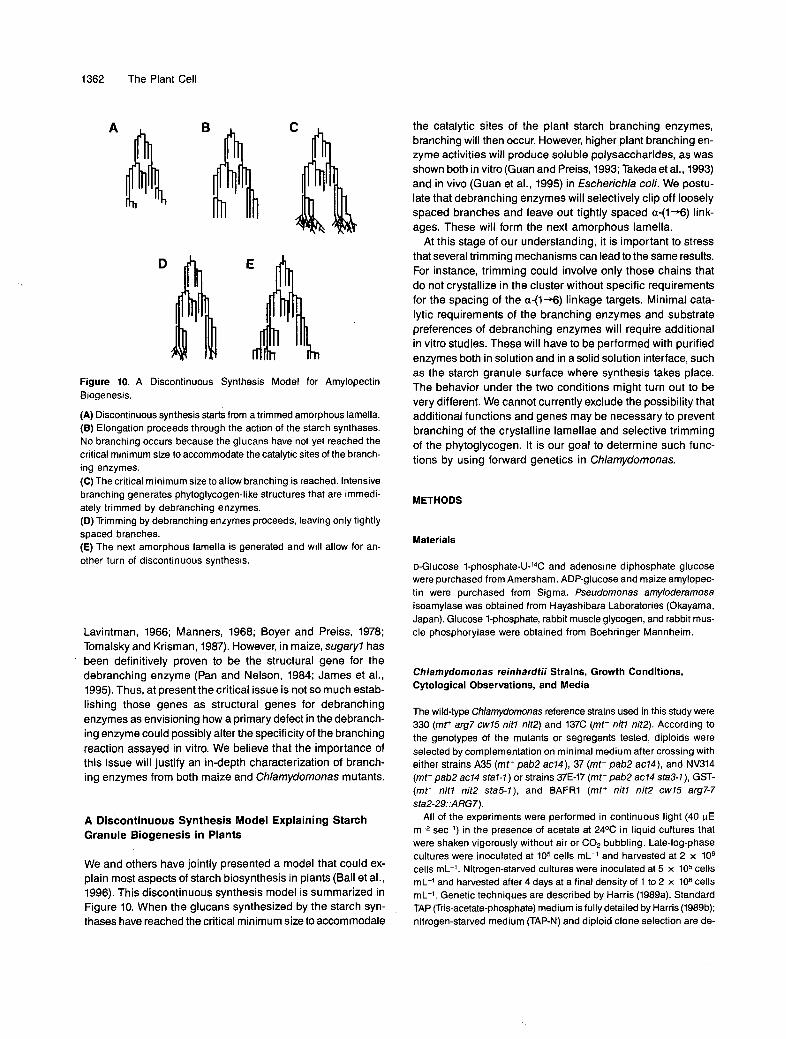
Figure 10. A Discontinuous Synthesis Model for Amylopectin Biogenesis.
(A) Discontinuous synthesis starts from a trimmed amorphous lamella. (6) Elongation proceeds through the action of the starch synthases. No branching occurs because the glucans have not yet reached the critical minimum size to accommodate the catalytic sites of the branch- ing enzymes. (C) The critical minimum size to allow branching is reached. lntensive branching generates phytoglycogen-like structures that are immedi- ately trimmed by debranching enzymes. (D) Trimming by debranching enzymes proceeds, leaving only tightly spaced branches. (E) The next amorphous lamella is generated and will allow for an- other turn of discontinuous synthesis.
Lavintman, 1966; Manners, 1968; Boyer and Preiss, 1978; Tomalsky and Krisman, 1987). However, in maize, sugaryl has been definitively proven to be the structural gene for the debranching enzyme (Pan and Nelson, 1984; James et al., 1995). Thus, at present the critical issue is not so much estab- lishing those genes as structural genes for debranching enzymes as envisioning how a primary defect in the debranch- ing enzyme could possibly alter the specificity of the branching reaction assayed in vitro. We believe that the importance of this issue will justify an in-depth characterization of branch- ing enzymes from both maize and Chlamydomonas mutants.
A Discontinuous Synthesis Model Explaining Starch Granule Biogenesis in Plants
We and others have jointly presented a model that could ex- plain most aspects of starch biosynthesis in plants (Ball et al., 1996). This discontinuous synthesis model is summarized in Figure 10. When the glucans synthesized by the starch syn- thases have reached the critical minimum size to accommodate
the catalytic sites of the plant starch branching enzymes, branching will then occur. However, higher plant branching en- zyme activities will produce soluble polysaccharides, as was shown both in vitro(Guan and Preiss, 1993; Takedaet al., 1993) and in vivo (Guan et al., 1995) in Escbericbia coli. We postu- late that debranching enzymes will selectively clip off loosely spaced branches and leave out tightly spaced a++) link- ages. These will form the next amorphous lamella.
At this stage of our understanding, it is important to stress that severa1 trimming mechanisms can lead to the same results. For instance, trimming could involve only those chains that do not crystallize in the cluster without specific requirements for the spacing of the a-(l+) linkage targets. Minimal cata- lytic requirements of the branching enzymes and substrate preferences of debranching enzymes will require additional in vitro studies. These will have to be performed with purified enzymes both in solution and in a solid solution interface, such as the starch granule surface where synthesis takes place. The behavior under the two conditions might turn out to be very different. We cannot currently exclude the possibility that additional functions and genes may be necessary to prevent branching of the crystalline lamellae and selective trimming of the phytoglycogen. It is our goal to determine such func- tions by using forward genetics in Chlamydomonas.
METHODS
Materials
o-Glucose I-ph~sphate-U-~~C and adenosine diphosphate glucose were purchased from Amersham. ADP-glucose and maize amylopec- tin were purchased from Sigma. Pseudomonas amyloderamosa isoamylase was obtained from Hayashibara Laboratories (Okayama, Japan). Glucose 1-phosphate, rabbit muscle glycogen, and rabbit mus- cle phosphorylase were obtained from Boehringer Mannheim.
Chlamydomonas reinhardtii Strains, Growth Conditions, Cytological Observations, and Media
The wild-type Chlamydomonas reference strains used in this study were 330 (mt+ arg7 cw75 nit7 nit2) and 137C (mt- nit7 nit2). According to the genotypes of the mutants or segregants tested, diploids were selected by complementation on minimal medium after crossing with either strains A35 (mt+ pab2 ac74), 37 (mt- pab2 ac74), and NV314 (mt- pab2 ac74 sta7-7) or strains 37E-17 (mt- pab2 ac74 sta3-7), GST- (mt- nit7 nit2 sta5-7), and BAFRl (mt+ nitl nit2 cw75 arg7-7 sta2-29::ARG7).
All of the experiments were performed in continuous light (40 pE m-* sec-l) in the presence of acetate at 24OC in liquid cultures that were shaken vigorously without air or C02 bubbling. Late-log-phase cultures were inoculated at 105 cells mL-l and harvested at 2 x 10⁶ cells mL-l. Nitrogen-starved cultures were inoculated at 5 x 10⁵ cells mL-l and harvested after 4 days at a final density of 1 to 2 x 10⁶ cells mL-l. Genetic techniques are described by Harris (1989a). Standard TAP (Tris-acetate-phosphate) medium is fully detailed by Harris (1989b); nitrogen-starved medium (TAP-N) and diploid clone selection are de-

Starch Synthesis in Chlamydomonas 1363
scribed by Ball et al. (1990, 1991) and Delrue et al. (1992). Fixation and embedding protocols are described by Harris (1989~).
Transformation-Mediated Gene Disruption
Transformation mediated by standard glass beads (Kindle, 1990) was performed on the cell wall-deficient arginine-requiring strain 330 with 1 pg of pARG7.8 carrying the wild-type arginosuccinate lyase gene. Transformam were selected by complementation of arginine aux- drophy present in the recipient strain. The transformants were screened for starch structure and amount by spraying iodine directly on replica plates (Fontaine et al., 1993). The frequency of mutant selection in the starch pathway was on the order of 10-3. Six transformation experi- ments yielded five independent alleles of the SJA7 gene.
Genetic Analysis of the Disrupted Strains
Of the five transformants, only strain BAFJ4 (mt+ nit7 nit2 cw75 arg7-7 staM::ARG7) yielded sufficient zygotes when crossed with A35 (mt+ pab2 ac74) to make standard genetic analysis feasible. The segre- gants were selected at random and checked for all markers included in the cross. Fifty percent of the progeny were wild type with respect to starch, as is expected for a single Mendelian defect. All other mark- ers segregated as expected. Sufficient fusion was obtained with all of the transformants to yield vegetative diploids. Heterozygous diploids thus were obtained for each of the five mutants by crossing with A35 (mt+ pab2 ac74) and picking 5-day-old colonies from minimal medium. The diploid nature of the clones was double-checked by meas- uring cell volume, determining mating type, and characterizing phenotypically (complementation of the starch defect and presence of pARG7.8 on DNA gel blots).
The diploid clones obtained from strain BAFJ6 (mt+ nit7 nit2 cw75 arg7-7 sta77::ARG7) were crossed with strain 37 and yielded abun- dant triploid zygotes. Good germination and survival were obtained to the point that triploid genetic analysis could be performed routinely. All of the markers @ab2, ac74, nit7 and nit2, cw75) segregated as ex- pected according to the dose of mutant allele present in the triploid zygote. A single recessive (nit7 and nit2, cw75) allele fitted the 16.6% mutant meiotic progeny expectation, whereas the double recessive (pab2, ac74) segregated like standard heterozygous zygotes at meio- sis. Normal interchromosomal and intrachromosomal recombinations between nitl and nit2 and between pab2 and ac74, respectively, in triploids were observed. The segregation of the starch defect fitted the 16.6% distribution and could thus be attributed to a single reces- sive defect. Recombinant clones with suitable genotypes were obtained and used in a standard complementation analysis.
The presence of pARG7.8 was followed throughout the analysis. Ten micrograms of restricted genomic DNAs was probed with an Nrul-Sal1 321-bp fragment covering a small part of the bacterial tetracycline re- sistance gene contained in pARG7.8, which is proximal to the ARG7 gene. 60th sta77rrARG7 and sta72rrARG7 yielded complex DNA gel blot patterns involving multiple insertion events. To our surprise, the entire complex pattern cosegregated with the mutant trait in both of these cases. The DNA gel blot patterns generated by sta7-3::ARGZ sta7-4::ARGz and sta7-5::ARG7 were simpler. Strain S carrying sta7- 5::ARG7displays a unique signal when digested with Pstl. Our attempts to subject this strain to standard or triploid genetic analysis have not been successful.
Measures of Starch Levels, Starch Purification, and Spectral Properties of the lodine-Starch Complex
A full account of amyloglucosidase assays, starch purification on Per- coll gradients, and A,,, measurements can be found in Delrue et al. (1992).
Crude Extract Preparation, Enzyme Assays, Partia1 Purification of Enzyme Activities, and Zymograms
Soluble crude extracts were always prepared from late-log-phase cells (2 x 10⁶ cells mL-l) grown in high-salt acetate medium under con- tinuous light (80 pE m-2 sec-l). Amylase, phosphoglucomutase, ADP-glucose pyrophosphorylase, and phosphorylase activities were monitored by using the standard assays described by Ball et al. (1991), lglesias et al. (1994), and Van den Koornhuyse et al. (1996). For solu- ble starch synthases and branching enzymes, the assays used are those described by Fontaine et al. (1993) and Libessart et al. (1995). The analysis was completed by zymograms as detailed by Fontaine et al. (1993) and Maddelein et al. (1994).
Starch Fractionation, Nuclear Magnetic Resonance, MatrixAssisted Laser Desorptionllonization Mass Spectra Analysis, and Thin-Layer Chromatography
Separation of starch fractions on TSK HW-75(S) columns (Merck) was performed as described by Delrue et al. (1992). Sepharose CL9B sepa- ration of amylose and amylopectin was performed as described by Libessart et al. (1995). Nuclear magnetic resonance (NMR) analysis was performed with the same setup and conditions as those described by Fontaine et al. (1993). The leve1 of branching was estimated by in- tegration of the same regions of proton resonances of the monosubstituted and disubstituted glucose (6~5.2 and 4.9 ppm, respec- tively) (Gidley, 1985). Reducing end signals appeared in the dextrin samples. To estimate the percentage of reducing ends, we integrated the signals due to the a (5.1 ppm) and p (4.5 ppm) anomeric forms of the reducing ends with respect to the same region of the monosub- stituted glucose proton resonances. The average degree of polymerization (DP) was deduced by calculating the reverse ratio. Matrix-assisted laser desorption/ionization mass spectra (MALD-MS) were measured on a Vision 2000 time-of-flight mass spectrometer (Fin- nigan MAT, Brennen, Germany), using 2,5dihydroxybenzoic acid as a matrix (10 mg mL-’ in MeCN-H20 [70/30, v/v]); 1 pL of sample so- lution (1 to 5 Wg pL-l) was mixed with l WL of matrix solution on a stainless steel target. Desorptionlionization was performed using an N2 laser at 337 nm. Twenty spectra were summed to enhance the signal-to-noise ratio. Crude separation of small chains (DP < 16) could also be monitored by thin-layer chromatography on silica gel60 (Kiesel- gel 60; Merck), using butanol-ethanollH,O (5/5/4 [vlv]) and orcinol sulfuric staining.
Phytoglycogen Purification and Debranching
Water soluble polysaccharides (WSPs) were prepared from 10 L of nitrogen-starved culture, inoculated at 105 cells mL-’, and harvested after 7 days of growth under continuous light (80 pE m-2 sec-l) on high-salt acetate medium. Algae were ruptured by passage in a French press (10,000 p.s.i.) at a density of 108 cells mL-l in water. The crude

1364 The Plant Cell
extract was immediately frozen at -8OOC and thawed after a minimum of 4 hr of storage. Cell debris were discarded by spinning at 10,OOOg for 30 min. The supernatant was extracted with 2:l chloroform-meth- ano1 and centrifuged at 3000gfor 15 min. The water-methanol soluble fraction was concentrated by rotary evaporation and desalted by dial- ysis at 4OC. The desalted fraction was concentrated, adjusted to 10% DMSO, and loaded on a TSK HW-50(F) column, as described by Maddelein et al. (1994). The amount of glucan and X,,, values were determined for each fraction.
After pooling and dialysis of the fractions of interest, phytoglycogen was analyzed by NMR or isoamylase-mediated debranching. Isoamy- lase-mediated debranching was performed as described by Maddelein et al. (1994). The debranched products were analyzed by high-per- formance anion exchange chromatography with pulsed amporometric detection and gel filtration on TSK HW-50(F) columns as described by Maddelein et al. (1994). The degree-of-polymerization scale on the TSK HW-50(F) columns was generated by using the Xmax values of the fractions as internal standards according to Banks et al. (1971).
Zymogram Detection of Starch Hydrolytic Activities
We used a zymogram technique adapted from the method developed by Lacks and Springhorn (1980), who used a-amylase as a model en- zyme to study renaturation and detection. An in-depth discussion on renaturation-detection techniques can be found in Gabriel and Gersten (1992). Soluble crude extracts were prepared as described above and immediately stored at -7OOC. The lysate was cleared by spinning at 10,OOOg for 10 min at 4OC. Proteins were measured using the Bio-Rad protein assay kit. Protein (100 to 500 pg) in 100 pL of 25 mM Tris-glycine, pH 8.3, 1% SDS, 5% P-mercaptoethanol was denaturated by boiling in a water bath for 5 min. The denatured proteins can be stored at 4OC without subsequent loss of enzyme activity or immediately loaded on a 30:l (acrylbis), 7.5% acrylamide, 0.1% SDS, 0.75" thick, de- naturing polyacrylamide gel containing 0.3% of autoclaved soluble potato starch or maize amylopectin (Sigma).
Electrophoresis was performed at room temperature at 15 V cm-' for 120 min by using the Mini-Protean II cell (Bio-Rad) in 25 mM Tris- glycine, pH 8.3, 1 mM DTT, 0.1% SDS buffer. At the end of the run, the gel was washed twice with gentle shaking for 1 hr in 100 mL of 0.04 M Tris at room temperature to remove SDS and renaturated pro- teins. The gel was then incubated overnight in 25 mM Tris-glycine, pH 8.3, 20 mM DTT at 25%. The reaction was stopped, and the gel was stained by adding 0.25% KI, 0.025% 12. The zymogram was im-
(Sigma). Electrophoresis was performed for 5 hr, as described above. Following renaturation, sections of the gel containing the hydrolytic activities were sliced in 1-cm-wide horizontal sections. The strips were separately incubated for 72 hr in a 5-mL volume of 25 mM Tris-glycine, pH 8.3,20 mM DTT at 25OC. The sections containing hydrolytic activi- ties were identified by using for each gel a vertical section as internal standard and staining it with iodine after 12 hr of incubation. After 3 days, the incubation buffers containing the dextrins were collected. The strips were again stained with iodine to determine whether most of the dextrin had diffused from the gel. The incubation buffer was then desalted by gel fitration chromatography on a TSK HW-40 column equilibrated in 0.50/0 acetic acid and concentrated by rotary evapora- tion. A similar procedure was followed to purify dextrins from Chlamydomonas amylase. Autoclaved amylopectin eluted from gels in the absence of enzyme activity was used as an undigested stan- dard. We thus produced between 1 and 2 mg of dextrin from each starch hydrolase. The dextrins were subjected to thin-layer chroma- tography, MALD-MS, and NMR analysis.
ACKNOWLEDGMENTS
This work was supported by a European Union grant (No. FAlR PL95- 568), by the Université des Sciences et Technologies de Lille, the Ministere de I'Education Nationale, the Centre National de Ia Recherche Scientifique (CNRS) (Unité Mixte de Recherche du CNRS No. 111, Director André Verbert), and by special grants from the starch- processing company Roquette Frhres (Lestrem, France), from the Région Nord Pas-de-Calais (réseau microbiologie), and from the seed company Limagrain. We thank Loi'c Brunet and André Dhainaut (Unité de Formation et de Recherches de Biologie, Université des Sciences et Technologies de Lille) for helping with the electron microscopy. We express our special thanks to Marie-Christine Slomianny for techni- cal advice in protein biochemistry and to Yves Leroy and Bruno Domon for providing guidance with MALD-MS analysis. We express our grati- tude to David Manners for helpful discussions.
Received March 19, 1996; accepted June 6, 1996.
. -
mediately photographed. The molecular mass of each enzyme activity detected on zymogram was measured with the same polyacrylamide
REFERENCES
gel for the blue- and red-staining bands and with a 12% acrylamide gel for the white-staining band. Some sections of the gel were stained with Coomassie Brilliant Blue R 250, and others were renaturated and incubated as described above.
Partia1 Purification and Elution of Dextrin Products
Ten micrograms of crude extract protein was loaded on a (2 x 50 cm) Sephacryl S300 gel (Pharmacia) equilibrated in 25 mM Tris-glycine, pH 8.3, 20 mM DTT, and 50 mM NaCI, with a flow rate of 60 mL hr-l. One-milliliter fractions were collected, and the four fractions contain- ing the blue activity but still containing a red-staining hydrolase were loaded after denaturation on four large (16 x 18 cm), 1.5-mm-thick polyacrylamide gels containing 0.3% autoclaved maize amylopectin
Ball, S.G. (1995). Recent views on the biosynthesis of the starch gran- ule. Trends Glycosci. Glycotechnol. 7, 405-415.
Ball, S.G., Dirick, L., Decq, A., Martiat, J.C., and Matagne, R.F. (1990). Physiology of starch storage in the monocellular alga Chlamydo- monas reinhardtii. Plant Sci. 66, 1-9.
Ball, S., Marianne, T., Dirick, L., Fresnoy, M., Delrue, B., and Decq, A. (1991). A Chlamydomonas reinhardtii low-starch mutant is defec- tive for 3-phosphoglycerate activation and orthophosphate inhibition of ADP-glucose pyrophosphorylase. Planta 185, 17-26.
Ball, S., Guan, ti.-P., James, M., Myers, A., Keellng, P., Mouille, G., BulBon, A., Colonna, P., and Preiss, J. (1996). From glycogen to amylopectin: A model for the biogenesis of the plant starch gran- ule. Cell, in press.

Starch Synthesis in Chlamydomonas 1365
Banks, W., Greenwood, C.T., and Khan, K.M. (1971). The interaction of linear amylose oligomers with iodine. Carbohydr. Res. 17,25-33.
Black, R.C., Loerch, J.D., McArdle, F.J., and Creech, R.G. (1966). Genetic interactions affecting maize phytoglycogen and the phytoglycogen-forming branching enzyme. Genetics 53, 661-668.
Boyer, C., and Preiss, J. (1978). Multiple forms of starch branching enzyme of maize: Evidence for independent genetic control. Bio- chem. Biophys. Res. Commun. 80, 169-175.
Caspar, T. (1994). Genetic dissection of the biosynthesis, degrada- tion, and biological functions of starch. In Arabidopsis, Monograph 27, E.M. Meyerowitz and C. Somerville, eds (Cold Spring Harbor, NY: Cold Spring Harbor Laboratory), pp. 913-936.
Caspar, T., Huber, S.C., and Somerville, C. (1985). Alterations in growth, photosynthesis, and respiration in a starchless mutant of Arabidopsis thaliana (L.) deficient in chloroplast phosphoglucomu- tase activity. Plant Physiol. 79, 11-17.
Delrue, B., Fontaine, T., Routier, F., Decq, A., Wieruszeski, J.M., Van den Koornhuyse, N., Maddelein, M.-L., Fournet, B., and Ball, S. (1992). Waxy Chlamydomonas reinhardtii: Monocellular alga1 mu- tants defective in amylose biosynthesis and granule-bound starch synthase accumulate a structurally modified amylopectin. J. Bac- teriol. 174, 3612-3620.
Erlander, S. (1958). Proposed mechanism for the synthesis of starch from glycogen. Enzymologia 19, 273-283.
Fontaine, T., D’Hulst, C., Maddelein, M.-L., Routier, F., Marianne- Pepin, T., Decq, A., Wieruszeski, J.M., Delrue, B., Van den Koornhuyse, N., Bossu, J.P., Fournet, B., and Ball, S.G. (1993). Toward an understanding of the biogenesis of the starch granule: Evidence that Chlamydomonas soluble starch synthase II controls the synthesis of intermediate size glucans of amylopectin. J. Biol. Chem. 268, 16223-16230.
French, D. (1984). Organization of starch granules. In Starch, Chem- istry and Technology, R.L. Whistler, J.N. BeMiller, and E.F. Paschall, eds (New York: Academic Press), pp. 183-247.
Gabriel, O., and Gersten, D.M. (1992). Staining for enzymatic activ- ity after gel electrophoresis, I. Anal. Biochem. 203, 1-21.
Geddes, R. (1985). Glycogen: A structural viewpoint. In The Polysac- charides, Vol. 3, G.O. Aspinall, ed (San Diego, CA: Academic Press),
Gidley, M.J. (1985). Quantification of the structural features of starch polysaccharides by N.M.R. spectroscopy. Carbohydr. Res. 139, 85-93.
Guan, H.P., and Preiss, J. (1993). Differentiation of the properties of the branching isoenzymes from maize. Plant Physiol. 102,1269-1273.
Guan, H.P., Kuriki, T., Sivak, M., and Preiss, J. (1995). Maize branch- ing enzyme catalyzes synthesis of glycogen-like polysaccharide in glgS-deficient Escherichia coli. Proc. Natl. Acad. Sci. USA 92,
Hanson, K.R., and McHale, N.A. (1988). A starchless mutant of Nico- tiana sylvestris containing a modified plastid phosphoglucomutase. Plant Physiol. 88, 838-844.
Harris, E.H. (1989a). Genetic analysis. In The Chlamydomonas Source- book: A Comprehensive Guide to Biology and Laboratory Use, E. Harris, ed (San Diego, CA: Academic Press), pp. 399-446.
Harris, E.H. (1989b). Culture and storage methods. In The Chlamydo- monas Sourcebook: A Comprehensive Guide to Biology and Laboratory Use, E. Harris, ed (San Diego, CA: Academic Press),
pp. 283-336.
964-967.
pp. 25-63.
Harris, E.H. (1989~). Histological techniques for Chlamydomonas. In The Chlamydomonas Sourcebook: A Comprehensive Guide to Bi- ology and Laboratory Use, E. Harris, ed (San Diego, CA: Academic Press), pp. 581-586.
Iglesias, A.A., Charng, Y., Ball, S., and Preiss, J. (1994). Character- ization of the kinetic, regulatory, and structural properties of ADP-glucose pyrophosphorylase from Chlamydomonas reinhardtii. Plant Physiol. 104, 1287-1294.
Imberty, A., Buléon, A., Tran, V., and Pérez, S. (1991). Recent ad- vances in knowledge of starch structure. Starke 43, 375-384.
James, M.G., Robertson, D.S., and Meyers, A.M. (1995). Character- ization of the maize gene sugaryl, a determinant of starch composition in kernels. Plant Cell 7, 417-429.
Jenkins, P.J., Cameron, R.E., and Donald, A.M. (1993). A universal feature in the starch granules from different botanical sources. Starke 45, 417-420.
Kakefuda, G., and Duke, S.H. (1984). Electrophoretic transfer as a technique for the detection and identification of the plant amylolytic enzymes in polyacrylamide gels. Plant Physiol. 75, 278-280.
Kindle, K.L. (1990). High-frequency nuclear transformation of Chla- mydomonas reinhardtii. Proc. Natl. Acad. Sci. USA 87, 1228-1232.
Lacks, S.A., and Springhorn, S.S. (1980). Renaturation of enzymes after polyacrylamide gel electrophoresis in the presence of sodium dodecyl sulfate. J. Biol. Chem. 255, 7467-7473.
Lavintman, N. (1966). The formation of branched glucans in sweet corn. Arch. Biochem. Biophys. 116, 1-8.
Libessart, N., Maddelein, M.-L., Van den Koornhuyse, N., Decq, A., Delrue, B., Mouille, O., D’Hulst, C., and Ball, S.G. (1995). Stor- age, photosynthesis and growth: The conditional nature of mutations affecting starch synthesis and structure in Chlamydomonas. Plant Cell 7, 1117-1127.
Lin, T.-P., Caspar, T., Somerville, C., and Preiss, J. (1988). lsolation and characterization of a starchless mutant of Arabidopsis thaliana (L.) Heynh. lacking ADP-glucose pyrophosphorylase activity. Plant Physiol. 86, 1131-1135.
Maddelein, M.-L., Libessart, N., Bellanger, F., Delrue, B., DHulst, C., Van den Koornhuyse, N., Fontaine, T., Wieruszeski, J.M., Decq, A., and Ball, S.G. (1994). Toward an understanding of the biogenesis of the starch granule: Determination of granule-bound and soluble starch synthase functions in amylopectin synthesis. J. Biol. Chem. 269, 25150-25157.
Manners, D.J. (1968). Studies on carbohydrate metabolizing enzymes. Part XIX. Sweet-corn branching enzymes. Carbohydr. Res. 8,7241.
Manners, D.J. (1989). Recent developments in our understanding of amylopectin structure. Carbohydr. Polymers 11, 87-112.
Müller-Rober, B., and Kossmann, J. (1994). Approaches to influence starch quantity and starch quality in transgenic plants. Plant Cell Environ. 17, 601-613.
Müller-Rober, B., Sonnewald, U., and Willmitzer, L. (1992). Inhibi- tion of the ADP-glucose pyrophosphorylase in transgenic potatoes leads to sugar-storing tubers and influences tuber formation and expression of tuber storage protein genes. EMBO J. 11, 1229-1238.
Nakamura, Y., Umemoto, T., Ogata, N., Kuboki, Y., Yano, M., and Saski, T. (1996a). Starch debranching enzyme (R-enzyme or pul- lulanase) from developing rice endosperm: Purification, cDNA and chromosomal localization of the gene. Planta 199, 203-218.
Nakamura, Y., Umemoto, T., Takahata, Y., Komae, K., Amano, E., and Satoh, H. (1996b). Changes in structure of starch and enzyme

1366 The Plant Cell
activities affected by sugafy mutations in developing rice endosperm: Possible role of starch debranching enzyme in amylopectin biosyn- thesis. Physiol. Plant., in press.
Nelson, O.E., and Pan, D. (1995). Starch synthesis in maize en- dosperms. Annu. Rev. Plant Physiol. Plant MOI. Biol. 46, 475-496.
Pan, D., and Nelson, O.E. (1984). A debranching enzyme deficiency in endosperms of the Sugary-7 mutants of maize. Plant Physiol. 74, 324-328.
Preiss, J. (1991). Biology and molecular biology of starch synthesis and its regulation. In Oxford Surveys of Plant Molecular and Cell Biology, Vol. 7, B.J. Miflin, ed (Oxford, UK: Oxford University Press),
Preiss, J., and Sivak, M.N. (1996). Starch synthesis in sinks and sources. In Photoassimilate Distribution in Plants and Crops: Source-Sink Relationships, E. Zamski and A.A. Schaffer, eds (New York: Marcel Dekker, Inc.), pp. 63-96.
Ramazanov, Z., Rawat, M., Henk, M.C., Mason, C.B., Matthews, S.W., and Moroney, J.V. (1994). The induction of the C02- concentrating mechanism is correlated with the formation of the starch sheath around the pyrenoid of Chlamydomonas reinhardtii. Planta 195, 210-216.
Rammesmayer, G., and Praznik, W. (1992). Fast and sensitive stain- ing method of Qenzyme, a-amylase, R-enzyme, phosphorylase and
pp. 59-114.
soluble starch synthase separated by starch-polyacrylamide gel elec- trophoresis. J. Chromatogr. 623, 399-402.
Robin, J.P., Mercier, C., Charbonnière, R., and Guilbot, A. (1974). Lintnerized starches: Gel filtration and enzymatic studies of insolu- ble residues from prolonged acid treatment of potato starch. Cereal Chem. si, 389-406.
Shannon, J.C., and Garwood, D.L. (1984). Genetics and physiology of starch development. In Starch: Chemistry and Technology, 2nd ed, R.L. Whistler, J.N. Bemiller, and E.F. Paschall, eds (Orlando, FL: Academic Press), pp. 25-86.
Sumner, J.B., and Somers, G.F. (1944). The water soluble polysac- charide of sweet corn. Arch. Biochem. 4, 4-7.
Takeda, Y., Guan, H.P., and Preiss, J. (1993). Branching of amylose by the branching isoenzymes of maize endosperm. Carbohydr. Res.
Tomalsky, D.S., and Krisman, C.R. (1987). The degree of branching in (al,4)-(al,6)-linked glucopolysaccharides is dependent on intrinsic properties of the branching enzymes. Eur. J. Biochem. 168,393-397.
Van den Koornhuyse, N., Libessart, N., Delrue, B., Zabawinski, C., Decq, A., Iglesias, A., Preiss, J., and Ball, S. (1996). Control of starch composition and structure through substrate supply in the monocellular alga Chlamydomonas reinhardtii. J. Biol. Chem. 271,
240, 253-263.
16281-16287.

DOI 10.1105/tpc.8.8.1353 1996;8;1353-1366Plant Cell
G. Mouille, M. L. Maddelein, N. Libessart, P. Talaga, A. Decq, B. Delrue and S. BallPreamylopectin Processing: A Mandatory Step for Starch Biosynthesis in Plants.
This information is current as of October 16, 2014
Permissions 98X
https://www.copyright.com/ccc/openurl.do?sid=pd_hw1532298X&issn=1532298X&WT.mc_id=pd_hw15322
eTOCs http://www.plantcell.org/cgi/alerts/ctmain
Sign up for eTOCs at:
CiteTrack Alerts http://www.plantcell.org/cgi/alerts/ctmain
Sign up for CiteTrack Alerts at:
Subscription Information http://www.aspb.org/publications/subscriptions.cfm
is available at:Plant Physiology and The Plant CellSubscription Information for
ADVANCING THE SCIENCE OF PLANT BIOLOGY © American Society of Plant Biologists