Biosynthesis of Smaller-Sized Platinum Nanoparticles Using ...

lable at ScienceDirect
Insect Biochemistry and Molecular Biology 54 (2014) 61e68
Contents lists avai
Insect Biochemistry and Molecular Biology
journal homepage: www.elsevier .com/locate/ ibmb
Mode of action of allatostatins in the regulation of juvenile hormonebiosynthesis in the cockroach, Diploptera punctata
Juan Huang a, Elisabeth Marchal a, b, Ekaterina F. Hult a, Sven Zels b, Jozef Vanden Broeck b,Stephen S. Tobe a, *
a Department of Cell and Systems Biology, University of Toronto, Toronto, Canadab Department of Biology, Zoological Institute, KU Leuven, B-3000 Leuven, Belgium
a r t i c l e i n f o
Article history:Received 24 July 2014Received in revised form27 August 2014Accepted 2 September 2014Available online 10 September 2014
In memory of Dr. Grahame E. Pratt who diedon March 8, 2014
Keywords:Allatostatin receptorDiploptera punctataG protein-coupled receptorsJuvenile hormoneRNA interference
Abbreviations: AstR, allatostatin receptor; PTX, phamster ovary; HEK, human embryonic kidney; CRE, cadenylate cyclase; MA, mevalonic acid; DPPM, dstandard error of the mean.* Corresponding author. Department of Cell and S
Toronto, 25 Harbord Street, Toronto, Ontario, Canada.E-mail address: [email protected] (S.S. To
http://dx.doi.org/10.1016/j.ibmb.2014.09.0010965-1748/© 2014 Elsevier Ltd. All rights reserved.
a b s t r a c t
The FGLamide allatostatins (FGL/ASTs) are a family of neuropeptides with pleiotropic functions, includingthe inhibition of juvenile hormone (JH) biosynthesis, vitellogenesis and muscle contraction. In thecockroach, Diploptera punctata, thirteen FGLa/ASTs and one allatostatin receptor (AstR) have beenidentified. However, the mode of action of ASTs in regulation of JH biosynthesis remains unclear. Here,we determined the tissue distribution of Dippu-AstR. And we expressed Dippu-AstR in vertebrate celllines, and activated the receptor with the Dippu-ASTs. Our results show that all thirteen ASTs activatedDippu-AstR in a dose dependent manner, albeit with different potencies. Functional analysis of AstR inmultiple cell lines demonstrated that activation of the AstR receptor resulted in elevated levels of Ca2þ
and cAMP, which suggests that Dippu-AstR can act through the Gaq and Gas protein pathways. The studyon the target of AST action reveals that FGL/AST affects JH biosynthesis prior to the entry of acetyl-CoAinto the JH biosynthetic pathway.
© 2014 Elsevier Ltd. All rights reserved.
1. Introduction
Allatostatins (ASTs), a family of pleiotropic neuropeptides, wereoriginally named for their ability to inhibit juvenile hormone (JH)biosynthesis by corpora allata (CA) rapidly and reversibly (Bendenaet al., 1999). Three families of ASTs have been identified in insectsand named as: FGLa/ASTs (A-type), MIP/ASTs (B-type) and thePISCF/ASTs (C-type) (Coast and Schooley, 2011). The distributionand function of the three families of ASTs has been reviewed by Stayand Tobe (2007). The best documented role of FGLa/AST is theirability to inhibit JH biosynthesis by corpora allata (CA) (Stay andTobe, 2007). Later studies demonstrated other functions of FGLa/ASTs, including regulation of myotropic activity in gut tissues (Duveet al., 1995; Lange et al., 1995) and cardiac rhythm (Vilaplana et al.,1999), inhibition of vitellogenin synthesis in the fat body (Martin
ertussis toxin; CHO, ChineseAMP responsive element; AC,iphosphomevalonate; SEM,
ystems Biology, University ofTel.: þ1 416 978 3517.
be).
et al., 1996), and stimulation of enzyme activity in the lumen ofthe midgut (Fuse et al., 1999).
As neuropeptides, ASTs exert their effects by binding to a Gprotein-coupled receptor, AstR. FGLa/AST receptors, which arestructurally related to the mammalian galanin receptor, were firstidentified in the fly Drosophila melanogaster DAR-1 (Birgul et al.,1999) and DAR-2 (Lenz et al., 2000), and later in the silkworm,Bombyx mori (Secher et al., 2001) and the stick insect, Carausiusmorosus (Auerswald et al., 2001). In Diploptera punctata, putativeAstRs have previously been partially characterized using photo-affinity labeling and a radioligand-binding assay in the CA and brain(Cusson et al., 1991; Yu et al., 1995). Lungchukiet et al. (2008b)identified a putative FGLa/AST receptor gene in D. punctata.Silencing this gene resulted in a significant increase in JH biosyn-thesis (Lungchukiet et al., 2008a,b).
The responses of FGLa/AST receptor to FGLa/ASTs have beenassayed in D. melanogaster (DAR-1 and DAR-2) (Birgul et al., 1999;Larsen et al., 2001), B. mori (Secher et al., 2001) and the cockroach,Periplaneta americana (Gade et al., 2008). The studies in B. mori andP. americana focused on the response of AstR to exogenous ASTs. Thesignaling pathway of AstRwas, however, not completely elucidated.Larsen et al. (2001) expressedDrosophila FGLa/AST receptors, DAR-1and DAR-2 in CHO cells and activated themwith four putative FGLa/

J. Huang et al. / Insect Biochemistry and Molecular Biology 54 (2014) 61e6862
ASTs from D. melanogaster AST and four FGLa/ASTs from D. punctatain the presence or absence of pertussis toxin (PTX). They found thatPTX caused a complete loss of Ca2þ signal in cells expressing DAR-1,and a decreased Ca2þ signal in cells expressing DAR-2. Their resultssuggested that the activation of DAR-1 and DAR-2 by FGLa/ASTscoupled to multiple signaling pathways, including Gi/o protein andother, PTX-insensitive G-proteins.
Although the function of AST has been well-studied, little isknown about the precise target of AST action. Previous studiesfocused on select enzymes in the JH biosynthetic pathway. It is nowknown that thirteen enzymes are involved in the JH biosyntheticpathway (Fig. S1) (Belles et al., 2005; Nouzova et al., 2011). Thepotential targets of action of ASTs were originally studied byemploying different known JH precursors (Pratt et al., 1991, 1989).The results suggested that the inhibitory action of AST on JHbiosynthesis resides in step(s) prior to mevalonate. However,neither HMG-CoA synthase nor HMG-CoA reductase activity wasaffected by ASTs (Sutherland and Feyereisen, 1996). These authorsproposed that the target of AST action on JH biosynthesis is locatedin step(s) prior to the JH biosynthetic pathway, and may be relatedto the transport of citrate from mitochondria to cytosol and/or tothe cleavage of citrate to yield acetyl-CoA.
Our study focuses on the viviparous cockroach, D. punctata, inwhich ASTs were first characterized. The first gonadotropic cycle ischaracterized by a precise regulation of JH biosynthesis necessaryto coordinate a specific series of reproductive events closelycorrelated with oocyte growth. This makes D. punctata an idealmodel for amore in depth study of themode of action of ASTand itssignal transduction pathway. We have analyzed the spatialexpression pattern of the AST precursor and AstR which is consis-tent with their roles in regulating JH biosynthesis. Moreover, byusing an aequorin-based assay and expressing the receptor in amammalian system, we have unambiguously identified that AST isa ligand for the candidate AstR (Lungchukiet et al., 2008b) and thatthis receptor can couple to Ca2þ and cAMP.
Thirteen FGLa/ASTs have been identified in D. punctata. Thesepeptides share a conserved C-terminal Tyr (Phe)eXaaePhee-GlyeLeueNH2, which is believed to be the main functional regionfor the inhibition of JH biosynthesis (Donly et al., 1993; Marchalet al., 2013a). The FGLa/ASTs inhibit JH biosynthesis by CA at lowconcentrations in vitro but with different potencies (Tobe et al.,2000). Our study has also examined the relationship betweenbinding affinity and potency with a view to understanding the sitesof action of the peptides.
The precise target of ASTaction remains unclear so far. Our studydid not find any significant changes in the transcript level of genesencoding enzymes in JH biosynthetic pathway. The rescue of AST-induced JH inhibition by JH precursors suggests that the target ofAST action is prior to the entry of Acetyl-CoA into the JH biosyn-thetic pathway.
2. Materials and methods
2.1. Insects
D. punctata were reared in cages and fed with lab chow andwater at libitum at 27 �C in a dark room. Newly molted female adultcockroaches were picked from the colony and raised in separatecontainers. Mated status was confirmed by the presence of aspermatophore.
2.2. Tissue collection
Cockroach tissueswere dissected under a dissectingmicroscope.Basal oocyte length was measured to determine the physiological
age of cockroaches. Selected tissues were dissected and cleaned insterile cockroach ringer solution (150 mM NaCl, 12 mM KCl, 10 mMCaCl2.2H2O, 3 mM MgCl2.6H2O, 10 mM HEPES, 40 mM Glucose, pH7.2), and stored at �80 �C to prevent degradation.
2.3. RNA extraction and cDNA synthesis
Pooled samples were homogenized using a plastic pestle andRNAwas extracted using the RNeasy Mini Kit (Qiagen) according tothe manufacturer's instructions. An additional DNase treatment(RNase-free DNase set, Qiagen) was performed to eliminate po-tential genomic DNA contamination. RNA of CAwas extracted usingthe RNAqueous®-Micro Kit (Ambion). DNase treatment was per-formed to eliminate genomic DNA contamination. The quantity andquality of RNA was determined using a Nanodrop spectrophotom-eter (Thermo Scientific). An equal amount of RNA was transcribedwith Superscript III reverse transcriptase (Invitrogen Life Technol-ogies) utilizing random hexamers as described in the protocol. Theresulting cDNA was diluted tenfold.
2.4. Quantitative real time-PCR (q-RT-PCR)
Prior to target gene profiling, Tubulin and EF1a were chosen asthe optimal housekeeping genes according to a previous study(Marchal et al., 2013b). The q-RT-PCR reactions were performed intriplicate on a CFX384 Touch™ Real-Time PCR Detection System(Bio-Rad) in a final volume of 10 ml, containing 1 ml of cDNA, 5 mlIQ™ SYBR® Green Supermix (Bio-Rad), 1 ml forward and reverseprimer (5 mM) and 2 ml of MQ-water. The reactionwas incubated for3 min in 95 �C, followed by 40 cycles with following thermal pro-file: 95 �C, 10 s; 59 �C, 30 s. Target specificity was confirmed byrunning a few representative q-RT-PCR products on an agarose gelcontaining GelRed™ (Biotium). Realtime primers used for q-RT-PCRare listed in Table S1. Primer sets were validated by determiningrelative standard curves for each gene transcript using a five-foldserial dilution of a calibrator cDNA sample. Efficiency and correla-tion coefficient (R2) are shown in Table S1. The primer sets for genesin the JH biosynthetic pathway were chosen according to Huanget al. (submitted for publication). The quantity of mRNA for eachtested gene relative to reference genes was determined asdescribed by Vandesompele et al. (2002).
2.5. Peptides and substrates
Thirteen Dippu-ASTs were custom synthesized by GLBiochemical Ltd., Shanghai (China). Peptides were purified by highperformance liquid chromatography (HPLC) (purity �95%). Pep-tides were dissolved in water to obtain a concentration of 1 mM.Peptide solutions were stored at �80 �C prior to further processingand dilution.
Acetyl-CoA, mevalonic acid (MA), diphosphomevalonate(DPPM) and farnesol were purchased from SigmaeAldrich Canada.Substrates were dissolved in water before use.
2.6. Cell culture and transfection
Chinese hamster ovary (CHO) WTA11 and PAM28 cells stablyexpressing apoaequorin (Euroscreen, Belgium) and human em-bryonic kidney (HEK) 293 cells were cultured in monolayers inDulbecco's Modified Eagles Medium nutrient mixture F12-Ham(DMEM/F12) (Sigma) supplemented with 10% heat-inactivatedfetal calf serum (Invitrogen), 100 IU/ml penicillin and 100 mg/mlstreptomycin (Invitrogen). An additional 250 mg/ml Zeocin (Invi-trogen) was added to the medium for CHO-WTA11 cells, and anadditional 5 mg/ml Puromycin (Sigma) was added to the medium

Br NC CA Fb Ov Te AG MG MT0
2
4
6
Rel
ativ
em
RN
Aqu
antit
y
Dippu-AstRDippu-AST
Fig. 1. Relative expression levels of Dippu-AstR (black) and Dippu-AST (gray) mRNA intissues of day 4 males and mated females. The data represent averages of 3 pools (10animals per pool), run in triplicate. Tissues tested are brain (Br), nerve cord (NC),corpora allata (CA), fat body (Fb), ovary (Ov), midgut (MG) and Malpighian tubules(MT) from females and testes (Te) and accessory gland (AG) from males. Valuesrepresent mean ± SEM.
J. Huang et al. / Insect Biochemistry and Molecular Biology 54 (2014) 61e68 63
for PAM cells. The cells were cultured at 37 �C with a constantsupply of 5% CO2.
Transfections with pcDNA3.1D-Dippu-AstR or emptypcDNA3.1D vector were carried out in T75 flasks at 60e80% con-fluency. Transfection medium for CHO cells was prepared using theLipofectamine LTX kit (Invitrogen) with 3.75 ml Opti-MEM, 7.5 mgvector construct and 18.75 ml Plus™ Reagent in a 5 ml polystyreneround-bottom tube. After 5 min incubation at room temperature,45 ml LTX (Invitrogen) was added. Transfection mediumwas addeddropwise to the cells after 30 min incubation at room temperature.The transfection medium of the HEK293 cells was similar to that ofthe CHO cells, except that 9 mg DNA construct (6 mg of vectorconstruct and 3 mg of reporter gene plasmid) was added to themedium. The luciferase ORF, downstream of a cAMP responsiveelement (CRE) served as the reporter gene. After transfection, cellswere incubated overnight and an additional 10 ml of culture me-dium was added. The cells were allowed to grow for another nightprior to intracellular Ca2þ or cAMP measurements.
2.7. Aequorin assay
CHO cells were detached using 1 � Phosphate Buffered Saline(PBS) containing 0.2% EDTA (pH 8.0), collected and pelleted bycentrifugation in DMEM/F12 medium. The number of viable andnonviable cells was determined using a NucleoCounter NC-100™(Chemometic). The cells were then resuspended to a density of5 � 106 cells/ml in sterile filtered DMEM/bovine serum albumin(BSA) medium (DMEM/F12 with L-glutamine and 15 mM HEPES,without Phenol red, supplemented with 0.1% BSA). A concentrationof 5 mM coelenterazine h (Invitrogen) was added and the cells wereincubated for 4 h in the dark at room temperature with gentleshaking to reconstitute the holo-enzyme aequorin. The cells werethen diluted 10-fold in BSA medium and incubated for another30 min in the dark with gentle shaking. Peptides were dissolved inBSA medium and dispensed in 50 ml aliquots into the wells of awhite 96-well plate. Fifty ml of the cell suspensionwas injected intoeach well and light emission was recorded using a Mithras LB940multimode microplate reader (Berthold Technologies) over 30 s.The cells were lysed by injection of 50 ml 0.3% Triton X-100 and lightemission was monitored for an additional 8 s. The total response(ligand þ Triton-X100) is the representative for the quantity ofviable cells present in the well. BSAmediumwas used as a negativecontrol in each row of the plate, and 1 mM ATP served as a positivecontrol. The negative response was subtracted from the lumines-cence measured in wells of the same row. Calculations were madeusing the output file from Microwin software (Berthold Technolo-gies) in Excel (Microsoft). Further analysis was done in Excel andGraphPad Prism 6.
2.8. cAMP reporter assay
HEK cells were detached using 1 � PBS containing 0.2% EDTA(pH 8.0), collected and pelleted by centrifugation in DMEM/F12medium. The number of viable and nonviable cells was determinedusing a NucleoCounter NC-100™ (Chemometic). The cells werethen resuspended to a concentration of 1 � 106 cells/ml in DMEM/F12 containing 200 mM 3-isobutyl-1-methylxanthine (IBMX,Sigma) to prevent cAMP breakdown. Peptides were dissolved inIBMX medium in the presence or absence of 20 mM forskolin, andwere dispensed in 50 ml aliquots into the wells of a white 96-wellplate. Fifty ml of cell suspension was added into each well and theplate was incubated in a CO2 incubator (5% CO2) at 37 �C for 3.5 h.Fifty ml of Steadylite Plus substrate (PerkinElmer) was then added toeach well and the plate was incubated in the dark for 15 min whilegently shaking. Light emission resulting from the luciferase
enzymatic activity was recorded for 5 s/well using a Mithras LB940.Results were analyzed using the output of MicroWin and furtherprocessed by Excel and GraphPad Prism 6.
2.9. Radiochemical assay (RCA)
The in vitro RCA for JH biosynthesis was performed as describedby Feyereisen and Tobe (1981) and modified by Tobe and Clarke(1985). Two incubations were conducted in the JH biosynthesisrescue experiments. CA were incubated in TC199 medium with10�6 M AST7 for 3 h, and then transferred to fresh medium with10�6 M AST7 and JH precursors for a second 3 h incubation. JHbiosynthesis was determined after each incubation. The rate of JHbiosynthesis during the first incubation was used as the controlvalue.
2.10. RNA interference (RNAi)
Dippu-AstR dsRNA constructs were prepared using the MEGA-script® RNAi Kit (Ambion). A 368 bp fragment was amplified by PCRusing forward and reverse primers with T7 promoters (TAA-TACGACTCACTATAGGGAGA) attached to the 5
0end. The sequences
for forward and reverse primer were 50-TGTAATCA-
TACGGCTAACGGATC-30and 5
0-AATGGTAGAACGTAGTCTGTTGC-3
0,
respectively. Fragments were subcloned and sequenced to verifythe presence of the T7 promoter. The amplified fragment was usedin an RNA transcription reaction and incubated overnight to yieldannealed dsRNA transcripts. A nuclease digestionwas subsequentlyperformed to remove ssRNA and DNA remaining in the product.The dsRNA was further purified according to the manufacturer'sinstructions (Ambion). Concentration of the dsRNA construct wasdetermined using a nanodrop instrument (Thermo Fisher ScientificInc.). Five-fold diluted dsRNA was run on a 1.2% agarose gel toexamine the quality and integrity of the construct. The controldsRNA was prepared using a PCR fragment amplified from a non-coding region of pJET1.2 cloning vector (Thermo Scientific).
Dippu-AstR dsRNA was diluted in cockroach saline to a concen-tration of 250 ng/ml. Each adult female was injected with 4 ml ofdsRNA solution on day 0, 2 and 4 after the final moult. CA weredissected on day 6 and stored in liquid nitrogen prior to RNAextraction. A second set of cockroaches was injected with Dippu-AstR dsRNA in the same scheme as described above. CA weredissected on day 6 and cleaned in TC199 medium (GIBCO; 1.3 mMCa2þ, 2% Ficoll, methionine-free) for their use in the radiochemicalassay (RCA) determining the effect of AST on JH biosynthesis. Basaloocyte length was measured during dissection.

J. Huang et al. / Insect Biochemistry and Molecular Biology 54 (2014) 61e6864
3. Results
3.1. Tissue distribution of Dippu-AST and Dippu-AstR
The tissue distribution of Dippu-AST and Dippu-AstR was deter-mined in adult male and female cockroaches using q-RT-PCR(Fig. 1). Nine tissues were used to examine the tissue specificity ofDippu-AST and Dippu-AstR: brain (Br), nerve cord (NC), corporaallata (CA), fat body (Fb), ovary (Ov), midgut (MG) and Malpighiantubules (MT) from females and accessory gland (AG) and testes (Te)from males. The Dippu-AST gene is expressed in brain, nerve cordand midgut, which is consistent with the pleiotropic functions ofASTs (Fig. 1). Dippu-AstR shows the highest transcription levels inthe CA, followed by nerve cord, brain and fat body. All the othertissues tested showed either negligible or undetectable levels ofDippu-AstR mRNA.
3.2. Functional activation of Dippu-AstR with ASTs
We initially expressed Dippu-AstR in CHO-WTA11 cells. This cellline expresses the Ca2þ reporter apoaequorin and Ga16, a promis-cuous G protein that couples to most GPCRs with subsequentmobilization of intracellular Ca2þ (Stables et al., 1997). As shown inFig. 2, all 13 tested ASTs induced clear dose-dependent biolumi-nescence responses in AstR expressing cells. Most of the peptides
Fig. 2. Doseeresponse curves for ASTs in CHO-WTA11 cells expressing Dippu-AstR. Data pduplicate and are expressed as percentage of the maximal response. The zero response lev
showed similar degrees of biological efficacy (ability to activateAstR) but differed considerably in potency, with EC50 values rangingfrom 0.2 nM for AST6 to 30 nM, in the case of AST13 (Table 1). Ingeneral, the abilities of ASTs to activate AstR corresponded to theirpotencies as inhibitors of JH production by the CA, with theexception of AST1, AST5 and AST6 (Tobe et al., 2000) (Table 1).
To determine the second messenger pathways involved inDippu-AstR activation, we first expressed Dippu-AstR in CHO-PAM28 cells (lacking the promiscuous Ga16). The expression ofapoaequorin in the cell allows testing whether the receptor cancouple naturally through intracellular Ca2þ. Two ASTs (AST5 and 6)were chosen because of their high potency in inhibiting JHbiosynthesis or in activating Dippu-AstR. As shown in Fig. 3A, AST5and 6 induced dose-dependent intracellular Ca2þ responses inAstR-transfected CHO-PAM28 cells, with EC50 values of 21.4 nM and1.1 nM, respectively.
We subsequently tested whether the receptor coupled withcAMP in the signal transduction pathway. We expressed Dippu-AstR in HEK293 cells, which contain the luciferase gene underthe control of a cAMP response element. We assayed differentconcentrations of AST5 and AST6 in the presence or absence offorskolin. Forskolin activates adenylate cyclase (AC), which in-creases intracellular levels of cAMP. If AstR would couple negativelyto AC, we would expect lower levels of cAMP following applicationof ASTs. This was not the case. Application of AST5 or AST6 to
oints represent the average ± SEM of three independent measurements performed inel corresponds to treatment with BSA buffer only.

Table 1Potency of Dippu-ASTsa: activation of AstR in CHO-WTA11 cells (EC50) or inhibitory effect on JH release (IC50).
Allatostatin EC50b/nM Rankb
order (EC50)IC50
c/nM Rankc
order (IC50)Sequence
AST1 1.295 3 9.566 11 LeueTyreAspePheeGlyeLeueNH2AST2 2.033 4 0.4254 3 AlaeTyreSereTyreValeSereGlueTyreLyseArgeLeueProeValeTyreAsnePheeGlyeLeueNH2AST3 2.829 8 12.54 12 SereLyseMeteTyreGlyePheeGlyeLeueNH2AST4 2.489 6 1.139 5 AspeGlyeArgeMeteTyreSerePheeGlyeLeueNH2AST5 4.214 10 0.1063 1 AspeArgeLeueTyreSerePheeGlyeLeueNH2AST6 0.1929 1 2.725 8 AlaeArgeProeTyreSerePheeGlyeLeueNH2AST7 2.135 5 0.4118 2 AlaeProeSereGlyeAlaeGlneArgeLeueTyreGlyePheeGlyeLeueNH2AST8 4.087 9 3.275 10 GlyeGlyeSereLeueTyreSerePheeGlyeLeueNH2AST9 5.654 11 2 6 GlyeAspeGlyeArgeLeueTyreAlaePheeGlyeLeueNH2AST10 0.7717 2 0.9225 4 ProeValeAsneSereGlyeArgeSereSereGlyeSereArgePheeAsnePheeGlyeLeueNH2AST11 2.749 7 2.795 9 TyreProeGlneGlueHiseArgePheeSerePheeGlyeLeueNH2AST12 10.44 12 2.011 7 ProePheeAsnePheeGlyeLeueNH2AST13 29.99 13 12.84 13 IleeProeMeteTyreAspePheeGlyeIleeNH2
a Potency is defined as the dose required to achieve a given level of activation of AstR or inhibition of JH biosynthesis, and is listed in rank order.b EC50 was determined by AstR activation assay in CHO-WTA11 cells (details seen Fig. 1) with values from the present study shown in bold..c IC50 values are as reported by Tobe et al. (2000) (CA from day 2 virgin female).
J. Huang et al. / Insect Biochemistry and Molecular Biology 54 (2014) 61e68 65
transfected HEK cells did not cause any significant change in cAMPlevel. However, in the absence of forskolin, AST5 and AST6 treat-ments resulted in a dose-dependent increase in cAMP concentra-tion, as measured by assay of luciferase activity (Fig. 3B). The EC50values were 287.8 nM for AST5 and 24.3 nM for AST6. The EC50values for the cAMP-based luciferase assay in HEK293 cells werehigher than for the Ca2þ responses detected in either of the twoCHO cell lines, but the relative order of ligand potency was main-tained. CHO-WTA11, CHO-PAM28 and HEK293 cells transfectedwith pcDNA3.1D (empty vector) did not show any response to ASTs.
3.3. AST does not affect the transcript level of enzymes in the JHbiosynthetic pathway
The injection of AstR dsRNA on days 0, 2 and 4 resulted in a 67%knockdown of AstR mRNA levels in CA of day 6 females (Fig. 4A).The JH biosynthetic activity of CA was measured using the RCA. InAstR knockdown animals, JH production increased by 60% and theresponse of CA to AST decreased by 28% (Fig. 4B and C). Eleven of 13genes encoding enzymes in the JH biosynthetic pathway have beenidentified (Huang et al., submitted for publication). To determinethe target of AST action in the JH biosynthetic pathway, wemeasured the mRNA levels of the 11 genes encoding enzymescatalyzing different steps in the JH biosynthetic pathway. AlthoughAstR dsRNA treatment resulted in an increase in the rate of JHbiosynthesis, none of the relative transcript levels showed signifi-cant changes (Fig. 4D). The high variation of the transcript level ofJHAMT on day 6 can be explained by previous results that show a
-14 -13 -12 -11 -10 -9 -8 -7 -6 -5 -4-20
0
20
40
60
80
100
120
Dippu-AstR in CHO-PAM28
Concentration log[M]
%bi
olum
ines
cenc
e AST 5AST 6
A
Fig. 3. Doseeresponse curves for the bioluminescence response induced in (A) CHO-PAM28of three independent measurements performed in duplicate and are expressed as a percentabuffer only. Peptides tested are AST5 (squares) and AST6 (triangles).
great degree of fluctuation in JHAMT mRNA levels at times whenrates of JH biosynthesis are highly dynamic (Huang et al., submittedfor publication).
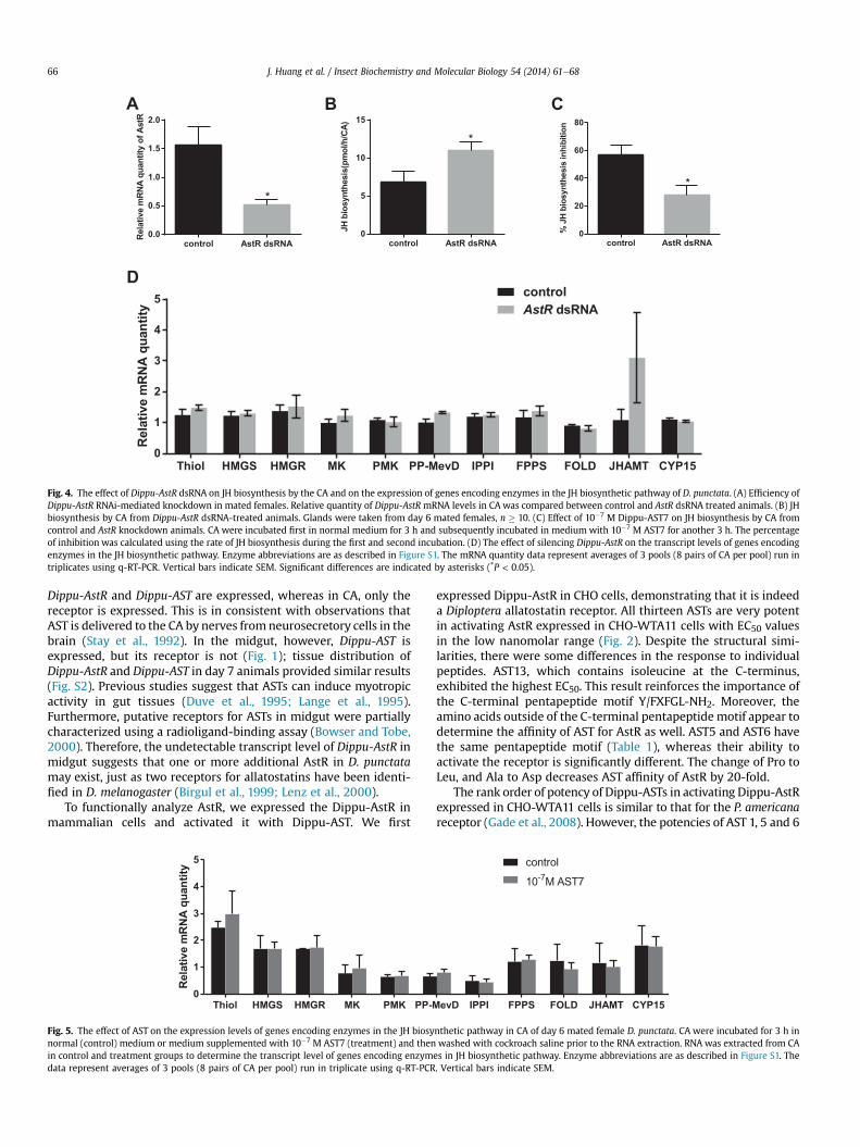
To confirm our results, we incubated CA in medium containing10�7 M AST7 for 3 h, which results in a 60e70% decrease in JHbiosynthesis (Tobe et al., 2000), and determined the transcript levelof the enzymes in the CA. As in the AstR RNAi experiments, themRNA levels of the 11 genes tested show no significant changes(Fig. 5).
3.4. JH precursors reverse AST-induced inhibition of JH biosynthesis
Earlier studies showed that mevalonate and farnesol were ableto partially reverse AST-induced inhibition of JH biosynthesis (Prattet al., 1991, 1989). To determine which enzyme(s) is/are affected byASTs in the JH biosynthetic pathway, we treated CA with select JHprecursors in the presence of 10�6 M AST7. If the activity of en-zymes was inhibited by AST, the JH precursors prior to the enzymewill not be able to reverse the inhibition of AST. As shown in Fig. 6,all exogenous JH precursors acetyl-CoA, mevalonic acid, diphos-phomevalonate and farnesol significantly stimulated the rate of JHbiosynthesis in the presence of AST7 (Fig. 6).
4. Discussion
Our observations on the expression of the Dippu-AstR andDippu-AST provide insights into the tissue-specific interaction be-tween the ligands and the receptor. In brain and nerve cord, both
-12 -11 -10 -9 -8 -7 -6 -5 -4-20
0
20
40
60
80
100
120
Dippu-AstR in HEK293
Concentration log[M]
%bi
olum
ines
cenc
e AST 5AST 6
B
and (B) HEK293 cells expressing Dippu-AstR. Data points represent the average ± SEMge of the maximal response. The zero response level corresponds to treatment with BSA
control AstR dsRNA0.0
0.5
1.0
1.5
2.0
Rel
ativ
em
RNA
quan
tity
ofAs
tR
*
control AstR dsRNA0
20
40
60
80
%JH
bios
ynth
esis
inhi
bitio
n
*
control AstR dsRNA0
5
10
15
JHbi
osyn
thes
is(p
mol
/h/C
A)
*
Thiol HMGS HMGR MK PMK PP-MevD IPPI FPPS FOLD JHAMT CYP150
1
2
3
4
5
Rel
ativ
em
RN
Aqu
antit
y
controlAstR dsRNA
A CB
D
Fig. 4. The effect of Dippu-AstR dsRNA on JH biosynthesis by the CA and on the expression of genes encoding enzymes in the JH biosynthetic pathway of D. punctata. (A) Efficiency ofDippu-AstR RNAi-mediated knockdown in mated females. Relative quantity of Dippu-AstR mRNA levels in CA was compared between control and AstR dsRNA treated animals. (B) JHbiosynthesis by CA from Dippu-AstR dsRNA-treated animals. Glands were taken from day 6 mated females, n � 10. (C) Effect of 10�7 M Dippu-AST7 on JH biosynthesis by CA fromcontrol and AstR knockdown animals. CA were incubated first in normal medium for 3 h and subsequently incubated in medium with 10�7 M AST7 for another 3 h. The percentageof inhibition was calculated using the rate of JH biosynthesis during the first and second incubation. (D) The effect of silencing Dippu-AstR on the transcript levels of genes encodingenzymes in the JH biosynthetic pathway. Enzyme abbreviations are as described in Figure S1. The mRNA quantity data represent averages of 3 pools (8 pairs of CA per pool) run intriplicates using q-RT-PCR. Vertical bars indicate SEM. Significant differences are indicated by asterisks (*P < 0.05).
J. Huang et al. / Insect Biochemistry and Molecular Biology 54 (2014) 61e6866
Dippu-AstR and Dippu-AST are expressed, whereas in CA, only thereceptor is expressed. This is in consistent with observations thatAST is delivered to the CA by nerves fromneurosecretory cells in thebrain (Stay et al., 1992). In the midgut, however, Dippu-AST isexpressed, but its receptor is not (Fig. 1); tissue distribution ofDippu-AstR and Dippu-AST in day 7 animals provided similar results(Fig. S2). Previous studies suggest that ASTs can induce myotropicactivity in gut tissues (Duve et al., 1995; Lange et al., 1995).Furthermore, putative receptors for ASTs in midgut were partiallycharacterized using a radioligand-binding assay (Bowser and Tobe,2000). Therefore, the undetectable transcript level of Dippu-AstR inmidgut suggests that one or more additional AstR in D. punctatamay exist, just as two receptors for allatostatins have been identi-fied in D. melanogaster (Birgul et al., 1999; Lenz et al., 2000).
To functionally analyze AstR, we expressed the Dippu-AstR inmammalian cells and activated it with Dippu-AST. We first
Thiol HMGS HMGR MK PMK PP-M0
1
2
3
4
5
Rel
ativ
em
RN
Aqu
antit
y
Fig. 5. The effect of AST on the expression levels of genes encoding enzymes in the JH biosynormal (control) medium or medium supplemented with 10�7 M AST7 (treatment) and thenin control and treatment groups to determine the transcript level of genes encoding enzymdata represent averages of 3 pools (8 pairs of CA per pool) run in triplicate using q-RT-PCR
expressed Dippu-AstR in CHO cells, demonstrating that it is indeeda Diploptera allatostatin receptor. All thirteen ASTs are very potentin activating AstR expressed in CHO-WTA11 cells with EC50 valuesin the low nanomolar range (Fig. 2). Despite the structural simi-larities, there were some differences in the response to individualpeptides. AST13, which contains isoleucine at the C-terminus,exhibited the highest EC50. This result reinforces the importance ofthe C-terminal pentapeptide motif Y/FXFGL-NH2. Moreover, theamino acids outside of the C-terminal pentapeptide motif appear todetermine the affinity of AST for AstR as well. AST5 and AST6 havethe same pentapeptide motif (Table 1), whereas their ability toactivate the receptor is significantly different. The change of Pro toLeu, and Ala to Asp decreases AST affinity of AstR by 20-fold.
The rank order of potency of Dippu-ASTs in activating Dippu-AstRexpressed in CHO-WTA11 cells is similar to that for the P. americanareceptor (Gade et al., 2008). However, the potencies of AST 1, 5 and 6
evD IPPI FPPS FOLD JHAMT CYP15
control
10-7M AST7
nthetic pathway in CA of day 6 mated female D. punctata. CA were incubated for 3 h inwashed with cockroach saline prior to the RNA extraction. RNA was extracted from CA
es in JH biosynthetic pathway. Enzyme abbreviations are as described in Figure S1. The. Vertical bars indicate SEM.

Acetyl CoA MA DPPM Farnesol0
5
10
15JH
bios
ynth
esis
(pm
ol/h
/CA
)
control
JH precursor
******
***
***
Fig. 6. JH precursors rescue the AST-induced JH inhibition. JH biosynthesis was eval-uated in CA that were first incubated in medium TC199 with 10�6 M AST7 (control),and then in medium with 10�6 M AST7 together with the individual precursors:Acetyl-CoA (100 mM), MA (100 mM), DPPM (100 mM) or farnesol (40 mM). CA weredissected from day 7 mated females. Each data point represents mean ± SEM (n � 10).
J. Huang et al. / Insect Biochemistry and Molecular Biology 54 (2014) 61e68 67
in activating AstR differ from their capacities to inhibit JH productionin vitro (Table 1). AST 5, which showed the highest inhibitory activity,demonstrated only a moderate potency in activating the expressedDippu-AstR, ranking 10 out of 13. In contrast, AST1, which was one ofthe weakest inhibitors of JH biosynthesis in vitro (ranking 11 out of13), appears to be one of the most potent peptides in receptor acti-vation. AST6, the structure of which is conserved in several insectorders, showed the highest potency in activating expressed Dippu-AstR (Bendena et al., 1999). Its ability to inhibit JH biosynthesis, onthe other hand, was moderate. Different reasons could contribute tothe differences between potency in activating the receptor andinhibiting JH biosynthesis in vitro. The susceptibility of ASTs todegradation by enzymes in the CA differs between ASTs, which couldresult in different potencies in inhibiting JH biosynthesis. AST5,which showed high resistance to the degradation by hemolymphand membrane preparations, presented the highest activity ininhibiting JH biosynthesis (Garside et al.,1997a,b). Furthermore, testswith cloned receptors in mammalian cells might not fully reflect theactual situation in insects. The posttranslational modifications ofAstR or the interacting proteins present in receptor-expressing cellscould affect the conformational and functional properties of thisreceptor. Nevertheless, we here chose mammalian cell lines becausethere may be fewer interacting proteins in this heterologous systemthan in insect cell lines.
The inhibition of JH biosynthesis by Dippu-ASTs is a complexprocess, probably involving more than one second messenger.Previous studies showed that ASTs regulate JH biosynthesis byacting through the inositol trisphosphate (IP3)/diglyceride (DAG)second messenger systems, in which protein kinase C (PKC) andCa2þ are involved (Feyereisen and Farnsworth, 1987b; Rachinskyand Tobe, 1996; Rachinsky et al., 1994). However, the role of Ca2þ
in regulation of JH biosynthesis is confounding. JH productionshows dose-dependency on extracellular Ca2þ in the medium,whereas the Ca2þ ionophore A23187 caused a rapid decline in JHrelease (Kikukawa et al., 1987). Inhibition of JH biosynthesis bybrain extracts was antagonized by inorganic Ca2þchannel blockers(Feyereisen and Farnsworth, 1987a). However, elevation of intra-cellular Ca2þ levels by treating CA with the Ca2þ-mobilizing drugthapsigargin diminished the inhibitory effect of ASTs (Rachinskyet al., 1994). The activation of Dippu-AstR induced a dose-dependent intracellular Ca2þ response in CHO-PAM28 cells(Fig. 3A), which supports the involvement of Ca2þ as a secondmessenger of AST. Aucoin et al. (1987) suggested that there may beboth a Ca2þ-dependent and a Ca2þ-independent pathway (cAMP)involved in the inhibition of JH biosynthesis. Incubating CA withbrain extracts resulted in an increase in the level of cAMP and a
decrease in JH biosynthesis (Aucoin et al., 1987). Forskolin, whichcauses a dose-dependent accumulation of cAMP, led to a rapid anddose-dependent inhibition of JH biosynthesis. Moreover, thesensitivity of CA to forskolin shows the same pattern as its sensi-tivity to AST during the first gonadotrophic cycle (Feyereisen andFarnsworth, 1987a; Meller et al., 1985). In contrast, Cusson et al.(1992) suggest that levels of cAMP do not increase followingtreatment of CA with ASTs. Our findings demonstrated that theactivation of Dippu-AstR induced a dose-dependent cAMP responsein HEK293 cells, which suggests that cAMP may be involved in thesignaling pathway of AST cells (Fig. 3B). Dippu-AstR appears todually couple through the Gas and Gaq pathways and triggers therelease of intracellular Ca2þ and the increase in cAMP levels. Studieson Drosophila AstRs suggested that DAR-1 and DAR-2 couple to Gi/omediated signaling and other G-proteins (Birgul et al., 1999; Larsenet al., 2001). However, activation of AstR in the presence of forskolindid not significantly influence the cAMP signal. This result suggeststhat there is no involvement of Gai in the Dippu-AstR pathway.
AST regulates JH biosynthesis by affecting either enzymesdirectly involved in the JH biosynthetic pathway or steps prior tothe production of acetyl-CoA. We have studied the effect of AST onthe transcript levels of genes in the JH biosynthetic pathway. Theinhibitory effect of AST on JH biosynthesis was rapid and reversible,which suggests that AST may not regulate JH biosynthesis throughtranscript levels (Pratt et al., 1991, 1989). Our study confirmed thisearlier assumption by revealing that the transcript levels of genes inthe JH biosynthetic pathway were not affected by AstR silencing orAST treatment in vitro (Figs. 4 and 5). Previous studies indicate thatASTs regulate JH biosynthesis through steps(s) prior to the pro-duction of mevalonate (Pratt et al., 1991, 1989). However, theenzyme activities of HMG-CoA synthase or HMG-CoA reductasewere not affected by AST (Sutherland and Feyereisen, 1996). Todetermine which enzyme in the JH biosynthetic pathway wasinfluenced by AST, we used several JH precursors to reverse theAST-induced inhibition of JH biosynthesis. All the JH precursors(including acetyl-CoA) were able to partially restore JH biosyn-thesis, thereby suggesting that the enzyme activities were notaffected by AST. The target of AST action probably lies prior to thestart of the JH biosynthetic pathway in regulating the production ofprecursors of acetyl-CoA such as the transport of citrate frommitochondria to cytosol and/or the cleavage of citrate to yieldacetyl-CoA (Sutherland and Feyereisen,1996). Further investigationis needed to identify the exact target of AST.
5. Conclusion
InD. punctata, ASTs play an important role in the regulation of JHbiosynthesis. The potency of the 13 ASTs in activating AstR revealsthe structureeactivity relationship of AST action. The activation ofAstR in CHO and HEK cells suggests that both Ca2þ and cAMP can beinvolved in the signal transduction of AST. Our results show thatAST does not affect the transcript levels and that the activities ofenzymes in the JH biosynthetic pathway remain normal followingtreatment with AST. AST probably affects JH biosynthesis prior tothe entry of precursors into the JH biosynthetic pathway. The exacttarget of AST action remains an open question.
Acknowledgments
The authors are very grateful to Dr. Jennifer Mitchell for use ofthe q-RT-PCR equipment and to Dr. William G. Bendena and Dr.Heleen Verlinden for technical assistance on AstR cell expression.The authors also thank the Natural Sciences and EngineeringResearch Council of Canada RGPIN 9408-09 to SST, China Scholar-ship Council Doctoral Award, the Interuniversity Attraction Poles

J. Huang et al. / Insect Biochemistry and Molecular Biology 54 (2014) 61e6868
Program (Belgian Science Policy Grant P7/40) and the KU LeuvenResearch Foundation (GOA/11/02) for financial support.
Appendix A. Supplementary material
Supplementary data related to this article can be found at http://dx.doi.org/10.1016/j.ibmb.2014.09.001.
References
Aucoin, R.R., Rankin, S.M., Stay, B., Tobe, S.S., 1987. Calcium and cyclic-AMPinvolvement in the regulation of juvenile hormone biosynthesis in Diplopterapunctata. Insect Biochem. 17, 965e969.
Auerswald, L., Birgul, N., Gade, G., Kreienkamp, H.J., Richter, D., 2001. Structural,functional, and evolutionary characterization of novel members of the alla-tostatin receptor family from insects. Biochem. Biophys. Res. Commun. 282,904e909.
Belles, X., Martin, D., Piulachs, M.D., 2005. The mevalonate pathway and the syn-thesis of juvenile hormone in insects. Annu. Rev. Entomol. 50, 181e199.
Bendena, W.G., Donly, B.C., Tobe, S.S., 1999. Allatostatins: a growing family ofneuropeptides with structural and functional diversity. Ann. N. Y. Acad. Sci. 897,311e329.
Birgul, N., Weise, C., Kreienkamp, H.J., Richter, D., 1999. Reverse physiology inDrosophila: identification of a novel allatostatin-like neuropeptide and itscognate receptor structurally related to the mammalian somatostatin/galanin/opioid receptor family. EMBO J. 18, 5892e5900.
Bowser, P.R.F., Tobe, S.S., 2000. Partial characterization of a putative allatostatinreceptor in the midgut of the cockroach Diploptera punctata. Gen. Comp.Endocrinol. 119, 1e10.
Coast, G.M., Schooley, D.A., 2011. Toward a consensus nomenclature for insectneuropeptides and peptide hormones. Peptides 32, 620e631.
Cusson, M., Prestwich, G.D., Stay, B., Tobe, S.S., 1991. Photoaffinity-labeling of alla-tostatin receptor proteins in the corpora allata of the cockroach, Diplopterapunctata. Biochem. Biophys. Res. Commun. 181, 736e742.
Cusson, M., Yagi, K.J., Guan, X.C., Tobe, S.S., 1992. Assessment of the role of cyclic-nucleotides in allatostatin-induced inhibition of juvenile hormone biosyn-thesis in Diploptera punctata. Mol. Cell. Endocrinol. 89, 121e125.
Donly, B.C., Ding, Q., Tobe, S.S., Bendena, W.G., 1993. Molecular cloning of the genefor the allatostatin family of neuropeptides from the cockroach Diplopterapunctata. Proc. Natl. Acad. Sci. U. S. A. 90, 8807e8811.
Duve, H., Wren, P., Thorpe, A., 1995. Innervation of the foregut of the cockroachLeucophaea maderae and inhibition of spontaneous contractile activity bycallatostatin neuropeptides. Physiol. Entomol. 20, 33e44.
Feyereisen, R., Farnsworth, D.E., 1987a. Comparison of the inhibitory effects of brainextract, high Kþ and forskolin on juvenile hormone synthesis by Diplopterapunctata corpora allata. Insect Biochem. 17, 939e942.
Feyereisen, R., Farnsworth, D.E., 1987b. Inhibition of insect juvenile hormone syn-thesis by phorbol 12-myristate 13-acetate. FEBS Lett. 222, 345e348.
Feyereisen, R., Tobe, S.S., 1981. A rapid partition assay for routine analysis of juvenilehormone release by insect corpora allata. Anal. Biochem. 111, 372e375.
Fuse, M., Zhang, J.R., Partridge, E., Nachman, R.J., Orchard, I., Bendena, W.G.,Tobe, S.S., 1999. Effects of an allatostatin and a myosuppressin on midgut car-bohydrate enzyme activity in the cockroach Diploptera punctata. Peptides 20,1285e1293.
Gade, G., Marco, H.G., Richter, D., Weaver, R.J., 2008. Structureeactivity studies withendogenous allatostatins from Periplaneta americana: expressed receptorcompared with functional bioassay. J. Insect Physiol. 54, 988e996.
Garside, C.S., Hayes, T.K., Tobe, S.S., 1997a. Degradation of Dip-allatostatins by he-molymph from the cockroach, Diploptera punctata. Peptides 18, 17e25.
Garside, C.S., Hayes, T.K., Tobe, S.S., 1997b. Inactivation of Dip-allatostatin 5 bymembrane preparations from the cockroach Diploptera punctata. Gen. Comp.Endocrinol. 108, 258e270.
Huang, J., Marchal, E., Hult, E.F., Tobe, S.S., Characterization of the Juvenile Hormonepathway in the viviparous cockroach, Diploptera punctata, (Submitted forpublication).
Kikukawa, S., Tobe, S.S., Solowiej, S., Rankin, S.M., Stay, B., 1987. Calcium as aregulator of juvenile hormone biosynthesis and release in the cockroach Dip-loptera punctata. Insect Biochem. 17, 179e187.
Lange, A.B., Bendena, W.G., Tobe, S.S., 1995. The effect of the 13 Dip-allatostatins onmyogenic and induced contractions of the cockroach (Diploptera punctata)hindgut. J. Insect Physiol. 41, 581e588.
Larsen, M.J., Burton, K.J., Zantello, M.R., Smith, V.G., Lowery, D.L., Kubiak, T.M., 2001.Type A allatostatins from Drosophila melanogaster and Diploptera punctataactivate two Drosophila allatostatin receptors, DAR-1 and DAR-2, expressed inCHO cells. Biochem Biophys. Res. Commun. 286, 895e901.
Lenz, C., Williamson, M., Grimmelikhuijzen, C.J.P., 2000. Molecular cloning andgenomic organization of a second probable allatostatin receptor fromDrosophila melanogaster. Biochem. Biophys. Res. Commun. 273, 571e577.
Lungchukiet, P., Donly, B.C., Zhang, J.R., Tobe, S.S., Bendena, W.G., 2008a. Molecularcloning and characterization of an allatostatin-like receptor in the cockroachDiploptera punctata. Peptides 29, 276e285.
Lungchukiet, P., Zhang, J.R., Tobe, S.S., Bendena, W.G., 2008b. Quantification ofallatostatin receptor mRNA levels in the cockroach, Diploptera punctata, usingreal-time PCR. J. Insect Physiol. 54, 981e987.
Marchal, E., Hult, E.F., Huang, J., Stay, B., Tobe, S.S., 2013a. Diploptera punctata as amodel for studying the endocrinology of arthropod reproduction and devel-opment. Gen. Comp. Endocrinol. 188, 85e93.
Marchal, E., Hult, E.F., Huang, J., Tobe, S.S., 2013b. Sequencing and validation ofhousekeeping genes for quantitative real-time PCR during the gonadotrophiccycle of Diploptera punctata. BMC Res. Notes 6, 237e246.
Martin, D., Piulachs, M.D., Belles, X., 1996. Inhibition of vitellogenin production byallatostatin in the German cockroach. Mol. Cell. Endocrinol. 121, 191e196.
Meller, V.H., Aucoin, R.R., Tobe, S.S., Feyereisen, R., 1985. Evidence for an inhibitoryrole of cyclic-AMP in the control of juvenile hormone biosynthesis by cockroachcorpora allata. Mol. Cell. Endocrinol. 43, 155e163.
Nouzova, M., Edwards, M.J., Mayoral, J.G., Noriega, F.G., 2011. A coordinatedexpression of biosynthetic enzymes controls the flux of juvenile hormoneprecursors in the corpora allata of mosquitoes. Insect Biochem. Mol. Biol. 41,660e669.
Pratt, G.E., Farnsworth, D.E., Fok, K.F., Siegel, N.R., McCormack, A.L., Shabanowitz, J.,Hunt, D.F., Feyereisen, R., 1991. Identity of a second type of allatostatin fromcockroach brains: an octadecapeptide amide with a tyrosine-rich addresssequence. Proc. Natl. Acad. Sci. U. S. A. 88, 2412e2416.
Pratt, G.E., Farnsworth, D.E., Siegel, N.R., Fok, K.F., Feyereisen, R., 1989. Identificationof an allatostatin from adult Diploptera punctata. Biochem. Biophys. Res. Com-mun. 163, 1243e1247.
Rachinsky, A., Tobe, S.S., 1996. Role of second messengers in the regulation of ju-venile hormone production in insects, with particular emphasis on calcium andphosphoinositide signaling. Arch. Insect Biochem. Physiol. 33, 259e282.
Rachinsky, A., Zhang, J., Tobe, S.S., 1994. Signal transduction in the inhibition ofjuvenile hormone biosynthesis by allatostatins e roles of diacylglycerol andcalcium. Mol. Cell. Endocrinol. 105, 89e96.
Secher, T., Lenz, C., Cazzamali, G., Sorensen, G., Williamson, M., Hansen, G.N.,Svane, P., Grimmelikhuijzen, C.J.P., 2001. Molecular cloning of a functionalallatostatin gut/brain receptor and an allatostatin preprohormone from thesilkworm Bombyx mori. J. Biol. Chem. 276, 47052e47060.
Stables, J., Green, A., Marshall, F., Fraser, N., Knight, E., Sautel, M., Milligan, G.,Lee, M., Rees, S., 1997. A bioluminescent assay for agonist activity at potentiallyany G protein-coupled receptor. Anal. Biochem. 252, 115e126.
Stay, B., Chan, K.K., Woodhead, A.P., 1992. Allatostatin-immunoreactive neuronsprojecting to the corpora allata of adult Diploptera punctata. Cell Tissue Res. 270,15e23.
Stay, B., Tobe, S.S., 2007. The role of allatostatins in juvenile hormone synthesis ininsects and crustaceans. Annu. Rev. Entomol. 52, 277e299.
Sutherland, T.D., Feyereisen, R., 1996. Target of cockroach allatostatin in thepathway of juvenile hormone biosynthesis. Mol. Cell. Endocrinol. 120, 115e123.
Tobe, S.S., Clarke, N., 1985. The effect of L-methionine concentration on juvenilehormone biosynthesis by corpora allata of the cockroach Diploptera punctata.Insect Biochem. 15, 175e179.
Tobe, S.S., Zhang, J.R., Bowser, P.R., Donly, B.C., Bendena, W.G., 2000. Biological ac-tivities of the allatostatin family of peptides in the cockroach, Diplopterapunctata, and potential interactions with receptors. J. Insect Physiol. 46,231e242.
Vandesompele, J., De Preter, K., Pattyn, F., Poppe, B., Van Roy, N., De Paepe, A.,Speleman, F., 2002. Accurate normalization of real-time quantitative RT-PCRdata by geometric averaging of multiple internal control genes. Genome Biol.3. RESEARCH0034.
Vilaplana, L., Maestro, J.L., Piulachs, M.D., Belles, X., 1999. Modulation of cardiacrhythm by allatostatins in the cockroach Blattella germanica (L.) (Dictyoptera,Blattellidae). J. Insect Physiol. 45, 1057e1064.
Yu, C.G., Hayes, T.K., Strey, A., Bendena, W.G., Tobe, S.S., 1995. Identification andpartial characterization of receptors for allatostatins in brain and corpora allataof the cockroach Diploptera punctata using a binding assay and photoaffinity-labeling. Regul. Pept. 57, 347e358.
Copyright © 2022 FDOKUMEN