Involvement of Auxin Biosynthesis and Transport in the ... - MDPI
11
plants Article Involvement of Auxin Biosynthesis and Transport in the Antheridium and Prothalli Formation in Lygodium japonicum Natsumi Ohishi 1 , Nanami Hoshika 2 , Mizuho Takeda 2 , Kyomi Shibata 2 , Hisakazu Yamane 1,2 , Takao Yokota 1,2 and Masashi Asahina 1,2,3, * Citation: Ohishi, N.; Hoshika, N.; Takeda, M.; Shibata, K.; Yamane, H.; Yokota, T.; Asahina, M. Involvement of Auxin Biosynthesis and Transport in the Antheridium and Prothalli Formation in Lygodium japonicum. Plants 2021, 10, 2709. https:// doi.org/10.3390/plants10122709 Academic Editors: John Hancock and Mikihisa Umehara Received: 4 November 2021 Accepted: 8 December 2021 Published: 9 December 2021 Publisher’s Note: MDPI stays neutral with regard to jurisdictional claims in published maps and institutional affil- iations. Copyright: © 2021 by the authors. Licensee MDPI, Basel, Switzerland. This article is an open access article distributed under the terms and conditions of the Creative Commons Attribution (CC BY) license (https:// creativecommons.org/licenses/by/ 4.0/). 1 Graduate School of Science and Engineering, Teikyo University, 1-1 Toyosatodai, Utsunomiya 320-8551, Tochigi, Japan; [email protected] (N.O.); [email protected] (H.Y.); [email protected] (T.Y.) 2 Department of Biosciences, Teikyo University, 1-1 Toyosatodai, Utsunomiya 320-8551, Tochigi, Japan; [email protected] (N.H.); [email protected] (M.T.); [email protected] (K.S.) 3 Advanced Instrumental Analysis Center, Teikyo University, 1-1 Toyosatodai, Utsunomiya 320-8551, Tochigi, Japan * Correspondence: [email protected]; Tel.: +81-28-627-7182 Abstract: The spores of Lygodium japonicum, cultured in the dark, form a filamentous structure called protonema. Earlier studies have shown that gibberellin (GA) induces protonema elongation, along with antheridium formation, on the protonema. In this study, we have performed detailed morphological analyses to investigate the roles of multiple phytohormones in antheridium formation, protonema elongation, and prothallus formation in L. japonicum. GA 4 methyl ester is a potent GA that stimulates both protonema elongation and antheridium formation. We found that these effects were inhibited by simultaneous application of abscisic acid (ABA). On the other hand, IAA (indole-3-acetic acid) promoted protonema elongation but reduced antheridium formation, while these effects were partially recovered by transferring to an IAA-free medium. An auxin biosynthesis inhibitor, PPBo (4-phenoxyphenylboronic acid), and a transport inhibitor, TIBA (2,3,5-triiodobenzoic acid), both inhibited protonema elongation and antheridium formation. L. japonicum prothalli are induced from germinating spores under continuous white light. Such development was negatively affected by PPBo, which induced smaller-sized prothalli, and TIBA, which induced aberrantly shaped prothalli. The evidence suggests that the crosstalk between these plant hormones might regulate protonema elongation and antheridium formation in L. japonicum. Furthermore, the possible involvement of auxin in the prothalli development of L. japonicum is suggested. Keywords: antheridium; auxin; gibberellin; abscisic acid; Lygodium japonicum; protonema 1. Introduction Ferns constitute the most diverse group of vascular plants, after seed plants, and are thought to have appeared on Earth more than 300 million years ago [1,2]. Ferns evolved after the diversion of bryophytes, including hornworts, liverworts, and mosses, which constitute the three early-diverged clades of land plants [3]. During this evolutionary process, bryophytes acquired simple reproductive ability by separating the sexes, while seed plants evolved with sophisticated reproductive ability by developing floral organs. Because ferns are evolutionally situated between bryophytes and seed plants, investi- gation of fern reproduction might provide a springboard for clarifying the evolution of reproductive mechanisms. Prothalli, the gametophyte stage of ferns, are developing antheridium and archego- nium that produce sperm and eggs, respectively. Upon fertilization, sporophytes begin to emerge, leading to spore production. In bracken fern (Pteridium aquilinum), meristic gametophytes with organized meristem develop into a heart-shaped gametophyte, while non-meristic gametophytes are irregularly shaped and form only antheridia [4]. The Plants 2021, 10, 2709. https://doi.org/10.3390/plants10122709 https://www.mdpi.com/journal/plants
-
Upload
khangminh22 -
Category
Documents
-
view
0 -
download
0
Transcript of Involvement of Auxin Biosynthesis and Transport in the ... - MDPI
plants
Article
Involvement of Auxin Biosynthesis and Transport in theAntheridium and Prothalli Formation in Lygodium japonicum
Natsumi Ohishi 1, Nanami Hoshika 2, Mizuho Takeda 2, Kyomi Shibata 2, Hisakazu Yamane 1,2, Takao Yokota 1,2
and Masashi Asahina 1,2,3,*
�����������������
Citation: Ohishi, N.; Hoshika, N.;
Takeda, M.; Shibata, K.; Yamane, H.;
Yokota, T.; Asahina, M. Involvement
of Auxin Biosynthesis and Transport
in the Antheridium and Prothalli
Formation in Lygodium japonicum.
Plants 2021, 10, 2709. https://
doi.org/10.3390/plants10122709
Academic Editors: John Hancock
and Mikihisa Umehara
Received: 4 November 2021
Accepted: 8 December 2021
Published: 9 December 2021
Publisher’s Note: MDPI stays neutral
with regard to jurisdictional claims in
published maps and institutional affil-
iations.
Copyright: © 2021 by the authors.
Licensee MDPI, Basel, Switzerland.
This article is an open access article
distributed under the terms and
conditions of the Creative Commons
Attribution (CC BY) license (https://
creativecommons.org/licenses/by/
4.0/).
1 Graduate School of Science and Engineering, Teikyo University, 1-1 Toyosatodai, Utsunomiya 320-8551,Tochigi, Japan; [email protected] (N.O.); [email protected] (H.Y.);[email protected] (T.Y.)
2 Department of Biosciences, Teikyo University, 1-1 Toyosatodai, Utsunomiya 320-8551, Tochigi, Japan;[email protected] (N.H.); [email protected] (M.T.); [email protected] (K.S.)
3 Advanced Instrumental Analysis Center, Teikyo University, 1-1 Toyosatodai, Utsunomiya 320-8551,Tochigi, Japan
* Correspondence: [email protected]; Tel.: +81-28-627-7182
Abstract: The spores of Lygodium japonicum, cultured in the dark, form a filamentous structurecalled protonema. Earlier studies have shown that gibberellin (GA) induces protonema elongation,along with antheridium formation, on the protonema. In this study, we have performed detailedmorphological analyses to investigate the roles of multiple phytohormones in antheridium formation,protonema elongation, and prothallus formation in L. japonicum. GA4 methyl ester is a potent GA thatstimulates both protonema elongation and antheridium formation. We found that these effects wereinhibited by simultaneous application of abscisic acid (ABA). On the other hand, IAA (indole-3-aceticacid) promoted protonema elongation but reduced antheridium formation, while these effects werepartially recovered by transferring to an IAA-free medium. An auxin biosynthesis inhibitor, PPBo(4-phenoxyphenylboronic acid), and a transport inhibitor, TIBA (2,3,5-triiodobenzoic acid), bothinhibited protonema elongation and antheridium formation. L. japonicum prothalli are induced fromgerminating spores under continuous white light. Such development was negatively affected byPPBo, which induced smaller-sized prothalli, and TIBA, which induced aberrantly shaped prothalli.The evidence suggests that the crosstalk between these plant hormones might regulate protonemaelongation and antheridium formation in L. japonicum. Furthermore, the possible involvement ofauxin in the prothalli development of L. japonicum is suggested.
Keywords: antheridium; auxin; gibberellin; abscisic acid; Lygodium japonicum; protonema
1. Introduction
Ferns constitute the most diverse group of vascular plants, after seed plants, and arethought to have appeared on Earth more than 300 million years ago [1,2]. Ferns evolvedafter the diversion of bryophytes, including hornworts, liverworts, and mosses, whichconstitute the three early-diverged clades of land plants [3]. During this evolutionaryprocess, bryophytes acquired simple reproductive ability by separating the sexes, whileseed plants evolved with sophisticated reproductive ability by developing floral organs.Because ferns are evolutionally situated between bryophytes and seed plants, investi-gation of fern reproduction might provide a springboard for clarifying the evolution ofreproductive mechanisms.
Prothalli, the gametophyte stage of ferns, are developing antheridium and archego-nium that produce sperm and eggs, respectively. Upon fertilization, sporophytes beginto emerge, leading to spore production. In bracken fern (Pteridium aquilinum), meristicgametophytes with organized meristem develop into a heart-shaped gametophyte, whilenon-meristic gametophytes are irregularly shaped and form only antheridia [4]. The
Plants 2021, 10, 2709. https://doi.org/10.3390/plants10122709 https://www.mdpi.com/journal/plants

Plants 2021, 10, 2709 2 of 11
Japanese Climbing Fern, Lygodium japonicum, develops prothalli under natural light con-ditions, where the archegonium forms around the notch and the antheridium around therhizoid [5]. L. japonicum, and some other ferns, are known to secrete pheromone-like sub-stances called antheridiogens (antheridium inducers), including GAs that control the ratioof males to females and induce the formation of antheridium to ensure genetic diversity inthe population [6–8].
In L. japonicum, rapidly growing meristematic gametes form archegonium, whileothers form antheridium within the germinating population. Moreover, it has been foundthat sensitivity to GA varies with the size of individuals [9]. GA is accepted by a receptorcalled GID1 (GA-insensitive dwarf 1); when GID1 receives a GA molecule, the DELLAprotein, an inhibitor of gibberellin response, is degraded [10,11]. It has been reported thatthe expression level of gibberellin biosynthetic genes is increased in mature L. japonicumprothalli and that the expression level of GID1 is increased in immature prothalli [9]. Thesestudies hypothesized that archegonia formed in mature individuals secrete GAs, whichaccelerate the development of antheridia in the surrounding young individuals, leading tothe production of sperm to be accepted by archegonia.
When grown in a medium containing GA under red light irradiation, L. japonicumspores are germinated, and as the resulting protonemata elongates, antheridia are formedon them (Figure 1). The antheridium primordium is formed from the mitotic plane,obliquely introduced at the upper end of the cells near the spore shell of the protonema [4],and comprises three jacket cells (basal cells, ring cells, and apical cap cells) that surround16 or 32 sperm cells. Protonema consists of a single row of cells, and due to its morphologicalsimplicity, has been used as a powerful tool to search for antheridium inducers of ferns [12–15].Such experimental techniques are also utilized in this study.
Figure 1. Prothalli, germinating protonema, and protonema with antheridia. (A) Germinating sporecultured in MS medium for approximately 20 days under white light formed prothalli. Scale barindicates 1 mm. ap, apical notch; cu, cushion; r, rhizoid (B) Germinating spore cultured in MSmedium containing 10–100 nM GA4-Me in the dark for 3 days (B) or 7 days (C) after red lightirradiation. The scale bars indicate 200 µm. (D) SEM image of germinating spore cultured in MSmedium containing GA4-Me in the dark for approximately 10 days after red light irradiation. Thescale bar indicates 100 µm. Arrows (black and white) indicate antheridium and red arrowheadsindicate protonema.
Plants 2021, 10, 2709 3 of 11
However, very little is known about whether plant hormones other than GAs areinvolved in this antheridium formation process, and how they interact with each other.IAA occurs in various species of plants, and is known to induce cell division, elongation,and differentiation, leading to fruit growth, apical and lateral bud development, as wellas stem and root elongation, which have been investigated at the cellular and molecularlevels. In addition, the biosynthesis of IAA from tryptophan has been shown to proceedthrough multiple pathways, among which a two-step reaction catalyzed by tryptophanaminotransferase (TAA) and flavin-containing monooxygenase (YUCCA) is a commonfeature in land plants [16,17]. In Zea mays, transcriptome analysis has demonstrated thatgenes encoding auxin-responsive transcription factors ARFs and TIR1 act as receptors forauxin. Even in Marchantia polymorpha, these genes are expressed during phloem formationand female reproductive organ formation [18]. Furthermore, in Physcomitrella patens, auxin-responsive genes and the PIN protein, responsible for auxin polarity transport, havebeen expressed during oogenesis and antheridium development [19]. Auxin-mediatedsignaling pathways for adventitious root development have been found to be conservedin Arabidopsis and the fern Ceratopteris richardi [20], suggesting that ferns also shouldmaintain similar genetic diversity of seed plants, with respects to reproductive organformation. However, auxin biosynthesis, as well as physiological functions, such asantheridium formation in ferns, have not yet been investigated in detail. Hickok andKiriluk [21] reported that gametophyte formation in the fern Ceratopteris thalictroides wasinhibited by exogenous auxins. Furthermore, the normal male and female ratio causedby an antheridiogen was significantly increased by IBA (indole-3-butyric acid). However,this effect was deemed to be indirectly elicited by IBA-mediated growth inhibition, andhence it was assumed that IBA did not directly substitute for antheridiogen. In this study,we have investigated how plant hormones GA, IAA, and ABA, which are known to beendogenous [12], are mutually interrelated in protonema elongation and antheridiumformation in L. japonicum.
2. Results and Discussion2.1. Effect of ABA and IAA on GA4-Me-Medaited Protonema Elongation and AntheridiumFormation of L. japonicum
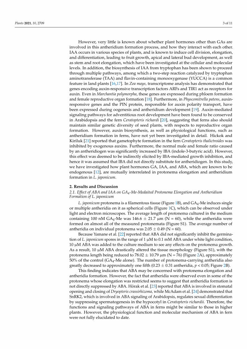
L. japonicum protonema is a filamentous tissue (Figure 1B), and GA4-Me induces singleor multiple antheridia on it as spherical cells (Figure 1C), which can be observed underlight and electron microscopes. The average length of protonema cultured in the mediumcontaining 100 nM GA4-Me was 146.6 ± 21.7 µm (N = 60), while the antheridia wereformed on almost all of the measured protonemata (Figure S1). The average number ofantheridia on individual protonema was 2.05 ± 0.49 (N = 60).
Because Yamane et al. [22] reported that ABA did not significantly inhibit the germina-tion of L. japonicum spores in the range of 1 µM to 0.1 mM ABA under white light condition,10 µM ABA was added to the culture medium to see any effects on the protonema growth.As a result, 10 µM ABA drastically altered the tissue morphology (Figure S1), with theprotonema length being reduced to 78.02 ± 10.79 µm (N = 76) (Figure 2A), approximately50% of the control (GA4-Me alone). The number of protonema-carrying antheridia alsogreatly decreased to approximately one fifth (0.23 ± 0.31 antheridia, p < 0.05; Figure 2B).
This finding indicates that ABA may be concerned with protonema elongation andantheridia formation. However, the fact that antheridia were observed even in some of theprotonema whose elongation was restricted seems to suggest that antheridia formation isnot directly suppressed by ABA. Hõrak et al. [23] reported that ABA is involved in stomatalopening and closing of Dryopteris crassirhizoma, while McAdam et al. [24] demonstrated thatSnRK2, which is involved in ABA signaling of Arabidopsis, regulates sexual differentiationby suppressing spermatogenesis in the hypocotyl in Ceratopteris richardii. Therefore, thefunctions and signaling pathways of ABA in ferns might be similar to those in higherplants. However, the physiological function and molecular mechanism of ABA in fernwere not fully elucidated to date.
Plants 2021, 10, 2709 4 of 11
Figure 2. Effect of simultaneous application of GA4-Me and ABA or IAA treatment on protonema elongation and antheridiaformation. (A) Boxplot for protonema length in each treatment. The bars indicate maximum and minimum values, andinterquartile ranges were shown. X indicates the mean value. (B) Protonemata distribution based on antheridium number.The average number of antheridia per individual spore in GA alone, GA + ABA, and GA + IAA were 2.05 ± 0.49, 0.23 ± 0.31,and 0, respectively. Statistical analysis was performed using the Steel–Dwass test. Different letters indicate statisticallysignificant differences (p < 0.05; N = 60 for GA alone; N = 76 for GA + ABA; N = 44 for GA + IAA).
In the presence of GA, protonema cells elongated in a straight fashion with transversedivisions. Simultaneous application of 10 µM IAA induced abnormal swelling due tolongitudinal cell division, and no apparent antheridium was observed (Figure S1). Themean length of the protonema was 200.52 ± 19.21 µm (N = 44) (Figure 2B), approximately1.3 times longer than the control (Figure 2B). These findings suggest that such abnormalmorphology of protonema cells should be responsible for the antheridium deficiency.
Altogether, it may be assumed that the signaling and biosynthesis of IAA, and alsoABA, are prerequisite factors in antheridium formation in L. japonicum.
2.2. Effect of Transferring of IAA-Treated Cells to an IAA-Free Medium
We examined whether the inhibitory effects of IAA were restored by transferring itto IAA-free media. Spores were cultured on GA plus IAA medium for 5 days, and thegerminated spores were transferred to a newly prepared GA medium and hormone-freemedium. As a reference experiment, the germinated spores were transferred to GA plusIAA medium. The protonema length of the cells transferred to GA medium and hormone-free medium was reduced, compared to that of the control (Figure S2). Antheridiumformation was only slightly recovered by transferring to a hormone-free medium, but wassignificantly rescued by GA medium (Figure S2). However, the recovered antheridiumwas not formed on cells lined up in series, but on sites where the cells had divided intotwo or three rows. This finding suggests that such abnormal protonema morphology is notdirectly linked to disrupted antheridium formation. It may be conceivable that normal celldivision occurred again in the IAA-free medium, and then antheridium was formed fromthe newly formed cells. These results indicated that GA was required for the formation ofantheridium and antheridia were induced from certain cells after protonema elongation,and that auxin promotes cell division while inhibiting antheridium formation inducedby GA.
Plants 2021, 10, 2709 5 of 11
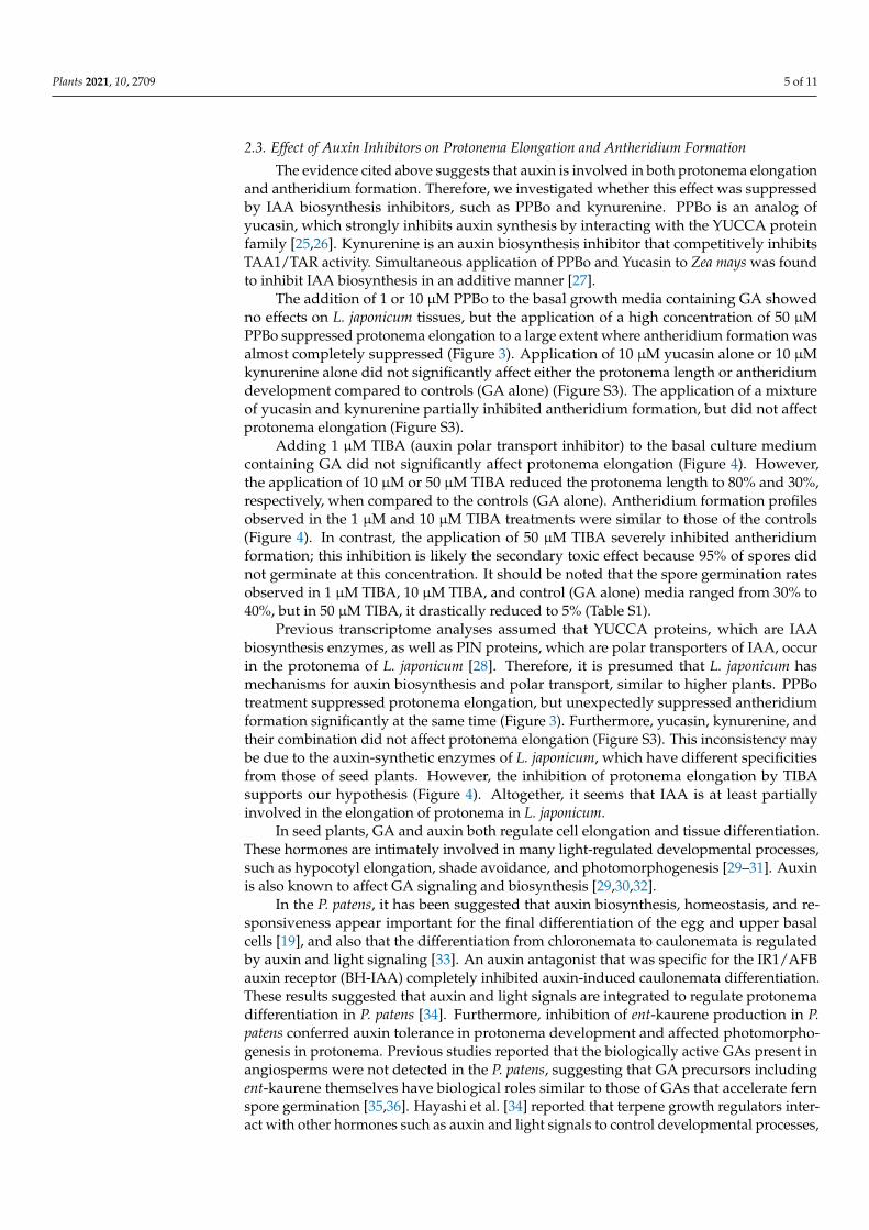
2.3. Effect of Auxin Inhibitors on Protonema Elongation and Antheridium Formation
The evidence cited above suggests that auxin is involved in both protonema elongationand antheridium formation. Therefore, we investigated whether this effect was suppressedby IAA biosynthesis inhibitors, such as PPBo and kynurenine. PPBo is an analog ofyucasin, which strongly inhibits auxin synthesis by interacting with the YUCCA proteinfamily [25,26]. Kynurenine is an auxin biosynthesis inhibitor that competitively inhibitsTAA1/TAR activity. Simultaneous application of PPBo and Yucasin to Zea mays was foundto inhibit IAA biosynthesis in an additive manner [27].
The addition of 1 or 10 µM PPBo to the basal growth media containing GA showedno effects on L. japonicum tissues, but the application of a high concentration of 50 µMPPBo suppressed protonema elongation to a large extent where antheridium formation wasalmost completely suppressed (Figure 3). Application of 10 µM yucasin alone or 10 µMkynurenine alone did not significantly affect either the protonema length or antheridiumdevelopment compared to controls (GA alone) (Figure S3). The application of a mixtureof yucasin and kynurenine partially inhibited antheridium formation, but did not affectprotonema elongation (Figure S3).
Adding 1 µM TIBA (auxin polar transport inhibitor) to the basal culture mediumcontaining GA did not significantly affect protonema elongation (Figure 4). However,the application of 10 µM or 50 µM TIBA reduced the protonema length to 80% and 30%,respectively, when compared to the controls (GA alone). Antheridium formation profilesobserved in the 1 µM and 10 µM TIBA treatments were similar to those of the controls(Figure 4). In contrast, the application of 50 µM TIBA severely inhibited antheridiumformation; this inhibition is likely the secondary toxic effect because 95% of spores didnot germinate at this concentration. It should be noted that the spore germination ratesobserved in 1 µM TIBA, 10 µM TIBA, and control (GA alone) media ranged from 30% to40%, but in 50 µM TIBA, it drastically reduced to 5% (Table S1).
Previous transcriptome analyses assumed that YUCCA proteins, which are IAAbiosynthesis enzymes, as well as PIN proteins, which are polar transporters of IAA, occurin the protonema of L. japonicum [28]. Therefore, it is presumed that L. japonicum hasmechanisms for auxin biosynthesis and polar transport, similar to higher plants. PPBotreatment suppressed protonema elongation, but unexpectedly suppressed antheridiumformation significantly at the same time (Figure 3). Furthermore, yucasin, kynurenine, andtheir combination did not affect protonema elongation (Figure S3). This inconsistency maybe due to the auxin-synthetic enzymes of L. japonicum, which have different specificitiesfrom those of seed plants. However, the inhibition of protonema elongation by TIBAsupports our hypothesis (Figure 4). Altogether, it seems that IAA is at least partiallyinvolved in the elongation of protonema in L. japonicum.
In seed plants, GA and auxin both regulate cell elongation and tissue differentiation.These hormones are intimately involved in many light-regulated developmental processes,such as hypocotyl elongation, shade avoidance, and photomorphogenesis [29–31]. Auxinis also known to affect GA signaling and biosynthesis [29,30,32].
In the P. patens, it has been suggested that auxin biosynthesis, homeostasis, and re-sponsiveness appear important for the final differentiation of the egg and upper basalcells [19], and also that the differentiation from chloronemata to caulonemata is regulatedby auxin and light signaling [33]. An auxin antagonist that was specific for the IR1/AFBauxin receptor (BH-IAA) completely inhibited auxin-induced caulonemata differentiation.These results suggested that auxin and light signals are integrated to regulate protonemadifferentiation in P. patens [34]. Furthermore, inhibition of ent-kaurene production in P.patens conferred auxin tolerance in protonema development and affected photomorpho-genesis in protonema. Previous studies reported that the biologically active GAs present inangiosperms were not detected in the P. patens, suggesting that GA precursors includingent-kaurene themselves have biological roles similar to those of GAs that accelerate fernspore germination [35,36]. Hayashi et al. [34] reported that terpene growth regulators inter-act with other hormones such as auxin and light signals to control developmental processes,

Plants 2021, 10, 2709 6 of 11
including caulonema differentiation. Altogether, it seems most plausible that interactionsof auxin with GA and light signaling are required for the morphogenesis and sexual organdifferentiation in L. japonicum. In addition to GA, IAA, and ABA, brassinosteroids (BRs)have recently been shown to occur in L. japonicum tissue [37]. Synergisms of IAA withBRs occur in many systems [38]. Therefore, the interaction of IAA with BRs also may be afuture research target.
Figure 3. Effect of IAA biosynthesis inhibitor on protonema elongation and antheridia formation.Germinating spores were cultured with 10 nM GA4-Me alone or with simultaneous application ofPPBo at 1, 10, and 50 µM (N = 51). All cells were cultured in the dark for 7 days after red lightirradiation. (A) Boxplot for protonema length in each treatment. The bars indicate maximum andminimum values, and interquartile ranges were shown. X indicates the mean value. Asterisksindicate statistically significant differences compared with GA alone (Steel’s test; n.s., not significant;**, p < 0.01). (B) Protonemata distribution based on antheridium number. The average number ofantheridia per individual spore in GA alone or with 1, 10, and 50 µM PPBo were 1.6 ± 0.1, 1.7 ± 0.11,1.6 ± 0.08, and 0.02 ± 0.02, respectively. Asterisks indicate statistically significant differencescompared with GA alone (Steel’s test; n.s., not significant; **, p < 0.01).

Plants 2021, 10, 2709 7 of 11
Figure 4. Effect of IAA transport inhibitor on protonema elongation and antheridium formation.Germinating spores were cultured with 100 nM GA4-Me alone or with simultaneous applicationof TIBA at 1, 10, and 50 µM. (A) Boxplot for protonema length in each treatment. The bars indicatemaximum and minimum values, and interquartile ranges were shown. X indicates the mean value.Asterisks indicate statistically significant differences compared with GA alone (Steel’s test; n.s.,not significant; *, p < 0.05). (B) Protonemata distribution based on antheridium number. Theaverage number of antheridia per individual spore in GA alone or with 1, 10, and 50 µM TIBA were2.76 ± 0.43, 3.08 ± 0.43, 2.29 ± 0.48 and 0.05 ± 0.13, respectively. Asterisks indicate statisticallysignificant differences compared with GA alone (Steel’s test; *, p < 0.05; **, p < 0.01). N = 38.
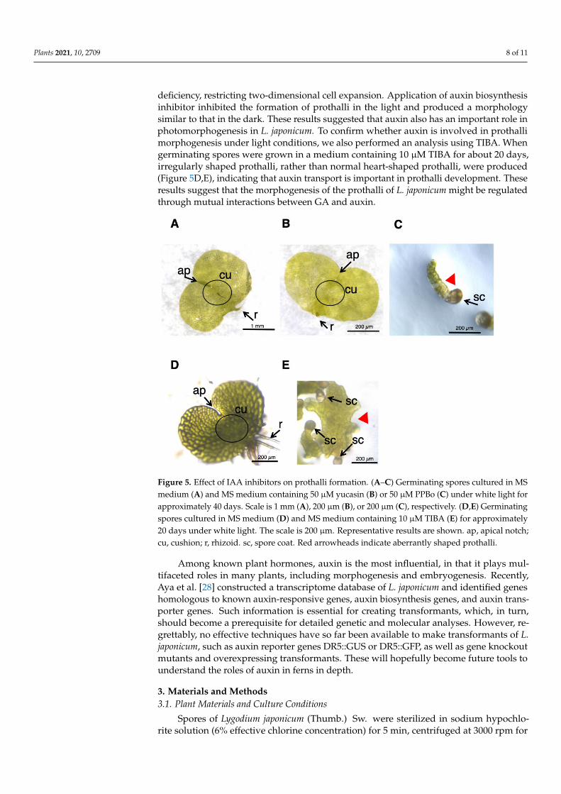
2.4. Effects of Auxin Inhibitors on the Morphology of L. japonicum Prothalli
L. japonicum spores were germinated in GA medium under white light, and ca. onemonth later, the cultures produced normal prothalli, as shown in Figure 5A. When 50 µMyucasin was supplied to the medium (Figure 5B), the prothalli obtained were indistinguish-able from those of the normal cells (Figure 5A). On the other hand, the tissue grown in50 µM PPBo medium remained at the stage of germinating spores, and no development ofprothalli was observed (Figure 5C), suggesting that high-concentration PPBo caused IAA
Plants 2021, 10, 2709 8 of 11
deficiency, restricting two-dimensional cell expansion. Application of auxin biosynthesisinhibitor inhibited the formation of prothalli in the light and produced a morphologysimilar to that in the dark. These results suggested that auxin also has an important role inphotomorphogenesis in L. japonicum. To confirm whether auxin is involved in prothallimorphogenesis under light conditions, we also performed an analysis using TIBA. Whengerminating spores were grown in a medium containing 10 µM TIBA for about 20 days,irregularly shaped prothalli, rather than normal heart-shaped prothalli, were produced(Figure 5D,E), indicating that auxin transport is important in prothalli development. Theseresults suggest that the morphogenesis of the prothalli of L. japonicum might be regulatedthrough mutual interactions between GA and auxin.
Figure 5. Effect of IAA inhibitors on prothalli formation. (A–C) Germinating spores cultured in MSmedium (A) and MS medium containing 50 µM yucasin (B) or 50 µM PPBo (C) under white light forapproximately 40 days. Scale is 1 mm (A), 200 µm (B), or 200 µm (C), respectively. (D,E) Germinatingspores cultured in MS medium (D) and MS medium containing 10 µM TIBA (E) for approximately20 days under white light. The scale is 200 µm. Representative results are shown. ap, apical notch;cu, cushion; r, rhizoid. sc, spore coat. Red arrowheads indicate aberrantly shaped prothalli.
Among known plant hormones, auxin is the most influential, in that it plays mul-tifaceted roles in many plants, including morphogenesis and embryogenesis. Recently,Aya et al. [28] constructed a transcriptome database of L. japonicum and identified geneshomologous to known auxin-responsive genes, auxin biosynthesis genes, and auxin trans-porter genes. Such information is essential for creating transformants, which, in turn,should become a prerequisite for detailed genetic and molecular analyses. However, re-grettably, no effective techniques have so far been available to make transformants of L.japonicum, such as auxin reporter genes DR5::GUS or DR5::GFP, as well as gene knockoutmutants and overexpressing transformants. These will hopefully become future tools tounderstand the roles of auxin in ferns in depth.
3. Materials and Methods3.1. Plant Materials and Culture Conditions
Spores of Lygodium japonicum (Thumb.) Sw. were sterilized in sodium hypochlo-rite solution (6% effective chlorine concentration) for 5 min, centrifuged at 3000 rpm for

Plants 2021, 10, 2709 9 of 11
30 s, and then washed with sterile distilled water. This process was repeated twice, andthen the spores were suspended in sterile D.W. Murashige and Skoog (MS) medium [39]with 0.5% (w/v) agar adjusted to pH 5.5 was used for the cultivation of L. japonicumspores. Plant hormones and their inhibitors used in this study were GA4-Me, ABA, IAA,5-[4-chlorophenyl]-2,4-dihydro-[1,2,4]-triazole-3-thione (Yucasin), 4-phenoxyphenylboronicacid (Kynurenine), 4-phenoxyphenylboronic acid (PPBo) and 2,3,5-triiodobenzoic acid(TIBA). These chemicals were dissolved in MeOH (GA, ABA, and IAA) or DMSO (Kynure-nine, PPBo, and TIBA) and pipetted into 12-well plates (MS-BC12R, Sumitomo Bakelite,Tokyo, Japan), and the solvent (MeOH) was air-dried aseptically before 2 mL MS agarmedium was dispensed. The final concentrations of hormones were adjusted to 10 or100 nM (GA4-Me) or 100 µM (ABA and IAA), and kynurenine, PPBo, and TIBA wereadjusted to 1, 10, or 50 µM (0.1% DMSO). PPBo and kynurenine were kindly gifted by Prof.T. Koshiba (Tokyo Metropolitan University) and Prof. K. Hayashi (Okayama University ofScience), respectively.
After solidification of the medium, approximately two drops of sterilized L. japonicumspore suspension were dispensed into each hole with a Pasteur pipette. The plates werekept in the dark in an incubator (25 ◦C; Nippon Medical & Chemical Instruments Co.,Osaka, Japan) for 2–5 days and then irradiated with red light for 24 h to induce germination.The plates were then incubated in the dark for an additional seven days.
3.2. Measurement of Protonema Elongation and Antheridium Formation
Protonemata and antheridia were fixed in 2.5% glutaraldehyde (0.2 M phosphatebuffer, pH 7.4) and observed under a stereomicroscope (M165FC, Leica Microsystems,Wetzlar, Germany). The length of the protonema and the number of antheridia weremeasured using ImageJ (https://imagej.nih.gov/ij/ (accessed on 1 November 2021)).The numbers of antheridia were only counted as morphologically distinct individuals.Experiments were performed using at least three independent plant samples. Scanningelectron microscopy (SEM) observations were performed using SU3500 (Hitachi-Hitech Co.,Tokyo, Japan) after ionic liquid coating (HILEM IL1000, Hitachi-Hitech) at an accelerationvoltage of 10 kV.
3.3. Effect of Auxin Inhibitors on Prothalli Development
Plates were prepared using the same procedure as in the protonema culture and thenincubated in an incubator in the dark for 2–5 days. After spore germination, the plateswere further incubated under white light at 25 ◦C for up to 40 days before being observedunder a stereomicroscope.
4. Conclusions
We examined the effects of endogenous plant hormones, auxin and ABA, on pro-tonema elongation and antheridium formation induced by GA in L. japonicum. It wasconcluded that the morphogenesis of the reproductive organs and prothalli of the fernmight be regulated through the interaction of auxin and ABA with GA.
Supplementary Materials: The following are available online at https://www.mdpi.com/article/10.3390/plants10122709/s1, Figure S1: Effect of simultaneous application of GA4-Me and ABA orIAA treatment on protonema elongation and antheridium formation. Protonema cultured with100 µM GA4-Me alone or simultaneous application of ABA or IAA. Arrows indicates antheridiumand red arrowheads indicate protonema. The scale bars indicate 200 µm; Figure S2: Growth mediumreplacement experiments. (A) Boxplot for protonema length in each treatment. The bars indicatemaximum and minimum values, and interquartile ranges were shown. X indicates the mean value.(B) Protonemata distribution based on antheridium number. Different letters indicate statisticallysignificant differences (Steel–Dwass test, p < 0.05); Figure S3: Effect of IAA biosynthesis inhibitortreatment on protonema elongation. (A) Boxplot for protonema length in each treatment. The barsindicate maximum and minimum values, and interquartile ranges were shown. X indicates the meanvalue. n.s., not significant. (B) Protonemata distribution based on antheridium number. Asterisks

Plants 2021, 10, 2709 10 of 11
indicate statistically significant differences compared with GA alone (Steel’s test; n.s., not significant;*, p < 0.05); Table S1: Germination rate on medium containing GA4-Me with TIBA.
Author Contributions: Investigation, N.O., N.H., M.T. and K.S.; project administration, T.Y. andM.A.; supervision, H.Y., T.Y. and M.A.; writing—original draft, M.A.; writing—review and editing,H.Y. and T.Y. All authors have read and agreed to the published version of the manuscript.
Funding: This work was supported in part by the Ministry of Education, Culture, Sports, Science,and Technology Program for the Strategic Research Foundation at Private Universities (grant no.S1311014 to M.A.), the Promotion and Mutual Aid Corporation for Private Schools of Japan (to M.A.),and the ACRO Research grant of Teikyo university (TeTe20-01 to M.A.).
Institutional Review Board Statement: Not applicable.
Informed Consent Statement: Not applicable.
Data Availability Statement: The data presented in this study are available in the article.
Acknowledgments: We are grateful to K. Miyamoto (Teikyo University, Japan) for their valuablesuggestions. We also thank T. Koshiba (Tokyo Metropolitan University, Japan) and K. Hayashi(Okayama University of Science, Japan) for their kind gift of auxin inhibitors, and E. Yumoto, T.Takeda, S. Akimoto, and K. Kikuchi (Teikyo University, Japan), and T. Yukawa (Hitachi-Hitech,Japan), for their technical assistance.
Conflicts of Interest: The authors have no conflict of interest to declare.
References1. Vasco, A.; Moran, R.C.; Ambrose, B.A. The Evolution, Morphology, and Development of Fern Leaves. Front. Plant Sci. 2013,
4, 345. [CrossRef] [PubMed]2. Kenrick, P. The Relationships of Vascular Plants. Philos. Trans. R. Soc. Lond. B Biol. Sci. 2000, 355, 847–855. [CrossRef] [PubMed]3. Zhang, J.; Fu, X.-X.; Li, R.-Q.; Zhao, X.; Liu, Y.; Li, M.-H.; Zwaenepoel, A.; Ma, H.; Goffinet, B.; Guan, Y.-L.; et al. The Hornwort
Genome and Early Land Plant Evolution. Nat. Plants 2020, 6, 107–118. [CrossRef] [PubMed]4. Gifford, E.M.; Foster, A.S. Morphology and Evolution of Vascular Plants, 3rd ed.; Freeman: New York, NY, USA, 1988; ISBN
0-7167-1946-0.5. Takahashi, N.; Kami, C.; Ota, I.; Morita, N.; Imaichi, R. Developmental Morphology of the Typical Cordate Gametophyte of a
Homosporous Leptosporangiate Fern, Lygodium japonicum (Lygodiaceae), Focusing on the Initial Cell Behavior of Two DistinctMeristems. Am. J. Bot. 2015, 102, 197–207. [CrossRef]
6. Takeno, K.; Furuya, M. Bioassay of Antheridiogen in Lygodium japonicum. Dev. Growth Differ. 1975, 17, 9–18. [CrossRef]7. Takeno, K.; Furuya, M. Inhibitory Effect of Gibberellins on Archegonial Differentiation in Lygodium japonicum. Physiol. Plant. 1977,
39, 135–138. [CrossRef]8. Yamauchi, T.; Oyama, N.; Yamane, H.; Murofushi, N.; Schraudolf, H.; Pour, M.; Furber, M.; Mander, L.N. Identification of
Antheridiogens in Lygodium circinnatum and Lygodium flexuosum. Plant Physiol. 1996, 111, 741–745. [CrossRef]9. Tanaka, J.; Yano, K.; Aya, K.; Hirano, K.; Takehara, S.; Koketsu, E.; Ordonio, R.L.; Park, S.-H.; Nakajima, M.; Ueguchi-Tanaka,
M.; et al. Antheridiogen Determines Sex in Ferns via a Spatiotemporally Split Gibberellin Synthesis Pathway. Science 2014, 346,469–473. [CrossRef]
10. Ueguchi-Tanaka, M.; Ashikari, M.; Nakajima, M.; Itoh, H.; Katoh, E.; Kobayashi, M.; Chow, T.; Hsing, Y.C.; Kitano, H.; Yamaguchi,I.; et al. GIBBERELLIN INSENSITIVE DWARF1 Encodes a Soluble Receptor for Gibberellin. Nature 2005, 437, 693–698. [CrossRef]
11. Ueguchi-Tanaka, M.; Nakajima, M.; Katoh, E.; Ohmiya, H.; Asano, K.; Saji, S.; Hongyu, X.; Ashikari, M.; Kitano, H.; Yamaguchi,I.; et al. Molecular Interactions of a Soluble Gibberellin Receptor, GID1, with a Rice DELLA Protein, SLR1, and Gibberellin. PlantCell 2007, 19, 2140–2155. [CrossRef]
12. Yamane, H.; Takahashi, N.; Takeno, K.; Furuya, M. Identification of Gibberellin A9 Methyl Ester as a Natural Substance RegulatingFormation of Reproductive Organs in Lygodium japonicum. Planta 1979, 147, 251–256. [CrossRef]
13. Yamane, H.; Yamaguchi, I.; Kobayashi, M.; Takahashi, M.; Sato, Y.; Takahashi, N.; Iwatsuki, K.; Phinney, B.O.; Spray, C.R.; Gaskin,P.; et al. Identification of Ten Gibberellins from Sporophytes of the Tree Fern, Cyathea australis. Plant Physiol. 1985, 78, 899–903.[CrossRef]
14. Takeno, K.; Yamane, H.; Yamauchi, T.; Takahashi, N.; Furber, M.; Mander, L.N. Biological Activities of the Methyl Ester ofGibberellin A73, a Novel and Principal Antheridiogen in Lygodium japonicum. Plant Cell Physiol. 1989, 30, 201–205. [CrossRef]
15. Sugai, M.; Takeno, K.; Furuya, M. Diverse Responses of Spores in the Light-Dependent Germination of Lygodium japonicum. PlantSci. Lett. 1977, 8, 333–338. [CrossRef]
16. Mashiguchi, K.; Tanaka, K.; Sakai, T.; Sugawara, S.; Kawaide, H.; Natsume, M.; Hanada, A.; Yaeno, T.; Shirasu, K.; Yao, H.; et al.The Main Auxin Biosynthesis Pathway in Arabidopsis. Proc. Natl. Acad. Sci. USA 2011, 108, 18512–18517. [CrossRef]

Plants 2021, 10, 2709 11 of 11
17. Sugawara, S.; Mashiguchi, K.; Tanaka, K.; Hishiyama, S.; Sakai, T.; Hanada, K.; Kinoshita-Tsujimura, K.; Yu, H.; Dai, X.;Takebayashi, Y.; et al. Distinct Characteristics of Indole-3-Acetic Acid and Phenylacetic Acid, Two Common Auxins in Plants.Plant Cell Physiol. 2015, 56, 1641–1654. [CrossRef]
18. Kato, H.; Ishizaki, K.; Kouno, M.; Shirakawa, M.; Bowman, J.L.; Nishihama, R.; Kohchi, T. Auxin-Mediated TranscriptionalSystem with a Minimal Set of Components Is Critical for Morphogenesis through the Life Cycle in Marchantia polymorpha. PLoSGenet. 2015, 11, e1005084. [CrossRef]
19. Landberg, K.; Pederson, E.R.A.; Viaene, T.; Bozorg, B.; Friml, J.; Jönsson, H.; Thelander, M.; Sundberg, E. The Moss Physcomitrellapatens Reproductive Organ Development Is Highly Organized, Affected by the Two SHI/STY Genes and by the Level of ActiveAuxin in the SHI/STY Expression Domain. Plant Physiol. 2013, 162, 1406–1419. [CrossRef]
20. Yu, J.; Zhang, Y.; Liu, W.; Wang, H.; Wen, S.; Zhang, Y.; Xu, L. Molecular Evolution of Auxin-Mediated Root Initiation in Plants.Mol. Biol. Evol. 2020, 37, 1387–1393. [CrossRef]
21. Hickok, L.G.; Kiriluk, R.M. Effects of Auxins on Gametophyte Development and Sexual Differentiation in the Fern CeratopterisThalictroides (L.) Brongn. Bot. Gaz. 1984, 145, 37–42. [CrossRef]
22. Yamane, H.; Sato, Y.; Takahashi, N.; Takeno, K.; Furuya, M. Endogenous Inhibitors for Spore Germination in Lygodium japonicumand Their Inhibitory Effects on Pollen Germinations in Camellia Japonica and Camellia sinensis. Agric. Biol. Chem. 1980, 44,1697–1699. [CrossRef]
23. Hõrak, H.; Kollist, H.; Merilo, E. Fern Stomatal Responses to ABA and CO2 Depend on Species and Growth Conditions. PlantPhysiol. 2017, 174, 672–679. [CrossRef]
24. McAdam, S.A.M.; Brodribb, T.J.; Banks, J.A.; Hedrich, R.; Atallah, N.M.; Cai, C.; Geringer, M.A.; Lind, C.; Nichols, D.S.;Stachowski, K.; et al. Abscisic Acid Controlled Sex before Transpiration in Vascular Plants. Proc. Natl. Acad. Sci. USA 2016, 113,12862–12867. [CrossRef]
25. Nishimura, T.; Hayashi, K.; Suzuki, H.; Gyohda, A.; Takaoka, C.; Sakaguchi, Y.; Matsumoto, S.; Kasahara, H.; Sakai, T.; Kato,J.; et al. Yucasin Is a Potent Inhibitor of YUCCA, a Key Enzyme in Auxin Biosynthesis. Plant J. 2014, 77, 352–366. [CrossRef]
26. Kakei, Y.; Yamazaki, C.; Suzuki, M.; Nakamura, A.; Sato, A.; Ishida, Y.; Kikuchi, R.; Higashi, S.; Kokudo, Y.; Ishii, T.; et al.Small-Molecule Auxin Inhibitors That Target YUCCA Are Powerful Tools for Studying Auxin Function. Plant J. 2015, 84, 827–837.[CrossRef]
27. Suzuki, H.; Yokawa, K.; Nakano, S.; Yoshida, Y.; Fabrissin, I.; Okamoto, T.; Baluška, F.; Koshiba, T. Root Cap-DependentGravitropic U-Turn of Maize Root Requires Light-Induced Auxin Biosynthesis via the YUC Pathway in the Root Apex. J. Exp. Bot.2016, 67, 4581–4591. [CrossRef]
28. Aya, K.; Kobayashi, M.; Tanaka, J.; Ohyanagi, H.; Suzuki, T.; Yano, K.; Takano, T.; Yano, K.; Matsuoka, M. De Novo TranscriptomeAssembly of a Fern, Lygodium japonicum, and a Web Resource Database, Ljtrans DB. Plant Cell Physiol. 2015, 56, e5. [CrossRef]
29. Swarup, R.; Parry, G.; Graham, N.; Allen, T.; Bennett, M. Auxin Cross-Talk: Integration of Signalling Pathways to Control PlantDevelopment. In Auxin Molecular Biology; Perrot-Rechenmann, C., Hagen, G., Eds.; Springer: Dordrecht, The Netherlands, 2002;pp. 411–426, ISBN 978-94-010-3917-8.
30. Weiss, D.; Ori, N. Mechanisms of Cross Talk between Gibberellin and Other Hormones. Plant Physiol. 2007, 144, 1240–1246.[CrossRef]
31. Verma, V.; Ravindran, P.; Kumar, P.P. Plant Hormone-Mediated Regulation of Stress Responses. BMC Plant Biol. 2016, 16, 86.[CrossRef]
32. Fu, X.; Harberd, N.P. Auxin Promotes Arabidopsis Root Growth by Modulating Gibberellin Response. Nature 2003, 421, 740–743.[CrossRef] [PubMed]
33. Imaizumi, T.; Kadota, A.; Hasebe, M.; Wada, M. Cryptochrome Light Signals Control Development to Suppress Auxin Sensitivityin the Moss Physcomitrella patens. Plant Cell 2002, 14, 373–386. [CrossRef] [PubMed]
34. Hayashi, K.; Horie, K.; Hiwatashi, Y.; Kawaide, H.; Yamaguchi, S.; Hanada, A.; Nakashima, T.; Nakajima, M.; Mander, L.N.;Yamane, H.; et al. Endogenous Diterpenes Derived from Ent-Kaurene, a Common Gibberellin Precursor, Regulate ProtonemaDifferentiation of the Moss Physcomitrella patens. Plant Physiol. 2010, 153, 1085–1097. [CrossRef] [PubMed]
35. Hirano, K.; Nakajima, M.; Asano, K.; Nishiyama, T.; Sakakibara, H.; Kojima, M.; Katoh, E.; Xiang, H.; Tanahashi, T.; Hasebe,M.; et al. The GID1-Mediated Gibberellin Perception Mechanism Is Conserved in the Lycophyte Selaginella moellendorffii but Notin the Bryophyte Physcomitrella patens. Plant Cell 2007, 19, 3058–3079. [CrossRef]
36. Anterola, A.; Shanle, E.; Mansouri, K.; Schuette, S.; Renzaglia, K. Gibberellin Precursor Is Involved in Spore Germination in theMoss Physcomitrella patens. Planta 2009, 229, 1003–1007. [CrossRef]
37. Yokota, T.; Ohnishi, T.; Shibata, K.; Asahina, M.; Nomura, T.; Fujita, T.; Ishizaki, K.; Kohchi, T. Occurrence of Brassinosteroids inNon-Flowering Land Plants, Liverwort, Moss, Lycophyte and Fern. Phytochemistry 2017, 136, 46–55. [CrossRef]
38. Clouse, S.D.; Sasse, J.M. BRASSINOSTEROIDS: Essential Regulators of Plant Growth and Development. Annu. Rev. Plant Physiol.Plant Mol. Biol. 1998, 49, 427–451. [CrossRef]
39. Murashige, T.; Skoog, F. A Revised Medium for Rapid Growth and Bio Assays with Tobacco Tissue Cultures. Physiol. Plant. 1962,15, 473–497. [CrossRef]