Biosynthesis of lignans in plant species of the section Linum
146
Biosynthesis of lignans in plant species of the section Linum: pinoresinol-lariciresinol reductase and justicidin B 7-hydroxylase Inaugural-Dissertation submitted to the Faculty of Mathematics and Natural Sciences Heinrich-Heine University, Düsseldorf in fulfillment of the requirement for doctoral degree (Dr. rer. nat) by Shiva Hemmati from Shiraz Düsseldorf, 2007
-
Upload
khangminh22 -
Category
Documents
-
view
5 -
download
0
Transcript of Biosynthesis of lignans in plant species of the section Linum
Biosynthesis of lignans in plant species of the section Linum:
pinoresinol-lariciresinol reductase and justicidin B 7-hydroxylase
Inaugural-Dissertation
submitted to the Faculty of Mathematics and Natural Sciences
Heinrich-Heine University, Düsseldorf
in fulfillment of the requirement for doctoral degree
(Dr. rer. nat)
by
Shiva Hemmati
from Shiraz
Düsseldorf, 2007
From the Institut für Entwicklungs-und Molekularbiologie der Pflanzen,
Heinrich-Heine-Universität Düsseldorf,
Universitätsstr. 1, D-40225 Düsseldorf, Germany
Printed with the permission of the
Faculty of Mathematics and Natural Sciences,
Heinrich-Heine University, Düsseldorf
Referee: Prof. Dr. A. W. Alfermann
Coreferee1: Prof. Dr. S. M. Li
Coreferee 2: Prof. Dr. D. Ober
Date of oral examination: 19.11.2007
Table of contents
Table of contents
1. Introduction
1.1. The genus Linum and its infrageneric taxa 1
1.1.1. Section Linum 1
1.1.1.1. Linum usitatissimum L. 2
1.1.1.2. Linum perenne L. 3
1.2. Primary and secondary metabolism 3
1.2.1. The building blocks of secondary metabolites 4
1.3. The shikimate-chorismate pathway 4
1.4. The general phenylpropanoid pathway 6
1.5. Lignans 8
1.5.1. Distribution of lignans 8
1.5.2. Structural diversity of lignans 10
1.5.3. Enantiomeric diversity of lignans 10
1.5.4. Pharmacological effects of lignans 11
1.6. Biosynthesis of lignans 12
1.6.1. Dimerization of two coniferyl alcohols 14
1.6.1.1. Stereoselective coupling by dirigent proteins 14
1.6.2. Pinoresinol-lariciresinol reductase (PLR) 16
1.6.2.1. Mechanism of hydride transfer 19
1.7. Plant cell cultures 21
1.8. Plant transformation by Agrobacterium rhizogenes 22
1.9. RNA interference (RNAi) 23
1.9.1. Mechanism of RNAi 25
1.10. Cytochrome P450 monooxygenases 26
1.10.1. The reaction mechanism of cytochrome P450s 28
1.11. Scope of the work 30
2. Materials and Methods
2.1. Materials
2.1.1. Oligonucleotides 31
I
Table of contents
2.1.2. Enzymes for molecular biology 32
2.1.3. Vectors 33
2.1.4. Bacterial strains 33
2.1.5. General chemicals 33
2.1.6. Instruments 39
2.1.7. Media 41
2.1.8. Buffers and solutions 42
2.2. Methods
2.2.1. Plant materials 45
2.2.2. Isolation of RNA 45
2.2.3. First strand cDNA synthesis 45
2.2.4. PCR components and programs 46
2.2.5. Cloning of a partial cDNA sequence of L. usitatissimum and L. perenne 48
2.2.6. Generation of full-length cDNA 48
2.2.7. Cloning of the DNA fragment in cloning and expression vectors 49
2.2.8. Preparation of competent bacteria 50
2.2.9. Transformation of E. coli 50
2.2.10. Preparation of Plasmid DNA 50
2.2.10.1. Mini plasmid preparation with the TENS reagent 51
2.2.10.2. Plasmid preparation by using QIAgen Miniprep kit 51
2.2.11. Restriction hydrolysis of plasmid DNA 51
2.2.12. Agarose gel electrophoresis 52
2.2.13. Determination of purity and concentration of DNA and RNA 52
2.2.14. Sequence analysis 52
2.2.15. Southern blotting 52
2.2.15.1. Isolation of genomic DNA 52
2.2.15.2. Restriction hydrolysis of gDNA and electrophoresis 53
2.2.15.3. Blotting 53
2.2.15.4. Probe labelling and hybridization 54
2.2.15.5. Stringency washes and detection 55
2.2.16. Heterologous expression of PLR in E. coli 55
2.2.17. Extraction of soluble proteins from plant cells (PLR) 56
II
Table of contents
2.2.18. Determination of protein concentrations according to Bradford 56
2.2.19. SDS polyacrylamide gel electrophoresis (SDS-PAGE) 57
2.2.20. Chemical synthesis of pinoresinol and dehydrodiconiferyl alcohol 57
2.2.21. Enzyme assays for PLR 58
2.2.21.1. Enzyme assays for PLR extracted from cell suspension of L. perenne 58
2.2.21.2. Enzyme assays with recombinant purified PLRs 59
2.2.22. RNAi constructs and transformations 59
2.2.22.1 Construction of hpRNAi vectors 59
2.2.22.2. Preparation of A. rhizogenes competent cells 60
2.2.22.3. Transformation of the competent Agrobacteria with hpRNAi
constructs 60
2.2.22.4. Plasmid preparation from Agrobacteria 61
2.2.22.5. Transformation of L. perenne shoot cultures 61
2.2.23. Analysis of transgenic lines 61
2.2.23.1. Molecular analysis of transgenic lines 61
2.2.23.2. Detection of 35S-PLR in transgenic hairy roots 62
2.2.23.3. Semiquantitative RT-PCR 62
2.2.23.4. Preparation of protein extracts from hairy roots 62
2.2.23.5 Enzyme assays for hairy roots 63
2.2.24. Biochemical characterization of JusB7H 64
2.2.24.1. Preparation of microsomes 64
2.2.24.2. Determination of general parameters 64
2.2.24.3. Standard enzyme assay for JusB7H 64
2.2.24.4. Determination of kinetic constants for JusB7H and NADPH 64
2.2.24.5. Preparation of JusB7H inhibitors 66
2.2.25. HPLC analysis 67
2.2.25.1. LC-ESI-MS analysis 69
2.2.25.2. LC-SPE-1H NMR analysis 69
3. Results 3.1. Analysis of lignans in L. usitatissimum and L. perenne H 70
3.1.1. Fragmentation pattern of lignans by LC-MS analysis 70
III
Table of contents
3.1.2. Analysis of lignans in cell suspension culture of L. perenne Himmelszelt 71
3.1.3. Biosynthesis of lignans in L. usitatissimum 72
3.2. PLRs from L. usitatissimum 73
3.2.1 Cloning of a sequence encoding a PLR (PLR-Lu2) from
leaves of L. usitatissimum 73
3.2.2. Heterologous expression of PLR-Lu2 in E. coli 75
3.2.3. Organ specific expression of PLR-Lu1 and PLR-Lu2
in L. usitatissimum 78
3.3. Detection of PLR activity in cell cultures of L. perenne 78
3.3.1. Cloning of a sequence encoding a PLR from L. perenne 79
3.3.2. Functional expression of PLR-Lp1 in E. coli 81
3.4. Sequence homology comparison of PLR-Lu2 and PLR-Lp1 84
3.5. RNA silencing of PLR-Lp1 in hairy roots of L. perenne 86
3.5.1. Construction of ihpRNAi vector 86
3.5.2. Selection of putative transformants by aminoglycosides 87
3. 5.3. Analysis of transgenic hairy roots 88
3.6. Identification of JusB7H from L. perenne H as a cytochrome
P450-dependent monooxygenase 92
3.6.1. Basic characteristics of JusB7H 94
3.6.2. Substrate specificity of JusB7H 96
3.6.3. Determination of Michaelis-Menten constants for Jus B and NADPH 97
4. Discussion
4.1. Biosynthesis of arylnaphthalene type lignans 99
4.2. Enantiospecificity in lignan biosynthesis 105
5. Summary 111
6. References 113
IV

Table of contents
7. Appendix
7.1. Markers 130
7.2. Vectors 131
8. Acknowledgements 134
V
Abbreviations
Abbreviations A Adenin ATP Adenosin triphosphate Amp Ampicillin APS Ammonium peroxidodisulfate BLAST Basic Local Alignment Tool bp Base pairs BSA Bovine serum albumin C Cytosine CaMV Cauliflower mosaic virus cDNA Complementary DNA CLOT Clotrimazole CTAB Hexadecyltrimethyl ammonium bromide CYP Cytochrome P450 Cyt c Cytochrome c dATP Deoxyadenosine-5′-triphosphate dCTP Deoxycytidine-5′-triphosphate DDC Dehydrodiconiferyl alcohol DIECA Sodium diethyldithiocarbaminate-trihydrate Diph Diphyllin DMSO Dimethylsulfoxide DNA Deoxyribonucleic acid dNTPs Deoxynucleotide-5′-triphosphate dsRNA Double strand RNA DTT 1,4-Dithiothreitol DW Dry weight EB Elution buffer EDTA Ethylenediamine tetraacetic acid Fig Figure G Guanine gDNA Genomic DNA h hour His Histidine IFR Isoflavone reductase ihpRNA Intron hairpin RNA IPTG Isopropyl-β-D-thiogalactopyranoside Jus B Justicidin B JusB7H Justicidin B 7-hydroxylase kb Kilobase kDa Kilodalton Km Michaelis-Menten constant KPi Kalium phosphate Lari Lariciresinol L. perenne H Linum perenne Himmelszelt L. usitatissimum F Linum usitatissimum Flanders LB Luria-Bertani
VI
Abbreviations
Matai Matairesinol min Minute mM Milimolar mRNA Messenger RNA MS Mass spectrometry NAA Naphtyl acetic acid NADH Nicotinamide adenine dinucleotide NADP+ Nicotinamide adenine dinucleotide phosphate (oxidized form) NADPH Nicotinamide adenine dinucleotide phosphate (reduced form) NMR Nuclear magnetic resonance OD Optical density ORF Open reading frame p.A. Pro analyse PCBER Phenylcoumaran benzylic ether reductases Pino Pinoresinol PLR Pinoresinol lariciresinol reductase PMSF Phenylmethansulfonyl fluride PS Pinoresinol synthase Ri Root inducing RISC RNA induced silencing complex RNase Ribonuclease rpm Round per minute Rt Retention time s seconds SDG Secoisolariciresinol diglucoside SDS Sodium dodecyl sulfate SDS-PAGE SDS-Polyacrylamide gel electrophoresis Seco Secoisolariciresinol siRNA Small interfering RNA T Thymine Tab Table TAE Tris-acetate-EDTA T-DNA Transfer DNA TE buffer Tris-EDTA TEMED N,N,N′,N′-tetramethyl ethylene diamine Tris Tris (hydroxymethyl) aminomethan UTR Untranslated region v/v Volume per volume w/v Weight per volume
VII
1
1. Introduction
1. Introduction
1.1. The genus Linum and its infrageneric taxa
The genus Linum is the type genus for the flax family, Linaceae (DC.) Dumort. The Linaceae
family is geographically widespread with about 300 species worldwide (Diederichsen and
Richards 2003). Several of the species are shrubs and occur in tropical areas, while perennial
and annual species are found in temperate areas of the world. This family is positioned in the
plant kingdom as follows: division: Spermatophyta; sub-Division: Angiospermae; class:
Dicotyledoneae; sub-Class: Rosidae; order: Linales. Within the Linaceae family, Linum, is the
largest genus and belongs to the tribe Linoideae. The infrageneric classification of the genus
Linum with different morphological, cytological and biochemical characters is still
controversial; it is usually divided into variable infrageneric groups, sometimes referred to as
subgenera, sometimes as sections. However, none of the systems proposed can be regarded as
satisfactory (Velasco and Goffman 2000). The wide range of diversity within the genus
continues to challenge its systematic treatment. Ockendon and Walters (1968) have
subdivided the genus Linum into 5 sections: Linum, Linastrum, Syllinum, Dasylinum and
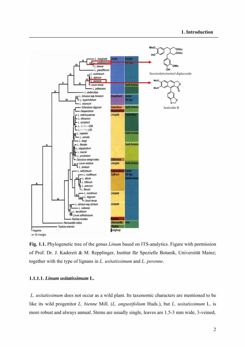
Cathartolinum in Flora Europaea. According to the recently established molecular phylogeny
of the Linaceae (Kadereit and Repplinger, unpublished results, Fig.1.1) the genus Linum has
two main clusters, one mainly consisting of the sections Linopsis and Syllinum and the other
contains section Linum.
1.1.1. Section Linum
Alternate, glabrous leaves without basal glands; eglandular sepals; free, blue purple or pink
petals; capitate, clavate or linear stigmas are the characteristics of plants in this section
according to Flora Europaea (Ockendon and Walters 1968). They can be homo- or
heterostylous; perennial, biennial or annual. 12 species of the section Linum have been
reported from Europe. L. narbonense, L. nervosum, L. aroanium, L. hologynum, L.
virgultorum, L. decumbens, L. bienne, L. usitatissimum and the L. perenne group. The L.
perenne group contains 4 species as follows: L. perenne, L. austriacum, L. punctatum and L.
leonii.
2
1. Introduction
Fig. 1.1. Phylogenetic tree of the genus Linum based on ITS-analytics. Figure with permission
of Prof. Dr. J. Kadereit & M. Repplinger, Institut für Spezielle Botanik, Universität Mainz;
together with the type of lignans in L. usitatissimum and L. perenne.
1.1.1.1. Linum usitatissimum L.
L. usitatissimum does not occur as a wild plant. Its taxonomic characters are mentioned to be
like its wild progenitor L. bienne Mill. (L. angustifolium Huds.), but L. usitatissimum L. is
more robust and always annual. Stems are usually single, leaves are 1.5-3 mm wide, 3-veined,
OO
O
OMeO
MeO
Justicidin B
Secoisolariciresinol diglucoside
OGluOGlu
OH
OH
OMe
MeO
3
1. Introduction
with 6-9 mm sepals. Capsules are 6-9 mm. L. usitatissimum is an important summer crop for
the production of oil and fiber. The translation of the Latin species epithet usitatissimum is
“the most useful one”, reflecting the several uses made of this plant.
1.1.1.2. Linum perenne L.
Stems are 10-60 cm, decumbent, ascending or erect. The middle cauline leaves are 1- to 3-
veined. Inflorescence usually many-flowered. Inner sepals are acute or obtuse. Pedicels are
erect. Capsules are 5-8 mm. Heterostylous. In Flora Europaea five subspecies are identified
for L. perenne L. as follows: subsp. perenne, subsp. anglicum, subsp. alpinum, subsp.
montanum, subsp. extraaxillare.
1.2. Primary and secondary metabolism
All organisms need to transfer and interconvert a vast number of organic compounds to
enable them to live, grow, and reproduce. They need to provide themselves with energy in the
form of ATP, and a supply of building blocks to construct their tissues. An integrated network
of enzyme mediated and carefully regulated chemical reactions is used for this purpose,
collectively referred to as intermediary metabolism, and the pathways involved are termed
metabolic pathways. Despite the extremely varied characteristics of living organisms, the
pathways for generally modifying and synthesizing carbohydrates, proteins, fats, and nucleic
acids are found to be essentially the same in all organisms, apart from minor variations. These
processes demonstrate the fundamental unity of all living matter, and are described as primary
metabolism, with the compounds involved in the pathways being termed primary
metabolites. There also exists an area of metabolism concerned with compounds which have
a much more limited distribution in nature. Such compounds, called secondary metabolites,
are only found in specific organisms, or groups of organisms, and are an expression of the
individuality of species. Secondary metabolites are not necessarily produced under all
conditions, and in the vast majority of cases the function of these compounds and their benefit
to the organism is not yet known. But it is logical to assume that all do play some vital role for
the well-being of the producer (Dewick 2002). Hartmann (1996) defined primary metabolism
as universal, uniform, conservative, and indispensable, and secondary metabolism as singular
4
1. Introduction
diverse, adaptive, dispensable for growth and development, but indispensable for survival.
Many of the secondary metabolites have important ecological functions in plants: they protect
plants against herbivores and microbial pathogens, they serve as attractants for pollinators and
seed-dispersing animals, and they function as agents of plant-plant competition and plant-
microbe symbioses (Taiz and Zeiger 2006). Undoubtly, the great utility of secondary
metabolites as medicinal drugs, flavoring agents, dyes, polymers, fibers, glues, oils, waxes
and perfumes have made them interesting for humankinds (Croteau et al. 2000). The above
generalizations distinguishing primary and secondary metabolism leave a grey area at the
boundary, so that some groups of natural products could be assigned to either division.
Hartmann (1996) suggested using “primary” and “secondary” in the sense of function; e.g.
Canavanine (1) as a nitrogen reservoir, is a primary metabolite, and as a toxic defense
chemical, is a secondary metabolite. Secondary metabolites are low molecular weight
compounds and their production is often low (less than 1% dry weight). More than 100,000
secondary metabolites have been described, with many more yet to be discovered; estimates
of total number in plants alone exceed 500,000 (Oksman-Caldentey and Inzé 2004; Hadacek
2002).
1.2.1. The building blocks of secondary metabolites
The building blocks for secondary metabolites are derived from primary metabolism as
indicated in Fig. 1.2. This scheme outlines how metabolites from fundamental processes of
photosynthesis, glycolysis, and the Krebs cycle are tapped off from energy generating
processes to provide biosynthetic intermediates. By far the most important building blocks
employed in the biosynthesis of secondary metabolites are derived from the intermediates
acetyl coenzyme A (acetyl-CoA), shikimic acid, mevalonic acid and 1-deoxyxylulose 5-
phosphate.
1.3. The shikimate-chorismate pathway
The shikimate pathway is the major metabolic route leading to the formation of aromatic
compounds especially L-phenylalanin, L-tyrosin and L-tryptophan in microorganisms and in
plants.
5
1. Introduction
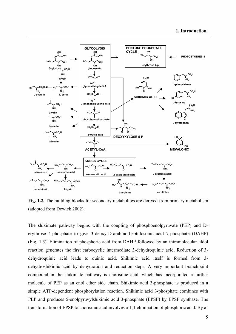
Fig. 1.2. The building blocks for secondary metabolites are derived from primary metabolism
(adopted from Dewick 2002).
The shikimate pathway begins with the coupling of phosphoenolpyruvate (PEP) and D-
erythrose 4-phosphate to give 3-deoxy-D-arabino-heptulosonic acid 7-phosphate (DAHP)
(Fig. 1.3). Elimination of phosphoric acid from DAHP followed by an intramolecular aldol
reaction generates the first carbocyclic intermediate 3-dehydroquinic acid. Reduction of 3-
dehydroquinic acid leads to quinic acid. Shikimic acid itself is formed from 3-
dehydroshikimic acid by dehydration and reduction steps. A very important branchpoint
compound in the shikimate pathway is chorismic acid, which has incorporated a further
molecule of PEP as an enol ether side chain. Shikimic acid 3-phosphate is produced in a
simple ATP-dependent phosphorylation reaction. Shikimic acid 3-phosphate combines with
PEP and produces 5-enolpyruvylshikimic acid 3-phosphate (EPSP) by EPSP synthase. The
transformation of EPSP to chorismic acid involves a 1,4-elimination of phosphoric acid. By a
O
OHOH
OHOH
OH
NH2
CO2H
SHNH2
CO2H OHNH2
CO2H
NH2
CO2H
NH2
CO2H
NH2
CO2H
NH2
CO2H
NH2
CO2HHO2C
NH2
S CO2H
NH2
CO2HNH2
O
OHOH
OHOH
PO
PO
OHOHC
PO
OHHO2C
OHO2C
OCOAS
O
CO2HHO2CO
CO2HHO2C
NH2
CO2HHO2C
NH2 NH
CO2HNH2
NH
NH2
CO2HNH2
POO
OH
OH
OHOH
OH
CO2H NH2
CO2H
NH2OH
CO2H
NH
NH2
CO2H
POO OH
OH
OH
OHCO2H
GLYCOLYSIS
glucose 6-p
glyceraldehyde 3-P
3-phosphoglyceric acid
OPHO2C
phosphoenolpyruvate
pyruvic acid
ACETYL-CoA
oxaloacetic acid 2-oxoglutaric acid
KREBS CYCLE
L-aspartic acid L-isoleucin
L-methionin L-lysin
D-glucose
glycin
L-serin L-cystein
L-valin
L-alanin
L-leucin
PENTOSE PHOSPHATE CYCLE
erythrose 4-p
PHOTOSYNTHESIS
SHIKIMIC ACID
L-glutamic acid
L-ornithine L-arginine
MEVALONIC
DEOXYXYLOSE 5-P
L-phenylalanin
L-tyrosine
L-tyrptophan
6
1. Introduction
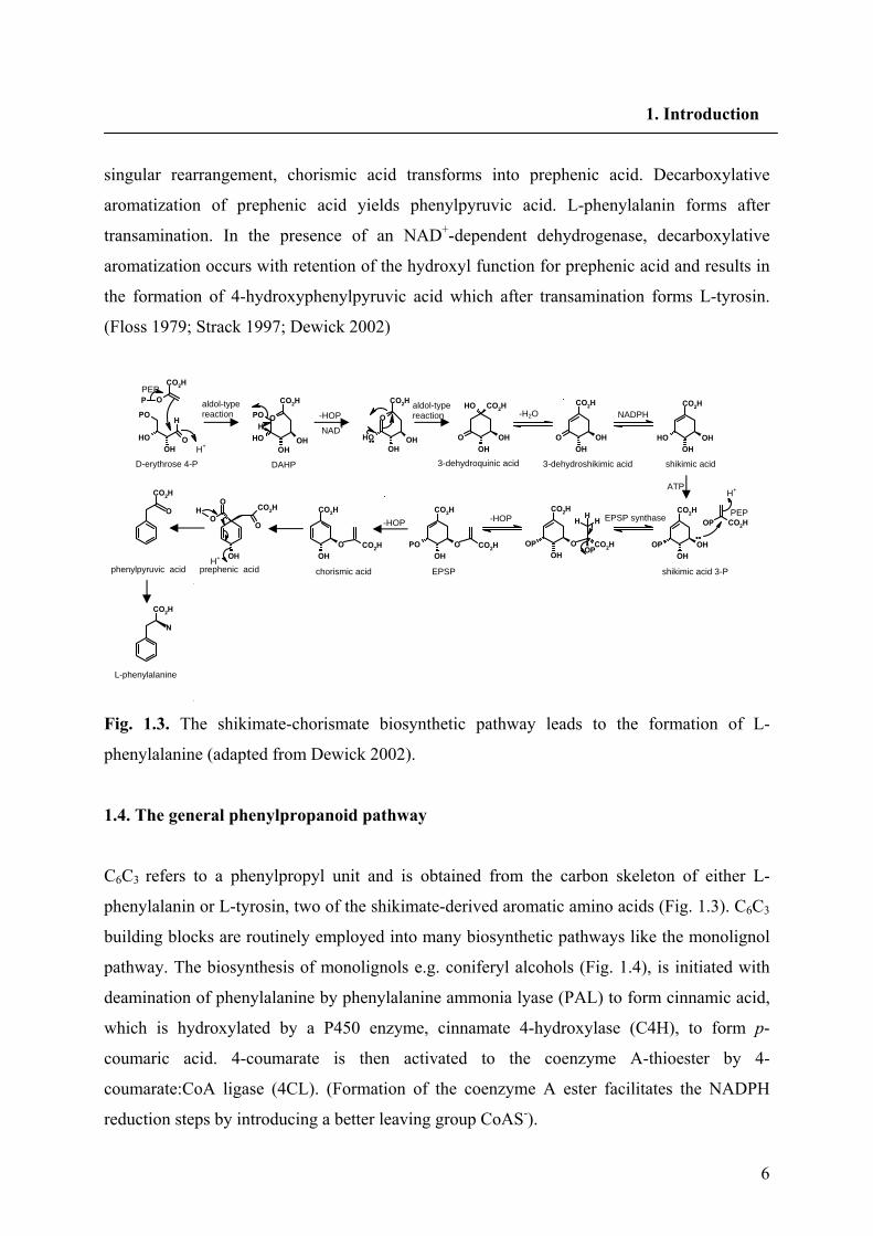
singular rearrangement, chorismic acid transforms into prephenic acid. Decarboxylative
aromatization of prephenic acid yields phenylpyruvic acid. L-phenylalanin forms after
transamination. In the presence of an NAD+-dependent dehydrogenase, decarboxylative
aromatization occurs with retention of the hydroxyl function for prephenic acid and results in
the formation of 4-hydroxyphenylpyruvic acid which after transamination forms L-tyrosin.
(Floss 1979; Strack 1997; Dewick 2002)
Fig. 1.3. The shikimate-chorismate biosynthetic pathway leads to the formation of L-
phenylalanine (adapted from Dewick 2002).
1.4. The general phenylpropanoid pathway
C6C3 refers to a phenylpropyl unit and is obtained from the carbon skeleton of either L-
phenylalanin or L-tyrosin, two of the shikimate-derived aromatic amino acids (Fig. 1.3). C6C3
building blocks are routinely employed into many biosynthetic pathways like the monolignol
pathway. The biosynthesis of monolignols e.g. coniferyl alcohols (Fig. 1.4), is initiated with
deamination of phenylalanine by phenylalanine ammonia lyase (PAL) to form cinnamic acid,
which is hydroxylated by a P450 enzyme, cinnamate 4-hydroxylase (C4H), to form p-
coumaric acid. 4-coumarate is then activated to the coenzyme A-thioester by 4-
coumarate:CoA ligase (4CL). (Formation of the coenzyme A ester facilitates the NADPH
reduction steps by introducing a better leaving group CoAS-).
PO
OHOH
O
CO2H
H
OHOH
OH
O
CO2H
OH OH
OH
OH
CO2H
O OHOH
O
CO2H
OHOH
CO2H
OH
O
CO2H
HPO
OHOH O
P
H+
PEP
D-erythrose 4-P
aldol-type reaction -HOP
NAD+
••
aldol-type reaction -H2O NADPH
OP CO2H
OHOH
CO2H
OP
H+
•• O
OH
CO2H
OP
HHH
OPCO2HO
OH
CO2H
PO CO2HOOH
CO2H
CO2H
O
CO2HO
O
OH
H
H+
CO2H
O
CO2H
N
DAHP 3-dehydroquinic acid 3-dehydroshikimic acid shikimic acid
PEP
shikimic acid 3-P
ATP
EPSP synthase -HOP -HOP
EPSPchorismic acidprephenic acidphenylpyruvic acid
L-phenylalanine
7
1. Introduction
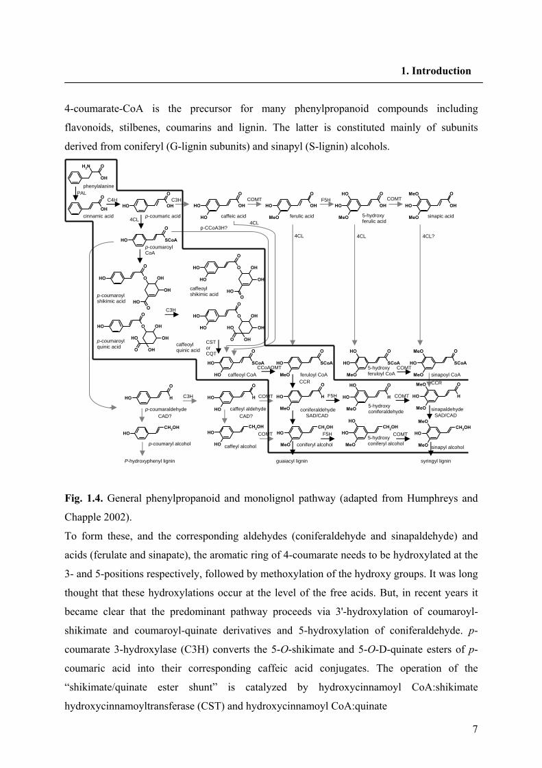
4-coumarate-CoA is the precursor for many phenylpropanoid compounds including
flavonoids, stilbenes, coumarins and lignin. The latter is constituted mainly of subunits
derived from coniferyl (G-lignin subunits) and sinapyl (S-lignin) alcohols.
Fig. 1.4. General phenylpropanoid and monolignol pathway (adapted from Humphreys and
Chapple 2002).
To form these, and the corresponding aldehydes (coniferaldehyde and sinapaldehyde) and
acids (ferulate and sinapate), the aromatic ring of 4-coumarate needs to be hydroxylated at the
3- and 5-positions respectively, followed by methoxylation of the hydroxy groups. It was long
thought that these hydroxylations occur at the level of the free acids. But, in recent years it
became clear that the predominant pathway proceeds via 3'-hydroxylation of coumaroyl-
shikimate and coumaroyl-quinate derivatives and 5-hydroxylation of coniferaldehyde. p-
coumarate 3-hydroxylase (C3H) converts the 5-O-shikimate and 5-O-D-quinate esters of p-
coumaric acid into their corresponding caffeic acid conjugates. The operation of the
“shikimate/quinate ester shunt” is catalyzed by hydroxycinnamoyl CoA:shikimate
hydroxycinnamoyltransferase (CST) and hydroxycinnamoyl CoA:quinate
OH
ONH2
OH
O
OH
O
OH OH
O
OH
OH
OH
O
OH
MeO
OH
O
OH
MeO
OH
OH
O
OH
MeO
MeO
SCoA
O
OH
O
O
OH OH
OH
OHO
O
O
OH OH
OH
OHO
OH
O
O
OH OH
OH
OH
OH
O
O
O
OH OH
OH
OH
OH
O
OH
SCoA
O
OH
OH
H
O
OH
OH
CH2OHOH
OH
H
O
OH
CH2OHOH
SCoA
O
OH
MeO
SCoA
O
OH
MeO
OH
SCoA
O
OH
MeO
MeO
H
O
OH
MeO
H
O
OH
MeO
OH
H
O
OH
MeO
MeO
CH2OHOH
MeO
CH2OHOH
MeO
OHCH2OH
OH
MeO
MeO
p-coumaric acid
phenylalanine
cinnamic acid caffeic acid ferulic acid 5-hydroxy ferulic acid
sinapic acid
p-coumaroyl CoA
p-coumaroyl shikimic acid
p-coumaroyl quinic acid
caffeoyl shikimic acid
caffeoyl quinic acid
caffeoyl CoA feruloyl CoA 5-hydroxy feruloyl CoA
CST or CQT
sinapoyl CoA
p-coumaraldehyde
p-coumaryl alcohol
caffeyl aldehyde coniferaldehyde 5-hydroxy coniferaldehyde sinapaldehyde
caffeyl alcohol coniferyl alcohol 5-hydroxy coniferyl alcohol
sinapyl alcohol
syringyl lignin
CCR
SAD/CAD
guaiacyl lignin P-hydroxyphenyl lignin
COMT
COMT
COMT
F5H
F5H COMT
COMT
CAD? CAD?
C3H
CCoAOMT
CCR
SAD/CAD
C3H
C3H C4H PAL
COMT F5H COMT
4CL? 4CL 4CL
4CL p-CCoA3H?
4CL
8
1. Introduction
hydroxycinnamoyltransferase (CQT), respectively. CST and CQT are reversible and would
have to function in both forward and reverse directions. Methylation of caffeoyl-CoA by
caffeoyl-CoA O-methyltransferase leads to formation of feruloyl-CoA. Ferulate 5-
hydroxylase (F5H) and caffeic acid/5-hydroxyferulic acid O-methyltransferase (COMT) act in
the level of aldehyde and alcohol respectively and result in the formation of sinapyl alcohol.
In any event, the feruloyl-CoA undergoes successive reduction reactions catalyzed by
cinnamoyl CoA reductase (CCR) and cinnamyl alcohol dehydrogenase (CAD), respectively,
to form coniferyl alcohol (Anterola et al. 2002; Humphreys and Chapple 2002; Dixon and
Reddy 2003).
1.5. Lignans
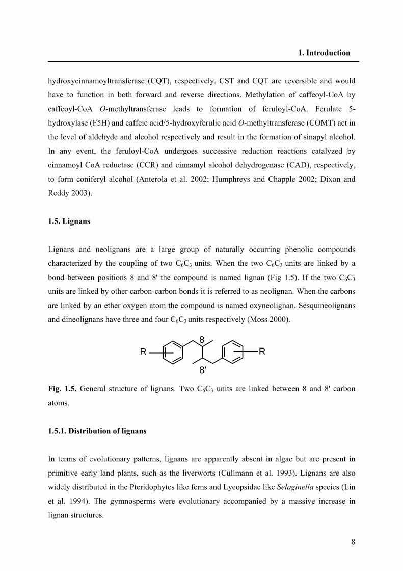
Lignans and neolignans are a large group of naturally occurring phenolic compounds
characterized by the coupling of two C6C3 units. When the two C6C3 units are linked by a
bond between positions 8 and 8' the compound is named lignan (Fig 1.5). If the two C6C3
units are linked by other carbon-carbon bonds it is referred to as neolignan. When the carbons
are linked by an ether oxygen atom the compound is named oxyneolignan. Sesquineolignans
and dineolignans have three and four C6C3 units respectively (Moss 2000).
Fig. 1.5. General structure of lignans. Two C6C3 units are linked between 8 and 8' carbon
atoms.
1.5.1. Distribution of lignans
In terms of evolutionary patterns, lignans are apparently absent in algae but are present in
primitive early land plants, such as the liverworts (Cullmann et al. 1993). Lignans are also
widely distributed in the Pteridophytes like ferns and Lycopsidae like Selaginella species (Lin
et al. 1994). The gymnosperms were evolutionary accompanied by a massive increase in
lignan structures.
8'
R R 8
9
1. Introduction
Lignan subgroups Examples Furofuran type
O
O
O
O
OH
OH
OMe
OMe
HH
O
O
OH
OH
OMe
OMe
HH
(+)-pinoresinol (+)-epipinoresinol
Furan type
O
OH
O
OH
OH
OMe
OMe
OH
HH
(+)-lariciresinol
Dibenzylbutan type
OHOH
OHOH
OH
OH
OMe
MeO
H
H
(-)-secoisolariciresinol
Dibenzylbutyrolacton type
O
O
OH
OH
OMe
MeOO
O
H
H
OH
OMe
MeOO
O
OMeMeO
H
H
(-)-matairesinol (-)-yatein
Dibenzylbutyrolactol type
O
OH
O
H
H
OO
O
OOH
(-)-cubebin
Arylnaphthalene type
O
O
MeOO
OMeO
OO
O
O
O
O
OMeOMe
MeO
justicidin B dehydrodeoxypodophyllotoxin
Aryltetralin type
O
O
O
O
O
O
OMeOMe
MeO
OH
(-)-podophyllotoxin
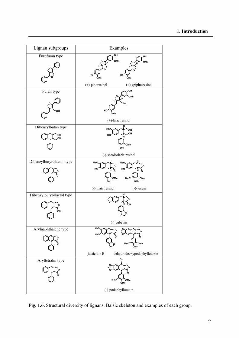
Fig. 1.6. Structural diversity of lignans. Baisic skeleton and examples of each group.
10
1. Introduction
In a somewhat similar manner, the transition to angiosperms was also accompanied by an
increase in lignan structural types/new skeletal forms. Lignan producing plants are distributed
throughout the six subclasses of Magnoliopsida; in contrast Liliopsida plants are rather poor
lignan sources. Umezawa (2003) have summerized the results on 108 families of lignan
producing plants in the division of Magnoliophyta.
1.5.2. Structural diversity of lignans
Further derivatisation of the general structure of lignans leads to a broad variety of
derivatives. Upon the way in which oxygen is incorporated into the skeleton and the
cyclization pattern, lignans are classified into the following eight subgroups: furofuran, furan,
dibenzylbutan, dibenzylbutyrolactone, aryltetralin, arylnaphthalene, dibenzocyclooctadiene
and dibenzylbutyrolactol (Fig. 1.6). Lignans of each subgroup vary substantially in oxidation
levels of both the aromatic rings and propyl side chains. Some lignans of furan,
dibenzylbutane, and dibenzocyclooctadiene have no oxygen at C9 (C9'), while some lignans
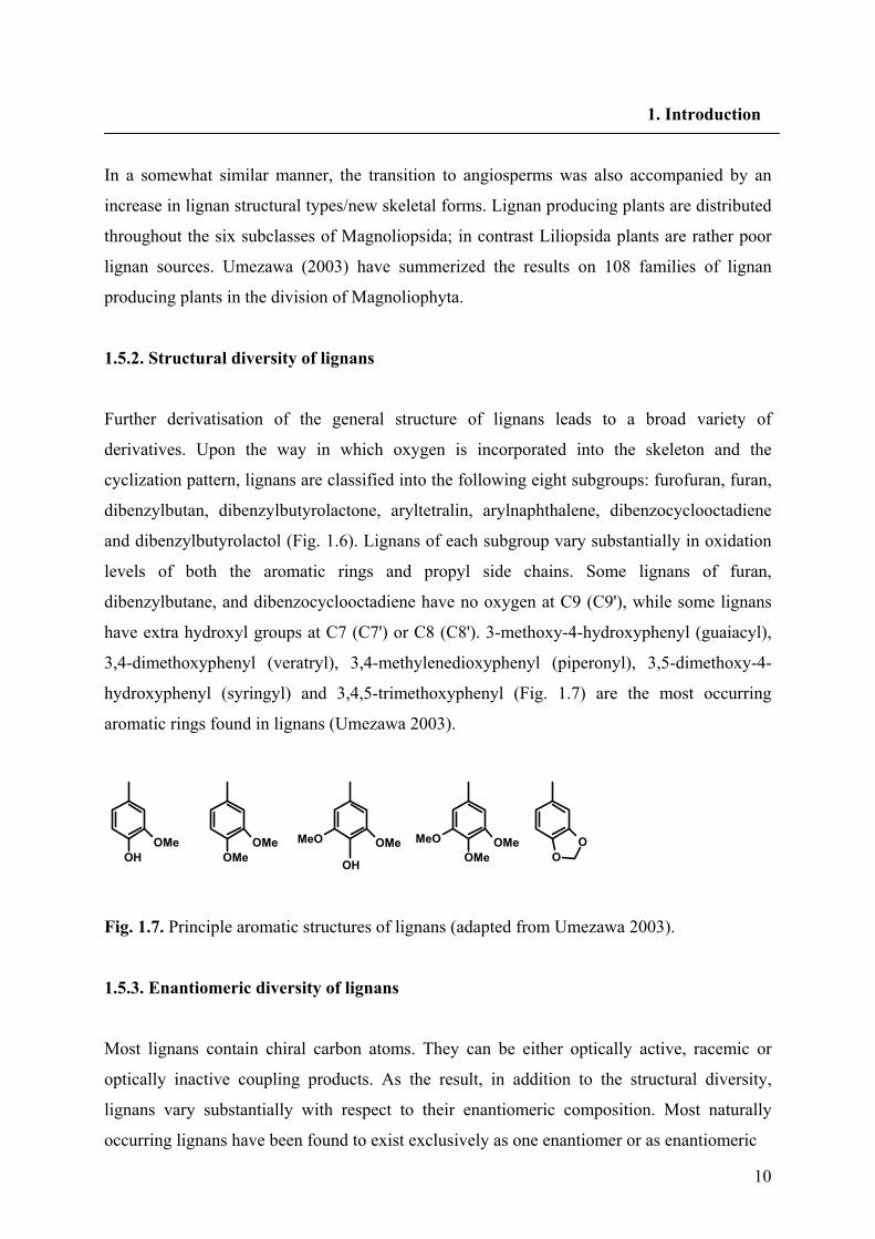
have extra hydroxyl groups at C7 (C7') or C8 (C8'). 3-methoxy-4-hydroxyphenyl (guaiacyl),
3,4-dimethoxyphenyl (veratryl), 3,4-methylenedioxyphenyl (piperonyl), 3,5-dimethoxy-4-
hydroxyphenyl (syringyl) and 3,4,5-trimethoxyphenyl (Fig. 1.7) are the most occurring
aromatic rings found in lignans (Umezawa 2003).
Fig. 1.7. Principle aromatic structures of lignans (adapted from Umezawa 2003).
1.5.3. Enantiomeric diversity of lignans
Most lignans contain chiral carbon atoms. They can be either optically active, racemic or
optically inactive coupling products. As the result, in addition to the structural diversity,
lignans vary substantially with respect to their enantiomeric composition. Most naturally
occurring lignans have been found to exist exclusively as one enantiomer or as enantiomeric
OHOMe OMe
OMeOMeMeO
OH
OMeOMe
MeOO
O
11
1. Introduction
mixtures with various enantiomeric compositions. Comparing the enantiomeric composition
of lignans from various species in different plant families Umezawa (2003) has concluded the
following statements: dibenzylbutyrolactone lignans are optically pure (>99% e.e.) while
furofuran and furan lignans are mixture of both enantiomers and exhibit various enantiomeric
compositions. For instance pinoresinol (Pino) which was isolated from cell suspension
cultures of L. album and L. usitatissimum was the mixture of both enantiomers (von
Heimendahl et al. 2005). Dibenzylbutyrolactone lignans are in most cases levorotatory except
that the lignans from Thymelaeaceae plants and Selaginella doederleinii are dextrorotatory.
Predominant enantiomers of furofuran, furan and dibenzylbutane lignans vary among plant
species, and even within different organs in a single plant species. Petiols of Arctium lappa
accumulate mainly (+)-secoisolariciresinol (Seco), but the seeds contain lignans with opposite
stereochemistry like (-)-matairesinol (Matai) and (-)-arctigenin (Suzuki et al. 2002). Seeds of
L. usitatissimum contain almost 99% (+)- and 1% (-)-SDG; whereas its aerial parts during
flowering stage contain (-)-enantiomers of lignans (Schmidt et al. 2006). It should be
mentioned that there are several conventions used for nominating chiral compounds. Based on
the actual geometry of each enantiomer (+)-Pino, (+)-lariciresinol (Lari) and (-)-Seco all have
R,R configuration at C-atoms 8,8′. (-)-Pino, (-)-Lari and (+)-Seco all have S,S configuration at
C-atoms 8,8′.
1.5.4. Pharmacological effects of lignans
Lignans are of considerable pharmacological and clinical interest in the treatment of cancer
and other diseases (Lee and Xiao 2003). The most important lignan for human health is
probably the cytotoxic aryltetralin lignan podophyllotoxin because its semisynthetic
derivatives like etoposide are used in cancer therapy (Imbert 1998). Justicidin B (Jus B), an
arylnaphthalene lignan without any optically active center can attract interest, because of its
fungicidal and antiprotozoal properties (Gertsch et al. 2003). It shows antiviral and anti-
inflammatory activities as well as inhibition of platelet aggregation (Chen et al. 1996;
MacRae et al. 1989; Rao et al. 2006). In addition, it is used as a lead compound for the design
of antirheumatic drugs (Baba et al. 1996). Recently, its strong cytotoxicity on chronic myeloid
and chronic lymphoid leukemia cell lines was shown (Vasilev et al. 2006). Diphyllin (Diph)
derivatives are putative remedies for topical chronic inflammatory disorders such as
12
1. Introduction
dermatitis and psoriasis while an acetylapioside derivative of Diph is a 5-lipoxygenase
inhibitor (Prieto et al. 2002).
Besides various physiological roles in plants, lignans such as Seco and Matai, which are
present in certain dietary components (e.g. flax (L. usitatissimum) seed), are thought to
substantially reduce incidence rates of breast, prostate and other hormonally linked cancers, in
humans on high fiber diets (Westcott and Muir, 2003). This so called chemoprotection occurs
through their metabolism by the action of gastrointestinal flora such as Clostridia species into
the mamelian lignans enterodiol and enterolactone, respectively (Axelson and Setchell, 1981;
Setchell et al. 1981). It’s now considered that dietary lignans impart these protective effects
because of one or more of their antioxidant, weak estrogenic/antiestrogenic, anti-aromatase,
antiangiogenic and anticarcinogenic/antitumor properties (Adlercreutz et al. 1993; Martin et
al. 1978; Osawa et al. 1985).
1.6. Biosynthesis of lignans
In terms of biosynthesis the enzymatic steps leading to formation of C6C3 monomeric units
have been described (chapter 1.4). The subsequent enzymatic transformations involved in
monomeeric coupling and post-coupling modifications are now being delinated. The lignan
biosynthesis starts with the coupling of two molecules of coniferyl alcohol and results in the
formation of Pino (chapter 1.6.1). Pino is reduced via Lari to Seco by the bifunctional
NADPH dependent enzyme pinoresinol-lariciresinol reductase (PLR) (chapter 1.6.2.1).
Secoisolariciresinol dehydrogenase (SDH) oxidizes Seco to Matai. The biosynthetic pathway
of aryltetralin type lignans in various plant species has been described previously (Federolf et
al. 2007, von Heimendahl et al. 2005; Kranz and Petersen 2003; Molog et al. 2001;
Sakakibara et al. 2003; Seidel et al. 2002).
The biosynthetic steps of other type of lignans like arylnaphthalene lignans were not
investigated up to now. But it is anticipated that the steps from coniferyl alcohol to Matai are
common in all pathways. Matai is believed to be a central intermediate leading to all diverse
lignan structures. Later steps in the biosynthesis of arylnaphthalenes most probably starting
from Matai are poorly understood. We have found that cell cultures of L. perenne
Himmelszelt accumulate Jus B and glycoside derivatives of Diph (Hemmati et al. 2007).
Schmidt and Vößing (2006) reported on the occurrence of 7(8)-dihydroisojusticidin B as
13
1. Introduction
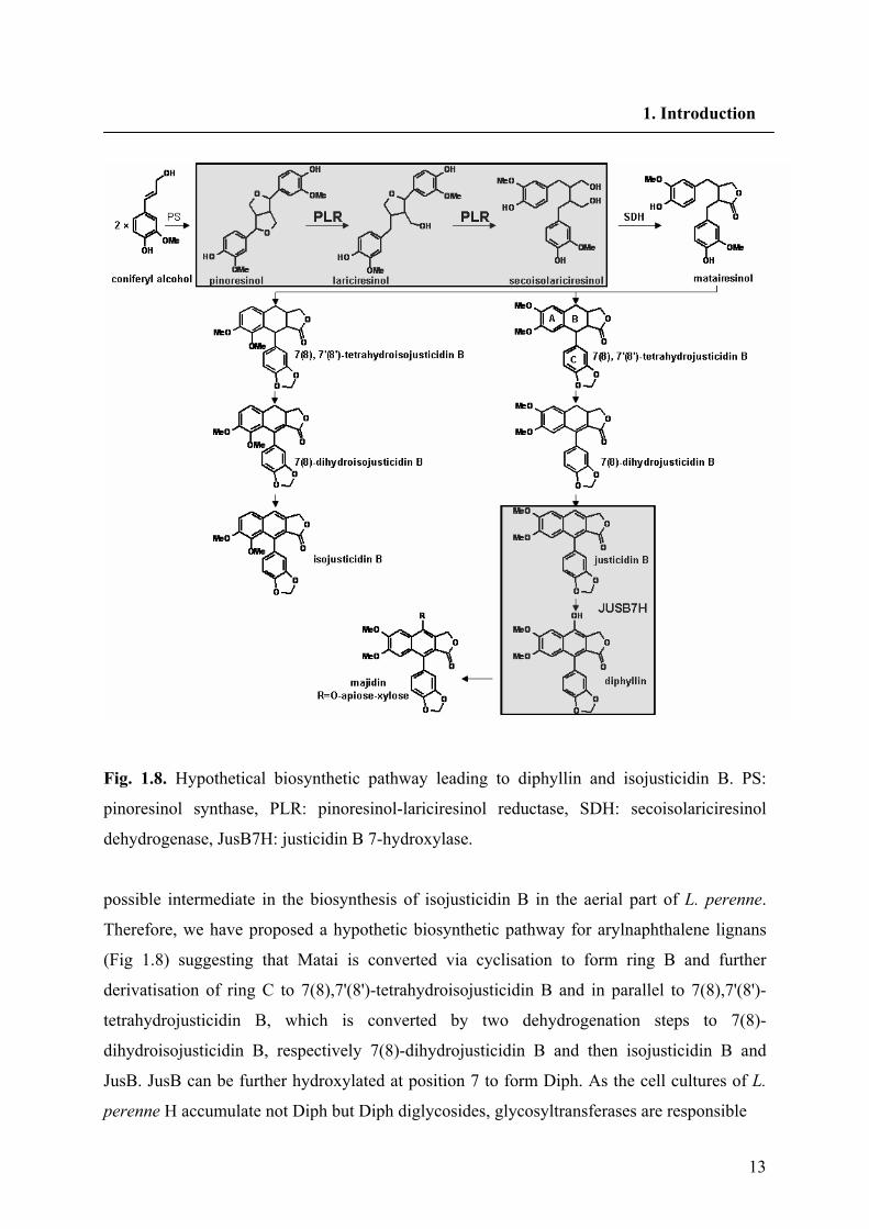
Fig. 1.8. Hypothetical biosynthetic pathway leading to diphyllin and isojusticidin B. PS:
pinoresinol synthase, PLR: pinoresinol-lariciresinol reductase, SDH: secoisolariciresinol
dehydrogenase, JusB7H: justicidin B 7-hydroxylase.
possible intermediate in the biosynthesis of isojusticidin B in the aerial part of L. perenne.
Therefore, we have proposed a hypothetic biosynthetic pathway for arylnaphthalene lignans
(Fig 1.8) suggesting that Matai is converted via cyclisation to form ring B and further
derivatisation of ring C to 7(8),7'(8')-tetrahydroisojusticidin B and in parallel to 7(8),7'(8')-
tetrahydrojusticidin B, which is converted by two dehydrogenation steps to 7(8)-
dihydroisojusticidin B, respectively 7(8)-dihydrojusticidin B and then isojusticidin B and
JusB. JusB can be further hydroxylated at position 7 to form Diph. As the cell cultures of L.
perenne H accumulate not Diph but Diph diglycosides, glycosyltransferases are responsible
14
1. Introduction
for the formation of majidin. Aspects of stereoselective coupling for the formation of Pino and
the consequent reductive steps by PLR are described in the following chapters.
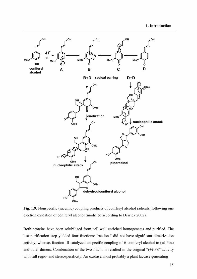
1.6.1. Dimerization of two coniferyl alcohols
Monolignol monomers, such as coniferyl alcohol are postulated to be dehydrogenated to
phenoxy radicals; One-electron oxidation of a simple phenol allows delocalization of the
unpaired electron, giving resonance forms. Radical pairing of resonance structures can then
provide a range of dimeric systems containing reactive quinonemethides, which are
susceptible to nucleophilic attack from hydroxyl groups in the same system, or by external
water molecules. Thus, coniferyl alcohol monomers can couple, generating linkages as
exemplified by guaiacylglycerol β-coniferyl ether (β arylether or 8-O-4' linkage),
dehydrodiconiferyl alcohol (DDC) (phenylcoumaran or 8-5' linkage) and Pino (resinol or 8-8'
linkage) Fig. 1.9.
1.6.1.1. Stereoselective coupling by dirigent proteins
As mentioned in chapter 1.5.3, plant lignans occur mainly as stereochemically pure (+)- or
(-)- enantiomers. It has been concluded that dimerization of monolignols would occur either
by a non-specific peroxidase followed by stereoselective biosynthetic steps or that the
dimerization is already stereospecific. Following incubation of [8-14C] coniferyl alcohol with
Forsythia suspensa soluble cell-free enzyme preparations, the racemic Pino was formed only
when H2O2 was supplied as cofactor, indicative of non specific peroxidase catalyzed coupling.
The insoluble residue from F. suspensa (free of soluble and ionically bound enzymes) was
able to catalyze the synthesis of Pino with enantiomeric excess for (+)-Pino. This fact
provides the first evidence for a direct highly stereoselective coupling process. The so-called
(+)-pinoresinol synthase (PS) was also detected in “crude cell wall preparations” of F.
intermedia, where it catalyzed the stereoselective 8-8' dimerization of two molecules of E-
coniferyl alcohol (Paré et al. 1994). (+)-PS has been purified and characterized, showing an
enzyme system consisting of two proteins, an oxidase and a dirigent protein, the later without
catalytic activity (Davin et al. 1997).
15
1. Introduction
Fig. 1.9. Nonspecific (racemic) coupling products of coniferyl alcohol radicals, following one
electron oxidation of coniferyl alcohol (modified according to Dewick 2002).
Both proteins have been solubilized from cell wall enriched homogenates and purified. The
last purification step yielded four fractions: fraction I did not have significant dimerization
activity, whereas fraction III catalyzed unspecific coupling of E-coniferyl alcohol to (±)-Pino
and other dimers. Combination of the two fractions resulted in the original “(+)-PS” activity
with full regio- and stereospecificity. An oxidase, most probably a plant laccase generating
•
•
• -H+
-e
H+
OH
OH
MeO
• O
OH
MeOO
OH
MeOO
OH
MeO
•
O
OH
MeO
•
coniferyl alcohol
A B C D
B+D D+Dradical pairing
OHH
O
OH
OOMe
OMe
H+
H+
OH
OMeO
O
OH
OMe
• •
O
O
OH
OH
OMe
OMe
O
OH
OH
OH
OMe
OMe
OH
OH
OH
OOMe
OMe
pinoresinol
dehydrodiconiferyl alcohol
nucleophilic attack
nucleophilic attack
enolization
16
1. Introduction
free radicals of E-coniferyl alcohol, was found in fraction III. Fraction I contained a protein
with an approximate MW of 78 kDa, possibly a trimer of 27 kDa subunits. The protein has no
catalytically active center. Since this protein determines the region- and stereospecificity
without having any catalytic properties, it was named “dirigent protein” from the latin
dirigere: to guide or to align. It was shown that chemical formation of coniferyl alcohol
radicals, with the help of flavin mononucleotide (FMN), flavin adenine dinucleotide (FAD) or
ammonium peroxidisulfate, was sufficient to form (+)-Pino in presence, but not in the
absence, of the “dirigent protein”. Therefore, it was proposed that the phenoxy radicals
formed by the oxidase (fraction III) are captured by the “dirigent protein”, leading to a
stereospecific and regiospecific coupling of the monomers. (+)-PS was strictly specific for
coniferyl alcohol; 4-coumaroyl and sinapoyl alcohols were not accepted (which gave racemic
products due to non-specific coupling). Formation of (+)-Pino in Forsythia, therefore, is made
possible by concomitant of two proteins (Fig. 1.10). The corresponding gene encoding the
dirigent protein was cloned and found to encode a protein of circa 18 kDa, which differed
from the native Forsythia protein of ~26 kDa due to post-translational glycosylation of the
native protein (Gang et al. 1999a).
1.6.2. Pinoresinol-lariciresinol reductase (PLR)
One of the first documented examples in the nature of benzylic ether reduction in plant
systems and the specificity of the reductase step(s) in terms of enantiospecificity and
diastereomic preferences was examined when the (+)- and (-)- enantiomers of Pino were
individually incubated with the Forsythia intermedia cell-free extracts (Katayama et al. 1993).
In the presence of NADPH, preferential conversion was observed into both (+)-Lari and (-)-
Seco, respectively. Incubation with (±)-Lari also established that only the (+)-antipode was
converted into (-)-Seco. This proves the existence of a bifunctional enantiospecific
pinoresinol-lariciresinol reductase (PLR) in the soluble protein extract of F. intermedia.
Isolation of a cDNA encoding a PLR from F. intermedia (PLR-Fi1) and its heterologous
expression showed the same enantiospecificity as for the crude extracts. The polypeptide had
a 312 amino acids and a calculated MW of 34.9 kDa. The presence of cDNAs corresponding
to two stereochemically distinct classes of PLRs in a single plant species western red cedar
(Thuja plicata), has been demonstrated by Fujita et al. (1999).
17
1. Introduction
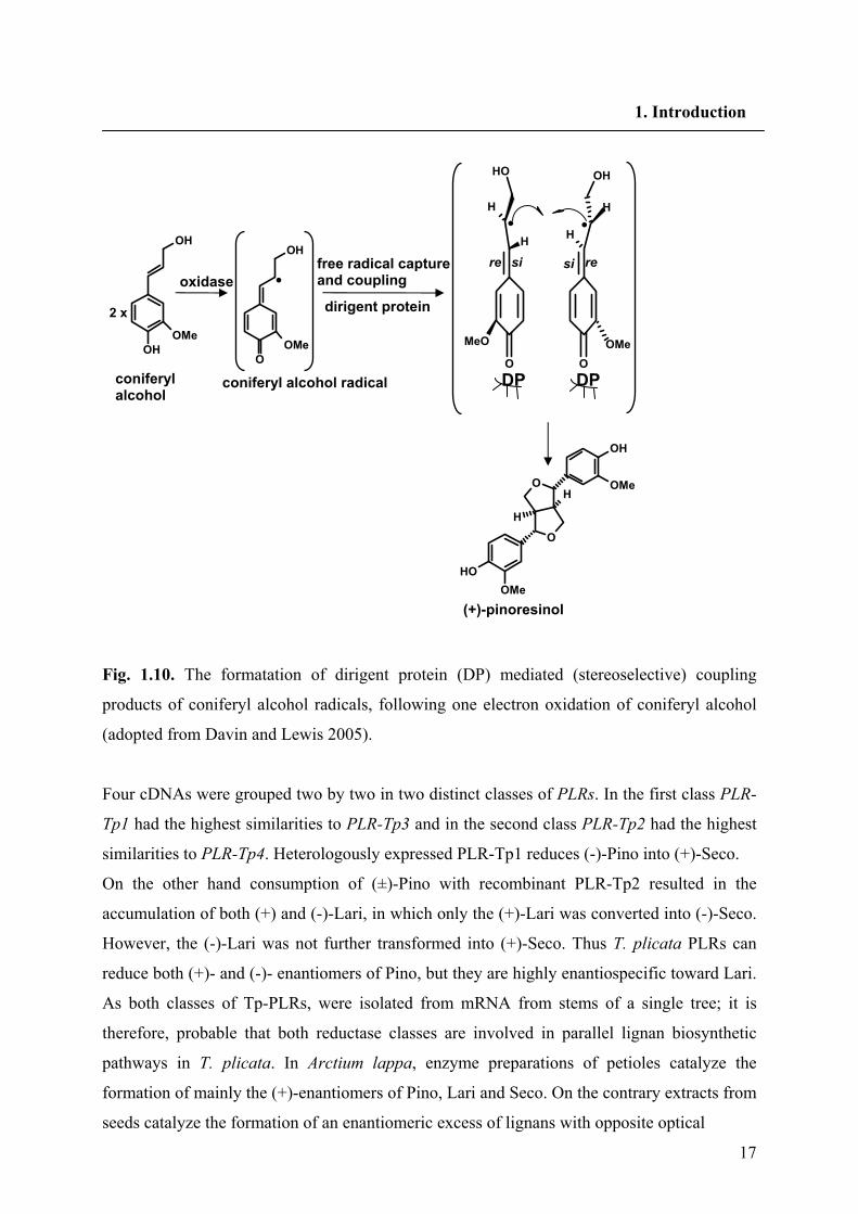
Fig. 1.10. The formatation of dirigent protein (DP) mediated (stereoselective) coupling
products of coniferyl alcohol radicals, following one electron oxidation of coniferyl alcohol
(adopted from Davin and Lewis 2005).
Four cDNAs were grouped two by two in two distinct classes of PLRs. In the first class PLR-
Tp1 had the highest similarities to PLR-Tp3 and in the second class PLR-Tp2 had the highest
similarities to PLR-Tp4. Heterologously expressed PLR-Tp1 reduces (-)-Pino into (+)-Seco.
On the other hand consumption of (±)-Pino with recombinant PLR-Tp2 resulted in the
accumulation of both (+) and (-)-Lari, in which only the (+)-Lari was converted into (-)-Seco.
However, the (-)-Lari was not further transformed into (+)-Seco. Thus T. plicata PLRs can
reduce both (+)- and (-)- enantiomers of Pino, but they are highly enantiospecific toward Lari.
As both classes of Tp-PLRs, were isolated from mRNA from stems of a single tree; it is
therefore, probable that both reductase classes are involved in parallel lignan biosynthetic
pathways in T. plicata. In Arctium lappa, enzyme preparations of petioles catalyze the
formation of mainly the (+)-enantiomers of Pino, Lari and Seco. On the contrary extracts from
seeds catalyze the formation of an enantiomeric excess of lignans with opposite optical
•
re re si si
O
H
OH
H
MeO
O
OH
H
OMe
H•
DP DP
OH
OH
OMe
O
OH
OMe
•
O
O
OH
OH
OMe
OMe
H
H
oxidase dirigent protein
free radical capture and coupling
(+)-pinoresinol
coniferyl alcohol radical coniferyl alcohol
2 x
18
1. Introduction
rotation. This observation can be accounted for by postulating that A. lappa has PLR isoforms
with different enantioselectivity and expression. However no PLR cDNAs to prove this
assumption were cloned (Suzuki et al. 2002).
L. usitatissimum seeds accumulate secoisolariciresinol diglucoside (SDG) to a very high
percentage (2%-4% by dry weight). There are two distinct diastereomers of SDG present in
flaxseed; the dominant form (~99%) is (+)-Seco. (Ford et al. 2001; Sicilia et al. 2003).
Experiments with radiolabeled phenylalanine revealed that (-)-Pino is converted to (+)-Seco
in flaxseed (Ford et al. 2001). The corresponding PLR cDNA (PLR-Lu1) encoding this
conversion was isolated and expressed heterologously by von Heimendahl et al. (2005).
On the basis of the SDG diastereomer, there also appears to be another pathway to SDG, with
the minor road being that to (-)-Seco. Recently, six lignans of the dibenzylbutyrolactone type
were identified in the aerial parts of flowering L. usitatissimum plants (Schmidt et al. 2006);
all were found to be laevorotatory and possess the absolute stereochemistry (8R, 8'R),
corresponding to (-)-bursehernin, (-)-yatein, (-)-hinokinin, (-)-matairesinol dimethyl ether, (-)-
thujaplicatin trimethyl ether and (-)-E-anhydropodorhizol. This finding gives further hints for
the demand of a second PLR with opposite enantiospecificity to the PLR-Lu1 In support of
this contention we have proposed two parallel biosynthetic pathways for Seco biosynthesis in
L. usitatissimum (Fig. 1.11).
Based on the high sequence similarity and reaction mechanism PLRs, phenylcoumaran
benzylic ether reductases (PCBERs) and isoflavone reductases (IFRs) are the first three
members of NADPH-dependent reductase enzymes forming the PIP reductase family (Fujita
et al. 1999, Koeduka et al. 2006 PNAS) (Fig. 1.12). DDC and its 7'-8' (allylic bond)-reduced
derivative dihydrodehydrodiconiferyl alcohol (DDDC), appear to be ubiquitous throughout
the plant kingdom, being found in a wide array of plant families, such as the Asteraceae,
Pinaceae and Urticaceae (Kraus and Spiteller 1997) suggesting the involvement of a universal
defense system. The 8-5' linked lignans can also co-occur in many species with their
phenylcoumaran benzylic ether reduced counterparts like isodihydrodehydrodiconiferyl
alcohol (IDDDC) and tetrahydrodehydrodiconiferyl alcohol (TDDC). It can be considered
that DDC and DDDC might undergo comparable reductive transformations, analogous to that
catalyzed by PLRs. The cDNA encoding PCBER were cloned from the gymnosperm Pinus
taeda and angiosperm Populus trichocarpa. The recombinant PCBER converted DDC and
DDDC into the benzylic ether reduced lignans IDDDC and TDDC, respectively. Kinetic
19
1. Introduction
studies have shown that the enzyme has higher affinity toward DDC rather than DDDC (Gang
et al. 1999b), suggesting that the biosynthesis of TDDC may involve initial formation of the
benzylic ether reduced intermediate prior to allylic bond reduction. PCBERs are regiospecific
but not enantiospecific toward their substrates. IFRs catalyze the reduction of α-ß unsaturated
ketones during isoflavonoid formation (Fig. 1.12).
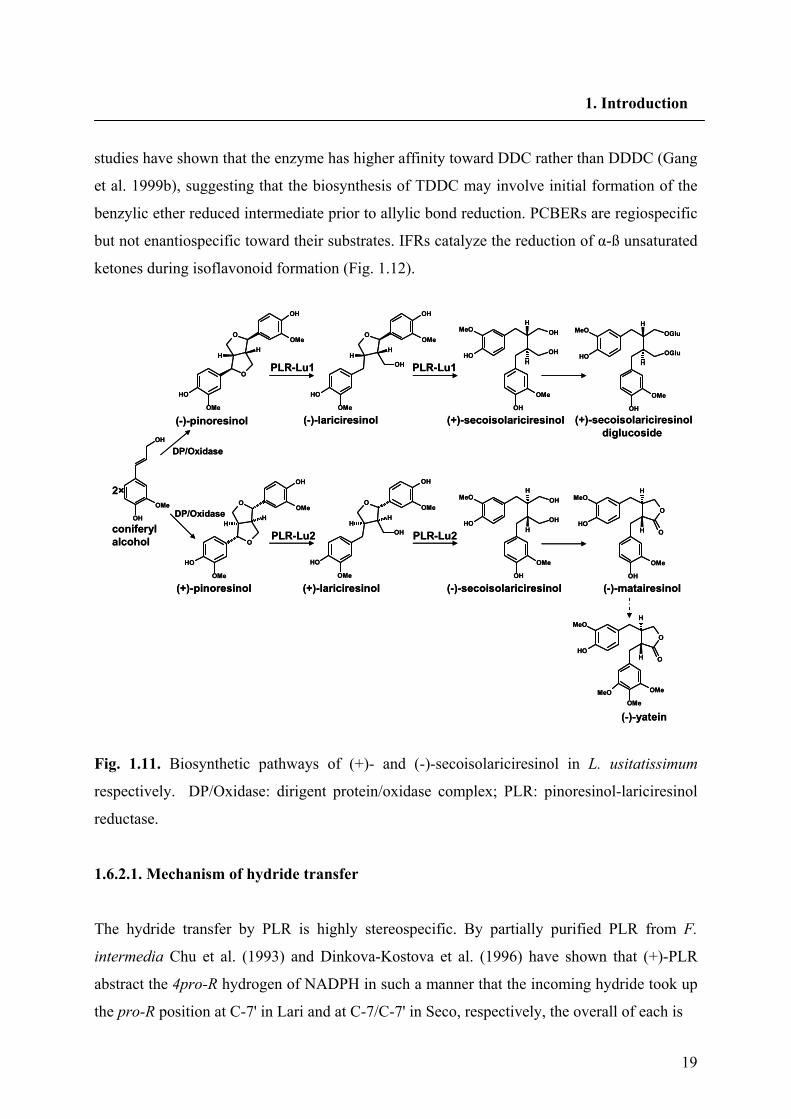
Fig. 1.11. Biosynthetic pathways of (+)- and (-)-secoisolariciresinol in L. usitatissimum
respectively. DP/Oxidase: dirigent protein/oxidase complex; PLR: pinoresinol-lariciresinol
reductase.
1.6.2.1. Mechanism of hydride transfer
The hydride transfer by PLR is highly stereospecific. By partially purified PLR from F.
intermedia Chu et al. (1993) and Dinkova-Kostova et al. (1996) have shown that (+)-PLR
abstract the 4pro-R hydrogen of NADPH in such a manner that the incoming hydride took up
the pro-R position at C-7' in Lari and at C-7/C-7' in Seco, respectively, the overall of each is
OH
OH
OMe O
OH
OH
OMe
OMe
HH
OH
OH
OH
OH
OH
OMe
MeOH
H
O
O
OH
OH
OMe
OMe
HH
O
O
OH
OH
OMe
OMe
HH
O
OH
OH
OMe
OMe
HH
OH
OH
OH
OH
OH
OMe
MeOH
H
OH
OH
OMe
MeOH
H
O
O
OH
OMe
MeOH
H
O
O
OMeMeO
PLR-Lu1 PLR-Lu1
PLR-Lu2 PLR-Lu2
2×
(-)-pinoresinol
(+)-pinoresinol
(-)-lariciresinol (+)-secoisolariciresinol (+)-secoisolariciresinoldiglucoside
(+)-lariciresinol (-)-secoisolariciresinol (-)-matairesinol
(-)-yatein
coniferyl alcohol
OH
OH
OGlu
OGlu
OMe
MeOH
H
DP/Oxidase
DP/Oxidase
OH
OH
OMe O
OH
OH
OMe
OMe
HH
OH
OH
OH
OH
OH
OMe
MeOH
H
O
O
OH
OH
OMe
OMe
HH
O
O
OH
OH
OMe
OMe
HH
O
OH
OH
OMe
OMe
HH
OH
OH
OH
OH
OH
OMe
MeOH
H
OH
OH
OMe
MeOH
H
O
O
OH
OMe
MeOH
H
O
O
OMeMeO
PLR-Lu1 PLR-Lu1
PLR-Lu2 PLR-Lu2
2×
(-)-pinoresinol
(+)-pinoresinol
(-)-lariciresinol (+)-secoisolariciresinol (+)-secoisolariciresinoldiglucoside
(+)-lariciresinol (-)-secoisolariciresinol (-)-matairesinol
(-)-yatein
coniferyl alcohol
OH
OH
OGlu
OGlu
OMe
MeOH
H
DP/Oxidase
DP/Oxidase
20
1. Introduction
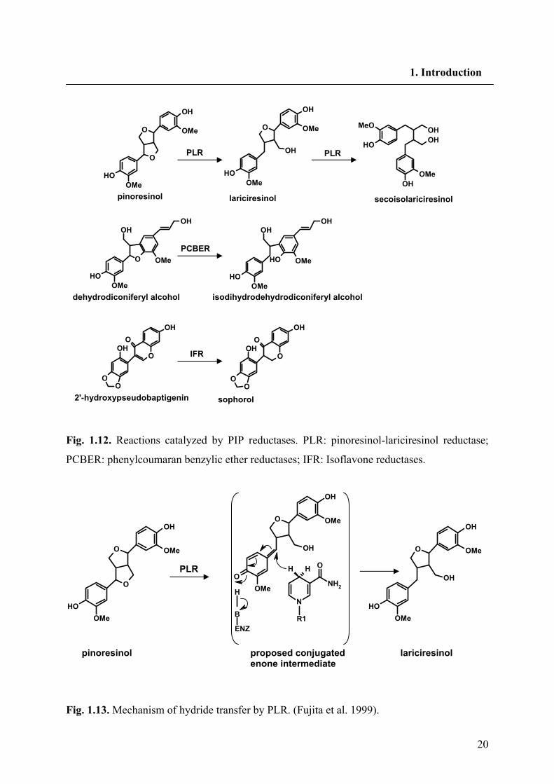
Fig. 1.12. Reactions catalyzed by PIP reductases. PLR: pinoresinol-lariciresinol reductase;
PCBER: phenylcoumaran benzylic ether reductases; IFR: Isoflavone reductases.
Fig. 1.13. Mechanism of hydride transfer by PLR. (Fujita et al. 1999).
O
OH
O
OMe
OMe
OH
N
NH2
OHH
R1B
ENZ
H
O
O
OH
OH
OMe
OMe
O
OH
OH
OMe
OMe
OHPLR
proposed conjugated enone intermediate
pinoresinol lariciresinol
O
O
OH
OH
OMe
OMe
O
OH
OH
OMe
OMe
OH
OHOH
OH
OH
OMe
MeO
O
OH
OH
OH
OMe
OMe
OH
OH
OH
OH
OMe
OMe
O
OO
OOH
OH
O
OO
OOH
OH
pinoresinol lariciresinol secoisolariciresinol
PLR PLR
dehydrodiconiferyl alcohol isodihydrodehydrodiconiferyl alcohol
2'-hydroxypseudobaptigenin sophorol
IFR
PCBER
21
1. Introduction
an inversion of configuration at these positions. Therefore these findings, rule out the
possibility of a SN1 mechanism involving a planar quinine methide transition state with
random hydride delivery from either side of the molecule (Fig. 1.13).
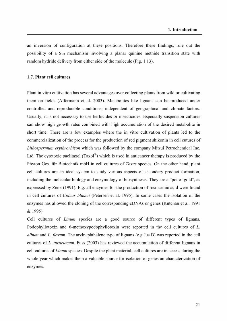
1.7. Plant cell cultures
Plant in vitro cultivation has several advantages over collecting plants from wild or cultivating
them on fields (Alfermann et al. 2003). Metabolites like lignans can be produced under
controlled and reproducible conditions, independent of geographical and climate factors.
Usually, it is not necessary to use herbicides or insecticides. Especially suspension cultures
can show high growth rates combined with high accumulation of the desired metabolite in
short time. There are a few examples where the in vitro cultivation of plants led to the
commercialization of the process for the production of red pigment shikonin in cell cutures of
Lithospermum erythrorhizon which was followed by the company Mitsui Petrochemical Inc.
Ltd. The cytotoxic paclitaxel (Taxol®) which is used in anticancer therapy is produced by the
Phyton Ges. für Biotechnik mbH in cell cultures of Taxus species. On the other hand, plant
cell cultures are an ideal system to study various aspects of secondary product formation,
including the molecular biology and enzymology of biosynthesis. They are a “pot of gold”, as
expressed by Zenk (1991). E.g. all enzymes for the production of rosmarinic acid were found
in cell cultures of Coleus blumei (Petersen et al. 1995). In some cases the isolation of the
enzymes has allowed the cloning of the corresponding cDNAs or genes (Kutchan et al. 1991
& 1995).
Cell cultures of Linum species are a good source of different types of lignans.
Podophyllotoxin and 6-methoxypodophyllotoxin were reported in the cell cultures of L.
album and L. flavum. The arylnaphthalene type of lignans (e.g Jus B) was reported in the cell
cultures of L. austriacum. Fuss (2003) has reviewed the accumulation of different lignans in
cell cultures of Linum species. Despite the plant material, cell cultures are in access during the
whole year which makes them a valuable source for isolation of genes an characterization of
enzymes.
22
1. Introduction
1.8. Plant transformation by Agrobacterium rhizogenes
Various species of bacteria are capable of transferring genes to higher plant species (Chung et
al. 2006). Among them, Agrobacterium tumefaciens is most widely studied. The soil bacteria
A. rhizogenes infects the plant tissues and leads to the formation of adventitious roots called
“hairy roots”. Fast and hormone independent growth and high genetic stability make hairy
root cultures superior in comparison to cell cultures. Hairy roots are known to produce the
same or even higher amounts of the metabolites found in normal roots. Integration of new
genes into hairy roots has opened another way for metabolic engineering (Guillon et al. 2006)
which is based on the ability to transform plant cells with the root-inducing (Ri) plasmid of A.
rhizogenes. Two important regions in the Ri plasmid are essential for transformation by
transferred DNA (T-DNA). These are the T-DNA itself as a mobile element and the virulence
region (vir) (Klee et al. 1987). The T-DNA is flanked by 25 bp directly repeated sequences.
Any DNA between these sequences will be transferred to the plant cell. The Ri plasmids are
grouped into two main classes according to the opines synthesized by hairy roots: agropine-
type (e.g., A4, 15834, LBA9402, 1855) and mannopine type (e.g., 8196, TR7, TR101) strains
(Sevón and Oksman-Caldentey 2002). The T-DNA of the agropine-type Ri-plasmid consists
of two separate T-DNA regions the TL-DNA and TR-DNA. The genes encoding auxin (tms1
and tms2) have been localized on the TR-DNA of the agropin type Ri plasmid. The
mannopine type Ri-plasmids contain only one T-DNA that shares considerable DNA
sequence homology with the TL-DNA of the agropine-type plasmids. The TL-DNA contains
four genetic loci, rolA, rolB, rolC and rolD, the first three of which mainly contribute to the
root initiation and maintenance. A plant cell becomes susceptible to Agrobacterium when it is
wounded. The wounded cells release phenolic compounds such as acetosyringone, that
activate the vir region of the bacterial plasmid and results in the formation of a T-strand as a
single strand copy of T-DNA. Control of vir gene expression is mediated by Vir A and Vir G
proteins. Vir A detects the released phenolic compounds, resulting in autophosphorylation,
finally activate vir gene transcription. Two other proteins (Vir D1 and Vir D2) recognize the
25 bp border sequence and produce single strand endonucleolytic cleavage in the bottom
strand of each border. After nicking the Vir D2 remains tightly associated with the 5' end of
the T-strand. The Vir E2 protein completely coates the T-strand, to prevent T-strand
nucleolytic degredation before integration.
23
1. Introduction
The T-strand with Vir D2 and Vir E2 makes a T complex by passing through the bactereial
and plant membranes and finally will integrate into the plant chromosome (Zupan and
Zambryski 1995).
Binary vectors contain origins of replication from a broad host-range plasmid. Using the
binary vector strategy a plasmid containing the left and right borders of T-DNA -but not the
virulence region- is modified in vitro to carry the gene to be transferred together with a
selectable marker. The modified plasmid DNA is transferred into E. coli and afterwards to an
Agrobacterium strain containing a helper plasmid that provides the vir functions. During
infection of plants the activated vir functions recognizes the left border sequence of the
modified plasmid and transfers all DNA between left and right borders to change a plant
chromosome (Hellens et al. 2000).
1.9. RNA interference (RNAi)
RNAi would be a useful approach to demonstrate if our cloned gene is involved in the
biosynthetic pathway of the corresponding metabolite. Most of the biosynthetic pathways for
secondary plant metabolites are still partially hypothetical. Reduction of the levels of a special
mRNA decreases the accumulation of the corresponding compound or group of compounds
which helps on the elucidation of the biosynthetic pathways. Alteration of the expression level
of our cloned gene can clarify its influence on the biosynthesis of arylnaphthalene lignans.
RNA silencing might have arisen as an ancient RNA surveillance system that is conserved
among eukaryotes, and that acts as a natural defense mechanism against invasive nucleic
acids, including viruses, transposons and perhaps other highly repetitive genomic sequences.
RNA silencing also plays a pivotal role in plant and animal development by providing an
elegant system of gene control that can occur through RNA degradation, translational
inhibition or chromatin modification (Wang and Metzlaff 2005).
The first report on a RNA interference (RNAi) type of phenomenon was reported in 1990
(Napoli et al. 1990; van der Krol et al. 1990; Smith et al; 1990). It was described that the
introduction of transcribed sense transgenes could down regulate the expression of
homologous endogenous genes, named co-suppression. Since in this type of silencing the
transcription is still active and the system is targeted after transcription level, the phenomenon
renamed to post transcriptional gene silencing (PTGS) (Fagard and Vaucheret 2000).
24
1. Introduction
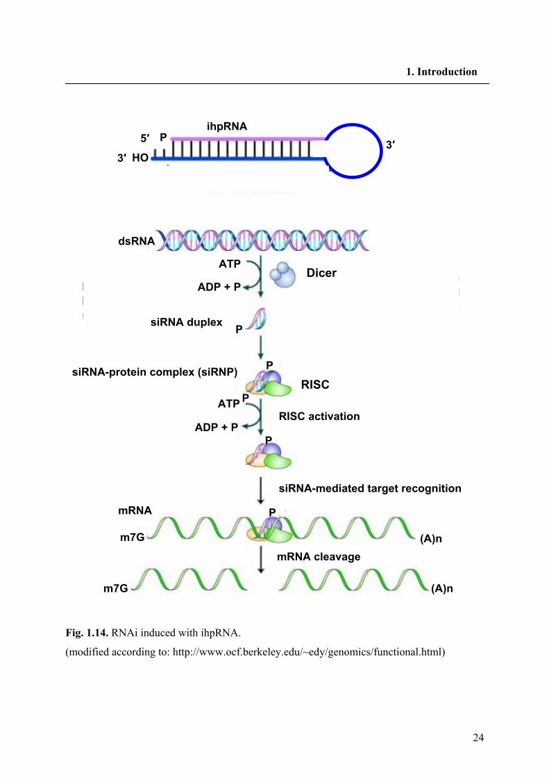
Fig. 1.14. RNAi induced with ihpRNA.
(modified according to: http://www.ocf.berkeley.edu/~edy/genomics/functional.html)
ihpRNA 3′ 5′ P
HO 3′
dsRNA
ATP
ADP + P
ADP + P
ATP
P
P
P
P
P
siRNA duplex
RISC
Dicer
siRNA-protein complex (siRNP)
RISC activation
siRNA-mediated target recognition
mRNA cleavage
mRNA
m7G
m7G
(A)n
(A)n

25
1. Introduction
The term RNAi was first coined when it was observed that both sense and antisense RNA
were able to silence gene expression in the nematode Caenorhabditis elegans (Fire et al.
1998). It was explained that the strongest trigger for gene silencing was the double stranded
RNA (dsRNA). DsRNA triggers the specific degradation of homolgous RNAs only within the
region of identity with the dsRNA (Elbashir et al. 2001).
1.9.1. Mechanism of RNAi
The RNAi pathway is initiated by recognition of dsRNA by a processing enzyme “Dicer” in
the cytoplasm (Fig. 1.14). Dicer is a ribonuclease in the RNase III family that cleaves dsRNA
into short double-stranded RNA fragments called small interfering RNA (siRNA) which are
about 20-25 nucleotids long, usually with a two base overhang on the 3' end (Hamilton and
Baulcombe 1999; Zamore et al. 2000). One of the two strands of each fragment, known as the
guide strand, is then incorporated into a complex named as the RNA-induced silencing
complex (RISC). The catalytically active components of the RISC complex are endonucleases
called argonate proteins, which cleave the target mRNA strand complementary to the bound
siRNA (Bernstein et al. 2001). The other anti-guide strand or passenger strand is degraded
during RISC activation. Degrading the mRNA results in substantially decreased levels of
protein translation and effective turning off the gene.
RNAi can be induced in plants by expressing artificial microRNAs (amiRNA), long hairpin
RNAs, modified viral RNAs or by directly inducing synthetic small interfering RNAs
(siRNAs) (Small 2007). An amenable approach of silencing has been to clone both sense and
antisense sequences, which are separated by an intron, under the same promoter. Upon
transcription, these sequences form a hairpin RNA (hpRNA) molecule that triggers gene
silencing (Smith et al. 2000). The silencing efficiency of hpRNA and antisense RNA has been
compared in a range of plant species: the hp strategy generally increases gene silencing by 90-
100% (Wesley et al. 2001) and is now the most widely used system for silencing genes in
plants (Mansoor et al. 2006). Hairpin RNAs are predominantly processed by the enzyme
Dicer-like 4 (DCL4). The produced siRNAs are loaded into the RNAi silencing complex of
which AGO1 is a major component. Gene discovery and metabolic engineering of plants are
two important usages of RNAi in plants. On the basis of the plant genomes sequenced so far
there is no true experimental evidence of function for the majority of sequenced genes.
26
1. Introduction
RNAi offers an easy, cost effective approach to generate “phenocopies” of genetic mutants
(Small 2007). Mansoor et al. (2006) have reviewed the engineering of novel traits in plants
through RNAi.
1.10. Cytochrome P450 monooxygenases
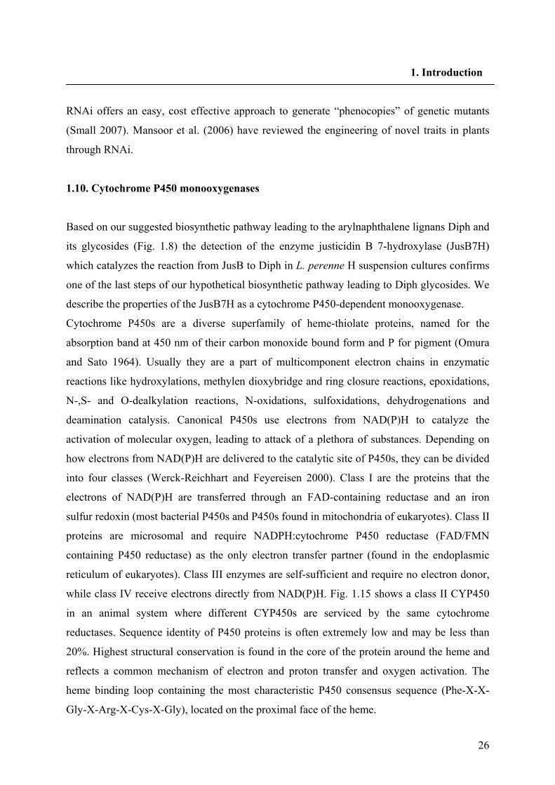
Based on our suggested biosynthetic pathway leading to the arylnaphthalene lignans Diph and
its glycosides (Fig. 1.8) the detection of the enzyme justicidin B 7-hydroxylase (JusB7H)
which catalyzes the reaction from JusB to Diph in L. perenne H suspension cultures confirms
one of the last steps of our hypothetical biosynthetic pathway leading to Diph glycosides. We
describe the properties of the JusB7H as a cytochrome P450-dependent monooxygenase.
Cytochrome P450s are a diverse superfamily of heme-thiolate proteins, named for the
absorption band at 450 nm of their carbon monoxide bound form and P for pigment (Omura
and Sato 1964). Usually they are a part of multicomponent electron chains in enzymatic
reactions like hydroxylations, methylen dioxybridge and ring closure reactions, epoxidations,
N-,S- and O-dealkylation reactions, N-oxidations, sulfoxidations, dehydrogenations and
deamination catalysis. Canonical P450s use electrons from NAD(P)H to catalyze the
activation of molecular oxygen, leading to attack of a plethora of substances. Depending on
how electrons from NAD(P)H are delivered to the catalytic site of P450s, they can be divided
into four classes (Werck-Reichhart and Feyereisen 2000). Class I are the proteins that the
electrons of NAD(P)H are transferred through an FAD-containing reductase and an iron
sulfur redoxin (most bacterial P450s and P450s found in mitochondria of eukaryotes). Class II
proteins are microsomal and require NADPH:cytochrome P450 reductase (FAD/FMN
containing P450 reductase) as the only electron transfer partner (found in the endoplasmic
reticulum of eukaryotes). Class III enzymes are self-sufficient and require no electron donor,
while class IV receive electrons directly from NAD(P)H. Fig. 1.15 shows a class II CYP450
in an animal system where different CYP450s are serviced by the same cytochrome
reductases. Sequence identity of P450 proteins is often extremely low and may be less than
20%. Highest structural conservation is found in the core of the protein around the heme and
reflects a common mechanism of electron and proton transfer and oxygen activation. The
heme binding loop containing the most characteristic P450 consensus sequence (Phe-X-X-
Gly-X-Arg-X-Cys-X-Gly), located on the proximal face of the heme.
27
1. Introduction
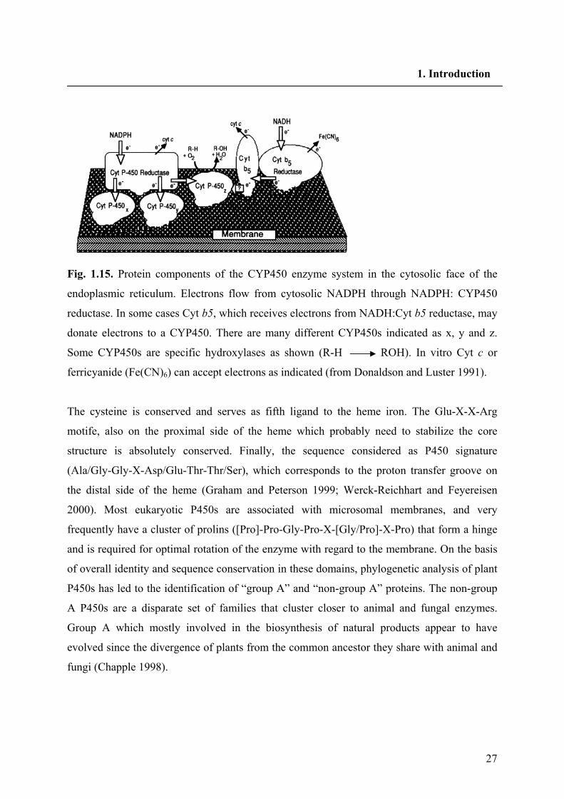
Fig. 1.15. Protein components of the CYP450 enzyme system in the cytosolic face of the
endoplasmic reticulum. Electrons flow from cytosolic NADPH through NADPH: CYP450
reductase. In some cases Cyt b5, which receives electrons from NADH:Cyt b5 reductase, may
donate electrons to a CYP450. There are many different CYP450s indicated as x, y and z.
Some CYP450s are specific hydroxylases as shown (R-H ROH). In vitro Cyt c or
ferricyanide (Fe(CN)6) can accept electrons as indicated (from Donaldson and Luster 1991).
The cysteine is conserved and serves as fifth ligand to the heme iron. The Glu-X-X-Arg
motife, also on the proximal side of the heme which probably need to stabilize the core
structure is absolutely conserved. Finally, the sequence considered as P450 signature
(Ala/Gly-Gly-X-Asp/Glu-Thr-Thr/Ser), which corresponds to the proton transfer groove on
the distal side of the heme (Graham and Peterson 1999; Werck-Reichhart and Feyereisen
2000). Most eukaryotic P450s are associated with microsomal membranes, and very
frequently have a cluster of prolins ([Pro]-Pro-Gly-Pro-X-[Gly/Pro]-X-Pro) that form a hinge
and is required for optimal rotation of the enzyme with regard to the membrane. On the basis
of overall identity and sequence conservation in these domains, phylogenetic analysis of plant
P450s has led to the identification of “group A” and “non-group A” proteins. The non-group
A P450s are a disparate set of families that cluster closer to animal and fungal enzymes.
Group A which mostly involved in the biosynthesis of natural products appear to have
evolved since the divergence of plants from the common ancestor they share with animal and
fungi (Chapple 1998).
28
1. Introduction
1.10.1. The reaction mechanism of cytochrome P450s
In the most common CYP450 oxygenase rections two electrons from CYP450 are donated to
O2, then one oxygen atom combines (hydroxylates) with the substrate and the second oxygen
atom is used to form a molecule of water:
NADPH + H + O2 + R-H NADP + H2O + ROH
The active center for catalysis is the iron-protoporphyrin IX (heme) with the thiolate of the
conserved cysteine residue as fifth ligand. Resting P450 is in the ferric form and partially six-
coordinated with a molecule of solvent.
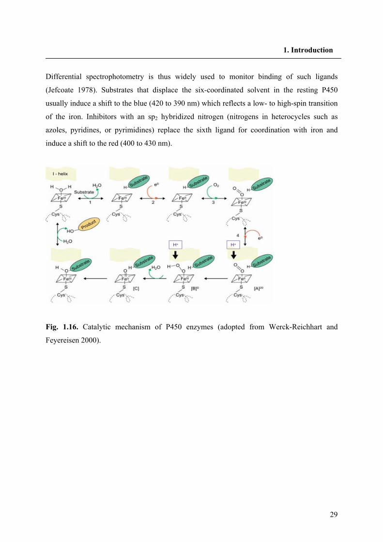
The well characterized portion of the catalytic sequence involves four steps, which are
indicated in Fig. 1.16. The first step is substrate binding, with displacement of the sixth ligand
solvent inducing a shift in the maximum of absorbance, spin state and redox potential of the
heme protein system; the second is one-electron reduction of the complex to a ferrous state,
driven by the increase in the redox potential which results from the previous step; the third is
binding of molecular oxygen to give a superoxide complex; and the fourth is a second
reduction step leading to an “activated oxygen species”. The exact nature of the very short-
lived activated oxygen species that carries on substrate attack long remained uncertain, but the
most recent data, from crystallography and mechanistic probes (Schlichting et al. 2000)
strongly suggest that it is actually a mixture of two electrophilic oxidants ([B]- and [C] in Fig.
1.16.). Both iron-peroxo [B]- and iron-oxo [C] complexes are formed by protonation of the
two-electrons-reduced dioxygen, a process that is allowed when a water channel forms in the
groove of the I-helix upon binding of O2. The oxo (oxyferryl) species, resulting from the
cleavage of the O-O bond - one atom of oxygen leaves with the two electrons and two protons
as water - is apparently the most abundant. The iron-hydroperoxo species inserts the elements
of OH+, producing protonated alcohols that can give cationic rearrangement products. The
iron-oxo species inserts an oxygen atom.
Carbon monoxide can bind ferrous P450 instead of dioxygen, inducing a shift of the
maximum of absorbance of the heme (called the Soret peak) to 450 nm (Omura and Sato
1964). CO is bound with high affinity and prevents binding and activation of O2. The result is
an inhibition of P450 activity. CO binding and inhibition can be reversed by light, with
maximal efficiency at 450 nm. Other ligands, substrates and inhibitors, induce absorbance
shifts of the Soret in P450 enzymes.
29
1. Introduction
Differential spectrophotometry is thus widely used to monitor binding of such ligands
(Jefcoate 1978). Substrates that displace the six-coordinated solvent in the resting P450
usually induce a shift to the blue (420 to 390 nm) which reflects a low- to high-spin transition
of the iron. Inhibitors with an sp2 hybridized nitrogen (nitrogens in heterocycles such as
azoles, pyridines, or pyrimidines) replace the sixth ligand for coordination with iron and
induce a shift to the red (400 to 430 nm).
Fig. 1.16. Catalytic mechanism of P450 enzymes (adopted from Werck-Reichhart and
Feyereisen 2000).
30
1. Introduction
1.11. Scope of the work
Seeds of L. usitatissimum as a plant species of the section Linum (Linaceae) contain almost
99% (+)- and 1% (-)-Seco. The PLR cDNA which was responsible for the formation of (+)-
Seco was cloned previously. In the aerial parts of L.usitatissimum lignans of the
dibenzylbutyrolactone type were identified (Schmidt et al. 2006); all were found to be (-)
isoforms. The question is that whether consequent reductive transformations to (+)- and (-)-
Seco are performed by a nonspecific PLR enzyme or via at least two PLR isoforms with
different enantiospecificity.
We have found that L. perenne H, as a typic example of the section Linum, accumulate
arylnaphthalene lignan types like Jus B and Diph without any chiral center. The question was
if the biosynthetic pathway of arylnaphthalene lignans starts with PLR. Cloning and aspects
of the enantiospecificity of the corresponding PLR is described in this work. This suggests but
does not prove that the PLR is involved in Jus B biosynthesis. Therefore, we intended to
down regulate the corresponding cloned gene by RNAi to see the influence on Jus B
biosynthesis.
Regarding the lignans which were found in L. perenne we have proposed a biosynthetic
pathway for arylnaphthalene lignan biosynthesis. Herein we describe the detection of the
enzyme justicidin B 7-hydroxylase (JusB7H) which catalyzes the reaction from JusB to Diph
in L. perenne H suspension cultures confirming the late steps of our hypothetical biosynthetic
pathway leading to Diph. We describe the properties of the JusB7H, a cytochrome P450-
dependent monooxygenase.
31
2. Materials and Methods
2. Materials and Methods
2.1. Materials
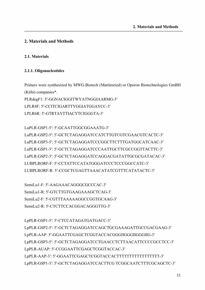
2.1.1. Oligonucleotides
Primers were synthesized by MWG-Biotech (Martinsried) or Operon Biotechnologies GmBH
(Köln) companies*.
PLRdegF1: 5′-GGNACIGGITWYATNGGIAARMG-3′
LPLR4F: 5′-CCITCIGARTTYGGIATGGAYCC-3′
LPLR6R: 5′-GTRTAYTTIACYTCIGGGTA-3′
LuPLR-GSP1-5′: 5′-GCAATTGGCGGAAATG-3′
LuPLR-GSP2-5′: 5′-GCTCTAGAGGATCCATCTTGTCGTCGAACGTCACTC-3′
LuPLR-GSP3-5′: 5′-GCTCTAGAGGATCCCGGCTTCTTTGATGGCATCAAC-3′
LuPLR-GSP1-3′: 5′-GCTCTAGAGGATCCCAATTGCTTCGCCGGTTACTTC-3′
LuPLR-GSP2-3′: 5′-GCTCTAGAGGATCCAGGACGATATTGCGCGATACAC-3′
LUBPLRORF-F: 5′-CCTATTCCATATGGGATCCCTCCCGGCCATC-3′
LUBPLRORF-R: 5′-CCGCTCGAGTTAAACATATCGTTTCATATACTC-3′
SemiLu1-F: 5'-AAGAAACAGGGCGCCCAC-3'
SemiLu1-R: 5'-GTCTTGTGAAGAAAGCTCAG-3'
SemiLu2-F: 5'-CGTTTAAAAAGGCCGGTGCAAG-3'
SemiLu2-R: 5'-CTCTTCCACGGACAGGGTTG-3'
LpPLR-GSP1-5′: 5′-CTCCATAGATGATGACC-3′
LpPLR-GSP2-5′: 5′-GCTCTAGAGGATCCAGCTGCGAAAGATTGCCGACGAAG-3′
LpPLR-AAP: 5′-GGAATTCGAGCTCGGTACCACGGGIIGGGIIGGGIIG-3′
LpPLR-GSP3-5′: 5′-GCTCTAGAGGATCCTGAACCTCTTAACATTCCCCGCCTCC-3′
LpPLR-AUAP: 5′-CCGGAATTCGAGCTCGGTACCAC-3′
LpPLR-AAP-3′: 5′-GGAATTCGAGCTCGGTACCACTTTTTTTTTTTTTTTTT-3′
LpPLR-GSP1-3′: 5′-GCTCTAGAGGATCCACTTCG TCGGCAATCTTTCGCAGCTC-3′
32
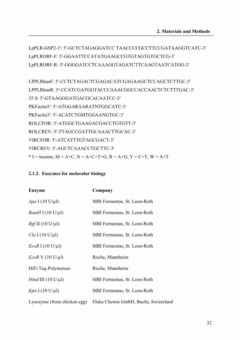
2. Materials and Methods
LpPLR-GSP2-3′: 5′-GCTCTAGAGGATCC TAACCCCGCCTTCCGATAAGGTCATC-3′
LpPLRORF-F: 5′-GGAATTCCATATGAAGCCGTGTAGTGTGCTCG-3′
LpPLRORF-R: 5′-GGGGATCCTCAAAGGTAGATCTTCAAGTAATCATGG-3′
LPPLRhanF: 5′-CCTCTAGACTCGAGACATCGAGAAGCTCCAGCTCTTGC-3′
LPPLRhanR: 5′-CCATCGATGGTACCCAAACGGCCACCAACTCTCTTTGAC-3′
35 S: 5′-GTAAGGGATGACGCACAATCC-3′
PKFactin5′: 5′-ATGGARAARATNTGGCATC-3′
PKFactin3′: 5′-ACATCTGMTGGAANGTGC-3′
ROLCFOR: 5'-ATGGCTGAAGACGACCTGTGTT-3'
ROLCREV: 5'-TTAGCCGATTGCAAACTTGCAC-3'
VIRCFOR: 5'-ATCATTTGTAGCGACT-3'
VIRCREV: 5'-AGCTCAAACCTGCTTC-3'
* I = inosine, M = A+C, N = A+C+T+G, R = A+G, Y = C+T, W = A+T
2.1.2. Enzymes for molecular biology
Enzyme
Company
Apa I (10 U/µl)
MBI Fermentas, St. Leon-Roth
BamH I (10 U/µl)
MBI Fermentas, St. Leon-Roth
Bgl II (10 U/µl)
MBI Fermentas, St. Leon-Roth
Cla I (10 U/µl)
MBI Fermentas, St. Leon-Roth
EcoR I (10 U/µl)
MBI Fermentas, St. Leon-Roth
EcoR V (10 U/µl)
Roche, Mannheim
HiFi Taq-Polymerase
Roche, Mannheim
Hind III (10 U/µl)
MBI Fermentas, St. Leon-Roth
Kpn I (10 U/µl)
MBI Fermentas, St. Leon-Roth
Lysozyme (from chicken egg)
Fluka Chemie GmbH, Buchs, Swizerland
33
2. Materials and Methods
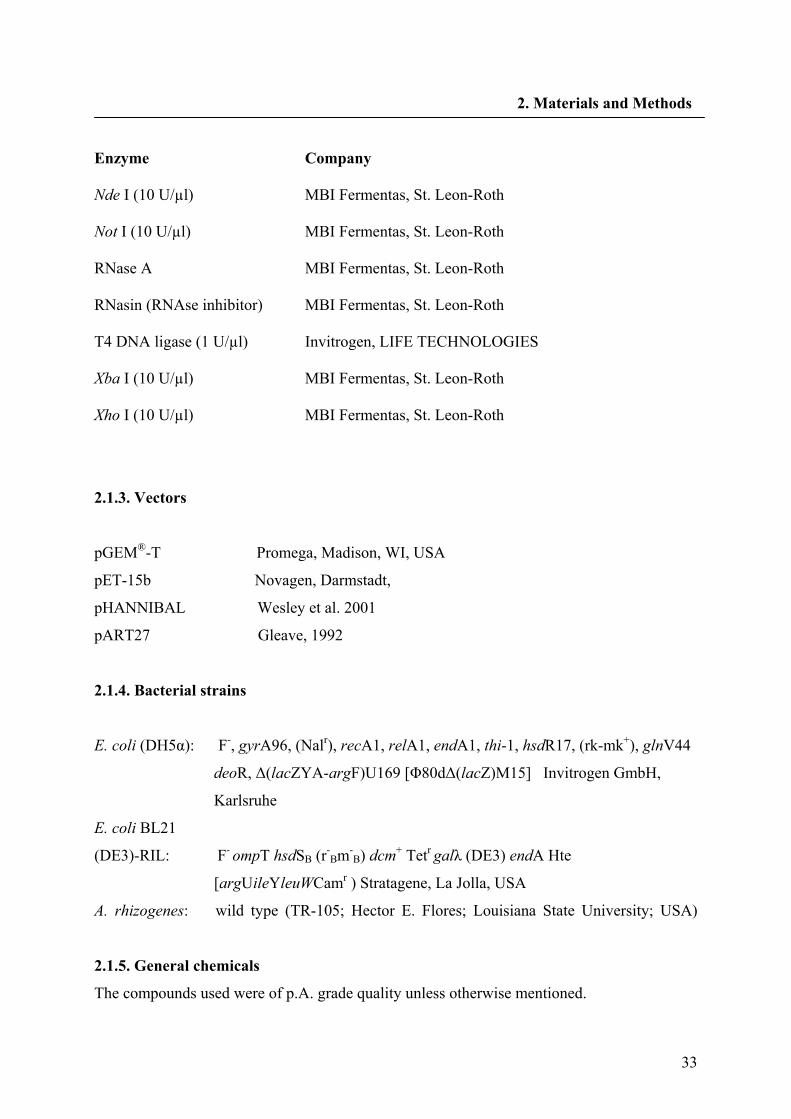
Enzyme
Company
Nde I (10 U/µl)
MBI Fermentas, St. Leon-Roth
Not I (10 U/µl)
MBI Fermentas, St. Leon-Roth
RNase A
MBI Fermentas, St. Leon-Roth
RNasin (RNAse inhibitor)
MBI Fermentas, St. Leon-Roth
T4 DNA ligase (1 U/µl)
Invitrogen, LIFE TECHNOLOGIES
Xba I (10 U/µl)
MBI Fermentas, St. Leon-Roth
Xho I (10 U/µl)
MBI Fermentas, St. Leon-Roth
2.1.3. Vectors
pGEM®-T Promega, Madison, WI, USA
pET-15b Novagen, Darmstadt,
pHANNIBAL Wesley et al. 2001
pART27 Gleave, 1992
2.1.4. Bacterial strains
E. coli (DH5α): F-, gyrA96, (Nalr), recA1, relA1, endA1, thi-1, hsdR17, (rk-mk+), glnV44
deoR, Δ(lacZYA-argF)U169 [Φ80dΔ(lacZ)M15] Invitrogen GmbH,
Karlsruhe
E. coli BL21
(DE3)-RIL: F- ompT hsdSB (r-Bm-
B) dcm+ Tetr galλ (DE3) endA Hte
[argUileYleuWCamr ) Stratagene, La Jolla, USA
A. rhizogenes: wild type (TR-105; Hector E. Flores; Louisiana State University; USA)
2.1.5. General chemicals
The compounds used were of p.A. grade quality unless otherwise mentioned.
34
2. Materials and Methods
Material
Company
1 kb Plus DNA Ladder
Invitrogen GmbH, Karlsruhe
1st Strand cDNA Synthesis Kit for RT-PCR
Roche Diagnostics GmbH, Mannheim
Acetic acid
Merck KgaA, Darmstadt
Aceton Sigma-Aldrich Laborchemikalien GmbH, Seelze
Acetonitrile
VWR International GmbH, Wien, Austria
Acetosyringone
Carl Roth GmbH & Co. Kg, Karlsruhe
Acrylamid/bisacrylamid
Carl Roth GmbH & Co. Kg, Karlsruhe
Agar-Agar
Duchefa, Haarlem, The Netherlands
Agarose
Invitrogen Life Technologies, Paisley, Scotland
Albumin, bovine (BSA) 96-99% Fraction V
Sigma Chemical Co, St. Louis, USA
Ammonium nitrate
Merck KgaA, Darmstadt
Ammonium sulfate
Carl Roth GmbH & Co. Kg, Karlsruhe
Ampicillin (sodium salt)
Grünenthal GmbH, Aachen
APS (ammonium peroxodisulfate)
Sigma Aldrich Chemie GmbH, Steinheim
Bacto®-Peptone
Carl Roth GmbH & Co. Kg, Karlsruhe
Bacto®-Trypton
DIFCO laboratories, Detroit, USA
Boric acid
Carl Roth GmbH & Co. Kg, Karlsruhe
Brom phenol blue
Serva GmbH & Co, Heidelberg
Calcium chloride dihydrate
Merck KgaA, Darmstadt
Cellulose acetate filter, 0.2 µm
Sartorius AG, Göttingen
Chloroform
Merck KgaA, Darmstadt
Chemiluminescent detection film
Roche Diagnostics GmbH, Mannheim
35
2. Materials and Methods
Clotrimazole (solvent MeOH)
Sigma Aldrich Chemie GmbH, Steinheim
Coniferyl alcohol
Fluka Chemie GmbH, Buchs, Switzerland
Coomasie-Brilliant blue G-250
Merck KgaA, Darmstadt
CTAB (cetyltrimethyl ammonium bromide)
Serva GmbH & Co, Heidelberg
Cupper (II)-sulfate pentahydrate
Fluka Chemie GmbH, Buchs, Switzerland
Cytochrome c (solvent H2O)
Fluka Chemie GmbH, Buchs, Switzerland
Denhardt´s solution (100×)
Eppendorf, Netheler
Developer: Kodak X-Ray developer LX24
Kodak, Darmstadt
Dichloromethane
Sigma-Aldrich Laborchemikalien GmbH, Seelze
DIECA
(sodium diethyldithiocarbaminate trihydrate)
Merck KgaA, Darmstadt
Dipotassium hydrogen phosphate trihydrate
Merck KgaA, Darmstadt
DNA-Agar Marine Bioproducts Inc., Delta, Canada
DNeasy Plant Mini Kit
Qiagen GmbH, Hilden
dNTPs
Carl Roth GmbH & Co. Kg, Karlsruhe
DTT (1,4 dithiothreitol) Applichem GmbH, Darmstadt
Ethidiumbromide
Fluka Chemie GmbH, Buchs, Switzerland
EDTA Merck KgaA, Darmstadt
Ethyl acetate
Fisher Scientific UK, Loughborough, UK
Expand High Fidelity PCR System
Roche Diagnostics GmbH, Mannheim
Ficoll 400
Serva GmbH & Co, Heidelberg
Fish sperm DNA
Roche Diagnostics GmbH, Mannheim
Fixer G334 Rapid Fixer
Agfa, Mortsel, Belgium
36
2. Materials and Methods
Glucose 6-phosphate
Sigma Aldrich Chemie GmbH, Steinheim
Glucose 6-phosphate dehydrogenase
Sigma Aldrich Chemie GmbH, Steinheim
Glycin
Carl Roth GmbH & Co. Kg, Karlsruhe
n-Hexan
Merck KgaA, Darmstadt
n-Hexan, HPLC grade
VWR Prolabo, France
Imidazol, 99%
Acros Organics, New Jersey, USA
Iron sulfate heptahydrat
Merck KgaA, Darmstadt
Isoamylalcohol
Mallinckrodt Baker B.V., Deventer, The
Natherlands
Isopropanol
Merck KgaA, Darmstadt
IPTG
(isopropyl-β-D-thiogalactopyranoside)
Carl Roth GmbH & Co. Kg, Karlsruhe
Leupeptin
USB, Cleveland, USA
Low DNA MassTM Ladder
Invitrogen GmbH, Karlsruhe
Low Melting Point Agarose
AppliChem GmbH, Darmstadt
Magnesium chloride hexahydrate
Merck KgaA, Darmstadt
McCown woody Plant Medium
Duchefa Biochemie B.V., Haarlem, The Netherlands
Methanol
Merck KgaA, Darmstadt
Miracloth
Calbiochem-Novabiochem Corp., USA
myo-Inositol
Merck KgaA, Darmstadt
NADH
Biomol Feinchemikalien, Hamburg
NADP+ (disodium salt)
Biomol Feinchemikalien, Hamburg
NADPH (tetrasodium salt)
AppliChem, Darmstadt
Nescofilm®
Azwell Inc., Osaka, Japan
Nick Translation System Invitrogen GmbH, Karlsruhe
37
2. Materials and Methods
Ni-NTA Agarose
Qiagen GmbH, Hilden
NAA (naphthyl acetic acid)
Merck KgaA, Darmstadt
Nylon membrane (HybondTM-N+)
Boehringer, Ingelheim
Paromomycin
Duchefa Biochem Haarlem, The Netherlands
PD10 columns
Amersham Biosciences, Freiburg
PMSF (Phenyl methane sulfonyl fluoride)
Carl Roth GmbH & Co. Kg, Karlsruhe
o-Phosphoric acid (85%)
Janssen Chimica, Beerse, Belgium
Polyclar 10
ISP Technologies INC., Wayne, New Jersey
PVP (Polyvinyl pyrrolidon) Sigma Aldrich Laborchemikalien GmbH, Seelze
Potassium dihydrogen phosphate
Merck KgaA, Darmstadt
Potassium hydroxide
Merck KgaA, Darmstadt
Potassium nitrate
Riedel-de Haën, Seelze
Protein Molecular Marker
Fermentas GmbH, St. Leon-Roth
QIAprep® Spin Miniprep Kit
Qiagen GmbH, Hilden
QIAquick® Gel Extraction Kit Qiagen GmbH, Hilden
QIAquick® PCR Purification Kit Qiagen GmbH, Hilden
Revert Aid M-MuL V reverse transcriptase
MBI Fermentas, St. Leon-Roth
RNeasy® Plant Mini Kit
Qiagen GmbH, Hilden
Rubidium chloride
Merck KgaA, Darmstadt
Saccharose
Pfeifer&Langen, Köln
Silica gel 60 (0.040-0.063 mm)
Merck KgaA, Darmstadt
Sodium chloride
Mallinckrodt Baker B.V., Deventer, The Netherlands
SDS (sodium dodecyl sulfate) Serva GmbH & Co, Heidelberg
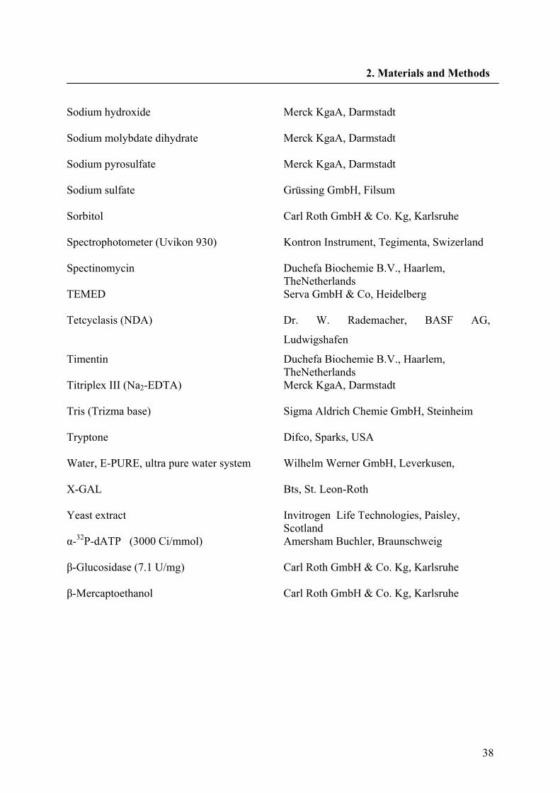
38
2. Materials and Methods
Sodium hydroxide
Merck KgaA, Darmstadt
Sodium molybdate dihydrate
Merck KgaA, Darmstadt
Sodium pyrosulfate
Merck KgaA, Darmstadt
Sodium sulfate
Grüssing GmbH, Filsum
Sorbitol
Carl Roth GmbH & Co. Kg, Karlsruhe
Spectrophotometer (Uvikon 930)
Kontron Instrument, Tegimenta, Swizerland
Spectinomycin
Duchefa Biochemie B.V., Haarlem, TheNetherlands
TEMED
Serva GmbH & Co, Heidelberg
Tetcyclasis (NDA)
Dr. W. Rademacher, BASF AG,
Ludwigshafen
Timentin
Duchefa Biochemie B.V., Haarlem, TheNetherlands
Titriplex III (Na2-EDTA)
Merck KgaA, Darmstadt
Tris (Trizma base)
Sigma Aldrich Chemie GmbH, Steinheim
Tryptone
Difco, Sparks, USA
Water, E-PURE, ultra pure water system
Wilhelm Werner GmbH, Leverkusen,
X-GAL
Bts, St. Leon-Roth
Yeast extract
Invitrogen Life Technologies, Paisley, Scotland
α-32P-dATP (3000 Ci/mmol)
Amersham Buchler, Braunschweig
β-Glucosidase (7.1 U/mg)
Carl Roth GmbH & Co. Kg, Karlsruhe
β-Mercaptoethanol
Carl Roth GmbH & Co. Kg, Karlsruhe
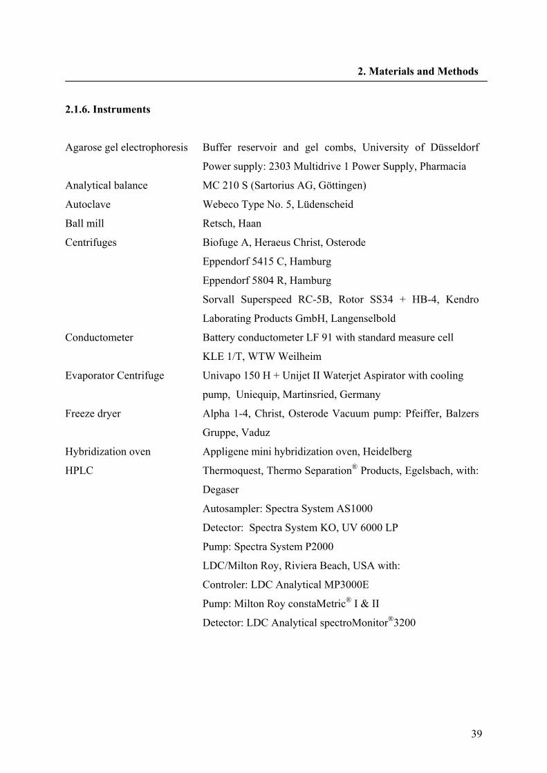
39
2. Materials and Methods
2.1.6. Instruments
Agarose gel electrophoresis Buffer reservoir and gel combs, University of Düsseldorf
Power supply: 2303 Multidrive 1 Power Supply, Pharmacia
Analytical balance MC 210 S (Sartorius AG, Göttingen)
Autoclave Webeco Type No. 5, Lüdenscheid
Ball mill Retsch, Haan
Centrifuges Biofuge A, Heraeus Christ, Osterode
Eppendorf 5415 C, Hamburg
Eppendorf 5804 R, Hamburg
Sorvall Superspeed RC-5B, Rotor SS34 + HB-4, Kendro
Laborating Products GmbH, Langenselbold
Conductometer Battery conductometer LF 91 with standard measure cell
KLE 1/T, WTW Weilheim
Evaporator Centrifuge Univapo 150 H + Unijet II Waterjet Aspirator with cooling
pump, Uniequip, Martinsried, Germany
Freeze dryer Alpha 1-4, Christ, Osterode Vacuum pump: Pfeiffer, Balzers
Gruppe, Vaduz
Hybridization oven Appligene mini hybridization oven, Heidelberg
HPLC Thermoquest, Thermo Separation® Products, Egelsbach, with:
Degaser
Autosampler: Spectra System AS1000
Detector: Spectra System KO, UV 6000 LP
Pump: Spectra System P2000
LDC/Milton Roy, Riviera Beach, USA with:
Controler: LDC Analytical MP3000E
Pump: Milton Roy constaMetric® I & II
Detector: LDC Analytical spectroMonitor®3200
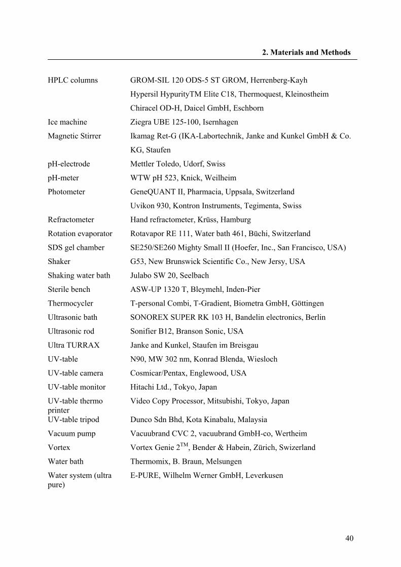
40
2. Materials and Methods
HPLC columns GROM-SIL 120 ODS-5 ST GROM, Herrenberg-Kayh
Hypersil HypurityTM Elite C18, Thermoquest, Kleinostheim
Chiracel OD-H, Daicel GmbH, Eschborn
Ice machine Ziegra UBE 125-100, Isernhagen
Magnetic Stirrer Ikamag Ret-G (IKA-Labortechnik, Janke and Kunkel GmbH & Co.
KG, Staufen
pH-electrode Mettler Toledo, Udorf, Swiss
pH-meter WTW pH 523, Knick, Weilheim
Photometer GeneQUANT II, Pharmacia, Uppsala, Switzerland
Uvikon 930, Kontron Instruments, Tegimenta, Swiss
Refractometer Hand refractometer, Krüss, Hamburg
Rotation evaporator Rotavapor RE 111, Water bath 461, Büchi, Switzerland
SDS gel chamber SE250/SE260 Mighty Small II (Hoefer, Inc., San Francisco, USA)
Shaker G53, New Brunswick Scientific Co., New Jersy, USA
Shaking water bath Julabo SW 20, Seelbach
Sterile bench ASW-UP 1320 T, Bleymehl, Inden-Pier
Thermocycler T-personal Combi, T-Gradient, Biometra GmbH, Göttingen
Ultrasonic bath SONOREX SUPER RK 103 H, Bandelin electronics, Berlin
Ultrasonic rod Sonifier B12, Branson Sonic, USA
Ultra TURRAX Janke and Kunkel, Staufen im Breisgau
UV-table N90, MW 302 nm, Konrad Blenda, Wiesloch
UV-table camera Cosmicar/Pentax, Englewood, USA
UV-table monitor Hitachi Ltd., Tokyo, Japan
UV-table thermo printer
Video Copy Processor, Mitsubishi, Tokyo, Japan
UV-table tripod Dunco Sdn Bhd, Kota Kinabalu, Malaysia
Vacuum pump Vacuubrand CVC 2, vacuubrand GmbH-co, Wertheim
Vortex Vortex Genie 2TM, Bender & Habein, Zürich, Swizerland
Water bath Thermomix, B. Braun, Melsungen
Water system (ultra pure)
E-PURE, Wilhelm Werner GmbH, Leverkusen
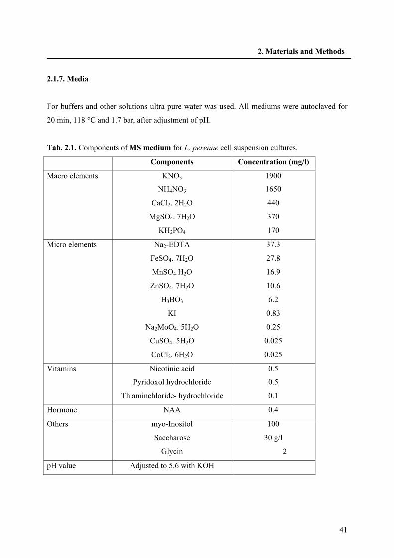
41
2. Materials and Methods
2.1.7. Media
For buffers and other solutions ultra pure water was used. All mediums were autoclaved for
20 min, 118 °C and 1.7 bar, after adjustment of pH.
Tab. 2.1. Components of MS medium for L. perenne cell suspension cultures.
Components Concentration (mg/l)
Macro elements KNO3
NH4NO3
CaCl2. 2H2O
MgSO4. 7H2O
KH2PO4
1900
1650
440
370
170
Micro elements Na2-EDTA
FeSO4. 7H2O
MnSO4.H2O
ZnSO4. 7H2O
H3BO3
KI
Na2MoO4. 5H2O
CuSO4. 5H2O
CoCl2. 6H2O
37.3
27.8
16.9
10.6
6.2
0.83
0.25
0.025
0.025
Vitamins Nicotinic acid
Pyridoxol hydrochloride
Thiaminchloride- hydrochloride
0.5
0.5
0.1
Hormone NAA 0.4
Others myo-Inositol
Saccharose
Glycin
100
30 g/l
2
pH value Adjusted to 5.6 with KOH
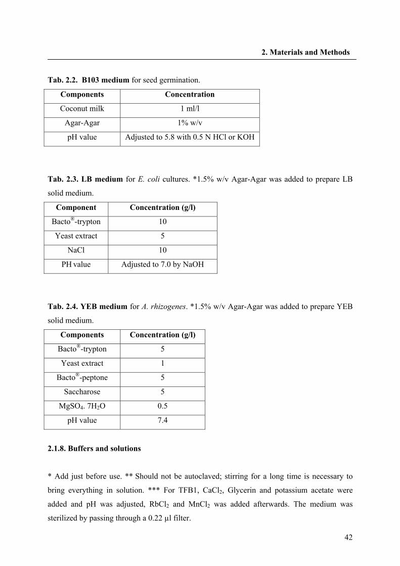
42
2. Materials and Methods
Tab. 2.2. B103 medium for seed germination.
Components Concentration
Coconut milk 1 ml/l
Agar-Agar 1% w/v
pH value Adjusted to 5.8 with 0.5 N HCl or KOH
Tab. 2.3. LB medium for E. coli cultures. *1.5% w/v Agar-Agar was added to prepare LB
solid medium.
Component Concentration (g/l)
Bacto®-trypton 10
Yeast extract 5
NaCl 10
PH value Adjusted to 7.0 by NaOH
Tab. 2.4. YEB medium for A. rhizogenes. *1.5% w/v Agar-Agar was added to prepare YEB
solid medium.
Components Concentration (g/l)
Bacto®-trypton 5
Yeast extract 1
Bacto®-peptone 5
Saccharose 5
MgSO4. 7H2O 0.5
pH value 7.4
2.1.8. Buffers and solutions
* Add just before use. ** Should not be autoclaved; stirring for a long time is necessary to
bring everything in solution. *** For TFB1, CaCl2, Glycerin and potassium acetate were
added and pH was adjusted, RbCl2 and MnCl2 was added afterwards. The medium was
sterilized by passing through a 0.22 µl filter.
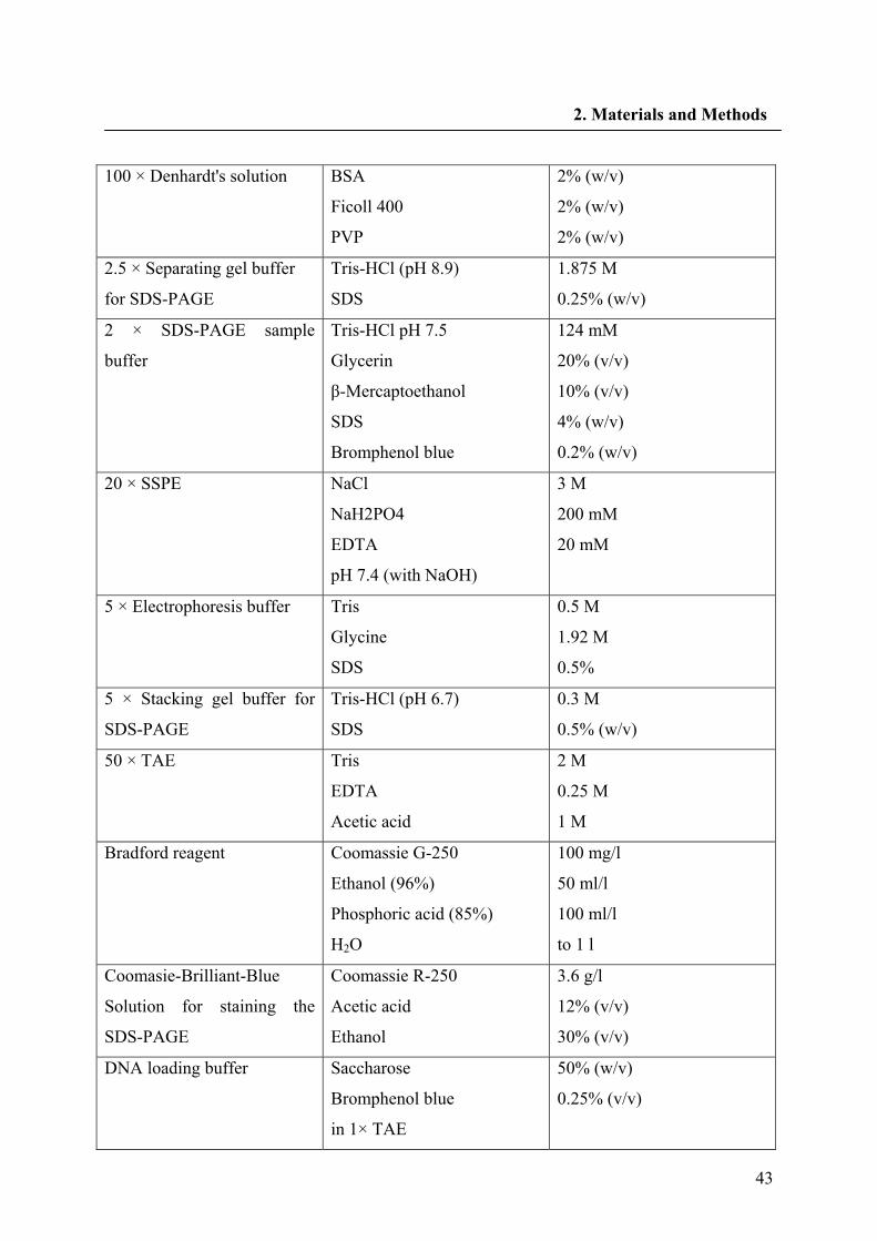
43
2. Materials and Methods
100 × Denhardt's solution BSA
Ficoll 400
PVP
2% (w/v)
2% (w/v)
2% (w/v)
2.5 × Separating gel buffer
for SDS-PAGE
Tris-HCl (pH 8.9)
SDS
1.875 M
0.25% (w/v)
2 × SDS-PAGE sample
buffer
Tris-HCl pH 7.5
Glycerin
β-Mercaptoethanol
SDS
Bromphenol blue
124 mM
20% (v/v)
10% (v/v)
4% (w/v)
0.2% (w/v)
20 × SSPE
NaCl
NaH2PO4
EDTA
pH 7.4 (with NaOH)
3 M
200 mM
20 mM
5 × Electrophoresis buffer Tris
Glycine
SDS
0.5 M
1.92 M
0.5%
5 × Stacking gel buffer for
SDS-PAGE
Tris-HCl (pH 6.7)
SDS
0.3 M
0.5% (w/v)
50 × TAE Tris
EDTA
Acetic acid
2 M
0.25 M
1 M
Bradford reagent Coomassie G-250
Ethanol (96%)
Phosphoric acid (85%)
H2O
100 mg/l
50 ml/l
100 ml/l
to 1 l
Coomasie-Brilliant-Blue
Solution for staining the
SDS-PAGE
Coomassie R-250
Acetic acid
Ethanol
3.6 g/l
12% (v/v)
30% (v/v)
DNA loading buffer Saccharose
Bromphenol blue
in 1× TAE
50% (w/v)
0.25% (v/v)
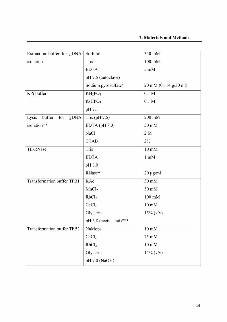
44
2. Materials and Methods
Extraction buffer for gDNA
isolation
Sorbitol
Tris
EDTA
pH 7.5 (autoclave)
Sodium pyrosulfate*
350 mM
100 mM
5 mM
20 mM (0.114 g/30 ml)
KPi buffer KH2PO4
K2HPO4
pH 7.1
0.1 M
0.1 M
Lysis buffer for gDNA
isolation**
Tris (pH 7.5)
EDTA (pH 8.0)
NaCl
CTAB
200 mM
50 mM
2 M
2%
TE-RNase Tris
EDTA
pH 8.0
RNase*
10 mM
1 mM
20 µg/ml
Transformation buffer TFB1 KAc
MnCl2
RbCl2
CaCl2
Glycerin
pH 5.8 (acetic acid)***
30 mM
50 mM
100 mM
10 mM
15% (v/v)
Transformation buffer TFB2 NaMops
CaCl2
RbCl2
Glycerin
pH 7.0 (NaOH)
10 mM
75 mM
10 mM
15% (v/v)
45
2. Materials and Methods
2.2. Methods
2.2.1. Plant materials
Flax (L. usitatissimum L.) plants (var. Flanders) were harvested 9 weeks after sowing the
seeds (at the early capsule stage), from the botanical garden, University of Duesseldorf,
Germany, in summer 2006 and 2007. Roots, stems, leaves and young immature seeds were
separated for the RNA isolation. The plant material was immediately frozen in liquid nitrogen
after harvesting and stored at -80 °C prior to analysis.
L. perenne Himmelszelt seeds were purchased (Jelitto, Schwarmstedt, Germany) and
germinated under sterile conditions on B103 medium at 25 °C in the dark. Shoot cultures
were established from seedlings on MS-medium (Murashige and Skoog 1962) without
hormone and incubated under permanent light. Single seedlings were used to initiate calli and,
subsequently, suspension cultures in MS-medium. Cells (5 g fresh weight) were transferred
every 7 days into 50 ml of fresh medium in 300 ml Erlenmeyer flasks and incubated on a
gyratory shaker at 120 rpm in the dark at 25 °C.
2.2.2. Isolation of RNA
RNA was extracted from the L. usitatissimum plant or a four day old suspension culture of L.
perenne by using the RNeasy® Plant Mini Kit.
2.2.3. First strand cDNA synthesis
cDNA of L. perenne was synthesized with the AMV cDNA synthesis kit. 2 µg RNA in the
final volume of 7 µl (H2O) was heated at 65 °C for 15 min and cooled on ice-water. 2 µl of 10
× reaction buffer, 4 µl MgCl2 (25 mM), 2 µl dNTP (10 mM), 2 µl Oligo p(dT)15 primer (from
AMV 1st strand cDNA synthesis kit), 1 µl RNase inhibitor, 0.8 µl AMV reverse transcriptase
and 0.8 µl gelatin in a final volume of 13 µl with H2O were mixed. The master mix was added
to the prepared RNA and incubated in a PCR cycler at 25 °C for 10 min, 42 °C for 60 min, 99
°C for 5 min, eventually cooled upto 20 °C.
46
2. Materials and Methods
cDNA from L. usitatissimum was synthesized with M-Mul V reverse transcriptase as follows:
2 µg RNA were mixed with 1 µl LppLR-AAP-3′ (10 pmol/µl) and H2O until 12.5 µl. The
mixture was cooled on ice-water after 5 min incubation at 70 °C (for 3′- and 5′-RACE
experiment incubation at 70 °C was extended until 10 min). 4 µl of 5 × reaction buffer (from
Revert Aid M-Mul V RT, Fermentas), 2 µl dNTP (10 mM) and 0.5 µl RNase inhibitor was
added to the RNA mixture. After 5 min preincubation of the mixture at 37 °C, 1 µl M-Mul V
RT (200 U) was added. The mixture was incubated at 42 °C and 70 °C for 60 and 10 min. For
3′-RACE the synthesized cDNA was mixed with 1 µl of RNase H. After 20 min incubation at
37 °C, the cDNA was purified with the PCR Purification Kit (QIAgen).
For 5′-RACE, 2 µg RNA of L. usitatissimum or L. perenne was mixed with 1 µl of
LuPLRGSP1-5′ (2.5 pmol/µl) or LpPLRGSP1-5′ (2.5 pmol/µl). H2O was added to a final
volume of 15.5 µl. This was added to the Master mix with M-Mul V RT containing 5 µl of 5
× reaction buffer to give a final volume of 24 µl. The synthesized cDNA was briefly
centrifuged and after 2 min preincubation at 37 °C 1 µl RNase H + 1 µl RNase T1 was added.
10 µl of the purified cDNA was used for tailing in combination with 5 µl of 5 × tailing buffer
and 2.5 µl dCTP (2.5 mM) in a final volume of 24 µl. After 3 min incubation at 94 °C,
cooling on ice and brief centrifugation, 1 µl TdT enzyme was added. The mixture was
incubated at 37 °C and 65 °C each for 10 min.
2.2.4. PCR components and programs
Polymerase chain reaction (PCR) was used to amplify the synthesized cDNA. All PCR sets
were performed according to Tab. 2.5. and Tab. 2.6. unless otherwise mentioned. As negative
control 2 µl sterile water was used instead of DNA.
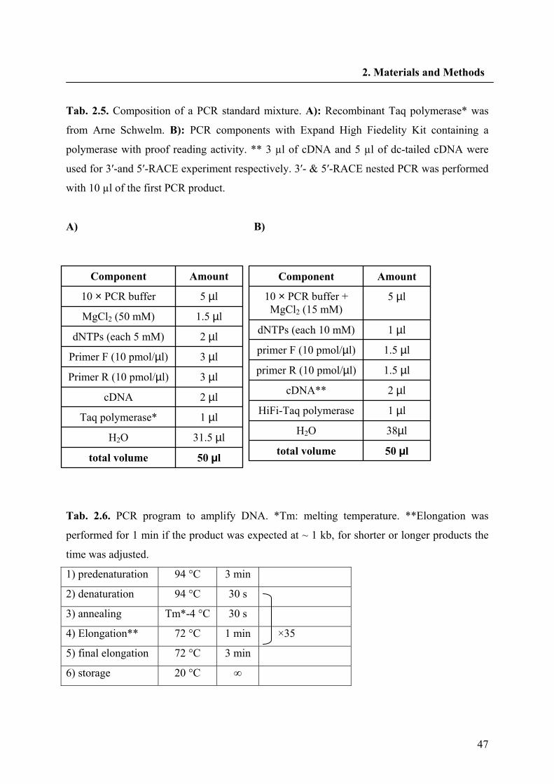
47
2. Materials and Methods
Tab. 2.5. Composition of a PCR standard mixture. A): Recombinant Taq polymerase* was
from Arne Schwelm. B): PCR components with Expand High Fiedelity Kit containing a
polymerase with proof reading activity. ** 3 µl of cDNA and 5 µl of dc-tailed cDNA were
used for 3′-and 5′-RACE experiment respectively. 3′- & 5′-RACE nested PCR was performed
with 10 µl of the first PCR product.
A) B)
Tab. 2.6. PCR program to amplify DNA. *Tm: melting temperature. **Elongation was
performed for 1 min if the product was expected at ~ 1 kb, for shorter or longer products the
time was adjusted.
1) predenaturation 94 °C 3 min
2) denaturation 94 °C 30 s
3) annealing Tm*-4 °C 30 s
4) Elongation** 72 °C 1 min ×35
5) final elongation 72 °C 3 min
6) storage 20 °C ∞
50 µl total volume 31.5 µl H2O
1 µl Taq polymerase* 2 µl cDNA 3 µl Primer R (10 pmol/µl)
3 µl Primer F (10 pmol/µl)
2 µl dNTPs (each 5 mM)
1.5 µl MgCl2 (50 mM) 5 µl 10 × PCR buffer
Amount Component
50 µl total volume
38µl H2O
1 µl HiFi-Taq polymerase
2 µl cDNA**
1.5 µl primer R (10 pmol/µl)
1.5 µl primer F (10 pmol/µl)
1 µl dNTPs (each 10 mM)
5 µl 10 × PCR buffer + MgCl2 (15 mM)
Amount Component
48
2. Materials and Methods
2.2.5. Cloning of a partial cDNA sequence of L. usitatissimum and L. perenne
Degenerated primers used to clone a first fragment of a PLR cDNA from L. usitatssimum and
L. perenne were designed based on a multiple sequence alignment (ClustalW, version 1.82) of
the following amino acid sequences: PLR-Fi1 (GenBank accession number U81158), PLR-
Tp1 (GenBank accession number AF242503), PLR-Tp2 (GenBank accession number
AF242504), PLR-Tp3 (GenBank accession number AF242505), PLR-Tp4 (GenBank
accession number AF242506), PLR-Th1 (GenBank accession number AF242501), PLR-Th2
(GenBank accession number AF242502), PCBER-Fi-2 (GenBank accession number
AF242492), PCBER-Pt1 (GenBank accession number AF242490), IFR-Mt1 (GenBank
accession number AF277052), IFR-Ms1 (GenBank accession number U17436) and IFR-Ps1
(GenBank accession number S72472). The resulting primers were LPLRdegF1 and LPLR6R
(Chapter 2.1.1). They were used to amplify a first PLR fragment by using a touch down PCR
with annealing temperatures ranging from 46-56 °C. LPLR4F and LPLR6R primer pairs
were used for a nested PCR.
2.2.6. Generation of full-length cDNAs
RACE experiments were used for rapid amplification of the 5′- and 3′- end of the obtained
cDNA fragment. cDNA was synthesized and purified using the 5′-RACE system version 2.0
(Invitrogene) (Chapter 2.2.5). Five microliters of the dC-tailed cDNA was next used as the
template in a 50 μl PCR reaction by using HiFi-Taq polymerase according to the
manufacturer with the gene specific primer LpPLR-GSP2-5′ and the adaptor primer: LpPLR-
AAP for L. perenne and LuPLR-GSP2-5′ and the adaptor primer: LpPLR-AAP for L.
usitatissimum. PCR amplification was carried out as described in Tab. 2.6 except for an
annealing temperature at 60 °C. A nested PCR was performed with LpPLR-GSP3-5′ and
LpPLR-AUAP for L perenne and LuPLR-GSP3-5′ and LpPLR-AUAP as gene specific
primers for L. usitatissimum. The reverse transcription for the 3′- end of the cDNA was the
same as 5′-RACE with the poly(dT) primer LpPLR-AAP-3′. The cDNA was amplified with
the LpPLR-GSP1-3′ primer and LpPLRAUAP for L. perenne and LuPLR-GSP1-3′ primer
and LpPLRAUAP for L. usitatissimum as mentioned above. The nested PCR was performed
with the gene specific primers LpPLR-GSP2-3′ and LpPLRAUAP for L. perenne and LuPLR-
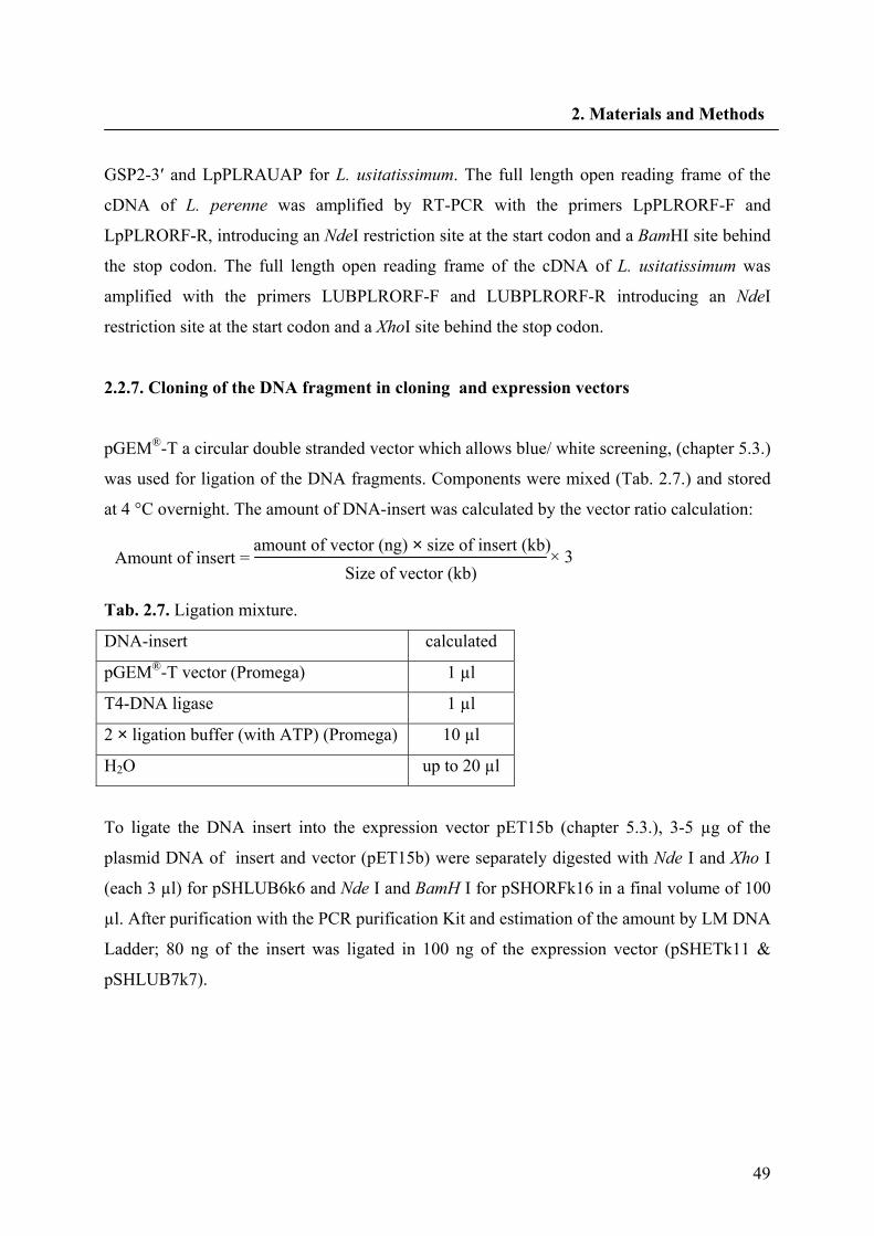
49
2. Materials and Methods
GSP2-3′ and LpPLRAUAP for L. usitatissimum. The full length open reading frame of the
cDNA of L. perenne was amplified by RT-PCR with the primers LpPLRORF-F and
LpPLRORF-R, introducing an NdeI restriction site at the start codon and a BamHI site behind
the stop codon. The full length open reading frame of the cDNA of L. usitatissimum was
amplified with the primers LUBPLRORF-F and LUBPLRORF-R introducing an NdeI
restriction site at the start codon and a XhoI site behind the stop codon.
2.2.7. Cloning of the DNA fragment in cloning and expression vectors
pGEM®-T a circular double stranded vector which allows blue/ white screening, (chapter 5.3.)
was used for ligation of the DNA fragments. Components were mixed (Tab. 2.7.) and stored
at 4 °C overnight. The amount of DNA-insert was calculated by the vector ratio calculation:
Tab. 2.7. Ligation mixture.
DNA-insert calculated
pGEM®-T vector (Promega) 1 µl
T4-DNA ligase 1 µl
2 × ligation buffer (with ATP) (Promega) 10 µl
H2O up to 20 µl
To ligate the DNA insert into the expression vector pET15b (chapter 5.3.), 3-5 µg of the
plasmid DNA of insert and vector (pET15b) were separately digested with Nde I and Xho I
(each 3 µl) for pSHLUB6k6 and Nde I and BamH I for pSHORFk16 in a final volume of 100
µl. After purification with the PCR purification Kit and estimation of the amount by LM DNA
Ladder; 80 ng of the insert was ligated in 100 ng of the expression vector (pSHETk11 &
pSHLUB7k7).
amount of vector (ng) × size of insert (kb) Size of vector (kb)
× 3 Amount of insert =
50
2. Materials and Methods
2.2.8. Preparation of competent bacteria
E. coli DH5α bacteria were transformed to LB-Agar medium and incubated overnight at 37
°C. 5 ml LB medium was inoculated with a single colony and incubated overnight at 37 °C.
The bacteria were transferred to 100 ml LB medium at 37 °C and grown until an OD600 =0.6
was reached. After 15 min incubation on ice, the cells were transferred to a prechilled 50 ml
centrifuge tube and centrifuged for 10 min at 4 °C, 3000 rpm. The pellet was dried by
removing the supernatant and resuspended in 30 ml TFBI buffer (1/3 of the original volume).
After 10 min incubation on ice the suspension was centrifuged 10 min at 4 °C, 3000 rpm. The
supernatant was removed and the dried pellet was resuspended in 4 ml TFBII buffer (1/3 of
the original volume). 200 µl of bacterial aliquets were dispensed in precooled eppendorf,
frozen in liquid nitrogen and stored at -80 °C (modified according to Hanahan 1983).
.
2.2.9. Transformation of E. coli
The ligation mixture was added to 200 µl competent cell (DH5α or BL21) which was thawed
on ice. The mixture was incubated on ice for 30 min (mixed each 10 min in between). Cells
were incubated at 42 °C in a water bath for 90 s and immediately cooled on ice. 800 µl LB
was added and the cells were incubated at 37 °C for 45 min (mixed in between). 100 µl of the
bacteria was plated to an LB indicator agar for a blue/white selection (100 µg/ml ampicillin,
125 µg/ml X-Gal and 0.5 µM IPTG). The remaining transformation mixture was sedimented
at 7000 rpm for 1 min, resuspended in 100 µl supernatant and plated on a separate LB
indicator agar plate. The agar plates were incubated at 37 °C for 12-14 h.
2.2.10. Preparation of Plasmid DNA
20 white colonies grown on LB indicator agar were transferred to 3 ml LB (100 µg/ml
ampicillin) and incubated at 37 °C overnight. 500 µl of the bacterial suspension was mixed
with 500 µl of 60% glycerin in LB for long term storage at -80 °C. The remaining suspension
was used for plasmid DNA preparation.
51
2. Materials and Methods
2.2.10.1. Mini plasmid preparation with the TENS reagents
For identification of positive clones, plasmid DNAs were isolated by an alkaline lysis method
according to Zhou et al. (1990). 1.5-2 ml E. coli overnight cultures were centrifuged at 13000
rpm for 2 min. The supernatant was decanted (50-100 µl was remained). The bacteria were
resuspended in the remaining media. 300 µl TENS solution (62.5 µl 1 M Tris pH 8.0, 12.5 µl
0.5 M EDTA pH 8.0, 4.456 ml H2O, 1.563 ml 0.4 N NaOH and 156.3 µl 20% SDS for 20
preparations) was added. The mixture was vigorously mixed for 2-3 s and incubated for 10-30
min on ice. After addition of 150 µl 3 M potassium acetate solution (pH 5.2 by acetic acid),
the reaction mixture was vortexed for 2-3 s and incubated for 10 min on ice. After
centrifugation at 13000 rpm for 5 min, the supernatant was transferred to a new tube and
mixed with 900 µl absolute ethanol. The mixture was centrifuged (13000 rpm 3-4 min) to
sediment the plasmid DNA and the supernatant was removed. The pellet was washed with
300-500 µl 70% ethanol and centrifuged at 13000 rpm for 3-4 min. The pellet was dried for a
few min and resolved in 70 µl TE-RNase buffer (chapter 2.1.8).
2.2.10.2. Plasmid preparation by using QIAgen Miniprep kit
Plasmid DNAs which were used for transformation, cloning and sequencing were prepared by
using the QIAprep® Spin Miniprep Kit according to the manufacturer. The kit is based on a
spin column technology. The DNA adsorbs to the silica membrane in the presence of “high
salt” conditions while concomitants pass through the column. Impurities are efficiently
washed away and the pure DNA is eluted with Tris buffer.
2.2.11. Restriction hydrolysis of plasmid DNA
5 µl plasmid DNA were mixed with 1 µl restriction enzyme (Apa I, Pst I when pGEM®-T
vector was used), 2 µl 10 × reaction buffer until the final volume of 20 µl with H2O. The
mixture was incubated at 37 °C for 2 h. The hydrolysis result was analyzed by agarose gel
electrophoresis.
52
2. Materials and Methods
2.2.12. Agarose gel electrophoresis
For DNA fragments > 1 kb and ≤ 1 kb, 0.8% and 1.5% (w/v) agarose in 1 × TAE buffer was
prepared, respectively. DNA loading buffer (1/10 of the DNA sample) was added to the DNA
and loaded on the gel. 1 kb Plus DNA Ladder was used to compare the size of the DNA. The
gels were run at 80-90 V in 1 × TAE until the blue marker was migrated 2/3 of the gel. The
gels were stained in ethidium bromide solution (5.2 µM ethidium bromide in 1 × TAE) for
20-30 min and photographed under UV light.
2.2.13. Determination of the purity and concentration of DNA and RNA
Low DNA MassTM Ladder and 1% Low Melting Agarose were used to estimate DNA
amounts for ligation. DNA and RNA concentration and purity was also determined
photometrically at 260 and 280 nm by GeneQuant II.
2.2.14. Sequence analysis
1-2 µg of the plasmid DNA was dried and sent for sequencing to MWG-BIOTECH AG in
Martinsried or 100 ng/µl (20 µl) DNA plasmid was sent to Macrogen (Gasan-dong,
Geumcheon-gu, Seoul, Korea). The sequence information was verified using Blast x at
http://www.ncbi.nlm.nih.gov/blast/.
2. 2.15. Southern blotting
2.2.15.1. Isolation of genomic DNA
Genomic DNA was isolated with the DNeasy® Plant Mini Kit according to the manufacturer.
For the genomic southern, gDNA was isolated by the CTAB method as follows: 3-7 g young
tissue (for suspension culture day 3) was frozen in liquid nitrogen after harvesting. The
material was ground to a fine powder with a precooled mortar and pistil. The powder was
filled in a 50 ml centrifuge tube and 25 ml ice-cold extraction buffer (chapter 2.1.8) was
added. The material was broken up on ice with an Ultraturrax (19000 rpm, thin pin) for 60 s.
53
2. Materials and Methods
The supernatant was separated and the pellet was resuspended in 2.25 ml extraction buffer
and 3.15 ml lysis buffer by using a rod of glass (chapter 2.1.8) consequently. After addition
of 1.05 ml 0.5% sarcosyl and vortexing for 10 s, the mixture was incubated at 65 °C for 90
min. 7.5 ml chloroform/isoamyl (24:1) alcohol was added and mixed. The hydrous
supernatant (about 6 ml) was transferred to a 12 ml centrifuge tube after 15 min centrifugation
at 5000 rpm. 6 ml isopropanol was added and the tube was inverted with caution until the
DNA was observed as a bundle. The mixture was centrifuged two times at 5000 rpm for 3
min and the supernatant was pipetted off. The DNA pellet was dried for 5-10 min and
resolved in 300-600 µl TE-RNAse at 65 °C for about 20 min.
2.2.15.2. Restriction hydrolysis of gDNA and electrophoresis
15 µl gDNA was mixed with 4 µl of the following restriction enzymes: EcoR I, EcoR V, Hind
III, Xba I and Bgl II separately and 15 µl of the corresponding buffers in a final volume of 150
µl with H2O. The mixtures were incubated at 37 °C for 4-6 h. 15 µl potassium phosphate
solution (3 M, pH 5.2) and 150 µl isopropanol were added to the 150 µl reaction mixture and
mixed by flicking. After centrifugation (13000 rpm, 2 min) the pellets were dried for 10 min
and resolved in 30 µl Tris buffer (10 mM, pH 8.0) and incubated at 65 °C for 30 min. After
gel electrophoresis the gel was photographed in the presence of a fluorescent ruler and the 1
kb Plus Ladder was cut from the gel.
2.2.15.3. Blotting
To denature the DNA, the gel was shaken in 1% HCl solution for 10-15 min (the marker
changed from blue to a dark green color). After rinsing with millipore water, the gel was
soaked and was shaken in NaOH solution (0.4 N) for 20 min. A plastic box was filled with
NaOH (0.4 M), and a glass plate was laid upon the box. Two 3 mm Whatman papers were cut
in pieces being longer than the glass plate, soaked in NaOH and laid upon the plate of glass
with its ends in the NaOH. The air bubbles in between the papers and plate were removed.
The gel was turned upside down and laid upon the paper. After rinsing few drops of NaOH on
the gel, the nylon membrane which was cut in the same size as the gel, was laid upon the gel.
Parafilm was used to form a barrier around the membrane, that the only route the moisture
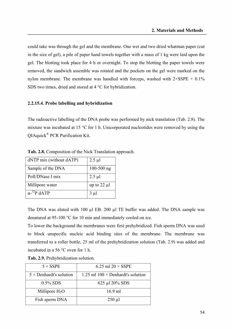
54
2. Materials and Methods
could take was through the gel and the membrane. One wet and two dried whatman paper (cut
in the size of gel), a pile of paper hand towels together with a mass of 1 kg were laid upon the
gel. The blotting took place for 4 h or overnight. To stop the blotting the paper towels were
removed, the sandwich assemble was rotated and the pockets on the gel were marked on the
nylon membrane. The membrane was handled with forceps, washed with 2×SSPE + 0.1%
SDS two times, dried and stored at 4 °C for hybridization.
2.2.15.4. Probe labelling and hybridization
The radioactive labelling of the DNA probe was performed by nick translation (Tab. 2.8). The
mixture was incubated at 15 °C for 1 h. Unicorporated nucleotides were removed by using the
QIAquick® PCR Purification Kit.
Tab. 2.8. Composition of the Nick Translation approach.
dNTP mix (without dATP) 2.5 µl
Sample of the DNA 100-500 ng
PolI/DNase I mix 2.5 µl
Millipore water up to 22 µl
α-32P dATP 3 µl
The DNA was eluted with 100 µl EB. 200 µl TE buffer was added. The DNA sample was
denatured at 95-100 °C for 10 min and immediately cooled on ice.
To lower the background the membranes were first prehybridized. Fish sperm DNA was used
to block unspecific nucleic acid binding sites of the membrane. The membrane was
transferred to a roller bottle, 25 ml of the prehybridization solution (Tab. 2.9) was added and
incubated in a 56 °C oven for 1 h.
Tab. 2.9. Prehybridization solution.
5 × SSPE 6.25 ml 20 × SSPE
5 × Denhardt's solution 1.25 ml 100 × Denhardt's solution
0.5% SDS 625 µl 20% SDS
Millipore H2O 16.9 ml
Fish sperm DNA 250 µl
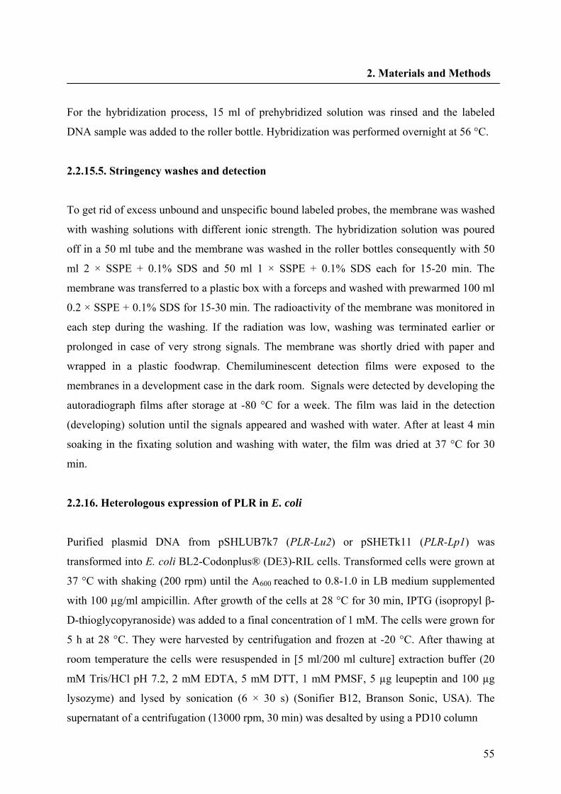
55
2. Materials and Methods
For the hybridization process, 15 ml of prehybridized solution was rinsed and the labeled
DNA sample was added to the roller bottle. Hybridization was performed overnight at 56 °C.
2.2.15.5. Stringency washes and detection
To get rid of excess unbound and unspecific bound labeled probes, the membrane was washed
with washing solutions with different ionic strength. The hybridization solution was poured
off in a 50 ml tube and the membrane was washed in the roller bottles consequently with 50
ml 2 × SSPE + 0.1% SDS and 50 ml 1 × SSPE + 0.1% SDS each for 15-20 min. The
membrane was transferred to a plastic box with a forceps and washed with prewarmed 100 ml
0.2 × SSPE + 0.1% SDS for 15-30 min. The radioactivity of the membrane was monitored in
each step during the washing. If the radiation was low, washing was terminated earlier or
prolonged in case of very strong signals. The membrane was shortly dried with paper and
wrapped in a plastic foodwrap. Chemiluminescent detection films were exposed to the
membranes in a development case in the dark room. Signals were detected by developing the
autoradiograph films after storage at -80 °C for a week. The film was laid in the detection
(developing) solution until the signals appeared and washed with water. After at least 4 min
soaking in the fixating solution and washing with water, the film was dried at 37 °C for 30
min.
2.2.16. Heterologous expression of PLR in E. coli
Purified plasmid DNA from pSHLUB7k7 (PLR-Lu2) or pSHETk11 (PLR-Lp1) was
transformed into E. coli BL2-Codonplus® (DE3)-RIL cells. Transformed cells were grown at
37 °C with shaking (200 rpm) until the A600 reached to 0.8-1.0 in LB medium supplemented
with 100 µg/ml ampicillin. After growth of the cells at 28 °C for 30 min, IPTG (isopropyl β-
D-thioglycopyranoside) was added to a final concentration of 1 mM. The cells were grown for
5 h at 28 °C. They were harvested by centrifugation and frozen at -20 °C. After thawing at
room temperature the cells were resuspended in [5 ml/200 ml culture] extraction buffer (20
mM Tris/HCl pH 7.2, 2 mM EDTA, 5 mM DTT, 1 mM PMSF, 5 µg leupeptin and 100 µg
lysozyme) and lysed by sonication (6 × 30 s) (Sonifier B12, Branson Sonic, USA). The
supernatant of a centrifugation (13000 rpm, 30 min) was desalted by using a PD10 column
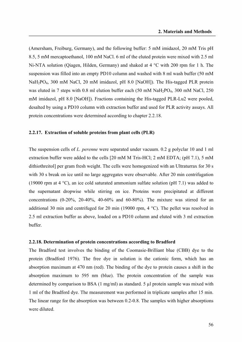
56
2. Materials and Methods
(Amersham, Freiburg, Germany), and the following buffer: 5 mM imidazol, 20 mM Tris pH
8.5, 5 mM mercaptoethanol, 100 mM NaCl. 6 ml of the eluted protein were mixed with 2.5 ml
Ni-NTA solution (Qiagen, Hilden, Germany) and shaked at 4 °C with 200 rpm for 1 h. The
suspension was filled into an empty PD10 column and washed with 8 ml wash buffer (50 mM
NaH2PO4, 300 mM NaCl, 20 mM imidazol, pH 8.0 [NaOH]). The His-tagged PLR protein
was eluted in 7 steps with 0.8 ml elution buffer each (50 mM NaH2PO4, 300 mM NaCl, 250
mM imidazol, pH 8.0 [NaOH]). Fractions containing the His-tagged PLR-Lu2 were pooled,
desalted by using a PD10 column with extraction buffer and used for PLR activity assays. All
protein concentrations were determined according to chapter 2.2.18.
2.2.17. Extraction of soluble proteins from plant cells (PLR)
The suspension cells of L. perenne were separated under vacuum. 0.2 g polyclar 10 and 1 ml
extraction buffer were added to the cells [20 mM M Tris-HCl; 2 mM EDTA; (pH 7.1), 5 mM
dithiothreitol] per gram fresh weight. The cells were homogenized with an Ultraturrax for 30 s
with 30 s break on ice until no large aggregates were observable. After 20 min centrifugation
(19000 rpm at 4 °C), an ice cold saturated ammonium sulfate solution (pH 7.1) was added to
the supernatant dropwise while stirring on ice. Proteins were precipitated at different
concentrations (0-20%, 20-40%, 40-60% and 60-80%). The mixture was stirred for an
additional 30 min and centrifuged for 20 min (19000 rpm, 4 °C). The pellet was resolved in
2.5 ml extraction buffer as above, loaded on a PD10 column and eluted with 3 ml extraction
buffer.
2.2.18. Determination of protein concentrations according to Bradford
The Bradford test involves the binding of the Coomasie-Brilliant blue (CBB) dye to the
protein (Bradford 1976). The free dye in solution is the cationic form, which has an
absorption maximum at 470 nm (red). The binding of the dye to protein causes a shift in the
absorption maximum to 595 nm (blue). The protein concentration of the sample was
determined by comparison to BSA (1 mg/ml) as standard. 5 µl protein sample was mixed with
1 ml of the Bradford dye. The measurement was performed in triplicate samples after 15 min.
The linear range for the absorption was between 0.2-0.8. The samples with higher absorptions
were diluted.
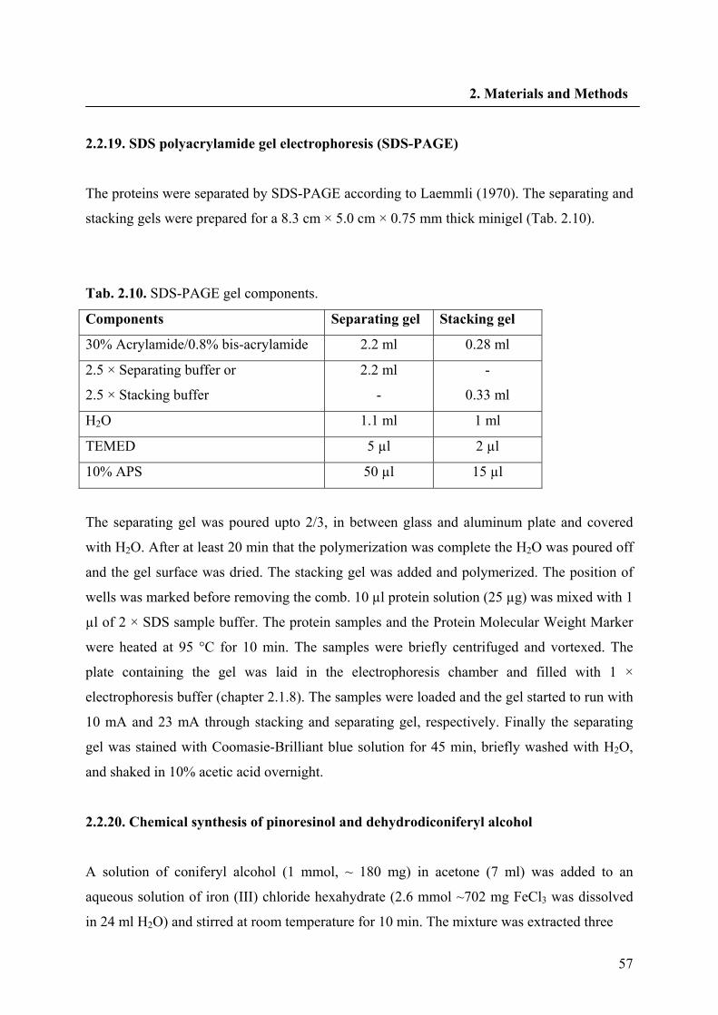
57
2. Materials and Methods
2.2.19. SDS polyacrylamide gel electrophoresis (SDS-PAGE)
The proteins were separated by SDS-PAGE according to Laemmli (1970). The separating and
stacking gels were prepared for a 8.3 cm × 5.0 cm × 0.75 mm thick minigel (Tab. 2.10).
Tab. 2.10. SDS-PAGE gel components.
Components Separating gel Stacking gel
30% Acrylamide/0.8% bis-acrylamide 2.2 ml 0.28 ml
2.5 × Separating buffer or
2.5 × Stacking buffer
2.2 ml
-
-
0.33 ml
H2O 1.1 ml 1 ml
TEMED 5 µl 2 µl
10% APS 50 µl 15 µl
The separating gel was poured upto 2/3, in between glass and aluminum plate and covered
with H2O. After at least 20 min that the polymerization was complete the H2O was poured off
and the gel surface was dried. The stacking gel was added and polymerized. The position of
wells was marked before removing the comb. 10 µl protein solution (25 µg) was mixed with 1
µl of 2 × SDS sample buffer. The protein samples and the Protein Molecular Weight Marker
were heated at 95 °C for 10 min. The samples were briefly centrifuged and vortexed. The
plate containing the gel was laid in the electrophoresis chamber and filled with 1 ×
electrophoresis buffer (chapter 2.1.8). The samples were loaded and the gel started to run with
10 mA and 23 mA through stacking and separating gel, respectively. Finally the separating
gel was stained with Coomasie-Brilliant blue solution for 45 min, briefly washed with H2O,
and shaked in 10% acetic acid overnight.
2.2.20. Chemical synthesis of pinoresinol and dehydrodiconiferyl alcohol
A solution of coniferyl alcohol (1 mmol, ~ 180 mg) in acetone (7 ml) was added to an
aqueous solution of iron (III) chloride hexahydrate (2.6 mmol ~702 mg FeCl3 was dissolved
in 24 ml H2O) and stirred at room temperature for 10 min. The mixture was extracted three
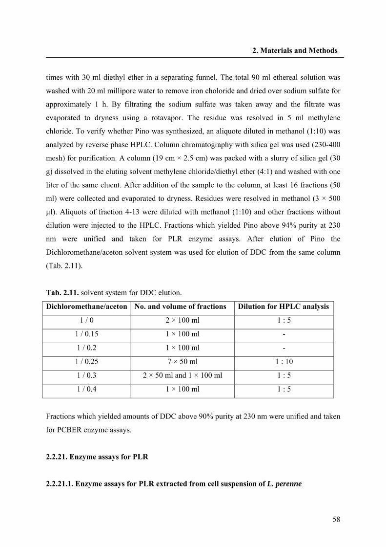
58
2. Materials and Methods
times with 30 ml diethyl ether in a separating funnel. The total 90 ml ethereal solution was
washed with 20 ml millipore water to remove iron choloride and dried over sodium sulfate for
approximately 1 h. By filtrating the sodium sulfate was taken away and the filtrate was
evaporated to dryness using a rotavapor. The residue was resolved in 5 ml methylene
chloride. To verify whether Pino was synthesized, an aliquote diluted in methanol (1:10) was
analyzed by reverse phase HPLC. Column chromatography with silica gel was used (230-400
mesh) for purification. A column (19 cm × 2.5 cm) was packed with a slurry of silica gel (30
g) dissolved in the eluting solvent methylene chloride/diethyl ether (4:1) and washed with one
liter of the same eluent. After addition of the sample to the column, at least 16 fractions (50
ml) were collected and evaporated to dryness. Residues were resolved in methanol (3 × 500
µl). Aliquots of fraction 4-13 were diluted with methanol (1:10) and other fractions without
dilution were injected to the HPLC. Fractions which yielded Pino above 94% purity at 230
nm were unified and taken for PLR enzyme assays. After elution of Pino the
Dichloromethane/aceton solvent system was used for elution of DDC from the same column
(Tab. 2.11).
Tab. 2.11. solvent system for DDC elution.
Dichloromethane/aceton No. and volume of fractions Dilution for HPLC analysis
1 / 0 2 × 100 ml 1 : 5
1 / 0.15 1 × 100 ml -
1 / 0.2 1 × 100 ml -
1 / 0.25 7 × 50 ml 1 : 10
1 / 0.3 2 × 50 ml and 1 × 100 ml 1 : 5
1 / 0.4 1 × 100 ml 1 : 5
Fractions which yielded amounts of DDC above 90% purity at 230 nm were unified and taken
for PCBER enzyme assays.
2.2.21. Enzyme assays for PLR
2.2.21.1. Enzyme assays for PLR extracted from cell suspension of L. perenne
59
2. Materials and Methods
Assays were performed in the presence of 400 µg enzyme extract, 10 µl Pino (10 mM) and 50
µl NADPH (25 mM) in 0.1 M Kpi buffer (pH 7.1) until the final volume of 500 µl. (NADPH
was added after 15 min incubation of the reaction mixture at 30 °C). After overnight
incubation the reaction was stopped and extracted with (3 × 500 µl in total) ethyl acetate.
2.2.21.2. Enzyme assays with recombinant purified PLRs
PLR eluted from PD10 column was used for the determination of enzyme activities. For PLR-
Lp1, the assay mixtures (250 μl) consisted of KPi buffer (0.1 M, pH 7.1), 0.0925 mM racemic
Pino, 2.5 mM NADPH and purified His tagged PLR-Lp1 in different concentrations (0.001,
0.005, 0.01, 0.05, 0.1, 0.5, 1, 3, 5, 7, 10, 15, 20 μg/assay). For PLR-Lu1 and PLR-Lu2 the
mixture were the same as mentioned for PLR-Lp1 except for 0.12 mM racemic Pino, 2.5 µg
purified His tagged PLR-Lu1 or 0.5 µg PLR-Lu2. Protein, buffer and Pino were preincubated
for 15 min at 30 °C. The enzyme reaction was initiated by addition of NADPH and terminated
by addition of 300 μl ethyl acetate after 3 h for PLR-Lp1. Assays for PLR-Lu1 was perfomed
in a time scale of 2, 5, 10, 15 and 20 min and for PLR-Lu2 the assays were incubated for 2, 5,
10, 15, 20, 25, 30 and 40 min, respectively. The assays were extracted with ethyl acetate
(3 × 300 μl in total). The combined ethyl acetate phases were dried under vaccum. The
residue was dissolved in 100 μl methanol and subjected to HPLC analysis.
2.2.22. RNAi constructs and transformations
2.2.22.1 Construction of hpRNAi vectors
After cloning the first piece of PLR of L. perenne, a 652-bp fragment of the coding region of
PLR-Lp1 (pSH1) was selected and amplified by PCR using the primers LPPLRhanF and
LPPLRhanR containing 2 sets of restriction sites at the 5' end. The forward primer contained
XbaI and XhoI and the reversed primer contained ClaI and KpnI restriction sites, respectively.
The PCR product was first cloned into pGEM®-T vector and sequenced (pSH2). To clone the
pSH2 in the sense orientation of a pHANNIBAL vector a restriction hydrolysis with three
enzymes (XhoI, KpnI and ApaI) were performed until a final volume of 100 µl. The mixture
was incubated at 37 °C for 4 h and purified with QIAquick® PCR Purification Kit. 2 µg of the
60
2. Materials and Methods
pHANNIBAL vector was digested with KpnI for 4 h and purified. After estimation of the
amount of digested pSH2 and pHANNIBAL, 80 ng of insert was ligated to 100 ng of vector
with the T4-DNA ligase (It should be mentioned that vector and H2O were heated at 45 °C for
5 min, cooled on ice and other components were added for ligation). The ligated DNA was
cloned in DH5α (selection was on the LB media containing ampicillin) and sequenced
(pSH3). pSH2 which was digested with XbaI, ClaI and ScaI was cloned in pSH3 (hydrolyzed
with XbaI and ClaI) in the antisense direction with the same procedure as above (pSH4).
pSH4 was digested with Not I at 37 °C for 4 h and purified to ligate into pART27 binary
vector. 3 µg of the vector was also digested with Not I. After 20 min heating at 65 °C, the
pART27 was treated with alkaline phosphatase (1 µl) in the presence of corresponding buffer,
incubated at 37 °C for 70 min and purified. pSH4 was ligated into pART27 cloned in DH5α
and sequenced (pSH5). Its notable that, selection of clones which were contained pART27
was a blue/white selection on a LB media containing spectinomycin. To prepare the control
constructs without sense and antisense PLRs, the expression cassette of the Not I hydrolyzed
pHANNIBAL was cloned into pART27 (pDI25).
2.2.22.2. Preparation of A. rhizogenes competent cells
The Agrobacteria (TR105) were streaked on an YEB agar plate from a glycerol stock and
were grown at 28 °C for app. 3 days until single colonies were appeared. 4 ml YEB broth
medium was inoculated with a single colony and incubated at 28 °C for 2 days. 100 ml YEB
medium was inoculated with the above 4 ml culture and was grown 28 °C for an additional 3-
4 h. The cultures were centrifuged at 5000 rpm in 50 ml tubes for 15 min. The pellet was
resuspended in 2 ml medium and stored in 200 µl aliquots at -80 °C.
2.2.22.3. Transformation of the competent Agrobacteria with hpRNAi constructs
100 ng-1 µg plasmid DNA was mixed with the thawed Agrobacteria and incubated on ice for
5 min. After 5 min incubation in liquid nitrogen, the mixture was incubated at 37 °C for 5
min. 800 µl YEB was added on ice to the mixture and incubated at 28 °C for 3-4 h (inverted
meanwhile). The spreaded bacteria on YEB agar plates containing spectinomycin, were
incubated at 28 °C for 3 days.
61
2. Materials and Methods
2.2.22.4. Plasmid preparation from Agrobacteria
Agrobacteria colonies were inoculated to 15 ml YEB media with spectinomycin (in 100 ml
erlenmeyer flasks) and grown for 1-2 days at 28 °C, 200 rpm. After centrifugation in a 15 ml
tube at 5000 rpm for 15 min, plasmid DNA was isolated with the QIAprep® Spin Miniprep
Kit by using double volumes of the buffers P1-P3. The DNA was eluted with 50 µl elution
buffer. 15 µl DNA was used for restriction digest with Not I.
2.2.22.5. Transformation of L. perenne shoot cultures
Transformed agrobacteria were grown on YEB medium in a 200 ml flask with 100 μg/ml
spectinomycin and 20 μM acetosyringone at 28 °C for 2 days until the OD600 was between
0.8-1.2. Medium was centrifuged in 5000 rpm for 15 min and the pellet was transformed with
a sterile scalpel to the young shoots. Shoots from in vitro cultures were wounded and infected
with the agrobacteria. After 2 days of cocultivation the explants were washed in 50 ml water
contained timentin® and incubated on hormone free McCown agar medium with 2% sugar
and 200 μg/ml timentin® in the dark. The hairy roots appeared 2 weeks after the infection.
The root segments were cut and transferred onto new medium when they were at least 25 mm
in length. Paromomycin (200 μg/ml) was used for selection of hairy roots carrying the
ihpRNAi construct with and without the PLR-Lp1 partial sequence. Hairy root clones were
kept in the dark at 25 °C and routinely subcultured every 25 days. After several subcultivation
cycles, hairy roots were transferred to liquid McCown medium. Subcultivation was performed
every 2 weeks by transferring 2 g fresh weight in liquid McCown medium (50 ml per 450 ml
jar) with 2% sugar on a rotary shaker (80 rpm).
2.2.23. Analysis of transgenic lines
2.2.23.1. Molecular analysis of transgenic lines
The DNeasy Plant Mini Kit was used to isolate total genomic DNA from 20 mg freeze dried
material from 7 day old hairy roots. The transformed status of hairy roots was verified by
PCR for the genes rolC and virC according to (Yang and Choi 2000). The PCR mixture and
62
2. Materials and Methods
program was as mentioned in Tab. 2.5 and Tab. 2.6. The annealing temperature for rolC
amplification with ROLCFOR and ROLCREV primer pairs and VIRCFOR and VIRCREV
primer pairs for virC amplification was 63 °C and 48 °C, respectively.
2.2.23.2. Detection of 35S-PLR in transgenic hairy roots
The oligonucleotide primers used for amplification of the PLR gene were 35 S and
LPPLRhanR. PCR amplification for the inserted plr gene was the same as mentioned in Tab.
2.6 with an annealing temperature at 59 °C.
2.2.23.3. Semiquantitative RT-PCR
First strand cDNA was reverse transcribed from 1 μg total RNA using Revert Aid M-MuL V
reverse transcriptase. PKFactin5′ and PKFactin3′ were used to amplify a 850 bp actin cDNA
fragment with an annealing temperature at 46 °C. Primer pairs and the PCR programme for
amplification of PLR-Lp1 cDNA from hairy roots of L. perenne were PLRORFF and
PLRORFR as mentioned in Tab. 2.5 and Tab. 2.6. DNA fragments were compared after 29
cycles of amplication. PCR products were analysed on a 0.8% agarose gel stained with
ethidium bromide.
The level of PLR-Lu1 and PLR-Lu2 in normal L.usitatissimum plant was also estimated by
semiquantitative RT-PCR. Gene specific primer pairs SemiLu1-F and SemiLu1-R were used
to amplify a PLR-Lu1 fragment and SemiLu2-F and SemiLu2-R for PLR-Lu2. DNA
fragments were compared after 26 cycles of amplication.
2.2.23.4. Preparation of protein extracts from hairy roots
Seven day old hairy roots of L. perenne H were separated from the medium. Protein was
extracted as described in chapter 2.2.17. The protein was precipitated between 0% and 60%
ammonium sulfate. Protein concentrations were determined as mentioned in chapter 2.2.18.
63
2. Materials and Methods
2.2.23.5 Enzyme assays for hairy roots
In order to determine the range of linearity of product formation PLR assays were either
conducted with 40 μg protein for different time intervals in the range from 30 to 180 min or
with different protein concentrations in the range from 0 to 100 μg protein/assay for 90 min.
Finally, specific enzyme activities of different ihpRNAi and control lines were compared with
assays containing 40 μg protein incubated for 90 min at 30 °C. PLR activity was calculated as
follows:
Aprobe: Integration level of sample peaks
Cst: Concentration of standard [mM]
Vtest: Volume of assay mixture [µl]
Ast: Integration level of standard peak
t: Incubation time [s]
Cprotein: Protein concentration [mg/ml]
Vprotein: volume of protein solution/assay [µl]
Aprobe × Cst × Vtest Ast × t
Absolute enzyme activity [nkat] =
Absolute enzyme activity [nkat]
Cprotein × Vprotein Specific enzyme activity [mkat/kg] =
64
2. Materials and Methods
2.2.24. Biochemical characterization of JusB7H
2.2.24.1. Preparation of microsomes
Five-day-old suspension cells of L. perenne H were harvested by vacuum filtration. All
further steps were carried out at 0-4 °C. Cells were homogenized by using a prechilled mortar
and pestele together with 0.2 g Polyclar 10, 0.17 g sea sand and 1 ml buffer [0.1 M Tris-HCl
(pH 7.5), 1 mM dithiothreitol, 1 mM diethyldithiocarbaminate] per gram fresh weight. The
homogenate was centrifuged at 8000 g for 20 min. The supernatant was adjusted to 50 mM
MgCl2 and stirred on ice for 30 min. The sediment containing aggregated membranes was
resuspended in buffer (as above) by a Potter and Elvehjem glass homogeniser after-
centrifugation at 48000 g for 25 min. Crude cell-free extracts for enzyme assays were
obtained by passing the resuspended pellet through a preequilibrated PD10 column using the
same buffer. Protein concentrations were determined according to chapter 2.2.18.
2.2.24.2. Determination of general parameters
pH, temperature, time and protein dependency of the JusB7H were characterized. 0.1 M Tris-
HCl buffer with the following pH values 6.0, 6.5, 7.0, 7.2, 7.5, 7.8, 8.0, 8.5 and 9.0 were
prepared to determine pH dependency. The assay condition was as mentioned in Tab. 2.12.
To determine the protein dependency, 0, 300, 500, 600, 700, 800 and 1000 µg microsome
preparations were added to the assay mixture. Enzyme assays which were stopped after 0, 15,
30, 45, 60, 75, 90, 105 and 120 min, used to determine time dependency of JusB7H. To
determine the optimum temperature for JusB7H, reactions were incubated at 0, 4.0, 20, 26,
28, 30, 32, 34, 37, 40, 50, 60 and 100 °C.
2.2.24.3. Standard enzyme assay for JusB7H
Standard assays contained 50 µM Jus B, 1 mM NADPH and 0.7 mg protein in a total volume
of 500 µl Tris-HCl (pH 7.5) with 1 mM dithiothreitol and 1 mM diethyldithiocarbaminate.
After vigorous mixing assays were routinely incubated for 75 min at 26 °C, stopped by
addition of 50 µl 6 M HCl and cooled on ice. An NADPH regenerating system consisting of
65
2. Materials and Methods
10 mM glucose-6-phosphate, 0.07 unit glucose-6-phosphate dehydrogenase and 4 mM DTT
were added to the assay, which was incubated for 5 h for the identification of the reaction
product Diph by HPLC-SPE-NMR. Stopped assays were extracted with ethyl acetate (3 × 500
µl). The combined ethyl acetate phases were dried under vaccum. The residues were
redissolved in 80µl methanol and subjected to HPLC analysis.
2.2.24.4. Determination of kinetic constants for JusB7H and NADPH
To determine the substrate (JusB) and cosubstrate (NADPH) dependency for JusB7H, their
concentrations for each enzyme assay were varied as Tab. 2.13. The determination of Km
values was the mean of at least three independent assays, each in duplicate. Km values were
calculated according to Lineweaver-Burk, Hanes-Woolf and Eadie-Scatchard equations.
Tab. 2.12. JusB7H standard assay. * To determine the optimum pH, values were in between
6-9. ** To determine protein dependency, values were in between 0-1000 µg protein.
Substance (solution) Volume
0.1 M Tris-HCl pH 7.5*
(with 1 mM DTT, 1mM DIECA)
Microsome**
100 mM NADPH (in H2O)
2.5 mM JusB (in DMSO)
x µl
y µl
5 µl
10 µl
Volume of the assay 500 µl
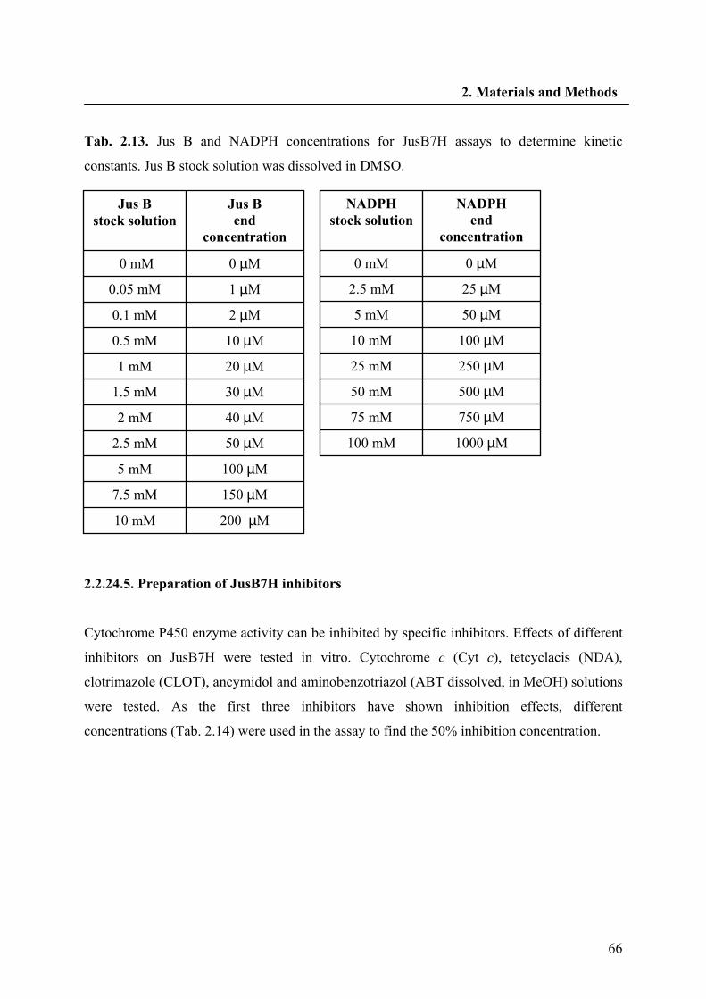
66
2. Materials and Methods
Tab. 2.13. Jus B and NADPH concentrations for JusB7H assays to determine kinetic
constants. Jus B stock solution was dissolved in DMSO.
2.2.24.5. Preparation of JusB7H inhibitors
Cytochrome P450 enzyme activity can be inhibited by specific inhibitors. Effects of different
inhibitors on JusB7H were tested in vitro. Cytochrome c (Cyt c), tetcyclacis (NDA),
clotrimazole (CLOT), ancymidol and aminobenzotriazol (ABT dissolved, in MeOH) solutions
were tested. As the first three inhibitors have shown inhibition effects, different
concentrations (Tab. 2.14) were used in the assay to find the 50% inhibition concentration.
200 µM 10 mM 150 µM 7.5 mM 100 µM 5 mM 50 µM 2.5 mM 40 µM 2 mM 30 µM 1.5 mM 20 µM 1 mM 10 µM 0.5 mM 2 µM 0.1 mM 1 µM 0.05 mM 0 µM 0 mM
Jus B end
concentration Jus B
stock solution
1000 µM 100 mM
750 µM 75 mM
500 µM 50 mM
250 µM 25 mM
100 µM 10 mM
50 µM 5 mM
25 µM 2.5 mM
0 µM0 mM
NADPH end
concentration NADPH
stock solution
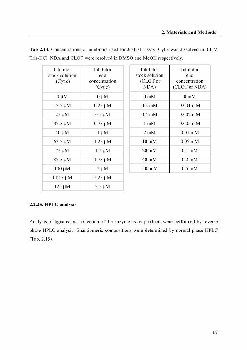
67
2. Materials and Methods
Tab 2.14. Concentrations of inhibitors used for JusB7H assay. Cyt c was dissolved in 0.1 M
Tris-HCl. NDA and CLOT were resolved in DMSO and MeOH respectively.
2.2.25. HPLC analysis
Analysis of lignans and collection of the enzyme assay products were performed by reverse
phase HPLC analysis. Enantiomeric compositions were determined by normal phase HPLC
(Tab. 2.15).
2.5 µM125 µM 2.25 µM112.5 µM
2 µM100 µM 1.75 µM87.5 µM 1.5 µM75 µM 1.25 µM62.5 µM
1 µM50 µM 0.75 µM37.5 µM 0.5 µM25 µM 0.25 µM12.5 µM
0 µM0 µM
Inhibitor end
concentration (Cyt c)
Inhibitor stock solution
(Cyt c)
0.5 mM 100 mM
0.2 mM 40 mM
0.1 mM 20 mM
0.05 mM 10 mM
0.01 mM 2 mM
0.005 mM 1 mM
0.002 mM 0.4 mM
0.001 mM 0.2 mM
0 mM 0 mM
Inhibitor end
concentration (CLOT or NDA)
Inhibitor stock solution
(CLOT or NDA)
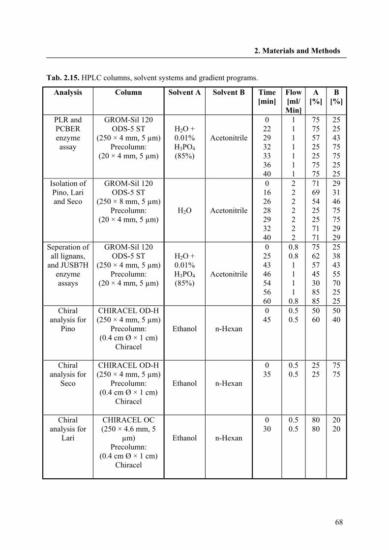
68
2. Materials and Methods
Tab. 2.15. HPLC columns, solvent systems and gradient programs.
Analysis Column Solvent A Solvent B Time [min]
Flow [ml/ Min]
A [%]
B [%]
PLR and PCBER enzyme assay
GROM-Sil 120 ODS-5 ST
(250 × 4 mm, 5 µm) Precolumn:
(20 × 4 mm, 5 µm)
H2O + 0.01% H3PO4 (85%)
Acetonitrile
0 22 29 32 33 36 40
1 1 1 1 1 1 1
75 75 57 25 25 75 75
25 25 43 75 75 25 25
Isolation of Pino, Lari and Seco
GROM-Sil 120 ODS-5 ST
(250 × 8 mm, 5 µm) Precolumn:
(20 × 4 mm, 5 µm)
H2O
Acetonitrile
0 16 26 28 29 32 40
2 2 2 2 2 2 2
71 69 54 25 25 71 71
29 31 46 75 75 29 29
Seperation of all lignans,
and JUSB7H enzyme assays
GROM-Sil 120 ODS-5 ST
(250 × 4 mm, 5 µm) Precolumn:
(20 × 4 mm, 5 µm)
H2O + 0.01% H3PO4 (85%)
Acetonitrile
0 25 43 46 54 56 60
0.8 0.8 1 1 1 1
0.8
75 62 57 45 30 85 85
25 38 43 55 70 25 25
Chiral analysis for
Pino
CHIRACEL OD-H (250 × 4 mm, 5 µm)
Precolumn: (0.4 cm Ø × 1 cm)
Chiracel
Ethanol
n-Hexan
0 45
0.5 0.5
50 60
50 40
Chiral analysis for
Seco
CHIRACEL OD-H (250 × 4 mm, 5 µm)
Precolumn: (0.4 cm Ø × 1 cm)
Chiracel
Ethanol
n-Hexan
0 35
0.5 0.5
25 25
75 75
Chiral analysis for
Lari
CHIRACEL OC (250 × 4.6 mm, 5
µm) Precolumn:
(0.4 cm Ø × 1 cm) Chiracel
Ethanol
n-Hexan
0 30
0.5 0.5
80 80
20 20
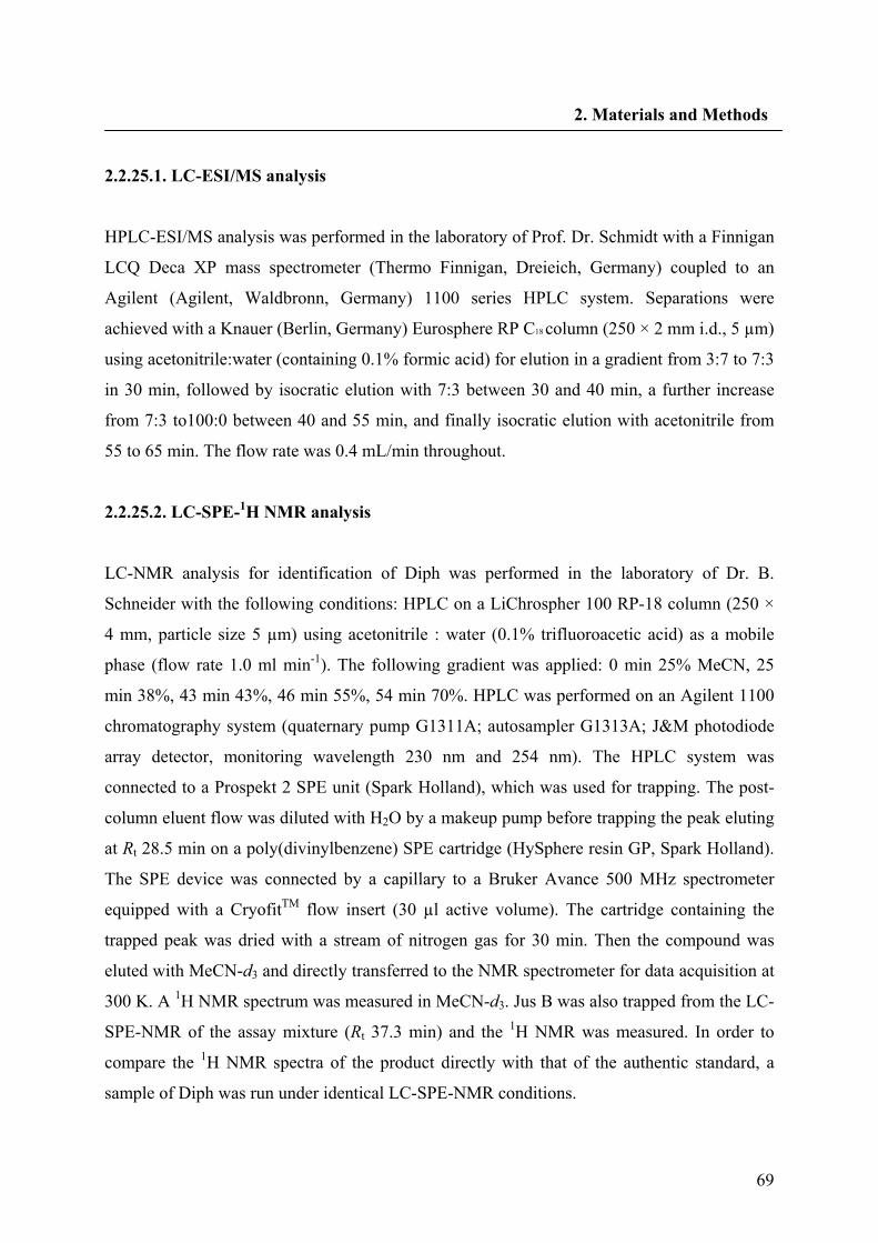
69
2. Materials and Methods
2.2.25.1. LC-ESI/MS analysis
HPLC-ESI/MS analysis was performed in the laboratory of Prof. Dr. Schmidt with a Finnigan
LCQ Deca XP mass spectrometer (Thermo Finnigan, Dreieich, Germany) coupled to an
Agilent (Agilent, Waldbronn, Germany) 1100 series HPLC system. Separations were
achieved with a Knauer (Berlin, Germany) Eurosphere RP C18 column (250 × 2 mm i.d., 5 µm)
using acetonitrile:water (containing 0.1% formic acid) for elution in a gradient from 3:7 to 7:3
in 30 min, followed by isocratic elution with 7:3 between 30 and 40 min, a further increase
from 7:3 to100:0 between 40 and 55 min, and finally isocratic elution with acetonitrile from
55 to 65 min. The flow rate was 0.4 mL/min throughout.
2.2.25.2. LC-SPE-1H NMR analysis
LC-NMR analysis for identification of Diph was performed in the laboratory of Dr. B.
Schneider with the following conditions: HPLC on a LiChrospher 100 RP-18 column (250 ×
4 mm, particle size 5 µm) using acetonitrile : water (0.1% trifluoroacetic acid) as a mobile
phase (flow rate 1.0 ml min-1). The following gradient was applied: 0 min 25% MeCN, 25
min 38%, 43 min 43%, 46 min 55%, 54 min 70%. HPLC was performed on an Agilent 1100
chromatography system (quaternary pump G1311A; autosampler G1313A; J&M photodiode
array detector, monitoring wavelength 230 nm and 254 nm). The HPLC system was
connected to a Prospekt 2 SPE unit (Spark Holland), which was used for trapping. The post-
column eluent flow was diluted with H2O by a makeup pump before trapping the peak eluting
at Rt 28.5 min on a poly(divinylbenzene) SPE cartridge (HySphere resin GP, Spark Holland).
The SPE device was connected by a capillary to a Bruker Avance 500 MHz spectrometer
equipped with a CryofitTM flow insert (30 µl active volume). The cartridge containing the
trapped peak was dried with a stream of nitrogen gas for 30 min. Then the compound was
eluted with MeCN-d3 and directly transferred to the NMR spectrometer for data acquisition at
300 K. A 1H NMR spectrum was measured in MeCN-d3. Jus B was also trapped from the LC-
SPE-NMR of the assay mixture (Rt 37.3 min) and the 1H NMR was measured. In order to
compare the 1H NMR spectra of the product directly with that of the authentic standard, a
sample of Diph was run under identical LC-SPE-NMR conditions.
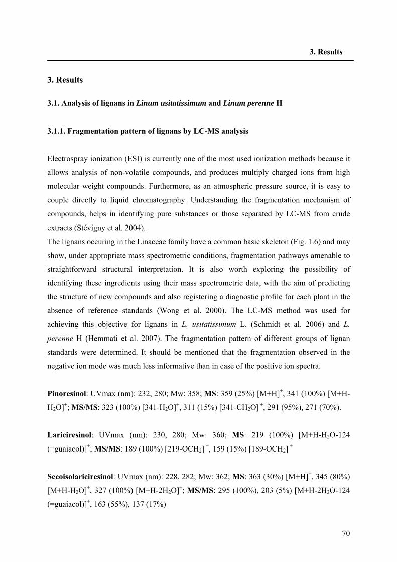
70
3. Results
3. Results
3.1. Analysis of lignans in Linum usitatissimum and Linum perenne H
3.1.1. Fragmentation pattern of lignans by LC-MS analysis
Electrospray ionization (ESI) is currently one of the most used ionization methods because it
allows analysis of non-volatile compounds, and produces multiply charged ions from high
molecular weight compounds. Furthermore, as an atmospheric pressure source, it is easy to
couple directly to liquid chromatography. Understanding the fragmentation mechanism of
compounds, helps in identifying pure substances or those separated by LC-MS from crude
extracts (Stévigny et al. 2004).
The lignans occuring in the Linaceae family have a common basic skeleton (Fig. 1.6) and may
show, under appropriate mass spectrometric conditions, fragmentation pathways amenable to
straightforward structural interpretation. It is also worth exploring the possibility of
identifying these ingredients using their mass spectrometric data, with the aim of predicting
the structure of new compounds and also registering a diagnostic profile for each plant in the
absence of reference standards (Wong et al. 2000). The LC-MS method was used for
achieving this objective for lignans in L. usitatissimum L. (Schmidt et al. 2006) and L.
perenne H (Hemmati et al. 2007). The fragmentation pattern of different groups of lignan
standards were determined. It should be mentioned that the fragmentation observed in the
negative ion mode was much less informative than in case of the positive ion spectra.
Pinoresinol: UVmax (nm): 232, 280; Mw: 358; MS: 359 (25%) [M+H]+, 341 (100%) [M+H-
H2O]+; MS/MS: 323 (100%) [341-H2O]+, 311 (15%) [341-CH2O] +, 291 (95%), 271 (70%).
Lariciresinol: UVmax (nm): 230, 280; Mw: 360; MS: 219 (100%) [M+H-H2O-124
(=guaiacol)]+; MS/MS: 189 (100%) [219-OCH2] +, 159 (15%) [189-OCH2] +
Secoisolariciresinol: UVmax (nm): 228, 282; Mw: 362; MS: 363 (30%) [M+H]+, 345 (80%)
[M+H-H2O]+, 327 (100%) [M+H-2H2O]+; MS/MS: 295 (100%), 203 (5%) [M+H-2H2O-124
(=guaiacol)]+, 163 (55%), 137 (17%)
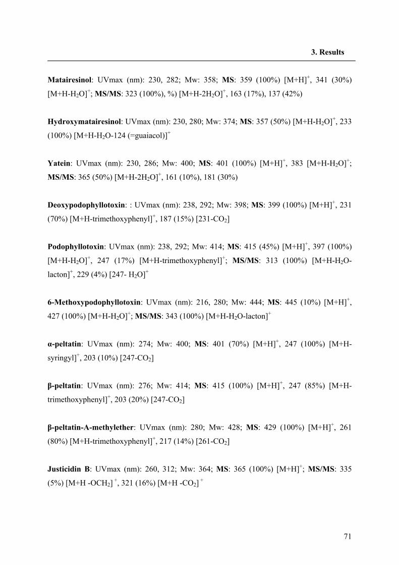
71
3. Results
Matairesinol: UVmax (nm): 230, 282; Mw: 358; MS: 359 (100%) [M+H]+, 341 (30%)
[M+H-H2O]+; MS/MS: 323 (100%), %) [M+H-2H2O]+, 163 (17%), 137 (42%)
Hydroxymatairesinol: UVmax (nm): 230, 280; Mw: 374; MS: 357 (50%) [M+H-H2O]+, 233
(100%) [M+H-H2O-124 (=guaiacol)]+
Yatein: UVmax (nm): 230, 286; Mw: 400; MS: 401 (100%) [M+H]+, 383 [M+H-H2O]+;
MS/MS: 365 (50%) [M+H-2H2O]+, 161 (10%), 181 (30%)
Deoxypodophyllotoxin: : UVmax (nm): 238, 292; Mw: 398; MS: 399 (100%) [M+H]+, 231
(70%) [M+H-trimethoxyphenyl]+, 187 (15%) [231-CO2]
Podophyllotoxin: UVmax (nm): 238, 292; Mw: 414; MS: 415 (45%) [M+H]+, 397 (100%)
[M+H-H2O]+, 247 (17%) [M+H-trimethoxyphenyl]+; MS/MS: 313 (100%) [M+H-H2O-
lacton]+, 229 (4%) [247- H2O]+
6-Methoxypodophyllotoxin: UVmax (nm): 216, 280; Mw: 444; MS: 445 (10%) [M+H]+,
427 (100%) [M+H-H2O]+; MS/MS: 343 (100%) [M+H-H2O-lacton]+
α-peltatin: UVmax (nm): 274; Mw: 400; MS: 401 (70%) [M+H]+, 247 (100%) [M+H-
syringyl]+, 203 (10%) [247-CO2]
β-peltatin: UVmax (nm): 276; Mw: 414; MS: 415 (100%) [M+H]+, 247 (85%) [M+H-
trimethoxyphenyl]+, 203 (20%) [247-CO2]
β-peltatin-A-methylether: UVmax (nm): 280; Mw: 428; MS: 429 (100%) [M+H]+, 261
(80%) [M+H-trimethoxyphenyl]+, 217 (14%) [261-CO2]
Justicidin B: UVmax (nm): 260, 312; Mw: 364; MS: 365 (100%) [M+H]+; MS/MS: 335
(5%) [M+H -OCH2] +, 321 (16%) [M+H -CO2] +
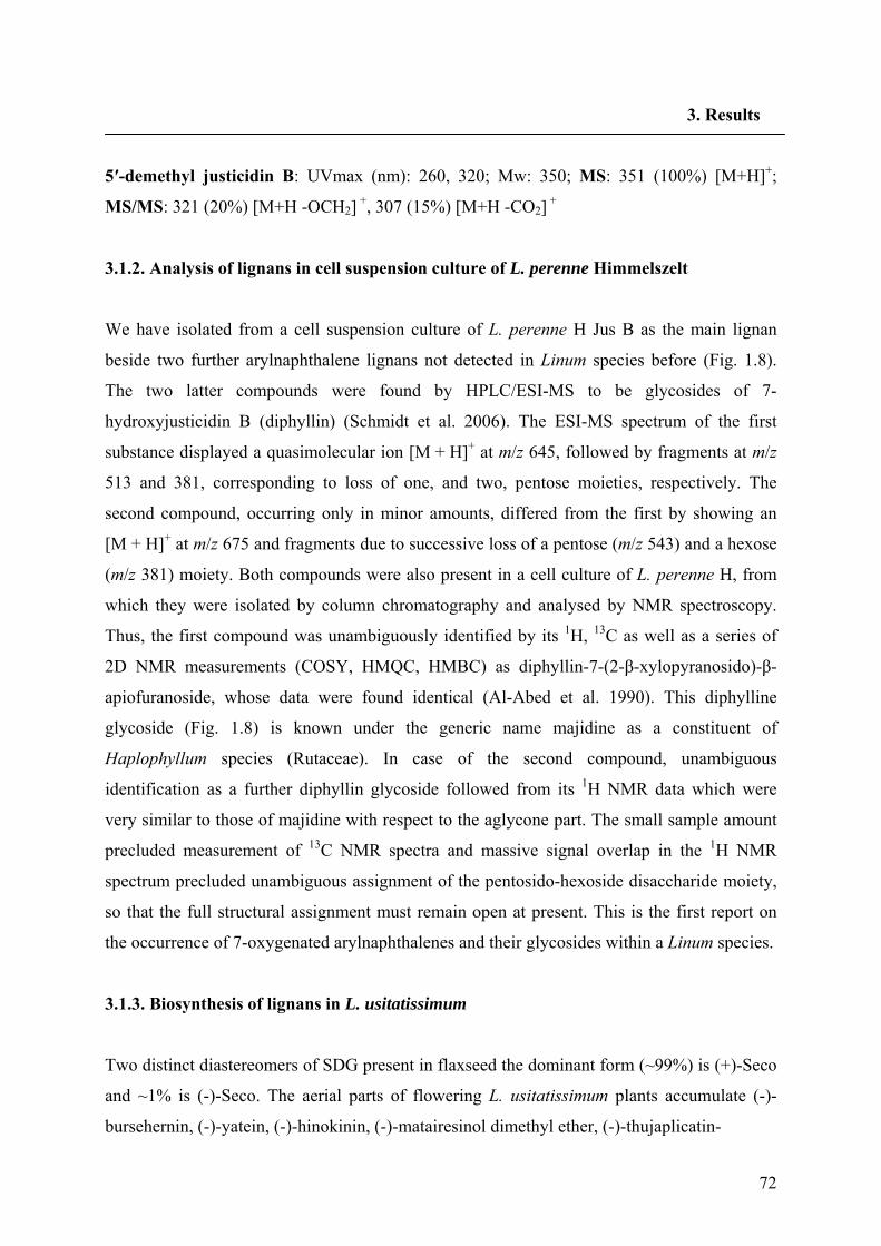
72
3. Results
5′-demethyl justicidin B: UVmax (nm): 260, 320; Mw: 350; MS: 351 (100%) [M+H]+;
MS/MS: 321 (20%) [M+H -OCH2] +, 307 (15%) [M+H -CO2] +
3.1.2. Analysis of lignans in cell suspension culture of L. perenne Himmelszelt
We have isolated from a cell suspension culture of L. perenne H Jus B as the main lignan
beside two further arylnaphthalene lignans not detected in Linum species before (Fig. 1.8).
The two latter compounds were found by HPLC/ESI-MS to be glycosides of 7-
hydroxyjusticidin B (diphyllin) (Schmidt et al. 2006). The ESI-MS spectrum of the first
substance displayed a quasimolecular ion [M + H]+ at m/z 645, followed by fragments at m/z
513 and 381, corresponding to loss of one, and two, pentose moieties, respectively. The
second compound, occurring only in minor amounts, differed from the first by showing an
[M + H]+ at m/z 675 and fragments due to successive loss of a pentose (m/z 543) and a hexose
(m/z 381) moiety. Both compounds were also present in a cell culture of L. perenne H, from
which they were isolated by column chromatography and analysed by NMR spectroscopy.
Thus, the first compound was unambiguously identified by its 1H, 13C as well as a series of
2D NMR measurements (COSY, HMQC, HMBC) as diphyllin-7-(2-β-xylopyranosido)-β-
apiofuranoside, whose data were found identical (Al-Abed et al. 1990). This diphylline
glycoside (Fig. 1.8) is known under the generic name majidine as a constituent of
Haplophyllum species (Rutaceae). In case of the second compound, unambiguous
identification as a further diphyllin glycoside followed from its 1H NMR data which were
very similar to those of majidine with respect to the aglycone part. The small sample amount
precluded measurement of 13C NMR spectra and massive signal overlap in the 1H NMR
spectrum precluded unambiguous assignment of the pentosido-hexoside disaccharide moiety,
so that the full structural assignment must remain open at present. This is the first report on
the occurrence of 7-oxygenated arylnaphthalenes and their glycosides within a Linum species.
3.1.3. Biosynthesis of lignans in L. usitatissimum
Two distinct diastereomers of SDG present in flaxseed the dominant form (~99%) is (+)-Seco
and ~1% is (-)-Seco. The aerial parts of flowering L. usitatissimum plants accumulate (-)-
bursehernin, (-)-yatein, (-)-hinokinin, (-)-matairesinol dimethyl ether, (-)-thujaplicatin-
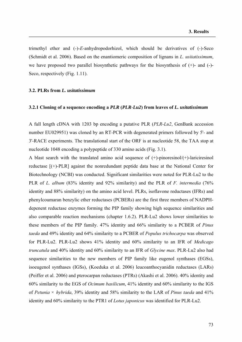
73
3. Results
trimethyl ether and (-)-E-anhydropodorhizol, which should be derivatives of (-)-Seco
(Schmidt et al. 2006). Based on the enantiomeric composition of lignans in L. usitatissimum,
we have proposed two parallel biosynthetic pathways for the biosynthesis of (+)- and (-)-
Seco, respectively (Fig. 1.11).
3.2. PLRs from L. usitatissimum
3.2.1 Cloning of a sequence encoding a PLR (PLR-Lu2) from leaves of L. usitatissimum
A full length cDNA with 1203 bp encoding a putative PLR (PLR-Lu2, GenBank accession
number EU029951) was cloned by an RT-PCR with degenerated primers followed by 5'- and
3'-RACE experiments. The translational start of the ORF is at nucleotide 58, the TAA stop at
nucleotide 1048 encoding a polypeptide of 330 amino acids (Fig. 3.1).
A blast search with the translated amino acid sequence of (+)-pinoresinol/(+)-lariciresinol
reductase [(+)-PLR] against the nonredundant peptide data base at the National Center for
Biotechnology (NCBI) was conducted. Significant similarities were noted for PLR-Lu2 to the
PLR of L. album (83% identity and 92% similarity) and the PLR of F. intermedia (76%
identity and 88% similarity) on the amino acid level. PLRs, isoflavone reductases (IFRs) and
phenylcoumaran benzylic ether reductases (PCBERs) are the first three members of NADPH-
depenent reductase enzymes forming the PIP family showing high sequence similarities and
also comparable reaction mechanisms (chapter 1.6.2). PLR-Lu2 shows lower similarities to
these members of the PIP family. 47% identity and 66% similarity to a PCBER of Pinus
taeda and 49% identity and 64% similarity to a PCBER of Populus trichocarpa was observed
for PLR-Lu2. PLR-Lu2 shows 41% identity and 60% similarity to an IFR of Medicago
truncatula and 40% identity and 60% similarity to an IFR of Glycine max. PLR-Lu2 also had
sequence similarities to the new members of PIP family like eugenol synthases (EGSs),
isoeugenol synthases (IGSs), (Koeduka et al. 2006) leucoanthocyanidin reductases (LARs)
(Peiffer et al. 2006) and pterocarpan reductases (PTRs) (Akashi et al. 2006). 40% identity and
60% similarity to the EGS of Ocimum basilicum, 41% identity and 60% similarity to the IGS
of Petunia × hybrida, 39% identity and 58% similarity to the LAR of Pinus taeda and 41%
identity and 60% similarity to the PTR1 of Lotus japonicus was identified for PLR-Lu2.
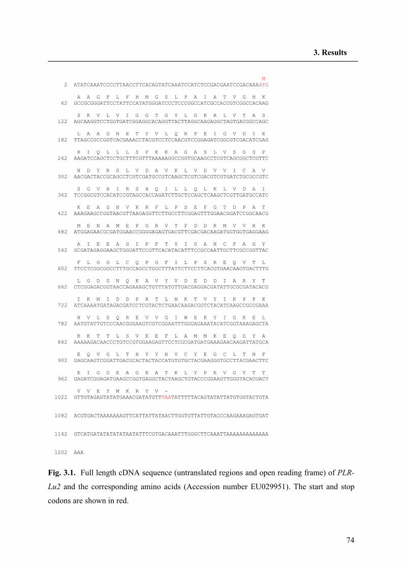
74
3. Results
M 2 ATATCAAATCCCCTTAACCTTCACAGTATCAAATCCATCTCCGACGAATCCGACAAAATG A A G F L F H M G S L P A I A T V G H K 62 GCCGCGGGATTCCTATTCCATATGGGATCCCTCCCGGCCATCGCCACCGTCGGCCACAAG S K V L V I G G T G Y L G K R L V T A S 122 AGCAAGGTCCTGGTGATCGGAGGCACAGGTTACTTAGGCAAGAGGCTAGTGACGGCCAGC L A A G H E T Y V L Q R P E I G V D I E 182 TTAGCCGCCGGTCACGAAACCTACGTCCTCCAACGTCCGGAGATCGGCGTCGACATCGAG K I Q L L L S F K K A G A S L V S G S F 242 AAGATCCAGCTCCTGCTTTCGTTTAAAAAGGCCGGTGCAAGCCTCGTCAGCGGCTCGTTC N D Y R S L V D A V K L V D V V I C A V 302 AACGACTACCGCAGCCTCGTCGATGCCGTCAAGCTCGTCGACGTCGTGATCTGCGCCGTC S G V H I R S H Q I L L Q L K L V D A I 362 TCCGGCGTCCACATCCGTAGCCACCAGATCTTGCTCCAGCTCAAGCTCGTTGATGCCATC K E A G N V K R F L P S E F G T D P A T 422 AAAGAAGCCGGTAACGTTAAGAGGTTCTTGCCTTCGGAGTTTGGAACGGATCCGGCAACG M E N A M E P G R V T F D D K M V V R K 482 ATGGAGAACGCGATGGAACCGGGGAGAGTGACGTTCGACGACAAGATGGTGGTGAGGAAG A I E E A G I P F T Y I S A N C F A G Y 542 GCGATAGAGGAAGCTGGGATTCCGTTCACATACATTTCCGCCAATTGCTTCGCCGGTTAC F L G G L C Q P G F I L P S R E Q V T L 602 TTCCTCGGCGGCCTTTGCCAGCCTGGCTTTATTCTTCCTTCACGTGAACAAGTGACTTTG L G D G N Q K A V Y V D E D D I A R Y T 662 CTCGGAGACGGTAACCAGAAAGCTGTTTATGTTGACGAGGACGATATTGCGCGATACACG I K M I D D P R T L N K T V Y I K P P K 722 ATCAAAATGATAGACGATCCTCGTACTCTGAACAAGACGGTCTACATCAAGCCGCCGAAA N V L S Q R E V V G I W E K Y I G K E L 782 AATGTATTGTCCCAACGGGAAGTCGTCGGAATTTGGGAGAAATACATCGGTAAAGAGCTA K K T T L S V E E F L A M M K E Q D Y A 842 AAAAAGACAACCCTGTCCGTGGAAGAGTTCCTCGCGATGATGAAAGAACAAGATTATGCA E Q V G L T H Y Y H V C Y E G C L T N F 902 GAGCAAGTCGGATTGACGCACTACTACCATGTGTGCTACGAAGGGTGCCTTACGAACTTC E I G D E A G E A T K L Y P E V G Y T T 962 GAGATCGGAGATGAAGCCGGTGAGGCTACTAAGCTGTACCCGGAAGTTGGGTACACGACT V V E Y M K R Y V - 1022 GTTGTAGAGTATATGAAACGATATGTTTAATATTTTTACAGTATATTATGTGGTACTGTA 1082 ACGTGACTAAAAAAAGTTCATTATTATAACTTGGTGTTATTGTACCCAAGAAAGAGTGAT 1142 GTCATGATATATATATAATATTTCGTGACAAATTTGGGCTTCAAATTAAAAAAAAAAAAA 1202 AAA
Fig. 3.1. Full length cDNA sequence (untranslated regions and open reading frame) of PLR-
Lu2 and the corresponding amino acids (Accession number EU029951). The start and stop
codons are shown in red.
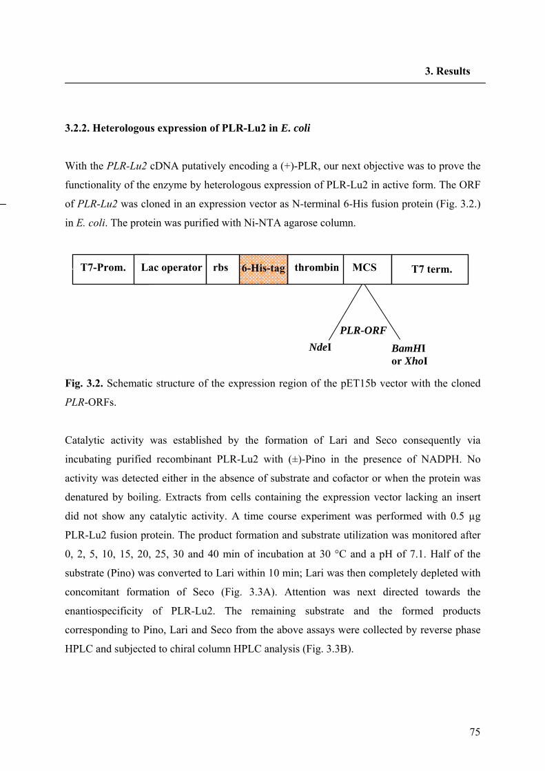
75
3. Results
3.2.2. Heterologous expression of PLR-Lu2 in E. coli
With the PLR-Lu2 cDNA putatively encoding a (+)-PLR, our next objective was to prove the
functionality of the enzyme by heterologous expression of PLR-Lu2 in active form. The ORF
of PLR-Lu2 was cloned in an expression vector as N-terminal 6-His fusion protein (Fig. 3.2.)
in E. coli. The protein was purified with Ni-NTA agarose column.
Fig. 3.2. Schematic structure of the expression region of the pET15b vector with the cloned
PLR-ORFs.
Catalytic activity was established by the formation of Lari and Seco consequently via
incubating purified recombinant PLR-Lu2 with (±)-Pino in the presence of NADPH. No
activity was detected either in the absence of substrate and cofactor or when the protein was
denatured by boiling. Extracts from cells containing the expression vector lacking an insert
did not show any catalytic activity. A time course experiment was performed with 0.5 µg
PLR-Lu2 fusion protein. The product formation and substrate utilization was monitored after
0, 2, 5, 10, 15, 20, 25, 30 and 40 min of incubation at 30 °C and a pH of 7.1. Half of the
substrate (Pino) was converted to Lari within 10 min; Lari was then completely depleted with
concomitant formation of Seco (Fig. 3.3A). Attention was next directed towards the
enantiospecificity of PLR-Lu2. The remaining substrate and the formed products
corresponding to Pino, Lari and Seco from the above assays were collected by reverse phase
HPLC and subjected to chiral column HPLC analysis (Fig. 3.3B).
6-His-tagT7-Prom. Lac operator rbs thrombin MCS T7 term.
PLR-ORF NdeI BamHI
or XhoI
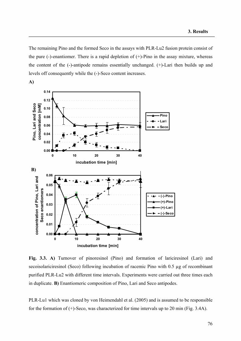
76
3. Results
The remaining Pino and the formed Seco in the assays with PLR-Lu2 fusion protein consist of
the pure (-)-enantiomer. There is a rapid depletion of (+)-Pino in the assay mixture, whereas
the content of the (-)-antipode remains essentially unchanged. (+)-Lari then builds up and
levels off consequently while the (-)-Seco content increases.
A)
0.00
0.02
0.04
0.06
0.08
0.10
0.12
0.14
0 10 20 30 40
incubation time [min]
Pino
, Lar
i and
Sec
o co
ncen
trat
ion
[mM
]
PinoLariSeco
B)
0.00
0.01
0.02
0.03
0.04
0.05
0.06
0 10 20 30 40
incubation time [min]
conc
entr
atio
n of
Pin
o, L
ari a
nd
Sec
o en
antio
mer
s
(-)-Pino(+)-Pino(+)-Lari(-)-Seco
Fig. 3.3. A) Turnover of pinoresinol (Pino) and formation of lariciresinol (Lari) and
secoisolariciresinol (Seco) following incubation of racemic Pino with 0.5 µg of recombinant
purified PLR-Lu2 with different time intervals. Experiments were carried out three times each
in duplicate. B) Enantiomeric composition of Pino, Lari and Seco antipodes.
PLR-Lu1 which was cloned by von Heimendahl et al. (2005) and is assumed to be responsible
for the formation of (+)-Seco, was characterized for time intervals up to 20 min (Fig. 3.4A).
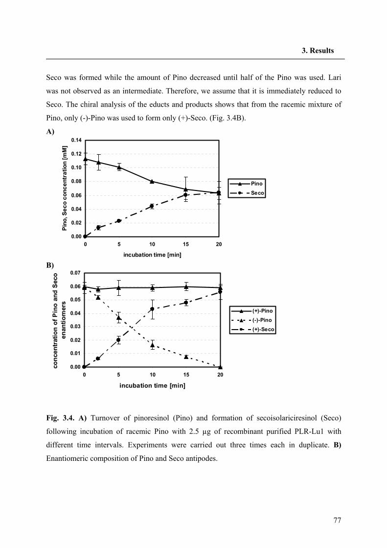
77
3. Results
Seco was formed while the amount of Pino decreased until half of the Pino was used. Lari
was not observed as an intermediate. Therefore, we assume that it is immediately reduced to
Seco. The chiral analysis of the educts and products shows that from the racemic mixture of
Pino, only (-)-Pino was used to form only (+)-Seco. (Fig. 3.4B).
A)
0.00
0.02
0.04
0.06
0.08
0.10
0.12
0.14
0 5 10 15 20
incubation time [min]
Pino
, Sec
o co
ncen
trat
ion
[mM
]
PinoSeco
B)
0.00
0.01
0.02
0.03
0.04
0.05
0.06
0.07
0 5 10 15 20
incubation time [min]
conc
entra
tion
of P
ino
and
Seco
en
antio
mer
s
(+)-Pino(-)-Pino(+)-Seco
Fig. 3.4. A) Turnover of pinoresinol (Pino) and formation of secoisolariciresinol (Seco)
following incubation of racemic Pino with 2.5 µg of recombinant purified PLR-Lu1 with
different time intervals. Experiments were carried out three times each in duplicate. B)
Enantiomeric composition of Pino and Seco antipodes.
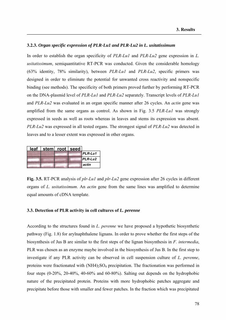
78
3. Results
3.2.3. Organ specific expression of PLR-Lu1 and PLR-Lu2 in L. usitatissimum
In order to establish the organ specificity of PLR-Lu1 and PLR-Lu2 gene expression in L.
usitatissimum, semiquantitative RT-PCR was conducted. Given the considerable homology
(63% identity, 78% similarity), between PLR-Lu1 and PLR-Lu2, specific primers was
designed in order to eliminate the potential for unwanted cross reactivity and nonspecific
binding (see methods). The specificity of both primers proved further by performing RT-PCR
on the DNA-plasmid level of PLR-Lu1 and PLR-Lu2 separately. Transcript levels of PLR-Lu1
and PLR-Lu2 was evaluated in an organ specific manner after 26 cycles. An actin gene was
amplified from the same organs as control. As shown in Fig. 3.5 PLR-Lu1 was strongly
expressed in seeds as well as roots whereas in leaves and stems its expression was absent.
PLR-Lu2 was expressed in all tested organs. The strongest signal of PLR-Lu2 was detected in
leaves and to a lesser extent was expressed in other organs.
Fig. 3.5. RT-PCR analysis of plr-Lu1 and plr-Lu2 gene expression after 26 cycles in different
organs of L. usitatissimum. An actin gene from the same lines was amplified to determine
equal amounts of cDNA template.
3.3. Detection of PLR activity in cell cultures of L. perenne
According to the structures found in L. perenne we have proposed a hypothetic biosynthetic
pathway (Fig. 1.8) for arylnaphthalene lignans. In order to prove whether the first steps of the
biosynthesis of Jus B are similar to the first steps of the lignan biosynthesis in F. intermedia,
PLR was chosen as an enzyme maybe involved in the biosynthesis of Jus B. In the first step to
investigate if any PLR activity can be observed in cell suspension culture of L. perenne,
proteins were fractionated with (NH4)2SO4 precipitation. The fractionation was performed in
four steps (0-20%, 20-40%, 40-60% and 60-80%). Salting out depends on the hydrophobic
nature of the precipitated protein. Proteins with more hydrophobic patches aggregate and
precipitate before those with smaller and fewer patches. In the fraction which was precipitated
seed root leaf stem PLR-Lu1 PLR-Lu2 actin
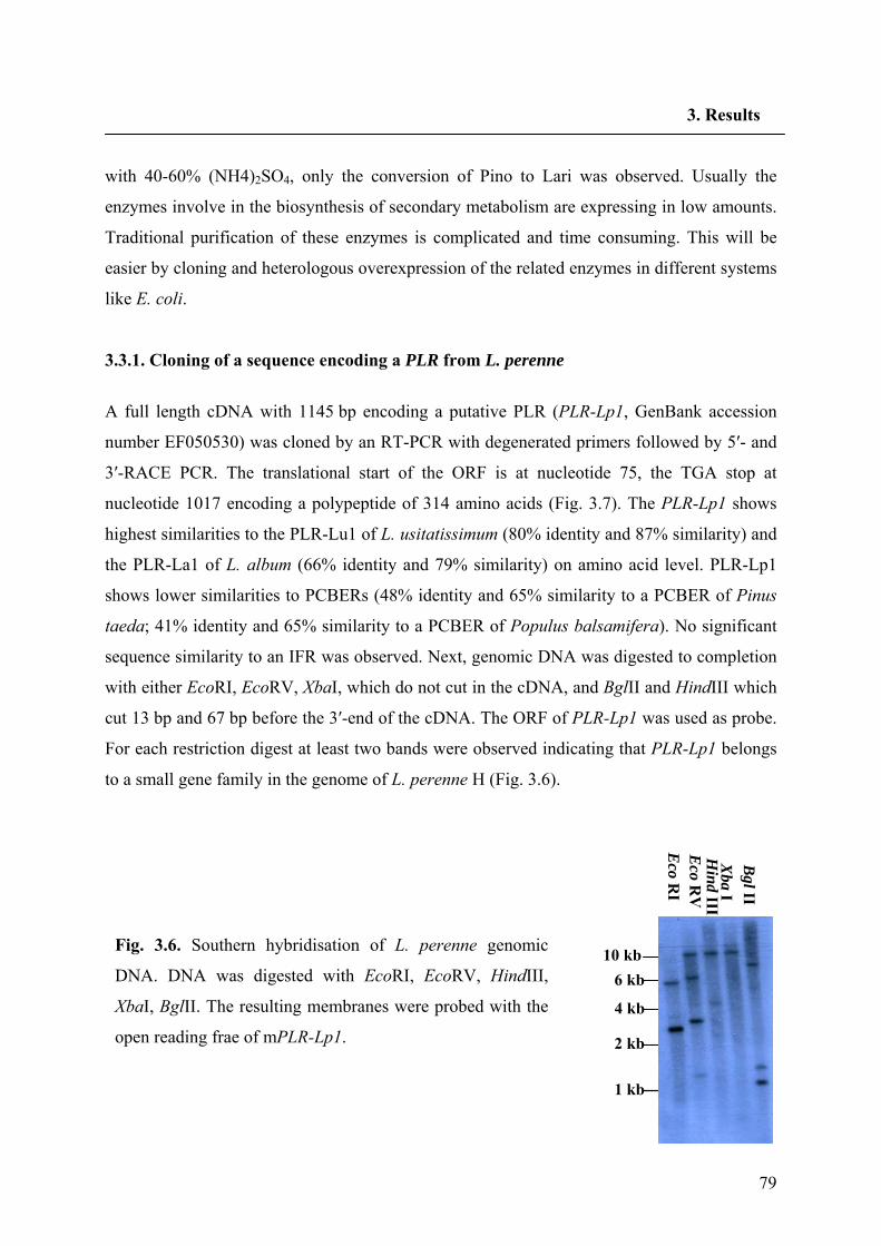
79
3. Results
with 40-60% (NH4)2SO4, only the conversion of Pino to Lari was observed. Usually the
enzymes involve in the biosynthesis of secondary metabolism are expressing in low amounts.
Traditional purification of these enzymes is complicated and time consuming. This will be
easier by cloning and heterologous overexpression of the related enzymes in different systems
like E. coli.
3.3.1. Cloning of a sequence encoding a PLR from L. perenne
A full length cDNA with 1145 bp encoding a putative PLR (PLR-Lp1, GenBank accession
number EF050530) was cloned by an RT-PCR with degenerated primers followed by 5′- and
3′-RACE PCR. The translational start of the ORF is at nucleotide 75, the TGA stop at
nucleotide 1017 encoding a polypeptide of 314 amino acids (Fig. 3.7). The PLR-Lp1 shows
highest similarities to the PLR-Lu1 of L. usitatissimum (80% identity and 87% similarity) and
the PLR-La1 of L. album (66% identity and 79% similarity) on amino acid level. PLR-Lp1
shows lower similarities to PCBERs (48% identity and 65% similarity to a PCBER of Pinus
taeda; 41% identity and 65% similarity to a PCBER of Populus balsamifera). No significant
sequence similarity to an IFR was observed. Next, genomic DNA was digested to completion
with either EcoRI, EcoRV, XbaI, which do not cut in the cDNA, and BglII and HindIII which
cut 13 bp and 67 bp before the 3′-end of the cDNA. The ORF of PLR-Lp1 was used as probe.
For each restriction digest at least two bands were observed indicating that PLR-Lp1 belongs
to a small gene family in the genome of L. perenne H (Fig. 3.6).
10 kb 6 kb
4 kb
2 kb
1 kb
Eco R
I E
co RV
H
ind III X
ba I B
gl II
Fig. 3.6. Southern hybridisation of L. perenne genomic
DNA. DNA was digested with EcoRI, EcoRV, HindIII,
XbaI, BglII. The resulting membranes were probed with the
open reading frae of mPLR-Lp1.
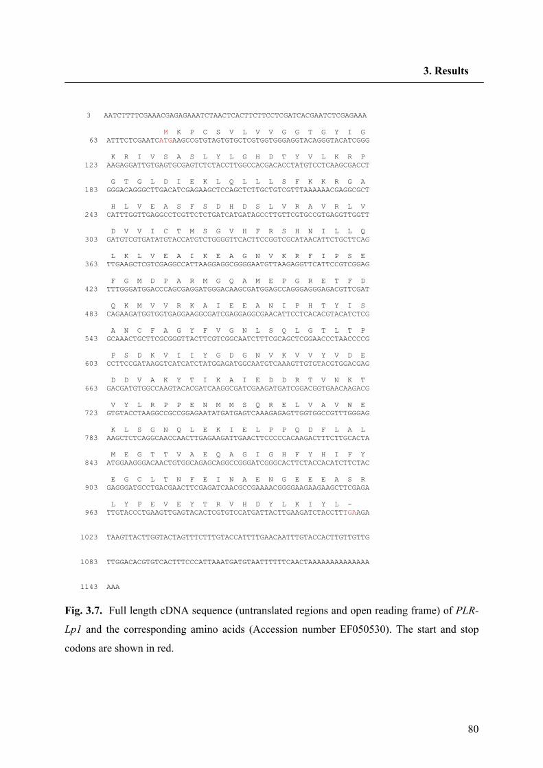
80
3. Results
3 AATCTTTTCGAAACGAGAGAAATCTAACTCACTTCTTCCTCGATCACGAATCTCGAGAAA M K P C S V L V V G G T G Y I G 63 ATTTCTCGAATCATGAAGCCGTGTAGTGTGCTCGTGGTGGGAGGTACAGGGTACATCGGG K R I V S A S L Y L G H D T Y V L K R P 123 AAGAGGATTGTGAGTGCGAGTCTCTACCTTGGCCACGACACCTATGTCCTCAAGCGACCT G T G L D I E K L Q L L L S F K K R G A 183 GGGACAGGGCTTGACATCGAGAAGCTCCAGCTCTTGCTGTCGTTTAAAAAACGAGGCGCT H L V E A S F S D H D S L V R A V R L V 243 CATTTGGTTGAGGCCTCGTTCTCTGATCATGATAGCCTTGTTCGTGCCGTGAGGTTGGTT D V V I C T M S G V H F R S H N I L L Q 303 GATGTCGTGATATGTACCATGTCTGGGGTTCACTTCCGGTCGCATAACATTCTGCTTCAG L K L V E A I K E A G N V K R F I P S E 363 TTGAAGCTCGTCGAGGCCATTAAGGAGGCGGGGAATGTTAAGAGGTTCATTCCGTCGGAG F G M D P A R M G Q A M E P G R E T F D 423 TTTGGGATGGACCCAGCGAGGATGGGACAAGCGATGGAGCCAGGGAGGGAGACGTTCGAT Q K M V V R K A I E E A N I P H T Y I S 483 CAGAAGATGGTGGTGAGGAAGGCGATCGAGGAGGCGAACATTCCTCACACGTACATCTCG A N C F A G Y F V G N L S Q L G T L T P 543 GCAAACTGCTTCGCGGGTTACTTCGTCGGCAATCTTTCGCAGCTCGGAACCCTAACCCCG P S D K V I I Y G D G N V K V V Y V D E 603 CCTTCCGATAAGGTCATCATCTATGGAGATGGCAATGTCAAAGTTGTGTACGTGGACGAG D D V A K Y T I K A I E D D R T V N K T 663 GACGATGTGGCCAAGTACACGATCAAGGCGATCGAAGATGATCGGACGGTGAACAAGACG V Y L R P P E N M M S Q R E L V A V W E 723 GTGTACCTAAGGCCGCCGGAGAATATGATGAGTCAAAGAGAGTTGGTGGCCGTTTGGGAG K L S G N Q L E K I E L P P Q D F L A L 783 AAGCTCTCAGGCAACCAACTTGAGAAGATTGAACTTCCCCCACAAGACTTTCTTGCACTA M E G T T V A E Q A G I G H F Y H I F Y 843 ATGGAAGGGACAACTGTGGCAGAGCAGGCCGGGATCGGGCACTTCTACCACATCTTCTAC E G C L T N F E I N A E N G E E E A S R 903 GAGGGATGCCTGACGAACTTCGAGATCAACGCCGAAAACGGGGAAGAAGAAGCTTCGAGA L Y P E V E Y T R V H D Y L K I Y L - 963 TTGTACCCTGAAGTTGAGTACACTCGTGTCCATGATTACTTGAAGATCTACCTTTGAAGA 1023 TAAGTTACTTGGTACTAGTTTCTTTGTACCATTTTGAACAATTTGTACCACTTGTTGTTG 1083 TTGGACACGTGTCACTTTCCCATTAAATGATGTAATTTTTTCAACTAAAAAAAAAAAAAA 1143 AAA
Fig. 3.7. Full length cDNA sequence (untranslated regions and open reading frame) of PLR-
Lp1 and the corresponding amino acids (Accession number EF050530). The start and stop
codons are shown in red.
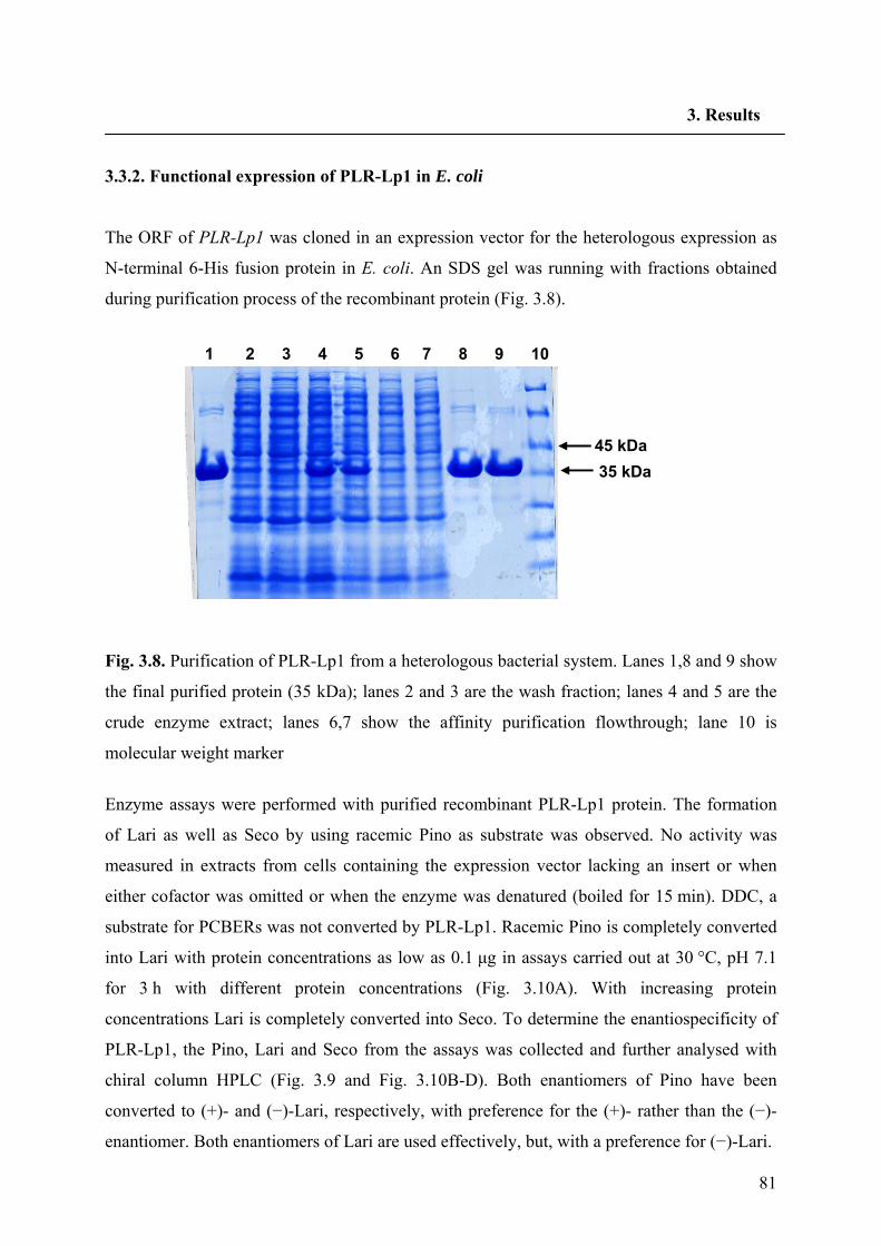
81
3. Results
3.3.2. Functional expression of PLR-Lp1 in E. coli
The ORF of PLR-Lp1 was cloned in an expression vector for the heterologous expression as
N-terminal 6-His fusion protein in E. coli. An SDS gel was running with fractions obtained
during purification process of the recombinant protein (Fig. 3.8).
Fig. 3.8. Purification of PLR-Lp1 from a heterologous bacterial system. Lanes 1,8 and 9 show
the final purified protein (35 kDa); lanes 2 and 3 are the wash fraction; lanes 4 and 5 are the
crude enzyme extract; lanes 6,7 show the affinity purification flowthrough; lane 10 is
molecular weight marker
Enzyme assays were performed with purified recombinant PLR-Lp1 protein. The formation
of Lari as well as Seco by using racemic Pino as substrate was observed. No activity was
measured in extracts from cells containing the expression vector lacking an insert or when
either cofactor was omitted or when the enzyme was denatured (boiled for 15 min). DDC, a
substrate for PCBERs was not converted by PLR-Lp1. Racemic Pino is completely converted
into Lari with protein concentrations as low as 0.1 μg in assays carried out at 30 °C, pH 7.1
for 3 h with different protein concentrations (Fig. 3.10A). With increasing protein
concentrations Lari is completely converted into Seco. To determine the enantiospecificity of
PLR-Lp1, the Pino, Lari and Seco from the assays was collected and further analysed with
chiral column HPLC (Fig. 3.9 and Fig. 3.10B-D). Both enantiomers of Pino have been
converted to (+)- and (−)-Lari, respectively, with preference for the (+)- rather than the (−)-
enantiomer. Both enantiomers of Lari are used effectively, but, with a preference for (−)-Lari.
1 2 3 4 5 6 7 8 9 10
45 kDa 35 kDa
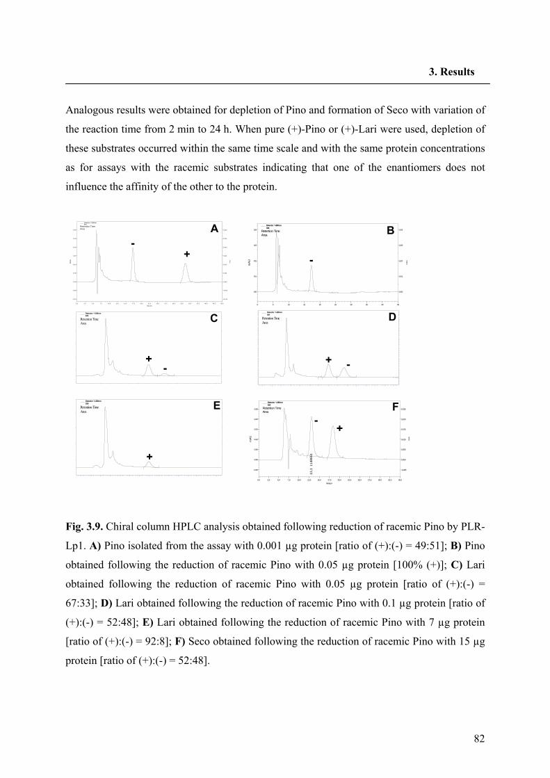
82
3. Results
Analogous results were obtained for depletion of Pino and formation of Seco with variation of
the reaction time from 2 min to 24 h. When pure (+)-Pino or (+)-Lari were used, depletion of
these substrates occurred within the same time scale and with the same protein concentrations
as for assays with the racemic substrates indicating that one of the enantiomers does not
influence the affinity of the other to the protein.
Fig. 3.9. Chiral column HPLC analysis obtained following reduction of racemic Pino by PLR-
Lp1. A) Pino isolated from the assay with 0.001 µg protein [ratio of (+):(-) = 49:51]; B) Pino
obtained following the reduction of racemic Pino with 0.05 µg protein [100% (+)]; C) Lari
obtained following the reduction of racemic Pino with 0.05 µg protein [ratio of (+):(-) =
67:33]; D) Lari obtained following the reduction of racemic Pino with 0.1 µg protein [ratio of
(+):(-) = 52:48]; E) Lari obtained following the reduction of racemic Pino with 7 µg protein
[ratio of (+):(-) = 92:8]; F) Seco obtained following the reduction of racemic Pino with 15 µg
protein [ratio of (+):(-) = 52:48].
Minutes
0,0 2,5 5,0 7,5 10,0 12,5 15,0 17,5 20,0 22,5 25,0 27,5 30,0 32,5 35,0 37,5 40,0 42,5 45,0
mA
U
-0,010
-0,005
0,000
0,005
0,010
0,015
0,020
0,025
0,030m
AU
-0,010
-0,005
0,000
0,005
0,010
0,015
0,020
0,025
0,030
17,4
994
903
33,6
979
300
Detector 1-280nm018
Retention TimeArea
Minutes0 5 10 15 20 25 30 35 40 45
mAU
0,00
0,01
0,02
0,03
0,04
mAU
0,00
0,01
0,02
0,03
0,04
17,3
884
673
Detector 1-280nm024
Retention TimeArea
Minutes0 2 4 6 8 10 12 14 16 18 20 22 24 26 28 30
mA
U
0,00
0,02
0,04
0,06
0,08
0,10
0,12
mA
U
0,00
0,02
0,04
0,06
0,08
0,10
0,1213
,042
114
73
15,0
10 1
5340
86
18,2
93 1
4997
81
Detector 1-280nm031
Retention TimeArea
Minutes0 2 4 6 8 10 12 14 16 18 20 22 24 26 28 30
mAU
0,00
0,02
0,04
0,06
0,08
mAU
0,00
0,02
0,04
0,06
0,08
14,9
82 4
2510
1
Detector 1-280nm039
Retention TimeArea
Minutes
0 2 4 6 8 10 12 14 16 18 20 22 24 26 28 30
mA
U
0,00
0,02
0,04
0,06
0,08
0,10
mA
U
0,00
0,02
0,04
0,06
0,08
0,10
15,0
03 1
0603
23
18,2
55 2
6603
1
Detector 1-280nm029
Retention TimeArea
Minutes0,0 2,5 5,0 7,5 10,0 12,5 15,0 17,5 20,0 22,5 25,0 27,5 30,0 32,5 35,0
mAU
-0,005
0,000
0,005
0,010
0,015
0,020
0,025m
AU
-0,005
0,000
0,005
0,010
0,015
0,020
0,025
13,1
114
8851
18,4
129
4308
Detector 1-280nm029
Retention TimeArea
A B
DC
E F
-+ -
-+
+-
-+
+
Minutes
0,0 2,5 5,0 7,5 10,0 12,5 15,0 17,5 20,0 22,5 25,0 27,5 30,0 32,5 35,0 37,5 40,0 42,5 45,0
mA
U
-0,010
-0,005
0,000
0,005
0,010
0,015
0,020
0,025
0,030m
AU
-0,010
-0,005
0,000
0,005
0,010
0,015
0,020
0,025
0,030
17,4
994
903
33,6
979
300
Detector 1-280nm018
Retention TimeArea
Minutes0 5 10 15 20 25 30 35 40 45
mAU
0,00
0,01
0,02
0,03
0,04
mAU
0,00
0,01
0,02
0,03
0,04
17,3
884
673
Detector 1-280nm024
Retention TimeArea
Minutes0 2 4 6 8 10 12 14 16 18 20 22 24 26 28 30
mA
U
0,00
0,02
0,04
0,06
0,08
0,10
0,12
mA
U
0,00
0,02
0,04
0,06
0,08
0,10
0,1213
,042
114
73
15,0
10 1
5340
86
18,2
93 1
4997
81
Detector 1-280nm031
Retention TimeArea
Minutes0 2 4 6 8 10 12 14 16 18 20 22 24 26 28 30
mAU
0,00
0,02
0,04
0,06
0,08
mAU
0,00
0,02
0,04
0,06
0,08
14,9
82 4
2510
1
Detector 1-280nm039
Retention TimeArea
Minutes
0 2 4 6 8 10 12 14 16 18 20 22 24 26 28 30
mA
U
0,00
0,02
0,04
0,06
0,08
0,10
mA
U
0,00
0,02
0,04
0,06
0,08
0,10
15,0
03 1
0603
23
18,2
55 2
6603
1
Detector 1-280nm029
Retention TimeArea
Minutes0,0 2,5 5,0 7,5 10,0 12,5 15,0 17,5 20,0 22,5 25,0 27,5 30,0 32,5 35,0
mAU
-0,005
0,000
0,005
0,010
0,015
0,020
0,025m
AU
-0,005
0,000
0,005
0,010
0,015
0,020
0,025
13,1
114
8851
18,4
129
4308
Detector 1-280nm029
Retention TimeArea
A B
DC
E F
-+ -
-+
+-
-+
+
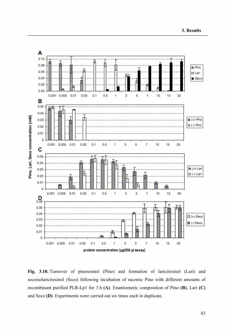
83
3. Results
Fig. 3.10. Turnover of pinoresinol (Pino) and formation of lariciresinol (Lari) and
secoisolariciresinol (Seco) following incubation of racemic Pino with different amounts of
recombinant purified PLR-Lp1 for 3 h (A). Enantiomeric composition of Pino (B), Lari (C)
and Seco (D). Experiments were carried out six times each in duplicate.
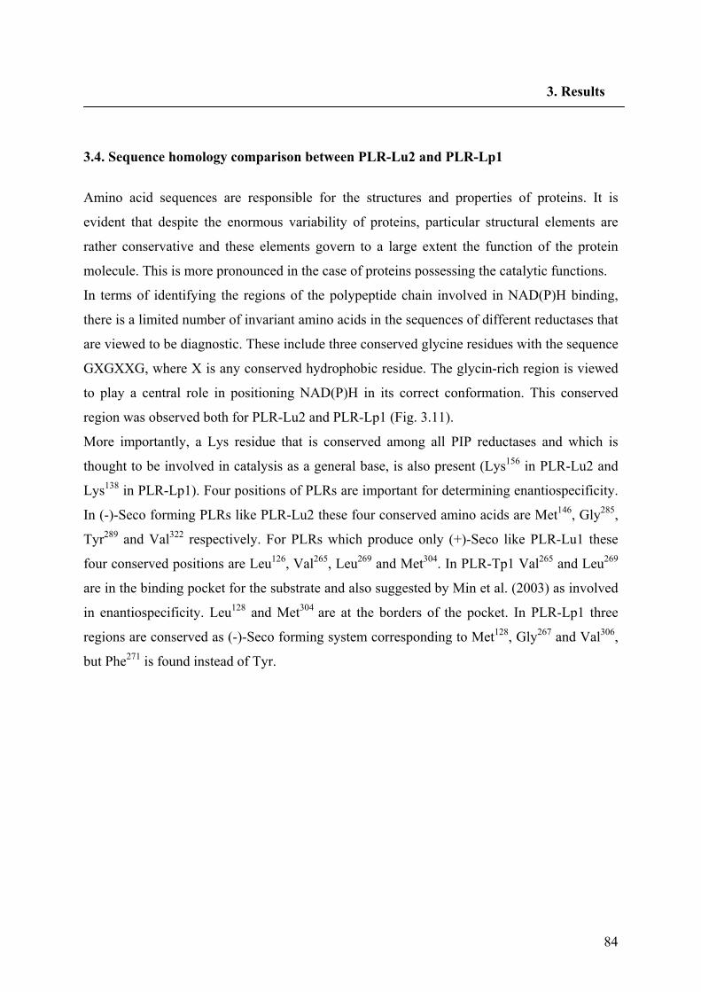
84
3. Results
3.4. Sequence homology comparison between PLR-Lu2 and PLR-Lp1
Amino acid sequences are responsible for the structures and properties of proteins. It is
evident that despite the enormous variability of proteins, particular structural elements are
rather conservative and these elements govern to a large extent the function of the protein
molecule. This is more pronounced in the case of proteins possessing the catalytic functions.
In terms of identifying the regions of the polypeptide chain involved in NAD(P)H binding,
there is a limited number of invariant amino acids in the sequences of different reductases that
are viewed to be diagnostic. These include three conserved glycine residues with the sequence
GXGXXG, where X is any conserved hydrophobic residue. The glycin-rich region is viewed
to play a central role in positioning NAD(P)H in its correct conformation. This conserved
region was observed both for PLR-Lu2 and PLR-Lp1 (Fig. 3.11).
More importantly, a Lys residue that is conserved among all PIP reductases and which is
thought to be involved in catalysis as a general base, is also present (Lys156 in PLR-Lu2 and
Lys138 in PLR-Lp1). Four positions of PLRs are important for determining enantiospecificity.
In (-)-Seco forming PLRs like PLR-Lu2 these four conserved amino acids are Met146, Gly285,
Tyr289 and Val322 respectively. For PLRs which produce only (+)-Seco like PLR-Lu1 these
four conserved positions are Leu126, Val265, Leu269 and Met304. In PLR-Tp1 Val265 and Leu269
are in the binding pocket for the substrate and also suggested by Min et al. (2003) as involved
in enantiospecificity. Leu128 and Met304 are at the borders of the pocket. In PLR-Lp1 three
regions are conserved as (-)-Seco forming system corresponding to Met128, Gly267 and Val306,
but Phe271 is found instead of Tyr.
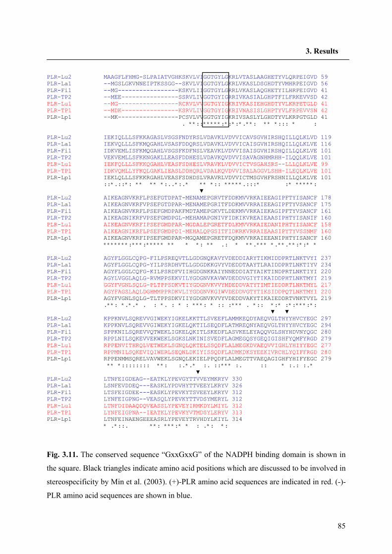
85
3. Results
PLR-Lu2 MAAGFLFHMG-SLPAIATVGHKSKVLVIGGTGYLGKRLVTASLAAGHETYVLQRPEIGVD 59 PLR-La1 --MGSLGKVNNEIPTKSSGG--SKVLVIGGTGYLGKRLVKASLDSGHDTYVMHRPEIGVD 56 PLR-Fi1 --MG-----------------KSKVLIIGGTGYLGRRLVKASLAQGHETYILHRPEIGVD 41 PLR-TP2 --MEE----------------SSRVLIVGGTGYIGRRIVKASIALGHPTFILFRKEVVSD 42 PLR-Lu1 --MG-----------------RCRVLVVGGTGYIGKRIVKASIEHGHDTYVLKRPETGLD 41 PLR-TP1 --MDK----------------KSRVLIVGGTGYIGKRIVNASISLGHPTYVLFRPEVVSN 42 PLR-Lp1 --MK-----------------PCSVLVVGGTGYIGKRIVSASLYLGHDTYVLKRPGTGLD 41 . **::*****:*:*:*.**: ** *::: * : PLR-Lu2 IEKIQLLLSFKKAGASLVSGSFNDYRSLVDAVKLVDVVICAVSGVHIRSHQILLQLKLVD 119 PLR-La1 IEKVQLLLSFKMQGAHLVSASFDDQRSLVDAVKLVDVVICAISGVHIRSHQILLQLKLVE 116 PLR-Fi1 IDKVEMLISFKMQGAHLVSGSFKDFNSLVEAVKLVDVVISAISGVHIRSHQILLQLKLVE 101 PLR-TP2 VEKVEMLLSFKKNGAKLLEASFDDHESLVDAVKQVDVVISAVAGNHMRHH-ILQQLKLVE 101 PLR-Lu1 IEKFQLLLSFKKQGAHLVEASFSDHESLVRAVKLVDVVICTVSGAHSRS--LLLQLKLVE 99 PLR-TP1 IDKVQMLLYFKQLGAKLIEASLDDHQRLVDALKQVDVVISALAGGVLSHH-ILEQLKLVE 101 PLR-Lp1 IEKLQLLLSFKKRGAHLVEASFSDHDSLVRAVRLVDVVICTMSGVHFRSHNILLQLKLVE 101 ::*.::*: ** ** *:..*:.* ** *:: *****.:::* :* *****: ▼ PLR-Lu2 AIKEAGNVKRFLPSEFGTDPAT-MENAMEPGRVTFDDKMVVRKAIEEAGIPFTYISANCF 178 PLR-La1 AIKEAGNVKRFVPSEFGTDPAR-MENAMEPGRITFDDKMVVRRAIEEAGIPFTYVSANCF 175 PLR-Fi1 AIKEAGNVKRFLPSEFGMDPAKFMDTAMEPGKVTLDEKMVVRKAIEKAGIPFTYVSANCF 161 PLR-TP2 AIKEAGNIKRFVPSEFGMDPGL-MEHAMAPGNIVFIDKIKVREAIEAASIPHTYISANIF 160 PLR-Lu1 AIKEAGNVKRFIPSEFGMDPAR-MGDALEPGRETFDLKMVVRKAIEDANIPHTYISANCF 158 PLR-TP1 AIKEAGNIKRFLPSEFGMDPDI-MEHALQPGSITFIDKRKVRRAIEAASIPYTYVSSNMF 160 PLR-Lp1 AIKEAGNVKRFIPSEFGMDPAR-MGQAMEPGRETFDQKMVVRKAIEEANIPHTYISANCF 160 *******:***:***** ** * *: ** .: * **.*** *.**.**:*:* * PLR-Lu2 AGYFLGGLCQPG-FILPSREQVTLLGDGNQKAVYVDEDDIARYTIKMIDDPRTLNKTVYI 237 PLR-La1 AGYFLGGLCQPG-YILPSRDHVTLLGDGDKKGVYVDEDDTAAYTLRAIDDPRTLNKTIYV 234 PLR-Fi1 AGYFLGGLCQFG-KILPSRDFVIIHGDGNKKAIYNNEDDIATYAIKTINDPRTLNKTIYI 220 PLR-TP2 AGYLVGGLAQLG-RVMPPSEKVILYGDGNVKAVWVDEDDVGIYTIKAIDDPHTLNKTMYI 219 PLR-Lu1 GGYFVGNLSQLG-PLTPPSDKVTIYGDGNVKVVYMDEDDVATYTIMTIEDDRTLNKTMYL 217 PLR-TP1 AGYFAGSLAQLDGHMMPPRDKVLIYGDGNVKGIWVDEDDVGTYTIKSIDDPQTLNKTMYI 220 PLR-Lp1 AGYFVGNLSQLG-TLTPPSDKVIIYGDGNVKVVYVDEDDVAKYTIKAIEDDRTVNKTVYL 219 .**: *.*.* . : *. : * : ***: * :: :*** . *:: *:* :*:***:*: ▼ ▼ PLR-Lu2 KPPKNVLSQREVVGIWEKYIGKELKKTTLSVEEFLAMMKEQDYAEQVGLTHYYHVCYEGC 297 PLR-La1 KPPKNVLSQREVVGIWEKYIGKELQKTILSEQDFLATMREQNYAEQVGLTHYYHVCYEGC 294 PLR-Fi1 SPPKNILSQREVVQTWEKLIGKELQKITLSKEDFLASVKELEYAQQVGLSHYHDVNYQGC 280 PLR-TP2 RPPLNILSQKEVVEKWEKLSGKSLNKINISVEDFLAGMEGQSYGEQIGISHFYQMFYRGD 279 PLR-Lu1 RPPENVITHRQLVETWEKLSGNQLQKTELSSQDFLALMEGKDVAEQVVIGHLYHIYYEGC 277 PLR-TP1 RPPMNILSQKEVIQIWERLSEQNLDKIYISSQDFLADMKDKSYEEKIVRCHLYQIFFRGD 280 PLR-Lp1 RPPENMMSQRELVAVWEKLSGNQLEKIELPPQDFLALMEGTTVAEQAGIGHFYHIFYEGC 279 ** *:::::::: **: :.*.* :. ::*** :. :: * :.: :.* ▼ PLR-Lu2 LTNFEIGDEAG--EATKLYPEVGYTTVVEYMKRYV 330 PLR-La1 LSNFEVDDEQ---EASKLYPDVHYTTVEEYLKRYV 326 PLR-Fi1 LTSFEIGDEE---EASKLYPEVKYTSVEEYLKRYV 312 PLR-TP2 LYNFEIGPNG--VEASQLYPEVKYTTVDSYMERYL 312 PLR-Lu1 LTNFDIDAAQDQVEASSLYPEVEYIRMKDYLMIYL 312 PLR-TP1 LYNFEIGPNA--IEATKLYPEVKYVTMDSYLERYV 313 PLR-Lp1 LTNFEINAENGEEEASRLYPEVEYTRVHDYLKIYL 314 * .*::. **: ***:* * : .*: *:
Fig. 3.11. The conserved sequence “GxxGxxG” of the NADPH binding domain is shown in
the square. Black triangles indicate amino acid positions which are discussed to be involved in
stereospecificity by Min et al. (2003). (+)-PLR amino acid sequences are indicated in red. (-)-
PLR amino acid sequences are shown in blue.
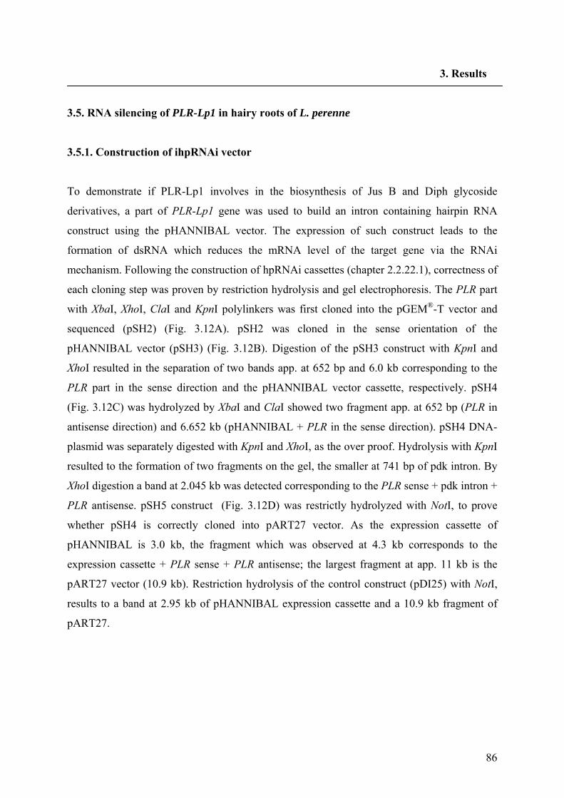
86
3. Results
3.5. RNA silencing of PLR-Lp1 in hairy roots of L. perenne
3.5.1. Construction of ihpRNAi vector
To demonstrate if PLR-Lp1 involves in the biosynthesis of Jus B and Diph glycoside
derivatives, a part of PLR-Lp1 gene was used to build an intron containing hairpin RNA
construct using the pHANNIBAL vector. The expression of such construct leads to the
formation of dsRNA which reduces the mRNA level of the target gene via the RNAi
mechanism. Following the construction of hpRNAi cassettes (chapter 2.2.22.1), correctness of
each cloning step was proven by restriction hydrolysis and gel electrophoresis. The PLR part
with XbaI, XhoI, ClaI and KpnI polylinkers was first cloned into the pGEM®-T vector and
sequenced (pSH2) (Fig. 3.12A). pSH2 was cloned in the sense orientation of the
pHANNIBAL vector (pSH3) (Fig. 3.12B). Digestion of the pSH3 construct with KpnI and
XhoI resulted in the separation of two bands app. at 652 bp and 6.0 kb corresponding to the
PLR part in the sense direction and the pHANNIBAL vector cassette, respectively. pSH4
(Fig. 3.12C) was hydrolyzed by XbaI and ClaI showed two fragment app. at 652 bp (PLR in
antisense direction) and 6.652 kb (pHANNIBAL + PLR in the sense direction). pSH4 DNA-
plasmid was separately digested with KpnI and XhoI, as the over proof. Hydrolysis with KpnI
resulted to the formation of two fragments on the gel, the smaller at 741 bp of pdk intron. By
XhoI digestion a band at 2.045 kb was detected corresponding to the PLR sense + pdk intron +
PLR antisense. pSH5 construct (Fig. 3.12D) was restrictly hydrolyzed with NotI, to prove
whether pSH4 is correctly cloned into pART27 vector. As the expression cassette of
pHANNIBAL is 3.0 kb, the fragment which was observed at 4.3 kb corresponds to the
expression cassette + PLR sense + PLR antisense; the largest fragment at app. 11 kb is the
pART27 vector (10.9 kb). Restriction hydrolysis of the control construct (pDI25) with NotI,
results to a band at 2.95 kb of pHANNIBAL expression cassette and a 10.9 kb fragment of
pART27.
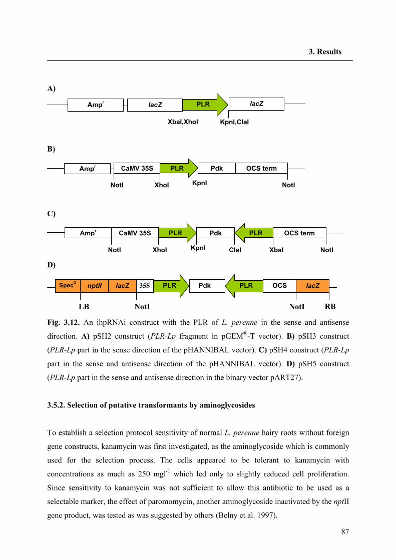
87
3. Results
A)
B)
C)
D)
Fig. 3.12. An ihpRNAi construct with the PLR of L. perenne in the sense and antisense
direction. A) pSH2 construct (PLR-Lp fragment in pGEM®-T vector). B) pSH3 construct
(PLR-Lp part in the sense direction of the pHANNIBAL vector). C) pSH4 construct (PLR-Lp
part in the sense and antisense direction of the pHANNIBAL vector). D) pSH5 construct
(PLR-Lp part in the sense and antisense direction in the binary vector pART27).
3.5.2. Selection of putative transformants by aminoglycosides
To establish a selection protocol sensitivity of normal L. perenne hairy roots without foreign
gene constructs, kanamycin was first investigated, as the aminoglycoside which is commonly
used for the selection process. The cells appeared to be tolerant to kanamycin with
concentrations as much as 250 mgl-1 which led only to slightly reduced cell proliferation.
Since sensitivity to kanamycin was not sufficient to allow this antibiotic to be used as a
selectable marker, the effect of paromomycin, another aminoglycoside inactivated by the nptII
gene product, was tested as was suggested by others (Belny et al. 1997).
35S Pdk OCS PLR PLR
NotI NotI
lacZ nptΙΙ SpecR lacZ
LB RB
NotI
CaMV 35S Pdk OCS term PLR PLR
NotI XhoI KpnI ClaI XbaI Ampr
CaMV 35S Pdk OCS termPLR
NotI XhoI KpnI NotI Ampr
PLR
XbaI,XhoI
lacZ
KpnI,ClaI
lacZ Ampr
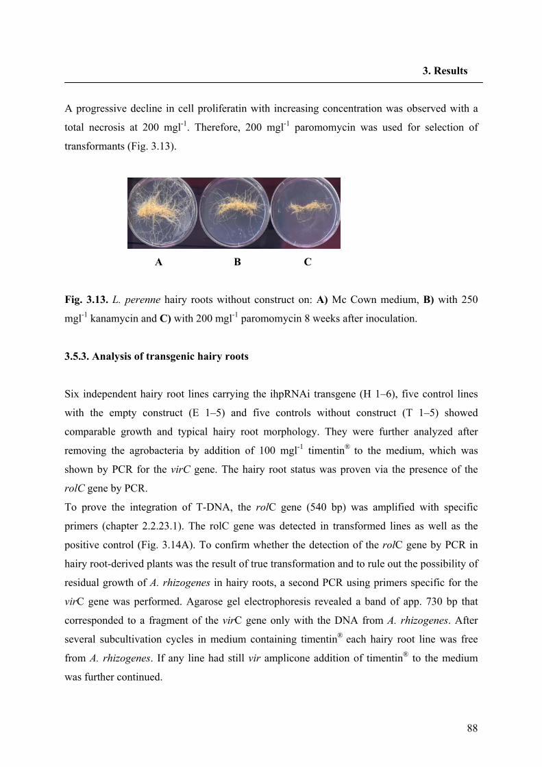
88
3. Results
A progressive decline in cell proliferatin with increasing concentration was observed with a
total necrosis at 200 mgl-1. Therefore, 200 mgl-1 paromomycin was used for selection of
transformants (Fig. 3.13).
A B C
Fig. 3.13. L. perenne hairy roots without construct on: A) Mc Cown medium, B) with 250
mgl-1 kanamycin and C) with 200 mgl-1 paromomycin 8 weeks after inoculation.
3.5.3. Analysis of transgenic hairy roots
Six independent hairy root lines carrying the ihpRNAi transgene (H 1–6), five control lines
with the empty construct (E 1–5) and five controls without construct (T 1–5) showed
comparable growth and typical hairy root morphology. They were further analyzed after
removing the agrobacteria by addition of 100 mgl-1 timentin® to the medium, which was
shown by PCR for the virC gene. The hairy root status was proven via the presence of the
rolC gene by PCR.
To prove the integration of T-DNA, the rolC gene (540 bp) was amplified with specific
primers (chapter 2.2.23.1). The rolC gene was detected in transformed lines as well as the
positive control (Fig. 3.14A). To confirm whether the detection of the rolC gene by PCR in
hairy root-derived plants was the result of true transformation and to rule out the possibility of
residual growth of A. rhizogenes in hairy roots, a second PCR using primers specific for the
virC gene was performed. Agarose gel electrophoresis revealed a band of app. 730 bp that
corresponded to a fragment of the virC gene only with the DNA from A. rhizogenes. After
several subcultivation cycles in medium containing timentin® each hairy root line was free
from A. rhizogenes. If any line had still vir amplicone addition of timentin® to the medium
was further continued.
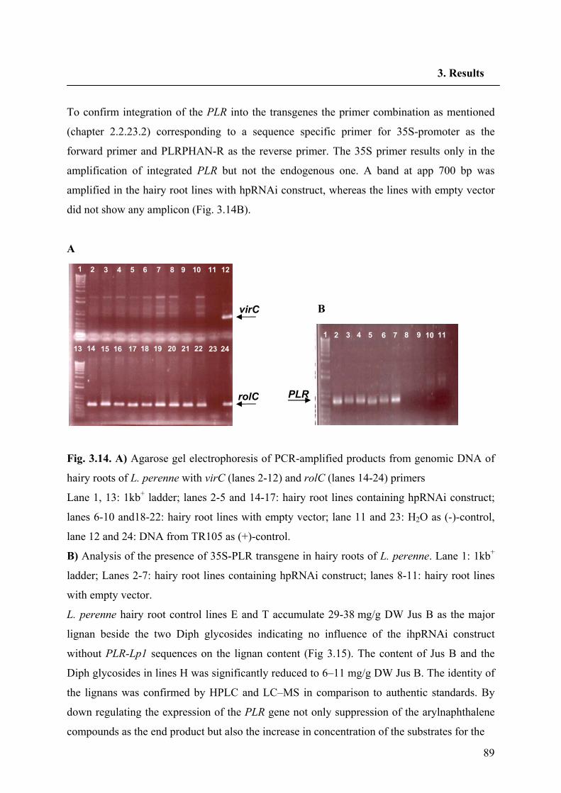
89
3. Results
To confirm integration of the PLR into the transgenes the primer combination as mentioned
(chapter 2.2.23.2) corresponding to a sequence specific primer for 35S-promoter as the
forward primer and PLRPHAN-R as the reverse primer. The 35S primer results only in the
amplification of integrated PLR but not the endogenous one. A band at app 700 bp was
amplified in the hairy root lines with hpRNAi construct, whereas the lines with empty vector
did not show any amplicon (Fig. 3.14B).
A
Fig. 3.14. A) Agarose gel electrophoresis of PCR-amplified products from genomic DNA of
hairy roots of L. perenne with virC (lanes 2-12) and rolC (lanes 14-24) primers
Lane 1, 13: 1kb+ ladder; lanes 2-5 and 14-17: hairy root lines containing hpRNAi construct;
lanes 6-10 and18-22: hairy root lines with empty vector; lane 11 and 23: H2O as (-)-control,
lane 12 and 24: DNA from TR105 as (+)-control.
B) Analysis of the presence of 35S-PLR transgene in hairy roots of L. perenne. Lane 1: 1kb+
ladder; Lanes 2-7: hairy root lines containing hpRNAi construct; lanes 8-11: hairy root lines
with empty vector.
L. perenne hairy root control lines E and T accumulate 29-38 mg/g DW Jus B as the major
lignan beside the two Diph glycosides indicating no influence of the ihpRNAi construct
without PLR-Lp1 sequences on the lignan content (Fig 3.15). The content of Jus B and the
Diph glycosides in lines H was significantly reduced to 6–11 mg/g DW Jus B. The identity of
the lignans was confirmed by HPLC and LC–MS in comparison to authentic standards. By
down regulating the expression of the PLR gene not only suppression of the arylnaphthalene
compounds as the end product but also the increase in concentration of the substrates for the
virC
rolC
1 2 3 4 5 6 7 8 9 10 11 12
13 14 15 16 17 18 19 20 21 22 23 24
1 2 3 4 5 6 7 8 9 10 11
PLR
B
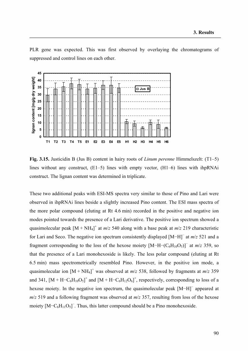
90
3. Results
PLR gene was expected. This was first observed by overlaying the chromatograms of
suppressed and control lines on each other.
0
5
10
15
20
25
30
35
40
45
T1 T2 T3 T4 T5 E1 E2 E3 E4 E5 H1 H2 H3 H4 H5 H6
ligna
n co
nten
t [m
g/g
dry
wei
ght]
Jus B
Fig. 3.15. Justicidin B (Jus B) content in hairy roots of Linum perenne Himmelszelt: (T1–5)
lines without any construct, (E1–5) lines with empty vector, (H1–6) lines with ihpRNAi
construct. The lignan content was determined in triplicate.
These two additional peaks with ESI-MS spectra very similar to those of Pino and Lari were
observed in ihpRNAi lines beside a slightly increased Pino content. The ESI mass spectra of
the more polar compound (eluting at Rt 4.6 min) recorded in the positive and negative ion
modes pointed towards the presence of a Lari derivative. The positive ion spectrum showed a
quasimolecular peak [M + NH4]+ at m/z 540 along with a base peak at m/z 219 characteristic
for Lari and Seco. The negative ion spectrum consistently displayed [M−H]− at m/z 521 and a
fragment corresponding to the loss of the hexose moiety [M−H−(C6H10O5)]− at m/z 359, so
that the presence of a Lari monohexoside is likely. The less polar compound (eluting at Rt
6.5 min) mass spectrometrically resembled Pino. However, in the positive ion mode, a
quasimolecular ion [M + NH4]+ was observed at m/z 538, followed by fragments at m/z 359
and 341, [M + H−C6H10O5]+ and [M + H−C6H12O6]+, respectively, corresponding to loss of a
hexose moiety. In the negative ion spectrum, the quasimolecular peak [M−H]− appeared at
m/z 519 and a following fragment was observed at m/z 357, resulting from loss of the hexose
moiety [M−C6H11O5]−. Thus, this latter compound should be a Pino monohexoside.
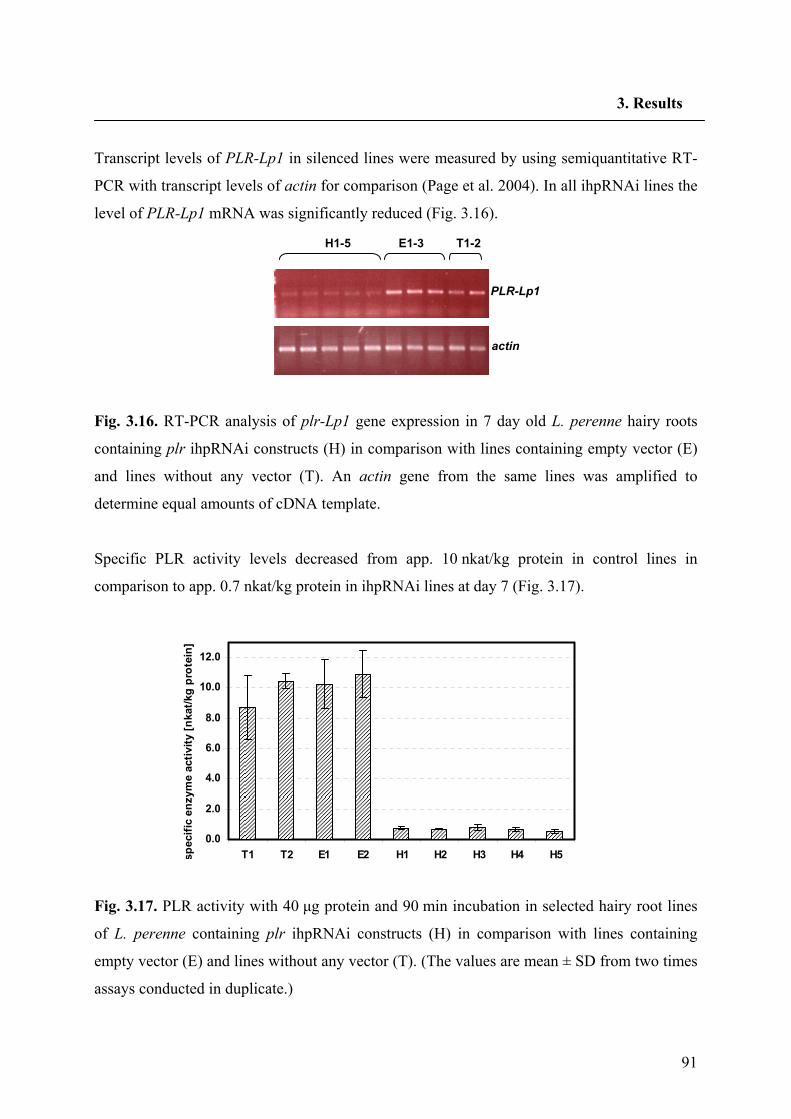
91
3. Results
Transcript levels of PLR-Lp1 in silenced lines were measured by using semiquantitative RT-
PCR with transcript levels of actin for comparison (Page et al. 2004). In all ihpRNAi lines the
level of PLR-Lp1 mRNA was significantly reduced (Fig. 3.16).
Fig. 3.16. RT-PCR analysis of plr-Lp1 gene expression in 7 day old L. perenne hairy roots
containing plr ihpRNAi constructs (H) in comparison with lines containing empty vector (E)
and lines without any vector (T). An actin gene from the same lines was amplified to
determine equal amounts of cDNA template.
Specific PLR activity levels decreased from app. 10 nkat/kg protein in control lines in
comparison to app. 0.7 nkat/kg protein in ihpRNAi lines at day 7 (Fig. 3.17).
0.0
2.0
4.0
6.0
8.0
10.0
12.0
T1 T2 E1 E2 H1 H2 H3 H4 H5spec
ific
enzy
me
activ
ity [n
kat/k
g pr
otei
n]
Fig. 3.17. PLR activity with 40 μg protein and 90 min incubation in selected hairy root lines
of L. perenne containing plr ihpRNAi constructs (H) in comparison with lines containing
empty vector (E) and lines without any vector (T). (The values are mean ± SD from two times
assays conducted in duplicate.)
actin
T1-2E1-3H1-5
PLR-Lp1
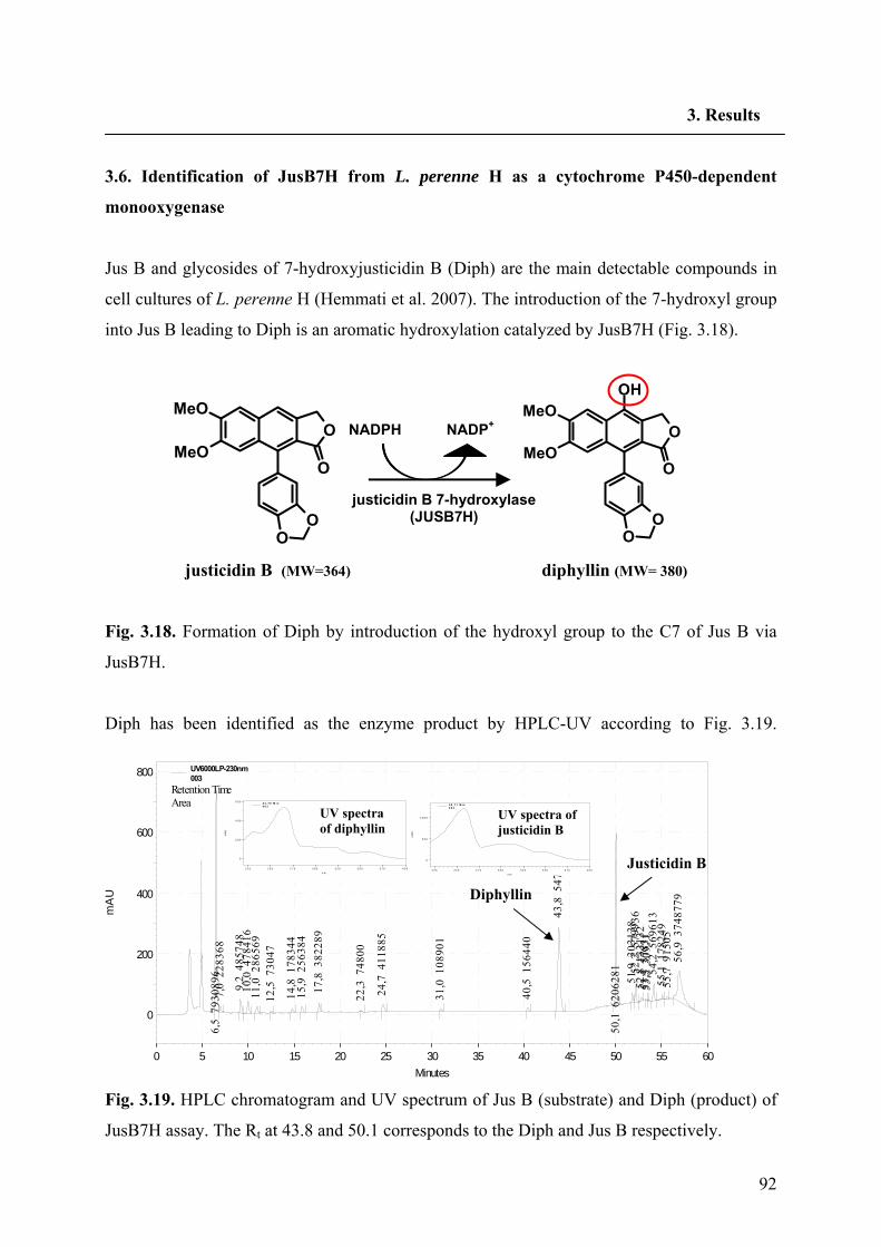
92
3. Results
3.6. Identification of JusB7H from L. perenne H as a cytochrome P450-dependent
monooxygenase
Jus B and glycosides of 7-hydroxyjusticidin B (Diph) are the main detectable compounds in
cell cultures of L. perenne H (Hemmati et al. 2007). The introduction of the 7-hydroxyl group
into Jus B leading to Diph is an aromatic hydroxylation catalyzed by JusB7H (Fig. 3.18).
justicidin B (MW=364) diphyllin (MW= 380)
Fig. 3.18. Formation of Diph by introduction of the hydroxyl group to the C7 of Jus B via
JusB7H.
Diph has been identified as the enzyme product by HPLC-UV according to Fig. 3.19.
Fig. 3.19. HPLC chromatogram and UV spectrum of Jus B (substrate) and Diph (product) of
JusB7H assay. The Rt at 43.8 and 50.1 corresponds to the Diph and Jus B respectively.
O
O
OO
MeO
MeOO
O
OO
MeO
MeOOH
NADPH NADP+
justicidin B 7-hydroxylase (JUSB7H)
Minutes0 5 10 15 20 25 30 35 40 45 50 55 60
mA
U
0
200
400
600
800
6,5
793
0896
7,0
228
368
9,2
485
748
10,0
478
416
11,0
286
569
12,5
730
47
14,8
178
344
15,9
256
384
17,8
382
289
22,3
748
00
24,7
411
885
31,0
108
901
40,5
156
440
43,8
547
4807
50,1
620
6281 51,9
303
138
52,2
579
936
52,8
133
132
53,3
578
3153
,5 3
0971
54,2
569
613
55,1
178
249
55,7
915
0556
,9 3
7487
79
UV6000LP-230nm003
Retention TimeArea
Diphyllin
Justicidin B n m
2 2 5 2 5 0 2 7 5 3 0 0 3 2 5 3 5 0 3 7 5 4 0 0
mAU
0
2 0 0
4 0 0
6 0 0
mAU
0
2 0 0
4 0 0
6 0 04 3 , 7 8 M i n0 0 3
UV spectra of diphyllin
n m
2 2 5 2 5 0 2 7 5 3 0 0 3 2 5 3 5 0 3 7 5 4 0 0
mAU
0
5 0 0
1 0 0 0
mAU
0
5 0 0
1 0 0 0
5 0 , 1 1 M i n0 0 3
UV spectra of justicidin B
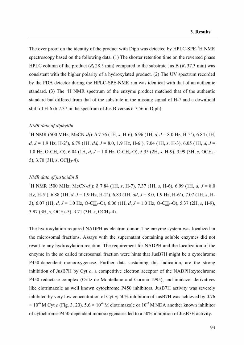
93
3. Results
The over proof on the identity of the product with Diph was detected by HPLC-SPE-1H NMR
spectroscopy based on the following data. (1) The shorter retention time on the reversed phase
HPLC column of the product (Rt 28.5 min) compared to the substrate Jus B (Rt 37.3 min) was
consistent with the higher polarity of a hydroxylated product. (2) The UV spectrum recorded
by the PDA detector during the HPLC-SPE-NMR run was identical with that of an authentic
standard. (3) The 1H NMR spectrum of the enzyme product matched that of the authentic
standard but differed from that of the substrate in the missing signal of H-7 and a downfield
shift of H-6 (δ 7.37 in the spectrum of Jus B versus δ 7.56 in Diph).
NMR data of diphyllin 1H NMR (500 MHz; MeCN-d3): δ 7.56 (1H, s, H-6), 6.96 (1H, d, J = 8.0 Hz, H-5’), 6.84 (1H,
d, J = 1.9 Hz, H-2’), 6.79 (1H, dd, J = 8.0, 1.9 Hz, H-6’), 7.04 (1H, s, H-3), 6.05 (1H, d, J =
1.0 Hz, O-CH2-O), 6.04 (1H, d, J = 1.0 Hz, O-CH2-O), 5.35 (2H, s, H-9), 3.99 (3H, s, OCH3-
5), 3.70 (3H, s, OCH3-4).
NMR data of justicidin B 1H NMR (500 MHz; MeCN-d3): δ 7.84 (1H, s, H-7), 7.37 (1H, s, H-6), 6.99 (1H, d, J = 8.0
Hz, H-5’), 6.88 (1H, d, J = 1.9 Hz, H-2’), 6.83 (1H, dd, J = 8.0, 1.9 Hz, H-6’), 7.07 (1H, s, H-
3), 6.07 (1H, d, J = 1.0 Hz, O-CH2-O), 6.06 (1H, d, J = 1.0 Hz, O-CH2-O), 5.37 (2H, s, H-9),
3.97 (3H, s, OCH3-5), 3.71 (3H, s, OCH3-4).
The hydroxylation required NADPH as electron donor. The enzyme system was localized in
the microsomal fractions. Assays with the supernatant containing soluble enzymes did not
result to any hydroxylation reaction. The requirement for NADPH and the localization of the
enzyme in the so called microsomal fraction were hints that JusB7H might be a cytochrome
P450-dependent monooxygenase. Further data sustaining this indication, are the strong
inhibition of JusB7H by Cyt c, a competitive electron acceptor of the NADPH:cytochrome
P450 reductase complex (Oritz de Montellano and Correia 1995), and imidazol derivatives
like clotrimazole as well known cytochrome P450 inhibitors. JusB7H activity was severely
inhibited by very low concentration of Cyt c; 50% inhibition of JusB7H was achieved by 0.76
× 10-6 M Cyt c (Fig. 3. 20). 5.6 × 10⁻⁴ M clotrimazole or 10-3 M NDA another known inhibitor
of cytochrome-P450-dependent monooxygenases led to a 50% inhibition of JusB7H activity.
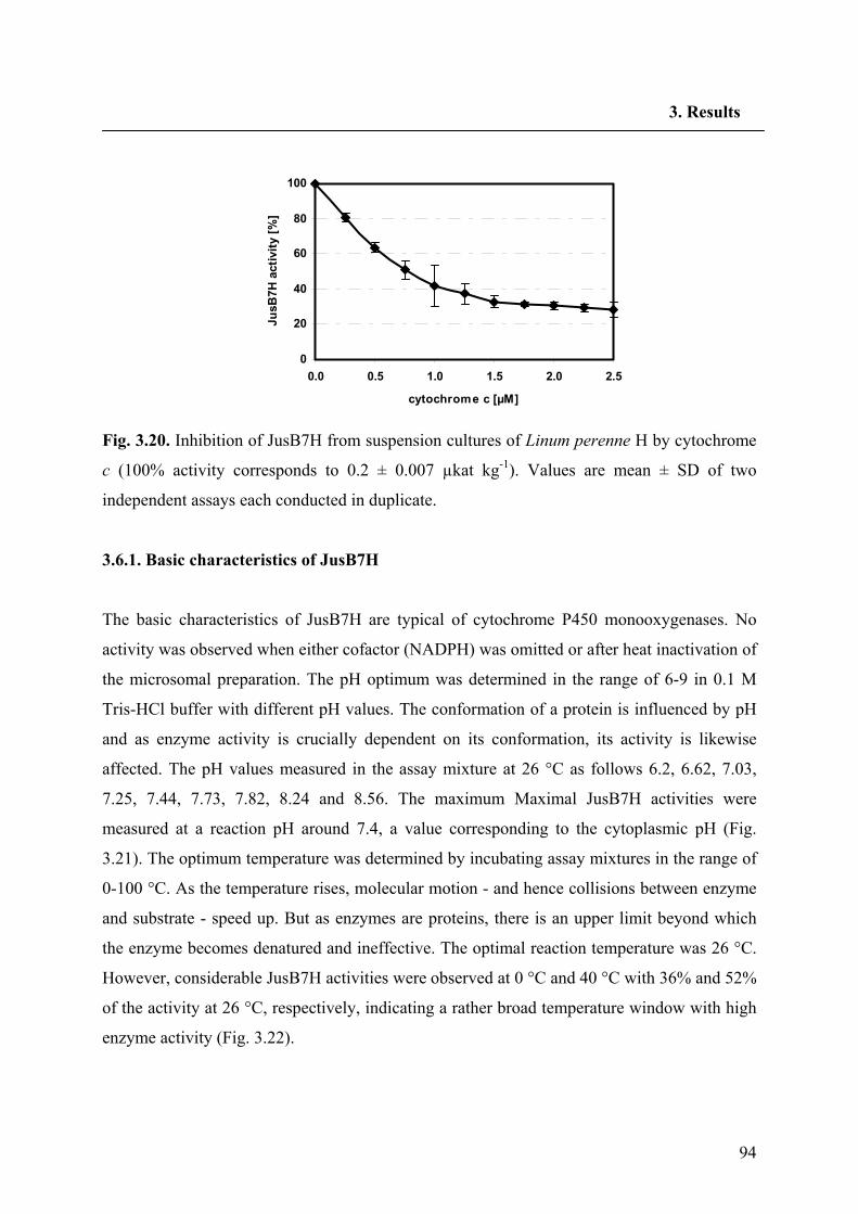
94
3. Results
Fig. 3.20. Inhibition of JusB7H from suspension cultures of Linum perenne H by cytochrome
c (100% activity corresponds to 0.2 ± 0.007 µkat kg-1). Values are mean ± SD of two
independent assays each conducted in duplicate.
3.6.1. Basic characteristics of JusB7H
The basic characteristics of JusB7H are typical of cytochrome P450 monooxygenases. No
activity was observed when either cofactor (NADPH) was omitted or after heat inactivation of
the microsomal preparation. The pH optimum was determined in the range of 6-9 in 0.1 M
Tris-HCl buffer with different pH values. The conformation of a protein is influenced by pH
and as enzyme activity is crucially dependent on its conformation, its activity is likewise
affected. The pH values measured in the assay mixture at 26 °C as follows 6.2, 6.62, 7.03,
7.25, 7.44, 7.73, 7.82, 8.24 and 8.56. The maximum Maximal JusB7H activities were
measured at a reaction pH around 7.4, a value corresponding to the cytoplasmic pH (Fig.
3.21). The optimum temperature was determined by incubating assay mixtures in the range of
0-100 °C. As the temperature rises, molecular motion - and hence collisions between enzyme
and substrate - speed up. But as enzymes are proteins, there is an upper limit beyond which
the enzyme becomes denatured and ineffective. The optimal reaction temperature was 26 °C.
However, considerable JusB7H activities were observed at 0 °C and 40 °C with 36% and 52%
of the activity at 26 °C, respectively, indicating a rather broad temperature window with high
enzyme activity (Fig. 3.22).
0
20
40
60
80
100
0.0 0.5 1.0 1.5 2.0 2.5
cytochrome c [µM]
JusB
7H a
ctiv
ity [%
]
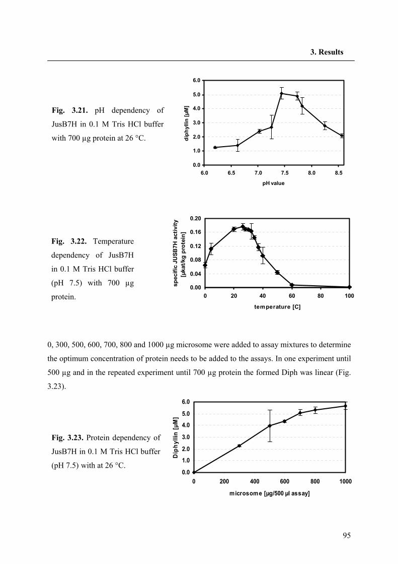
95
3. Results
0, 300, 500, 600, 700, 800 and 1000 µg microsome were added to assay mixtures to determine
the optimum concentration of protein needs to be added to the assays. In one experiment until
500 µg and in the repeated experiment until 700 µg protein the formed Diph was linear (Fig.
3.23).
0.0
1.0
2.0
3.0
4.0
5.0
6.0
6.0 6.5 7.0 7.5 8.0 8.5
pH valuedi
phyl
lin [µ
M]Fig. 3.21. pH dependency of
JusB7H in 0.1 M Tris HCl buffer
with 700 µg protein at 26 °C.
Fig. 3.23. Protein dependency of
JusB7H in 0.1 M Tris HCl buffer
(pH 7.5) with at 26 °C.
Fig. 3.22. Temperature
dependency of JusB7H
in 0.1 M Tris HCl buffer
(pH 7.5) with 700 µg
protein.
0.0
1.0
2.0
3.0
4.0
5.0
6.0
0 200 400 600 800 1000
microsome [µg/500 µl assay]
Dip
hylli
n [µ
M]
0.00
0.04
0.08
0.12
0.16
0.20
0 20 40 60 80 100
temperature [C]
spec
ific
JUSB
7H a
ctiv
ity
[µka
t/kg
prot
ein]
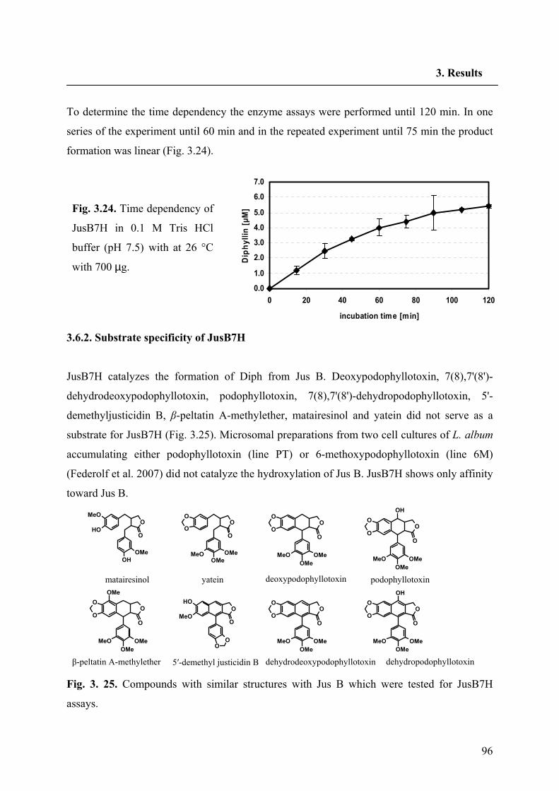
96
3. Results
To determine the time dependency the enzyme assays were performed until 120 min. In one
series of the experiment until 60 min and in the repeated experiment until 75 min the product
formation was linear (Fig. 3.24).
3.6.2. Substrate specificity of JusB7H
JusB7H catalyzes the formation of Diph from Jus B. Deoxypodophyllotoxin, 7(8),7'(8')-
dehydrodeoxypodophyllotoxin, podophyllotoxin, 7(8),7'(8')-dehydropodophyllotoxin, 5'-
demethyljusticidin B, β-peltatin A-methylether, matairesinol and yatein did not serve as a
substrate for JusB7H (Fig. 3.25). Microsomal preparations from two cell cultures of L. album
accumulating either podophyllotoxin (line PT) or 6-methoxypodophyllotoxin (line 6M)
(Federolf et al. 2007) did not catalyze the hydroxylation of Jus B. JusB7H shows only affinity
toward Jus B.
Fig. 3. 25. Compounds with similar structures with Jus B which were tested for JusB7H
assays.
Fig. 3.24. Time dependency of
JusB7H in 0.1 M Tris HCl
buffer (pH 7.5) with at 26 °C
with 700 µg.
OH
OH
OMe
MeOO
O
OMe
O
O
O
O
MeOOMe
OHO
OMeO
OO
O
O
O
O
OMeOMe
MeO
O
O
O
O
OMeOMe
MeO
OH
O
O
O
O
OMeOMe
MeO
OMe
O
O
O
O
OMeOMe
MeO
O
O
O
O
OMeOMe
MeO
OH
matairesinol yatein deoxypodophyllotoxin podophyllotoxin
β-peltatin A-methylether dehydrodeoxypodophyllotoxin dehydropodophyllotoxin 5′-demethyl justicidin B
0.0
1.0
2.0
3.0
4.0
5.0
6.0
7.0
0 20 40 60 80 100 120
incubation time [min]
Dip
hylli
n [µ
M]
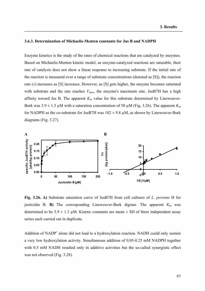
97
3. Results
3.6.3. Determination of Michaelis-Menten constants for Jus B and NADPH
Enzyme kinetics is the study of the rates of chemical reactions that are catalyzed by enzymes.
Based on Michaelis-Menten kinetic model, as enzyme-catalyzed reactions are saturable, their
rate of catalysis does not show a linear response to increasing substrate. If the initial rate of
the reaction is measured over a range of substrate concentrations (denoted as [S]), the reaction
rate (v) increases as [S] increases. However, as [S] gets higher, the enzyme becomes saturated
with substrate and the rate reaches Vmax, the enzyme's maximum rate. JusB7H has a high
affinity toward Jus B. The apparent Km value for this substrate determined by Lineweaver-
Burk was 3.9 ± 1.3 µM with a saturation concentration of 50 µM (Fig. 3.26). The apparent Km
for NADPH as the co-substrate for JusB7H was 102 ± 9.8 µM, as shown by Lineweaver-Burk
diagrams (Fig. 3.27).
A B
0.00
0.05
0.10
0.15
0.20
0 50 100 150 200
Justicidin B [µM]
spec
ific
JusB
7H a
ctiv
ity
[µka
t/kg
prot
ein]
-5
0
5
10
15
20
-1.0 -0.5 0.0 0.5 1.0
1/S [1/µM]
1/v
[kg
prot
ein/
µkat
]
Fig. 3.26. A) Substrate saturation curve of JusB7H from cell cultures of L. perenne H for
justicidin B. B) The corresponding Lineweaver-Burk digram. The apparent Km was
determined to be 3.9 ± 1.3 µM. Kinetic constants are mean ± SD of three independent assay
series each carried out in duplicate.
Addition of NADP+ alone did not lead to a hydroxylation reaction. NADH could only sustain
a very low hydroxylation activity. Simultaneous addition of 0.05-0.25 mM NADPH together
with 0.5 mM NADH resulted only in additive activities but the so-called synergistic effect
was not observed (Fig. 3.28).
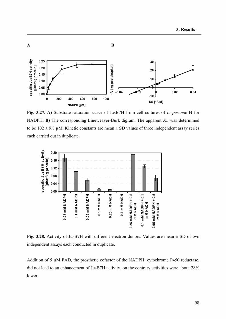
98
3. Results
A B
0.00
0.05
0.10
0.15
0.20
0.25
0 200 400 600 800 1000
NADPH [µM]
spec
ific
JusB
7H a
ctiv
ity
[µka
t/kg
prot
ein]
-10
0
10
20
30
-0.04 -0.02 0 0.02 0.04
1/S [1/µM]
1/v
[kg
prot
ein/
µkat
]
Fig. 3.27. A) Substrate saturation curve of JusB7H from cell cultures of L. perenne H for
NADPH. B) The corresponding Lineweaver-Burk digram. The apparent Km was determined
to be 102 ± 9.8 µM. Kinetic constants are mean ± SD values of three independent assay series
each carried out in duplicate.
0.00
0.04
0.08
0.12
0.16
0.20
0.25
mM
NA
DPH
0.1
mM
NA
DPH
0.05
mM
NA
DPH
0.5
mM
NA
DH
0.25
mM
NA
DH
0.1
mM
NA
DH
0.25
mM
NA
DPH
+ 0
.5m
M N
AD
H
0.1
mM
NA
DPH
+ 0
.5m
M N
AD
H
0.05
mM
NA
DPH
+ 0
.5m
M N
AD
H
spe
cific
Jus
B7H
act
ivity
[µka
t/kg
prot
ein]
Fig. 3.28. Activity of JusB7H with different electron donors. Values are mean ± SD of two
independent assays each conducted in duplicate.
Addition of 5 µM FAD, the prosthetic cofactor of the NADPH: cytochrome P450 reductase,
did not lead to an enhancement of JusB7H activity, on the contrary activities were about 28%
lower.
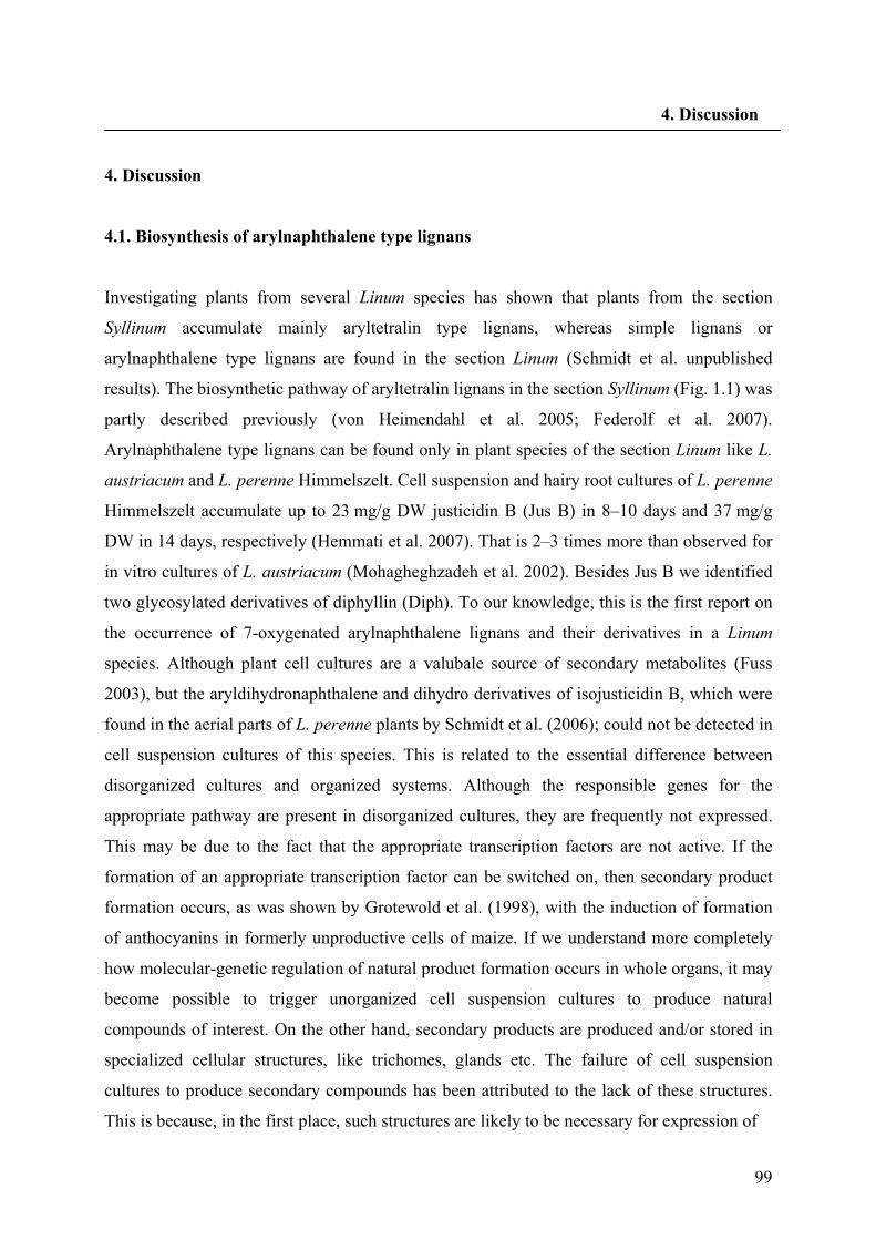
99
4. Discussion
4. Discussion
4.1. Biosynthesis of arylnaphthalene type lignans
Investigating plants from several Linum species has shown that plants from the section
Syllinum accumulate mainly aryltetralin type lignans, whereas simple lignans or
arylnaphthalene type lignans are found in the section Linum (Schmidt et al. unpublished
results). The biosynthetic pathway of aryltetralin lignans in the section Syllinum (Fig. 1.1) was
partly described previously (von Heimendahl et al. 2005; Federolf et al. 2007).
Arylnaphthalene type lignans can be found only in plant species of the section Linum like L.
austriacum and L. perenne Himmelszelt. Cell suspension and hairy root cultures of L. perenne
Himmelszelt accumulate up to 23 mg/g DW justicidin B (Jus B) in 8–10 days and 37 mg/g
DW in 14 days, respectively (Hemmati et al. 2007). That is 2–3 times more than observed for
in vitro cultures of L. austriacum (Mohagheghzadeh et al. 2002). Besides Jus B we identified
two glycosylated derivatives of diphyllin (Diph). To our knowledge, this is the first report on
the occurrence of 7-oxygenated arylnaphthalene lignans and their derivatives in a Linum
species. Although plant cell cultures are a valubale source of secondary metabolites (Fuss
2003), but the aryldihydronaphthalene and dihydro derivatives of isojusticidin B, which were
found in the aerial parts of L. perenne plants by Schmidt et al. (2006); could not be detected in
cell suspension cultures of this species. This is related to the essential difference between
disorganized cultures and organized systems. Although the responsible genes for the
appropriate pathway are present in disorganized cultures, they are frequently not expressed.
This may be due to the fact that the appropriate transcription factors are not active. If the
formation of an appropriate transcription factor can be switched on, then secondary product
formation occurs, as was shown by Grotewold et al. (1998), with the induction of formation
of anthocyanins in formerly unproductive cells of maize. If we understand more completely
how molecular-genetic regulation of natural product formation occurs in whole organs, it may
become possible to trigger unorganized cell suspension cultures to produce natural
compounds of interest. On the other hand, secondary products are produced and/or stored in
specialized cellular structures, like trichomes, glands etc. The failure of cell suspension
cultures to produce secondary compounds has been attributed to the lack of these structures.
This is because, in the first place, such structures are likely to be necessary for expression of
100
4. Discussion
the biosynthetic pathway, and secondly, the products are probably phytotoxic and failure to
sequester them may inhibit growth or kill the culture.
Biosynthesis of arylnaphthalene lignans was not investigated up to now but we have assumed
that the first steps of their biosynthetic pathway are catalyzed by the bifunctional enzyme
pinoresinol lariciresinol reductase (PLR). We cloned a PLR cDNA from L. perenne
Himmelszelt cell cultures (PLR-Lp1). The recombinant PLR-Lp1 completely used up the
racemic Pino before the resulting Lari was completely converted to Seco. Therefore, PLR-
Lp1 seems to work more like a pinoresinol reductase than a pinoresinol lariciresinol
reductase. It becomes obvious that the number of metabolites found in one species exceeds
the number of genes involved in their biosynthesis. The concept of one gene-one mRNA-one
protein is collapsing (Schwab 2003). Many present-day proteins are known to have the
capability of accepting more than one substrate, catalyzing more than one reaction and
forming more than one product. They are called multifunctional enzymes. The presence of
multifunctional proteins significantly enhances the metabolic efficiency of a cell and also
metabolome diversity. Since many proteins involved in the biosynthesis of secondary plant
metabolites are multifunctional, fewer enzymes are required to synthesize the various
structures. Schwab (2003) has reviewed examples of alcohol acyl-CoA transferases, O-
methyltransferases, terpene synthases and glycosyltransferases which have multifunctional
activity. PLR catalyzes the two consequent early steps of lignan biosynthesis and works as a
bifunctional enzyme.
To prove whether PLR-Lp1 is involved in the biosynthesis of arylnaphthalene lignans,
ihpRNAi experiments were carried out. The effect of gene suppression to find metabolic
pathway intermediates has been shown in many studies (Allen et al. 2004; Courdavault et al.
2005; Frick et al. 2004; Kumagai and Kouchi 2003; Nishihara et al. 2005). Chimeric genes in
which the coding regions are fused to regulatory sequences from other sources to create
artificial genes can be generated for transcription and translation in plants (Miki 2002). The
PLR part of L. perenne was amplified with polylinkers and cloned in the pGEM®-T vector.
To form the ihPRNA constructs the fragment was subcloned in the sense and antisense
orientation of a pHANNIBAL vector. hpRNA constructs require two copies of the target
sequence in an inverted-repeat orientation, in order to produce duplex RNA. Smith et al.
(2000) have found that it is necessary to introduce a spacer region between the arms of
hpRNA constructs for the stability of the inverted repeats in E. coli. However, replacing the
101
4. Discussion
spacer region of hpRNA constructs with a functional intron sequence increases the proportion
of independent silenced lines recovered from 50% to 100%. Hence Wesely et al. (2001) built
up a generic intron-spliced hpRNA (ihpRNA) vector by the name of pHANNIBAL, into
which the gene of choice could be directionally cloned to make sense and antisense arms.
Enhancement of the silencing efficiency by the presence of intron is that, the process of intron
excision from the construct by spliceosome might help to align the complementary arms of
the hairpin in an environment favouring RNA hybridization, promoting the formation of a
duplex. Alternatively, splicing may transiently increase the amount of hairpin RNA by
facilitating, or retarding, the hairpin′s passage from the nucleus, or by creating a smaller, less
nuclease-sensitive loop (Smith et al. 2000). pHANNIBAL based constructs are driven by the
constitutive 35S promoter. The 35S promoter is a typical example of a plant promoter from
cauliflower mosaic virus (CaMV), which is often used for constitutive expression. 35S as a
strong promoter causes high levels of gene expression in dicot plants. Trans-acting vir
functions encoded by the Ri plasmids and by the Agrobacterium chromosome act to promote
the integration into the plant genome of DNA segments which are delimited by cis-acting T-
DNA border sequences. This has led to the construction of binary vectors for use in
Agrobacterium-mediated plant transformation (MacBride and Summerfelt 1990). pART27 as
a binary vector, carries the RK2 minimal replicon for maintenance in Agrobacterium, the
ColE1 origin of replication for high-copy maintenance in E. coli and the Tn7
spectinomycin/stereptomycin resistance gene as a bacterial selectable marker. The
organizational structure of the T-DNA of pART27 has been constructed taking into account
the right to left border, 5′ to 3′ model of T-DNA transfer. The T-DNA carries the chimaeric
kanamycin resistance gene (nopaline synthase promoter-neomycin phsphotransferase-
nopaline synthase terminator) distal to the lacZ′ region. Timentin® which is mixture of
ticarcillin disodium and clavulanate potassium in the ratio of 15:1 (Duchefa 2003) was added
to the media to remove the agrobacteria two days after transformation. Since hairy roots of L.
perenne accumulate a very high amount of Jus B it was a good starting point to conclude the
influence of downregulation of possible genes maybe involved in arylnaphthalene
biosynthesis. The hairy roots without construct had shown resistance to kanamycin,
paromomycin (200 mgl-1) was next used as the selection marker for transformed lines. Park
and Facchini (2000) have used 50 mgl-1 paromomycin for the selection of the hairy roots of
Papaver somniferum and Eschscholzia californica but it can result in the observation of
102
4. Discussion
false positive lines. The hairy root lines harboring the RNAi construct containe the nptII gene.
nptII encodes the neomycin phosphotransferase II which results in the phosphorylation of
paromomycin. The comparison of PLR-Lp1 mRNA level, PLR-Lp1 activities and lignan
accumulation in hairy root lines which were transformed with only agrobacteria or with
agrobacteria with an empty vector control showed no difference between these lines. In
contrast, lines transformed with the ihpRNAi construct for plr gene suppression show
significant reduction of mRNA level and enzyme activities for PLR-Lp1 and significantly
lowered accumulation of Jus B and the Diphs. The level of Pino increased in these lines. Two
additional peaks never observed in the control lines which could be glycosides of Pino and
Lari were detected. The compounds can not be Pino and Lari monoglucosides since β-
glucosidases are used in our lignan extraction method. But, it is possible that Pino and Lari
can serve as substrates for the same enzymes which add the sugar moieties to the Diphs
accumulated in the in vitro cultures of L. perenne H. Nevertheless, the exact structures of the
compounds still remain to be identified. Since the RNAi construct was cloned in such a
manner that it is not highly specific for PLR-Lp1 but to silence possible PLR-like genes too, it
can be concluded that at least PLR-Lp1 is involved in the biosynthesis of Jus B and Diph
derivatives.
In order to be converted to Diph, Jus B has to be hydroxylated at position 7 of the aromatic
ring B. Several hydroxylases introducing OH-groups into aromatic rings have been shown to
be cytochrome P450-dependent monooxygenases, e.g. cinnamic acid 4-hydroxylase (Russell
and Conn 1967; Russell 1971), tabersonine 16-hydroxylase (St-Pierre and De Luca 1995),
Xanthon 6-hydroxylase (Schmidt et al. 2000), 8-dimethylallylnaringenin 2'-hydroxylase
(Yamamoto et al. 2001) and deoxypodophyllotoxin 6-hydroxylase (Molog et al. 2001;
Federolf et al. 2007). Subcellular localization in the microsomal fraction is typical of this
enzyme class. JusB7H activity was determined in the microsomal fraction after MgCl2-
precipitation and centrifugation at 48000 g. The supernatant of this centrifugation step
contained no hydroxylating activities. The CYP450 from plant sources are usually obtained in
microsomes. The microsomal preparations may include vesicles derived from the breakage of
various membranes such as those of ER, Golgi, plasma membrane, tonoplast, plastid
envelopes, nucleus and peroxisomes. The subcellular origins of microsomal membranes have
been determined in only a few cases. They are often ER bound however
immunohistochemical localization, would be specifically useful for this purpose (Donaldson
103
4. Discussion
and Luster 1991). This first hint that JusB7H is a cytochrome P450-dependent
monooxygenase was verified by the inhibition of the reaction by Cyt c, an electron acceptor
competing for electrons transferred from a NADPH:cytochrome P450 reductase to
cytochrome P450. As shown in the Fig. 1.15 NADPH:CYP450 reductase can donate electrons
in vitro to Cyt c as well as to CYP450, thus addition of exogenous Cyt c to a CYP450 reaction
can inhibit the reaction [some reversible inhibitors compete with substrates for occupancy of
the active site by coordinating to the heme iron atom (Oritz de Montellano 2005)]. 50%
inhibition of JusB7H activity was reached by the addition of 0.76 × 10⁻⁶ M Cyt c. This value
is comparable to concentrations necessary to inhibit other cytochrome P450 monooxygenases.
1.6 × 10⁻⁶ M Cyt c leads to a 50% inhibition of DOP6H of L. album (Federolf et al. 2007).
50% inhibition of tabersonine 16-hydroxylase is achieved by 10-6 M Cyt c (St-Pierre and De
Luca 1995). An inhibition of 90% for DOP6H of L. flavum was observed at a concentration of
2.5 µM Cyt c (Molog et al. 2001). The N-substituted imidazols like CLOT (Fig. 4.1) were
effective inhibitors for JusB7H. 5.6 × 10⁻⁴ M CLOT led to a 50% inhibition of JusB7H
activity. The activity of DOP6H from L. album and tabersonine 16-hydroxylase from
Catharanthus roseus is inhibited to 50 % by 1.1 × 10⁻⁵ M and 5 × 10⁻⁵ M CLOT respectively
(Federolf et al. 2007; St-Pierre and De Luca 1995). Tetcyclacis (NDA), a potent inhibitor of
cytochrome P450 enzymes showed less pronounced inhibition of JusB7H. 50% inhibition of
JusB7H activity is reached by the addition of 10-3 M NDA. 1.1 × 10⁻⁵ M and 10-4 M NDA is
sufficient to reach a 50% inhibition of DOP6H from L. album and tabersonine 16-hydroxylase
from Catharanthus roseus, respectively (Federolf et al. 2007; St-Pierre and De Luca 1995). A
structural feature common to tetcyclacis and the triazole-type compounds is an sp2-hybridised
nitrogen atom located at the periphery of the molecule in a heterocycle. It appears likely that
the electron-pair on this atom interacts with the central iron atom of cytochrome P-450 in the
enzymes, resulting in inhibition (Rademacher et al. 1987). Ancymidol as pyrimidine
derivative did not inhibit JusB7H, this was the case for a lot of other CYP450s like
monoterpene hydroxylation in mints (Karp et al. 1990). Further characteristics of JusB7H are
typical of cytochrome P450-dependent hydroxylases, e.g. the pH optimum is around 7.4, the
pH range of the cytoplasm. JusB7H is strictly dependent on NADPH as an electron donor,
whereas NADH can only sustain very low hydroxylation activities. For some cytochrome
P450-dependent reactions, simultaneous addition of NADPH and NADH lead to a synergistic
effect of these two reducing equivalents (Clemens et al. 1993; Gerardy and Zenk 1993).
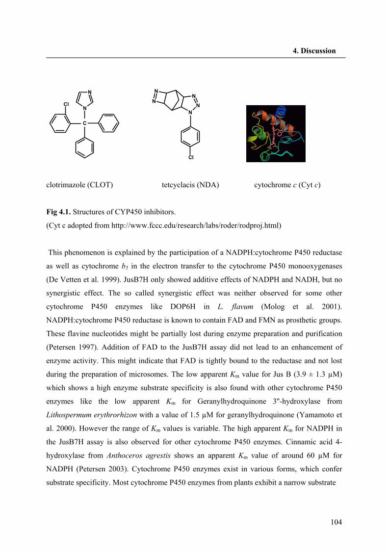
104
4. Discussion
C
N
N
Cl
NN
NN
N
Cl
clotrimazole (CLOT) tetcyclacis (NDA) cytochrome c (Cyt c)
Fig 4.1. Structures of CYP450 inhibitors.
(Cyt c adopted from http://www.fccc.edu/research/labs/roder/rodproj.html)
This phenomenon is explained by the participation of a NADPH:cytochrome P450 reductase
as well as cytochrome b5 in the electron transfer to the cytochrome P450 monooxygenases
(De Vetten et al. 1999). JusB7H only showed additive effects of NADPH and NADH, but no
synergistic effect. The so called synergistic effect was neither observed for some other
cytochrome P450 enzymes like DOP6H in L. flavum (Molog et al. 2001).
NADPH:cytochrome P450 reductase is known to contain FAD and FMN as prosthetic groups.
These flavine nucleotides might be partially lost during enzyme preparation and purification
(Petersen 1997). Addition of FAD to the JusB7H assay did not lead to an enhancement of
enzyme activity. This might indicate that FAD is tightly bound to the reductase and not lost
during the preparation of microsomes. The low apparent Km value for Jus B (3.9 ± 1.3 µM)
which shows a high enzyme substrate specificity is also found with other cytochrome P450
enzymes like the low apparent Km for Geranylhydroquinone 3''-hydroxylase from
Lithospermum erythrorhizon with a value of 1.5 µM for geranylhydroquinone (Yamamoto et
al. 2000). However the range of Km values is variable. The high apparent Km for NADPH in
the JusB7H assay is also observed for other cytochrome P450 enzymes. Cinnamic acid 4-
hydroxylase from Anthoceros agrestis shows an apparent Km value of around 60 µM for
NADPH (Petersen 2003). Cytochrome P450 enzymes exist in various forms, which confer
substrate specificity. Most cytochrome P450 enzymes from plants exhibit a narrow substrate
105
4. Discussion
specificity (Donaldson and Luster 1991, Sandermann 1992), which is also true for JusB7H.
Structurally related substances were not converted to the related hydroxylated products with
microsomal preparations of L. perenne H. Neither 7(8),7'(8')-dehydrodeoxypodophyllotoxin
nor deoxypodophyllotoxin and β-peltatin A-methylether with an aromatic and aliphatic ring
C, respectively, served as substrate for JusB7H. Jus B is the only known substrate for JusB7H
until now. These observations demonstrate that JusB7H of L. perenne H cell cultures is a
cytochrome P450-dependent monooxygenase which hydroxylates the C7 position of Jus B to
give Diph as the only product. Cloning and characterization of a cDNA encoding JusB7H will
be the topic for further studies. Since the cell cultures of L. perenne H accumulate Diph only
in glycosidic form Diph has to be converted to diglycosides which are more water soluble and
can be stored in the vacuole. Our data together with the cloning of a PLR of L. perenne H and
the findings of dihydroarylnaphthalenes in the aerial part of this plant species support our
hypothetical pathway leading to diphyllin glycosides (Fig. 1.8).
4.2. Enantiospecificity in lignan biosynthesis
The majority of naturally occurring lignans are optically active as one enantiomer or mixtures
with various enantiomeric compositions (Ayers and Loike, 1990). While the Seco isolated
from Forsythia plants was the pure (-)- enantiomer (Umezawa et al. 1990), this lignan isolated
from Wikstroemia sikokiana and A. lappa is a mixture of both (+)- and (-)-enantiomers
(Suzuki et al. 2002). The hydrolytically released SDG derived from the various lignan
oligomers in flax seeds is composed of ~99% (+)- and ~1% (-)-Seco. But in contrast the aerial
parts of flowering L. usitatissimum plants, only contain (-)-enantiomers of lignans derived
from (-)-Seco (Schmidt et al. 2006). Regardless the sign of optical rotation, Matai as a
dibenzylbutyrolactone lignan is the first enantiomeric pure compound in lignan biosynthesis
in many cases (Umezawa, 2003). Therefore, it seems that enantiomeric purity in lignan
biosynthesis is determined during the first steps, but on different levels.
Based on the SDG diastereomers two distinct forms of stereoselective coupling control should
be mediated in L. usitatissimum: that is each differentially engenders the formation of either
(+)-Pino or its corresponding antipode from coniferyl alcohol (Davin and Lewis, 2005). It is
argued that a dirigent gene homolog forming (+)-Pino has been heterologously expressed
from flaxseed (Davin and Lewis, 2003). The dirigent gene responsible for the formation of
106
4. Discussion
(-)-Pino is not reported yet. A cDNA encoding (-)-PLR (PLR-Lu1) was isolated from cell
cultures of L. usitatissimum (von Heimandahl et al. 2005). Enzyme assays with purified
recombinant PLR-Lu1 results in the formation of (+)-Seco. By action of PLR-Lu1, (+)-Seco
was rapidly formed by the reduction of (-)-Pino. Same as the PLR-Tp1 from T. plicata (Fujita
et al. 1999), the formation of (-)-Lari as an intermediate was not observed for PLR-Lu1.
Southern analysis indicated that the genome of L. usitatissimum contains a small gene family
of PLRs (von Heimendahl et al. 2005). The question is that whether consequent reductive
transformations to (+)- and (-)-Seco are performed by a nonspecific PLR enzyme or via at
least two PLR isoforms with different enantiospecificity. We have cloned a PLR cDNA
(PLR-Lu2) from the leaves of L. usitatissimum responsible for the formation of (-)-Seco. Thus
it could be rationalized that two pathways utilizing (-)- and (+)-PLR, being involved in the
formation of (+)- and (-)-Seco, respectively (Fig. 1.11). By the recombinant PLR-Lu2
approximately 50% of Pino was used up and the resulting Lari was completely converted to
Seco. Enantiomeric composition of the substrates and the products of the assays during 40
min was analysed. First (+)-Lari forms from (+)-Pino until no (+)-Pino is left. Then (-)-Seco
can be formed from (+)-Lari. In F. intermedia formation of Seco was only retarded and
occurred already when (+)-Pino was still in the reaction mixture (Katayama et al. 1993). PLRs
of T. plicata work only strictly enantiospecific in the formation of Seco from Lari (Fujita et al.
1999).
In plant species like A. lappa which accumulate lignans with opposite stereochemistry in
different organs no PLR cDNAs were cloned and also in T. plicata containing two PLR
classes with opposite enantiospecificity the relationship between the expression of PLRs and
enantiomeric composition of lignans in the plant is not reported. Now, with the PLR-Lu1 and
PLR-Lu2 sequence in hand it was possible to compare the expression patterns of both PLRs
with enantiomeric composition of the lignans in different organs of L. usitatissimum. As it is
expected in leaves and stems of L. usitatissimum accumulating the (-)-enantiomers of lignans,
only PLR-Lu2 which is responsible for the formation of (-)-Seco was transcriptionally active
(Fig. 3.5). Both PLR-Lu1 and PLR-Lu2 transcripts were present in seeds and contribute to the
synthesis of (+)- and (-)-Seco, respectively. As an additional complexity, PLR-Lu1 and PLR-
Lu2 were similarly expressed in seeds which is in contrast to the ratio of the SDG
diastereomers reported for the flaxseed. An explanation is that (+)-Seco is used mainly as a
107
4. Discussion
substrate for UDPG:glucosyltransferase to yield (+)-SDG but the (-)-Seco is further channeled
to the biosyntheis of (-)-yatein. However, the precise ratio of (+)- to (-)-SDG and other types
of lignans in flaxseed should be defined and the expression pattern of dirigent proteins
responsible for the formation of (+)- and (-)-Pino should be investigated.
Thus far it can be concluded that the enantiomeric composition of lignans in L. usitatissimum
is controlled by at least two separate gene subsets. PLR-Lu1 which is responsible for the
upstream steps in (+)-Seco biosynthesis and PLR-Lu2 in contrast catalyzes the formation of (-
)-Seco leads to the downstream products like (-)-yatein. The pattern of transcription levels of
both genes suggests that PLRs with different enantiospecificity are essential components of
metabolic control in various organs of the L. usitatissimum plant.
As mentioned , PLR of L. perenne (PLR-Lp1) was not enantiospecific and could utilize both
(+)- and (-)-enantiomers of Pino and Lari. However Fujita et al. (1999) have emphasized that
in assays with PLR-Tp1, if allowed to continue until depletion of the (±)-Pino occurs the (-)-
antipode is fully converted to (+)-Seco, whereas the (+)-antipode is only transformed to (+)-
Lari.
We analysed the enantiomeric composition of the substrate and the products of assays with
different protein concentrations in order to get hints for the enantiospecificity of PLR-Lp1.
The protein shows preference for (+)-Pino (R,R configuration at C-atoms 8,8′) in the first
reaction step, but preference for (−)-Lari (S,S configuration at C-atoms 8,8′) in the second
reaction step. All other PLRs cloned so far, like PLRs from T. plicata, F. intermedia, L.
album and L. usitatissimum, at least prefer for both reaction steps either the R,R configurated
or S,S configurated lignans (Dinkova-Kostova et al. 1996; Fujita et al. 1999; von Heimendahl
et al. 2005). Therefore, PLR-Lp1 is the first PLR with opposite enantiospecificity toward Pino
and Lari. This fact together with the preference for both Pino enantiomers before Lari
indicates that another gene encoding a PLR which is at least more specific for the conversion
of Lari to Seco could be present in the genome of L. perenne Himmelszelt which is also
indicated by Southern analysis. Katayama et al. (1993) have shown that the enzyme PLR is
not only highly enantiospecific, as it can also distinguish between the diastereomers (+)-Pino
and (+)-epipinoresinol, with the later apparently not serving as a substrate for reduction, i. e.
the difference in stereochemistry at C7' between Pino and epiPino deleteriously affects either
molecular recognition (binding) by the enzyme, or its subsequent reduction on the enzyme
active site.
108
4. Discussion
Several enzymes like PLR-Lu1 and PLR-Lu2 are encoded by multiple genes that are
regulated differently in response to a range of environmental and developemental stimuli,
adding genetic complexity. The isoenzymes encoded by a gene family show overlapping
catalytic activities towards the different substrates. This implies that mutations in one gene
does not necessarily lead to the loss of secondary plant metabolites but results in the moderate
shift of the product pattern. A single amino acid alteration in a protein can lead to a modified
substrate preference and an increase of a small number of metabolites or even formation of
new compounds depending on substrate availability. If the compounds are profitable for the
plant the individuals carrying the mutation will rapidly propagate. A mutation in a
multifunctional enzyme leads to a gradual shift in the composition of secondary plant
metabolite rather than the appearance of a new metabolite at the expense of an existing
compound.
Multifunctional isoenzymes are an ideal playground for evolution. Plants appear to have
adapted existing enzymes to their evolving needs, rather than inventing entirely novel
mechanisms. Multifunctional enzymes have already been proposed as the ancestor of present-
day enzyme (Jensen 1976). These broad specificity enzyme may be recruited for new
metabolic pathways, followed by further evolution toward more specific and efficient
catalysts. Its follows that gene duplication is an important factor in this concept as it provides
the raw material for the acquisition of new biosynthetic pathways.
As mentioned in the chapters 3.2.1 and 3.3.1 both PLR-Lu2 and PLR-Lp1 have high levels of
similarity to the other members of the PIP family. PCBERs, PLRs and IFRs are quite closely
related from a sequence homology perspective, i.e. PCBER is ~66% similar/~45% identical to
PLRs and ~65% similar/50% identical to IFRs (Davin and Lewis 2003). Significant homology
was noted for (+)-PLR with various IFRs from the legumes. This is of considerable interest
since isoflavonoids are formed via a related branch of the phenylpropanoid-acetate
metabolism. For example, in Medicago sativa, its IFR catalyzes the stereospecific conversion
of 2'-hydroxyformononetin to (3R)-vestitone in the biosynthesis of the phytoalexin, (-)-
medicarpin. This sequence similarity is more than a coincidence, given that both lignans and
isoflavonoids are offshoots of the general phenylpropanoid metabolism, with comparable
plant defense functions and pharmacological roles, e.g. as phytoestrogens. Both reductases
catalyze very similar reactions. It is tempting to speculate that the IFRs may have evolved
from PLRs. This is considered likely since the lignans are present in the pteridophytes,
109
4. Discussion
hornworts, gymnosperms, and angiosperms; hence, their pathways apparently evolved prior to
the isoflavonoids. Together, these similarities suggest that although plant defense mechanism
have been maintained, evolutionary divergence of the plant biochemical pathway has
occurred (Dinkova-Kostova et al. 1996). IFRs are enantiospecific toward their substrates. For
all of the IFRs identified to date, only the (-)-enantiomers of their products are formed. On the
other hand, the PLRs at least can exist in two distinct forms like T. plicata or L. usitatissimum
wich have both (+)- and (-)-PLRs. Gang et al. (1999b) have suggested that since PCBERs are
apparently highly ubiquitous in the plant kingdom and they are only regiospecific. In this
regard, PCBERs (being regiospecific) is the evolutionary forerunners of both PLRs and IFRs
(with the latter present thus far mainly in the Fabaceae), as the result enantiospecificity
evolving later. PCBER regiospecificity is best explained by a larger space in the substrate
binding pocket (in comparison to PLR-Tp1) were both (+)- and (-)-DDC can fit among the
hydrophobic side chains (Min et al. 2003). All three classes posses NADPH binding sites and
appear to be cytosolic enzymes, since they lack signal sequence for targeting to the secretory
pathway, mitochondria, chloroplast, or peroxisomes. Additionally, they may be regulated in a
comparable manner, not only at the transcriptional level (Dixon et al. 1995) but also at the
enzymatic level by protein phosphorylation, as evidenced by several potential
phosphorylation sites that are conserved among all of the members. Recently Fuss (2007) has
established a phylogenetic tree for the PIP family. One of the main clusters contains the PLRs
and the second main cluster involves IFRs and PCBERs. Interestingly, the group of PLRs is
divided into two subclusters. One contains PLRs of Coniferopsida, the other of
Magnoliopsida. The Magnoliopsida PLRs build two clusters. One cluster contains PLRs with
enantioselectivity for (+)-Pino (e.g. PLR-Lu2, PLR-La1, PLR-Fi1). A broad variety in
enantiospecificity can be found for the PLRs of the second cluster (e.g. PLR-Lp1 which is
relative enantiospecific or PLR-Lu1 with highly enantiospecificity toward (-)-Pino). Hence
two gene copies for encoding PLRs had to be in the genome of the Linum ancestor because
the PLRs of L. usitatissimum are closer related to other Linum species than to each other. Von
Heimendahl et al. (unpublished data) have found in parallel with PLRs with variable
enantiospecificity, a PCBER in A. thaliana with PLR activity which is also enantiospecific
toward (+)-Pino. Therefore, the progenitor of both PLRs and PCBERs has to be present in
primitive plants. This progenitor could show enantiospecificity because at least a PCBER of
A. thaliana prefers (+)-Pino in the assay for PLR activity. This is in contrast with Gang et al.
110
4. Discussion
(1999b) who predicted PCBERs as the ancestors for the enantiospecific PLRs and IFRs.
Therefore, enantiospecificity is an old invention in evolution of PIP family, which can be
changed or lost during evolution. Thus, new genes almost always arise by gene duplication
followed by divergence (Ober 2005). This leaves the organism with one gene that maintains
the original function and a second copy that is not restricted by natural selection. This second
copy can then accumulate mutations until, rarely, it has acquired a new function and might be
then become fixed in the population (Pichersky and Gang 2000).
111
5. Summary
5. Summary
Lignans are a class of secondary metabolites derived from two phenylpropanoid units that are linked by a C-C bond between 8 and 8′ of the side chain carbon atoms. Linum usitatissimum and Linum perenne H, two species of the section Linum of the Linaceae mainly accumulate dibenzylbutan [secoisolariciresinol (Seco)] and arylnaphthalene [justicidin B (Jus B) or diphyllin (Diph)] type lignans, respectively. Seco reduces the incidence of hormone dependent cancers. Jus B shows cytotoxicity against chronic leukemias. The first steps in the biosynthesis of lignans are discussed to be ubiquitous leading to all kind of structures. The coupling of two molecules of coniferyl alcohol yields pinoresinol (Pino) which is reduced by the pinoresinol-lariciresinol reductase (PLR) via lariciresinol (Lari) to Secoisolariciresinol (Seco). Seco is converted to matairesinol the predicted central intermediate in lignan biosynthesis. Since nothing was known about the biosynthesis of arylnaphthalene lignans, a pathway for the biosynthesis of arylnaphthalene lignans like Jus B and Diph, based on the lignan structures found in L. perenne was proposed. Firstly, a cDNA encoding a PLR (PLR-Lp1) from a cell culture of L. perenne was cloned. Hairy root lines which were transformed with an ihpRNAi construct to suppress PLR-Lp1 gene expression show less mRNA accumulation for the PLR-Lp1 gene and PLR enzyme activity. Jus B accumulation was reduced to 24% in comparison to control lines. These results prove the involvement of PLR in the biosynthesis of arylnaphthalene lignans. As a second enzyme of the proposed pathway, Justicidin B 7-hydroxylase (JusB7H) was characterized from a microsomal fraction prepared from a L. perenne H suspension culture. The enzyme catalyzes the last step in the biosynthesis of Diph by introducing a hydroxyl group in position 7 of Jus B. The hydroxylase activity was strongly inhibited by cytochrome c as well as other cytochrome P450 inhibitors like clotrimazole indicating the involvement of a cytochrome P450-dependent monooxygenase. JusB7H has a pH optimum of 7.4 and a temperature optimum of 26 °C. Jus B was the only substrate accepted by JusB7H with an apparent Km of 3.9 ± 1.3 µM. NADPH is predominantly accepted as the electron donor, but NADH was a weak cosubstrate. A synergistic effect of NADPH and NADH was not observed. The apparent Km for NADPH was 102 ± 10 µM. Most isolated naturally occurring lignans are optically active as a single enantiomer or mixtures with various enantiomeric compositions. Enantiomeric purity in lignan biosynthesis is determined during the first steps, but on different levels. Seeds of Linum usitatissimum contain almost 99% (+)- and 1% (-)-Seco-diglucoside. Aerial parts of L. usitatissimum accumulate only the (-)-enantiomers of lignans which should be derivatives of (-)-Seco. A recombinant PLR (PLR-Lu1) from L. usitatissimum seeds converts only (-)-pinoresinol (Pino) to (+)-Seco. A second cDNA PLR-Lu2 from the leaves of L. usitatissimum was isolated during this work. The recombinant protein converts only (+)-Pino to (-)-Seco. Expression profiling of both PLRs revealed that PLR-Lu1 was strongly expressed in seeds as well as roots whereas in leaves and stems its expression was absent. PLR-Lu2 was expressed in all tested organs with the strongest signal in leaves. Therefore, the enantiomeric composition of lignans in the organs of L. usitatissimum is determined by the relative action of two PLRs with opposite enantiospecificity rather than a single enzyme with low enantiospecificity. In contrast, experiments with the recombinant PLR of L. perenne, PLR-Lp1, revealed that it prefers (+)-Pino in the first reaction step, but (-)-Lari in the second step. It is the first PLR described with opposite enantiospecificity within the two reaction steps catalyzed by PLRs.
112
5. Zusammenfassung
Zusammenfassung
Lignane sind Dimere aus zwei Phenylpropanmolekülen, die jeweils über ihre 8-8′ Kohlenstoffatome der Seitenketten miteinander verknüpft sind. Linum usitatissimum und Linum perenne H, zwei Arten der Sektion Linum (Linaceae) akkumulieren hauptsächlich Lignane vom Typ der Dibenzylbutane [Secoisolariciresinol (Seco)] beziehungsweise Aryl-naphthalene [Justicidin B (Jus B) oder Diphyllin (Diph)]. Seco schützt vor Hormon-abhän-gigen Krebsarten. Jus B zeigt Cytotoxicität gegen chronische Leukämie. Die ersten Schritte der Biosynthese verschiedenster Lignane sollen gleich sein. Die Kopplung von zwei Molekülen Coniferylalkohol liefert Pinoresinol, welches von der Pinoresinol-Larici-resinol-Reduktase (PLR) über Lariciresinol (Lari) zu Secoisolariciresinol (Seco) reduziert wird. Seco wird zu Matairesinol, dem vermutlich zentralen Intermediat der Lignanbiosyn-these, umgesetzt. Da zu Beginn der vorliegenden Arbeit nichts zu Biosynthese der Arylnaphthalen-Lignane bekannt war, wurde hier ein Biosyntheseweg für JusB und Diph vorgeschlagen. Als erstes wurde eine cDNA für eine PLR (PLR-Lp1) aus Zellkulturen von L. perenne kloniert. „Hairy Root“-Linien, die zur Suppression der PLR-Lp1 Genexpression mit ihpRNAi Konstrukten transformiert waren, zeigten geringere Akkumulation des PLR-Lp1-Gens, so wie reduzierte PLR-Enzymaktivität. Die Akkumulation von JusB war auf 24 % im Vergleich zu Kontroll-linien abgesenkt. Diese Ergebnisse beweisen, dass die PLR an der Biosynthese des JusB beteiligt ist. Als zweites Enzym aus dem vorgeschlagenen Biosyntheseweg wurde die JusB-7-Hydroxylase (JusB7H) in Mikrosomen von Zellkulturen von L. perenne charakterisiert. Versuche mit Inhibitoren für Cytochrom P450 Monooxygenasen (Cytochrom c und Clotri-mazol) zeigen, dass die Hydroxylierung des Jus B zu Diph an Position 7 von einer Cytochrom P450 Monooxygenase katalysiert wird. Die JusB7H hat ein pH-Optimum von ca. 7.4 und ein Temperaturoptimum um 26 °C. Jus B wurde als einziges der getesteten Substrate akzeptiert mit einem apparenten Km von 3.9 ± 1.3 µM. NADPH ist der vorherrschende Elektronendonor. NADH ist ein nur schwaches Cosubstrat. Der apparente Km für NADPH ist 102 ± 10 µM. Die meisten in der Natur gefundenen Lignane sind optisch aktiv, wobei sie entweder als einzelnes Enantiomer oder Mischungen von Enantiomeren in variabler Zusammensetzung vorkommen. Enantiomerenreinheit scheint während der ersten Schritte der Biosynthese auf verschiedenen Ebenen festgelegt zu werden. In Samen von L. usitatissimum liegt zu 99 % (+)-Secoisolariciresinoldiglucosid, aber zu 1% auch das andere Enantiomer vor. In blühenden Pflanzen von L. usitatissimum kommen nur die (-)-Enantiomere von Lignanen vor. Eine rekombinante PLR (PLR-Lu1) aus L. usitatissimum Samen ist spezifisch für die Umwandlung von (-)-Pino zu (+)-Seco. Im Zuge dieser Arbeit wurde eine zweite cDNA für eine PLR (PLR-Lu2) aus Blättern von L. usitatissimum isoliert. Das heterolog exprimierte und gereinigte Protein ist enantiospezifisch für die Umwandlung von (+)-Pino zu (-)-Seco. Der Vergleich der Expressionsmuster der beiden PLRs ergab, dass die PLR-Lu1 in sich entwickelnden Samen und Wurzeln, nicht aber Blättern und Stängeln exprimiert wird. Die Transkripte der PLR-Lu2 wurden in allen untersuchten Organen besonders zahlreich aber in Blättern nachgewiesen. Daraus ist zu schließen, dass die enantiomere Zusammensetzung der Lignane in den Organen von L. usitatissimum durch das Wechselspiel von PLR-Lu1 und PLR-Lu2 bestimmt wird und nicht durch das Vorhandensein einer mehr oder weniger enantiounspezifischen PLR. Im Gegensatz dazu stehen die Befunde zur Enantiospezifität der PLR aus L. perenne, PLR-Lp1. Das heterolog exprimierte Protein bevorzugt im ersten Reaktionsschritt (+)-Pino, im zweiten aber (-)-Lari. Damit ist zum ersten Mal eine PLR kloniert worden, die in den beiden Reaktionschritten entgegengesetzte Enantioselektivität zeigt.
113
6. References
6. References
Adlercreutz, H., Bannwart, C., Wähälä, K., Mäkelä, T., Brunow, G., Hase, T., Arosemena,
P.J., Kellis Jr, J.T. Vickery, L.E. (1993) Inhibition of human aromatase by mammalian
lignans and isoflavonoid phytoestrogens. J. Steroid Biochem. Mol. Biol. 44: 147-153
Akashi, T., Koshimizu, S., Aoki, T., Ayabe, S. (2006) Identification of cDNAs encoding
pterocarpen reductase involved in isoflavon phytoalexin biosynthesis in Lotus japonicus by
EST mining. FEBS Lett. 580: 5666-5670
Al-Abed, Y., Sabri, S., Abu Zarga, M. (1990) Chemical constituents of the flora of Jordan.
Part V-B. Three new arylnaphthalene lignan glucosides from Haplophyllum buxbaumii, J.
Nat. Prod. 53: 1152–1161
Alfermann, A.W., Petersen, M., Fuss, E. (2003) Production of natural products by plant cell
biotechnology: results, problems and perspectives. In: Laimer, M., Rücker, W. (eds) Plant
Tissue Culture, 100 years since Gottlieb Haberlandt. Springer, pp. 153-166
Allen, R.S., Millgate, A. G., Chitty, J. A., Thisleton, J., Miller, J. A. C., Fist, A. J., Gerlach,
W, L., Larkin, P. J. (2004) RNAi-mediated replacement of morphine with the nonnarcotic
alkaloid reticulin in Opium poppy. Nat. Biotechnol. 22: 1559-1566
Anterola, A.M., Jeon, J.H., Davin, L.B., Lewis, N.G. (2002) Transcriptional control of
monolignol biosynthesis in Pinus taeda. J. Biol. Chem. 277: 18272-18280
Axelson, M., Setchell, K.D.R. (1981) The excretion of lignans in rats-evidence for an
intestinal bacterial source for this new group of compounds. FEBS Lett. 123: 337–342
Ayers, D.C. and Loike, J.D. (1990) Lignans: Chemical, biological and clinical properties.
Cambridge Univ. Press. pp. 12-84
114
6. References
Baba, A., Kawamura, N., Makino, H., Ohta, Y., Taketomi, S., Sohda, T. (1996) Studies on
disease-modifying antirheumatic drugs: Synthesis of novel quinoline and quinazoline
derivatives and their anti-inflamatory effect. J. Med. Chem. 39: 5176-5182
Belny M, Hérouart D, Thomasset B, David H, Jacquin-Dubreuil A, David A. (1997)
Transformation of Papaver somniferum cell suspension cultures with sam1 from A. thaliana
results in cell lines of different S-adenosyl-L-methionine synthetase activity. Phisiol. Plant.
99: 233-240
Bernstein, E., Caudy, A.A., Hammond, S.M., Hannon, G. (2001) Role of a bidentate
ribonuclease in the initiation step of RNA interference. Nature 49: 363-366
Bradford, M.M. (1976) A rapid and sensitive method for the quantitation of microgram
quantities of protein utilizing the principle of protein-dye binding. Anal. Biochem. 72: 248-
254
Croteau, R., Kutchan, T.M., Lewis, N.G. (2000) Natural Products (Secondary Metabolites).
In: Buchanan, B.B., Gruissem, W., Jones, R.L. (eds) Biochemistry & Molecular Biology of
Plants. Courier Companies, Inc., pp. 1250-1318
Chapple, C. (1998) Molecular genetics analysis of plant cytochrome P450-dependent
monooxygenases. Ann. Rev. Plant Physiol. Mol. Biol. 49: 311-343
Chen, C.C., Hsin, W.C., Ko, F.N., Huang, Y.L., Ou, J.C., Teng, C.M. (1996) Antiplatelet
arylnaphthalide lignans from Justicia procumbens. J. Nat. Prod. 59: 1149-1150
Chen, H., Nelson, R.S., Sherwood, J.L. (1994) Enhanced recovery of transformants of
Agrobacterium tumefacience after freez-thaw transformation and drug selection.
Biotechniques 16: 664-670
Clemens, S., Hinderer, W., Wittkampf, U., Barz, W. (1993) Characterization of cytochrome
P450 dependent isoflavone hydroxylase from chickpea. Phytochemistry 32:653-657
115
6. References
Chu, A., Dinkova, A., Davin, L.B., Bedgar, D.L., Lewis, N.G. (1993) Stereospecificity of (+)-
pinoresinol and (+)-lariciresinol reductases from Forsythia intermedia. J. Biol. Chem. 268:
27026-27033
Chung, S.M., Vaidya, M., Tzfira, T. (2006) Agrobacterium is not alone: gene transfer to
plants by viruses and other bacteria. Trends Plant Sci. 11: 1-4
Courdavault, V., Thiersault, M., Courtois, M., Gantet, P., Oudin, A., Doireau, P., St-Pierre,
B., Giglioli-Guivarc’h, N. (2005) CaaX-prenyltransferase are essential for expression of genes
involved in the early stages of monoterpenoid biosynthetic pathway in Catharanthus roseus
cells. Plant Mol. Biol. 57: 855-870
Cullmann, F., Adam, K-P., Becker, H. (1993) Bisbenzyls and lignans from Pellia epiphylla.
Phytochemistry 34: 831-834
Davin, L.B., Bedgar, D. L., Katayama, T., Lewis, N. G. (1990) On the stereoselective
synthesis of (+)-pinoresinol in Forsythia suspensa from its achiral precursor, coniferyl
alcohol. Phytochemistry 31: 3869-3874
Davin, L.B., Lewis, N.G. (2000) Dirigent Proteins and Dirigent sites Explain the Mystery of
Specificity of Radical Precursor Coupling in Lignan and Lignin Biosynthesis. Plant. Physiol.
123: 453-461
Davin, L.B., Lewis, N.G. (2003) An historical perspective on lignan biosynthesis:
monolignol, allyphenol and hydroxycinnamic acid coupling and downstream metabolism.
Phytochem. Rev. 2: 257-288
Davin, L.B., Lewis, N.G. (2005) Dirigent phenoxy radical coupling: advances and challenges.
Curr. Opin. Biotech. 16: 398-406
116
6. References
Davin, L.B., Wang, H.-B., Crowell, A.L., Bedgar, D.L., Martin, D.M., Sarkanen, S., Lewis,
N.G. (1997) Stereoselective bimolecular phenoxy radical coupling by an auxiliary (dirigent)
protein without an active center. Science 275: 362-366
De Vetten, N., Ter Horst, J., Van Schaik, H.P., De Boer, A., Mol, J., Koes, R. (1999) A
cytochrome b5 is required for full activity of flavonoid 3', 5'-hydroxylase, a cytochrome P450
involved in the formation of blue flower colors. Proc. Natl. Acad .Sci. USA 96: 778-783
Dewick, P.M. (2002) Medicinal Natural Products A Biosynthetic Approach. Second Edition;
Wiley.
Diederichsen, A., Richards, K. (2003) Cultivated flax and the genus Linum L. Taxonomy and
germplasm conservation. In: Muir, A.D., Westcott, N.D. (eds) Taylor and Francis Inc.,
London, UK and NY, USA, pp 22-54
Dinkova-Kostova, A.T., Gang, D.R., Davin, L.B., Bedgar, D.L., Chu, A., Lewis, N.G. (1996)
(+)-Pinoresinol/(+)-lariciresinol reductase from Forsythia intermedia. J. Biol. Chem. 271:
29473-29482
Dixon, R.A., Harrison MJ, Paiva NL. (1995) The isoflavonoid phytoalexin pathway: From
enzymes to genes to transcription factors. Physiol. Plant. 93: 385-392
Dixon, R.A., Reddy, M.S.S. (2003) Biosynthesis of monolignols. Genomic and reverse
genetic approaches. Phytochem. Rev. 2: 289-306
Donaldson, R.P., Luster, D.G. (1991) Multiple forms of plant cytochrome P-450. Plant
Physiol. 96: 669-674
Doyle, J.J., Doyle, J.L. (1990) Isolation of plant DNA from fresh tissue. Focus 12: 13-15
Duchefa catalogue 2003-2005, Haarlem, The Netherlands
117
6. References
Elbashir, S.M., Lendeckel, W., Tuschl, T. (2001) RNA interference is mediated by 21- and
22-nucleotide RNAs. Genes & Development 15: 188-200
Empt, U., Alfermann, A. W., Pras, N., Petersen, M. (2000) The use of plant cell culture for
the production of podophyllotoxin and related lignans. J. Appl. Bot-Angew. Bot. 74: 145-150
Fagard, M., Vaucheret, H. (2000) (Trans) Gene silencing in plants: How many mechanism?
Annu. Rev. Plant Physiol. Plant Mol. Biol. 51: 167-194
Federolf, K., Alfermann, A.W., Fuss, E. (2007) Aryltetralin-lignan formation in two different
cell lines of Linum album: deoxypodophyllotoxin 6-hydroxylase, a key-enzyme for the
formation of 6-methoxypodophyllotoxin. Phytochemistry 68:1397-1406
Fire, A., Xu, S.Q., Montgomery, M.K., Kostas, S.A., Driver, S.E., Mello, C.C. (1998) Potent
and specific genetic interference by double-stranded RNA in Caenorhabditis elegans. Nature
391: 806-811
Floss, G.H. (1979) The shikimate pathway. In: Swain, T., Harborne, J.B., van Sumere, C.F.
(eds) Recent advances in phytochemistry, vol 12: Biochemistry of plant phenolics. Plenum
Press, New York, pp 59-89
Ford, J.D., Huang, K.–S., Wang, H.B., Davin, L.B., Lewis, N.G. (2001) Biosynthetic pathway
to the cancer chemopreventive secoisolariciresinol diglucoside-hydroxymethylglutaryl ester-
linked lignan oligomers in flax (Linum usitatissimum) seed. J. Nat. Prod. 64: 1388-1397
Frick, S., Chitty, J. A., Kramell, R., Schmidt, J., Allen, R. S., Larkin, P. J., Kutchan, T. M.
(2004). Transformation of Opium poppy (Papaver somniferum L.) with antisense berberine
bridge enzyme gene (anti-bbe) via somatic embryogenesis results in altered ratio of alkaloids
in latex but not in roots. Transgenic Res. 13: 607-613
118
6. References
Fujita, M., Gang, D.R., Davin, L.B., Lewis NG. (1999) Recombinant pinoresinol-lariciresinol
reductase from Western Red Cedar (Thuja plicata) catalyze opposite enantiospecific
conversions. J. Biol. Chem. 274: 618-627
Fuss, E. (2003) Lignans in plant cell and organ culture: An overview. Phytochem. Rev. 2:307-
320
Fuss, E. (2007) Biosynthesis of lignans. Habilitationsschrift. Heinrich-Heine University,
Düsseldorf
Gang, D.R., Costa, M.A., Fujita, M., Dinkova-Kostova, A.T., Wang, H.-B., Burlat, V.,
Martin, W., Sarkanen, S., Davin, L.B., Lewis, N.G., (1999a) Regiochemical control of
monolignol radical coupling: a new paradigm for lignin and lignan biosynthesis. Chem. Biol.
6: 143-151
Gang, D.R., Kasahara, H., Xia, Z.-Q., Vander Mijnsbrugge, K., Bauw, G., Boerjan, W., Van
Montagu, M., Davin, L.B., Lewis, N.G. (1999b) Evolution of plant defense mechanism. J.
Biol. Chem. 274: 7516-7527
Gerardy, R., Zenk, M.H. (1993) Formation of salutaridine from (R)-reticuline by a membrane
bound cytochrome P-450 enzyme from Papaver somniferum. Phytochemistry 32: 79-86
Gertsch, T., Tobler, R.T., Brun, R., Sticher O., Heilmann, S. (2003) Antifungal, antiprotozoal,
cytotoxic and piscicidal properties of justicidin B and a new arylnaphthalide lignan from
Phyllanthus piscatorum. Planta Med. 69: 420-424
Gleave, A.P. (1992) A versatile binary vector system with a T-DNA organisational structure
conductive to efficient integration of cloned DNA into the plant genome. Plant Mol. Biol. 20:
1203-1207
Graham, S.E., Peterson, J.A. (1999) How similar are P450s and what can their differences
teach us. Arch. Biochem. Biophys. 369: 24-29
119
6. References
Grotewold, E., Chamberlin, M., Snook, M., Siame, B., Butler, L., Swenson, L., Maddock, S.,
St. Clair, G., Bowen, B. (1998) Engineering secondary metabolism in maize cells by ectotopic
expression of transcription factors. Plant Cell 10: 721-740
Guillon, S., Trémouillaux-Guiller, J., Pati, P.K., Rideau, M., Gantet, P. (2006) Hairy root
research: recent scenario and exciting prospects. Curr. Opin. in Plant Biol. 9: 341-346
Hadacek, F. (2002) Secondary Metabolites as Plant Traits: Current Assessment and Future
Perspectives. Critic. Rev. Plant Sci. 21: 273-322
Hamilton, A.J., Baulcombe, D.C. (1999) A species of small antisense RNA in
posttranscriptional gene silencing in plants. Science 286: 950-952
Hanahan, D. (1983) Studies on transformation of Escherichia coli with plasmids. J. Mol. Biol.
166: 557-580
Hano, C., Martin, I., Fliniaux, O., Legrand, B., Gutierrez, L., Arroo, Mesnard, F., Lamblin, F.
and Lainé, E. (2006) Pinoresinol-lariciresinol reductase gene expression and
secoisolariciresinol diglucoside accumulation in developing flax (Linum usitatissimum) seeds.
Planta 224: 1291-1301
Hartmann, T. (1996) Diversity and variability of plant secondary metabolism: a mechanistic
view. Entomol. Gen. Appl. 80: 177-188
von Heimendahl, C.B., Schäfer, K.M., Eklund, P., Sjöholm, R., Schmidt, T.J., Fuss, E. (2005)
Pinoresinol-lariciresinol reductase with different stereospecificity from Linum album and
Linum usitatissimum. Phytochemistry 66: 1254-1263
Hellens, R., Mullineaux, P., Klee, H. (2000) A guide to Agrobacterium binary Ti vectors.
Trends plant Sci. 5: 446-451
120
6. References
Hemmati, S., Schmidt, T.J., and Fuss, E. (2007) (+)-Pinoresinol/(-)-lariciresinol reductase
from Linum perenne Himmelszelt involved in the biosynthesis of justicidin B. FEBS Lett.
581: 603-610
Humphreys, J.M., Chapple, C. (2002) Rewriting the lignin roadmap. Curr. Opin. in Plant
Biol. 5: 224-229
Imbert, T.F. (1998) Discovery of podophyllotoxin. Biochimie 80: 207-222
Jefcoate, C.R. (1978) Measurement of substrate and inhibitor binding to microsomal
cytochrome P450 by optical difference spectroscopy. Methods Enzymol. 52: 258-279
Jensen, R.A. (1976) Enzyme recruitment in the evolution of the new function. Ann. Rev.
Microbiol. 30: 409-425
Van der Krol, A.R., Mur, L.A., Beld, M., Mol, J.N.M., Stuitje, A.R. (1990) Flavonoid genes
in Petunia: addition of a limited number of gene copies may lead to a suppression of gene
expression. Plant Cell 2: 291-299
Karp, F., Mihailiak, C.A., Harris, J.L., Croteau, R. (1990) Monoterpen biosynthesis:
specificity of the hydroxylations of (-)-limonene by enzyme preparations from peppermint
(Mentha piperita) spearmint (Mentha spicata) and Perilla (Perilla frutescens) leaves. Arch
Biochem. Biophys. 276: 219-226
Katayama, T., Davin, L.B., Chu, A., Lewis N.G. (1993) Novel benzylic ether reductions in
lignan biogenesis in Forsythia intermedia. Phytochemisty 33: 581-591
Klee, H., Horsch, R., Rogers, S. (1987) Agrobacterium-mediated plant transformation and its
applications to plant biology. Ann. Rev. Plant Physiol. 38: 467-486
Koeduka, T., Fridman, E., Gang, D.R., Vassao, D.G., Jackson, B.L., Kish, C.M., Orlova, I.,
Spassova, S.M., Lewis, N.G., Noel, J.P., Baiga, T.J., Dudareva, N., Pichersky, E. (2006)
121
6. References
Eugenol and isoeugenol, characteristic aromatic constituents of spices, are biosynthesized via
reduction of a coniferyl alcohol ester. Proc. Natl. Acad. Sci. 27: 10128-10130
Kranz, K., Petersen, M. (2003) β-Peltatin 6-O-methyltransferase from suspension cultures of
Linum nodiflorum. Phytochemistry 64: 453-458
Kraus, C., Spiteller, G. (1997) Comparison of phenolic compounds from galls and shoots of
Picea glauca. Phytochemistry 44: 59-67
Kumagai, H., Kouchi, H. (2003) Gene silencing by expression of hairpin RNA in Lotus
japonicus roots and root nodules. MPMI 16: 663-668
Kutchan, T.M. (1995) Alkaloid biosynthesis: the basis for metabolic engineering of medicinal
plants. Plant Cell 7: 1059-1070
Kutchan, T., Diettrich, H., Bracher, D., Zenk, M.H. (1991) Enzymology and molecular
biology of alkaloid biosynthesis. Tetrahedron 47: 5945-5954
Kwon, M., Davin, L.B., Lewis, N.G. (2001) In situ hybridization and immunolocalization of
lignan reductases in woody tissues: implications for heartwood formation and other forms of
vascular tissue preservation. Phytochemistry 57: 899-914
Laemmli, U.K. (1970) Cleavage of structural proteins during the assembly of the head of
bacteriophage T4. Nature 227: 680-685
Lee, K-H., Xiao, Z. (2003) Lignans intreatment of cancer and other diseases. Phytochem.
Rev. 2: 341-362
Lin, R.C., Skaltsounis, A.L., Seguin, E., Tilleguin, F., Koch, M. (1994) Phenolic constituents
of Selaginella doederleinii. Planta Med. 60: 168-170
122
6. References
MacBride, K.E., Summerfelt, K.R. (1990) Improved binary vectors for Agrobacterium-
mediated plant transformation. Plant Mol. Biol. 14: 269-276
MacRae, W.D., Hudson, J.B., Towers, G.H. (1989) The antiviral action of lignans. Planta
Med. 55: 531-535
Martin, P.M., Horwitz, KB., Ryan, D.S. and McGuire, W.L. (1978) Phytoestrogen interaction
with estrogen receptors in human breast cancer cells. Endocrinology 103: 1860-1867
Mansoor, S., Amin, I., Hussain, M., Zafar, Y., Briddon, R.W. (2006) Engineering novel traits
in plants through RNA interference. Trends Plant Sci.11: 559-565
Miki, B. (2002) Transgene expression and control. In vitro Cell Dev. Biol. Plant 38: 139-145
Min, T., Kasahara, H., Bedgar, D.L., Youn, B., Lawrence, P.K., Gang, D.R., Halls, S.C., Park,
H., Hilsenbeck, J.L., Davin, L.B., Lewis, N.G. Kang, C.H. (2003) Crystal Structures of
Pinoresinol-Lariciresinol and Phenylcoumaran Benzylic Ether Reductases and Their
Relationship to Isoflavone Reductases. J. Biol. Chem. 278: 50714-50723
Mohagheghzadeh, A., Schmidt, T.J., Alfermann, A.W. (2002) Arylnaphthalene lignans from
in vitro cultures of Linum austriacum. J. Nat. Prod. 65: 69-71
Molog, G.A., Empt, U., Kuhlmann, S., van Uden, W., Pras, N., Alfermann, A.W., Petersen,
M. (2001) Deoxypodophyllotoxin 6-hydroxylase, a cytochrome P450 monooxygenase from
cell cultures of Linum flavum involved in the biosynthesis of cytotoxic lignans. Planta 214:
288-294
Moss, G.P. (2000) Nomenclature of lignans and neolignans (IUPAC recommendations 2000).
Pure Appl. Chem. 72: 1493-1523
Murashige, T. Skoog, F. (1962) A revised medium for rapid growth and bioassays with
tobacco tissue cultures. Physiol. Plant 15: 473

123
6. References
Napoli, C., Lemieux, C., Jorgensen, R. (1990) Introduction o a chimeric chalcone synthase
gene into Petunia results in reversible co-suppression of homologous gene in trans. Plant Cell
2: 279-289
Nishihara, M., Nakatsuka, T., Yamamura, S. (2005) Flavonoid components and flower color
change in transgenic tobacco plants by suppression of chalcone isomerase gene. FEBS Lett.
579: 6074-6078
Nukul, G.S., Abu Zarga, M.H., Sabri, S.S., Al-Eisawi, D.M. (1987) Chemical constituents of
the Flora of Jordan, part III. Mono-O-acetyl diphyllin Apioside, a new arylnaphthalene lignan
from Haplophyllum buxbaumii. J. Nat. Prod. 50: 748-750
Ober, D. (2005) Seeing double: gene duplication and diversification in plant secondary
metabolism. Trends Plant Sci. 10: 444-449
Ockendon, D.J., Walters, S.M. (1968) Linum. In: Tutin, T.G., Heywood, V.H., Burges, N.A.,
Moore, D.M., Valentine, D.H., Walters, S.M., Webb, D.A. (eds) Flora Europaea, Cambridge
University Press, Cambridge, vol 2 pp 206-211
Okigawa, M., Maeda, T., Kawano, N. (1970) The isolation and structure of three new lignans
from Justica procumbens Linn. VAR. Leucantha honda. Tetrahedron 26: 4301-4305
Oksman-Caldentey, K.M., Inzé, D. (2004) Plant cell factories in the post-genomic era: new
ways to produce designer secondary metabolites. Trends Plant Sci. 9: 433-440
Omura, T., Sato, R. (1964) The carbon monoxide-binding pigment of liver microsomes. I.
Evidence for its hemoprotein nature. J. Biol. Chem. 239: 2370-2378
Oritz de Montellano, P.R., Correia, M.A. (1995) Inhibition of cytochrome P450 enzymes. In:
Oritz de Montellano, P.R. (eds) Cytochrome P450: structure, mechanism, and biochemistry,
2nd edn. Plenum, New York, pp 305-364
124
6. References
Oritz de Montellano, P.R. (2005) Cytochrome P450: Structure, Mechanism and Biochemistry.
Springer.
Osawa., T., Nagata, M., Namiki, M., Fukuda, Y. (1985) Sesamolinol, a novel antioxidant
isolated from sesame seeds. Agric. Biol. Chem. 49: 3351-3352
Page, J. E., Hause, G., Raschke, M., Gao, W., Schmidt, J., Zenk, M. H., Kutchan, T. M.
(2004) final steps of the 1-deoxy-D-xylose 5-phosphate (DXP) pathway to isoprenoids in
plants using virus induced gene silencing. Plant physiol. 134: 1401-1413
Paré, P.W., Wang, H.B., Lewis, N.G. (1994) (+)-Pinoresinol synthase: a stereoselective
oxidase catalyzing 8, 8'-lignan formation in Forsythia intermedia. Tetrahedron Lett. 35:
4731-4734
Park, S., Facchini, P. (2000) Agrobacterium rhizogenes-mediated transformation of opium
poppy, Papaver somniferum L., and California poppy, Eschscholzia californica Cham., root
cultures. J. Exp. Bot. 51: 1005-1016
Petersen, M. (1997) Cytochrome P450-dependent hydroxylation in the biosynthesis of
rosmarinic acid in Coleus. Phytochemistry 45: 1165-1172
Petersen, M. (2003) Cinnamic acid 4-hydroxylase from cell cultures of hornwort Anthoceros
agrestis. Planta 217: 96-101
Petersen, M., Szabo, E., Mainhard, J., Krawatzki, B., Gertlowski, C., Kempin, B., Fuss, E.
(1995) Biosynthesis and accumulation of rosmarinic acid in suspension cultures of Coleus
blumei. Plant Cell Tiss. Org. Cul. 43: 89-93
Pfeiffer, J., Kühnel, C., Brandt, J., Duy, D., Punyasiri, P.A.N., Forkmann, G. Fischer, T.C.
(2006) Biosynthesis of flavan 3-ols by leucoanthocyanidin 4-reductases and anthocyanidin
reductases in leaves of grape (Vitis vinifera L.), apple (Malus x domestica Borkh.) and other
crops. Plant Physiol. Biochem. 44: 323-334

125
6. References
Pichersky, E., Gang, D.R. (2000) Genetics and biochemistry of secondary metabolites in
plants: an evolutionary perspective. Trends Plant Sci. 5: 439-445
Prieto, J.M., Giner. R.M., Recio, M.C., Schinella, G., Máñez, S., Rios, J.L. (2002) Diphyllin
acetylapioside, a 5-lipoxygenase inhibitor from Haplophyllum hispanicum. Planta Med. 68:
359-360
Puricelli ,L., Innocenti, G., Piacente, S., Caniato, R., Filippini, R., Cappelletti, E.M. (2002)
Production of lignans by Haplophyllum patavinum in vivo and in vitro. Heterocycles 56: 607-
612
Rademacher, W., Fritsch, H., Graebe, J.E., Sauter, H., Jung, J. (1987) Tetcyclacis and
triazole-type plant growth retardants: their influence on the biosynthesis of gibberellins and
other metabolic processes. Pestic. Sci. 21: 241-252
Rao, Y.K., Fang, S.H., Teng, Y.M. (2006) Anti-inflammatory activities of constituents
isolated from Phyllantus polyphyllus. J. Ethnopharmacol.103: 181-186
Rogers, C.M. (1972) The taxonomic significance of fatty acid content of seeds of Linum.
Brittonia 24: 415-419
Russell, D.W. (1971) The metabolism of aromatic compounds in higher plants. X. Properties
of the cinnamic acid 4-hydroxylase of pea seedlings and some aspects of its metabolic and
developmental control. J. Biol. Chem. 246: 3870-3878
Russell, D.W., Conn, E.E. (1967) The cinnamic acid 4-hydroxylase of pea seedlings. Arch.
Biochem. Biophys. 122: 256-258
Sakakibara, N., Suzuki, S., Umezawa, T., Shimada, M. (2003) Biosynthesis of yatein in
Anthriscus sylvestris. Org. Biomol. Chem. 1: 2474-2485
Sandermann, H. Jr. (1992) Plant metabolism of xenobiotics. Trends Biochem. Sci.17: 82-84
126
6. References
Schlichting, I., Berendzen, J., Chu, K., Stock, A.M., Maves, S.A., Benson, D.E., Sweet, R.M.,
Ringe, D., Petsko, G.A., Sligar, S.G. (2000) The catalytic pathway of cytochrome P450cam at
atomic resolution. Science 287: 1615-1622
Schmidt, T. J., Hemmati, S., Fuss, E., Alfermann, A. W. (2006) A combined HPLC-UV and
HPLC-MS method for the identification of lignans and its application to the lignans of Linum
usitatissimum L and L. bienne Mill. Phytochem. Anal. 17: 299-311
Schmidt, W., Peters, S., Beerhues, L. (2000) Xanthone 6-hydroxylase from cell cultures of
Centaurium erythraea RAFN and Hypericum androsaemum L. Phytochemistry 53: 427-431
Schmidt, T.J., Vößing, S. (2006) A novel aryldihydronaphthalene lignan from Linum perenne
L. Planta Med. 72: 1006-1006
Schwab, W. (2003) Metabolome diversity: too few genes, too many metabolites?
Phytochemistry 62: 837-849
Setchell, K.D.R., Lawson, A.M., Conway, E., Taylor, N.F., Kirk, D.N., Cooley, G., Farrant,
R.D., Wynn, S., Axelson, M. (1981) The definite identification of the lignans trans-2,3-bis(3-
hydroxybenzyl-γ-butyrolactone and 2,3-bis(3-hydroxybenzyl)butane-1,4-diol in human and
animal urine. Biochem. J. 197: 447–458
Sevón, N., Oksman Caldentey, K.M. (2002) Agrobacterium rhizogenes-Mediated
Transformation: Root Cultures as a Source of Alkaloids. Planta Med. 68: 859-868
Sicilia, T., Niemeyer, H.B., Honig, D.M., Metzler, M. (2003) Identification and
stereochemical characterization of lignans in flaxseed and pumpkin seeds. J. Agric. Food
Chem. 51: 1181-1188
Seidel, V., Windhövel, J., Eaton, G., Alferman, A. W., Aroo, R.R., Medarde, M., Petersen,
M., Woolley, J.G. (2002) Biosynthesis of podophyllotoxin in Linum album cell cultures.
Planta 215: 1031-1039
127
6. References
Small, I. (2007) RNAi for revealing and engineering plant gene functions. Curr. Opin.
Biotech. 18: 148-153
Smith, C.J.S., Watson, C.F., Bird, C.R., Ray, J., Schuch, W., Grierson, D. (1990) Expression
of a truncated tomato polygalactronase gene inhibits expression of the endogenous gene in
transgenic plants. Mol. Gen. Genet. 224: 477-481
Smith, N.A., Singh, S.P., Wang, M.B., Stoutjesdijk, P.A., Green, A.G., Waterhous, P.M.
(2000) Total silencing by intron-spliced hairpin RNAs. Nature 407: 319-320
Stévigny, C., Jiwan, J-L.H., Rozenberg, R., de Hoffmann, E., Quetin-Leclercq J. (2004) Key
fragmentation patterns of aporphine alkaloids by electrospray ionization with multistage mass
spectrometry. Rapid Commun. Mass Spectrom. 18: 523-528
Strack, D. (1997) Phenolic metabolism. In: Dey, P.M., Harborne, J.B. (eds) Plant
Biochemistry. Academic Press, London, pp 387-416
St-Pierre, B., De Luca, V. (1995) A cytochrome P-450 monooxygenase catalyzes the first step
in the conversion of tabersonine to vindoline in Catharanthus roseus. Plant Physiol. 109: 131-
139
Suzuki, S., Umezawa, T. and Shimada, M. (2002) Stereochemical diversity in lignan
biosynthesis of Arctium lappa L. Biosci. Biotech. Biochem. 66: 1262-1269
Taiz, L., Zeiger, E. (2006) Plant Physiology. Fourth Edition. Sinauer Associates, Inc.
Sunderland
Umezawa, T. (2003) Diversity in lignan biosynthesis. Phytochem. Rev. 2: 371-390
Umezawa, T., Davin, L.B., Lewis, N.G. (1990) Formation of the lignan, (-)-
secoisolariciresinol, by cell free extracts of Forsythia intermedia. Biochem. Biophys. Res.
Commun. 171: 1008-1014
128
6. References
Umezawa, T., Kuroda, H., Isohata, T., Higuchi, T., Shimada, M. (1994) Enantioselective
lignan synthesis by cell-free extracts of Forsythia koreana. Biosci. Biotech. Biochem. 58:
230-234
Umezawa, T., Okunishi, T. and Shimada, M. (1997) Stereochemical diversity in lignan
biosynthesis. Wood Res. 84: 62-75
Vasilev, N., Elfahmi, Bos, R., Kayser, O., Momekov, G., Konstantinov, S., Inokova, I. (2006)
Production of jucticidin B, a cytotoxic arylnaphthalene lignan from genetically transformed
root cultures of Linum leonii. J. Nat. Prod. 69: 1014-1017
Velasco, L., Goffman, F.D. (2000) Tocopherol, plastochromanol and fatty acid patterns in the
genus Linum. Plant Syst. Evol. 221: 77-88
Wang, M-B., Metzlaff, M. (2005) RNA silencing and antiviral defense in plants. Curr. Opin.
Plant Biol. 8: 216-222
Werck-Reichhart, D. Feyereisen, R. (2000) Cytochrome P450: a success story. Genome Biol.
1: 3003. 1-3003.9
Watson, J., M. Fusaro, A. F., Wang,M., Waterhous, P. M. (2005) RNA silencing platforms in
plants. FEBS Lett. 579: 5982-5987
Westcott, N.D., Muir, A.D. (2003) Flax seed lignan in disease prevention and health
promotion. Phytochem. Rev. 2: 401-417
Wesley, S.V., Helliwell, C.A., Smith, N.A., MingBo, W., Rouse, D.T., Liu, Q., Gooding,
P.S., Singh, S.P., Abbott, D., Stoutjesdijk P.A., Robinson, S.P., Gleave, A.P., Green, A.G.,
Waterhouse, P.M. (2001) Construct design for efficient, effective and highthoughput gene
silencing in plants. Plant J. 27: 581-590
129
6. References
Wong, S-K., Tsui, S-K., kwan, S-Y., Su, X-L., Lin, R-C. (2000) Identification and
characterization of Podophyllum emodi by API-LC/MS/MS. J. Mass Spectrom. 35: 1246-
1251
Xia, Z.-Q., Costa, M. A., Péllisier, H., Davin, L.B., Lewis, N.G. (2001) Secoisolariciresinol
dehydrogenase purification, cloning and functional expression. J. Biol. Chem. 276: 12614-
12623
Yamamoto, H., Inoue, K., Li, S.M., Heide, L. (2000) Geranylhydroquinone 3''-hydroxylase, a
cytochrome P-450 monooxygenase from Lithospermum erythrorhizon cell suspension
cultures. Planta 210:312-317
Yamamoto, H., Yatou, A., Inoue, K. (2001) 8-Dimethylallylnaringenin 2'-hydroxylase, the
crucial cytochrome P450 monooxygenase for lavandulylated flavanone formation in Sophora
flavescens cultured cells. Phytochemistry 58: 671-676
Yang, D.-C., Choi, Y.-E. (2000) Production of transgenic plants via Agrobacterium
rhizogenes mediated transformation of Panax ginseng. Plant Cell Rep. 19: 491-496
Zamore, P.D., Tuschl, T., Sharp, P.A., Bartel, D.P. (2000) RNAi: double-stranded RNA
directs the ATP-dependent cleavage of mRNA at 21 to 23 nucleotide intervals. Cell 101: 25-
33
Zenk, M.H. (1991) Chasing the enzymes of secondary metabolism: Plant cell cultures as a pot
of gold. Phytochemistry 30: 3861-3863
Zhou, C., Yang, Y., Jong, A.Y. (1990) Mini-prep in ten minutes. Biotechniques 8: 172-173
Zupan, J.R., Zambryski, P. (1995) Tansfer of T-DNA from Agrobacterium to the plant cell.
Plant Physiol. 107: 1041-1047

130
7. Appendix
7. Appendix 7.1. Markers
Fig. 7.1. 1 kb Plus DNA Ladder (Invitrogen)
Fig. 7.2. Protein Molecular Weight Marker (MBI-Fermentas).
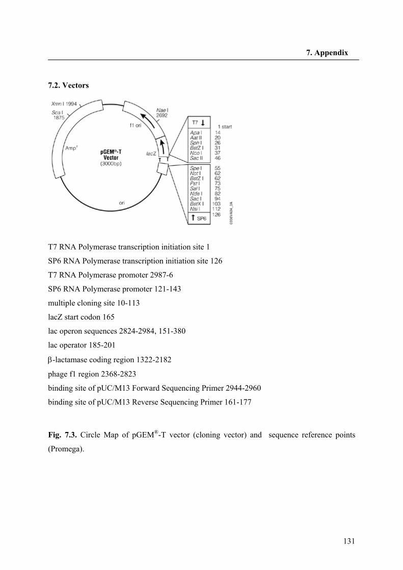
131
7. Appendix
7.2. Vectors
T7 RNA Polymerase transcription initiation site 1
SP6 RNA Polymerase transcription initiation site 126
T7 RNA Polymerase promoter 2987-6
SP6 RNA Polymerase promoter 121-143
multiple cloning site 10-113
lacZ start codon 165
lac operon sequences 2824-2984, 151-380
lac operator 185-201
β-lactamase coding region 1322-2182
phage f1 region 2368-2823
binding site of pUC/M13 Forward Sequencing Primer 2944-2960
binding site of pUC/M13 Reverse Sequencing Primer 161-177
Fig. 7.3. Circle Map of pGEM®-T vector (cloning vector) and sequence reference points
(Promega).
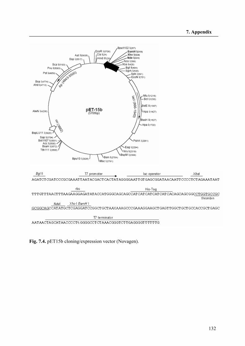
132
7. Appendix
Fig. 7.4. pET15b cloning/expression vector (Novagen).
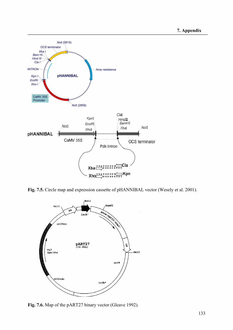
133
7. Appendix
Fig. 7.5. Circle map and expression cassette of pHANNIBAL vector (Wesely et al. 2001).
Fig. 7.6. Map of the pART27 binary vector (Gleave 1992).
134
8. Acknowledgements
8. Acknowledgements
I would like to thank all of those who have been contributed in the work presented here. I wish to express my deep gratitude to Prof. Dr. A. Wilhelm Alfermann for giving me the opportunity to join his group, his interest to the progress of the work, for his trust in me, financial supports and fruitful discussions. Thanks for your patience and hospitality during all these years. I am very grateful to Dr. Elisabeth Fuss for her generous supervision, for the encouragement and for teaching me how to write scientific papers. Moreover, I will appreciate her knowledge in molecular biology. Thank you Lisa that your office was always open to us to ask questions. I am also indebted to Prof. Dr. Thomas J. Schmidt for giving me the opportunity to stay in his laboratory for the first months. Thanks Thomas that you were always for us their, for recording the NMR spectra and revision of the manuscripts. Furthermore, I would like to thank Dr. R. A. Edrada for LC–MS runs and Dr. B. Schneider for LC-NMR analysis. Too much thanks to my colleagues who have been working together with me in the laboratory. Thank you all for your collaborations and for providing an enjoyable atmosphere. I would like to thank the “Düsseldorfer Entrepreneurs Foundation” for the financial support during the last 18 months of the work. Special thanks to Prof. Dr. D. Riesner, the founder of the organization. I wish to thank Dr. A. Mohagheghzadeh for his affords to do my research in the laboratory of Prof. Dr. Alfermann. Last but not least my cordial thanks go to my mother for her loving support and patience during this journey.

135
I certify that this thesis is entirely my own work. All used sources and resources are cited.
Düsseldorf, 19.11.2007
…………………………….. Shiva Hemmati
PUBLICATIONS Hemmati S, von Heimendahl CBI, Fuss E. Pinoresinol-lariciresinol reductases with opposite enantiospecificity determine the enantiomeric composition of lignans in Linum usitatissimum. (in preparation) Hemmati S, Schneider B, Schmidt TJ, Federolf K, Alfermann AW, Fuss E. Justicidin B 7-hydroxylase, a cytochrome P450 monooxygenase from cell cultures of Linum perenne involve in the biosynthesis of diphyllin. Phytochemistry (in press) Hemmati S, Schmidt TJ, Fuss E. (+)-Pinoresinol/(-)-lariciresinol reductase from Linum perenne Himmelszelt involved in the biosynthesis of justicidin B. FEBS Letters (2007) 581: 603-610 Mohagheghzadeh A, Gholami A, Hemmati S, Ardakani MR, Schmidt TJ, Alfermann AW. Root cultures of linum species section Syllinum as rich sources of 6-methoxypodophyllotoxin. Zeitschrift fur Naturforschung-Section C, Journal of Biosciences (2007), 62 (1-2): 43-49 Schmidt, TJ, Hemmati, S, Fuss, E, Alfermann, AW. A combined HPLC-UV and HPLC-MS method for the identification of lignans and its application to the lignans of Linum usitatissimum L. and L. bienne Mill. Phytochemical Analysis (2006) 17: 299-311 Ardakani MS, Hemmati S, Mohagheghzadeh A. Effect of elicitors on the enhancement of podophyllotoxin biosynthesis in suspension cultures of Linum album. DARU (2005) 13: 56-60 Mohagheghzadeh A, Gholami A, Soltani M, Hemmati S, Alfermann AW. Linum mucronatum: organ to organ lignan variation. Zeitschrift fur Naturforschung-Section C, Journal of Biosciences (2005), 60 (5-6): 508-510 Mohagheghzadeh A, Hemmati S, Mehregan I, Alfermann AW. Linum persicum: Lignans and placement in Linaceae. Phytochemistry Reviews (2003) 2: 363-369 SEMINARS AND CONGRESSES
(+)-Pinoresinol/(-)-lariciresinol reductase from Linum perenne Himmelszelt involved in the biosynthesis of justicidin B. In: 50 years of the phytochemical society of Europe, Cabridge, UK, April 11-14, 2007 (poster) Evolution and diversity of lignan biosynthesis in the genus Linum. In: 5th Kurt-Mothes-Doctoral Student-Workshop, Halle, Germany, October 5-7, 2005 (lecture) Pinoresinol-lariciresinol reductase in justicidin B producing cell cultures of Linum perenne. In: XVII International Botanical Congress, Vienna, Austria, July 17-23, 2005 (poster) Lignans in Linum perenne. In: Section Plant Natural Product, German Society of Botany, Kaub on Rhein, Germany, March 16-18, 2005 (lecture)

Chemodiversity of lignan accumulation in the genus Linum. In: Botany Symposium, Braunschweig, Germany, September 5-10, 2004 (poster)
Diversity of lignans in Linum species. In: Future Trends in Phytochemistry, Gargnano, Italy, May 5-8, 2004 (poster) Quantification of aryltetralin lignans in plant parts and ontogenic stages of Linum album. In: Phytochemistry and Biology of Lignans, Bornheim-Walberberg, Germany, April 6-9, 2003 (poster) In vitro cultures of Linum album and screening for high lignan producing lines. In: Future Trends in Phytochemistry, Gargnano, Italy, May 22-25, 2002 (poster)