A novel injectable chitosan/polyglutamate polyelectrolyte complex hydrogel with hydroxyapatite for...
8
Carbohydrate Polymers 89 (2012) 1123–1130 Contents lists available at SciVerse ScienceDirect Carbohydrate Polymers jo u rn al hom epa ge: www.elsevier.com/locate/carbpol A novel injectable chitosan/polyglutamate polyelectrolyte complex hydrogel with hydroxyapatite for soft-tissue augmentation Dian-Yu Ji a , Tzong-Fu Kuo b , Hong-Da Wu a , Jen-Chang Yang c,d,∗,1 , Shang-Yang Lee a,d,e,∗∗,1 a School of Dentistry, College of Oral Medicine, Taipei Medical University, Taipei 110, Taiwan, ROC b School of Veterinary Medicine, National Taiwan University, Taipei 106, Taiwan, ROC c School of Dental Technology, College of Oral Medicine, Taipei Medical University, Taipei 110, Taiwan, ROC d Center for Teeth Bank and Dental Stem Cell Technology, Taipei Medical University, Taipei 110, Taiwan, ROC e Dental Department of Taipei Medical University Municipal Wan-Fang Hospital, Taipei 116, Taiwan, ROC a r t i c l e i n f o Article history: Received 13 February 2012 Received in revised form 25 March 2012 Accepted 26 March 2012 Available online 4 April 2012 Keywords: Polyelectrolyte complex Chitosan Polyglutamate Hydroxyapatite Dermal filler Soft-tissue augmentation a b s t r a c t This study demonstrated a chitosan (CS)/polyglutamate (PG) polyelectrolyte complex (PEC) hydrogel combined with spherical hydroxyapatite (HAp) particles as an injectable dermal filler for soft-tissue augmentation. The CS/PG PEC hydrogel with oppositely charged ionic cross-linking, a high gel content, and low degradation rate was introduced as a carrier to achieve high shape and volume stability. An MTT assay indicated that the CS/PG PEC had satisfactory cell biocompatibility. This PEC/HAp hydrogel showed good structural integrity in a PBS solution for up to 60 days. Clinical manageability was indexed by an injection force measurement through sterile 27-gauge needles using a texture analyzer. In an animal study, 0.2 mL of the PEC and PEC/hydroxyapatite (HAp) were implanted within the dorsal dermis of a swine ear. Injected tissue areas were biopsied 2 weeks, and 2 and 6 months after the injection. According to the histomorphometric results, the PEC and PEC/HAp groups showed percentages of retention of the maximum height of the cross-section of about 44% and 73% at 6 months. New collagen was observed in the central position indicating a possible collagenesis effect. These results suggest that this PEC/HAp system can be used as an alternative for soft-tissue augmentation. © 2012 Elsevier Ltd. All rights reserved. 1. Introduction Soft-tissue augmentation is a nonsurgical procedure for inject- ing a substance in dermal or subcutaneous tissues for esthetics or supporting purposes with the advantages of a low risk of com- plications and a short recovery time. Based on the criteria of correction longevity, dermal fillers can be classified as tempo- rary, semi-permanent, and permanent. Unlike permanent filler materials such as silicone which may eventually induce patients to develop granulomas (Lemperle, Morhenn, & Charrier, 2003), temporary dermal fillers such as collagen and hyaluronic acid generally elicit low immune responses but have short duration periods of 3–6 months (Broder & Cohen, 2006). On the other hand, ∗ Corresponding author at: School of Dentistry, College of Oral Medicine, Taipei Medical University, 250 Wu-Hsing Street, Taipei 110, Taiwan, ROC. Tel.: +886 2 27361661x5149; fax: +886 2 27362295. ∗∗ Corresponding author at: School of Dental Technology, Taipei Medical Univer- sity, 250 Wu-Hsing Street, Taipei 110, Taiwan, ROC. Tel: +886 2 2736 1661x5124; fax: +886 2 27362295. E-mail addresses: [email protected] (J.-C. Yang), [email protected] (S.-Y. Lee). 1 These authors contributed equally to this work. semi-permanent particle filler systems such as hydroxyapatite (HAp) (Radiesse ® , BioForm Medical, San Mateo, CA), tri- calcium phosphate (-TCP) (Atléan ® ), and poly-l-lactic acid (PLA) (Sculptra ® ) loaded in carriers reveal longevity and collagen stimu- latory properties (Johl & Burgett, 2006; Thioly-Bensoussan, 2008). Radiesse ® is a commercial product composed of a suspension of 30% HAp particles in the 70% carboxymethyl cellulose (CMC) carrier system. The safety profile of HAp, an inorganic constituent of teeth and bone, has been extensively evaluated (Havlik, 2002). After injection, the CMC carrier gradually undergoes degradation and macrophage phagocytosis over a period of 6–8 weeks (Flaharty, 2004). Finally, only HAp particles were left to provide mechanical volume support; thus additional treatment is usually planned for moderate or deep wrinkles after 6 weeks (Redbord, Busso, & Hanke, 2011; Thioly-Bensoussan, 2008). Polyelectrolyte complexes (PEC), mixtures of two poly- electrolytes with opposite charges (cationic and anionic) and electrostatic interactions (Li et al., 2007), received considerable attention in the past several decades. PEC hydrogels prepared from natural polymers, such as polysaccharides and polypeptides, have additional advantages of being nontoxic and bioabsorbable (Cascone, Sim, & Downes, 1995). PEC cross-linked compositions are essential for the resulting gel properties for use in biomedical application (Dai et al., 2007; Wu et al., 2011). 0144-8617/$ – see front matter © 2012 Elsevier Ltd. All rights reserved. http://dx.doi.org/10.1016/j.carbpol.2012.03.083
-
Upload
taipeimedical -
Category
Documents
-
view
3 -
download
0
Transcript of A novel injectable chitosan/polyglutamate polyelectrolyte complex hydrogel with hydroxyapatite for...
Aw
Da
b
c
d
e
a
ARRAA
KPCPHDS
1
ispcrmttgp
MT
sf
(
0h
Carbohydrate Polymers 89 (2012) 1123– 1130
Contents lists available at SciVerse ScienceDirect
Carbohydrate Polymers
jo u rn al hom epa ge: www.elsev ier .com/ locate /carbpol
novel injectable chitosan/polyglutamate polyelectrolyte complex hydrogelith hydroxyapatite for soft-tissue augmentation
ian-Yu Ji a, Tzong-Fu Kuob, Hong-Da Wua, Jen-Chang Yangc,d,∗,1, Shang-Yang Leea,d,e,∗∗,1
School of Dentistry, College of Oral Medicine, Taipei Medical University, Taipei 110, Taiwan, ROCSchool of Veterinary Medicine, National Taiwan University, Taipei 106, Taiwan, ROCSchool of Dental Technology, College of Oral Medicine, Taipei Medical University, Taipei 110, Taiwan, ROCCenter for Teeth Bank and Dental Stem Cell Technology, Taipei Medical University, Taipei 110, Taiwan, ROCDental Department of Taipei Medical University Municipal Wan-Fang Hospital, Taipei 116, Taiwan, ROC
r t i c l e i n f o
rticle history:eceived 13 February 2012eceived in revised form 25 March 2012ccepted 26 March 2012vailable online 4 April 2012
eywords:olyelectrolyte complex
a b s t r a c t
This study demonstrated a chitosan (CS)/polyglutamate (PG) polyelectrolyte complex (PEC) hydrogelcombined with spherical hydroxyapatite (HAp) particles as an injectable dermal filler for soft-tissueaugmentation. The CS/PG PEC hydrogel with oppositely charged ionic cross-linking, a high gel content,and low degradation rate was introduced as a carrier to achieve high shape and volume stability. An MTTassay indicated that the CS/PG PEC had satisfactory cell biocompatibility. This PEC/HAp hydrogel showedgood structural integrity in a PBS solution for up to 60 days. Clinical manageability was indexed by aninjection force measurement through sterile 27-gauge needles using a texture analyzer. In an animal
hitosanolyglutamateydroxyapatiteermal filleroft-tissue augmentation
study, 0.2 mL of the PEC and PEC/hydroxyapatite (HAp) were implanted within the dorsal dermis of aswine ear. Injected tissue areas were biopsied 2 weeks, and 2 and 6 months after the injection. Accordingto the histomorphometric results, the PEC and PEC/HAp groups showed percentages of retention of themaximum height of the cross-section of about 44% and 73% at 6 months. New collagen was observedin the central position indicating a possible collagenesis effect. These results suggest that this PEC/HAp
alter
system can be used as an. Introduction
Soft-tissue augmentation is a nonsurgical procedure for inject-ng a substance in dermal or subcutaneous tissues for esthetics orupporting purposes with the advantages of a low risk of com-lications and a short recovery time. Based on the criteria oforrection longevity, dermal fillers can be classified as tempo-ary, semi-permanent, and permanent. Unlike permanent filleraterials such as silicone which may eventually induce patients
o develop granulomas (Lemperle, Morhenn, & Charrier, 2003),emporary dermal fillers such as collagen and hyaluronic acidenerally elicit low immune responses but have short durationeriods of 3–6 months (Broder & Cohen, 2006). On the other hand,
∗ Corresponding author at: School of Dentistry, College of Oral Medicine, Taipeiedical University, 250 Wu-Hsing Street, Taipei 110, Taiwan, ROC.
el.: +886 2 27361661x5149; fax: +886 2 27362295.∗∗ Corresponding author at: School of Dental Technology, Taipei Medical Univer-ity, 250 Wu-Hsing Street, Taipei 110, Taiwan, ROC. Tel: +886 2 2736 1661x5124;ax: +886 2 27362295.
E-mail addresses: [email protected] (J.-C. Yang), [email protected]. Lee).
1 These authors contributed equally to this work.
144-8617/$ – see front matter © 2012 Elsevier Ltd. All rights reserved.ttp://dx.doi.org/10.1016/j.carbpol.2012.03.083
native for soft-tissue augmentation.© 2012 Elsevier Ltd. All rights reserved.
semi-permanent particle filler systems such as hydroxyapatite(HAp) (Radiesse®, BioForm Medical, San Mateo, CA), � tri-calcium phosphate (�-TCP) (Atléan®), and poly-l-lactic acid (PLA)(Sculptra®) loaded in carriers reveal longevity and collagen stimu-latory properties (Johl & Burgett, 2006; Thioly-Bensoussan, 2008).
Radiesse® is a commercial product composed of a suspensionof 30% HAp particles in the 70% carboxymethyl cellulose (CMC)carrier system. The safety profile of HAp, an inorganic constituentof teeth and bone, has been extensively evaluated (Havlik, 2002).After injection, the CMC carrier gradually undergoes degradationand macrophage phagocytosis over a period of 6–8 weeks (Flaharty,2004). Finally, only HAp particles were left to provide mechanicalvolume support; thus additional treatment is usually planned formoderate or deep wrinkles after 6 weeks (Redbord, Busso, & Hanke,2011; Thioly-Bensoussan, 2008).
Polyelectrolyte complexes (PEC), mixtures of two poly-electrolytes with opposite charges (cationic and anionic) andelectrostatic interactions (Li et al., 2007), received considerableattention in the past several decades. PEC hydrogels preparedfrom natural polymers, such as polysaccharides and polypeptides,
have additional advantages of being nontoxic and bioabsorbable(Cascone, Sim, & Downes, 1995). PEC cross-linked compositionsare essential for the resulting gel properties for use in biomedicalapplication (Dai et al., 2007; Wu et al., 2011).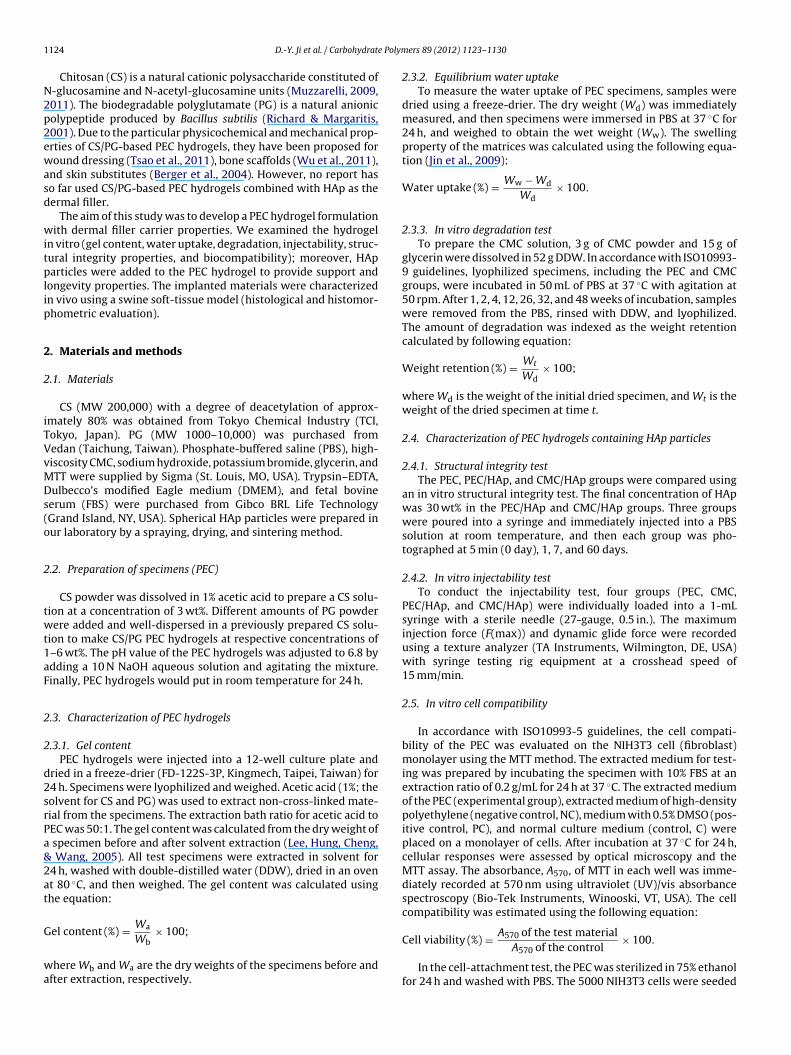
1 e Polym
N2p2ewasd
witplip
2
2
iTVvMDs(o
2
twt1aF
2
2
d2srPa&2at
G
wa
124 D.-Y. Ji et al. / Carbohydrat
Chitosan (CS) is a natural cationic polysaccharide constituted of-glucosamine and N-acetyl-glucosamine units (Muzzarelli, 2009,011). The biodegradable polyglutamate (PG) is a natural anionicolypeptide produced by Bacillus subtilis (Richard & Margaritis,001). Due to the particular physicochemical and mechanical prop-rties of CS/PG-based PEC hydrogels, they have been proposed foround dressing (Tsao et al., 2011), bone scaffolds (Wu et al., 2011),
nd skin substitutes (Berger et al., 2004). However, no report haso far used CS/PG-based PEC hydrogels combined with HAp as theermal filler.
The aim of this study was to develop a PEC hydrogel formulationith dermal filler carrier properties. We examined the hydrogel
n vitro (gel content, water uptake, degradation, injectability, struc-ural integrity properties, and biocompatibility); moreover, HAparticles were added to the PEC hydrogel to provide support and
ongevity properties. The implanted materials were characterizedn vivo using a swine soft-tissue model (histological and histomor-hometric evaluation).
. Materials and methods
.1. Materials
CS (MW 200,000) with a degree of deacetylation of approx-mately 80% was obtained from Tokyo Chemical Industry (TCI,okyo, Japan). PG (MW 1000–10,000) was purchased fromedan (Taichung, Taiwan). Phosphate-buffered saline (PBS), high-iscosity CMC, sodium hydroxide, potassium bromide, glycerin, andTT were supplied by Sigma (St. Louis, MO, USA). Trypsin–EDTA,ulbecco’s modified Eagle medium (DMEM), and fetal bovine
erum (FBS) were purchased from Gibco BRL Life TechnologyGrand Island, NY, USA). Spherical HAp particles were prepared inur laboratory by a spraying, drying, and sintering method.
.2. Preparation of specimens (PEC)
CS powder was dissolved in 1% acetic acid to prepare a CS solu-ion at a concentration of 3 wt%. Different amounts of PG powderere added and well-dispersed in a previously prepared CS solu-
ion to make CS/PG PEC hydrogels at respective concentrations of–6 wt%. The pH value of the PEC hydrogels was adjusted to 6.8 bydding a 10 N NaOH aqueous solution and agitating the mixture.inally, PEC hydrogels would put in room temperature for 24 h.
.3. Characterization of PEC hydrogels
.3.1. Gel contentPEC hydrogels were injected into a 12-well culture plate and
ried in a freeze-drier (FD-122S-3P, Kingmech, Taipei, Taiwan) for4 h. Specimens were lyophilized and weighed. Acetic acid (1%; theolvent for CS and PG) was used to extract non-cross-linked mate-ial from the specimens. The extraction bath ratio for acetic acid toEC was 50:1. The gel content was calculated from the dry weight of
specimen before and after solvent extraction (Lee, Hung, Cheng, Wang, 2005). All test specimens were extracted in solvent for4 h, washed with double-distilled water (DDW), dried in an ovent 80 ◦C, and then weighed. The gel content was calculated usinghe equation:
Wa
el content (%) =Wb× 100;
here Wb and Wa are the dry weights of the specimens before andfter extraction, respectively.
ers 89 (2012) 1123– 1130
2.3.2. Equilibrium water uptakeTo measure the water uptake of PEC specimens, samples were
dried using a freeze-drier. The dry weight (Wd) was immediatelymeasured, and then specimens were immersed in PBS at 37 ◦C for24 h, and weighed to obtain the wet weight (Ww). The swellingproperty of the matrices was calculated using the following equa-tion (Jin et al., 2009):
Water uptake (%) = Ww − Wd
Wd× 100.
2.3.3. In vitro degradation testTo prepare the CMC solution, 3 g of CMC powder and 15 g of
glycerin were dissolved in 52 g DDW. In accordance with ISO10993-9 guidelines, lyophilized specimens, including the PEC and CMCgroups, were incubated in 50 mL of PBS at 37 ◦C with agitation at50 rpm. After 1, 2, 4, 12, 26, 32, and 48 weeks of incubation, sampleswere removed from the PBS, rinsed with DDW, and lyophilized.The amount of degradation was indexed as the weight retentioncalculated by following equation:
Weight retention (%) = Wt
Wd× 100;
where Wd is the weight of the initial dried specimen, and Wt is theweight of the dried specimen at time t.
2.4. Characterization of PEC hydrogels containing HAp particles
2.4.1. Structural integrity testThe PEC, PEC/HAp, and CMC/HAp groups were compared using
an in vitro structural integrity test. The final concentration of HApwas 30 wt% in the PEC/HAp and CMC/HAp groups. Three groupswere poured into a syringe and immediately injected into a PBSsolution at room temperature, and then each group was pho-tographed at 5 min (0 day), 1, 7, and 60 days.
2.4.2. In vitro injectability testTo conduct the injectability test, four groups (PEC, CMC,
PEC/HAp, and CMC/HAp) were individually loaded into a 1-mLsyringe with a sterile needle (27-gauge, 0.5 in.). The maximuminjection force (F(max)) and dynamic glide force were recordedusing a texture analyzer (TA Instruments, Wilmington, DE, USA)with syringe testing rig equipment at a crosshead speed of15 mm/min.
2.5. In vitro cell compatibility
In accordance with ISO10993-5 guidelines, the cell compati-bility of the PEC was evaluated on the NIH3T3 cell (fibroblast)monolayer using the MTT method. The extracted medium for test-ing was prepared by incubating the specimen with 10% FBS at anextraction ratio of 0.2 g/mL for 24 h at 37 ◦C. The extracted mediumof the PEC (experimental group), extracted medium of high-densitypolyethylene (negative control, NC), medium with 0.5% DMSO (pos-itive control, PC), and normal culture medium (control, C) wereplaced on a monolayer of cells. After incubation at 37 ◦C for 24 h,cellular responses were assessed by optical microscopy and theMTT assay. The absorbance, A570, of MTT in each well was imme-diately recorded at 570 nm using ultraviolet (UV)/vis absorbancespectroscopy (Bio-Tek Instruments, Winooski, VT, USA). The cellcompatibility was estimated using the following equation:
Cell viability (%) = A570 of the test materialA570 of the control
× 100.
In the cell-attachment test, the PEC was sterilized in 75% ethanolfor 24 h and washed with PBS. The 5000 NIH3T3 cells were seeded

Polymers 89 (2012) 1123– 1130 1125
o5w1etw(o
2
tUm(rwwttashsm
mcitealfsswJaatB
2
ofastes
3
3
3
daPi
D.-Y. Ji et al. / Carbohydrate
n a PEC hydrogel and incubated at 37 ◦C in an incubator with a% CO2 atmosphere for 72 h. Cell-containing specimens were fixedith a 2 wt% glutaraldehyde solution followed by treatment with
wt% OsO4 (post-fixed). Specimens were dehydrated in a gradedthanol series (30, 50, 70, 80, 90, and 100 wt%), and then subjectedo liquid CO2 critical-point drying. Finally, cells containing the PECere gold-coated and examined by scanning electron microscopy
SEM) (Hitachi S2400, Tokyo, Japan) under an accelerating voltagef 15–20 kV.
.6. In vivo soft-issue augmentation test
The animal study was performed in strict accordance with pro-ocols approved by the Animal Care Committee of Taipei Medicalniversity (LAC-99-0093). Six Lanyu swines with a mean age of 3onths were supplied by the Taitung Animal Propagation Station
Taitung, Taiwan). Using a 27-gauge needle, 0.2 mL of filler mate-ials was injected in the dorsal dermis of both ears of each swine,hile under general anesthesia. PEC, PEC/HAp, and CMC/HAp fillersere injected into the respective experimental groups; the injec-
ion resulted in the appearance of a dome-shaped augmentationhat could be easily palpated. The locations of the injection sitesnd type of material injected were marked on a transparent plasticheet placed over the ear for later identification. Specimens werearvested after 2 weeks, and 2 and 6 months, and the resultantoft-tissue augmentation was examined by histological and histo-orphometric methods.Each implanted site was harvested at 2 weeks, and 2 and 6
onths after the injection. Biopsies obtained from each site wereut to a specific area (2 cm × 2 cm). Harvested specimens were fixedn 10% buffered formalin for 1 week, and were cut perpendicular tohe skin surface with a surgical blade at a thickness of 5 mm (Pitarut al., 2007). Finally, specimens were made into paraffin sectionsnd stained with Masson’s trichrome stain to investigate the col-agen fiber density in the dermis. Each specimen was examinedor the shape of the injected materials, preservation of its initialhape over time, the host response at the implant site, and micro-copic degradation of the implanted materials. Stained sectionsere observed under optical microscopy (Olympus EX51, Tokyo,
apan). A histomorphometric method was used to quantify themount of soft tissue within the augmentation area. The maximumugmentation height of a cross-section was determined and quan-ified using Image J analysis software (National Institutes of Health,ethesda, MD, USA).
.7. Statistical analysis
The mean and standard deviation (SD) of the maximum heightf a cross-section for each group of filler materials was calculatedor various implantation time periods. Data were compared using
one-way analysis of variance (ANOVA) to evaluate the statisticalignificance of the measured data. Post hoc Tukey’s test was usedo compare the significance of deviations in the measured data ofach group. In all cases, differences were considered statisticallyignificant at p < 0.05.
. Results and discussion
.1. Characterization of the PEC hydrogels
.1.1. Gel content and equilibrium water uptakeThe gel content is a basic parameter to calibrate gel formation
ue to cross-linking because not all of the macromonomers eventu-lly join the gel network. Fig. 1 displays the gel content for variousEC formulations. Each sample was determined by measuring itsnsoluble part after extraction in solvent. The highest and lowest
Fig. 1. Gel content (GC) at 25 ◦C in an acid solution (�) and equilibrium wateruptake (WU) at 37 ◦C in PBS for 24 h (©) of PEC hydrogel formed by mixing differentconcentrations of PG with 3% chitosan (CS) (n = 4).
gel contents were 35.46% and 14.08% for samples with 4% and 1%PG, respectively. The swelling property is an important parameterto characterize cross-linked structures. The ability to hold sufficientamounts of water inside the network structures is an importantparameter to characterize the swelling properties of hydrogels (Jinet al., 2009). Equilibrium water uptake values of the designed spec-imens were within 1043–2448% as shown in Fig. 1. According tothe above results, the PEC hydrogel formulation with the high gelcontent had a lower water uptake value (1043%). Therefore, basedon the above results, a composition of 4% PG and 3% CS may havebeen nearest to the stoichiometric formulation and was chosen forthe following experiments.
The swelling property inversely matched the gel content of thehydrogels (Sun, Cao, Su, & Tan, 2009). Hydrogels with the higher gelcontent had a lower swelling ratio which was largely restricted bythe cross-linking density. The gel content of the PEC was associatedwith the swelling behavior. The higher gel content resulted in ahigher cross-linked density, which confines the macromoleculesto a limited spatial volume; so the water uptake value is largelyrestricted. Moreover it caused lower sample volume expansion andavoids tissue oppression in bodily fluids (Peter et al., 2010; Wu, Ji,Chang, Yang, & Lee, 2012).
The gel content and swelling behavior of the hydrogels wereaffected by different molecular weights when treated with thesame cross-linking method (Tranquilan-Aranilla, Yoshii, Dela Rosa,& Makuuchi, 1999). Hyaluronic acid with methacrylic anhy-dride to modify the hydrogel is one of the simplest and mostwidely used reactions (Burdick & Prestwich, 2011). Swelling ratiosof hyaluronic acid-cross-linked hydrogels range 8–42 (Burdick,Chung, Jia, Randolph, & Langer, 2005). In a previous study, astoichiometric formulation of a PEC hydrogel also had a long degra-dation property (Tsao et al., 2011; Wu et al., 2011).
3.1.2. In vitro degradation testIn accordance with ISO10993-9 guidelines, weight loss of the
PEC in PBS at 37 ◦C was measured over time. Fig. 2 indicates rapidweight loss of the PEC in the first week (51.1% weight loss); after-wards, the degradation rate decreased. During the period of 1–48weeks, the shape of the PEC was almost a completely intact mor-phology; thus the weight retention of the designed PEC cross-linkedstructure was 22.4% at 26 weeks and even 15.4% at 48 weeks.
A modified CS-alginate gel system showed degradation of 38.6%within 28 days (Jin et al., 2009). Wu et al. (2011) reported a CS-based PEC hydrogel, which degraded by about 27.7% and 47.4%within 1 and 26 weeks. According to Tsao et al.’s report, a CS/PG PEC
1126 D.-Y. Ji et al. / Carbohydrate Polymers 89 (2012) 1123– 1130
F(
htat
3
3
P
ig. 2. Degradation profile of CMC and the PEC hydrogel at 48 weeks in PBS at 37 ◦Cn = 3).
ydrogel showed degradation of 21% within 28 days. In this study,he degradation of the PEC hydrogel was about 59.6% at 28 daysnd 76.6% at 26 weeks. This system could be maintained for morehan 26 weeks.
.2. Characterization of PEC hydrogels containing HAp particles
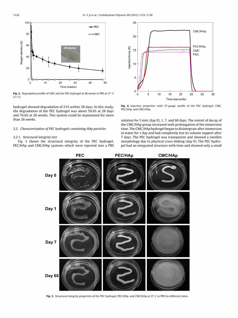
.2.1. Structural integrity testFig. 3 shows the structural integrity of the PEC hydrogel,
EC/HAp and CMC/HAp systems which were injected into a PBS
Fig. 3. Structural integrity properties of the PEC hydrogel, PEC
Fig. 4. Injection properties with 27-gauge needle of the PEC hydrogel, CMC,PEC/HAp, and CMC/HAp.
solution for 5 min (day 0), 1, 7, and 60 days. The extent of decay ofthe CMC/HAp group increased with prolongation of the immersiontime. The CMC/HAp hydrogel began to disintegrate after immersion
in water for 1 day and had completely lost its volume support after7 days. The PEC hydrogel was transparent and showed a swollenmorphology due to physical cross-linking (day 0). The PEC hydro-gel had an integrated structure with time and showed only a small/HAp, and CMC/HAp at 25 ◦C in PBS for different times.

D.-Y. Ji et al. / Carbohydrate Polymers 89 (2012) 1123– 1130 1127
Fig. 5. (a) MTT reduction by NIH3T3 fibroblast cells exposed to the PEC extractcompared to MTT reduction in the control (C) (cells with medium alone), negativecontrol (NC) (cells with high-density polyethylene), and positive control (PC) (cellswith 0.5% DMSO) for 24 h. (b) Optical photomicrograph (100×) of NIH3T3 fibroblastculture in PEC hydrogel extract medium. (c) Scanning electron micrographs showingtT
raivw
odw
he growth of NIH3T3 cells on the PEC hydrogel for 3 days (magnification, 2000×).he cell density was 106 cells/mL.
eduction in volume after day 7. The PEC/HAp hydrogel remainedlmost stable in its initial shape, and no obvious decay or dis-ntegration was observed at 60 days. However, the supportingolume ability and structural integrity of the PEC/HAp hydrogelere stronger than those of the CMC/HAp hydrogel.
Structural integrity is discussed in terms of preventing washoutf injectable bone cement supporting the original shape andimensions (Lin et al., 2010). When the PEC hydrogel carrieras introduced to the filler system, a network that spread all
Fig. 6. Schematic illustration of time-dependence degradation and collagenesismodel for (a) CMC/HAp and (b) PEC/HAp dermal fillers.
over the HAp particles was formed, resulting in good mainte-nance of its shape even after exposure to PBS. The high viscosityand polyelectrolyte reaction between the PEC hydrogel and HAphelped the filler system retain its original position and shape. Asa result, the ability of the dermal filler to maintain its structuralintegrity was dramatically enhanced with the addition of the PEChydrogel.
3.2.2. Injection properties testAn ideal injection property is to be able to deliver the dermal
filler through a large-gauge needle, so it must possess low vis-cosity at high shear (Falcone, Doerfler, & Berg, 2007). The easeof injectability (low injection force) of dermal fillers is impor-tant to clinicians (e.g., hand fatigue) and comfort for patientsto improve their treatment experience and satisfaction (Lorenc,Nir, & Azachi, 2010). The injection force is the minimum forceapplied to the syringe to achieve injection (Tezel & Fredrickson,2008).
Fig. 4 plots the force necessary to expel the samples througha 27-gauge needle from a 1-mL syringe. For comparison, respec-tive injection forces required for PEC, CMC, PEC/HAp, and CMC/HApwere about 14, 15, 16, and 22 N. The PEC hydrogel groups (PECand PEC/HAp) required less force than the CMC solution groups(CMC and CMC/HAp). The injection force of a commercial CMC/HApdermal filler (Radiesse®) through a 27-gauge needle with a 1.3-mL syringe was nearly 6.0 lbf (about 27 N) (Busso & Voigts,2008). Cilurzo et al.’s study reported a range of injection forcesof 125–160 mPa (45–57.6 N) with some difficulty, and values of<125 mPa (45 N) had smooth properties. Thus, the PEC as a dermalfiller offers several advantages over the preformed augmentationapproach in terms of ease of injection and minimizing the invasive-ness of surgery.
3.3. In vitro cell compatibility and cell attachment
Cell compatibility is an important characteristic of a materialintended for biomedical applications. Cell compatibility was eval-
uated in NIH3T3 cells (fibroblast) by the MTT assay according toISO10993-5 guidelines. Results are shown in Fig. 5a, and the PEC,control, and negative control groups showed no statistically sig-nificant differences, which reveals that the PEC hydrogel had little
1128 D.-Y. Ji et al. / Carbohydrate Polymers 89 (2012) 1123– 1130
Fig. 7. (a) Dome-shaped appearance of the implanted PEC hydrogel, PEC/HAp, and CMC/HAp specimens examined throughout the course of the study. The white double-pointed arrows indicate sites of the maximum height measured in each section. (b) Epidermal aspect of the implants after different implantation times illustrating cellularityn plant
ccttpsthMtm1cbi
ew collagen bundles (arrows) which were deposited in between the matrix of the imo color in text, the reader is referred to the web version of the article.)
ytotoxicity (95.9%). Moreover, values of the positive and negativeontrols were 20.3% and 101.2%, respectively, which proves that theests were done correctly. Cells cultured with extracted medium ofhe PEC are shown in Fig. 5b; the extraction induced no cell mor-hological changes of shrinkage or lysis. SEM photographs (Fig. 5c)how that NIH3T3 cells had adhered to the material surface, andhe pseudopodia were well extended on it. Thus, the designed PECydrogel displayed satisfactory biocompatibility and cell affinity.oreover, there was little toxicity evident in the cell-compatibility
est. In comparison, hydrogels using a chemical cross-linking agentay be associated with cytotoxicity (Huang-Lee, Cheung, & Nimni,
990), and physical ionic cross-linking displayed much fewer bio-ompatibility problems. Our PEC hydrogel may be able to provideetter stability and more-satisfactory biocompatibility than chem-
cal cross-linking methods.
t materials (M) (magnification, 20× and 200×). (For interpretation of the references
3.4. In vivo histological and histomorphometric evaluations ofsoft-issue augmentation
Three injected materials (PEC, PEC/HAp, and CMC/HAp) werelocated in the dermis between the epidermis and ear cartilage andwere harvested at 2 weeks, and 2 and 6 months postoperatively. Thematerial degradation and tissue collagenesis response with time oftwo dermal fillers (CMC/HAp and PEC/HAp) in the animal modelare shown in Fig. 6.
Host tissue responses to an implanted material can be evalu-ated using histologic methods. Histological sections of skin were
subjected to Masson’s trichrome staining to visualize extracellularmatrix (ECM) components and morphological changes. The soft-tissue augmentation efficacy was evaluated by histological andhistomorphometric methods. Fig. 7 shows the staining results of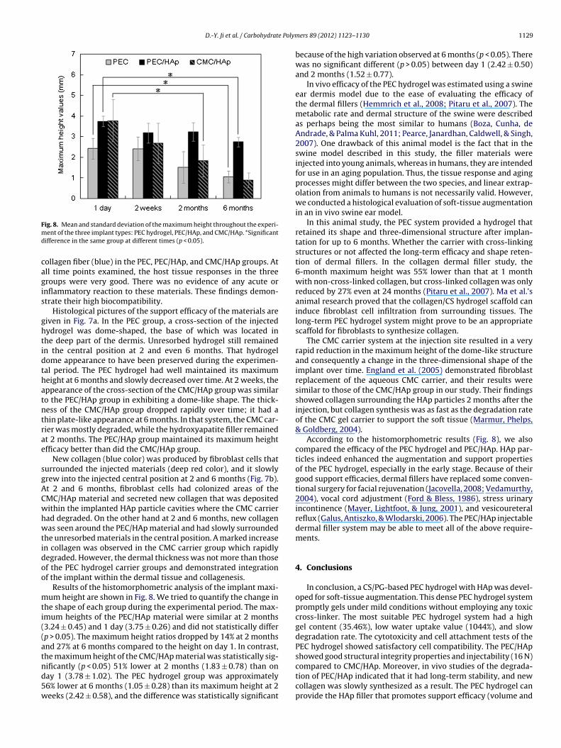
D.-Y. Ji et al. / Carbohydrate Polym
Fmd
cagis
ghtidthatntrae
sgACwhwtidoo
mti((atnd5w
ig. 8. Mean and standard deviation of the maximum height throughout the experi-ent of the three implant types: PEC hydrogel, PEC/HAp, and CMC/HAp. *Significant
ifference in the same group at different times (p < 0.05).
ollagen fiber (blue) in the PEC, PEC/HAp, and CMC/HAp groups. Atll time points examined, the host tissue responses in the threeroups were very good. There was no evidence of any acute ornflammatory reaction to these materials. These findings demon-trate their high biocompatibility.
Histological pictures of the support efficacy of the materials areiven in Fig. 7a. In the PEC group, a cross-section of the injectedydrogel was dome-shaped, the base of which was located inhe deep part of the dermis. Unresorbed hydrogel still remainedn the central position at 2 and even 6 months. That hydrogelome appearance to have been preserved during the experimen-al period. The PEC hydrogel had well maintained its maximumeight at 6 months and slowly decreased over time. At 2 weeks, theppearance of the cross-section of the CMC/HAp group was similaro the PEC/HAp group in exhibiting a dome-like shape. The thick-ess of the CMC/HAp group dropped rapidly over time; it had ahin plate-like appearance at 6 months. In that system, the CMC car-ier was mostly degraded, while the hydroxyapatite filler remainedt 2 months. The PEC/HAp group maintained its maximum heightfficacy better than did the CMC/HAp group.
New collagen (blue color) was produced by fibroblast cells thaturrounded the injected materials (deep red color), and it slowlyrew into the injected central position at 2 and 6 months (Fig. 7b).t 2 and 6 months, fibroblast cells had colonized areas of theMC/HAp material and secreted new collagen that was depositedithin the implanted HAp particle cavities where the CMC carrierad degraded. On the other hand at 2 and 6 months, new collagenas seen around the PEC/HAp material and had slowly surrounded
he unresorbed materials in the central position. A marked increasen collagen was observed in the CMC carrier group which rapidlyegraded. However, the dermal thickness was not more than thosef the PEC hydrogel carrier groups and demonstrated integrationf the implant within the dermal tissue and collagenesis.
Results of the histomorphometric analysis of the implant maxi-um height are shown in Fig. 8. We tried to quantify the change in
he shape of each group during the experimental period. The max-mum heights of the PEC/HAp material were similar at 2 months3.24 ± 0.45) and 1 day (3.75 ± 0.26) and did not statistically differp > 0.05). The maximum height ratios dropped by 14% at 2 monthsnd 27% at 6 months compared to the height on day 1. In contrast,he maximum height of the CMC/HAp material was statistically sig-
ificantly (p < 0.05) 51% lower at 2 months (1.83 ± 0.78) than onay 1 (3.78 ± 1.02). The PEC hydrogel group was approximately6% lower at 6 months (1.05 ± 0.28) than its maximum height at 2eeks (2.42 ± 0.58), and the difference was statistically significanters 89 (2012) 1123– 1130 1129
because of the high variation observed at 6 months (p < 0.05). Therewas no significant different (p > 0.05) between day 1 (2.42 ± 0.50)and 2 months (1.52 ± 0.77).
In vivo efficacy of the PEC hydrogel was estimated using a swineear dermis model due to the ease of evaluating the efficacy ofthe dermal fillers (Hemmrich et al., 2008; Pitaru et al., 2007). Themetabolic rate and dermal structure of the swine were describedas perhaps being the most similar to humans (Boza, Cunha, deAndrade, & Palma Kuhl, 2011; Pearce, Janardhan, Caldwell, & Singh,2007). One drawback of this animal model is the fact that in theswine model described in this study, the filler materials wereinjected into young animals, whereas in humans, they are intendedfor use in an aging population. Thus, the tissue response and agingprocesses might differ between the two species, and linear extrap-olation from animals to humans is not necessarily valid. However,we conducted a histological evaluation of soft-tissue augmentationin an in vivo swine ear model.
In this animal study, the PEC system provided a hydrogel thatretained its shape and three-dimensional structure after implan-tation for up to 6 months. Whether the carrier with cross-linkingstructures or not affected the long-term efficacy and shape reten-tion of dermal fillers. In the collagen dermal filler study, the6-month maximum height was 55% lower than that at 1 monthwith non-cross-linked collagen, but cross-linked collagen was onlyreduced by 27% even at 24 months (Pitaru et al., 2007). Ma et al.’sanimal research proved that the collagen/CS hydrogel scaffold caninduce fibroblast cell infiltration from surrounding tissues. Thelong-term PEC hydrogel system might prove to be an appropriatescaffold for fibroblasts to synthesize collagen.
The CMC carrier system at the injection site resulted in a veryrapid reduction in the maximum height of the dome-like structureand consequently a change in the three-dimensional shape of theimplant over time. England et al. (2005) demonstrated fibroblastreplacement of the aqueous CMC carrier, and their results weresimilar to those of the CMC/HAp group in our study. Their findingsshowed collagen surrounding the HAp particles 2 months after theinjection, but collagen synthesis was as fast as the degradation rateof the CMC gel carrier to support the soft tissue (Marmur, Phelps,& Goldberg, 2004).
According to the histomorphometric results (Fig. 8), we alsocompared the efficacy of the PEC hydrogel and PEC/HAp. HAp par-ticles indeed enhanced the augmentation and support propertiesof the PEC hydrogel, especially in the early stage. Because of theirgood support efficacies, dermal fillers have replaced some conven-tional surgery for facial rejuvenation (Jacovella, 2008; Vedamurthy,2004), vocal cord adjustment (Ford & Bless, 1986), stress urinaryincontinence (Mayer, Lightfoot, & Jung, 2001), and vesicoureteralreflux (Galus, Antiszko, & Wlodarski, 2006). The PEC/HAp injectabledermal filler system may be able to meet all of the above require-ments.
4. Conclusions
In conclusion, a CS/PG-based PEC hydrogel with HAp was devel-oped for soft-tissue augmentation. This dense PEC hydrogel systempromptly gels under mild conditions without employing any toxiccross-linker. The most suitable PEC hydrogel system had a highgel content (35.46%), low water uptake value (1044%), and slowdegradation rate. The cytotoxicity and cell attachment tests of thePEC hydrogel showed satisfactory cell compatibility. The PEC/HApshowed good structural integrity properties and injectability (16 N)
compared to CMC/HAp. Moreover, in vivo studies of the degrada-tion of PEC/HAp indicated that it had long-term stability, and newcollagen was slowly synthesized as a result. The PEC hydrogel canprovide the HAp filler that promotes support efficacy (volume and
1 e Polym
hm
R
B
B
B
B
B
B
C
C
D
E
F
FF
G
H
H
H
J
J
J
L
130 D.-Y. Ji et al. / Carbohydrat
eight). For this reason, the novel CS/PG PEC hydrogel with HApay be a good potential material for soft-tissue augmentation.
eferences
erger, J., Reist, M., Mayer, J. M., Felt, O., Peppas, N. A., & Gurny, R. (2004). Structureand interactions in covalently and ionically crosslinked chitosan hydrogels forbiomedical applications. European Journal of Pharmaceutics and Biopharmaceu-tics, 57(1), 19–34.
oza, J. C., Cunha, V. S., de Andrade, C. D., & Palma Kuhl, I. C. (2011). Experimen-tal dermatological surgery: An animal model for developing skills with dermalfillers. Indian Journal of Dermatology, 56(3), 303–305.
roder, K. W., & Cohen, S. R. (2006). An overview of permanent and semipermanentfillers. Plastic and Reconstructive Surgery, 118(3 Suppl.), 7S–14S.
urdick, J. A., Chung, C., Jia, X., Randolph, M. A., & Langer, R. (2005). Controlled degra-dation and mechanical behavior of photopolymerized hyaluronic acid networks.Biomacromolecules, 6(1), 386–391.
urdick, J. A., & Prestwich, G. D. (2011). Hyaluronic acid hydrogels for biomedicalapplications. Advanced Materials, 23(12), H41–H56.
usso, M., & Voigts, R. (2008). An investigation of changes in physical properties ofinjectable calcium hydroxylapatite in a carrier gel when mixed with lidocaineand with lidocaine/epinephrine. Dermatologic Surgery, 34(Suppl. 1), S16–S23,discussion S24.
ascone, M. G., Sim, B., & Downes, S. (1995). Blends of synthetic and naturalpolymers as drug delivery systems for growth hormone. Biomaterials, 16(7),569–574.
ilurzo, F., Selmin, F., Minghetti, P., Adami, M., Bertoni, E., Lauria, S., et al.(2011). Injectability evaluation: An open issue. AAPS PharmSciTech, 12(2),604–609.
ai, Z., Yin, J., Yan, S., Cao, T., Ma, J., & Chen, X. (2007). Polyelectrolyte com-plexes based on chitosan and poly (l-glutamic acid). Polymer International, 56(9),1122–1127.
ngland, L. J., Tan, M. H., Shumaker, P. R., Egbert, B. M., Pittelko, K., Orentreich, D.,et al. (2005). Effects of monopolar radiofrequency treatment over soft-tissuefillers in an animal model. Lasers in Surgery and Medicine, 37(5), 356–365.
alcone, S. J., Doerfler, A. M., & Berg, R. A. (2007). Novel synthetic dermal fillers basedon sodium carboxymethylcellulose: Comparison with crosslinked hyaluronicacid-based dermal fillers. Dermatologic Surgery, 33(Suppl. 2), S136–S143, dis-cussion S143.
laharty, P. (2004). Radiance. Facial Plastic Surgery, 20(2), 165–169.ord, C. N., & Bless, D. M. (1986). A preliminary study of injectable collagen in human
vocal fold augmentation. Otolaryngology: Head and Neck Surgery, 94(1), 104–112.alus, R., Antiszko, M., & Wlodarski, P. (2006). Clinical applications of hyaluronic
acid. Polski Merkuriusz Lekarski, 20(119), 606–608.avlik, R. J. (2002). Hydroxyapatite. Plastic and Reconstructive Surgery, 110(4),
1176–1179.emmrich, K., Van de Sijpe, K., Rhodes, N. P., Hunt, J. A., Di Bartolo, C., Pallua, N., et al.
(2008). Autologous in vivo adipose tissue engineering in hyaluronan-based gels– A pilot study. Journal of Surgical Research, 144(1), 82–88.
uang-Lee, L. L., Cheung, D. T., & Nimni, M. E. (1990). Biochemical changes and cyto-toxicity associated with the degradation of polymeric glutaraldehyde derivedcrosslinks. Journal of Biomedical Materials Research, 24(9), 1185–1201.
acovella, P. F. (2008). Use of calcium hydroxylapatite (Radiesse) for facial augmen-tation. Clinical Interventions in Aging, 3(1), 161–174.
in, R., Moreira Teixeira, L. S., Dijkstra, P. J., Karperien, M., van Blitterswijk, C. A.,Zhong, Z. Y., et al. (2009). Injectable chitosan-based hydrogels for cartilage tissueengineering. Biomaterials, 30(13), 2544–2551.
ohl, S. S., & Burgett, R. A. (2006). Dermal filler agents: A practical review. Current
Opinion in Ophthalmology, 17(5), 471–479.ee, M. W., Hung, C. L., Cheng, J. C., & Wang, Y. J. (2005). A newanti-adhesion film synthesized from polygalacturonic acid with 1-ethyl-3-(3-dimethylaminopropyl)carbodiimide crosslinker. Biomaterials, 26(18),3793–3799.
ers 89 (2012) 1123– 1130
Lemperle, G., Morhenn, V., & Charrier, U. (2003). Human histology and persistence ofvarious injectable filler substances for soft tissue augmentation. Aesthetic PlasticSurgery, 27(5), 354–366, discussion 367.
Li, Q. L., Chen, Z. Q., Darvell, B. W., Liu, L. K., Jiang, H. B., Zen, Q., et al. (2007).Chitosan–phosphorylated chitosan polyelectrolyte complex hydrogel as anosteoblast carrier. Journal of Biomedical Materials Research Part B: Applied Bio-materials, 82(2), 481–486.
Lin, Q., Lan, X., Li, Y., Yu, Y., Ni, Y., Lu, C., et al. (2010). Anti-washout carboxymethylchitosan modified tricalcium silicate bone cement: Preparation, mechanicalproperties and in vitro bioactivity. Journal of Materials Science Materials inMedicine, 21(12), 3065–3076.
Lorenc, Z. P., Nir, E., & Azachi, M. (2010). Characterization of physical properties andhistologic evaluation of injectable Dermicol-p35 porcine-collagen dermal filler.Plastic and Reconstructive Surgery, 125(6), 1805–1813.
Ma, L., Gao, C., Mao, Z., Zhou, J., Shen, J., Hu, X., et al. (2003). Collagen/chitosan porousscaffolds with improved biostability for skin tissue engineering. Biomaterials,24(26), 4833–4841.
Marmur, E. S., Phelps, R., & Goldberg, D. J. (2004). Clinical, histologic and electronmicroscopic findings after injection of a calcium hydroxylapatite filler. Journalof Cosmetic and Laser Therapy, 6(4), 223–226.
Mayer, R., Lightfoot, M., & Jung, I. (2001). Preliminary evaluation of calcium hydroxy-lapatite as a transurethral bulking agent for stress urinary incontinence. Urology,57(3), 434–438.
Muzzarelli, R. A. A. (2009). Chitins and chitosans for the repair of wounded skin,nerve, cartilage and bone. Carbohydrate Polymers, 76(2), 167–182.
Muzzarelli, R. A. A. (2011). Chitosan composites with inorganics, morphogeneticproteins and stem cells, for bone regeneration. Carbohydrate Polymers, 83(4),1433–1445.
Pearce, J. W., Janardhan, K. S., Caldwell, S., & Singh, B. (2007). Angiostatin and integrinalphavbeta3 in the feline, bovine, canine, equine, porcine and murine retina andcornea. Veterinary Ophthalmology, 10(5), 313–319.
Peter, M., Ganesh, N., Selvamurugan, N., Nair, S. V., Furuike, T.,Tamura, H., et al. (2010). Preparation and characterization ofchitosan–gelatin/nanohydroxyapatite composite scaffolds for tissueengineering applications. Carbohydrate Polymers, 80(3), 687–694.
Pitaru, S., Noff, M., Blok, L., Nir, E., Goldlust, A., & Savion, N. (2007). Long-term efficacyof a novel ribose-cross-linked collagen dermal filler: A histologic and histomor-phometric study in an animal model. Dermatologic Surgery, 33(9), 1045–1054,discussion 1054.
Redbord, K. P., Busso, M., & Hanke, C. W. (2011). Soft-tissue augmentation withhyaluronic acid and calcium hydroxyl apatite fillers. Dermatologic Therapy, 24(1),71–81.
Richard, A., & Margaritis, A. (2001). Poly(glutamic acid) for biomedical applications.Critical Reviews in Biotechnology, 21(4), 219–232.
Sun, S., Cao, H., Su, H., & Tan, T. (2009). Preparation and characterization of a novelinjectable in situ cross-linked hydrogel. Polymer Bulletin, 62(5), 699–711.
Tezel, A., & Fredrickson, G. H. (2008). The science of hyaluronic acid dermal fillers.Journal of Cosmetic and Laser Therapy, 10(1), 35–42.
Thioly-Bensoussan, D. (2008). Non-hyaluronic acid fillers. Clinics in Dermatology,26(2), 160–176.
Tranquilan-Aranilla, C., Yoshii, F., Dela Rosa, A. M., & Makuuchi, K. (1999).Kappa-carrageenan–polyethylene oxide hydrogel blends prepared by gammairradiation. Radiation Physics and Chemistry, 55(2), 127–131.
Tsao, C. T., Chang, C. H., Lin, Y. Y., Wu, M. F., Wang, J. L., Young, T. H., et al. (2011). Eval-uation of chitosan/�-poly(glutamic acid) polyelectrolyte complex for wounddressing materials. Carbohydrate Polymers, 84(2), 812–819.
Vedamurthy, M. (2004). Soft tissue augmentation – Use of hyaluronic acid as dermalfiller. Journal of Dermatology, Venereology and Leprology, 70(6), 383–387.
Wu, H.-D., Ji, D.-Y., Chang, W.-J., Yang, J.-C., & Lee, S.-Y. (2012). Chitosan-based poly-
electrolyte complex scaffolds with antibacterial properties for treating dentalbone defects. Materials Science and Engineering C, 32(2), 207–214.Wu, H.-D., Yang, J.-C., Tsai, T., Ji, D.-Y., Chang, W.-J., Chen, C.-C., et al. (2011). Devel-opment of a chitosan–polyglutamate based injectable polyelectrolyte complexscaffold. Carbohydrate Polymers, 85(2), 318–324.