Rational Design of Substituted Diarylureas: A Scaffold for Binding to G-Quadruplex Motifs
Upload
independentCategory
view
0download
0
ARTICLE IN PRESS
0142-9612/$ - se
doi:10.1016/j.bi
�Correspondgradables and
Portugal. Tel.:
E-mail addr
Biomaterials 27 (2006) 6123–6137
www.elsevier.com/locate/biomaterials
Novel hydroxyapatite/chitosan bilayered scaffold for osteochondraltissue-engineering applications: Scaffold design and its performance
when seeded with goat bone marrow stromal cells
Joaquim M. Oliveiraa,b, Marcia T. Rodriguesa,b, Simone S. Silvaa,b, Patrıcia B. Malafayaa,b,Manuela E. Gomesa,b, Carlos A. Viegasc, Isabel R. Diasc, Jorge T. Azevedod,
Joao F. Manoa,b, Rui L. Reisa,b,�
a3B’s Research Group—Biomaterials, Biodegradables and Biomimetics, Campus de Gualtar, 4710-057-Braga, PortugalbDepartment of Polymer Engineering, University of Minho, Campus de Azurem, 4800-058-Guimaraes, Portugal
cDepartment of Veterinary Sciences, University of Tras-os-Montes e Alto Douro, Quinta dos Prados, 5000-911-Vila Real, PortugaldDepartment of Animal Science, University of Tras-os-Montes e Alto Douro, Quinta dos Prados, 5000-911-Vila Real, Portugal
Received 11 April 2006; accepted 26 July 2006
Available online 30 August 2006
Abstract
Recent studies suggest that bone marrow stromal cells are a potential source of osteoblasts and chondrocytes and can be used to
regenerate damaged tissues using a tissue-engineering (TE) approach. However, these strategies require the use of an appropriate scaffold
architecture that can support the formation de novo of either bone and cartilage tissue, or both, as in the case of osteochondral defects.
The later has been attracting a great deal of attention since it is considered a difficult goal to achieve. This work consisted on developing
novel hydroxyapatite/chitosan (HA/CS) bilayered scaffold by combining a sintering and a freeze-drying technique, and aims to show the
potential of such type of scaffolds for being used in TE of osteochondral defects. The developed HA/CS bilayered scaffolds were
characterized by Fourier transform infra-red spectroscopy, X-ray diffraction analysis, micro-computed tomography, and scanning
electron microscopy (SEM). Additionally, the mechanical properties of HA/CS bilayered scaffolds were assessed under compression. In
vitro tests were also carried out, in order to study the water-uptake and weight loss profile of the HA/CS bilayered scaffolds. This was
done by means of soaking the scaffolds into a phosphate buffered saline for 1 up to 30 days. The intrinsic cytotoxicity of the HA scaffolds
and HA/CS bilayered scaffolds extract fluids was investigated by carrying out a cellular viability assay (MTS test) using Mouse
fibroblastic-like cells. Results have shown that materials do not exert any cytotoxic effect. Complementarily, in vitro (phase I) cell culture
studies were carried out to evaluate the capacity of HA and CS layers to separately, support the growth and differentiation of goat
marrow stromal cells (GBMCs) into osteoblasts and chondrocytes, respectively. Cell adhesion and morphology were analysed by SEM
while the cell viability and proliferation were assessed by MTS test and DNA quantification. The chondrogenic differentiation of
GBMCs was evaluated measuring the glucosaminoglycans synthesis. Data showed that GBMCs were able to adhere, proliferate and
osteogenic differentiation was evaluated by alkaline phosphatase activity and immunocytochemistry assays after 14 days in osteogenic
medium and into chondrocytes after 21 days in culture with chondrogenic medium. The obtained results concerning the physicochemical
and biological properties of the developed HA/CS bilayered scaffolds, show that these constructs exhibit great potential for their use in
TE strategies leading to the formation of adequate tissue substitutes for the regeneration of osteochondral defects.
r 2006 Elsevier Ltd. All rights reserved.
Keywords: Hydroxyapatite; Chitosan; Bilayered scaffold; Osteochondral tissue engineering; Stromal cells; Autologous model
e front matter r 2006 Elsevier Ltd. All rights reserved.
omaterials.2006.07.034
ing author. 3B’s Research Group—Biomaterials, Biode-
Biomimetics, Campus de Gualtar, 4710-057-Braga,
+351 253 604 781; fax: +351253 604 492.
ess: [email protected] (R.L. Reis).
1. Introduction
In the skeleton, articular cartilages allows a stablemovement with near frictionless and consequently actas a unique protective interface for the underlying bone.

ARTICLE IN PRESSJ.M. Oliveira et al. / Biomaterials 27 (2006) 6123–61376124
This is achieved in part, since articular cartilages resists tocompressive forces due to the ability to distribute loads[1,2]. General features of cartilage tissue includes: (i) asmall number of chondrocytes with low proliferation rateand having both catabolic and anabolic functions, (ii) ahigh content of water where metabolites and a largeamount of electrolytes are dissolved, (iii) an abundantextracellular matrix (ECM) composed by different types ofcollagens (mainly type II) and proteoglycan aggrecan,which is mainly responsible for the elasticity, and (iv)possess no nerve and vascular systems [1,3]. Actually, thedifferent cell numbers, organization and distribution, aswell as organic and water content across cartilage areresponsible for the existence of three distinct layers withrespect to depth, i.e. superficial, middle and deep [4]. In thisrespect, Kuettner et al. [4] have suggested that thevariations among articular cartilages is mainly due tosignificant changes in biochemical composition and bio-mechanical properties of the three layers.
It is well known that cartilage has a limited regenerationcapacity upon damage. In certain extent, this is explainedby the fact of being an avascular tissue; regenerative cellscannot migrate to the defect void unless the lesion goesbeyond the subchondral bone plate [5,6]. Therefore, aregenerative response will be only possible when it isprovided an artificial access by the marrow [7]. Thisconsideration is subjacent on several clinical strategies thatare currently employed on the treatment of small cartilagelesions namely, the subchondral abrasion, Pridie perfora-tions, micro-fracture, and transplantation of osteochondralplugs [6,8–10]. Although, in most cases after clinicalintervention the repaired tissue tends to degenerate andoccur the formation of the undesired fibrocartilage[5,11,12]. More recently, it was reported an alternativeapproach, which consists on injecting autologous chon-drocytes at the defect void [13–15]. Even though, thisstrategy does not avoid the excision of healthy cartilagefrom the joint for further expansion of the freshly isolatedchondrocytes in vitro. Despite the encouraging results,there are a few critical requirements for the success of thisapproach. For example, the need to obtain high cellnumbers, while maintaining chondrocyte functions. In thisrespect, it is well known [16,17] that conditions favouringmaintenance of phenotype are usually different from thosethat favour a high cell density, and consequently there arelimitations in the number of cells obtained in vitro forsubsequent clinical application. Therefore, the low rate ofsuccess especially in large lesions and in older people, andthe procedural constraints are the major drawback of thesetechniques [18].
In order to eliminate donor site morbidity, which is onethe major limitation of autografts, several tissue-engineered(TE) cartilaginous cell-scaffolds constructs have beenproposed [19,20]. But, since we have been able to gainnew insights related with the role of subchondral bone, itseems obvious the enormous implications in the field of TEnamely, in respect to the development of more adequate
scaffolds architectures for cartilage healing. In this respect,several authors have been proposing the use of bilayeredosteochondral constructs for the improved regeneration ofcartilage defects [21–23]. Basically, this conceptual ap-proach consists on developing a single 3D porous structurethat combines a mechanical support resembling thesubchondral bone (bone-like layer), while also providinga chondrogenic support in the top for the repairing ofcartilage (cartilage-like layer) [24,25]. Materials like hydro-xyapatite (HA) [26–28] and chitosan (CS) [29–31] havebeen widely employed to develop suitable 3D supports forapplications in TE of bone and cartilage, respectively.Therefore, on the basis of the osteochondral approaches,the development of bilayered osteochondral scaffoldscombining both HA and CS layers thus seems a goodapproach. Although, a strong mechanical interface be-tween mineral substrate and polymer matrix is required forpreventing a premature failure of the scaffold. Schek et al.[25] has been described the use of poly lactic acid (PLA)rods for connecting a HA and PLA phases of a biphasiccomposite scaffold. Nevertheless, the effect of the poly-meric rods on bone and cartilage regeneration is notstudied. Therefore, there is the need of more simple andreliable strategies to manufacture bilayered scaffolds forosteochondral applications.Besides the choice of adequate materials and the
optimization of the mechanical properties and architecturefor the scaffolds, TE strategies requires the use of cellsources, that should be viable in terms of clinical practiceand that can be effective in new tissue formation. Researchfocused on tissue regeneration by means of using mesench-ymal stem cells (MSCs) is increasingly attracting a greatdeal of attention, since MSCs have the ability todifferentiate into various mesenchymal lineages [32–34]under specific culture conditions [35]. In fact, MSCs areone of the most promising candidates for transplantationafter seeding into TE implants [36–39].In this study it is described the developmental route,
physicochemical characterization and biological evaluationof novel 3D macroporous HA/CS bilayered scaffolds. HAscaffolds (bone-like layer) were firstly produced by meansof sintering. Then, the CS porous layer (cartilage-like layer)was obtained by means of freeze-drying a CS solutionpoured onto the top of the HA scaffolds. The physico-chemical characterization of both organic and inorganiclayers was performed by Fourier transform infra-red(FTIR) spectroscopy, X-ray diffraction (XRD) analysis,micro-computed tomography (m-CT), and scanning elec-tron microscopy attached with an X-ray detector (SEM/EDS). The mechanical properties of the HA, CS porouslayers and HA/CS bilayered scaffolds were assessed bycompression tests. In vitro tests were also carried out toinvestigate the water-uptake and weight loss behaviour ofthe HA/CS bilayered scaffolds by soaking into a phosphatebuffered saline (PBS) for 1 up to 30 days.The intrinsic cytotoxicity of the HA porous layer and
HA/CS bilayered scaffolds leachables was examined by
ARTICLE IN PRESSJ.M. Oliveira et al. / Biomaterials 27 (2006) 6123–6137 6125
carrying out a cellular viability assay (MTS test) usingMouse fibroblastic-like cells, which were previously incontact with the different concentrations of the scaffoldsextract fluids. Moreover, a preliminary in vitro cultivation,proliferation and differentiation study was performed uponseeding of goat bone marrow stromal cells (GBMCs) ontoHA and CS layers (Phase I). GBMCs have been shown topresent high proliferation and viability rates in fresh orcryopreserved cultures. These cells are quite stable in 2Dcultures and differentiate into osteogenic and chondrogenicphenotypic cells in the presence of supplemented osteo-genic and chondrogenic media, respectively. Beside that, itis possible to use an autologous approach with the goatanimal model and, in future experiments, evaluate in vivo,the osteochondral constructs functionality of HA and CSusing marrow cells harvested from same animal implantedwith the in vitro TE construct. Relative GBMCs viabilityand proliferation was assessed by MTS test and DNAquantification, while cellular adhesion and morphologywas investigated by SEM. Osteogenic differentiation wasassessed by alkaline phosphatase (ALP) activity andimmunocytochemistry techniques for type I collagenand osteopontin. Glucosaminoglycans (GAGs) quantifica-tion assay was performed to evaluate chondrogenicdifferentiation.
Future studies will be designed to investigate thecapacity of the HA/CS bilayered scaffolds to favoursimultaneous osteoblasts and chondrocytes proliferationand differentiation upon seeding GBMCs, leading to theformation of adequate tissue substitutes for the regenera-tion of osteochondral defects. With this approach it isaimed to develop an autologous model for furtherimplantantion in vivo.
2. Materials and methods
2.1. Synthesis of HA
Analytical reagent grade calcium hydroxide, Ca(OH)2 (Riedel-de Haen,
Germany) and ortho-phosphoric acid, H3PO4 (Panreac, Spain) were used
on the synthesis of the HA powders. An controlled drop-wise automated
apparatus was developed by means of using a peristaltic pump (Gilson
Miniplus 3, France) for adding a diluted solution of ortho-phosphoric acid
to the calcium hydroxide solution, in a reaction vessel. Synthesis
conditions were adjusted to obtain a stoichiometric HA, i.e. Ca/P ratio
of 1.67. After complete addition of the acid solution, the final pH of
medium was adjusted to 11 with concentrated ammonium hydroxide
(Riedel-de Haen, Germany). Vigorous stirring was continued during HA
precipitation. The supernatant was removed after 24 h of maturation, and
the precipitate filtered under vacuum. The resulting filtrate was dried at
60 1C for 24 h and milled in a mortar until obtaining a fine powder.
2.2. Preparation of HA scaffolds and HA/CS bilayered scaffolds
HA/CS bilayered scaffolds were produced by adopting a two step
procedure. Firstly, HA porous layer (bone-like layer) with 10mm height
and 8mm diameter were obtained by means of impregnating a
polyurethane (PU) sponge with HA powders, as previously reported by
Oliveira et al. [40]. The elimination of the organic matrix consisted on
burning the impregnated sponges in the furnace (Fornoceramica-ATR
901, Portugal) at 900 1C for 24 h, followed by sintering at 1300 1C for 3 h,
at ramp rate of 2.5 1Cmin�1. Secondly, a 3wt% CS solution was prepared
by dissolving CS of medium molecular weight (Aldrich, Germany) and a
degree of deacetylation of �85% in 0.2M acetic acid solution. The
bilayered HA/CS scaffolds were fabricated by placing the HA scaffolds
into cylindrical silicon moulds (20� 8mm) and transferring the 3wt% CS
solution. To guarantee that CS interpenetrated into the HA scaffolds it
was left resting for 1min at room temperature. Moulds were freeze at
�80 1C and lyophilized (Telstar-Cryodos �80, Spain) up to 4 days to
complete remove the frozen solvent, and allow obtaining the ‘cartilage-like
layer’ on the top of the ‘bone-like layer’. Then, HA/CS bilayered scaffolds
were neutralized using a 0.1M sodium hydroxide solution as previously
described elsewhere [41], freeze at �80 1C and once lyophilized. Finally,
the resulting HA/CS bilayered scaffolds were sterilized using ethylene
oxide before carrying out the cell culture studies.
2.3. Physicochemical characterization
2.3.1. FTIR spectroscopy
Infra-red spectra of specimen powders were recorded on a Perkin-
Elmer spectrometer (Perkin-Elmer 1600 series equipment, USA). Prior
analysis transparent KBr (Riedel-de Haen, Germany) pellets were
prepared by mixing in the ratio of 1:10 of sample/KBr (wt/wt), followed
by uniaxially pressing the powders under vacuum. All spectra were
obtained between 4400 and 450 cm�1 at a 2 cm�1 resolution.
2.3.2. XRD analysis
X-ray diffractometer (Philips PW 1710, The Netherlands) employing
Cu–Ka radiation was used to investigate the cristallinity and phase
content of the HA scaffolds on powder. Data was collected from 2 to 651
2y values, with a step size of 0.021, and a counting time of 2 s step�1.
2.3.3. Surface topography characterization
The surface morphology and pore size of the HA scaffolds and HA/CS
bilayered scaffolds were examined under the SEM/EDS (Leica Cambridge
S-360, UK). Prior to the microstructure analysis, specimens were coated
with gold using a Fisons Instruments Coater (Polaron SC 502, UK) with a
current set at 18mA, for a coating time of 120 s.
2.3.4. Micro-computed tomography
The qualitative information of the HA/CS bilayered scaffolds
architecture was obtained by microtomography imaging using a Scanco
20 equipment (Scanco Medicals, Switzerland) with penetrative X-rays of
50 keV. The X-ray scans were acquired in high-resolution mode (11 mm).
Mimicss software (Materialise, Belgium) was used to visualize the 2D
X-ray sections images of the HA/CS bilayered scaffolds. From the CT
data set, 300 slices of a region of interest corresponding to the interface
between HA and CS was used to investigate the continuity of the HA/CS
bilayered scaffolds. A customized threshold technique was used to
enhance the CS contrast and remove noise.
2.3.5. Mechanical testing
Compression tests (dry state) were performed using a Universal Testing
Machine (Instron 4505) possessing a load cell of 50 kN, at room
temperature. The gauge length and diameter of all specimens was 16mm
and 8mm, respectively. Tests were conducted with a constant strain rate of
2mmmin�1, and up to failure or until 60% reduction in specimen height.
The modulus (E) was determined by linear regression from the slopes in
the initial elastic portion of the stress-strain diagram. A minimum number
of 10 specimens were tested, and then E was averaged from the 10
measurements.
2.3.6. Water uptake and weight loss studies
Water uptake and weight loss of the HA scaffolds and HA/CS
bilayered scaffolds were evaluated after immersion into a PBS (Sigma-
Aldrich, Germany) solution at pH 7.4 for time periods up to 30 days.
ARTICLE IN PRESSJ.M. Oliveira et al. / Biomaterials 27 (2006) 6123–61376126
All experiments were conducted at 3771 1C, in triplicate. Percentage of
water uptake (WUs) after each time of immersion (t) was calculated using
Eq. (1), where Wd and Ww correspond to the weight of the HA/CS
bilayered scaffolds in dry and wet state, respectively:
WUs; t ¼ ½ðWw;t �WdÞ=Wd� � 100. (1)
The percentage of weight loss (WLs) after each time of immersion (t)
was calculated using Eq. (2), where Wi and Wf corresponds to the weight
they before and after immersion into PBS solution, respectively:
WLs; t ¼ ½ðW f ;t �W iÞ=W i� � 100. (2)
2.4. In vitro cell culture studies
2.4.1. Cytotoxicity screening
MTS (3-(4,5-dimethylthiazol-2-yl)-5-(3-carboxymethoxyphenyl)-2-(4-
sulfophenyl)-2H-tetrazolium) test was performed to determine the
cytotoxicity of HA porous scaffolds and HA/CS bilayered scaffolds
leachables that might result from the processing methodology used to
obtain the HA scaffolds and/or leachables of the polymeric component of
the bilayered materials. Mouse fibroblastic-like cells (L929 cells; ECACC,
UK), were cultured in basic medium: Dulbecco’s Modified Eagle’s
Medium (DMEM), Sigma-Aldrich, USA) without phenol red supplemen-
ted with 10% foetal bovine serum, FBS (Gibco, UK) and antibiotic-
antimycotic (1% A/B, Gibco, UK) solution containing 10,000 unitsml�1
penicillin G sodium, 10,000mgml�1 streptomycin sulphate and 25mgml�1
amphotericin B as Fungizones in 0.85% saline. L929 cells were incubated
at 37 1C in an atmosphere containing 5% of CO2, and after achieving
confluence a cell suspension was prepared with a concentration of
8� 104 cellsml�1 and seeded onto 96-well plates. L929 cells were
incubated for 3 days with different concentrations of HA and HA/CS
bilayered scaffolds extracts (25%, 50%, 75% and 100%). Eight replicates
were considered per sample. Extracts of all samples were prepared as
previously described by Gomes et al. [42].
L929 cells relative viability (%) was determined for each extract
concentration and compared to tissue culture polystyrene (TCPS). Latex
extracts were used as a positive control of cellular death. All cytotoxicity
tests were performed in triplicate.
2.4.2. Isolation and culture of GBMCs
Bone marrow stromal cells were isolated from the iliac crest of adult
goats and cultured with basic culture medium—DMEM supplemented
with 10% FBS and 1% A/B. Cells were cultured until confluence and sub-
cultured at P1 and P2 before seeding. The relative viability of GBMCs on
the HA scaffolds was also determined by carrying out an MTS assay, after
each time point. For this purpose, cells-HA scaffold constructs were
transferred into a 48-well plate and washed twice with sterile PBS. Culture
medium without FBS and without phenol red was mixed with MTS in a
5:1 ratio, added to the wells, until totally cover the constructs, and
incubated for 3 h at 37 1C in a 5% CO2 incubator. After the incubation
period, 100ml of MTS and medium mixture were transferred into each well
of a 96-well plate and absorbance was read at 490 nm.
2.4.3. Seeding of GBMCs onto the HA porous layer and 3D culturing
(Phase IA)
GBMCs cells were enzymatically lifted with 3ml of trypsin after
reaching 80% of confluence at P1 or P2. A cell suspension was prepared
and seeded onto the HA porous layer in a scaffold drop-wise manner, at a
cellular density of 2.1� 105 cellsml�1. Cells/scaffolds constructs were
cultured in 48-well plates for 3 days with basic medium in static
conditions. For inducing osteogenic differentiation, cell-seeded scaffolds
were cultured with an osteogenic media consisting of alpha-MEM
(Minimal Essential Medium Eagle alpha modification, Sigma-Aldrich,
USA) supplemented with osteogenic supplements namely, 10�8 M dex-
amethasone (Sigma-Aldrich, USA), 50 mgml�1 ascorbic acid (Sigma,
USA) and 10mM b-glycerophosphate (Sigma, USA). The constructs were
cultured for 3 days in static cultures with basal medium plus 14 days in
osteogenic medium (17 days) and under dynamic conditions by using a lab
rotator (Model DSR 2800V, Digisystem Laboratory Instruments Inc.,
Taiwan). Samples were collected on day 0 (12 h after seeding), day 3 and
day 17 for further studies.
2.4.4. Seeding of GBMCs onto the CS porous layer and 3D culturing
(Phase IB)
To induce chondrogenic differentiation, CS scaffolds were seeded with
2.5� 105 cells and cultured for 7, 21 and 28 days with chondrogenic
medium consisting of DMEM (Sigma-Aldrich, USA) supplemented with
10Zgml�1 TGF-b (Sigma-Aldrich, USA), ITS+1 (100� liquid media
supplement, Sigma, USA), 0.1M sodium pyruvate (Sigma-Aldrich, USA),
35mM proline (Sigma-Aldrich, USA), 17mM ascorbic acid (Sigma-
Aldrich, USA) and 1mM dexamethasone (Sigma-Aldrich, USA). The
cell-scaffold constructs were also cultured under dynamic conditions by
using a lab rotator system. Samples were retrieved after 7, 21 and 28 days
of chondrogenic culture. Before chondrogenic medium was added,
samples were cultured for 3 days in basal medium in static conditions.
In both cases, culture media were changed every 2–3 days.
2.4.5. Evaluation of GBMCs adhesion and morphology
Culturing GBMCs adhesion and morphology was investigated by SEM
analysis. For this purpose, after each culturing period, samples were
removed from culture, washed in PBS, fixed in 2.5% glutaraldehyde,
rinsed two times with PBS and dehydrated in series of ethanol
concentrations. The samples were then dried at room temperature and
sputter coated with gold before observation under the SEM.
2.4.6. Assessment of GBMCs proliferation and differentiation
GBMCs-HA and -CS construct celullarity was determined using a
fluorimetric dsDNA quantification kit (PicoGreen, Molecular Probes).
For this purpose, samples collected on days 0, 3 and 17, were washed twice
with a sterile PBS (Sigma, USA) solution and transferred into 1.5ml
microtubes containing 1ml of ultra-pure water. GBMCs-HA and -CS
were incubated for 1 h at 37 1C in a water-bath and then stored in a �80 1C
freezer until testing. Prior to DNA quantification constructs were thawed
and sonicated for 15min. Samples and standards (ranging between 0 and
2mgml�1) were prepared per each well of an opaque 96-well plate were
added 28.7ml of sample or standard plus 71.3ml of PicoGreen solution and
100ml of Tris–EDTA buffer. Triplicates were made for each sample or
standard. The plate was incubated for 10min in the dark and fluorescence
was measured on a microplate ELISA reader (BioTek, USA) using an
emission of 490 nm and an absorbance wavelength of 520 nm. A standard
curve was created and sample DNA values were read off from the
standard graph.
ALP activity was measured to evaluate osteoblastic differentiation.
Cell-HA scaffold constructs used for DNA quantification assay were used
to determine ALP levels. As cells were lysed during the above-mentioned
procedure, both DNA and other cell-produced proteins were in
suspension in the supernatant solution. So, to each well of a 96-well plate
were added 20 ml of sample plus 60ml of substrate solution: 0.2% (w/v) p-
nytrophenyl (pNP) phosphate (Sigma, USA) in a substrate buffer: 1 M
diethanolamine HCl (Merck, Germany), at pH 9.8. The plate was then
incubated in the dark for 45min at 37 1C. After the incubation period,
80ml of a stop solution: 2M NaOH (Panreac, Spain) containing 0.2mM
EDTA (Sigma, USA), was added to each well. Standards were prepared
with 10 mmolml�1 pNP and the stop solutions in order to achieve the final
concentrations ranging between 0 and 0.3mmolml�1. Triplicates were
made for each sample and standard. Absorbance was read at 405 nm and
sample concentrations were read off from standard graph.
Immunocytochemistry technique was performed by fixing cell-HA
scaffold constructs in an Accustain formalin solution, 10% neutral
buffered (Sigma-Aldrich, USA) for 1 h at 4 1C, washing with PBS and
including the constructs into methacrylate blocks. These blocks were
cut into 10mm thick slides and kept overnight at 80 1C before being
used for subsequent immunocytochemistry analysis. The procedures

ARTICLE IN PRESSJ.M. Oliveira et al. / Biomaterials 27 (2006) 6123–6137 6127
immunocytochemistry were carried out following the instructions included
in commercial kit: the RTU Vector Stain kit-Universal Elite ABC kit
PK7200 (Vector Laboratories Inc., USA). This kit was used with a
Peroxidase Substract Kit-DAB SK4100 (Vector Laboratories Inc.,
Burlingame, USA). For this study, samples were incubated with collagen
type I (1/100, Chemicon, USA) for 1.6 h and the biotinylated secondary
antibody was incubated for 1 h prior to wash and incubate the constructs
with DAB for about 6min. Also an osteopontin antibody (1/100, Rabitt
polyclonal to osteopontin, Abcam, UK) was tested using slides from the
same constructs. Although in this particular case, a fluorescent secondary
antibody was used instead of a biotinylated one and kept overnight at
80 1C. The metacrylate sections were incubated with osteopontin antibody,
washed and then Alexa Fluor 488 secondary antibody (Molecular Probes,
Invitrogen, UK) was added to cell-HA scaffold constructs. The incubation
periods used were the same for both collagen type I and osteopontin
antibodies. The observation of the constructs was performed using an
Axio Imager Z1 microscope (Zeiss, Germany).
GAGs quantification assay [43] was used to determine extracellular
chondrogenic matrix formation after days 7, 21 and 28 of chondrogenic
medium culture. Cell-CS scaffold constructs used for this assay were the
same used for ALP and DNA assays. GAGs standards were obtained by
preparing a chondroitin sulphate solution ranging from 0 and 30mgml�1.
In each well of a 96-well plate, 20 ml of sample or standard were added in
triplicates and then 250ml per well of dimethylmethylene blue (DMB,
Sigma-Aldrich, USA) was added and mixed. The optical density was
measured immediately at 525 nm on a microplate ELISA reader. A
standard curve was created and GAGs sample values were read off from
the standard graph.
Fig. 1. Macroscopic appearance of the HA/CS bilayered scaffolds.
3. Results and discussion
3.1. Characterization
The exploitation of a variety of biomaterials andcultured cells in TE approaches offers a wide range ofpossibilities for the successful repair of different skeletondamages. The ideal strategy for the treatment of osteo-chondral defects requires a scaffold architecture possessinga ‘cartilage-like’ layer (upper layer) and a ‘bone-like’ layer(bottom layer) [44]. With this approach, the porousbilayered structure is expected to act as a support for cellsfunctions and ultimately, leading to the de novo formationof an articular cartilage-like tissue in the cartilage void,while providing a good fixation on the underlying bone.
Previously, it was shown [45] that it is possible to developa 3D macroporous HA structures by impregnation of PUsponges with HA particles, followed by burning theorganic template and sintering the ceramic at 1300 1C.Moreover, it is well known that a wide number of highlyporous scaffold systems may be prepared by means offreezing a polymer solution and subsequently freeze-drying. In this study, we developed a novel bilayeredscaffold by means of pouring a 3wt% CS solution uponthe sintered HA scaffolds followed by a freeze-dryingprocedure. Fig. 1 shows the macroscopic appearance of theHA/CS bilayered scaffolds. The macro image shows twodistinct layers, which corresponds to HA (bottom layer)and CS (upper layer). The improved stability at theinorganic–organic interface and overall integrity of theHA/CS bilayered scaffolds was achieved by partially
impregnating the porous ceramic layer with the polymericone.Fig. 2 shows the FTIR spectra of the HA and CS layers
of the HA/CS bilayered scaffolds. The FTIR analysisrevealed the presence of the typical bands of CS on theupper layer namely, the absorption bands of the amide Iand amide II groups at 1650 and 1586 cm�1 (y), respec-tively [46,47]. On the other hand, the IR spectrum alsoexhibited the peaks at 900 and 1155 cm�1 (K) associated tosaccharine structure, and aliphatic C–H stretching bands inthe region of 2996–882 cm�1 (� ). The more intense andbroad absorption band in the CS is attributed to the N–Hstretching and O–H stretching vibration (’) located in thespectral region of 3200–3500 cm�1.Regarding the FTIR spectrum of lower layer of the HA/
CS bilayered scaffolds, it were detected intense absorptionbands at 598 and 559 cm�1 (+), and 1020 cm�1 (&), whichare assigned to phosphate groups with the frequency of

ARTICLE IN PRESS
4000 3500 3000 2500 2000 1500 1000 500
% T
ran
smit
tan
ce (
a.u
.)
Wavenumber (cm-1)
sintered HA
chitosan
x
+
θ
O
Fig. 2. FTIR spectra of HA and CS layers of the HA/CS bilayered
scaffolds.
10 20 30 40 50 60
*
*
*
*
*
**
*
*
* - hydroxyapatite
2θ (degrees)
Inte
nsi
ty (
a.u
.)
Fig. 3. XRD pattern of the HA scaffolds of the HA/CS bilayered
scaffolds.
J.M. Oliveira et al. / Biomaterials 27 (2006) 6123–61376128
vibration n4 and n3, respectively. The small and sharp bandobserved at 3572 cm�1 (0) corresponds to the stretchingmode of hydroxyl group, which is characteristic of calciumphosphates such as HA [48]. The FTIR data showed notypical peaks of the PU sponge in the HA layer afterperforming the heat treatments. This result is an importantone, because it shows that no traces of PU sponge werepresent in the HA layer, thus avoiding the presence oforganic matter that somehow could be responsible foreliciting some cytotoxic effect. Therefore, this result hasshown that by means of using the burning and sinteringcycle herein described, it is possible to completely eliminatethe ‘sacrifice matrix’ consisting of PU sponge.
It has been reported that at temperatures higher than1200 1C, HA became unstable and frequently loses the–OH groups, and transforms decomposition products suchas b-tricalcium phosphates (TCP) [48,49]. When thetemperature exceeds 1250 1C, an allotropic transformationmay also occur and b-TCP may originate a-TCP [48,49].Although, in a previous work we have shown that thesynthesized HA is highly stable upon sintering at 1300 1Cfor 3 h, i.e. no –OH group loss was detected and no phasetransformation observed [45]. Thus, understanding thesintering behaviour of HA allows us to control thechemical composition and design a porous ceramicstructure with controlled grain size, microstructure andmechanical properties, since sintering temperature also hasa dramatic effect on densification. Actually, it has beenshown that a decrease in porosity, i.e. increase in particleagglomeration is accompanied by an increase in themechanical strength, due to the homogeneity and moreclosed packed HA grains [50].
In this study, the XRD was also used to investigate thecrystallinity and phase content of inorganic layer of theHA/CS bilayered scaffolds. Fig. 3 shows the XRD patternof the ceramic porous layer (on powder) after burning theorganic template at 900 1C followed by sintering at 1300 1C
for 3 h. The XRD data is in good attainment with theFTIR, showing that the ceramic layer consists of highlycrystalline form of HA [51,52]. As expected, the pouringand freeze-drying of a CS solution upon the ceramicporous layer did not affect both crystallinity and phasecontent of the HA.From the SEM images shown in Fig. 4, it can be seen
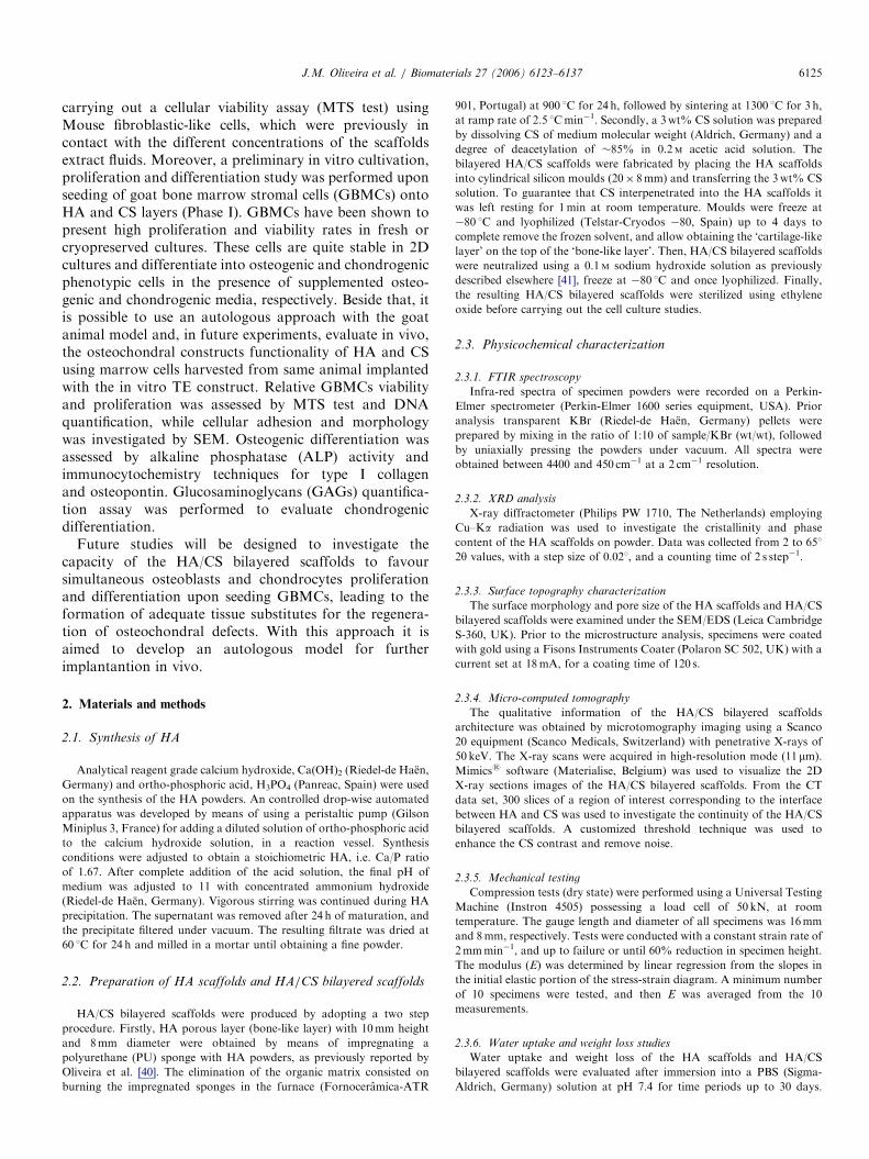
that HA/CS bilayered scaffolds microarchitecture consistsof a highly porous and open pore structure. Fig. 4A showsthe interface of HA/CS bilayered scaffold at the surface,where it is possible to observe the two distinct porouslayers. Fig. 4B shows a typical HA pore at the HA/CSinterface, showing the good interpenetration of CS into theHA scaffolds. This observation suggests that a goodbonding exist between the two layers, which is known tobe a requisite necessary to assure a good integrity andfunctionality of the osteochondral construct (scaffold+cells). Moreover, the SEM examination of the HAscaffolds revealed that pores are highly interconnectedand possess a size diameter in the range of 50–500 mm(Fig. 4C).Several authors [53,54] have shown that freezing condi-
tions, concentration of polymer and freeze-drying para-meters has great influence on the final microstructure anddegree of interconnectivity of the scaffolds. Fig. 4D showsthe SEM micrograph of a CS pore at the typical transversalcross-section of CS layer. This study revealed that CS poresquite resembles a typical spongy 3D structure, with openpores, anisotropic porosity and pore size ranging from20–600 mm. It is also possible to observe the formation ofmicro fibres during the freeze-drying process. This can bean advantage since these fibres can act as an additionalanchor-framework for improved cells adhesion. Therefore,the results depicted in Fig. 4 have shown that by combininga sintering and freeze-drying technique it is possible todevelop HA/CS bilayered scaffolds with an adequate poresize and pore distribution for ingrowths of cells and
ARTICLE IN PRESS
Fig. 4. SEM micrographs of the HA/CS bilayered scaffolds: interface (A), typical pore at interface (B), pore of HA scaffolds (C) and pore of CS layer (D).
J.M. Oliveira et al. / Biomaterials 27 (2006) 6123–6137 6129
diffusion of nutrients, while facilitating vascularization,and thus to be used in TE purposes [55,56]. Although, it isnoteworthy that SEM analysis is only a reliable techniqueto obtain information about 2D systems, i.e. investigate themacro and micro-structure of the scaffold in a non-destructive way at the surface, and shows limited perfor-mance for assessing the pore interconnectivity of thematerials. Moreover, the scaffold should be sectioned bydestructive techniques towards obtaining the detailedinformation of their inner part using the SEM, thus theoriginal morphology of the scaffold will be destroyed.Therefore, in this study the 3D architecture of the materialswas investigated in a non-destructive way, by means ofusing a micro-CT analysis.
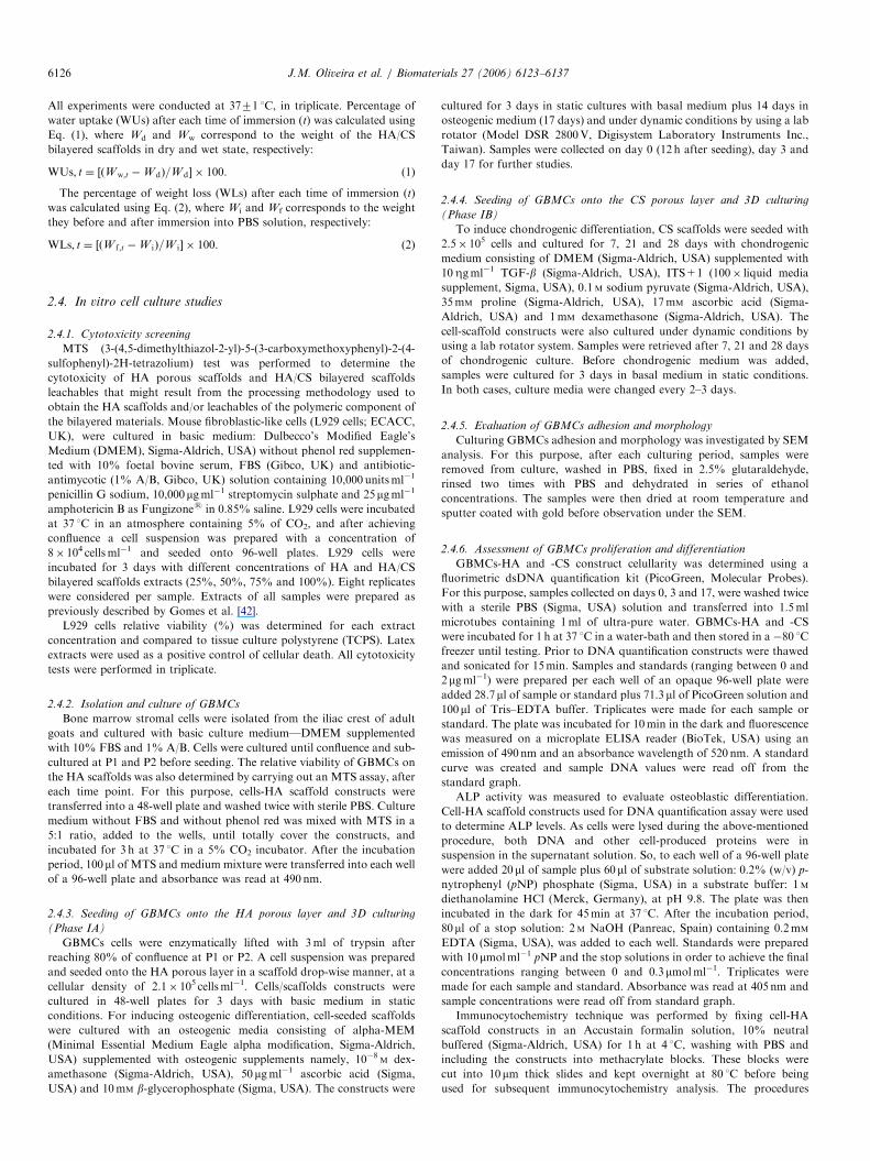
Fig. 5 shows the m-CT analysis of the HA/CS bilayeredscaffolds. A customized threshold technique was carriedout and has shown to be effective on assessing the pore sizeand interconnectivity of CS (Fig. 5A). From the m-CT 2Dscans it was possible to assess that pore size is higher thanthose obtained by the SEM (surface study). Actually, theinner pores of CS was assessed to be up to 2mm in length,which may be explained by the fact that m-CT allows tostudy the inner 3D architecture of the materials. Fig. 5Bshows the 2D transversal view of the HA scaffolds, whichcorroborated the SEM findings. This results clearly showed
that the HA pore network is highly interconnected. FromFig. 5C it became possible to observe that CS perfectlyinterpenetrated into the HA, thus showing the goodcontinuity of the HA/CS bilayered scaffold. This result isencouraging since a strong mechanical interface betweenthe HA and CS is required for the good performance of theconstruct.Previous reported results have shown that the mean
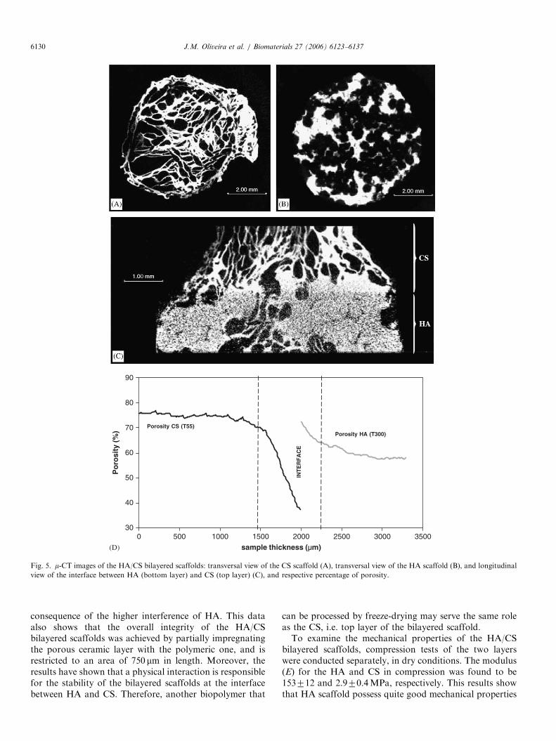
porosity of the HA scaffolds (bottom layer) of the HA/CSbilayered scaffolds consisted of 67.875.1% [45]. In thisstudy, the percentage of porosity of the HA and CS layerswere also determined (Fig. 5D). The results have shownthat HA possess a porosity of 59.371.7%, which is in goodagreement with the previous reported data, and that CSlayer consisted of 74.671.2%. Therefore, the HA/CSbilayered scaffolds have shown to possess adequateporosity [56] towards being used in TE scaffolding.Despite, the percentage of porosity at the interface wasnot possible to determine due to the limitation of thetechnique, i.e. since the coefficient of attenuation of HA ismuch higher that that of CS, the threshold applied fordetermining the porosity of HA layer (T300) is differentfrom that for assessing the porosity of CS layer (T55). Infact, the observation of the dramatic lowering of porosityof CS layer at the interface consists of an artefact and is a
ARTICLE IN PRESS
30
40
50
60
70
80
90
0 500 1000 1500 2000 2500 3000 3500
sample thickness (µm)
Po
rosi
ty (
%) Porosity HA (T300)
Porosity CS (T55)
INT
ER
FA
CE
(D)
Fig. 5. m-CT images of the HA/CS bilayered scaffolds: transversal view of the CS scaffold (A), transversal view of the HA scaffold (B), and longitudinal
view of the interface between HA (bottom layer) and CS (top layer) (C), and respective percentage of porosity.
J.M. Oliveira et al. / Biomaterials 27 (2006) 6123–61376130
consequence of the higher interference of HA. This dataalso shows that the overall integrity of the HA/CSbilayered scaffolds was achieved by partially impregnatingthe porous ceramic layer with the polymeric one, and isrestricted to an area of 750 mm in length. Moreover, theresults have shown that a physical interaction is responsiblefor the stability of the bilayered scaffolds at the interfacebetween HA and CS. Therefore, another biopolymer that
can be processed by freeze-drying may serve the same roleas the CS, i.e. top layer of the bilayered scaffold.To examine the mechanical properties of the HA/CS
bilayered scaffolds, compression tests of the two layerswere conducted separately, in dry conditions. The modulus(E) for the HA and CS in compression was found to be153712 and 2.970.4MPa, respectively. This results showthat HA scaffold possess quite good mechanical properties
ARTICLE IN PRESSJ.M. Oliveira et al. / Biomaterials 27 (2006) 6123–6137 6131
for being used in bone TE. On the other hand, the value forCS scaffolds are in good attainment with those for normalhuman articular cartilage, which has been shown to have acompressive modulus ranging from 1.9 to 14.4MPa [57,58].Thus, the HA/CS bilayered scaffolds demonstrated quitepromising mechanical properties for being used in TE ofosteochondral defects. Actually, it has been shown that thebiomechanical properties of articular cartilage greatlydepends on composition, density of its ECM andinterstitial fluid flow (water and solutes) [59]. Therefore,in wet conditions is expected a reduction of the mechanicalproperties. However, it must be noted that after implanta-
0 5 10
1.4
1.2
1.0
0.8
0.6
0.4
0.2
0.0
Wei
gh
t lo
ss (
%)
Soaking
0 5 100
10
20
30
40
50
60
70
80
90
100
Wat
er u
pta
ke (
%)
Soaking(Α)
(Β)
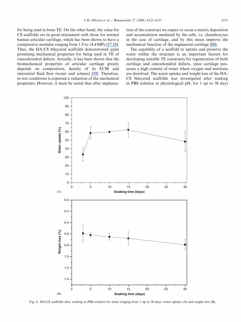
Fig. 6. HA/CS scaffolds after soaking in PBS solution for times rang
tion of the construct we expect to occur a matrix depositionand accumulation mediated by the cells, i.e. chondrocytesin the case of cartilage, and by this mean improve themechanical function of the engineered cartilage [60].The capability of a scaffold to uptake and preserve the
water within the structure is an important feature fordeveloping suitable TE constructs for regeneration of bothcartilage and osteochondral defects, since cartilage pos-sesses a high content of water where oxygen and nutrientsare dissolved. The water uptake and weight loss of the HA/CS bilayered scaffolds was investigated after soakingin PBS solution at physiological pH, for 1 up to 30 days
15 20 25 30
time (days)
15 20 25 30
time (days)
ing from 1 up to 30 days: water uptake (A) and weight loss (B).

ARTICLE IN PRESSJ.M. Oliveira et al. / Biomaterials 27 (2006) 6123–61376132
(Fig. 6). From Fig. 6A it can be seen that the water uptakeprofile of the HA/CS scaffolds reach a maximum ofswelling of approximately 65% (w/w), after 5 days. Fromday 5 until day 30 the water uptake capability of the HA/CS remained constant (Fig. 6A). After immersion of HA/CS bilayered scaffolds in PBS, no significant changes onthe weight loss were observed. In fact, the weight lossprofile showed that from day 3 until day 30, the HA/CSscaffolds showed a reduction in weight of approximately0.8% (Fig. 6B). It has been demonstrated that HAdissolves slowly, both in vitro and in vivo [61,62]. There-fore, this gradual weight loss should be mainly due to theslow loss of the highly hydrophilic CS scaffold by asolution-mediated dissolution, i.e. leaching of the watersolubles rather than HA scaffold. A decrease in CSmolecular weight lowers the water-uptake and favoursdissolution [63]. Thus, the overall performance of the HA/CS scaffolds may be easily tailored by using differentformulations namely, varying the CS molecular weight andcross-linking [41]. Nevertheless, these results are quiteinteresting since the HA/CS bilayered scaffolds has shownto have enough stability, and preserve their size andphysical integrity. This evidences their good performancefor applications in TE of osteochondral defects andsubsequent implantation.
3.2. Cytotoxicity screening for the HA scaffolds and HA/
CS bilayered scaffolds
The intrinsic cytotoxicity of the HA scaffolds and HA/CS bilayered scaffolds was assessed by culturing L929 cellswith extract fluids for 72 h. As observed in an invertedmicroscope cells exhibited a normal morphology prolifer-ated well and established a monolayer (data not shown).
25% 50% 75% 100% Latex0
20
40
60
80
100
120
140
% V
iab
ility
Extract fluids
HA/CS bilayered scaffold
HA scaffold
Latex
Fig. 7. Cytotoxicity screening for the HA scaffolds and HA/CS bilayered
scaffolds. L929 cells were incubated with different concentrations of
leachables obtained from HA scaffolds, HA/CS bilayered scaffolds, and
latex (positive control).
Fig. 7 shows the cell viability assessed by carrying out theMTS test. From the results it is possible to assess that cellsare viable in the presence of the HA and HA/CS scaffoldleachables. In fact, the viability levels are similar amongdifferent HA and HA/CS concentrations, reaching themaximum viability of �100%. This shows that both HAscaffolds and HA/CS scaffolds do not exert any cytotoxiceffect on L929 cells. On the contrary, Latex rubber results(positive control) performed in the same conditions; do notovertake 1.0% of viable cells.
3.3. In vitro assessement of GBMCs adhesion, proliferation
and differentiation
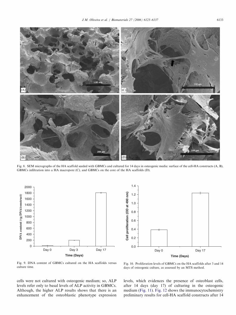
The current investigations using biodegradable implantsseeded MSCs have shown to be effective on the repair ofosteochondral defects, in vivo [19]. In this work, we firstanalysed the ability of the HA porous layer to successfullysupport the GBMCs adhesion and functions, in vitro(Phase IA). As mentioned above, when treating osteochon-dral defects this ‘bone-like’ layer is aimed at provide therepair of the subchondral bone [64], while providing a goodfixation on the underlying bone, during cartilage regenera-tion. Fig. 8 shows the SEM micrographs obtained fromsamples resulting from culturing GBMCs onto the HAporous layer in osteogenic medium, for 14 days. It ispossible to observe the high cellular density on the surfaceof the HA porous layer, which indicates that cells adheredand grew favourably (Fig. 8A). Additionally, it is alsopossible to observe that cells did not obstruct the pores ofthe scaffold at the surface (Fig. 8B), and perfectly adhere,spread actively and presented a flatten morphology ontothe surface of the biomaterial (Fig. 8C). In fact, thetransversal section analysis indicates that cells were able topenetrate deeper into the scaffold core (Fig. 8D). This is animportant observation since a good cell distribution withinthe whole scaffold greatly affects the overall performanceof the construct (e.g. not limiting the diffusion of nutrientsand oxygen) and shows that scaffold architecture allowedcells ingrowths and a sufficient diffusion of nutrients.Additionally, the formation of cells micovilli is alsoobserved, which is suggestive of cell activation [65]. Inour opinion, this result is quite promising since itdemonstrates that the HA porous layer possess anadequate pore size and pore distribution to effectivelyallow the cells to adhere and maintain their functions.Analysis of DNA values showed a significant enhancementcomparatively to initial time points (Fig. 9). The cellnumber increased, and hence a high cellular proliferationoccurred in the presence of osteogenic media. The DNAbiochemical analysis was corroborated by MTS assay(Fig. 10) showing that cells not only proliferated well inHA porous layer but also were viable. These resultsillustrate that HA porous layer is a suitable 3D supportfor cellular proliferation. The functional activity of theGBMCs on the HA porous layer was determined byquantifying the ALP activity (Fig. 11). From days 0 to 3,
ARTICLE IN PRESS
Fig. 8. SEM micrographs of the HA scaffold seeded with GBMCs and cultured for 14 days in osteogenic media: surface of the cell-HA constructs (A, B),
GBMCs infiltration into a HA macropore (C), and GBMCs on the core of the HA scaffolds (D).
Day 0 Day 3 Day 170
200
400
600
800
1000
1200
1400
1600
1800
2000
DN
A c
onte
nt (
ηg D
NA
/con
stru
ct)
Time (Days)
Fig. 9. DNA content of GBMCs cultured on the HA scaffolds versus
culture time.
Day 0 Day 170.0
0.2
0.4
0.6
0.8
1.0
1.2
1.4
Cel
l pro
lifer
atio
n (
OD
at
490
nm
)
Time (Days)
Fig. 10. Proliferation levels of GBMCs on the HA scaffolds after 3 and 14
days of osteogenic culture, as assessed by an MTS method.
J.M. Oliveira et al. / Biomaterials 27 (2006) 6123–6137 6133
cells were not cultured with osteogenic medium; so, ALPlevels refer only to basal levels of ALP activity in GBMCs.Although, the higher ALP results shows that there is anenhancement of the osteoblastic phenotype expression
levels, which evidences the presence of osteoblast cells,after 14 days (day 17) of culturing in the osteogenicmedium (Fig. 11). Fig. 12 shows the immunocytochemistrypreliminary results for cell-HA scaffold constructs after 14

ARTICLE IN PRESSJ.M. Oliveira et al. / Biomaterials 27 (2006) 6123–61376134
days of culturing. The labelling was quite intense forosteopontin, which demonstrates that osteoblast differen-tiation was achieved in these constructs (Fig. 12A).Immunolabeling for collagen type I was also detected,thus showing the production of collagenous matrix(Fig. 12C). Therefore, this data confirm that GBMCs wereable to differentiate in the HA layer after culturing in anosteogenic media for 14 days.
GBMCs seeded onto CS layer present a good viabilityand proliferation (data not showed). SEM micrographsshow the attachment of cells possessing size and shape
Day 0 Day 3 Day 170.0
0.2
0.4
0.6
0.8
1.0
1.2
AL
P a
ctiv
ity
(µm
ol/4
5min
/con
stru
ct)
Time (days)
Fig. 11. ALP activity assay of GBMCs cells cultured on the HA scaffolds.
Fig. 12. Immunolabeling for osteopontin marker (A) and respective negative
days of culturing. Immunolabeling for collagen type I marker (C) and resp
constructs after 14 days of culturing.
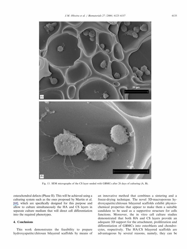
consistent with those of chondrocytes throughout thescaffolds, i.e. round chondrocyte-like cells (Fig. 13). It isalso possible to observe a good distribution of cells andmaintenance of the rounded cell morphology after day 28(Fig. 13A). At higher magnification the moderate celladhesion to the substrate can also be seen (Fig. 13B), whichis generally indicative of a cell performing its differentia-tion function [66]. Despite, cell numbers and penetrationwithin scaffolds appeared to improve in areas presentinglarger and interconnected pores.Glycosaminoglycans (GAGs) are an important compo-
nent of proteoglycan and are typically present in thecartilaginous ECM [67]. GAGs content was determined toassess the formation of newly formed ECM and thus ifoccur the differentiation of GBMCs into chondrocytes inthese cell-scaffold constructs, at days 7, 21 and 28 (Fig. 14).The biochemical analysis demonstrated that the content ofGAGs increased from days 7 to 21, indicating that occurbiosynthesis due to the differentiation of the GBMCs intothe chondrogenic lineage. GAGs levels seems to be low;however it was not found in the literature any referencevalues to compare with. Nevertheless, CS scaffoldsobtained by a freeze-drying technique suggested to be asuitable 3D support for chondrogenic differentiation ofGBMCs, and their maintenance over a period of 28 days.Future studies will be designed to investigate the capacity
of the HA/CS bilayered scaffolds to favour simultaneous butindependent osteoblasts and chondrocytes proliferation anddifferentiation upon seeding GBMCs, leading to the forma-tion of adequate tissue substitutes for the regeneration of
control (without antibody) (B) on the cell-HA scaffold constructs after 14
ective negative control (without antibody) (D) on the cell-HA scaffold
ARTICLE IN PRESS
Fig. 13. SEM micrographs of the CS layer seeded with GBMCs after 28 days of culturing (A, B).
J.M. Oliveira et al. / Biomaterials 27 (2006) 6123–6137 6135
osteochondral defects (Phase II). This will be achieved using aculturing system such as the ones proposed by Martin et al.[68], which are specifically designed for this purpose andallow to culture simultaneously the HA and CS layers inseparate culture medium that will direct cell differentiationinto the required phenotypes.
4. Conclusions
This work demonstrates the feasibility to preparehydroxyapatite/chitosan bilayered scaffolds by means of
an innovative method that combines a sintering and afreeze-drying technique. The novel 3D-macroporous hy-droxyapatite/chitosan bilayered scaffolds exhibit physico-chemical properties that appear to make them a suitablecandidate to be used as a supportive structure for cellsfunctions. Moreover, the in vitro cell culture studiesdemonstrated that both HA and CS layers provide anadequate 3D support for the attachment, proliferation anddifferentiation of GBMCs into osteoblasts and chondro-cytes, respectively. The HA/CS bilayered scaffolds areadvantageous by several reasons, namely, they can be

ARTICLE IN PRESS
0
1
2
3
Day 28Day 21Day 7
GA
Gs
conc
entr
atio
n (µ
g/m
l/co
nstr
uct)
Time
Fig. 14. GAGs quantification assay of GBMCs cells cultured on the CS
layer.
J.M. Oliveira et al. / Biomaterials 27 (2006) 6123–61376136
designed with several sizes and controlled architecture to fitpatient specific injuries and cell functions, respectively. TheHA/CS bilayered scaffolds showed promising biologicalbehaviour and may therefore find applications in tissueengineering of bone and osteochondral defects.
Acknowledgements
Funding was provided by Portuguese Foundation forScience and Technology (FCT) through POCTI and/orFEDER programmes. Joaquim M. Oliveira was alsosupported by a PhD scholarship from Portuguese Founda-tion for Science and Technology (FCT) (SFRH/BD/21786/2005). We are thankful to Materialise (Belgium) for theMIMICSs software provided and LBI, namely GeraldZanoni (Vienna, Austria) for the m-CT scans in the frameof the European Union funded STREP Project HIPPO-CRATES (NMP3-CT-2003-505758). This work was alsocarried out under the scope of the European NoEEXPERTISSUES (NMP3-CT-2004-500283).
References
[1] Temenoff JS, Mikos AG. Review: tissue engineering for regeneration
of articular cartilage. Biomaterials 2000;21:431–40.
[2] Cohen NP, Foster RJ, Mow VC. Composition and dynamics of
articular cartilage: structure, function and maintaining healthy state.
J Orthop Sports Phys Ther 1998;28:203–15.
[3] Aigner T, Stove J. Collagens—major component of the physiological
cartilage matrix, major target of cartilage degeneration, major tool in
the cartilage repair. Adv Drug Deliv Rev 2003;55:1569–93.
[4] Kuettner KE, Cole AA. Review: cartilage degeneration in different
human joints. Osteoarthr Cartilage 2005;13:93–103.
[5] Buckwalter JA. Articular cartilage: injuries and potential for healing.
J Orthop Sports Phys Ther 1998;28:192–202.
[6] Redman SN, Oldfield SF, Archer CW. Current strategies for articular
cartilage repair. Eur Cells Mater 2005;9:23–32.
[7] Glowacki J. In vitro engineering of cartilage. J Rehab Res Dev
2000;37:171–7.
[8] Miller BS, Steadman JR, Briggs KK, Rodrigo JJ, Rodkey WG.
Patient satisfaction and outcome after microfracture of degenerative
knee. J Knee Surg 2004;17:13–7.
[9] Krishnan SP, Skinner JA. (i) Novel treatments for early osteoarthritis
of the knee. Curr Orthop 2005;19:407–14.
[10] Akizuki S, Yasukawa Y, Takizawa T. Does arthroscopic abrasion
arthroplasty promote cartilage regeneration in osteoarthritic knees
with eburnation? A prospective study of high tibial osteotomy with
abrasion arthroplasty versus high tibial osteotomy alone. Arthro-
scopy 1997;13:9–17.
[11] Shapiro F, Koide S, Glimcher MJ. Cell origin and differentiation in
the repair of full-thickness defects of articular cartilage. J Bone J Surg
Am 1993;75:532–53.
[12] Tibesku CO, Szuwart T, Kleffner TO, Schlegel PM, Jahn UR, Van
Aken H, et al. Hyaline cartilage degenerates after autologous
osteochondral transplantation. J Orthop Res 2004;22:1210–4.
[13] Gillogly SD, Voight M, Blackburn T. Treatment of articular cartilage
defects of the knee with autologous chondrocyte implantation.
J Orthop Sports Phys Ther 1998;28:241–51.
[14] Minas T. Autologous chondrocyte implantation in arthritic knee.
Orthopedics 2003;26:945–7.
[15] Brittberg M. Autologous chondrocyte transplantation. Clin Orthop
1999;367:S147–55.
[16] Glowacki TJ, Trepman E, Folkman J. Cell shape and phenotypic
expression in chondrocytes. Proc Soc Exp Biol Med 1983;172:93–8.
[17] Yoo J, Barthel T, Nishimura K, Solchaga L, Caplan AI, Goldberg V,
et al. The chondrogenic potential of human bone-marrow-derived
mesenchymal progenitor cells. J Bone J Surg Am 1998;80:1745–57.
[18] O’Driscoll SW. The healing and regeneration of articular cartilage.
J Bone J Surg 1998;80-A:1795–812.
[19] Uematsu K, Hattori K, Ishimoto Y, Yamauchi J, Habata T,
Takakura Y, et al. Cartilage regeneration using mesenchymal stem
cells and a three-dimensional poly-lactic-glycolic acid (PLGA)
scaffold. Biomaterials 2005;26:4273–9.
[20] Solchaga L, Temenoff J, Gao J, Mikos A, Caplan A, VM G. Repair
of osteochondral defects with hyaluronan- and polyester-based
scaffolds. Osteoarthr Cartilage 2005;13:297.
[21] Lima EG, Mauck RL, Han SH, Park S, Ng KW, Ateshian GA, et al.
Functional tissue engineering of chondral and osteochondral
constructs. Biorheology 2004;41:577–90.
[22] Schaefer D, Martin I, Shastri P, Padera RF, Langer R, Freed LE,
et al. In vitro generation of osteochondral composites. Biomaterials
2000;21:2599–606.
[23] Schaefer D, Martin I, Jundt G, Seidel J, Heberer M, Grodzinsky A,
et al. Tissue-engineered composites for the repair of large osteochon-
dral defects. Arthritis Rheum 2002;46:2524–34.
[24] Gao J, Dennis JE, Solchaga LA, Goldberg VM, Caplan AI. Repair of
osteochondral defect with tissue-engineered two-phase composite
material of injectable calcium phosphate and hyaluronan sponge.
Tissue Eng 2002;8:827–37.
[25] Schek RM, Taboas JM, Segvich SJ, Hollister SJ, Krebsbach PH.
Engineered osteochondral grafts using biphasic composite solid free-
form fabricated scaffolds. Tissue Eng 2004;10:1376–85.
[26] Kotobuki N, Ioku K, Kawagoe D, Fujimori H, Goto S, Ohgushi H.
Observation of osteogenic differentiation cascade of living mesench-
ymal stem cells on transparent hydroxyapatite ceramics. Biomaterials
2005;26:779–85.
[27] Kasten P, Vogel J, Luginbuhl R, Niemeyer P, Tonak M, Lorenz H,
et al. Ectopic bone formation associated with mesenchymal stem cells
in a resorbable calcium deficient hydroxyapatite carrier. Biomaterials
2005;26:5879–89.
[28] Livingston Arinzeh T, Tran T, Mcalary J, Daculsi G. A comparative
study of biphasic calcium phosphate ceramics for human mesench-
ymal stem-cell-induced bone formation. Biomaterials 2005;26:
3631–8.
[29] Lu JX, Prudhommeaux F, Meunier A, Sedel L, Guillemin G.
Effects of chitosan on rat knee cartilages. Biomaterials 1999;20:
1937–44.

ARTICLE IN PRESSJ.M. Oliveira et al. / Biomaterials 27 (2006) 6123–6137 6137
[30] Di Martino A, Sittinger M, Risbud MV. Chitosan: a versatile
biopolymer for orthopaedic tissue-engineering. Biomaterials 2005;
26:5983–90.
[31] Yamane S, Iwasaki N, Majima T, Funakoshi T, Masuko T, Harada
K, et al. Feasibility of chitosan-based hyaluronic acid hybrid
biomaterial for a novel scaffold in cartilage tissue engineering.
Biomaterials 2005;26:611.
[32] Jiang Y, Jahagirdar BN, Reinhardt RL, Schwartz RE, Keene CD,
Ortiz-Gonzalez XR, et al. Pluripotency of mesenchymal stem cells
derived from adult marrow. Nature 2002;418:41–9.
[33] Reyes M, Lund T, Lenvik T, Aguiar D, Koodie L, Verfaillie CM.
Purification and ex vivo expansion of postnatal human marrow
mesodermal progenitor cells. Blood 2001;98:2615–25.
[34] Im II G, Shin Y-W, Lee K-B. Do adipose tissue-derived mesenchymal
stem cells have the same osteogenic and chondrogenic potential as
bone marrow-derived cells? Osteoarthr Cartilage 2005;13:845.
[35] Kitamura S, Ohgushi H, Hirose M, Funaoka H, Takakura Y, Ito H.
Osteogenic differentiation of human bone marrow-derived mesench-
ymal cells cultured on alumina ceramics. Artif Organs 2004;28:72–82.
[36] Caplan AI, Elyaderani M, Mochizuki Y, Wakitani S, Goldberg VM.
Principles of cartilage repair and regeneration. Clin Orthop Relat Res
1997;342:254–69.
[37] Uemura T, Dong J, Wang Y, Kojima H, Saito T, Iejima D, et al.
Transplantation of cultured bone cells using combinations of
scaffolds and culture techniques. Biomaterials 2003;24:2277.
[38] Ohgushi H, Kotobuki N, Funaoka H, Machida H, Hirose M, Tanaka
Y, et al. Tissue engineered ceramic artificial joint-ex vivo osteogenic
differentiation of patient mesenchymal cells on total ankle joints for
treatment of osteoarthritis. Biomaterials 2005;26:4654–61.
[39] Rahaman MN, Mao JJ. Stem cell-based composite tissue constructs
for regenerative medicine. Biotechnol Bioeng 2005;91:261–84.
[40] Oliveira JM, Silva SS, Mano JF, Reis RL. Innovative technique for
the preparation of porous bilayer hydroxyapatite/chitosan scaffolds
for osteochondral applications. Kyoto (JPN), Zurich, Switzerland:
Key Eng Mater, Trans Tech Pub; 2006.
[41] Silva SS, Oliveira JM, Mano JF, Reis RL. Preparation and
characterization of novel chitosan/soy protein porous for tissue
engineering applications. Adv Mater Forum 2006;514–516:1000–4.
[42] Gomes ME, Reis RL, Cunha AM, Blitterswijk CA, de Bruijn JD.
Cytocompatibility and response of osteoblastic-like cells to starch-
based polymers: effect of several additives and processing conditions.
Biomaterials 2001;22:1911.
[43] Hollander AP, Hatton PV. Biopolymer methods in tissue engineering
(methods in molecular biology series). Totowa, NJ: Human Press
Inc.; 2004.
[44] Kupper JH, van Gool L, Burkle A. Molecular genetic systems to
study the role of poly(ADP-ribosyl)ation in the cellular response to
DNA damage. Biochimie 1995;77(6):450.
[45] Oliveira JM, Silva SS, Malafaya PB, Rodrigues MT, Gomes ME,
Mano JF, et al. Development and physichochemical characterization
of novel hydroxyapatite scaffolds for bone tissue engineering
applications. J Mater Sci-Mater Med 2006 (in press).
[46] Yamaguchi I, Itoh S, Suzuki M, Sakane M, Osaka A, Tanaka J. The
chitosan prepared from crab tendon I: the characterization and
mechanical properties. Biomaterials 2003;24:2031–6.
[47] Kolhe P, Kannan RM. Improvement in ductibility of chitosan
through blending and copolymerization with PEG: FTIR investiga-
tion of molecular interactions. Biomacromolecules 2003;4:173–80.
[48] Raynaud S, Champion E, Bernache-Assollant D, Thomas P. Calcium
phosphate with variable Ca/P atomic ratio. I. Synthesis, character-
ization and thermal stability of powders. Biomaterials 2002;
23:1065–72.
[49] Tampieri A, Celotti G, Szontagh F, Landi E. Sintering
and characterization of HA and TCP bioceramics with control of
their strength and phase purity. J Mater Sci-Mater Med 1997;
8:29–37.
[50] Mostafa NY. Characterization, thermal stability and sintering of
hydroxyapatite powders prepared by different routes. Mater Chem
Phys 2005;94:333.
[51] Kotobuki N, Ioku K, Kawagoe D, Fujimori H, Goto S, Ohgushi H.
Observation of osteogenic differentiation cascade of living mesench-
ymal stem cells on transparent hydroxyapatite ceramics. Biomaterials
2005;26:779.
[52] Cyster LA, Grant DM, Howdle SM, Rose FRAJ, Irvine DJ,
Freeman D, et al. The influence of dispersant concentration on the
pore morphology of hydroxyapatite ceramics for bone tissue
engineering. Biomaterials 2005;26:697.
[53] Ho M-H, Kuo P-Y, Hsieh H-J, Hsieh T-Y, Hou L-T, Lai J-Y, et al.
Preparation of porous scaffolds by using freeze-extraction and freeze-
gelation methods. Biomaterials 2004;25:129–38.
[54] Madihally SV, Matthew HWT. Porous chitosan scaffolds for tissue
engineering. Biomaterials 1999;20:1133–42.
[55] Karageorgiou V, Kaplan D. Porosity of 3D biomaterial scaffolds and
osteogenesis. Biomaterials 2005;26:5474–91.
[56] Hutmacher DW. Scaffolds in tissue engineering bone and cartilage.
Biomaterials 2000;21:2529–43.
[57] Zheng-Qiu G, Jiu-Mei X, Xiang-Hong Z. The development of
artificial articular cartilage—PVA-hydrogel. Biomed Mater Eng
1998;8:75–81.
[58] Magnussen RA, Guilak F, Vail TP. Cartilage degeneration in post-
collapse cases of osteonecrosis of the human femoral head: altered
mechanical properties in tension, compression, and shear. J Orthop
Res 2005;23:576.
[59] Evans RC, Quinn TM. Solute diffusivity correlates with mechanical
properties and matrix density of compressed articular cartilage. Arch
Biochem Biophys 2005;442:1.
[60] Miyata S, Furukawa KS, Ushida T, Nitta Y, Tateishi T. Static and
dynamic mechanical properties of extracellular matrix synthesized by
cultured chondrocytes. Mater Sci Eng C 2004;24:425.
[61] Driessens FCM, Ramselaar MMA, Schaken HG, Stols ALH, Van
Mullem PJ, de Wijn JR. Chemical reactions of calcium phosphate
implants after implantation in vivo. J Mater Sci-Mater Med
1992;3:413–7.
[62] Klein CPAT, de Blieck-Hogemrst JMA, Wolket JGC, de Groot K.
Studies of the solubility of different calcium phosphate ceramic
particles in vitro. Biomaterials 1990;11:509.
[63] Berger J, Reist M, Mayer JM, Felt O, Peppas NA, Gurny R.
Structure and interactions in covalently and ionically crosslinked
chitosan hydrogels for biomedical applications. Eur J Pharm
Biopharm 2004;57:19–34.
[64] Tamai N, Myoui A, Hirao M, Kaito T, Ochi T, Tanaka J, et al. A
new biotechnology for articular cartilage repair: subchondral
implantation of a composite of interconnected porous hydroxyapa-
tite, synthetic polymer (PLA-PEG), and bone morphogenetic protein-
2 (rhBMP-2). Osteoarthr Cartilage 2005;13:405.
[65] Xynos ID, Hukkanen MVJ, Batten JJ, Buttery LD, Hench LL, Polak
JM. Bioglass 45S5 stimulates osteoblast turnover and enhances bone
formation in vitro: implications and applications for bone tissue
engineering. Calcif Tissue Int 2000;67:321–9.
[66] Yaylaoglu MB, Yildiz C, Korkusuz F, Hasirci V. A novel
osteochondral implant. Biomaterials 1999;20:1513–20.
[67] Barnewitz D, Endres M, Kruger I, Becker A, Zimmermann J, Wilke
I, et al. Treatment of articular cartilage defects in horses with
polymer-based cartilage tissue engineering grafts. Biomaterials
2006;27:2882–9.
[68] Wendt D, Jakob M, Martin I. Bioreactor-based engineering of
osteochondral grafts: from model systems to tissue manufacturing.
J Biosci Bioeng 2005;100:489–94.
Copyright © 2022 FDOKUMEN