different strategies to obtain antimicrobial biodegradable films ...
282
UNIVERSIDAT POLITÈCNICA DE VALÈNCIA INSTITUTO UNIVERSITARIO DE INGENIERÍA DE ALIMENTOS PARA EL DESARROLLO DIFFERENT STRATEGIES TO OBTAIN ANTIMICROBIAL BIODEGRADABLE FILMS FOR FOOD APPLICATIONS, USING STARCH AND/OR CHITOSAN WITH OR WITHOUT ESSENTIAL OILS DOCTORAL THESIS Presented by: Cristina Encarnación Valencia Sullca Supervisors: Lorena Atarés Huerta María Vargas Colás Valencia, June 2017
-
Upload
khangminh22 -
Category
Documents
-
view
2 -
download
0
Transcript of different strategies to obtain antimicrobial biodegradable films ...

UNIVERSIDAT POLITÈCNICA DE VALÈNCIAINSTITUTO UNIVERSITARIO DE INGENIERÍA DE ALIMENTOS
PARA EL DESARROLLO
DIFFERENT STRATEGIES TO OBTAIN ANTIMICROBIALBIODEGRADABLE FILMS FOR FOOD APPLICATIONS,USING STARCH AND/OR CHITOSAN WITH ORWITHOUT ESSENTIAL OILS
DOCTORAL THESIS
Presented by:
Cristina Encarnación Valencia Sullca
Supervisors:
Lorena Atarés Huerta
María Vargas Colás
Valencia, June 2017


UNIVERSIDAD POLITÈCNICA DE VALÈNCIA
INSTITUTO UNIVERSITARIO DE INGENIERÍA DE ALIMENTOS
PARA EL DESARROLLO
Da. Lorena Atarés Huerta y Da. María Vargas Colás, profesoras Titulares de Universidad de la Uni-
versitat Politècnica de València.
CONSIDERAN: que la memoria titulada “DIFFERENT STRATEGIES TO OBTAIN ANTIMICROBIAL
BIODEGRADABLE FILMS FOR FOOD APPLICATIONS, USING STARCH AND/OR CHITOSAN
WITH OR WITHOUT ESSENTIAL OILS”, que presenta Da. Cristina Encarnación Valencia Sullca,
para aspirar al grado de Doctor por la Universitat Politècnica de València, realizada en el Instituto
de Ingeniería de Alimentos para el Desarrollo (IuIAD–UPV) bajo su dirección, reúne las condiciones
adecuadas para constituir su tesis doctoral, por lo que AUTORIZAN a la interesada su presentación.
Valencia a 31 de mayo de 2017
Fdo.: Lorena Atarés Huerta Fdo.: María Vargas Colás
Directora de la tesis Directora de la tesis


This work has been founded by the Ministerio de Economía y Competitividad (Spain) through
the Projects AGL2013-42989-R and AGL2016-76699-R, and the Programa Nacional de Becas
del Perú (Pronabec).


A Dios
A mis amados padres (Elena y Ubaldo)
A mis amados hermanos (Maritza, Joaquín, Iris, Ismael)
A mi ángel de la guarda (mi amado hermano Alfredo)


ACKNOWLEDGEMENTS
Primero y antes que nada, dar gracias a Dios, por estar conmigo en cada paso que doy, por fortalecer
mi corazón e iluminar mi mente y por haber puesto en mi camino a aquellas personas que han sido mi
soporte y compañía durante todo el periodo de estudio.
Agradecer hoy y siempre a mis amados padres Elena y Ubaldo porque a pesar de no estar presentes
físicamente, se que procuran mi bienestar desde mi país, Perú, y esta claro que, si no fuese por
el esfuerzo realizado por ellos, mis estudios realizados no hubiesen sido posible. A mis hermanos
Maritza, Joaquín, Iris, Ismael, y mis sobrinos, porque a pesar de la distancia, el ánimo, cariño, apoyo
y alegría que me brindan me dan la fortaleza necesaria para seguir adelante. Mi padrino Gerardo y
Lucia por su cariño, consejos y hospitalidad durante mi estadia en Barcelona, los aprecio mucho.
Gracias al Gobierno del Perú, especificamente al Ministerio de Educación con el programa de Becas
Pronabec, por la gran oportunidad de poder realizar mis estudios de doctorado en Valencia-España.
Muchas gracias de corazón Dra Amparo Chiralt, Dra. Lorena Atarés y Dra. Maria Vargas por abrirme
las puertas del grupo de investigación y darme su confianza y por permitirme aprender y trabajar a
su lado. La experiencia y conocimiento de cada una de ustedes ha sido muy enriquecedor para mi
formación academica. Les estare por siempre agradecida.
Gracias al “Laboratoire d’Ingénierie et des Biomolécules” y en especial al Dr. Laura Sanchez Gonzalez
y la Dra. Elmira Arab Tehrany de la Université de Lorraine.Ha sido una experiencia inolvidable haber
podido compartir mi estancia con ustedes. Merci beaucoup!!!!!!!!
Gracias Cristhian Rodriguez por tu apoyo, aliento y cariño incodicional durante estos años, siempre te
estare agradecida y a toda tu linda familia en especial a tus padres Berta y Gilberto. Gracias por su
cariño, animos y amistad, los llevo en mi corazón y en mis oraciones.
A todos mis amigos muy cercanos que he sentido su compañía durante estos años. Gracias: Gina,
Rosario, Jonathan, Nohemi, Cesár, Willy, Luz, Carmen, Paul, Sandrita, Danitza, Fernando, Jesus,
Rosmery, Ronny, Mauricio, Nestor, Ines, etc, asimismo, algunos becarios de pronabec que ha sido
un gusto conocerlos y compartir experiencias y conocimientos con ustedes, gracias por su amistad:
Mirian, Estefany, Daysi, Galo, Elio, Edwin, Eva, Oscar..etc.

Gracias Robert Valverde por tu apoyo, cariño y animos, asimismo, gracias a tu linda familia, ha sido
una bendición conocerlos, mi estancia en Nancy-Francia ha sido y será la mejor experiencia que he
tenido, nunca lo olvidare los llevo en mi corazón. Gracias totales. Asimismo, gracias Silvia, Madeleine,
Vilma, Juanca, Nahuel, Viviana..ha sido un gusto conocerlos y compartir momentos lindos, gracias
por su amistad y cariño..y al grupo latino de los miercolitos gracias por su linda amistad y momentos
compartidos en Nancy, gracias: Magaly, Carmen, Mariana Limones, Arturo, Estibaly, Natalia, Thibaud
y Mariana, Karen, Ferdali, Pilar, Anne-Claire..etc.
A todos mis compañeros del laboratorio, a los que conocido y siguen en la actualidad, sus consejos,
ánimos y cariño es reciproco y ha sido una gran experiencia conocerlos durante estos años. En
especial a mis chicas que son como unas hermanas para mi, Moltes gracies: Emma, Ana, Olga,
Amalia, Alina, Mayra, Sofia, Justine, Raquel, Angela, Susana, Lizhet. Gracias por enseñarme muchas
cosas ¡son geniales, las quiero y siempre las llevare en mi corazón!
A los estudiantes de máster y pregrado a quienes tuve la oportunidad de orientar en su trabajo
experimental de final de carrera y de master, la verdad que ha ido una experiencia inolvidable, gracias:
Miriam, Jennifer y Sara, agradezco su esfuerzo y contribución.

PREFACE
DISSERTATION OUTLINE
This Doctoral Thesis is structured in five sections: Introduction, Objectives, Chapters, General
Discussion and Conclusions.
The INTRODUCTION section revises the state of the art concerning the properties of starch
films and the antimicrobial action of chitosan and essential oils used to develop biodegradable
films. Encapsulation techniques with interest for developing edible films are also reviewed.
The OBJECTIVES section presents the general and specific objectives of the Thesis,
which is focused on the development of biodegradable films based on hydrocolloids (starch
and/or chitosan) with antimicrobial properties that were produced by different techniques
(compression molding and/or casting).
The obtained results are organized in five CHAPTERS, each one corresponding to a scientific
publication that includes the usual sections: introduction, materials and methods, results and
discussion and conclusion.
Chapter 1, entitled “Physical characterization of cassava starch – chitosan films
obtained by compression molding”, evaluated the effect of the incorporation of chitosan,
glycerol and polyethylene glycol (PEG) as plasticizers on the physical and antimicrobial
properties of cassava starch films obtained by compression molding. Results showed that
chitosan incorporation provided the films with only a slight antimicrobial capacity and thus, in
the subsequent chapters, different strategies to overcome this drawback were used.
Chapter 2, entitled “Thermoplastic cassava starch – chitosan films containing essential
oils”, was focused in chitosan films (containing or not different essential oils as active
ingredients) that were obtained by casting. The casted films were used to develop bilayer films
with cassava starch films produced by compression molding. The bilayer films were effective
at controlling the bacterial growth in pork meat samples, but the thermal treatment used to
obtain the bilayers reduced its effectiveness. Therefore, in chapters 3 to 5 other strategies
were developed to incorporate essential oils in the chitosan matrix and to improve the final
retention of active ingredients at the same time that a control release in different food systems
is promoted.

Chapter 3, entitled “Influence of liposome encapsulated essential oils on properties of
chitosan films”, is centered in the encapsulation of active ingredients (eugenol or cinnamon
leaf essential oil) in lecithin liposomes that were added to chitosan films obtained by casting.
Films were evaluated in terms of their physical properties and antimicrobial performance in
different food systems, as compared to chitosan films that included non-encapsulated active
ingredients.
Chapter 4, entitled “Release kinetics and antimicrobial properties of chitosan-essential
oils films as affected by encapsulation within lecithin nanoliposomes”, evaluated the
antimicrobial properties of chitosan films containing active ingredients (eugenol or cinnamon
leaf essential oil) in free form or encapsulated in lecithin liposomes encapsulated. The release
kinetics of eugenol and the specific migration of the films in different food simulants were also
analyzed.
Chapter 5, entitled “Chitosan films containing encapsulated eugenol in alginate
microcapsules”, showed another encapsulation strategy to improve the functionality and
retention of active compound in chitosan–based matrices. The films were evaluated in their
physical and antimicrobial properties as well as the kinetics of eugenol release in food
simulants.
Finally, the most important CONCLUSIONS of the Thesis are shown.

DISSEMINATION OF RESULTS
Published:
Valencia-Sullca, C., Miriam Jiménez, Alberto Jiménez, Atarés, L., Vargas, M., Chiralt, A.
(2016). Influence of liposome encapsulated essential oils on properties of chitosan films.
Published online in Wiley Online Library: DOI 10.1002/pi.5143.
Submitted:
Valencia-Sullca, C., Atarés, L., Vargas, M., Chiralt, A. Physical characterization of cassava
starch-chitosan films obtained by compression molding.
Valencia-Sullca, C., Vargas, M., Atarés, L., Chiralt, A. Thermoplastic cassava starch-chitosan
films containing essential oils.
Valencia-Sullca, C., Vargas, M., Atarés, L., Chiralt, A. Active chitosan films for meat
conservation.
Valencia-Sullca, C., Atarés, L., Vargas, M., Chiralt, A. Chitosan films containing encapsulated
eugenol in alginate microcapsules.
The results have been also presented in national and international conferences:
Poster – Valencia-Sullca, C., Atarés, L., Vargas, M., Chiralt, A. (2014). Propiedades físicas
de películas biodegradables de almidón de yuca y quitosano obtenidas por moldeado por
compresión. “III International Conference of Food Innovation”, October, 20-23. Concordia,
Entre Ríos, Argentina.
Poster – Valencia-Sullca, C., Atarés, L., Vargas, M., Chiralt, A. (2015). Properties of bilayer
films of starch-chitosan containing oregano oil. “3rd International Meeting on Packaging
Material / Bioproduct Interactions (Matbim 2015)”. University of Zaragoza, Spain.
Poster and article – Valencia-Sullca, C., Miriam Jiménez, Alberto Jiménez, Atarés, L.,
Vargas, M., Chiralt, A. (2015). Influence of liposome encapsulated essential oils on properties
of chitosan films. “5th International Conference on Biobased and Biodegradable Polimers
(Biopol 2015)”. Donostia – San Sebastián, España.

Poster – Valencia-Sullca, C., Soler-Beatty, J., Atarés, L., Vargas, M., Chiralt, A. (2016).
Eugenol liposomes into chitosan films: active release kinetics and antimicrobial properties.
“6th International Symposium on Food Packaging (ILSI 2016): Scientific Developments
Supporting Safety and Innovation”. Barcelona, Spain.
Poster – Valencia-Sullca, C., Atarés, L., Vargas, M., Chiralt, A. (2016). Properties of bilayer
films of starch-chitosan containing cinnamon leaf oil. First meeting of Peruvian scientists in
Europe (Sinapsis 2016). Paris, France.
Poster – Valencia-Sullca, C., Atarés, L., Vargas, M., Chiralt, A. (2017). Antimicrobial activity
of chitosan films containing encapsulated eugenol in alginate microcapsules. “3rd International
& 3th national student congress of science and technology (Avecta 2017)”. Valencia, Spain.
Poster – Valencia-Sullca, C., Camara, S., Atarés, L., Vargas, M., Chiralt, A. (2017). Chitosan
films containing encapsulated eugenol in alginate microcapsules. “4rd International Meeting
on Packaging Material / Bioproduct Interactions (Matbim 2017)”. Porto, Portugal.
PREDOCTORAL STAYS AT FOREIGN INSTITUTIONS
Laboratoire d’Ingénierie et des Biomolécules (LIBio), École Nationale Supérieure d’Agronomie
et des Industries Alimentaies (ENSAIA), Université de Lorraine, Nancy, France. From April
2016 to July 2016, under the supervision of Dra. Laura Sánchez González. Development of
alginate/chitosan microcapsules containing eugenol.

TABLE OF CONTENTS
I. INTRODUCTION .............................................................................................................. 29
I.1. Starch as base material for biodegradable packaging applications . . . . . . . . . 31
I.2. Cassava starch . . . . . . . . . . . . . . . . . . . . . . . . . . . . . . . . . . . . 34
I.3. Chitosan and its applications in biodegradable packaging . . . . . . . . . . . . . 40
I.4. Essential oils and their application into biodegradable films . . . . . . . . . . . . 43
I.5. Improving the functional properties of essential oils by encapsulation techniques . 57
I.6. Microencapsulation methods . . . . . . . . . . . . . . . . . . . . . . . . . . . . . 70
II. OBJECTIVES ................................................................................................................. 121
III. RESULTS .........................................................................................................................125
III.1. CHAPTER 1
Physical characterization of cassava starch-chitosan films obtained by compression
molding . . . . . . . . . . . . . . . . . . . . . . . . . . . . . . . . . . . . . . . 129
III.2. CHAPTER 2
Thermoplastic cassava starch-chitosan films containing essential oils . . . . . . . . . 159
III.3. CHAPTER 3
Influence of liposome encapsulated essential oils on properties of chitosan films . . . 185
III.4. CHAPTER 4
Release kinetics and antimicrobial properties of chitosan-essential oils films as
affected by encapsulation within lecithin nanoliposomes . . . . . . . . . . . . . 215
III.5. CHAPTER 5
Chitosan films containing encapsulated eugenol in alginate microspheres . . . . . . . 235
IV. GENERAL DISCUSSION ............................................................................................. 267
V. CONCLUSION ............................................................................................................... 279

TABLES
Table I.1 Summary of results obtained for cassava starch films obtained by different
techniques (casting and thermoplastic processing) . . . . . . . . . . . . . . . . 37
Table I.2 Main constituents of cinnamon leaf and oregano essential oils and their wt.
percentage range, as reported by several authors . . . . . . . . . . . . . . . . . 45
Table I.3 Recent papers studying the effect of essential oils on CH films properties
(Scanning Electron Microscopy (SEM), elastic modulus (EM, MPa), tensile
strength (TS, MPa), elongation at break (E, %), water vapour permeability
(WVP), oxygen permeability (OP), gloss and transparency . . . . . . . . . . . . 49
Table I.4 Recent studies dealing with the effect of EOs addition on the antimicrobial
activity of CH films or coating . . . . . . . . . . . . . . . . . . . . . . . . . . . . 53
Table I.5 Advantages and disadvantages of wall material to encapsulation . . . . . . 63
Table I.6 A representative list of encapsulated essential oils and other compounds
with different methods for encapsulation . . . . . . . . . . . . . . . . . . . . . . 67
Table I.7 Advantages and disadvantages of different microencapsulation technologies 77
Table 1.1. Thermal properties of the films analyzed by TGA (T0, Tmax, % Mass loss
over degradation). Mean values and standard deviation . . . . . . . . . . . . . 140
Table 1.2. Thermal properties of films analyzed by DSC. Mean values and standard
deviation . . . . . . . . . . . . . . . . . . . . . . . . . . . . . . . . . . . . . . . 143
Table 1.3. Thickness, moisture content and water vapour permeability (WVP) of films
equilibrated at 53 % RH. Mean values and standard deviation . . . . . . . . . . 144
Table 1.4. Tensile properties (elastic modulus: EM, tensile strength: TS and
deformation: E %, at break) of all films equilibrated at 53 % RH after 1 and 5
week storage. Mean values and standard deviation . . . . . . . . . . . . . . . . 146
Table 1.5. Lightness (L*), chroma (C*ab), hue (h*ab), whiteness index (WI) and gloss
at 60◦ after 1 and 5 week storage. Mean values and standard deviation . . . . . 150
Table 1.6. The antimicrobial activity of the films on pork meat and stored 7 days at
10◦C. Mean values and standard deviation . . . . . . . . . . . . . . . . . . . . 151
Table 2.1. Thermal properties of the films and essential oils (T0, Tmax, Mass loss
during degradation). Mean values and standard deviation . . . . . . . . . . . . 171
Table 2.2. Tensile properties (elastic modulus: EM, tensile strength: TS and
elongation: E %, at break) of all films equilibrated at 53 % RH after 1 week
storage. Mean values and standard deviation . . . . . . . . . . . . . . . . . . . 173

Table 2.3. Thickness, water vapor permeability (WVP) and oxygen permeability (OP)
of the films. Mean values and standard deviation . . . . . . . . . . . . . . . . . 174
Table 2.4. Lightness (L*), chroma (C*ab), hue (h*ab), whiteness index (WI) and gloss
at 60◦. Mean values and standard deviation . . . . . . . . . . . . . . . . . . . . 176
Table 3.1. Density and ζ-potential of nanoliposome dispersions (ND) and film-
forming dispersions (FFD). Mean values and standard deviation. Lec: lecithin,
Eu: eugenol, CLEO: cinnamon leaf essential oil, CH: chitosan . . . . . . . . . . 197
Table 3.2. Mass fraction of eugenol in the dried films (mg/g film solids), extracted
in the dried film and initially incorporated, and percentage retention (extracted
with respect to the initially added). Lec: lecithin, Eu: eugenol, CLEO: cinnamon
leaf essential oil, CH: chitosan . . . . . . . . . . . . . . . . . . . . . . . . . . . 199
Table 3.3. Thickness and tensile parameters (elastic modulus, EM; tensile strength,
TS; percentage elongation, %E) of the films. Mean values and standard
deviation. Lec: lecithin, Eu: eugenol, CLEO: cinnamon leaf essential oil, CH:
chitosan . . . . . . . . . . . . . . . . . . . . . . . . . . . . . . . . . . . . . . . 199
Table 3.4. Water content (XW), water vapor permeability (WVP) and solubility (g of
solubilized solids/100 g of initial solids) of the films. Mean values and standard
deviation. Lec: lecithin, Eu: eugenol, CLEO: cinnamon leaf essential oil, CH:
chitosan . . . . . . . . . . . . . . . . . . . . . . . . . . . . . . . . . . . . . . . 200
Table 3.5. Lightness (L*), chroma (C*ab), hue (h*ab) and gloss (60◦) of the
films. Mean values and standard deviation. Lec: lecithin, Eu: eugenol, CLEO:
cinnamon leaf essential oil, CH: chitosan . . . . . . . . . . . . . . . . . . . . . 201
Table 3.6. Onset temperature (T0) and maximum degradation rate temperure of the
films. Mean values and standard deviation. Lec: lecithin, Eu: eugenol, CLEO:
cinnamon leaf essential oil, CH: chitosan . . . . . . . . . . . . . . . . . . . . . 207
Table 3.7. Escherichia coli counts in liquid (TSA Broth) and solid media (TSA Agar)
at 10◦C. Mean values ± standard deviation Lec: lecithin, Eu: eugenol, CLEO:
cinnamon leaf essential oil, CH: chitosan. ng: no growth . . . . . . . . . . . . . 208
Table 3.8. Listeria innocua counts in liquid (TSA Broth) and solid media (TSA Agar).
Mean values ± standard deviation Lec: lecithin, Eu: eugenol, CLEO: cinnamon
leaf essential oil, CH: chitosan. ng: no growth . . . . . . . . . . . . . . . . . . . 209
Table 4.1. Total migration (10 days, 20◦C) of material from the films and eugenol
extracted and retained in methanol after 24 hours. Mean values and standard
deviation . . . . . . . . . . . . . . . . . . . . . . . . . . . . . . . . . . . . . . . 225

Table 4.2. Apparent diffusivity (D) and parameters of Peleg’s model: amount of
active compound released at equilibrium in the simulant (M∞) and its release
rate (1/k1), and maximum release ratio (M∞/M0): mass of active released at
equilibrium in the simulant related to the initial mass of the active in the film
expressed with respect to the amount determined by methanol extraction (1) or
by acetic acid extraction (2) . . . . . . . . . . . . . . . . . . . . . . . . . . . . . 227
Table 4.3. Total specific migration of eugenol (mg eugenol/g meat) from the films in
meat samples stored at 10◦C. Mean values and standard deviation, in brackets 229
Table 5.1. Density, pH and ζ-potential of the dispersions an emulsion used to prepare
the microspheres. Mean values and standard deviation . . . . . . . . . . . . . . 248
Table 5.2. Elastic modulus (EM), tensile strength (TS), elongation at break ( %E),
water vapor permeability (WVP), oxygen permeability (OP) and moisture
content. Mean values and standard deviation . . . . . . . . . . . . . . . . . . . 251
Table 5.3. Lightness (L*), chroma (C*ab), hue (h*ab), whiteness index (WI) and
internal transmittance (Ti) at 450 nm. Mean values and standard deviation . . . 252
Table 5.4. Parameters of Peleg’s model: amount of eugenol released from CHEU
at equilibrium in the simulant (M∞) and its release rate (1/k1), and maximum
release ratio (M∞/M0): mass of active released at equilibrium in the simulant
related to the initial mass of the active in the film (expressed with respect to the
theoretical incorporated amount (1) and with respect to the amount determined
by acetic acid extraction (2) . . . . . . . . . . . . . . . . . . . . . . . . . . . . . 255
Table 5.5. Diffusion coefficient (D) and parameters of the Korsmeyer-Peppas model
(rate constant (k) and diffusional exponent (n)) . . . . . . . . . . . . . . . . . . 255

FIGURES
Figure I.1 Chemical structure of amylose and amylopectin . . . . . . . . . . . . . . . 32
Figure I.2 Chemical structure of chitosan . . . . . . . . . . . . . . . . . . . . . . . . 40
Figure I.3 Chemical structure of eugenol and carvacrol . . . . . . . . . . . . . . . . 45
Figure I.4 Morphology of different types of microspheres . . . . . . . . . . . . . . . . 58
Figure I.5 Chemical structure of β-D-mannuronic acid and α-L-guluronic acid . . . . 60
Figure I.6 The “Eggs-bo” model for alginate gelation with calcium ions . . . . . . . . 60
Figure I.7 Set-ups of different ways of making alginate microspheres by extrusion . . 71
Figure 1.1. Scanning electron microscopy micrographs of the cross-sections of
the films. (a) Polymer: plasticizer proportion 70:30.(b) Polymer: plasticizer
proportion 60:40 . . . . . . . . . . . . . . . . . . . . . . . . . . . . . . . . . . 139
Figure 1.2. Typical thermogravimetric curves (mass loss vs. temperature) and first
derivative (mg/s vs temperature) for a) Polymer: plasticizer proportion 70:30
and b) Polymer: plasticizer proportion 60:40 . . . . . . . . . . . . . . . . . . . . 142
Figure 1.3. Typical stress-strain curves of the films after 1 week (left) and 5 weeks
(right) of storage at 53 % RH . . . . . . . . . . . . . . . . . . . . . . . . . . . . 147
Figure 1.4. Spectral distribution of the internal transmittance (Ti) of the films. (a)
Ratio polymer: plasticizer 70:30. (b) Ratio polymer: plasticizer 60:40. After 1
week and 5 weeks of storage . . . . . . . . . . . . . . . . . . . . . . . . . . . . 148
Figure 2.1. FESEM micrographs of cross-sections of bilayer films A and B) CS-CH
at different magnifications C) CS-CH-OEO showing the layer interface, D) CS-
CH-CLEO showing the layer interface . . . . . . . . . . . . . . . . . . . . . . . 169
Figure 2.2. Typical thermogravimetric curves (weight loss vs. temperature) and first
derivative (mg/s vs temperature) for a) monolayer films and b) bilayer films . . . 170
Figure 2.3. shows the spectral distribution curves of the internal transmittance (Ti)
of the monolayer and bilayer films . . . . . . . . . . . . . . . . . . . . . . . . . 175
Figure 2.4. Total aerobial and coliform counts of non-coated pork samples and
samples coated with the films. Mean values and standard deviation . . . . . . . 177
Figure 3.1. Typical particle size distribution curves of the nanoliposome dispersions
Lec: lecithin, Eu: eugenol, CLEO: cinnamon leaf essential oil . . . . . . . . . . . 198
Figure 3.2. Spectral distribution curves of internal transmittance (Ti) of the films. Lec:
lecithin, Eu: eugenol, CLEO: cinnamon leaf essential oil, CH: chitosan . . . . . 202

Figure 3.3. SEM micrographs of the cross section of the chitosan films with and
without eugenol and cinnamon leaf essential oil in free form (left) or lecithin
liposomes (right). Lec: lecithin, Eu: eugenol, CLEO: cinnamon leaf essential oil,
CH: chitosan . . . . . . . . . . . . . . . . . . . . . . . . . . . . . . . . . . . . . 204
Figure 3.4. SEM micrographs of the surface of the chitosan films with eugenol (top)
and cinnamon leaf essential oil (below) in free form (left) or lecithin liposomes
(right). Lec: lecithin, Eu: eugenol, CLEO: cinnamon leaf essential oil, CH: chitosan205
Figure 3.5. First derivative of weight loss vs. temperature curves obtained from TGA.
Lec: lecithin, Eu: eugenol, CLEO: cinnamon leaf essential oil, CH: chitosan . . . 206
Figure 4.1. Kinetics of eugenol release from CH-EU films (experimental data in
closed symbols, Peleg’s fit plotted with a continuous line) and CH-Lec-EU films
(experimental data in open symbols, Peleg’s fit plotted with a dashed line) in
different food simulants. . . . . . . . . . . . . . . . . . . . . . . . . . . . . . . . 226
Figure 4.2. Microbial count of samples inoculated with L. innocua or E. coli during
storage at 10◦C. Mean values and standard deviations. . . . . . . . . . . . . . 228
Figure 4.3. Total aerobial and coliform counts in pork meat slices during storage at
10◦C. Mean values and standard deviation. . . . . . . . . . . . . . . . . . . . . 230
Figure 4.4. Chromatic parameters and total color difference as regards non-coated
samples (∆E) after 13 days of cold storage. The initial counts are indicated by
a continuous line. *Indicates significant differences (p<0.05). The initial counts
are indicated by a continuous line. *Indicates significant differences (p<0.05) . . 231
Figure 5.1. Typical particle size distribution curves of the microspheres . . . . . . . . 249
Figure 5.2. a) Strain sweep dependence of the storage moduli (G’) and loss moduli
(G”) of the microspheres dispersions at 1 Hz and b) Frequency dependence of
the storage moduli (G’) and loss moduli (G”) . . . . . . . . . . . . . . . . . . . . 250
Figure 5.3. SEM micrographs of the (a) surface and (b) cross-section of chitosan
film (left) and chitosan film with alginate-eugenol microspheres (CH/MEU) (right) 253
Figure 5.4. a) Typical thermogravimetric curves (mass loss vs. temperature) and b)
first derivative (mg/s vs temperature) for CH and CH/MEU films . . . . . . . . . 253
Figure 5.5. Eugenol release curves from chitosan films containing encapsulated
eugenol-alginate microspheres in the food simulants: ethanol 10 % (v / v),
ethanol 50 % (v / v), acetic acid 3 % (w / v) and Isooctane. Experimental data
(symbols) and Fitted Peleg’s (continuous lines) . . . . . . . . . . . . . . . . . . 254

Figure 5.6. Antimicrobial activity of the different films against L. Innocua and E.Coli
at 10◦C. Figures a) and c): assays in liquid medium. Figures b) and c): solid
media assays . . . . . . . . . . . . . . . . . . . . . . . . . . . . . . . . . . . . 256


ABSTRACT
The development of biodegradable active packaging materials is one of the challenges
of society in order to solve the environmental problems associated with plastic waste
while improving food preservation. In the present Doctoral Thesis, different strategies have
been approached for the preparation and characterization of biodegradable films based
on hydrocolloids (cassava starch (CS) and chitosan (CH)) with antimicrobial properties.
Bioactive films have been obtained by adding essential oils with proven antimicrobial activity,
namely cinnamon leaf (CLEO), oregano (OEO) and eugenol (EU). The active agents were
incorporated into the polymer matrix by homogenization or encapsulation in lecithin liposomes
or alginate microspheres, and films were obtained either by casting. The physicochemical
properties of the films were analyzed as a function of their composition, as well as their
antimicrobial activity through in vitro and in vivo tests.
Blend CS-CH films were obtained by compression molding, at two different polymers:
plasticizer ratios (70:30 and 60:40). The structural, thermal and physical properties of CS films
were affected by the incorporation of CH and the polymer: plasticizer proportion. The films with
the highest plasticizer ratio had higher moisture content, were more permeable to water vapor,
less rigid and less resistant. The incorporation of CH had a positive effect on the mechanical
properties of the films, which became stiffer and more resistant to break but less stretchable.
However, CS and CH exhibited lack of miscibility by melt blending, and the films exhibited a
heterogeneous structure. PEG was crystallized to great an extent in the films, thus limiting its
plasticizing effect. CH incorporation provided the films with only a slight antimicrobial capacity.
Bilayer films were obtained by melt blending thermoplastic CS and adding a second layer
of casted CH. The two exhibited good interfacial adhesion and the bilayer films had better
mechanical resistance than starch monolayers, although they were less stretchable due to
the interfacial control of the film fracture. Chitosan was effective at controlling the bacterial
growth in meat pork, although the thermal treatment used to obtain the bilayers reduced its
effectiveness, revealing the loss of amino groups during treatment. The addition of CLEO and
OEO did not promote any antimicrobial action in CH mono and bilayer films applied to pork
meat.

CLEO and EU were incorporated into CH films by forming lecithin nanoliposomes, which
acted as carriers of bioactive components. The encapsulation of eugenol or CLEO in lecithin
liposomes led to the films retaining a higher amount of incorporated eugenol, whereas only
1 % – 2 % was retained when the active compounds was incorporated by direct emulsification.
The overall migration of the films in hydrophilic simulants exceeded the established legal limit
for food packaging materials. However, encapsulation in nanoliposomes reduced migration
in all simulants. In in vitro antimicrobial efficacy assays, all films were effective against L.
innocua and E. coli, with no significant effect of the active compound or mode of incorporation.
However, encapsulation led to controlled and sustained release over time in pork samples
stored for 13 days at 10◦C.
Alginate microspheres containing eugenol (MEU) were obtained and incorporated into CH
films, whose structural and physical properties were affected. The microspheres were visible in
the film structure by SEM analysis. MEU incorporation improved the oxygen barrier properties
while increasing the water vapor permeability of the films. Microspheres incorporation also
modified the mechanical behavior of the films, reducing their stiffness and strength at break
while increasing their stretchability. The fastest EU release was observed 3 % acetic acid food
simulant, which was attributed to the higher solubility of the chitosan matrix in this solvent. The
addition of the microcapsules conferred antimicrobial capacity to the films against L. innocua
and E. coli.

RESUMEN
El desarrollo de materiales de envase biodegradables activos es uno de los retos de la
sociedad para resolver los problemas medioambientales asociados a los residuos plásticos
y mejorar la conservación de los alimentos, alargando su vida útil. En la presente Tesis
Doctoral, se han analizado diferentes estrategias para la obtención y caracterización de
películas biodegradables a base de hidrocoloides (almidón de yuca (A) y quitosano (Q)) con
características antimicrobianas. Se obtuvieron películas bioactivas gracias a la incorporación
de aceites esenciales de capacidad antimicrobiana comprobada: hoja de canela (AC),
orégano (AO) y eugenol (EU). Los agentes activos se incorporaron en la matriz polimérica
de quitosano por homogenización o encapsulación en liposomas de lecitina o microesferas
de alginato, y las películas se obtuvieron mediante casting. Las propiedades fisicoquímicas
de las películas se analizaron en función de su composición, así como su actividad
antimicrobiana mediante análisis in vitro e in vivo.
Se obtuvieron películas por termo-compresión, a base de mezclas A-Q, con dos proporciones
polímeros:plastificante (70:30 y 60:40). Las propiedades estructurales, térmicas y físicas de
las películas de almidón de yuca obtenidas se vieron afectadas por la incorporación de Q y
la proporción polímero: plastificante. Las películas con la mayor proporción de plastificante
tuvieron mayor contenido en humedad y fueron más permeables al vapor de agua, menos
rígidas y menos resistentes a la rotura. La incorporación de Q tuvo un efecto positivo sobre
las propiedades mecánicas de las películas, que aumentaron su rigidez y resistencia a la
fractura, reduciéndose su extensibilidad. Sin embargo, el A y Q presentaron una miscibilidad
limitada por mezclado en fundido, y las películas exhibieron una estructura heterogénea.
A su vez, el polietilenglicol se cristalizó en gran medida en las películas, lo que limitó su
efecto plastificante. La incorporación de quitosano proporcionó a las películas sólo una ligera
actividad antimicrobiana.
Se obtuvieron películas bicapa por procesado en seco de A y vertido/secado de una capa de
Q. Ambos polímeros mostraron buena adhesión interfacial, y las bicapas mostraron mejor
resistencia mecánica que las monocapas de almidón, aunque fueron menos extensibles
debido al efecto de la interfase sobre la fractura. El quitosano fue efectivo en el control del
crecimiento bacteriano en carne picada de cerdo, aunque su eficiencia se vio reducida debido
al tratamiento térmico en las bicapas, lo que parece indicar la pérdida de grupos amino

durante el tratamiento. La incorporación de los aceites esenciales (AC y AO) no mejoró la
acción antimicrobiana en las monocapas y bicapas de CH al aplicarse sobre carne de cerdo
picada.
Se incorporaron aceite esencial de hoja de canela (AC) y eugenol (EU) en películas de
Q utilizando nanoliposomas de lecitina. La encapsulación permitió una elevada proporción
de retención de compuestos volátiles. La migración total de las películas en simulantes
hidrofílicos superó el límite legal establecido para materiales de envase en contacto con
alimentos. Sin embargo, la encapsulación en nanoliposomas redujo la migración en todos
los simulantes. En ensayos in vitro de eficacia antimicrobiana, todas las películas fueron
efectivas frente a L. innocua y E. coli, sin efecto significativo del compuesto activo ni del
modo de incorporación. Sin embargo, la encapsulación propició una liberación controlada y
sostenida en el tiempo en muestras de carne de cerdo almacenadas durante 13 días a 10◦C.
Se obtuvieron microesferas de alginato con eugenol y se incorporaron en películas de
quitosano, cuyas propiedades físicas y estructurales se vieron afectadas. Las microesferas
fueron visibles en la estructura de las películas por SEM. La incorporación de las
microesfereas promovió una mejora significativa en las propiedades barrera al oxígeno, a la
vez que un aumento en la permeabilidad al vapor de agua de las películas. La adición de las
microesferas también modificó el comportamiento mecánico de las películas, disminuyendo
su rigidez y elasticidad, y aumentando su extensibilidad. La liberación más rápida de eugenol
se observó en el simulante ácido acético (3 %), lo que se atribuyó a la mayor disolución de
la matriz de quitosano en este solvente. La adición de las microcápsulas confirió capacidad
antimicrobiana a los films de quitosano contra L. innocua y E. coli.

RESUM
El desenvolupament de materials d’envàs biodegradables actius és un dels reptes de la
societat per a resoldre els problemes mediambientals associats als residus plàstics i millorar
la conservació dels aliments, allargant la seua vida útil. En la present Tesi Doctoral, s’han
analitzat diferents estratègies per a l’obtenció i caracterització de pel·lícules biodegradables
de hidrocoloids (midó de mandioca i quitosano amb característiques antimicrobianes. Es
van obtenir pel·lícules bioactives gràcies a la incorporació d’olis essencials de capacitat
antimicrobiana comprovada: fulla de canyella (AC), orenga (AO) i eugenol (EU). Els agents
actius es van incorporar en la matriu polimèrica de quitosano per homogenització o
encapsulació en liposomes de lecitina o microesferes d’alginato, i les pel·lícules es van obtenir
per “casting”. Les propietats fisicoquímiques de les pel·lícules es van analitzar en funció de la
seua composició, així com la seua activitat antimicrobiana mitjançant anàlisi in vitro i in vivo.
Es van obtenir pel·lícules per termo- compressió de mescles midó-quitosano, amb dues
proporcions polímers:plastificant (70:30 i 60:40). Les propietats estructurals, tèrmiques i
físiques de les pel·lícules de midó de yuca obtingudes es van veure afectades per la
incorporació de quitosano i la proporció polímer:plastificant. Les pel·lícules amb la major
proporció de plastificant van tenir major contingut en humitat i van ser més permeables al
vapor d’aigua, menys rígides i menys resistents al trencament. La incorporació de quitosano
va tenir un efecte positiu sobre les propietats mecàniques de les pel·lícules, que van
augmentar la seua rigidesa i resistència a la fractura, reduint-se la seua extensibilitat. No
obstant açò, el midó i quitosano van presentar una miscibilidad limitada per termoprocessat, i
les pel·lícules van exhibir una estructura heterogènia. El polietilenglicol va cristal·litzar en gran
manera en les pel·lícules, la qual cosa va limitar el seu efecte plastificant. La incorporació de
quitosano va proporcionar a les pel·lícules només una lleugera activitat antimicrobiana.
Es van obtenir pel·lícules bicapa formades per una capa de midó obtinguda per processament
en sec i un altra capa de quitosano obtinguda per “casting”. Tots dos polímers van mostrar
bona adhesió interfacial, i les bicapes van mostrar millor resistència mecànica que les
monocapes de midó, encara que van ser menys extensibles a causa de l’efecte de la interfase
sobre la fractura. El quitosano va ser efectiu en el control del creixement bacterià en carn
picada de porc, encara que la seua eficiència es va veure reduïda a causa del tractament
tèrmic en les bicapes, la qual cosa sembla indicar la pèrdua de grups amino durant el

tractament. La incorporació dels olis essencials (AC i AO) no va millorar l’acció antimicrobiana
en les monocapes i bicapes de CH en aplicar-se sobre carn de porc.
Es van incorporar oli essencial de fulla de canyella (AC) i eugenol (EU) en pel·lícules
de quitosano utilitzant nanoliposomes de lecitina. L’encapsulació va permetre una elevada
proporció de retenció de compostos volàtils. La migració total de les pel·lícules en simulants
hidrofílics va superar el límit legal establert per a materials d’envàs en contacte amb aliments.
No obstant açò, l’encapsulació en nanoliposomes va reduir la migració en tots els simulants.
En assajos in vitro d’eficàcia antimicrobiana, totes les pel·lícules van ser efectives enfront de
L. innocua i E. coli, sense efecte significatiu del compost actiu ni de la manera d’incorporació.
No obstant açò, l’encapsulació va propiciar un alliberament controlat i sostinguda en el temps
en mostres de carn de porc emmagatzemades durant 13 dies a 10 oC.
Es van obtenir microesferes d’alginato amb eugenol i es van incorporar en pel·lícules de
quitosano, les propietats físiques i estructurals de les quals es van veure afectades. Les
microesferes van ser visibles en l’estructura de les pel·lícules per SEM. La incorporació de
les microesferes va promoure una millora significativa en les propietats barrera a l’oxigen,
alhora que un augment en la permeabilitat al vapor d’aigua de les pel·lícules. L’addició de
les microesferes també va modificar el comportament mecànic de les pel·lícules, disminuint
la seua rigidesa i elasticitat, i augmentant la seua extensibilitat. L’alliberament més ràpid de
eugenol es va observar en el simulant àcid acètic (3 %), la qual cosa es va atribuir a la major
dissolució de la matriu de quitosano en aquest solvent. L’addició de les microcàpsules va
conferir capacitat antimicrobiana als films de quitosano contra L. innocua i E. coli.

I. INTRODUCTION


I. INTRODUCTION
Petroleum-based conventional plastics are used for a wide variety of applications, such as
food packaging, due to their durability, good mechanical and barrier properties, ease of
processing and low cost. However, in recent years an increasing concern has arisen due to the
environmental implications, given that these materials take hundreds of years to decompose
(Sun et al., 2013; Debiagi et al., 2014).
Aiming to solve this problem, biodegradable polymers such as chitin, lignin, cellulose, starch
and proteins have been widely studied. All these natural polymers have proved to be ideal
raw materials for preparing biodegradable composites (Garlotta, 2001), hence may be an
alternative to polluting synthetic polymers. Apart from being biodegradable, they are non-toxic
and edible, and they can serve as carriers of antimicrobial and antioxidant agents (Balaguer
et al., 2014), which allows for the development of active food packaging materials.
I.1 Starch as base material for biodegradable packaging applications
Starch is one of the most promising materials for biodegradable packaging applications, given
its low cost, easy renewability, wide availability, sustainable production and good processability
by means of conventional techniques (Shen et al., 2009; Jiménez et al., 2012; Cai et al., 2014;
Garcia et al., 2014; López et al., 2014; Soares et al., 2014; Cano et al., 2014; Alves et al., 2015;
Lourdin et al., 1995). It is one of the most abundant natural polysaccharide raw materials. For
instance, the starch content is around 30 to 80 % in cereals (maize, wheat and rice), 25 to
50 % in legumes (pea and bean) and 60 to 90 % in tubers (potato and cassava) (Espinosa,
2008).
Starch granules can vary in shape, size, structure and chemical composition, depending on
the source (Smith, 2001). Native starch is chemically composed of two main macromolecular
components: amylose and amylopectin (Figure I.1). Amylose is a nearly linear polymer of
α-1,4 anhydroglucose units that has excellent film-forming ability, rendering strong, isotropic,
odorless, tasteless and colorless films (Campos et al., 2011). Amylopectin is a highly branched
polymer of short α-1, 4 chains linked by α-1, 6 glucosidic branching points occurring every 25-
30 glucose units (Durrani & Donald, 1995; Liu, 2005).
Native starches take the form of granules where both amylose and amylopectin are structured
by hydrogen-bonding, containing crystalline and non-crystalline regions in alternating layers
(Jenkins et al., 1993). Amylose and the branching points of amylopectin form the amorphous
regions while the short-branched chains in the amylopectin are the main crystalline
components. So, the higher content of amylopectin in native starch means greater crystallinity
31

I. INTRODUCTION
Figure I.1 Chemical structure of amylose and amylopectin.
(Cheetham & Tao, 1998). Starch generally contains 20 to 25 % amylose and 75 to 80 %
amylopectin (Brown & Poon, 2005). For instance, wheat, corn and potato starches contain
20-30 % amylose, while its content in waxy starches is lower than 5 % and in high-amylose
starches is as high as 50-80 % (Liu, 2005). The amylose:amylopectin ratio, and hence
the starch origin, significantly impact its mechanical strength and flexibility, since these are
affected by the strength of the crystalline region (Ruiz, 2006).
Starch granules are insoluble in cold water due to the fact that strong hydrogen bonds
hold the starch chains together (Jiménez et al., 2012). However, when starch is heated in
water, the crystalline structure is disrupted and water molecules interact with the hydroxyl
groups of amylose and amylopectin, producing the partial solubilisation of starch (Hoover,
2001). Heating starch suspensions in an excess of water or another solvent with the ability
to form hydrogen bonds and at high temperatures (between 65 and 100◦C approximately,
depending on the type of starch) provokes an irreversible gelatinization (de-structuration)
process. This process is greatly affected by the kind of solvent and the starch/solvent ratio, and
it introduces irreversible changes in the starch granules, such as lixiviation of amylose, loss
of crystallinity, water absorption and swelling of the granules (Zhong et al., 2009; Carvalho,
2008). The gelatinization process initiates at low temperature and continues until the granules
are completely disrupted, according to the following steps (Ratnayake & Jackson, 2007):
(1) the absorption of water by starch granules promotes an increase in starch polymer
mobility in the amorphous regions; (2) Starch polymers in the amorphous regions rearrange,
often forming new intermolecular interactions; (3) with increasing hydrothermal effects, the
polymers become more mobile and lose their intermolecular interactions and overall granular
structure. At the end of the process, low molecular weight amylose chains are highly hydrated,
including aggregates, which are also hydrated. After the gelatinization, there is a spontaneous
32

I. INTRODUCTION
recrystallization process, when the linear chains of amylose and amylopectin re-associate by
hydrogen bonds (Cano, 2015).
Thus, although native starch is not a thermoplastic material, thermoplastic starch (TPS) can
be obtained after gelatinization (i.e. heat treatment with plasticizers). Subsequently, TPS can
be processed like conventional polymers, and starch films can be obtained by two main
techniques: solution casting followed by drying (wet method) and thermoplastic processing
(dry method) (Romero-Bastidas et al., 2005; Paes et al., 2008).
When using the wet or casting process, the granules of native starch have to be disrupted
through a gelatinization process in an excess of water in order to obtain TPS, hence enabling
starch to form a film (Carvalho, 2008). The complete process could be divided into several
steps: gelatinization and dispersion of the raw material, homogenization of the blends, casting
on leveled petry or Teflon R© dishes, and drying under controlled temperature and relative
humidity. Usually, the presence of plasticizers (such as glycerol) is also necessary in order to
reduce the brittleness of the pure starch films obtained by casting. Some authors mentioned
that in the films preparation from starch it is crucial to add plasticizers that facilitate the flow
and increase the flexibility of the film, increasing its resistance to breakage (Sothornvit &
Krochta, 2005; Habitante et al., 2008). For this purpose polyalcohols such as glycerol, sorbitol,
mannitol, sucrose, invert sugar, propylene glycol and polyethylene glycol are used (Gontard et
al., 1993; Mali et al., 2006).
When dealing with dry processes, raw materials with thermoplastic properties that can be
molded by thermal or mechanical processes with the aid of plasticizers are used so that
the obtained materials do not present fragility at room temperature (Forsell et al., 1997).
In this sense, Carvalho, (2008) described thermoplastic starch (TPS) as an amorphous or
semi-crystalline material composed of gelatinized or destructurized starch containing one or
a mixture of plasticizers. TPS can be repeatedly softened and hardened so that it can be
moulded/shaped by the action of heat and shear forces, thus allowing its processing to be
conducted with the techniques commonly used in the plastics industry.
TPS can be processed with the standard equipment’s used for synthetic polymers such as
compression molding, extrusion, co-extrusion, injection molding, blowing extrusion, flat film
extrusion and blowing radiation (Van Soest et al., 1996b; Zhai et al., 2004).
The final properties of the starch materials are affected by the type of starch, chemical
modifications and processing conditions (Chaudhary et al., 2008), but this polymer generally
33

I. INTRODUCTION
yields an odourless, colourless and transparent polymer matrix with very low oxygen
permeability, which can protect food products by forming an oxygen barrier (Vásconez et al.,
2009). However, it also exhibits some disadvantages such as a strong hydrophilic character, a
relatively high water vapour permeability (Lafargue et al., 2007; Chen et al., 2008; Phan The
et al., 2009; Wu et al., 2010) as compared to synthetic polymers (Curvelo et al., 2001; Avérous
& Boquillon, 2004; Ma et al., 2009; Castillo et al., 2013), and unstable mechanical properties
due to the phenomenon of recrystallization throughout time (López et al., 2013; Taghizadeh &
Favis, 2013; Salaberria et al., 2014).
Different strategies have been tested aiming to improve the properties of starch-based
materials, such as thermo-mechanical processing and the incorporation of plasticizers
(Ortega-Toro, 2015; Moscicki et al., 2012; López et al., 2013; Yu et al., 2013), starch
modification in which the hydroxyls have been replaced by ester or ether groups (e.g.
carboxymethyl starch and hydroxypropylated starch) (Olsson et al., 2014), reinforcement
through fibers (Behall et al., 1998; Wollerdorfer & Bader, 1998), nanoparticles (Matsui et al.,
2004; Souza et al., 2012), or clays (Huang et al., 2005; Wang et al., 2002), or blending with
nanoclays to form starch nanocomposites (Liu et al., 2009), bilayer or multilayers formation
(Ortega-Toro et al., 2015; Dole et al., 2005), using a compatibilizer to enhance interfacial
adhesion of starch (Wang & Wang, 2003; Zuo et al., 2015). Depending on the qualities of
the components, blending with other biodegradable polymers may lead to improved physical
properties of the starch materials, such as reduced water vapor and oxygen permeabilities,
mechanical improvement or better optical properties (Cuq et al., 1998; Wang et al., 2003;
Avérous & Boquillon, 2004; Villada et al., 2007; Cano et al., 2014). Chitosan has proved its
efficiency in this respect, mainly due to its more hydrophobic character as compared to starch
(Ouattara et al., 2000a; Chillo et al., 2008; Lazaridou & Biliaderis, 2002; Fajardo et al., 2010;
Pelissari et al., 2011; Pelissari et al., 2012; Lopez et., 2014; Shapi’I & Othman, 2016).
I.2 Cassava starch
Cassava starch has been used to produce environmentally safe food packaging films with
excellent properties such as high transparency and low oxygen permeability (Poovarodom &
Praditdoung, 2003). Coherently with the starch properties reported above, it is a cheap and
abundant material, able to form a continuous polymer matrix (Bergo et al., 2012; Bergo et al.,
2010a; Bergo et al., 2010b; Bergo et al., 2008), and flexible, tasteless, odourless, colorless,
transparent, nontoxic and biologically degradable films (Chiumarelli & Hubinger, 2014; Belibi et
al., 2014; Chang et al., 2000), even without plasticizer (Vicentini et al., 2005). Cassava starch
34

I. INTRODUCTION
is appreciated for its paste clarity, low gelatinization temperature and good gel stability (Sedas
& Kubiak, 1994). The suitability of cassava starch for films production was demonstrated by
Hernández-Medina et al. (2008), since it presented great swelling power (Cheng et al., 1996;
Moorthy, 2002), elasticity and water absorption capacity when compared to makal, sweet
potato and sago starches. These results were coherent with Cheng et al. (1996), Moorthy
(2002), Gujska et al. (1994), Bello-Pérez (1995) and Novelo & Betancur (2005). Table I.1 gives
an overview of the most relevant results recently reported for cassava starch films obtained by
either casting or thermoplastic processing.
In spite of the advantages described, the application of cassava starch films is still limited by its
high solubility in water, brittleness and difficult processing, hence requiring the improvement
of its mechanical and barrier properties (Pelissari et al., 2009). A commonly used approach
to overcome these drawbacks and provide further functional properties is to blend starch
with other natural biopolymers in order to formulate composite materials (Jagannath et al.,
2003; Yu et al., 2006) as well as testing the effectiveness of the plasticizer added at different
ratios. Parra et al. (2004) observed improved mechanical and water barrier behavior when
polyethylene glycol (PEG) and glutaraldehyde (GLU) were added to cassava starch films.
Some authors found that glycerol and different sugars were compatible with cassava starch,
improving their flexibility and avoiding the formation of fractures during the manipulation of
the film, but they greatly diminished its barrier power, which improved with the addition of
nanoparticles of clays (Su et al., 2010; Souza et al., 2012). Brandelero et al. (2011) reported
that the structural properties of the films directly influence their barrier properties, since more
compact films gave rise to lower water vapour permeability and manufacturing defects, such
as pores and cracks, increased this property.
Several studies have been focused on blending cassava starch with other polymers, either
synthetic or biodegradable, in order to improve their physical properties. Acosta et al. (2015)
studied the incorporation of gelatin into cassava starch matrices containing glycerol as a
plasticizer, and concluded that the blended films exhibited significantly higher hardness,
resistance to break and extensibility than pure starch films. Other authors reported that
by increasing the concentration of carboxymethylcellulose in cassava starch films, tensile
strength was increased, whereas elongation at break and solubility in water were reduced.
(Tongdeesoontorn et al., 2011). In another study, Chillo et al. (2008) reported that the
mechanical and barrier properties of cassava starch-based films obtained by casting were
influenced by chitosan and glycerol concentrations. The chitosan addition had a possitive
35

I. INTRODUCTION
effect on the mechanical properties, whereas glycerol had a positive influence on the barrier
properties. Similar results were obtained by Dang et al. (2015) for films obtained by blow
extrusion. Likewise, Pelissari et al. (2012) reported that small amounts of chitosan (<5 %)
increased the mechanical resistance of cassava starch films obtained by extrusion.
Soares et al., (2013) studied thermopressed TPS and poly (lactic acid) (TPS/PLA) blends
coated with chitosan by spraying and immersion, and found that the chitosan coating reduced
the water solubility. Soares et al. (2014) tested the effect of slow cooling on the same
formulations. They found that the mechanical properties did not improve with the slow cooling,
but water permeability was decreased. Shirai et al. (2013) mentioned that PLA reduced
the water permeability and the opacity, while increasing the rigidity of films obtained by
blow extrusion. Wootthikanokkhan et al. (2012) developed maleated thermoplastic cassava
starch /PLA blends obtained by compression moulding. Besides the blending ratio, time and
temperature affected the mechanical, rheological and morphological properties of blends.
Teixeira et al. (2012) found that the fiber present in the cassava bagasse acted as a
reinforcement in the thermoplastic starch-PLA blends obtained by extrusion, thus increasing
the tensile strength. Olivato et al. (2012) studied the influence of citric acid, malic acid and
tartaric acid in starch/poly (butylene adipate co-terephthalate) films obtained by blow extrusion.
Organic acids improved the properties of the blends by crosslinking the polymeric chains,
yielding more resistant and less permeable films.
Prachayawarakorn & Pomdage (2014) observed a significant improvement in the mechanical
properties due to carrageenan addition on TPS/low-density polyethylene composite blends
reinforced by cotton fibres and processed by compounding and injection moulding. Müller et
al. (2012) incorporated nanoclays on cassava starch films obtained by compression moulding,
and reported the increased strength and decreased water permeability due to nanoclays.
Similar results were reported by Lomelí-Ramírez et al. (2014) by incorporating green coconut
fibre into cassava starch. Melo et al. (2011) obtained cassava starch films with xanthan
gum and glycerol by extrusion and casting. Xanthan gum addition improved the permeability
to water vapor, being better by casting. Also, they observed that there were no significant
differences in mechanical properties in the tests performed at different relative humidities.
36

I. INTRODUCTION
Tab
leI.1
Sum
mar
yof
resu
ltsob
tain
edfo
rca
ssav
ast
arch
film
sob
tain
edby
diffe
rent
tech
niqu
es(c
astin
gan
dth
erm
opla
stic
proc
essi
ng).
Oth
erfi
lmC
S:
Xra
tio
Pla
stic
izer
Ap
plie
dF
ilms
Rel
evan
tre
sult
Ref
eren
ces
com
po
un
ds
(X)
met
ho
dco
nd
itio
ned
Gel
atin
0-10
0:10
0-0
Gly
cero
l:C
astin
g54
%R
.H.a
tG
ood
Aco
sta
etal
.
30%
25◦C
-48
h.m
echa
nica
l(2
015)
prop
ertie
s
Car
bo
xym
eth
ylce
llulo
se5:
0-40
Gly
cero
l:C
astin
g53
%R
.H.a
tG
ood
Tong
dees
oont
orn
30%
25◦C
-24
h.m
echa
nica
let
al.(
2011
)
prop
ertie
s
Ch
ito
san
4:0-
1G
lyce
rol:
Cas
ting
57.5
-57.
7%
R.H
.G
ood
Chi
lloet
al.(
2008
)
0-1.
25%
at25
◦C
-48h
.m
echa
nica
l
prop
ertie
s
Nan
ocl
ays
0-96
:0-6
Gly
cero
l:C
ompr
essi
on75
-0%
R.H
.E
ffect
onth
eM
ülle
ret
al.
25%
mou
ldin
gW
VP
(201
2)
Co
con
ut
fib
er0-
8:0-
30G
lyce
rol:
Com
pres
sion
60%
R.H
.at
Goo
dth
erm
alLo
mel
í-R
amír
ezet
.
30%
mou
ldin
g25
◦C
.st
abili
tyal
.(20
14)
Po
ly(l
acti
cac
id)
(PL
A)
20-4
0:60
-80
Gly
cero
l:C
ompr
essi
onN
.R.
Goo
dW
ootth
ikan
okkh
an
25%
mou
ldin
gm
echa
nica
let
al.(
2012
)
prop
ertie
s
N.R
:Not
Rep
orte
d
37

I. INTRODUCTION
Tab
leI.1
(Con
tinue
d).
Oth
erfi
lmC
S:
Xra
tio
Pla
stic
izer
Ap
plie
dF
ilms
Rel
evan
tre
sult
Ref
eren
ces
com
po
un
ds
(X)
met
ho
dco
nd
itio
ned
Po
ly(l
acti
cac
id)
70:3
0+C
HG
lyce
rol:
Com
pres
sion
N.R
.P
LAst
abili
zed
Soa
res
etal
.
(PL
A),
Ch
ito
san
(spr
ay)
30%
mou
ldin
gth
eW
VP
(201
3)
Po
ly(l
acti
cac
id)
70:3
0+C
HG
lyce
rol:
Com
pres
sion
N.R
.E
ffect
onth
eS
oare
set
al.
(PL
A),
Ch
ito
san
(0.1
%)
30%
mou
ldin
gW
VP
(201
4)
Ch
ito
san
70-8
2:0-
5G
lyce
rol:
Ext
rusi
on64
%R
.H.a
tM
ore
rigid
film
sP
elis
sari
etal
.
18-2
5%
25◦C
-48h
.(2
012)
Cla
y5:
0-0.
1G
lyce
rol:
Ext
rusi
on75
%R
.H.a
tE
ffect
onth
eS
ouza
etal
.
nan
op
arti
cles
0.75
-1.2
5%
25◦C
-7d
.W
VP
(201
2)
Po
ly(l
acti
cac
id)
80:2
0G
lyce
rol:
Ext
rusi
on53
%R
.H.
Goo
dTe
ixei
raet
al.
(PL
A),
Cas
sava
30%
mec
hani
cal
(201
2)
bag
asse
prop
ertie
s
Bu
tyle
ne
adip
ate
0-80
:0-5
0G
lyce
rol:
Blo
wex
trus
ion
90-6
4%
R.H
.at
Goo
dB
rand
eler
oet
al.
cote
rep
hth
alat
e30
%48
h-30
d.m
echa
nica
l(2
011)
(PB
AT
)pr
oper
ties
N.R
:Not
Rep
orte
d
38

I. INTRODUCTION
Tab
leI.1
(Con
tinue
d).
Oth
erfi
lmC
S:
Xra
tio
Pla
stic
izer
Ap
plie
dF
ilms
Rel
evan
tre
sult
Ref
eren
ces
com
po
un
ds
(X)
met
ho
dco
nd
itio
ned
Bu
tyle
ne
adip
ate
55:4
5+0.
75-1
.5%
Gly
cero
l:B
low
extr
usio
n75
-33
%R
.H.
WV
Pw
ere
Oliv
ato
etal
.
co-t
erep
hth
alat
eP
BA
T)/
(Tar
taric
,mal
ic18
%im
prov
edw
hen
(201
2)
Tart
aric
,mal
ican
dan
dci
tric
acid
)th
eac
ids
wer
e
citr
icac
idad
ded
Ch
ito
san
70:3
0+C
H(0
.1%
)G
lyce
rol:
Blo
wex
trus
ion
62-4
2%
R.H
.-2
d.G
ood
Dan
get
al.,
(201
5)
30%
mec
hani
cal
prop
ertie
s
Bu
tyle
ne
adip
ate
60:4
0P
BA
TG
lyce
rol:
Blo
wex
trus
ion
62-4
2%
R.H
.-W
VP
wer
eS
hira
ieta
l.(2
013)
co-t
erep
hth
alat
eP
BA
T)/
75:2
5P
LA32
%48
h.W
VP
Po
ly(l
acti
cac
id)(
PL
A)
Ble
nd
sw
ith
no
n-
70:0
-10
%(C
G)
Gly
cero
l:In
ject
ion
mol
ding
60%
R.H
.at
Goo
dP
rach
ayaw
arak
orn
bio
deg
rad
able
pla
stic
s/+
5%
(CF
)30
%23
◦C
-24
h.m
echa
nica
let
al.(
2014
)
Car
rag
een
an(C
G)
and
prop
ertie
s
cott
on
fib
ers
(CF
)
Xan
than
gu
m20
:0-1
0%
Gly
cero
l:E
xtru
sion
and
60%
R.H
.at
Xan
than
gum
Mel
oet
al.(
2011
)
co-t
erep
hth
alat
eP
BA
T)/
20%
cast
ing
25◦C
-7d
.ad
ditio
nim
prov
ed
Po
ly(l
acti
cac
id)(
PL
A)
the
WV
P
39

I. INTRODUCTION
I.3 Chitosan and its applications in biodegradable packaging
CH is a linear co-polymer cationic polysaccharide consisting of β (1-4)-linked 2-
amino-2-deoxy-D-glucose (D-glucosamine) and 2-acetamido-2-deoxy-D-glucose (N-acetyl-D-
glucosamine) units (Figure I.2). The properties, biodegradability and biological role of chi-
tosan is frequently dependent on the relative proportions of N-acetyl-D-glucosamine and D-
glucosamine residues (George & Abraham, 2006).
This renewable, non-toxic, biodegradable, biocompatible polymer (Shahidi et al., 1999;
Beverlya et al., 2008; Barikani et al., 2014) is obtained from chitin’s partial deacetylation by
alkaline hydrolysis or enzymatic method (Morley et al., 2006; Sagheer et al., 2009). Chitin is
the primary structural component of the outer skeletons of crustaceans, and is also found in
many other species such as molluscs, insects and fungi (George & Abraham, 2006).
Figure I.2 Chemical structure of chitosan.
Chitosan can exhibit different degrees of deacetylation (DDA), defined as the percentage of
primary amino groups in the polymer backbone, as well as different average molecular weights
(Roberts, 1992). The protonation of amino groups increases the polyelectrolyte charge,
leading to changes in structure, properties and applications. In general, an N-deacetylation
degree between 55 % and 70 % is considered as low, 70 to 85 % is considered as medium,
85 to 95 % is referred to as high and more than 95 % is called ultrahigh (He et al., 2016).
Likewise, different types of CH can be found according to its molecular weight: low (50,000
- 190,000 Da), medium (190,000 - 310,000 Da) and high (310,000 - 375,000 Da) (Shahidi &
Abuzaytoun, 2005).
Most chitosans are insoluble in water and in most common organic solvents (van den Broek
et al., 2015). However, they can be easily dissolved in acidic aqueous solutions below pH
6.3, although at concentrations above >2 wt % they become very viscous (Kaur & Dhillon,
2014; Yeul & Rayalu, 2013). The conformation of CH in solution greatly depends on both the
40

I. INTRODUCTION
structural parameters of the molecule (DDA and chain length) and solution parameters, such
as ionic strength, pH and dielectric constant of the solvent (Sorlier et al., 2002). Therefore, the
properties of the solvent play a very important role in the behaviour of chitosan-based film-
forming solutions or dispersions. Kim et al. (2006) reported that the water permeability was
significantly affected by the different DDA of chitosan, solvent pH and type of acid (formic acid,
lactic acid, acetic acid and propionic acid), which interacted strongly with each other. Higher
water permeability was measured for highly de-acetylated chitosan.
Since chitosan has shown to have antimicrobial properties against bacteria, yeasts, moulds
and fungi (Friedman & Juneja, 2010; Rabea et al., 2003; Saggiorato et al., 2012; Avila-Sosa et
al., 2012, Perdones et al, 2012, 2014 & 2016; Alves-Silva et al., 2013; Roselló et al., 2015), it
finds application not only as a component of packaging material but also as a food additive or
preservative to both retard microorganisms growth in food and ultimately improve the quality
and shelf life of the product (Kong et al., 2010).
The mode of action of chitosan is influenced by different factors (Kong et al., 2010) such as its
molecular weight (Kong et al., 2010; Park et al., 2008, 2011; Zheng & Zhu, 2003) and degree
of deacetylation (Verlee et al., 2017), as well has type of microorganism. Some works reported
that chitosan is most active at the cell surface of fungi or bacteria leading to permeabilization
(Verlee et al., 2017), thus leading to leakage of intracellular material resulting in cell death
(Chung & Chen, 2008; Costa et al., 2012; Liu et al., 2004; Raafat et al., 2008).
The mechanism of antibacterial action of CH against Gram-positive and Gram-negative
bacteria is different because of the structural differences of the bacteria (Kong et al., 2010). In
the case of Gram negative bacteria, there are two mechanisms which are believed to be active
at the outer membrane (Verlee et al., 2017). The first mechanism is linked to the chelation
effect of chitosan with different cations when the pH is above the pKa (Bassi et al., 1999; Goy
et al., 2009; Wang et al., 2005). Chitosan (pKa 6.3-6.5) has the best antimicrobial properties
at low pH due to the protonated amino groups. This can result in the disruption of the cell wall
integrity and disturbs the uptake of important nutrients (Ca2+, Mg2+, etc.) (Goy et al., 2009;
Kong et al., 2010). The second effect is the electrostatic interaction of chitosan and the anionic
parts of the lipopolysaccharide at the outer membrane (Helander et al., 2001; Liu et al., 2004).
For Gram positive bacteria, it has been described that chitosan binds non-covalently with
teichoic acids incorporated in the peptidoglycan layer (Raafat et al., 2008). These teichoic
acids on the cell surface are important for cell division and other fundamental aspects of the
41

I. INTRODUCTION
Gram positive bacterial physiology (Brown et al., 2013). Most likely, the electrostatic interaction
of chitosan with these teichoic acids disrupts the functioning of the teichoic acids, and this
probably leads to disruption of the cell functioning. Other cationic bactericides have also shown
activity towards Gram positive bacteria due to these anionic teichoic acids (Neuhaus and
Baddiley, 2003). Electrostatic interactions between the anionic cell surface and chitosan are
important factors determining the antimicrobial activity of chitosan against fungi and bacteria
(Verlee et al., 2017).
Apart from these, other mechanisms have been suggested, such as chitosan affecting DNA
expression by binding to nucleic acids (Galvan Marquez et al., 2013; Junguang et al., 2007;
Park et al., 2011) or chitosan working as chelating agent of essential minerals (Benhabiles et
al., 2012; Jing et al., 2007; Goy et al., 2009; Martinez-Camacho et al., 2013)
Due to chitosan’s antimicrobial activity, non-toxicity and low permeability to oxygen, chitosan
films have a great potential to be used as active packaging material (Kanatt et al., 2012; Leceta
et al., 2013). In fact, this polymer presents an excellent film-forming ability with no need for
plasticizing compounds (Domard & Domard, 2001; Kittur et al., 1998; Vargas & González-
Martínez, 2010). Therefore, chitosan can be used as a carrying material in films with bioactive
substances or antimicrobial agents, these gradually migrating from the packaging onto the
surface of the food (Elsabee & Abdou, 2013) and hence controlling microbial contamination
on fresh or processed foods. Moreover, the incorporation of chitosan into biodegradable films
based in other materials can provide them with antimicrobial character, offering protection
against contamination and microbial spoilage.
Chitosan presents excellent biocompatibility with other biopolymers and lipids and, as
compared with starch, presents more hydrophobic character (Mendes et al., 2016; Dang
& Yoksan, 2015). Therefore, blending TPS and chitosan represents an alternative route to
obtain more humidity resistant materials (van den Broek et al., 2015). It has been reported
that increasing concentrations of chitosan affect the mechanical properties of starch films,
resulting in an increase in tensile strength. Also, water absorption and water vapor permeability
of cassava starch films may be reduced due to the hydrophobic character of chitosan
(Bangyekan et al., 2006). In this sense, Vásconez et al. (2009) reported that the addition
of chitosan reduced water vapor permeability and water solubility of cassava starch films,
whereas the mechanical properties were significantly improved as the chitosan ratio increased
in the films, these being more resistant and extensible. Moreover, chitosan seemed to inhibit
starch retrogradation. Similar results were observed by Pelissari et al. (2012), who reported
42

I. INTRODUCTION
that a higher concentration of chitosan favored the formation of more rigid and opaque,
and less permeable films. Likewise, Dang et al. (2015) observed that the incorporation of
chitosan caused increased tensile strength, rigidity and thermal stability, as well as reduced
water absorption and surface stickiness. The same effect on the mechanical properties was
observed by Zhai et al. (2004) in corn starch/chitosan films. In addition, X-ray diffraction and
scanning electron microscope analysis of the blend films indicated the interaction between the
polymers. Other authors reported that the incorporation of chitosan resulted in an increase
in film solubility, tensile strength and elongation at break, along with a decrease in Young’s
modulus and water vapor permeability (Ren et al., 2017).
In other study, Shen et al. (2010) developed antimicrobial films with sweet potato starch
and chitosan, where chitosan incorporation improved the tensile strength and elongation at
break while decreasing the oxygen permeability, water vapor permeability and water solubility.
Similar results were observed for potato films containing chitosan (Mollah et al., 2016). In rice
starch/chitosan films, Bourtoom & Chinnan (2008) observed an increase in tensile strength
and water vapor permeability, along with decreased elongation at break and film solubility
due to chitosan incorporation. However, too high a concentration of chitosan yielded phase
separation between starch and chitosan. On the other hand, Bonilla et al. (2013a) found that
chitosan addition into starch formulations yielded glossier and thicker films, while improving
the mechanical properties and inhibiting starch retrogradation. Also, a notable antimicrobial
effect was detected in the blend films when the proportion of chitosan was 50 %.
I.4 Essential oils and their application into biodegradable films
In recent years there has been a considerable pressure by consumers to reduce or eliminate
chemically synthesized additives in foods. On the other hand, the interest in the possible
use of natural alternatives to food additives to prevent bacterial and fungal growth has
notably increased (Lanciotti et al., 2004). In this sense, the use of natural products such as
essential oils (EOs), as food preserving agents is being promoted (Quesada et al., 2016).
EOs have shown antioxidant properties as well as antimicrobial effects against mold, yeasts,
bacteria and viruses, mainly due to their bioactive components such as flavonoids, terpenes,
carotenes, etc. (Burt, 2004; Kuorwel et al., 2011). In addition, most of them are classified as
Generally Recognized As Safe (GRAS) (Ruiz-Navajas et al., 2013).
EOs are aromatic liquids obtained from plant materials (flowers, buds, seeds, leaves, bark,
etc.). Steam distillation is the most common method for extracting EOs without loss of their
43

I. INTRODUCTION
properties. EOs can include more than sixty individual components but major components can
constitute up to 85 % of the oil, whereas other components are present only as a trace (Burt,
2004). The main constituents of EOs are terpenes (such as limonene, β-pinene, p-cymene, β-
linalool, citral, γ-terpinene and estragol) and phenols (such as carvacrol, thymol and eugenol).
The small terpenoids and phenolic compounds found in their chemical composition appear to
be responsible for the antimicrobial activity of different EOs (Helander et al., 1998), since the
pure compounds are able to significantly decrease pathogenic bacteria counts in foods (Burt,
2004). The antimicrobial effect of EOs against bacteria has been extensively studied and
reported (Smith-Palmer et al., 1998; Burt, 2004; Bakkali et al., 2008; Sivakumar & Bautista-
Baños, 2014; Rivera Calo et al., 2015). Gram-positive bacteria have been reported to be
slightly more sensitive to EOs than Gram-negative (Aguirre et al., 2013; Burt, 2004; Corrales
et al., 2014; Martucci et al., 2015; Smith-Palmer et al., 2001). This difference is related to the
presence of an additional, relatively impermeable, outer membrane surrounding the cell wall
in Gram-negative bacteria, which restricts the diffusion of hydrophobic compounds through its
lipopolysaccharide covering (Burt, 2004; Sánchez-González et al., 2011b).
Rather than a single specific mechanism, the combined effect of different substances explains
the antimicrobial activity of EOs (Burt, 2004; Corrales et al., 2014). Some authors indicated
that phenolic compounds of EOs, such as carvacrol, eugenol and thymol, can attack the
phospholipid cell membrane leading to an increase in both the permeability and leakage of
the cytoplasm, or an interaction with enzymes located on the cell wall (Burt, 2004; Campos et
al., 2011; Zivanovic et al., 2005; Matan et al., 2006).
Clove, cinnamon, oregano and rosemary are considered as the most common spices and
herbs with strong antimicrobial activity (Weerakkody et al., 2010). Their EOs contain carvacrol,
cinnamaldhyde, eugenol and camphor, which have been identified as the major chemical
components responsible for exerting antimicrobial activity (El-Massry et al., 2008; Kordali et
al., 2008; Singh et al., 2007; Zawirska-Wojtasiak & Wasowicz, 2009). Table I.2 shows the
main components of oreganum and cinnamon leaf essential oils, and Figure I.3 shows the
chemical structure of their major compounds (eugenol and carvacrol).
Of the essential oils, oregano has been found to be one of the most effective against
microbial growth, as demonstrated in numerous studies (Aguirre et al., 2013; Emiroglu et
al., 2010; Zivanovic et al., 2005). Carvacrol, thymol, γ-therpinene and p-cymene are its
principal constituents (Benavides et al., 2012; Burt, 2004; Jouki et al., 2014; Kuorwel et al.,
2011; Sadaka et al., 2014; Sánchez-González et al., 2011b) and the antimicrobial activity is
44

I. INTRODUCTION
Figure I.3 Chemical structure of eugenol and carvacrol.
Table I.2 Main constituents of cinnamon leaf and oregano essential oils and their wt.
percentage range, as reported by several authors.
Essential oil CS: Compounds % References
Cinnamon leaf Eugenol 73.27-83.04 Ribeiro-Santos et
(Cinnamomun β-caryophyllene 5.38 al. (2017)
zeylanicum) Benzylbenzoate 4.04-3.84 Ribes et al. (2017)
Linalool 3.31
Cinnamyl acetate 2.53 -1.09
Oregano Carvacrol 43.24-71.8 Morshedloo et al.
(Origanum vulgare) γ-terpinene 13.52-14.16 (2017)
p-cymene 11.46-11.63 Pesavento et al.
Thymol 1.6-4.51 (2015)
Azevedo et al.
(2015)
related to the synergic effect established among them. Carvacrol is a phenolic compound with
demonstrated antimicrobial activity against bacteria (Periago et al., 2004), fungi (Daferera et
al., 2003) and yeasts (Arora & Kaur, 1999), showing a high potential to improve the shelf
life and safety of perishable foods (Holley & Patel, 2005). This compound disintegrates the
outer membrane of Gram-negative bacteria, releasing lipopolysaccharides and increasing the
permeability of the cytoplasmic membrane to ATP (Burt, 2004).
Cinnamon leaf essential oil (CLEO) is recognized for its flavor and aroma in addition to
its antimicrobial properties (Singh et al., 2007; Ayala-Zavala et al., 2008). Antifungal and
antioxidant properties of CLEO are due to volatile components such as cinnamaldehyde and
eugenol (Combrinck et al., 2011). Its main component, eugenol, has been long known for
its analgesic, local anesthetic, anti-inflammatory and antibacterial effects (Hemaiswarya &
45

I. INTRODUCTION
Doble, 2009). It is a known antibacterial agent against pathogens including Escherichia coli,
Listeria monocytogenes (Blaszyk & Holley, 1998), Campylobacter jejuni, Salmonella enterica,
Staphylococcus aureus, Lactobacillus sakei and Helicobacter pyroli (Friedman et al., 2002;
Walsh et al., 2003). Du et al. (2009) prepared apple films with either cinnamon, allspice or
clove bud essential oils and placed them on media plates inoculated with either E. coli, S.
enterica or L. monocytogenes. For all three bacteria, cinnamon oil had the largest zone of
inhibition, which indicates its antimicrobial power. The FDA (1997) classified clove, oregano,
thyme, nutmeg, basil, mustard and cinnamon essential oils as GRAS. The main component
of cinnamon leaf essential oil, eugenol (4-allyl-2-methoxyphenol), and the main component of
oregano EO (carvacrol) have also been recognized as GRAS (Hemaiswarya & Doble, 2009;
Hyldgaard et al., 2012, FDA 2016).
Despite their effectiveness and safety at low concentrations making them suitable food
preservatives, the main drawbacks of EOs are their high volatility and their notorious odor,
which can alter organoleptic properties of fresh food, thus limiting their utilization (Burt &
Reinders, 2003; Corrales et al., 2014). Also, EOs are very sensitive to thermal effects and
oxidation (Piletti et al., 2017; Ribeiro- Santos et al., 2017). Inclusion in a biodegradable
polymer film could be an effective strategy aiming to solve these disadvantages. This way,
active packaging materials incorporating EOs as natural preservatives could be formulated
via either direct incorporation into the packaging (by coating, adsorption or direct blend with
the polymer, Sadaka et al., 2014) or microencapsulation using different techniques (Bakry et
al., 2016). In this sense, packaging materials, edible films and coatings can be fortified with
these antimicrobials in order to create a protective barrier to help to control the growth of
pathogenic and spoilage microorganisms and to prevent post-contamination of foods (Kristo
et al., 2008; Azeredo, 2009). The antimicrobial activity of EOs would then take place as their
components, entrapped in the polymeric matrices, are released either by direct contact with
the food or into the headspace surrounding it. The controlled release of these antimicrobials
into the food product may then prolong its shelf life, quality and safety. However, it should be
noted that the release may be affected by many factors, including electrostatic interactions
between the antimicrobial agent and the polymer chains, structural changes induced by the
presence of antimicrobial, and environmental conditions (Avila-Sosa et al., 2012).
Antimicrobial biodegradable films can be formulated by using inherently antimicrobial
polymers as matrix material, such as chitosan. EOs could then contribute to preserve the
film, at the same time that their compounds interact with the polymer molecules and can
46

I. INTRODUCTION
act as plasticizers (Ruiz-Navajas et al., 2013). Chitosan-based films and coatings have been
formulated by incorporation of different EOs such as lemon, basil, thyme, cinnamon leaf
and oregano (Bonilla et al., 2013b; Bonilla et al., 2014; Sánchez-González et al., 2011a,
2011b, 2011c, 2011d and 2011e; Wang et al., 2011; Randazzo et al., 2016; Abdollahi et
al., 2012; Hosseini et al., 2009; Fernández-Pan et al., 2015: Perdones et al., 2012; Ojagh et
al., 2010; Pelissari, et al. 2009). The effect of the EOs incorporation on the film properties
depends on the concentration, on the type of molecules, the structure and stability of the film-
forming emulsion (lipid droplets in the chitosan aqueous phase) (Perdones, 2015) and the
homogenization technique (Atarés & Chiralt, 2016). Table I.3 shows a representative selection
of recent studies dealing with chitosan based films incorporated with EOs by homogenization,
in order to give an overview of the tests usually performed when dealing with the effect of EOs
addition on the structure and physical properties of CH films.
When EOs are incorporated into the film forming dispersion by emulsification or homogeniza-
tion techniques, fine emulsions are obtained containing polymer at the continuous aqueous
phase. Droplet reduction favours the intimate EO incorporation into the polymer matrix, giving
rise to increased interactions between the polymer and the oils, which may result in a less
cohesive polymer matrix (Bonilla et al., 2012). Ojagh et al. (2010) studied the incorporation
of cinnamon EO into chitosan films, and observed sheet-like structures stacked in compact
layers when the oil was added. The films with this lipid showed improved water barrier pro-
perties, higher tensile strength and lower stretchability. Similar results were observed by Shen
& Kamdem (2015) for CH films containing citronella and cedarwood essential oils. Bonilla et
al. (2012) found that the induced close contact polymer-oil caused by high pressure homo-
genization gave rise to more fragile films of increased stretchability, due the weakening effect
on the chitosan matrix where chain interaction forces are reduced. Other authors observed
that adding non-encapsulated essential oils only caused a slight elastic modulus reduction of
CH films, which can be explained by the low lipid ratio and the arrangement of the lipid dro-
plets in the film matrix (Jiménez et al., 2014; Valencia-Sullca et al., 2016). The tensile strength
decrease may be accompanied by either an increase or a decrease of percentage elonga-
tion (Valencia-Sullca et al., 2016). In this sense, the EOs proportion determines its effect on
elongation at break (Pranoto et al., 2005).
Recent studies have analysed the effect of EO incorporation on barrier properties of CH
films. Reyes-Chaparro et al. (2015) reported a positive effect of clove extract incorporation
on the water vapour barrier properties of chitosan films. WVP reduction, coherently with the
47

I. INTRODUCTION
concentration of EO, was also observed by Pelissari et al. (2009). Due to the hydrophobic
character and complexity of EOs, as these are incorporated the increased film hydrophobicity
will reduce the water absorption hence increasing oxygen permeability (Atarés & Chiralt,
2016). Perdones et al. (2014) reported a significant increase in oxygen permeability of chitosan
films when oleic acid or CLEO were incorporated, especially at the highest lipid content.
The optical properties of chitosan-based films are also affected by the nature and
concentration of the added lipids. Moradi et al. (2012) reported that the incorporation of Zataria
multiflora Boiss EO did not affect the colour in the CH films, while the incorporated grape seed
extract had gave them a brownish color. Ojagh et al. (2010) elaborated chitosan films with
cinnamon EO and reported a yellowness increase. Likewise, Siripatrawan & Harte (2010)
incorporated green tea extract into chitosan films, and found significant colour changes as
the extract concentrations increased. A transparency reduction caused by EOs addition was
also observed by López-Mata et al. (2013) when working with carvacrol incorporated CH
films. The same effect was observed by Peng & Li (2014), when lemon, thyme and cinnamon
EOs were added into CH films. The gloss of the films is linked to the morphology of their
surface; the smoother the surface, the higher the gloss. EOs led to a decrease in the gloss
of CH films, regardless of EO concentration (Perdones, 2015). This gloss decrease might
be explained by the increase in the surface roughness of the composite films. Being highly
volatile mixtures, EOs incorporation may lead to rough coating surfaces, presenting plenty of
voids and contributing to gloss reduction (Sánchez-González et al., 2010b).
48

I. INTRODUCTION
Tab
leI.3
Rec
entp
aper
sst
udyi
ngth
eef
fect
ofes
sent
ialo
ilson
CH
film
spr
oper
ties
(Sca
nnin
gE
lect
ron
Mic
rosc
opy
(SE
M),
elas
ticm
odul
us(E
M,M
Pa)
,
tens
ilest
reng
th(T
S,M
Pa)
,elo
ngat
ion
atbr
eak
(E,%
),w
ater
vapo
urpe
rmea
bilit
y(W
VP
),ox
ygen
perm
eabi
lity
(OP
),gl
oss
and
tran
spar
ency
.
Ess
enti
alo
ilsM
atri
x:
oil
T-%
RH
SE
ME
MT
SE
WV
PO
PG
loss
Ti
Ref
eren
ces
pro
po
rtio
nco
nd
itio
nin
g60
◦
Cin
nam
on
2:0-
2%
25◦C
-51
%+
-+
++
--
-O
jagh
etal
.(20
10)
Zat
aria
mu
ltifl
ora
20:0
-20
25◦C
-52
%-
-+
++
--
-M
orad
ieta
l.(2
012)
Bo
iss,
Gra
pe
seed
Tea
tree
1:0-
2%
20◦C
-54.
4%
++
++
+-
++
Sán
chez
-Gon
zále
zet
al.
(201
0a)
Ber
gam
ot
1:0-
3%
20◦C
-54.
4%
-+
++
+-
--
Sán
chez
-Gon
zále
zet
al.
(201
0b)
Gre
ente
aex
trac
t2:
0-20
%25
◦C
-50
%-
-+
+-
--
+S
iripa
traw
an&
Har
te(2
010)
Bas
il,th
yme
1:0-
1%
5◦C
-58
%+
++
++
-+
-B
onill
aet
al.(
2011
)
Bon
illa
etal
.(20
12)
Cin
nam
on
Lea
f0:
0-1
%5-
20◦C
-60
%+
++
++
++
-P
erdo
nes
etal
.(20
14)
49

I. INTRODUCTION
Tab
leI.3
(Con
tinue
d).
Ess
enti
alo
ilsM
atri
x:
oil
T-%
RH
SE
ME
MT
SE
WV
PO
PG
loss
Ti
Ref
eren
ces
pro
po
rtio
nco
nd
itio
nin
g60
◦
Bas
il,th
yme
1:0-
1%
5◦C
-59
%-
++
++
-+
+P
erdo
nes
etal
.(20
16)
Th
yme,
clo
ve,C
inn
amo
n1:
0-1.
5%
25◦C
-51
%+
-+
++
--
-H
osse
inie
tal.
(200
9)
Ro
sem
ary
2:0-
1.5
%25
◦C
-50
%+
-+
++
--
+A
bdol
lahi
etal
.(20
12)
Car
vacr
ol
2:0-
1%
25◦C
-50
%-
-+
++
--
-Y
uan
etal
.(20
15)
Cin
nam
on
/Clo
veb
ud
2:0-
10%
25◦C
-50
%+
-+
-+
--
-W
ang
etal
.(20
11)
Car
away
1:0-
2%
23◦C
-50
%-
-+
++
+-
-H
rom
iset
al.(
2015
)
Lem
on
/Th
yme/
Cin
nam
on
2:0-
1%
25◦C
-53
%+
-+
++
--
-P
eng
and
Li(2
014)
50

I. INTRODUCTION
Table I.4 summarizes some recent studies dealing with the effect of EOs addition on
the antimicrobial activity of CH films or coatings. The antimicrobial effect provided by EO
incorporation is highly dependent on the EO and microorganism selected. Several studies
report that the incorporation of the EO enhanced the antimicrobial efficiency of chitosan
against different strains (E. coli, L. monocytogenes, S. aureus, Salmonella typhimurium,
Bacillus cereus) whereas it had little effect on the physico-chemical properties of films (Pranoto
et al., 2005; Zivanovic et al., 2005). Contrarily, Sánchez-González et al. (2011c) reported that
when CH films were tested against E.coli, the incorporation of essential oils (tea tree, bergamot
and lemon) into the films led to a significant decrease of their antimicrobial effectiveness.
This was attributed to the dilution effect of CH when EO is present, thus being less available
for microorganisms, and it reflects the milder antimicrobial effect of the EO compounds as
compared to CH.
CH films containing cinnamon bark EO showed strong antimicrobial activity against food-
borne pathogens, particularly Gram-positive L. monocytogenes which, shows the potential
of this oil to be incorporated in biopolymer antimicrobial films to improve microbiological
safety (Ma et al., 2016). Likewise, Hafsa et al. (2016) confirmed the antimicrobial efficiency
of chitosan films containing Eucalyptus globulus EO, which could be used as active films
due to its excellent antimicrobial and antioxidant activities. Perdones et al. (2014) reported
that CLEO incorporated CH films showed antifungal activity against A. niger, B. cinerea
and R. stolonifer and allowed for a significantly increased shelf-life of strawberries infected
with R. stolonifer. The effect of mint and thyme essential oils against L. monocytogenes in
cabbage was also reported, and it was found that the antimicrobial effect of chitosan films
was even more pronounced with the addition of EOs (Jovanovic et al., 2016a). Similarly,
Wang et al. (2011) prepared chitosan films incorporated with cinnamon, clove and anise EOs,
and they found that cinnamon oil - chitosan films exhibited a synergistic effect, which was
related to the constant release of cinnamon EO. Quesada et al. (2016) stored meat samples
covered with CH films containing thyme EO. In this case, the incorporation of EO resulted in
decreased yeast populations, whereas aerobic mesophilic bacteria, lactic acid bacteria, and
enterobacteria were not affected. However, EO improved the protection against lipid oxidation
and gave a better appearance to the packaged meat.
The antimicrobial efficacy of EOs incorporated biodegradable films is highly dependent on
the controlled release of the active EO compounds. In this sense, it was reported that
the antimicrobial efficacy of chitosan-films containing bergamot EO could be improved by
51

I. INTRODUCTION
maintaining higher water content in the films, hence favouring the diffusion of limonene
towards the product surface. The solubility of limonene is higher in non-polar foods media,
such as fats, but its release from the film is slowed down by contact with non-polar food
simulants, due to the lack of film hydration which would facilitate mass transfer (Sánchez-
González et al., 2011d). Many studies reported that EOs efficiency could be affected by the
partial evaporation of the volatile compounds during the film preparation (Sánchez-González,
2010b; Perdones et al., 2014; Zhaveh et al., 2015; Valencia-Sullca et al., 2016). In this sense,
the use of encapsulation techniques represents a new approach to overcome this limitation,
as will be described below.
52

I. INTRODUCTION
Tab
leI.4
Rec
ents
tudi
esde
alin
gw
ithth
eef
fect
ofE
Os
addi
tion
onth
ean
timic
robi
alac
tivity
ofC
Hfil
ms
orco
atin
g.
Ess
enti
alo
ilsM
atri
x:
oil
Mic
roo
rgan
ism
ste
sted
Fo
od
syst
emM
ain
effe
ctR
efer
ence
s
pro
po
rtio
n
Eu
caly
ptu
s2:
0-4
%E
scherichia
Coli
Aga
rdi
skIn
hibi
tion
ofgr
owth
Haf
saet
al.(
2016
)
glo
bu
lus
Sta
phylo
coccus
aure
us
diff
usio
nm
etho
d
Pseudom
onas
aeru
gin
osas
Candid
aalb
icans
Candid
apara
psilo
sis
Cin
nam
on
bar
k1:
0-3
%E
scherichia
Coli
Aga
rdi
skIn
hibi
tion
ofgr
owth
Ma
etal
.(20
16)
Lis
teria
Monocyto
genes
diff
usio
nm
etho
d
Salm
onella
ente
rica
An
ise,
Bas
il,1.
5:0-
4%
Escherichia
Coli
Aga
rdi
skO
rega
noex
hibi
ted
Ziv
anov
icet
al.
cori
and
er,
Lis
teria
Monocyto
genes
diff
usio
nm
etho
dth
est
rong
est
(200
5)
ore
gan
oan
timic
robi
al
activ
ity
Lem
on
,th
yme,
2:0-
1%
Escherichia
Coli
Aga
rdi
skG
ood
antim
icro
bial
Pen
g&
Li(2
014)
cin
nam
on
Sta
phylo
coccus
aure
us
diff
usio
nm
etho
dac
tivity
53

I. INTRODUCTION
Tab
leI.4
(Con
tinue
d)
Ess
enti
alo
ilsM
atri
x:
oil
Mic
roo
rgan
ism
ste
sted
Fo
od
syst
emM
ain
effe
ctR
efer
ence
s
pro
po
rtio
n
Gar
lic1:
0-4
%E
scherichia
coli
Aga
rdi
lutio
nIn
hibi
tion
ofgr
owth
Pra
noto
etal
.(20
05)
Salm
onella
typhim
urium
met
hod
Pseudom
onas
aeru
gin
osas
Bacill
us
cere
us
Ber
gam
ot,
lem
on
,1:
0-3
%E
scherichia
coli
Aga
rdi
lutio
nTe
atr
eeex
hibi
ted
Sán
chez
-Gon
zále
zet
tea
tree
Lis
teria
Monocyto
genes
met
hod
the
high
est
al.(
2011
c)
Sta
phylo
coccus
aure
us
antim
icro
bial
activ
ity
Cin
nam
on
2:0-
2%
Escherichia
coli
Aga
rdi
skG
ood
antim
icro
bial
Sán
chez
-Gon
zále
zet
tea
tree
Lis
teria
Monocyto
genes
diff
usio
nm
etho
dac
tivity
al.(
2011
c)
Lacto
bacill
us
pla
taru
m
Lacto
bacill
us
sakie
Pseudom
onas
fluore
scens
Car
vacr
ol
0.4:
0-1.
5%
Escherichia
coli
Aga
rdi
skG
ood
antim
icro
bial
Lópe
z-M
ata
etal
.
Salm
onella
typhim
urium
diff
usio
nm
etho
dac
tivity
(201
3)
54

I. INTRODUCTION
Tab
leI.4
(Con
tinue
d)
Ess
enti
alo
ilsM
atri
x:
oil
Mic
roo
rgan
ism
ste
sted
Fo
od
syst
emM
ain
effe
ctR
efer
ence
s
pro
po
rtio
n
Tea
tree
1:0-
2%
Lis
teria
Monocyto
genes
Aga
rdi
lutio
nG
ood
antim
icro
bial
Sán
chez
-Gon
zále
s
Penic
illum
italic
um
met
hod
activ
ityet
al.(
2010
a)
Bas
il,th
yme
1:0.
5-1
%A
sper
gillu
sni
ger
Con
tact
met
hod
Did
noti
nhib
itth
eP
erdo
nes
etal
.
Botr
ytis
cin
ere
aan
dhe
adsp
ace
grow
th(2
016)
Rhiz
opus
sto
lonifer
met
hod
Cin
nam
on
leaf
1:0-
1%
Asperg
illus
nig
er
Aga
rdi
skG
ood
antif
unga
lP
erdo
nes
etal
.
Botr
ytis
cin
ere
adi
ffus
ion
met
hod
activ
ity(2
014)
Rhiz
opus
sto
lonifer
Str
awbe
rrie
s
coat
ing
Ore
gan
o,c
ori
and
er,b
asil,
1:0-
4%
Lis
teria
Monocyto
genes
Aga
rdi
skR
educ
tion
the
Ziv
anov
icet
al.
anis
Escherichia
Coli
diff
usio
nm
etho
dba
cter
ialo
nm
eat
(200
5)
Mea
t
Th
yme
1:0-
2%
Yea
st,a
erob
icm
esop
hilic
,P
ork
mea
tP
rote
cts
agai
nst
Sán
chez
-Gon
zále
s
lact
icac
id,e
nter
obac
teria
lipid
oxid
atio
nQ
uesa
daet
al.(
2016
)
55

I. INTRODUCTION
Tab
leI.4
(Con
tinue
d)
Ess
enti
alo
ilsM
atri
x:
oil
Mic
roo
rgan
ism
ste
sted
Fo
od
syst
emM
ain
effe
ctR
efer
ence
s
pro
po
rtio
n
Min
t,th
yme
0.5-
2:0-
0.2
%Lis
teria
Monocyto
genes
Fre
shca
bbag
eR
educ
tion
ofJo
vano
vic
etal
.
and
blac
kra
dish
grow
thba
cter
ial
(201
6a)
Jova
novi
cet
al.
(201
6b)
Bas
il,th
yme
2:0-
1.5
%Lis
teria
Monocyto
genes
Por
km
eat
Dec
reas
ein
Bon
illa
etal
.(20
14)
Escherichia
coli
antib
acte
rial
Col
iform
s,to
tala
erob
ial
infe
ctio
nle
vels
coun
t
Lem
on
1:0-
3%
Botr
ytis
cin
ere
aS
traw
berr
ies
with
Inhi
bitio
nof
grow
thP
erdo
nes
etal
.
orw
ithou
tcoa
ting
(201
2)
Tea
tree
,th
yme,
1:0-
1%
Penic
illum
italic
um
Ora
nge
(Coa
ting)
Tea
tree
show
nC
háfe
ret
al.(
2012
)
ber
gam
ot
antib
acte
rial
Ore
gan
o5-
10:1
-5%
Asperg
illus
nig
er
Gra
pes
with
orIn
hibi
tion
ofgr
owth
dos
San
tos
etal
.
Rhiz
opus
sto
lonifer
with
outc
oatin
g(2
012)
56

I. INTRODUCTION
I.5 Improving the functional properties of essential oils by encapsulation techniques
The main drawbacks affecting the applicability of EOs as food preservatives (high volatility,
strong odor and chemical sensitivity) could be mitigated by including them in the formulation
of biodegradable films and coatings. Moreover, controlled release could be attained by using
the encapsulation technology, a process where tiny particles or droplets are surrounded by
a coating wall, or are embedded in a homogeneous or heterogeneous matrix, to form small
capsules (Gharsallaoui et al., 2007; Calvo et al., 2011).
Encapsulation has been defined as the packaging technology of a specific component
(solid, liquid or gas material) within a matrix (proteins, polysaccharides, etc.), forming small
particles called microcapsules (Desai & Park, 2005; Gharsallaoui et al., 2007). Hence, it
consists of coating or entrapping a material or mixture (guest) into other materials or systems
(host/carrier), aiming for protection against oxidation and/or heat degradation, controlled
release rate under specific conditions, and convenience in the handling of active ingredients
(Bhandari et al., 1998; Fang & Bhandari, 2010; Shrestha et al., 2017). Depending on the
physico-chemical properties of the core, the wall composition, and the microencapsulation
technique used, the types, size and shape of particles can be modulated. In addition, all
these conditions could also affect the functional properties and the potential applications of
the encapsulated components (Gibbs et al., 1999; Gharsallaoui et al., 2007; Bakry et al.,
2016). Morphology of different types of microcapsules are shown in Figure 1.4. In its simplest
form, a microcapsule is a small sphere with a uniform wall, where the core may be a crystalline
material, a jagged adsorbent particle, an emulsion, a suspension of solids or a suspension of
smaller microcapsules (Gharsallaoui et al 2007).
Generally, microcapsules are sized between 1 and 1000 µm comprising an active agent
surrounded by a natural or synthetic polymeric membrane (Martins et al., 2014). The
encapsulated substance can be called the core, fill, active, internal or payload phase. The
substance that is encapsulating is often called the coating, membrane, shell, capsule, carrier
material, external phase or matrix (Wandrey et al., 2009; Fang & Bhandari, 2010).
The carrier material of encapsulates used in food products or processes should be food grade
and able to form a barrier for the active agent and its surroundings (Zuidam & Shimoni, 2010).
In the food industry, flavours, antimicrobials, antioxidants, vitamins and probiotics account for
some examples of substances that may be encapsulated in order to control its action (Nedovic
et al., 2011).
57

I. INTRODUCTION
Figure I.4 Morphology of different types of microcapsules (Bakry et al., 2016).
By creating a physical barrier between the core and wall materials, encapsulation protects
sensitive ingredients from the external environment, particularly moisture, pH and oxidation
(Nesterenko et al., 2013), thus increasing the stability of the bioactive compounds during
processing and storage, and preventing undesirable interactions with the food matrix
(McClements & Lesmes, 2009). Microencapsulation can be carried out using a wide range
of techniques, providing isolation, entrapment, protection or controlled release of sensitive or
reactive materials (Martins et al., 2014). It is one of the most effective methods to date to
achieve controlled release of the compounds (Moretti et al., 2004; Hussain & Maji, 2008).
The core material is gradually diffused through the capsule walls, thereby offering controlled
release properties under desired conditions (Fang & Bhandari, 2010).
The wall material determines the stability of microparticles, the process efficiency and the
degree of protection for the core (Bakry et al., 2016). The encapsulated agent can be released
by several mechanisms such as mechanical action, heat, diffusion, pH, biodegradation and
dissolution. The selection of the technique and shell material depends on the final application
of the product, considering physical and chemical stability, concentration, required particle
size, release mechanism and manufacturing costs (Martins et al., 2014).
A wide variety of substances may be used to coat or encapsulate the payload phase. However,
due to legal restrictions, different compounds widely accepted for drug encapsulation but
not certified as GRAS, cannot be used in the food industry. Several criteria must be
taken into account when selecting the wall material for a given application. Some of
58

I. INTRODUCTION
these are linked to its physico-chemical properties, such as solubility, molecular weight,
glass/melting transition, crystallinity, diffusibility, film forming and emulsifying properties,
mechanical strength, compatibility with the food product, appropriate thermal or dissolution
behaviour, release profile and particle size (Brazel, 1999). The wall material must be
food-grade, biodegradable and able to form a barrier between the internal phase and its
surroundings (Bakry et al., 2016). It should also allow for encapsulation providing functionality
to the final product. Moreover, the costs should be also considered.
The materials most widely used for encapsulation in food applications are plant polysaccha-
rides (starch and derivatives, cellulose and derivatives), plant exudates and extracts (gums,
galactomannans, pectin, soluble soybean polysaccharides), microbial and animal polysaccha-
rides (dextran, chitosan, xanthan and gellan), lipid materials (fatty acids, fatty alcohols, waxes,
glycerides and phospholipids) and others (Poly (N-vinyl-2-pyrrolidone), paraffin, shellac) (Ne-
vodic et al., 2011; Wandrey et al., 2009). Apart from these, recent studies have been found
on the use of alginate, lecithin, inulin and whey protein. Table I.5 summarizes the advantages
and disadvantages of using different wall materials for encapsulation, on the basis of recent
studies on this area.
Alginate has traditionally been used as a thickening agent, a gelling agent and a colloidal
stabilizer in the food and beverage industry. Due to several unique properties, it can be
used as a matrix for the entrapment and/or delivery of a variety of proteins and cells. These
properties include: (i) a relatively inert aqueous environment within the matrix; (ii) a mild room
temperature encapsulation process free of organic solvents; (iii) a high gel porosity which
allows for high diffusion rates of macromolecules; (iv) the ability to control this porosity with
simple coating procedures and (v) dissolution and biodegradation of the system under normal
physiological conditions (Gombotz & Wee, 2012). Also, alginate hydrogels are extensively
used in microcapsules because of their simplicity, non-toxicity, biocompatibility and low cost
(Rowley et al., 1999; Krasaekoopt et al., 2003; Vishwakarma et al., 2016).
Alginate is a family of unbranched binary copolymers comprising a backbone of (1-4)-linked
β-D-mannuronic acid (M) and α-L-guluronic acid (G) residues (Draget et al., 2006) (Figure.
I.5). Depending on the algae source, the composition and the sequence in D-mannuronic and
L-guluronic acids vary widely, thus influencing its functional properties. G-units have a bucked
shape while M-units tend to extended band. Two G-units aligned side-by-side result in the
formation of a hole with specific dimension, which is able to bind selectively divalent cations
(Kasra-Kermanshahi et al., 2010).
59

I. INTRODUCTION
Figure I.5 Chemical structure of β-D-mannuronic acid and α-L-guluronic acid.
The most attractive application of alginate is the calcium-induced gelation resulting from
specific and strong interactions between calcium ions and guluronate residues in alginate
(Grant et al., 1973). To prepare alginate capsules, sodium alginate droplets fall into a
solution containing a multivalent cations (usually Ca2+). The droplets form gel spheres
instantaneously, forming a three dimensional structure due to a polymer crosslinking by
exchange of sodium ions from the guluronic acids with divalent cations (Ca2+, Sr2+ or Ba2+).
This results in a chain-chain association to form the “egg box model” (Figure I.6) which has
been accepted as a general model to describe the gel formation (Rousseau et al., 2004). The
process yields very porous alginate capsules, which are also sensitive to acidic environment
(Gouin, 2004; Mortazavian et al., 2008).
Figure I.6 The “Eggs-box"model for alginate gelation with calcium ions (Rousseau et al.,
2004).
Lecithin is the name given to a mixture of glycolipids, triglycerides, and phospholipids (such as
phosphatedylcholine, phosphatidylethanolamine, and phosphatidylinositol) (Machado et al.,
2014). The major source of lecithin is soy beans, followed by eggs, milk, sunflower seeds and
rapeseed (Szuhaj, 2005). As defined in the guidelines of European Community Regulation
N◦. 1272/2008, lecithin is recognized as a non-hazardous compound, commonly used as an
emulsifying and surfactant agent, and does not have any specific limitations on its use in food
(Jatoi et al., 2017). The effectiveness of lecithin as a nanoemulsion stabilizer is attributed to
60

I. INTRODUCTION
the self-assembly of the molecule at the oil-in-water (O/W) interface and subsequent formation
of a thick viscoelastic film strengthened by hydrogen bonding between phosphate groups on
neighboring molecules (Shchipunov, 2001). Lecithin has been successfully used for essential
oils encapsulation (Valencia-Sullca et al., 2016).
Table I.6 shows a representative list of recent studies on the encapsulation of essential
oils utilizing different methods. Beirão-da-Costa et al. (2013) developed inulin microcapsules
containing oregano EO, by spray drying at different solids concentration and drying
temperatures. The EO was efficiently encapsulated showing different releasing profiles as
a function of structural dissimilarities produced by drying conditions. Similar results were
observed by Alvarenga et al. (2012) for oregano EO microcapsules obtained by spray drying.
Hosseini et al. (2013b) studied the oregano EO encapsulation in chitosan nanoparticles
by emulsification-ionic gelation of chitosan with sodium tripolyphosphate (TPP) and found
that the content of OEO in the nanoparticles influenced its release rate. Mint based oil
microcapsules were successfully produced by spray drying using guar gum modified with
n-octenyl succinic anhydride and oleic acid (Sarkar et al., 2013). Dong et al. (2011) developed
gelatin/gum arabic microcapsules containing peppermint oil by the coacervation technique,
using transglutaminase as hardening agent, and observed that only 7 % of peppermint
essential oil was released from microcapsules during 40 days of storage in cold water, thereby
showing excellent storage stability.
Rosemary oil-arabic gum microencapsules were prepared by spray-drying (de Barros
Fernandes et al., 2014), and it was found that microencapsulation improved the functional
activity of EO through high volatiles retention. Previously, de barros Fernandes et al. (2013)
had reported that the chemical composition of encapsulated rosemary oil was not affected by
the encapsulation process. Chatterjee & Bhattacharjee, (2013) microencapsulated eugenol-
rich clove extracts in maltodextrin and gum arabic matrices using spray drying, and attained
controlled release of the clove extract antioxidants. The same method was used by Dima et
al. (2016) to study the encapsulation of coriander essential oil in various materials (chitosan,
alginate, chitosan/alginate, and chitosan/inulin). The microcapsules obtained were resistant
to pH and temperature variations, ensuring a slow release of the essential oil. Chitosan/poly
(ethylene oxide) nanofibers mats containing cinnamaldehyde were obtained by electrospining
(Rieger & Schiffman, 2014). The intrinsic antibacterial activity of chitosan along with the
quick release of cinnamaldehyde enabled high inactivation rates against Escherichia coli and
Pseudomonas aeruginosa.
61

I. INTRODUCTION
The encapsulation of thyme has been widely studied using different methods (Benavides et
al., 2016; Ghaderi-Ghahfarokhi et al., 2016; Chung et al., 2013). In this sense, Benavides
et al. (2016) reported that thyme-loaded alginate microspheres can be successfully prepared
by an oil-in-water emulsion by ionic gelation. The microencapsulation process did not affect
the thyme antimicrobial activity. Similar results were observed by Ghaderi-Ghahfarokhi et al.
(2016), who stated that the encapsulation of thyme essential oil in chitosan nanoparticles may
be a promising technology for the control of microbial growth. Microcapsules containing thyme
oil were prepared by in situ polymerization, using melamine-form aldehyde prepolymer as a
wall material (Chung et al., 2013). The release rate of thyme oil was sustained for a long time,
and depended on both the storage temperature and the emulsifier type. Microencapsulation
of pimento EO using chitosan and k-carrageenan resulted in antimicrobial activity against
Candida utilis, Bacillus cereus and Bacillus subtilis. The release rate of oil increased with the
chitosan content, wich suggested that encapsulated essential oil can be used in the meat
industry to increase the functionality of meat products (Dima et al., 2014).
62

I. INTRODUCTION
Tab
leI.5
Adv
anta
ges
and
disa
dvan
tage
sof
wal
lmat
eria
lto
enca
psul
atio
n.
Wal
lmat
eria
lA
dva
nta
ges
Dis
adva
nta
ges
Ref
eren
ces
Alg
inat
eS
impl
icity
Ver
ypo
rous
Sol
iman
etal
.(20
13)
Non
toxi
city
Alg
inat
ebe
ads
are
sens
itive
Geo
rge
&A
brah
am(2
006)
Bio
com
patib
ility
toth
eac
idic
envi
ronm
ent
Pas
para
kis
&B
ouro
poul
os
Low
cost
(200
6)
The
high
estm
ucoa
dhes
ive
stre
ngth
Bai
mar
k&
Sris
uwan
(201
4)
Paq
ues
etal
.(20
14)
Ch
ito
san
Use
fulf
oren
caps
ulat
ion
ofhy
drop
hilic
The
poor
wat
erso
lubi
lity
Geo
rge
&A
brah
am(2
006)
mac
rom
olec
ules
The
easy
diss
olut
ion
ofch
itosa
nD
onet
al.(
2001
)
Bio
degr
adab
lein
the
low
pHS
riupa
yoet
al.(
2005
)
Pos
sess
eshe
atsh
rinki
ngpr
oper
tyH
osse
inie
tal.
(201
3a)
Bio
com
patib
ility
Mar
tínet
al.(
2010
)
Low
toxi
city
Use
das
coat
ing
mat
eria
lsas
wel
l
Lec
ith
inN
on-h
azar
dous
com
poun
dP
ires
etal
.(20
17)
Itha
sbe
enus
edas
enca
psul
antm
ater
ialf
orC
heng
etal
.(20
17)
hydr
opho
bic
com
poun
ds.
The
iram
phip
hilic
natu
re,a
vaila
bilit
yan
dex
celle
nt
func
tiona
lity
63

I. INTRODUCTION
Tab
leI.5
(Con
tinue
d).
Wal
lmat
eria
lA
dva
nta
ges
Dis
adva
nta
ges
Ref
eren
ces
Gu
mar
abic
Pro
duce
sth
em
osts
tabl
eem
ulsi
ons
with
oils
Hig
hco
sts
Alv
eset
al.(
2014
)
Com
patib
lew
ithm
ostg
ums,
star
ches
,Li
mite
dsu
pply
Lvet
al.(
2014
)
carb
ohyd
rate
san
dpr
otei
ns.
Qua
lity
varia
tions
Gha
rsal
laou
ieta
l.(2
007)
Mal
tod
extr
inLo
wco
stA
gglo
mer
atio
nA
lves
etal
.(20
14)
Hig
hso
lubi
lity
inw
ater
Low
glas
str
ansi
tion
tem
pera
ture
Bae
&Le
e(2
008)
Goo
dfla
vor
(Cry
stal
lizat
ion)
Gha
rsal
laou
ieta
l.(2
007)
Poo
rem
ulsi
fyin
gca
paci
ty
Gu
arg
um
Low
cost
Dem
ands
alte
ratio
nof
itsS
arka
ret
al.(
2013
)
chem
ical
arch
itect
ure
orch
ain
Thi
mm
a&
Tam
mis
hetti
(200
3)
size
Itla
cks
emul
sify
ing
activ
ity
Pec
tin
Can
beus
edto
form
biop
olym
erpa
rtic
les
inH
igh
degr
adat
ion
rate
Joye
&M
cCle
men
ts(2
014)
com
bina
tion
with
othe
rpo
lym
ers
deV
oset
al.(
2010
)
Isus
edas
gelli
ngag
enti
nfo
odG
eorg
e&
Abr
aham
(200
6)
64

I. INTRODUCTION
Tab
leI.5
(Con
tinue
d).
Wal
lmat
eria
lA
dva
nta
ges
Dis
adva
nta
ges
Ref
eren
ces
Gel
atin
Cle
ar,e
last
ic,t
rans
pare
nt,a
ndth
erm
o-re
vers
ible
Hig
hde
form
atio
nof
caps
ules
Sut
apha
nit&
Chi
tpra
sert
etal
.
gels
Low
the
valu
esof
visc
oela
stic
(201
4)
Bio
degr
adab
lepa
ram
eter
sZ
uida
m&
Ned
ovic
(201
0)
Wat
er-s
olub
ility
Em
ulsi
fyin
gab
ility
Low
cost
β-C
yclo
dex
trin
Pro
tect
sac
tive
com
poun
dsag
ains
toxi
datio
n,It
has
low
ther
mal
stab
ility
Aya
la-Z
aval
aet
al.(
2008
)
heat
degr
adat
ion,
and
evap
orat
ion
Mar
tínet
al.(
2010
)
Inu
linN
on-t
oxic
Hig
hm
olec
ular
wei
ght
Gar
cia
etal
.(20
13)
Bio
com
patib
leM
ore
visc
ous
whe
ndi
ssol
ved
Men
sink
etal
.(20
15)
Wat
erso
lubl
eT
hehi
gher
mol
ecul
arw
eigh
tals
o
Bio
degr
adab
leco
rrel
ates
with
alo
wer
solu
bilit
y
Che
appo
lym
er
Wh
eyp
rote
inG
ellin
gan
dem
ulsi
ficat
ion
prop
ertie
sN
otst
able
athi
ghpr
otei
nG
unas
ekar
anet
al.(
2006
)
Exc
elle
nten
caps
ulat
ing
agen
tofo
ilsco
ncen
trat
ions
Bae
&Le
e(2
008)
Mild
gela
tion
Gba
ssie
tal.
(200
9)
Bio
com
patib
ility
Gun
asek
aran
etal
.(20
07)
Bio
degr
adab
ility
Sag
lam
etal
.(20
14)
Non
-tox
icity
65

I. INTRODUCTION
Tab
leI.5
(Con
tinue
d).
Wal
lmat
eria
lA
dva
nta
ges
Dis
adva
nta
ges
Ref
eren
ces
Cel
lulo
sea.
-C
ellu
lose
acet
ate
ph
thal
ate
(CA
P)
:
Non
-tox
icS
olub
leat
high
erpH
(≥6)
Joye
&M
cCle
men
ts(2
014)
Inso
lubl
ein
acid
med
ia(p
H≤
5)
b.-
Car
bo
xym
eth
ylce
llulo
se(C
MC
):
Low
cost
Hig
hhy
drop
hilic
char
acte
rA
rion
(200
0)
Bio
com
patib
lean
dbi
odeg
rada
ble
and
wat
er-s
olub
le.
Ke
etal
.(20
14)
Ant
i-adh
esiv
epr
oper
tyS
akai
etal
.(20
09)
c.-
Eth
ylce
llulo
se:
Gun
duz
etal
.(20
13)
Has
been
used
asa
hydr
opho
bic
poly
mer
topr
oduc
eIs
notb
iode
grad
able
Mon
tes
etal
.(20
11)
hollo
w-s
hell
part
icle
san
dpa
rtic
les
with
aso
lidco
re.
Hyd
roph
obic
poly
mer
Mus
cher
teta
l.(2
009)
Non
-tox
ic,n
on-a
llerg
enic
and
non
-irrit
ant
66

I. INTRODUCTION
Tab
leI.6
Are
pres
enta
tive
listo
fenc
apsu
late
des
sent
ialo
ilsan
dot
her
com
poun
dsw
ithdi
ffere
ntm
etho
dsfo
ren
caps
ulat
ion.
Co
rem
ater
ial
Wal
lmat
eria
lC
ore
:w
allr
atio
Met
ho
ds
Rel
evan
tre
sult
Ref
eren
ces
(Ess
enti
alo
ils)
Min
tG
uar
gum
(GA
)20
%(G
A)
w/v
:15
%S
pray
dryi
ngG
A-O
SA
Sar
kar
etal
.(20
13)
hydr
olyz
ate
mod
ified
mic
roca
psul
es
with
N-o
cten
ylsh
owed
bette
r
succ
inic
anhy
drid
em
icro
caps
ules
(OS
A)
Ro
sem
ary
Mod
ified
star
ch,
29.4
:20.
9%
(w/w
)S
pray
dryi
ngT
hero
sem
ary
oil
deB
arro
sF
erna
ndes
mal
tode
xtrin
mai
ntai
ned
allo
fits
etal
.(20
14)
mai
nco
nstit
uent
s
Eu
gen
ol-
rich
clo
veM
alto
dext
rin(M
),2,
5g
:12
g(M
)-S
pray
dryi
ngC
ontr
olle
dre
leas
eof
Cha
tterje
e&
extr
act
gum
arab
ic(G
A)
6g
(GA
)th
ean
tioxi
dant
Bha
ttach
arje
e(2
013)
Co
rian
der
Sod
ium
algi
nate
,5
%:2
0%
Spr
aydr
ying
The
CH
Dim
aet
al.(
2016
)
chito
san
mic
roca
psul
es
rele
ase
rate
atpH
2.5
and
algi
nate
atpH
6.5
Th
ymo
l,β
-Cyc
lode
xtrin
N.R
.F
reez
edr
ying
Rel
ease
was
Pon
ceC
eval
los
etal
.
cin
nam
ald
ehyd
ede
tect
edat
RH
<84
%.
(201
0)
N.R
:Not
Rep
orte
d
67

I. INTRODUCTION
Tab
leI.6
(Con
tinue
d).
Co
rem
ater
ial
Wal
lmat
eria
lC
ore
:w
allr
atio
Met
ho
ds
Rel
evan
tre
sult
Ref
eren
ces
(Ess
enti
alo
ils)
Cin
nam
ald
ehyd
eC
hito
san,
poly
0-5
%:0
.42
%E
lect
rosp
inni
ngIn
hibi
tion
ofgr
owth
Rie
ger
&S
chiff
man
(eth
ylen
eox
ide)
(201
4)
Th
yme
Sod
ium
algi
nate
1%
:1%
Em
ulsi
ficat
ion-
Ioni
cG
ood
antim
icro
bial
Ben
avid
eset
al.
gela
tion
activ
ity(2
016)
Th
yme
Chi
tosa
n0.
8-
1E
mul
sific
atio
n-Io
nic
Goo
dth
erm
alG
hade
ri-G
hahf
arok
hi
gela
tion
stab
ility
and
etal
.(20
16)
antio
xida
ntac
tivity
Ore
gan
oC
hito
san
0.04
-0.3
2g
:E
mul
sific
atio
n-Io
nic
The
cont
ento
fH
osse
inie
tal.
1%
(CH
)ge
latio
nor
egan
oin
fluen
ced
(201
3a)
the
rele
ase
rate
Clo
ver,
cin
nam
on
Sod
ium
0.04
-1.2
8g:
Em
ulsi
ficat
ion-
Ioni
cG
ood
mec
hani
cal
Zha
nget
al.(
2017
)
bar
k,le
mo
ng
rass
Trip
olyp
hosp
hate
0.5
%ge
latio
nan
dan
tibac
teria
l
Cel
lulo
seth
ere
leas
era
te
nano
crys
tals
Sat
ure
jah
ort
ensi
sS
odiu
mal
gina
te1-
3%
:1%
Mul
tiple
Goo
dan
tioxi
dant
Hos
sein
ieta
l.
emul
sific
atio
n-I
onic
activ
ityan
d(2
013b
)
gela
tion
inhi
bito
ryef
fect
68

I. INTRODUCTION
Tab
leI.6
(Con
tinue
d).
Co
rem
ater
ial
Wal
lmat
eria
lC
ore
:w
all
rati
oM
eth
od
sR
elev
ant
resu
ltR
efer
ence
s
(Ess
enti
alo
ils)
Eu
gen
ol
Chi
tosa
n,po
ly1:
0-1.
25E
mul
sific
atio
n-I
onic
Goo
dan
tioxi
dant
Wor
anuc
h&
Yok
san
gela
tion
activ
ity(2
013)
Th
yme
Pol
y(vi
nyla
lcoh
ol)
N.R
.In
situ
poly
mer
izat
ion
The
sust
aine
dC
hung
etal
.(20
13)
(PV
A)
rele
ase
prop
ertie
sfo
r
alo
ngtim
e
Cin
nam
on
leaf
,Le
cith
in5w
t%:5
wt%
Lipo
som
eIn
hibi
tion
ofgr
owth
Val
enci
a-S
ullc
aet
al.
eug
eno
len
trap
men
t(2
016)
Clo
ve,t
hym
e,S
odiu
mal
gina
te1-
0.3
Ext
rusi
onIn
hibi
tion
ofgr
owth
Sol
iman
etal
.(20
13)
cin
nam
on
Bas
ilG
elat
in,
N.R
.S
impl
eC
oace
rvat
ion
Goo
dpr
otec
tion
Sut
apha
nite
tal.
glut
aral
dehy
deag
ains
tphy
sica
land
(201
4)
chem
ical
Pim
ien
toC
hito
san,
30-6
7:1
Com
plex
Rel
ease
rate
Dim
aet
al.(
2014
)
k-ca
rrag
eena
nco
acer
vatio
nin
crea
sed
with
the
incr
ease
ofC
Hco
nten
t
Jasm
ine
Gel
atin
,gum
arab
ic1
%(w
/v)
:1C
ompl
exIt
exhi
bite
dpe
rfec
tLv
etal
.(20
14)
coac
erva
tion
mor
phol
ogy
N.R
:Not
Rep
orte
d
69

I. INTRODUCTION
I.6 Microencapsulation methods
A large number of microencapsulation methods have been developed, which can be adapted
to different types of active agents and shell materials, generating particles with a variable
range of sizes, shell thicknesses and permeability, providing a tool to modulate the release
rate of the active principle (Martins et al., 2014). According to their nature, encapsulation
techniques can be classified into three classes: chemical processes (ionic gelation, in
situ polymerization); physicochemical techniques (coacervation, liposome encapsulation,)
and physical processes (spray drying, spray chilling/cooling) (Gibbs et al., 1999; Zuidam
& Heinrich, 2009; Martín et al., 2010; Carvalho et al.,2016). The microencapsulation
method should generally be simple, reproducible, fast, effective and easy to implement on
industrial scale. Their choice depends on aspects such as physicochemical properties of the
encapsulated and encapsulating material, the release characteristics of the encapsulated
compound, purpose and cost (Carvalho et al., 2016). Some of the most important and
usual microencapsulation methods to produce humid and dry capsules are discussed below.
Likewise, different microencapsulation technologies and their advantages and disadvantages
are shown in the Table 1.7.
I.6.1 Methods to produce humid capsules
Ionic gelation / extrusion has attracted great interest due to its highly compatible, non-
toxic, organic solvent free, convenient and controllable benefits (Zhang et al., 2017). It has
been applied utilizing different polysaccharides as encapsulating materials such as alginate,
k-carrageenan, gellan gum, xanthan, pectin, chitosan and gelatin (Abdel-Hafez et al., 2014;
Benavides et al., 2016; Yoksan et al., 2010; Calvo et al., 1997; Hosseini et al., 2013a; Ghaderi-
Ghahfarokhi et al., 2016; Nagavarma et al., 2012). This is the most popular method for humid
microcapsules production (Green et al., 1996; Koyama and Seki 2004; Özer et al., 2008).
This technique has been used successfully for microencapsulation of essential oils such as
Satureja hortensis (Hosseini et al., 2013b), thyme (Benavides et al., 2016), Lippia sidoides
(Ghaderi Ghahfarokhi et al., 2016), oregano (Paula et al., 2011) and eugenol (Woranuch &
Yoksan 2013).
Extrusion has been recently used to encapsulate some vegetable and essential oils, including
olive, clove, thyme and cinnamon, for food and pesticide industries (Sun-Waterhouse et al.,
2011; Soliman et al., 2013). Extrusion consists of dropping droplets of an aqueous solution of
polymer, which may contain an active ingredient, into a gelling bath (in the case of alginate,
70

I. INTRODUCTION
gelling bath would be Ca2+ solution) (Figure I.7). The dripping tool can be simply a pipette,
a syringe, a vibrating nozzle, a spraying nozzle, jet cutter or atomizing disk (Wandrey et al.,
2009). The principal advantage of extrusion microencapsulation of oils is the stability of the
oils against oxidation, low surface oil and a prolonged shelf-life compared to that of spray-dried
essential oils (Gouin, 2004).
Figure I.7 Set-ups of different ways of making alginate microspheres by extrusion. Aqueous
solution alginate and active are atomized by jet-cutter (a), pipette or vibrating nozzle (b),
atomizing disk (c), coaxial air-flow (d), or electrostatic potential (e). The droplets fall into a
batch of Ca2+, resulting in instantaneous formation of calcium alginate microspheres
(Zuidam & Nedovic, 2010).
Contrarily, this method provides capsules of large sizes and different shapes, which limits the
use of extruded essential oils in various applications (Desai & Jin Park, 2005; Reis et al.,
2006). Although ionic gelation is a simple and mild technique, the gels produced are porous,
which can accelerate oxygen permeation through the matrix or allow for the release of active
compounds with low molar mass that are inserted into the gels (Sezer & Akbuga, 1999). To
circumvent this limitation and improve the functionality of the gels in film or microparticle form,
several authors suggest mixing with other biopolymers (Devi & Kakati, 2013) or coating with
a layer of polyelectrolytes with the opposite charge by electrostatic complexation (Gbassi et
al., 2011; Hébrard et al., 2010), as well as coating with a second polymer to provide better
protection (Kailasapathy, 2009).
71

I. INTRODUCTION
Co-Extrusion might be utilized to prepare spherical microbeads with a hydrophobic core and
a hydrophilic or hydrophobic shell (Zuidam & Shimoni, 2010). This technology is based on a
laminar liquid jet that is broken into equally sized droplets by a vibrating nozzle (Prüsse et
al., 2008; Del Gaudio et al., 2005). Co-extrusion consists of a concentric feed tube through
which wall and core materials are pumped separately to the nozzles mounted on the outer
surface of the device (Desai & Jin Park, 2005). While the core material flows through the
center tube, wall material flows through the outer tube (Bakry et al., 2016). The droplets are
then gelled in a cross-linking solution. The diameter of microcapsules is controlled by two main
factors, namely the flow rate and the polymer solution viscosity (Del Gaudio et al., 2005). The
coextrusion method was used in the encapsulation of olive oil using alginate microspheres
(Sun-Waterhouse et al., 2011), and the encapsulated oil was found to be more stable during
storage. Similar results were reported for canola oil (Wang et al., 2013).
Liposome entrapment or liposomal microencapsulation is based on the formation of
liposomes, i.e. spherical vesicles formed of an aqueous core and amphiphilic lipid bilayer
(Gulati et al., 1998; Walde & Ichikawa, 2001). Being biodegradable, biocompatible, nontoxic
and nonimmunogenic (Voinea & Simionescu, 2002), liposomes have attracted attention as
carriers for pharmaceutical drugs (Torchilin & Weissig, 2003), given that active agents can
be entrapped within their aqueous compartment at a low yield, or within or attached to the
membrane at a high yield (Sherry et al., 2013).
Liposomes are formed when phospholipids such as lecithin or cholesterol are dispersed
in aqueous media and exposed to high shear rates by using microfluidization, sonication
or colloid mill. The underlying mechanism for the formation of liposomes is basically
the hydrophilic-hydrophobic interactions between phospholipids and water molecules. Bath
sonication is preferably chosen for preparation of liposomes as a relatively mild process (Dua
et al., 2012). Frequency, power input and sonication time are the main factors that control the
size distribution of the final emulsion product.
Liposome applicability in the food industry is still limited due to their chemical and physical
instability upon storage (Were et al. 2003; Gouin 2004; Taylor et al. 2005; Kosaraju et al. 2006;
Mozafari et al. 2006; Takahashi et al. 2007; Zuidam et al., 2003). However, there are several
studies using liposomal encapsulation of ciprosine to avoid loss during cheese processing
(Picón et al., 1994) and liposomal nanoencapsulation of phenolic compounds (de Assis et. al.,
2014). Sherry et al. (2013) reported that encapsulation in liposomes is a valuable tool in order
to preserve the anti-microbial activity of essential oils.
72

I. INTRODUCTION
Liposomes are usually classified according to their lamellarity and size. Small unilamellar
vesicles range between 20 and 100 nm, large unilamellar vesicles are bigger than 100
nm, and multilamellar vesicles excede 0.5 µm (Mishra et al., 2011; Rongen et al., 1997).
Nanoliposomes are liposomes with a diameter at nanometer scale (Zhang et al., 2012). The
good biocompatibility of phospholipids makes nanoliposomes an ideal carrier system with
applications in different fields including food, cosmetics, pharmaceutics and tissue engineering
(Mozafari, 2010; Nirmala et al., 2011). The combinations of incorporated bioactive compounds
can be protected against degradation and improve their stability and solubility (such as
solubilization of hydrophilic components in hydrophobic matrices and vice versa) (Klaypradit
& Huang, 2008; Jafari et al., 2008; de Assis et al., 2014). Also, nanoliposomes can provide
controlled release of bioactive agents, including food and nutraceutical ingredients, in the right
place at the right time, so they increase the efficiency and cellular uptake of the encapsulated
material. Reactive, sensitive or volatile additives (vitamins, enzymes, antioxidants, weight
loss etc.) can be transformed into stable ingredients using nanoliposomes (Mozafari, 2007;
Machado et al., 2014). Nanoliposome formation has been used successfully to protect
cinnamon leaf essential oil and eugenol (Valencia-Sullca et al., 2016), as well as rapeseed
essential oil (Jiménez et al., 2014).
Coacervation consists of the separation of a polymeric solution into two liquid phases, a
polymer-rich phase called coacervate and a diluted phase called equilibrium solution, induced
by media modifications (specific pH, temperature, ionic strength and polyion concentrations),
followed by the coating of the coacervate phase around suspended core particles or
suspended droplets (Martín et al., 2010). Therefore, coacervation is a three-step process:
(i) formation of an oil-in-water (o/w) emulsion (active compound is dispersed in the aqueous
phase and polymer is dissolved in the organic phase); (ii) deposition of the liquid polymer
coating upon the core material; and (iii) stabilization and hardening the coating material to
form a self-sustaining microcapsules (Lazko et al., 2004; Carvalho et al., 2016).
The most important factors for the coacervation technique are the volume of dispersed phase,
the ratio of incompatible polymer to coating polymer, the stirring rate of the dispersion and
the core material to be encapsulated (Nihant et al., 1995). In the coacervation technique, the
composition and viscosity for polymers solution in supernatant phases act on size distribution,
surface morphology and internal porosity of the microcapsules (Nihant et al., 1994; Nihant et
al., 1995).
73

I. INTRODUCTION
Coacervation avoids the use of high-temperature, which is an advantage for heat-labile active
ingredients (Dima et al., 2014). It can provide protection against degradative reactions, prevent
the loss of volatile aromatic ingredients, control release and improve the stability of the flavour
and essential oils core materials (Xiao et al., 2014). However, due to the high processing
cost and complexity of procedure, coacervation is not used much at industrial scale. Also,
it cannot be used for producing very small microspheres (John et al., 2011) and the most
common problem is agglomeration of microcapsules (Lam et al., 2014). Complex coacervation
has been used to microencapsulate pimiento, clove, jasmine and sweet orange essential oils
(Dima et al., 2014; Thimma & Tammishetti, 2003; Lv et al., 2014; Jun-Xia, et al., 2011). The
microencapsulation efficiency of jasmine oil by complex coacervation with soybean protein
gelatin/gum arabic varied from 76 % to 81 % (Lv et al., 2014).
Other more recent methods for humid capsules obtentnion such as interfacial polymerization
and in situ polymerization have been included in Table I.7.
I.6.2 Methods to produce dry capsules
Microencapsulation by spray-drying is one of the oldest encapsulation methods, as well as
one of the most common and cheapest techniques for producing microcapsules (Martín et
al., 2010). Spray drying microencapsulation is a simple, flexible, rapid, low cost process and
easy scaling-up technique (Aniesrani Delfiya et al., 2014). In this method, the material for
encapsulation is homogenized with the carrier material. Then the mixture is fed into a spray
dryer and atomized with a nozzle or spinning wheel. The contact between drop and hot air
takes place during atomization. Rapid evaporation of solvent (usually water) maintains droplet
temperature at low level and allows quasi-instantaneously entrapping the active compound.
The microcapsules are then collected after they fall to the bottom of the drier (Gibss et al.,
1999; Martín et al., 2010). There are different parameters to optimize spray-drying such as
air flow, feed rate, feed temperature, inlet air temperature and outlet air temperature (Vega &
Roos, 2006; O’Riordan et al., 2001). Spray drying is widely used for the drying of heat-sensitive
food products, pharmaceuticals and other substances, including essential oils (Sarkar et al.,
2013; de Barros Fernandes et al., 2014; (Chatterjee et al., 2013; Dima et al., 2016; Bringas-
Lantigua et al., 2012; Beirão-da-Costa et al., 2013; Beirão-da- Costa et al., 2012).
Spray-drying disadvantages are linked to the high temperature used to dry, which can cause
the loss of some low-boiling point compounds or the oxidation of some food components
such as poly unsaturated fatty acids (Heinzelmann et al., 2000). Plus, the wall material should
74

I. INTRODUCTION
have low viscosity at relatively high concentrations (Carvalho et al., 2016). It produces a very
fine powder which needs further processing, such as agglomeration by fluidized bed coating
process (Turchiuli et al., 2005).
Freeze-drying is used for the dehydration of almost all heat-sensitive materials and aromas
such as oils. Besides protecting heat-sensitive core materials, freeze-drying is simple and
easy to operate (Bakry et al., 2016). In the freeze-drying process solvent is removed from
a frozen solution by vacuum sublimation, maintaining the drying chamber pressure and
temperature bellow the triple point of solvent (Santivarangkna et al., 2007; Solanki et al.,
2013). Freeze-dried materials seem to have the maximum retention of volatile compounds
in comparison to that of spray-drying (Krokida & Philippopoulos, 2006). This technique has
been used successfully for microencapsulating some oils such as fish (Tamjidi et al., 2013),
flaxseed (Karaca et al., 2013), walnut (Calvo et al., 2011), and olive (Silva et al., 2013), as well
as thymol and cinnamaldehyde (Ponce Cevallos et al., 2010).
The major disadvantages of freeze-drying are the high energy input and long processing time.
In addition, during processing a barrier with an open porous structure between the active agent
and its surroundings is formed, offering poor protection when prolonged release of an active
is required (Zuidam & Shimoni 2010).
Spray-freeze-drying method combines processing steps that are common to freeze-drying
and spray-drying (Anandharamakrishnan et al., 2010). Spray-freeze drying was used for the
first time for producing docosahexaenoic acid (DHA) in microencapsulated form. Stability of
the spray-freeze-dried microencapsulated DHA oil was higher under the storage conditions.
The main advantages of spray-freeze-drying techniques are controlled size and high capsules
surface area. This technique has nevertheless some disadvantages including the high use of
energy, the long processing time and the cost, which is 30-50 times higher than spray drying
(Zuidam & Nedovic, 2010).
Spray-chilling, spray-cooling are similar processes as spray-drying, but no water is
evaporated and the air used is cold, which enables particle solidification (Alvim et al., 2016).
As spray drying, this technique is rapid and takes place in one step (Lamprecht & Bodmeier,
2012). It is based on the atomization of a mixture of the active substance and a melted lipid
material in a cold chamber (temperature below the lipid’s melting point) in which the droplets
in contact with the cool air solidify to form solid lipid microparticles that retain and protect
the active substance (Zuidam & Shimoni, 2010). These processes are suitable to encapsulate
75

I. INTRODUCTION
water soluble materials which can be volatilized during thermal processing. They are also very
used for encapsulation of aroma compounds to improve heat stability and control release.
This encapsulation technique has potential for industrial scale manufacture (Gouin, 2004). It
can generate small beads, which are desirable in food processing. The major limitations are
the high process costs, the special handling and storage conditions required (Madene et al.,
2006), the low capacity of encapsulation and the release of core material during storage (Sato
& Ueno, 2005). The production of lipid microcapsules containing tocopherol was possible in
lipid matrices using spray chilling, showing high efficiency of microencapsulation and high
levels of retention of the active product (Diaz Gamboa et al., 2011). Likewise, Alvim et al.
(2016) reported similar results in microparticles containing ascorbic acid.
Fluid bed coating is an encapsulation method where a coating is applied onto powder
particles in a batch processor or a continuous set-up. The powder particles are suspended by
an air stream at a specific temperature and sprayed with an atomized, coating material (Joye &
McClements 2014). The coating material might be an aqueous solution of cellulose or starch
derivatives, proteins and gums (Dewettinck & Huyghebaert, 1999). The fluid bed processing
is widely used in cosmetic industry, particularly for encapsulating spray-dried flavours (Hede
et al., 2008). This method can also be used to produce multilayer coatings (Champagne et
al., 2010). This method allows for control over temperature, and the particles obtained by this
technique are uniform in size. On the other hand, it is difficult to master for long duration and
may take long time (Carvalho et al., 2016). Fluid bed coating has been used to improve the
stability against oxidation of fish oil (Anwar et al., 2010).
Electrospinning is a combination between two techniques, namely electrospray and
spinning. In this technique, a high electric field is applied to a fluid which comes out from the tip
of a die that acts as one of the electrodes. This leads to droplet deformation and finally to the
ejection of a charged jet from the tip towards the counter electrode, hence to the formation of
continuous capsules (cylinder). Cinnamaldehyde was successfully electrospun and delivered
from chitosan/poly(ethylene oxide) nanofiber mats, without the use of a surfactant, which
enabled high inactivation rates against Escherichia coli and Pseudomonas aeruginosa (Rieger
& Schiffman, 2014). The main advantage of electrospinning technique is that capsules are
very thin with large surface areas (Agarwal et al., 2008).
76

I. INTRODUCTION
Tab
leI.7
Adv
anta
ges
and
disa
dvan
tage
sof
diffe
rent
mic
roen
caps
ulat
ion
tech
nolo
gies
.
En
cap
sula
tio
nm
eth
od
sA
dva
nta
ges
Dis
adva
nta
ges
Ref
eren
ces
Ion
icg
elat
ion
/Ext
rusi
on
Sim
ple
and
high
Pol
ymer
izat
ion
reac
tion
isdi
fficu
ltto
cont
rol
Ban
sode
etal
.(20
10)
enca
psul
atio
nef
ficie
ncy.
Lam
&G
amba
ri(2
014)
Incr
ease
dst
abili
tyof
oils
Exp
ensi
ve(d
oubl
eth
eco
st)
than
spra
ydr
ying
Gou
in(2
004)
agai
nsto
xida
tion.
Inef
ficie
ncy
inpr
oduc
ing
larg
em
icro
sphe
res
Sol
iman
etal
.(20
13)
Red
uce
the
evap
orat
ion
Lim
ited
choi
ceof
wal
lmat
eria
lD
esai
&Ji
nP
ark
(200
5)
rate
ofes
sent
ialo
ils.
Rei
set
al.(
2007
)
Lip
oso
me
entr
apm
ent
Bio
degr
adab
le,
Che
mic
alan
dph
ysic
alin
stab
ility
upon
stor
age
Voi
nea
&S
imio
nesc
u(2
002)
bioc
ompa
tible
,non
toxi
can
din
espe
cial
lyem
ulsi
fied
food
prod
ucts
Moz
afar
ieta
l.(2
006)
noni
mm
unog
enic
.Ta
kaha
shie
tal.
(200
7)
Co
-Ext
rusi
on
Siz
e-co
ntro
lled
Req
uire
spec
ialh
andl
ing
cond
ition
sD
elG
audi
oet
al.(
2005
)
mic
rosp
here
sS
un-W
ater
hous
eet
al.(
2011
)
Sta
bilit
ydu
ring
stor
age.
Wan
get
al.(
2013
)
Co
acer
vati
on
:H
igh
enca
psul
atio
nIs
notu
sed
muc
hin
indu
stria
lsca
leM
aden
eet
al.(
2006
)
Sim
ple
coac
erva
tio
nef
ficie
ncy.
Exp
ensi
vem
etho
dX
iao
etal
.(20
14)
Co
mp
lex
coac
erva
tio
nE
ffici
entc
ontr
olof
part
icle
Agg
rega
tion
ofpa
rtic
les
(agg
lom
erat
ion)
Est
evin
hoet
al.(
2013
)
size
.H
ard
scal
ing-
upS
utap
hani
t&C
hitp
rase
rt(2
014)
Eva
pora
tion
ofvo
latil
esD
ima
etal
.(20
14)
Pos
sibi
lity
ofdi
ssol
utio
nof
activ
eco
mpo
und
Lam
etal
.(20
14)
into
the
proc
essi
ngso
lven
tJo
hnet
al.(
2011
)
Oxi
datio
nof
prod
uct
Not
beus
edfo
rpr
oduc
ing
smal
lmic
rosp
here
s
77

I. INTRODUCTION
Tab
leI.7
(Con
tinue
d).
En
cap
sula
tio
nm
eth
od
sA
dva
nta
ges
Dis
adva
nta
ges
Ref
eren
ces
Sp
ray
Dry
ing
Sim
ple
and
vers
atile
Non
-uni
form
part
icle
sM
aden
eet
al.(
2006
)
Can
beop
erat
edon
aLo
woi
lloa
ding
leve
lLa
m&
Gam
bari
(201
4)
cont
inuo
usba
sis
Req
uire
furt
her
proc
essi
ngE
stev
inho
etal
.(20
13)
App
lied
ona
larg
esc
ale
Pos
sibi
lity
oflo
stlo
w-b
oilin
gpo
int
Gha
rsal
laou
ieta
l.(2
007)
Sui
tabl
efo
rin
dust
rial
arom
atic
sF
uchs
etal
.(20
06)
appl
icat
ion
The
oxid
atio
nof
poly
unsa
tura
ted
fatty
Hei
nzel
man
net
al.(
2000
)
Rap
idan
dre
lativ
ely
low
cost
acid
sdu
eto
high
tem
pera
ture
sop
erat
ions
Fre
eze
Dry
ing
Sim
ple
and
easy
toop
erat
eT
hehi
ghen
ergy
inpu
tand
Bak
ryet
al.(
2016
)
Pro
tect
she
at-s
ensi
tive
core
long
proc
essi
ngtim
e.V
elas
coet
al.(
2003
)
mat
eria
lsA
nop
enpo
rous
stru
ctur
ebe
twee
nth
eZ
uida
m&
Shi
mon
i(20
10)
Mor
ere
sist
antt
oox
idat
ion
activ
eag
enta
ndits
surr
ound
ings
isfo
rmed
Sp
ray
Fre
eze
Dry
ing
Con
trol
led
size
The
use
ofhi
ghen
ergy
Zui
dam
&N
edov
ic(2
010)
Larg
ersp
ecifi
csu
rfac
ear
eaT
helo
ngpr
oces
sing
time
Sem
yono
vet
al.(
2010
)
than
spra
y-dr
ied
caps
ules
The
cost
whi
chis
30-
50tim
esex
pens
ive
than
spra
y-dr
ying
78

I. INTRODUCTION
Tab
leI.7
(Con
tinue
d).
En
cap
sula
tio
nm
eth
od
sA
dva
nta
ges
Dis
adva
nta
ges
Ref
eren
ces
Sp
ray
chill
ing
/Sp
ray
Sui
tabl
efo
rw
ater
-sol
uble
Hig
hpr
oces
sco
sts
Mad
ene
etal
.(20
06)
coo
ling
mat
eria
lsR
equi
resp
ecia
lhan
dlin
gan
dG
ouin
etal
.(20
04)
Che
apes
tenc
apsu
latio
nst
orag
eco
nditi
ons
Dia
zG
ambo
aet
al.(
2012
)
tech
nolo
gyS
ato
&U
eno
(200
6)
Pot
entia
lofi
ndus
tria
lsca
le
man
ufac
ture
Gen
erat
essm
alle
rbe
ads
Flu
idb
edco
atin
gLo
wop
erat
iona
lcos
tsLo
ngtim
epr
oces
sM
aden
eet
al.(
2006
)
Hig
hth
erm
alef
ficie
ncy
proc
ess
Tech
nolo
gydi
fficu
ltto
cont
rol
Lam
&G
amba
ri(2
014)
Tota
ltem
pera
ture
cont
rol
for
long
erdu
ratio
nC
arva
lho
etal
.(20
16)
Eas
ysc
ale-
upC
ham
pagn
eet
al.(
2010
)
Ele
ctro
spin
nin
gP
rodu
ctio
nof
very
thin
The
abili
tyto
spin
anoi
lor
anot
her
Aga
rwal
etal
.(20
08)
caps
ules
wat
er-im
mis
cibl
eph
ase
islim
ited
Rie
ger
&S
chiff
man
(201
4)
Larg
esu
rfac
ear
eas
79

I. INTRODUCTION
REFERENCES
Abdel-Hafez, S. M., Hathout, R. M.,& Sammour, O. A. (2014). Towards better modeling
of chitosan nanoparticles production: Screening different factors and comparing two
experimental designs. International Journal of Biological Macromolecules, 64, 334-340.
Abdollahi, M., Rezaei, M., & Farzi, G. (2012).Improvement of active chitosan film properties
with rosemary essential oil for food packaging. International Journal of Food Science and
Technology, 47(4), 847-853.
Acosta,S., Jiménez, A.,Chafer,M.,González-Martínez,C. & Chiralt. (2015). Physical properties
and stability of starch-gelatin based films as affected by the addition of esters of fatty acids.
Food Hydrocolloids 49, 135-143.
Agarwal, S., Wendorff, J. H., & Greiner, A. (2008). Use of electrospinning technique for
biomedical applications. Polymer, 49(26), 5603-5621.
Aguirre, A., Borneo, R., & León, A. E. (2013). Antimicrobial, mechanical and barrier properties
of triticale protein in films incorporated with oregano essential oil. Food Bioscience, 1, 2-9.
Alvarenga Botrel, D., Vilela Borges, S., Victória de Barros Fernandes, R., Dantas Viana, A.,
Maria Gomes da Costa, J., & Reginaldo Marques, G. (2012). Evaluation of spray drying
conditions on properties of microencapsulated oregano essential oil. International Journal of
Food Science & Technology, 47(11), 2289-2296.
Alves, S.F., Borges, L.L., Santos, T.O., Paula, J.R., Conceição, E.C.,& Bara, M.T.F.
(2014).Microencapsulation of essential oil from fruits of Pterodon emarginatus using gum
arabic and maltodextrin as wall materials: composition and stability. Dry. Technol.32,96-105.
Anandharamakrishnan, C., Rielly, C. D., & Stapley, A. G. F. (2010). Spray-freeze-drying of
whey proteins at sub-atmospheric pressures. Dairy Science & Technology, 90(2-3), 321-334.
Avérous, L., & Boquillon, N. (2004). Biocomposites based on plasticized starch: Thermal and
mechanical behaviours. Carbohydrate Polymers, 56(2), 111-122.
Aniesrani Delfiya, D.S., Thangavel, K., Natarajan, N., Kasthuri, R.,& Kailappan, R. (2014).
Microencapsulation of turmeric oleoresin by spray drying and in vitro release studies of
microcapsules. J. Food Process Eng 38,37-48.
Avila-Sosa, R., Palou, E., Jiménez-Munguía, M. T., Nevárez-Moorillón, G. V., Navarro-Cruz,
80

I. INTRODUCTION
A. R., & López-Malo, A. (2012). Antifungal activity by vapor contact of essential oils added to
amaranth, chitosan, or starch edible films. International Journal of Food Microbiology, 153,66-
72.
Alves-Silva, J., Dias dos Santos, S.M., Pintado, M.E., Pérez-Álvarez, J.A., Fernández-López,
J.,& Viuda-Martos, M.(2013). Chemical composition and in vitro antimicrobial, antifungal and
antioxidant properties of essential oils obtained from some herbs widely used in Portugal.
Food Control, 32,371-378.
Alvim, I.D.,Stein, M.A., Koury, I.P. ,Hellmeister Dantas, F.B. ,& de Camargo Vianna Cruz,
C.L. (2016). Comparison between the spray drying and spray chilling microparticles contain
ascorbic acid in a baked product application. Food Science and Technology 65, 689-694.
Arora,D.S. & Kaur, J.(1999). Antimicrobial activity of spices International Journal of
Antimicrobial Agents, 12, 257-262.
Arion, H. (2002). Les hydrogels de carboxy-methyl-cellulose (CMC) pour le remplissage des
implants mammaires. Notre experience depuis 15 ans. Ann. Chir. Plast. Esth., 46,55-59.
Atarés, L., & Chiralt, A. (2016). Review Essential oils as additives in biodegradable films and
coatings for active food packaging. Trends in Food Science&Technology 48,51-62.
Ayala-Zavala, J.F., Soto-Valdez, H., Gonzalaz-Leon, A., Alvarez-Parrilla, E.,Martin-Belloso,
O., & Gonzalez-Aguilar, G.A. (2008). Microencapsulation of cinnamon leaf (Cinnamomum
zeylanicum) and garlic (Allium sativum) oils in beta-cyclodextrin. J Incl Phenom Macrocycl
Chem, 60,359-368.
Azeredo, H.M.C. (2009). Nanocomposites for food packaging applications, Review. Food
Research International, 42, 1240-1253.
Azevedo,A.N.,Ribeiro Buarque,P., Oliveira Cruz,E.M.,Fitzgerald Blank,A.,Barreto Alves,P.,
Nunes, M.L.,Lins de Aquino Santana, L.C. (2014). Response surface methodology for
optimisation of edible chitosan coating formulations incorporating essential oil against several
foodborne pathogenic bacteria. Food Control, 43,1-9.
Bae, E.K., & Lee, S.J. (2008). Microencapsulation of avocado oil by spray drying using whey
protein and maltodextrin. Journal of Microencapsulation. Micro and Nano Carriers. Volume 25,
2008 - Issue 8.
Bhandari, B. R., D’Arcy, B. R., & Bich, L. L. T. (1998). Lemon oil to b-cyclodextrin ratio effect
81

I. INTRODUCTION
on the inclusion efficiency of b-cyclodextrin and the retention of oil volatiles in the complex.
Journal of Agricultural and Food Chemistry, 46(4), 1494-1499.
Baimark, Y.,& Srisuwan, Y. (2014). Preparation of alginate microspheres by waterin-oil
emulsion method for drug delivery: Effect of Ca2+ post-cross-linking. Advanced Powder
Technology, 25(5), 1541-1546.
Bakkali,F., Averbeck, S., Averbeck, D., & Idaomar, M.(2008). Biological effects of essential
oils–a review. Food Chem Toxicol.,46,446-475.
Bakry,A.M.,Abbas,S., Ali,B.,Majeed,H., Abouelwafa,M.Y.,Mousa,A.,& Liang,L. (2016). Micro-
encapsulation of Oils: A Comprehensive Review of Benefits, Techniques, and Applications.
Comprehensive Reviews in Food Science and Food Safety, 15.
Balaguer,M.P.,Fajardo,P.,Gartner,H.,Gomez-Estaca,J.,Gavara,R.,Almenar,E.,& Hernandez -
Munoza, P. (2014). Functional properties and antifungal activity of films based on gliadins
containing cinnamaldehyde and natamycin. International Journal of Food Microbiology,173,
3,62-71.
Bangyekan, C., Aht-Ong,D., & Srikulkit,K. (2006). Preparation and properties evaluation of
chitosan-coated cassava starch films, Carbohydr. Polym., 63, 61-71.
Bansode, S.S., Banarjee, S.K., Gaikwad, D.D., Jadhav, S.L., & Thorat, R.M.(2010).
Microencapsulation: a review.Int. J. Pharm. Sci. Rev. Res.1,38-43.
Barikani,M., Oliaei,E., Seddiqi,H. & Honarkar, H. (2014).Preparation and application of chitin
and its derivatives: A review,Iranian Polymer Journal, 23 (4), 307-326.
Bassi, R., Prasher, S. O., & Simpson, B. K. (1999). Effects of organic acids on the adsorption
of heavy metal ions by chitosan flakes. Journal of Environmental Science and Health, Part A,
34, 289-294.
Blaszyk, M., & Holley, R.A.(1998). Interaction of monolaurin, eugenol and sodium citrate on
growth of common meat spoilage and pathogenic organisms. Int. J. Food Microbiol.,39, 175-
183.
Behall,K.M.,Scholfield,D.J., & Canary,J. (1988). Effect of starch structure on glucose and
insulin responses in adults, Am. J. Clin. Nutr.,47,428-432.
Beirão-da-Costa, S., Duarte, C., Bourbon, A.I., Pinheiro, A.C., Serra, A.T., Moldão Martins,
82

I. INTRODUCTION
M., Nunes Januário, M.I., Vicente, A.A., Delgadillo, I., Duarte, C., & Beirão da Costa, M.L.
(2012). Effect of the matrix system in the delivery and in vitro bioactivity of microencapsulated
oregano essential oil. J Food Eng 110,190-199.
Beirão- da- Costa, S., Duarte, C., Bourbon, A.I., Pinheiro, A.C., Januário, M.I.N.,Vicente, A.A.,
Beirão-da-Costa, M.L., & Delgadillo, I.( 2013). Inulin potential for encapsulation and controlled
delivery of Oregano essential oil. Food Hydrocolloids 33,199-206.
Belibi, P. C., Daou, T. J., Ndjaka, J. M. B., Nsom, B., Michelin, L., & Durand, B. (2014).
A comparative study of some properties of cassava and tree cassava starch films. Physics
Procedia, 55, 220-226.
Bello-Pérez A. (1995). Amilopectina. Caracterización molecular y funcional Irapuato,
Guanajuato México. Tesis doctoral en ciencias biotecnologías de plantas. Centro de
investigación y estudios avanzados del instituto politécnico nacional CINVESTAV-IPN.
Benavides, S., Villalobos-Carvajal, R., & Reyes, J. E. (2012). Physical, mechanical and
antibacterial properties of alginate films: Effect of the crosslinking degree and oregano
essential oil concentration. Journal of food engineering, 110, 232-239.
Benavides, S.,Cortés, P.,Parada, J., & Franco, W. (2016). Development of alginate
microspheres containing thyme essential oil using ionic gelation. Food Chemistry 204,77-83.
Benhabiles, M. S., Salah, R., Lounici, H., Drouiche, N., Goosen, M. F. A. & Mameri, N. (2012).
Antibacterial activity of chitin: chitosan and its oligomers prepared from shrimp shell waste.
Food Hydrocolloids, 29, 48-56.
Bergo, P., Sobral, P. J. A., & Prison, J. M. (2010a). Effect of glycerol on physical properties of
cassava starch films. Journal of Food Processing and Preservation, 34 (2), 401-410.
Bergo, P., Sobral, P. J. A., Guevara, V. C. I., & Vadala, A. C. S. (2010b). Semi crystalline
behaviour of cassava starch films plasticized with glycerol. In L. Rosa & F. Margarido
(Eds.), Advanced materials forum v, pt 1 and 2 (Vol. 636-637, 1-2 pp. 745-752). Materials
Science Forum. 5th International Materials Symposium/14th Conference of the Sociedade-
Portuguesade-Materiais, Lisbon, Portugal, Apr 05-08, 2009.
Bergo, P., Freitas Moraes, I. C., & do Amaral Sobral, P. J. (2012). Effects of moisture content
on structural and dielectric properties of cassava starch films. Starchstarke, 64 (10), 835-839.
Beverlya,R.L. Janes,M.E., , Prinyawiwatkula,W. & Nob, H.K.(2008). Edible chitosan films on
83

I. INTRODUCTION
ready-to-eat roast beef for the control of Listeria monocytogenes. Food Microbiology, 25,
3,534-537.
Bonilla, J., Vargas, M., Atarés, L, & Chiralt, A.(2011) Physical properties of chitosanbasil
essential oil edible films as affected by oil content and homogenization conditions. Procedia
Food Science, 1, 50-56.
Bonilla, J., Atarés, L., Vargas, M., & Chiralt, A. (2012).Effect of essential oils and
homogenization conditions on properties of chitosan based films. Food Hydrocolloids 26:1,
9-16.
Bonilla, J., Atarés, L., Vargas, M., & Chiralt, A. (2013a). Properties of wheat starch film-forming
dispersions and films as affected by chitosan addition. Journal of Food Engineering, 114(3),
303-312.
Bonilla, J., Talón, M., Atarés, L., Vargas, M. & Chiralt, A.(2013b). Effect of the incorporation
of antioxidants on physicochemical and antioxidant properties of wheat starch-chitosan films.
Journal of Food Engineering, 118:3,271-278.
Bonilla, J., Atarés, L., Vargas, M., & Chiralt, A. (2014). Effect of Chitosan Essential Oil Films
on the Storage-Keeping Quality of Pork Meat Products. Food Bioprocess Technology 7:8,
2443-2450.
Bonilla Lagos, J.; Vicentini, N.M.; Dos Santos, R.M.C.; Bittante, A.M.Q.B. and Sobral, P. J.A.
(2015). Mechanical properties of cassava starch films as affected by different plasticizers and
different relative humidity conditions. International Journal of Food Studies. 4(1), 116-125.
Bourtoom, T. & Chinnan, M. S. (2008). Preparation and properties of rice starch-chitosan blend
biodegradable film. Lebensmittel-Wissenschaft Und-Technologie, Vol.41, No.9, (November
2008), pp. 1633-1641. ISSN: 0023-6438.
Burt, S.A., & Reinders, R.D. (2003). Antibacterial activity of selected plant essential oils
against Escherichia coli O157:H7. Letters in Applied Microbiology, 36 (3), 162-167.
Burt, S. (2004). Essential oils: their antibacterial properties and potential applications in foods-
a review. International Journal of Food Microbiology 94,223-253.
Brandelero, R. P. H., Grossmann, M. V. G & Yamashita, F. (2011). Effect of the method of
production of the blends on mechanical and structural properties of biodegradable starch films
84

I. INTRODUCTION
produced by blown extrusion. Carbohydrate Polymers, 86, 1344-1350.
Brazel, C.S. (1999). Microencapsulation: offering solutions for the food industry. Cereal Foods
World, 44,388-90.
Bringas-Lantigua, M., Valdes, D., & Pino,J.A.(2012). Influence of spray-dryer air temperatures
on encapsulated lime essential oil.Int.J. Food Sci. Technol.47, 1511-1517.
Brown, W. H., & Poon, T. (2005). Introduction to organic chemistry. (3rd ed.). New York: Wiley.
Cai, J., X.
Brown, S., Santa Maria, J. P. & Walker, S. (2013). Wall teichoic acids of gram-positive bacteria.
Annual Review of Microbiology, 67, 313-363.
Brugnerotto, J., Desbrières, J., Roberts, G., & Rinaudo, M. (2001).Characterization of chitosan
by steric exclusion chromatography. Polymer, 42, 9921-9927.
Cai, J., Xiong, Z., Zhou, M., Tan, J., Zeng, F., Ma, M., Lin, S., & Xiong, H. (2014).
Thermal properties and crystallization behavior of thermoplasticstarch/poly (ecaprolactone)
composites. Carbohydrate Polymers, 102, 746-754.
Calvo, P., Remunan-Lopez, C., Vila-Jato, J.L., Alonso, M.J. (1997).Chitosan and chito-
san/ethylene oxide-propylene oxide block copolymer nanoparticles as novel carriers for pro-
teins and vaccines. Pharm Res, 14: 1431-1436.
Calvo, P., Castano, A.L., Hernandez, M.T., & Gonzalez-Gomez, D.(2011). Effects of
microcapsule constitution on the quality of microencapsulated walnut oil. Eur J Lipid Sci
Technol 113:1273-1280.
Campos, C. A., Gerschenson, L. N., & Flores, S. K. (2011). Development of edible films and
coatings with antimicrobial activity. Food and Bioprocess Technology, 4(6), 849-875.
Cano, A. (2015). Different strategies to improve the functionality of biodegradable films based
on starch and other polymers, Doctoral Thesis. Supervised by: C. González-Martínez,A.
Chiralt & M. Cháfer. Universitat Politècnica de València, Spain.
Cano, A., Jiménez, A., Cháfer, M., Gónzalez, C., & Chiralt, A. (2014). Effect of
amylose:amylopectin ratio and rice bran addition on starch films properties. Carbohydrate
Polymers, 111, 543-555.
Carvalho, A.J.F. (2008). Starch: Major sources, properties and applications as thermoplastic
85

I. INTRODUCTION
materials. In: Elsevier: Monomers, polymers and composites from renewable resources.
Elsevier, Amsterdam, 321-342.
Carvalho,I.T.,Estevinho, B.N., & Santos, L. (2016). Application of microencapsulated essential
oils in cosmetic and personal healthcare products-a review. International Journal of Cosmetic
Science,38, 109-119.
Castillo, L., López, O., López, C., Zaritzky, N., García, M. A., Barbosa, S., & Villar, M. (2013).
Thermoplastic starch films reinforced with talc nanoparticles. Carbohydrate Polymers, 95, 664-
674.
Cháfer, M., Sánchez-González, L., González-Martínez, C., & Chiralt, A. (2012). Fungal decay
and shelf life of oranges coated with chitosan and bergamot, thyme,and tea tree essential oils.
Journal of Food Science 77:8, E182-E187.
Champagne, C. P., Raymond, Y., & Tompkins, T. A. (2010). The determination of viable counts
in probiotic cultures microencapsulated by spray-coating. Food Microbiology, 27(8), 1104-
1111.
Chang, Y. P., Cheah, P. B., & Seow, C. C. (2000). Plasticizing-antiplasticizing effects of water
on physical properties of tapioca starch films in the glassy state. Journal of Food Science, 65
(3), 445-451.
Chatterjee, D., & Bhattacharjee, P. (2013). Comparative evaluation of the antioxidant efficacy
of encapsulated and un-encapsulated eugenol-rich clove extracts in soybean oil: shelf-life and
frying stability of soybean oil. J Food Eng 117,545-550.
Chaudhary, A. L., Miler, M., Torley, P. J., Sopade, P. A., & Halley, P. J. (2008). Amylose
content and chemical modification effects on the extrusion of thermoplastic starch from maize.
Carbohydrate Polymers, 74, 907-913.
Chen, M.J., & Chen,K.N.(2007). Applications of probiotic encapsulation in dairy products.
Encapsulation and Controlled Release Technologies in Food Systems, 83-112.
Chen, J., Liu, Ch., Chen, Y., Chen Y. and Chang, P.R. (2008). Structural characterization and
properties of starch/konjac glucomannan blend films. Carbohydrate Polymers, 74, 946-952.
Cheng, Y., Tsai, M., & Tseng, K. (1996). Effect of amylose content on the rheological property
86

I. INTRODUCTION
of rice starch. Cereal Chemistry,73, 415-420.
Cheng,Y.S.,Lu,P.M.,Huang, C.Y.,& Wu, J.J. (2017). Encapsulation of lycopene with lecithin
and -tocopherol by supercritical antisolvent process for stability enhancement. The Journal of
Supercritical Fluids, 7.
Cheetham, N. W. H., & Tao, L. (1998). Variation in crystalline type with amylose content in
maize starch granules: an X-ray powder diffraction study. Carbohydrate Polymers, 36(4), 277-
284.
Chillo,S.,Flores,S.,Mastromatteo,M.,Conte,A., Gerschenson,L.,& Del Nobile, M.A. (2008).
Influence of glycerol and chitosan on tapioca starch-based edible film properties .Journal of
Food Engineering, 88,159-168.
Chiumarelli, M. & Hubinger, M. D. (2014). Evaluation of edible films and coatings formulated
with cassava starch, glycerol, carnauba wax and stearic acid. Food Hydrocolloids, 38, 20-27.
Chung, Y. C. & Chen, C. Y. (2008). Antibacterial characteristics and activity of acid-soluble
chitosan. Bioresource Technology, 99, 2806-2814.
Chung, S.K., Seo, J.Y., Lim, J.H., Park, H.H., Yea, M.J., & Park, H.J.(2013). Microencapsula-
tion of essential oil for insect repellent in food packaging system. J. Food Sci. 78, E709-E714.
Combrinck, S., Regnier, T., & Kamatou, G.P.P. (2011). In vitro activity of eighteen essential
oils and some major components against common postharvest fungal pathogens of fruit. Ind
Crop Prod, 33,344-349.
Corrales, M., Fernández, A., & Han, J.H. (2014). Chapter 7, Antimicrobial packaging systems.
In J. H. Han (Ed.), Innovations in food packaging (second edition) (pp. 133 -170). Academia
Press: San Diego, CA, USA.
Costa, E. M., Silva, S., Pina, C., Tavaria, F. K. & Pintado, M. M. (2012). Evaluation and insights
into chitosan antimicrobial activity against anaerobic oral pathogens. Anaerobe, 18, 305-309.
Cuq, B., Gontard, N., & Guilbert, S. (1998). Proteins Agricultural Polymers for Packaging
Production. Cereal Chem.,75 (1) 1-9.
Curvelo, A. A. S., Carvalho, A. J. F., & Agnelli, J. A. M. (2001). Thermoplastic starchcellulosic
fibers composites: preliminary results. Carbohydrate Polymers, 45: 183-188.
Daferera,D.J., Ziogas,B.N., & Polissiou, M.C . (2003).The effectiveness of plant essential
87

I. INTRODUCTION
oils on the growth of Botrytis cinerea, Fusarium sp. and Clavibacter michiganensis subs.
Michiganensis Crop Protection, 22, 39-44.
Dang,K.M., & Yoksan,R. (2015). Development of thermoplastic starch blown film by
incorporating plasticized chitosan. Carbohydr. Polym., 115,575-581.
de Assis,L.M., Machado, A.R., de Souza da Motta, A., Vieira Costa, J.A. & Souza-
Soares, L.A.(2014). Development and Characterization of Nanovesicles Containing Phenolic
Compounds of Microalgae Spirulina Strain LEB-18 and Chlorella pyrenoidosa. Advances in
Materials Physics and Chemistry, 4, 6-12.
de Barros Fernandes, R.V., Borges, S.V., Botrel, D.A., Silva, E.K., Gomes da Costa, J.M.,
& Queiroz, F. (2013). Microencapsulation of Rosemary Essential Oil: Characterization of
Particles. Journal Drying Technology An International Journal , 31,11.
de Barros Fernandes, R.V., Marques, G.R., Borges, S.V., & Botrel, D.A.(2014). Effect of solids
content and oil load on the microencapsulation process of rosemary essential oil. Ind Crop
Prod 58,173-181.
de Vos, P., Faas, M. M., Spasojevic, M., & Sikkema, J. (2010). Encapsulation for preservation
of functionality and targeted delivery of bioactive food components. International Dairy Journal,
20(4), 292-302.
Debiagi, F., Kobayashi, R.K.T., Nakazato, G., Panagio, L.A., & Mali, S. (2014). Biodegradable
active packaging based on cassava bagasse, polyvinyl alcohol and essential oils. Ind. Crops
Prod. 52, 664-670.
Desai K.G.H., & Park H.J. (2005). Recent developments in microencapsulation of food
ingredients. Drying Technol; 23,1361-13694.
Del Gaudio, P., Colombo, P., Colombo, G., Russo, P., & Sonvico, F. (2005). Mechanisms of
formation and disintegration of alginate beads obtained by prilling. International Journal of
Pharmaceutics,302(1-2), 1-9.
Devi, N., & Kakati, D.K. (2013). Smart porous microparticles based on gelatin/sodium alginate
polyelectrolyte complex. J. Food Eng. 117, 193-204.
Dewettinck, K., & Huyghebaert, A. (1999). Fluidized bed coating in food technology. Trend
88

I. INTRODUCTION
Food Sci Technol, 10,163-168.
Diaz Gamboa, O.,Guaraldo Gonçalves,L., & Ferreira Grosso, C. (2011). Microencapsulation
of tocopherols in lipid matrix by spray chilling method. Procedia Food Science 1, 1732-1739.
Dima, C., Cotârlet, M., Alexe, P., & Dima, S. (2014). Reprint of “Microencapsulation of essential
oil of pimento [Pimenta dioica (L) Merr.] by chitosan/k-carrageenan complex coacervation
method”. Innov. Food Sci. Emerg. Technol.25, 97-105.
Dima, C., Pãtrasçu, L., Cantaragiu, A.,Alexe, P., & Dima, S. (2016). The kinetics of the
swelling process and the release mechanisms of Coriandrum sativum L. essential oil from
chitosan/alginate/inulin microcapsules. Food Chemistry 195,39-48.
Dole, P., Avérous, L., Joly, C., Della Valle, G., & Bliard, C. (2005). Evaluation of Starch-PE
Multilayers:Processing and Properties. Polymer engineering and science. Volume 45, 217-
224.
Don,T.M., Hsu,S.C., & Chiu, W.Y. (2001).Structures and thermal properties of chitosan-
modified poly(methyl methacrylate).J. Polym. Sci Part A Polym. Chem., 39,16-46.
Dong, Z.J., Ma, Y., Hayat, K., Jia, C.S., Xia, S.Q., & Zhang, X.M.(2011). Morphology and
release profile of microcapsules encapsulating peppermint oil by complex coacervation. J
Food Eng, 104,455-460.
Domard, A. & Domard, M.(2001).Chitosan: Structure-properties relationship and biomedical
applications. In D. Severian (Ed.) Polymeric biomaterials, 187-212. Marcel Decker Inc. (New
York).
dos Santos, N.S.T., Athayde Aguiar, A.J.A., de Oliveira, C.E.V., Veríssimo de Sales, C., de
Melo e Silva, S., Sousa da Silva, R., Stamford, T.C.M., de Souza, E. L.(2012). Efficacy of the
application of a coating composed of chitosan and Origanum vulgare L. essential oil to control
Rhizopus stolonifer and Aspergillus niger in grapes (Vitis labrusca L.). Food Microbiology
32:2,345-353.
Du,X.W.,Olsen,C.W.,Avena-Bustillos,R.J.,McHugh,T.H.,Levin,C.E. & Friedman, M. (2009).Ef-
fects of allspice, cinnamon, and clove bud essential oils in edible apple films on physical
properties and antimicrobial activities,Journal of Food Science, 74,M372-M378.
Durrani, C. M., & Donald, A. M. (1995). Physical characterisation of amylopectin gels. Polym.
89

I. INTRODUCTION
Gels Networlrs, 3, 1-27.
Draget, K. I., Gaserod, O., Aune, I., Andersen, P. O., Storbakken, B., Stokke, B. T., Smidsrod,
O. (2001). Effects of molecular weight and elastic segment flexibility on syneresis in Ca-
alginate gel. Food Hydrocolloids,15,485-490.
El-Massry,K.F.,Farouk,A. & Abou-Zeid, M. (2008).Free radical scavenging activity and
lipoxygenase inhibition of rosemary (Rosmarinus officinalis L) volatile oil Journal of Essential
Oil Bearing Plants, 11 (5), 536-543.
Elsabee, M.Z., & Abdou, E.S. (2013). Chitosan based edible films and coatings: A review.
Materials Science and Engineering C,33,1819-1841.
Emiroglu, Z. K., Yemis, G. P., Coskun, B. K., & Candogan, K. (2010). Antimicrobial activity
of soy edible films incorporated with thyme and oregano essential oils on fresh ground beef
patties. Meat Science, 86, 283-288.
Espinosa Solís, V. (2008). Thesis: Estudios estructurales de almidón de fuentes no
convencionales: Mango (Mangifera indica L.) y plátano (Musa paradisiaca L.). (2008).
Director: Bello Pérez, L.A. Instituto Politécnico Nacional, Yartepec, Morelos.
Estevinho,B.N.,Rocha,F.,Santos, L., & Alves,A. (2013). Microencapsulation with chitosan by
spray drying for industry applications ? A review. Trends in Food Science & Technology,31,
138-155.
Fajardo,P., Martins,J.T., Fuciños,C.,Pastrana,L.,Teixeira,J.A., & Vicente, A.A. (2010). Evalua-
tion of a chitosan-based edible film as carrier of natamycin to improve the storability of Saloio
cheese. Journal of Food Engineering, 101, 349-356.
Fang, Z., & Bhandari, B. (2010). Encapsulation of polyphenols - A review. Trends in Food
Science and Technology, 21(10), 510-523. FDA. (1997). USFDA, Grape Seed Extract GSE
Notification Generally Recognized As Safe Exemption Claim from the Requirement for
Premarket Approval Pursuant to Proposed, 21 CFR 170.36.
FDA.(2016). Electronic Code of Federal Regulations (eCFR). Part 182 - Substances Generally
Recognized as Safe, Section 182.20 - Essential oils, oleoresins (solvent-free), and natural
extractives (including distillates). Title 21, Volume 3, revised as of September 1, 2016b.
Fernández-Pan, I., Maté, J. I., Gardrat, C., & Coma, V. (2015).Effect of chitosan molecular
weight on the antimicrobial activity and release rate of carvacrol-enriched films.Food
90

I. INTRODUCTION
Hydrocolloids, 51,60-68.
Forsell P.M., Mikkila J., Moates G.K., Parker R. (1997). Phase and glass transition behavior
of concentrated barley starch-glycerol-water mixtures, a model for thermoplastic starch.
Carbohydrate Polymers 34, 275-282.
Fuchs,M.,Turchiuli,C.,Bohin,M., Cuvelier,M.E.,Ordonnaud,C., Peyrat-Maillard,M.N., & Dumou-
lin, E. (2006). Encapsulation of oil in powder using spray drying and fluidised bed agglomera-
tion. Journal of Food Engineering,75,27-35.
Friedman, M., Henika, P.R., & Mandrell, R.E.(2002). Bactericidal activities of plant essential
oils and some of their isolated constituents against Campylobacter jejuni, Escherichia coli,
Listeria monocytogenes, and Salmonella enterica. J. Food Prot., 65, 1545-1560.
Friedman,M. & Juneja, V.K. (2010).Review of antimicrobial and antioxidative activities of
chitosans in food. Journal of Food Protection, 73 (9) (2010),1737-1761.
Ghaderi-GhahfarokhiMohsen,M., Barzegar, M., Ali Sahari, M., & Hossein Azizi, M. (2016).
Nanoencapsulation Approach to Improve Antimicrobial and Antioxidant Activity of Thyme
Essential Oil in Beef Burgers During Refrigerated Storage.Food and Bioprocess Technology,
9, 1187-1201.
Galvan Marquez, I., Akuaku, J., Cruz, I., Cheetham, J., Golshani, A. & Smith, M. L. (2013).
Disruption of protein synthesis as antifungal mode of action by chitosan. International Journal
of Food Microbiology, 164, 108-112.
Garlotta,D. (2001). A literature review of poly (Lactic Acid), J. Polym. Environ., 9, 63-84.
Garcia, P., Vega, j., Jimenez, P., Santos, J. and Robert, Paz. (2013). Alpha-tocopherol mi-
crospheres with cross-linked andacetylated inulin and their release profile in a hydrophilicmo-
del. Eur. J. Lipid Sci. Technol., 115, 811-819.
Garcia, P. S., Grossmann, M. V. E., Shirai, M. A., Lazaretti, M. M., Yamashita, F., Muller, C. M.
O., & Mali, S. (2014). Improving action of citric acid as compatibiliser in starch/polyester blown
films. Industrial Crops and Products, 52, 305-312.
Gbassi,G.K., Vandamme,T., Ennahar,S., & Marchioni, E.(2009).Microencapsulation of Lacto-
bacillus plantarum spp. in an alginate matrix coated with whey proteins.International Journal
91

I. INTRODUCTION
of Food Microbiology, 129, 103-105.
George,M. , & Abraham, T.E. (2006). Polyionic hydrocolloids for the intestinal delivery of
protein drugs: Alginate and chitosan?a review. Journal of Controlled Release 114, 1-14.
Gharsallaoui, A.,Roudaut,G.,Chambin,O., Voilley,A., & Saurel, R. (2007). Applications of
spray-drying in microencapsulation of food ingredients: An overview. Food Research
International, 40, 9, 1107-1121.
Gibbs B.F., Kermasha S., Alli, I., & Mulligan, C.N. (1999). Encapsulation in the food industry:
a review. Int J Food Sci. Nutr.; 50,213-24.
Gontard, N., Guilbert, S., & Cuq, J.L. (1993). Water and glycerol as plasticizers affect
mechanical and water vapor barrier properties of an edible wheat gluten film. J. Food Sci.
58: 206-11.
Gombotz, R.W & Wee, S.F. (2012). Protein release from alginate matrices. Advanced Drug
Delivery Reviews , 64, 194-205.
Gouin, S. (2004). Microencapsulation: industrial appraisal of existing technologies and trends.
Trends in Food Science & Technology, 15(7), 330-347.
Goy,R.C., de Britto,D. & Assis, O.B.G. (2009). A review of the antimicrobial activity of chitosan
Polimeros-Ciencia E Tecnologia, 19 (3),241-247.
Gulati, M., Grover, M., Singh, S., & Singh, M.(1998). Lipophilic drug derivatives in liposomes.
Int. J. Pharm, 165,129-68.
Gujska E., Reinhard W.D., & Khan K. (1994). Physicochemical properties of oat starches.
Food Research International. Canada,28,15-26.
Gunasekaran,A.,Ngai, E.W.T., & McGaughey, R.E. (2006). Information technology and
systems justification: A review for research and applications. European Journal of Operational
Research 173, 957-983.
Gunasekaran, S., Ko,S. and Xiao, L. (2007). Use of whey proteins for encapsulation and
controlled delivery applications.Journal of Food Engineering, 31,31-40.
Gunduz, O., Ahmad, Z., Stride, E., & Edirisinghe, M. (2013).Continuous generation of ethyl
92

I. INTRODUCTION
cellulose drug delivery nanocarriers for microbubbles. Pharm Res; 30,225-37.
Grant, G. T., Morris, E. R., Rees, D. A., Smith, P. J. C., & Thom, D. (1973). Biological
interactions between polysaccharides and divalent cations ? egg-box model. FEBS Letters,
32, 195-198.
Green, K. D., Gill, I. S., Khan, J. A., & Vulfson, E. N. (1996). Microencapsulation of yeast cells
and their use as a biocatalyst in organic solvents. Biotechnology and Bioengineering, 49(5),
535-543.
Habitante, A.M.B.Q., Sobral, P. J. A., Carvalho, R. A., Solorza-Feria, J., & Bergo, P.V.A.
(2008). Phase transitions of cassava starch dispersions prepared with glycerol solutions.
Journal of Thermal Analysis and Calorimetry, 93, 599-604.
Hafsa, J., Smach, M. a., Ben Khedher, M. R., Charfeddine, B., Limem, K., Majdoub, H.
& Rouatbi, S. (2016). Physical, antioxidant and antimicrobial properties of chitosan films
containing Eucalyptus globulus essential oil. LWT - Food Science and Technology, 68, 356-
364.
He, X.,Li,K., Xing,R. ,Liu,S., Hu,L. & Li,P. (2016). The production of fully deacetylated chitosan
by compression method. The Egyptian Journal of Aquatic Research, 42, 1, 75-81.
Hébrard, G., Hoffart, V., Beyssac, E., Cardot, J.M., Alric, M., & Subirade, M.(2010). Coated
whey protein/alginate microparticles as oral controlled delivery systems for probiotic yeast. J.
Microencapsul. 27, 292-302.
Hede, P.D., Bach, P., & Jensen, A.D. (2008). Two-fluid spray atomization and pneumatic
nozzles for fluid bed coating/agglomeration purposes: a review. Chem. Eng. Sci. 63, 3821-
3842.
Heinzelmann, K., Franke, K., Velasco, J., & Marquez-Ruiz, G. (2000). Microencapsulation of
fish oil by freeze drying techniques and influence of process parameters on oxidative stability
during storage. European Food Research and Technology, 211(4), 234-239.
Helander, I.M., Alakomi, H.L., Latva-Kala, K., Mattila-Sandholm, T., Pol, I.,Smid, E.J., Gorris,
L.G.M., & Wright, A.V. (1998). Characterization of the action of selected essential oil
components on Gram-negative bacteria. Journal of Agricultural and Food Chemistry, 46:9,
3590-3595.
Helander, I. M., Nurmiaho-Lassila, E. L., Ahvenainen, R., Rhoades, J. & Roller, S. (2001).
93

I. INTRODUCTION
Chitosan disrupts the barrier properties of the outer membrane of Gram-negative bacteria.
International Journal of Food Microbiology, 71, 235-244.
Hernández-Medina, M., Torruco-Uco, J., Chel-Guerrero, L., & Betancur-Ancona, D., (2008).
Ciencia e Tecnología de Alimentos. Campinas 28 (3): 718:726.
Hemaiswarya, S., & Doble, M. (2009). Synergistic interaction of eugenol with antibiotics
against Gram negative bacteria. Phytomedicine, 16,997-1005.
Holley,R.A., & Patel, D. (2005).Improvement in shelf-life and safety of perishable foods by
plant essential oils and smoke antimicrobials Review Food Microbiology, 22,273-292.
Hosseini, M. H., Razavi, S. H., & Mousavi, M. A. (2009).Antimicrobial, physical and mechanical
properties of chitosan-basedfilms incorporated with thyme, clove and cinnamon essential
oils.Journal of Food Processing and Preservation, 33(6), 727-743.
Hosseini, S.F., Zandi, M., Rezaei, M., & Farahmandghavi, F.(2013a). Two-step method for
encapsulation of oregano essential oil in chitosan nanoparticles: preparation, characterization
and in vitro release study. Carbohydr Polym., 5;95(1):50-6.
Hosseini, S. M., Hosseini, H., Mohammadifar, M. A., Mortazavian, A. M., Mohammadi, A.,
Khosravi-Darani, K., Shojaee-Aliabadi, S., Khaksar, R. (2013b). Incorporation of essential oil
in alginate microparticles by multiple emulsion/ionic gelation process. International Journal of
Biological Macromolecules,62,582-588.
Hoover, R. (2001). Composition, molecular structure, and physicochemical properties of tuber
and root starches: a review. Carbohydrate Polymers, 45, 253-267.
Hromis, N. M., Lazic, V. L., Markov, S. L., Vastag, Z. G., Popovic, S. Z., Suput, D.
Z., D?ini?,N.R., Veli?anski,A.S.,& Popovic,L. M. (2015).Optimization of chitosan biofilm
properties by addition of caraway essential oil and beeswax.Journal of Food Engineering,
158,86-93.
Huang, M.F., Yu, J.G., Ma,X.F., & Jin,P. (2005). High performance biodegradable thermoplas-
tic starch?EMMT nanoplastics, Polymer, 46, 3157-3162.
Hussain, M.R., & Maji, T.K.(2008). Preparation of genipin cross-linked chitosan-gelatin
microcapsules for encapsulation of Zanthoxylum limonella oil (ZLO) using salting-out method.
94

I. INTRODUCTION
J. Microencapsul. 25, 414-420.
Hyldgaard,M., Meyer,R.L., Peng,M., Hibberd,A.A.,Fischer,J., Sigmundsson,A., & Mygind, T.
(2015).Binary combination of epsilon-poly-L-lys and isoeugenol affect progression of spoilage
microbiota in fresh turkey meat and delay onset of spoilage in Pseudomonas putida challenged
meat.International Journal of Food Microbiology,213,131-142.
Jafari, S. M., Assadpoor, E., Bhandari, B., & He, Y. (2008). Nano-particle encapsulation of fish
oil by spray drying. Food Research International, 41(2), 172-183.
Jagannath,J.H.,Nanjappa,C.,Das Gupta,D.K., & Bawa,A.S. (2003). Mechanical and barrier
properties of edible starch-protein-based films, J. Appl. Polym. Sci.,88, 64-71.
Jatoi, M.A., Juríc, S., Vidrih, R., Vincekovíc, M., Vukovíc, M. & Jemric, T. (2017). The effects of
postharvest application of lecithin to improve storage potential and quality of fresh goji (Lycium
barbarum L.) berries. Food Chemistry,230,241-249.
Jenkins, P. J., Cameron, R. E., & Donald, A. M. (1993). A universal feature in the structure of
starch granules from different botanical sources. Starch/Stärke, 45(12), 417-420.
Jiménez, A., Fabra, M. J., Talens, P., & Chiralt, A. (2012). Effect of re-crystallization on tensile,
optical and water vapour barrier properties of corn starch films containing fatty acids. Food
Hydrocolloids, 26, 302-310.
Jiménez, A., Sánchez-González, L., Desobry, S., Chiralt, A., Tehrany, E. A. (2014). Influence
of nanoliposomes incorporation on properties of film forming dispersions and films based on
corn starch and sodium caseinate. Food Hydrocolloids, 35, 159-169.
Jing, Y.J., Hao,Y.J., Qu,H., Shan,Y., Li,D.S. & Du, R.Q. (2007). Studies on the antibacterial
activities and mechanisms of chitosan obtained from cuticles of housefly larvae Acta Biologica
Hungarica, 58 (1), 75-86.
Jovanovic,G.D., Klaus,A.S.& Niksic, M.P. (2016a). Antimicrobial Activity of Chitosan Films
with Essential Oils against Listeria monocytogenes on Cabbage. Jundishapur J. Micro-
biol.,9,34804.
Jovanovic,G.D., Klaus,A.S. & Niksic, M.P. (2016b). Antimicrobial activity of chitosan
coatings and films against Listeria monocytogenes on black radish. Revista Argentina de
95

I. INTRODUCTION
Microbiología,48,2,128-136.
Joye,I. J., & McClements, D. J. (2014).Biopolymer-based nanoparticles and microparticles:
Fabrication, characterization, and application. Current Opinion in Colloid and Interface
Science, 19(5), 417-427.
John, R. P., Tyagi, R. D., Brar, S. K., Surampalli, R. Y., & Prévost, D. (2011). Bio-encapsulation
of microbial cells for targeted agricultural delivery. Critical Reviews in Biotechnology, 31(3),
211-226.
Junguang, X. U., Xiaoming, Z., Xiuli, W., Zongbao, Z. & Yuguang, D. U. (2007). Oligochitosan
inhibits Phytophthora capsici by penetrating the cell membrane and putative binding to
intracellular targets. Pesticide Biochemistry and Physiology, 88, 167-175.
Jun-Xia,X., Hai-Yan,Y., & Jian,Y.(2011).Microencapsulation of sweet orange oil by complex
coacervation with soybean protein isolate/gum Arabic. Food Chem., 125, 1267-1272.
Kailasapathy, K. (2009). Encapsulation technologies for functional foods and nutraceutical
product development. Cab Reviews: Perspectives in Agriculture, Veterinary Science, Nutrition
and Natural Resources, 4. http://doi.org/10.1079/PAVSNNR20094033.
Kanatt, S.R., Rao, M.S., Chawla, S.P., & Sharma, A. (2012). Active chitosanepolyvinyl alcohol
films with natural extracts. Food Hydrocolloids, 29,290-297.
Karaca, A.C., Nickerson, M., & Low,N.H. (2013). Microcapsule production employing chickpea
or lentil protein isolates and maltodextrin: physicochemical properties and oxidative protection
of encapsulated flaxseed oil. Food Chem, 139,448-457.
Karthik, P., & Anandharamakrishnan, C. (2013). Microencapsulation of Docosahexaenoic Acid
by Spray-Freeze-Drying Method and Comparison of its Stability with Spray-Drying and Freeze-
Drying Methods. Food Bioprocess Technol.,6,2780-2790.
Kasra-Kermanshahi,R., Fooladi,J. & Peymanfar. S. (2010).Isolation and microencapsulation
of Lactobacillus spp. from corn silage for probiotic application. Iran J Microbiol., 2(2), 98-102.
Kaur,S. & Dhillon, G.S. (2014). The versatile biopolymer chitosan: Potential sources,
evaluation of extraction methods and applications, Critical Reviews in Microbiology, 40 (2),
155-175.
Ke, Y.,Liu, G.S., Wang, J.H., Xue,W.,Du, C., & Wu, G.(2014). Preparation of carboxymethyl
96

I. INTRODUCTION
cellulose based microgels for cell encapsulation. Express Polymer Letters, Vol.8, No.11, 841-
849.
Kim, K.M., Son, J.H., Kim, S.K.,Weller,C.L. & Hanna, M.A. (2006). Properties of chitosan films
as a function of pH and solvent type. Journal of Food Science, 71 (3), E119-E124.
Kittur, F.S., Kumar, K.R., & Tharanathan, R.N.(1998) Functional packaging properties of
chitosan films. Zeitschrift für Lebensmitteluntersuchung und-Forschung A 206:1, 44-47.
Kong, M., Chen, X.G., Xing, K., & Park, H.J. (2010).Antimicrobial properties of chitosan and
mode of action: A state of the art review. International Journal of Food Microbiology, 144:1,
51-63.
Kordali,S.,Cakir,A.,Ozer,H.,Cakmakci,R.,Kesdek,M. & Mete, E. (2008).Antifungal, phytotoxic
and insecticidal properties of essential oil isolated from Turkish Origanum acutidens and its
three components, carvacrol, thymol and p-cymene Bioresource Technology, 99 (18), 8788-
8795.
Kosaraju,S.L., Tran,C., & Lawrence, A. (2006). Liposomal delivery systems for encapsulation
of ferrous sulfate: preparation and characterization. J Liposome Res, 16,347-358.
Koyama, K., & Seki, M. (2004). Cultivation of yeast and plant cells entrapped in the low-viscous
liquidcore of an alginate membrane capsule prepared using polyethylene glycol. Journal of
Bioscience and Bioengineering, 97(2), 111-118.
Krasaekoopt, W., Bhandari, B., & Deeth, H. (2003). Evaluation of encapsulation techniques of
probiotics for yoghurt. International Dairy Journal, 13(1), 3-13.
Kristo, E., Koutsoumanis, K. P., & Biliaderis, C.G. (2008). Thermal, mechanical and water
vapor barrier properties of sodium caseinate films containing antimicrobials and their inhibitory
action on Listeria monocytogenes. Food Hydrocolloids, 22, 373-386.
Krokida,M.K. and Philippopoulos, C. (2006). Volatility of apples during air and freeze drying. J
Food Eng 73,135-41.
Kuorwel, K.K.; Cran, M.J.; Sonneveld, K.; Miltz, J.; Bigger, S.W. (2011). Essential oils and their
principal constituents as antimicrobial agents for synthetic packaging films. J. Food Sci.76,
R164-R177.
Klaypradit, W., & Huang, Y. W. (2008). Fish oil encapsulation with chitosan using ultrasonic
97

I. INTRODUCTION
atomizer. LWT-Food Science and Technology, 41(6), 1133-1139.
Kurozawa, L. E., Morassi, A. G., Vanzo, A. A., Park, K. J. and Hubinger, M. D. (2009).Influence
of spray drying conditions on physicochemical properties of chicken meat powder. Drying
Technology, 27, 1248-1257.
Lam, P.L., & Gambari, R. (2014). Advanced progress of microencapsulation technologies:
in vivo and in vitro models for studying oral and transdermal drug deliveries. J. Control.
Release178,25-45.
Lamprecht, A., & Bodmeier, R. Microencapsulation. (2012).Ullmann’s Encyclopedia of
Industrial Chemistry, 157-171. Wiley VCH, Weinheim.
Lanciotti, R., Gianotti, A., Patrignani, F., Belletti, N., Guerzoni, M.E., & Gardini, F. (2004). Use
of natural aroma compounds to improve shelflife and safety of minimally processed fruits.
Trends in Food Science & Technology, 15, 201-208.
Lafargue, D., Lourdin, D., & Doublier, J. L. (2007). Film-forming properties of a modified
starch/k-carrageenan mixture in relation to its rheological behavior. Carbohydrate Polymers,
70, 101-111.
Lazko, J., Popineau, Y., & Legrand, J. (2004). Soy glycinin microcapsules by simple
coacervation method. Colloids Surf. B. Biointerfaces, 37,1-8.
Lazaridou,A., & Biliaderis, C. (2002). Thermophysical properties of chitosan. Chitosan-starch
and chitosan-pullulan films near the glass transition. Carbohydrate Polymers, 48,179-190.
Leceta, I., Guerrero, P., Ibarburu, I., Dueñas, M. T., & De la Caba, K. (2013). Characterization
and antimicrobial analysis of chitosan-based films. Journal of Food engineering, 116(4), 889-
899.
Lomelí-Ramírez, M. G., Kestur, S. G., Manríquez-González, R., Iwakiri, S., Bolzon de Muniz,
G., & Flores-Sahagun, T. S. (2014). Bio-composites of cassava starch-green coconut fiber:
Part II?structure and properties. Carbohydrate Polymers, 102, 576-583.
López, O. V., Zaritzky, N. E., Grossmann, M. V. E., & García, M. A. (2013). Acetylated and
native corn starch blend films produced by blown extrusión. Journal of Food Engineering,
116(2), 286-297.
Lopez, O. V., Garcia, M. A., Villar, M. A., Gentili, A., Rodriguez, M. S., & Albertengo, L. (2014).
98

I. INTRODUCTION
Thermo-compression of biodegradable thermoplastic corn starch films containing chitin and
chitosan. LWT - Food Science and Technology, 57, 106-115.
López-Mata, M.A.,Ruiz-Cruz, S.,Silva-Beltrán, N.P., Ornelas-Paz, J.J., Zamudio-Flores, P.B.
& Burruel-Ibarra, S.E. (2013). Physicochemical, Antimicrobial and Antioxidant Properties of
Chitosan Films Incorporated with Carvacrol. Molecules, 18(11), 13735-13753.
Lourdin, D., Della Valle, G., & Colonna, P. (1995). Influence of amylose content on starch films
and foams. Carbohydrate Polymers, 27, 261-270.
Liu, H., Du, Y., Wang, X. & Sun, L. (2004). Chitosan kills bacteria through cell membrane
damage. International Journal of Food Microbiology, 95, 147-155.
Liu, Z. (2005). Edible films and coatings from starch. In J. H. Han (Ed.), Innovations in food
packaging. London: Elsevier Academic Press.
Liu, H., Xie, F., Yu, L., Chen, L. and Li, L. (2009). Thermal processing of starch-based
polymers. Progress in Polymer Science, 34, 1348-1368.
Lv, Y., Zhang, X., Abbas,S., & Karangwa, E. (2012). Simplified optimization for microcapsule
preparation by complex coacervation based on the correlation between coacervates and the
corresponding microcapsule. Journal of Food Engineering 111, 225-233.
Ma, X., Chang, P., Yu, J., & Stumborg, M. (2009). Properties of biodegradable citric
acidmodified granular starch/thermoplastic pea starch composites. Carbohydrate Polymers,
75, 1-8.
Ma, Q. M., Zhang, Y., Critzer, F., Davidson, P. M., Zivanovic, S. & Zhong, Q. X. (2016).Physical,
mechanical, and antimicrobial properties of chitosan films with microemulsions of cinnamon
bark oil and soybean oil.Food Hydrocolloids, 52,533-542.
Machado, A. R., de Assis, L. M., Machado, M. I. R., & de Souza-Soares, L. A. (2014).
Importance of lecithin for encapsulation processes. African Journal of Food Science, 8(4),
176-183.
McClements D. & Lesmes U. (2009). Structure-function relationships to guide rational design
and fabrication of particulate food delivery systems. Trends Food Sci Technol.; 20,448-457.
Madene, A., Jacquot, M., Scher, J., & Desobry, S. (2006). Flavour encapsulation and
99

I. INTRODUCTION
controlled release ?a review.Int. J. Food Sci. Technol., 41, 1-21.
Mali, S., Sakanaka, L. S., Yamashita, F., & Grossmann, M. V. E. (2005). Water sorption
and mechanical properties of cassava starch films and their relation to plasticizing effect.
Carbohydrate Polymers, 60, 283-289.starch products, Journal of Applied Polymer Science.
79, 2548-2557.
Mali, S., Grossmann, M. V. E., García, M. A., Martino, M. N., & Zaritzky, N. E. (2006). Effects
of controlled storage on thermal, mechanical and barrier properties of plasticized films from
different starch sources. Journal of Food Engineering, 75(4), 453-460.
Matan, N., Rimkeeree, H., Mawson, A.J., Chompreeda, P., Haruthaithanasan, V., & Parker,
M. (2006). Antimicrobial activity of cinnamon and clove oils under modified atmosphere
conditions. International Journal of Food Microbiology. 107, 180-185.
Martín,A.,Varona,S.,Navarrete, A., & Cocero, M.J. (2010). Encapsulation and Co-Precipitation
Processes with Supercritical Fluids: Applications with Essential Oils. The Open Chemical
Engineering Journal,4, 31-41.
Martins,I.M.,Barreiro, M.F.,Coelho, M., & Rodrigues, A.E. (2014). Microencapsulation of
essential oils with biodegradable polymeric carriers for cosmetic applications. Chemical
Engineering Journal 245, 191-200.
Martinez-Camacho,A.P., Cortez-Rocha,M.O., Graciano-Verdugo,A.Z., Rodriguez-Felix,F.,Castillo-
Ortega,M.M., Burgos-Hernandez,A., Ezquerra-Brauer,J.M. & Plascencia-Jatomea, M.(2013).Extruded
films of blended chitosan, low density polyethylene and ethylene acrylic acid Carbohydrate
Polymers, 91 (2), 666-674.
Martucci, J. F., Gende, L. B., Neira, L. M., & Ruseckaite, R. A. (2015). Oregano and lavender
essential oils as antioxidant and antimicrobial additives of biogenic gelatin films. Industrial
Crops and Products, 71, 205-213.
Matsui K. N., Laratonda F.D.S., Paes S.S., Luiz D.B., Pires A.T.N., & Laurindo J.B. (2004).
Cassava bagasse-Kraft paper composite: analysis of influence of impregnation with starch
acetate on tensile strength and water absorption properties. Carbohydrate Polymers 55,237-
243.
Melo, C.P.B., Grossmann, M.V.E., Yamashita, F., Youssef, E.Y., Dall Antonia L. H., & Mali
S. (2011). Effect of manufacturing process and xanthan gum addition on the properties of
100

I. INTRODUCTION
cassava starch films. J. Polym. Environ.,19,739-749.
Mendes,J.F.,Paschoalin,R.T.,Carmona,V.B.,SenaNeto,A.R.,Marques,A.C.P.,Marconcini,J.M., Mat-
toso,L.H.C.,Medeiros,E.S., & Oliveira,J.E. (2016). Biodegradable polymer blends based on
corn starch and thermoplastic chitosan processed by extrusion, Carbohydr. Polym., 137, 452-
458.
Mensink, M.A., Frijlink, H.W., Maarschalka,K.V.D.V., & Hinrichs, W.L.J. (2015). Inulin, a
flexible oligosaccharide. II: Review of its pharmaceutical applications. Carbohydrate Polymers
134,418-428.
Mestdagh, M.M., & Axelos, M.A.V. (1998). Physico-chemical properties of polycarboxylate gel
phase and their incidence on the retention/release of solutes. Biopolymer Sci.: Food Non Food
Appl. 91, 303-314 (Les Colloques 91, INRA Ed.).
Mishra, G.P., Bagui, M., Tamboli, V. and Mitra, A.K. (2011). Recent applications of liposomes
in ophthalmic drug delivery. J Drug Deliv 2011: Article ID 863734.
Mollah, M.Z.I., Akter, N.,Quader,F.B., Sultana,S., & Khan,R.A. (2016). Biodegradable Colour
Polymeric Film (Starch-Chitosan) Development: Characterization for Packaging Materials.
Open Journal of Organic Polymer Materials, 6, 11-24.
Montes, A., Gordillo, M.D., Pereyra, C. and Martinez de la Ossa, E.J.(2011). Co-precipitation
of amoxicillin and ethyl cellulose microparticles by supercritical antisolvent process. J Supercrit
Fluids, 60, 75-80.
Moorthy S., (2002). Physicochemical and functional properties of tropical tuber starches.
Starch/Starke, 54, 559-592.
Moradi, M., Tajikb, H., Rohanib, S. M. R., Oromiehiec, A. R., Malekinejadd, H., Aliakbarlub,
J., & Hadian,M.(2012). Characterization of antioxidant chitosan film incorporated with Zataria
multiflora Boiss essential oil and grape seed extract. LWTeFood Science and Technology,
46(2), 477-484.
Moretti, M., Sanna-Passino, G., Demontis, S., & Bazzoni, E.(2004). Essential oil formulations
useful as a new tool for insect pest control. AAPS Pharm. Sci. Technol.,3, 13.
Morley,K.L., Chauve,G., Kazlauskas,R., Dupont,C., Shareck,F. & Marchessault, R.F. (2006).
Acetyl xylan esterase-catalyzed deacetylation of chitin and chitosan. Carbohydr. Polym.,
101

I. INTRODUCTION
63,310-315.
Moscicki, L., Mitrus, M., Wójtowicz, A., Oniszczuk, T., Rejak, A., & Janssen, L. (2012).
Application of extrusion-cooking for processing of thermoplastic starch (TPS). Food Research
International, 47(2), 291-299.
Mortazavian, A. M., Azizi, A., Ehsani, M. R., Razavi, S. H., Mousavi, S. M., Sohrabvandi,
S., & Reinheimer, J. A. (2008). Survival of encapsulated probiotic bacteria in Iranian
yogurt drink (Doogh) after the product exposure to simulated gastrointestinal conditions.
Milchwissenschaft, 63(4), 427-429.
Mozafari,M.R., Flanagan, J., Matia-Merino, L., Awati, A., Omri, A., Suntres, Z.E., & Singh, H.
(2006).Recent trends in the lipid-based nanoencapsulation of antioxidants and their role in
foods. J Sci Food Agric,86,2038-2045.
Mozafari, M. R. (Ed.). (2007). Nanomaterials and nanosystems for biomedical applications.
Springer Science & Business Media.
Mozafari, M.R. (2010). Nanoliposomes: preparation and analysis. Methods in Molecular
Biology,605, 29-50.
Morshedloo,M.R.,Craker, L.E.,Salami,A.,Nazeri,V., Sang,H., & Maggi, F. (2017). Effect of
prolonged water stress on essential oil content, compositions and gene expression patterns of
mono- and sesquiterpene synthesis in two oregano (Origanum vulgare L.) subspecies. Plant
Physiology and Biochemistry, 111,119-128.
Müller, C. M. O., Laurindo, J. B., & Yamashita, F. (2012). Composites of thermoplastic starch
and nanoclays produced by extrusion and thermopressing. Carbohydrate Polymers, 89, 504-
510.
Muschert,S.,Siepmann,F.,Leclercq, B.,Carlin, B., & Siepmann, J. (2009). Prediction of drug
release from ethylcellulose coated pellets. Journal of Controlled Release 135,71-79.
Nagavarma, B.V.N., Yadav, H.K.S, Vasudha, A.A., Shivakumar, H.G. (2012). Different tech-
niques for preparation of polymeric nanoparticles- a review. Asian Journal of Pharmaceutical
and Clinical Research,5,3.
Namaldi, A., Çalik, P., & Uludag, Y.(2006). Effects of spray drying temperature and additives
102

I. INTRODUCTION
the stability of serine alkaline protease powders. Drying Technology, 24, 1495-1500.
Neuhaus, F. C. & Baddiley, J. (2003). A continuum of anionic charge: Structures and functions
of D-alanyl-teichoic acids in gram-positive bacteria. Microbiology and Molecular Biology
Reviews, 67, 686-723.
Nihant, N., Stassen, S., Grandfils, C., Jérôme, R., Teyssié, P. and Goffinet, G. (1994).
Microencapsulation by Coacervation of Poly (lactide-co-glycolide). III. Characterization of the
Final Microspheres.Polymer International, 34.
Nihant, N., Grandfils, C., Jérôme, R., & Teyssié, P. (1995). Microencapsulation by coacervation
of poly (lactide-co-glycolide) IV. Effect of the processing parameters on coacervation and
encapsulation. Journal of Controlled Release, 35(2-3), 117-125.
Nirmala, R., Park, H.-M., Navamathavan, R., Kang, H.-S., El-Newehy, M. H., & Kim, H. Y.
(2011). Lecithin blended polyamide-6 high aspect ratio nanofiber scaffolds via electrospinning
for human osteoblast cell culture. Materials Science and Engineering C, 31, 486-493.
Nedovic,V.,Kalusevica, A., Manojlovic,V., Levic, S., & Bugarski, B. (2011). An overview of
encapsulation technologies for food applications. Procedia Food Science, 1806-1815.
Nesterenko, A., Alric, I., Silvestre, F., & Durrieu, V. (2013). Vegetable proteins in
microencapsulation: A review of recent interventions and their effectiviness. Industrial Crops
and Products.42,469-479.
Novelo,C.L., & Betancur, A.D. (2005). Chemical and functional properties of phaseolus lunatus
and manihot esculenta starch blends starch/starke v.57 n.9 pp 431-441.
Olivato, J. B., Grossmann, M. V. E., Bilck, A. P.; & Yamashita, F. (2012). Effect of organic acids
as additives on the performance of thermoplastic starch/polyester blown films. Carbohydrate
Polymers, 90, 159-164.
Olsson, E., Johansson, C., Larsson, J., & Järnström, L. (2014). Montmorillonite for starch
based barrier dispersion coating-Part 2: Pilot trials and PE-lamination. Applied Clay Science,
97-98, 167-173.
Ojagh, S. M., Rezaei, M., Razavi, S. H. & Hosseini, S. M. H. (2010). Development and
evaluation of a novel biodegradablefilm made from chitosan and cinnamon essential oil with
103

I. INTRODUCTION
low affinity toward water.Food Chemistry, 122,161-166.
O’Riordan, K., Andrews, D., Buckle, K., & Conway, P. (2001). Evaluation of microencapsulation
of a Bifidobacterium strain with starch as an approach to prolonging viability during storage.
Journal of Applied Microbiology, 91(6), 1059-1066.
Ortega-Toro,R. , Morey, I.,Talens,P., & Chiralt, A. (2015). Active bilayer films of thermoplastic
starch and polycaprolactone obtained by compression molding. Carbohydrate Polymers, 127,
282-290.
Ouattara, B., Simard, R.E., Piette, G., Begin, A., & Holley, R.A.(2000a). Diffusion of acetic and
propionic acids from chitosan-based antimicrobial packaging films. Journal of Food Science,
65:5,768-773.
Ouattara, B., Simard, R.E., Piette, G., Begin, A., & Holley, R.A. (2000b) Inhibition of surface
spoilage bacteria in processed meats by application of antimicrobial films prepared with
chitosan. International Journal of Food Microbiology 62:1,139-148.
Özer, B., Uzun, Y. S., & Kirmaci, H. A. (2008). Effect of Microencapsulation on Viability
of Lactobacillus acidophilus LA-5 and Bifidobacterium bifidum BB-12 During Kasar Cheese
Ripening. International Journal of Dairy Technology, 61(3), 237-244.
Paes, S.S., Yakimets, I., & Mitchell, J.R. (2008). Influence of gelatinization process on
functional properties of cassava starch films. Food Hydrocolloids, 22, 788-797.
Phan The, D., Debeaufort, F., Voilley, A., & Luu, D. (2009). Biopolymer interactions affect the
functional properties of edible films based on agar, cassava starch and ar abinoxylan blends.
Journal of Food Engineering, 90, 548-558.
Paques, J. P., Sagis, L. M. C., van Rijn, C. J. M., & van der Linden, E. (2014). Nanospheres
of alginate prepared through w/o emulsification and internal gelation with nanoparticles of
CaCO3. Food Hydrocolloids, 40, 182-188.
Park, Y., Kim, M. H., Park, S. C., Cheong, H., Jang, M. K., Nah, J. W., & Hahm, K. S. (2008).
Investigation of the antifungal activity and mechanism of action of LMWS-chitosan. J Microbiol
Biotechnol, 18(10), 1729-1734.
Park, S. C., Nah, J. W., & Park, Y. (2011). pH-dependent mode of antibacterial actions
of low molecular weight water-soluble chitosan (LMWSC) against various pathogens.
104

I. INTRODUCTION
Macromolecular research, 19(8), 853-860.
Parra, D., Tadini, C., Ponce, P., & Lugao, A. (2004). Mechanical properties and water vapor
transmission in some blends of cassava starch edible films. Carbohydrate Polymer, 58, 475-
481.
Pasparakis, G., & Bouropoulos, B. (2006). Swelling Studies and in Vitro Release of Verapamil
from Calcium Alginate and Calcium Alginate-Chitosan Beads. Int J Pharm 32, (1-2), 34-42.
Paula, H.C.B..,Matoso Sombra, F., de Freitas Cavalcante, R., Abreu, F.M.O.S.,& de Paula,
R. C.M.(2011). Preparation and characterization of chitosan/cashew gum beads loaded with
Lippia sidoide sessential oil. Materials Science and Engineering C,31,173-178.
Pelissari, F.M., Grossmann,M.V.E., Yamashita, F. & Pineda, E.A.G. (2009). Antimicrobial,
mechanical,and barrier properties of cassava starch- chitosan films incorporated with oregano
essential oil. J. Agric. Food Chem., 57,7499-7504.
Pelissari, F.M.,Yamashita,F., & Grossmann, M.V.E. (2011). Extrusion parameters related to
starch/chitosan active films properties. International Journal of Food Science and Technology,
46,702-710.
Pelissari,F.M.,Yamashita,F.,García,M.A.,Martino,M.N, Zaritzky,N.E., & Grossmann, M.V.E.
(2012).Constrained mixture design applied to the development of cassava starch-chitosan
blown films. Journal of Food Engineering, 108, 262-267.
Peng,Y. & Li, Y. (2014). Combined effects of two kinds of essential oils on physical, mechanical
and structural properties of chitosan films, 36,287-293.
Pesavento, G., Calonico,C., Bilia,A.R.,Barnabei,M.,Calesini,F., Addona,R.,Mencarelli,L., Car-
magnini,L., Di Martino,M.C., & Lo Nostro, A. (2015). Antibacterial activity of Oregano, Rosma-
rinus and Thymus essential oils against Staphylococcus aureus and Listeria monocytogenes
in beef meatballs. Food Control, 54,188-199.
Periago,P.M., Delgado, B.,Fernández,P.S., & Palop, A .(2004). Use of carvacrol and cymene
to control growth and viability of Listeria monocytogenes cells and predictions of survivors
using frequency distribution functions. Journal of Food Protection, 67, 1408-1416.
Perdones, A., Sánchez-González, L., Chiralt, A., & Vargas, M. (2012). Effect of chitosan
lemon essential oil coatings on storage-keeping quality of strawberry. Postharvest Biology
105

I. INTRODUCTION
and Technology, 70,32-41.
Perdones, A.,Vargas,M.,Atarés,L. & Chiralt, A. (2014). Physical, antioxidant and antimicrobial
properties of chitosan-cinnamon leaf oil films as affected by oleic acid. Food Hydrocolloids,36,
256-264.
Perdones, A. (2015). Antifungal chitosan-based films and coatings containing essential oils
for fruit applications. Doctoral Thesis. Supervised by: A. Chiralt & M.Vargas. Universitat
Politècnica de València, Spain.
Perdones,A.,Chiralt,A. & Vargas, M. (2016). Properties of film-forming dispersions and films
based on chitosan containing basil or thyme essential oil. Food Hydrocolloids,57,271-279.
Picón, A., Gaya, P., Medina, M. & Nunez, M. (1994). Microencapsulation of cyprosins
from flowers of cynara cardunculus l. In: Dehydrationrehydration liposomes. Biotechnol.
Lett.,16(10),1031-34.
Piletti, R.,Bugiereck, A.M.,Pereira, A.T.,Gussati, E.,Dal Magro, J., Mello, J.M.M.,Dalcanton,
F.,Ternus, R.Z.,Soares, C., Riella, H.G., & Fiori, M.A. (2017). Microencapsulation of eugenol
molecules by β-cyclodextrine as a thermal protection method of antibacterial action. Materials
Science and Engineering C, 75,259-271.
Pires, L.N., Brandão, G.C., & Teixeira, L.S.G. (2017).Determination of phospholipids
in soybean lecithin samples via the phosphorus monoxide molecule by high-resolution
continuum source graphite furnace molecular absorption spectrometry. Food Chemistry
225,162-166.
Poovarodom, N., & Praditdoung, S. (2003). The development of biodegradable packages from
Cassava starch. Thailand: Kasetsart University.
Ponce Cevallos,P.A.,Buera, M.P., & Elizalde, B.E. (2010). Encapsulation of cinnamon and
thyme essential oils components (cinnamaldehyde and thymol) inb-cyclodextrin: Effect of
interactions with water on complex stability. Journal of Food Engineering, 99, 70-75.
Prachayawarakorn, J. and Pomdage, W. (2014). Effect of carrageenan on properties of
biodegradable thermoplastic cassava starch/low density polyethylene composites reinforced
by cotton fibers. Materials and Design, 61, 264-269.
Pranoto, Y., Salokhe, V. M., & Rakshit, S. K. (2005). Physical and antibacterial properties
of alginate-based edible film incorporated with garlic oil. Food Research International, 38(3),
106

I. INTRODUCTION
267-272.
Prüsse, U., Bilancetti, L., Bu?ko, M., Bugarski, B., Bukowski, J., Gemeiner, P., Lewi?ska, D.,
Manojlovic, V., Massart, B., Nastruzzi, C.,Nedovic, V.,Poncelet, D.,Siebenhaar, S., Tobler,
L., Tosi, A., Vikartovská, A., & Vorlop, K.D.(2008). Comparison of different technologies for
alginate beads production. Chemical Papers, 62(4), 364-374.
Quesada, J.,Sendra,E., NavarroC. & Sayas-Barberá, E. (2016). Antimicrobial Active
Packaging including Chitosan Films with Thymus vulgaris L. Essential Oil for Ready-to-Eat
Meat. Foods,5, 57.
Raafat, D., von Bargen, K., Haas, A. & Sahl, H. G. (2008). Insights into the mode of action
of chitosan as an antibacterial compound. Applied and Environmental Microbiology, 74, 3764-
3773.
Rabea,E.I., Badawy,M.E.T., Stevens,C.V., Smagghe,G. & Steurbaut, W. (2003).Chitosan as
antimicrobial agent: Applications and mode of action Biomacromolecules, 4(6),1457-1465.
Randazzo, W., Jimenez-Belenguer, A., Settanni, L., Perdones, A., Moschetti, M., Palazzolo,
E., Guarrasi, V., Vargas, M., Germana, M.A. & Moschetti, G. (2016). Antilisterial effect of citrus
essential oils and their performance in ediblefilm formulations.Food Control, 59,750-758.
Ratnayake, W. S., & Jackson, D. S. (2007). A new insight into the gelatinization process of
native starches. Carbohydrate Polymers, 67, 511-529.
Ren, L.,Yan,X.,Zhou,J.,Tong,J., & Su, X. (2017). Influence of chitosan concentration on
mechanical and barrier properties of corn starch/chitosan films. International Journal of
Biological Macromolecules.
Reis, C. P., Neufeld, R. J., Vilela, S., Ribeiro, A. J., & Veiga, F. (2006). Review and current
status of emulsion/dispersion technology using an internal gelation process for the design of
alginate particles. Journal of Microencapsulation, 23(3), 245-257.
Reyes-Chaparro, P., Gutierrez-Mendez,N., Salas-Muñoz,E., Ayala-Soto,J.G., Chavez-Flores,D.,
& Hernández-Ochoa, L. (2015). Effect of the Addition of Essential Oils and Functional Ex-
tracts of Clove on Physicochemical Properties of Chitosan-Based Films. International Journal
of Polymer Science, 714254, 6.
Ribes, S.,Fuentes,A.,Talens,P.,Barat,J.M.,Ferrari,G., & Donsì, F. (2017). Influence of emulsi-
fier type on the antifungal activity of cinnamon leaf, lemon and bergamot oil nanoemulsions
107

I. INTRODUCTION
against Aspergillus niger. Food Control,73,784-795.
Ribeiro-Santos, R., Andrade, M., & Sanches-Silva, A. (2017). Application of encapsulated
essential oils as antimicrobial agents in food packaging. Current Opinion in Food Science,
14,78-84.
Rieger, K.A., & Schiffman, J.D. (2014). Electrospinning an essential oil: Cinnamaldehyde
enhances the antimicrobial efficacy of chitosan/poly(ethylene oxide) nanofibers. Carbohydrate
Polymers 113,561-568.
Rivera Calo, J., Crandall, P.G., O’Bryan, C.A., & Ricke, S.C. (2015). Essentials oils as
antimicrobials in food systems - A review. Food Control, 54,111-119.
Roberts, G. A. (1992). Structure of chitin and chitosan. In Chitin Chemistry (pp. 1-53).
Macmillan Education UK.
Roccia, P., Martínez, M.L., Llabot, J.M., & Ribotta, P.D. (2014). Influence of spray-drying
operating conditions on sunflower oil powder qualities. Powder Technol 254,307-313.
Romero-Bastidas C., Bello-Perez L., Garcia M.A., Martino M.N., Solorza-Feria J.,& Zaritzky
N.E. (2005). Physicochemical and microstructural characterization of films prepared by
thermal and cold gelatinization from non-conventional sources of starches. Carbohydrate
Polymers, 60, 235-244.
Rousseau, I., Cerf, D. L., Picton, L., Argillier, J. F., Muller, G. (2004). Entrapment and release
of sodium polystyrene sulfonate (SPS) from calcium alginate gel beads. European Polymer
Journal. 40, pp. 2709-2715.
Roselló, J., Semperea, F., Sanz-Berzosa, I., Chiralt, A., & Santamarina, P. (2015). Antifungal
activity and potential use of essential oils against Fusarium culmorum and Fusarium
verticillioides.Journal of Essential Oil Bearing Plants, 18(2),359-367.
Rowley, J. A., Madlambayan, G., & Mooney, D. J. (1999). Alginate hydrogels as synthetic
extracellular matrix materials. Biomaterials, 20(1), 45-53.
Ruiz A. G. (2006). Obtención y caracterización de un polímero biodegradable a partir del
almidón de yuca. Ingeniería y Ciencia ISSN 1794-9165. Vol. 2, N◦ 4 Pag. 5-28.
Ruiz-Navajas,Y., Viuda-Martos,M., Sendra,E., Perez-Alvarez,J.A., & Fernandez-Lopez,J.(2013).
In vitro antioxidant and antifungal properties of essential oils obtained from aromatic herbs en-
108

I. INTRODUCTION
demic to the southeast of Spain. J Food Prot., 76 (7), 1218-25.
Sadaka, F., Nguimjeu, C., Brachais, C-H., Vroman, I., Tighzert, L., & Couvercelle, J-
P. (2014). Review on antimicrobial packaging containing essential oils and their active
biomolecules.Innovative Food Science and Emerging Technologies.
Sagheer,F.A.A., Al-Sughayer,M.A., Muslim,S. & Elsabee, M.Z. (2009).Extraction and charac-
terization of chitin and chitosan from marine sources in Arabian Gulf,Carbohydr. Polym., 77,
410-419.
Saggiorato, A. G., Gaio, I., Treichel, H., De Oliveira, D., Cichoski, A. J., & Cansian, R. L.
(2012). Antifungal activity of basil essential oil (Ocimum basilicumL.): evaluation in vitro and
on an Italian-type sausage surface.Food and Bioprocess Technology, 5,378-384.
Saglam,D., Venema, P., de Vries, R. and van der Linden , E. (2014). Exceptional heat stability
of high protein content dispersions containing whey protein particles. Food Hydrocolloids 34,
68-77.
Sakai, S.,Ogushi,Y., & Kawakami, K. (2009). Enzymatically crosslinked carboxymethylcellulose-
tyramine conjugate hydrogel: Cellular adhesiveness and feasibility for cell sheet technology.
Acta Biomaterialia 5, 554-559.
Salaberria, A. M., Labidi, J., & Fernandes, S. C. M. (2014). Chitin nanocrystals and nanofibers
as nano-sized fillers into thermoplastic starch-based biocomposites processed by melt-mixing.
Chemical Engineering Journal, 256, 356-364.
Samborska, K., Witrowa-Rajchert, D., & Gonçalves, A. (2005). Spray-drying of ?-amylase -
The effect of process variables on the enzyme inactivation. Drying Technology, 23, 941-953.
Sánchez-González, L., Chiralt, A., González-Martínez, C., & Cháfer, M. (2011a). Effect of
essential oils on properties of film forming emulsion and films based on hydroxypropylmethyl-
cellulose and chitosan. Journal of Food Engineering 105, 246-253.
Sánchez-González, L., Vargas, M., González-Martínez, C., Chiralt, A., & Cháfer, M. (2011b).
Use of essential oils in bioactive edible coatings. Food Engineering Reviews 3, 1-16.
Sánchez-González, L., Cháfer, M., Hernández, M., Chiralt, A., & González-Martínez, C.
(2011c). Antimicrobial activity of polysaccharide films containing essential oils. Food Control,
109

I. INTRODUCTION
22, 1302-1310.
Sánchez-González, L., Cháfer, M., González-Martínez, C., Chiralt, A., & Desobry, S. (2011d).
Study of the release of limonene present in chitosan films enriched with bergamot oil in food
stimulants. Journal of Food Engineering,105,138-143.
Sánchez-González, L., Pastor, C., Vargas, M., Chiralt, A., González-Martínez, C., & Cháfer,
M. (2011e). Effect of HPMC and Chitosan coatings with and without bergamot essential oil on
quality and safety of cold stored grapes. Postharvest Biology and Technology 60, 57-63.
Sánchez-González, L., González-Martínez, C., Chiralt, A., & Cháfer, M.(2010a). Physical and
antimicrobial properties of chitosan-tea tree essential oil composite films. Journal of Food
Engineering 98,443-452.
Sánchez-González, L., Ch afer, M., Chiralt, A., & González-Martínez, C. (2010b). Physical
properties of edible chitosan films containing bergamot essential oil and their inhibitory action
on Penicillium italicum. Carbohydrate Polymers, 82(2),277-283.
Santivarangkna, C., Kulozik, U., & Foerst, P. (2007). Alternative drying processes for the
industrial preservation of lactic acid starter cultures. Biotechnology Progress, 23(2), 302-315.
Santos, M.G., Carpinteiro, D.A., Thomazini, M., Rocha-Selmi, G.A., Cruz, A.G., Rodrigues,
C.E.C., & Favaro-Trindade, C.S. (2014).Coencapsulation of xylitol and menthol by double
emulsion followed by complex coacervation and microcapsule application in chewing
gum.Food Res. Int.66, 454-462.
Scarfato, P., Avallone, E., Iannelli, P., De Feo, V., & Acierno, D.(2007).Synthesis and
characterization of polyurea microcapsules containing essential oils with antigerminative
activity. J. Appl. Polym. Sci. 105, 3568-3577.
Shahidi, F., Arachchi, J.K.V., & Jeon, Y.J. (1999). Food applications of chitin and chitosans.
Trends in Food Science and Technology, 10,37-51.
Shahidi, F. & Abuzaytoun, R. (2005). Chitin, Chitosan, and Co-Products: Chemistry,
Production, Applications, and Health Effects. Advances in Food and Nutrition Research,49,
93-135.
Shchipunov, Y.A. (2001).Lecithin organogel: a micellar system with unique properties, Colloids
110

I. INTRODUCTION
Surfaces A: Physicochem. Eng. Asp., 183-185, 541-554.
Shapi’i, R. A., & Othman, S. H. (2016). Effect of concentration of chitosan on the mechanical,
morphological and optical properties of tapioca starch film. International Food Research
Journal 23(Suppl): S187-S193.
Sarkar, S., Gupta, S., Variyar, P.S., Sharma, A., & Singhal, R.S. (2013). Hydrophobic
derivatives of guar gum hydrolyzate and gum arabic as matrices for microencapsulation of
mint oil. Carbohydrate Polymers, 95,177-82.
Sato, K., & Ueno, S. (2005). Polymorphism in Fats and Oils. In Bailey’s Industrial Oil and Fat
Products. John Wiley & Sons, Inc.
Sedas,V.T.P. and Kubiak, K.N.W. (1994).Cassava starch in the food industry Food Marketing
and Technology, 8,18-20.
Sezer, A.D., & Akbuga, J.(1999). Release characteristics of chitosan treated alginate beads:
II. Sustained release of a low molecular drug from chitosan treated alginate beads. J.
Microencapsulation 16, 687-696.
Shen, L., Haufe, J., & Patel, M. K. (2009). Product overview and market projection of emerging
bio-based plastics. PRO-BIP. Ultrecht: European Polysaccharide network of excellence
(EPNOE) and European Bioplastics.
Shen, X.L., Wu, J.M., Chen, Y., & Zhao, G. (2010). Antimicrobial and physical properties
of sweet potato starch films incorporated with potassium sorbate or chitosan. Food
Hydrocolloids,24, 285-290.
Sheng, Z., & Kamdem, D.P. (2015).Development and characterization of biodegradable
chitosan films containing two essential oils. International Journal of Biological Macromolecules
74,289-296.
Sherry,M.,Charcosset,C., Fessi, H., & Greige-Gerges, H. (2013). Essential oils encapsulated
in liposomes: a review. Journal of Liposome Research, 23, 4.
Smith-Palmer,A., Stewart, J. & Fyfe, L.(1998). Antimicrobial properties of plant essential oils
and essences against ?ve important food-borne pathogens. Letters in Applied Microbiology,
26, 118-122.
Smith-Palmer, A., Stewart, J., & Fyfe, L.(2001). The potential application of plant essential oils
111

I. INTRODUCTION
as natural food preservatives in soft cheese. Food Microbiology, 18:4,463-470.
Smith, A. M. (2001). The biosynthesis of starch granules. Biomacromolecules, 2(2), 335-341.
Shrestha, M., Ho, T.M., & Bhandari, B.R. (2017). Encapsulation of tea tree oil by amorphous
beta-cyclodextrin poder. Food Chemistry 221,1474-1483.
Shirai, M. A., Olivato, J. B., Garcia, P. S., Müller, C. M. O., Grossmann, M. V. E., & Yamashita,
F. (2013). Thermoplastic starch/polyester films: Effects of extrusion process and poly (lactic
acid) addition. Materials Science and Engineering C, 33, 4112-4117.
Semyonov,D., Ramon,O., Kaplun,Z., Levin-Brener,L., Gurevich,N., & Shimoni, E. (2010).
Microencapsulation of Lactobacillus paracasei by spray freeze drying. Food Research
International ,43, 193-202.
Silva, K.A., Coelho, M.A.Z., Calado, V.M.A., Rocha-Leão, M.H.M. (2013). Olive oiland lemon
salad dressing microencapsulated by freeze-drying. LWT-FoodSci Technol, 50,569-74.
Singh,G.,Maurya,S.,de Lampasona,M.P. & Catalan, C.A.N. (2007). A comparison of chemical,
antioxidant and antimicrobial studies of cinnamon leaf and bark volatile oils, oleoresins and
their constituents Food and Chemical Toxicology, 45 (9), 1650-1661.
Singh, M.N., Hemant, K.S.Y., Ram, M., & Shivakumar, H.G.(2010). Microencapsulation: a
promising technique for controlled drug delivery.Res. Pharm. Sci.,5, 65-77.
Siripatrawan, U., & Harte, B. R. (2010). Physical properties and antioxidant activity of an active
film from chitosan incorporated with green tea extract. Food Hydrocolloids, 24,770-775.
Sivakumar, D., & Bautista-Baños, S. (2014).A review on the use of essential oils for
postharvest decay control and maintenance of fruit quality during storage. Crop Protection
64,27-37.
Sivaramakrishnan, S., Gangadharan, D., Nampoothiri, D. K., Soccol, C. R., & Pandey, A.
(2006). α-amylases from microbial sources - An overview on recent development. Food
Technology and Biotechnology, 44(2), 173-184.
Solanki, H. K., Pawar, D. D., Shah, D. A., Prajapati, V. D., Jani, G. K., Mulla, A. M., & Thakar, P.
M. (2013). Development of Microencapsulation Delivery System for Long-Term Preservation
of Probiotics as Biotherapeutics Agent. BioMed Research International, 620719.
Soliman, E.A., El-Moghazy, A.Y., El-Din, M.S.M., & Massoud M.A. (2013).Microencapsulation
112

I. INTRODUCTION
of essential oils within alginate: formulation and in vitro evaluation of antifungal activity. J
Encapsulat Adsorp Sci, 3,48-55.
Soares, F. C., Yamashita, F., Müller, C. M. O., & Pires, A. T. N. (2013). Thermoplastic
starch/poly(lactic acid) sheets coated with cross-linked chitosan. Polymer Testing, 32,94-98.
Soares, F. C., Yamashita, F., Müller, C. M. O., & Pires, A. T. N. (2014). Effect of cooling and
coating on thermoplastic starch/ poly(lactic acid) blend sheets. Polymer Testing, 33,34-39.
Sorlier ,P.,Viton ,C. & Domard, A. (2002). Relation between Solution Properties and Degree
of Acetylation of Chitosan: Role of Aging. Biomacromolecules, 3(6),1336-1342.
Sothornvit, R., & Krochta, J.M. (2005). Plasticizers in edible films and coating. In Innovations
in food packaging J.H. Han (ED). Elsevier Academic Press, San Diego CA, 403-433.
Souza A.C., Benze R., Ferrao E.S., Ditchfield C., Coelho AC.C.V., & Tadini C.C. (2012)
Cassava starch biodegradable films: Influence of glycerol and day nanoparticles content
on tensile and barrier properties and glass transition temperature. LWT-Food Science and
Technology 46,110-117.
Smidsrod, O. (1974). Molecular basis for some physical properties of alginate in gels state.
Faraday Disc. Chem. Soc. 57, 263-274.
Sriupayo,J., Supaphol,P., Blackwell,J., & Rujiravanit, R. (2005).Preparation and characteriza-
tion of α-chitin whisker-reinforced chitosan nanocomposite films with or without heat treatment
Carbohydr. Polym., 62,130-136.
Su,J.F., Huang Z., Yuan X.Y., Wang X.Y., & Li,M. (2010). Structure and properties of
carboxymethyl cellulose/soy protein isolate blend edible films crosslinked by Mallard reactions.
Carbohydrate Polymers, 79(1),145-153.
Sun, Q., Sun, C., & Xiong, L.( 2013). Mechanical, barrier and morphological properties of pea
starch and peanut protein isolate blend films. Carbohydr. Polym. 98, 630-637.
Sun-Waterhouse, D., Zhou, J., Miskelly, G.M., Wibisono, R., & Wadhwa, S.S. (2011). Stability
of encapsulated olive oil in the presence of caffeic acid. Food Chem., 126, 1049-1056.
Sutaphanit, P.,& Chitprasert, P. (2014). Optimisation of microencapsulation of holy basil
essential oil in gelatin by response surface methodology. Food Chem 150,313-320.
Szuhaj, B. F. (2005). Lecithins. In F. Shahidi (Ed.), Bailey?s industrial oil and fat products (pp.
113

I. INTRODUCTION
361-440). New Jersey: John Wiley & Sons Inc.
Taghizadeh, A., & Favis, B. D. (2013). Carbon nanotubes in blends of polycaprolacto-
ne/thermoplastic starch. Carbohydrate Polymers, 98, 189-198.
Takahashi,M., Inafuku, K.I., Miyagi, T., Oku, H., Wada, K., Imura, T., & Kitamoto, D.
(2007). Efficient preparation of liposomes encapsulating food materials using lecithins by a
mechanochemical method. J Oleo Sci, 56(1),35-42.
Tamjidi, F., Nasirpour, A., & Shahedi, M.( 2013). Mixture design approach for evaluation of fish
oil microencapsulation in gelatin-acacia gum coacervates. Int J Polym Mater Polym Biomat
62,444-449.
Taylor, T.M., Davidson, P.M., Bruce, B.D., & Weiss, J. (2005) Liposomal nanocapsules in food
science and agriculture. Crit Rev Food Sci Nutr, 45,587-605.
Teixeira, E., Da Róz, A., Carvalho, A., & Curvelo, A. (2007). The effect of glycerol/sugar/water
and sugar/water mixtures on the plasticization of thermoplastic cassava starch. Carbohydrate
Polymers, 69, 619-624.
Teixeira, E., Curvelo, A. A. S., Corrêa, A. C., Marconcini, J. M., Glenn, G. M., & Mattoso, L. H.
C. (2012). Properties of thermoplastic starch from cassava bagasse and cassava starch and
their blends with poly (lactic acid). Industrial Crops and Products, 37, 61-68.
Tongdeesoontorn, W., Mauer, L.J., Wongruong, S., Sriburi, P., & Rachtanapun, P. (2011).
Effect of carboxymethyl cellulose concentration on physical properties of biodegradable
cassava starch- based films. Journal Chemistry Central, 5-6.
Torchilin, V.P., & Weissig, V. (2003). Liposomes, 2nd edn. New York: Oxford university press;
2003. p. 396.
Turchiuli, C., Bohin, M., Cuvelier, M.E., Ordonnaud, C., Peyrat-Maillard, M.N., & Dumoulin, E.
(2005). Oil encapsulation by spray drying and fluidised bed agglomeration. Innov. Food Sci.
Emerg. Technol.6,29-35.
Thimma,R.T., & Tammishetti,S. (2003). Study of complex coacervation of gelatin with
sodium carboxymethyl guar gum: microencapsulation of clove oil and sulphamethoxazole. J.
Microencapsul.,20,203-210.
Tsai, W.C., & Rizvi, S.S.H. (2016). Liposomal microencapsulation using the conventional
114

I. INTRODUCTION
methods and novel supercritical fluid processes. Trends in Food Science & Technology,55,61-
71.
Valencia-Sullca, C., Jiménez, M., Jiménez, A., Atarés, L., Vargas, M., & Chiralt,A. (2016).
Influence of liposome encapsulated essential oils on properties of chitosan films. Published
online in Wiley Online Library. DOI 10.1002/pi.5143.
van den Broek, L.A.M.,Knoop, R.J.I., Kappen, F.H.J. & Boeriu, C.G.(2015). Chitosan films and
blends for packaging material. Carbohydrate Polymers 116,237-242.
Van Soest J.J.G. y otros 3 autores (1996b).Changer in the mechanical properties of
thermoplastic potato starch in relation with changer in β-type crystallinity. Carbohydr. Polym
29: (3) 225-232.
Vargas, M. & González-Martínez, C. (2010). Recent patents on food applications of chitosan.
Recent Patents on Food, Nutrition and Agriculture 2,121-128.
Vásconez, M. B., Flores, S. K., Campos, C. A., Alvarado, J., & Gerschenson, L. N. (2009).
Antimicrobial activity and physical properties of chitosan?tapioca starch based edible films
and coatings. Food Research International, 42,762-769.
Vega, C., and Roos, Y. H. (2006). Invited review: spray-dried dairy and dairy-like emulsions-
compositional considerations. Journal of Dairy Science, 89(2), 383-401.
Velasco, J., Dobarganes, C., Marquez-Ruiz, G.(2003). Variables affecting lipid oxidation in
dried microencapsulated oils. Grasas Aceites 54:304-414.
Verlee, A.,Mincke ,S. & Stevens, C.V. (2017). Recent developments in antibacterial and
antifungal chitosan and its derivatives. Carbohydrate Polymers, 164,268-283.
Vicentini, N. M., Dupuy, N., Leitzelman, M., Cereda, M. P., & Sobral, P. J. A. (2005).
Prediction of cassava starch edible film properties by chemometric analysis of infrared spectra.
Spectroscopy Letters, 38 (6), 749-767.
Villada, H. S, Acosta H.A., & Velasco, R.J. (2007). Biopolymers Naturals used in biodegrable
packging., 12(2), (5-13).
Vishwakarma, G.S., Gautam, N., Badu, J.N., Mittal, S., & Jaitak, V. (2016). Polymeric
Encapsulates of Essential Oils and Their Constituents: A Review of Preparation Techniques,
115

I. INTRODUCTION
Characterization, and Sustainable Release Mechanisms. Journal Polymer Reviews, 56, 4.
Voinea M, Simionescu M. (2002). Designing of “intelligent” liposomes for efficient delivery of
drugs. J Cell Mol Med 6,465-474.
Walde,P., & Ichikawa,S. (2001). Enzymes inside lipid vesicles: preparation, reactivity and
applications. Biomol. Eng., 18,143-177.
Walsh, S.E., Maillard, J.Y., Russell, A.D., Catrenich, C.E., Charbonneau, D.L., & Bartolo, R.G.
(2003). Activity and mechanisms of action of selected biocidal agents on Gram-positive and
negative bacteria. J. Appl. Microbiol.,94, 240-247.
Wandrey C., Bartkowiak A., & Harding S.E.(2009). Materials for Encapsulation In: Zuidam
N.J., Nedovic, V.A. (Eds.) Encapsulation Technologies for Food Active Ingredients and Food
Processing, Springer: Dordrecht, The Netherlands,31-100.
Wang,H.,Sun,X., & Seib,P. (2002). Mechanical properties of poly(lactic acid) and wheat starch
blends with methylenediphenyl diisocyanate, J. Appl. Polym. Sci., 84,1257-1262.
Wang, Y.J., & Wang, L. (2003). Physicochemical properties of common and waxy corn
starches oxidized by different levels of sodium hypochlorite, Carbohydr. Polym.,52,207-217.
Wang Y., Rakotonirainy M., Papua W., (2003) Thermal behavior of Zeina-base biodegradable
films. Starch/Starke 55: 25-29.
Wang, X., Du, Y., Fan, L., Liu, H. & Hu, Y. (2005). Chitosan- metal complexes as antimicrobial
agent: Synthesis, characterization and structure-activity study. Polymer Bulletin, 55, 105-113.
Wang, L., Liu, F., Jiang, Y., Chai, Z., Li, P., Cheng, Y., Jing, H., & Leng, H. (2011). Synergistic
antimicrobial activities of natural essential oils with chitosan films. Journal of Agricultural and
Food Chemistry, 59, 12411-12419.
Wang,W., Waterhouse, G.I.N., & Sun-Waterhouse, D. (2013). Co-extrusion encapsulation of
canola oil with alginate: Effect of quercetin addition to oil core and pectin addition to alginate
shell on oil stability. Food Research International 54 , 837-851.
Weerakkody,N,S., Caffin,N.,Turner,M.S. & Dykes,G.A. (2010). In vitro antimicrobial activity of
less-utilized spice and herb extracts against selected food-borne bacteria. Food Control, 21,
1408-1414.
Were, L.M., Bruce, B.D., Davidson, P.M., & Weiss, J. (2003) Size, stability, and entrapment
116

I. INTRODUCTION
efficiency of phospholipid nanocapsules containing polypeptide antimicrobials. J Agric Food
Chem ,51(27),8073-8079.
Wollerdorfer, M., & Bader,H.(1998). Influence of natural fibres on the mechanical properties of
biodegradable polymers, Ind. Crop. Prod., 8, 105-112.
Wootthikanokkhan, J., Wongta, N., Sombatsompop, N., Kositchaiyong, A., Wong -On, J.,na
Ayutthaya, S. I., & Kaabbuathong, J. (2012). Effect of blending conditions on mechanical,
thermal, and rheological properties of plasticized poly(lactic acid)/ maleated thermoplastic
starch blends. Journal of Applied Polymer Science, 124, 1012-1019.
Woranuch,S., & Yoksan, R. (2013). Eugenol-loaded chitosan nanoparticles: I. Thermal stability
improvement of eugenol through encapsulation. Carbohydrate Polymers, 96, 578-585.
Wu, H., Liu, Ch., Chen, J., Chen, Y., Anderson, D.P., & Chang, P.R. (2010). Oxidized pea
starch/chitosan composite films: Structural characterization and properties. Journal of Applied
Polymer Science, 118, 3082-3088.
Xiao, Z., Liu, W., Zhu, G., Zhou, R., & Niu, Y. (2014). A review of the preparation and
application of flavour and essential oils microcapsules based on complex coacervation
technology. J. Sci. Food Agric. 94, 1482-1494.
Yeul,V.S., & Rayalu, S.S. (2013).Unprecedented chitin and chitosan: A chemical overview
Journal of Polymers and the Environment, 21 (2),606-614.
Yoksan, R. R., Jirawutthiwongchai, J., & Arpo, K. (2010). Encapsulation of ascorbyl palmitate
in chitosan nanoparticles by oil-in-water emulsion and ionic gelation processes.Colloids and
Surfaces B: Biointerfaces, 76, 292-297.
Yu,L.,Dean,K., & Li,L. (2006). Polymer blends and composites from renewable resources,
Prog. Polym. Sci. 31 (2006) 576-602.
Yu, F., Prashanthaa, K., Soulestin, J., Lacrampea, M. F., & Krawczak, P. (2013). Plasticized-
starch/poly(ethylene oxide) blends prepared by extrusion. Carbohydrate Polymers, 91(1): 253-
261.
Yuan, G. F., Lv, H., Yang, B. J., Chen, X. E., & Sun, H. Y. (2015).Physical properties,
antioxidant and antimicrobial activity of chitosan films containing carvacrol and pomegranate
117

I. INTRODUCTION
peel extract.Molecules, 20(6), 11034-11045.
Zhang,H.Y.,Tehrany,E.A., Kahn,C.J.F., Ponçot, M.,Linder,M. & Cleymand,F. (2012). Effects of
nanoliposomes based on soya, rapeseed and fish lecithins on chitosan thin films designed for
tissue engineering. Carbohydrate Polymers, 88,618-627.
Zhang, H.,Jung, J.,& Zhao, Y. (2017). Preparation and characterization of cellulose nanocrys-
tals films incorporated with essential oil loaded β-chitosan beads. Food Hydrocolloids,69,164-
172.
Zhai, L., Fevzi, C.C., Cohen, R.E., & Rubner, M. (2004). Stable superhydrophobic Coating
from polyelectrolyte multilayers. American Chemical Society 4 (7),1349-1353.
Zawirska-Wojtasiak,R. & Wasowicz, E. (2009). GC analysis of rosemary aroma isolated
traditionally by distillation and by SPME. Journal of Essential Oil Research, 21 (1), 8-15.
Zhaveh,S.,Mohsenifar,A.,Beiki,M.,Tahereh Khalili,S.,Abdollahi,A.,Rahmani-Cherati,T., & Ta-
batabaei, M. (2015). Encapsulation of Cuminum cyminum essential oils in chitosan-caffeic
acid nanogel with enhanced antimicrobial activity against Aspergillus flavus. Industrial Crops
and Products, 69,251-256.
Zheng, L.-Y. & Zhu, J.-F. (2003). Study on antimicrobial activity of chitosan with different
molecular weights. Carbohydrate Polymers, 54, 527-530.
Zhong, F., Li, Y., Ibanz, A. M., Oh, M. H., Mckenzie, K. S., & Shoemaker, C. (2009). The effect
of rice variety and starch isolation method on the pasting and rheological properties of rice
starch pastes. Food Hydrocolloids, 23, 406-414.
Zuidam, N. J., & Shimoni, E. (2010). Overview of microencapsulates for use in food products
or processes and methods to make them. In N. J. Zuidam, & V. Nedovic (Eds.), Encapsulation
technologies for active food ingredients and food processing (pp. 3-29). New York: Springer.
Zuidam N. J., & Heinrich J. (2009). Encapsulation of aroma. In: Zuidam, N.J., Nedovic,
V.A., (Eds.). Encapsulation Technologies for Food Active Ingredients and Food Processing;
Springer: Dordrecht, the Netherlands; 127-60.
Zuidam, N. J., & Nedovic, V. (Eds.). (2010). Encapsulation Technologies for Active Food
Ingredients and Food Processing. New York, NY: Springer New York. Retrieved from
118

I. INTRODUCTION
http://link.springer.com/10.1007/978-1-4419-1008-0.
Zuo,Y.F.,Gu,J.,Qiao,Z.,Tan,H.,Cao,J., & Zhang,Y. (2015). Effects of dry method esterification
of starch on the degradation characteristics of starch/polylactic acid composites, Int. J. Biol.
Macromol. ,72,391-402.
119


II. OBJECTIVES


II. OBJECTIVES
The general objective of this work was to develop active biodegradable films based on
hydrocolloids (starch and chitosan), using different strategies. Films were obtained either
by melt blending or casting. Antimicrobial compounds (eugenol, oregano essential oil or
cinnamon leaf essential oil) were incorporated into the polymer matrix by homogenization
or encapsulation in lecithin liposomes or alginate microspheres.
For this purpose, several specific objectives were defined:
To characterize the structural, physical and antimicrobial properties of films based on
thermoplastic cassava starch and chitosan, obtained by compression moulding, as
affected by plasticizer/starch/chitosan ratios (Chapter 1).
To characterize the physical, structural and antimicrobial properties of bilayer consisting
of one layer of thermoplastic cassava starch films obtained by melt blending and a
second layer of casted chitosan films. The effect of the addition of essential oils (oregano
or cinnamon leaf) in the chitosan layer was evaluated (Chapter 2).
To assess the effect of the incorporation technique on the retention of essential oils
(eugenol and cinnamon leaf essential oil) in casted chitosan films. The active ingredients
were incorporated either in free form or encapsulated within lecithin liposomes and the
impact of the addition technique on film microstructure and, physical properties and
antimicrobial activity and functionality when applied to pork meat were also evaluated
(Chapter 3 and 4).
To develop microcapsules containing eugenol by ionic gelation of alginate and
incorporate them into chitosan casted films to evaluate their effect on film physical
properties, eugenol release kinetics and antimicrobial activity (Chapter 5).
123


III. RESULTS


III. RESULTS
III.1. CHAPTER 1: PHYSICAL CHARACTERIZATION OF CASSAVA STARCH - CHITOSAN
FILMS OBTAINED BY COMPRESSION MOLDING.
III.2. CHAPTER 2: THERMOPLASTIC CASSAVA STARCH-CHITOSAN FILMS CONTAINING
ESSENTIAL OILS.
III.3. CHAPTER 3: INFLUENCE OF LIPOSOME ENCAPSULATED ESSENTIAL OILS ON
PROPERTIES OF CHITOSAN FILMS.
III.4. CHAPTER 4: RELEASE KINETICS AND ANTIMICROBIAL PROPERTIES OF
CHITOSAN-ESSENTIAL OILS FILMS AS AFFECTED BY ENCAPSULATION WITHIN LECIT-
HIN NANOLIPOSOMES.
III.5. CHAPTER 5: CHITOSAN FILMS CONTAINING ENCAPSULATED EUGENOL IN
ALGINATE MICROCAPSULES
127


III.1 CHAPTER 1
PHYSICAL CHARACTERIZATION OF CASSAVA STARCH– CHITOSAN FILMS OBTAINED BY COMPRESSIONMOLDING
Cristina Valencia-Sullca, Lorena Atarés, Maria Vargas & Amparo Chiralt
Department of Food Technology - Institute of Food Engineering for Development, Universitat
Politècnica de València, Camino de Vera s/n, 46022 Valencia, Spain.


III.1 CHAPTER 1
ABSTRACT
Biodegradable cassava starch-chitosan blend films were obtained by melt bending and
compression molding at 160◦C, using glycerol and poliethylene-glicol as plasticizers. Both
the starch:chitosan and the polymer:plasticizer ratios were varied in order to analyze the
effect of both on the properties of the films. Additionally, the antimicrobial activity of 70:30
polymer:plasticizer films was tested. Both polymers had limited miscibility by melt blending,
which resulted in heterogeneous film structure, as observed by SEM. All film components
were thermally stable up to 200◦C, which garranted their thermostability during processing,
although fims with chitosan exhibited browning promoted by thermal process. Poliethylene-
glicol partially crystallized in the thermoprocessed films, to a greater extent as the chitosan
ratio increased, which limited its plasticizing effect. This indicates its phase separation in the
film, mostly provoked by the starch substitution with chitosan. As expected, the films with
the highest plasticizer ratio had higher moisture content and were more permeable to water
vapour, less rigid and less resistant to break. As the chitosan proportion increased, the films
became more rigid, more resistant, less stretchable and had a more saturated yellowish color,
related with some component reaction. Chitosan incorporation provided the films with only a
slight antimicrobial capacity, contrarily to previous results in casting films.
Keywords: biodegradable films, blend films, chitosan, cassava starch, compression
molding.
131


III.1 CHAPTER 1
1. INTRODUCTION
In recent years, increasing interest on biodegradable materials has developed mainly due to
concerns over the disposal of conventional synthetic plastic materials derived from petroleum.
Being resistant to microbial attack and biodegradation, synthetic plastic materials take a
long time to be degraded and most of them end up over-burdening the environment. In
this sense, recent investigations have focussed on the development and characterization of
biodegradable films from natural polymers (Arvanitoyannis, 1999; Fang et al., 2005). Starch
has been widely used for the preparation of biodegradable films because of its abundance,
low cost, renewable nature, biocompatibility, non-toxicity (Carvalho, 2008) and low oxygen
permeability (Jiménez et al., 2012a; Zhang et al., 2014). However, starch-based materials
show several disadvantages, which reduce their applicability as packaging material, such
as their highly hydrophilic character, limited mechanical properties and the retrogradation
phenomena that occur during aging (Ortega-Toro et al., 2015). The utilization of a plasticizer,
which normally is the second major component of starch based films, helps to overcome film
brittleness caused by intermolecular forces.
Aiming to improve starch film characteristics, it has been combined with both synthetic and
natural polymers (López et al., 2014). Amongst them, chitosan has led to positive results in
films obtained by casting (Chillo et al., 2008; Fajardo et al., 2010; Lazaridou & Biliaderis,
2002; Quattara et al., 2000). Incorporating chitosan into starch-based films has been reported
to reduce their water affinity and improve their mechanical properties, due to the formation
of intermolecular hydrogen bonds between the amino and hydroxyl groups of chitosan and
the hydroxyl groups of starch (Xu et al., 2005). Moreover, the antibacterial and antifungal
properties of chitosan, which have been studied by several authors (Friedman & Juneja, 2010;
Rabea et al., 2003; Avila-Sosa et al., 2012), could provide the blend films with antimicrobial
properties.
Native starch can be converted into thermoplastic starch (TPS) by the disruption of the
polymeric chains interactions, under specific conditions of temperature and /or mechanical
energy, in the presence of a plasticizer (Tomé et al., 2012; Chivrac et al., 2009). In order
to obtain TPS, scaling-up processing methods using equipments designed for synthetic
polymers is indispensable (Sothornvit et al., 2007; Thunwall et al., 2008). For example,
extrusion, blowing, injection and thermocompression are viable alternatives due to their
energy-efficiency and high productivity (Flores et al., 2010; Pellissari et al., 2012; Thunwall
et al., 2006). As opposed to casting, these techniques are suitable for film production at an
133

III.1 CHAPTER 1
industrial scale (Tomé et al., 2012).
The aim of this work was to characterize the structural, physical and thermal behaviour of
starch-chitosan blend films with glycerol and polyethylene glycol (PEG) as plasticizers. The
polymer:plasticizer and the starch:chitosan ratios were varied in order to assess their effect
on the properties of the films obtained by compression molding. The effect of storage time and
the antimicrobial properties of the films were also studied.
2. MATERIALS AND METHODS
2.1. Materials
Cassava starch (CS) was supplied by Quimidroga S.A. (Barcelona, Spain). Chitosan (CH) of
high molecular weight (practical grade, >75 % deacetylation degree, Batch MKBP1333V) and
polyethylene glycol 4000 (PEG) were purchased from Sigma-Aldrich (Madrid, Spain). Glycerol
(Gly) and Mg (NO3)2 were provided by Panreac Química, S.A. (Castellar del Vallés, Barcelona,
Spain). For the microbiology study, pork meat was purchased in a local supermarket and
processed at the laboratory. Buffered peptone water, Violet Red Bile Agar and Plate Count
Agar were provided by Scharlau (Barcelona, Spain).
2.2. Film preparation
Eight formulations based on CS, CH, with Gly and PEG as plasticizer (P), were prepared.
In all cases, plasticizers were mixed in a constant Gly: PEG mass ratio 75:25. Two series
of formulations were prepared with polymer:plasticizer mass ratios 70:30 and 60:40. In each
series, CS:CH mass ratios were 100:0, 90:10, 80:20 and 70:30.
After weighing, CS, CH and plasticizers were dispersed in distilled water. The formulations
were hot-mixed on a two-roll mill (Model LRM-M-100, Labtech Engineering, Thailand) at 160◦C
and 10 rpm for 30 minutes until a homogeneous paste was obtained. Before compression
molding, the paste was conditioned at 25◦C and 53 % RH using Mg (NO3)2 oversaturated
solutions for 72 h. The films were obtained by compression molding (Model LP20, Labtech
Engineering, Thailand). Four grams of the paste were put onto steel sheets and preheated on
the heating unit for 5 min. The films were performed at 160◦C for 2 min at 50 bar, followed
by 6 min at 120 bar; thereafter, a cooling cycle (40◦C /min) was applied for 3 min. The films
obtained were conditioned at 25◦C and 53 % RH for 1 or 5 weeks before characterization.
134

III.1 CHAPTER 1
2.3. Characterization of the films
2.3.1 Microstructural analysis
The microstructural analysis of the cross-sections of the films was carried out by means of a
scanning electron microscope (JEOL JSM-5410, Japan, model JSM-5410). The film samples
were maintained in desiccators with P2O5 in order to eliminate film moisture. Film pieces (5x5
mm approximately) were cryofractured and mounted on cupper stubs. After gold coating, the
samples were observed using an accelerating voltage of 10 kV.
2.3.2 Thermogravimetric analysis (TGA)
The thermal stability of the films and their components was analyzed using a thermogravime-
tric analyzer (TGA/SDTA 851e, Mettler Toledo, Schwerzenbach, Switzerland), equipped with
an ultra-micro weighing scale (±0.1 µg), under nitrogen flow (50 mL/min). The analysis was
carried out during heating from 25 to 600◦C at 10◦C/min. Approximately 3 mg of sample we-
re used in each test, considering at least two replicates per formulation. Initial degradation
temperature (T0), i. e. the temperature at which 10 % mass loss is registered, and maximum
degradation rate temperature (Tmax) were recorded.
2.3.3 Differential scanning calorimetry (DSC)
The thermal properties were analyzed using a differential scanning calorimeter (DSC 1 Star
System, Mettler- Toledo, Inc., Switzerland) with a 20 mL/min nitrogen flow. Film samples were
desiccated with P2O5 and crushed with a mortar. Two samples (approximately 10 mg) per
formulation were placed into aluminium pans and sealed. An empty sample pan was taken
as a reference. A first heating step was done from 0◦C to 160◦C at 50◦C/min to remove any
residual water, followed by a cooling step to 0◦C at the same speed, and a second heating to
200◦C at 10◦C /min.
135

III.1 CHAPTER 1
2.3.4 Moisture content and water vapour permeability (WVP)
The moisture content of film samples previously conditioned at 53 % RH was determined with
a gravimetric method. Five samples per formulation were considered. Water was eliminated
from them using a two-step method: desiccation in a vacuum oven (60◦C - 24 h), and storage
in desiccators with P2O5 until constant weight was reached. The results were expressed as g
of water per 100 g of dry film.
The water vapour permeability (WVP) of the film samples was determined by means of
the ASTM E96-95 (ASTM, 1995) gravimetric method, taking the modification proposed by
McHugh et al. (1993) into account. Distilled water was placed in Payne permeability cups
(Elcometer SPRL, Hermelle/s Argenteau, Belgium) of 3.5 cm in diameter. Six round samples
per formulation were cut, and the thickness was measured to the nearest 0.0025 mm in six
points per sample (Electronic Digital Micrometer, Comecta S.A., Barcelona, Spain). WVP
was determined at 25◦C and 53-100 % RH gradient which was generated by using an
oversaturated Mg (NO3)2 solution and pure water, respectively. The cups were weighed every
1.5 h, for 24 h with an analytical scale (ME36S Sartorius, Alemania). Once the steady state
was reached, the slope obtained from the weight loss vs. time was used to calculate WVP
(g.mm.KPa−1.h−1.m−2).
2.3.5 Mechanical properties
A universal test machine (TA-XT plus, Stable Micro Systems, Surrey, United Kingdom) was
used to determine the elastic modulus (EM), tensile strength at break (TS) and percentage of
elongation at break ( %E) of the film samples. These parameters were obtained from stress-
Hencky strain curves, following ASTM standard method D882 (ASTM, 2001). Ten film stripes
(25 mm wide and 100 mm long) per formulation were tested. Film thickness was measured in
four positions along the stripe to the nearest 0.0025 mm with a hand-held digital micrometer
(Electronic Digital Micrometer, Comecta S.A., Barcelona, Spain). Equilibrated samples were
mounted in the film-extension grips of the testing machine and stretched at 50 mm min−1 until
breaking. The relative humidity of the environment was held constant, at approximately 53 %
during the tests, which were performed at 25◦C.
2.3.6 Optical properties
The surface reflectance spectra of the films were obtained using a MINOLTA spectrocolori-
meter, model CM-3600d (Minolta CO., Tokyo, Japan), illuminant D65 and observer 10◦. The
Kubelka-Munk theory for multiple scattering was applied to the reflection spectra to determine
136

III.1 CHAPTER 1
the film’s transparency (Hutchings, 1999). The surface reflectance spectra were determined
from 400 to 700 nm using both a white and a black background. The measurements were
taken at four random points. The internal transmittance (Ti) of the films was determined using
Eq. (1), where R0 is the reflectance of the sample measured on a black background. Para-
meters a and b were calculated by means of Eqs. (2) and (3), where R is the reflectance of
the sample backed by a standard white standard background and Rg is the reflectance of the
white background.
Ti =
√
(a−R0)2 − b2 Eq.1
a =1
2(R+
R0 −R+Rg
R0Rg
) Eq.2
b =√
a2 − 1 Eq.3
Coordinates lightness (L*), chrome (C*ab) and hue (h*ab) of the films were obtained from the
surface reflectance spectra. Moreover, the whiteness index (WI) was calculated according to
Atarés et al. (2010). The gloss was determined at a 60◦ incidence angle by means of a flat
surface gloss meter (Multi Gloss 268, Minolta, Germany), following the ASTM standard D523
method (ASTM, 1999). The measurements of each sample were taken in triplicate and four
films were measured per formulation. The results were expressed as gloss units (GU), relative
to a highly polished surface of black glass standard with a value near to 100 GU.
2.3.7 Antimicrobial properties
The four film formulations with a polymer: platicizer mass ratio 70:30 were tested in their
antibacterial properties using pork meat and following a methodology adapted from Bonilla
et al. (2014). Each test sample was obtained by molding 10 g of meat in a petri dish (5
cm diameter), and coating with the films. Non-coated samples (control) and samples coated
were stored in duplicate at 10◦C for 7 days. To perform the microbiological analyses, each
sample was homogenized in a Stomacher (Bag Mixer 400, Interscience) with 90 mL of sterile
buffered peptone water for 2 min. Then, serial dilutions were made and plated out. Total viable
and coliform microorganism counts were determined at 0, 1, 4 and 7 days. Total aerobic
counts were determined in Plate Count Agar incubated at 37◦C for 48 h. While coliforms were
determined in Violet Red Bile Agar incubated at 37◦C for 48 h. All tests were made in triplicate.
137

III.1 CHAPTER 1
2.4 Statistical analysis
The statistical analysis of the data was performed through analysis of variance (ANOVA) using
Statgraphics Centurion XVI. II. Fisher’s least significant difference (LSD) procedure was used.
3. RESULTS AND DISCUSSION
3.1 Microstructural analysis
Figure 1.1 shows the micrographs of the cross section of the films after a 1 week storage
period at 25◦C and 53 % RH. Starch samples showed a quite homogenous structure where
no starch granules were observed, which indicates that they were gelatinized during the roller
mill step. Nevertheless, all samples exhibited micro-cracks, which reflects the sample fragility
after dessication with P2O5.
The increase in the plasticizer content did not change this behavior. Incorporation of chitosan
into the film matrix was not homogenous due to the lack of miscibility of polymers by melt
blending. Although a homogeneous matrix was obtained when starch-chitosan blend films
were obtained by casting the aqueous solutions of both macromolecules at different ratios
(Bonilla et al., 2013), no total compatibility of polymers was obtained in dry conditions. This
may be due to difficulties in the melt blending, where the much higher viscosity of the medium
and the difficulties for chain extension inhibit the establishment of adequate interactions
between the polymer segments. Therefore, some flakes with different sizes of chitosan in
the starch matrix can be observed in samples regardless the ratio of chitosan. These were
observed in different zones of the films with stochastic distribution. Even at the lowest chitosan
ratio, immiscibility of melt polymers was evident.
138

III.1 CHAPTER 1
Figure 1.1. Scanning electron microscopy micrographs of the cross-sections of the films. (a)
Polymer: plasticizer proportion 70:30. (b) Polymer: plasticizer proportion 60:40.
139

III.1 CHAPTER 1
3.2 Thermogravimetric analysis (TGA)
TGA is considered as the best method for studying the thermal stability of polymers
(Abdelrazek et al., 2010). Figure 1.2 shows the mass loss curves of all samples (films and
film components) over heating and its first derivative. Table 1.1 shows the values of the initial
degradation temperature (T0) and maximum degradation rate temperature (Tmax) of the films
components and films, as well as the percentage mass loss of all samples at the end of
the TGA test (600◦C). The Tmax values of the pure compounds (native CS, CH, Gly and
PEG) were 321◦C, 302◦C, 251◦C and 407◦C, respectively. Similar results have been reported
previously (Pelissari et al., 2009; Dou et al., 2009; Zampori et al., 2012). Therefore, the T0
values of all film components were higher than 200◦C, which guarantees their stability in the
elaboration of the films by compression molding.
Table 1.1 Thermal properties of the films analyzed by TGA (T0, Tmax, % Mass loss over
degradation). Mean values and standard deviation.
Samples T0 (◦C) Tmax (◦C) % Mass loss
Native CS 294.5 ± 0.4 321.3 ± 0.4 90.5 ± 0.3
CH 278.93 ± 0.08 302.5 ± 0.5 76.12 ± 0.14
Gly 220.7 ± 0.3 250.8 ± 0.4 96.95 ± 0.07
PEG 372.8 ± 0.3 406.91 ± 0.11 98.32 ± 0.11
(CS100-CH0)70-P30 299.5 ± 0.3(e) 319.3 ± 0.5(ab) 92.43 ± 0.07(f)
(CS90-CH10)70-P30 291.1 ± 1.4(cd) 318.82 ± 0.13(ab) 84.20 ± 0.10(cd)
(CS80-CH20)70-P30 281.2 ± 3.5(b) 319.8 ± 0.6(bc) 82.23 ± 0.19(a)
(CS70-CH30)70-P30 268.7 ± 1.4(a) 319.91 ± 0.11(bc) 81.7 ± 0.6(a)
(CS100-CH0)60-P40 296.2 ± 1.4(de) 318.1 ± 1.5(a) 86.61 ± 0.30(e)
(CS90-CH10)60-P40 272.5 ± 6.4(a) 319.1 ± 0.7(ab) 83.3 ± 0.5(b)
(CS80-CH20)60-P40 286.3 ± 0.5(bc) 322.92 ± 0.12(d) 84.76 ± 0.05(d)
(CS70-CH30)60-P40 293.1 ± 1.2(de) 321.1 ± 0.7(c) 83.60 ± 0.15(bc)
Different superscript letters (a,b,c,..) within the same column indicate significant differences
among film formulations (p <0.05).
140

III.1 CHAPTER 1
The mass loss curves of the film samples (Figure 1.2) showed three separate steps. The
first corresponds to the initial weight loss attributed to the loss of water. The percentage mass
reduction was coherent with the water content of these (about 10-15 %). The second stage
at around 200◦C can be attributed to the degradation of glycerol (Dick et al., 2015; Dou et
al., 2009). The third mass loss step at about 320◦C corresponds to the degradation of the
polymers, and it supossed the most important mass loss.
The addition of chitosan up to the mass ratio 70:30 (CS:CH) did not affect the thermal
stability of the films. Both pure polymers showed similar degradation pattern and their blend
degradation were mainly affected by the presence of plastizicers. Degradation process in
the blend films was more extended, starting at lower temperature due to the early glycerol
degradation. Pelissari et al. (2009) also reported similar Tmax values for cassava starch-
chitosan films produced by extrusion. The reported values of T0 and Tmax of the starch
films, regardless the plasticizer proportion, were similar to those reported for corn starch
and cassava starch films obtained by compression molding and extrusion (Ortega-Toro et
al., 2014; Dang & Yoksan, 2015). Additionally, it was observed that, as chitosan proportion
was increased in the film formulation, the percentage mass loss at 600◦C was reduced, in
agreement with the greater mass residue of CH. This is coherent with previous studies (Dang
& Yoksan, 2015) on cassava starch-chitosan films.
141

III.1 CHAPTER 1
Figure 1.2. Typical thermogravimetric curves (mass loss vs. temperature) and first derivative
(mg/s vs temperature) for a) Polymer: plasticizer proportion 70:30 and b) Polymer: plasticizer
proportion 60:40.
3.3. Differential scanning calorimetry (DSC)
Table 1.2 shows the thermal properties (Tm, ∆Hm and % crystalinity of PEG) of the studied
films conditioned for 1 week at 25◦C and 53 % RH, along with those of pure PEG. No glass
transition or melting of polymers were observed in the temperature range analysed, but both in
the first and the second heating steps, a melting endotherm attributable to PEG was observed
at the temperatures (Tm) reported in Table 1.2, with the corresponding ∆Hm values. Pure PEG
sample shows an endothermic peak at 62.5◦C, with a melting enthalpy of 194 J/g. Similar
results were obtained by Song et al. (2008) and Boscá et al. (2002).
142

III.1 CHAPTER 1
Table 1.2 Thermal properties of films analyzed by DSC. Mean values and standard deviation.
SamplesTm (◦C) ∆Hm (J/g PEG) % Crystallinity (PEG)
1st heating 2nd heating 1st heating 2nd heating 1st heating 2nd heating
(CS100-CH0)70-P30 56.4 ± 0.6(a)(x) 50.4 ± 1.1(bc)(y) 68.6 ± 0.4(f)(x) 28.9 ± 0.9(e)(y) 35.4 ± 0.7(b)(x) 8.8 ± 0.2(b)(y)
(CS90-CH10)70-P30 58.1 ± 0.6(bc)(x) 52.4 ± 0.3(d)(y) 73.4 ± 0.9(e)(x) 47.3 ± 0.5(c)(y) 37.9 ± 0.6(c)(x) 24.97 ± 0.01(g)(y)
(CS80-CH20)70-P30 56.84 ± 0.03(ab)(x) 50.8 ± 0.2(bc)(y) 81.2 ± 1.4(d)(x) 38.9 ± 1.4(d)(y) 42.0 ± 0.7(d)(x) 20.1 ± 0.4(e)(y)
(CS70-CH30)70-P30 58.6 ± 1.2(c)(x) 51.0 ± 0.7(cd)(y) 104 ± 4(b)(x) 57 ± 2(b)(y) 53.4 ± 0.5(f)(x) 29.70 ± 0.5(h)(y)
(CS100-CH0)60-P40 58.51 ± 0.02(c)(x) 48.8 ± 0.4(a)(y) 73.8 ± 1.4(e)(x) 14.59 ± 0.12(g)(y) 38.2 ± 0.7(c)(x) 6.40 ± 0.06(a)(y)
(CS90-CH10)60-P40 60.6 ± 1.7(d)(x) 50.5 ± 0.4(bc)(y) 91.4 ± 2.1(c)(x) 41 ± 0.6(d)(y) 47.2 ± 0.9(e)(x) 21.8 ± 0.2(f)(y)
(CS80-CH20)60-P40 60.2 ± 1.2(d)(x) 50.79 ± 0.11(bc)(y) 95 ± 2(c)(x) 30.1 ± 0.9(e)(y) 48.9 ± 1.1(e)(x) 17.57 ± 0.13(d)(y)
(CS70-CH30)60-P40 57.3 ± 0.6(abc)(x) 49.4 ± 0.5(ab)(y) 62 ± 4(g)(x) 21.3 ± 1.4(f)(y) 32.2 ± 0.5(a)(x) 11.31 ± 0.15(c)(y)
PEG 62.5 ± 0.3(e)(x) 56.4 ± 0.5(e)(y) 194 ± 3(a)(x) 174 ± 3(a)(y)
Different superscript letters (a,b,c,..) within the same column indicate significant differences
among formulations (p <0.05). Different superscript letters (x,y) within the same formulation
indicate significant differences between different heating (p <0.05).
Over the first heating step of the film samples, this endotherm was observed at slightly lower
temperatures, the Tm values ranging between 56 and 61◦C. Tm reduction was also observed
over the second heating step when the thermal history of the sample had been deleted.
This reduction in the melting temperature, with respect to that of pure PEG, suggests that
some film component are partially miscible with PEG, depressing its melting point, coherently
with previous reports (Song et al., 2008). In a compatible blend, the melting temperature of
one component is often reduced owing to the increasing of lattice defects resulted from the
partial miscibility of the noncrystalline phase. In fact, previous studies demonstrated attractive
interactions between chitosan and PEG by viscometry analysis (Jiang & Han, 1998).
The degree of crystallinity of PEG in the films was estimated from the enthalpy values of pure
compound assuming complete crystallization in this case. In general, crystallization degree
increased when the CH ratio rose in the film, which could be related with the previously
commented interactions between these components. Nevertheless, sample with the higesth
CH ratio and 40 % plasticizers exhibit the lowest crystallization degree. Higher plasticization
promotes molecular mobility and so the polymer crystallization. The PEG crystallization in
the films will limit its plasticizing effect since the crystallized fraction is in a separated phase
without interactions with the polymer chains. Then, the reduction of PEG ratio in the films
could be considered.
143

III.1 CHAPTER 1
3.4. Thickness, moisture content and water vapour permeability (WVP)
Table 1.3 shows the values of thickness along with water vapour permeability (WVP) and
moisture content for 1 and 5 week storage at 53 % RH-25◦C. As can be observed, the film
thickness increases when CH was incorporated into the film (from 170 µm to 260 µm). Similar
results were reported by López et al. (2014) and Pelissari et al. (2009) studying starch and
chitosan films obtained by extrusion. As observed in Figure 1.1, the parcial incompatibility
of both polymers leads to more heterogeneous structure and this in turn results in a film
thickening effect.
Table 1.3 Thickness, moisture content and water vapour permeability (WVP) of films
equilibrated at 53 % RH. Mean values and standard deviation.
Film
Thickness WVP Moisture content
(µm) (g mm kPa−1 h−1 m−2) (g water/ 100g dry film)
Week 1 Week 5 Week 1 Week 5
(CS100-CH0)70-P30 199 ± 12(b) 16 ± 3(abc)(1) 15.1 ± 0.9(bc)(1) 10.4 ± 0.4(a)(1) 11.8 ± 0.4(b)(2)
(CS90-CH10)70-P30 216 ± 5(c) 15.3 ± 0.8(ab)(1) 15.5 ± 0.4(bc)(1) 9.8 ± 0.5(a)(1) 11.8 ± 0.8(b)(2)
(CS80-CH20)70-P30 237 ± 6(d) 14.10 ± 0.11(a)(1) 13.2 ± 0.6(a)(1) 9.7 ± 1.1(a)(1) 10.2 ± 0.5(a)(1)
(CS70-CH30)70-P30 260 ± 12(f) 14.7 ± 1.5(a)(1) 14.5 ± 0.9(ab)(1) 9.9 ± 0.8(a)(1) 10.7 ± 0.2(a)(2)
(CS100-CH0)60-P40 170 ± 6(a) 21 ± 3(e)(1) 16.9 ± 1.2(de)(2) 12.4 ± 0.5(b)(1) 13.7 ± 1.2(c)(2)
(CS90-CH10)60-P40 193 ± 8(b) 19 ± 2(cd)(1) 17.1 ± 1.1(de)(1) 12.8 ± 0.4(b)(1) 15.0 ± 1.1(d)(2)
(CS80-CH20)60-P40 229 ± 4(d) 20 ± 2(de)(1) 16.3 ± 1.1(cd)(1) 12.2 ± 0.4(b)(1) 14.6 ± 0.2(cd)(2)
(CS70-CH30)60-P40 247 ± 10(e) 18 ±2(bcd)(1) 18 ± 2(e)(1) 12.4 ± 0.5(b)(1) 14.4 ± 0.7(cd)(2)
Different superscript letters (a,b,c..) within the same column indicate significant differences
among formulations (p <0.05). Different superscript numbers (1,2) within the same row
indicate significant differences for the same formulation with different storage time. (p <0.05).
The moisture content values ranged between 10 and 15 g water / 100 g dry film. It was
significantly increased after 5 storage weeks (p <0.05), indicating that films equilibrate slowly
with the ambient relative humidity, even though storage time did not affect WVP significantly.
Similar results have been reported previously by Ortega-Toro et al. (2014) for CS-HPMC films
obtained by compression molding, and by Jiménez et al. (2012b) in CS- fatty acids films
obtained by casting. In these studies, storing the films for 5 weeks resulted in a significant
increase (p <0.05) in the film moisture content with no effect on the WVP values, regardless of
the film composition. Very small differences in the moisture content of the films were observed
for the different CS:CH ratios, although the increase in the plasticizer content promoted
moisture gain.
144

III.1 CHAPTER 1
WVP is an important property that is directly related to film capacity to hinder the water vapor
transport applied to foods. In this sense, Ma et al. (2008) pointed out that WVP must be as
low as possible to avoid the water transference. The polymer: plasticizer ratio had a significant
effect on both the moisture content and WVP (p <0.05), and regardless the storage time, as
the plasticizer proportion was increased, both properties rose. Previous works have reported
this effect of plasticizer on the moisture content on the films (Mali et al., 2006) and WVP (Alves
et al., 2007; Chillo et al., 2008). The addition of plasticizer modifies the molecular organization
making the structure less compact and therefore more permeable. An increase in inter-chain
spacing due to inclusion of plasticizer molecules may promote water vapor diffusivity through
the film, hence enabling water vapor transmission (Yang & Paulson, 2000). Contrarily, the
CS:CH mass ratio did not have a significant effect on WVP.
3.5. Mechanical properties
The elastic modulus (EM), tensile strength at break (TS) and percentage elongation at break
( %E) are useful parameters to describe the mechanical properties of films, and closely related
to the film microstructure (Ninnemann, 1968; Mc Hugh & Krochta, 1994). EM indicates the
rigidity of the material, TS represents the film resistance to break, whereas elongation at
break ( %E) is determined at the point where the film breaks under tensile testing and gives
information about the film flexibility and stretchability (Fabra et al., 2008). Table 1.4 shows
these parameters for each film formulation after 1 and 5 week storage at 25◦C and 53 % RH,
and Figure 1.3 represents the corresponding stress-strain curves of the films. The polymer:
plasticizer ratio had a significant effect on both EM and TS (p <0.05), whereas no effect on %E
was found.
145

III.1 CHAPTER 1
Table 1.4 Tensile properties (elastic modulus: EM, tensile strength: TS and deformation: E %,
at break) of all films equilibrated at 53 % RH after 1 and 5 week storage. Mean values and
standard deviation.
FilmsEM (MPa) TS (MPa) % (E ( %))
Week 1 Week 5 Week 1 Week 5 Week 1 Week 5
(CS100-CH0)70-P30 321 ± 19(b)(1) 542 ± 28(e)(2) 7.4 ± 0.5(a)(1) 13.3 ± 0.8(b)(2) 7.9 ± 0.6(h)(1) 5.2 ± 0.3(e)(2)
(CS90-CH10)70-P30 422 ± 12(d)(1) 761 ± 49(f)(2) 13.7 ± 0.7(e)(1) 14.5 ± 0.4(c)(2) 4.7 ± 0.3(f)(1) 2.0 ± 0.2(a)(2)
(CS80-CH20)70-P30 596 ± 22(g)(1) 930 ± 20(g)(2) 16.8 ± 0.3(f)(1) 18.1 ± 0.5(d)(2) 3.3 ± 0.2(e)(1) 1.88 ± 0.12(a)(2)
(CS70-CH30)70-P30 732 ± 16(h)(1) 1118 ± 36(h)(2) 20.0 ± 0.9(g)(1) 22.2 ± 0.8(e)(2) 2.8 ± 0.2(d)(1) 1.91 ± 0.13(a)(2)
(CS100-CH0)60-P40 245 ± 6(a)(1) 145 ± 11(a)(2) 9.3 ± 0.3(b)(1) 8.6 ± 0.2(a)(2) 6.4 ± 0.5(g)(1) 8.5 ± 0.3(g)(2)
(CS90-CH10)60-P40 344 ± 15(c)(1) 281 ± 16(b)(2) 9.8 ± 0.4(bc)(1) 8.3 ± 0.5(a)(2) 2.31 ± 0.14(bc)(1) 5.6 ± 0.4(f)(2)
(CS80-CH20)60-P40 463 ± 14(e)(1) 350 ± 20(c)(2) 10.1 ± 0.6(c)(1) 8.1 ± 0.7(a)(2) 2.1 ± 0.2(ab)(1) 4.4 ± 0.3(d)(2)
(CS70-CH30)60-P40 546 ± 32(f)(1) 459 ± 35(d)(2) 10.8 ± 0.8(d)(1) 8.5 ± 0.3(a)(2) 1.93 ± 0.13(a)(1) 2.6 ± 0.2(bc)(2)
Different superscript letters (a,b,c..) within the same column indicate significant differences
among formulations (p <0.05). Different superscript numbers (1,2) within the same row
indicate significant differences for the same formulation with different storage time. (p <0.05).
The films with higher plasticizer ratio (60:40) were significantly less rigid and less resistant
(lower EM and TS) than those with ratio the 70:30, both after 1 and 5 week storage. This is
in agreement with previous reports. Da Róz et al. (2006) observed that the quantity and type
of plasticizer influenced the mechanical properties of thermoplastic starch (TPS) obtained by
melt processing, and a softening effect caused by the plasticizing of the amorphous phase
was observed. Sothornvit et al. (2007) observed a significant reduction of EM and TS as
the glycerol ratio increased in compression molded whey protein films. The same trend was
reported in other studies (Alves et al., 2007; Mali et al., 2004; Sobral et al., 2001). The
molecular size, configuration and total number of functional hydroxide groups of the plasticizer
as well as its compatibility with the polymer could affect the interactions between the plasticizer
and the polymer (Yang & Paulson, 2000). Being a small molecule, glycerol can get into the
polymer chains and weaken the interactions between them (Su et al., 2010).
The CS:CH ratio also affected the mechanical properties significantly, and as the CH ratio was
increased, the films became stiffer, more resistant and less stretchable (p <0.05), regardless
the storage time (Table 1.4, Figure 1.3). In fact, no plastic deformation was observed for CH
containing films (Figure 1.3).
146

III.1 CHAPTER 1
Figure 1.3. Typical stress-strain curves of the films after 1 week (left) and 5 weeks (right) of
storage at 53 % RH.
The improvement of the tensile parameters in starch films caused by chitosan addition had
previously been observed both in films obtained by casting (Bonilla et al., 2013; Chillo et al.,
2008) and by extrusion (López et al., 2014; Bourtoom & Chinan, 2008; Pelissari et al., 2012).
This effect could be attributed to the intermolecular hydrogen bonding between starch -OH
groups with chitosan and chitin - NH2 groups (López et al., 2014). As the chitosan proportion
increased in the film formulation, more -NH2 groups are available to form hydrogen bonds with
starch, hence improving the film resistance (Pelissari et al. 2012). The stretchability reduction
in starch blend films caused by chitosan addition has been previously reported by several
authors (Bourtoom & Chinan, 2008; Pelissari et al., 2012; Xu et al., 2005; Lóopez et al.,
2014). Pelissari et al. (2012) justified this reduction by pointing out the potential increase of
the starch crystallinity caused by chitosan, although no starch melting was observed in the
studied formulations at the ususal temperature (150-200◦C, López et al. 2014). The mixture
effect increasing the structural heterogeneity could also result in some film shortening and an
additional effect of the PEG’s reduced plasticizing efficiency, due to its greater crystallization
in CH containing films, could also be pointed out.
Storage time did not modify the tensile parameters following a clear pattern. In fact, the effect
of plasticizer content and CH was more coherent at 5 storage weeks probably by the complete
film equilibration, which avoid variability associated to differences in the moisture content.
The small changes observed in tensile behavior must be attributed to the moisture content
equilibration more than to relevant changes in the polymer matrix during storage.
147

III.1 CHAPTER 1
3.6 Optical properties: transparency, colour and gloss
Figure 1.4 shows typical internal transmittance spectra of films, from 400 to 700 nm, as a
transparency indicator. Higher values of internal transmittance (Ti) are related with higher
structural homogeneity and more transparent films (Villalobos et al., 2005). Of the films tested,
those with no CH were the most transparent, regardless the plasticizer ratio and the storage
time (Figure 1.4). Cassava starch has previously shown high transparency in a comparative
study reported by Cano et al. (2014).
On the other hand, the incorporation of CH brought about a decrease in Ti, closely related
to the chitosan proportion incorporated. This is in accordance to the increased heterogeneity
of the films caused by chitosan incorporation, as previously described. The heterogeneous
structure implies the occurrence of changes in the refractive index and higher light dispersion
resulting in transparency loss, which is especially remarkable at low wavelength. This
transparency reduction of starch films caused by chitosan addition was previously observed
by López et al. (2014), Pelissari et al. (2012) and Dang & Yoksan (2015). The selective
reduction of Ti at low wavelengths agrees with the yellowness development in the films during
thermoprocessing, as commented on below.
Figure 1.4. Spectral distribution of the internal transmittance (Ti) of the films. (a) Ratio
polymer: plasticizer 70:30. (b) Ratio polymer: plasticizer 60:40. After 1 week and 5 weeks of
storage.
148

III.1 CHAPTER 1
The results of the optical parameters (lightness, chrome, hue, whiteness index and gloss)
of the films after 1 and 5 weeks storage at 25◦C and 53 % RH are reported in Table
1.5. Regardless of the plasticizer proportion and the storage time, increasing the chitosan
proportion resulted in a lightness and hue reduction, as well as a chroma increase (p <0.05).
In fact, the films with increased CH proportion became more saturated yellowish color. WI
was reduced consequently. Similar results were reported when chitosan was incorporated in
cassava starch films obtained by extrusion (Dang & Yoksan, 2015), cassava starch films were
processed by blown film extrusion (Khanh & Rangrong, 2015) and corn starch films were
obtained by thermo-compression (López et al., 2014).
This color change has been attributed to the occurrence of Maillard reactions between
amino groups of CH and the present carbonyl groups in the starch fraction (López et al.,
2014). This reaction is promoted by the processing temperature and, in the early stage,
involves the formation of conjugates between the carbonyl and amine groups, producing
Schiff bases, which subsequently cyclizes in the Amadori compounds and insoluble polymeric
compounds, referred as melanoidins. It is remarkable that films with 40 % plastizicer exhibited
less browning after processing, but the reaction progressed during the film storage.
The film gloss is related to the surface morphology (Sánchez-González et al., 2010). In this
study, all film formulations had low gloss values at both 1 and 5 storage weeks. This is in
agreement with previous results on starch-HPMC thermo-compressed films (Ortega-Toro et
al., 2014). The limited compatibility of both polymers, as observed in section 3.1., may be
responsible for the films surface roughness and the consequent reduced gloss.
149

III.1 CHAPTER 1
Table 1.5 Lightness (L*), chroma (C*ab), hue (h*ab), whiteness index (WI) and gloss at 60◦
after 1 and 5 week storage. Mean values and standard deviation.
Films L* C*ab h*ab WI Gloss (60◦)
Wee
k1
(CS100-CH0)70-P30 74.4 ± 0.3(e)(1) 8.1 ± 0.3(a)(1) 84.41 ± 0.12(f)(1) 73.1 ± 0.4(g)(1) 6.4 ± 1.1(a)(1)
(CS90-CH10)70-P30 62.7 ± 0.5(d)(1) 24.5 ± 0.6(b)(1) 73.9 ± 0.4(d)(1) 55.3 ± 0.7(e)(1) 7.1 ± 0.6(b)(1)
(CS80-CH20)70-P30 58.8 ± 0.7(b)(1) 29.8 ± 0.6(e)(1) 69.9 ± 0.4(b)(1) 49.2 ± 0.9(b)(1) 9.0 ± 0.9(d)(1)
(CS70-CH30)70-P30 52.8 ± 0.2(a)(1) 32.2 ± 0.7(f)(1) 66.7 ± 0.4(a)(1) 43.20 ± 0.03(a)(1) 9.3 ± 1.2(d)(1)
(CS100-CH0)60-P40 73.3 ± 0.3(f)(1) 8.3 ± 0.2(a)(1) 85.4 ± 0.4(g)(1) 72.0 ± 0.3(f)(1) 5.9 ± 0.7(a)(1)
(CS90-CH10)60-P40 59.1 ± 0.4(b)(1) 28.8 ± 0.6(d)(1) 71.7 ± 0.5(c)(1) 50.0 ± 0.7(b)(1) 7.3 ± 0.8(b)(1)
(CS80-CH20)60-P40 63.0 ± 0.3(d)(1) 27.5 ± 0.5(c)(1) 74.4 ± 0.3(e)(1) 54.0 ± 0.5(d)(1) 7.4 ± 0.3(b)(1)
(CS70-CH20)60-P40 62.1 ± 1.1(c)(1) 29.50 ± 0.12(e)(1) 71.5 ± 0.4(c)(1) 51.7 ± 0.9(c)(1) 8.0 ± 0.8(c)(1)
Wee
k5
(CS100-CH0)70-P30 73.6 ± 0.4(f)(1) 8.6 ± 0.6(a)(1) 83.8 ± 1.2(f)(1) 72.3 ± 0.3(g)(2) 6.6 ± 0.6(bc)(1)
(CS90-CH10)70-P30 65.5 ± 1.1(e)(2) 26.4 ± 0.4(b)(2) 76.6 ± 1.1(e)(2) 56.8 ± 0.4(e)(2) 6.9 ± 0.4(d)(1)
(CS80-CH20)70-P30 61.6 ± 0.3(d)(2) 30.7 ± 0.5(d)(2) 73.2 ± 0.3(c)(2) 50.8 ± 0.5(c)(2) 8.6 ± 0.5(e)(1)
(CS70-CH30)70-P30 54.5 ± 0.7(b)(2) 35.3 ± 0.6(g)(2) 68.5 ± 0.6(a)(2) 42.6 ± 0.9(a)(1) 8.7 ± 0.5(e)(2)
(CS100-CH0)60-P40 72.8 ± 0.2(g)(1) 8.7 ± 0.7(a)(1) 83.7 ± 0.9(f)(2) 71.4 ± 0.2(f)(1) 6.5 ± 0.6(b)(2)
(CS90-CH10)60-P40 61.8 ± 0.4(d)(2) 27.8 ± 0.7(c)(2) 74.3 ± 0.5(d)(2) 52.7 ± 0.7(d)(2) 6.0 ± 0.4(a)(2)
(CS80-CH20)60-P40 53.4 ± 0.4(a)(2) 33.4 ± 0.2(f)(2) 68.3 ± 0.2(a)(2) 42.8 ± 0.2(a)(2) 6.2 ± 0.4(a)(2)
(CS70-CH30)60-P40 58.6 ± 1.3(c)(2) 32.7 ± 0.5(e)(2) 70.6 ± 0.7(b)(2) 47.2 ± 1.2(b)(2) 6.9 ± 0.5(cd)(2)
Different superscript letters (a,b,..) within the same column indicate significant differences
among formulations (p <0.05). Different superscript numbers (1,2) within the same row
indicate significant differences for the same formulation with different storage time. (p <0.05).
3.7. Antimicrobial properties
Table 1.6 shows the progress over storage time (up to 7 days) of the total aerobic and coliform
counts corresponding to the control (meat without film) and pork meat samples coated with
the four films with the proportion of polymer: plasticizer (70:30). In all cases, the total aerobic
and coliform counts increased over time, and the chitosan proportion in the films seemed to
have a slight effect on the progression of the counts and the final values. Cassava starch films
with no chitosan (CS100-CH0)70-P30 had no antibacterial effect, and the bacterial growth in this
case was even slightly higher than that found for control samples (p <0.05). After 7 days of
storage, the lowest populations corresponded to the films with the highest chitosan proportion
(p <0.05).
150

III.1 CHAPTER 1
Table 1.6 The antimicrobial activity of the films on pork meat and stored 7 days at 10◦C.
Mean values and standard deviation.
Time (days)
Films 0 1 4 7
colif
orm
s
(log
CF
U/g
)
Control 0.27 ± 0.02(a)(1) 1.07 ± 0.04(c)(2) 3.50 ± 0.02(d)(3) 5.36 ± 0.03(c)(4)
(CS100-CH0)70-P30 0.64 ± 0.03(c)(1) 1.30 ± 0.04(e)(2) 3.67 ± 0.02(e)(3) 5.59 ± 0.03(d)(4)
((CS90-CH10)70-P30 0.67 ± 0.03(c)(1) 1.17 ± 0.05(d)(2) 3.42 ± 0.04(c)(3) 5.37 ± 0.04(c)(4)
(CS80-CH20)70-P30 0.57 ± 0.03(b)(1) 0.99 ± 0.04(b)(2) 3.10 ± 0.03(b)(3) 5.14 ± 0.03(b)(4)
(CS70-CH30)70-P30 0.28 ± 0.02(a)(1) 0.69 ± 0.05(a)(2) 2.96 ± 0.09(a)(3) 4.53 ± 0.11(a)(4)
Tota
laer
obic
coun
ts
(log
CF
U/g
)
Control 1.28 ± 0.07(b)(1) 2.68 ± 0.08(c)(2) 4.74 ± 0.07(c)(3) 6.43 ± 0.04(d)(4)
(CS100-CH0)70-P30 1.45 ± 0.04(c)(1) 2.86 ± 0.13(d)(2) 5.12 ± 0.03(e)(3) 6.55 ± 0.04(e)(4)
(CS90-CH10)70-P30 1.88 ± 0.03(e)(1) 3.21 ± 0.14(e)(2) 4.53 ± 0.04(d)(3) 5.47 ± 0.02(c)(4)
(CS80-CH20)70-P30 1.75 ± 0.03(d)(1) 2.11 ± 0.04(b)(2) 4.15 ± 0.04(b)(3) 5.26 ± 0.09(b)(4)
(CS70-CH30)70-P30 1.13 ± 0.06(a)(1) 1.71 ± 0.08(a)(2) 3.79 ± 0.09(a)(3) 5.01 ± 0.05(a)(4)
Different superscript letters (a,b,c..) within the same column indicate significant differences
among formulations for the same microbiological analysis (p <0.05). Different superscript
numbers (1,2,3,4) within the same row indicate significant differences for the same formulation
with different storage time. (p <0.05).
However, the previously reported antimicrobial effect of chitosan was not remarkable in this
study, since only a 1 log reduction was achieved. Probably the method for film preparation led
to strong interactions between the hydroxyl groups in starch and amino groups in chitosan,
thus affecting the diffusion phenomenon of chitosan from the film matrix into the model food
system studied (López et al., 2014). Likewise, Pelissari et al. (2009) reported that chitosan
incorporation into cassava starch films produced by extrusion did not result in antimicrobial
action. Probably, the antimicrobial activity of chitosan in films produced by casting is due to the
previous solution in acetic acid, which protonates the NH2 groups of chitosan and enhances
solubility. Whenever chitosan dissolution is unnecessary (extrusion, compression molding) the
NH2 groups are not protonated, which could explain the reduced antimicrobial activity.
151

III.1 CHAPTER 1
4. CONCLUSIONS
The structural, thermal and physical properties of cassava starch films were affected by the
incorporation of chitosan and the polymer:plasticizer proportion. Starch and chitosan exhibited
lack of miscibility by melt blending, and the films exhibited a heterogeneous structure. All
film components degraded over 200◦C, guaranteeing their thermal stability over thermal
processing. Chitosan incorporation did not notably modify the thermal stability of the films,
although heat induced Maillard reaction occurred during the films’ thermoprocessing, causing
films yellowing. PEG was crystallized to great an extent in the films which will limit its
plastizicing effect. The films with the highest plasticizer ratio had higher moisture content,
were more permeable to water vapour, less rigid and less resistant. As the CH ratio increased,
the films became stiffer, more resistant to break but less stretchable, which could be attributed
to the intermolecular hydrogen bonding between starch and chitosan. Moreover, increasing
the chitosan proportion resulted in lightness and hue reduction, as well as chroma increase.
Contrarily to previous results in casting films, chitosan incorporation provided the films with
only a slight antimicrobial capacity, probably linked to the film thermal processing, which could
limit the CH diffusion into the food.
REFERENCES
Abdelrazek, E. M., Elashmawi, I. S., & Labeeb, S. (2010). Chitosan filler effects on the
experimental characterization, spectroscopic investigation and thermal studies of PVA/PVP
blend films. Physica B, 405, 2021-2027.
Alves V.D., Mali s., Beleia A., & Grossmann M.V. (2007). Effect of glycerol and amylose
enrichment on cassava starch film properties. J. Food Engineering. 78, 941-946.
Arvanitoyannis, I. (1999). Totally-and-partially biodegradable polymer blends based on
natural and synthetic macromolecules: preparation and physical properties and potential as
food packaging materials. Journal of Macromolecular Science - Review in Macromolecular
Chemistry and Physics C39 (2), 205-271.
ASTM E96-95. (1995). Standard test methods for water vapour transmission of materials.
InStandards designations: E96-95. Annual book of ASTM standards (pp. 406e413).
Philadelphia, PA: American Society for Testing and Materials.
ASTM. (1999). Standard test method for specular gloss. InDesignation (D523). Annual book
152

III.1 CHAPTER 1
of ASTM standards, Vol. 06.01. Philadelphia, PA: American Society for Testing and Materials.
ASTM. (2001). Standard test method for tensile properties of thin plastic sheeting. InStandard
D882 Annual book of American standard testing methods (pp. 162-170). Philadelphia, PA:
American Society for Testing and Materials.
Atarés, L., Bonilla, J., & Chiralt, A. (2010). Characterization of sodium caseinate-based edible
films incorporated with cinnamon or ginger essential oils. Journal of Food Engineering 100,
678-687.
Avila-Sosa, R., Palou, E., Jimenez-Munguía, M. T., Nevárez-Moorillón, G. V., Navarro-Cruz,
A. R., & López-Malo, A. (2012). Antifungal activity by vapor contact of essential oils added
to amaranth, chitosan, or starch edible films. International Journal of Food Microbiology, 153,
66-72.
Bonilla, J.,Atarés, L.,Vargas, M., & Chiralt, A. (2013). Properties of wheat starch film-forming
dispersions and films as affected by chitosan addition. Journal of Food Engineering, 114,
303-312.
Bonilla, J., Fortunati, E., Atarés, L., Chiralt A., & Kenny, J. (2014). Physical, structural and
antimicrobial properties of poly vinyl alcohol-chitosan biodegradable films. Food Hydrocolloids
35 ,463-470.
Boscá, M. T., Margarit Bellver, M. V. & Martin Ramos, J. D. (2002). Characterization and
solubility study of binary mixtures of flunarizine/polyethylene glycol 4000. Ars Pharm, 43, 73-
82.
Bourtoom, T., & Chinnan, M.S. (2008). Preparation and properties of rice starch-chitosan
blend biodegradable film. LWT-Food Sci. Technol. 41, 1633-1641.
Cano, A., Jiménez, A., Cháfer,M., González-Martínez, C.,& Chiralt, A.(2014). Effect of
amylose:amylopectin ratio and rice bran addition on starch films properties. Carbohydrate
Polymers. 111 (0), 543-555.
Carvalho, A.J.F. (2008). Starch: Major sources, properties and applications as thermo-
plastic materials. In: Monomers, Polymers and Composites from Renewable Resources,
M.N.Belgacem, and A. Gandini (eds.), Elsevier, Amsterdam.
Chillo, S., Flores, S., Mastromatteo, M., Conte, A., Gerschenson, L., & Del Nobile, M.A. (2008).
Influence of glycerol and chitosan on tapioca starch-based edible film properties. J. Food Eng.
153

III.1 CHAPTER 1
88, 159-168.
Chivrac, F., Pollet, E. & Avérous, L. (2009).“Progress in nano-biocomposites base don
polysaccharides and nanoclays”,Mater.Sci.Eng.Reports 67 (1), 1-17.
Da Róz, A., Carvalho, A., Gandini, A., & Curvelo, A. (2006). The effect of plasticizers on
thermoplastic starch compositions obtained by melt processing. Carbohydrate Polymers 63,
417-424.
Dang, K., & Yoksan, R. (2015). Development of thermoplastic starch blown film by
incorporating plasticized chitosan. Carbohydrate Polymers 115, 575-581.
Dick,M., Haas,T., Gomaac,A., Subiradec,M., De Oliveira, A., & Hickmann, S.(2015).
Edible film production from chia seed mucilage: Effect of glycerol concentration on its
physicochemical and mechanical properties. Carbohydrate Polymers 130,198-205.
Dou, B., Dupont, V., Williams, P. T., Chen, H., & Ding, Y. (2009). Thermogravimetric kinetics
of crude glycerol. Bioresource technology, 100(9), 2613-2620.
Fabra, M., Talens, P & Chiralt, A. (2008). Tensile properties and water vapor permeability of
sodium. Journal of Food Engineering 85, 393-400.
Fajardo, P., Martins, J. T., Fuciños, C., Pastrana, L., Teixeira, J. A., & Vicente, A. (2010).
Evaluation of a chitosan-based edible film as carrier of natamycin to improve the storability of
Saloio cheese. Journal of Food Engineering, 101,349-356.
Fang, J., Fawler, P., Eserig, C., González, R., Costa, J., & Chamudis, L. (2005). Development
of biodegradable laminate films derived from naturally occurring carbohydrate polymers.
Carbohydrate Polymer 60, 39-42.
Flores, S., Costa, D., Yamashita, F., Gerschenson, L., & Grossmann Eiras, M. (2010). Mixture
design for evaluation of potassium sorbate and xanthan gum effect on properties of tapioca
starch films obtained by extrusion. Materials Science & Technology, 30(1), 196 - 202.
Friedman, M. & Juneja, V.K. (2010).Review of antimicrobial and antioxidative activities of
chitosans in food. Journal of Food Protection, 73 (9) (2010),1737-1761.
Hutchings, J. B. (1999). Food color and appearance (2nd Ed.). Gaithersburg, Maryland, USA:
Aspen Publishers, Inc.
Jiang, W. H., & Han, S. J. (1998). Study of interaction between polyethylene glycol and
154

III.1 CHAPTER 1
chitosan by viscosity method. Journal of Polymer Science Part B: Polymer Physics, 36(8),
1275-1281.
Jiménez, A., Fabra, M. J., Talens, P., & Chiralt, A. (2012a). Edible and biodegradable starch
films: A review. Food Bioprocessing Technology, 5, 2058-2076.
Jiménez, A., Fabra, M. J., Talens, P., & Chiralt, A. (2012b). Effect of re-crystallization on
tensile, optical and water vapour barrier properties of corn starch films containing fatty acids.
Food Hydrocolloids, 26(1), 302-310.
Khanh, M., & Rangrong, Y. (2015). Development of thermoplastic starch blown film by
incorporating plasticized chitosan. Carbohydrate Polymers 115, 575-581.
Lazaridou, A. & Biliaderis, C. (2002). Thermophysical properties of chitosan. Chitosan-starch
and chitosan-pullulan films near the glass transition. Carbohydrate Polymers, 48,179-190.
López, O., Garcia, A., Villar, M., Gentili, A., Rodriguez, M., & Albertengo, L. (2014). Thermo-
compression of biodegradable thermoplastic corn starch films containing chitin and chitosan.
Food Science and Technology, 57, 106-115.
Ma, X.,Chang, P.R., & Yu, J. (2008). Properties of biodegradable thermoplasticc pea
starch/carboxymethyl cellulose and pea starch / microcrystalline cellulose composites.
Carbohydrate Polymers, 72(3),369-375.
Mali, S., Grossmann, M.V.E., García, M.A., Martino, M.N., & Zaritzky, N.E., (2004). Barrier,
mechanical an optical properties of plasticized yam starch films. Carbohydrate Polymers 56,
129-135.
Mali, S., Grossmann, M. V. E., García, M. A., Martino, M. N., & Zaritsky, N. E. (2006). Effects
of controlled storage on thermal, mechanical and barrier properties of plasticized films from
different starch sources. Journal of Food Engineering, 75, 453-460.
Mc Hugh, T. H., Avena-Bustillos, R., & Krochta, J. M. (1993). Hydrophobic edible films:
modified procedure for water vapour permeability and explanation of thickness effects. Journal
of Food Science, 58(4), 899-903.
Mc Hugh, T. H. & Krochta, J. M. (1994).Water vapour permeability properties of edible whey
protein-lipid emulsion films. Journal of the American Oil Chemists Society, 71, 307-312.
Ninnemann, K. W. (1968). Measurements of physical properties of flexible films. In: Sweeting,
155

III.1 CHAPTER 1
OJ, editor. Science and Technology of Polymer Films. London: Interscience., pp. 546-649.
Ortega-Toro, R., Jiménez,A., Talens, P & Chiralt, A.(2014). Properties of starch-hydroxypropyl
methylcellulose based films obtained by compression molding. Carbohydrate Polymers, 109
(0) 155-165.
Ortega-Toro, R., Morey, I., Talens, P. & Chiralt, A. (2015). Active bilayer films of thermoplastic
starch and polycaprolactone obtained by compression molding. Carbohydrate Polymers 127
(2015) 282-290.
Pelissari F., Grossmann M., Yamashita F., & Pineda E. (2009). Antimicrobial, Mechanical and
Barrier Properties of cassava starch-chitosan films incorporated with oregano essential oil.
Journal of Agricultural and Food Chemistry. 57, 7499-7504.
Pelissari, F. M., Yamashita, F., García, M. A., Martino, M. N., Zaritzky, N. E., & Grossmann,
M. V. E. (2012). Constrained mixture design applied to the development of cassava starch-
chitosan blown films.Journal of Food Engineering, 108, 262-267.
Quattara, B., Simard, R. E., Piette, G., Begin, A., & Holley, R. A. (2000). Diffusion of acetic and
propionic acids from chitosan-based antimicrobial packaging films.Journal of Food Science,
65(5), 768 - 773.
Rabea, E.I., Badawy,M.E.T., Stevens,C.V., Smagghe,G. & Steurbaut, W. (2003).Chitosan as
antimicrobial agent: Applications and mode of action Biomacromolecules, 4(6),1457-1465.
Sánchez-González, L., Cháfer, M., Chiralt, A., & González-Martínez, C. (2010). Physical
properties of edible chitosan films containing bergamot essential oil and their inhibitory action
on Penicillium italicum. Carbohydrate Polymers 82, 277-283.
Sobral, P.J.A., Menegalli, F.C., Hubinger, M.D., & Roques, M.A.(2001). Mechanical water
vapor barrier and thermal properties of gelatin based edible films. Food Hydrocolloids 15 (6),
423-432.
Song, R., Xue, R., He, L. H., Liu, Y., & Xiao, Q. L. (2008). The structure and properties of
chitosan/polyethylene glycol/silica ternary hybrid organic-inorganic films. Chinese Journal of
Polymer Science, 26(05), 621-630.v.
Sothornvit, R., Olsen, C. W., McHugh, T. H., & Krochta, J. M. (2007). Tensile properties of
compression-molded whey protein sheets: determination of molding condition and glycerol-
content effects and comparison with solution-cast films. Journal of Food Engineering, 78,
156

III.1 CHAPTER 1
855-860.
Su J.F., Huang Z., Yuan X.Y., Wang X.Y., & Lim, M. (2010). Structure and properties
of carboxymethyl cellulose/soy protein isolate blend edible films crosslinked by Maillard
reactions. Carbohydrate Polymers, 79(1), 145-153.
Thunwall, M., Boldizar, A., & Rigdahl, M. (2006). Compression molding and tensile properties
of thermoplastic potato starch materials. Biomacromolecules, 7, 981 - 986.
Thunwall, M., Kuthanova, V., Boldizar, A., & Rigdahl, M. (2008). Film blowing of thermoplastic
starch.Carbohydrate Polymers, 71, 583-590.
Tomé, L., Fernandes, S., Sadocco, P., Causio, J., Silvertre, A., Neto, P., & Freire,
C.(2012). Antibacterial thermoplastic starch- chitosan based materials prepared by melt-
mixing.BioResources 7 (3), 3398-3409.
Villalobos, R., Chanona, J., Hernández, P., Gutiérrez, G., & Chiralt, A. (2005). Gloss and
transparency of hydroxypropyl methylcellulose films containing surfactants as affected by their
microstructure. Food Hydrocolloids, 19(53-61).
Xu, Y.X., Kim, K.M., Hanna, M.A., & Nag, D. (2005). Chitosan-starch composite film:
preparation and characterization. Ind. Crops Prod. 21, 185-192.
Yang, L., & Paulson, A. T. (2000). Mechanical and water vapour barrier properties of edible
gellan. Food research international 33, 563-570.
Zampori, L., Dotelli, G., Gallo, P., Cristiani, C., Zorzi, F., & Finocchio, E. (2012). Thermal
characterization of a montmorillonite, modified with polyethylene-glycols (PEG1500 and
PEG4000), by in situ HT-XRD and FT IR: Formation of a high-temperature phase. Applied
Clay Science 59-60,140-147.
Zhang, Y., Rempel, C., & McLaren, R. (2014). Thermoplastic starch. In Innovations in food
packaging (2nd ed., pp. 391-412). A volume in Food Science and Technology.
157


III.2 CHAPTER 2
THERMOPLASTIC CASSAVA STARCH – CHITOSANFILMS CONTAINING ESSENTIAL OILS
Cristina Valencia-Sullca, Lorena Atarés, Maria Vargas & Amparo Chiralt
Department of Food Technology - Institute of Food Engineering for Development, Universitat
Politècnica de València, Camino de Vera s/n, 46022 Valencia, Spain.


III.2 CHAPTER 2
ABSTRACT
Starch-chitosan bilayer films, containing or not essential oils in the casted chitosan layer were
obtained by thermo-compression. Bilayer films exhibited a good interfacial adhesion and better
mechanical resistance than starch monolayers, although they were less stretchable and less
transparent. Starch-chitosan films were effective at controlling the bacterial growth in pork
meat, but the thermal treatment applied to obtain the bilayers reduced their antimicrobial
properties. The addition of essential oils did not promote any antimicrobial action in CH mono
and bilayer films applied to pork meat. The final amount of essential oils in the films was very
limited by the potential losses occurred during film processing method. Other strategies to
incorporate the essential oils into chitosan-based films must be used in order to improve their
final retention in the film matrix and their effective release into the coated food.
Keywords: thermoplastic starch; chitosan, thermal degradation, tensile properties,
antimicrobial.
161


III.2 CHAPTER 2
1. INTRODUCTION
In recent years, the interest in biodegradable films has grown mainly due to general concern
about the disposal of conventional synthetic plastic materials since their full degradation
generally requires a long period of time (Xu et al., 2005). Starch has been considered for
many years as a biodegradable polymer with a high potential for packaging applications
(Doane et al., 1992; Shogren, 1998). Biodegradable films based on hydrocolloids such as
starch can act as barriers to control the transfer of moisture, oxygen, carbon dioxide, lipids,
and flavor components, thus preventing quality deterioration and increase the shelf-life of
food products (Ghanbarzadeh & Oromiehi, 2009; Naushad Emmambux &Stading, 2007). The
use of starch from different sources (corn, cassava, wheat, rice, potato, pea, etc.) to obtain
biodegradable plastics is being extensively studied since starch is abundant and accessible at
a relatively low cost (Mali et al., 2006). Several authors have shown the possibility to transform
native starch into thermoplastic-like products under restructuring and plasticization conditions
(Tomka, 1991; Swanson, et al., 1993). Furthermore, the feasibility of processing starch by
using plastic-processing equipment has long been demonstrated (Tomka, 1991).
Thermoplastic starch (TPS) can be processed in the same way as synthetic plastics through
extrusion and injection units (Averous et al., 2000) and thermocompression (Flores et al.,
2010; Pellissari et al., 2012; Thunwall et al., 2006). The thermocompression method is useful
as a processing method because of its simplicity and capability of producing films (Lopez,
et al., 2014). During the extrusion of starch, the combination of shear, temperature and
plasticizers allows for producing a molten thermoplastic material by disruption of the native
crystalline granular structure and plasticization. This plasticized starch could be suitable
for injection moulding or thermoforming (Avérous et al., 2001). TPS is a very hydrophilic
product (Averous et al., 2000) and yields films with high water sensitivity (Zobel, 1988) and
poor mechanical properties (Van Soest, 1996), which change with time (crystallization due
to ageing and plasticization by water adsorption). The combination of starch with chitosan
(CH) led to the improvement of the functional properties of the films while conferring them
antimicrobial properties (Bonilla et al., 2014).
Chitosan is a biodegradable biopolymer mainly extracted from chitin from shrimp waste (Wong
et al., 1992). Chitosan has shown excellent properties such as bio-compatibility, non-toxicity
and adsorption to negatively charged interfaces since it is positively charged in acid media
(Lertsutthiwong et al., 2002; Weska et al., 2007 ; Youn, et al., 2007; Dutta et al., 2004).
163

III.2 CHAPTER 2
The use of films as carriers of antimicrobial agents, such as essential oils, represents an
interesting approach for the external incorporation of such active ingredients onto food system
surfaces. Essential oils, which exhibit both antimicrobial and antioxidant capacity (Bakkali et
al., 2008), were incorporated into the formulation of antimicrobial films based on chitosan to
extend the shelf-life of pork meat (Bonilla et al., 2014). Oregano essential oil (OEO) has been
shown to possess higher antimicrobial activity than other essential oils such as thyme or basil
essential oil (Burt, 2004), and it has been effective at inhibiting the microbial growth of some
foodborne microorganisms, such as Staphylococcus aureus, Escherichia coli and Bacillus
subtilis (Lv et al., 2011). Carvacrol, the main compound of OEO, was effective in inhibiting the
growth and survival of Listeria monocytogenes, Aeromonas hydrophila, and Pseudomonas
fluorescens (de Sousa et al., 2012). Cinnamon leaf essential oil (CLEO) is recognized for
its aroma and medicinal properties (Ayala-Zavala et al., 2008; Singh et al., 2007). The main
component of CLEO is eugenol (70-95 %), followed by cinnamaldehyde, which can be present
in a proportion of 1-5 % (Vangalapati, et al., 2012). In addition, CLEO antimicrobial and
antifungal properties have also drawn great attention in many studies (Chang et al., 2001, Kim
et al., 2004, Park et al., 2000; Singh et al., 1995). Essential oils and their active ingredients
can diffuse from the film into the coated food to control target microorganisms. In this sense,
the combination of biodegradable polymers (Starch- CH) as bilayers, where CH encapsulates
the antimicrobial agent and is not submitted to the heat blending step, can be an interesting
approach to obtain new biodegradable films with antimicrobial activity. However, there are
not previous studies on the development of these types of bilayer films, to the best of our
knowledge.
The aim of this work was to characterize the thermal behavior, optical, barrier, mechanical
and antimicrobial properties of bilayer films prepared with thermoplastic starch and chitosan,
containing or not oregano or cinnamon leaf essential oil in the chitosan layer.
2. MATERIALS AND METHODS
2.1. Materials
Cassava starch was supplied by Quimidroga S.A. (Barcelona, Spain), high molecular weight
chitosan (practical grade, >75 % deacetylation degree, Batch MKBP1333V), polyethylene
glycol (PEG), Tween 85 and Glycerol (Gly) were supplied by Sigma-Aldrich (Madrid, Spain).
Oregano (OEO) and cinnamon leaf (CLEO) essential oils were provided by Herbes del Molí
(Alicante, Spain) and Mg (NO3)2 was obtained from Panreac Química, S.A. (Castellar del
164

III.2 CHAPTER 2
Vallés, Barcelona, Spain). Pork meat, which was used for the microbiological study, was
purchased in a local supermarket. Tryptone Phosphate Water (peptone buffered water),
Violet Red Bile Agar (VRB agar) and Plate Count Agar (PCA) were provided by Scharlau
Microbiology (Barcelona, Spain).
2.2. Film preparation
Chitosan-based films were obtained by casting as previously described by Bonilla et al.,
(2013). Chitosan (1.0 % w/w) was dispersed in an aqueous solution of glacial acetic acid
(1.0 % v/w) under magnetic stirring at 40◦C for 24 hours. The film-forming dispersions (FFDs)
were obtained by adding OEO or CLEO at 0.25 % (w/w) and Tween 85 at 0.1 % (w/w).
FFDs were homogenized with a rotor-stator (13500 rpm, 4 minutes, Yellow Line DL 25 Basic,
IKA, Janke y Kunkel, Germany) and degassed at room temperature with a vacuum pump.
Subsequently, FFDs were cast in a framed and leveled polytetrafluorethylene (PTFE) plate
(diameter = 15 cm, 5.6 mg solids/cm2) and dried at room temperature at 45 % RH. Sample
codes for chitosan films, with or without OEO and CLEO were: CH, CH:OEO, and CH:CLEO.
Cassava starch films (CS) were obtained by melt blending and compression molding.
Cassava starch, glycerol (Gly) and polyethyleneglycol (PEG) were dispersed in water in a
polymer: plasticizer mass ratio of 1:0.3. Both plasticizers were mixed in a Gly: PEG mass
ratio of 3:0.05 The formulations were melt blended on a two-roll mill (Model LRM-M-100,
Labtech Engineering, Thailand) at 160◦C for 30 minutes and afterwards were submitted to
compression molding with a hot plates hydraulic press (Model LP20, Labtech Engineering,
Thailand) at 160◦C and 1.2 x 10⁷ Pa. This film was named as CS. Chitosan-starch bilayer
films were obtained by compressing CS film and chitosan based films, at 100◦C for 2 minutes
by means of the hot plate hydraulic press. The obtained bilayer films were named as follows:
CS-CH; CS-CH:OEO and CS:CH:CLEO. Taking into account the relative weight of each layer
with the same area, the ratio S:CH sheets was 3:1.
All films were stored at 25◦C and 53 % relative humidity (RH) for one week prior performing the
analysis of thermal, barrier, optical and mechanical properties. Film thickness was measured
using a Palmer digital micrometer (Comecta, Barcelona, Spain) to the nearest 0.001 mm. Six
to eight random positions in each film sample were considered.
165

III.2 CHAPTER 2
2.3. Field emission scanning electron microscopy (FESEM)
The microstructural analysis of the cross-sections of the bilayer films was carried out by means
of a field emission scanning electron microscope (Ultra 55, Zeiss, Oxford Instruments, UK).
The film samples were maintained in desiccators with P2O5 to guarantee that water was not
present in the sample and two samples per film formulation were analyzed. Film pieces, 0.5
cm2 approximately in size, were cryofractured from films and fixed on copper stubs, gold
coated, and observed using an accelerating voltage of 2 kV.
2.4. Thermogravimetric analysis
A thermogravimetric analyzer (TGA/SDTA 851e, Mettler Toledo, Schwerzenbach, Switzer-
land), equipped with an ultra-micro weighing scale (± 0.1 µg), was used to determine the
thermal stability of the film samples under nitrogen flow (50 mL/min). The analysis was carried
out using the following temperature program: heating from 25 to 600◦C at a 10◦C/min heating
rate. Approximately 3 mg of each sample were used in each test, considering at least two
replicates for each one. Initial degradation temperature (T0) and the temperature of the maxi-
mum degradation rate (Tmax), were registered from the first derivative of the resulting weight
loss curves.
2.5. Mechanical properties
Mechanical properties were measured with a Universal Test Machine (TA.XT plus, Stable
Micro Systems, Haslemere, England), following the ASTM standard method D882 (ASTM,
2001). Equilibrated film samples (25 mm wide and 100 mm long) were mounted in the film-
extension grips (A/TG model), which were set 50 mm apart. Tension test was performed at 50
mm/min. Stress-strain curves were obtained and the tensile strength at break (TS), percentage
of elongation at break (ǫ ( %)) and elastic modulus (EM) were calculated. Ten replicates carried
out per formulation.
2.6. Water vapor and oxygen permeability
The water vapor permeability (WVP) of the films was determined by using the ASTM E96-
95 (ASTM, 1995) gravimetric method, taking into account the modification proposed by Mc
Hugh et al. (1993). Films were selected for WVP tests based on the lack of physical defects
such as cracks, bubbles, or pinholes. Distilled water was placed in Payne permeability cups
(3.5 cm diameter, Elcometer SPRL, Hermelle /s Argenteau, Belgium) to expose the film to
100 % RH on one side. Once the films were secured, each cup was placed in a relative
166

III.2 CHAPTER 2
humidity equilibrated cabinet at 25 ◦C, with a fan placed on the top of the cup to reduce
resistance to water vapor transport. RH of the cabinets (53 %) was held constant using
oversaturated solutions of magnesium nitrate-6-hydrate (Panreac Química, SA, Castellar del
Vallés, Barcelona). The cups were weighed every 1.5 h for 24 h with an analytical scale
(ME36S Sartorius, Germany, 0.0001 g) and water vapor transmission rate was determined
from the slope obtained from the regression analysis of weight loss data versus time, once
the steady state had been reached, divided by the film area. From this data, water vapor
permeability values were obtained, taking into account the average valus of film thickness in
each case. The equation proposed by McHugh et al. (1993) was used to correct the effect of
concentration gradients established in the stagnant air gap inside the cup.
The oxygen barrier capacity of the films was evaluated by measuring oxygen permeability
(OP) by means of an Ox-Tran 1/50 system (Mocon, Minneapolis, USA) at 25◦C (ASTM
Standard Method D3985-95, 2002). Measurements were taken at 53 % HR in films previously
equilibrated at the same RH. Films were exposed to pure nitrogen flow on one side and pure
oxygen flow on the other side. The OP was calculated by dividing the oxygen transmission rate
by the difference in the oxygen partial pressure on the two sides of the film, and multiplying by
the average film thickness. At least three replicates per formulation were taken into account.
2.7. Optical properties
The transparency of the films was determined by applying the Kubelka-Munk theory
(Hutchings, 1999) for multiple scattering to the reflection spectra. When the light passes
through the film, it is partially absorbed and scattered, which is quantified by the absorption (K)
and the scattering (S) coefficients. Internal transmittance (Ti) of the films was quantified using
(Eq.1). In this Equation R0 is the reflectance of the film on an ideal black background. Equation
(2) and (3) are used to calculate a and b parameters, respectively. R in equation 2 is the
reflectance of the sample layer backed by a known reflectance (Rg). The surface reflectance
spectra of the films were determined from 400 to 700 nm with a spectrocolorimeter CM-5
(Konica Minolta Co., Tokyo, Japan) on both a white and a black background. All measurements
were performed at least in triplicate for each sample on the free film surface during its drying.
In bilayer films, measurements were carried out on the CH layer.
167

III.2 CHAPTER 2
Ti =
√
(a−R0)2 − b2 Eq.1
a =1
2(R+
R0 −R+Rg
R0Rg
) Eq.2
b =√
a2 − 1 Eq.3
Color coordinates of the films, L*, C*ab (Eq.(4)) and h*ab (Eq.(5)) from the CIELAB color space
were determined, using D65 illuminant and 10◦ observer and taking into account R∞ (Eq.(6)),
which is the reflectance of an infinitely thick layer of the material.
C∗
ab =√
a∗2 + b∗2 Eq.4
h∗ab = arctanb∗
a∗Eq.5
R∞ = a− b Eq.6
Whiteness index (WI) was determined according to equation 7.
WI = 100 − ((100 − L∗)2 + a∗2 + b∗2)0,5 Eq.7
Gloss was measured using a flat surface gloss meter (MultiGloss 268, Minolta, Langenhagen,
Germany) at an angle of 600 with respect to the normal to the film surface, according to the
ASTM standard D523 (ASTM, 1999). Prior to gloss measurements, films were conditioned at
25◦C and 53 % RH for one week. Gloss measurements were carried out over a black matte
standard plate and were taken in triplicate. Results were expressed as gloss units, relative to
a highly polished surface of standard black glass with a value close to 100. For bilayer films,
gloss was measured on the CH layer. For CH casted monolayer films, gloss measurements
were performed on the free film surface for water evaporation.
2.8. Antimicrobial properties
The antimicrobial capacity of the films (monolayers and bilayers) was tested in pork meat, as
described in previous studies (Bonilla et al., 2013). Films were put in contact with the meat
surface by the CH side. Pork (about 10 g) was molded by using petri dishes (5 cm in diameter)
to obtain the test samples. The surface of the meat was coated with the films. Non-coated
samples (control) and samples coated with the different types of films were stored in duplicate
at 10◦C for 7 days in a thermostat cabinet (Aqualytic GmbH & Co, Dortmund, Germany).
To perform microbiological analyses, 10 g of each sample were aseptically obtained and
homogenized in a Stomacher (Bag Mixer 400, Interscience) with 90 mL of sterile buffered
168

III.2 CHAPTER 2
peptone water for 2 min. Aliquots were serially diluted in buffered peptone water and plated
out following standard methodologies. Total aerobial and coliform microorganism counts were
determined at 0, 1, 4 and 7 days. Total aerobial counts were determined in Plate Count agar
incubated at 37◦C for 48 h. Coliform microorganisms were determined in Violet Red Bile Agar
incubated at 37◦C for 48 h. All tests were performed in triplicate.
2.9. Statistical analysis
Statistical analyses were performed through analysis of variance (ANOVA) using Statgraphics
Centurion XVI- II. Fisher’s least significant difference (LSD) procedure at 95 % was used.
3. RESULTS AND DISCUSSION
3.1 Microestructure
Figure 2.1 shows the FESEM micrographs of the cross-section bilayer films, where the two
polymer layers can be clearly distinguished.
Figure 2.1. FESEM micrographs of cross-sections of bilayer films A and B) CS-CH at
different magnifications C) CS-CH-OEO showing the layer interface, D) CS-CH-CLEO
showing the layer interface.
169

III.2 CHAPTER 2
The estimated thickness of each monolayer agreed with the initial values of the respective
monolayers, thus maintaining the 3:1 ratio for starch and chitosan layers. The micrographs
showed very good adhesion of both polymer layers and no detachment was observed, which
confirmed the good compatibility of polymers at the interface. CS:CH bilayers exhibited a
continuous homogeneous aspect at both phases, while starch layer showed some micro-
cracks, which are related with the more brittle nature of this film under the observation
conditions, (theoretical 0 water content), as reported by Jiménez et al. (2012) for starch
films. Moreover, the absence of starch granules, points to the effectiveness of the shear-
thermoprocessing at gelatinizing the starch granules. In the CS:CH bilayers with oregano or
clove essential oils, the oil droplets or their voids created after cryofracture can be clearly
distinguished in the CH layer.
3.2. Thermal characterization
The thermal degradation of the films was analyzed by TGA, which provides information about
the thermal stability of polymers and the potential effect of the essential oils added, due to their
potential diffusion into both layers and interactions with each macromolecule (Ortega-Toro et
al., 2014). Figure 2.2 shows the typical curves of the initial mass losses of the monolayers and
bilayers, and the derivative curves with the peaks associated with the different weight losses
caused by thermal degradation.
Figure 2.2. Typical thermogravimetric curves (weight loss vs. temperature) and first
derivative (mg/s vs temperature) for a) monolayer films and b) bilayer films.
170

III.2 CHAPTER 2
All films exhibited weight loss below 130◦C, which must be attributed to the loss of adsorbed
and bound water. By comparing monolayer samples, CH films exhibited weight loss between
130-240◦C (about 10 %) regardless the presence of EO, which could be due to the partial
deamination of the chains at this temperature range (de Brito & Campana-Filho, 2007).
Afterwards, polymer chains degrade in a faster step between 270◦C and 320◦C showing a
less sharp pattern than starch monolayers. Tmax of pure starch films was 308◦C, which is in
agreement with previous studies (Pelissari et al., 2009; Dang & Yoksan, 2015). In contrast,
Tmax of CH monolayers was 269◦C, near to that reported by other authors (Tripathi et al.,
2009; Chen et al., 2008; Lewandowska, 2009; Bonilla et al., 2013). As seen in Table 2.1, no
great effect of the EO was observed in thermodegradation pattern of CH, except the small
loss (about 5 %) occurred at very high temperature (400-450◦C) and a slight increase in
temperature of the maximum degradation rate (Tmax).
Table 2.1 Thermal properties of the films and essential oils (T0, Tmax, Mass loss during
degradation). Mean values and standard deviation.
Time (days)
Films T0 (◦C) Tmax (◦C) %Mass loss
Mon
olay
er
film
s
CS 279.44 ± 0.18(g) 308.40 ± 0.14(h) 86.94 ± 0.01(g)
CH 241.21 ± 0.05(c) 269.13 ± 0.06(c) 65.15 ± 0.01(a)
CH:OEO 248.34 ± 0.42(d) 276.49 ± 0.01(e) 70.79 ± 0.02(c)
CH:CLEO 247.4 ± 0.2(d) 275.82 ± 0.01(d) 67.07 ± 0.01(b)
Bila
yer
film
s
CS:CH 271.50 ± 0.17(e) 308.05 ± 0.07(g) 82.92 ± 0.06(e)
CS: CH:OEO 274.4 ± 0.4(f) 307.82 ± 0.02(f) 82.81 ± 0.01(d)
CS:CH:CLEO 274.6 ± 0.4(f) 307.81 ± 0.03(f) 83.84 ± 0.02(f)
Oregano oil 131.2 ± 0.9(a) 151.09 ± 0.12(a) 82.83 ± 0.01(d)
Cinnamon leaf oil 133.3 ± 0.6(b) 171.75 ± 0.11(b) 82.84 ± 0.01(d)
Different superscripts letters (a,b,c,..) within the same column indicate significant differences
among formulations (p <0.05).
The small weight loss in films containing EO could be attributed to the losses of strongly
bonded molecules of the essential oils, which are delivered after the polymer degradation.
Phenolic compounds, such as the main of components of oregano and clove essential oils,
have the ability to crosslink with amino groups of the CH chains, becoming bonded to the
matrix (Pelissari et al., 2009; Reyes-Chaparro et al., 2015). This behavior has been also
observed by Ramos et al. (2012) and Reyes-Chaparro et al. (2015) for essential oils included
171

III.2 CHAPTER 2
in CH matrix. No losses of free essential oils compounds were observed at their volatilization
temperature (150◦C-170◦C, Table 3.1), which could indicate that the main part of non-bonded
compounds evaporated during the film drying step by the steam drag effect associated to the
water evaporation, as observed in previous studies for similar film composition (Perdones et
al., 2016). Table 3.1 shows the temperatures for initial and maximum degradation rates of
polymers, where the lower values of CH than starch can be observed, as well as the small
increase in Tmax of CH films promoted by essential oils. This effect could be due to the polymer
bonding of the EO phenolic compounds, which can slightly modify thermal resistance.
As concerns bilayer films, thermodegradation pattern was very similar and closer to that of
starch monolayer, in agreement with the greatest ratio of this polymer in the films. The lack
of appreciable differences due to the essential oils, as observed for CH monolayers, can be
related with the small weight fraction of the essential oil in the double sheet. Nevertheless, an
additional loss of essential oil compounds could occur during the bilayer thermocompression,
mainly due to the steam drag effect associated to the residual water evaporation. Therefore,
the processing parameters, in particular temperature and time should be optimized to avoid
excessive evaporation and loss of these compounds incorporated (Dobkowski, 2006; Ramos
et al., 2012).
3.3 Mechanical properties
As shown in Table 2.2, CS film exhibited the lowest elastic modulus and tensile strength
values and the highest elongation at break. In monolayer films, the addition of EOs to CH
films improved the stretchability and reduced the film stiffness. This effect can be attributed
to the developed interactions CH-phenolic compounds that weaken the CH chain interaction
forces, causing interruptions of the polymer chain aggregation in the matrix, which favors the
sliding of the chains during film stretching and reduce the strength of the matrix (Bonilla et al.,
2012).
The obtained values for pure CH monolayers were similar to those previously reported by
Bonilla et al., (2012) and Vargas et.al., (2011), while the incorporation of OEO and CLEO
provoked similar effects to that previously observed by Bonilla et al., (2012) for CH films
containing basil or thyme essential oils. The discontinuities introduced in the chitosan matrix
by oil droplets (Figure 1C and 1D), will also contribute to the loss of the film cohesion and
mechanical resistance.
172

III.2 CHAPTER 2
Table 2.2 Tensile properties (elastic modulus: EM, tensile strength: TS and elongation: E %,
at break) of all films equilibrated at 53 % RH after 1 week storage. Mean values and standard
deviation.
Films EM (MPa) TS (MPa) E ( %)M
onol
ayer
film
sCS 565 ± 12(a) 17.3 ± 0.4(a) 7.5 ± 0.3(f)
CH 1521 ± 52(f) 51 ± 5(f) 4.7 ± 0.3(c)
CH:OEO 1078 ± 21(d) 40 ± 3(d) 6.2 ± 0.5(e)
CH:CLEO 1417 ± 27(e) 43 ± 3(e) 5.9 ± 0.4(d)
Bila
yer
film
s
CS:CH 921 ± 80(c) 20± 2(c) 2.28 ± 0.14(b)
CS: CH:OEO 910 ± 38(b) 17 ± 1(a) 1.9 ± 0.2(a)
CS:CH:CLEO 918 ± 4(d) 18± 2(b) 2.4 ± 2.1(b)
Different superscript (a,b,c,..) within the same column indicate significant differences among
formulations (p <0.05).
All bilayer films showed higher values of EM and TS than CS monolayers, but lower than
the CH monolayer. Likewise, they exhibited lower extensibility than pure CS films and CH-
based monolayers, especially when the CH layer contained essential oils. This behavior
demonstrated the reinforcement effect produced by CH monolayer in the prevalently starch
films, despite its lower ratio in the double sheet. However, bilayers lost flexibility probably due
to the controlling effect of the S-CH interface at film fracture. The strong CH-starch bonding at
the interface could make the chain sliding at this zone difficult, provoking film fracture instead
of plastic deformation. In this sense, the enhanced film extensibility by EO was not evidenced
in thermo-compressed bilayers since the interfacial adhesion of the monolayers controlled
the film fracture. Nevertheless, with the CH layer adhesion, starch films gained stiffness and
resistance to fracture.
3.4 Film thickness and barrier properties
Table 2.3 shows the thickness values of the monolayer and bilayer films. Film thickness of
the CH monolayers slightly decreased when OEO or CLEO was incorporated into the film
formulation (from 66 µm to 52 µm), which is in agreement with the volatile losses during film
formation since a similar total solid amount per surface unit was casted in all cases.
173

III.2 CHAPTER 2
Table 2.3 Thickness, water vapor permeability (WVP) and oxygen permeability (OP) of the
films. Mean values and standard deviation.
Film Thickness WVP OP
(µm) (g mm kPa−1 h−1 m−2) (cm3m−1s−1 Pa−1) x 10−13
Mon
olay
er
film
s
CS 171 ± 5(c) 9.38 ± 0.11(f) 0.601 ± 0.009(b)
CH 66 ± 3(b) 5.35 ± 0.14(a) 0.781 ± 0.011(c)
CH:OEO 54 ± 3(a) 6.9 ± 0.2(d) -
CH:CLEO 55 ± 2(a) 5.9 ± 0.3(b) -
Bila
yer
film
s
CS:CH 221 ± 5(f) 7.3 ± 0.6(c) 0.097 ± 0.003(a)
CS: CH:OEO 209 ± 10(e) 8.03 ± 0.18(e) -
CS:CH:CLEO 207 ± 9(e) 7.87 ± 0.14(e) -
Different superscript (a,b,c,..) within the same column indicate significant differences among
formulations (p <0.05).
A reduction in film thickness due to essential oil addition was previously reported in similar
studies, and was explained by important losses of essential oils during the drying step of
the films, mainly due to the steam drag effect associated to water evaporation (Perdones et
al., 2016). As a consequence, the thickness of the bilayers that incorporated EO were also
slightly lower than that of CS:CH bilayer. Nevertheless, no significant flow of the polymer
layers occurred during the thermocompression step, as revealed the final bilayer thickness,
which was close to the sum of the respective monolayers. FESEM observations (Figure 1)
corroborate that in bilayer films the thickness of each film layer practically maintained their
initial value, being the S:CH proportion about 3:1. In fact, the low temperature applied in
thermocompression (100◦C) would not justify the polymer flow. However, at this temperature
the residual water content of the film sheets could evaporate also leading to losses of the EOs
by steam drag effect.
Table 2.3 also shows the barrier properties of the films. CH-based monolayer films showed
the lowest WVP values, and the addition of essential oils (OEO or CLEO) slightly increased
the WVP of CH films. This effect was also observed by Bonilla et al. (2012) and Vargas
et al. (2009) and was attributed to the loss of CH matrix cohesion associated to the chain
interactions with the essential oil compounds. As expected, bilayer films exhibited values
of WVP between those found for CS and CH monolayers, and essential oils also slightly
promoted an increase in the WVP values of bilayer films. As previously reported by Bonilla et
al. (2013), the oxygen permeability of pure CS and CH films was very low, but the oxygen
174

III.2 CHAPTER 2
barrier properties of CS:CH bilayers were ever more improved with respect to CS or CH
monolayers. This effect could be due to the polymer interactions at the interface, which
generate a perpendicular layer to mass transfer in the bilayer, with great resistance to the
gas transport.
3.5 Optical properties
Figure 2.3 shows the spectral distribution curves of the internal transmittance (Ti) of the
monolayer and bilayer films.
Figure 2.3. Spectral distribution curves of internal transmittance (Ti) of the (a) monolayer and
(b) bilayer films.
The results obtained revealed significant differences between films. CS monolayer was the
most transparent, whereas CH monolayers showed a higher opacity, especially when they
contained essential oils, whose dispersion in the matrix promotes light scattering, thus
reducing film transparency to a different extent depending on the essential oil. Oregano
EO provoked greater reduction in transparency than cinnamon leaf EO, according to their
different refractive index and coloration of their constituents, which provokes light selective
absorption. As expected, bilayer films were less transparent, since an additional change in the
film refractive index occurs at the polymer interface, enhancing light scattering.
From the reflectance spectra of an infinite film thickness, lightness (L*), hue (h*ab), chroma
(C*ab) and whiteness index (WI) of each film were calculated (Table 2.4). Cassava starch
films showed higher lightness and WI than CH monolayers. The incorporation of essential oils
led to a significant reduction in both WI and lightness values of CH films and yielded films with
a yellower, more saturated color (lower hue values and higher chroma). This affected both
mono and bilayer films. Therefore, bilayer films were darker than CS monolayers with a more
saturated yellowish, especially when incorporated essential oils.
175

III.2 CHAPTER 2
Table 2.4 Lightness (L*), chroma (C*ab), hue (h*ab), whiteness index (WI) and gloss at 60◦.
Mean values and standard deviation.
Films L* Cab hab WI Gloss (60◦)
Mon
olay
ers CS 86.4 ± 0.7(d) 5.5 ± 0.2(a) 81 ± 2(c) 84.9 ± 0.2(e) 41 ± 2(e)
CH 68 ± 1(c) 10.6 ± 0.3(b) 106.9 ± 0.6(f) 66.4 ± 0.9(d) 49 ± 2(g)
CH:OEO 63.3 ± 0.4(a) 17.64 ± 0.12(c) 88.2 ± 0.2(e) 59.1 ± 0.4(b) 54 ± 1(h)
CH:CLEO 63.5 ± 0.5(a) 18.6 ± 0.3(d) 88.7 ± 0.2(e) 59.1 ± 0.3(b) 47 ± 2(f)
Bila
yers CS:CH 70.9 ± 0.5(d) 25.4 ± 0.6(f) 79.9 ± 0.4(b) 61.4 ± 0.5(c) 23 ± 1(a)
CS:CH:OEO 66.1 ± 0.8(b) 26.83 ± 0.13(g) 77.4 ± 0.5(a) 57 ± 1(a) 26 ± 1(c)
CS:CH:CLEO 69 ± 2(c) 22.7 ± 0.5(e) 83.1 ± 0.7(d) 62 ± 1(c) 23 ± 2(a)
Different superscript (a,b,c,..) within the same column indicate significant differences among
formulations (p <0.05).
The gloss of the films is linked to the morphology of their surface and generally, the smoother
the surface, the glossier the film. Table 4 shows the gloss values at 60◦ of all monolayer
and bilayer films. Incorporation of essential oils did not notably affect the gloss of the CH
monolayers despite their potential effect on the film surface morphology due to their dispersed
nature in the films, which could increase the surface roughness (Bonilla et al., 2012).
Thermo-compression in bilayers, greatly reduced the gloss of the CH face, which can be
attributed to the rearrangement of the chains near the surface, where temperature was reach
the highest value, provoking a less oriented more disordered chain arrangement and affecting
the surface roughness.
3.6 Antimicrobial properties
Figure 2.4 shows the progress over storage time (up to 7 days) of coliform counts in non-
coated and coated pork meat. CH monolayers were the most effective at reducing the growth
of total aerobial and coliform microorganisms during the whole storage period, and the growth
inhibition coincides with that reported by Bonilla et al. (2014) pork meat.
The incorporation of essential oils into the monolayer films caused a slight increase in the
microbial counts. This indicates that CH was more effective than the essential oils, at their
final concentration in the films, to inhibit the bacterial growth. The reduction of the effective
ratio of chitosan in films containing essential oils explains the decrease in the antimicrobial
effectiveness caused by a dilution effect (Sánchez-González et al. 2011).
176

III.2 CHAPTER 2
Figure 2.4. Total aerobial and coliform counts of non-coated pork samples and samples
coated with the films. Mean values and standard deviation.
Bilayer films were slightly less effective than CH monolayers at controlling the bacterial growth,
especially at the end of incubation time, where more marked differences between films were
observed. The loss of antimicrobial capacity of CH in bilayers could be due to the partial
deamination of the CH chains during the thermo-compression step, as deduced from the
thermal analysis. TGA of the bilayers do not exhibit the CH weight loss occurred in CH
monolayers between 130-200◦C, which could indicate that this event occurred during the film
processing. Several authors indicated the role of amino groups in the antimicrobial activity of
CH (Kong et al., 2010; Verlee et al., 2017). At the end of storage, all films led to a reduction in
microbial load as compared to non-coated samples and samples coated with pure CS films,
revealing the antimicrobial action of CH, affected to a different extent by the dilution effect in
the matrix and thermal treatment.
177

III.2 CHAPTER 2
5. CONCLUSION
It was possible to obtain starch-chitosan bilayer films, containing or not essential oils in the
chitosan layer, by thermo-compression, exhibiting a good interfacial adhesion between the
polymer layers. Starch-chitosan bilayer films showed better mechanical resistance than starch
monolayers, although they were less stretchable due to the interfacial control of the film
fracture. Bilayer films were slightly less transparent but showed acceptable optical properties.
Chitosan was effective at controlling the bacterial growth in meat pork. However, the thermal
treatment used to obtain the bilayers reduced its effectiveness, revealing the loss of amino
groups during treatment, as it was also confirmed by thermal analyses. Essential oils did not
exhibit antimicrobial action in CH mono and in bilayers when applied to pork meat. The final
amount of essential oils in the films was very limited by the potential losses occurred during the
casting and thermoprocessing methods that were used for film production. Other strategies to
incorporate the antimicrobial essential oils in the films must be utilized in order to improve the
final retention of essential oils in polymer matrices and their effective release into the food
media to exceed the minimally inhibitory concentration.
REFERENCES
ASTM. (1995).Standard test methods for water vapour transmission of materials. InStandards
designations: E96-95. Annual book of ASTM standards, (pp. 406 - 413). Philadelphia, PA:
American Society for Testing and Materials.
ASTM. (1999). Standard test method for specular gloss. Standard Designations: D523.
In Annual book of ASTM standards. Philadelphia, PA: American Society for Testing and
Materials.
ASTM. (2001). Standard test method for tensile properties of thin plastic sheeting. InStandard
D882 Annual book of American standard testing methods, (pp. 162 - 170). Philadelphia, PA:
American Society for Testing and Materials.
ASTM. (2002). Standard test method for oxygen gas transmission rate through plastic film
and sheeting using a coulometric sensor. Standard Designations: 3985-95. In Annual book of
ASTM standards. Philadelphia, PA: American Society for Testing and Materials.
Averous, L., Moro, L., Dole, P., & Fringan, C. (2000). Properties of thermoplastic blends:
starch-polycaprolactone. Polymer 41, 4157-4167. Avérous,L., Fringant, C., & Moro, L.(2001).
178

III.2 CHAPTER 2
Starch-Based Biodegradable Materials Suitable for Thermoforming Packaging. Starch/Stärke
53, 368-371.
Ayala-Zavala, J., Soto-Valdez, H., González-León, A., Álvarez-Parrilla, E., MartínBelloso,
O., & Gonzélez-Aguilar, G. (2008). Microencapsulation of cinnamon leaf (Cinnamomum
zeylanicum) and garlic (Allium sativum) oils in cyclodextrin. Journal of Inclusion Phenomena
and Macrocyclic Chemistry 60, 359-368.
Bakkali, F., Averbeck, S., Averbeck, D., & Idaomar, I. (2008). Biological effects of essential oil
sea review. Food and Chemical Toxicology, 46,446- 475.
Bonilla, J., Atarés, L., Vargas, M., & Chiralt, A. (2012).Effect of essential oils and
homogenization conditions on properties of chitosan based films. Food Hydrocolloids 26:1,
9-16.
Bonilla, J., Talán, E., Atarés, L., Vargas, M., & Chiralt, A. (2013). Effect of the incorporation of
antioxidants on physicochemical of wheat starch-chitosan films. Journal of Food Engineering,
118 (3), 271-278.
Bonilla, J., Vargas, M., Atarés, A., & Chiralt, A. (2014). Effect of Chitosan Essential Oil Films
on the Storage-Keeping Quality of Pork Meat Products. Food and Bioprocess Technology, 7,
2443-2450.
Burt, S. (2004). Essential oils: their antibacterial properties and potential applications in foods
a review. Int. J. Food Microbiol, 94,223-253.
Chang, S., Chen, P., & Chang, S. (2001). Antibacterial activity of leaf essential oils and their
constituents from Cinnamomum osmophloeum. J. Ethnopharmacol., 77, 123-127.
Chen, C. H., Wang, F. Y., Mao, C. F., Liao, W. T., & Hsieh, C. D. (2008). Studies of chitosan:
II. Preparation and characterization of chitosan/poly (vinyl alcohol)/ gelatin ternary blend films.
International Journal of Biological Macromolecules, 43, 37-42.
Dang, K.M., & Yoksan, R. (2015). Development of thermoplastic starch blown film by
incorporating plasticized chitosan. Carbohydr. Polym., 115,575-581.
de Sousa J. P., de Azerêdo G. A., de Araújo Torres R., da Silva Vasconcelos M. A., da
Conceição M. L., & de Souza E. L. (2012). Synergies of carvacrol and 1,8-cineole to inhibit
bacteria associated with minimally processed vegetables. Int. J. Food Microbiol., 154, 145-
179

III.2 CHAPTER 2
151.
De Brito, D., & Campana-Filho, S.P. (2007). Kinetics of the thermal degradation of chitosan.
Thermochimica Acta, 465,73-82.
Doane, W., Swanson, C., & Fanta,G.(1992). Emerging polymeric materials based on starch,
ACS Symposium Series N◦476,197-230.
Dobkowski, Z. (2006). Thermal analysis techniques for characterization of polymer materials.
Polymer degradation and stability, 91(3), 488-493.
Dutta, P.K.,Dutta, J., & Tripathi, V.S. (2004).Chitin and chitosan : Chemistry , properties and
applications. Journal of Scientific & Industrial Research. 63, 20-31.
Flores, S., Costa, D., Yamashita, F., Gerschenson, L., & Grossmann Eiras, M. (2010). Mixture
design for evaluation of potassium sorbate and xanthan gum effect on properties of tapioca
starch films obtained by extrusion. Materials Science & Technology, 30(1), 196-202.
Ghanbarzadeh, B., & Oromiehi, A. R. (2009). Thermal and mechanical behavior of laminated
protein films. Journal of Food Engineering, 90,517-524.
Hutchings, J. B. (1999). Food color and appearance (2nd Ed.). Gaithersburg, Maryland, USA:
Aspen Publishers, Inc.
Jiménez, A., Fabra, M. J., Talens, P., & Chiralt, A. (2012). Effect of re-crystallization on tensile,
optical and water vapour barrier properties of corn starch films containing fatty acids. Food
Hydrocolloids, 26, 302-310.
Kim, H., Park, S., & Park, H. (2004). Inactivation of Escherichia coli O157:H7 by cinnamic
aldehyde purified from Cinnamomum cassias hoot. Food Microbiol., 21, 105-110.
Kong, M., Chen, X.G., Xing, K., & Park, H.J. (2010).Antimicrobial properties of chitosan and
mode of action: A state of the art review. International Journal of Food Microbiology, 144:1,
51-63.
Lertsutthiwong,P., Ng, C., Chandrkrachang, S., & Stevens,W. (2002). Effect of chemical
treatment on the characteristics of shrimp chitosan, Journal of Metals, Materials and Minerals
12, 11-18.
Lewandowska, K. (2009). Miscibility and thermal stability of poly(vinyl alcohol)/ chitosan
mixtures. Thermochimica Acta, 493, 42-48. Lopez, O., Garcia, A., Villar, M., Gentili, A.,
180

III.2 CHAPTER 2
Rodriguez, M., & Albertengo, L. (2014). Thermo-compression of biodegradable thermoplastic
corn starch films containing chitin and chitosan. Food Science and Technology, 57,106-115.
Lv , F., Liang, H., Yuan, Q., & Li C. (2011). In vitro antimicrobial effects and mechanism of
action of selected plant essential oil combinations against four food-related microorganisms.
Food Res. Int. 44, 3057-3064.
Mali, S., Grossmann, M. V., García, M., Martino, M., & Zaritzky, N. (2006).Effects of controlled
storage on thermal, mechanical and barrier properties of plasticized films from different starch
sources. Journal of Food Engineering, 75(4), 453-460.
Mc Hugh, T. H., Avena-Bustillos, R., & Krochta, J. M. (1993). Hydrophobic edible films:
modified procedure for water vapour permeability and explanation of thickness effects. Journal
of Food Science, 58(4), 899-903.
Naushad Emmambux, N., & Stading, M. (2007). In situ tensile deformation of zein films with
plasticizers and filler materials. Food Hydrocolloids, 21, 1245-1255.
Ortega-Toro, R., Jiménez,A., Talens, P & Chiralt, A.(2014). Properties of starch-hydroxypropyl
methylcellulose based films obtained by compression molding. Carbohydrate Polymers, 109,
155-165.
Park, I., Lee, H., Lee, S., Park, J., & Ahn, Y. (2000). Insecticidal and fumigant activities
of Cinnamomum cassia bark-derived materials against Mechoris ursulus (Coleoptera:
Attelabidae).J. Agric. Food Chem., 48, 2528-2531.
Pelissari, F.M., Grossmann,M.V.E., Yamashita, F. & Pineda, E.A.G. (2009). Antimicrobial,
mechanical,and barrier properties of cassava starch-chitosan films incorporated with oregano
essential oil. J. Agric. Food Chem., 57,7499-7504.
Pelissari, F. M., Yamashita, F., García, M. A., Martino, M. N., Zaritzky, N. E., & Grossmann,
M. V. E. (2012). Constrained mixture design applied to the development of cassava starch-
chitosan blown films. Journal of Food Engineering, 108, 262-267.
Perdones,A.,Chiralt,A. & Vargas, M. (2016). Properties of film-forming dispersions and films
based on chitosan containing basil or thyme essential oil. Food Hydrocolloids,57,271-279.
Ramos, M., Jiménez, A., Peltzer, M., & Garrigós, M. C. (2012). Characterization and
antimicrobial activity studies of polypropylene films with carvacrol and thymol for active
181

III.2 CHAPTER 2
packaging. Journal of Food Engineering, 109(3), 513-519.
Reyes-Chaparro, P., Gutierrez-Mendez,N., Salas-Muñoz,E., Ayala-Soto,J.G., Chavez-Flores,D.,
& Hernández-Ochoa, L. (2015). Effect of the Addition of Essential Oils and Functional Ex-
tracts of Clove on Physicochemical Properties of Chitosan-Based Films. International Journal
of Polymer Science, 714254,6 pages.
Sánchez-González, L., Chiralt, A., González-Martínez, C., & Cháfer, M. (2011). Effect of es-
sential oils on properties of film forming emulsion and films based on hydroxypropylmethylce-
llulose and chitosan. Journal of Food Engineering, 105(2), 246-253.
Shogren,R.(1998).Starch: Properties and Materials Applications, in Biopolymers from
renewable resources (Ed. D. L.Kaplan), Springer-Verlag, Berlin, pp. 30-46.
Swanson, C.L., Shogren, R.L., Fanta, G.F., & Imam, S.H. J. (1993). Starch-plastic materials-
Preparation, physical properties, and biodegradability (a review of recent USDA research).Env
Polym Deg. 1(2),155-156.
Singh, G., Maurya, S., deLampasona, M.P., & Catalan, C.A.N. (2007). A comparison of
chemical, antioxidant and antimicrobial studies of cinnamon leaf and bark volatile oils,
oleoresins and their constituents. Food and Chemical Toxicology, 45, 1650-1661.
Singh, H., Srivastava, M., Singh, A., & Srivastava, A. (1995).Cinnamon bark oil, a potent
fungitoxicant against fungi causing respiratory tract mycoses. Allergy, 50, pp. 995-999.
Tomka, I. (1991). Thermoplastic starch. Adv. Exp. Med. Biol. 302, 627.
Thunwall, M., Boldizar, A., & Rigdahl, M. (2006). Compression molding and tensile properties
of thermoplastic potato starch materials. Biomacromolecules, 7, 981-986.
Tripathi, S., Mehrotra, G. K., & Dutta, P. K. (2009). Physicochemical and bioactivity of cross-
linked chitosanePVA film for food packaging applications. International Journal of Biological
Macromolecules, 45, 372-376.
Van Soest, J.J.G. (1996). Starch plastics structure-property relationships. P&L Press,
Wageningen, Germany/ Utrecht University (Netherland). Vangalapati, M., Satya, S. N.,
Prakash, D. V. S., & Avanigadda, S. (2012). A review on pharmacological activities and clinical
effects of cinnamon species. Research Journal of Pharmaceutical, Biological and Chemical
182

III.2 CHAPTER 2
Sciences, 3, 653-663.
Vargas, M., Albors, A., Chiralt, A., & González-Martínez, C. (2009). Characterization of
chitosan-oleic acid composite films. Food Hydrocolloids, 23(2).
Vargas, M., Perdones, A., Chiralt, Cháfer, M., & González-Martínez, C. (2011).Effect of
homogenization conditions on physicochemical properties of chitosan-based film-forming
dispersions and films. Food Hydrocolloids 25(5), 1158-1165.
Verlee, A., Mincke, S. & Stevens, C.V. (2017). Recent developments in antibacterial and
antifungal chitosan and its derivatives. Carbohydrate Polymers, 164,268-283.
Weska, R., Moura, J., Batista, L., Rizzi, J., & Pinto, L. (2007).Optimization of deacetylation in
the production of chitosan from shrimp wastes: use of response surface methodology, Journal
of Food Engineering 80, 749-753.
Wong, D.W.S., Gastineau, F.A., Gregorski, K.S., Tillin, S.J., & Pavlath, A.E., (1992). Chitosan-
lipid films: microstructure and surface energy. J. Agric. Food Chem. 40, 540-544.
Xu, Y. X., Kimb, K. M., Hanna, M. A., & Nag, D. (2005). Chitosan- starch composite film:
preparation and characterization. Industrial Crops and Products, 21(2), 185-192.
Youn, D., No, H., & Prinyawiwatkul, W. (2007). Physical characteristics of decolorized chitosan
as affected by sun drying during chitosan preparation, Carbohydrate Polymers 69, 707-712.
Zobel, H.F. (1988). Starch crystal transformations and their industrial importance. Starch/Stärke
,40, 1.
183


III.3 CHAPTER 3
INFLUENCE OF LIPOSOME ENCAPSULATEDESSENTIAL OILS ON PROPERTIES OF CHITOSANFILMS
Cristina Valencia-Sullca; Miriam Jiménez; Alberto Jiménez; Lorena Atarés; Maria Vargas*; &
Amparo Chiralt
wileyonlinelibrary.com.2016. (DOI 10.1002/pi.5143)
Department of Food Technology - Institute of Food Engineering for Development, Universitat
Politècnica de València, Camino de Vera s/n, 46022 Valencia, Spain.


III.3 CHAPTER 3
ABSTRACT
The effect of the encapsulation of eugenol and cinnamon leaf essential oil (CLEO) in lecithin
liposomes on the losses of these compounds during the chitosan film formation process by
casting was evaluated. Film-forming dispersions and films with eugenol or CLEO (either free
or encapsulated) were obtained and characterized. The content of eugenol in active films
was quantified by means of solvent extraction and gas chromatograph (GC) analysis. The
encapsulation of eugenol or CLEO in lecithin liposomes led to the films retaining 40-50 % of the
incorporated eugenol, whereas only 1-2 % was retained when eugenol was incorporated by
direct emulsification. Films with liposomes exhibited a lamellar microstructure which improved
film extensibility and increased water vapour barrier capacity with respect to those with free
emulsified compounds. Liposomes also modified the optical properties of the films, reducing
their gloss, increasing colour saturation and making them redder in colour. The encapsulation
of volatile active compounds in liposomes appears to be a good strategy for obtaining active
films with essential oils.
Keywords: chitosan, eugenol, liposome, lecithin, cinnamon leaf essential oil, encapsu-
lation.
187


III.3 CHAPTER 3
1. INTRODUCTION
Biopolymer films containing antioxidant/antimicrobial compounds, useful for the development
of active packaging materials, are of great interest for the purposes of food quality and safety
preservation and as a means of extending shelf-life. In this sense, the use of biodegradable
polymers is advisable in order to reduce the environmentally harmful effects caused by the
use of synthetic polymer-based packages and in order to limit the exploitation of constantly
shrinking oil reserves (Jiménez et al., 2013).
Chitosan (CH) is a cationic, non-toxic, biodegradable polysaccharide, compatible with other
biopolymers, which film-forming properties have been extensively studied (Zhai et al., 2004).
This natural biopolymer can be obtained from the deacetylation of the chitin present in
crustacean exoskeletons, and it has potential applications in the food industry on the basis of
its described characteristics and its antimicrobial properties. CH films exhibit good mechanical
and structural properties and constitute a good barrier to gases and aromas.
The incorporation of essential oils (EO) into the chitosan matrix could improve its functionality
for food preservation purposes, since antimicrobial/antioxidant properties would be enhanced
at the same time as the water barrier capacity of the films was improved, in line with the
increase in the films’ hydrophobic fraction (Perdones et al., 2014). Particularly, cinnamon leaf
essential oil (CLEO) and its main compound, eugenol, have been described as antibacterial
and antifungal agents at relatively low concentrations in previous studies (Singh et al., 2007;
De Martino et al., 2009; Bajpai et al., 2012). The minimal inhibitory concentration (MIC)
of eugenol against Listeria monocytogenes and Escherichia coli is 1.5 g/L and 1.0 g/L,
respectively (Shah et al., 2013). Eugenol has been recognized as safe by the FDA (Food and
Drug Administration, 2009) and approved by the European Union as a safe savoring agent for
foods (Sebaaly et al., 2015).
Different essential oils have been widely used in the formulation of active biodegradable
films (Atarés & Chiralt,2016) but, on top of their potential sensory impact on the coated or
packaged product, the losses of these volatile compounds during film preparation represent
an added problem (Sánchez-González et al., 2011; Bakkali et al., 2008). Incorporation of EOs
into hydrophilic biopolymer films has been carried out by emulsification in the aqueous film-
forming dispersion of the polymer before film formation by casting and drying (Wu et al., 2015;
Imran et al., 2012). Nevertheless, during the film drying step, oil droplets flocculate, coalesce
and cream to the top of the drying film, where oil components volatilize together with water at
a lower temperature than their boiling point (steam distillation) (Perdones et al., 2014).
189

III.3 CHAPTER 3
The encapsulation of essential oil compounds before film preparation can mitigate both the
losses and the sensory impact of EOs, also contributing to modulate the release kinetics of
actives into the product. In this sense, the use of liposomes or nanoliposomas (Zhang et al.,
2012), that can act as carrier systems of a wide range of compounds, represent an interesting
alternative. Nevertheless, the presence of these lipid structures in the film matrix may affect
the functional (mechanical, barrier or optical) film properties as packaging material.
The aim of this study was to assess the effect of the encapsulation of eugenol (Eu) and
cinnamon leaf essential oil (CLEO) within lecithin nanoliposomes on their retention in chitosan
films during film formation, as compared to free compounds. Likewise, the effects of the
incorporation of nanoencapsulated compounds on the film structure and physical (barrier,
tensile and optical) properties were analysed, in comparison with the free-form incorporation.
2. MATERIALS AND METHODS
2.1. Materials
High molecular weight chitosan (practical grade, >75 % deacetylation degree, Batch
MKBP1333V, supplied by Sigma-Aldrich Chemie, Steinheim, Germany) was used as film-
forming polymer. Glacial acetic acid, magnesium nitrate-6-hydrate and diethyl ether (ethanol
stabilized) were supplied by Panreac Química SLU (Castellar del Vallés, Barcelona,
Spain). For nanoliposome formulation, non-GMO sunflower seed lecithin with 20 %
phosphatidylcholine, supplied by Lipoid H20 (Lipoid gmbh, Ludwigshafen, Germany) was
used. Cinnamon leaf oil (Herbes de Molí, Coop. V., Benimarfull, Alicante, Spain) and its main
component, eugenol (Sigma Aldrich Química S.L., Madrid, Spain) were used as antimicrobial
agents. SephadexR G50 and anhydrous sodium sulphate were purchased from Sigma Aldrich
Química S.L., (Madrid, Spain) and Triton X100 from Carl Roth GmbH (Karlsruhe, Germany).
2.2. Preparation of nanoliposome dispersions
Preparation of nanoliposome dispersions was carried out according to a previously described
method (Zhang et al., 2012; Jiménez et al., 2014). Lecithin was dispersed in distilled water
(5 wt %) and stirred for 4 hours at 700 rpm. Eugenol or CLEO at 5 wt % was incorporated to
the lecithin dispersion and afterwards three different liposome samples were obtained (Lec,
Lec-Eu and Lec-CLEO) by sonication at 20 kHz for 10 minutes with one-second pulses. The
ultrasound probe was placed in the center of the sample.
190

III.3 CHAPTER 3
The encapsulation efficiency in the nanoliposome dispersions was determined. To this end,
Sephadex R© gel filtration was conducted. Sephadex G50 (0.5g) was added to swell in
deionized water (10 mL) for 6h. A layer of about 5 cm of gel was formed. In order to eliminate
the excess water, the column was centrifuged at 1,500 rpm for 7 min (Medifriger-BL, P-Selecta,
Barcelona, Spain). Finally, 1 ml Lec-Eu or Lec-ClEO liposome sample was added on the top of
the column and the centrifugation was repeated. The gel-filtered liposomes were destabilized
by the addition of 3 mL of 0.15 w/v % Triton X100 followed by vortexing.
The active compounds were recovered by extraction with 2 ml diethylether and centrifugation
at 2000 rpm for 10 min, which were repeated three times. The extract was dehydrated with
anhydrous sodium sulphate, afterwards filtered and dry nitrogen flow was used to eliminate the
remaining solvent. Finally, the extract was stored in a desiccator with silicagel, and the mass of
encapsulated active compound was determined. The results of encapsulation efficiency were
expressed as the ratio between the encapsulated and the incorporated active compound.
2.3. Preparation of chitosan films with active compounds
A solution of 1 % chitosan (w/w) was prepared in a 1 % (v / w) acetic acid solution. This
was stirred at room temperature for 24 hours and then filtered with a sieve (120 µm pore
size). The active compounds (Eu and CLEO) were incorporated in two different forms: either
encapsulated in lecithin liposomes or by direct emulsification. For this purpose, 0.5 g of active
compound (Eu or CLEO), either as free form or as lecithin liposomes (in this case 1.0 g of the
active liposome dispersion) were added to 90 g of the chitosan solution. In this way, a 26 or
36 wt % of Eu or CLEO in the film dry solids was obtained for films with and without lecithin,
respectively. This percentage was selected to overcome the values of the minimal inhibitory
concentration (MIC) of actives against some typical patogens or food spoilage microorganisms
(Shah et al., 2013; Olasupo et al., 2003) according to previuos studies with chitosan films
containg CLEO (Perdones et al., 2014). A control film with lecithin liposomes without active
compound was also obtained by incorporating to 0.5 g of lecithin as liposome dispersion (26
wt % of the film solids).
Film-forming dispersions with liposomes were kept under stirring for 2 hours before
casting, while those containing free active compounds were homogenized in an Ultraturrax
homogenizer (Yellow Line Model DI 25 basic, IKA, Germany) at 13,500 rpm for 4 minutes.
Thus, six film-forming dispersions (FFD) were obtained: pure chitosan (CH), control with
lecithin (CH/Lec), films with eugenol or cinnamon leaf essential oil, non-encapsulated (CH/Eu
191

III.3 CHAPTER 3
and CH/CLEO) and encapsulated in lecithin (CH/Lec-Eu and CH/Lec-CLEO). The FFDs were
poured into 150 mm diameter Teflon plates (1 g of solids per plate). The films were obtained
by drying at 25◦C and 45 % relative humidity.
Prior to characterization, the films were conditioned for one week at 53 % relative humidity,
using saturated solutions of Mg (NO3)2, at 25◦C.
2.4. Characterization of nanoliposome and film-forming dispersions
Both nanoliposome and film-forming dispersions (ND and FFD) were characterized in triplicate
as to their density, ζ-potential and particle size. The density of the different samples was
measured using the pycnometer method. In order to determine the diameter (z-average)
and ζ-potential of nanoliposomes, the samples were diluted in distilled water (1:100) and
measured at 25◦C in a Zetasizer Nano-Z device (Nano series model Zen 2600, Malvern
Instruments, Worcestershire, United Kingdom). This equipment measures the electrophoretic
mobility through light scattering caused by the dispersed particles, and the ζ-potential is
determined using the Smoluchowsky model. All of the samples were measured in triplicate.
2.5. Film characterization
2.5.1. Eugenol retention in the films
Retention of the EO compounds in the films during the film formation was determined through
the total eugenol content in the films analysed by GC. To this end, 0.3 g film samples were
extracted with 10 mL of diethyl ether for 24 hours. This time was set after preliminary tests as
the total extraction time. 2 µl of extracts were injected (injection temperature, 50◦C) in a gas
chromatograph (Hewlett Packard 6890 FID GC System) with flame ionization detector (GC-
FID) using a DB-5 column (30 mm x 0.32 mm of internal diameter); 0.25 µm film thickness
(V J & Scientifics, Agilent, Palo Alto, USA). Helium (1 mL / min) was used as carrier gas. The
heating schedule was: heating at 5◦C/min from 50◦C to 130◦C, and holding at 130◦C for 5
min, followed by heating at 10◦C/min up to 200◦C (Alma et al., 2007). For the purposes of
eugenol quantification, a calibration curve using eugenol solutions in ethyl ether in the range
of 50-2000 ppm was obtained. The wt % of Eu in CLEO was also determined through GC
analysis under the same conditions. The amount of retained Eu in the films was obtained.
For films contining CLEO, the quantified Eu peak in each extract and the corresponding Eu
percentage in the CLEO were taken into account. Results were expressed as the percentage
of retained eugenol in the film (mass of extracted compound/mass of incorporated compound).
192

III.3 CHAPTER 3
2.5.2. Thickness
Film thickness was measured using a digital electronic micrometer (Palmer model, Comecta
S.A., Barcelona, Spain) to the nearest 0.0005 mm. Six measurements per sample were taken
performed in random positions, and the results were used to analyze the tensile and barrier
properties of the films.
2.5.3. Tensile properties
The tensile behaviour of films was tested by using a universal test Machine (TA.XTplus model,
Stable Micro Systems, Haslemere, England). The elastic modulus (EM), tensile strength
(TS) and elongation at fracture ( %E) were determined following ASTM standard method
D882 (ASTM, 2001). These parameters were obtained from the stress-Henky strain curves
calculated from the force-distance data.
Equilibrated film strips (25 mm wide, 100 mm long) were mounted in the film-extension grips
of the testing machine and stretched at 50 mm min−1 until breaking. At least eight replicates
were obtained per formulation.
2.5.4. Moisture content, water vapor permeability and solubility
The moisture content of the films was determined using a gravimetric method. Firstly, the
film samples were dried in a convection oven at 60 ◦C for 24 h, and then in a vacuum oven
(Vacioterm-T, JP-selecta S.A., Barcelona, Spain) at 60◦C and 0.8 bar for 48 h. Finally, in order
to remove the residual moisture, the samples were conditioned in a dessicator with P2O5 till
constant weight. The results were expressed as g of water per 100 g of dry film.
The water vapour permeability (WVP) of the films was determined by using the ASTM E96-
95 (ASTM, 1995) gravimetric method, taking into account the modification proposed by other
authors McHugh et al. (1993).Films were selected based on the lack of physical defects,
such as cracks, bubbles, or pinholes. Six samples per formulation were cut, and distilled
water was placed in Payne permeability cups (3.5 cm diameter, Elcometer SPRL, Hermelle
/s Argenteau, Belgium) to expose the film to 100 % RH on one side. Once the films were
secured, each cup was placed in a relative humidity equilibrated cabinet at 25◦C, with a fan
placed on the top of the cup in order to reduce resistance to water vapor transport. The RH
of the cabinets (53 %) was held constant using oversaturated solutions of magnesium nitrate-
6-hydrate. The cups were weighed every 1.5 h for 24 h with an analytical scales (ME36S
Sartorius, Alemania - 0.0001 g). Once the steady state had been reached, water vapor
193

III.3 CHAPTER 3
transmission rate was determined from the slope obtained from the regression analysis of
weight loss data versus time, divided by the film area. WVP was obtained taking into account
the average film thickness in each case. The equation proposed by other studies McHugh et
al. (1993) was used to correct the effect of concentration gradients established in the stagnant
air gap inside the cup.
In order to determine the film solubility, film samples were immersed in double distilled water
(film: water mass ratio 1:200), and kept at 25 ◦C for 24 hours. After this, the films were dried
in a convection oven (JP Selecta, SA Barcelona, Spain) at 60◦C for 24 hours. Finally, the film
samples were dried in a vacuum oven at 60◦C to constant mass. Solubility was expressed as
the percentual loss of the dry solids of the film by water solution, as rrefered to the initial mass
of the dry film.
2.5.5. Optical properties
The opacity of the films was determined by applying the Kubelka-Munk theory for multiple
scattering (Judd & Wyszecki, 1975; Hutchings, 1999). A spectrocolorimeter (CM-3600d
Minolta CO., Tokyo) was used to obtain the reflection spectra of the films on a white (R)
and a black (R 0) background between 400 and 700 nm, as well as the spectrum of the white
background used (R g). From these spectra, the internal transmittance (Ti, a transparency
indicator) and R∞ (the reflectance of an infinitely thick film), were calculated using equations
(1) to (4).
Ti =
√
(a+R0)2 − b2 Eq.1
a =1
2(R+
R0 −R+Rg
R0Rg
) Eq.2
b =√
a2 − 1 Eq.3
R∞ = a− b Eq.4
Three measurements were taken on each film and three films were considered per formulation.
From R∞ spectra, the CIEL*a*b* color coordinates were determined using the 10◦ observer
and the D65 illuminant as reference (Hutchings, 1999). Moreover, hue (hab*) and chroma
(C*ab) were calculated by using equations (5) and (6).
h∗ab = arctanb∗
a∗Eq.5
C∗
ab =√
a∗2 + b∗2 Eq.6
194

III.3 CHAPTER 3
Gloss was measured using a flat surface gloss meter Multi Gloss 268 (Minolta, Langenhagen,
Germany) at a 60◦ angle of incidence according to ASTM D523 standard (ASTM, 1999). The
film samples were placed on a matte black surface, and nine measurements per formulation
were taken on the side of the film that was exposed to the atmosphere during drying.
2.5.6. Scanning electron microscopy (SEM)
The microstructural analysis of the cross-sections and surface images of the films was carried
out using a scanning electron microscope (model JEOL JSM-5410, Japan). The film samples
were maintained in desiccators with P2O5 in order to eliminate film moisture. Film pieces (0.5
cm2 approximately) were cryofractured by immersion in liquid nitrogen and mounted on copper
stubs. After gold coating, the samples were observed using an accelerating voltage of 10 kV.
2.5.6. Thermogravimetric analysis (TGA)
The thermal stability of the films was studied by TGA using a Mettler-Toledo thermobalance
(model TGA / SDTA 851e, Schwarzenbach, Switzerland). Crushed film samples (3mg) were
gradually heated at 7◦C / min from room temperature to 600◦C under nitrogen flow (50 mL /
min). The onset temperature (T0) and maximum degradation rate temperature (Tmax) of the
films were registered in two replicates per formulation.
2.6. Antimicrobial activity
Listeria innocua (CECT 910) and Escherichia coli (CECT 101) were supplied by the Spanish
Type Culture Collection (CECT, Burjassot, Spain). These bacterial cultures were regenerated
(from a culture stored at -25◦C) by transferring a loopful into 10 mL of Tryptone Soy Broth
(TSB, Scharlab, Barcelona, Spain) and incubating at 37◦C for 24 hours. From this culture, a
10 µL aliquot was again transferred into 10 mL of TSB and grown at 37◦C for 24 hours more in
order to obtain a culture in exponential phase of growth. Afterwards, this bacterial culture was
appropriately diluted in TSB tubes to get a target inoculum of 105 CFU/mL. Circular samples
of 55 mm in diameter, obtained from the different types of film formulations were placed on
inoculated Tryptose Soya Agar plates (solid medium test) and in Tryptose Soya Agar tubes
(liquid medium test). Inoculated tubes and plates without film were used as control samples.
Immediately after the inoculation and after 6 days at 10◦C the microbial counts on Tryptone
Soy Agar (TSA, Scharlab, Barcelona, Spain) plates were determined. In the liquid medium
tests (broth tubes), serial dilutions were made and poured onto TSA dishes which were
incubated for 24 hours at 37◦C. For the solid medium tests (agar plates) the dishes content
195

III.3 CHAPTER 3
was removed aseptically and placed in a sterile plastic bag with 90 mL of Buffered Peptone
Water. The bags were homogenized with a Stomacher (Interscience BagMixer Stomacher 400
W Homogenizer, France). Serial dilutions were made and then poured into TSA Petri dishes.
Samples were incubated at 37◦C for 24 hours before colonies were counted. All the tests were
run in triplicate.
2.7. Statistical analysis
The statistical analysis of the data was performed through an analysis of variance (ANOVA)
using Statgraphics Centurion XVI.II. Fisher’s least significant difference (LSD) procedure was
used.
3. Results and discussion
3.1. Properties of nanoliposome and film-forming dispersions
The encapsulation degree in the nanoliposome dispersions was 98.2 ± 0.7 %, without
significant differences for the type of active (Eu or CLEO). So, the non-encapsulated oil ratio
was negligible in both cases, which indicates that the direct incorporation of ND into the FFD
is an adequate method for the formulation of films with nanoliposomes.
The density and ζ-potential values of ND and FFD are shown in Table 3.1. No significant
differences were found for the density values of nanoliposome dispersions with essential oil
or eugenol. The lecithin nanoliposomes exhibited negative ζ-potential values, with a greater
negative charge when the active compounds were incorporated. The FFD had high positive
values of ζ-potential due to the charges of the chitosan chain with protonated amine groups
at low pH (Falguera et al., 2011;Leceta et al., 2013).The FFD with liposomes exhibited the
highest values of positive ζ-potential, without no significant differences between them, which
indicates that the positively charged chitosan molecules adsorbed on the negatively charged
nanoliposomes, thus giving rise to a reversed surface charge and greater values of ζ-potential
than pure CH dispersions and those containing emulsified compounds.
196

III.3 CHAPTER 3
Table 3.1 Density and ζ-potential of nanoliposome dispersions (ND) and film-forming
dispersions (FFD). Mean values and standard deviation. Lec: lecithin, Eu: eugenol, CLEO:
cinnamon leaf essential oil, CH: chitosan.
ND ρ (kg/m3) ζ- Potencial (mV)
Lec 1007 ± 2(a) -35.1 ± 1.6(c)
Lec-Eu 1009 ± 1(a) -43.0 ± 0.7(a)
Lec-CLEO 1009 ± 2(a) -41.4 ± 0.5(b)
FFD ρ (kg/m3) ζ (mV)
CH 1004 ± 3(a) 30.3 ± 1.4(a)
CH/Eu 1006 ± 1(a) 43 ± 3(b)
CH/CLEO 1006 ± 1(a) 57 ± 2(c)
CH/Lec 1018 ± 2(b) 65 ± 3(d)
CH/Lec-Eu 1005 ± 1(a) 66.4 ± 0.3(d)
CH/Lec-CLEO 1005 ± 1(a) 67.3 ± 0.5(d)
Different superscript letters (a, b, c, d) within the same column indicate significant differences
among formulations (p <0.05).
On the other hand, the addition of emulsified active compounds also promoted the increase in
ζ-potential values, which indicates that CH molecules also adsorb on the droplet surface, but
to a lesser extent than on negatively charged liposomes, due to the electrostatic interactions
with the positively charged chains. CH adsorption will contribute to the stability of the
liposomes during the film drying, in which water loss can lead to phase transitions in the lipid
structures (Krog, 1990; Larsson & Dejmek,1990) releasing the encapsulated material. The
size distribution of lipid particles dispersed in the film-forming emulsions affects the properties
of the final film, such as water vapor permeability and mechanical properties (Pérez-Gago &
Krochta, 2001).
Figure 3.1 shows the particle size distribution in the obtained liposome dispersions, and the
zeta average size values in the three nanoliposome dispersions, where significant differences
can be seen (p <0.05). Incorporating eugenol or essential oil to liposomes led to an increase
in the size of the smaller particles, hence an increase in the average zeta size. Nevertheless,
no notable differences were observed for size distributions of liposomes loaded with both
components. Pure lecithin nanoliposomes had the smallest average size (90.6 nm) and the
incorporation of the active compounds resulted in larger particles due to their incorporation in
the liposome core. The size of lipid associations in an aqueous medium tends to increase as
197
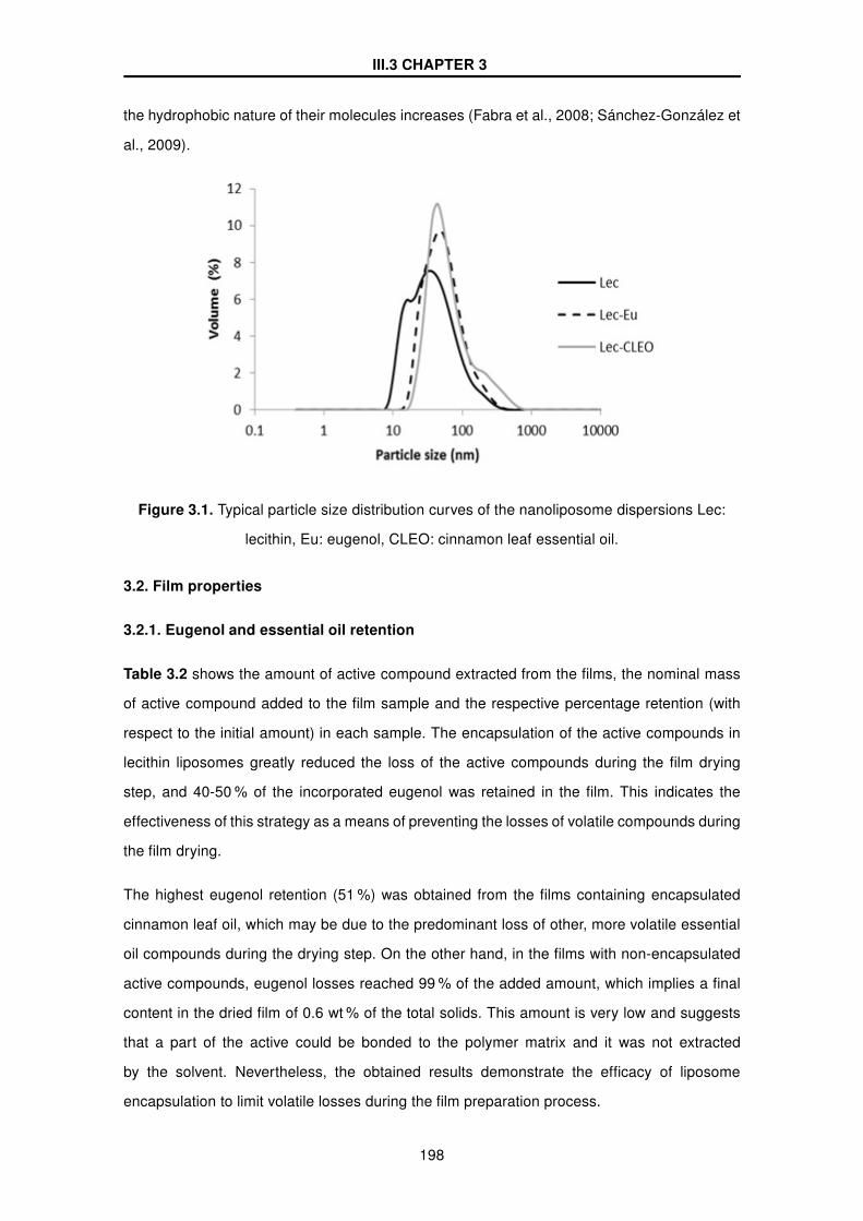
III.3 CHAPTER 3
the hydrophobic nature of their molecules increases (Fabra et al., 2008; Sánchez-González et
al., 2009).
Figure 3.1. Typical particle size distribution curves of the nanoliposome dispersions Lec:
lecithin, Eu: eugenol, CLEO: cinnamon leaf essential oil.
3.2. Film properties
3.2.1. Eugenol and essential oil retention
Table 3.2 shows the amount of active compound extracted from the films, the nominal mass
of active compound added to the film sample and the respective percentage retention (with
respect to the initial amount) in each sample. The encapsulation of the active compounds in
lecithin liposomes greatly reduced the loss of the active compounds during the film drying
step, and 40-50 % of the incorporated eugenol was retained in the film. This indicates the
effectiveness of this strategy as a means of preventing the losses of volatile compounds during
the film drying.
The highest eugenol retention (51 %) was obtained from the films containing encapsulated
cinnamon leaf oil, which may be due to the predominant loss of other, more volatile essential
oil compounds during the drying step. On the other hand, in the films with non-encapsulated
active compounds, eugenol losses reached 99 % of the added amount, which implies a final
content in the dried film of 0.6 wt % of the total solids. This amount is very low and suggests
that a part of the active could be bonded to the polymer matrix and it was not extracted
by the solvent. Nevertheless, the obtained results demonstrate the efficacy of liposome
encapsulation to limit volatile losses during the film preparation process.
198

III.3 CHAPTER 3
Table 3.2 Mass fraction of eugenol in the dried films (mg/g film solids), extracted in the dried
film and initially incorporated, and percentage retention (extracted with respect to the initially
added). Lec: lecithin, Eu: eugenol, CLEO: cinnamon leaf essential oil, CH: chitosan.
Film Extracted Incorporated % Retention
CH/Eu 4.9 ± 1.0(b) 357 1
CH/Lec-Eu 108.9 ± 1.3(c) 263 41
CH/CLEO 5.4 ± 1.3(d) 318 2
CH/Lec-CLEO 118 ± 3(e) 234 51
Different superscript letters (a, b, c) within the same column indicate significant differences
among formulations (p <0.05).
3.2.2. Thickness and tensile properties
Table 3.3 shows the thickness and tensile parameters of the films. The chitosan film thickness
ranged between 33 and 49 µm, and it decreased when free essential oils were incorporated.
This is coherent with the partial volatilization of the oils, involving an effective reduction in the
amount of solids per area unit of the film. The addition of encapsulated compounds provoked
an increase in the films’ thickness with respect to pure chitosan in line with the lower losses
of volatiles and a different microstructural arrangement of the film components as previously
observed by other authors (Jiménez et al., 2014).
Table 3.3 Thickness and tensile parameters (elastic modulus, EM; tensile strength, TS;
percentage elongation, %E) of the films. Mean values and standard deviation. Lec: lecithin,
Eu: eugenol, CLEO: cinnamon leaf essential oil, CH: chitosan.
Film Thickness (µm) EM (MPa) TS (MPa) %E
CH 43 ± 2(b) 1660 ± 170(d) 53 ± 4(b) 6 ± 1(a)
CH/Eu 33 ± 2(a) 1623 ± 70(d) 46 ± 4(ab) 8 ± 3(a)
CH/CLEO 35 ± 2(a) 1460 ± 30(c) 42 ± 3(a) 7 ± 4(a)
CH/Lec 45 ± 1(b) 1145 ± 4(b) 41 ± 1(a) 11 ± 3(a)
CH/Lec-Eu 48 ± 2(c) 840 ± 80(a) 651 ± 10(b) 31 ± 8(b)
CH/Lec-CLEO 49 ± 2(c) 860 ± 70(a) 43 ± 3(a) 27 ± 4(b)
Different superscript letters (a, b, c) within the same column indicate significant differences
among formulations (p <0.05).
199

III.3 CHAPTER 3
The elastic modulus (EM), tensile strength (TS) and deformation at break ( %E) describe the
tensile properties of the films, which are closely related to their structure (Mc Hugh & Krochta,
1994). The EM significantly decreased subsequent to the addition of lecithin liposomes, both
single and loaded with actives, which can be attributed to the interruptions in the polymer
matrix introduced by lipid particles. This leads to a loss of network cohesion, as observed
by other authors when studying different matrices (Fabra et al., 2008; Sánchez-González et
al., 2009). Adding non-encapsulated oils only caused a slight EM reduction, which can be
explained by the lower lipid ratio and the arrangement of the lipid droplets in the film matrix.
Whereas TS and %E were not affected by the addition of non-encapsulated oils, the films with
liposomes exhibited increased extensibility, with similar TS values to the CH control film. Pure
chitosan films were mechanically stronger and less extensible than films with liposomes. This
different behavior may be explained by the different structural arrangement of components, as
commented on below.
3.2.3. Moisture content, water vapor permeability and solubility
Water vapor permeability (WVP) is a relevant property directly related to the usefulness of the
film in food applications, and should be as low as possible to prevent the transfer of water (Ma
et al., 2008). Table 3.4 shows the moisture content, water vapor permeability and solubility of
the films.
Table 3.4 Water content (Xw), water vapor permeability (WVP) and solubility (g of solubilized
solids/100 g of initial solids) of the films. Mean values and standard deviation. Lec: lecithin,
Eu: eugenol, CLEO: cinnamon leaf essential oil, CH: chitosan.
Films Xw WVP Solubility ( %)
(g water/ 100 g dry film) (g.mm. kPa−1.h−1.m2)
CH 39 ± 2(f) 4.3 ± 0.2(d) 67 ± 2(d)
CH/Eu 25.4 ± 0.8(ab) 3.7 ± 0.2(c) 43 ± 1(c)
CH/CLEO 25.6 ± 1.2(ab) 4.6 ± 0.4(e) 40 ± 1(bc)
CH/Lec 30.1 ± 1.7(d) 2.4 ± 0.2(b) 40 ± 2(b)
CH/Lec-Eu 24.2 ± 0.6(a) 2.2 ± 0.2(a) 37 ± 2(a)
CH/Lec-CLEO 26.9 ± 0.8(cd) 1.90± 0.2(a) 35 ± 1(a)
Different superscript letters (a, b, c, d) within the same column indicate significant differences
among formulations (p <0.05).
200

III.3 CHAPTER 3
The equilibrium moisture content of the films decreased significantly when both free and
encapsulated lipid compounds were added, since the proportion of active sites for water
adsorption per unit dry mass decreases (Fabra et al., 2010). Likewise, possible interactions
between the chitosan chains and the active compounds could reduce the availability
of the chitosan polar groups to form hydrophilic bonds with water molecules (Shen &
Kamdem, 2015). As compared to the films with non-encapsulated active compounds, lecithin
incorporation resulted in a significant WVP decrease. A different final amount of lipid in
the films, as well as the differences in their structural arrangement, may explain their
different effectiveness at reducing water vapor permeability. Film solubility in water was also
significantly reduced by the addition of lipids (lecithin or EO compounds); the greater the
total lipid content, the lower the solubility values, coherently with the increased hydrophobic
character of the film (Ojagh et al., 2010).
3.2.4. Optical properties
According to other studies (Hutchings, 1999), the transparency and brightness of the material
are the most important optical properties with which to assess the direct impact on the
appreciation of the color and appearance of a coated/packaged product. Table 3.5 shows the
values of lightness (L*), chroma (C*ab), hue (h*ab) and gloss at 60◦ of the different samples.
Due to the typical color of lecithin, films with liposomes were darker, with a more saturated
reddish color than the pure chitosan films.
Table 3.5 Lightness (L*), chroma (C*ab), hue (h*ab) and gloss (60◦) of the films. Mean values
and standard deviation. Lec: lecithin, Eu: eugenol, CLEO: cinnamon leaf essential oil, CH:
chitosan.
Films L* C*ab h*ab Gloss (60◦)
CH 91 ± 2(e) 4 ± 2(a) 109 ± 5(c) 51 ± 6(d)
CH/Eu 75 ± 4(cd) 14 ± 6(b) 66 ± 6(a) 19 ± 2(b)
CH/CLEO 77.3 ± 1.3(d) 15.0 ± 1.3(b) 66 ± 2(a) 15 ± 3(a)
CH/Lec 67.4 ± 1.1(a) 23.0 ± 0.2(c) 80.4 ± 0.7(c) 33 ± 2(c)
CH/Lec-Eu 69.6 ± 1.1(ab) 23.0 ± 0.3(c) 78.4 ± 0.3(c) 15 ± 2(a)
CH/Lec-CLEO 72.0 ± 0.3(bc) 26.3 ± 0.4(c) 80.0 ± 0.2(c) 20 ± 3(b)
Different superscript letters (a, b, c, d) within the same column indicate significant differences
among formulations (p <0.05).
201

III.3 CHAPTER 3
This effect was also previously observed in starch-sodium caseinate films with lecithin
liposomes (Jiménez et al., 2014).The gloss was decreased by incorporating active
compounds, especially in free form, which can be attributed to the increase in surface
roughness associated with the creaming of lipids during drying, as previously observed for
lipid containing films (Sánchez-González et al., 2009; Fabra et al., 2009).
Figure 3.2 shows the spectral distribution curves of Ti. The incorporation of free lipids, and
particularly the incorporation of liposomes, reduced the Ti of the films; in line with the presence
of a dispersed phase in the matrix which enhances light scattering. In the case of liposomes,
the absorbance of lecithin also contributes to lower the transmittance of the films.
Figure 3.2. Spectral distribution curves of internal transmittance (Ti) of the films. Lec:
lecithin, Eu: eugenol, CLEO: cinnamon leaf essential oil, CH: chitosan.
3.2.5. Microstructural properties
A microstructural study of the films allows the arrangement of the components in the dry films
to be observed, and correlates these observations with other functional-characteristics, such
as the barrier, mechanical and optical properties (Cano et al., 2014). Figures 3.3 and 3.4
show the micrographs of the cross section and surface of the obtained films, respectively. The
holes corresponding to eugenol or oil droplets in the cross section of the films can be seen
when these are incorporated in free form (CH/Eu, CH/CLEO). On the surface of the films with
these free compounds, some droplet footprint can also be observed, whereas the films with
liposomes show a smoother surface. These holes were probably caused by the evaporation of
the volatile compounds during the film drying or during SEM observation under high vacuum
conditions.
202

III.3 CHAPTER 3
On the other hand, when active compounds are incorporated in liposome form, no visible
drops can be seen in the structure whereas laminar formations can be observed, where void
layers intercalate with polymer fibrous arrangements. This appearance suggests that lamellar
liposomal lipid associations were formed during the film drying step, in line with the liotropic
mesomorphism of lipid associations (Larsson & Dejmek, 1990), which would be inserted
between the polymer layers producing a laminated structure. In this arrangement, eugenol or
cinnamon essential oil would occupy the hydrophobic core of the laminar structures. CH/Lec
films also exhibited a laminar structure, but the separation between layers expands when there
is eugenol or essential oil in the formulation. So, as the water content in the FFD is reduced
during film drying, the micellar associations undergo phase transitions and lamellar structures
tend to form, containing lipids at the hydrophobic core (Krog, 1990).
This laminar structure explains the greater elongation capability of the films containing
liposomes during the tensile test, especially those containing eugenol or essential oil, since the
layers can easily slide during the test, hence allowing for a greater deformation before fracture.
However, the weaker cohesive force between layers, as compared to that acting between
polymer chains, would lead to softer films (less resistant to deformation) with lower elastic
modulus, as shown in Table 3.3. Likewise, the formation of lipid layers perpendicular to the
mass transport in the film, also explains the great reduction in the water vapour permeability
and water solubility of the films when liposomes were added to the film formulation.
203

III.3 CHAPTER 3
Figure 3.3. SEM micrographs of the cross section of the chitosan films with and without
eugenol and cinnamon leaf essential oil in free form (left) or lecithin liposomes (right). Lec:
lecithin, Eu: eugenol, CLEO: cinnamon leaf essential oil, CH: chitosan.
204

III.3 CHAPTER 3
Figure 3.4. SEM micrographs of the surface of the chitosan films with eugenol (top) and
cinnamon leaf essential oil (below) in free form (left) or lecithin liposomes (right). Lec:
lecithin, Eu: eugenol, CLEO: cinnamon leaf essential oil, CH: chitosan.
205

III.3 CHAPTER 3
3.2.6. Thermogravimetric analysis (TGA)
Figure 3.5 shows the thermal degradation curves (derivative curve: DTGA) of chitosan films,
where three stages can be observed. The first mass loss step below about 100◦C is due to the
evaporation of residual water from the films. The second step can either be attributed to the
degradation/volatilization of low molecular weight fractions, such as volatile compounds and
lecithin, or to the losses of some groups (such as NH2) from chitosan chains, and the third step
is associated with the decomposition of polymer units, as observed by other authors (Shen &
Kamdem, 2015). Films containing lecithin exhibited greater weight loss in the second stage,
which can be attributed to the gradual decomposition of lecithin (about 20 % weight loss at
270◦C) as deduced from the TGA of pure lecithin. The boiling point of eugenol is 253◦C (Van
Roon et al., 2002), which implies that when Eu or CLEO are present in the film, their thermal
release would overlap with the polymer’s degradation temperature range.
Figure 3.5. First derivative of weight loss vs. temperature curves obtained from TGA. Lec:
lecithin, Eu: eugenol, CLEO: cinnamon leaf essential oil, CH: chitosan.
Table 3.6 shows the onset temperature (T0) and maximum degradation rate temperature
(Tmax) of the polymer for the different films. The onset and Tmax temperature values of
polymer decomposition were hardly affected by the lipid presence, which indicates that no
strong interactions occurred among lipid molecules and polymer chains. Nevertheless, the
thermal degradation of lipid components affected the thermogram shape. The films with free
eugenol or CLEO had the highest onset temperature and degradation rate of the polymer,
which could indicate that the eugenol remaining in the film may be strongly bonded to the
206

III.3 CHAPTER 3
polymer chains, thus affecting their thermal behavior. In this sense, its extraction could be
limited, this contributing to the very low retention determined, as previously comented.
Table 3.6 Onset temperature (T0) and maximum degradation rate temperure of the films.
Mean values and standard deviation. Lec: lecithin, Eu: eugenol, CLEO: cinnamon leaf
essential oil, CH: chitosan.
Films T0 (◦C) Tmax (◦C)
CH 193 ± 4(a) 248 ± 3(a)
CH/Eu 214 ± 3(b) 249 ± 2(a)
CH/CLEO 212 ± 3(b) 248 ± 6(a)
CH/Lec 193 ± 2(a) 243 ± 2(a)
CH/Lec-Eu 193 ± 4(a) 245 ± 2(a)
CH/Lec-CLEO 195 ± 5(a) 247 ± 2(a)
Different superscript letters (a, b, c) within the same column indicate significant differences
among formulations (p <0.05).
3.3. Antimicrobial activity
Table 3.7 and 3.8 show E. coli and L. innocua counts, respectively immediately after the
inoculation of the bacterial culture (0 days) and after 6 days of cold storage. Microbial
counts were significantly reduced by film application right after the inoculation. The higher
antibacterial effect (lower bacterial counts) was obtained for E. coli, which is in agreement with
the greatest antimicrobial effect of chitosan against Gram - negative bacteria (Devlieghere et
al., 2004), as compared to Gram -positive bacteria such as L. innocua.
207

III.3 CHAPTER 3
Table 3.7 Escherichia coli counts in liquid (TSA Broth) and solid media (TSA Agar) at 10ÂoC.
Mean values ± standard deviation Lec: lecithin, Eu: eugenol, CLEO: cinnamon leaf essential
oil, CH: chitosan. ng: no growth.
Film
Escherichia coli (log cfu/g)
Broth Agar
0 days 6 days 0 days 6 days
Control 5.7 ± 0.3(ax) 8.15 ± 0.03(ay) 5.58 ± 0.07(ax) 6.69 ± 0.07(ay)
CH 2.7 ±0.3(bx) 2.16 ±0.02(by) 3.63 ±0.03(b) 4.02 ±0.04(b)
CH/Eu ng ng 2.56 ±0.08(c) ng
CH/CLEO ng ng 2.55 ±0.06(c) ng
CH/Lec 2.95 ±0.04(bx) 2.07 ±0.02(by) 3.92 ±0.02(b) 3.92 ±0.04(b)
CH/Lec-Eu ng ng 2.70 ± 0.08(c) ng
CH/Lec-CLEO ng ng 2.74 ± 0.03(c) ng
Different superscript letters (a, b, c) within the same column indicate significant differences
among formulations (p <0.05). Different superscript letters (x, y) within the same column
indicate significant differences due to storage time.
For both bacteria and in liquid medium, the application of films containing the active ingredient
led to no bacterial growth during the whole period of cold storage. This can be explained by
the solution of the film matrix and the rapid release of the active ingredient. In the agar plates,
immediately after inoculation, samples coated with films containing eugenol or cinnamon leaf
essential oil led to a signficant reduction in the growth of both bacteria as compared to non-
coated (control) and samples coated with CH or CH/Lec In samples coated with films containg
the active ingredient no growth was detected at the end of the storage period.
208

III.3 CHAPTER 3
Table 3.8 Listeria innocua counts in liquid (TSA Broth) and solid media (TSA Agar). Mean
values ± standard deviation Lec: lecithin, Eu: eugenol, CLEO: cinnamon leaf essential oil,
CH: chitosan. ng: no growth.
Film
Listeria innocua (log cfu/g)
Broth Agar
0 days 6 days 0 days 6 days
Control 5.40 ± 0.02(ax) 7.81 ± 0.03(ay) 5.38 ± 0.07(ax) 7.04 ± 0.01(ay)
CH 4.44 ±0.03(bx) ng 4.16 ±0.01(b) 4.20 ±0.02(b)
CH/Eu ng ng 2.83 ±0.05(c) ng
CH/CLEO ng ng 2.89 ±0.04(c) ng
CH/Lec 4.46 ±0.02(by) ng 4.28 ±0.02(b) 4.32 ±0.01(b)
CH/Lec-Eu ng ng 2.97 ±0.05(c) ng
CH/Lec-CLEO ng ng 2.97 ±0.03(c) ng
Different superscript letters (a, b, c) within the same column indicate significant differences
among formulations (p <0.05). Different superscript letters (x, y) within the same column
indicate significant differences due to storage time.
4. CONCLUSIONS
The incorporation of lecithin liposomes containing eugenol or cinnamon leaf essential oil
into chitosan films obtained by casting allowed for a high retention ratio (40-50 %) of volatile
compounds, as compared to the 1-2 % which is retained when they are free incorporated by
emulsification. Films with liposomes exhibited a lamellar microstructure where lipid lecithin
layers seem to alternate with polymer layers, due to the liposome phase transitions during
the progressive film drying. This microstructure improved the extensibility of the films, while
reducing their firmness and water vapor permeability. Liposome incorporation modified the
optical properties of the films; the gloss was reduced, chrome was increased and the films
became reddish, coherently with the chromatic properties of lecithin. The addition of the
active ingredients enhanced the antimicrobial activity of pure chitosan films. The incorporation
of lecithin as encapsulating agent did not affect the antimicrobial effectiveness of the films.
Therefore, the encapsulation of volatile active compounds in liposomes emerges as a good
strategy for the obtention of active films with essential oils.
209

III.3 CHAPTER 3
REFERENCES
Alma, H.M., Ertas, M., Nitz, S., & Kollmannsberger, H. (2007). Chemical composition and
content of essential oil from the bud of cultivated Turkish clove (Syzygium aromaticum
L.).BioResources,2(2),265-269.
Atarés, L. & Chiralt, A. (2016). Essential oils as additives in biodegradable films and coatings
for active food packaging. Trends Food Sci Technol., 48, 51-62.
ASTM. (1995). Standard test methods for water vapour transmission of materials, Standard
designations: E96-95 Annual book of ASTM standards, Philadelphia, PA: American Society
for Testing and Materials, pp. 406-413 (1995).
ASTM. (1999).Standard test method for specular gloss, In Designation (D523), Annual book
of ASTM standards, Vol. 06.01, Philadelphia, PA: American Society for Testing and Materials.
ASTM. (2001). Standard test method for tensile properties of thin plastic sheeting, Standard
D882, In Annual book of American standard testing methods, Philadelphia, PA: American
Society for Testing and Materials, pp. 162-170.
Bakkali, F., Averbeck, S., Averbeck, D., & Idaomar, M. (2008). Biological effects of essential
oils-A review. Food Chem. Toxicol,. 46(2), 446-475.
Bajpai, V.K., Baek, K.H. & Kang, S.C. (2012). Control of Salmonella in foods by using essential
oils: A review. Food Res. Int. 45, 722-734.
Cano, A., Jiménez, A., Cháfer, M., Gónzalez, C., & Chiralt, A. (2014). Effect of
amylose:amylopectin ratio and rice bran addition on starch films properties. Carbohydr.
Polym., 111, 543-555.
De Martino, L., De Feo, V., Fratianni, F., & Nazzaro, F. (2009). Chemistry, antioxidant,
antibacterial and antifungal activities of volatile oils and their components. Natural Product
Communications 4: 1741-1750.
Devlieghere, F., Vermeulen, A., & Debevere,J. (2004). Chitosan: antimicrobial activity,
interactions with food components and applicability as a coating on fruit and vegetables. Food
Microbiology.,21(6), 703-714.
Fabra, M.J., Talens, P., & Chiralt, A.(2008).Tensile properties and water vapor permeability of
210

III.3 CHAPTER 3
sodium caseinate films containing oleic acid-beeswax mixtures. J. Food Eng.,85(3), 393-400.
Fabra, M.J., Talens, P., & Chiralt, A. (2009). Microstructure and optical properties of sodium
caseinate films containing oleic acid-beeswax mixtures. Food Hydrocolloids 23(3), 676-683.
Fabra, M.J., Talens, P., & Chiralt, A. (2010). Water sorption isotherms and phase transitions
of sodium caseinate-lipid films as affected by lipid interactions.Food Hydrocolloids, 24(4),384-
391.
Falguera, V., Quintero, J.P., Jiménez, A., Muñoz, J.A., & Ibarz, A. (2011).Edible films and
coatings: Structures, active functions and trends in their use. Trends Food Sci. Technol, 22(6),
292-303.
FDA. (2009). US Food and Drug Administration. Code of Federal Regulations, Title 21-Food
and Drugs, Part 172, Subpart F, Section 172.515. Hutchings, J.B. (1999). Food and colour
appearance, 2nd edition, Gaithersburg, MD: Chapman and Hall Food Science Book, Aspen
Publication. ISBN: 978-1-4613-6007-0 (Print) 978-1-4615-2373-4 (Online).
Imran,M., Revol-Junelles, A.M., René, N., Jamshidian, M., Akhtar, M.J., Arab-Tehrany, E. &
Desobry, S. (2012). Food Hydrocolloids, 29(2),407-419.
Jiménez, A., Fabra, M.J., Talens, .P & Chiralt, A.(2013). Physical properties and antioxidant
capacity of starch-sodium caseinate films containing lipids. J. Food Eng., 116(3), 695-702.
Jiménez, A., Sánchez-González, L., Desobry, S., Chiralt, A., & Tehrany, E.A. (2014). Food
Hydrocolloids, 35,159-169.
Judd, D.B., & Wyszecki, G. (1975). Colour in business, science and industry, 3rd edition, New
York, NY: John Wiley and Sons, Inc., ISBN 0471452122.
Krog, N.J.(1990).Food emulsifiers and their chemical and physical properties, In Larsson EDK
and Friberg SE (Eds.), Food emulsions. New York and Basel: Marcel Dekker, Inc., pp.127-180.
Larsson, K., & Dejmek, P. (1990). Crystal and liquid crystal structures of lipids, In Larsson EDK
and Friberg SE (Eds.), Food emulsions. New York and Basel: Marcel Dekker, Inc., pp.97-125.
Leceta, I., Guerrero, P., & De la caba, K. (2013).Functional properties of chitosan-based films.
Carbohydr. Polym., 93(1), 339-346.
Ma, X., Chang, P.R., & Yu, J. (2008). Properties of biodegradable thermoplastic
pea starch/carboxymethyl cellulose and pea starch/microcrystalline cellulose composi-
211

III.3 CHAPTER 3
tes.Carbohydr. Polym. ,72(3),369-375.
Mc Hugh, T. H., Avena-Bustillos, R., & Krochta, J. M. (1993). Hydrophobic edible films:
modified procedure for water vapour permeability and explanation of thickness effects. Journal
of Food Science, 58(4), 899-903.
Mc Hugh, T. H. & Krochta, J. M. (1994).Water vapour permeability properties of edible whey
protein-lipid emulsion films. Journal of the American Oil Chemists Society, 71, 307-312.
Ojagh, S.M., Rezaei, M., Razavi, S.H., & Hashem, S.M. (2010). Development and evaluation
of a novel biodegradable film made from chitosan and cinnamon essential oil with low affinity
toward water. Food Chem. 122,161-166.
Olasupo, N.A., Fitzgerald, D.J., Gasson, M.J., & Narbad, A. (2003). Activity of natural an-
timicrobial compounds against Escherichia coli and Salmonella enterica serovar Typhimu-
rium.Lett. Appl. Microbiol,. 37, 448-451.
Perdones, Á., Vargas, M., Atarés, L. & Chiralt, A. (2014). Physical, antioxidant and
antimicrobial properties of chitosan-cinnamon leaf oil films as affected by oleic acid. Food
Hydrocolloids 36, 256-264.
Pérez-Gago, M.B., & Krochta, J.M. (2001). Lipid Particle Size Effect on Water Vapor
Permeability and Mechanical Properties of Whey Protein/Beeswax Emulsion Films. J Agric
Food Chem.,49, 996-1002.
Sánchez-González, L., Vargas, M., González-Martínez, C., Chiralt, A., & Cháfer, M. (2009).
Characterization of edible films based on hydroxypropylmethylcellulose and tea tree essential
oil. Food Hydrocolloids, 23(8), 2102-2109.
Sánchez-González, L., Chiralt, A., González-Martínez, C., & Cháfer, M. (2011). Effect of es-
sential oils on properties of film forming emulsions and films based on hydroxypropylmethyl-
cellulose and chitosan. J. Food Eng. 105(2), 246-253.
Sebaaly, C., Jraij, A., Fessi, H, Charcosset, C., & Greige-Gerges, H. (2015). Preparation and
characterization of clove essential oil-loaded liposomes.Food Chem., 178, 52-62.
Singh, G., Maurya, S., de Lampasona, M.P., & Catalan, C.A.N. (2007). A comparison
of chemical, antioxidant and antimicrobial studies of cinnamon leaf and bark volatile oils,
212

III.3 CHAPTER 3
oleoresins and their constituents. Food Chem. Toxicol., 45, 1650-1661.
Shah, B., Davidson, M. & Zhong, Q. (2013). Nanodispersed eugenol has improved
antimicrobial activity against Escherichia coli O157:H7 and Listeria monocytogenes in bovine
milk. Int. J. Food Microbiol. 161, 53-59.
Shen, Z., & Kamdem, D.P.(2015). Development and characterization of biodegradable
chitosan films containing two essential oils. Int. J. Biol. Macromol. 74, 289-296.
Wu, J., Liu, H., Ge, S., Wang, S., Qin, Z., Chen, L., Zheng, Q., Liu, Q., & Zhang, Q. (2015).
The preparation, characterization, antimicrobial stability and in vitro release evaluation of fish
gelatin films incorporated with cinnamon essential oil nanoliposomas. Food Hydrocolloids,43,
427-435.
Zhai, M., Zhao, L., Yoshii, F., & Kume, T. (2004). Study on antibacterial starch/chitosan blend
film formed under the action of irradiation, Carbohydr. Polym. 57(1), 83-88.
Zhang, H.Y., Tehrany, E.A., Kahn, C.J.F., Ponçot, M., Linder, M., & Cleymand, F. (2012).
Effects of nanoliposomes based on soya, rapeseed and fish lecithins on chitosan thin films
designed for tissue engineering. Carbohydr. Polym., 88, 618-627.
Van Roon, A., Parsons, J.R., & Govers, H.A.J. (2002). Gas chromatographic determination
of vapour pressure and related thermodynamic properties of monoterpenes and biogenically
related compounds. J. Chromatogr. A. 955, 105-115.
213


III.4 CHAPTER 4
RELEASE KINETICS AND ANTIMICROBIALPROPERTIES OF CHITOSAN-ESSENTIAL OILS FILMSAS AFFECTED BY ENCAPSULATION WITHIN LECITHINNANOLIPOSOMES
Cristina Valencia-Sullca, Lorena Atarés, Maria Vargas, Amparo Chiralt
Department of Food Technology - Institute of Food Engineering for Development, Universitat
Politècnica de València, Camino de Vera s/n, 46022 Valencia, Spain.


III.4 CHAPTER 4
ABSTRACT
The antimicrobial capacity of CH-based films containing eugenol (EU) or cinnamon leaf
essential oil (CLEO), as affected by their encapsulation within lecithin nanoliposomes, was
tested against Escherichia coli and Listeria innocua. Films were able to reduce microbial load
all types of samples (liquid and solid media and pork meat), and more significantly for E.
coli. The evaluation of the overall migration in the films showed that in hydrophilic simulants
(ethanol 10 % and acetic acid 3 %), the limit that the EU Directive stablishes was exceeded
and thus the potential application of these films would be limited to lipophilic food products.
The retention capacity of the active ingredients in the polymer matrix was revealed by the
kinetics of eugenol release in four food simulants (ethanol 10 %, ethanol 50 %, acetic acid 3 %
and isooctane). For all of them, the rate constant (K1) obtained by applying Peleg’s model,
was higher for films containing lecithin, whereas diffusivity values were reduced. Films were
able to slow down the changes in color parameters and increase the shelf-life of cold stored
pork meat.
Keywords: biopolymer, essential oil, cinnamon, eugenol, antimicrobial, migration.
217


III.4 CHAPTER 4
1. INTRODUCTION
Chitosan (CH) is a cationic, non-toxic, biodegradable film-forming polysaccharide, compatible
with other biopolymers, which can be obtained from the deacetylation of the chitin present in
crustacean exoskeletons, and shows antimicrobial properties (Zhai et al., 2004). CH-based
films exhibit good mechanical and structural properties and constitute a good barrier to gases
and aromas. The incorporation of active ingredients such as essential oils into CH-based
films could improve their functionality, since the antimicrobial properties of the film would be
enhanced (Vargas et al., 2012). Cinnamon leaf essential oil (CLEO) and its main compound,
eugenol (EU), have been described as antibacterial and antifungal agents at relatively low
concentrations in previous studies (De Martino et al., 2009; Bajpai et al., 2012). Eugenol
has been recognized as safe by the US Food and Drug Administration (US FDA, 2009) and
approved by the European Union as a safe flavoring agent for foods (Sebaaly et al., 2015).
Different essential oils have been widely used in the formulation of active biodegradable films
based on chitosan. Essential oils have been incorporated into hydrophilic biopolymer films
by emulsification in the aqueous film-forming dispersion (FFD) of the polymer before film
formation by means of different homogenization methods and the films were obtained by
casting (Sánchez-González et al., 2009; Bonilla et al., 2011). However, the losses of volatile
compounds during casting technique shows an important drawback since during the film
drying step, oil droplets flocculate, coalesce and cream to the top of the drying film, where
oil components volatilize together with water at a lower temperature than their boiling point
(Perdones et al., 2016). The encapsulation of essential oil compounds before film preparation
can reduce the losses of volatile compounds at the same time that the sensory impact of
essential oils is minimized. Moreover, encapsulation can modulate the release kinetics of
active compounds into the food product surface. In this sense, the use of liposomes or
nanoliposomes, which can act as carrier systems of a wide range of compounds, represent
an interesting alternative (Zhang et al., 2012).
The antimicrobial effect of films containing liposomes depends on both the nature of the
active ingredients (essential oils) and on the capacity of the film to release enough active
compound to the coated food at a determined contact time and at equilibrium (partition
coefficient). Moreover, the release kinetics of liposomes depends on interactions between
the encapsulated active ingredients and the polymer matrix together with the solubility of the
active compounds in the food system. In this sense, the release kinetics should be evaluated
in systems with different polarities, which simulate different kinds of foods, such as standard
food simulants (European Commission Regulation, 2011).
219
III.4 CHAPTER 4
In order to guarantee antimicrobial effectiveness and food safety it is essential to evaluate the
release kinetics of the active compound into the food throughout the storage time (Tehrany
and Desobry, 2007). The time needed to reach a concentration level of active in the food
that surpasses the minimum inhibitory concentration of the target microorganisms must be
estimated to ensure food safety. For prediction purposes, and to evaluate the compound
release rate from the films, and the concentration reached in the food simulant Fick equation
(Del Nobile et al., 2008; Sánchez-González, et al., 2012) or first order kinetics models have
been used (Barba et al., 2015; Biddeci et al., 2016).
The aim of this work was to evaluate the antimicrobial effect of chitosan films containing
eugenol or cinnamon essential oil as affected by their encapsulation within lecithin
nanoliposomes.
2. MATERIALS AND METHODS
2.1. Reagents
Chitosan (CH) and eugenol (EU) were supplied by Sigma-Aldrich Chemie (Steinheim,
Germany). Cinnamon leaf essential oil (CLEO) was provided by Herbes del molí (Alicante,
Spain). Sunflower Lecithin (Lec), Lipoid H20- 20 % - phosphaatidilcoline no-GMO, was
supplied by LipoidGmbH (Frigenstraβe, Germany). Methanol, ethanol, glacial acetic acid,
isooctane, glycerol and magnesium nitrate was purchased from Panreac Química SLU
(Castellar del Vallés, Barcelona; Spain).
L. innocua (CECT 910) and E. coli (CECT 10) lyophilized strains were obtained from Colección
Española de Cultivos Tipo (CECT, Burjassot, Spain). Buffered peptone water and culture
media were supplied by Scharlab S.L. (Mas d’EnCisa, Barcelona, Spain).
2.2. Preparation of the nanoliposome dispersions
The preparation of nanoliposome dispersions was carried out according to a previously
described method (Jiménez et al., 2014). Lecithin was dispersed in distilled water ( 5 wt %)
and stirred for 4 h at 700 rpm Eugenol or CLEO at 5 wt % was incorporated into the lecithin
dispersion and afterwards three different liposome samples were obtained (Lec, Lec-Eu and
Lec-CLEO) by sonication at 20 kHz for 10 min with 1 s pulses. The ultrasound probe was
placed in the centre of the sample. An aqueous solution of lecithin was prepared (5 wt %) by
stirring at room temperature during 3-4 hours.
220

III.4 CHAPTER 4
2.3. Preparation of the film-forming dispersions and films
A solution of 1 % CH (w/w) was prepared in a 1 % (v/w) acetic acid solution by stirring at
room temperature for 24 h. The active compounds (eugenol or CLEO) were incorporated in
two different forms: encapsulated in liposomes or by direct emulsification. These FFDs were
named as CH-EU, CH-CLEO, CH-Lec-Eu, and CH-Lec-CLEO and were obtained by adding
0.5 g of active compound (eugenol or CLEO) as free form or 10 g of the liposome dispersion
to 90 g of the CH solution. The amount of active was selected to overcome the values of the
minimal inhibitory concentration of actives against some typical pathogens or food spoilage
microorganisms according to previous studies with CH films containing CLEO (Perdones et
al., 2014).
CH FFDs with and without lecithin nanoliposomes free of active ingredients were also
prepared. These FFDs were name as CH and CH-Lec, respectively.
FFDs were homogenized with a rotor-stator (Yellow Line DI 25 basic, IKA, Germany) at 13500
rpm for 4 minutes. Films were obtained by casting the FFDs in Teflon R© leveled plates (150
mm in diameter, 1 g solids/plate), which were dried at room temperature and 46 % RH for
24-48h.
Films were peeled of from the casting plates and conditioned in hermetic jars with an
oversaturated Mg (NO3)2 solution (53 % RH) during a week prior performing the analysis.
2.4. Overall migration in food simulants
The overall migration test was carried out, according to Regulation 10/2011/EC (European
Directive, 14/1/2011) for food contact materials. The overall migration test, which determines
the migration of a specific packaging material in food simulants, was carried out with simulant
A (ethanol 10 % v/v, aqueous food), simulant B (acetic acid 3 % w/v, aqueous food with pH
lower than 4.5) and simulant D2 (isooctane, food with a fatty continuous phase). The ratio
film surface/ volume simulant was 6 dm2/kg, and the test was carried out at 20◦C in duplicate
for 10 days. Afterwards, the solvent was transferred to cups and evaporated at 60◦C until
a constant weight was reached. (24-48h). The overall migration of each film formulation was
determined as the weight of residue after drying and expressed as mg/dm2 of film. All analyses
were carried out in duplicate.
221

III.4 CHAPTER 4
2.5. Kinetics of eugenol release in food simulants
The kinetics of release of EU from the films was tested in four food simulants: A (ethanol 10 %
v/v, aqueous food), simulant B (acetic acid 3 % w/v, aqueous food with pH lower than 4.5),
simulant D1 (ethanol 50 % v/v, mimics alcoholic food and oil-in water emulsions and simulant
D2 (isooctane, food with a fatty continuous phase). Film samples (4 cm2) were placed in
flasks with 100 mL of the corresponding simulant at 20◦C and under continuous stirring. After
different contact times up to equilibrium (24 h), the samples were taken from the flasks, and
the absorbance at 282 nm was measured (Spectrophotometer Helios Zeta UVeVis, Thermo
Fisher Scientific, UK). Thus, the EU profile concentration in each simulant over time could
be determined by the absorbance measurements using the standard calibration curve. All
analyses were performed in triplicate for three different flasks containing the different film
samples. The liquid phase in contact with the active-free films was used as blank for the
absorbance measurements, for each simulant and time.
Peleg’s model (Eq. 1), was applied to experimental data in order to predict the release kinetics
(Peleg,1988).
Mt = M0 +t
k1 + k2tEq.1
where Mt is total eugenol released at time t, M0 is the initial amount of eugenol released, k1
is the kinetic constant of the model that is inversely related to the mass transfer rate at the
beginning of the process, and k2 is a constant of the model that is related to the asymptotic
value, which can be related to the equilibrium value (1/M∞ where M∞ is the amount of active
released at equilibrium).
Fick’s second law was considered to model the diffusion process of EU in the films bilayer films
towards the food simulants. Film samples can be considered as infinite plane sheets with the
half thickness as a characteristic dimension, where the active compound diffuses only in an
axial direction. The diffusional long-time equation for an infinite plane sheet (Crank, 1975),
considering with eight terms, was used to determine the values of diffusion coefficient (D) of
EU into the different solvents (Eq. 2):
Mt
M∞
= 1− 8
π2
∞∑
n=0
1
(2n + 1)2exp
−D(2n+ 1)2π2t
L2
Eq.2
222

III.4 CHAPTER 4
Where:
D: diffusion coefficient (m2/s)
Mt : mass of eugenol released at time t (g)
M∞: the mass of compound released at equilibrium (g)
L: half thickness of film (m)
2.6 Antimicrobial properties of the films
Listeria innocua (CECT 910) and Escherichia coli (CECT 101) were supplied by the Spanish
Type Culture Collection (CECT, Burjassot, Spain). These bacterial cultures were regenerated
(from a culture stored at -25◦C) by transferring a loopful into 10mL of Tryptone Soy Broth (TSB)
(Scharlab, Barcelona, Spain) and incubating at 37◦C for 24h. From this culture, a 10 µL aliquot
was again transferred into 10 mL of TSB and grown at 37◦C for 24h more in order to obtain a
culture in the exponential phase of growth. Afterwards, this bacterial culture was appropriately
diluted in TSB tubes to get a target inoculum of 105 CFU mL−1. Circular samples (55 mm in
diameter) of the films were sterilized under shortwave UV light and aseptically placed in TSB
tubes (liquid medium test), on inoculated Tryptose Soya Agar (TSA) (Scharlab) plates (solid
medium test) and on pork meat sample slices. Inoculated broth tubes, agar plates and meat
slices without film were used as control samples. Immediately after the inoculation and after 6
days at 10◦C the microbial counts on TSA plates were determined. In the liquid medium tests
(broth tubes), serial dilutions were made and poured onto TSA dishes which were incubated
for 24 h at 37◦C. For the solid medium tests (agar plates) and the raw pork meat slices, the
dish content was removed aseptically and placed in a sterile plastic bag with 90 mL of Buffered
Peptone Water.
The bags were homogenized with a Stomacher (Interscience BagMixer Stomacher 400 W
Homogenizer, St Nom la Bretèche, France). Serial dilutions were made and then poured into
TSA Petri dishes. Samples were incubated at 37◦C for 24 h before colonies were counted. All
the tests were run in triplicate.
2.7. Effect of the films on the shelf-life of pork meat
The effect of film application on the shelf-life of pork meat was tested, as described in previous
studies (Bonilla et al., 2013). Pork meat was purchased in a local supermarket and slices of
5 cm in diameter (about 10 g) were obtained. The surface of the meat slices was coated with
the films. Non-coated samples (control) and samples coated with the different types of films
were stored in duplicate at 10◦C for 7 days in a thermostat cabinet (Aqualytic GmbH & Co,
Dortmund, Germany).
223

III.4 CHAPTER 4
In order to evaluate the specific migration of eugenol into the coated meat samples, at different
times of cold storage, the films containing EU were peeled off the coated pork meat slices
and eugenol was extracted with methanol and quantify by means of the measurement of
absorbance. To this end, film samples (4 cm2 ) were kept in 100 mL methanol under stirring
(300 rpm) for 24 h at 20◦C. The extract was filtered and quantitatively diluted to measure the
absorbance at 282 nm using a UV-visible spectrophotometer (Helios Zeta UVeVis, Thermo
Fisher Scientific, UK), using the methanol extract of eugenol-free CH films as blank solution.
All analyses were carried out in duplicate.
To perform microbiological analyses, 10 g of each sample were aseptically obtained and
homogenized in a Stomacher (Bag Mixer 400, Interscience) with 90 mL of sterile buffered
peptone water for 2 min. Aliquots were serially diluted in buffered peptone water and plated
out following standard methodologies. Total aerobial counts were determined in Plate Count
agar incubated at 37◦C for 48 h. Coliform microorganisms were determined in Violet Red Bile
Agar incubated at 37◦C for 48 h. All tests were performed in triplicate. Color changes in meat
slices during cold storage were obtained by using a spectrocolorimeter CM-3600d (Minolta
Co, Tokyo, Japan) with a 30 mm diameter window. In coated samples, films were peeled
off the coated pork meat slices prior performing the measurements. Reflection spectra were
obtained over a black (R0) and a white (R) standard background, of known reflectance (Rg) in
6 different areas of the meat sample. From these spectra, the infinite reflection spectra of the
samples (R∞) was obtained (Eq. 3-5)
R∞ = a− b Eq.3
a =1
2(R+
R0 −R+Rg
R0Rg
) Eq.4
b =√
a2 − 1 Eq.5
From R∞= spectra, and using D65 illuminant /10◦ observer, CIE-L* a* b* coordinates (CIE,
1986), and chromatic parameters hue (h*ab, Eq.6) and chroma (C*ab, Eq.7), and the total color
difference as regard a non-coated sample were obtained (∆E, Eq.8) The color of each sample
was measured in quintuplicate.
C∗
ab =√
a∗2 + b∗2 Eq.6
h∗ab = arctanb∗
a∗Eq.7
∆E =√∆L∗ +∆a∗ +∆b∗ Eq.8
224

III.4 CHAPTER 4
2.8. Statistical analysis
Statistical analyses were performed through analysis of variance (ANOVA) using Statgraphics
Centurion XVI-II. Fisher’s least significant difference (LSD) procedure at 95 % was used.
3. RESULTS AND DISCUSSION
3.1. Total migration of eugenol
Table 4.1 shows total migration of material in three food simulants of different polarity and pH
(3 % aqueous acetic acid solution, 10 % aqueous ethanol solution and isooctane).
Table 4.1 Total migration (10 days, 20◦C) of material from the films. Mean values and
standard deviation.
FilmTotal migration (mg/ dm2 film)
Ethanol 10 % Acetic acid 3 % Isooctane
CH 485 ± 2(a1) 655 ± 8(a2) 9.2 ± 0.3(a3)
CH-Lec 144 ± 2(b1) 309 ± 5(b2) 3.30 ± 0.14(b3)
CH-EU 397 ± 3(c1) 567 ± 10(c2) 5.30 ± 0.14(c3)
CH-CLEO 383 ± 4(d1) 561 ± 4(c2) 4.5 ± 0.12(c3)
CH-Lec-EU 93.5 ± 1.5 (e1) 139 ± 2(d2) 0.9 ± 0.4(d3)
CH-Lec-CLEO 91.0 ± 1.3(e1) 134 ± 5(d2) 0.9 ± 0.2(d3)
Different letters in the same column (a-e) indicate significant differences (p<0.05) among
films. Different numbers in the same row (1-3) indicate significant differences (p<0.05) among
simulant for the same film.
Total migration was affected by both the pH and polarity of the simulant together with the
solubility of film components in the release media. In hydrophilic simulants, specific migration
values exceeded the limit of 10 mg /dm2 regulated by the European Directive for food contact
materials (European Commission, 2011). Moreover, as expected, for all films, the highest
migration levels were obtained in 3 % acetic acid (simulant A), since acid media promote
chitosan solution and hence the diffusion of film ingredients into the release solvent. On the
contrary, in isooctane migration levels were lower than the regulated limit of 10 mg/dm2,
especially in films containing lecithin nanoliposomes. The encapsulation of essential oils in
lecithin nanoliposomes reduced significantly migration levels, especially as compared to pure
CH films. This can be explained by the reduction in swelling promoted by the addition of
lipid compounds in the polymer matrix (Hromiš et al., 2015). In all solvents, no significant
differences in the migration levels were detected due to the type of added active ingredient.
225

III.4 CHAPTER 4
The reported results agree with the effect of film microstructure in migration levels (Valencia-
Sullca et al., 2016). In this previous study, microstructure observations revealed that the
addition of active ingredients encapsulated in lecithin nanoliposomes led to the formation of
laminar structures, where the essential oils were located at the core of the nanocapsules.
These laminar structures containing lecithin limited the interactions between the film
ingredients and the solvent, thus reducing total migration.
3.2. Kinetics of eugenol release in food simulants
Figure 4.1 shows the mass of eugenol released, as regards of the mass released at
equilibrium, as a function of time, together with the Peleg’s model fit, obtained for films
containing pure eugenol (encapsulated or not in lecithin nanoliposomes).
Figure 4.1. Kinetics of eugenol release from CH-EU and CH-Lec-EU films in different food
simulants.
The kinetics of eugenol release from CH-EU films in hydrophilic simulants was fast. Release
occurs in two steps: (1) fast release and diffusion of eugenol from film surface and (2) slower
release diffusion of eugenol in the polymer matrix. In isooctane, the maximum release was
achieved after 20 minutes, and a lower amount of eugenol was released. Films containing
nanoliposomes (CH-Lec-EU) showed smoother curves, which is in agreement with a slower
release kinetics. The above mentioned laminated microstructure makes eugenol diffusion
difficult, leading to lower release rates.
226

III.4 CHAPTER 4
Table 4.2 shows Peleg’s model parameters together with the diffusivity values predicted by
Fick Model. Diffusivity values of eugenol were significantly affected by the type of simulant
and by its encapsulation with lecithin (p<0.05), which reduced diffusion levels in all solvents
except for 50 % ethanol. In films formulated with EU in free form, diffusivity values significantly
decrease with the decrease in polarity of the solvent, which reveals eugenol affinity for
hydrophilic media. In films containing lecithin-eugenol nanoliposomes, the opposite trend
was observed: the reduction in polarity promoted an increase in diffusivity values except
for isooctane, where the lowest diffusivity value was obtained, which is consistent with the
reported kinetics of release (Figure 1).
Table 4.2 Apparent diffusivity (D) and parameters of Peleg’s model: amount of active
compound released at equilibrium in the simulant (M∞) and its release rate (1/k1), and
maximum release ratio (M∞/M0, mass of active released at equilibrium in the simulant
related to the initial mass of the active in the film expressed with respect to the amount
determined by methanol extraction (1) or by acetic acid extraction (2).
Film Simulant D x 1013 (m2/s) 1/k1 (mg/min) M∞ = 1/k2 (mg/100 g film) M∞/M01 R2
CH
-EU
Ethanol 10 % 5.33 ± 0.03(c)(x) 0.00410 ± 0.00011(d)(x) 2.686 ± 0.122(b)(x) 0.636 ± 0.026(b) >0.999
Acetic acid 3 % 8.50 ± 0.11(d)(x) 0.00152 ± 0.00005(c)(x) 2.969 ± 0.054(c)(x) 0.713 ± 0.028(c) >0.998
Ethanol 50 % 4.84 ± 0.08(b)(x) 0.00076 ± 0.00003(b)(x) 3.503 ± 0.067(d)(x) 0.828 ± 0.032(d) >0.999
Isooctane 3.02 ± 0.07(a)(x) 0.000068 ± 0.000003(a)(x) 1.256 ± 0.043(a)(x) 0.035 ± 0.002(a) >0.996
CH
-Lec
-EU Ethanol 10 % 2.17 ± 0.07(c)(y) 0.00209 ± 0.00007(d)(y) 1.994 ± 0.071(b)(y) 0.754 ± 0.032(b) >0.999
Acetic acid 3 % 0.60 ± 0.02(b)(y) 0.00026 ± 0.00001(b)(y) 2.669 ± 0.045(c)(y) 1.000 ± 0.039(c) >0.998
Ethanol 50 % 6.32 ± 0.09(d)(y) 0.00059 ± 0.00001(c)(y) 2.095 ± 0.047(b)(y) 0.790 ± 0.020(b) >0.999
Isooctane 0.21 ± 0.01(a)(y) 0.000070 ± 0.000003(a)(x) 1.788 ± 0.063(a)(y) 0.671 ± 0.016(a) >0.995
Different letters in the same column (a-h) indicate significant differences (p<0.05) among films.
Different numbers in the same column (1-2) indicate significant differences (p<0.05) among
films for the same simulant.
Parameters of Peleg’s model showed a more rapid release of eugenol from films with
non-encapsulated eugenol (higher values of 1/k1) as compared to films containing lecithin
nanoliposomes. This coincides with the results found by Wu et al. (2015) in gelatin films
containing cinnamon leaf essential oil.
For all hydrophilic solvents, the plateau value of Peleg’s model (M∞) was lower in films
containing nanoliposomes. This parameter is affected by the initial content of eugenol in the
film (M0), being 0.347 g EU/g dry film and 0.257 g EU/g dry film for CH-EU and CH-Lec-EU
films, respectively. T0 minimize the effect of the difference in initial eugenol content in the films,
the ratio M∞/M0 after extraction in methanol was calculated.
227

III.4 CHAPTER 4
3.3. Antimicrobial activity against Escherichia coli and Listeria innocua
Figure 4.2 shows the evolution of E. coli and L. innocua during cold storage in Triptose
Soya broth (TSB), Tryptose Soya Agar (TSA) plates and pork meat slices, as affected by
the application of the films.
Figure 4.2. Microbial count of samples inoculated with L. innocua or E. coli during storage at
10◦C. Mean values and standard deviations.
Chitosan films significantly reduced microbial growth in all types of samples, especially
as regards E. coli. This coincides with previous studies that reported a more significant
antibacterial effect of CH against Gram-negative bacteria, such as E. coli, due to the higher
negative charge of cell membrane thus promoting the interactions of the cationic groups of
chitosan (O’Callaghan & Kerry, 2016).
The antibacterial effect of CH was more marked in TSB, which is coherent with the highest
solubility of CH in liquid media. On the contrary, in solid media polymer diffusivity is limited,
which implied a lower antimicrobial effect, especially when films were applied to meat slices. In
the three types of samples, there were not detected differences due to the addition of lecithin,
which coincides with previous studies (Jiménez et al., 2015).
228

III.4 CHAPTER 4
Films containing active ingredients (EU or CLEO) had a bactericidal effect against both
microorganisms, and significantly in inoculated TSB and TSA media. Previous studies have
shown a synergic effect of CH films and cinnamon leaf essential oil against E. coli (Wang et al.,
2011). No significant effects were observed due to the encapsulation of the active ingredients
in lecithin nanoliposomes.
3.4. Effect of the films on the shelf-life of pork meat
The amount of EU that was release from CH-EU and CH-Lec-EU films in contact with meat
slices after different times of cold storage was obtained by extraction of the remaining EU in
the film. The results are shown in Table 4.3.
Table 4.3 Total specific migration of eugenol (mg eugenol/g meat) from the films in meat
samples stored at 10◦C. Mean values and standard deviation, in brackets.
Film 0 days 2 days 6 days 13 days
CH-EU 1.2 ± 0,4(a1) 2.12 ± 0.03(a2) 2.90 ± 0.03(a3) 2.53 ± 0.04(a23)
CH-Lec-EU 0.710 ± 0,006(b1) 2.133 ± 0.004(a2) 2.34 ± 0.05(b3) 2.38 ± 0.03(b3)
Different letters (a, b) in the same column indicate significant differences between films at the
same storage time (p<0.05). Different numbers (1, 2, 3) in the same row indicate differences
due to storage time for the same film (p<0.05).
The amount of eugenol released to meat slices from the two types of films increased
during storage. Encapsulation of eugenol in lecithin nanoliposomes delayed eugenol release,
especially at the beginning of the storage, which suggested better results at long term as
regards antimicrobial activity.
Figure 4.3 shows total aerobial and coliform microorganism counts in non-inoculated pork
meat slices as affected by film application. All films showed antimicrobial activity and reduced
total aerobial and coliform counts as compared to non-coated samples (control). Results agree
with Bonilla et al. (2014). All films showed a bactericidal effect in meat slices at the beginning of
storage. After two days, in samples coated with CH and CH-Lec, an increase in the microbial
load was detected, with no significant differences between samples. Films formulated with
active ingredients kept the bactericidal effect against coliforms during 9 days of storage, and
no significant differences were detected due to the encapsulation of active compounds. During
the whole storage period, films containing essential oils led to microbial loads lower than
5 CFU/g, which is maximum value allowed according the 88/657/EEC European Directive
(Council Directive, 1988).
229

III.4 CHAPTER 4
Figure 4.3. Total aerobial and coliform counts in pork meat slices during storage at 10◦C.
Mean values and standard deviation.
The antibacterial effect of the films was less significant in terms of total aerobial counts as
compared to coliforms, which are the microorganism that are more affected by the antibacterial
action of chitosan, as commented on above.
3.5. Color
Figure 4.4 shows the values of chromatic parameters and color change as regards non-coated
samples after 13 days of cold storage.
Lightness of samples was reduced after 13 days of storage, due to surface dehydration, while
hue values was slightly increased, which is in agreement with the loss of red color. Films
containing cinnamon leaf essential significantly reduced the changes in color parameters
during storage, especially in terms of total color difference. The latter could be explained by
the antioxidant effect of cinnamon leaf essential oil films (Perdones et al., 2014).
230

III.4 CHAPTER 4
Figure 4.4. Chromatic parameters and total color difference as regards non-coated samples
(∆E) after 13 days of cold storage. The initial counts are indicated by a continuous line.
*Indicates significant differences (p<0.05). The initial counts are indicated by a continuous
line. *Indicates significant differences (p<0.05).
231

III.4 CHAPTER 4
5. CONCLUSIONS
Encapsulation of eugenol and cinnamon leaf essential oil in lecithin nanoliposomes reduced
total migration of chitosan-based films and led to a delayed release in food simulants. All films
were effective at controlling and reducing the growth of L. innocua and E. coli both in liquid
and solid media as well as in pork meat slices during cold storage. No significant differences
in terms of antibacterial efficacy were obtained between films containing active ingredients.
The encapsulation of eugenol promoted its control release when chitosan films were applied
to pork meat samples and thus the antimicrobial effect and protective effect in terms of color
changes of such films was enhanced. Therefore, the encapsulation of eugenol and cinnamon
leaf essential oils in lecithin nanoliposomes yielded active films that can extend the shelf-life
of cold stored pork meat slices.
REFERENCES
Bajpai, V.K., Baek, K.H. & Kang, S.C. (2012). Control of Salmonella in foods by using essential
oils: A review. Food Res. Int. 45, 722-734.
Barba, C., Eguinoa, A., Maté, J.I. (2015) Preparation and characterization of β-cyclodextrin
inclusion complexes as a tool of a controlled antimicrobial release in whey protein edible films,
LWT-Food Science and Technology, 64, 1362-1369.
Biddeci, G., Cavallaro, G., Di Blasi, F., Lazzara, G., Massaro, M., Milioto, S., Spinelli, G.
(2016). Halloysite nanotubes loaded with peppermint essential oil as filler for functional
biopolymer film, Carbohydrate Polymers,152, 548-557.
Bonilla, J., Atarés, L., Vargas, M., Chiralt, A. (2011). Effect of essential oils and
homogenization conditions on properties of chitosan-based films. Food Hydrocolloids, 1 (26),
9-16.
Commission Regulation (EU) No 10/2011 of 14 January 2011 on plastic materials and articles
intended to come into contact with food. Council Directive, 88/657/EEC. (1998). Laying down
the requierements for the production of , and trate in, mincet meat , meat in pieces of less
than 100 grams and meat preparations and amending Directives 64/433/EEC,71/118/EEC
and 72/462/EEC. Official Journal of the European Communities , L382, 3-14.
De Martino, L., De Feo, V., Fratianni, F., & Nazzaro, F. (2009). Chemistry, antioxidant,
antibacterial and antifungal activities of volatile oils and their components. Natural Product
232

III.4 CHAPTER 4
Communications 4, 1741-1750 (2009).
Del Nobile, M. A., Conte, A., Incoronato, A. L., Panza, O. (2008). Antimicrobial efficacy and
release kinetics of thymol from zein films. Journal of Food Engineering, 89, 57-63.
FDA. (2009). US Food and Drug Administration. Code of Federal Regulations, Title 21-Food
and Drugs, Part 172, Subpart F, Section 172.515.
Hromiš, N.; Lazic, V.; Markov, S.; Vaštag, Å12 .; Popovic, S.; Šuput, D.; Džinic, N.; Velicanski, A.;
Popovic, L. (2015). Optimization of chitosan biofilm properties by addition of caraway essential
oil and beeswax. Journal of Food Engineering, 158, 86-93.
Jiménez, A.; Sánchez-González, L.; Desobry, S.; Chiralt, A.; Tehrany, E. (2014). Influence
of nanoliposomes incorporation on properties of film forming dispersions and films based on
corn starch and sodium caseinate. Food Hydrocolloids, 35, 159-169.
O’Callaghan, K.; Kerry, J. (2016). Preparation of low-and medium-molecular weight chitosan
nanoparticles and their antimicrobial evaluation against a panel of microorganisms, including
cheese-derived cultures. Food Control, 69, 256-261.
Peleg, M. (1988). An Empirical Model for the Description of Moisture Sorption Curves. Journal
of Food Science, 53, 1216-1217.
Perdones, A., Vargas, M., Atarés, L., Chiralt, A. (2014). Physical, antioxidant and antimicrobial
properties of chitosan-cinnamon leaf oil films as affected by oleic acid. Food Hydrocolloids,
36, 256-64.
Perdones, A., Chiralt A., Vargas, M. (2016). Properties of film forming dispersions and films
based on chitosan containing basil and thyme essential oils. Food Hydrocolloids 57, 271-279.
Sánchez-González L., Vargas, M., González-Martínez, C., Chiralt A., Cháfer. M. (2009).
Characterization of edible films based on hydroxypropylmethylcellulose and tea tree essential
oil. Food Hydrocolloids, 23, 2102-2109.
Sánchez-González, L., Cháfer, M., González-Martínez, C., Chiralt, A., Desobry, S. (2011).
Study of the release of limonene present in chitosan films enriched with bergamot oil in food
simulants. Journal of Food Engineering, 105, 138-143.
Sebaaly, C., Jraij, A., Fessi, H, Charcosset, C., & Greige-Gerges, H. (2015). Preparation and
233

III.4 CHAPTER 4
characterization of clove essential oil-loaded liposomes.Food Chem., 178, 52-62.
Tehrany, E.A., Desobry, S. (2007). Partition coefficient of migrants in food simulants/polymers
systems. Food Chemistry, 101: 1714-1718.
Valencia-Sullca, C.; Jiménez, M.; Jiménez, A.; Atarés, L.; Vargas, M. ;Chiralt, A. (2016).
Influence Of Liposome Encapsulated Essential Oils On Properties Of Chitosan Films. Polymer
International, 65:979-987.
Vargas, M.; Sánchez González, L.; Cháfer, M. T.; Chiralt, A., González-Martínez, C. (2012).
Edible chitosan coatings for fresh and minimally processed foods. Emerging Food Packaging
Technologies: Principles and Practice. (66-95). Woodhead Publishing, Oxford.
Wang, L.; Liu, F.; Jiang, Y.; Chai, Z.; Li, P.; Cheng, Y.; Jing, H.; Leng, X. (2011). Synergistic
Antimicrobial Activities of Natural Essential Oils with Chitosan Films. J. Agric. Food Chem.,
59, 12411-12419.
Wu, J.; Liu, H.; Ge, S.; Wang, S.; Qin, Z.; Chen, L.; Zheng, Q.; Liu, Q.; Zhang, Q. (2015).
The preparation, characterization, antimicrobial stability and in vitro release evaluation of fish
gelatin films incorporated with cinnamon essential oil nanoliposomes. Food Hydrocolloids, 43,
427-435.
Zhai, M., Zhao, L., Yoshii, F., & Kume, T. (2004). Study on antibacterial starch/chitosan blend
film formed under the action of irradiation, Carbohydr. Polym. 57(1): 83-88.
Zhang, H.Y., Tehrany, E.A., Kahn, C.J.F., Ponçot, M., Linder, M., & Cleymand, F. (2012).
Effects of nanoliposomes based on soya, rapeseed and fish lecithins on chitosan thin films
designed for tissue engineering. Carbohydr. Polym., 88, 618-627.
234

III.5 CHAPTER 5
CHITOSAN FILMS CONTAINING ENCAPSULATEDEUGENOL IN ALGINATE MICROCAPSULES
Cristina Valencia-Sullca, Lorena Atarés, Maria Vargas & Amparo Chiralt
Department of Food Technology - Institute of Food Engineering for Development, Universitat
Politècnica de València, Camino de Vera s/n, 46022 Valencia, Spain.


III.5 CHAPTER 5
ABSTRACT
Alginate microspheres containing eugenol as active ingredient were obtained by extrusion
and ionic gelation. The effect of their incorporation on the mechanical and barrier properties
of chitosan was evaluated as well as the kinetics of eugenol release kinetics and antimicrobial
effectivity of the films. Microspheres increased the stretchability of chitosan the films at the
same time that the oxygen barrier properties were improved. Microspheres were visible in the
structure of the film and their release rate in food simulants was enhanced in 3 % acetic acid.
However, the release rate of eugenol was reduced markedly in non-polar systems (isooctane).
The incorporation of encapsulated eugenol into chitosan films enhanced their antimicrobial
efficacy.
Keywords: microcapsules, eugenol, alginate, controlled release, encapsulation.
237


III.5 CHAPTER 5
1. INTRODUCTION
In order to solve the problems generated by plastic waste, many efforts have been done to
obtain an environmental friendly material (Almasi et al., 2010). Most of the research focuses on
substitution of the petroleum-based plastics by biodegradable materials with similar properties
and low cost (Mali et al., 2005; Mariniello et al., 2007; Larotonda et al., 2005).
Chitosan (CH), a cationic polysaccharide consisting of (1, 4)-linked-2- amino-deoxy-b-D-
glucan, is the deacetylated form of chitin (Yuan et al., 2016). CH, which is non-toxic,
biodegradable and compatible with other biopolymers, exhibits antibacterial and gel-forming
properties, and has been employed for film preparation (Zhai et al., 2004; Dutta, et al., 2009;
Kong et al., 2010; Cruz-Romero et al., 2013). Chitosan films are promising systems to be
used as active ingredient carriers for their application in food preservation, since they can
carry and release compounds with antimicrobial or antioxidant abilities (Elsabee and Abdou,
2013; Ruiz-Navajas et al., 2013; Perdones et al., 2016).
Essential oils (EOs) have also been used in the formulation of biodegradable films, since they
exhibit antimicrobial and antioxidant capacities (Bakkali et al., 2008). They have also been
utilized due to their antifungal, antiviral and insecticidal properties (Kordali et al., 2005; Pezo
et al., 2006). Essential oils have been categorized as GRAS (Generally Recognized As Safe)
by US Food and Drug Administration, which has triggered the interest of the food industry in
their use as potential alternatives to synthetic additives (Soni et al., 2016). Essential oils have
been widely used for food preservation (Viuda-Martos et al., 2010; Hyldgaard et al., 2012;
Tongnuanchan and Benjakul, 2014) since their strong components can act against pathogenic
bacteria (Nazzaro et al., 2013). EOs incorporation into a film matrix may improve its water-
vapour barriers because of the hydrophobicity of these (Tongnuanchan and Benjakul, 2014;
Vergis et al., 2013).
Eugenol (4-allyl-2-methoxyphenol), is the main component of cinnamon essential oil (Ribeiro-
Santos et al., 2017; Ribes et al., 2017). This naturally occurring phenol is present in essential
oils of many plants and has proved to be active against many pathogenic bacteria, fungi and
viruses (Devi et al., 2010; Ali et al., 2005). Several authors have reported its activity against
pathogens including Escherichia coli O157:H7, Listeria monocytogenes, Campylobacter
jejuni, Salmonella enterica, Staphylococcus aureus, Lactobacillus sakei and Helicobacter
pyroli, acting primarily by disrupting the cytoplasmic membrane (Gil & Holley, 2006; Devi
et al., 2010). Moreover, the hydrophobic nature of eugenol (EU) enables it to penetrate the
239

III.5 CHAPTER 5
lipopolysaccharide of the Gram-negative bacterial cell membrane and alter the cell structure,
which subsequently results in the leakage of intracellular constituents (Hemaiswarya & Doble,
2009).
However, the antimicrobial activity of EOs in general is limited, since these are unstable
and fragile volatile compounds. EOs compounds can be easily degraded by oxidation,
volatilization, heating or interaction with light, and should be protected if their efficiency is
to be preserved (Asbahani et al., 2015). Moreover, and specifically in film formulation, the
incorporation of essential oils as active compounds may imply large volatile losses during the
incorporation/casting/drying process. Over the drying step, the oil droplets can be unstable
and grow by coalescence until a fraction of the lipids separate off and rise to the upper surface
of the film rendering it less effective for shelf life extension (Jiménez et al., 2010).
An interesting approach aiming to reduce this limitation would be to incorporate the essential
oil or active compound within polymer microcapsules, which would then be included in the
formulation of the active film. This way, EOs stability could be increased by encapsulation,
hence prolonging the benefits of the active compounds through controlled release (Hong
and Park, 1999; Asbahani et al., 2015). The microencapsulation method should generally
be simple, reproducible, fast, effective and easy (Carvalho et al., 2016). Also, their
choice depends on aspects such as physico-chemical properties of the encapsulated and
encapsulating material, the release characteristics of the encapsulated compound, purpose
and cost (Krishnan et al., 2005; Kanakdande et al., 2007).
Alginate has been widely used as microencapsulation material as it is non-toxic, biocompa-
tible, cheap, biodegradable and compatible with many food systems (Léonard et al., 2013;
Gombotz & Wee, 2012; Lee & Mooney, 2012). The encapsulation of many different oils for
nutrition, therapeutics, and flavoring or aromas in alginate matrices has been reported (Abang
et al., 2012; Sun-Waterhouse et al.,2011; Wang et al.,2013). The aim of the present work was
to prepare alginate microcapsules containing eugenol as active ingredient, and evaluate the
effect of their incorporation into chitosan films on the properties of the films. Additionally, the
analysis of eugenol release kinetics and antimicrobial effectivity of the films were carried out.
240

III.5 CHAPTER 5
2. MATERIALS AND METHODS
2.1 Materials
Sodium alginate (Batch MKBH8463Vz) with an M/G ratio ≈ 1.56, chitosan (Batch SLBC2867V,
Batch MKBP1333V), acetic acid (Batch SZBF1810V) and eugenol (Batch MKBH9640V)
were supplied by Sigma-Aldrich Chemie (Steinheim, Germany), as well as the UV grade
solvents (methanol, ethanol, acetic acid and isooctane. Calcium chloride dehydrate (batch
V1F064083H) was purchased from International MgbH (Darmstadt, Germany). Phosphorus
pentoxide and magnesium nitrate-6-hydrate were supplied by Panreac Química SLU (Castellar
del Vallés, Barcelona, Spain). Tryptone Soy Broth (TSB) and Tryptose Soya Agar (TSA) were
supplied by Scharlab (Barcelona, Spain).
2.2 Preparation and characterization of the microspheres
A sodium alginate solution (2 % wt) was prepared in distilled water at room temperature by
magnetic stirring for 24 h at 250 rpm. Separately, an eugenol emulsion (2 % wt. in water)
was prepared by homogenization at 13500 rpm-4 minutes, using a rotor-stator homogenizer
(model DL 25 Basic, IKA, Janke and Kunkel, Germany). This emulsion was sonicated at 40
kHz (Sonicator Vibra cell 75115, 500 W, Bioblock Scientific Co.) at 40 % of full power for 600
s (30s on /30s off). The ultrasound probe was placed in the center of the sample.
Alginate solution (2 % wt.) and eugenol sonicated emulsion (2 % wt.) were mixed in a 1:1
weight ratio to obtain 1 % wt blend of each (Alg1%-Eu1%), which was used for the preparation
of the microcapsules containing eugenol (MEU). Additionally, sodium alginate solutions (1 %
and 2 % wt.) and eugenol sonicated emulsions (1 % and 2 % wt.) were prepared as control
samples. These were referred to as Alg1%, Alg2%, Eu1% and Eu2%.
Alginate solutions, eugenol emulsions and the blend (Alg1%Eu1%) were characterized in
triplicate as to their density, pH, ζ-potential and particle size. The density was measured
using the pycnometer method, and the pH by means of a pH meter model GLP +21 (Crison
Instruments SA, Hospital del Llobregat, Barcelona, Spain). The diameter (z-average) and ζ-
potential of the emulsions were measured using a Zetasizer Nano-Z device (Nano series
model Zen 2600, Malvern Instruments, Worcestershire, United Kingdom), which measures
the electrophoretic mobility through light scattering caused by the dispersed particles,
and the ζ-potential is therefore determined using the Smoluchowski approximation. Before
measurements, the samples were diluted with distilled water (1:100). Three readings were
made per sample and each measurement was repeated three times at 25◦C.
241
III.5 CHAPTER 5
Microspheres were made using the Encapsulator B-395 Pro (BÜCHI Labortechnik, Flawil,
Switzerland). The vibration frequency was adjusted at 1800 Hz to generate 1800 droplets per
second. The flow rate was 12 mL.min−1. The nozzle was 150 µm diameter. Droplets fell in a
5 % wt. CaCl2 solution to allow for microspheres (MEU) formation. These were maintained in
the gelling bath for 10 min under magnetic stirring (200 rpm) at room temperature, after which
they were filtered and washed with distilled water. In order to obtain microcapsules loaded with
chitosan, a solution of chitosan (0.2 % wt.) was prepared in a 0.1 % (v / w) acetic acid solution,
and stirred at room temperature for 24 hours. Alginate-Eugenol microcapsules (MEU) were
incubated in the chitosan solution (1:1 mass ratio) under magnetic stirring, for either 1 or 10
minutes to obtain MEU/CH1 and MEU/CH10. Alginate microcapsules (M) without eugenol were
used as control. All the samples were stored at 4◦C prior to microcapsules characterization or
film preparation.
2.3 Characterization of microspheres
The particle size analyses of the emulsions were carried out using a laser diffractometer
(Mastersizer, 3000; Malvern Instruments, Worcestershire, UK) with ultrasound application, in
ten replicates per formulation. Results were reported as the volume weighted mean globule
size (Eq.1):
D [4, 3] =∑
nid4i /nid
3i Eq.1
Where ni is the number of particles; di is the diameter of the particle (µm).
The mechanical properties of the alginate microcapsules were investigated according to Bekhit
et al. (2016). A rotational rheometer Malvern Kinexus pro (Malvern Instruments, Orsay, France)
with a plate-and-plate (20 mm) geometry was used. Briefly, the microspheres were filtered
and gently whipped with a filter paper to remove the excess of water and finally re-dispersed
in water for a final weight ratio of 1:1. Next, the mechanical properties of the microspheres
dispersions were investigated by linear rheology. Firstly, an amplitude sweep was conducted
at a frequency of 1 Hz by changing the shear strain from 0.01 % to 10 % in order to determine
the linear viscoelastic region. On the basis of this test, a value of strain within the linear regime
(0.1 %) was then used in the subsequent frequency sweep with the change of frequency
between 0.1 and 10 Hz. All measurements were performed in triplicate at 25◦C.
242

III.5 CHAPTER 5
2.4 Preparation and characterization of the films
Chitosan films were obtained by casting as it has been previously described by Bonilla et
al. (2014). Chitosan (1.0 % wt.) was dispersed in an aqueous solution of glacial acetic acid
(1.0 % v/w) under magnetic stirring at 40◦C. The alginate-eugenol microspheres (MEU) were
incorporated into the chitosan film-forming solution in a chitosan: microspheres mass ratio of
1:1.5. Films were obtained by casting the film-forming solutions at a constant surface density
(5.856 mg of dry solids/ cm2) to obtain CH/ MEU and control films (CH). Films were stored
at 25◦C and 53 % relative humidity (RH) for one week. Film thickness was determined in
six points of the films, using a Palmer digital micrometer (Comecta, Barcelona, Spain) to the
nearest 0.005 mm.
2.4.1 Mechanical behavior, moisture content and barrier properties
The mechanical properties were analyzed by means of tensile tests (ASTM standard method
D882, ASTM, 2001). A universal testing Machine (TA.XTplus model, Stable Micro Systems,
Haslemere, England) was used. Equilibrated film strips (25 mm wide, 100 mm long) were
mounted in the film-extension grips of the testing machine and stretched at 50 mm min−1 until
breaking. The true stress (σ) vs. Hencky strain (ǫH) curves were registered, from which the
mechanical parameters elastic modulus (EM), tensile strength at break (TS) and elongation
percentage at break ( %E) were obtained. At least eight replicates were carried out per
formulation.
The moisture content of the films was determined using a gravimetric method. Firstly, the
film samples were dried in a convection oven at 60◦C for 24 h, and then in a vacuum oven
(Vacioterm-T, JP-selecta S.A., Barcelona, Spain) at 60◦C and 0.8 bar for 48 h. Finally, in order
to remove the residual moisture, the samples were conditioned in a dessicator with P2O5 to
constant weight. The results were expressed as g of water per 100 g of dry film.
Water vapor permeability (WVP) of the films was determined gravimetrically using the ASTM
E96-95 (ASTM, 1995) gravimetric method, taking into account the modification proposed by
Mc Hugh et al. (1993). Films were selected for WVP tests based on the lack of physical
defects such as cracks, bubbles or pinholes. Distilled water was placed in Payne permeability
cups (3.5 cm diameter, Elcometer SPRL, Hermelle /s Argenteau, Belgium) to expose the
film to 100 % RH on one side. Once the films were secured, each cup was placed in a
relative humidity equilibrated cabinet at 25◦C, with a fan placed on the top of the cup to
reduce resistance to water vapor transport. RH of the cabinets (53 %) was held constant using
243

III.5 CHAPTER 5
oversaturated solutions of magnesium nitrate-6-hydrate. The cups were weighed every 1.5 h
for 24 h with an analytical scale (ME36S Sartorius, Alemania - 0.0001 g) and water vapor
transmission rate was determined from the slope obtained from the regression analysis of
weight loss data versus time, once the steady state had been reached, divided by the film
area. From this data, water vapor permeability values were obtained, taking into account the
average film thickness in each case. The equation proposed by Mc Hugh et al. (1993) was
used to correct the effect of concentration gradients established in the stagnant air gap inside
the cup.
The oxygen permeability (OP) of the films was determined at 53 % RH and 25◦C using an OX-
TRAN (Model 2/21 ML Mocon Lippke, Neuwied, Germany). The samples were conditioned
at the relative humidity level of the test in a desiccator using magnesium nitrate-6-hydrate
saturated solutions. Two samples were placed in the equipment for analysis, and the
transmission values were determined every 20 min until equilibrium was reached. The OP
was calculated by dividing the oxygen transmission rate by the difference in oxygen partial
pressure between the two sides of the film and multiplying by the average film thickness in
every case.
2.4.2 Optical properties
The internal transmittance of the films was determined by applying the Kubelka-Munk theory
(Hutchings, 1999) for multiple scattering to the reflection spectra. The surface reflectance
spectra of the films were determined from 400 to 700 nm with a spectrocolorimeter CM-5
(Konica Minolta Co., Tokyo, Japan) on both a white and a black background. All measurements
were performed at least in triplicate for each sample on the free film surface during its
drying. When the light passes through the film, it is partially absorbed and scattered, which is
quantified by the absorption (K) and the scattering (S) coefficients. Internal transmittance (Ti)
of the films was quantified using (Eq.2). In this Equation R0 is the reflectance of the film on an
ideal black background. Eq.(3) and (4) are used to calculate a and b parameters, respectively.
R in equation 2 is the reflectance of the sample layer backed by a known reflectance (Rg).
Ti =
√
(a−R0)2 − b2 Eq.2
a =1
2(R+
R0 −R+Rg
R0Rg
) Eq.3
b =√
a2 − 1 Eq.4
244

III.5 CHAPTER 5
Color coordinates of the films, L*, C*ab (Eq.(5)) and h*ab (Eq.(6)) from the CIELAB color space
were determined, using D65 illuminant and 100 observer, and taking into account R∞ (Eq.(7))
which corresponds with the reflectance of an infinitely thick layer of the material.
C∗
ab =√
a∗2 + b∗2 Eq.5
h∗ab = arctanb∗
a∗Eq.6
R∞ = a− b Eq.7
Moreover, the Whiteness index (WI) was calculated by using Eq.8.
WI = 100−(
(100 − L)∗ + a∗2 + b∗2)0,5
Eq.8
2.4.3 Scanning Electron Microscopy (SEM)
Microstructural analysis of the films was carried out by SEM using a scanning electron
microscope JEOL JSM-5410 (Japan). Film samples were maintained in a desiccator with
P2O5 for one week to ensure desiccation. Sample films were frozen in liquid nitrogen and
cryofractured to observe the cross-section microstructure. Surface images were also obtained.
Films were fixed on copper stubs, gold coated, and observed using an accelerating voltage of
10 kV.
2.4.4 Thermogravimetric analysis (TGA)
The thermal stability of the films was analyzed using a thermogravimetric analyzer (TGA/SDTA
851e, Mettler Toledo, Schwerzenbach, Switzerland). Approximately 3 mg of preconditioned
freeze dried sample were used in each test. The sample was heated from room temperature
to 600◦C, under nitrogen flow (50 mL/min), at 10◦C/min. Two replicates per formulation were
run.
2.4.5 Eugenol retention in the films and eugenol release kinetics in different simulants
The amount of eugenol in the CH/MEU films after one week conditioning (53 % RH and 25◦C)
was determined by triplicate with a UV-visible spectrophotometer (Helios Zeta UVeVis, Thermo
Fisher Scientific, United Kingdom). Film samples (100 mg) were cut, introduced into 100 ml
of acetic acid 3 % (w/v) and stirred overnight. After 24h, the absorbance at 282 nm was
measured and transformed into eugenol content extracted from the film (mg Eu/g dry film),
using the corresponding calibration curve. The retention percentage of eugenol in the films
245

III.5 CHAPTER 5
was calculated by referring the mass of eugenol extracted to the mass of eugenol initially
incorporated (Eq.9).
Retention of eugenol = (Extracted Eu (g) )/(Incorporated Eu (g)).100 Eq.9
Release kinetics of eugenol was evaluated in four simulants, namely ethanol 10 % (v/v), acetic
acid 3 % (w/v), ethanol 50 % (v/v) and isooctane (Regulation 10/2011/EC). Film samples (100
mg) were weighed, placed in flasks with 100 mL of the corresponding simulant and kept
under stirring at room temperature up to 10h. At different contact times, the absorbance of
the solvent was measured at 282 nm, aiming to quantify the released eugenol. All analyses
were performed in triplicate.
The results of eugenol release over time (Mt) were fitted to Peleg model, Korsmeyer-Peppas
equation and Fick diffusional model.
Peleg’s model (Peleg, 1988) was used to describe the release of eugenol in the simulants.
The experimental values (Mt vs. t) were fitted to the model (Eq.10) in order to calculate the
parameters k1 (min/g) and k2 (g−1)
t
Mt −M0= k1 + k2.t Eq.10
Where:
Mt: mass of eugenol released into the simulant in a time t
M0: initial mass of eugenol released into the simulant
k1: model parameter, inversely related to the eugenol release rate
k2: model parameter, inversely related to the asymptotic value of Mt (M∞)
Therefore, this model allows for the prediction of the asymptotic value of Mt (M∞) released in
each simulant, with Eq.11:
M∞ = M0 + 1/k2 Eq.11
Additionally, M∞ was divided by the mass of eugenol initially incorporated in the film, hence
obtaining the partition coefficient. Korsmeyer-Peppas model (Siepmann and Peppas, 2011;
Hosseini et al 2013; Tavares et al., 2016) (Eq.12), is generally used to analyze the release of
active compounds through a polymer matrix.
Mt
M∞
= k.tn Eq.11
246

III.5 CHAPTER 5
Where:
Mt
M∞
: mass of eugenol released at time t referred to the asymptotic release
k : kinetic constant of the model
n: release exponent, which indicates the mechanism associated with the release of the active
principle (Tavares et al., 2016). The values of n indicate the release mechanism involved. If n
is close to 0.5, the release takes place through Fickian diffusion, while lower values indicate
quasi-Fickian diffusion (Siepmann and Peppas, 2011; Maderuelo et al., 2011). Higher values
are related to non-Fickian release or anomalous transport (Paula et al., 2011).
The release process can also be described by the diffusional model. Fick’s second law of
diffusion in a plane sheet (Crank, 1975) was applied to model the diffusion process of eugenol
in the CH films towards the food simulants. The diffusional long-time equation for an infinite
plane sheet (Eq. 12) was used to determine the diffusion coefficient (D) of eugenol into the
solvents, considering 8 terms of the equation.
Mt
M∞
= 1− 8
π2
∞∑
n=0
1
(2n + 1)2exp
−D(2n+ 1)2π2t
L2
Eq.12
Where:
D: diffusion coefficient (m2/s)
Mt : mass of eugenol released at time t (g)
M∞: the mass of compound released at equilibrium (g)
L: half thickness of film (m)
2.5 Antimicrobial activity
The antimicrobial activity of the films (CH and CH/MEU) was tested againts Listeria innocua
(CECT 910, Gram positive) and Escherichia coli (CECT 101, Gram negative) bacteria, both
supplied by the Spanish Type Culture Collection (CECT, Burjassot, Spain). The bacterial
cultures, initially stored at -25◦C, were regenerated by transferring a loopful into 10 mL of TSB
and incubating at 37◦C for 24 hours. From this culture, a 10 µL aliquot was again transferred
into 10 mL of TSB, which was incubated for 24 h at the same temperature to obtain a culture
in exponential phase of growth. This bacterial culture was appropriately diluted in TSB tubes
to get a target inoculum of 105 CFU/mL, which was used both for liquid medium and solid
medium tests.
247

III.5 CHAPTER 5
Antimicrobial tests in liquid medium were performed using tubes of TSB (9.9 mL) where 100
µL of the inoculum were added. UV treated film samples (55 mm diameter) were introduced in
the test tubes. Over storage at 10◦C up to 13 days, microbial counts were performed. To this
aim, serial dilutions were made and poured onto TSA dishes which were incubated for 24 h at
37◦C. Inoculated tubes without film sample were used as control.
Inoculated TSA plates (100 µL inoculum per plate) were used for the solid medium tests. UV
treated film samples (55 mm diameter) were placed on the plates and stored at 10◦C up to 13
days. Microbial counts were performed making serial dilutions onto TSA dishes, which were
incubated for 24 h at 37◦C. Inoculated plates without film sample were used as control.
2.6 Statistical analysis
The statistical analysis of the data was performed through analysis of variance (ANOVA) using
Statgraphics Centurion XVI.II. Fisher’s least significant difference (LSD) procedure was used.
3. RESULTS AND DISCUSSION
3.1 Physicochemical properties of alginate solution, eugenol emulsions and microsp-
heres
The density, pH, ζ-potential and z-average size values of alginate solutions and eugenol
emulsions are shown in Table 5.1. All samples exhibited negative ζ-potential values,
especially Alg1%-Eu1%, and thus the electrostatic interaction with the positively charged
chitosan solution to develop the coated microsphere will be promoted.
Table 5.1 Density, pH and ζ-potential of the dispersions an emulsion used to prepare the
microcapsules. Mean values and standard deviation.
Samples ρ (kg/m3) pH ζ-Potential (mV) Z-average (nm)
Alg1% 1004 ± 2(b) 7.01 ± 0.03(d) -65.3 ± 1.2(c) -
Alg2% 1009 ± 3(d) 6.98 ± 0.04(d) -72.6 ± 1.1(a) -
Eu1% 97 ± 4(a) 6.03 ± 0.04(a) -25.7 ± 0.6(e) 333.1 ± 0.6(a)
Eu2% 998 ± 3(a) 6.32 ± 0.03(b) -53.8 ± 0.9(d) 744.4 ± 1.1(b)
Alg1%-Eu1% 1006 ± 2(c) 6.74 ± 0.03(c) -69.8 ± 1.3(b) 842.1 ± 0.9(c)
Different superscript letters (a,b,c,..) within the same column indicate significant differences
among formulations (p <0.05).
All microspheres showed unimodal size distributions. The presence of eugenol in the
encapsulating material, and the addition of the chitosan coating promoted an increase in
248

III.5 CHAPTER 5
microcapsules size. The increase in particle size of microspheres by the addition of essential
oil, has been reported in several studies (Banerjee et al., 2013; Hosseini et al., 2013;
Lertsutthiwong et al., 2008; Dima et al. 2016). The broadest particle size distribution was
reported for chitosan coated microspheres that were submitted to 10 minutes of incubation
(MEU/CH10).
Figure 5.1 shows the particle size distribution of microspheres (M), microcapsules carrying
eugenol (MEU) and microcapsules coated with chitosan incubated for 1 or 10 minutes
(MEU/CH1 and MEU/CH10, respectively).
Figure 5.1. Typical particle size distribution curves of the microspheres.
3.2 Mechanical behavior of the microcapsules
The elastic (G’) and viscous (G”) moduli of M, M10 and M10 coated with chitosan during 1 and
10 minutes were investigated by dynamic mechanical analyses. Rheological frequency sweep
tests were performed on the microcapsule dispersions at a constant s of 0.1 %, which was
confirmed to be in the linear viscoelastic range for all microsphere dispersions as shown in
the strain sweep results (Figure 5.2a).
As shown in Figure 5.2b, the G’ vales were higher than G”, confirming that the microsphere
dispersions have essentially an elastic rather than a viscous character. This characteristic
indicates that the deformation energy is recovered in the elastic stretching of chemical bonds
(Stendahl, et al., 2006).
249

III.5 CHAPTER 5
Figure 5.2. a) Strain sweep dependence of the storage moduli (G’) and loss moduli (G”) of
the microspheres dispersions at 1 Hz and b) Frequency dependence of the storage moduli
(G’) and loss moduli (G”).
All mechanical spectra show similar mechanical profile and a no frequency dependence
behavior of G’ and G”, which indicates that the microsphere dispersions at the studied volume
fraction exhibited a good mechanical stability. The coated microspheres showed the highest
G’ and G” values followed by simple the alginate microsphere dispersion (M).
Coating alginate microspheres with a chitosan layer via electrostatic interactions between the
free alginate carboxylic groups and the amino groups of chitosan enhanced the mechanical
stability of the microsphere dispersions. Indeed, the M/CH10 systems showed the highest
G’ value, followed by the system coated for 1 min (M/CH1), with no significant differences
between them. From the mechanical data, it can be assumed that 1 min was enough to trigger
the electrostatic interaction between the oppositely charged polysaccharides.
3.3 Mechanical and barrier properties of films
The tensile properties, elastic modulus (EM), tensile strength at fracture (TS) and percentage
of elongation at break ( %E), are shown in Table 5.2, together with thickness, water content
and barrier properties.
250

III.5 CHAPTER 5
Table 5.2 Elastic modulus (EM), tensile strength (TS), elongation at break ( %E), water vapor
permeability (WVP), oxygen permeability (OP) and moisture content. Mean values and
standard deviation.
Film Thickness EM (MPa) TS (MPa) % E WVP OP Xw
(g.mm.kPa−1.h−1m−2) (cm3.mm.m−2.atm−1day−1) (g-water/100 g dry film)
CH 65.0 ± 0.4(a) 1668 ± 29(a) 52 ± 3(a) 6.0 ± 1.6(a) 4.42 ± 0.18(a) 0.54 ± 0.08(a) 14.1 ± 1.1(a)
CH/MEU 125.0 ± 0.6(b) 927 ± 17(b) 27 ± 2(b) 11.0 ± 2(b) 8.79 ± 1.09(b) 0.35 ± 0.07(b) 16.3 ± 1.2(b)
Different superscript letters (a,b) within the same column indicate significant differences
among formulations (p <0.05).
The obtained values for mechanical properties for pure CH films were similar to those reported
by Bonilla et al. (2012). The incorporation of microcapsules caused a significant decrease
in EM and TS (p<0.05), along with a significant increase (p<0.05) in %E, hence yielding
more extensible films with lower rigidity and resistance to fracture. Similar results have been
reported by Woranuch & Yoksan (2013) in films loaded with eugenol-chitosan nanoparticles
with application for active packaging.
The addition of microcapsules significantly increased the WVP of the films (p<0.05), which
can be explained by the change in the molecular organization of the film promoted by
microcapsules addition that in turn promotes a less compact and more permeable structure
with a higher moisture content. The incorporation of microcapsules into films implied a slight
decrease in the oxygen permeability values. This can be explained by the decrease in oxygen
permeability due to the change in molecular orientation of the polymeric chains (Salame &
Steingiser, 1977), and the solubilization and diffusion of eugenol through the film surface
(Woranuch & Yoksan, 2013).
3.4 Optical properties
Table 5.3 lightness, chroma, hue, whiteness index and internal transmittance at 450 nm of
the films. The incorporation of the microcapsules slightly affected the lightness and chroma.
However, hue, whiteness index and internal transmittance were not significant affected by the
addition of microspheres.
251

III.5 CHAPTER 5
Table 5.3 Lightness (L*), chroma (C*ab), hue (h*ab), whiteness index (WI) and internal
transmittance (Ti) at 450 nm. Mean values and standard deviation.
Film L* C*ab h*ab WI Ti (450 nm)
CH 82.3 ± 0.9(a) 21.3 ± 0.7(a) 89.1 ± 0.8(a) 72.5 ± 0.4(a) 0.84 ± 0.03(a)
CH/MEU 77.0 ± 0.7(b) 18.1 ± 0.3(b) 92.2 ± 0.2(a) 68.4 ± 0.6(a) 0.81 ± 0.05(a)
Different superscript letters (a,b) within the same column indicate significant differences
among formulations (p <0.05).
3.5 Microstructure
The final microstructure of the film depends on the interactions of its components, and the
drying conditions of the film-forming dispersions and has a great impact on the properties of
the films (Mali et al., 2002). Also, the microstructural analysis provides relevant information
about the organization of the components and helps to understand the values obtained for the
barrier, mechanical and optical properties of the films. Figure 5.3 show the surface and cross
sections of the films, obtained by SEM. In general, pure CH film showed a homogeneous
microstructure with a compact and ordered continuous matrix. When microcapsules are
incorporated to the CH matrix, film surface microstructure exhibits globular forms as can
be appreciated in their surface whereas the sections of CH/MEU films exhibited holes
corresponding to the incorporated microcapsules, which can be seen in globular forms.
3.6 Thermogravimetric analysis (TGA)
Figure 5.4 shows the obtained thermograms for the different films (CH and CH/MEU).
In general, two significant weight loss steps were observed in TGA curve of both films.
The weight loss between 40-120◦C is probably due to the vaporization of water and the
residual acetic acid. The second weight loss occurred between 170-300◦C may be due to
the thermal degradation of CH, as previously reported by several authors (Tripathi et al., 2009;
Bonilla et al., 2013) and the degradation of carboxylic groups present in the sodium alginate
microcapsule structure in the case of CH/MEU. Kulig et al. (2016) reported that the degradation
of carboxylic groups of sodium alginate appeared at a temperature range between 178-190◦C.
In this temperature range, the degradation of the active compound (eugenol) can also occur,
since the thermal degradation of pure eugenol has been reported in the range of 80-270◦C
(Choi et al., 2009).
252

III.5 CHAPTER 5
Figure 5.3. SEM micrographs of the (a) surface and (b) cross-section of chitosan film (left)
and chitosan film with alginate-eugenol microcapsules (CH/MEU) (right).
Figure 5.4. a) Typical thermogravimetric curves (mass loss vs. temperature) and b) first
derivative (mg/s vs. temperature) for CH and CH/MEU films.
253

III.5 CHAPTER 5
3.7 Kinetics of release of eugenol
The mass of eugenol released at a time t referred to the mass of eugenol released in the
equilibrium (Mt / M∞) was plotted vs. time for the four simulants (Figure 5.5).
Figure 5.5. Eugenol release curves from chitosan films containing encapsulated
eugenol-alginate microcapsules in the food simulants: ethanol 10 % (v / v), ethanol 50 % (v /
v), acetic acid 3 % (w / v) and Isooctane. Experimental data (symbols) and Fitted Peleg’s
(continuous lines).
The fastest release of eugenol was observed in 3 % acetic acid, where a sudden increase and
maintenance in the amount of eugenol released, close to the asymptotic value was observed.
Acetic acid 3 % promoted the hydration and swelling of both the CH polymer matrix and the
alginate microcapsules, thus resulting in the promotion of the active compound release. The
slowest delivery rate occurred in ethanol 10 %.
Table 5.4 shows the values of Peleg’s model parameters. A good model fitting was achieved in
all cases (R2 >0.99). As commented on above, the parameters of the Peleg’s model showed a
faster release of eugenol from the film in acid acetic 3 % (lower values of k1) due to the greater
accessibility of eugenol to the solvent, coherently with the obtained ratios (M∞/M) for each
simulant.
Release data were also fitted to Korsmeyer-Peppas model for (driving force, 1-Mt/M∞, lower
than 5) and the results are shown in Table 5.5, together with diffusivity predicted by Fick’s
model.
254

III.5 CHAPTER 5
Table 5.4 Parameters of Peleg’s model: amount of eugenol released from CHEU at
equilibrium in the simulant (M∞) and its release rate (1/k1), and maximum release ratio
(M∞/M0): mass of active released at equilibrium in the simulant related to the initial mass of
the active in the film (expressed with respect to the theoretical incorporated amount (1) and
with respect to the amount determined by acetic acid extraction (2).
Simulant 1/k1 M∞ = 1/k2 M∞/M10 M∞/M2
0 R2
(mg/min) (mg/100 g film)
Ethanol 10 % 0.0036 ± 0.0002 (a) 1.35 ± 0.03(b) 0.74 ± 0.03 (b) 0.61 ± 0.03 (b) >0.997
Á. Acético 3 % 0.1743 ± 0.0003 (d) 1.82 ± 0.05 (d) 1.00 ± 0.03 (d) 0.75 ± 0.02 (d) >0.999
Ethanol 50 % 0.0222 ± 0.0004 (c) 1.57 ± 0.05 (c) 0.86 ± 0.03 (c) 0.65 ± 0.03 (c) >0.999
Isooctane 0.0044 ± 0.0002 (b) 0.64 ± 0.02 (a) 0.35 ± 0.02 (a) 0.27 ± 0.01 (a) >0.998
*M0 = 12.9 ± 0.4 mg EU/g dry film (retention is 60.2 ± 0.7 %).
Different superscript letters (a, b,c,d) within the same column indicate significant differences
among formulations (p <0.05).
According to Korsmeyer-Peppas model, a value of n equal to 0.5 means that the release is
related to a Fickian diffusion mechanism of first order; on the contrary, when n is equal to
1, polymer relaxation or degradation occurs, leading to zero-order release. An anomalous
Fickian diffusion occurs when n values are between 0.5 and 1 (Mandal, Mann and Kundu,
2009). As shown in Table 5.5, n values indicated Fickian behavior when the release occurred
in ethanol 50 %. The low value of n reported in acetic acid can be explained by the partial film
solution in these solvents, which contributes to a non-Fickian eugenol release.
Table 5.5 Diffusion coefficient (D) and parameters of the Korsmeyer-Peppas model (rate
constant (k) and diffusional exponent (n)).
Film Simulant D x 1013 (m2/s) R2 n k R2
CH
/MEU
Ethanol 10 % 0.170 ± 0.002(a) 0.992 0.867 ± 0.004(d) 0.134 ± 0.006(a) >0.981
Acetic acid 3 % 1.147 ± 0.038(d) 0.996 0.274 ± 0.004(a) 0.682 ± 0.006(d) >0.974
Ethanol 50 % 0.370 ± 0.010(c) 0.996 0.503 ± 0.010(c) 0.402 ± 0.011(c) >0.975
Isooctane 0.253 ± 0.012(b) 0.986 0.391 ± 0.008(b) 0.341 ± 0.005(b) >0.942
Different superscript letters (a, b) within the same column indicate significant differences
among formulations (p <0.05).
255

III.5 CHAPTER 5
3.8 Antimicrobial activity
The effect of different (CH and CH/MEU) films on the growth and survival of E. coli and L.
innocua microorganisms at 10◦C is shown in Figure 5.6.
In the liquid medium, the application of CH and CH/M films showed a higher antibacterial effect
for E. coli through practically the whole storage period, as microbial counts were significantly
reduced. This can be explained by the reported more significant effect of CH against Gram-
negative bacteria like E. coli (Cárdenas et al., 2008., Goy et al., 2016). The incorporation
of microspheres (containing the active compound) into the CH films had a significant effect
in the growth of both L. innocua and E. coli, as compared to non-coated samples (control)
and sample coated with pure CH films. This can be explained by the combined effect of low
temperature and eugenol release. Some authors reported that eugenol disrupts the bacteria
cytoplasmic membrane nd increases its non-specific permeability (Bennis et al., 2004; Gill &
Holley, 2006; Hemaiswarya & Doble, M, 2009; Nazzaro et al., 2013).
Figure 5.6. Antimicrobial activity of the different films against L. Innocua and E.Coli at 10◦C.
Figures a) and b): assays in liquid medium. Figures c) and d): solid media assays.
256

III.5 CHAPTER 5
4. CONCLUSION
The addition of alginate microspheres containing eugenol into CH films increased the
stretchability of the films at the same time that the oxygen barrier properties were improved.
Microcapsules were visible in the structure of the film and their release rate in food simulants
was enhanced when 3 % acetic acid was used, but it fell markedly in ethanol 10 %.
Incorporating antimicrobial agents such as eugenol encapsulated into chitosan film enhanced
the antimicrobial efficacy of chitosan, as the active ingredients diffused to hydrophilic food
systems.
REFERENCES
Abang,S., Chan, E.S., & Poncelet, D. (2012). Effects of process variables on the
encapsulation of oil in ca-alginate capsules using an inverse gelation technique. Journal of
Microencapsulation, Micro and Nano Carriers, 29,5.
Agnihotri, S.A.,Mallikarjuna,N.N.,& Aminabhavi, T.M. (2004). Recent advances on chitosan-
based micro-and nanoparticles in drug delivery. Journal of Controlled Release,100,5-28.
Ali, S.M, Khan, A.A., Ahmed, I., Musaddiq, M., Ahmed, K.S., Polasa, H., Venkateswar Rao,
L.,Habibullah,C.M., Sechi, L.A., & Ahmed, N.(2005). Antimicrobial activities of Eugenol and
Cinnamaldehyde against the human gastric pathogen Helicobacter pylori. Annals of Clinical
Microbiology and Antimicrobials, 4, 20.
Alsami, H., Ghanbarzadeh, B., & Entezami, A.A. (2010). Physicochemical properties of starch-
CMC-nanoclay biodegradable films. International Journal of Biological Macromolecules, 46,1-
5.
Asbahani, A.E., Miladi, K., Badri, W., Sala, M., Aït Addi, E.H., Casabianca, H., Mousadik, A.E.,
Hartmann, D., Jilale, A., Renaud, F.N.R., & Elaissari, A. (2015). Essential oils: From extraction
to encapsulation. International Journal of Pharmaceutics, 483,220-243.
ASTM. (2001). Standard test method for tensile properties of thin plastic sheeting. InStandard
D882 Annual book of American standard testing methods (pp. 162-170). Philadelphia, PA:
American Society for Testing and Materials.
ASTM E96-95. (1995). Standard test methods for water vapour transmission of materials.
InStandards designations: E96-95. Annual book of ASTM standards (pp. 406-413).
257

III.5 CHAPTER 5
Philadelphia, PA: American Society for Testing and Materials.
ASTM D523-89. (1995). Standard test method for specular gloss. Standard Designation:
Annual book of American society for testing materials, West Conshohocken, PA, USA.
Bakkali, F., Averbeck, S., Averbeck, D., & Idaomar, I.(2008). Biological effects of essential oil
sea review. Food and Chemical Toxicology, 46,446-475.
Barba, C., Eguinoa, A., & Maté, J. I. (2015). Preparation and characterization of β-cyclodextrin
inclusion complexes as a tool of a controlled antimicrobial release in whey protein edible films.
LWT-Food Science and Technology, 64(2), 1362-1369.
Banerjee, S.; Chattopadhyay, P.; Ghosh, A., Goyary, D., Karmakar, S., & Veer, V.(2013).
Influence of process variables on essential oil microcapsule properties by carbohydrate
polymer-protein blends. Carbohydrate Polymers, 93(2), 691-697.
Ben Messaoud,G., Sánchez-González,L.,Probst, L. and Desobry, S.(2016). Influence of inter-
nal composition on physicochemical properties of alginate aqueous-core capsules."Journal of
colloid and interface science 469, 120-128.
Bennis, S., Chami, F.,Chami, N., Rhayour, K., Tantaoui-Elaraki, A., & Remmal, A.(2004).
Eugenol induces damage of bacterial and fungal envelope. Moroccan Journal of Biology, 1,
33-39.
Bertuzzi,M.A., Castro Vidaurre,E.F., Armada,M., & Gottifredi, J.C. (2007). Water vapor
permeability of edible starch based films. Journal of Food Engineering,80, 972-978.
Biddeci, G., Cavallaro, G., Di Blasi, F., Lazzara, G., Massaro, M., Milioto, S.,Parisi, F., Riela,
S., & Spinelli, G. (2016). Halloysite nanotubes loaded with peppermint essential oil as filler for
functional biopolymer film. Carbohydrate Polymers, 152, 548-557.
Bonilla, J., Atarés, L., Vargas, M., & Chiralt, A.(2012). Effect of essential oils and
homogenization conditions on properties of chitosan-based films. Food Hydrocolloids,26, 9-
16.
Bonilla, J.,Fortunati, E.,Atarés, L.,Chiralt, A., & Kenny, J.M. (2013). Physical, structural
and antimicrobial properties of poly vinyl alcohol-chitosan biodegradable films. Food
Hydrocolloids,1-8.
Bonilla, J., Atarés, L.,Vargas, M., & Chiralt, A. (2014). Physical, structural and antimicrobial
258

III.5 CHAPTER 5
properties of poly vinyl alcohol-chitosan biodegradable films. Food Hydrocolloids, 35, 463-
470.
Bustos, R.O., Alverti, F.V., & Matiacevich, S.V.(2016). Edible antimicrobial films based on
microencapsulated lemongrass oil. J Food Sci Technol,53(1),832-839.
Carvalho, I.T., Estevinho, B.N., & Santos, L. (2016). Application of microencapsulated
essential oils in cosmetic andpersonal healthcare products-a review. International Journal of
Cosmetic Science,38, 109-119.
Cárdenas, G., Anaya, P., von Plessing, C., Rojas, C., & SepÃolveda, J. (2008). Chitosan
composite films. Biomedical applications. J Mater Sci: Mater Med, 9, 2397-2405.
Choi, M.J., Soottitantawat,A.,Nuchuchua,O.,Min,S.G., & Ruktanonchai, U.(2009). Physical
and light oxidative properties of eugenol encapsulated by molecular inclusion and emulsion-
diffusion method. Food Research International, 42,148-156.
Chung, D., Papadakis, S.E., & Yam, K.L. (2002). Simple models for assessing migration from
food packaging films. Food Additives and Contaminants, 19, 6, 611-617.
Crank, J. (1975). The Mathematics of Diffusion: 2d Ed. Clarendon Press. ISBN 0198533446.
Cran, M.J., Rupika, L.A.S., Sonneveld, K., Miltz, J., & Bigger, S.W. (2010). Release of Naturally
Derived Antimicrobial Agents from LDPE Films. journal of food science,75,2.
Cruz-Romero, M.C., Murphy, T., Morris, M., Cummins, E., Kerry, J.P., 2013. Antimicrobial
activity of chitosan, organic acids and nano-sized solubilisates for potential use in smart
antimicrobially-active packaging for potential food applications. Food Control 34, 393-397.
Devi, K.P., Nisha, S.A.,Sakthivel,R.,& Pandian, S.K.(2010). Eugenol (an essential oil of clove)
acts as an antibacterial agent against Salmonella typhi by disrupting the cellular membrane.
Journal of Ethnopharmacology, 130,107-115.
Del Nobile, M. A., Conte, A., Incoronato, A. L., & Panza, O. (2008). Antimicrobial efficacy and
release kinetics of thymol from zein films. Journal of Food Engineering, 89(1), 57-63.
Dima, C., Patrascu, L.,Cantaragiu, A., Alexe, P.,Dima, S.(2016). The kinetics of the
swelling process and the release mechanisms of Coriandrum sativumL. essential oil from
chitosan/alginate/inulin microcapsules. Food Chemistry 195, 39-48.
Dutta, P. K., Tripathi, S., Mehrotra, G. K., & Dutta, J. (2009). Perspectives for chitosan based
259

III.5 CHAPTER 5
antimicrobial films in food applications. Food Chemistry, 114(4), 1173-1182.
Elsabee, M. Z., & Abdou, E. S. (2013). Chitosan based edible films and coatings: A review.
Materials Science & Engineering, C: Materials for Biological Applications,33(4),1819-1841.
Gill,A.O.,& Holley, R.A.(2006). Disruption of Escherichia coli, Listeria monocytogenes and
Lactobacillus sakei cellular membranes by plant oil aromatics. International Journal of Food
Microbiology, 108,1-9.
Gombotz, W. R., & Wee, S. F. (2012). Protein release from alginate matrices. Adv. Drug Deliv.
Rev., 64,194-205.
Goy,R.C., Morais,S.T.B., & Assis, O.B.G. (2016). Evaluation of the antimicrobial activity of
chitosan and its quaternized derivative on E. coli and S. aureus growth. Revista Brasileira de
Farmacognosia, 26 ,122-127.
Helander, I. M., Nurmiaho-Lassila, E. L., Ahvenainen, R., Rhoades, J., Roller, S. (2001).
Chitosan disrupts the barrier properties of the outer membrane of gram-negative bacteria.
Int. J. Food Microbiol. 71, 235-244.
Hemaiswarya, S., & Doble, M. (2009). Synergistic interaction of eugenol with antibiotics
against Gram negative bacteria. Phytomedicine,16, 997-1005.
Hong, K., & Park, S. (1999). Melamine resin microcapsules containing fragrant oil: synthesis
and characterization. Mater. Chem. Phys., 58,128-131.
Hosseini, S.M.,Hosseini, H., Mohammadifara, M.A.,Mortazaviana,A.M.,Mohammadia, A.,Khosravi-
Darani, K., Shojaee-Aliabadi, S., Dehghanc, S., & Khaksar, R. (2013). Incorporation of es-
sential oil in alginate microparticles by multiple emulsion/ionic gelation process. International
Journal of Biological Macromolecules,62, 582-588.
Hutchings, J. B. (1999). Food color and appearance (2nd Ed.). Gaithersburg, Maryland, USA:
Aspen Publishers, Inc.
Hyldgaard, M., Mygind, T., & Meyer, R. L. (2012). Essential Oils in Food Preservation: Mode of
Action, Synergies, and Interactions with Food Matrix Components. Frontiers in Microbiology,
3, 12.
Jamshidian, M., Tehrany, E. A., & Desobry, S. (2012). Release of synthetic phenolic
260

III.5 CHAPTER 5
antioxidants from extruded poly lactic acid (PLA) film. Food Control, 28(2), 445-455.
Jiménez, M., Fabra, J., Talens, P., & Chiralt, A. (2010). Effect of lipid self-association
on the microstructure and physical properties of hydroxypropyl-methylcellulose edible films
containing fatty acids. Carbohydr Polym.82, 585-593.
Kanakdande, D., Bhosale, R. & Singhal,R.S. (2007). Stability of cumin oleoresin microen-
capsulated in different combination of gumarabic, maltodextrin and modified starch.Carbohydr.
Polym. 67, 536-541.
Kanatt, S. R., Rao, M. S., Chawla, S. P., & Sharma, A. (2012). Active chitosan-polyvinyl alcohol
films with natural extracts. Food Hydrocolloids, 29, 290-297.
Kashiri, M., Cerisuelo, J.P.,Domínguez,I., López-Carballo,G.,Muriel-Gallet,V.,Gavara, R., &
Hernández-Muñoz, P.(2017). Zein films and coatings as carriers and release systems of Zata-
ria multiflora Boiss. essential oil for antimicrobial food packaging. Food Hydrocolloids,70,260-
268.
Kong, M., Chen, X.G., Xing, K., Park, H.J., 2010. Antimicrobial properties of chitosan and
mode of action: A state of the art review. Int. J. Food Microbiol. 144, 51-63.
Kordali, S., Kotan,R., Mavi, A., Cakir, A., Ala, A., & Yildirim, A. (2005). Determination
of the Chemical Composition and Antioxidant Activity of the Essential Oil of Artemisia
dracunculus and of the Antifungal and Antibacterial Activities of Turkish Artemisia absinthium,
A. dracunculus, Artemisia santonicum, and Artemisia spicigera Essential Oils. J. Agric. Food
Chem., 53(24), 9452-9458.
Kuorwel, K. K.,Cran, M.J., Sonneveld, K.,Miltz, J., & Bigger, S.W. (2013). Migration of
antimicrobial agents from starch-based films into a food simulant. LWT - Food Science and
Technology,50,432-438.
Kulig, D.,Zimoch-Korzycka, A., Jarmoluk, A., & Marycz, K.(2016). Study on Alginate-Chitosan
Complex Formed with Different Polymers Ratio. Polymers,8, 167.
Krishnan, S., Bhosale, R. & Singhal, R.S. (2005).Microencapsulation of cardamom
oleoresin:evaluation of blends of gum arabic, malto d-extrin and a modified starch as wall
mate-rials. Carbohydr. Polym. 61,95-102.
Larotonda, F.D.S., Matsui, K.N.,Sobral,P.J.A., & Laurindo, J.B.(2005). Hygroscopicity and
water vapor permeability of Kraft paper impregnated with starch acetate.J. Food Eng., 71,394-
261

III.5 CHAPTER 5
402.
Leick, S., Kemper, A., & Rehage, H. (2011). Alginate/poly-l-lysine capsules: mechanical
properties and drug release characteristics. Soft Matter, 7(14), 6684-6694.
Lee, K.Y., & Mooney, D.J. (2012). Alginate: properties and biomedical applications. Prog
Polym Sci., 37(1), 106-126.
Léonard, L., Gharsallaoui, A., Ouaali, F., Degraeve, P., Waché, Y., Saurel, R., & Oulahal, N.
(2013).Preferential localization of Lactococcus lactis cells entrapped in a caseinate/alginate
phase separated system. Colloids and Surfaces B: Biointerfaces, 109, 266-272.
Leuba, J., & Stössel, P. (1986). Chitosan and other polyamines: antifungal activity and
interaction with biological membranes, p. 215-221 In Muzzarelli R., Jeuniaux C., Gooday G.,
editors. (ed.). Chitin in nature and technology. Plenum Press, New York, NY.
Lertsutthiwong, P., Noomun, K., Jongaroonngamsang, N., Rojsitthisak, P., & Nimmannit, U.
(2008). Preparation of alginate nanocapsules containing turmeric oil. Carbohydrate Polymers,
74(2), 209-214.
Maderuelo, C., Zarzuelo, A., & Lanao, J. M. (2011). Critical factor in the release of drugs from
sustained release hydrophilic matrices.Journal of Controlled Release, 154, 2-19.
Mali, S., Grossmann, M.V.E., Garcia, M.A., Martino,M.N., & Zaritzky, N.E. (2002). Microstruc-
tural characterization of yam starch films. Carbohydrate Polymers, 50,379-386.
Mali, S., Sakanaka, L.S.,Yamashita,F., & Grossmann, M.V.E. (2005). Water sorption and
mechanical properties of cassava starch films and their relation to plasticizing effect.
Carbohydr. Polym.,60, 283-289.
Mascheroni,E.,Guillard,V.,Nalin,F., Mora,L., & Piergiovanni, L. (2010). Diffusivity of propolis
compounds in Polylactic acid polymer for the development of anti-microbial packaging films.
Journal of Food Engineering,98,3, 294-301.
Matricardi, P., Pontoriero, M., Coviello, T., Casadei, M. A., & Alhaique, F. (2008). In situ
cross-linkable novel alginate-dextran methacrylate IPN hydrogels for biomedical applications:
mechanical characterization and drug delivery properties. Biomacromolecules, 9(7), 2014-
2020.
Mastromatteo, M., Barbuzzi, G., Conte,A., & Del Nobile, M.A.(2009). Controlled release of
262

III.5 CHAPTER 5
thymol from zein based film. Innovative Food Science & Emerging Technologies, 10, 2,222-
227.
Marinirello,L.,Giosafatto, C.V.L.,Moschetti, G., Aponte, M, Masi, P., Sorrentino, A., & Porta,
R.(2007). Fennel Waste-Based Films Suitable for Protecting Cultivations.Biomacromolecules,
8, 3008-3014.
Muzzarelli R., Tarsi,R., Filippini,O., Giovanetti, E., Biagini, G., & Varaldo, P.E.(1990).
Antimicrobial properties of N-carboxybutyl chitosan. Antimicrob. Agents Chemother, 34,2019-
2023.
Nazzaro, F.,Fratianni,F.,De Martino,L.,Coppola,R., & De Feo, V. (2013). Effect of Essential
Oils on Pathogenic Bacteria. Pharmaceuticals, 6, 12.
Paula, H. C. B., Sombra, F. M., Cavalcante, R. d. F., Abreu, F. O. M. S., & de Paula, R. C.
M. (2011). Preparation and characterization of chitosan/cashew gum beads loaded with Lippia
sidoides essential oil. Materials Science & Engineering C: Materials for Biological Applications,
31(2), 173-178.
Prombutara, P., Kulwatthanasal, Y., Supaka, Peleg, M. (1988). An Empirical Model for the
Description of Moisture Sorption Curves. Journal of Food Science, 53(4), 1216-1217.
Perdones, A., Vargas, M., & Chiralt, A. (2014). Physical, antioxidant and antimicrobial
properties of chitosan-cinnamon leaf oil films as affected by oleic acid. Food Hydrocolloids,
36,256-264.
Perdones, A., Chiralt, A., & Vargas, M. (2016). Properties of film-forming dispersions and films
based on chitosan containing basil or thyme essential oil. Food Hydrocoll. 2016, 57, 271-279.
Petchwattana, N., & Naknaen, P. (2015). Utilization of thymol as an antimicrobial agent for
biodegradable poly (butylene succinate). Materials Chemistry and Physics, 163, 369-375.
Pezo, D., Salafranca, J., & Nerín, C. (2006). Design of a method for generation of gas-phase
hydroxyl radicals, and use of HPLC with fluorescence detection to assess the antioxidant
capacity of natural essential oils. Analytical and Bioanalytical Chemistry, 385, 1241-1246.
Piletti, R., Bugiereck, A.M., Pereira, A.T., Gussati, E., Dal Magrod, J., Mello, J.M.M.,
Dalcanton, F., Ternusb, R.Z., Soares, C., Riella, H.G., & Fiori, M.A. (2017). Microencapsulation
of eugenol molecules by β-cyclodextrine as a thermal protection method of antibacterial action.
263

III.5 CHAPTER 5
Materials Science and Engineering, 75,259-271.
Pirvu, C.D., Hlevca, C., Ortan, A., Prisada, R. (2010). Elastic vesicles as drugs carriers
through the skin. Farmacia 58, 128-135.
Ramos, M., Beltrán, A., Peltzer, M., Valente, A.J.M., & Garrigós, M.C. (2014). Release and
antioxidant activity of carvacrol and thymol from polypropylene active packaging films. LWT -
Food Science and Technology,58, 470-477.
Ribes, S., Fuentes, A., Talens, P., Barat, J.M., Ferrari,G., & Donsì, F. (2017). Influence
of emulsifier type on the antifungal activity of cinnamon leaf, lemon and bergamot oil
nanoemulsions against Aspergillus niger. Food Control, 73,784-795.
Ribeiro-Santos, R., Andrade, M., & Sanches-Silva, A. (2017). Application of encapsulated
essential oils as antimicrobial agents in food packaging. Current Opinion in Food Science,
14:78-84.
Riquelme, N., Miranda, M., & Matiacevich, S. (2016). Effect of processing conditions on the
optical properties of films based on alginate, caseinate and lemongrass oil. Journal of Food,
14,2.
Ruiz-Navajas, Y., Viuda-Martos, M., Sendra, E., Perez-Alvarez, J.A., & Fernández-López, J.
(2013). In vitro antibacterial and antioxidant properties of chitosan edible films incorporated
with thymus moroderi or thymus piperella essential oils. Food Control,30, 386-392.
Salame, M., & Steingiser, S. (1977). Barrier polymers. Polymer Plastics Technology and
Engineering, 8, 155-175.
Sánchez-González, L., Cháfer, M., González-Martínez, C., Chiralt, A., & Desobry, S. (2011).
Study of the release of limonene present in chitosan films enriched with bergamot oil in food
simulants. Journal of Food Engineering, 105(1), 138-143.
Sicarusa, V.(2012). Food Packaging Permeability Behaviour: A Report. International Journal
of Polymer Science, Article ID 302029, 11. Siepmann, J., & Peppas, N. A. (2011). Higuchi
equation: derivation, applications, use and misuse. International journal of pharmaceutics,
418(1), 6-12.
Soni, A., Kandeepan, G., Mendiratta, S.K.,Shukla, V., & Kumar, A. (2016). Development and
characterization of essential oils incorporated carrageenan based edible film for packaging of
264

III.5 CHAPTER 5
chicken patties. Nutrition & Food Science. DOI:http://dx.doi.org/10.1108/nfs-05-2015-0065.
Souza, J.M., Caldasa, A.L.,Tohidi,S.D., Molina,J., Souto,A.P., Fangueiro, R.,& Zille, A. (2014).
Properties and controlled release of chitosan microencapsulated limonene oil. Revista
Brasileira de Farmacognosia, 24,691-698.
Sun-Waterhouse, D., Zhou, J., Miskelly, G. M., Wibisono, R., & Wadhwa, S. S. (2011). Stability
of encapsulated olive oil in the presence of caffeic acid. Food Chemistry, 126(3), 1049-1056.
Shateri-Khalilabad, M., & Yazdanshenas, M.E. (2013). Fabrication of superhydrophobic,
antibacterial, and ultraviolet-blocking cotton fabric. J. Text. I 104, 861-869.
Stendahl, J.C., Rao, M.S, Guler, M.O. and Stupp, S.I. (2006).Intermolecular forces in the self-
assembly of peptide amphiphile nanofibers, Adv. Funct. Mater. 16,499-508.
Thakhiew, W., Waisayawan, P., & Devahastin , S. (2011). Comparative Evaluation of
Mathematical Models for Release of Antioxidant from Chitosan Films Prepared by Different
Drying Methods. Journal Drying Technology,29,12.
Tavares, J.K.,Ulson de Souza,A.A., de Oliveira,J.B.,Priamo,W.L., & Guelli Ulson de
Souza,S.M.A. (2016). Modeling of the controlled release of betacarotene into anhydrous
ethanol from microcapsules. OpenNano,1,25-35.
Tawakkal, I. S., Cran, M. J., & Bigger, S. W. (2016). Release of thymol from poly (lactic
acid)-based antimicrobial films containing kenaf fibres as natural filler. LWT-Food Science and
Technology, 66, 629-637.
Tsai, T., Chien, H.F.,Wang,T.H., Huang,C.T.,Ker,Y.B., & Chen, C.T. (2011). Chitosan Augments
Photodynamic Inactivation of Gram-Positive and Gram-Negative Bacteria. Antimicrob Agents
Chemother, 55(5), 1883-1890.
Tongnuanchan, P., & Benjakul, S. (2014). Essential Oils: Extraction, Bioactivities, and Their
Uses for Food Preservation. Journal of Food Science, 79(7), R1231-R1249.
Tripathi,S.,Mehrotra,G.K., & Dutta, P.K. (2009). Physicochemical and bioactivity of cross-
linked chitosan-PVA film for food packaging applications. International Journal of Biological
Macromolecules,45,372-376.
Valencia-Sullca, C., Jiménez, M., Jiménez, A., Atarés, L., Vargas, M., & Chiralt,A. (2016).
Influence of liposome encapsulated essential oils on properties of chitosan films. Published
265

III.5 CHAPTER 5
online in Wiley Online Library. DOI 10.1002/pi.5143.
Vergis, J., Gokulakrishnan, P., Agarwal, R. K., & Kumar, A. (2013). Essential Oils as Natural
Food Antimicrobial Agents: A Review. Critical Reviews in Food Science and Nutrition, 55(10),
1320-1323.
Viuda-Martos, M., Ruiz-Navajas, Y., Fernandez-Lopez, J., & Perez-Alvarez, J. A. (2010).
Effect of added citrus fibre and spice essential oils on quality characteristics and shelf-life
of mortadella. Meat Science, 85(3), 568-576.
Vyazovkin, S. 2012. Thermogravimetric Analysis. Characterization of Materials. 1-12.
Wang, W., Waterhouse,G.I.N., Sun-Waterhouse, D. (2013).Co-extrusion encapsulation of
canola oil with alginate: Effect of quercetin addition to oil core and pectin addition to alginate
shell on oil stability. Food Research International, 54,837-851.
Wen Fan,W.,Yan, W.,Xu,Z., & Ni, H. (2012). Formation mechanism of monodisperse, low
molecular weight chitosan nanoparticles by ionic gelation technique. Colloids and Surfaces
B: Biointerfaces,90,21-27.
Woranuch, S., & Yoksan, R. (2013). Eugenol-loaded chitosan nanoparticles: II. Application in
bio-based plastics for active packaging. Carbohydrate polymers, 96 (2), 586-592.
Yuan, G., Chen, X., & Li, D. (2016).Chitosan films and coatings containing essential oils:
The antioxidant and antimicrobial activity, and application in food systems. Food Research
International, 89,117-128.
Zhai,M., Zhao,L., Yoshii, F & Kume, T. (2004). Study on antibacterial starch/chitosan blend
film formed under the action of irradiation. Carbohydrate Polymers, 57, 83-88.
266

IV. GENERAL DISCUSSION


IV. GENERAL DISCUSSION
There is a considerably growing interest on the development of biodegradable packaging
materials obtained from renewable and natural sources, such as starch and chitosan. These
materials could eventually replace conventional plastic polymers partially or entirely, since
they are low cost, renewable and environmentally friendly. However, the use of starch
biodegradable materials is often limited by its high hydrophilicity and its poor mechanical
properties when compared with conventional materials. Different strategies (use of plasticizers
or blending with other polymers such as chitosan) aim to improve these inconveniences, thus
enabling a wide range of materials with diverse properties and a broad range of applications.
Research in food packaging have also focused on the incorporation of essential oils aiming
to extend food products shelf-life. However, their use is limited by their strong flavor, low
water solubility, high volatility and proneness to oxidation. Encapsulation techniques could
help improving the stability of essential oils, allowing for higher efficacy and controlled release
into the food matrix, which is why these new techniques are gaining global interest across the
food industry and research community.
However, more studies are required to develop practical applications of starch or chitosan
based films for food preservation. Research should be also focused on the selection of specific
active compounds to target pathogenic microorganisms and products, subsequently studying
the effect of these compounds on the film functional properties, release kinetics (into food
simulants and specific foods), and antimicrobial studies (both in vitro and in vivo).
The following figures summarize the main physical properties of all films obtained and studied
in this doctoral thesis, allowing for a straightforward comparison of the results and the
analysis of the composition’s main effects on the out coming parameters. For the purpose
of comparison, the films obtained in the different chapters will be named as different chapter,
as described in Table IV.1.
269

IV. GENERAL DISCUSSION
Tab
leIV
.1:c
ompi
latio
nof
allt
hefil
ms
obta
ined
inth
isdo
ctor
alth
esis
,org
aniz
edby
chap
ter.
Cha
pter
Des
crip
tion
Rel
ativ
em
ass
prop
ortio
n(d
ryba
sis)
Not
atio
n
CS
CH
PO
EO
CLE
OE
uLe
cA
lg
1
Cas
sava
star
ch(C
S)-
chito
san
(CH
)bl
end
film
s
obta
ined
byco
mpr
essi
onm
oldi
ng,w
ithva
ryin
g
prop
ortio
nof
plas
ticiz
er(P
)
0.7
00.
3-
--
--
(CS100-C
H0) 70-P
30
0.63
0.07
0.3
--
--
-(C
S90-C
H10) 70-P
30
0.56
0.14
0.3
--
--
-(C
S80-C
H20) 70-P
30
0.49
0.21
0.3
--
--
-(C
S70-C
H30) 70-P
30
0.6
00.
4-
--
--
(CS100-C
H0) 60-P
40
0.54
0.06
0.4
--
--
-(C
S90-C
H10) 60-P
40
0.48
0.12
0.4
--
--
-(C
S80-C
H20) 60-P
40
0.42
0.18
0.4
--
--
-(C
S70-C
H30) 60-P
40
2
Mon
olay
eran
dbi
laye
rfil
ms
whe
reth
ech
itosa
n(C
H)
laye
rw
asob
tain
edby
cast
ing,
and
the
cass
ava
star
ch(C
S)
laye
rw
asob
tain
edby
com
pres
sion
mol
ding
.Ore
gano
esse
ntia
loil
(OE
O)
orci
nnam
on
leaf
esse
ntia
loil
(CLE
O)
wer
ein
corp
orat
edin
the
CH
laye
r
1-
0.3
--
--
-C
S(m
onol
ayer
)
-1
0-
--
--
CH
(mon
olay
er)
-1
-0.
25-
--
-C
H:O
EO
(mon
olay
er)
-1
--
0.25
--
-C
H:C
LEO
(mon
olay
er)
10.
330.
3-
--
--
CS
:CH
(bila
yer)
10.
330.
30.
08-
--
-C
S:C
H:O
EO
(bila
yer)
10.
330.
3-
0.08
--
-C
S:C
H:C
LEO
(bila
yer)
3/4
Chi
tosa
n(C
H)
film
sw
ithci
nnam
onle
afes
sent
ialo
il
(CLE
O)
oreu
geno
l(E
u),e
ither
inco
rpor
ated
infr
ee
form
oren
caps
ulat
edin
leci
thin
(Lec
)lip
osom
es,
obta
ined
byca
stin
g
-0.
9-
--
0.5
--
CH
/Eu
-0.
9-
-0.
5-
--
CH
/CLE
O
-0.
9-
--
-0.
5-
CH
/Lec
-0.
9-
--
0.5
0.5
-C
H/L
ec-E
u
-0.
9-
-0.
5-
0.5
-C
H/L
ec-C
LEO
5C
hito
san
(CH
)fil
ms
with
euge
nol(
Eu)
enca
psul
ated
inal
gina
tem
icro
caps
ules
(M),
obta
ined
byca
stin
g
-1
--
-0.
0225
-0.
0225
CH
/MEU
270

IV. GENERAL DISCUSSION
Mechanical properties
Figure IV.1 shows the correlation between EM (Elastic Modulus, MPa) and TS (Tensile
Strength, MPa) of all the films.
As these two parameters are plotted, a strong correlation between the two is noticeable.
The least rigid and resistant films were those obtained in the first study (chapter 1), based
on CS-CH blends, with high plasticizer content and obtained by compression molding. The
addition of chitosan and plasticizers into cassava starch films reduced the intermolecular
interaction between polymeric chains of starch and chitosan, when were obtained by melting
and compression molding, thus resulting in materials with low rigidity and poor tensile strength.
Films with improved mechanical properties were obtained in the following studies, when films
with lower plasticizer proportion, or formulated without plasticizer, were obtained by casting.
That was the case of chapter 3, where the incorporation of Lec and Eu or CLEO resulted
in a reduced plasticizing effect, and the films showed increased rigidity and resistance. The
formulations with Eu and CLEO encapsulated into lecithin liposomes escaped from the general
trend observed, showing higher TS. Probably, the specific interactions between the CH, the
lecithin and the essential oils in the complex structure of these films contributed to their high
tensile strength.
271

IV. GENERAL DISCUSSION
In chapter 2, a two-fold behavior can be observed: bilayer and CS films showed lower
EM and TS than monolayer CH films, which is most likely attributable to the plasticizer
incorporated in the CS layer (1:0.3 polymer:plasticizer mass ratio). Monolayer CH films with or
without essential oils (study 2) showed improved mechanical properties, with high rigidity and
resistance. The absence of plasticizer in these formulations is most likely responsible for these
results. In addition, the incorporation of essential oils into CH films (chapter 2) provoked a light
decrease of EM and TS, an effect attributable to the breakup of film network caused by the
addition of essential oils. Figure IV.2 shows the dispersion graph for percentage elongation
( %E) versus EM.
All the films obtained had reduced stretchability (less than about 10 %), except for two
formulations of chapter 3, namely CH/Lec-Eu and CH/Lec-CLEO, both containing active
compounds encapsulated into lecithin liposomes. Apparently, the incorporation of essential
oils in encapsulated form contributed to the film structure with the formation of matrix
discontinuities. The interactions between them, lecithin and chitosan affected the mechanical
behavior of the films, significantly increasing the stretchability of the structure.
Water barrier properties
Figure IV.3 shows the water vapor permeability (WVP) values of all the films, aiming for a
comparison between the chapter.
As Figure IV.3 shows, the water vapor permeability of films in chapter 1 was significantly
higher than that of the rest. This is probably attributable to the high plasticizer proportion of
the formulations in this study, where polymer:plasticizer mass ratios were as high as 1:0.43
and 1:0.67, aiming for the obtention of films by compression molding. Surely, the incorporation
272

IV. GENERAL DISCUSSION
Figure IV.3 Values of the water vapour permeability (WVP) of the films.
of a high proportion of plasticizers in to the CS- CH blends, contributed to the high water
permeability of the films, which also became higher as plasticizer concentration increased.
The hygroscopic character of the plasticizers leading to an increase the hydrophilic group
availability and in free volume of the system, increasing the water vapour transmission rate
through the films.
On the other hand, in chapter 2, only CS layer had plasticizer (1:0.3 polymer:plasticizer mass
ratio), and no plasticizer was needed for fllm obtention in the rest of the studies. This resulted
in an improvement of the water barrier properties of the films in the remaining chapter, all of
which were obtained by casting.
As compared with monolayer CH formulation in chapter 2, the incorporation of Eu or CLEO
and more so the addition of nanoliposomes containing these essential oils into the CH matrix
(chapter 3 and 4) provoked a significant decrease of WVP. The hydrophobic nature of essential
oils could affect the hydrophilic or hydrophobic property of the film, giving as result a decrease
of WVP. Moreover, the hydrogen and covalent interactions between the chitosan network and
essential oils limit the availability of hydrogen groups to form hydrophilic bonding with water,
subsequently leading to a decrease in the affinity of chitosan film to water and decreasing
273

IV. GENERAL DISCUSSION
WVP. The additional WVP reduction by nanoliposome incorporation is probably due to the
interaction of the polar-head groups located in the outer membrane of nanoliposomes with the
chitosan matrix, resulting in an even dispersion of nanoliposomes in the film, where the water
diffusion rate was reduced.
Figure IV.4 shows the strong inverse correlation found between the stiffness/strength of the
films and their water permeability. As commented, the most to water vapor permeable films
were those obtained by blending of CS and CH (chapter 1), especially at the highest plasticizer
content. These films also showed the lowest rigidity and resistance. As the chitosan proportion
in the films was increased, and the plasticizer proportion was reduced or eliminated, the
films became more stiff and resistant, as well as a better barrier to water vapor, which was
additionally improved by the presence of fatty materials (lecithin, eugenol and CLEO).
Optical properties
Figure IV.5 shows the chromatic parameters (hue vs. chroma) for the different film
formulations.
The hue of all films ranged between 60 and 110, corresponding to yellowish color. The most
reddish hue was found for CH/Eu and CH/CLEO (chapter 3), whose color modification can be
attributed to the active agents incorporated. The films with the lowest chrome were those of
CS with no CH. As CH was incorporated in the formulations, and as the active compounds
were added, the color saturation was increased.
Essential oils and particularly the incorporation of liposomes reduced provoked small changes
in the color parameters and whiteness index. On the one hand, a displacement of the hue to
274

IV. GENERAL DISCUSSION
more yellow color and an increase in color saturation was observed when essential oils in free
form were present in the chitosan films. On the other hand, films with liposomes were darker,
with a more saturated reddish color than the pure chitosan films. This effect can be due to the
presence of a dispersed phase in the CH film matrix, which promoted light dispersion through
the film and at the film surface, thus affecting transparency and gloss, respectively.
On the other hand, not only did the essential oils incorporation affect the film optical properties,
but also the film processing method. In this sense, thermo-compression in bilayers, greatly
reduced the gloss of the CH face and films were less transparent than monolayers, especially
when incorporated essential oils. As previously commented on, this effect can be due the
thermal treatment and the formation of conjugated Schiff’s bases, which are intermediate
products of the Maillard reaction between amino groups of CH and the present carbonyl
groups in the starch fraction, leading to a decrease in transparency.
Thermal properties
Figure IV.6 shows the correlation between Tmax and T0, obtained from thermogravimetric
analyses of all the films.
There was a very sharp correlation between the T0 and the Tmax obtained from the
thermogravimetric analyses. The highest thermal resistance was found for the films in chapter
1, obtained by compression molding. These were closely followed by bilayers and CS
monolayer in chapter 2, where the same process was applied. The CH casted monolayers in
chapter 2 were less thermally resistant. Finally, the films obtained in chapter 3 and 4 showed
the weakest resistance to heat.
275

IV. GENERAL DISCUSSION
Antimicrobial capacity
The antimicrobial capacity of the films was in each case determined by the composition and
the film obtention process. Table IV.2 shows the reduction in microbial counts in coated meat
samples (inoculated or not), as regards to non-coated samples.
Table IV.2: Reduction in microbial counts (Microbial count t = 7 days - Microbial counts t = 0
days) in cold stored pork meat samples.
Microbial reduction Chapter 1 Chapter 2 Chapter 3/4
at 7 days (Log cfu/g)
Non-inoculated pork
meat samples
Total aerobial 0.96-1.42 0.65-1.33 2.24-4.72
Coliform 0.22-0.83 0.63-1.67 4.29-5.97
Inoculated pork meat
samples
Listeria innocua - - 1.76-4.22
Escherichia coli - - 3.13-4.66
CS-CH blend films in chapter 1 showed very weak antimicrobial activity (lower than 2 logs),
which was attributed to the inactivation of chitosan due to the compression molding process.
A reduced antimicrobial activity was also observed from films in chapter 2, especially from
CS:CH bilayer films obtained by the same process. Better antimicrobial activity was obtained
from casted CH films with or without OEO or CLEO (chapters 3 and 4), where no deamination
of the chitosan would be expected. In these chapters, CH films with no essential oils added,
showed antimicrobial activity against E. coli and L. innocua and the incorporation of essential
276

IV. GENERAL DISCUSSION
oils (both free and encapsulated) was effective at inhibiting or reducing the microbial growth.
The same trend was observed against the natural aerobic microbiota in pork meat. The
antibacterial activity of chitosan casted films was furtherly improved by the incorporation of
alginate microcapsules loaded with eugenol, as seen in Chapter 5.
The antimicrobial activity of active compounds is dependent on their own nature, the type of
bacteria targeted and the matrix where they are incorporated. In this sense, in chapter 4 it was
shown a more significant antibacterial activity in liquid medium as compared to solid medium
and pork meat samples. In this respect, release studies into food simulants and real foods are
necessary before practical applications of active films.
277


V. CONCLUSIONS


V. CONCLUSIONS
1. The structural homogeneity and physical properties of blend films obtained by compression
molding were affected by the cassava starch:chitosan ratio. The increase in the plasticizer
content did not improve film homogeneity. As the plasticizer content was increased, both
the moisture content and water vapor permeability rose, coherently with a less compact
and therefore more permeable structure. Moreover, the films with higher plasticizer ratio
were significantly less rigid and less resistant. Chitosan addition improved film stiffness and
strength. Chitosan incorporation provided the films with only a slight antimicrobial capacity,
probably due to thermal processing, which could limit chitosan diffusion into the food system.
Moreover, deamination of chitosan taking place during thermal processing led to a reduced
antimicrobial activity.
2. The thermo-compression of one layer of melt blended cassava starch and a second
layer of casted chitosan, containing or not essential oils (oregano or cinnamon leaf) yielded
bilayer films with good interfacial adhesion. Bilayers showed a better mechanical resistance
as compared to starch monolayers, although bilayers were less stretchable as compared to
chitosan and starch monolayers, due to the interfacial control of the film fracture. Essential oil
addition promoted an increase in the water vapor permeability values of bilayer films, while the
oxygen permeability was reduced. The thermal treatment used to obtain the bilayers reduced
chitosan film effectiveness, revealing the loss of amino groups of chitosan during treatment,
which was confirmed by thermal analyses.
3. The encapsulation of eugenol or cinnamon leaf essential oil in lecithin liposomes allowed
for a high retention ratio of volatile compounds in chitosan films, as compared to films with
non-encapsulated essential oils. Films with liposomes exhibited a lamellar microstructure,
which improved the extensibility of the films, while reducing their firmness and water vapor
permeability. Liposome incorporation modified film optical properties, coherently with the
chromatic properties of lecithin. Films showed antimicrobial activity proven in liquid and solid
media. However, when films were applied on meat samples, the interactions with the complex
food matrix led to a little decrease in its antimicrobial capacity. Owing to the controlled-release,
films containing encapsulated active compounds showed a sustained antimicrobial activity.
Color changes were better preserved by the application of films containing cinnamon leaf
essential oil. The encapsulation of volatile active compounds in lecithin liposomes was proved
to be a good strategy for developing chitosan-based active films with essential oils.
4. The incorporation of alginate-eugenol microspheres into chitosan films obtained by casting
caused a significant improvement in the oxygen barrier properties, while increasing water
281

V. CONCLUSIONS
vapor permeability. The mechanical behavior of the films was affected, since microsphere
led to reduced stiffness and elasticity. The release of the eugenol encapsulated through the
polymer layer and into liquid and solid media enhanced the antimicrobial capacity of chitosan
films.
282