2012 In Vitro Evaluation of Polyurethane-Chitosan Scaffolds for Tissue Engineering
6
Journal of Biomaterials and Nanobiotechnology, 2012, 3, 440-445 10.4236/jbnb.2012.34044 Published Online October 2012 (http://www.SciRP.org/journal/jbnb) In Vitro Evaluation of Polyurethane-Chitosan Scaffolds for Tissue Engineering Imelda Olivas-Armendariz 1 , Perla García-Casillas 1 , Adriana Martel Estrada 1 , Alberto Martínez-Villafañe 2 , Laura A. A. de la Rosa 3 , Carlos A. Martínez-Pérez 1* 1 Institute of Engineering and Technology, Autonnomous University of the City of Juarez, UACJ, Ciudad Juárez, México; 2 Department of Physics of Materials, Research Center of Advanced Materials, Chihuahua, México; 3 Institute of Biomedical Science, Autonomous University of the City of Juárez, UACJ, Ciudad Juárez, México. Email: * [email protected] Received July 17 th , 2012; revised August 16 th , 2012; accepted September 2 nd , 2012 ABSTRACT In this work the use of Polyurethane (PU)-Chitosan(CH) scaffolds prepared by thermal induced phase separation (TIPS) for osteoblast proliferation and bone mineralization is described. Primary rat calvaria osteoblasts were seeded in the scaffolds and it was shown that supported cell adhesion and growth. The behavior osteoblast cells growing in the scaf- fold in function of the different ratio of PU and CH is presented. The results showed that TIPS is an appropriate tech- nique for the production of PU-CH scaffolds with high potential for application as cell scaffolds in bone tissue engi- neering. Keywords: Biomaterials; Composite 1. Introduction Tissue engineering combines cell biology, materials chemistry, and processing to recreate viable tissue that restore and maintain the function of the body. The scaf- fold, a three dimensional porous material is created to support the cell source and growth factors to facilitate the differentiation of the cells and its proliferation. Much has been done, but the state of the art is far from perfect scaffolds production, and there is much more to do in order to realize the potential of tissue engineering. The requirements are complex and specific to the structure and function of the tissue of interest [1]. Therefore, the scaffold fabrication technique needs to be developed ap- propriately to manufacture the scaffold with the desired physical-chemical and mechanical properties. Also, fac- tors such as porosity and pore size are very important for vascularization and the tissue in growth but they are dif- ficult to control precisely during the process [2]. In the search to find the material that meet all this characteris- tics it is common to make blends of two or more poly- mers [3-6]. Chitosan, a natural polysaccharide, has gen- erated an enormous interest due to its diverse advantages like its low cost, easy access (by deacetylation of chitin, for being the second most abundant natural polymer), positive charge biocompatibility, and antimicrobial ac- tiveity [7-9]. However, the poor flexibility in the regula- tion of mechanical properties and the limits of degrada- tion has restricted its use. On the other hand, the PU has been utilized for the development of several kinds of materials and devices for biomedical applications, the preparation process and its velocity of degradation can be controlled and the physical and mechanical properties can be adjusted. In a previous work we present PU-CH blends bonded by chemical interaction with tunable properties [10]. Here in we present the evaluation for application for bone tissue engineering by means of seeding osteoblast rat calvaria cells into the scaffold; the results in cell proliferation and growth as well its miner- alization suggest that these scaffolds are potentially ap- propriate for bone tissue engineering. 2. Materials and Methods 2.1. Scaffolds Preparation The preparation of the Polyurethane prepolymer was prepared following a methodology previously described [2]. Briefly, Polycaprolactone diol (Mn 2000) and Poly- caprolactone triol (Mn 900) were dissolved in 1.4 diox- ane with c-hexane as a co-solvent. Once the reagents were dissolved; 1.6 diisocyanatehexane was added and 0.5% in weight of dibutyltin dilaurate was used as cata- lyst, the ratio PU/solvent was 20% w/v. Afterwards, chi- tosan of high molecular weight dissolved in a 0.5 M ace- * Corresponding author. Copyright © 2012 SciRes. JBNB
Transcript of 2012 In Vitro Evaluation of Polyurethane-Chitosan Scaffolds for Tissue Engineering
Journal of Biomaterials and Nanobiotechnology, 2012, 3, 440-445 10.4236/jbnb.2012.34044 Published Online October 2012 (http://www.SciRP.org/journal/jbnb)
In Vitro Evaluation of Polyurethane-Chitosan Scaffolds for Tissue Engineering
Imelda Olivas-Armendariz1, Perla García-Casillas1, Adriana Martel Estrada1, Alberto Martínez-Villafañe2, Laura A. A. de la Rosa3, Carlos A. Martínez-Pérez1*
1Institute of Engineering and Technology, Autonnomous University of the City of Juarez, UACJ, Ciudad Juárez, México; 2Department of Physics of Materials, Research Center of Advanced Materials, Chihuahua, México; 3Institute of Biomedical Science, Autonomous University of the City of Juárez, UACJ, Ciudad Juárez, México. Email: *[email protected] Received July 17th, 2012; revised August 16th, 2012; accepted September 2nd, 2012
ABSTRACT In this work the use of Polyurethane (PU)-Chitosan(CH) scaffolds prepared by thermal induced phase separation (TIPS) for osteoblast proliferation and bone mineralization is described. Primary rat calvaria osteoblasts were seeded in the scaffolds and it was shown that supported cell adhesion and growth. The behavior osteoblast cells growing in the scaf- fold in function of the different ratio of PU and CH is presented. The results showed that TIPS is an appropriate tech- nique for the production of PU-CH scaffolds with high potential for application as cell scaffolds in bone tissue engi- neering. Keywords: Biomaterials; Composite
1. Introduction Tissue engineering combines cell biology, materials chemistry, and processing to recreate viable tissue that restore and maintain the function of the body. The scaf- fold, a three dimensional porous material is created to support the cell source and growth factors to facilitate the differentiation of the cells and its proliferation. Much has been done, but the state of the art is far from perfect scaffolds production, and there is much more to do in order to realize the potential of tissue engineering. The requirements are complex and specific to the structure and function of the tissue of interest [1]. Therefore, the scaffold fabrication technique needs to be developed ap- propriately to manufacture the scaffold with the desired physical-chemical and mechanical properties. Also, fac- tors such as porosity and pore size are very important for vascularization and the tissue in growth but they are dif- ficult to control precisely during the process [2]. In the search to find the material that meet all this characteris- tics it is common to make blends of two or more poly- mers [3-6]. Chitosan, a natural polysaccharide, has gen- erated an enormous interest due to its diverse advantages like its low cost, easy access (by deacetylation of chitin, for being the second most abundant natural polymer), positive charge biocompatibility, and antimicrobial ac- tiveity [7-9]. However, the poor flexibility in the regula-
tion of mechanical properties and the limits of degrada- tion has restricted its use. On the other hand, the PU has been utilized for the development of several kinds of materials and devices for biomedical applications, the preparation process and its velocity of degradation can be controlled and the physical and mechanical properties can be adjusted. In a previous work we present PU-CH blends bonded by chemical interaction with tunable properties [10]. Here in we present the evaluation for application for bone tissue engineering by means of seeding osteoblast rat calvaria cells into the scaffold; the results in cell proliferation and growth as well its miner- alization suggest that these scaffolds are potentially ap- propriate for bone tissue engineering.
2. Materials and Methods 2.1. Scaffolds Preparation The preparation of the Polyurethane prepolymer was prepared following a methodology previously described [2]. Briefly, Polycaprolactone diol (Mn 2000) and Poly- caprolactone triol (Mn 900) were dissolved in 1.4 diox- ane with c-hexane as a co-solvent. Once the reagents were dissolved; 1.6 diisocyanatehexane was added and 0.5% in weight of dibutyltin dilaurate was used as cata- lyst, the ratio PU/solvent was 20% w/v. Afterwards, chi- tosan of high molecular weight dissolved in a 0.5 M ace- *Corresponding author.
Copyright © 2012 SciRes.��������������������������������������������������������������������������������JBNB
In Vitro Evaluation of Polyurethane-Chitosan Scaffolds for Tissue Engineering 441
tic acid solution with a ration CH/solvent of 5% w/v was added and stirred to obtain different homogeneous mix- tures of 90/10, 80/20, 70/30, 60/40, 50/50 (PU/Chitosan % wt). Once homogenized the solution was templated at –78˚C, then the solvent was extracted by a freeze drying system for 48 h afterwards the composite was cured at 60˚C under reduced pressure for 48 h.
2.2. Osteoblast Cell Culture The ability of scaffolds to support osteoblast cell adhe- sion and growth was evaluated for the different PU/CH composites, the scaffolds were sterilized by immersion in 70% ethanol for 5 minutes, followed by washing with sterile DI water and finally exposure under UV light in a laminar flow hood for 12 hours each side of the scaffold. After that, the scaffolds were incubated at 37˚C in Į-MEM and 10% v/v FBS overnight in order to degas them. Primary rat calvaria osteoblasts were obtained by collagenase digestion of calvaria bone from 14 days old Wistar rats. In all the experiments, cells at first passage were used. The cells were seeded onto the PU, chitosan, and PU/chitosan scaffolds at a density of 50,000 cells per sample. Cells were cultured in Į-MEM supplemented with 10% FBS, 3% penicillin-streptomycin, 3 mM ȕ- glycerophosphate and 10 µg/ml ascorbic acid, and then placed in an incubator at 37˚C with 5% CO2 and humidi- fied air for a periods of 3, 7, 14, 21 and 28 days.
At the end of this period the scaffolds were gently rinsed with PBS and then the adherent cells were tryp- sin-detached and counted using a hemocytometer (blood counting chamber neubauer improved double ruled). The number of adherent cells was expressed as the percentage of those originally seeded on each respective sample surface. Cell proliferation was assessed using colorimet- ric indicator Alamar Blue assay (Alamar BioSciences, Monterrey, México). After the incubation time the sam- ples were washed with PBS and placed into fresh, sterile 12-well culture plates. 2 ml of Į-MEM containing 1% v/v FBS and 10% v/v Alamar Blue indicator were added to each plate containing samples, and the plates were incubated for 4 h at 37˚C. Absorbance of the extracted dye, which is proportional to the number of cells attached to the scaffold, was measured spectrophotometrically with a microplate reader (microplate spectrophotometer Bench mark plus, BIO-RAD) at wavelengths of 570 and 600 nm. In order to quantify the number of cells attached to the scaffolds, a calibration curve from a known num-ber of osteoblast cells reacting with the Alamar Blue in-dicator was generated.
Also, alkaline phosphatase activity of the cells was measured as an early marker of the osteoblastic phenol- type using an alkaline phosphatase substrate kit (PIERCE biotechnology, Monterrey, México). Cells were lysed
with 1% Triton X-100 in DEPC-treated water and three freeze-thaw cycles. The lysed cells were collected and stored at –70˚C. A volume of the sample was added to 100 µl of the p-nitrophenyl phosphate solution and incu- bated at room temperature for 30 minute and then the reaction was stopped by the addition of 50 µl of 2 N NaOH. The production of p-nitrofenol was determined by the absorbance at 405 nm. The results of alkaline phosphatase activity were normalized by the number of cells on the scaffolds. At designated time points, cell attachment and proliferation were visualized qualitatively using Scanning Electron Microscopy. The cells on sam- ples were fixed at room temperature with 1% and 3% of gluteraldehyde for 1 and 24 h, respectively. The samples were then dehydrated sequentially using ethanol series (50%, 70%, 90%, 95%, and 100%) for 10 minutes each. Samples were dried overnight, and analyzed by SEM. In addition, energy-dispersive spectroscopy was performed to assess the elemental composition of the inorganic elements deposited on the scaffolds.
3. Results and Discussion Polyurethane Chitosan composites had a high and inter- connected porosity, a necessary requirement for tissue formation in vitro and in vivo. It is well known the proc- essing parameters like template temperature and polymer concentration have an enormous influence in the mor- phological and properties of the materials; in this work the temperature was hold at –78˚C. Therein the chitosan scaffolds can have a bigger porosity than PU under the same processing conditions so it can be observed a dif- ferent morphology and structure since the Chitosan and blends show greater porosity than pure polyurethane, therefore the union of the chitosan and polyurethane has an influence in the morphology of the composite. This can be tuned in a way that the scaffold maintains the form desired and the indispensable space for the forma- tion of new tissue via proliferation and differentiation of cells or secretion of its extracellular matrix. There are compatibility and miscibility between both polymers according with our previous results and can be prepared according to the require conditions [10].
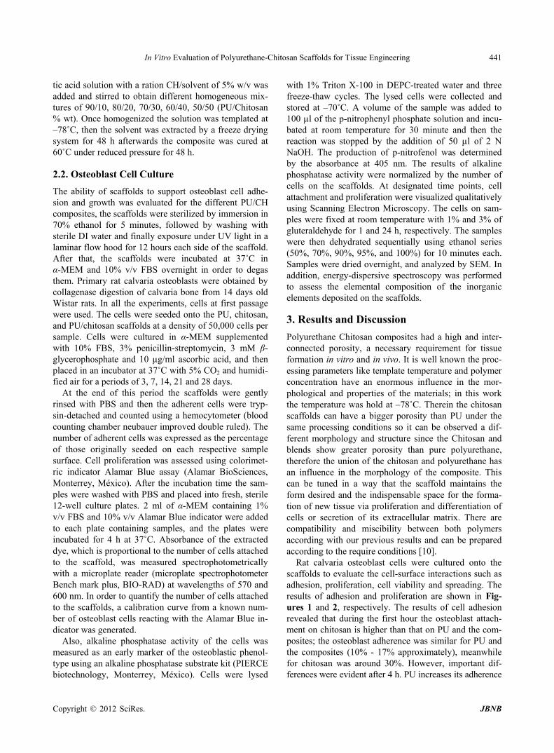
Rat calvaria osteoblast cells were cultured onto the scaffolds to evaluate the cell-surface interactions such as adhesion, proliferation, cell viability and spreading. The results of adhesion and proliferation are shown in Fig-ures 1 and 2, respectively. The results of cell adhesion revealed that during the first hour the osteoblast attach- ment on chitosan is higher than that on PU and the com- posites; the osteoblast adherence was similar for PU and the composites (10% - 17% approximately), meanwhile for chitosan was around 30%. However, important dif- ferences were evident after 4 h. PU increases its adherence
Copyright © 2012 SciRes.��������������������������������������������������������������������������������JBNB
In Vitro Evaluation of Polyurethane-Chitosan Scaffolds for Tissue Engineering 442
Figure 1. Cell adhesion on PU, chitosan, and PU/chitosan composites.
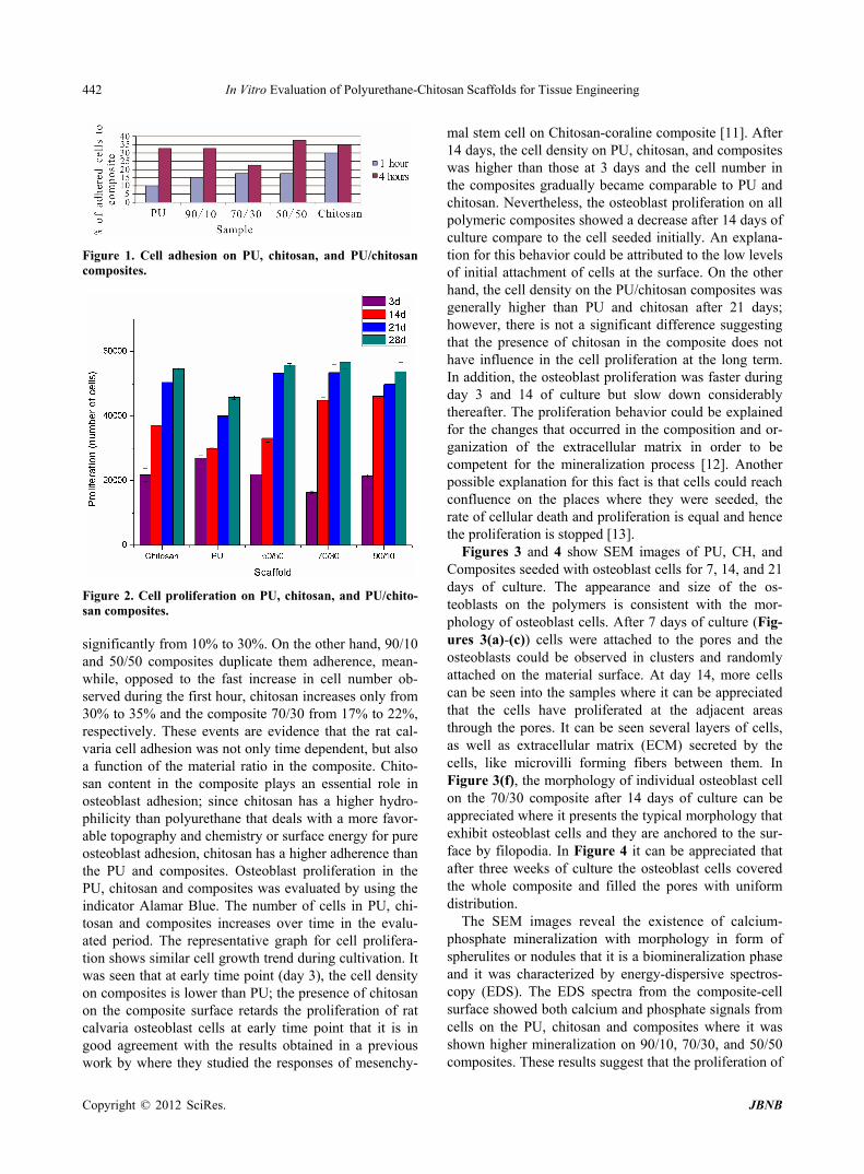
Figure 2. Cell proliferation on PU, chitosan, and PU/chito- san composites. significantly from 10% to 30%. On the other hand, 90/10 and 50/50 composites duplicate them adherence, mean- while, opposed to the fast increase in cell number ob- served during the first hour, chitosan increases only from 30% to 35% and the composite 70/30 from 17% to 22%, respectively. These events are evidence that the rat cal- varia cell adhesion was not only time dependent, but also a function of the material ratio in the composite. Chito- san content in the composite plays an essential role in osteoblast adhesion; since chitosan has a higher hydro- philicity than polyurethane that deals with a more favor- able topography and chemistry or surface energy for pure osteoblast adhesion, chitosan has a higher adherence than the PU and composites. Osteoblast proliferation in the PU, chitosan and composites was evaluated by using the indicator Alamar Blue. The number of cells in PU, chi- tosan and composites increases over time in the evalu- ated period. The representative graph for cell prolifera- tion shows similar cell growth trend during cultivation. It was seen that at early time point (day 3), the cell density on composites is lower than PU; the presence of chitosan on the composite surface retards the proliferation of rat calvaria osteoblast cells at early time point that it is in good agreement with the results obtained in a previous work by where they studied the responses of mesenchy-
mal stem cell on Chitosan-coraline composite [11]. After 14 days, the cell density on PU, chitosan, and composites was higher than those at 3 days and the cell number in the composites gradually became comparable to PU and chitosan. Nevertheless, the osteoblast proliferation on all polymeric composites showed a decrease after 14 days of culture compare to the cell seeded initially. An explana- tion for this behavior could be attributed to the low levels of initial attachment of cells at the surface. On the other hand, the cell density on the PU/chitosan composites was generally higher than PU and chitosan after 21 days; however, there is not a significant difference suggesting that the presence of chitosan in the composite does not have influence in the cell proliferation at the long term. In addition, the osteoblast proliferation was faster during day 3 and 14 of culture but slow down considerably thereafter. The proliferation behavior could be explained for the changes that occurred in the composition and or- ganization of the extracellular matrix in order to be competent for the mineralization process [12]. Another possible explanation for this fact is that cells could reach confluence on the places where they were seeded, the rate of cellular death and proliferation is equal and hence the proliferation is stopped [13].
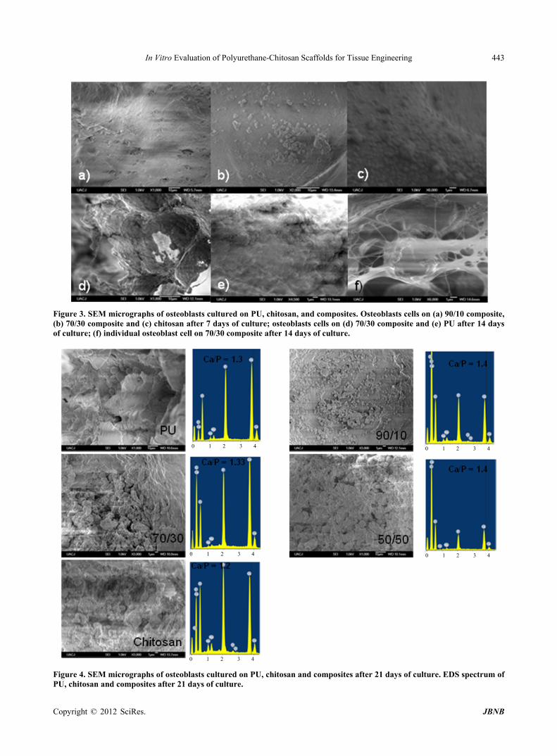
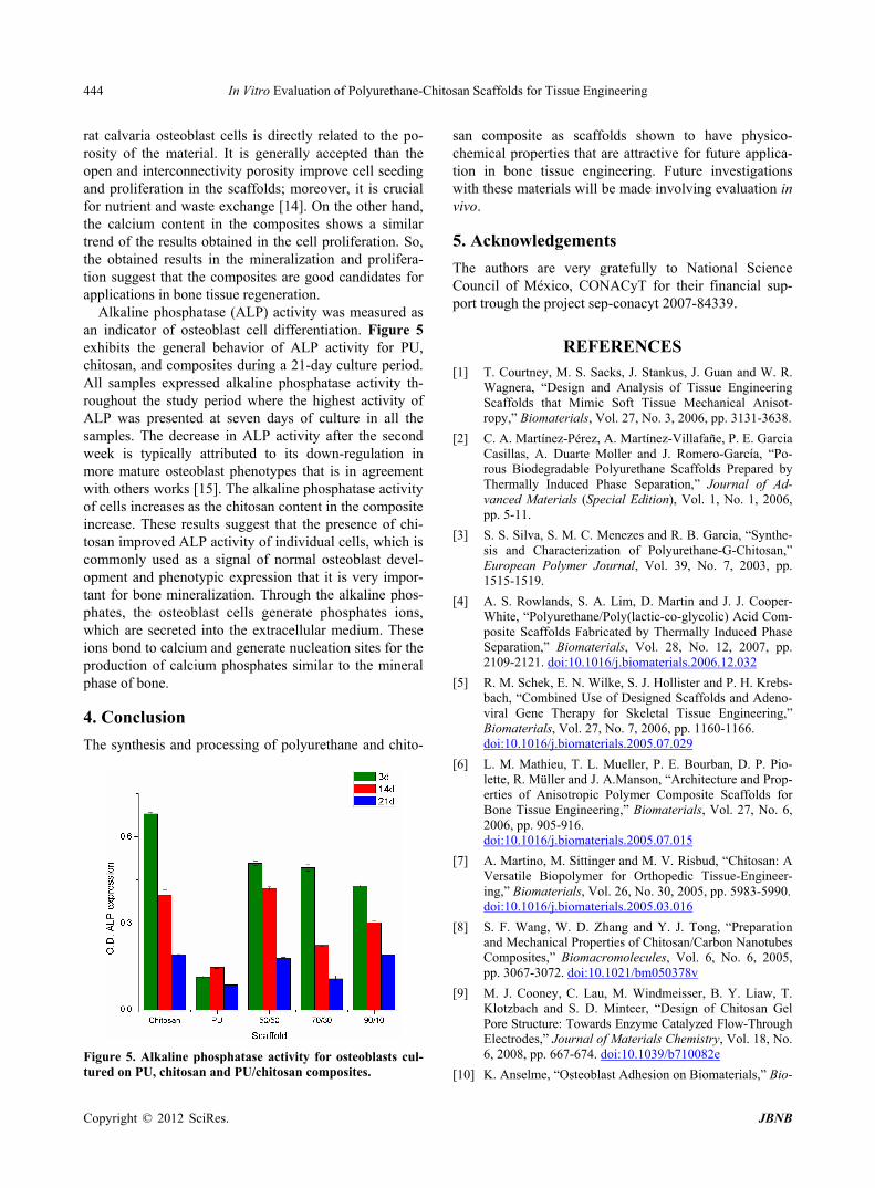
Figures 3 and 4 show SEM images of PU, CH, and Composites seeded with osteoblast cells for 7, 14, and 21 days of culture. The appearance and size of the os- teoblasts on the polymers is consistent with the mor- phology of osteoblast cells. After 7 days of culture (Fig- ures 3(a)-(c)) cells were attached to the pores and the osteoblasts could be observed in clusters and randomly attached on the material surface. At day 14, more cells can be seen into the samples where it can be appreciated that the cells have proliferated at the adjacent areas through the pores. It can be seen several layers of cells, as well as extracellular matrix (ECM) secreted by the cells, like microvilli forming fibers between them. In Figure 3(f), the morphology of individual osteoblast cell on the 70/30 composite after 14 days of culture can be appreciated where it presents the typical morphology that exhibit osteoblast cells and they are anchored to the sur- face by filopodia. In Figure 4 it can be appreciated that after three weeks of culture the osteoblast cells covered the whole composite and filled the pores with uniform distribution.
The SEM images reveal the existence of calcium- phosphate mineralization with morphology in form of spherulites or nodules that it is a biomineralization phase and it was characterized by energy-dispersive spectros- copy (EDS). The EDS spectra from the composite-cell surface showed both calcium and phosphate signals from cells on the PU, chitosan and composites where it was shown higher mineralization on 90/10, 70/30, and 50/50 composites. These results suggest that the proliferation of
Copyright © 2012 SciRes.��������������������������������������������������������������������������������JBNB
In Vitro Evaluation of Polyurethane-Chitosan Scaffolds for Tissue Engineering
Copyright © 2012 SciRes.��������������������������������������������������������������������������������JBNB
443
Figure 3. SEM micrographs of osteoblasts cultured on PU, chitosan, and composites. Osteoblasts cells on (a) 90/10 composite, (b) 70/30 composite and (c) chitosan after 7 days of culture; osteoblasts cells on (d) 70/30 composite and (e) PU after 14 days of culture; (f) individual osteoblast cell on 70/30 composite after 14 days of culture.
0 1 2 3 4
0 1 2 3 4
0 1 2 3 4
0 1 2 3 4
0 1 2 3 4
Figure 4. SEM micrographs of osteoblasts cultured on PU, chitosan and composites after 21 days of culture. EDS spectrum of
U, chitosan and composites after 21 days of culture. P
In Vitro Evaluation of Polyurethane-Chitosan Scaffolds for Tissue Engineering 444
rat calvaria osteoblast cells is directly related to the po-rosity of the material. It is generally accepted than the open and interconnectivity porosity improve cell seeding and proliferation in the scaffolds; moreover, it is crucial for nutrient and waste exchange [14]. On the other hand, the calcium content in the composites shows a similar trend of the results obtained in the cell proliferation. So, the obtained results in the mineralization and prolifera-tion suggest that the composites are good candidates for applications in bone tissue regeneration.
Alkaline phosphatase (ALP) activity was measured as an indicator of osteoblast cell differentiation. Figure 5 exhibits the general behavior of ALP activity for PU, chitosan, and composites during a 21-day culture period. All samples expressed alkaline phosphatase activity th- roughout the study period where the highest activity of ALP was presented at seven days of culture in all the samples. The decrease in ALP activity after the second week is typically attributed to its down-regulation in more mature osteoblast phenotypes that is in agreement with others works [15]. The alkaline phosphatase activity of cells increases as the chitosan content in the composite increase. These results suggest that the presence of chi-tosan improved ALP activity of individual cells, which is commonly used as a signal of normal osteoblast devel-opment and phenotypic expression that it is very impor-tant for bone mineralization. Through the alkaline phos- phates, the osteoblast cells generate phosphates ions, which are secreted into the extracellular medium. These ions bond to calcium and generate nucleation sites for the production of calcium phosphates similar to the mineral phase of bone.
4. Conclusion The synthesis and processing of polyurethane and chito-
Figure 5. Alkaline phosphatase activity for osteoblasts cul-tured on PU, chitosan and PU/chitosan composites.
san composite as scaffolds shown to have physico-chemical properties that are attractive for future applica-tion in bone tissue engineering. Future investigations with these materials will be made involving evaluation in vivo.
5. Acknowledgements The authors are very gratefully to National Science Council of México, CONACyT for their financial sup-port trough the project sep-conacyt 2007-84339.
REFERENCES [1] T. Courtney, M. S. Sacks, J. Stankus, J. Guan and W. R.
Wagnera, “Design and Analysis of Tissue Engineering Scaffolds that Mimic Soft Tissue Mechanical Anisot-ropy,” Biomaterials, Vol. 27, No. 3, 2006, pp. 3131-3638.
[2] C. A. Martínez-Pérez, A. Martínez-Villafañe, P. E. Garcia Casillas, A. Duarte Moller and J. Romero-García, “Po-rous Biodegradable Polyurethane Scaffolds Prepared by Thermally Induced Phase Separation,” Journal of Ad-vanced Materials (Special Edition), Vol. 1, No. 1, 2006, pp. 5-11.
[3] S. S. Silva, S. M. C. Menezes and R. B. Garcia, “Synthe-sis and Characterization of Polyurethane-G-Chitosan,” European Polymer Journal, Vol. 39, No. 7, 2003, pp. 1515-1519.
[4] A. S. Rowlands, S. A. Lim, D. Martin and J. J. Cooper- White, “Polyurethane/Poly(lactic-co-glycolic) Acid Com- posite Scaffolds Fabricated by Thermally Induced Phase Separation,” Biomaterials, Vol. 28, No. 12, 2007, pp. 2109-2121. doi:10.1016/j.biomaterials.2006.12.032
[5] R. M. Schek, E. N. Wilke, S. J. Hollister and P. H. Krebs-bach, “Combined Use of Designed Scaffolds and Adeno-viral Gene Therapy for Skeletal Tissue Engineering,” Biomaterials, Vol. 27, No. 7, 2006, pp. 1160-1166. doi:10.1016/j.biomaterials.2005.07.029
[6] L. M. Mathieu, T. L. Mueller, P. E. Bourban, D. P. Pio-lette, R. Müller and J. A.Manson, “Architecture and Prop-erties of Anisotropic Polymer Composite Scaffolds for Bone Tissue Engineering,” Biomaterials, Vol. 27, No. 6, 2006, pp. 905-916. doi:10.1016/j.biomaterials.2005.07.015
[7] A. Martino, M. Sittinger and M. V. Risbud, “Chitosan: A Versatile Biopolymer for Orthopedic Tissue-Engineer- ing,” Biomaterials, Vol. 26, No. 30, 2005, pp. 5983-5990. doi:10.1016/j.biomaterials.2005.03.016
[8] S. F. Wang, W. D. Zhang and Y. J. Tong, “Preparation and Mechanical Properties of Chitosan/Carbon Nanotubes Composites,” Biomacromolecules, Vol. 6, No. 6, 2005, pp. 3067-3072. doi:10.1021/bm050378v
[9] M. J. Cooney, C. Lau, M. Windmeisser, B. Y. Liaw, T. Klotzbach and S. D. Minteer, “Design of Chitosan Gel Pore Structure: Towards Enzyme Catalyzed Flow-Through Electrodes,” Journal of Materials Chemistry, Vol. 18, No. 6, 2008, pp. 667-674. doi:10.1039/b710082e
[10] K. Anselme, “Osteoblast Adhesion on Biomaterials,” Bio-
Copyright © 2012 SciRes.��������������������������������������������������������������������������������JBNB
In Vitro Evaluation of Polyurethane-Chitosan Scaffolds for Tissue Engineering 445
materials, Vol. 21, No. 7, 2000, pp. 667-681. doi:10.1016/S0142-9612(99)00242-2
[11] M. Gravel, T. Gross, R. Vago and M. Tabrizian, “Re-sponses of Mesenchymal Stem Cell to Chitosan-Coralline Composites Microstructured Using Coralline as Gas Forming Agent,” Biomaterials, Vol. 27, No. 9, 2006, pp. 1899-1906. doi:10.1016/j.biomaterials.2005.10.020
[12] I. Manjubala, I. Ponomarev, I. Wilke and K. D. Jandt, “Growth of Osteoblast-Like Cells on Biomimetic Apa- tite-Coated Chitosan Scaffolds,” Journal of Biomedical Materials Research Part B: Applied Biomaterials, Vol. 84, No. 1, 2008, pp. 7-16. doi:10.1002/jbm.b.30838
[13] A. J. Salgado, M. E. Gomes, A. Chou, O. Coutinho, R. L. Reis and D. W. Hutmacher, “Preliminary Study on the Adhesion and Proliferation of Human Osteoblasts on
Starch-Based Scaffolds,” Materials Science and Engi-neering C, Vol. 20, No. 1-2, 2002, pp. 27-33. doi:10.1016/S0928-4931(02)00009-7
[14] M. Zanetta, N. Quirici, F. Demarosi, M. C. Tanzi, L. Ri-mondini and S. Fare “Ability of Polyurethane Foams to Support Cell Proliferation and the Differentiation of MSCs into Osteoblasts,” Acta Biomaterial, Vol. 5, No. 4, 2009, pp. 1126-1136. doi:10.1016/j.actbio.2008.12.003
[15] O. Tsigkou, J. R. Jones, J. M. Polak and M. M. Stevens, “Differentiation of Fetal Osteoblasts and Formation of Mineralized Bone Nodules by 45S5 Bioglass® Condi-tioned Medium in the Absence of Osteogenic Supple-ments,” Biomaterials, Vol. 30, No. 21, 2009, pp. 3542- 3550. doi:10.1016/j.biomaterials.2009.03.019
Copyright © 2012 SciRes.��������������������������������������������������������������������������������JBNB